Somites and Axon Guidance - DigitalCommons@USU
17
Scanning Microscopy Scanning Microscopy Volume 2 Number 1 Article 40 8-18-1987 Somites and Axon Guidance Somites and Axon Guidance Kathryn W. Tosney University of Michigan Follow this and additional works at: https://digitalcommons.usu.edu/microscopy Part of the Biology Commons Recommended Citation Recommended Citation Tosney, Kathryn W. (1987) "Somites and Axon Guidance," Scanning Microscopy: Vol. 2 : No. 1 , Article 40. Available at: https://digitalcommons.usu.edu/microscopy/vol2/iss1/40 This Article is brought to you for free and open access by the Western Dairy Center at DigitalCommons@USU. It has been accepted for inclusion in Scanning Microscopy by an authorized administrator of DigitalCommons@USU. For more information, please contact [email protected].
-
Upload
khangminh22 -
Category
Documents
-
view
0 -
download
0
Transcript of Somites and Axon Guidance - DigitalCommons@USU
Scanning Microscopy Scanning Microscopy
Volume 2 Number 1 Article 40
8-18-1987
Somites and Axon Guidance Somites and Axon Guidance
Kathryn W. Tosney University of Michigan
Follow this and additional works at: https://digitalcommons.usu.edu/microscopy
Part of the Biology Commons
Recommended Citation Recommended Citation Tosney, Kathryn W. (1987) "Somites and Axon Guidance," Scanning Microscopy: Vol. 2 : No. 1 , Article 40. Available at: https://digitalcommons.usu.edu/microscopy/vol2/iss1/40
This Article is brought to you for free and open access by the Western Dairy Center at DigitalCommons@USU. It has been accepted for inclusion in Scanning Microscopy by an authorized administrator of DigitalCommons@USU. For more information, please contact [email protected].

Scanning Microscopy, Vol. 2, No. 1, 1988 (Pages 427-442) 0891 - 7035/88$3.00+.00 Scanning Microscopy International, Chicago (AMF O'Hare), IL 60666 USA
SOMITES AND AXON GUIDANCE
Kathryn W. Tosney
Department of Biology University of Michigan Ann Arbor, Ml 48109
(Received for publication April 30, 1987, and in revised form August 18, 1987)
Abstract
The somites are arrayed in a repeating pattern along the longitudinal axis of the embryo , as are the developing sensory and sympathetic ganglia and the spinal nerves. This pattern is not a coincidence : the somite imposes a segmental pattern on the cells and axons that invade it. Both neural crest cells and axons prefer the anterior portion of the sclerotome (the ventral part of the somite) for outgrowth . What differences in anterior and posterior sclerotome are responsible? I used scanning electron microscopy to ask whether these populations differed on the tissue level in chick embryos . This study shows that differences in tissue organization are of insufficient magnitude or develop too late to explain the preference of neural crest cells and axons for the anterior half of each sclerotome . For instance, the extracellular matrix does not differ dramatically in density at the dorsal sclerotome boundary and yet neural crest cells promptly enter the anterior sclerotome when they reach this boundary . These cells have access to the cell processes of somitic cells that extend through the matrix. This suggests that neural crest cells could detect important differences in anterior and posterior populations by direct cell contact. Likewise , barriers and consistent differences in cell density , shape or orientation were not obvious before or during initial axon outgrowth . The absence of significant differences in tissue organization suggests that axons and neural crest cells become segmented by responding to diffusible cues , to differences in extracellular material or to the cell surfaces of individual anterior and posterior sclerotome cells.
KEY WORDS: Axon guidance, Cell interactions, Extracellular matrix, Growth cones, Motoneurons, Neural crest, Sclerotome, Segmentation, Somites, Spinal nerves.
*Address for correspondence: Department of Biology , Natural Science Building, University of Michigan, Ann Arbor, Ml 48109. Phone number: (313) 764-9964
427
Introduction
The somites have long been regarded as important to the segmentation of spinal nerves and ganglia. Recently, Keynes and Stern (1984) showed that motor axons do not see somites as physical barriers and grow out between them ; nor do they bundle together in the middle of each somite. The motor axons normally grow out in the anterior portion of each somite. In addition, spinal nerves form in the anterior portion of a somite even when it has been surgically displaced to a posterior position . This elegant work caught the imagination of many researchers because it showed that segmentation must arise from an active interaction (of some undefined sort) between axons and populations of cells within the somite ; the somite is not merely an impenetrable tissue or a passive medium for axonal fasciculation . Soon thereafter other researchers reported that neural crest cells also prefer anterior over posterior somite for migration (Rickmann et al., 1985; Loring and Erickson, 1986, 1987) suggesting that the somite imposed its own segmental pattern on all the cells and axons that invaded it.
In this paper I review the role of somitic tissues in the patterned development of the peripheral nervous system. In particular, I discuss the relationship between somite structure and axonal (and neural crest) guidance, as revealed by a study, reported here, using scanning electron microscopy (SEM).
Segmentation and neuronal specificity
Segmentation has obvious consequences for the development of a normal gross anatomical nerve pattern: the spinal nerves form discrete bundles that are packaged by supporting cells and positioned in proper sequence with the vertebrae. Does segmentation also have consequences for the specific innervation of axonal targets? Until recently , this was an exciting and plausible possibility .
The role of segmentation in the specificity of innervation has been most thoroughly assessed for

Kathryn W. Tosney
the development of limb innervation . In this system, motor axons in the spinal nerves congregate at the limb base, deploy into the proper nerve trunk pathway and innervate individual muscles with a high degree of precision (Tosney and Landmesser, I985a). Each of the limb muscles is derived from a specific subset of somites (Beresford, 1983), and, at least in the hindlimb, the motoneurons that innervate each muscle lie in the spinal cord segments that correspond roughly to the semitic source of each muscle target (Lance-Jones, 1985). Landmesser (1984), Lance-Jones (1985) and Keynes and Stern (1985) proposed that these relationships might be causal. For instance, the migrating myoblasts could be labeled on the basis of their segmental level of origin ; motoneurons with corresponding labels could preferentially associate with matching muscle cells within the limb . When, however, this possibility was investigated experimentally, it became clear that the correlations between somite and motoneuron position are coincidental. Lance-Jones (1987) and Keynes et al. (1987) have shown that muscles are innervated with normal specificity even when they are derived from somites from foreign levels. Therefore, muscles do not receive their identity from the muscle cells that are derived from particular somites; they are apparently patterned by the connective tissue component of the limb which is derived from the unsegmented lateral mesoderm (Chevallier and Kieny, 1982; Lance-Jones, 1987). If targets are labeled segmentally, the labels are not derived from the somites .
The process of spinal nerve segmentation itself appears to be irrelevant to the specificity of hindlimb innervation. When somites are removed before axon outgrowth, limb muscles are innervated with their normal specificity despite the fact that spinal nerve segmentation has been abolished (Tosney, 1986 and in preparation). Therefore, interactions between growth cones and the somites are not essential to the generation of specific neuromuscular connections in the limb.
The hypothesis that neurons and their targets are segmentally labeled does not necessarily imply that neural populations are physically organized in a segmentally repetitive manner within the spinal cord; few if any neural populations are so arranged, including the motoneuron pools that innervate limb muscles in the chick (Landmesser, 1978) or the quail (S. Tyrell, L. Coulter, S. Schroeter and K. Tosney, in preparation) and the interneurons that send ascending or descending projections through the Jumbosacral region of the spinal cord (G. Schlosser and K. Tosney, submitted for publication) . Despite the fact that preganglionic neurons that innervate sympathetic ganglia are distributed in a continuous column in the spinal cord (R. Hume, personal communication), this population may use segmental cues during pathfinding. This population projects in stereotyped directions that are defined by the segmental level in which
428
the axons find themselves, even when they are trans planted to different segments (Yip, 1986) and their projection is independent of the target (Yip, 1987). In addition, these neurons display a subtle segmental preference during reinnervation (Wigston and Sanes, 1982).
It is perhaps more plausible that segmental labels are important to the specific outgrowth of neurons that innervate segmentally arranged targets such as the intercostal and the epaxial (back and intervertebral) muscles. A special relationship between semitic tissues and one of these neural populations has been demonstrated: the myotome provides a chemotactic cue that is essential for the outgrowth of the epaxial motoneurons (Tosney, I987a) . In addition, this population obeys the constraints of the anterior vs posterior somite environment , even under experimental conditions in which it must travel into an adjacent segment to reach a target (Tosney, I987a) . It is, however, not known whether these motoneurons can recognize and prefer to innervate the epaxial muscle in their own segment. This does not appear to be the case in Xenopus in which neurites from the spinal cord are attracted to myotome but do not exhibit a segmental preference when grown in culture (McCaig, 1986). The role of segment-specific labels, if any, in the generation of connections with specific targets remains unclear .
Although segmental cues may or may not be vital for target selection, one observation suggests that the somites may be segmentally labeled and that all the neural populations that emerge in the spinal nerves can detect the segmental difference . Axons that exit the posterior of each spinal cord segment invariably turn toward the anterior and grow through the anterior portion of the corresponding somite; none extend into the anterior portion of the next somite to the posterior, even though this would often be closer (Lim and Keynes, 1986). This suggests that motoneuron growth cones recognize, in some manner, the somite of their own segment. However, the possibility that this segmental matching arises through a match in maturity of the populations, rather than a match in a hypothetical segmental label, has not yet been ruled out.
Segmentation and other patterned aspects of the development of the gross anatomical nerve pattern appear to be caused by more general guidance cues that can channel the outgrowth of axons from a wide variety of populations (see Landmesser, 1984, for review). The gross anatomical nerve pattern appears to be stereotyped because it depends on the stereotyped development of the surrounding embryonic tissues, some of which provide a more permissive environment for axonal advancement. The environment may, as a consequence of its own patterned development, supply substrata or diffusible cues that channel the leading tips of neurites, the growth cones. This proposal is consistent with studies in culture

Semites and axon guidance
showing that growth cones can detect and be guided by differences in the adhesiveness of substrata (Letourneau, 1972), by diffusible chemotactic cues (c.f., Gunderson and Barrett, 1980) and by neuritepromoting substances released by other embryonic populations (c.f., Collins, 1978). During normal development, general guidance of this sort brings axons into regions where they can respond to populationspecific cues and assures that the gross nerve pattern is appropriate for the local embryonic environment (Tosney and Landmesser, 1984). The discovery of the mechanisms underlying formation of the gross anatomical nerve pattern is thus one important goal in understanding how axons normally project to the ir targets in the developing embryo.
What portion of the so mite imposes seomentation?
The somite has become a rather complex structure by the time of axon outgrowth in the chick embryo. The dorsal dermamyotome has formed and given rise to an outer dermatome and an inner myotome; the ventral portion of the somite has lost its epithelial organization and formed sclerotome, a mesenchymal population that will eventually give rise to vertebrae. The relative contribution of these semitic derivatives to segmentation had been ascribed both to the dermamyotome (Detwiler, 1934; Loring and Erickson, 1986, 1987) and the sclerotome (Keynes and Stern , 1984; Rickmann et al., 1985; Tosney and Landmesser, I985b; Stern et al. , 1986). This controversy was resolved with fine embryonic surgery . The pattern of spinal nerve and dorsal root ganglion (DRG) segmentation is normal in the absence of the dermamyotome (Tosney, I987a), but is abolished following total somite deletion (Tosney, 1986 and in preparation, see also Lewis et al., 1981 ). It is, then , the sclerotome that is vital for segmentation of the peripheral nervous system in the trunk of the chick embryo.
Differences in anterior and posterior sclerotome
The anterior and posterior halves of each sclerotome in fact appear to be composed of distinctly different populations of cells (reviewed by Stern and Keynes, 1986). For instance, a distinct intrasegmental boundary, von Ebner's fissure, separates these populations later in development. In addition, under experimental conditions, the populations are immiscible with each other. When somites are surgically reconstructed from like halves, the cells mix, irrespective of their segmental level of origin; cells from unlike halves form a border between them (Stern and Keynes, 1987). Moreover, each half appears to contribute to different vertebrae: the anterior half of one sclerotome and the posterior half of the next rostral sclerotome have been reported to fuse and form a single vertebra (c.f., Beresford, 1983). The distinction between the populations appears to be determined during segmentation
429
(Stern and Keynes, 1986, 1987). Only one difference has been found , however, on
the molecular level: peanut agglutinin binds to posterior but not to anterior sclerotome cells (Stern et al., 1986). Three molecules known to mediate adhesion, fibronectin (Theiry et al., 1982; Rickmann et al. , 1985), laminin (Rickmann et al., 1985; Rogers et al., 1986) and the neural cell adhesion molecule, NCAM (Tosney et al. , 1986) are not differentially distributed between anterior and posterior sclerotomes. Glycosaminoglycans may become more concentrated in each poster ior sclerotome after axon outgrowth (Tosney and Landmesser, I985b) but are equivalently distributed at earlier times (unpublished results of A. J. Larner as cited in Keynes and Stern, 1986). However , the possibility that various proteoglycans or adhesive components (such as C-SAT, JG-22 or herparin sulphate proteoglycan) differ significantly in sclero tome halves has yet to be addressed.
Anterior and posterior sclerotome development viewed with SEM
A possibility that should not be overlooked is that anterior and posterior populations differ in their organization at the tissue level. For instance, neural crest and axon invasion could be facilitated by enlarged cell spaces or a particular distribution of extracellular matrix (ECM) in the anterior sclerotome . It is also possible that axons or neural crest cells are prevented from entering the posterior sclerotome by an intervening barrier such as a blood vessel or heavy deposition of ECM.
To determine whether differences of this sort were present, the development of anterior and posterior sclerotome populations was examined with SEM. Chick embryos were staged using Hamburger and Hamilton (1951) criteria and prefixed 10-15 minutes in 2% glutaraldehyde in 0.1 M cacodylate buffer before cutting them along the desired plane with iridectomy scissors . This gives a very clean surface for observation , since cells damaged by the cut are washed away in subsequent processing (see Tosney, 1982). Fixation was continued overnight, the embryos were postfixed in 2% osmium tetroxide in 0.1 M cacodylate , dehydrated in alcohol, critical point dried and sputter coated with gold-palladium. Specimens were viewed in an ISi DS130 scanning electron microscope at 10 kV.
The sclerotomes were revealed by transecting embryos along the longitudinal axis between the somite and neural tube, through the medial somite, or frontally along the plane of axon outgrowth . These planes of section are illustrated in Schematic Diagram 1, on the next page, for those who are not familiar with the anatomy of avian embryos.
It is obviously important that the anterior (A) and posterior (P) positions of tissues be recognizable in the embryos during examination . When the embryo
Kathryn W. Tosney
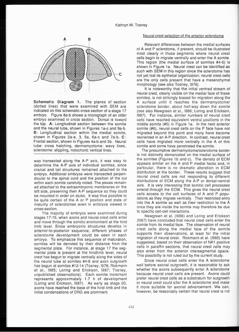
Schematic Diagram 1. The planes of section (dotted lines) that were examined with SEM are indicated on this schematic cross section of a stage 17 embryo. Figure 8a-b shows a micrograph of an older embryo examined in cross section. Dorsal is toward the top. A: Longitudinal section between the somite and the neural tube, shown in Figures 1 a-c and 9a-b. B: Longitudinal section within the medial so mite, shown in Figures 2a-e, 3, Sa, 6a-c and 7a-b. C: Frontal section, shown in Figures 4a-b and Sb. Neural tube: cross hatching, dermamyotome: wavy lines, sclerotome: stippling, notochord: vertical lines.
was transected along the A-P axis, it was easy to determine the A-P axis of individual somites, since cranial and tail structures remained attached to the embryo. Additional embryos were transected perpendicular to the spinal cord and the position of the cut within each somite carefully noted. The pieces remained attached to the extraembryonic membranes on the left side, preserving their A-P sequence so they could be mounted in order on stubs. It was thus possible to be quite certain of the A or P position and state of maturity of sclerotomes even in embryos viewed in cross-section.
The majority of embryos were examined during stages 17-19, when axons and neural crest cells enter and move through the somitic environment at the hind limb level. Since embryonic structures develop in anterior-to-posterior sequence, different phases of sclerotome development could be seen in each embryo. To emphasize this sequence of maturation, somites will be denoted by their distance from the segmental plate. For instance, at stage 17 the segmental plate is present at the hindlimb level, neural crest has begun to migrate ventrally along the sides of the neural tube at somites #4-6 and axon outgrowth has begun at somites #12-14 (Tosney, 1978; Rickmann et al., 1985; Loring and Erickson, 1987; Tosney, unpublished observations) . Each somite increment represents approximately 1.7 h of development (Loring and Erickson, 1987). As early as stage 20, axons have reached the base of the hind limb and the initial condensations of DRG are prominent.
430
Neural crest selection of the anterior sclerotome
Relevant differences between the medial surfaces of A and P sclerotome, if present, should be illustrated most clearly in those segments where neural crest cells begin to migrate ventrally and enter the A so mite. This region (the medial surface of somites #4-6) is shown in Figure 1 a. Neural crest can be identified as such with SEM in this region since the sclerotome has not yet lost its epithelial organization; neural crest cells are the only cells present that have a mesenchymal morphology (see also Tosney, 1978).
It is noteworthy that the initial ventrad stream of neural crest, clearly visible on the medial face of these somites, is not strikingly biased for migration along the A surface until it reaches the dermamyotome/ sclerotome border, about half-way down the somite (see also Newgreen et al., 1986; Loring and Erickson, 1987). For instance, similar numbers of neural crest cells have reached equivalent ventral positions in the middle somite (#5) in Figure 1 a. In the next anterior somite (#6), neural crest cells on the P face have not migrated beyond this point and many have become reoriented in an A-P direction. In contrast, neural crest cells have migrated more ventrally in the A of this somite and some have penetrated the somite.
The presumptive dermamyotome/sclerotome border is not distinctly demarcated on the medial surface of the so mites (Figures 1 b and c). The density of ECM appears similar on the A and P medial faces and, in particular, there is no dramatic alteration in ECM distribution at the border . These results suggest that neural crest cells are not responding to different distributions of ECM along the A-P or dorsal-ventral axis. It is very interesting that somitic cell processes extend through the ECM. This gives the neural crest cells access to the cell surfaces of A and P populations as they migrate ventrally. Their restricted entry into the A somite as well as their restriction to the A once they are inside the somite may therefore be due to specific cell-cell interactions.
Newgreen et al. (1986) and Loring and Erickson (1987) have concluded that neural crest cells enter the somite from its medial face. The observation of neural crest cells along the medial face of the somite supports tl1eir observations, at least for the initial migration of neural crest. Rickmann et al. (1985) have suggested, based on their observation of NK1 positive cells in paraffin sections, that neural crest cells may also enter from the anterior intersegmental space. This possibility is not ruled out by the current study.
Since neural crest cells enter the A sclerotome well before axonal outgrowth, it is reasonable to ask whether the axons subsequently enter A sclerotome because neural crest cells are present. Axons could prefer neural crest cells as a substratum for outgrowth or neural crest could alter the A sclerotome and make it more suitable for axonal advancement. We can, however, be reasonably certain that neural crest is not

Semites and axon guidance
essential for the segmental patterning of axonal outgrowth . For instance, in vitro, neurites from spinal cord explants often grow out in association with NK1 positive (putative neural crest) cells , but destruction of these cells does not prevent the bias for increased neurite outgrowth in the presence of A somite cells (Stern et al., 1986). In addition, the segmentation of spinal nerves is unaltered following substantial deletion of neural crest in vivo (Scott, 1984; Rickmann et al., 1985; Yip, 1986).
Four studies suggest that neural crest cells may, however , be important to the distal progression of spinal nerves. Following what appears to be complete neural crest deletion, Rickmann et al. (1985) report that spinal nerve outgrowth may be slower than normal and Carpenter and Hollyday (1986) state that some spinal nerves do not reach the base of the limb. In both studies, the segmental position of spinal nerves was normal. In addition, Tanaka and Landmesser (1987) and Noakes and Bennett (1987) report that the outgrowing tips of nerves are surrounded by a halo of neural crest cells. However , previous studies have reported normal axonal outgrowth after neural crest
431
deletion (Lewis et al., 1983; Scott, 1984; Yip, 1986). The difficulty of totally removing neural crest cells may explain the contrast in these experimental results. It is apparent that the relationship between neural crest migration and axonal outgrowth should receive further investigation .
The somite itself is not a one-way street that assures the distal outgrowth of spinal nerves. In other systems (c.f., Nardi, 1983; Berlot and Goodman, 1984; Caudy and Bentley, 1986) and in vitro (Gunderson and Park, 1984) axons can grow directionally by moving up a gradient of substratum adhesiveness , a type of guidance termed "haptotax is". Haptotaxis appears to have been disproved as an explanation for distal outgrowth of spinal nerves, since axons grow distally in the absence of the somites (Lewis et al. , 1981; Tosney, 1986 and in preparation) . In addition , although these researchers did not directly address this issue , a study by Keynes and Stern (1984) suggests that the somite does not normally present haptotactic cues that are replaced by other t issues when the somite is absent. When Keynes and Stern showed that spinal nerves grow out only through the A
Figure 1. Segmentat ion of neural crest migration . 1 a. The neural tube was removed to expose the medial surface of somites at the level of neural crest migrat ion, 4 (left) to 6 somites anterior to the segmental plate. Anterior is to the right. Neural crest cells (nc) are clearly visible and are not segmentally distributed until they reach the dermamyotomesclerotome border (arrows), half-way down the somite. ECM is similar in density dorsal (top) and ventral to this border, as shown at higher magnificat ion in 1 b (somite 5) and 1 c (somite 6). Semitic cell processes (e.g., small arrowheads) extend through the ECM. Crest cells (e.g., large arrowhead) enter anterior but not posterior sclerotome. (n: notochord, e: ectoderm). Stage 17. Bars= 50µm (a), 10 µm (b, c).
Kathryn W. Tosney
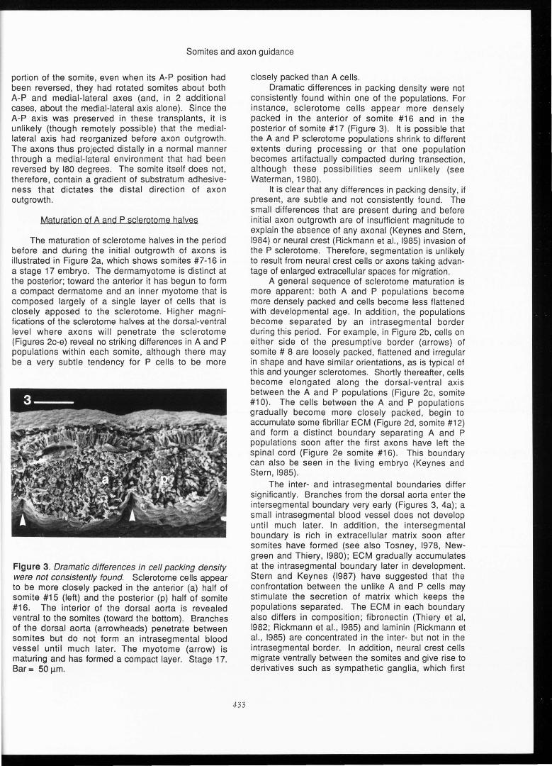
Figure 2. Maturation of A and P sclerotome halves before and during the initial outgrowth of axons. 2a. Populations within the medial somite were exposed by a longitudinal cut adjacent to the neural tube (visible at n) from somite #7 (far left ; before axon outgrowth) through somite #16 (after initial outgrowth) . Dorsal is to the top . d : dermatome . Bar = 100 µm . The ventral regions (through which axons grow) of selected sclerotomes are shown in 2b-e. In each of these figures, the center of the somite is indicated by an arrow. A: anterior . Bar = 10 µm . The figures
432
illustrate a subtle tendency for cells to be more closely packed in the posterior half. A general sequence of maturation is more obvious; cells become more rounded and more closely packed in both populations. An intrasegmental boundary develops during this period. Cells at the midline are initially similar to other sclerotome cells (2b, somite # 8) begin to elongate (2c, so mite #10) accumulate ECM (curved arrow, 2d, somite #12) and gradually become closely packed, forming a distinct plane through the sclerotome (2e, somite #16) . Stage 17.
Semites and axon guidance
portion of the somite, even when its A-P position had been reversed, they had rotated somites about both A-P and medial-lateral axes (and, in 2 additional cases, about the medial-lateral axis alone). Since the A-P axis was preserved in these transplants, it is unlikely (though remotely possible) that the mediallateral axis had reorganized before axon outgrowth . The axons thus projected distally in a normal manner through a medial-lateral environment that had been reversed by 180 degrees. The somite itself does not, therefore, contain a gradient of substratum adhesiveness that dictates the distal direction of axon outgrowth .
Maturation of A and P sclerotome halves
The maturation of sclerotome halves in the period before and during the initial outgrowth of axons is illustrated in Figure 2a, which shows somites #7-16 in a stage 17 embryo. The dermamyotome is distinct at the posterior; toward the anterior it has begun to form a compact dermatome and an inner myotome that is composed largely of a single layer of cells that is closely apposed to the sclerotome . Higher magnifications of the sclerotome halves at the dorsal-ventral level where axons will penetrate the sclerotome (Figures 2c-e) reveal no striking differences in A and P populations within each somite, although there may be a very subtle tendency for P cells to be more
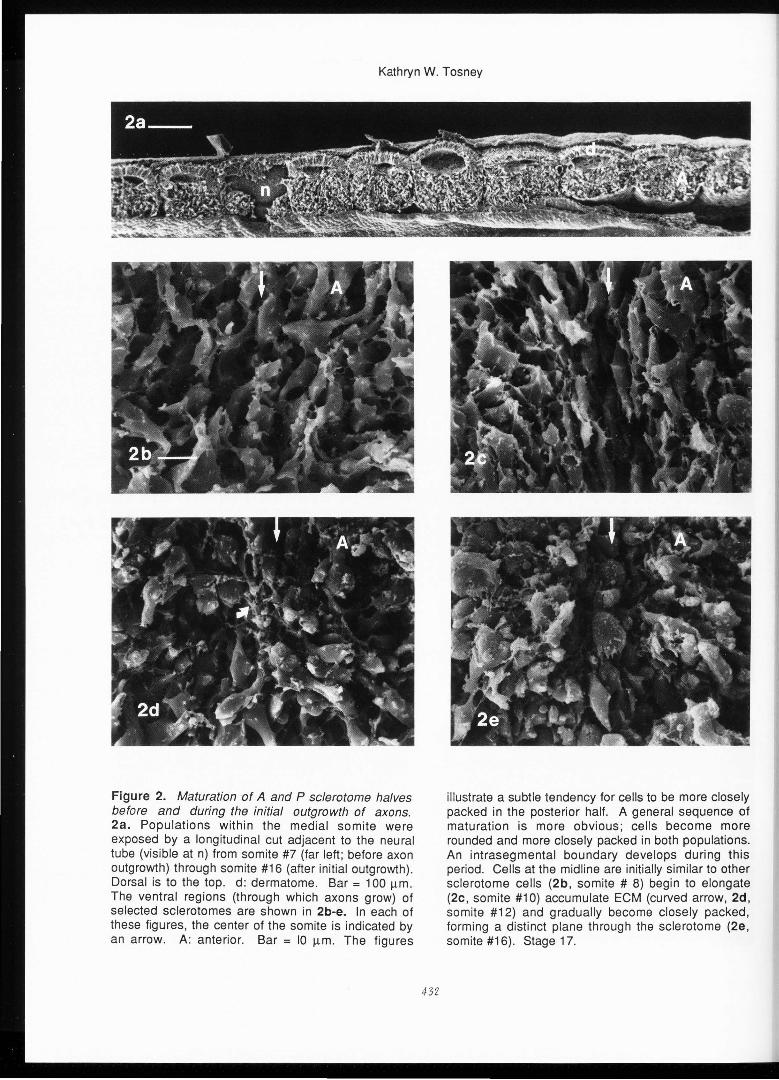
Figure 3. Dramatic differences in cell packing density were not consistently found. Sclerotome cells appear to be more closely packed in the anterior (a) half of somite #15 (left) and the posterior (p) half of somite #16. The interior of the dorsal aorta is revealed ventral to the somites (toward the bottom). Branches of the dorsal aorta (arrowheads) penetrate between somites but do not form an intrasegmental blood vessel until much later. The myotome (arrow) is maturing and has formed a compact layer. Stage 17. Bar= 50 µm.
433
closely packed than A cells. Dramatic differences in packing density were not
consistently found within one of the populations . For instance, sclerotome cells appear more densely packed in the anterior of somite #16 and in the posterior of somite #17 (Figure 3). It is possible that the A and P sclerotome populations shrink to different extents during processing or that one population becomes artifactually compacted during transaction, although these possibilities seem unlikely (see Waterman, 1980).
It is clear that any differences in packing density, if present, are subtle and not consistently found . The small differences that are present during and before initial axon outgrowth are of insufficient magnitude to explain the absence of any axonal (Keynes and Stern, 1984) or neural crest (Rickmann et al., 1985) invasion of the P sclerotome. Therefore, segmentation is unlikely to result from neural crest cells or axons taking advantage of enlarged extracellular spaces for migration.
A general sequence of sclerotome maturation is more apparent : both A and P populations become more densely packed and cells become less flattened with developmental age. In addition, the populations become separated by an intrasegmental border during this period . For example, in Figure 2b, cells on either side of the presumptive border (arrows) of somite # 8 are loosely packed, flattened and irregular in shape and have similar orientations, as is typical of this and younger sclerotomes . Shortly thereafter, cells become elongated along the dorsal-ventral axis between the A and P populations (Figure 2c, somite #10) . The cells between the A and P populations gradually become more closely packed, begin to accumulate some fibrillar ECM (Figure 2d, somite #12) and form a distinct boundary separating A and P populations soon after the first axons have left the spinal cord (Figure 2e somite #16) . This boundary can also be seen in the living embryo (Keynes and Stern, 1985).
The inter- and intrasegmental boundaries differ significantly. Branches from the dorsal aorta enter the intersegmental boundary very early (Figures 3, 4a); a small intrasegmental blood vessel does not develop until much later . In addition, the intersegmental boundary is rich in extracellular matrix soon after somites have formed (see also Tosney, 1978, Newgreen and Thiery, 1980); ECM gradually accumulates at the intrasegmental boundary later in development. Stern and Keynes (1987) have suggested that the confrontation between the unlike A and P cells may stimulate the secretion of matrix which keeps the populations separated. The ECM in each boundary also differs in composition; fibronectin (Thiery et al, 1982; Rickmann et al., 1985) and laminin (Rickmann et al., 1985) are concentrated in the inter- but not in the intrasegmental border. In addition, neural crest cells migrate ventrally between the somites and give rise to derivatives such as sympathetic ganglia, which first
Kathryn W. Tosney
develop intersegmentally (see Loring and Erickson, 1987). Unlike the intersegmental boundary , the intrasegmental boundary does not penetrate the dermamyotome or its derivatives at any stage (see also Stern and Keynes, 1986, 1987).
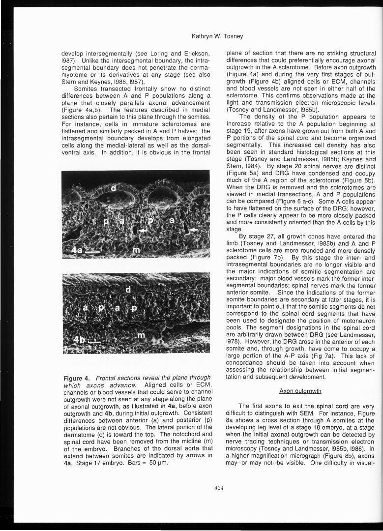
Semites transected frontally show no distinct differences between A and P populat ions along a plane that closely parallels axonal advancement (Figure 4a,b) . The features described in medial sections also pertain to this plane through the somites. For instance, cells in immature sclerotomes are flattened and similarly packed in A and P halves; the intrasegmental boundary develops from elongated cells along the medial-lateral as well as the dorsalventral axis. In addition, it is obvious in the frontal
Figure 4. Frontal sections reveal the plane through which axons advance . Aligned cells or ECM, channels or blood vessels that could serve to channel outgrowth were not seen at any stage along the plane of axonal outgrowth , as illustrated in 4a, before axon outgrowth and 4b , during initial outgrowth. Consistent differences between anterior (a) and posterior (p) populations are not obvious. The lateral portion of the dermatome (d) is toward the top. The notochord and spinal cord have been removed from the midline (m) of the embryo. Branches of the dorsal aorta that extend between somites are indicated by arrows in 4a. Stage 17 embryo. Bars = 50 µm.
434
plane of section that there are no striking structural differences that could preferentially encourage axonal outgrowth in the A sclerotome. Before axon outgrowth (Figure 4a) and during the very first stages of outgrowth (Figure 4b) aligned cells or ECM, channels and blood vessels are not seen in either half of the sclerotome . This confirms observations made at the light and transmission electron microscopic levels (Tosney and Landmesser, I985b).
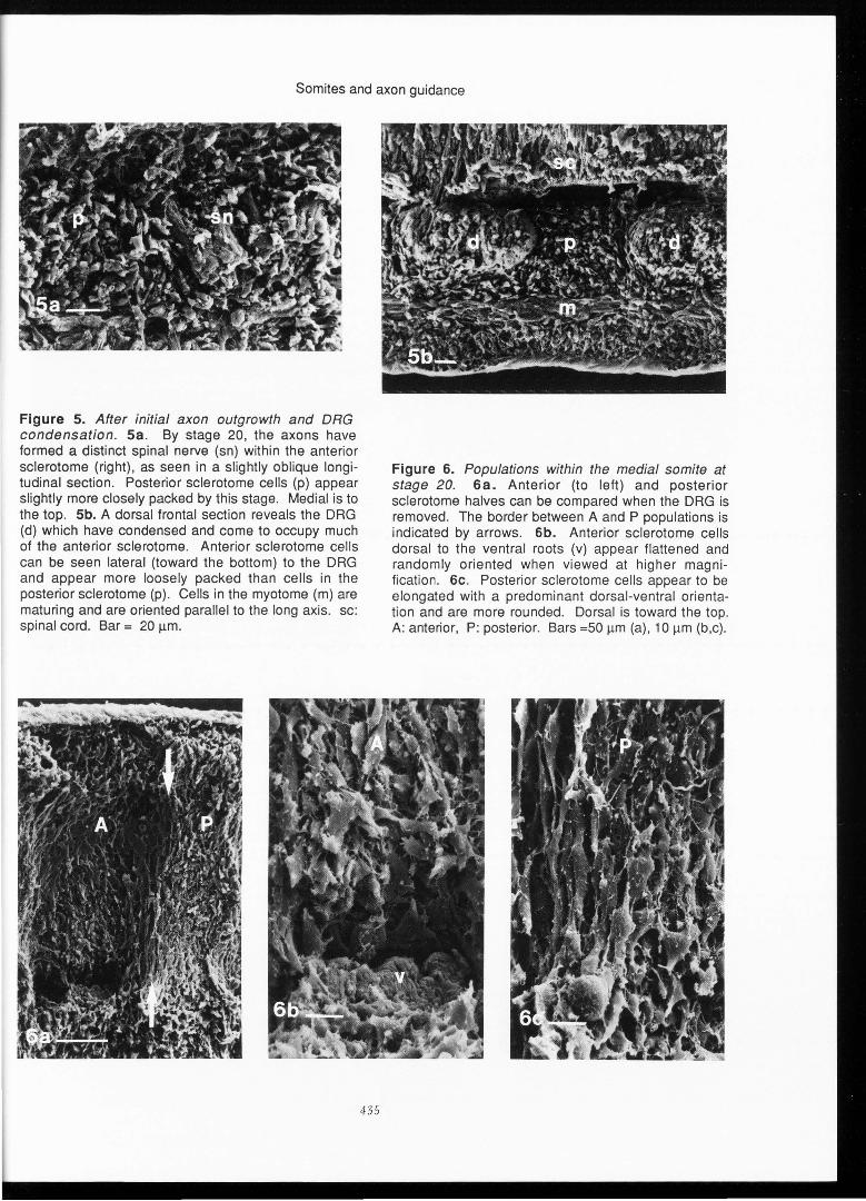
The density of the P population appears to increase relative to the A population beginning at stage 19, after axons have grown out from both A and P portions of the spinal cord and become organized segmentally . This increased cell density has also been seen in standard histological sections at this stage (Tosney and Landmesser , I985b; Keynes and Stern, 1984). By stage 20 spinal nerves are distinct (Figure Sa) and DRG have condensed and occupy much of the A region of the sclerotome (Figure Sb). When the DRG is removed and the sclerotomes are viewed in medial transections , A and P populations can be compared (Figure 6 a-c). Some A cells appear to have flattened on the surface of the DRG; however, the P cells clearly appear to be more closely packed and more consistently oriented than the A cells by this stage.
By stage 27, all growth cones have entered the limb (Tosney and Landmesser, I985b) and A and P sclerotome cells are more rounded and more densely packed (Figure 7b). By this stage the inter- and intrasegmental boundaries are no longer visible and the major indications of semitic segmentation are secondary: major blood vessels mark the former intersegmental boundaries ; spinal nerves mark the former anterior somite . Since the indications of the former somite boundaries are secondary at later stages , it is important to point out that the semitic segments do not correspond to the spinal cord segments that have been used to designate the position of motoneuron pools. The segment designations in the spinal cord are arbitrarily drawn between DRG (see Landmesser, 1978). However, the DRG arose in the anterior of each somite and, through growth, have come to occupy a large portion of the A-P axis (Fig 7a). This lack of concordance should be taken into account when assessing the relationship between initial segmentation and subsequent development.
Axon outgrowth
The first axons to exit the spinal cord are very difficult to distinguish with SEM. For instance , Figure 8a shows a cross section through A somites at the developing leg level of a stage 18 embryo, at a stage when the initial axonal outgrowth can be detected by nerve tracing techniques or transmission electron microscopy (Tosney and Landmesser, I985b, 1986). In a higher magnification micrograph (Figure 8b), axons may--or may not--be visible . One difficulty in visual-
Semites and axon guidance
Figure 5. After initial axon outgrowth and DRG condensation . Sa . By stage 20, the axons have formed a distinct spinal nerve (sn) within the anterior sclerotome (right), as seen in a slightly oblique longitudinal section. Posterior sclerotome cells (p) appear slightly more closely packed by this stage. Medial is to the top . Sb. A dorsal frontal section reveals the DRG (d) which have condensed and come to occupy much of the anterior sclerotome . Anterior sclerotome cells can be seen lateral (toward the bottom) to the DRG and appear more loosely packed than cells in the posterior sclerotome (p). Cells in the myotome (m) are maturing and are oriented parallel to the long axis. sc: spinal cord. Bar = 20 µm.
435
Figure 6. Populations within the medial somite at stage 20. 6a. Anterior (to left) and posterior sclerotome halves can be compared when the DRG is removed. The border between A and P populations is indicated by arrows . 6b. Anterior sclerotome cells dorsal to the ventral roots (v) appear flattened and randomly oriented when viewed at higher magnification . 6c . Posterior sclerotome cells appear to be elongated with a predominant dorsal-ventral orientation and are more rounded. Dorsal is toward the top. A: anterior, P: posterior. Bars =50 µm (a), 10 µm (b,c).

Kathryn W. Tosney
izing these axons is that those at the exposed surface were usually cut after only 10-15 minutes of prefixation and were probably washed away during later procedures. S. Rogers and R. Waterman (personal communication) of the University of New Mexico have undertaken the demanding task of using SEM to document interactions of growth cones with their embryonic environment during outgrowth.
It is clear that the environment immediately exterior to the spinal cord presents no obvious barrier to outgrowth in the posterior of each segment. Careful examination of a number of cross sections like that
Figure 7. Sclerotome populations after axons have entered the limb. 7a. A longitudinal section shows that DRG (e.g., arrowheads) have come to occupy a large portion of the A-P axis by stage 27. (L: limb bud.; anterior is to the right.) 7b. Segmental boundaries are no longer distinct by this stage. lntersegmental blood vessels (arrows), DRG (d) and spinal nerves (sn) are visible. The A and P sclerotome cells have become more rounded as chondrogenesis begins ; differences between A and P populations are not obvious. Anterior is to the left. Bars = 500 µm (a), 50 µm (b).
436
shown in Figure 8a-b and a number of frontal sections like that shown in Figure 5b did not reveal a consistent pattern of blood vessels or other structures that could prevent axons from entering the P sclerotome.
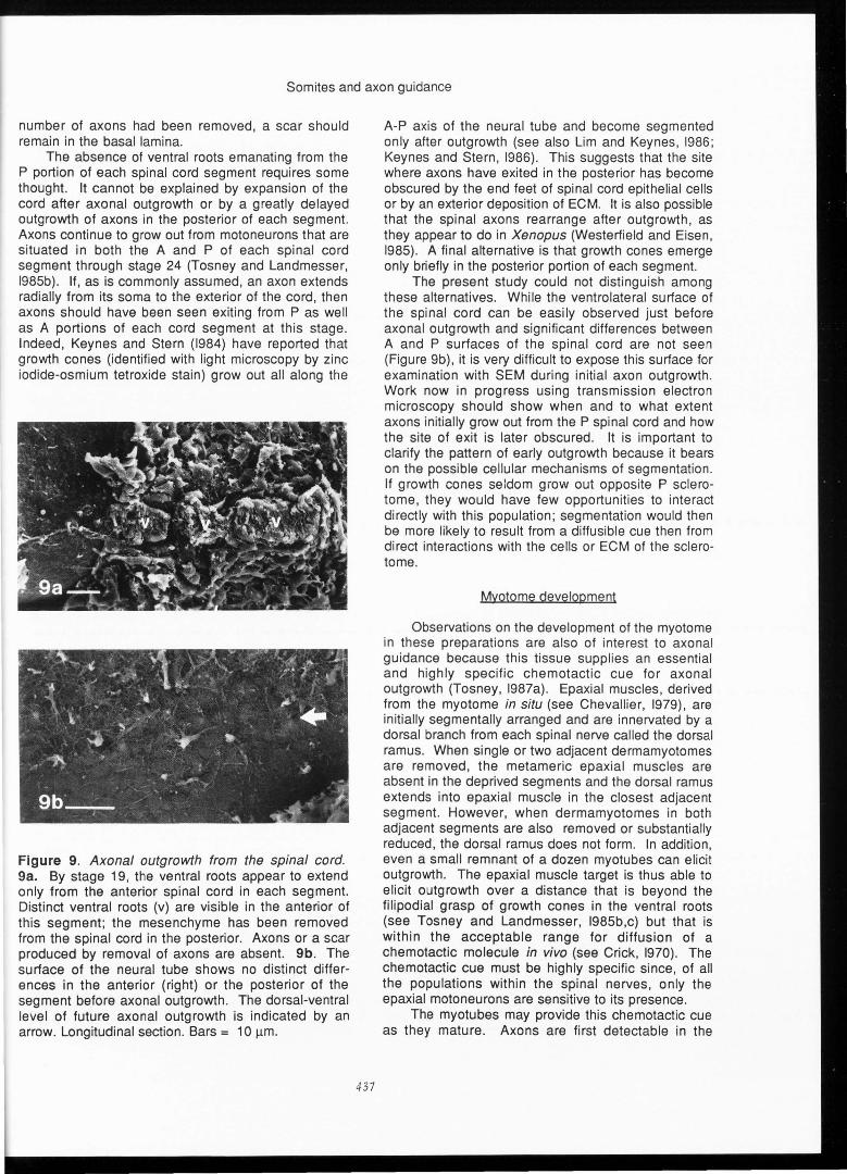
It was surprising to find that , from stage 19 on, the ventral roots extend only from the A spinal cord in each segment. For instance, in Figure 9a, distinct ventral roots can be seen in the A of the segment; the sclerotome has been removed from the P of the segment, revealing the spinal cord. Axons are not joining the ventral roots from this region. Their absence is apparently not an artifact of dissection, since if a large
Figure 8. Initial axon outgrowth. Ba. Cross section through an anterior somite at the hindlimb level of a stage 18 embryo. The area between the white lines is shown at higher magnification in 8b. The first axons to exit the spinal cord are difficult to distinguish with SEM; two may be present in this micrograph (arrow). Note that sclerotome cells are not oriented parallel to the direction of axonal extension. The cells of the myotome (m) have elongated parallel to the long axis of the embryo and are cut in cross section. (sc: spinal cord). Bars= 100 µm (a), 10 µm (b).
Somites and axon guidance
number of axons had been removed, a scar should remain in the basal lamina.
The absence of ventral roots emanating from the P portion of each spinal cord segment requires some thought. It cannot be explained by expansion of the cord after axonal outgrowth or by a greatly delayed outgrowth of axons in the posterior of each segment . Axons continue to grow out from motoneurons that are situated in both the A and P of each spinal cord segment through stage 24 (Tosney and Landmesser , I985b). If, as is commonly assumed , an axon extends radially from its soma to the exterior of the cord, then axons should have been seen exiting from P as well as A portions of each cord segmen t at this stage . Indeed, Keynes and Stern (1984) have reported that growth cones (identified with light microscopy by zinc iodide-osmium tetroxide stain) grow out all along the
Figure 9. Axonal outgrowth from the spinal cord. 9a. By stage 19, the ventral roots appear to extend only from the anterior spinal cord in each segment. Distinct ventral roots (v) are visible in the anterior of this segment ; the mesenchyme has been removed from the spinal cord in the posterior . Axons or a scar produced by removal of axons are absent. 9b. The surface of the neural tube shows no distinct differences in the anterior (right) or the posterior of the segment before axonal outgrowth . The dorsal-ventral level of future axonal outgrowth is indicated by an arrow. Longitudinal section. Bars = 10 µm.
437
A-P axis of the neural tube and become segmented only after outgrowth (see also Lim and Keynes, 1986; Keynes and Stern , 1986). This suggests that the site where axons have exited in the posterior has become obscured by the end feet of spinal cord epithelial cells or by an exterior deposition of ECM. It is also possible that the spinal axons rearrange after outgrowth, as they appear to do in Xenopus (Westerfield and Eisen, 1985). A final alternative is that growth cones emerge only briefly in the posterior portion of each segment.
The present study could not dist inguish among these alternatives. While the ventrolateral surface of the spinal cord can be easily observed just before axonal outgrowth and significant differences between A and P surfaces of the spinal cord are not seen (Figure 9b), it is very difficult to expose this surface for examination with SEM during initial axon outgrowth . Work now in progress using transm ission electron microscopy should show when and to what extent axons initially grow out from the P spinal cord and how the site of exit is later obscured. It is important to clarify the pattern of early outgrowth because it bears on the possible cellular mechanisms of segmentation . If growth cones seldom grow out opposite P sclerotome , they would have few opportun ities to interact directly with this population ; segmentat ion would then be more likely to result from a diffusible cue then from direct interactions with the cells or ECM of the sclerotome.
Myotome development
Observations on the development of the myotome in these prepa rations are also of interest to axonal guidance because this tissue supplies an essential and highly specific chemotactic cue for axonal outgrowth (Tosney , I987a). Epaxial muscles , derived from the myotome in situ (see Chevall ier , 1979), are initially segmentally arranged and are innervated by a dorsal branch from each spinal nerve called the dorsal ramus. When single or two adjacent dermamyotomes are removed , the metamer ic epaxial muscles are absent in the deprived segments and the dorsal ramus extends into epax ial muscle in the closest adjacent segme nt. However , when dermamyotomes in both adjacent segments are also removed or substant ially reduced, the dorsal ramus does not form. In addition, even a small remnant of a dozen myotubes can elicit outgrowth. The epaxial muscle target is thus able to elicit outgrowth over a distance that is beyond the filipodial grasp of growth cones in the ventral roots (see Tosney and Landmesser, I985b,c) but that is within the acceptable range for diffusion of a chemotactic molecule in vivo (see Crick , 1970). The chemotactic cue must be highly specific since , of all the populations within the spinal nerves, only the epaxial motoneurons are sensitive to its presence.
The myotubes may provide this chemotactic cue as they mature . Axons are first detectable in the
Kathryn W. Tosney
dorsal ramus soon after the first myoblasts form (Tosney, unpublished observations) . At this stage, the myoblasts display immunoreactivity to the neural cell adhesion molecule (Tosney et al., 1986) and to myosin (Holtzer et al., 1957), thick and thin filaments are visible with electron microscopy (Pryzylbylski and Blumberg, 1966) and many myoblasts have become postmitotic (Langman and Nelson, 1968; Summerbell et al., 1986). In Xenopus, an in vitro study has also shown that the developing myotubes (but not their precursors in the segmental plate) provide a chemotactic cue for axonal outgrowth from the spinal cord (McCaig, 1986).
The current study confirms that the myotome is maturing at the levels where axonal outgrowth begins. For instance, the myotome has formed a compact layer beneath the dermatomes in somites #15-16 in Figure 3. When the myotome is cut in cross section during the early phase of axon outgrowth (Figure 8b) its cells are found to be arranged parallel to the long axis of the embryo, presaging the orientation of muscle fibers that develop in the epaxial muscles. By stage 20, elongated cells in the myotome may represent myoblasts that have begun to fuse (Figure Sb).
Possible mechanisms of segmentation
A number of possible mechanisms could explain the segmentation of axonal outgrowth by the sclerotome tissues . 1) Growth cones may respond to structural features of A and P sclerotome halves such as differences in the density of cells or ECM, the orientation of cells or ECM, or physical barriers. The study reported here shows that this mechanism is unlikely to be of first importance to the development of segmentation. 2) Diffusible cues from A sclerotome may attract growth cones or preferentially enhance their rate of advance. 3) A diffusible substance from P sclerotome may repel growth cones or slow their rate of advance . 4) Growth cones may prefer A over P sclerotome cells or the ECM in the A sclerotome as a substratum for advancement. 5) Growth cones may cease extending processes (become "contact paralyzed") when they contact P cells. It is worthwhile to point out that neural crest cells may or may not respond to the same differences that guide axons.
Despite the fact that fasciculation alone is not sufficient for the development of segmental patterns (Tosney, 1986 and in preparation), the search for the mechanisms that produce segmentation is complicated by the documented preference (Tosney and Landmesser, I985b) of growth cones for other axons as a substratum during outgrowth . Axons that exit opposite the P of each sclerotome have been reported to enter A sclerotome by fasciculating with axons that have entered the A compartment (Keynes and Stern, 1984). Such growth cones may not be within filipodial reach of A sclerotome cells; if so, their choice of substratum would be restricted to P sclerotome and other neurites. This complication means that the pertinent
438
navigational cues may be more subtle then expected . Any factor that slows growth cone advancement into P sclerotome (e.g., a diffusible substance , substratum preference or contact paralysis) may tend to enhance the growth cones' choice of neurites as a substratum. In effect, this would augment the choice of those growth cones that had entered the A sclerotome.
There is some positive evidence that a diffusible cue is important to segmentation . Anterior somite enhances outgrowth from spinal cord explants in culture to a greater degree than does P somite, although both tissues enhance outgrowth to some degree (Stern et al., 1986). The observation that axons extend first from the A portion of each segment (Keynes and Stern, 1984) is also consistent with a diffusible cue. However, this observation could also be explained by the gradient of maturity along the A-P axis. Axons may grow out from the A of each spinal cord segment simply because it is developmentally older, rather than because outgrowth is enhanced by a diffusible cue from A sclerotome. On the other hand, if axons in the A of 2 or more consecutive segments extend before those in the P of the same segments, it would lend support to the hypothesis that a diffusible cue influences the pattern of outgrowth. We do not yet know whether initial outgrowth is punctate or smoothly graded along the A-P axis.
At first glance, an attractive substance from A sclerotome appears unlikely, since the somite is not essential for axonal outgrowth per se. Axons (and neural crest cells) grow out when the somites have been removed but their segmental patterning is abolished (Lewis et al., 1981; Phelan and Hollyday, 1986; Tosney, 1986 and in preparation) and DRG condensations tend to be larger than normal (Tosney, in preparation) . These results suggest that P sclerotome normally inhibits neural crest and axon outgrowth, an inhibition that is removed when somites are deleted (see also Keynes and Stern , 1986). However, it is possible that an earlier interaction with unsegmented plate may be enough to elicit outgrowth when somites are removed. Another possibility is that a variety of tissues produce substances that enhance axon outgrowth: those tissues that remained following somite deletion may have been sufficient to elicit outgrowth. If this is the case, then outgrowth may normally be patterned because A and P sclerotome produce such a substance to different degrees .
In support of the substratum preference hypothesis, it is clear that growth cones can distinguish differences in the surfaces of A and P sclerotome cells. In culture, growth cones from spinal cord explants are more commonly found on the surface of A then on the surface of P somite cells (Stern et al., 1986). Preliminary results in which interactions among identified populations were videotaped in culture (Tosney, I987b and work in progress) show that A and P sclerotome cells do, in fact, elicit different behavior from motoneuron growth cones . Motoneurons are labeled by

Somites and axon guidance
injecting spinal nerves with Oil, a non-toxic fluorescent dye (Honig and Hume, 1986) 5-8 h before explanting small pieces of the ventrolateral spinal cord . Axons that are bright red after outgrowth are unequivocally motoneuronal. A or P sclerotome cells are labeled with rhodamine or flouroscein to distinguish them from fibroblastic spinal cord cells. In these cultures, motoneuron growth cones extend onto and over the surface of A cells but remain on the laminin substratum after contacting P cells. Therefore , motoneurons appear to prefer A sclerotome cells as a substratum when grown on laminin. Spinal cord neurites cultured on polylysine have been reported to grow onto the surface of both A and P cells (Stern et al., 1986). Given these observations, one can posit a hierarchy of adhesive preferences, in which growth cones prefer, in order, anterior sclerotome , lam inin, posterior sclerotome and polylysine substrata. The further observation that motoneurons continue to extend processes when in contact with P cells (Tosney , I987b) rules out the possibility that P sclerotome cells directly inhibit the motile capabilities of these growth cones ; contact paralysis does not contribute to segmentation . These preliminary results suggest that substratum preference could , at least in part, be responsible for the segmental pattern of axonal outgrowth.
Conclusion
A major advantage of SEM is that one can view intact tissues and determine their organization without resorting to tedious reconstruction techniques . This advantage was paramount in determining whether the tissue-level organization of anterior and posterior sclerotomes halves could be important for the segmentation of neural crest migration and axonal outgrowth .
Neural crest cells that migrate ventrally along the medial face of the somite become segmented as they penetrate anterior but not posterior sclerotome. The distributions of extracellular matrix in the anterior and posterior of each somite were similar at the border between dermamyotome and sclerotome, where neural crest cells first penetrate the somite. The extracellular matrix may differ in composition in the two compartments . Since the filipodia of sclerotome cells extend through the extracellular matrix it is also possible that specific cell interactions guide neural crest cells into the anterior sclerotome.
Later in development, before and during axon invasion, the anterior and posterior sclerotome cells are remarkably similar in shape, orientation and packing density and present no obvious channels or barriers for axonal outgrowth. Differences are of insufficient magnitude or develop too late to satisfactorily explain the failure of axons to extend through posterior sclerotome . The absence of all but the most subtle differences in tissue organization suggests that the
439
important differences between anterior and posterior sclerotome populations are diffusible substances or molecular substrata to be found in the extracellular matrix or on the surface of sclerotome cells.
Acknowledgements
This work was supported by National Institute of Health grant NS-21308. I thank Ann Gulley and David Bay for photographic assistance and Robert Oakley and Gerhard Schlosser for reading the manuscript. Preliminary results on cells in culture were obtained in the laboratory of Dr. Jonathan Raper at the MaxPlanck -lnstitut tor Entwicklungsbiologie, TUbignen , West Germany. I thank him, Dr. F. Bonhoeffer and Dr. S. Chang for their kind assistance .
References
Beresford B (1983) Brachia! muscles in the chick embryo: the fate of individual somites J. Embryo/. Exp. Morph.77, 99-116.
Berlot J, Goodman CS (1984) Guidance of peripheral pioneer neurons in the grasshopper: Adhesive heirarchy of epithelial and neural substrates. Science 233 , 493-496.
Carpenter EM, Hollyday M (1986) Defective innervation of chick limbs in the absence of presumptive Schwann cells . Soc. Neurosci. Abs. 12, 1210.
Caudy M, Bentley D (1986) Pioneer growth cone morphologies reveal proximal increases in substrate affinity within leg segments of grasshopper embryos. J. Neurosci. 6, 364-379.
Chevallier A (1979) Role of the somitic mesoderm in the development of the thorax in bird embryos . II. Origin of thoracic and appendicular musculature . J. Embryo/. Exp. Morph. 49, 73-88.
Chevallier A, Kieny M (1982) On the role of the connective tissue in the patterning of the chick limb musculature. Wilhelm Roux 's Arch. 191, 277-280.
Collins F (1978) Axon initiation of ciliary neurons in vivo. Develop. Biol. 65, 50-57.
Crick F (1970) Diffusion in embryogenesis . Nature 31 , 420-422.
Dalgleish AE (1985) A study of the development of thoracic vertebrae in the mouse assisted by autoradiography. Acta Anat. 122, 91-98.
Detwiler SR (1934) An experimental study of spinal nerve segmentation in Ablystoma with reference to the plurisegmental contribution to the brachia! plexus. J. Exp. Zoo/. 67 , 395-441.
Gunderson RW, Barrett JN (1980) Characterization of the turning response with dorsal root neurites toward nerve growth factor . J. Cell Biol. 87, 546-554.
Gunderson RW, Park KHC (1984) The effects of conditioned media on spinal neurites: Subtrateassociated changes in neurite direction and adherence. Develop. Biol. 104, 18-27.
Hamburger V, Hamilton HL (1951) A series of
Kathryn W . Tosney
normal stages in the development of the chick embryo. J. Morphol . 88 : 49-82.
Holtzer H, Marshall JM , Finck H (1957) An analysis of myogenesis by the use of fluorescent antimyos in. J. Biophysic and Biochem. Cytol. 3, 709.
Honig MG, Hume R (1986) Fluorescent carbocyanine dyes allow living neurons of identified origin to be studied in long-term cultures . J. Cell Biol . 103 , 171-187 .
Keynes RJ, Stern CD (1984) Segmentation in the vertebrate nervous system. Nature 310, 786-789 .
Keynes RJ, Stern CD (1985) Segmentation and neural development in vertebrates . Trends in Neurosci . 8 , 220-223.
Keynes RJ, Stern CD (1986) Semites and neural development. IN: Semites in Developing Embryos. pp 289-299 . (eds) R. Bellairs , DA Ede , JW Lash NATA ASI series , A, Life Sciences, vol. 118, Plenum Publishing Co, NY.
Keynes RJ, Stir ling RV, Stern CD, Summerbell D (1987) The specificity of motor innervation of the chick wing does not depend upon the segmental origin of muscles. Development, 99 , 565-575.
Lance-Jones CJ (1985) The semitic origin of limb muscles in the chick embryo : a correlation with motor innervation. Soc. Neurosci . Abs. 11,288 .2
Lance-Jones CJ (1987) Motoneuron axon guidance: The development of specific projections to two muscles in the embryon ic chick limb. Brain, Behavior and Evolution In Press.
Lander AD, Fujii DK, Gospodarowicz D, Reichardt LF (1982). Character ization of a factor that promotes neurite outgrowth : evidence link ing act ivity to a heparin sulfate proteoglycan . J. Cell Biol . 94 , 574-585.
Landmesser LT (1978) The development of motor projection patterns in the chick hindlimb. J. Physiol . 284 , 391-414.
Landmesser LT (1984) The development of specific motor pathways in the chick embryo . Trends in Neurosci . 7, 336-339 .
Langman J , Nelson GR (1968) A radioautographic study of the development of the somite in the chick embryo . J. Embryo/. Exp. Morphol . 19 , 217-226.
Letourneau PC (1972) Cell-to-substratum adhesion and guidance of axona! elongation . Develop . Biol. 44, 92-101 .
Lewis J, Chevallier A, Kieny M, and Wolpert L (1981) Muscle nerve branches do not develop in chick wings devoid of muscle. J. Embryo/. exp. Morph. 64, 211-232.
Lewis J, AI-Ghaith L, Swanson G, Khan A (1983) The control of axon outgrowth in the developing chick wing. IN: Limb Development and Regeneration Part A, pp 195-205, Alan R Liss, Inc, NY, NY
Lim TM , Keynes RJ (1986) Segmentation in the neural tube of the chick embryo . Soc. Neurosci . Abs. 12 , 316.
Loring JF , Erickson CA (1986) Patterns of neural
440
crest migration in the chick embryo. Soc . Neurosci. Abs. 12, 1209.
Loring JF , Erickson CA (1987) Neural crest cell migratory pathways in the chick embryo . Develop . Biol . 121 , 230-236 .
Mccaig CD (1986) Myoblasts and myoblastconditioned medium attract the earliest spinal neurites from frog embryos . J. Physiol . 375 , 39-54 .
Nardi, JB (1983) Neural pathfinding in the developing wings of Manduca sexta. Develop. Biol . 95 , 163-174.
Newgreen D, Schee M, Kastner V (1986) Morphogenesis of sc lerotome and neural crest in avian embryos . In vivo and in vitro studies on the role of notochordal extracellular material.Ce// Tiss. Res. 244 , 299-313.
Newgreen D, Thiery JP (1980) Fibronectin in early avian embryos : Synthesis and distribution along the migration pathways of neural crest cells. Cell Tiss. Res. 211 , 269-291 .
Noakes PG, Bennett , MR (1987) . Growth of axons into developing muscles of the chick forelimb is preceded by cells that stain with Schwann cell antibodies. J. Comp. Neurol . 259, 330-347 .
Patterson PH (1985) On the role of proteases, their inhibitors and the extracellular matrix in promoting neurite outgrowth. J. Physiol . Paris 80, 207-211 .
Phelan K, Hollyday M (1986) Pathway selection in muscleless chick limbs. Soc. Neurosci. Abs. 12 , 1210.
Pryzybylski RJ, Blumberg JM (1966) Ultrastructural aspects of myogenesis in the chick . Lab. Invest . 15 , 836-863 .
Rickmann M, Faucet JW , Keynes RJ (1985) The migration of neural crest cells and the growth of motor axons through the rostral half of the chick somite. J. Embryo/. Exp. Morphol . 90, 437-455.
Rogers SL, Edso KJ, Letourneau PC, Meloan SC (1986) Distribution of laminin in the developing peripheral nervous system of the chick . Develop . Biol. 113 , 429-435.
Scott SA (1984) The effects of neural crest deletions on the developmen t of sensory innervation patterns in embryonic chick hind limb. J. Physiol . 352 , 385-404 .
Stern CD, Sisodiya SM, Keynes RJ (1986) Interactions between neurites and somite cells : inhibition and stimulation of nerve growth in the chick embryo . J. Embryo/. Exp. Morphol. 91, 209-226 .
Stern CD, Keynes RJ (1986) Cell lineage and the formation and maintenance of half so mites . IN: Semites in Developing Embryos. pp 147-159 . (eds) R Bellairs, DA Ede, JW Lash. NATO ASI series A, Life Sciences vol 118, Plenum publishing Co, NY.
Stern CD, Keynes RJ (1987) Interactions between somite cells: the formation and maintenance of segment boundaries in the chick embryo. Development 99, 261-272 .
Summerbell D, Coetzee H, Hornbruch A (1986) A unique population of non-dividing cells in the somites .

Semites and axon guidance
IN: Semites in Developing Embryos. pp 105-117. (eds) R Bellairs, DA Ede, JW Lash. NATO ASI series A, Life Sciences vol 118., Plenum publishing Co, NY.
Tanaka H, Landmesser L (1987) Potential axonal guidance features in the chick limb revealed by antibodies to neural crest and extracellular matrix molecules. Develop. Biol. In press.
Thiery JP, Duband JL, Delouvee A (1982) Pathways and mechanisms of avian trunk neural crest cell migration and localization. Develop. Biol. 93, 324-434.
Tosney KW (1978) The early migration of neural crest cells in the trunk region of the avian embryo : an electron microscopic study. Develop. Biol . 62, 317-333.
Tosney KW (1982) The segregation and early migration of cranial neural crest cells in the avian embryo . Develop. Biol. 89, 13-24.
Tosney KW (1986) Proximal tissues and patterned neurite outgrowth into the chick hindlimb. Soc. Neurosci. Abs. 12, 121 o.
Tosney KW (I987a) Proximal tissues and patterned neurite outgrowth at the lumbosacral level of the chick embryo: Deletion of the dermamyotome. Develop . Biol. In press.
Tosney KW (I987b) Growth cone interactions with anterior and posterior sclerotome. Soc. Neurosci. Abs. 13, In press.
Tosney KW. Landmesser LT (1983) Identification of cultured motoneurons by retrograde transport of HRP. Soc. Neurosci. Abs, 8, 42.
Tosney KW, Landmesser LT (1984) Pattern and specificity of axonal outgrowth following varying degrees of chick limb bud ablation . J. Neurosci . 4, 2158-2527
Tosney KW, Landmesser LT (I985a) Specificity of motoneuron growth cone outgrowth in the chick embryo . J. Neurosci. 5, 2336-2344.
Tosney KW, Landmesser LT (I985b) Development of the major pathways for neurite outgrowth in the chick hindlimb. Develop . Biol. 109, 193-214.
Tosney KW, Landmesser LT (I985c) Growth cone morphology and trajectory in the lumbosacral region of the chick embryo. J. Neurosci . 5, 2345-2358.
Tosney KW, Landmesser LT (1986) Neu rites and growth cones in the chick embryo . Enhanced tissue preservation and visualization of HAP-labeled subpopulations in serial 25µm plastic sections cut on a rotary microtome. J. Histochem. Cytochem. 34, 953-957.
Tosney KW, Watanabe M, Landmesser L, Rutishauser U (1986) The distribution of NCAM in the chick hindlimb during axon outgrowth and synaptogenesis . Develop. Biol. 114, 437-452 .
Verbout AJ (1976). Critical review of the "Neugliederung" concept in relation to the development of the vertebral column. Acta Biotheoretica 25, 219-258.
Waterman RE (1980) Preparation of embryonic
44 1
tissues for SEM. Scanning Electron Microsc. 1980 II, 21-44 .
Westerfield M, Eisen JS (1985) The growth of motor axons in the spinal cord of Xenopus embryos . Develop. Biol. 109, 96-101 .
Wigston D, Sanes J (1982) Selective reinnervation of adult mammalian muscles by axons from different segmental levels. Nature (Lond.) 299, 464-467 .
Yip J (1986) Axonal guidance in avian sympathetic preganglionic neurons. Soc. Neurosci . Abs. 12, 1503.
Yip J (1987) Target cues are not re'1uired for the guidance of sympathetic preganglionic axons . Dev . Brain Res. 32, 155-159.
Discussion with Reviewers
C. A. Erickson: How does the author explain the inability of the A and P portions of sclerotome to fuse within an individual somite, and yet there is reason to believe that A and P portions of adjacent sornites fuse to form the vertebrae? .fil!!hQr: Vertebral formation would appear to require a resegmentation in which A and P portions of adjacent sclerotomes fuse and the intrasegmental border corresponds to the border between vertebrae . Although the evidence for resegmentation is weak (see review by Verbout, 1976), the concept is attractive because it would explain why the intervertebral muscles that develop in situ from each myotome later span two adjacent vertebrae, an arrangement which is important to the control of movements of the vertebral column. The finding that A and P sclerotome halves are immiscible populations during early development (Stern and Keynes, 1987) also presents a problem for this view. However, this observation would be consistent with resegmentation if each sclerotome half had a different developmental fate . This seems to be the case in the mouse, in which the P sclerotome has been reported to give rise to the bone of the neural arch and transverse processes while the A sclerotome contributes to the connective tissue of the intervertebral arch space (Dalgleish, 1985). One could also envision a developmental alteration in the adhesive relationships among these populations that would explain their subsequent association. Stern and Keynes (1986, 1987) have also pointed out that as a boundary forms between somites, it defines both the P edge of the newly formed so mite and the A edge of the somite that will form next. A and P halves of sequential somites are thus, in some sense, of the same developmental age and might be postulated to mature together and later to adhere to one another . The question of resegmentation should remain open until the development of vertebrae has been studied in more detail.
Kathryn W. Tosney
C. A. Erickson: Our studies show that there is an asymmetric development of the myotome at the earliest stages of its development. Specifically the myotome develops anteriorly first and then posteriorly . Why was this not observed in the SEM views? Al.!1.tlQr: The longitudinal sections in the present study are at too low a magnification for an asymmetry to be obvious. In addition, the early phases in myotome development can best be seen with SEM when the overlying dermatome has been removed. I am currently using SEM to describe the development of the myotome and epaxial muscle .
E. A, G. Chernoff : Have there been any unique antigens (or adhesive materials) identified on the axons that would suggest the nature of the adhesive difference in the two regions of the sclerotome? Author: Not yet. The search for surface differences on motoneuron growth cones has recently become a more promising approach, since it is now feasible to identify motoneuron populations in culture where the cell surface is more accessible . Focusing on the properties of growth cones also opens another intriguing possibility- -growth cones may modify their environment as they extend by spontaneously releasing proteases such as collagenase or a plasminogen activator (see review by Patterson, 1985). These could aid the growth cones ' penetrat ion of A sclerotome by degrading particular ECM or cell-surface components that lie within the A sclerotome .
E. A. G. Chernoff: Is there any evidence for the production of Neurite Promoting Factors by somitic tissues, such as those which are found in conditioned medium and are active when bound to poly-ornithinecoated substrata (PNPF's studied in the laboratories of F. Collins and S. Varon)? Author: Many types of neurons, including chick motoneurons (Tosney and Landmesser, 1983) respond to a substratum-bound factor from non-neuronal cells. One of the most common factors appears to be a complex of heparin sulfate proteoglycan (Lander et al., 1982) and laminin (see Patterson, 1985). However , it is still uncertain whether this or similar factors are produced by somitic tissues. Stern et al. (1986) have grown neural tubes on collagen or poly-lysine substrata in medium that had been conditioned for 3 days by A or P sclerotome cells . Neither medium stimulated neurite outgrowth, despite the fact that coculture with cells from either A or P sclerotome did stimulate outgrowth, with greater stimulation from A cells . It is possible that these cells provide a diffusible factor that will bind to the substratum but that the substance is too labile to be detected in conditioned medium .
C. A. Erickson : Has the author ever grown the ventral root neurons in medium conditioned by A vs P sclerotome? My impression from neural crest cells and also from the author's work is that there could very well be
442
a repulsive (negat ive) chemotaxis event occurring . Au.1b..o.r: Since, as above , a diffusible factor may be labile, coculture rather than conditioned media may be the best way to uncover a putative diffusible (chemotactic) cue, since a continuous source of the factor would be available . The difficulty in using this approach is in distinguishing between influences that are diffusible and those that are due to cell contact. The notion that P sclerotome could have a negative influence is supported by Stern et al.'s (1986) demonstration that neurite outgrowth from spinal cord explants is less profuse and more fasciculated in coculture with P cells. Their studies suggest that inhibitory properties could be due to a cell-surface glycoprotein or glycolipid found on P sclerotome cells; this may have been detected by peanut agglutinin, which binds to the disaccharide D-galactose , N-acetyl D-galactosamine . However, a diffusible substance is not ruled out , since growth cones respond differentially to sclerotome populations before any differences in lectin binding can be detected. We need to know if the differences in neurite outgrowth in culture arise while growth cones are still at a distance from sclerotome cells or only after direct cell contact. My laboratory is addressing this issue by videotaping outgrowth of identified motoneuron growth cones in coculture with A or P sclerotome cells .
S. L. Rogers : In your preliminary experiments on growth cone interactions with sclerotome cells, have you noted any differences in the behavior of A and P cells (flattening, blabbing , motility , etc)? .8..u..t.h.o.r: Although the videotapes have yet to be analyzed in enough detail to detect subtle differences , no population -specific reactions to the contact of motoneuron growth cones are obvious . Likewise, A and P sclerotome cells appear to react to one another similarly . For instance, both populations appear to be contact paralyzed upon contact with like and unlike sclerotome cells .
However , preliminary results suggest that there are intriguing differences in the interactions of sclerotome populations with sensory growth cones (which also obey the environmental constraints of the A vs P sclerotome environment, K. Tosney, unpublished observations). Like motoneurons, sensory growth cones are not contact paralysed by either population. Unlike motoneurons, sensory growth cones do not advance on the surface of either A or P sclerotome cells when cultured on laminin, perhaps because they have a different hierarchy of adhesive preferences with laminin at the head of the list. In addition, both sclerotome populations are contact paralyzed by sensory growth cones and a population difference is qualitatively obvious in their response: the P sclerotome cells actively bleb upon contact and move away more rapidly from the contacting growth cone . The significance of this behavior for normal development is unclear .