Ultrastructural study of relationships between c-kit immunoreactive interstitial cells and other...
11
Abstract C-kit immunocytochemistry was performed on ultrathin sections of human distal colon. Our attention was focused on relationships between c-kit immunoreac- tive interstitial cells (c-kit ICs) and muscular cells and nervous elements located in the external muscular layers of the colonic wall. C-kit ICs established membrane ap- position with both nerve fibers and smooth muscle cells of, respectively, the longitudinal and circular muscle lay- ers, the myenteric area, and the extremus submucosus plexus. C-kit ICs also surrounded the external submuco- sus plexus and established membrane appositions with nerve elements located inside the myenteric ganglia. These membrane appositions were observed either at the level of the c-kit IC bodies or at that of their cytoplasmic processes. In some cases, membrane appositions were observed concomitantly between the c-kit ICs, nerve fi- bers, and smooth muscle cells. In all the regions studied, the c-kit ICs were also found to be located in the close vicinity of blood vessels and to have established close contacts with non-immunoreactive fibroblast-like cells. The results of the present study shed essential light on the relationships of c-kit ICs with the neighboring mus- cle cells and nerve elements, and confirm that the inter- calated c-kit ICs well fit with the so-called “interstitial cells of Cajal”. Key words C-kit · Interstitial cells · Ultrastructure · Man · Colon Introduction The knowledge available about the interstitial cells of Cajal (ICCs) in the mammalian digestive tract has pro- gressed considerably over recent years, thanks to the dis- covery of the tyrosine kinase (c-kit) receptor (Yarden et al. 1987) they express. Two populations of morphologi- cally distinct immunoreactive c-kit cells have been iden- tified in the digestive tract of mammals and humans, namely the mastocytes (Vliagoftis et al. 1997) and the interstitial cells (Sanders 1996). C-kit immunoreactive interstitial cells (c-kit ICs) have been found to exist in various species, such as mice (Maeda et al. 1992; Huizinga et al. 1995; Torihashi et al. 1997; Ward et al. 1997), rats (Isozaki et al. 1995; Ishikawa et al. 1997; Horiguchi and Komuro 1998), and guinea pigs (Komuro and Zhou 1996; Burns et al. 1997; Seki et al. 1998), as well as humans (Horie et al. 1993; Matsuda et al. 1993; Rumessen 1994; Vanderwinden et al. 1996a,b; Hagger et al. 1997, 1998; Horisawa et al. 1998; Romert and Mikkelsen 1998; Torihashi et al. 1999; Wester et al. 1999). All in all, these studies have shown that the distri- bution and morphology of the c-kit ICs vary from one species to another, as well as from one region of the di- gestive tract to another, within a given species. From the functional point of view, it has been sug- gested that the activation of the c-kit receptor by its li- gand, the stem cell factor, may trigger the development of intestinal pacemaker activity (Maeda et al. 1992) as well as the contractile activity of muscle layers (Sato et al. 1996). The results obtained on W (Ward et al. 1994; Huizinga et al. 1995) and Steel mutant mice (Ward et al. 1995), or by injecting ACK2 antibodies (Tokutomi et al. 1995; Sato et al. 1996) have suggested that there may ex- ist several different functional categories of c-kit ICs. The c-kit ICs located either in the myenteric area of the stomach and small intestine or in the extremus submuco- sus plexus of the colon may be involved in the genesis and propagation of slow waves (Sanders 1996) whereas those located in the myenteric region of the colon may be involved in the spatial coordination of motility and C. Mazzia · C. Porcher · Y. Julé · M. Henry ( ✉ ) Département de Physiologie et Neurophysiologie, Laboratoire de Neurobiologie des Fonctions Végétatives, CNRS-ESA 6034, Faculté des Sciences Marseille Saint-Jérôme, 13397 Marseille cedex 20, France e-mail: [email protected] Tel.: +33-4-91288759, Fax: +33-4-91282731 M.-O. Christen Laboratoires Solvay-Pharma, 42, rue Rouget de Lisle, 92151 Suresnes, France Histochem Cell Biol (2000) 113:401–411 Digital Object Identifier (DOI) 10.1007/s004180000154 ORIGINAL PAPER Christophe Mazzia · Christophe Porcher · Yvon Julé Marie-Odile Christen · Monique Henry Ultrastructural study of relationships between c-kit immunoreactive interstitial cells and other cellular elements in the human colon Accepted: 16 March 2000 / Published online: 11 April 2000 © Springer-Verlag 2000
-
Upload
univ-avignon -
Category
Documents
-
view
1 -
download
0
Transcript of Ultrastructural study of relationships between c-kit immunoreactive interstitial cells and other...

Abstract C-kit immunocytochemistry was performedon ultrathin sections of human distal colon. Our attentionwas focused on relationships between c-kit immunoreac-tive interstitial cells (c-kit ICs) and muscular cells andnervous elements located in the external muscular layersof the colonic wall. C-kit ICs established membrane ap-position with both nerve fibers and smooth muscle cellsof, respectively, the longitudinal and circular muscle lay-ers, the myenteric area, and the extremus submucosusplexus. C-kit ICs also surrounded the external submuco-sus plexus and established membrane appositions withnerve elements located inside the myenteric ganglia.These membrane appositions were observed either at thelevel of the c-kit IC bodies or at that of their cytoplasmicprocesses. In some cases, membrane appositions wereobserved concomitantly between the c-kit ICs, nerve fi-bers, and smooth muscle cells. In all the regions studied,the c-kit ICs were also found to be located in the closevicinity of blood vessels and to have established closecontacts with non-immunoreactive fibroblast-like cells.The results of the present study shed essential light onthe relationships of c-kit ICs with the neighboring mus-cle cells and nerve elements, and confirm that the inter-calated c-kit ICs well fit with the so-called “interstitialcells of Cajal”.
Key words C-kit · Interstitial cells · Ultrastructure ·Man · Colon
Introduction
The knowledge available about the interstitial cells ofCajal (ICCs) in the mammalian digestive tract has pro-gressed considerably over recent years, thanks to the dis-covery of the tyrosine kinase (c-kit) receptor (Yarden etal. 1987) they express. Two populations of morphologi-cally distinct immunoreactive c-kit cells have been iden-tified in the digestive tract of mammals and humans,namely the mastocytes (Vliagoftis et al. 1997) and theinterstitial cells (Sanders 1996). C-kit immunoreactiveinterstitial cells (c-kit ICs) have been found to exist invarious species, such as mice (Maeda et al. 1992;Huizinga et al. 1995; Torihashi et al. 1997; Ward et al.1997), rats (Isozaki et al. 1995; Ishikawa et al. 1997; Horiguchi and Komuro 1998), and guinea pigs (Komuroand Zhou 1996; Burns et al. 1997; Seki et al. 1998), aswell as humans (Horie et al. 1993; Matsuda et al. 1993;Rumessen 1994; Vanderwinden et al. 1996a,b; Hagger etal. 1997, 1998; Horisawa et al. 1998; Romert and Mikkelsen 1998; Torihashi et al. 1999; Wester et al.1999). All in all, these studies have shown that the distri-bution and morphology of the c-kit ICs vary from onespecies to another, as well as from one region of the di-gestive tract to another, within a given species.
From the functional point of view, it has been sug-gested that the activation of the c-kit receptor by its li-gand, the stem cell factor, may trigger the developmentof intestinal pacemaker activity (Maeda et al. 1992) aswell as the contractile activity of muscle layers (Sato etal. 1996). The results obtained on W (Ward et al. 1994;Huizinga et al. 1995) and Steel mutant mice (Ward et al.1995), or by injecting ACK2 antibodies (Tokutomi et al.1995; Sato et al. 1996) have suggested that there may ex-ist several different functional categories of c-kit ICs.The c-kit ICs located either in the myenteric area of thestomach and small intestine or in the extremus submuco-sus plexus of the colon may be involved in the genesisand propagation of slow waves (Sanders 1996) whereasthose located in the myenteric region of the colon maybe involved in the spatial coordination of motility and
C. Mazzia · C. Porcher · Y. Julé · M. Henry (✉)Département de Physiologie et Neurophysiologie,Laboratoire de Neurobiologie des Fonctions Végétatives,CNRS-ESA 6034, Faculté des Sciences Marseille Saint-Jérôme,13397 Marseille cedex 20, Francee-mail: [email protected].: +33-4-91288759, Fax: +33-4-91282731
M.-O. ChristenLaboratoires Solvay-Pharma, 42, rue Rouget de Lisle,92151 Suresnes, France
Histochem Cell Biol (2000) 113:401–411Digital Object Identifier (DOI) 10.1007/s004180000154
O R I G I N A L PA P E R
Christophe Mazzia · Christophe Porcher · Yvon JuléMarie-Odile Christen · Monique Henry
Ultrastructural study of relationships between c-kit immunoreactive interstitial cells and other cellular elements in the human colon
Accepted: 16 March 2000 / Published online: 11 April 2000© Springer-Verlag 2000

the genesis of myenteric potential oscillations (Rae et al.1998). Lastly, the c-kit ICs located inside the externalsmooth muscle layers of the stomach may participatepreferentially in the regulation of neural inputs such asthose mediated by NO (Burns et al. 1996). In humans,comparative studies on the distribution of the c-kit ICsbetween healthy and pathological tissue from patientswith pyloric stenosis (Langer et al. 1995; Vanderwindenet al. 1996a), chronic idiopathic intestinal pseudo-ob-struction (Isozaki et al. 1997), Hirschsprung’s disease(Yamataka et al. 1995; Vanderwinden et al.1996b; Hori-sawa et al. 1998), and ulcerative colitis (Rumessen 1996)have shown that these diseases involve the disappear-ance of some populations of c-kit ICs, which argues infavor of the existence of several functional categories ofc-kit ICs.
Almost all the data available in the literature, so far,on the c-kit ICs have been based on the results of lightmicroscopic studies. In a recent transmission electronmicroscopic study on human colon, the ultrastructuralcharacteristics of the c-kit ICs were defined (Torihashi etal. 1999). No ultrastructural data are available to date,however, on the relationships between the c-kit ICs andother cellular components of the digestive tract. The aimof the present study was therefore to investigate, in thehuman colon, these relationships at the ultrastructurallevel, focusing in particular on the cellular componentsinvolved in intestinal motility, namely the neuronal ele-ments and the smooth muscle cells.
Materials and methods
Normal distal colon was obtained from four patients undergoingoperative removal procedures for colonic carcinoma (one maleand three females, aged 49–72 years, median 62 years). The pa-tients had no other gastrointestinal diseases. After resection, themucosa was gently excised and excluded from surgical specimens.Tissues were immediately fixed overnight in 4% paraformalde-hyde solution in 0.1 M phosphate-buffered saline (PBS), pH 7.4 at4°C, rinsed in PBS, cryopreserved overnight in sucrose (30%) at4°C, embedded in Tissue Tek OCT compound (Miles, Elkhart,Ind., USA), and finally snap-frozen in CO2. Tissues were cut on acryostat into 30-µm-thick transverse sections. Free-floating sec-tions were first incubated in 0.3% H2O2 solution in PBS, in orderto inhibit endogenous peroxidase, for 20 min at room temperature.To minimize the non-specific labeling, sections were pretreated in3% normal goat serum and 1% bovine serum albumin solution inPBS, for 1 h at room temperature. Tissues were then rinsed in PBSand incubated with rabbit polyclonal antibody against human c-kitreceptor diluted at 1:200 (Santa Cruz Biotechnology, Santa Cruz,Calif., USA), for 24 h at 4°C. After being rinsed in PBS, the sec-tions were incubated with a biotinylated goat anti-rabbit antibody(Jackson Laboratory, Bar Harbor, Me., USA) at a dilution of1:200, for 1 h at room temperature, rinsed in PBS, and finally in-cubated with ABC coupled to horseradish peroxidase kit (VectorLaboratories, Burlingame, Calif., USA) diluted at 1:200, for 1 h atroom temperature. The horseradish peroxidase reaction productwas revealed by incubation with 0.04% 3,3′-diaminobenzidine(Sigma, St. Louis, Mo., USA), 2.5% nickel chloride (Sigma) and0.01% H2O2 solution in 0.1 M sodium acetate buffer, for 5 min atroom temperature. Sections were then immersed in 2% OsO4 in0.1 M phosphate buffer, pH 7.4, for 45 min at room temperature.Finally, sections were dehydrated in a graded ethanol series andembedded in Durcupan (Fluka, Buchs, Switzerland). The specific-
ity of the immunolabeling was checked by incubating the c-kit an-tiserum with its own antigen, SC-168p (Santa Cruz Biotechnolo-gy) at a concentration of 10 µM before incubating it with sections;this led to the complete absence of immunolabeling. No immuno-labeling was also observed when the c-kit antiserum was omitted.
Semithin and ultrathin sections were cut using a Leica ultrami-crotome. Semithin sections were mounted on slides and counter-stained with azure blue. Sections showing relevant areas contain-ing c-kit-immunopositive cells were selected and mounted on aDurcupan block and then cut out for ultrathin sections, which weremounted on formvar-coated grids, and contrasted with uranyl ace-tate and lead citrate before being examined with a Philips EM400T electron microscope.
The present study was approved by the Ethics Committees ofthe Faculté de Médecine de Marseille (France).
Results
Immunohistochemistry
The c-kit ICs located in the longitudinal and circularmuscle layers, the myenteric area containing the gangli-onic and interganglionic regions, and at the interface be-tween the inner circular muscle layer and the submucosawere characterized on semithin sections. In the longitu-dinal muscle layers, the c-kit ICs were found to be even-ly distributed and to be oriented parallel to the longitudi-nal axis of the muscle fibers (Fig. 1A). They were char-acterized by long cytoplasmic processes inserted be-tween the muscle cells. C-kit ICs were also present alongthe septa, where they were frequently interconnected,forming a dense network. In the myenteric area, c-kit ICswere detected around the myenteric ganglia (Fig. 1B,C)and in the interganglionic region. They contacted theganglia and their processes were often wrapped aroundthe ganglia (Fig. 1B,C) or even penetrated them(Fig. 1B). In the circular muscle layer, the distributionpattern of c-kit ICs was similar to that observed in thelongitudinal muscle layer (Fig. 1D,E). At the interfacebetween the inner circular muscle layer and the submu-cosa, c-kit ICs were dispersed over the extremus sub-mucosus plexus within the connective tissue separatingthe inner circular muscle layer from the external sub-mucosus plexuses (Fig. 1E,F) and around the externalsubmucosus plexuses (Fig. 1G). In all the regions stud-ied, the c-kit ICs were sometimes found to run besideblood vessels or to be wrapped around them (Fig. 1A).
402
Fig. 1A–G Semithin sections of human distal colon stained withazure blue. A Presence of c-kit interstitial cells (ICs) intercalatedbetween muscle bundles (arrows) of the longitudinal muscle layer(LM), in the septa (S, arrows with double heads), and aroundblood vessels (asterisks). B,C C-kit ICs (arrows) surround a my-enteric ganglion (MG) with, in C, a long and slender process; notein B, the presence of numerous c-kit-immunopositive processes(small arrows) inside the myenteric ganglion. D,E C-kit ICs (ar-rows) intercalated between muscle cells of the circular muscle lay-er (CM); note in D, the thin perinuclear cytoplasm and the longprocess and, in E, a c-kit IC in the septa (arrows with doubleheads). E,F In the extremus submucosus plexus, c-kit ICs (smallarrows) are adjacent to the circular muscle cells. G C-kit ICs (ar-rows) surround an external submucous plexus (thick arrow). Bar50 µm in A,C–G; 60 µm in B
▲

403

404

Immunoelectron microscopy
In the longitudinal and circular muscle layers of the hu-man colon, the c-kit ICs were the only c-kit immunore-active cellular elements detected, apart from the mas-tocytes. All the other categories of cells observed, name-ly glial cells, Schwann cells, covering cells, and bloodcells (polymorphonuclear granulocytes and macrophagesin particular), were non-c-kit immunoreactive; a similarpattern of non-c-kit immunoreactivity emerged in thecase of nerve fibers and muscle cells.
The c-kit ICs were characterized by a dense labelingwhich occurred along the plasma membrane. In somecases, this labeling was distributed in patches occurringalong the plasma membrane (Fig. 2A), both at the levelof the cell bodies and along the whole cytoplasmic pro-cesses. Dense c-kit reaction product was also diffuselyscattered throughout the cytoplasm (Fig. 2D,E). The c-kit ICs were found to have a voluminous oval non-im-munoreactive nucleus with a large amount of marginalheterochromatin and widely dispersed chromatin(Fig. 2A,E). The c-kit ICs had a narrow perinuclear cyto-plasm (Figs. 2A,B,E, 5A) and long cytoplasmic process-es which projected for long distances between the sur-rounding cells (Fig. 2B). Their cytoplasm contained nu-merous mitochondria and caveolae (Figs. 2A,C, 5A), afew secondary lysosomes, microfilaments, and interme-diate filaments (Fig. 4B). The c-kit ICs had a discontinu-ous basal lamina and invaginations containing aggre-gates of elastin (Figs. 2D, 3D). In some cases, the c-kit ICs located in the connective tissue, interposed be-tween the extremus submucosus and the external sub-mucosus plexuses, showed fibrils of collagen which con-tacted their plasma membrane (Fig. 4D).
In all three regions studied, i.e., the myenteric area,the external muscle layer, and the interface between theinner circular muscle layer and the submucosa, the c-kit ICs established relationships with cells of variouskinds. These relationships could be either membrane ap-positions, in which case the intercellular space was lessthan 50 nm in size, or close proximity (up to 2 µm). Inthe present study, we focused in particular on the mem-brane appositions occurring between c-kit ICs on the onehand and smooth muscle cells and nerves on the other.
The density of the immunoreactive labeling observed inc-kit ICs did not allow us to identify clearly the presenceof any membrane thickening at the level of these cells,which made it impossible to determine the exact natureof the membrane appositions observed. The membraneappositions, along with the respective nerves and smoothmuscle cells, could be located either at the level of the c-kit IC bodies or at that of their cytoplasmic processes.
It is worth recalling here that, in humans, there existsan anatomical specificity as far as the myenteric gangliaare concerned (Faussone-Pellegrini et al. 1990b). Theseganglia, which can be deeply buried in either the longitu-dinal or circular muscle layer, are of two kinds: thetype 1 ganglia (MG1), which are surrounded by a singlesheath of connective tissue, and sometimes also by anadditional discontinuous layer of covering cells(Fig. 2A), and the type 2 ganglia (MG2), which are sur-rounded by a further muscle sheath consisting of two toten layers of muscle cells (Fig. 3C). At the level of theMG1 ganglia, the cytoplasmic processes of c-kit ICswere wrapped closely around the ganglia (Fig. 2A,B);the processes sometimes even penetrated the ganglia andformed membrane appositions with the soma of myen-teric neurons and nerve varicosities (Fig. 2C). As far asthe MG2 ganglia were concerned, c-kit ICs were detect-ed in the enveloping muscle sheath (Fig. 2D,E). Here,they formed membrane appositions with the muscle cellscomprising the sheath; the basal lamina of the musclecells sometimes extended as far as the plasma membraneof the c-kit ICs (Fig. 2D). The c-kit ICs were also some-times linked to the smooth muscle cells by elastin bridg-es (Fig. 2E). In the interganglionic region, the c-kit ICsestablished membrane appositions with the nerve fibersand the muscle cells.
Inside the longitudinal and circular muscle layers, thec-kit ICs established membrane appositions with nervebundles (Fig. 3A,B) and muscle cells (Fig. 3B–D); appo-sitions of this kind were also observed within the septa.In some cases, we observed contacts occurring concomi-tantly between the c-kit ICs, nerve fibers, and musclecells (Fig. 3B). At the interface between the inner circu-lar muscle layer and the submucosa, c-kit ICs belongingto the extremus submucosus plexus were observed in theproximity of muscle cells and nerves (Fig. 4A,B); in this
405
Fig. 2A–E Electron micrographs of c-kit ICs in relation to myen-teric ganglia either devoid (MG1) or surrounded by a myentericmuscle sheath (MG2). A,B C-kit ICs (arrows) in close proximityof an MG1; c-kit ICs are coated with collagen (c) and separatedfrom the ganglion by unlabeled covering cells (double arrow-heads); in A, c-kit IC process is in close proximity with a longitu-dinal muscle cell (MC); note also the presence in the plasmamembrane of caveolae (small arrowheads). C C-kit IC process(arrow) in an MG1 containing a mitochondria (m), which is si-multaneously apposed to a myenteric neuron (MN, arrowheads)and a varicose nerve (white asterisk) containing numerous mito-chondria. D,E Interrelationships between c-kit ICs (arrows) andmuscle cells (MC) of the muscle sheaths surrounding an MG2; inE, c-kit IC is linked to the muscle cell by an elastin bridge (doublearrows). e Elastin. Bars 1 µm
Fig. 3A–D Electron micrographs of c-kit ICs closely apposed tonerve and muscle cells in the circular and longitudinal muscle lay-ers. A,B In septa, c-kit IC processes (arrows with double heads) inclose association with nerve fiber bundles (NFB). A C-kit IC pro-cess apposed (arrowhead) to a nerve fiber containing large granu-lar vesicles. B Long c-kit IC process containing mitochondria si-multaneously apposed to a nerve bundle and a muscle cell (MC).C C-kit IC (arrows) apposed to a longitudinal muscle cell in thevicinity of MG2 enveloped by a myenteric muscle sheath (MMS).D High magnification of a portion of C (large arrow); note theclose apposition (arrowhead) between c-kit IC (arrows) and amuscle cell; numerous elastin bundles (e) are present within in-vaginations of a c-kit IC process. c Collagen. Bars 1 µm
▲▲

406
Fig. 3A–D
407
Fig. 4A–D

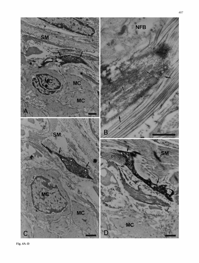
case, membrane appositions were observed with themuscle cells (Fig. 4A). In the connective tissue, bundlesof collagen fibers established contacts with the plasmamembrane of the c-kit ICs (Fig. 4D).
In all the regions studied, the c-kit ICs were alsofound to be located in the close vicinity of blood vessels(Fig. 5A,C) and to have established close contacts withnon-immunoreactive fibroblast-like cells (Fig. 5B).
Discussion
The results of the present study indicates that c-kit ICsare present in all the muscular layers of the distal colon-ic wall studied here, in agreement with the previouslypublished immunohistological (Horie et al. 1993; Matsuda et al. 1993; Yamataka et al. 1995; Rumessen1996; Vanderwinden et al 1996b; Hagger et al. 1997;Romert and Mikkelsen 1998) and immunocytological(Torihashi et al. 1999) data.
The present results show that the c-kit ICs in the ex-ternal muscle layers are of the myoid type which is con-sistent with other recently published data (Torihashi etal. 1999). The ultrastructural features of the c-kit ICscharacterized in this previous study and the present onewere found to be similar to those of the ICCs located in the same regions of the colonic wall (Faussone-Pelle-grini et al. 1990a,b; Rumessen et al. 1993; Rumessen1994). It therefore seems very likely that the c-kit ICs ofthe myoid type are ICCs expressing the c-kit receptor.The possibility cannot however be ruled out that theremay exist ICCs which do not express the c-kit receptor(Torihashi et al. 1999), or which do so only under specif-ic conditions, as found to occur in the case of the ICCspresent in the deep muscular plexus of the mouse (Wester et al. 1999). Another category of c-kit ICs, in-cluding ICs of a mixed type, was found here to exist atvarious levels in the colonic wall, especially at the inter-face between the inner circular muscle layer and the sub-mucosa. These c-kit ICs were not involved in any veryclose relationships with the surrounding nerve fibers andmuscle cells. They might be taken as fibroblast cellswhich have become differentiated, depending on their re-lationships with the surrounding cellular microenviron-ment (Komuro 1990).
Although previous authors have concluded that thereexist no ICCs in the longitudinal and circular muscle lay-ers of the human colon (Christensen and Rick 1987; Faussone-Pellegrini 1987; Rumessen 1994), the presentdata show the existence of c-kit ICs in both of these
muscle layers as well as in the septa, in line with otherprevious studies (Hagger et al. 1997; Torihashi et al.1999). Here we have shown, in addition, that c-kit ICsare frequently inserted between the nerve fibers and themuscle cells, with which they establish membrane appo-sitions. This latter finding indicates that these c-kit ICscorrespond to the ICCs defined as being non-nerve andnon-muscle cells interposed between nerve fibers andsmooth muscle cells (Thuneberg 1989). Their close rela-tionships with nerve and muscle elements strongly sug-gest that they may be directly involved in the neuroregu-latory processes underlying colonic motility (Hagger etal. 1997). It is worth mentioning that recent electrophysi-ological findings have suggested that the ICCs located inthe median part of the colonic circular muscle layer mayalso be involved in the rhythmic motor activity (Rae etal. 1998).
The presence of c-kit IC processes inside the myen-teric ganglia of the human colon has never been clearlyestablished so far on the basis of immunohistological da-ta. Up to now, either their existence has been completelyruled out (Romert and Mikkelsen 1998) or c-kit immu-noreactivity has been reported to occur only in some ofthe glial cells (Matsuda et al. 1993). The results of thepresent study show that the expression of the c-kit recep-tor takes place only on the c-kit ICs closely surroundingand sometimes penetrating the myenteric ganglia, MG1,devoid of a muscle sheath. These c-kit ICs are probablyidentical to the ICCs previously described in the myen-teric plexuses of the human colon (Faussone-Pellegriniet al. 1990b). The exact function of the ICCs surround-ing the myenteric plexuses has not yet been determined.The present finding that there exist close relationshipsbetween processes projecting from the c-kit ICs and theganglionic neurons certainly suggests that the c-kit ICsmay be involved in ganglionic neurotransmission. In ad-dition, recently published electrophysiological data havesuggested that they may participate in the genesis of themyenteric potential oscillations occurring in the smoothmuscle cells in the longitudinal and circular layers (Raeet al. 1998). As far as the c-kit ICs located at the inter-face between the longitudinal and circular muscle layersare concerned, if one makes analogies with the ICCs lo-cated in the same region, it seems likely that they may beinvolved in particular in the electrical coupling betweenthe longitudinal and circular muscle layers, which is re-sponsible for the coordination of motor activities (Rae etal. 1998).
In the present study, c-kit ICs belonging to the plexusentericus extremus located at the interface between theinner circular muscle layer and the submucosa werefound to be present. This result is in agreement with pre-vious data obtained in other mammals (Stach 1972; Berezin et al. 1988). Although this finding is not inagreement with the data obtained by some previous au-thors, who observed the presence of these cells only atthe level of the proximal colon (Romert and Mikkelsen1998), it corroborates that of other authors, who detectedICCs at the level of the distal colon (Vanderwinden et al.
408
Fig. 4A–D Electron micrographs of c-kit ICs located in the sub-mucosa. A C-kit IC process (arrows) closely apposed (arrowhead)to a muscle cell (MC). B High magnification of a c-kit IC process(arrows) containing numerous intermediate filaments (small ar-rows) in close proximity of a nerve fiber bundle (NFB). C,D C-kitIC processes (arrows) in the vicinity of muscle cells (MC); notethe presence in C of numerous mitochondria (white asterisks) and,in D, collagen bundles (c) closely associated with the c-kit IC. SMSubmucosa. Bars 1 µm
▲

409
Fig. 5A–C Electron micro-graphs of interrelationships be-tween c-kit ICs, blood vessels,and a fibroblast-like cell. A Inthe myenteric area, a c-kit ICprocess (large arrow) in closeproximity of a blood vessel(BV); note in the vicinity of thec-kit IC (small arrows) contain-ing caveolae (small arrow-heads), the presence of a nervefiber bundle (NFB) and a mus-cle cell (MC). B,C In the circu-lar muscle layer, c-kit ICs (arrows) are, in B, closely ap-posed (arrowheads) to an unla-beled fibroblast-like cell (double arrowheads) and, in C, run in the close proximityof a blood vessel (BV). Bars 1 µm

1996b; Hagger et al. 1997; Torihashi et al. 1999). We es-tablished here that c-kit ICs belonging to the plexus en-tericus extremus frequently establish relationships withthe inner circular muscle cells. The existence of these re-lationships supports the idea that the ICCs of the plexusentericus extremus may be involved in the initiation ofthe slow peristaltic waves in the circular muscle layer, inline with recent electrophysiological data (Rae et al.1998).
In conclusion, the results of the present study shed es-sential light on the ultrastructural characteristics of the c-kit ICs, especially as regards their relationships with theneighboring muscle cell and nerve elements. Altogether,these data unequivocally confirm the idea that the ex-pression of the c-kit receptors takes place at the level ofmost ICCs. The role played by the c-kit receptors alongwith the ICCs and their ligand, the stem cell factor, stillremains to be elucidated. Further studies are thereforenow required on this topic, in order to improve ourknowledge of the regulatory mechanisms underlying in-testinal motility.
Acknowledgements The present work was performed at the Cen-tre Pluridisciplinaire de Microscopie Electronique et de Microana-lyse (CP2 M) of the Faculté des Sciences de Marseille SaintJérôme, Université Aix-Marseille III, France.
References
Berezin I, Huizinga JD, Daniel EE (1988) Interstitial cells of Cajalin the canine colon: a special communication network at theinner border of the circular muscle. J Comp Neurol 273:42–51
Burns AJ, Lomax AEJ, Torihashi S, Sanders KM, Ward SM(1996) Interstitial cells of Cajal mediate inhibitory transmis-sion in the stomach. Proc Natl Acad Sci USA 93:12008–12013
Burns AJ, Herbert TM, Ward SM, Sanders KM (1997) Interstitialcells of Cajal in the guinea-pig gastrointestinal tract as re-vealed by c-kit immunohistochemistry. Cell Tissue Res290:11–20
Christensen J, Rick GA (1987) Intrinsic nerves in the mammaliancolon: confirmation of a plexus at the circular muscle-submu-cosal interface. J Auton Nerv Syst 21:223–231
Faussone-Pellegrini MS (1987) Comparative study of interstitialcells of Cajal. Acta Anat (Basel) 130:109–126
Faussone-Pellegrini MS, Cortesini C, Pantalone D (1990a) Neuro-muscular structures specific to the submucosal border of thehuman colonic circular muscle layer. Can J Physiol Pharmacol68:1437–1446
Faussone-Pellegrini MS, Pantalone D, Cortesini C (1990b)Smooth muscle cells, interstitial cells of Cajal and myentericplexus interrelationships in the human colon. Acta Anat (Basel) 139:31–44
Hagger R, Finlayson C, Jeffrey I, Kumar D (1997) Role of the in-terstitial cells of Cajal in the control of gut motility. Br J Surg84: 445–450
Hagger R, Gharaie S, Finlayson C, Kumar D (1998) Regional andtransmural density of interstitial cells of Cajal in human colonand rectum. Am J Physiol 275:G1309–G1316
Horie K, Fujita J, Takakura K, Kanzaki H, Suginami H, Iwai M,Nakayama H, Mori T (1993) The expression of c-kit protein inhuman adult and fetal tissues. Hum Reprod 8:1955–1962
Horiguchi K, Komuro T (1998) Ultrastructural characterization ofinterstitial cells of Cajal in the rat small intestine using controland Ws/Ws mutant rats. Cell Tissue Res 293:277–284
Horisawa M, Watanabe Y, Torihashi S (1998) Distribution of c-kitimmunopositive cells in normal human colon and in Hirsch-sprung’s disease. J Pediatr Surg 33:1209–1214
Huizinga JD, Thuneberg L, Kluppel M, Malysz J, Mikkelsen HB,Bernstein A (1995) w/kit gene required for interstitial cells ofCajal and for intestinal pacemaker activity. Nature 373:347–349
Ishikawa K, Komuro T, Hirota S, Kitamura Y (1997) Ultrastruc-tural identification of the c-kit-expressing interstitial cells inthe rat stomach: a comparison of control and Ws/Ws mutantrats. Cell Tissue Res 289:137–143
Isozaki K, Hirota S, Nakama A, Miyagawa JI, Shinomura Y, Xu Z,Nomura S (1995) Disturbed intestinal movement, bile refluxto the stomach, and deficiency of c-kit expressing cells inWs/Ws mutant rats. Gastroenterology 109:456–464
Isozaki K, Hirota S, Miyagawa J, Taniguchi M, Shinomura Y,Matsuzawa Y (1997) Deficiency of c-kit+ cells in patientswith a myopathic form of chronic idiopathic intestinal pseudo-obstruction. Am J Gastroenterol 92:332–334
Komuro T (1990) Re-evaluation of fibroblasts and fibroblast-likecells. Anat Embryol 182:103–112
Komuro T, Zhou DS (1996) Anti-c-kit protein immunoreactivecells corresponding to the interstitial cells of Cajal in the guin-ea-pig small intestine. J Auton Nerv Syst 61:169–174
Langer JC, Berezin I, Daniel EE (1995) Hypertrophic pyloric ste-nosis: ultrastructural abnormalities of enteric nerves and theinterstitial cells of Cajal. J Pediatr Surg 30:1535–1543
Maeda H, Yamagata A, Nishikawa S, Yoshinaga K, Kobayashi S,Nishi K, Nishikawa S (1992) Requirement of c-kit for devel-opment of interstitial pacemaker system. Development 116:369–375
Matsuda R, Takahashi T, Nakamura S, Sekido Y, Nishida K, SetoM, Seito T, Sugiura T, Ariyoshi Y, Takahashi T (1993) Expres-sion of the c-kit protein in human solid tumors and in corre-sponding fetal and adult normal tissues. Am J Pathol 142:339–346
Rae MG, Fleming N, McGregor DB, Sanders KM, Keef KD(1998) Control of motility patterns in the human colonic circu-lar muscle layer by pacemaker activity. J Physiol (Lond) 510:309–320
Romert P, Mikkelsen HB (1998) c-kit immunoreactive interstitialcells of Cajal in the human small and large intestine. Histo-chem Cell Biol 109:195–202
Rumessen JJ (1994) Identification of interstitial cells of Cajal.Significance for studies of human small intestine and colon.Dan Med Bull 41:275–293
Rumessen JJ (1996) Ultrastructure of interstitial cells of Cajal atthe colonic submuscular border in patients with ulcerative co-litis. Gastroenterology 111:1447–1455
Rumessen JJ, Peters S, Thuneberg L (1993) Light- and electronmicroscopical studies of interstitial cells of Cajal and musclecells at the submucosal border of human colon. Lab Invest68:481–495
Sanders KM (1996) A case for interstitial cells of Cajal as pace-makers and mediators of neurotransmission in the gastrointes-tinal tract. Gastroenterology 111:492–515
Sato D, Lai ZF, Tokutomi N, Tokutomi Y, Maeda H, Nishikawa S,Nishikawa S, Ogawa M, Nishi K (1996) Impairment of kit-de-pendent development of interstitial cells alters contractile re-sponses of murine intestinal tract. Am J Physiol 271:G762–G771
Seki K, Zhou DS, Komuro T (1998) Immunohistochemical studyof the c-kit expressing cells and connexin 43 in the guinea-pigdigestive tract. J Auton Nerv Syst 68:182–187
Stach W (1972) Der Plexus entericus extremus des Dickdarmesund seine Beziehungen zu den interstitiellen Zellen (Cajal). ZMikrosk Anat Forsch 85:245–272
Thuneberg L (1989) Interstitial cells of Cajal. In: Schultz GS,Wood JD, Rauner BB (eds) Handbook of physiology – thegastrointestinal system, vol 1. American Physiological Soci-ety, Bethesda, pp 349–386
410

Tokutomi N, Maeda H, Tokutomi Y, Sato D, Sugita M, NishikawaS, Nishikawa S, Nakao J, Imamura T, Nishi K (1995) Rhyth-mic Cl– current and physiological roles of the intestinal c-kit-positive cells. Pflugers Arch 431:169–177
Torihashi S, Ward SM, Sanders KM (1997) Development of c-kit-positive cells and the onset of electrical rhythmicity in murinesmall intestine. Gastroenterology 112:144–155
Torihashi S, Horisawa M, Watanabe Y (1999) c-kit immunoreac-tive interstitial cells in the human gastrointestinal tract. J Au-ton Nerv Syst 75:38–50
Vanderwinden JM, Liu H, Laet MH de, Vanderhaeghen JJ (1996a)Study of the interstitial cells of Cajal in infantile hypertrophicpyloric stenosis. Gastroenterology 111:279–288
Vanderwinden JM, Rumessen JJ, Liu H, Descamps D, Laet MHde, Vanderhaeghen JJ (1996b) Interstitial cells of Cajal in hu-man colon and in Hirschsprung’s disease. Gastroenterology111:901–910
Vliagoftis H, Worobec AS, Metcalfe DD (1997) The protoonco-gene c-kit and c-kit ligand in human disease. J Allergy ClinImmunol 100:435–440
Ward SM, Burns AJ, Torihashi S, Sanders KM (1994) Mutation ofthe proto-oncogene c-kit blocks development of interstitial
cells and electrical rhythmicity in murine intestine. J Physiol(Lond) 480:91–97
Ward SM, Burns AJ, Torihashi S, Harney SC, Sanders KM (1995)Impaired development of interstitial cells and intestinal electri-cal rhythmicity in steel mutants. Am J Physiol 269:C1577–C1585
Ward SM, Harney SC, Bayguinov JR, McLaren GJ, Sanders KM(1997) Development of electrical rhythmicity in the murinegastrointestinal tract is specifically encoded in the tunica mus-cularis. J Physiol (Lond) 505:241–258
Wester T, Eriksson L, Olsson Y, Olsen L (1999) Interstitial cells ofCajal in the human fetal small bowel as shown by c-kit immu-nohistochemistry. Gut 44:65–71
Yamataka A, Kato Y, Tibboel D, Murata Y, Sueyoshi N, FujimotoT, Nishiye H, Miyano T (1995) A lack of intestinal pacemaker(c-kit) in aganglionic bowel of patients with Hirschsprung’sdisease. J Pediatr Surg 30:441–444
Yarden Y, Kuang WJ, Yang-Feng T, Coussens L, Munemitsu S,Dull TJ, Chen E, Schlessinger J, Francke U, Ullrich A (1987)Human proto-oncogene c-kit: a new cell surface receptor tyro-sine kinase for an unidentified ligand. EMBO J 6:3341–3351
411