Spontaneous Intracerebral Hemorrhage Image Analysis Methods: A Survey

Brain interstitial fluid TNF-α after subarachnoid hemorrhage
Khalid A. Hanafy, MD, PhD1, Bartosz Grobelny, BS2, Luis Fernandez, MD1, Pedro Kurtz,MD1, ES Connolly, MD1,2, Stephan A. Mayer, MD, FCCM1,2, Christian Schindler, MD,PhD3,4, and Neeraj Badjatia, MD, MSc1,21Division of Critical Care Neurology, Columbia University College of Physicians and Surgeons, NewYork, NY, 100322Department of Neurology, Department of Neurological Surgery, Columbia University College ofPhysicians and Surgeons, New York, NY, 100323Department of Medicine, Columbia University College of Physicians and Surgeons, New York, NY,100324Department of Microbiology, Columbia University College of Physicians and Surgeons, New York,NY, 10032
AbstractObjective: TNF-α is an inflammatory cytokine that plays a central role in promoting the cascadeof events leading to an inflammatory response. Recent studies have suggested that TNF-α may playa key role in the formation and rupture of cerebral aneurysms, and that the underlying cerebralinflammatory response is a major determinate of outcome following subrarachnoid hemorrhage(SAH).
Methods: We studied 14 comatose SAH patients who underwent multimodality neuromonitoringwith intracranial pressure (ICP) and cerebral microdialysis as part of their clinical care. Continuousphysiological variables were time-locked every 8 hours and recorded at the same point that braininterstitial fluid TNF-α was measured in brain microdialysis samples. Significant associations weredetermined using generalized estimation equations.
Results: Each patient had a mean of 9 brain tissue TNF-α measurements obtained over an averageof 72 hours of monitoring. TNF-α levels rose progressively over time. Predictors of elevated braininterstitial TNF-α included higher brain interstitial fluid glucose levels (β=0.066, P<0.02),intraventricular hemorrhage (β=0.085, P<0.021), and aneurysm size >6 mm (β=0.14, p<0.001). Therewas no relationship between TNF-α levels and the burden of cisternal SAH; concurrent measurementsof serum glucose, or lactate-pyruvate ratio.
Interpretation: Brain interstitial TNF-α levels are elevated after SAH, and are associated with largeaneurysm size, the burden of intraventricular blood, and elevation brain interstitial glucose levels.
Keywordstumor necrosis factor-α; intraventricular hemorrhage; cerebral inflammatory response; cerebralmicrodialysis; brain interstitial fluid
© 2009 Elsevier B.V. All rights reserved.Corresponding Author: Khalid Hanafy MD, PhD.Publisher's Disclaimer: This is a PDF file of an unedited manuscript that has been accepted for publication. As a service to our customerswe are providing this early version of the manuscript. The manuscript will undergo copyediting, typesetting, and review of the resultingproof before it is published in its final citable form. Please note that during the production process errors may be discovered which couldaffect the content, and all legal disclaimers that apply to the journal pertain.
NIH Public AccessAuthor ManuscriptJ Neurol Sci. Author manuscript; available in PMC 2011 April 15.
Published in final edited form as:J Neurol Sci. 2010 April 15; 291(1-2): 69–73. doi:10.1016/j.jns.2009.12.023.
NIH
-PA Author Manuscript
NIH
-PA Author Manuscript
NIH
-PA Author Manuscript

IntroductionThe pathophysiology of aneurysmal subarachnoid hemorrhage (aSAH) has long remainedelusive. The presence and subsequent lysis of red blood cells in the subarachnoid space andcerebral cisterns produces an inflammatory response mediated by pro-inflammatory cytokines(1). This response has been theorized to sequester nitric oxide, resulting in vasospasm anddelayed cerebral ischemia or infarction, which is associated with poor outcomes (1,2).However, strategies aimed solely at improving cerebral blood flow or mitigating vasospasmfollowing aSAH have had limited success in improving overall morbidity or mortality (3-5).This may be explained in part by a perspective that incorporates cerebral vasospasm as but onesequela of a larger cerebral inflammatory response (CIR) occurring throughout the brain. Inthis view, interactions between neurons, vasculature, and microglia following aneurysmrupture might be altered by a diffuse tissue inflammatory response triggered by the initialbleeding event. (6).
TNF-α is a critical inflammatory cytokine that plays a central role in initiating and promotingthe cascade of events leading to an inflammatory response. This includes the accumulation ofoxygen radicals, the upregulation of expression of endothelial and leukocyte adhesionmolecules, and the recruitment of macrophages and neutrophils (7). Recent studies havesuggested that TNF-α levels are elevated in the CSF of SAH (8,9).
Several studies have attempted to correlate levels of TNF- α in serum and cerebrospinal fluid(CSF) with poor outcome and have arrived at conflicting results (8-12). However, the blood-brain and blood-CSF barriers may be variably disrupted following aSAH, rendering theseapproaches unreliable in measuring the level of TNF-α directly associated with SAH pathology.By contrast, cerebral microdialysis allows reliable, continuous sampling of intracerebralinterstitial fluid. We hypothesized that TNF-α measured within the brain interstitial fluid 4-6days after aneurysm rupture (SAH day) is proportional to the cerebral inflammatory response.We studied a series of poor-grade aSAH patients to determine what variables were associatedwith the CIR as determined by TNF-α in brain interstitial fluid.
Subjects and MethodsPatients
We studied 14 SAH patients who were enrolled in the Columbia University SAH OutcomesProject (SHOP) between December 2007 and April 2009. SHOP is a prospective observationalcohort study that involves archiving of the medical record and one-year follow-up ofneurological, cognitive, and functional outcome. The present analysis was limited to patientswho underwent brain multimodality neuromonitoring of intracranial pressure (ICP) andcerebral microdialysis as part of their clinical care as per institutional protocol, with a minimumof 24 hours of high-quality recoverable data. Microdialysis samples were processed hourly forlactate, pyruvate, and glucose in the ICU and used to guide clinical management. TNF-α levelswere measured from effluent microdialysis samples stored at −70° C in a post-hoc fashion.SAH days 4-6 were chosen because there is evidence that TNF-α does not change in the serumof aSAH patients from ictus to SAH day 3, but it does increase in the cerebrospinal fluid fromSAH days 4-10 (9,11) This study was approved by the Columbia University InstitutionalReview Board (IRB).
Clinical VariablesBaseline clinical, demographic, and physiological parameters were recorded on admission tothe Columbia University Medical Center neuro-ICU. Clinical status was evaluated by
Hanafy et al. Page 2
J Neurol Sci. Author manuscript; available in PMC 2011 April 15.
NIH
-PA Author Manuscript
NIH
-PA Author Manuscript
NIH
-PA Author Manuscript

recording the patient's worst Hunt-Hess score during the course of hospitalization, and withGlasgow Coma Scale scores recorded every other hour by the ICU nursing staff.
Radiological VariablesAdmission CT scans were independently evaluated by a study neurointensivist for the amountand location of subarachnoid blood (SAH sum score, scaled 0 = no blood, 30 = all cisterns andfissures completely filled), (13) intraventricular blood (IVH sum score, scaled 0 = no blood,12 = all ventricles completely filled), (14) the presence and degree of hydrocephalus, (15) andthe presence of global cerebral edema (2). Bicaudate index was defined as distance betweenthe medial borders of the caudate nuclei divided by the distance between the skull temples(16). Aneurysm size was defined by admission CT angiogram or cerebral angiogram as largestcross-sectional diameter in millimeters.
Physiologic VariablesAll physiological variables recorded at the bedside, including heart rate, mean arterial pressure,intracranial pressure (ICP), cerebral perfusion pressure (CPP), bladder temperature, andmultimodality data were stored in an SQL database. The Solar 8000i utilizing the GeneralElectric Medical Systems Information Technologies' Unity Network® is the patientphysiologic monitor. A high resolution data acquisition system (BedmasterEX, Excel MedicalElectronics, Jupiter, FL) uses the open architecture of the Unity Network® to automaticallyacquire vital signs, alarm, and waveform data from all the patient monitoring devices in theNICU. Digital data is acquired every 5 seconds and recorded in a SQL database. Waveformdata is stored at a resolution of 240 Hz in binary files. Brain metabolism data are incorporatedinto the data acquisition system utilizing the communications (COM) port on the device andis plugged into a serial-to-TCP/IP interface device (Equinox ESP-8, Avocent, Sunrise, FL).All continuous physiological variables, including standard microdialysis variables, were timelocked every 8 hours and measured at the same point that brain interstitial fluid TNF-α wasmeasured.
Clinical ManagementPatient care for SAH conformed to guidelines established by the American Heart Association(17). Hemodynamic and fluid management was targeted to maintain cerebral perfusionpressure (CPP) >70 mm Hg, unless a higher level was associated with favorable effects on thelactate:pyruvate ratio (goal <40) and intracranial pressure (goal <20 mm Hg). Hemoglobinlevels below 7 mg/dL were used to trigger blood transfusion unless there was evidence of activecerebral or myocardial ischemia, in which case a trigger of 10 mg/dl was used. Fever wasaggressively treated using surface cooling (Arctic Sun Cooling System, Medivance Inc,Louisville, CO), with shivering controlled using a stepwise institutional protocol (18).
Cerebral MicrodialysisA CMA 106 microdialysis perfusion pump (CMA Microdialysis®) was used to perfuse theinterior of the catheter with sterile artificial cerebro-spinal fluid (Na+ 148 mmol/L, Ca2+ 1.2mmol/L, Mg2+ 0.9 mmol/L, K+ 2.7 mmol/L, Cl− 155 mmol/L) at a rate of 0.3 μl/min. Sampleswere colle cted every 60 min into microvials, and immediately analyzed at the bedside forglucose, lactate and pyruvate (mmol/L) with the CMA 600 analyzer (CMA Microdialysis®).At least 1 hour passed after the insertion of the probe and the start of the sampling, to allowfor normalization of changes due to probe insertion. The analyzer was automatically calibratedon initiation and every six hours using standard calibration solutions from the manufacturer.Quality controls at three different concentrations for each marker were performed daily.
Hanafy et al. Page 3
J Neurol Sci. Author manuscript; available in PMC 2011 April 15.
NIH
-PA Author Manuscript
NIH
-PA Author Manuscript
NIH
-PA Author Manuscript
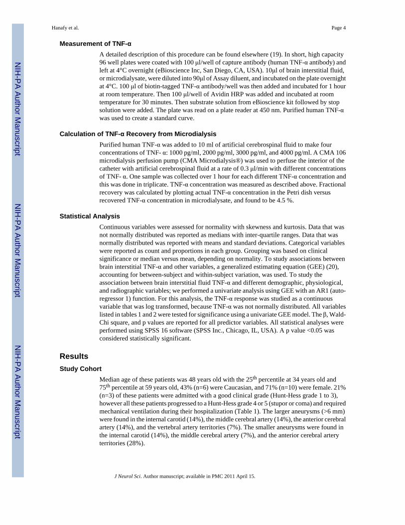
Measurement of TNF-αA detailed description of this procedure can be found elsewhere (19). In short, high capacity96 well plates were coated with 100 μl/well of capture antibody (human TNF-α antibody) andleft at 4°C overnight (eBioscience Inc, San Diego, CA, USA). 10μl of brain interstitial fluid,or microdialysate, were diluted into 90μl of Assay diluent, and incubated on the plate overnightat 4°C. 100 μl of biotin-tagged TNF-α antibody/well was then added and incubated for 1 hourat room temperature. Then 100 μl/well of Avidin HRP was added and incubated at roomtemperature for 30 minutes. Then substrate solution from eBioscience kit followed by stopsolution were added. The plate was read on a plate reader at 450 nm. Purified human TNF-αwas used to create a standard curve.
Calculation of TNF-α Recovery from MicrodialysisPurified human TNF-α was added to 10 ml of artificial cerebrospinal fluid to make fourconcentrations of TNF- α: 1000 pg/ml, 2000 pg/ml, 3000 pg/ml, and 4000 pg/ml. A CMA 106microdialysis perfusion pump (CMA Microdialysis®) was used to perfuse the interior of thecatheter with artificial cerebrospinal fluid at a rate of 0.3 μl/min with different concentrationsof TNF- α. One sample was collected over 1 hour for each different TNF-α concentration andthis was done in triplicate. TNF-α concentration was measured as described above. Fractionalrecovery was calculated by plotting actual TNF-α concentration in the Petri dish versusrecovered TNF-α concentration in microdialysate, and found to be 4.5 %.
Statistical AnalysisContinuous variables were assessed for normality with skewness and kurtosis. Data that wasnot normally distributed was reported as medians with inter-quartile ranges. Data that wasnormally distributed was reported with means and standard deviations. Categorical variableswere reported as count and proportions in each group. Grouping was based on clinicalsignificance or median versus mean, depending on normality. To study associations betweenbrain interstitial TNF-α and other variables, a generalized estimating equation (GEE) (20),accounting for between-subject and within-subject variation, was used. To study theassociation between brain interstitial fluid TNF-α and different demographic, physiological,and radiographic variables; we performed a univariate analysis using GEE with an AR1 (auto-regressor 1) function. For this analysis, the TNF-α response was studied as a continuousvariable that was log transformed, because TNF-α was not normally distributed. All variableslisted in tables 1 and 2 were tested for significance using a univariate GEE model. The β, Wald-Chi square, and p values are reported for all predictor variables. All statistical analyses wereperformed using SPSS 16 software (SPSS Inc., Chicago, IL, USA). A p value <0.05 wasconsidered statistically significant.
ResultsStudy Cohort
Median age of these patients was 48 years old with the 25th percentile at 34 years old and75th percentile at 59 years old, 43% (n=6) were Caucasian, and 71% (n=10) were female. 21%(n=3) of these patients were admitted with a good clinical grade (Hunt-Hess grade 1 to 3),however all these patients progressed to a Hunt-Hess grade 4 or 5 (stupor or coma) and requiredmechanical ventilation during their hospitalization (Table 1). The larger aneurysms (>6 mm)were found in the internal carotid (14%), the middle cerebral artery (14%), the anterior cerebralartery (14%), and the vertebral artery territories (7%). The smaller aneurysms were found inthe internal carotid (14%), the middle cerebral artery (7%), and the anterior cerebral arteryterritories (28%).
Hanafy et al. Page 4
J Neurol Sci. Author manuscript; available in PMC 2011 April 15.
NIH
-PA Author Manuscript
NIH
-PA Author Manuscript
NIH
-PA Author Manuscript

Microdialysis CharacteristicsMedian values for lactate, pyruvate, lactate:pyruvate ratio, glucose, and TNF-α are shown(Table 2). The microdialysis values were derived from 91 measurements of 14 patients over 3days (SAH days 4-6). Four different concentrations were used to calculate the recovery ofTNF-α in the microdialysate. From this, the actual concentration of TNF-α in the braininterstitial fluid was calculated. The brain interstitial TNF-α levels are averaged for all 14patients at each of the 9 measured time points during the 72 hour monitoring period (Figure1).
Predictors of TNF-α as a Linear VariableThere were no significant demographic predictors of TNF-α in this cohort of 14 patients. Ofall the continuous physiological variables collected between SAH days 4-6, systemic glucose(β=0.005, p=0.063) approached significance, while brain interstitial fluid glucose (β=0.066,p=0.018) was significantly associated with TNF-α. All infusions were tested for significantassociations with TNF-α, and none were significant, including insulin. The intraventricularsum score dichotomized patients into those with intraventricular hemorrhage and those withoutwas a significant predictor of the TNF-α response (p=0.021, β=0.085). However, the mostsignificant predictor of the TNF-α response on univariate analysis was aneurysm size(p<0.001), dichotomized at the median of 6 mm (Table 3). This is evident by the largest valuesfor Wald-Chi square, 21.2 and β, 0.14.
DiscussionIn this study we found a progressive rise in brain interstitial TNF-α levels between SAH days4 to 6. Large aneurysm size, the existence of intraventricular hemorrhage, and increased levelsof glucose in the microdialysate were significantly associated with TNF-α elevation. The factthat TNF-α levels increase throughout SAH days 4-6 may provide an explanation for thecumulative risk of complications that occur after ictus. TNF-α levels may correlate with latecomplications of aSAH such as delayed cerebral infarction (DCI). There is growing evidencethat DCI involves a progressive inflammatory burden that is independent of vasospasm. Thisis supported by the fact that clinical trials directed at ameliorating vasospasm have not resultedin improved outcomes (4,5). Furthermore, current theories behind the evolution of DCI invokeimpaired fibrinolytic activity, an inflammatory cascade, and endothelial dysfunction in theformation of microthrombi, all of which would likely involve TNF-α (21-24).
We found that large aneurysm size correlates with increasing TNF-α levels and there is bothanimal and human data that support this notion. When SAH is achieved by direct puncture ofthe ICA in rats, causing damage to vascular wall, blood and inflammatory mediators arereleased that result in a 40-50% mortality, similar to the vascular damage caused by ananeurysm (25,26). In humans, non-aneurysmal SAH, like perimesencephalic and traumaticSAHs, are associated with DCI less frequently than aSAH (27-31). The fact that aneurysm sizewas shown to be associated with increasing TNF-α seems to be in support of our findings.
Even though the heme burden does not explain all the pathology seen in aSAH, it clearly is animportant component to the pathology. Animal models of intraventricular hemorrhage (IVH)where blood is injected into the ventricles show a highly inflammatory pattern in theperiventricular space (32,33). In addition to this, the effect of IVH was studied in the aSAHpopulation and shown to be an independent predictor of vasospasm and poor outcome (34,35). Consistent with this, we found intraventricular hemorrhage to be associated with increasedTNF-α.
Hanafy et al. Page 5
J Neurol Sci. Author manuscript; available in PMC 2011 April 15.
NIH
-PA Author Manuscript
NIH
-PA Author Manuscript
NIH
-PA Author Manuscript

Finally, we found brain interstitial fluid glucose to be significantly associated with increasingTNF-α in the brain. There is evidence to support hyperglycemia in the setting of aninflammatory response and that admission hyperglycemia is associated with poor outcome(36,37). The correlation between brain interstitial fluid glucose and increasing TNF-α ispreliminary, but is in accord with the increased insulin resistance seen in any inflammatoryresponse. Furthermore, there is recent evidence that in addition to TNF-α being involved ingrowth and rupture of aneurysms (7,8,38,39), the PPAR-γ (peroxisome proliferator-activatedreceptor) transduction pathway is similarly involved (40). When PPAR-γ agonists such asrosiglitazone were given to mice that spontaneously develop thoraco-abdominal aorticaneurysms, the incidence of aneurysm formation and rupture significantly decreased comparedto controls. In addition, the expression of TNF-α, IL-6, and E-selectin were markedly reduced(40). As rosiglitazone is an insulin sensitizer, one would expect decreased serum glucose inthis population too, even though this was not the case. Therefore, it is possible, that in theprocess of cerebral aneurysm formation and rupture, the PPAR-γ pathway is somehowinhibited, resulting in hyperglycemia and increased TNF-α levels. While this argument isderived from few data, it does provide a novel perspective of study of the cerebral inflammatoryresponse after aSAH as it relates to the PPAR-γ pathway.
To our knowledge, this is the first study of the brain interstitial fluid TNF-α in patients afteraneurysmal subarachnoid hemorrhage. With this in mind, there are several importantlimitations that can be addressed in future studies with a larger cohort of patients. Our patientpool was small and most patients were poor grade on admission. Second, defining the cerebralinflammatory response by TNF-α levels alone, is likely not a complete description of theinflammatory cascade occurring within the brain. TNF-α was chosen for two reasons. First, itis associated with almost all inflammatory cascades that have been described (41). Second, ofall inflammatory mediators studied, TNF-α, had the most data in the aSAH population. In fact,SAH days 4-6 were chosen because there is evidence that TNF-α does not change in the serumof aSAH patients from ictus to SAH day 3, but it does increase in the cerebrospinal fluid fromSAH days 4-10 (9,11). We hope that this study, although preliminary in findings, will lead tofurther research of the cerebral inflammatory response in aneurysmal SAH.
References1. Dumont AS, D R, Chow MM, Lin CL, Calisaneller T, Ley KF, Kassell NF, Lee KS. Cerebral vasospasm
after subarachnoid hemorrhage: putative role of inflammation. Neurosurgery 2003;53:133–5.2. Claassen J, C J, Kreiter KT, Du EY, Connolly ES, Mayer SA. Global cerebral edema after subarachnoid
hemorrhage: frequency, predictors, and impact on outcome. Stroke 2002;33:1225–32. [PubMed:11988595]
3. Haley EC Jr, K N, Apperson-Hansen C, Maile MH, Alves WM. A randomized, double-blind, vehicle-controlled trial of tirilazad mesylate in patients with aneurysmal subarachnoid hemorrhage: acooperative study in North America. J Neurosurg 1997;86:467–74. [PubMed: 9046304]
4. Pickard JD, M G, Illingworth R, Shaw MD, Teasdale GM, Foy PM, Humphrey PR, Lang DA, NelsonR, Richards P, et al. Effect of oral nimodipine on cerebral infarction and outcome after subarachnoidhaemorrhage: British aneurysm nimodipine trial. BMJ 1989;298:636–42. [PubMed: 2496789]
5. Vajkoczy P, M B, Weidauer S, Raabe A, Thome C, Ringel F, Breu V, Schmiedek P. Clazosentan(AXV-034343), a selective endothelin A receptor antagonist, in the prevention of cerebral vasospasmfollowing severe aneurysmal subarachnoid hemorrhage: results of a randomized, double-blind,placebo-controlled, multicenter phase IIa study. J Neurosurg 2005;103:9–17. [PubMed: 16121967]
6. Cahill J, C J, Zhang JH. Mechanisms of early brain injury after subarachnoid hemorrhage. J CerebBlood Flow Metab 2006;26:1341–53. [PubMed: 16482081]
7. Jayaraman T, P A, Shin YS, Li X, Mayer J, Chaudhry H, Niimi Y, Silane M, Berenstein A. TNF-alpha-mediated inflammation in cerebral aneurysms: a potential link to growth and rupture. Vasc HealthRisk Manag 2008;4:805–17. [PubMed: 19065997]
Hanafy et al. Page 6
J Neurol Sci. Author manuscript; available in PMC 2011 April 15.
NIH
-PA Author Manuscript
NIH
-PA Author Manuscript
NIH
-PA Author Manuscript

8. Aoki T, K H, Shimamura M, Nakagami H, Wakayama K, Moriwaki T, Ishibashi R, Nozaki K, MorishitaR, Hashimoto N. NF-kappaB is a key mediator of cerebral aneurysm formation. Circulation2007;116:2830–40. [PubMed: 18025535]
9. Mathiesen T, E G, Ulfarsson E, Andersson B. Cerebrospinal fluid interleukin-1 receptor antagonistand tumor necrosis factor-alpha following subarachnoid hemorrhage. J Neurosurg 1997;87:215–20.[PubMed: 9254084]
10. Fassbender K, H B, Rossol S, Bertsch T, Schmeck J, Schütt S, Fritzinger M, Horn P, Vajkoczy P,Kreisel S, Brunner J, Schmiedek P, Hennerici M. Inflammatory cytokines in subarachnoidhaemorrhage: association with abnormal blood flow velocities in basal cerebral arteries. J NeurolNeurosurg Psychiatry 2001;70:534–7. [PubMed: 11254783]
11. Witkowska AM, B M, Socha K, Kochanowicz J, Mariak Z, Konopka M. TNF-alpha and sICAM-1in intracranial aneurismal rupture. Arch Immunol Ther Exp (Warsz) 2009;57:137–40. [PubMed:19340565]
12. Gruber A, R K, Graninger W, Donner A, Illievich MU, Czech T. Ventricular cerebrospinal fluid andserum concentrations of sTNFR-I, IL-1ra, and IL-6 after aneurysmal subarachnoid hemorrhage. JNeurosurg Anesthesiol 2000;12:297–306. [PubMed: 11147377]
13. Hijdra A, vG J, Nagelkerke NJ, Vermeulen M, van Crevel H. Prediction of delayed cerebral ischemia,rebleeding, and outcome after aneurysmal subarachnoid hemorrhage. Stroke 1988;19:1250–6.[PubMed: 3176085]
14. Brouwers PJ, D D, Vermeulen M, Lindsay KW, Hasan D, vG J. Amount of blood on computedtomography as an independent predictor after aneurysm rupture. Stroke 1993:809–14. [PubMed:8506552]
15. van Gijn J, H A, Wijdicks EF, Vermeulen M, van Crevel H. Acute hydrocephalus after aneurysmalsubarachnoid hemorrhage. J Neurosurg 1985:355–62. [PubMed: 4020461]
16. Vermeij FH, H D, Vermeulen M, Tanghe HL, van Gijn J. Predictive factors for deterioration fromhydrocephalus after subarachnoid hemorrhage. Neurology 1994;44(10):1851–5. [PubMed: 7936235]
17. Bederson JB, CE J, Batjer HH, Dacey RG, Dion JE, Diringer MN, Duldner JE Jr, Harbaugh RE, PatelAB, Rosenwasser RH. Guidelines for the management of aneurysmal subarachnoid hemorrhage: astatement for healthcare professionals from a special writing group of the Stroke Council, AmericanHeart Association. Stroke 2009;40:994–1025. [PubMed: 19164800]
18. Badjatia N, S E, Prescutti M, Fernandez L, Fernandez A, Buitrago M, Schmidt JM, M S. Metabolicbenefits of surface counter warming during therapeutic temperature modulation. Crit Care Med2009;37:1893–7. [PubMed: 19384208]
19. C. JR. ELISA. Theory and practice. Methods Mol Biol 1995:1–218.20. Hilbe, JHaJ. Generalized Estimation Equations. Chapman and Hall; Boca Raton, FL: 2003.21. Frijns CJ, F R, Algra A, van Mourik JA, van Gijn J, Rinkel GJ. Early circulating levels of endothelial
cell activation markers in aneurysmal subarachnoid haemorrhage: associations with cerebralischaemic events and outcome. J Neurol Neurosurg Psychiatry 2006:77–83. [PubMed: 16361599]
22. Peltonen S, J S, Kaste M, Lassila R. Hemostasis and fibrinolysis activation after subarachnoidhemorrhage. J Neurosurg 1997;87:207–14. [PubMed: 9254083]
23. Suzuki M, K A, Otawara Y, Hirashima Y, Takaku A, Ogawa A. Extrinsic pathway of bloodcoagulation and thrombin in the cerebrospinal fluid after subarachnoid hemorrhage. Neurosurgery1999;44:487–93. [PubMed: 10069585]
24. Hirashima Y, N S, Endo S, Kuwayama N, Naruse Y, Takaku A. Elevation of platelet activating factor,inflammatory cytokines, and coagulation factors in the internal jugular vein of patients withsubarachnoid hemorrhage. Neurochem Res 1997;22:1249–55. [PubMed: 9342729]
25. Adams HP Jr, K N, Torner JC, Nibbelink DW, Sahs AL. Early management of aneurysmalsubarachnoid hemorrhage. A report of the Cooperative Aneurysm Study. J Neurosurg 1981;54:141–5. [PubMed: 7005404]
26. Bederson JB, G I, Guarino L. Cortical blood flow and cerebral perfusion pressure in a newnoncraniotomy model of subarachnoid hemorrhage in the rat. Stroke 1995;26:1086–91. [PubMed:7762027]
27. Cánovas D, G A, Jato M, Rubio F. Non-aneurysmal subarachnoid hemorrhage: 60 cases. Neurologia2006;21:704–9. [PubMed: 17106823]
Hanafy et al. Page 7
J Neurol Sci. Author manuscript; available in PMC 2011 April 15.
NIH
-PA Author Manuscript
NIH
-PA Author Manuscript
NIH
-PA Author Manuscript

28. Fukuda T, H M, Ito H. Does traumatic subarachnoid hemorrhage caused by diffuse brain injury causedelayed ischemic brain damage? Comparison with subarachnoid hemorrhage caused by rupturedintracranial aneurysms. Neurosurgery 1998;43:1040–9. [PubMed: 9802847]
29. Hui FK, T L, Tanaka T, Cawley CM, Zhang YJ. Clinical Differences Between AngiographicallyNegative, Diffuse Subarachnoid Hemorrhage and Perimesencephalic Subarachnoid Hemorrhage.Neurocrit Care 2009;11:64–70. [PubMed: 19277905]
30. Kang DH, P J, Lee SH, Park SH, Kim YS, Hamm IS. Does non-perimesencephalic typenonaneurysmal subarachnoid hemorrhage have a benign prognosis? J Clin Neurosci 2009;16:904–8. [PubMed: 19362482]
31. Shigemori M, T T, Hirohata M, Maruiwa H, Kaku N, Kuramoto S. Clinical significance of traumaticsubarachnoid hemorrhage. Neurol Med Chir (Tokyo) 1990;30:396–400. [PubMed: 1700319]
32. Pang D, S R, Horton JA. Lysis of intraventricular blood clot with urokinase in a canine model: Part1-3. Neurosurgery 1986;19:540–73. [PubMed: 3491338]
33. Xi G, K R, Hoff JT. Pathophysiology of brain edema formation. Neurosurg Clin N Am 2002;13:371–83. [PubMed: 12486926]
34. Claassen J, B G, Kreiter K, Bates J, Du YE, Copeland D, Connolly ES, Mayer SA. Effect of cisternaland ventricular blood on risk of delayed cerebral ischemia after subarachnoid hemorrhage: the Fisherscale revisited. Stroke 2001;32:2012–20. [PubMed: 11546890]
35. Kramer AH, H M, Nathan B, Gress D, Dumont AS, Kassell NF, Bleck TP. A comparison of 3radiographic scales for the prediction of delayed ischemia and prognosis following subarachnoidhemorrhage. J Neurosurg 2008;109:199–207. [PubMed: 18671630]
36. Brierre S, K R, Deboisblanc BP. The endocrine system during sepsis. Am J Med Sci 2004;328:238–47. [PubMed: 15486539]
37. Claassen J, V A, Kreiter KT, Kowalski RG, Du EY, Ostapkovich N, Fitzsimmons BF, Connolly ES,Mayer SA. Effect of acute physiologic derangements on outcome after subarachnoid hemorrhage.Crit Care Med 2004;32:832–8. [PubMed: 15090970]
38. Fontanella M, R I, Gallone S, Rubino E, Fenoglio P, Valfrè W, Garbossa D, Carlino C, Ducati A,Pinessi L. Tumor necrosis factor-alpha gene and cerebral aneurysms. Neurosurgery 2007:668–72.[PubMed: 17415203]
39. Jayaraman T, B V, Li X, Mayer J, Silane M, Shin YS, Niimi Y, Kiliç T, Gunel M, Berenstein A.Tumor necrosis factor alpha is a key modulator of inflammation in cerebral aneurysms. Neurosurgery2005;57:558–64. [PubMed: 16145536]
40. Jones A, D R, Torsney E, Howe F, Dunkley M, Gnaneswaran Y, Gaze D, Nasr H, Loftus IM, T M,Cockerill GW. Rosiglitazone reduces the development and rupture of experimental aortic aneurysms.Circulation 2009;119(24):3125–32. [PubMed: 19506106]
41. C. M. The role of TNF superfamily members in T-cell function and diseases. Nat Rev Immunol2009;9:271–85. [PubMed: 19319144]
Hanafy et al. Page 8
J Neurol Sci. Author manuscript; available in PMC 2011 April 15.
NIH
-PA Author Manuscript
NIH
-PA Author Manuscript
NIH
-PA Author Manuscript

Figure 1.mean of log TNF-α between SAH days 4-6
Hanafy et al. Page 9
J Neurol Sci. Author manuscript; available in PMC 2011 April 15.
NIH
-PA Author Manuscript
NIH
-PA Author Manuscript
NIH
-PA Author Manuscript

NIH
-PA Author Manuscript
NIH
-PA Author Manuscript
NIH
-PA Author Manuscript
Hanafy et al. Page 10
Table 1
Patient Characteristics (N=14)
Demographic Variables:
Age (years) 48 (34-59)
Female 10 (71)
Caucasian 6 (43)
BMI (kg/m2) 24 (23-28)
Past Medical History of
Hypertension 2 (14)
Diabetes Mellitus 1 (7)
Clinical Variables:
Worst Hunt Hess Grade
4 stupor/moderate hemiparesis 1 (7)
5 coma/decerebrate posturing 13 (93)
APACHE II subscore* 14 (11-17)
GCS 7 (5-13)
Physiological Variables during monitoring:
GCS 6 (4-8)
Temperature (°C) 37 (34.4-37.3)
Systemic Glucose (mg/dl) 138 (113-171)
Cerebral Perfusion Pressure (mmHg) 84 (74-103)
Intracranial Pressure (cm H2O) 10 (6-13)
White Blood Cell Count (×103 cells/mm3) 13 (10.8-17.1)
Radiographic variables on Admission:
Bicaudate index16 0.17 (0.14-0.19)
Occurrence of IVH 12 (86)
HIJDRA score13 18 (12-24)
Global Cerebral Edema 6 (43)
Aneurysm size (mm) 6 (5.2-8.3)
Data are given as N (%) or Median (25-75% IQR)
*An ICU mortality prediction ranging from 0-125 that incorporates physiological derangements, age, and chronic health problems and excludes GCS
J Neurol Sci. Author manuscript; available in PMC 2011 April 15.

NIH
-PA Author Manuscript
NIH
-PA Author Manuscript
NIH
-PA Author Manuscript
Hanafy et al. Page 11
Table 2
Microdialysis Characteristics
Median Normal range IQR (25-75%)
Microdialysis Variables:
Lactate (mmol/L) 3.7 (2.0-3.8) (2.4-4.4)
Pyruvate (μmol/L) 103 (119-213) (84-162)
Lactate:Pyruvate ratio (LPR) 28.3 (19-27) (23.1-33.1)
Glucose (mmol/L) 1.12 (0.8-2.6) (0.71-1.92)
Tumor Necrosis Factor-α (pg/ml) 753.1 (372.1-1100.5)
N=91 measurements of all microdialysis values
J Neurol Sci. Author manuscript; available in PMC 2011 April 15.

NIH
-PA Author Manuscript
NIH
-PA Author Manuscript
NIH
-PA Author Manuscript
Hanafy et al. Page 12
Table 3
Significant Predictors of TNF-α by GEE
βWald Chi
square p
Demographic variables:
Age (>48years) −0.069 2.18 0.14
Female −0.096 2.86 0.091
Caucasian 0.031 0.028 0.99
BMI (kg/m2) −0.003 0.95 0.33
Past medical History of:
-Hypertension 0.039 0.88 0.35
-Diabetes Mellitus 0.039 0.88 0.35
Admission ClinicalVariables:
Hunt Hess grade 0.11 3.57 0.59
Apache II subscore 0.001 0.011 0.92
GCS −0.012 2.64 0.1
Continuous Variablesduring monitoring (q8):
GCS −0.012 1.33 0.25
Temperature (°C) 0.004 0.031 0.86
Systemic glucose (mg/dl) 0.005 3.46 0.063
Cerebral Perfusion Pressure(mmHg) −0.005 0.685 0.41
Intracranial Pressure (cm H2O) 0.001 0.14 0.71
White Blood Cell count(× 103cells/mm3) 0.001 0.01 0.92
MD Lactate (mmol/L) −0.045 2.47 0.12
MD Pyruvate (μmol/L) 0.001 0.57 0.45
MD LPR −0.008 5.15 0.06
MD Glucose (mmol/L) 0.066 5.63 0.018
Radiographic Variables:
Bicaudate Index −0.43 0.14 0.71
Existence of IVH 0.085 5.33 0.021
HIJDRA score 0.006 0.71 0.93
Global Cerebral Edema 0.03 0.115 0.74
Aneurysm Size (>6 mm) 0.14 21.2 <0.001
J Neurol Sci. Author manuscript; available in PMC 2011 April 15.
Copyright © 2022 FDOKUMEN