Artifact quantification and tractography from 3T MRI after placement of aneurysm clips in...
11
RESEARCH ARTICLE Open Access Artifact quantification and tractography from 3T MRI after placement of aneurysm clips in subarachnoid hemorrhage patients Faraz Khursheed 1 , Fiona Rohlffs 1 , Shuichi Suzuki 2,3 , Dong H Kim 1 and Timothy M Ellmore 1* Abstract Background: The application of advanced 3T MRI imaging techniques to study recovery after subarachnoid hemorrhage (SAH) is complicated by the presence of image artifacts produced by implanted aneurysm clips. To characterize the effect of these artifacts on image quality, we sought to: 1) quantify extent of image artifact in SAH patients with implanted aneurysm clips across a range of MR sequences typically used in studies of volumetry, blood oxygen level dependent signal change (BOLD-fMRI), and diffusion-weighted imaging (DW-MRI) and 2) to explore the ability to reconstruct white matter pathways in these patients. Methods: T1- and T2-weighted structural, BOLD-fMRI, and DW-MRI scans were acquired at 3T in two patients with titanium alloy clips in ACOM and left ACA respectively. Intensity-based planimetric contouring was performed on aligned image volumes to define each artifact. Artifact volumes were quantified by artifact/clip length and artifact/ brain volume ratios and analyzed by two-way (scan-by-rater) ANOVAs. Tractography pathways were reconstructed from DW-MRI at varying distances from the artifacts using deterministic methods. Results: Artifact volume varied by MR sequence for length (p = 0.007) and volume (p < 0.001) ratios: it was smallest for structural images, larger for DW-MRI acquisitions, and largest on fMRI images. Inter-rater reliability was high (r = 0.9626, p < 0.0001), and reconstruction of white matter connectivity characteristics increased with distance from the artifact border. In both patients, reconstructed white matter pathways of the uncinate fasciculus and inferior fronto-occipital fasciculus were clearly visible within 2 mm of the artifact border. Conclusions: Advanced 3T MR can successfully image brain tissue around implanted titanium aneurysm clips at different spatial ranges depending on sequence type. White matter pathways near clip artifacts can be reconstructed and visualized. These findings provide a reference for designing functional and structural neuroimaging studies of recovery in aSAH patients after clip placement. Keywords: artifact, BOLD-fMRI, diffusion-weighted imaging, inferior fronto-occipital fasciculus, intracranial aneurysm clip, magnetic resonance imaging, subarachnoid hemorrhage, titanium alloy, uncinate fasciculus Background Recent advances in aneurysmal subarachnoid hemor- rhage (aSAH) management have led to consistent improvement in survival, with case fatality rates decreas- ing 0.9% every year without a rebound increase in the proportion of survivors with severe disabilities [1]. Most published data on aSAH use the Glasgow Outcome Scale (GOS) and modified Rankin Scale (mRS) that place patients with mild disabilities in a ‘Good Recovery’ cate- gory. However, even patients who make a good recovery based on these outcome scales suffer from varying degrees of cognitive deficits, including most prominently problems with memory [2,3]. These deficits impair day- to-day functioning, impact workplace productivity, and affect social interactions often during the most produc- tive years of patients ’ lives. While a growing body of literature documents these cognitive deficits in aSAH * Correspondence: [email protected] 1 The Vivian L. Smith Department of Neurosurgery and Mischer Neuroscience Institute, The University of Texas Medical School at Houston, 6431 Fannin St., Houston TX 77030, USA Full list of author information is available at the end of the article Khursheed et al. BMC Medical Imaging 2011, 11:19 http://www.biomedcentral.com/1471-2342/11/19 © 2011 Khursheed et al; licensee BioMed Central Ltd. This is an Open Access article distributed under the terms of the Creative Commons Attribution License (http://creativecommons.org/licenses/by/2.0), which permits unrestricted use, distribution, and reproduction in any medium, provided the original work is properly cited.
-
Upload
ccny1-cuny -
Category
Documents
-
view
1 -
download
0
Transcript of Artifact quantification and tractography from 3T MRI after placement of aneurysm clips in...

RESEARCH ARTICLE Open Access
Artifact quantification and tractography from 3TMRI after placement of aneurysm clips insubarachnoid hemorrhage patientsFaraz Khursheed1, Fiona Rohlffs1, Shuichi Suzuki2,3, Dong H Kim1 and Timothy M Ellmore1*
Abstract
Background: The application of advanced 3T MRI imaging techniques to study recovery after subarachnoidhemorrhage (SAH) is complicated by the presence of image artifacts produced by implanted aneurysm clips. Tocharacterize the effect of these artifacts on image quality, we sought to: 1) quantify extent of image artifact in SAHpatients with implanted aneurysm clips across a range of MR sequences typically used in studies of volumetry,blood oxygen level dependent signal change (BOLD-fMRI), and diffusion-weighted imaging (DW-MRI) and 2) toexplore the ability to reconstruct white matter pathways in these patients.
Methods: T1- and T2-weighted structural, BOLD-fMRI, and DW-MRI scans were acquired at 3T in two patients withtitanium alloy clips in ACOM and left ACA respectively. Intensity-based planimetric contouring was performed onaligned image volumes to define each artifact. Artifact volumes were quantified by artifact/clip length and artifact/brain volume ratios and analyzed by two-way (scan-by-rater) ANOVAs. Tractography pathways were reconstructedfrom DW-MRI at varying distances from the artifacts using deterministic methods.
Results: Artifact volume varied by MR sequence for length (p = 0.007) and volume (p < 0.001) ratios: it wassmallest for structural images, larger for DW-MRI acquisitions, and largest on fMRI images. Inter-rater reliability washigh (r = 0.9626, p < 0.0001), and reconstruction of white matter connectivity characteristics increased withdistance from the artifact border. In both patients, reconstructed white matter pathways of the uncinate fasciculusand inferior fronto-occipital fasciculus were clearly visible within 2 mm of the artifact border.
Conclusions: Advanced 3T MR can successfully image brain tissue around implanted titanium aneurysm clips atdifferent spatial ranges depending on sequence type. White matter pathways near clip artifacts can bereconstructed and visualized. These findings provide a reference for designing functional and structuralneuroimaging studies of recovery in aSAH patients after clip placement.
Keywords: artifact, BOLD-fMRI, diffusion-weighted imaging, inferior fronto-occipital fasciculus, intracranial aneurysmclip, magnetic resonance imaging, subarachnoid hemorrhage, titanium alloy, uncinate fasciculus
BackgroundRecent advances in aneurysmal subarachnoid hemor-rhage (aSAH) management have led to consistentimprovement in survival, with case fatality rates decreas-ing 0.9% every year without a rebound increase in theproportion of survivors with severe disabilities [1]. Most
published data on aSAH use the Glasgow Outcome Scale(GOS) and modified Rankin Scale (mRS) that placepatients with mild disabilities in a ‘Good Recovery’ cate-gory. However, even patients who make a good recoverybased on these outcome scales suffer from varyingdegrees of cognitive deficits, including most prominentlyproblems with memory [2,3]. These deficits impair day-to-day functioning, impact workplace productivity, andaffect social interactions often during the most produc-tive years of patients’ lives. While a growing body ofliterature documents these cognitive deficits in aSAH
* Correspondence: [email protected] Vivian L. Smith Department of Neurosurgery and Mischer NeuroscienceInstitute, The University of Texas Medical School at Houston, 6431 Fannin St.,Houston TX 77030, USAFull list of author information is available at the end of the article
Khursheed et al. BMC Medical Imaging 2011, 11:19http://www.biomedcentral.com/1471-2342/11/19
© 2011 Khursheed et al; licensee BioMed Central Ltd. This is an Open Access article distributed under the terms of the CreativeCommons Attribution License (http://creativecommons.org/licenses/by/2.0), which permits unrestricted use, distribution, andreproduction in any medium, provided the original work is properly cited.

survivors using objective batteries of neuropsychologicaltests [2-6], far fewer studies have attempted to relate cog-nitive deficits to specific neurobiological changes.The acquisition of advanced neuroimaging sequences
and application of recently developed analysis techniquespromises to shed more light on the relationship betweenbrain changes following aSAH and specific cognitive defi-cits. Such techniques include 1) high spatial resolution(1 mm3) structural MRI volumes to measure corticalthickness and gray/white matter density using voxelbased morphometric techniques [7,8], 2) high temporalresolution (1-2 sec) blood oxygen level dependent func-tional MRI [9] to measure rest-state [10,11] and task-evoked hemodynamic changes [12], and 3) diffusion-weighted MRI (DW-MRI) to measure white matter integ-rity and connectivity [13-16]. A recent set of voxel-basedmorphometric studies employed structural MRI acquiredat 1.5 tesla and demonstrated gray matter atrophy inmultiple brain areas following aSAH that, in some cases,correlate with neuropsychological deficits [17-20]. How-ever, many more systematic imaging studies will berequired to unravel the complex set of brain changescaused by accumulation of subarachnoid blood, lesionsinduced by intraparenchymal hemotomas frequently seenwith aneurysm rupture (Fisher Grade 4 [21]), vasospasm-induced ischemic damage, and surgical retraction/manip-ulation of local tissue. The widespread availability ofhigh-field (3 tesla and greater) in clinical and researchMRI centers will reduce the time to acquire advancedsequences and provide increased signal-to-noise ratioscompared to lower field scanners [22-24].Treatment of many aSAH patients involves the surgical
placement of non-ferromagnetic (e.g., titanium alloy)intracranial aneurysm clips [25,26]. These patients areroutinely scanned clinically at 1.5 tesla field strength[27,28] if the implanted clips are confirmed by writtenrecord to be made from titanium or other non-metallicmaterials that have been tested [29-37]. Clips made withferromagnetic materials are unsafe even at low fieldstrengths [38,39] and if operative records indicate theseare implanted then patients cannot be scanned. Recentstudies have assessed various characteristic (e.g., torqueand translational attraction, MRI-related heating, andartifacts) in high-field strength MR environments of clipsmade from a range of non-ferromagnetic materials,including titanium alloy, at high field strengths from 3 upto 8 tesla [40-44]. While it may be routine clinical prac-tice to obtain MRI scans in patients with these clips, theimplants produce a noticeable artifact in the images.These artifacts have never been quantitatively describedin vivo at high field (> 1.5 tesla) in aSAH patients,although basic characteristics of artifacts have beenreported for imaging obtained with clips in phantoms at1.5 tesla and higher [29,34,45,46].
Given previous studies of MRI artifacts from aneurysmclip implants in humans conducted at low field strengthand studies at both low and high field strengths con-ducted with phantoms, we expected at the outset of thisstudy to find that artifact size depends on the type ofpulse sequence. The novelty of our study is that it is con-ducted at 3T in vivo with patients who have a history ofSAH, and that artifact size is quantified across a range ofacquisitions using parameters that are typically found inadvanced research MRI studies of cognitive function.The major motivation to characterizing the MR artifactin vivo rather than in phantoms is that the results can beinterpreted from a clinically-relevant perspective in orderto give neurosurgeons, neuroradiologists and researchersalike an idea of how much signal loss varies across theseacquisitions before they embark on large-sample imagingstudies of neurocognitive recovery after SAH. A descrip-tion of artifact characteristics obtained in vivo across arange of advanced image acquisition sequences is neces-sary in order to understand the extent of signal integrityaround the aneurysm clip. Only after the boundary of sig-nal loss around the clip is quantified can the signalchanges arising from damage due to the hemorrhage,normal signal change from healthy brain tissue, and con-nectional neuroanatomy near implanted clips, like thatmade possible by DW-MRI tractography, be adequatelysummarized.
ObjectivesImage artifact due to the presence of titanium alloyaneurysm clips is generally acknowledged to be minimal.Yet no in vivo data is available from aSAH patients toguide clinicians and researchers in estimating the extentof aneurysm clip artifact in images reconstructed fromadvanced high-field (> 1.5 tesla) MR sequences. In thispaper, we test the null hypothesis that the resulting arti-fact from implanted titanium alloy clips is spatially simi-lar across high-resolution T1- and T2-weightedstructural, T2* echo-planar BOLD-fMRI, and DW-MRIimages when clinical characteristics related to the extentof aSAH and clip characteristics are similar. We alsoexplore the ability to reconstruct temporal and frontallobe white matter fiber pathways reconstructed usingdeterministic tractography methods from the DW-MRIacquisitions, and quantify pathway density at differentdistances from the artifact border.
MethodsPatientsTwo patients with a history of SAH who were treated byplacement of titanium alloy aneurysm clips were imagedafter giving informed verbal and written consent forenrollment in the study. Patients gave informed consentfor their brain images to be published. This research
Khursheed et al. BMC Medical Imaging 2011, 11:19http://www.biomedcentral.com/1471-2342/11/19
Page 2 of 11

study was approved by The Committee for the Protec-tion of Human Subjects of The University of TexasHealth Science Center at Houston (HSC-MS-09-0497).The two patients were similar in terms of the degreeand distribution of SAH, its intraparenchymal compo-nent, the degree and severity of vasospasm [47], the sur-gical approach taken, and characteristics of implantedclips, including composition, in-vivo clip orientation,and dimensions (for details of each, see Table 1). Wedid not study patients with clips made from Phynox,MP35N, and Elgiloy, as these clips are not routinelyimplanted at our institution, nor did we study patientswith curved, right-angle or other more complicated clipconfigurations.
Image AcquisitionThe following brain scans were obtained using a 3T Phi-lips Intera scanner (Philips Medical Systems, BothellWA; 8-channel SENSE acquisition): (1) T2-weightedturbo-spin echo 3D volume acquisition (repetition [TR]/
echo time [TE] = 2500/367 ms; ETL 120, pixel band-width 380, FA = 90 degrees, matrix size = 256 × 256;field of view [FOV] = 240 mm, 186 0.94 mm thick con-tiguous, sagittal slices), (2) high-resolution 3D T1-weighted magnetization-prepared rapid acquisitionturbo field echo sequence (TR/TE = 8.4/3.9ms; FA = 8deg; matrix size = 256 × 256; FOV = 240 mm; slicethickness = 1.0 mm thick sagittal slices), (3) a set of dif-fusion-weighted image volumes (32-directions highangular resolution) using the gradient overplus optionwith one B0 (non-diffusion weighted) image volumeacquired before the acquisition of one repetition of thediffusion-weighted scans (TR/TE = 8500/67 ms; FA =90 deg; matrix size 128 × 128; FOV = 224 mm; 2 mmthick axial slices, b-value of 800 s/mm2), (4) a set offunctional images obtained with a gradient-recalledecho-planar imaging (EPI) sequence sensitive to theblood-oxygen level dependent (BOLD) signal (TR/TE =2000/30 ms; FA = 90 deg; matrix size = 80 × 80; FOV =220 mm; 3 mm thick axial slices, 150 dynamics). These
Table 1 Patient Demographics and Aneurysm Clip Characteristics
Patient A Patient B
Patient Demographics
Age 62 36
Sex F F
Hunt and Hess Grade 3 4
Fisher’s Grade 4 4
SAH Distribution Bilateral, L > R Bilateral, L > R
Aneurysm location ACOM Left ACA
Intraparenchymal Component Left Frontal Left Frontal
Focal Neurological Deficits Yes Yes
Vasospasm Clinically significant requiring endovascularintervention
Clinically significant requiring endovascularintervention
Surgery
Approach Left Pterional Left Pterional
Operative findings Hemosiderin deposition on brain surface Significant subarachnoid blood
Time between SAH and Surgery 11 months 9 days
Time between Surgery and 3T MRScan
5 months 15 months
Clip Specifications
Manufacturer Aesculap Yasargil FT720T (Aesculap, Germany) Aesculap Yasargil FT760T(Aesculap, Germany)
Composition Titanium AlloyTi6A14V ISO 5823-3
Titanium AlloyTi6A14V ISO 5823-3
Size 7 mm (mini) 11 mm (Standard)
Weight 0.08 g 0.19 g
Width 0.95 mm 1.35 mm
Shape Straight Straight
Khursheed et al. BMC Medical Imaging 2011, 11:19http://www.biomedcentral.com/1471-2342/11/19
Page 3 of 11

particular sequence parameters were selected for usehere because they are commonly applied in structuraland functional studies of cognition in both healthy con-trols [48] and different patient groups [49,50]. IdenticalMR parameters were used to scan both patients; noattempt was made to adjust these parameters on anindividual patient basis to minimize artifact extent.
Image AnalysisImage visualization and artifact quantification was per-formed using the AFNI package [51]. Two raters with fel-lowship training in neuroimage analysis and who wereblind to each other’s results performed intensity-basedplanimetric contouring to define each artifact. The T2,EPI and DW image volumes were aligned to the T1volume using the 3dAlineate alignment algorithm with amutual information cost function in AFNI. Each ratercreated 3D binary masks on the aligned volumes thatencompassed the entire spatial extent of the artifact oneach patient’s T1, T2, mean EPI, and the B0 diffusion-weighted scans. The artifact was clearly visible in each ofthe four different acquisitions, and verified numericallyto consist of a low intensity (i.e., black) region whereinthe highest intensity image value at the center of the arti-fact was verified across all patients and all scans to neverexceeded 10% of the global image maximum. The consis-tent behavior of the artifact intensity allowed for thechoice of an intensity-based threshold to be automaticallychosen during 3D contouring of the artifact border. Afterthe artifact was delineated, the long-axis and total volumefor each of these binary masks was then measured bysumming mask voxel volumes. Most importantly, forcomparison across patients and MR sequences, artifactvolume was expressed as a percentage of whole brainvolume, defined as the total gray and white matter of thebrain as computed in Freesurfer v4.5 [52,53].To determine whether the clip artifact differed signifi-
cantly as a function of scan sequence and rater, two 2-way analyses of variance were conducted using SPSS(v.13). In the first ANVOA, the dependent variable wasthe artifact length/clip length ratio, and the two fixed fac-tors were rater (2-levels: rater 1 & 2) and scan sequence(4-levels: T1, T2, EPI, and DW). In the second ANOVA,the dependent variable was the artifact volume/brainvolume ratio, and the two fixed factors were rater (2-levels: rater 1 & 2) and scan sequence (4-levels: T1, T2,EPI, and DW). These ANOVAs had 3 and 15 numeratorand denominator degrees of freedom, allowing for apriori p < 0.05 statistical threshold for the fixed effects ofrater and scan sequence. Due to the few patients studied,no statistical interpretation about the random effect ofpatient was possible.To quantify how the number of white matter tracto-
graphy pathways varied as a function of distance from
the artifact, diffusion tractography pathways were com-puted from the set of aligned diffusion-weighted imagesaccording to previously described methods [54,55].Then, the artifact mask delineated on the diffusion-weighted scan was morphologically dilated in threedimensions by increments of the native-space DW voxelresolution (2 mm), and all pathways intersecting withthe dilated mask were saved and visualized in DTIQuery [56].
ResultsThe means and standard deviations of the two raters’measurements of each patient’s artifact are listed byimaging sequence in Table 2.For both patients, the artifact appeared smallest on
T2-weighted structural volume (0.21% to 0.45% ofwhole brain volume, artifact/clip length ratio of 2.82 to2.89) and the largest on the EPI functional image (3.21%to 3.94% of whole brain volume, artifact/clip lengthratio of 4.75 to 6.55).Significant main effects of scan type were found by the
ANOVA of artifact length/clip length ratio (F(3,15) =8.51, p = 0.007), and the ANOVA of artifact volume/brain volume ratio (F(3,15) = 22.25, p < 0.001) supportingthe hypothesis the artifact size varies among the differ-ent MR acquisition sequences. Further evidence wasfound by computing post-hoc tests for significant (p <0.05) differences among the means (Bonferroni cor-rected for multiple comparisons). These showed differ-ences between the structural (T1 and T2) and EPIsequences for the artifact length/clip length ratios, andbetween the structural (T1 and T2) and EPI and DWsequences for the artifact volume/brain volume ratios.Differences between the EPI and DW artifact volume/brain volume ratios trended toward but failed to reachsignificance (p = 0.11).Inter-rater reliability for all measurements (artifact
volume, artifact as percentage of brain volume, and arti-fact length) was very high (r = 0.9626, p < 0.0001)demonstrating objective differentiation between raters ofthe artifact size as a function of MR sequence. In gen-eral, inter-rater variability in estimation of artifact lengthwas low (Range: 0.21-1.80 mm for T2, 4.41-4.72 mm forEPI) and artifact volume was relatively high (Range:101-185 mm3 for T2, 2728-4767 mm3 for EPI). Therewere no significant main effects of rater found in eitherthe ANOVA of artifact length/clip length ratio or theANOVA of artifact volume/brain volume ratio, showingthat estimates of artifact size were consistent and didnot differ systematically by individual rater.Artifact size differences are easily visualized by com-
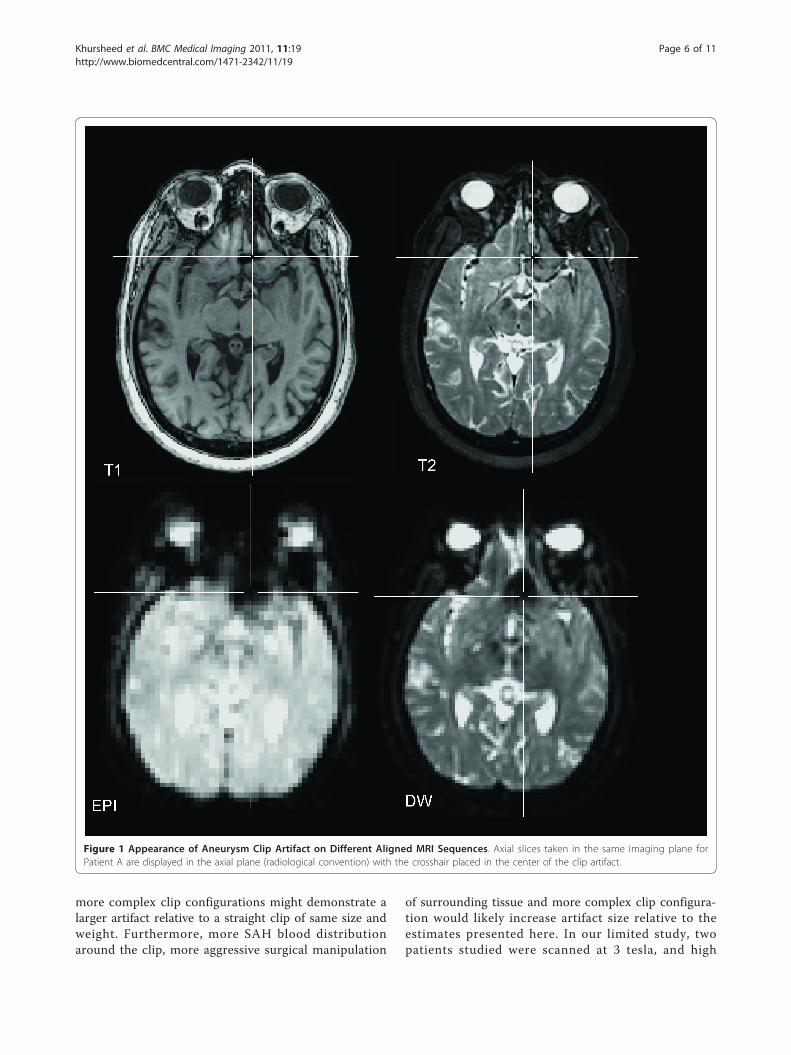
paring an image plane across different MR sequencestaken through the center of the artifact (Figure 1). Onall sequences in both patients, the maximum length
Khursheed et al. BMC Medical Imaging 2011, 11:19http://www.biomedcentral.com/1471-2342/11/19
Page 4 of 11

measured along the long axis of the artifact was largerthan the actual clip length, ranging from 2.82 times thelength of the clip for the T2 structural sequence to 6.5times the length of the clip for the EPI sequence.Computed whole-brain volumes were 229,705 and
242,419 mm3 for patient’s A and B respectively. Thesevalues are in line with an average of whole brainvolumes, 235,821 (SD 22,571), computed from a groupof similar aged controls (N = 7, 3F, 1LH, 54.0 years, SD7.8) published recently [49]. To help aide visualizationof clip artifact volume in relation to brain size, exampleartifact volume reconstructions from the range of MRsequences are shown in relation to patient cortical sur-face models in Figure 2.The ability to reconstruct nearby white matter fiber
pathways as a function of distance from the artifact bor-der was examined in both patients. The artifact maskdelineated on the diffusion-weighted sequence wasexpanded by 3D morphological dilation incrementally in2 mm steps (Figure 3) and the volume of the white mat-ter tracts intersecting the dilated mask was found toincrease markedly from the border, from 592 microliters within 2 mm of the artifact border to 2752 microliters within 6 mm of the border for Patient A with the7 mm clip, and from 1348 to 4556 in Patient B with thelarger 11 mm clip. After just one increment of dilation,pathways of the uncinate fasciculus (connecting anteriortemporal and ventral frontal lobe) and pathways of theinferior fronto-occipital fasciculus, connecting parieto-occipital and ventral frontal lobe [57-59], were visible inthe data of both patients (Figure 3).
DiscussionWidespread application of advanced imaging techniquesto investigate changes in brain structure, function, andconnectivity following SAH is presently slowed by thefact that interpretation of the images obtained frompatients with implanted aneurysm clips is complicatedby the presence of an artifact in and around the clip.
The spatial extent of the artifact with respect to clip sizeand type of MR sequence has not been previouslydescribed from images collected from aSAH patients invivo. The main objective in conducting this study was todescribe the extent of image artifact produced byimplanted titanium aneurysm clips in aSAH patientsacross a set of advanced MR sequences necessary toquantify structural, functional, and connectivity patterns.A secondary objective was to describe using DW-MRItractography methods how well white matter connec-tional patterns could be reconstructed around the clipartifact. This second objective is critical, we believe, forbeginning to understanding how well modern neuroima-ging approaches to studying white matter can be appliedin SAH patients who have implanted aneurysm clips.We focused on just two subarachnoid hemorrhage
patients with similar clinical characteristics, includingsurgical approach, aneurysm location, Fisher’s SAHgrade [21], global distribution of subarachnoid blood,and implanted clip size and orientation. We acknowl-edge several limitations of this approach. First, the clini-cal characteristic of these patients were not matchedexactly. Second, the small number of patients studieddoes not allow generalization of the findings to theentire population of aSAH patients who are treated neu-rosurgically with clips. We cannot predict for certainbased on the findings presented here how artifact willmanifest in patients with clips placed on, for example,the middle cerebral or basilar arteries. However, the twopatients imaged in this study had clips placed after rup-ture of ACOM and ACA. Estimates based on large sam-ple studies of western [60] and eastern [61,62] patientpopulations indicate that these locations account forbetween 29 to 38 percent of total intracranial aneurysmruptures. This suggests that, despite the limited scope ofour study, our findings may be generalizable to a size-able subset of the population with titanium clips placedat these locations. Finally, we studied patients implantedonly with straight titanium alloy clips. Patients with
Table 2 Artifact Measurements as a Function of MR Sequence
Sequence Artifact Volumemm3
% Of Brain Volume Artifact Lengthmm
Artifact/Clip Length Ratio
Patient A T1 1038 (366) 0.45 (0.16) 25.31 (0.09) 3.61
(7 mm clip) T2 484 (185) 0.21 (0.08) 20.28 (1.80) 2.89
EPI 7366 (4767) 3.21 (2.08) 45.91 (4.72) 6.55
DW 3792 (168) 1.65 (0.07) 36.62 (2.39) 5.23
Patient B T1 2223 (428) 0.92 (0.18) 34.52 (1.19) 3.18
(11 mm clip) T2 1093 (101) 0.45 (0.04) 31.05 (0.21) 2.82
EPI 9557 (2728) 3.94 (1.13) 52.33 (4.41) 4.75
DW 6897 (2644) 2.85 (1.09) 42.97 (0.36) 3.90
The mean of the two raters’ estimates is listed with standard deviation in parentheses.
Khursheed et al. BMC Medical Imaging 2011, 11:19http://www.biomedcentral.com/1471-2342/11/19
Page 5 of 11
more complex clip configurations might demonstrate alarger artifact relative to a straight clip of same size andweight. Furthermore, more SAH blood distributionaround the clip, more aggressive surgical manipulation
of surrounding tissue and more complex clip configura-tion would likely increase artifact size relative to theestimates presented here. In our limited study, twopatients studied were scanned at 3 tesla, and high
Figure 1 Appearance of Aneurysm Clip Artifact on Different Aligned MRI Sequences. Axial slices taken in the same imaging plane forPatient A are displayed in the axial plane (radiological convention) with the crosshair placed in the center of the clip artifact.
Khursheed et al. BMC Medical Imaging 2011, 11:19http://www.biomedcentral.com/1471-2342/11/19
Page 6 of 11

Figure 2 Example Clip Artifact Volume Reconstructions in Relation to Cortical Surface Models. Basal and mid-sagittal views of eachpatient’s cortical surface model are shown shaded by curvature (top row) and as a point cloud (second row). Superimposed on the point coulddisplays to aide artifact visualization are the reconstructed clip artifact volumes colored according to MR sequence in descending order by clipvolume as percentage of brain volume (T2 = green, T1 = yellow, DWI = blue, EPI = red). These artifact volumes are 0.27%, 0.56%, 1.70%, and4.67% respectively for Patient A (two columns on left), and 0.48%, 1.04%, 3.62%, and 4.74% respectively for Patient B (two columns on right).
Khursheed et al. BMC Medical Imaging 2011, 11:19http://www.biomedcentral.com/1471-2342/11/19
Page 7 of 11

resolution T1- and T2-weighted structural, BOLD-fMRI,and DW-MRI sequences were acquired. The imagevolumes were aligned to the native image space of thehigh resolution T1 anatomical scan, and experiencedraters used semi-automated tracing methods to delineatethe image artifact around the aneurysm clip on each setof image volumes. For each sequence, a 3D mask of theartifact was produced. Mask volumes were comparedacross sequences and between raters. Inter-rater reliabil-ity was highly significant, indicating that the extent ofthe image artifact was reproducible across sequences.Most importantly, we found evidence to reject our initial
null hypothesis that the spatial extent of the image arti-fact is the same across 3T MR acquisition sequences.Our main result is that the size of the clip artifact variesconsiderably; it is smallest in volume on the structuralscans and largest on the echo-planar BOLD-fMRI scans,with the artifact on the DW-MRI sequence falling inbetween.The result from our second objective is that we were
surprised to learn that it is possible to reconstruct whitematter pathways very close to the artifact border usingDW-MRI data. For the two patients studied here, path-ways comprising uncinate and inferior fronto-occipital
Figure 3 White Matter Tractography Reconstruction Ability Increases as a Function of Distance from Artifact Border. The artifact masksderived from the diffusion-weighted image volume were dilated in 2 mm increments, and the volume of white matter tracts that intersectedany component of the dilated mask was computed. Components of the uncinate fasciculus (loop from temporal to ventral frontal) and inferiorfronto-occipital fasciculus (occipital to temporal) in both patients’ left hemisphere can be resolved within 2 mm of the artifact border by visualinspection of the orange tractography pathways. Both views are sagittal left hemisphere where A, P, S, and I indicate anterior, posterior, superiorand inferior directions.
Khursheed et al. BMC Medical Imaging 2011, 11:19http://www.biomedcentral.com/1471-2342/11/19
Page 8 of 11

fasciculi [57-59] were both visible within 2 mm of theartifact border. After applying a three dimensional arti-fact mask generation method, we also provide multipleobjective measures, including an artifact/clip-lengthratio and artifact volume/brain tissue volume ratio thatcan be used to guide neuroimaging researchers in esti-mating the size of the artifact if clip characteristics likelength are known.In hindsight, it is not too surprising that the clip arti-
fact differs considerably in size given that the sequencesrequired to measure structural, functional, and diffusion-weighted connectivity information differ according toradiofrequency pulse design, gradient readout times, andspatial resolution. It is necessary that these parametersdiffer in order for scanners to be capable of measuringdifferent MR signal characteristics, and it is important toknow how clip artifact size will differ as a function oftype of imaging sequence. In this study, we made noattempt to adjust parameters in individual patients tominimize artifact. This was a deliberate choice made tominimize signal variance as a function of acquisitionparameters so that within-group (SAH) analyses andbetween-group (SAH vs. matched control) can be con-ducted in future larger-sample studies. For functionalimaging, there is a necessary tradeoff between high spa-tial and temporal resolution when acquiring fMRI-BOLDsequences. In our echo-planar fMRI sequence, we used a2 second repetition time, meaning that hemodynamicactivity throughout 34 slices representing the entirevolume of the brain is captured every two seconds. Thisacquisition speed is common for task-evoked and reststate designs. Fast fMRI acquisitions require larger voxelsizes of ~ 3 mm in order to image most of the brain.Advances in head coil design, including the number ofchannels to record the signal, will in the future allowacquisition of higher spatial resolution scans in a shortertime, but the spatial-temporal tradeoff will remain.Artifact size in the fMRI-BOLD sequence acquired here
is also significantly magnified by a susceptibility artifact atthe base of the brain, near where many aneurysm clips arelikely to be placed. This well-known dropout of signal dueto a large static magnetic field inhomogeneity that occursnear air/brain interfaces is also present in echo-planaracquisitions in healthy controls. The interaction betweenclip location and signal dropout due to the brain tissue/aircavity susceptibility complicated delineation of the clip arti-fact because both artifacts appeared as low intensityregions and overlapped to some extent. However, the tis-sue-air artifact extends outside the border of the brain, soby constraining our artifact delineation to within-brain bor-ders as defined by the high-resolution anatomical imageswe were able to minimize this error to some extent.Outside the boundary of our artifact masks, imaging
signal integrity recovers quickly. This was demonstrated
by reconstructing diffusion tensor tractography fiberpathways as the artifact mask volumes were parametri-cally dilated in three dimensions. As we used standarddiffusion tensor imaging methods here with relativelyfew gradient orientations, future investigations shouldevaluate the feasibility of collecting higher direction dif-fusion-weighted sequences in order to more accuratelyreconstruct white matter pathways in SAH patientsusing methods like Q-ball [63] and diffusion spectrumtechniques [64]. Nevertheless, during dilation, the maskvolume was expanded to include tissue outside the arti-fact and the number of tractography pathways increasedrapidly as the mask was dilated at successively higheriterations, indicating an increase in our ability to exam-ine connectivity characteristics just a few millimetersaway from the artifact mask. This is critically importantas aSAH may involve subtle damage to brain tissue nearthe rupture site. The damage may cause atrophy ofnearby gray matter, altered connectivity of nearby whitematter to other brain areas, or a combination of both.Altered network connectivity between brain regions nearthe rupture site (i.e., hippocampal complex) and regionsdistal to the rupture site (i.e., ventrolateral prefrontal cor-tex) is one hypothesis that could account for complexdeficits [4], like those of memory, mood, and decisionmaking, that have been reported in aSAH patients. Toadequately test this hypothesis, white matter connectivity,for tracts thought to be important for memory like theuncinate fasciculus (shown in Figure 3 for both patients)must be reconstructed, and the extent of artifact due tothe aneurysm clip must be quantifiable and reproduciblein order to know in which areas connectivity informationis reliable. The results presented here show how this maybe achieved through construction of a spatial confidenceboundary of signal integrity, represented numerically byan artifact mask volume. These volumes should be con-structed from the MR imaging data obtained from eachaSAH patient. Across patients, the spatial extent of theseartifact masks may be considered in a group analysis ofthe imaging data. Using this approach, it would be possi-ble to quantify the degree of uncertainty across patientsabout whether signal in a particular region of the brain iscompletely or partially affected by clip artifact. Impor-tantly, this method is also generalizable to other methodsof assessing connectivity, like functional connectivitycomputed from rest-state fMRI [65].
ConclusionsIn summary, we show that artifact due to the presenceof titanium clips differs across a range of advancedhigh-field (3 tesla) MR imaging sequences in aneurysmalsubarachnoid hemorrhage patients. This artifact is easilyquantifiable and 3D volumes of confidence can be pro-duced outside of which the MR signal for measuring
Khursheed et al. BMC Medical Imaging 2011, 11:19http://www.biomedcentral.com/1471-2342/11/19
Page 9 of 11

structure, function, and diffusion-weighted connectivityinformation may be obtained. We also show that DW-MRI tractography can reveal white matter pathways inclose proximity to the artifact borders. These resultshave important methodological implications for studyingbrain changes with advanced neuroimaging tools inpatients who recover from subarachnoid hemorrhageafter neurosurgical treatment.
FundingThis project was supported by the Brain AneurysmFoundation and the Vivian L. Smith Foundation forNeurological Research. Partial funding for the purchaseof the Philips 3T scanner used to collect the imagingdata was provided by NIH S10 RR19186. No fundingwas provided by manufacturers of clips studied.
Abbreviations3T: three tesla; AFNI: Analysis of Functional Neuroimages; ANOVA: analysis ofvariance; ACA: anterior cerebral artery; ACOM: anterior communicating artery;aSAH: aneurysmal subarachnoid hemorrhage; BOLD: blood oxygen leveldependent; DTI: diffusion tensor imaging; DW-MRI: diffusion-weightedmagnetic resonance imaging; EPI: echo-planar imaging; ETL: echo trainlength; FA: flip angle; fMRI: functional magnetic resonance imaging; FOV:field of view; GOS: Glasgow Outcome Scale; mRS: modified Rankin Scale;MRI: magnetic resonance imaging; TE: echo time; TR: repetition time
AcknowledgementsWe thank Vipulkumar S. Patel for help collecting the MRI data, Vicki Massie andJamie Yost from Aesculap, USA, for providing detailed technical specificationsabout the aneurysm clips, Yuxiang Zhou, Ph.D. for help interpreting imagingdata, and Florencia Leyton for help with patient scheduling.
Author details1The Vivian L. Smith Department of Neurosurgery and Mischer NeuroscienceInstitute, The University of Texas Medical School at Houston, 6431 Fannin St.,Houston TX 77030, USA. 2Department of Diagnostic and InterventionalImaging, The University of Texas Medical School at Houston, 6431 Fannin St.,Houston TX 77030, USA. 3Neurointerventional/Neuro Endovascular,Radiological Sciences, University of California, Irvine, 101 The City DriveSouth, Rt. 140, South Bldg. 56, Mail Code: 5005, Orange, CA 92868, USA.
Authors’ contributionsFK, FR, and TME collected the MRI data. DK did the surgeries and referredpatients for research. FK and TME analyzed the data. SS helped interpret theimaging data. FK and TME wrote the paper. All authors read and approvedthe final manuscript.
Competing interestsThe authors declare that they have no competing interests.
Received: 25 May 2011 Accepted: 4 October 2011Published: 4 October 2011
References1. Lovelock CE, Rinkel GJ, Rothwell PM: Time trends in outcome of
subarachnoid hemorrhage: Population-based study and systematicreview. Neurology 2010, 74(19):1494-1501.
2. Hackett ML, Anderson CS: Health outcomes 1 year after subarachnoidhemorrhage: An international population-based study. The AustralianCooperative Research on Subarachnoid Hemorrhage Study Group.Neurology 2000, 55(5):658-662.
3. Orbo M, Waterloo K, Egge A, Isaksen J, Ingebrigtsen T, Romner B: Predictorsfor cognitive impairment one year after surgery for aneurysmalsubarachnoid hemorrhage. J Neurol 2008, 255(11):1770-1776.
4. Al-Khindi T, Macdonald RL, Schweizer TA: Cognitive and functionaloutcome after aneurysmal subarachnoid hemorrhage. Stroke 2010, 41(8):e519-536.
5. Schweizer TA, Macdonald RL: Brain hemorrhage: assessing outcome aftersubarachnoid hemorrhage. Nat Rev Neurol 2010, 6(8):427-428.
6. Haug T, Sorteberg A, Finset A, Lindegaard KF, Lundar T, Sorteberg W:Cognitive functioning and health-related quality of life 1 year afteraneurysmal subarachnoid hemorrhage in preoperative comatosepatients (Hunt and Hess Grade V patients). Neurosurgery 2010,66(3):475-484, discussion 484-475.
7. Ashburner J, Friston KJ: Voxel-based morphometry–the methods.Neuroimage 2000, 11(6 Pt 1):805-821.
8. Good CD, Johnsrude IS, Ashburner J, Henson RN, Friston KJ, Frackowiak RS:A voxel-based morphometric study of ageing in 465 normal adulthuman brains. Neuroimage 2001, 14(1 Pt 1):21-36.
9. Ogawa S, Tank DW, Menon R, Ellermann JM, Kim SG, Merkle H, Ugurbil K:Intrinsic signal changes accompanying sensory stimulation: functionalbrain mapping with magnetic resonance imaging. Proc Natl Acad Sci USA1992, 89(13):5951-5955.
10. Shehzad Z, Kelly AM, Reiss PT, Gee DG, Gotimer K, Uddin LQ, Lee SH,Margulies DS, Roy AK, Biswal BB, et al: The resting brain: unconstrainedyet reliable. Cereb Cortex 2009, 19(10):2209-2229.
11. Zhang D, Johnston JM, Fox MD, Leuthardt EC, Grubb RL, Chicoine MR,Smyth MD, Snyder AZ, Raichle ME, Shimony JS: Preoperative sensorimotormapping in brain tumor patients using spontaneous fluctuations inneuronal activity imaged with functional magnetic resonance imaging:initial experience. Neurosurgery 2009, 65(6 Suppl):226-236.
12. Amiez C, Kostopoulos P, Champod AS, Collins DL, Doyon J, Del Maestro R,Petrides M: Preoperative functional magnetic resonance imagingassessment of higher-order cognitive function in patients undergoingsurgery for brain tumors. J Neurosurg 2008, 108(2):258-268.
13. Basser PJ: Inferring microstructural features and the physiological stateof tissues from diffusion-weighted images. NMR in biomedicine 1995, 8(7-8):333-344.
14. Basser PJ, Pajevic S, Pierpaoli C, Duda J, Aldroubi A: In vivo fibertractography using DT-MRI data. Magn Reson Med 2000, 44(4):625-632.
15. Beaulieu C: The basis of anisotropic water diffusion in the nervoussystem - a technical review. NMR in biomedicine 2002, 15(7-8):435-455.
16. Behrens TE, Woolrich MW, Jenkinson M, Johansen-Berg H, Nunes RG,Clare S, Matthews PM, Brady JM, Smith SM: Characterization andpropagation of uncertainty in diffusion-weighted MR imaging. MagnReson Med 2003, 50(5):1077-1088.
17. Bendel P, Koivisto T, Hanninen T, Kolehmainen A, Kononen M,Hurskainen H, Pennanen C, Vanninen R: Subarachnoid hemorrhage isfollowed by temporomesial volume loss: MRI volumetric study. Neurology2006, 67(4):575-582.
18. Bendel P, Koivisto T, Kononen M, Hanninen T, Hurskainen H, Saari T,Vapalahti M, Hernesniemi J, Vanninen R: MR imaging of the brain 1 yearafter aneurysmal subarachnoid hemorrhage: randomized studycomparing surgical with endovascular treatment. Radiology 2008,246(2):543-552.
19. Bendel P, Koivisto T, Niskanen E, Kononen M, Aikia M, Hanninen T,Koskenkorva P, Vanninen R: Brain atrophy and neuropsychologicaloutcome after treatment of ruptured anterior cerebral arteryaneurysms: a voxel-based morphometric study. Neuroradiology 2009,51(11):711-722.
20. Martinaud O, Perin B, Gerardin E, Proust F, Bioux S, Gars DL, Hannequin D,Godefroy O: Anatomy of executive deficit following ruptured anteriorcommunicating artery aneurysm. Eur J Neurol 2009, 16(5):595-601.
21. Claassen J, Bernardini GL, Kreiter K, Bates J, Du YE, Copeland D, Connolly ES,Mayer SA: Effect of cisternal and ventricular blood on risk of delayedcerebral ischemia after subarachnoid hemorrhage: the Fisher scalerevisited. Stroke 2001, 32(9):2012-2020.
22. Alvarez-Linera J: 3T MRI: advances in brain imaging. Eur J Radiol 2008,67(3):415-426.
23. Blamire AM: The technology of MRI–the next 10 years? Br J Radiol 2008,81(968):601-617.
24. Okada T, Yamada H, Ito H, Yonekura Y, Sadato N: Magnetic field strengthincrease yields significantly greater contrast-to-noise ratio increase:Measured using BOLD contrast in the primary visual area. Acad Radiol2005, 12(2):142-147.
Khursheed et al. BMC Medical Imaging 2011, 11:19http://www.biomedcentral.com/1471-2342/11/19
Page 10 of 11

25. Colby GP, Coon AL, Tamargo RJ: Surgical management of aneurysmalsubarachnoid hemorrhage. Neurosurgery clinics of North America 2010,21(2):247-261.
26. Dujovny M, Agner C, Ibe O, Perlin A: Self-closing aneurysm clip: ahistorical review. Neurol Res 2010, 32(10):1011-1020.
27. Ferris NJ, Kavnoudias H, Thiel C, Stuckey S: The 2005 Australian MRI safetysurvey. AJR Am J Roentgenol 2007, 188(5):1388-1394.
28. Evans JC, Smith ET, Nixon TE: A national survey of attitudes towards theuse of MRI in patients known to have intracranial aneurysm clips. Br JRadiol 2001, 74(888):1118-1120.
29. Olsrud J, Latt J, Brockstedt S, Romner B, Bjorkman-Burtscher IM: Magneticresonance imaging artifacts caused by aneurysm clips and shunt valves:dependence on field strength (1.5 and 3 T) and imaging parameters. JMagn Reson Imaging 2005, 22(3):433-437.
30. Ooka K, Shibuya M, Suzuki Y: Motion and image artifacts of variousintracranial aneurysm clips in a magnetic field. Acta Neurochir (Wien)1996, 138(10):1241-1245.
31. Pride GL Jr, Kowal J, Mendelsohn DB, Chason DP, Fleckenstein JL: Safety ofMR scanning in patients with nonferromagnetic aneurysm clips. J MagnReson Imaging 2000, 12(1):198-200.
32. Shellock FG, Kanal E: Yasargil aneurysm clips: evaluation of interactionswith a 1.5-T MR system. Radiology 1998, 207(3):587-591.
33. Shellock FG, Tkach JA, Ruggieri PM, Masaryk TJ, Rasmussen PA: Aneurysmclips: evaluation of magnetic field interactions and translationalattraction by use of “long-bore” and “short-bore” 3.0-T MR imagingsystems. AJNR Am J Neuroradiol 2003, 24(3):463-471.
34. Shellock FG, Valencerina S: In Vitro Evaluation of MR Imaging Issues at 3Tfor Aneurysm Clips Made from MP35N: Findings and InformationApplied to 155 Additional Aneurysm Clips. AJNR Am J Neuroradiol 2009.
35. Kanal E, Shellock FG: MR imaging of patients with intracranial aneurysmclips. Radiology 1993, 187(3):612-614.
36. Kanal E, Shellock FG: Aneurysm clips: effects of long-term and multipleexposures to a 1.5-T MR system. Radiology 1999, 210(2):563-565.
37. Sutherland GR, Kelly JJ, Boehm DW, Klassen JB: Ceramic aneurysm clips.Neurosurgery 2008, 62(5 Suppl 2):ONS400-405, discussion ONS405-406.
38. Becker RL, Norfray JF, Teitelbaum GP, Bradley WG Jr, Jacobs JB, Wacaser L,Rieman RL: MR imaging in patients with intracranial aneurysm clips. Ajnr1988, 9(5):885-889.
39. Klucznik RP, Carrier DA, Pyka R, Haid RW: Placement of a ferromagneticintracerebral aneurysm clip in a magnetic field with a fatal outcome.Radiology 1993, 187(3):855-856.
40. Kangarlu A, Shellock FG: Aneurysm clips: evaluation of magnetic fieldinteractions with an 8.0 T MR system. J Magn Reson Imaging 2000,12(1):107-111.
41. Kakizawa Y, Seguchi T, Horiuchi T, Hongo K: Cerebral aneurysm clips inthe 3-tesla magnetic field. Laboratory investigation. J Neurosurg 2010,113(4):859-869.
42. Watanabe A, Seguchi T, Koyama J, Aoyama T, Miyahara T, Kakizawa Y,Hongo K: Investigation of radiofrequency-induced temperature elevationof aneurysm clips in a 3.0-tesla magnetic resonance environment.Neurosurgery 2007, 61(5):1062-1065, discussion 1065-1066.
43. ASTM: American Society for Testing and Materials (ASTM) International.Designation F 2503-05: standard practice for marking medical devicesand other items for safety in the magnetic resonance environment.West Conshohocken, PA: ASTM International; 2005.
44. Shellock FG, Woods TO, Crues JV: MR labeling information for implantsand devices: explanation of terminology. Radiology 2009, 253(1):26-30.
45. Shellock FG, Kanal E: Aneurysm clips: evaluation of MR imaging artifactsat 1.5 T. Radiology 1998, 209(2):563-566.
46. Romner B, Olsson M, Ljunggren B, Holtas S, Saveland H, Brandt L, Persson B:Magnetic resonance imaging and aneurysm clips. Magnetic propertiesand image artifacts. J Neurosurg 1989, 70(3):426-431.
47. Pradilla G, Chaichana KL, Hoang S, Huang J, Tamargo RJ: Inflammation andcerebral vasospasm after subarachnoid hemorrhage. Neurosurgery clinicsof North America 21(2):365-379.
48. Nath AR, Beauchamp MS: Dynamic changes in superior temporal sulcusconnectivity during perception of noisy audiovisual speech. J Neurosci2011, 31(5):1704-1714.
49. Ellmore TM, Hood AJ, Castriotta RJ, Stimming EF, Bick RJ, Schiess MC:Reduced volume of the putamen in REM sleep behavior disorderpatients. Parkinsonism & related disorders 2010, 16(10):645-649.
50. Khursheed F, Tandon N, Tertel K, Pieters TA, Disano MA, Ellmore TM:Frequency-specific electrocorticographic correlates of working memorydelay period fMRI activity. Neuroimage 2011, 56(3):1173-1782.
51. Cox RW: AFNI: software for analysis and visualization of functionalmagnetic resonance neuroimages. Comput Biomed Res 1996,29(3):162-173.
52. Dale AM, Fischl B, Sereno MI: Cortical surface-based analysis. I.Segmentation and surface reconstruction. Neuroimage 1999, 9(2):179-194.
53. Fischl B, Sereno MI, Dale AM: Cortical surface-based analysis. II: Inflation,flattening, and a surface-based coordinate system. Neuroimage 1999,9(2):195-207.
54. Ellmore TM, Beauchamp MS, Breier JI, Slater JD, Kalamangalam GP,O’Neill TJ, Disano MA, Tandon N: Temporal lobe white matter asymmetryand language laterality in epilepsy patients. Neuroimage 2010,49(3):2033-2044.
55. Ellmore TM, Beauchamp MS, O’Neill TJ, Dreyer S, Tandon N: Relationshipsbetween essential cortical language sites and subcortical pathways. JNeurosurg 2009, 111(4):755-766.
56. Sherbondy A, Akers D, Mackenzie R, Dougherty R, Wandell B: Exploringconnectivity of the brain’s white matter with dynamic queries. IEEE TransVis Comput Graph 2005, 11(4):419-430.
57. Catani M, Howard RJ, Pajevic S, Jones DK: Virtual in vivo interactivedissection of white matter fasciculi in the human brain. Neuroimage2002, 17(1):77-94.
58. Fernandez-Miranda JC, Rhoton AL Jr, Alvarez-Linera J, Kakizawa Y, Choi C,de Oliveira EP: Three-dimensional microsurgical and tractographicanatomy of the white matter of the human brain. Neurosurgery 2008,62(6 Suppl 3):989-1026, discussion 1026-1028.
59. Fernandez-Miranda JC, Rhoton AL Jr, Kakizawa Y, Choi C, Alvarez-Linera J:The claustrum and its projection system in the human brain: amicrosurgical and tractographic anatomical study. J Neurosurg 2008,108(4):764-774.
60. Forget TR Jr, Benitez R, Veznedaroglu E, Sharan A, Mitchell W, Silva M,Rosenwasser RH: A review of size and location of ruptured intracranialaneurysms. Neurosurgery 2001, 49(6):1322-1325, discussion 1325-1326.
61. Lai HP, Cheng KM, Yu SC, Au Yeung KM, Cheung YL, Chan CM, Poon WS,Lui WM: Size, location, and multiplicity of ruptured intracranialaneurysms in the Hong Kong Chinese population with subarachnoidhaemorrhage. Hong Kong medical journal = Xianggang yi xue za zhi/HongKong Academy of Medicine 2009, 15(4):262-266.
62. Osawa M, Hongo K, Tanaka Y, Nakamura Y, Kitazawa K, Kobayashi S: Resultsof direct surgery for aneurysmal subarachnoid haemorrhage: outcomeof 2055 patients who underwent direct aneurysm surgery and profile ofruptured intracranial aneurysms. Acta Neurochir (Wien) 2001,143(7):655-663, discussion 663-654.
63. Tuch DS: Q-ball imaging. Magn Reson Med 2004, 52(6):1358-1372.64. Wedeen VJ, Hagmann P, Tseng WY, Reese TG, Weisskoff RM: Mapping
complex tissue architecture with diffusion spectrum magnetic resonanceimaging. Magn Reson Med 2005, 54(6):1377-1386.
65. Carter AR, Astafiev SV, Lang CE, Connor LT, Rengachary J, Strube MJ,Pope DL, Shulman GL, Corbetta M: Resting interhemispheric functionalmagnetic resonance imaging connectivity predicts performance afterstroke. Ann Neurol 2010, 67(3):365-375.
Pre-publication historyThe pre-publication history for this paper can be accessed here:http://www.biomedcentral.com/1471-2342/11/19/prepub
doi:10.1186/1471-2342-11-19Cite this article as: Khursheed et al.: Artifact quantification andtractography from 3T MRI after placement of aneurysm clips insubarachnoid hemorrhage patients. BMC Medical Imaging 2011 11:19.
Khursheed et al. BMC Medical Imaging 2011, 11:19http://www.biomedcentral.com/1471-2342/11/19
Page 11 of 11