NMDA Receptor Antagonist Felbamate Reduces Behavioral Deficits and Blood-Brain Barrier Permeability...
13
732 JOURNAL OF NEUROTRAUMA Volume 24, Number 4, 2007 © Mary Ann Liebert, Inc. Pp. 000–000 DOI: 10.1089/neu.2007.0181 NMDA Receptor Antagonist Felbamate Reduces Behavioral Deficits and Blood–Brain Barrier Permeability Changes after Experimental Subarachnoid Hemorrhage in the Rat ANTONINO GERMANÒ, 1 MARIELLA CAFFO, 1 FILIPPO FLAVIO ANGILERI, 1 FRANCESCA ARCADI, 2 JENNIFER NEWCOMB-FERNANDEZ, 3 GERARDO CARUSO, 1 FRANCESCO MELI, 8 JOSE A. PINEDA, 9 STEPHEN B. LEWIS, 5 KEVIN K.W. WANG, 7 PLACIDO BRAMANTI, 2 CHIARA COSTA, 6 and RONALD L. HAYES 7 ABSTRACT Increased levels of glutamate and aspartate have been detected after subarachnoid hemorrhage (SAH) that correlate with neurological status. The NMDA receptor antagonist felbamate (FBM; 2- phenyl-1,3-propanediol dicarbamate) is an anti-epileptic drug that elicits neuroprotective effects in different experimental models of hypoxia-ischemia. The aim of this dose-response study was to eval- uate the effect of FBM after experimental SAH in rats on (1) behavioral deficits (employing a bat- tery of assessment tasks days 1–5 post-injury) and (2) blood–brain barrier (BBB) permeability changes (quantifying microvascular alterations according to the extravasation of protein-bound Evans Blue by a spectrophotofluorimetric technique 2 days post-injury). Animals were injected with 400 L of autologous blood into the cisterna magna. Within 5 min, rats received daily oral admin- istration of FBM (15, 30, or 45 mg/kg) for 2 or 5 days. Results were compared with sham-injured controls treated with oral saline or FBM (15, 30, or 45 mg/kg). FBM administration significantly ameliorated SAH-related changes in Beam Balance scores on days 1 and 2 and Beam Balance time on days 1–3, Beam Walking performance on days 1 and 2, and Body Weight on days 3–5. FBM also decreased BBB permeability changes in frontal, temporal, parietal, occipital, and cerebellar cor- tices; subcortical and cerebellar gray matter; and brainstem. This study demonstrates that, in terms 1 Neurosurgical Clinic, Department of Neurosciences, Psychiatry and Anaesthesiology, University of Messina School of Med- icine, Messina, Italy. 2 IRCCS Centro Neurolesi Bonino-Pulejo, Messina, Italy. 3 Newcomb Scientific, LLC, Houston, Texas. 4 Center for Traumatic Brain Injury Studies, Evelyn F. and William L. McKnight Brain Institute, Department of Pediatrics, University of Florida, Gainesville, Florida. 5 Department of Neurological Surgery, University of Florida, Gainesville, Florida. 6 C.I.T.S.A.L., University of Messina School of Medicine, Messina, Italy. 7 Center for Traumatic Brain Injury Studies, Evelyn F. and William L. McKnight Brain Institute, Department of Neuroscience, University of Florida, Gainesville, Florida. 8 Neurosurgical Clinic, University of Palermo, Palermo, Italy. 9 Department of Pediatrics, Washington University School of Medicine, Seattle, Washington.
-
Upload
independent -
Category
Documents
-
view
1 -
download
0
Transcript of NMDA Receptor Antagonist Felbamate Reduces Behavioral Deficits and Blood-Brain Barrier Permeability...

732
JOURNAL OF NEUROTRAUMAVolume 24, Number 4, 2007© Mary Ann Liebert, Inc.Pp. 000–000DOI: 10.1089/neu.2007.0181
NMDA Receptor Antagonist Felbamate Reduces BehavioralDeficits and Blood–Brain Barrier Permeability Changes after
Experimental Subarachnoid Hemorrhage in the Rat
ANTONINO GERMANÒ,1 MARIELLA CAFFO,1 FILIPPO FLAVIO ANGILERI,1FRANCESCA ARCADI,2 JENNIFER NEWCOMB-FERNANDEZ,3 GERARDO CARUSO,1FRANCESCO MELI,8 JOSE A. PINEDA,9 STEPHEN B. LEWIS,5 KEVIN K.W. WANG,7
PLACIDO BRAMANTI,2 CHIARA COSTA,6 and RONALD L. HAYES7
ABSTRACT
Increased levels of glutamate and aspartate have been detected after subarachnoid hemorrhage(SAH) that correlate with neurological status. The NMDA receptor antagonist felbamate (FBM; 2-phenyl-1,3-propanediol dicarbamate) is an anti-epileptic drug that elicits neuroprotective effects indifferent experimental models of hypoxia-ischemia. The aim of this dose-response study was to eval-uate the effect of FBM after experimental SAH in rats on (1) behavioral deficits (employing a bat-tery of assessment tasks days 1–5 post-injury) and (2) blood–brain barrier (BBB) permeabilitychanges (quantifying microvascular alterations according to the extravasation of protein-boundEvans Blue by a spectrophotofluorimetric technique 2 days post-injury). Animals were injected with400 �L of autologous blood into the cisterna magna. Within 5 min, rats received daily oral admin-istration of FBM (15, 30, or 45 mg/kg) for 2 or 5 days. Results were compared with sham-injuredcontrols treated with oral saline or FBM (15, 30, or 45 mg/kg). FBM administration significantlyameliorated SAH-related changes in Beam Balance scores on days 1 and 2 and Beam Balance timeon days 1–3, Beam Walking performance on days 1 and 2, and Body Weight on days 3–5. FBM alsodecreased BBB permeability changes in frontal, temporal, parietal, occipital, and cerebellar cor-tices; subcortical and cerebellar gray matter; and brainstem. This study demonstrates that, in terms
1Neurosurgical Clinic, Department of Neurosciences, Psychiatry and Anaesthesiology, University of Messina School of Med-icine, Messina, Italy.
2IRCCS Centro Neurolesi Bonino-Pulejo, Messina, Italy.3Newcomb Scientific, LLC, Houston, Texas.4Center for Traumatic Brain Injury Studies, Evelyn F. and William L. McKnight Brain Institute, Department of Pediatrics,
University of Florida, Gainesville, Florida.5Department of Neurological Surgery, University of Florida, Gainesville, Florida.6C.I.T.S.A.L., University of Messina School of Medicine, Messina, Italy.7Center for Traumatic Brain Injury Studies, Evelyn F. and William L. McKnight Brain Institute, Department of Neuroscience,
University of Florida, Gainesville, Florida.8Neurosurgical Clinic, University of Palermo, Palermo, Italy.9Department of Pediatrics, Washington University School of Medicine, Seattle, Washington.

of behavioral and microvascular effects, FBM is beneficial in a dose-dependent manner after ex-perimental SAH in rats. These results reinforce the concept that NMDA excitotoxicity is involvedin the cerebral dysfunction that follows SAH.
Key words: behavioral deficits; blood–brain barrier; cognitive deficits; felbamate; NMDA receptor; sub-arachnoid hemorrhage
INTRODUCTION
DESPITE SIGNIFICANT IMPROVEMENTS in recent decadesin the management of patients with aneurysmal sub-
arachnoid hemorrhage (SAH), including early aneurysmsurgery, endovascular techniques, and improved intensivecare, the disease is still associated with significant mortal-ity and long-term neurological morbidity and cognitivedeficits (Germanò et al., 1997, 1998a; Saveland et al.,1992). These impairments have been ascribed mainly to is-chemic brain injury occurring either during the initial bleed-ing episode or as a consequence of macro- and microvas-cular dysfunction and delayed ischemic deterioration(d’Avella et al., 1994, 1996; Delgado et al., 1985, 1986;Doczi, 1985; Doczi et al., 1986; Germanò et al., 1992, 2000;Jackowski et al., 1990; Johshita et al., 1990; Sasaki et al.,1985, 1986; Siesjo, 1992a,b; Zuccarello, 1989). Detailedknowledge about the pathological ischemic mechanisms inpatients with SAH is critical to develop more efficient treat-ments. To this end, pathophysiological and experimentaldata together have significant clinical implications for themanagement of aneurysm patients and for investigating therationale of new pharmacological approaches.
Substantial evidence has accumulated supporting therole of excitatory amino acid (EAA) neurotransmitters inthe pathogenesis of the global brain dysfunction that fol-lows SAH. Recently, bedside intracerebral microdialysismonitoring of patients with SAH exhibiting signs of de-layed ischemia revealed dramatic changes in extracellu-lar concentrations of excitatory amino acids, includingglucose, piruvate, lactate, glycerol, and glutamate, con-firming that these characteristic metabolic changes occurduring vasospasm (Unterberg et al., 2001; Zouh et al.,1996). Moreover, a correlation between EAA concentra-tions and patient clinical status has been demonstrated(Enblad et al., 1996; Hutchinson et al., 2000; Inage et al.,2000; Nilsson et al., 1996, 1999; Persson et al., 1996;Sarrafzadeh et al., 1998, 2002; Saveland et al., 1996;Schulz et al., 2000; Staub et al., 2000).
Felbamate (FBM; 2-phenyl-1,3-propanediol dicarba-mate) is a new, potent, nonsedative, anti-epileptic drugthat elicits neuroprotective effects (in vitro and in vivo)in different experimental model of hypoxia-ischemia. Inparticular, FBM was protective against hypoxic damageinduced in slices of rat hippocampus (Rekling, 2003;
EFFECTS OF FBM AFTER EXPERIMENTAL SAH
733
Wallis et al., 1990, 1992). In neonatal rats, FBM reducedcortical infarct and hippocampal necrosis following bi-lateral cortical ligation and hypoxia (Arcadi et al., 1997;Sbuaib, et al., 1996; Wasterlain et al., 1996). FBM ad-ministration reduced delayed hippocampal neuronal deathinduced by transient cerebral ischemia in the Mongoliangerbil (Wasterlain et al., 1992). Several mechanisms ofaction for FBM have been identified, including inhibitionof voltage-sensitive sodium and calcium channels, po-tentiation of �-amino-butyric acid (GABA)–mediatedchloride currents, and interaction with the NMDA recep-tor complex through inhibition of the strychnine-insensi-tive glycine binding site (McCabe et al., 1993; Swiaderet al., 2003; Taylor et al., 1995). Specifically, FBM re-duces excitatory glutamatergic neurotransmission and in-tracellular calcium concentrations (Soderpalm, 2002).
Our laboratory has developed an experimental ratmodel of SAH that has provided extensive informationabout changes induced by intracisternal blood injectionon hemodynamic, angiographic, biochemical, pathophys-iologic, and acute and chronic behavioral parameters, andparallels those changes seen in humans after SAH(d’Avella et al., 1990, 1994, 1996; Germanò et al., 1992,1994, 2000). This model has also enabled our laboratoryto study the effects of systemic administration of differ-ent molecules on these parameters (d’Avella et al., 1993;Germanò et al., 1998b, 2002; Imperatore et al., 2000).
The purpose of the present study was to evaluate theeffects of FBM administration on behavioral and mi-crovascular changes in this in vivo rodent model of SAH.In these dose-response studies, we first evaluated the ef-fect of FBM on the the behavioral consequences of SAHby employing a battery of well-characterized assessmenttests over a 5-day observation period. In the second ex-periment, we measured the effect of FBM on microvas-cular blood–brain barrier (BBB) permeability changes 2days after SAH by quantifying the extravasation of pro-tein-bound Evans Blue using a spectrophotofluorimetrictechnique in different areas of the brain parenchyma.
METHODS
The experimental protocol was approved by the Ethi-cal Committee on the Care and Use of Laboratory Ani-

mals at our Institution, and conforms to the University ofMessina guidelines for the care and the use of animals inresearch.
Experimental Design and Induction ofSubarachnoid Hemorrhage
Studies were conducted using 96 male AlbinoSprague-Dawley rats (Charles River Italia SpA, Calco,Lecco, Italy), weighing approximately 250 g. Animalswere housed (four per cage, which measured 580 �385 � 200 mm) at a constant temperature of 22°C, un-der a 12-h, light–dark cycle (light switched on at 6:00a.m.), with free access to food and water.
Rats were divided into eight experimental groups (n �12). Rats were randomly assigned to one of the eightgroups. Groups I (sham-injured � saline) and V (SAH �saline) served as controls—the latter to evaluate the ef-fects of SAH on the investigational parameters. GroupsI–IV (sham-injured � saline, sham-injured � FBM 15mg/kg, sham-injured � FBM 30 mg/kg, and sham-in-jured � FBM 45 mg/kg) were used to evaluate the pos-sible effects of vehicle and FBM administration on theinvestigational parameters in sham-injured rats. GroupsV–VIII (SAH � saline, SAH � FBM 15 mg/kg, SAH �FBM 30 mg/kg, and SAH � FBM 45 mg/kg) were nec-essary to compare the effects of FBM administration onSAH-induced changes in investigational parameters. Theexperiments were conducted as follows: on day �1, base-line pre-assessment for the behavioral tasks was per-formed. On day 0, animals underwent SAH or shamsurgery. During days 1–5, rats were subjected to the be-havioral tests. On day 2, six animals from each groupwere sacrificed to assess BBB permeability changes.These time points were chosen because we have shownthat significant behavioral changes peak 24 h after SAHand last for up to 5 days (Germanò et al., 1994, 1998b,2002; Imperatore et al., 2000). We choose to assess BBBpermeability changes 2 days after injury because previ-ous experiments conducted in our laboratory demon-strated that significant BBB alterations peaked at 48 h(Germanò et al., 1992, 1998b, 2000, 2002; Imperatore etal., 2000).
Surgical Procedures
All procedures were performed under intraperitonealketamine (Ketalar, Parke-Davis; 150 mg/kg in a volumeof 3 mL/kg) anesthesia. Rectal temperatures were con-trolled at approximately 37°C using heat lamps. SAH wasinduced in 48 rats (Groups V–VIII) by administering au-tologous blood into the subarachnoid space via the cis-terna magna. Details of the procedure have been previ-ously published (d’Avella et al., 1990, 1993, 1994, 1996;
Germanò et al., 1992, 1994, 1998b, 2000, 2002; Imper-atore et al., 2000). Briefly, the atlanto-occipital mem-brane was exposed through a midline occipital incision.For simulation of SAH, 400 �L of autologous arterialnon-heparinized blood was injected over a period of ap-proximately 30 sec into the cisterna magna via a 30-gaugeneedle fitted to a 500-�L Hamilton syringe.
Sham surgery was performed in 48 rats (Group I–IV).Specifically, the atlanto-occipital membrane was exposedthrough a midline occipital incision and punctured as de-scribed above. No intracisternal injection was made sinceprevious studies performed in our laboratory using thisrodent model demonstrated that mock CSF-injected rats(using Ringer’s solution) did not display any enduringbehavioral detriment. In addition, there was no apprecia-ble change in BBB quantitative assessment in these ani-mals as compared to control un-injected rats (Germanòet al., 1992).
Drug Administration
Within 5 min of sham-injury (Groups II–IV) or SAH(Groups VI–VIII), rats received oral administration ofFBM (generously provided by Shering Plough, Milano,Italy) as a bolus at a dose of 15 mg/kg (Groups II andVI), 30 mg/kg (Groups III and VII), or 45 mg/kg (GroupsIV and VIII). Rats in Group 1 and V received vehicle(sterile saline) as a bolus. Rats were given oral FBM ororal saline for either 2 or 5 consecutive days by gavage,according to the general treatment plan. This dose regi-men was selected on the basis of previous clinical andexperimental evidence demonstrating the pharmacody-namic, pharmacokinetic, and toxicological profile ofFBM (Adusumalli et al., 1991; Graves et al., 1989; Lep-pik et al., 1993; Palmer et al., 1993; Wallace Laborato-ries, 2003).
Behavioral Assessment Protocol
Beam Balance Test, Beam Walking Test, and measureof body weight were used to characterize the enduringbehavioral deficits over a 5-day period after SAH or shamsurgery, and the effect of FBM on these deficits. Base-line pre-assessment of the behavioral tasks was per-formed on day �1 prior to SAH or sham surgery. Thesetests have been extensively used in our laboratory in thisexperimental rodent SAH model, and we have reportedsignificant behavioral changes that peak 24 h after SAHthat last for up to 5 days (Germanò et al., 1994, 1998b,2002; Imperatore et al., 2000).
Beam balance. The Beam Balance Test is a task thatassesses both motor and vestibular functioning by quan-tifying the animal’s ability to balance on a narrow
GERMANÒ ET AL.
734
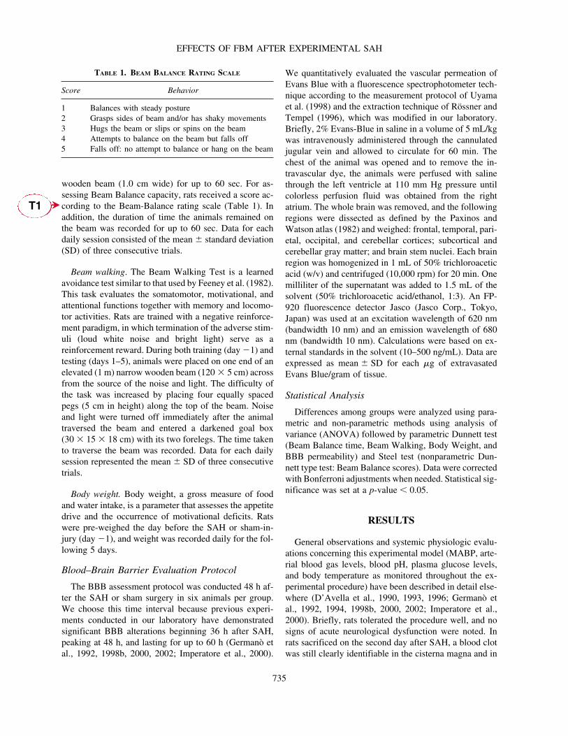
wooden beam (1.0 cm wide) for up to 60 sec. For as-sessing Beam Balance capacity, rats received a score ac-cording to the Beam-Balance rating scale (Table 1). Inaddition, the duration of time the animals remained onthe beam was recorded for up to 60 sec. Data for eachdaily session consisted of the mean � standard deviation(SD) of three consecutive trials.
Beam walking. The Beam Walking Test is a learnedavoidance test similar to that used by Feeney et al. (1982).This task evaluates the somatomotor, motivational, andattentional functions together with memory and locomo-tor activities. Rats are trained with a negative reinforce-ment paradigm, in which termination of the adverse stim-uli (loud white noise and bright light) serve as areinforcement reward. During both training (day �1) andtesting (days 1–5), animals were placed on one end of anelevated (1 m) narrow wooden beam (120 � 5 cm) acrossfrom the source of the noise and light. The difficulty ofthe task was increased by placing four equally spacedpegs (5 cm in height) along the top of the beam. Noiseand light were turned off immediately after the animaltraversed the beam and entered a darkened goal box(30 � 15 � 18 cm) with its two forelegs. The time takento traverse the beam was recorded. Data for each dailysession represented the mean � SD of three consecutivetrials.
Body weight. Body weight, a gross measure of foodand water intake, is a parameter that assesses the appetitedrive and the occurrence of motivational deficits. Ratswere pre-weighed the day before the SAH or sham-in-jury (day �1), and weight was recorded daily for the fol-lowing 5 days.
Blood–Brain Barrier Evaluation Protocol
The BBB assessment protocol was conducted 48 h af-ter the SAH or sham surgery in six animals per group.We choose this time interval because previous experi-ments conducted in our laboratory have demonstratedsignificant BBB alterations beginning 36 h after SAH,peaking at 48 h, and lasting for up to 60 h (Germanò etal., 1992, 1998b, 2000, 2002; Imperatore et al., 2000).
We quantitatively evaluated the vascular permeation ofEvans Blue with a fluorescence spectrophotometer tech-nique according to the measurement protocol of Uyamaet al. (1998) and the extraction technique of Rössner andTempel (1996), which was modified in our laboratory.Briefly, 2% Evans-Blue in saline in a volume of 5 mL/kgwas intravenously administered through the cannulatedjugular vein and allowed to circulate for 60 min. Thechest of the animal was opened and to remove the in-travascular dye, the animals were perfused with salinethrough the left ventricle at 110 mm Hg pressure untilcolorless perfusion fluid was obtained from the rightatrium. The whole brain was removed, and the followingregions were dissected as defined by the Paxinos andWatson atlas (1982) and weighed: frontal, temporal, pari-etal, occipital, and cerebellar cortices; subcortical andcerebellar gray matter; and brain stem nuclei. Each brainregion was homogenized in 1 mL of 50% trichloroaceticacid (w/v) and centrifuged (10,000 rpm) for 20 min. Onemilliliter of the supernatant was added to 1.5 mL of thesolvent (50% trichloroacetic acid/ethanol, 1:3). An FP-920 fluorescence detector Jasco (Jasco Corp., Tokyo,Japan) was used at an excitation wavelength of 620 nm(bandwidth 10 nm) and an emission wavelength of 680nm (bandwidth 10 nm). Calculations were based on ex-ternal standards in the solvent (10–500 ng/mL). Data areexpressed as mean � SD for each �g of extravasatedEvans Blue/gram of tissue.
Statistical Analysis
Differences among groups were analyzed using para-metric and non-parametric methods using analysis ofvariance (ANOVA) followed by parametric Dunnett test(Beam Balance time, Beam Walking, Body Weight, andBBB permeability) and Steel test (nonparametric Dun-nett type test: Beam Balance scores). Data were correctedwith Bonferroni adjustments when needed. Statistical sig-nificance was set at a p-value � 0.05.
RESULTS
General observations and systemic physiologic evalu-ations concerning this experimental model (MABP, arte-rial blood gas levels, blood pH, plasma glucose levels,and body temperature as monitored throughout the ex-perimental procedure) have been described in detail else-where (D’Avella et al., 1990, 1993, 1996; Germanò etal., 1992, 1994, 1998b, 2000, 2002; Imperatore et al.,2000). Briefly, rats tolerated the procedure well, and nosigns of acute neurological dysfunction were noted. Inrats sacrificed on the second day after SAH, a blood clotwas still clearly identifiable in the cisterna magna and in
EFFECTS OF FBM AFTER EXPERIMENTAL SAH
735
TABLE 1. BEAM BALANCE RATING SCALE
Score Behavior
1 Balances with steady posture2 Grasps sides of beam and/or has shaky movements3 Hugs the beam or slips or spins on the beam4 Attempts to balance on the beam but falls off5 Falls off: no attempt to balance or hang on the beam
T1
�

the basal cisterns. No extradural hemorrhages werefound, while the presence of blood in the ventricles wasdetected in approximately 25% of cases. In rats sacrificedon day 5 post-SAH, no blood clot was visible in the cis-terna magna or in other brain loci. Ketamine, vehicle, orFBM administration per se did not induce any significantalteration in the rodent physiologic parameters, neither incontrol animals nor in SAH rats.
Behavioral Assessment
None of the experimental groups differed significantlyfrom one another in baseline pre-injection assessments(day �1) (Figs. 1–4).
Beam balance. Vehicle and FBM administration(Groups I–IV) did not induce any significant beam bal-ance alteration in sham-injured animals (Figs. 1 and 2).The SAH-injured saline-treated rats (Group V) exhibitedsignificant deficits on days 1 and 2 in beam balance
scores (p � 0.05) and on days 1–3 for beam balance time(p � 0.05), as compared to sham-injured saline-treatedanimals. SAH-injured 45 mg/kg FBM-treated animals(Group VIII) exhibited a significantly improved beambalance score on days 1 (p � 0.01) and 2 (p � 0.05) ascompared to SAH-injured saline-treated animals. SAH-injured 30 mg/kg FBM-treated animals (Group VII) ex-hibited a significantly improved beam balance score onday 2 (p � 0.05), compared to SAH-injured saline-treated animals (Fig. 1). SAH-injured animals who re-ceived 15, 30, or 45 mg/kg FBM (Groups VI–VIII) ex-hibited a significantly improved beam balance time ondays 1–3 (p � 0.05) as compared to SAH-injured saline-treated animals (Fig. 2).
Beam walking. Vehicle and FBM administration didnot induce any significant beam walking alteration insham-injured animals (Fig. 3; groups I–IV). The SAH-injured saline-treated rats (Group V) exhibited signifi-
GERMANÒ ET AL.
736
FIG. 1. Bar graph showing beam balance test scores for each daily time point (day �1 to day 5). The values shown are themeans of three consecutive trials � SD for each group. *p � 0.05 and **p � 0.01 SAH � saline (Group V) vs. SAH � FBM 15,30, and 45 mg/kg groups (Groups VI–VIII). Vehicle and FBM administration did not induce any significant change in beam bal-ance score in sham-injured animals. The SAH-injured saline-treated rats had significant deficits in beam balance scores on days1 and 2, compared to sham-injured saline-treated animals. SAH-injured 45 mg/kg FBM-treated animals had a significantly im-proved score on days 1 and 2, and SAH-injured 30 mg/kg FBM-treated animals had a significantly improved beam balance scoreon day 2, compared to SAH-injured saline-treated animals.
F1-4 �

cantly increased latency to traverse the beam on days 1–4(day 1, p � 0.001; day 2, p � 0.01; days 3–4, p � 0.05),compared to sham-injured saline-treated animals. InSAH-injured animals who received 15, 30, or 45 mg/kgFBM, latency to traverse the beam was significantly re-duced on days 1 and 2 (day 1 FBM 45 and 30 mg/kg,p � 0.05; FBM 15 mg/kg, p � 0.01; day 2 p � 0.05),compared to SAH-injured saline-treated rats.
Body weight. Vehicle and FBM administration did notinduce any significant body weight alteration in sham-in-jured animals (Groups I–IV). The SAH-injured saline-treated rats (Group V) exhibited significant decreasedbody weight on days 1–5 (p � 0.05), as compared tosham-injured saline-treated animals. Administration of45 mg/kg FBM significantly reduced the SAH-relatedloss in body weight on days 3–5 (p � 0.05), as comparedto SAH-vehicle treated rats. Conversely, administrationof 15 or 30 mg/kg FBM did not affect the SAH-related
loss in body weight, as compared to SAH-vehicle treatedrats (Fig. 4).
Blood–Brain Barrier Evaluations
Table 2 summarizes the mean concentration � SD of ex-travasated Evans Blue dye (expressed as micrograms pergram of brain tissue) for all loci examined in the eight ex-perimental groups. In sham-injured saline-treated rats, base-line levels of Evans Blue ranged from 2.075 � 0.45, to2.787 � 0.09 (Group I). Values obtained in sham-injuredand SAH-injured saline-treated animals were consistentwith those described in previous experiments performed inour laboratory (D’Avella et al., 1990, 1993, 1996; Germanòet al., 1992, 1994, 1998b, 2000, 2002; Imperatore et al.,2000). As compared with sham-injured saline-treated ani-mals, in SAH-injured saline-treated rats (Group V), EvansBlue dye extravasation was significantly increased (p �0.001) in the frontal, temporal, parietal, occipital, and cere-bellar cortices, subcortical gray matter, cerebellar nuclei,
EFFECTS OF FBM AFTER EXPERIMENTAL SAH
737
FIG. 2. Beam-balance test. Bar graph comparing the time (in seconds) animals remained on the beam between all groups ofrats at daily time points (days �1 to 5). The values shown are means of three consecutive trials � SD of the means for eachgroup. *p � 0.05, SAH � saline vs. SAH � FBM 15, 30, and 45 mg/kg groups (Groups VI–VIII). Vehicle and FBM adminis-tration did not induce any significant change in beam balance time in sham-injured animals. The SAH-injured saline-treated ratsexhibited significant deficits in beam balance time on days 1–3, compared to sham-injured saline-treated animals. SAH-injuredanimals who received 15, 30, or 45 mg/kg FBM (Groups VI–VIII) had significantly improved beam balance time on days 1–3,compared to SAH-injured saline-treated animals.
T2�

and brainstem (Group V). In sham-injured animals, FBMadministration per se did not cause any significant changein Evans Blue dye extravasation (Groups II–IV). As com-pared to the SAH-injured saline-treated animals, 15 mg/kgFBM administration (Group VI) significantly decreased theSAH-induced BBB permeability changes in frontal, tem-poral, and parietal cortices, and brainstem (p � 0.01); 30mg/kg FBM administration (Group VII) significantly de-creased the SAH-induced BBB permeability changes infrontal, temporal, and cerebellar cortices, subcortical graymatter, and brainstem (p � 0.01), and parietal cortex (p �0.05); and 45 mg/kg FBM administration (Group VIII) sig-nificantly decreased the SAH-induced BBB permeabilitychanges in frontal, temporal, parietal, and occipital cortices,subcortical gray matter, cerebellar nuclei, brainstem (p �0.01), and cerebellar cortex (p � 0.01).
DISCUSSION
In this study, we evaluated the effects of FBM in a ro-dent model of SAH by assessing the (1) behavioral
changes over a 5-day period and (2) BBB function 48 hafter SAH. FBM significantly reduced SAH-related be-havioral alterations and microvascular BBB breakdownin a dose-dependent manner. In sham-injured animals,FBM administration did not demonstrate any significanteffect on the chosen investigational parameters. The datareported here may also have potential clinical implica-tions for the management of SAH patients.
Considerable clinical and experimental work hasshown that SAH induces focal and generalized distur-bances of several brain functions, such as reduced cere-bral blood flow (d’Avella et al, 1996; Delgado et al.,1986; Jackowski et al, 1990; Prunnel et al, 2003, 2004),impaired control of vascular autoregulation (d’Avella etal., 1994; Delgado et al., 1985; Sasaki et al., 1982, 1985,1986), BBB dysfunction (d’Avella et al., 1994; Doczi etal., 1985, 1986; Germanò et al., 1992, 1998b, 2000, 2002;Gules et al, 2003; Imperatore et al., 2000; Zuccarello etal., 1989), brain edema and increased intracranial pres-sure (ICP) (Jackowski et al, 1990), and free-radical gen-eration associated with lipid peroxidation phenomena(d’Avella et al., 1990, 1994; Doczi, 1985; Doczi et al.,
GERMANÒ ET AL.
738
FIG. 3. Beam-walking test. Bar graph showing time (in seconds) taken to traverse the beam at daily time points (days �1 to5). The values shown are means of three consecutive trials � SD of the means for each group. *p � 0.05 and **p � 0.01 SAH �saline vs. SAH � FBM groups. Vehicle and FBM administration did not induce any significant beam walking alteration in sham-injured animals. The SAH-injured saline-treated rats had a significantly increased latency to traverse the beam on days 1–4, com-pared to sham-injured saline-treated animals. In SAH-injured animals who received 15, 30, or 45 mg/kg FBM, latency to traversethe beam was significantly reduced on days 1–2, as compared with SAH-injured saline-treated rats.

1986; Enblad et al., 1996; Germanò et al., 1992, 1998b,2000, 2002; Imperatore et al., 2000; Jackowski et al.,1990; Kawamata et al., 1997; Koide et al., 1985; Nilssonet al., 1996, 1999; Persson et al., 1996; Siesjo et al,1992a,b; Smith et al., 1996; Staub et al., 2000; Yoshidaet al., 1980; Zuccarello et al., 1989).
Though mortality rates related to SAH have signifi-cantly decreased over the last decade, the issue of long-term cognitive and neuropsychological outcome is gain-ing increasing attention by the neurosurgical community(Germanò et al., 1997, 1998a; Saveland et al., 1992). Theneurotoxic effects of widespread subarachnoid blood as-sociated with transient cerebral ischemia cause diffusecortical damage that occurs immediately after the hem-orrhage (Doczi, 1985; Doczi et al. 1986; Enblad et al.,1996; Gotoh et al., 1984; Hutchinson et al., 2000; Jack-owski et al., 1990; Nilsson et al., 1996, 1999; Persson etal., 1996; Sarrafzadeh et al., 1998, 2002; Saveland et al.,
1996; Schulz et al., 2000; Staub et al., 2000; Untemberget al., 2001). This damage results from the interaction ofnumerous pathophysiologic factors where presumably,the blood itself and/or active substances derived in partfrom the degradation of the extravasated blood are cen-trally involved. Previous investigations conducted in ourlaboratory have demonstrated that this rodent model ofSAH is associated with enduring measurable behavioraland neurologic alterations that may parallel those seen inhumans after SAH (Germanò et al., 1994, 1997, 1998a,b,2002; Imperatore et al., 2000). It should be emphasizedthat, because of the significant interspecies difference, adirect extrapolation of our results to humans may not beappropriate. However, these results further confirm therat model of SAH as a viable laboratory instrument forthe study of the pathophysiology of SAH and providenormative values for evaluation of new treatment modal-ities.
EFFECTS OF FBM AFTER EXPERIMENTAL SAH
739
FIG. 4. Body weight. Bar graph showing changes in body weight (in grams) of body weight at daily time points (days �1 to5). The values shown are means � SD of the means for each group. *p � 0.05 SAH � saline vs. SAH � FBM 45 mg/kg group.Vehicle and FBM administration did not induce any significant body weight alteration in sham-injured animals. The SAH-injuredsaline-treated rats exhibited significant decreased body weight on days 1–5, compared to sham-injured saline-treated animals. Ad-ministration of 45 mg/kg FBM significantly reduced the SAH-related loss in body weight on days 3–5, as compared with SAH-vehicle treated rats.

TA
BL
E2.
RE
SU
LT
SO
FB
BB
PE
RM
EA
BIL
ITY
CH
AN
GE
AS
SE
SS
ME
NT
SIN
AL
LG
RO
UP
SO
FR
AT
S
Eva
ns B
lue
dye
(�g/
g ti
ssue
)
Sham
�Sh
am �
FB
MSh
am �
FB
MSh
am �
FB
MSA
H �
SAH
�F
BM
SAH
�F
BM
SAH
�F
BM
Are
ave
hicl
e15
mg/
kg30
mg/
kg45
mg/
kgve
hicl
e15
mg/
kg30
mg/
kg45
mg/
kg
Fron
tal
cort
ex2.
272
� 0
.36
2.36
6�
0.6
62.
554
� 0
.44
2.87
4�
0.5
58.
619
� 1
.83
6.21
9�
1.3
3**
5.73
1�
1.1
5**
5.65
� 0
.75*
*0T
empo
ral
cort
ex2.
075
� 0
.45
2.58
7�
0.2
62.
498
� 0
.49
2.56
7�
0.4
27.
359
� 1
.11
6.34
0�
0.3
0**
5.55
� 0
.29*
*5.
058
� 0
.35*
*Pa
riet
al c
orte
x2.
233
� 0
.02
2.11
5�
0.5
52.
441
� 0
.22
2.65
4�
0.2
47.
779
� 0
.18
6.57
3�
0.0
9**
7.63
4�
0.0
4*5.
552
� 0
.08*
*O
ccip
ital
cort
ex2.
749
� 0
.44
2.44
1�
0.4
42.
544
� 0
.15
2.88
7�
0.2
48.
111
� 0
.71
6.97
8�
0.9
676.
952
� 0
.86
5.36
2�
0.4
8**
Subc
ortic
al G
M (
CPT
)2.
214
� 0
042.
156
� 0
.32
2.87
4�
0.2
42.
245
� 0
.35
7.07
6�
0.2
96.
975
� 0
.45.
534
� 0
.11*
*5.
413
� 0
.06*
*C
ereb
ella
r co
rtex
2.78
7�
0.0
92.
145
� 0
.44
2.55
2�
0.6
52.
335
� 0
.45
6.86
1�
0.6
6.43
6�
0.4
35.
141
� 0
.44*
*5.
296
� 0
.44*
Cer
ebel
lar
nucl
ei2.
167
� 0
.21
2.55
4�
0.6
72.
756
� 0
.51
2.54
6�
0.3
55.
049
� 0
.95
5.02
3�
1.0
24.
801
� 0
.82
3.78
6�
0.6
2**
Bra
in s
tem
2.66
1�
0.1
32.
658
� 0
.55
2.68
7�
0.5
72.
879
� 0
.66
5.65
2�
0.2
74.
996
� 0
.52*
*4.
502
� 0
.35*
*4.
279
� 0
.4**
Cha
nges
in B
BB
per
mea
bilty
as
evid
ence
d by
ext
rava
sate
d E
vans
Blu
e dy
e. T
he B
BB
per
mea
bilit
y ch
ange
s w
ere
mea
sure
d 2
days
aft
er s
ham
or
SAH
sur
gery
in a
ll gr
oups
of
rats
.V
alue
s ar
e ex
pres
sed
as th
e m
eans
� S
D fo
r six
ani
mal
s in
eac
h gr
oup.
G
M, g
ray
mat
ter.
*p�
0.05
; **p
�0.
01 S
AH
� s
alin
e vs
. SA
H�
FB
M g
roup
s.

Early changes in BBB function have been suspected tobe one of the major causative factors responsible for post-SAH cerebral dysfunctions (Doczi, 1985; Jackowski et al.,1990; Joshita et al., 1990; Zuccarello et al., 1989). An im-pairment of BBB following SAH has been demonstratedin humans, which develops after the acute stage of SAH,and has been correlated with the development of delayedcerebral ischemia and poor clinical outcome (Doczi, 1985;Doczi et al., 1986). This phenomenon is independent ofraised ICP, hypertension, brain edema and cerebralswelling, reduced cerebral blood flow, and disrupted brainmetabolism, which may also in turn, disrupt the BBB(d’Avella et al., 1990, 1993, 1994, 1996; Delgado et al.,1986; Doczi, 1985; Doczi et al., 1986; Germanò et al.,1992, 1994, 1998b, 2000, 2002; Gules et al., 2003; Im-peratore et al., 2000; Jackowski et al., 1990; Joshita et al.,1990; Koide et al., 1985; Peterson et al., 1990; Prunell etal., 2003, 2004; Sasaki et al., 1985, 1986; Zuccarello etal., 1989). However, the state of the capillary system af-ter experimental SAH remains controversial and therehave been relatively few studies focused on post-SAHBBB alterations. Two models of barrier opening to wa-ter-soluble materials have been proposed. For one model,it is postulated that capillary wall deformation caused byendothelial shrinkage and/or capillary vasodilatationstretches and opens the interendothelial tight junctions(d’Avella et al., 1994). For the other model, it is hypoth-esized that increased vesicular activity augments transferof material between blood and brain either by shuttling ofmicrovesicles across the endothelium or by the coalesc-ing of vesicles to form continuous intracellular channelsthrough the endothelium (Fenstermarcher et al., 1984).Recent qualitative and quantitative experimental investi-gations conducted in our laboratory employing this SAHrodent model consistently demonstrated that a markedcapillary permeability increase occurs at the very acutestage after experimental SAH (Germanò et al., 2000).
Considerable evidence has accumulated supporting therole of EAA neurotransmitters not only as mediators ofbrain injury, but in the pathogenesis of global brain dys-function that follows SAH. Data collected in both clini-cal and experimental settings have reported remarkablechanges in levels of lactate, glycerol, glutamate, and as-partate, in addition to changes in the lactate/piruvate ra-tio. These levels are in turn, affected by ischemic deteri-oration, with changes in metabolite levels varying withrespect to substance, location, and time (Enblad et al.,1996; Hutchinson et al., 2000; Inage et al., 2000; Nils-son et al., 1996, 1999; Persson et al., 1996; Sarrafzadehet al., 1998, 2002; Saveland et al., 1996; Schulz et al.,2000; Staub et al., 2000; Untemberg et al., 2001).
Brain vasculature is affected by NMDA receptors. Di-etrich et al. (1992) demonstrated that the intraventricular
injection of NMDA caused the opening of the BBB inrats, and pretreatment with the NMDA antagonists MK-801 (Yang et al., 1994), memantine (Gorgulu et al.,2000), and citicoline (Onal et al., 1997) reduced the oc-currence of BBB breakdown in rodent models of focalcerebral ischemia. The administration of the anticon-vulsivant noncompetitive EAA antagonist, remacemidehydrochloride, significantly reduced the occurrence ofpost-SAH cerebral vasospasm (Zuccarello et al., 1994).Additional support for the vascular role of the EEAs isdemonstrated by the presence of EAA receptors in cere-bral endothelial cells (Krizbai et al., 1998; Sharp et al.,2003).
FBM is a novel anti-epileptic drug with a broad anti-convulsivant profile (Arcadi et al., 1997; McCabe et al.,1993; Reckilin et al., 2003; Sbuaib et al., 1996; Soder-palm et al., 2002; Swiader et al., 2003; Wallis et al., 1992;Wasterlain et al., 1992; Taylor et al., 1995). The dosageof FBM as an anticonvulsant is 1200 mg/day for adultsand 15 mg/kg/day for pediatric patients. Dosage may beincreased to a maximum of 3600 mg/day in adults and45 mg/kg/day in pediatric patients, respectively (Palmeret al., 1993, Wallace Laboratories, 2003) FBM enters thecentral nervous system with a brain/plasma coefficient ofapproximately 0.9 (Leppik et al., 1993). The peak anti-convulsant response after oral administration of FBM isreached after 1–4 h (Graves et al., 1989). FBM inhibitsvoltage-sensitive sodium channels probably by prolong-ing their inactivation and decreasing the firing rate ofneurons. Voltage-sensitive calcium channels are alsoblocked by FBM. At high concentrations of the drug,FBM can potentiate GABA-mediated chloride currents.Initially, FBM was thought to interact with the phency-clidine/MK-801 receptor sites acting as an antagonist. Ithad also been considered a non-competitive antagonist ofglycine and glutamate. Moreover, recently published datahave shown that FBM interacts with the NR1-2B sub-type of NMDA receptors. FBM competitively inhibitsglycine-enhanced NMDA-induced intracellular calciumcurrents in mice and induced the stimulation of receptor-gated calcium ion channels, which appear to be regulatedby glycine (Swiader et al., 2003). This amino acid in-creases NMDA-evoked currents in different tissues by in-creasing the opening frequency of the NMDA channel.McCabe et al. (1993) showed that the neuroprotective ef-fects of FBM are mediated by its binding to a strychnine-insensitive glycine receptor. It is generally accepted thatneurotransmission mediated through the NMDA recep-tor complex is associated with ischemic neuronal injury.Thus, it could be theorised that reducing the magnitudeof the effects strictly linked to SAH-induced transientcerebral ischemia may ameliorate the overall outcome ofpatients with aneurysm.
EFFECTS OF FBM AFTER EXPERIMENTAL SAH
741

To date, no other study has examined the effects ofFBM on experimental SAH-induced brain dysfunction.In the current study, FBM showed beneficial propertiesat doses tolerated by animals and humans. The reportedeffect of FBM in experimental SAH is not surprising con-sidering that the alterations characterising this experi-mental model (and also the clinical picture of SAH) couldbe related in part, to secondary transient cerebral isch-emia. These results support the protective properties ofFBM and stress the need for a more profound under-standing of the mechanisms underlying the SAH-inducedinjury. Moreover, these observations provide a theoreti-cal pharmacological rationale for FBM’s beneficial ef-fect on the behavioral deficits and microvascular changesinduced after SAH, which is based on intrinsic physio-logical, biochemical, and anatomical factors. Further ef-forts in understanding the mechanism of action that un-derlies the neuroprotective effects of FBM after SAH andthe potential clinical benefits of this drug are warranted.
REFERENCES
ADUSUMALLI, V.E., YANG, J.T., WONG, K.K., et al.(1991). Felbamate pharmacokinetics in the rat, rabbit, anddog. Drug Metab. Dispos. 19, 1116–1125.
ARCADI, F.A., IMPERATORE, C., LO PRESTI, R., et al.(1997). Neuroprotective activity of felbamate following tran-sient forebrain ischemia in the gerbil. Trends Neurosci. 20,39–40.
D’AVELLA, D., GERMANO, A., SANTORO, G., et al.(1990). Effects of experimental subarachnoid hemorrhage onCSF eicosanoids in the rat. J. Neurotrauma 7, 121–129.
D’AVELLA, D., ZUCCARELLO, M., and TOMASELLO, F.(1993). Protective effect of U-78157G on local cerebral glu-cose utilization in the acute stage following subarachnoid he-morrhage (SAH), in: Cerebral Vasospasm, J.M. Findlay (ed),Elsevier Science Publishers B.V.: Amsterdam, pps. 427–430.
D’AVELLA, D., GERMANO, A., and TOMASELLO, F.(1994). Early blood-brain barrier changes after experimentalsubarachnoid haemorrhage: a quantitative and electron mi-croscopy study, in: New Trends in Management of Cerebro-Vascular Malformations. A. Pasqualin and R. Da Pian (eds),Springer-Verlag: New York, pps. 48–51.
D’AVELLA, D., CICCIARELLO, R., ZUCCARELLO, M., et al.(1996). Brain energy metabolism in the acute stage of experi-mental subarachnoid haemorrhage: local changes in cerebralglucose utilization. Acta Neurochir. (Wien). 138, 737–744.
DELGADO, T.J., BRISMAR, J., and SVENDGAARD, N.A.(1985). Subarachnoid haemorrhage in the rat: angiographyand fluorescence microscopy of the major cerebral arteries.Stroke 16, 595–605.
DELGADO, T.J., ARBAB, M.A., DIEMER, N.H., et al. (1986).Subarachnoid haemorrhage in the rat: cerebral blood flowand glucose metabolism during the late phase of cerebral va-sospasm. J. Cereb. Blood Flow Metab. 6, 590–599.
DIETRICH, W.D., ALONSO, O., HALLEY, M., et al. (1992).Intraventricular infusion of N-methyl-D-aspartate. Acuteblood–brain barrier consequences. Acta Neuropathol. 84,621–629.
DOCZI, T. (1985). The pathogenetic and prognostic signifi-cance of blood-brain barrier damage at the acute stage ofaneurysmal subarachnoid hemorrhage. Clinical and experi-mental studies. Acta Neurochir. (Wien.) 77, 110–132.
DOCZI, T., JOO, F., ADAM G., et al. (1986). Blood–brain bar-rier damage during the acute stage of subarachnoid hemor-rhage, as exemplified by a new animal model. Neurosurgery18, 733–739.
ENBLAD, P., VALTYSSON, J., ANDERSSON, J., et al.(1996). Simultaneous intracerebral microdialysis andpositron emission tomography in the detection of ischemiain patients with subarachnoid hemorrhage. J. Cereb. BloodFlow Metab. 16, 637–644.
FEENEY, D.M., GONZALEZ, A., and LAW, W.A. (1982). Am-phetamine, haloperidol and experience interact to affect rateof recovery after motor cortex injury. Science 271, 855–877.
FENSTERMACHER, J.D., and RAPAPORT, S.I. (1984).Blood–brain barrier, in: E.M. Renkin, and C.C. Michel (eds),Handbook of Physiology, Vol 4: The Cardiovascular System.Bethesda, MD: American Physiological Society, pps.969–1000.
GERMANO, A., D’AVELLA, D., CICCIARELLO, R., et al.(1992). Blood–brain barrier permeability changes after ex-perimental subarachnoid hemorrhage. Neurosurgery 30,882–886.
GERMANO, A., DIXON, E., D’AVELLA, D., et al. (1994).Behavioural deficits following experimental subarachnoidhemorrhage in the rat. J. Neurotrauma 11, 345–353.
GERMANO, A., TISANO, A., RAFFAELE, M., et al. (1997).Is there a group of early aneurysm surgery patients who canexpect to achieve a complete long-term neuropsychologicalrecovery? Acta Neurochir. 139, 507–514.
GERMANO, A., CARUSO, G., CAFFO, M., et al. (1998a).Does subarachnoid hemorrhage per se induce long-term neu-ropsychological and cognitive alterations? Acta Neurochir.140, 805–812.
GERMANO, A., IMPERATORE, C., D’AVELLA, D., et al.(1998b). Antivasospastic and brain protective effects of a hy-droxyl radical scavenger (AVS) on cerebral vasospasm andBBB permeability changes after experimental SAH. J. Neu-rosurg. 88, 1075–1081.
GERMANO, A., D’AVELLA, D., IMPERATORE, C., et al.(2000). Time course of blood–brain barrier permeabilitychanges after experimental subarachnoid hemorrhage. ActaNeurochir. 142, 575–581.
GERMANO, A., COSTA, C., DEFORD, S.M., et al. (2002).Systemic administration of a calpain inhibitor reduces be-havioral deficits and blood–brain barrier permeabilitychanges after experimental subarachnoid hemorrhage in therat. J. Neurotrauma 19, 887–896.
GORGULU, A., KINS, T., COBANOGLU, S., et al. (2000).Reduction of edema and infarction by memantine and MK-801 after focal cerebral ischaemia and reperfusion in the rat.Acta Neurochir. 142, 1287–1292.
GERMANÒ ET AL.
742
GOTOH, O., KOIDE, T., ASANO, T. et al. (1984). A modelto study ischemic brain edema in rats and the influence ofdrugs, in: Recent Progress in the Study and Therapy of BrainEdema, K.G. Go and A. Baethmann (eds), Plenum: NewYork, pps. 499–508.
GRAVES, N.M., LUDDEN, T.M., HOLMES, G.B., et al.(1989). Pharmacokinetics of felbamate, a novel antiepilepticdrug: application of mixed-effect modelling to clinical trials.Pharmacotherapy 9, 372–376.
GULES, I., SATOH, M., NANDA, A., et al. (2003). Apopto-sis, blood–brain barrier, and subarachnoid hemorrhage. ActaNeurochir. Suppl. 86, 483–487.
HUTCHINSON, P.J., AL-RAWI, P.G., O’CONNELL, M.T., etal. (2000). Biochemical changes related to hypoxia duringcerebral aneurysm surgery:combined microdialysis and tis-sue oxygen monitoring: case report. Neurosurgery 46,201–205.
IMPERATORE, C., GERMANÒ, A., D’AVELLA, D., et al.(2000). Effects of the radical scavenger AVS on behavioraland BBB changes after experimental subarachnoid hemor-rhage. Life Sci. 66, 779–790.
INAGE, Y.W., ITOH, M., WADA, K., et al. (2000). Glutamatetransporters in neonatal cerebellar subarachnoid hemorrhage.Pediatr. Neurol. 23, 42–48.
JACKOWSKI, A., CROCKARD, A., BURNSTOCK, G., et al.(1990). The time course of intracranial pathophysiologicalchanges following experimental subarachnoid hemorrhage inthe rat. J. Cereb. Blood Flow Metab. 10, 835–849.
JOSHITA, H., KASSELL, N.F., and SASAKI, T. (1990).Blood–brain barrier disturbance following subarachnoid he-morrhage in rabbits. Stroke 21, 1051–1058.
KAWAMATA, T., PETERSON, J.W., BUN, T., et al. (1997).Augmentation of both hemolysate-induced contraction andactivation of protein kinase C by submaximum activation incanine cerebral arteries in vitro. J. Neurosurg. 87, 908–915.
KOIDE, T., GOTOH, O., ASANO, T., et al. (1985). Alterationsof the eicosanoid synthetic capacity of rat brain microvesselsfollowing ischemia: relevance to ischemic brain edema. J.Neurochem. 44, 85–93.
KRIZBAI, I.A., DELI, M.A., PESTENACZ A., et al. (1998).Expression of glutamate receptors on cultured cerebral en-dothelial cells. J. Neurosci. Res. 54, 814–819.
LEPPIK, I.E., and WOLDD, D.L. (1993). Antiepileptic med-ication interactions. Neurol. Clin. 11, 905–21.
MCCABE, R.T., WASTERLAIN, C.G., KUCHARCZYK, N,et al. (1993). Evidence for anticonvulsivant and neuropro-tectant action of felbamate mediated by strychnine-insensi-tive glycine receptors. J. Pharmacol. Exp. Ther. 264,1248–1252.
NILSSON, D.G., SAVELAND, H., BORIS-MOLLER, F., etal. (1996). Increased levels of glutamate in patients with sub-arachnoid hemorrhage as measured by intreacerebral micro-dialysis. Acta Neurochir. Suppl. (Wien.) 67, 45–47.
NILSSON, D.G., BRANDT, L., UNGERSTEDT, U., et al.(1999). Bedside detection of brain ischemia using intracere-bral microdialysis: subarachnoid hemorrhage and delayed is-chemic deterioration. Neurosurgery 45, 1176–1184.
ONAL, M.Z., LI, F., TATLISUMAK, T., et al. (1997). Syner-
gistic effects of citicoline and MK-801 in temporary exper-imental focal ischemia in rats. Stroke 28, 1060–1065.
PALMER, K.J., and MCTAVISH, D. (1993). Felbamate: a re-view of its pharmacodynamic and pharmacokinetic proper-ties, and therapeutic efficacy in epilepsy. Drugs 45, 1041–65.
PAXINOS, G., and WATSON, C. (1982). The Rat Brain inStereotaxic Coordinates. Academic Press: New York.
PERSSON, L., VALTYSSON, J., ENBLAD, P., et al. (1996).Neurochemical monitoring using intracerebral microdialysisin patients with subarachnoid hemorrhage. J. Neurosurg. 84,606–616.
PETERSON, J.W., ROUSSOS, L., and KWUN, B.D. (1990).Evidence of the role of hemolysis in experimental vasospasm.J. Neurosurg. 72, 775–781.
PRUNELL, G.F., MATHIESEN, T., DIEMER, N.H., et al.(2003). Experimental subarachnoid hemorrhage: subarach-noid blood volume, mortality rate, neuronal death, cerebralblood flow, and perfusion pressure in three different rat mod-els. Neurosurgery 52, 165–175.
PRUNELL, G.F., MATHIESEN, T., and SVENDGAARD,N.A. (2004). Experimental subarachnoid hemorrhage: cere-bral blood flow and brain metabolism during the acute phasein three different models in the rat. Neurosurgery 54,426–436.
REKLING, J.C. (2003). Neuroprotective effects of anticonvul-sivants in rat hippocampal slice cultures exposed to oxy-gen/glucose deprivation. Neurosci. Lett. 335, 167–170.
RÖSSNER, W., and TEMPEL, K. (1966). Quantitative bes-timmung der permeabilität der sogenannten blut-hirn-schranke für Evans-Blau (T 1824). Med. Pharmacol. Exp. 14,169–182.
SARRAFZADEH, A.S., UNTEBERG, A.W., and LANKSCH,W.R. (1998). Bedside-microdialysis for early detection of va-sospasm after subarachnoid hemorrhage. Case report and re-view of the literature. Zentralbl. Neurochir. 59, 269–273.
SARRAFZADEH, A.S., SAKOWITZ, O.W., and KIENING,K.L. (2002). Bedside microdialysis: a tool to monitor cere-bral metabolism in subarachnoid hemorrhage patients? Crit.Care Med. 30, 1062–1070.
SASAKI, T., ASANO, T., and TAKAKURA, K. (1982). In-hibitory effects of drugs on the vasoconstriction induced bythe cerebrospinal fluid of SAH, in: Proceedings of the 11thConference on Surgical Treatment of Stroke. T. Iwabuchi(ed), Neuron-sha: Tokyo, pps. 183–188.
SASAKI, T., KASSELL, N.F., and YAMASHITA, M. (1985).Barrier disruption in the major cerebral arteries following ex-perimental subarachnoid hemorrhage. J. Neurosurg. 63,433–440.
SASAKI, T., KASSELL, N.F., and ZUCCARELLO, M. (1986).Barrier disruption in the major cerebral arteries during theacute stage after experimental subarachnoid hemorrhage.Neurosurgery 19, 177–184.
SAVELAND, H., HILLMAN, J., BRANDT, L., et al. (1992).Overall outcome in aneurysmal subarachnoid hemorrhage: aprospective study from neurosurgical units in Sweden dur-ing a 1-year period. J. Neurosurg. 76, 729–734.
SAVELAND, H., NILSSON, O.G., BORIS-MOLER, F., et al.(1996). Intracerebral microdialysis of glutamate and aspar-
EFFECTS OF FBM AFTER EXPERIMENTAL SAH
743

tate in two vascular territories after aneurysmal subarachnoidhemorrhage. Neurosurgery 38, 12–19.
SBUAIB, A., WAQAAR, T., IJAZ, M.S., et al. (1996). Neu-roprotection of felbamate: a 7- and 28-day study in transientforebrain ischemia in gerbils. Brain Res. 727, 65–70.
SCHULZ, M.K., WANG, L.P., TANGE, M., et al. (2000). Cere-bral microdialysis monitoring: determination of normal andischemic cerebral metabolisms in patients with aneurysmalsubarachnoid hemorrhage. J. Neurosurg. 93, 808–814.
SHARP, C.D., HINES, I., HOUGHTON, J., et al. (2003). Glu-tamate causes a loss in human cerebral endothelial barrier in-tegrity through activation of NMDA receptor. Am. J. Phys-iol. Heart Circ. Physiol. 285, H2592–H2598.
SIESJO, B.K. (1992a). Pathophysiology and treatment of focalcerebral ischemia. Part I: Pathophysiology. J. Neurosurg. 77,169–184.
SIESJO, B.K. (1992b). Pathophysiology and treatment of focalcerebral ischemia. Part II: Mechanisms of damage and treat-ment. J. Neurosurg. 77, 337–354.
SMITH, S.L., SCHERCH, H.M., and HALL, E.D. (1996). Pro-tective effects of tirilazad mesylate and metabolite U-89678against blood–brain barrier damage after subarachnoid hem-orrhage and lipid peroxidative neuronal injury. J. Neurosurg.84, 229–233.
SODERPALM, B. (2002). Anticonvulsivants: aspects of theirmechanisms of action. Eur. J. Pain 6, Suppl A, 3–9.
STAUB, F., GRAF, R., GABEL, P., et al. (2000). Multiple in-terstitial substances measured by microdialysis in patientswith subarachnoid hemorrhage. Neurosurgey 47, 1106–1116.
SWIADER, M., GASIOR, M., WIESLOZ, M., et al. (2003).Influence of some convulsivant agents on the protective ac-tivity of a novel antiepileptic drug, felbamate, against max-imal electroshock in mice. Pol. J. Pharmacol. 55, 649–653.
TAYLOR, L.A., MCQUADE, R.D., and TICE, M.A. (1995).Felbamate, a novel antiepileptic drug, reverses N-methyl-D-aspartate/glycine-stimulated increases in intracellular Ca2�
concentration. Eur. J. Pharmacol. 289, 229–233.UNTEBERG, A.W., SAKOWITZ, O.W., SARRAFZADEH,
A.S., et al. (2001). Role of bedside microdialysis in the di-agnosis of cerebral vasospasm following aneurysmal sub-arachnoid hemorrhage. J. Neurosurg. 94, 740–749.
UYAMA, O., OKAMURA, N., YANASE, M., et al. (1988).Quantitative evaluation of vascular permeability in the ger-bil brain after transient ischemia using Evans blue fluores-cence. J. Cereb. Blood Flow Metab. 8, 282–284.
WALLACE LABORATORIES. (2003). Product Information:FelbatolR (felbamate). Wallace Laboratories: Cranbury, NJ.
WALLIS, R.A., PANIZZON, K.L., FAIRCHILD, M.D., et al.(1990). Felbamate is a potent antihypoxic agent in hip-pocampal slice. Neurology 40, Suppl. 1, 193.
WALLIS, R.A., PANIZZON, K.L., FAIRCHILD, M.D., et al.(1992). Protective effects of felbamate against hypoxia in therat hippocampal slice. Stroke 23, 547–551.
WASTERLAIN, C.G., ADAMS L.M., HATTORI, H., et al.(1992). Felbamate reduces hypoxic-ischemic brain damagein vivo. Eur. J. Pharmacol. 212, 278–278.
WASTERLAIN, C.G., ADAMS L.M., WICHMANN, J.K., etal. (1996). Felbamate protects CA1 neurons from apoptosisin a gerbil model of global ischemia. Stroke 27, 1236–1240.
YANG, G., CHAN, P.H., BABUNA, O.A., et al. (1994). Re-duction of vasogenic edema and infarction by MK-801 in ratsafter temporary focal cerebral ischemia. Neurosurgery 34,339–345.
YOSHIDA, S., INOH, S., and ASANO, T. (1980). Effect oftransient ischemia on free fatty acids and phospholipids inthe gerbil brain. Lipid peroxidation as possible cause ofpostischemic injury. J. Neurosurg. 53, 323–331.
ZOUH, D., MAYBERG, M.R., LONDON, S., et al. (1996). Re-duction of intracellular glutathione levels produces sustainedarterial narrowing. Neurosurgery 39, 991–997.
ZUCCARELLO, M., and ANDERSON, D.K. (1989). Protec-tive effect of a 21-aminosteroid on the blood–brain barrierfollowing subarachnoid hemorrhage in rats. Stroke 20,367–371.
ZUCCARELLO, M., LEWIS, A.I., UPPUTURI, S., et al.(1994). Effect of racemide hydrochloride on subarachnoidhemorrhage-induced vasospasm in rabbits. J. Neurotrauma11, 691–698.
Address reprint requests to:Filippo Flavio Angileri, M.D.
Neurosurgical ClinicDepartment of Neurosciences,
Psychiatry and AnaesthesiologyAOU Policlinico G. Martino
Via Consolare Valeria, 198125 Messina, Italy
E-mail: [email protected]
GERMANÒ ET AL.
744