NMDA and Dopamine Converge on the NMDA-Receptor to Induce ERK Activation and Synaptic Depression in...
14
NMDA and Dopamine Converge on the NMDA-Receptor to Induce ERK Activation and Synaptic Depression in Mature Hippocampus Hanoch Kaphzan 1 , Kenneth J. O’Riordan 2 , Kile P. Mangan 2 , Jonathan M. Levenson 2 , Kobi Rosenblum 1 * 1 Center for Brain and Behavior, Department of Neurobiology and Ethology, Haifa University, Haifa, Israel, 2 Department of Pharmacology and the Waisman Center, University of Wisconsin School of Medicine and Public Health, Madison, Wisconsin, United States of America The formation of enduring internal representation of sensory information demands, in many cases, convergence in time and space of two different stimuli. The first conveys the sensory input, mediated via fast neurotransmission. The second conveys the meaning of the input, hypothesized to be mediated via slow neurotransmission. We tested the biochemical conditions and feasibility for fast (NMDA) and slow (dopamine) neurotransmission to converge on the Mitogen Activated Protein Kinase signaling pathways, crucial in several forms of synaptic plasticity, and recorded its effects upon synaptic transmission. We detected differing kinetics of ERK2 activation and synaptic strength changes in the CA1 for low and high doses of neurotransmitters in hippocampal slices. Moreover, when weak fast and slow inputs are given together, they converge on ERK2, but not on p38 or JNK, and induce strong short-term synaptic depression. Surprisingly, pharmacological analysis revealed that a probable site of such convergence is the NMDA receptor itself, suggesting it serves as a detector and integrator of fast and slow neurotransmission in the mature mammalian brain, as revealed by ERK2 activation and synaptic function. Citation: Kaphzan H, O’Riordan KJ, Mangan KP, Levenson JM, Rosenblum K (2006) NMDA and Dopamine Converge on the NMDA-Receptor to Induce ERK Activation and Synaptic Depression in Mature Hippocampus. PLoS ONE 1(1): e138. doi:10.1371/journal.pone.0000138 INTRODUCTION The decision-making regarding the meaning of a given sensory input is hypothesized to be dependent upon ‘‘heterosynaptic modulation’’ [1]. In general, two types of synapses are involved: ionotropic and metabotropic (also called ‘‘fast’’ and ‘‘slow’’). The ‘‘dialogue’’ between these different types of synapses, along with their reciprocal interactions, is referred as heterosynaptic modu- lation [1]. One example of heterosynaptic facilitation in the nervous system is the interaction between ionotropic glutamate receptors and metabotropic dopamine receptors in the mature hippocampus [2,3]. In the hippocampus, heterosynaptic modulation is involved in Long-Term Potentiation (LTP) and consolidation of long-term memory. Several studies have demonstrated that heterosynaptic modulation facilitates the induction of NMDA-dependent early and late-phases of LTP via tetanizing stimuli in area CA1 of the hippocampus [4,5,6,7]. In addition, blocking dopaminergic neurotransmission prevents or attenuates late-phase LTP [1,4,6]. Moreover, strong dopaminergic input by itself can induce LTP in the absence of tetanizing stimulation [4]. Modulation of signal transduction pathways is an attractive mechanism for heterosynaptic intonation of synaptic plasticity. Many signal transduction pathways have been implicated in the induction of synaptic plasticity in the hippocampus, and many of these pathways are interconnected, providing several opportunities for modulation. Thus, certain signal transduction molecules could serve as ‘‘coincidence detectors’’ of sensory information and its value, and would facilitate memory consolidation [8]. For example, a diminutive and insignificant sensory input might converge on the signal transduction induced as a result of the saliency of another input, resulting in enhanced activation of downstream signaling pathways and eventually result in induction of synaptic plasticity and/or consolidation of long-term memory. Numerous signaling molecules could potentially serve as a read out of coincidence detectors, but prominent in neurons are those that comprise the Mitogen Activated Protein Kinase (MAPK) signaling cascades, which include several distinct kinases that are critical for normal neuronal function [9]. They include the Extracellular signal Regulated-Kinases (ERKs), c-Jun-N-terminal kinases (JNKs) and p38 MAPK [10]. Among these, ERK1/2 occupies a unique place in the hippocampus as it has been implicated in behavioral memory, long-lasting synaptic plasticity, and biochemical information processing at the molecular level [8,10]. Specifically, ERK is involved in both early- and late-phase hippocampal LTP [11,12], and a number of different learning paradigms, including fear conditioning and spatial learning (Sweatt, 2004). Surprisingly, the convergent activation of MAPKs by dopamine and NMDA has not been studied systematically in vitro or in vivo, especially given that the temporal pattern of MAPK activation is commonly used as a crucial parameter for the readout cellular responses [13,14], and up today, though suggested, no study has solidly supported the hypothesis of MAPKs as potential fast and slow coincident detectors. That is not to rule out other molecules, acting upstream to MAPKs, that might serve as coincident detectors, such as adenylyl-cyclases [15,16]. The current study is unique in its examination of the convergence of NMDA and dopamine-mediated signaling in a direct pharmacological stimu- lation paradigm (not a dopaminomimetic agent), employing Academic Editor: Rachel Wong, University of Washington, United States of America Received September 19, 2006; Accepted December 7, 2006; Published December 27, 2006 Copyright: ß 2006 Kaphzan et al. This is an open-access article distributed under the terms of the Creative Commons Attribution License, which permits unrestricted use, distribution, and reproduction in any medium, provided the original author and source are credited. Funding: This research was supported by ISF and Psychobiology grants to KR. Competing Interests: The authors have declared that no competing interests exist. * To whom correspondence should be addressed. E-mail: [email protected] PLoS ONE | www.plosone.org 1 December 2006 | Issue 1 | e138
Transcript of NMDA and Dopamine Converge on the NMDA-Receptor to Induce ERK Activation and Synaptic Depression in...
NMDA and Dopamine Converge on the NMDA-Receptorto Induce ERK Activation and Synaptic Depression inMature HippocampusHanoch Kaphzan1, Kenneth J. O’Riordan2, Kile P. Mangan2, Jonathan M. Levenson2, Kobi Rosenblum1*
1 Center for Brain and Behavior, Department of Neurobiology and Ethology, Haifa University, Haifa, Israel, 2 Department of Pharmacology and theWaisman Center, University of Wisconsin School of Medicine and Public Health, Madison, Wisconsin, United States of America
The formation of enduring internal representation of sensory information demands, in many cases, convergence in time andspace of two different stimuli. The first conveys the sensory input, mediated via fast neurotransmission. The second conveysthe meaning of the input, hypothesized to be mediated via slow neurotransmission. We tested the biochemical conditions andfeasibility for fast (NMDA) and slow (dopamine) neurotransmission to converge on the Mitogen Activated Protein Kinasesignaling pathways, crucial in several forms of synaptic plasticity, and recorded its effects upon synaptic transmission. Wedetected differing kinetics of ERK2 activation and synaptic strength changes in the CA1 for low and high doses ofneurotransmitters in hippocampal slices. Moreover, when weak fast and slow inputs are given together, they converge onERK2, but not on p38 or JNK, and induce strong short-term synaptic depression. Surprisingly, pharmacological analysisrevealed that a probable site of such convergence is the NMDA receptor itself, suggesting it serves as a detector and integratorof fast and slow neurotransmission in the mature mammalian brain, as revealed by ERK2 activation and synaptic function.
Citation: Kaphzan H, O’Riordan KJ, Mangan KP, Levenson JM, Rosenblum K (2006) NMDA and Dopamine Converge on the NMDA-Receptor to InduceERK Activation and Synaptic Depression in Mature Hippocampus. PLoS ONE 1(1): e138. doi:10.1371/journal.pone.0000138
INTRODUCTIONThe decision-making regarding the meaning of a given sensory
input is hypothesized to be dependent upon ‘‘heterosynaptic
modulation’’ [1]. In general, two types of synapses are involved:
ionotropic and metabotropic (also called ‘‘fast’’ and ‘‘slow’’). The
‘‘dialogue’’ between these different types of synapses, along with
their reciprocal interactions, is referred as heterosynaptic modu-
lation [1]. One example of heterosynaptic facilitation in the
nervous system is the interaction between ionotropic glutamate
receptors and metabotropic dopamine receptors in the mature
hippocampus [2,3].
In the hippocampus, heterosynaptic modulation is involved in
Long-Term Potentiation (LTP) and consolidation of long-term
memory. Several studies have demonstrated that heterosynaptic
modulation facilitates the induction of NMDA-dependent early
and late-phases of LTP via tetanizing stimuli in area CA1 of the
hippocampus [4,5,6,7]. In addition, blocking dopaminergic
neurotransmission prevents or attenuates late-phase LTP [1,4,6].
Moreover, strong dopaminergic input by itself can induce LTP in
the absence of tetanizing stimulation [4].
Modulation of signal transduction pathways is an attractive
mechanism for heterosynaptic intonation of synaptic plasticity.
Many signal transduction pathways have been implicated in the
induction of synaptic plasticity in the hippocampus, and many of
these pathways are interconnected, providing several opportunities
for modulation. Thus, certain signal transduction molecules could
serve as ‘‘coincidence detectors’’ of sensory information and its
value, and would facilitate memory consolidation [8]. For
example, a diminutive and insignificant sensory input might
converge on the signal transduction induced as a result of the
saliency of another input, resulting in enhanced activation of
downstream signaling pathways and eventually result in induction
of synaptic plasticity and/or consolidation of long-term memory.
Numerous signaling molecules could potentially serve as a read
out of coincidence detectors, but prominent in neurons are those
that comprise the Mitogen Activated Protein Kinase (MAPK)
signaling cascades, which include several distinct kinases that are
critical for normal neuronal function [9]. They include the
Extracellular signal Regulated-Kinases (ERKs), c-Jun-N-terminal
kinases (JNKs) and p38 MAPK [10]. Among these, ERK1/2
occupies a unique place in the hippocampus as it has been
implicated in behavioral memory, long-lasting synaptic plasticity,
and biochemical information processing at the molecular level
[8,10]. Specifically, ERK is involved in both early- and late-phase
hippocampal LTP [11,12], and a number of different learning
paradigms, including fear conditioning and spatial learning
(Sweatt, 2004).
Surprisingly, the convergent activation of MAPKs by dopamine
and NMDA has not been studied systematically in vitro or in vivo,
especially given that the temporal pattern of MAPK activation is
commonly used as a crucial parameter for the readout cellular
responses [13,14], and up today, though suggested, no study has
solidly supported the hypothesis of MAPKs as potential fast and
slow coincident detectors. That is not to rule out other molecules,
acting upstream to MAPKs, that might serve as coincident
detectors, such as adenylyl-cyclases [15,16]. The current study is
unique in its examination of the convergence of NMDA and
dopamine-mediated signaling in a direct pharmacological stimu-
lation paradigm (not a dopaminomimetic agent), employing
Academic Editor: Rachel Wong, University of Washington, United States ofAmerica
Received September 19, 2006; Accepted December 7, 2006; Published December27, 2006
Copyright: � 2006 Kaphzan et al. This is an open-access article distributed underthe terms of the Creative Commons Attribution License, which permitsunrestricted use, distribution, and reproduction in any medium, provided theoriginal author and source are credited.
Funding: This research was supported by ISF and Psychobiology grants to KR.
Competing Interests: The authors have declared that no competing interestsexist.
* To whom correspondence should be addressed. E-mail: [email protected]
PLoS ONE | www.plosone.org 1 December 2006 | Issue 1 | e138

biochemical and extra-cellular electrophysiological essays, while
emphasizing the time dependency of the combined stimuli, the
possible molecular mechanism of such convergence, the site
accountable for it, and its expression through MAPK’s cascades.
In the present study, we determined the dose and temporal
activation patterns of ERK2 activation by each neurotransmitter
alone and simultaneously. We then tested the hypothesis that
members of the MAPK family serve as coincidence detectors of
NMDA and dopaminergic signaling in the mature brain. In
exploring this hypothesis, we discovered that regulation of ERK
phosphorylation behaves as a possible cellular site of NMDA-
dopamine signaling convergence. Moreover, we examined the
effect of these two neurotransmitters, in the various concentrations
upon the synaptic transmission in the CA1. In agreement with the
molecular analysis, electrophysiological analysis revealed that
NMDA and dopamine in low doses induced different effects then
high doses, and that the co-application converges on the NR to
induce strong short-term synaptic depression. Our results suggest
a molecular framework that explains how the similarity in
consolidation processes between strong sensory input alone and
convergence of multiple weak sensory inputs.
RESULTS
Hippocampal slice preparation and MAPK
measurementsFor in vitro testing of the hypothesis that dopamine and NMDA
converge on the MAPK signaling cascade in the mature brain, we
used pharmacological manipulation of mature hippocampal slices
in an ACSF-perfused interface chamber (see Materials and
Methods, also[17]). In agreement with previous findings, we
detected increased ERK1/2 activation immediately following slice
preparation [18]. This increase was accompanied by variability in
ERK1/2 activation, which returned to baseline levels 4–5 h
following slice preparation (data not shown). A physiological
interpretation of the importance of this prolonged incubation
period has been previously discussed [19]. Therefore, all of our
experiments were done after slices were incubated for 5 h.
Following the calibration experiments, we first tested activation
of ERK2 by application of a high dose of NMDA (100 mM) with
10 mM glycine [20,21]. In agreement with previous observations
[20], we detected significant and transient ERK2 activation by
100 mM NMDA, which peaked at 5 min (2.1460.09, n = 8)
relative to the control (160.06, n = 8). However, the activation was
not affected by pretreatment with and co-application of 1 mM TTX
(2.1160.16, n = 8), demonstrating that ERK2 activation by NMDA
and glycine is due specifically to activation of NMDA-Rs and not to
NMDA-induced synaptic activity or increase in excitability.
Dose dependency of ERK2 activation by NMDA and
dopamineTo test the effect of dopaminergic heterosynaptic modulation upon
NMDA neurotransmission we determined the concentrations of
dopamine and NMDA that elicited the minimum detectable
activation of ERK2, and also the exact timing of activation.
Application of 10 mM NMDA resulted in a gradual increase in
phosphorylation of ERK2; activation was not significant at 5 min
of NMDA application (1.1160.04, p = 0.06, n = 6), but was
significant at 10, 30 and 60 min (1.1560.04, p,0.05, n = 6;
1.2860.11, p,0.05, n = 6; and 1.3160.09, p,0.01, n = 6,
respectively). The control level was 160.02, n = 6. Also, in the
first 60 min of NMDA application there was no dip in the ERK2
activation level (Fig. 1A).
Application of 100 mM NMDA resulted in a different temporal
pattern of ERK2 activation, which was maximal after 5 min of
NMDA application (1.6660.11, p,0.0001, n = 6), and decayed
rapidly. Activation of ERK2 was still significant 10 and 30 min
after NMDA application compared to control (1.4260.07,
p,0.01, n = 6; 1.2160.08, p,0.05, n = 6, respectively). By
60 minutes of application, phosphorylation of ERK2 was not
significantly different from the control level (1.1260.11, p = 0.3,
n = 6; control, 160.01, n = 6) (Fig. 1B). Moreover, at 30 and
60 min of NMDA application the levels of P-ERK2 were
significantly lower than those measured after 5 min (p,0.01).
Thus, weak activation of NMDA-receptors (NMDA-R) induced
a sustained increase in pERK2 while strong activation of NMDA-
Rs induced a robust, but transient activation of ERK2. This
pattern of NMDA-induced regulation of ERK is consistent with
previously published findings [20,21].
Application of 10 mM dopamine resulted in a weak and
transient ERK2 activation, which peaked within 10 min
(1.1760.04, p,0.05, n = 6) and then decayed to levels no different
from that of the control (160.006, n = 6) (Fig. 1C). Application of
100 mM dopamine resulted in a faster and more sustained
activation of ERK2, which approached its peak within 5 min
(1.2560.02, p,0.05, n = 6), and plateau at 10 and 30 min
(1.2860.09, p,0.01, n = 6; 1.2660.11, p,0.05, n = 6). After
60 min of 100 mM dopamine application, ERK2 activation
remained significantly different from control (1.1860.02,
p,0.05, n = 6; 160.05, n = 6, respectively), but had decayed to
a level significantly less than the 5 min peak (p,0.05) (Fig. 1D).
Thus, weak dopaminergic signaling induced a transient increase in
pERK2 while strong dopaminergic signaling induced a sustained
activation of ERK2.
ERK2 activation kinetics under co-application of
dopamine and NMDAStrong activation of ERK has been implicated in induction of
synaptic plasticity and consolidation of long-term memory. We
hypothesized that heterosynaptic modulation at the level of ERK
would result from two, weak signals converging synergistically to
induce robust activation of ERK. Therefore, we decided to look
for convergence of NMDA and dopamine signaling specifically at
the low doses of these compounds, as convergence upon ERK
would likely be easier to detect.
We first analyzed the time dependency of ERK2 activation
associated with the convergence of the low doses (10 mM) of
dopamine and NMDA. The pattern of ERK2 activation found in
response to this co-stimulation was distinctive both in time
dependency and magnitude: activation was faster and stronger
than that obtained with a low concentration of either NMDA or
dopamine. Moreover, the magnitude and kinetics of NMDA-
dopamine co-application were similar to the application of high
dose of NMDA (figure1B). Co-application of NMDA and
dopamine elicited significant activation of ERK2 (1.4160.08,
p,0.005, n = 6) within 5 min, activation peaked within 10 min
(1.5860.12, p,0.0001, n = 6), and began to decay within 30 min
(1.3460.07, p,0.01, n = 6). However, even at 60 min the
phosphorylation of ERK remained significantly higher than that
of the control (1.2460.08, p,0.05, n = 6; 1.060.02, n = 6,
respectively) (Fig. 2).
Dopamine and NMDA stimulation converge on ERK2
but not p38 nor JNKIn conclusion, it appeared that the convergence of NMDA and
dopamine, when given in low doses, resulted in a faster and
Heterosynaptic Modulation
PLoS ONE | www.plosone.org 2 December 2006 | Issue 1 | e138
stronger activation of ERK2 than that obtained with either
separately. Because of slight differences shown to be among
different batches of slices, in order to determine whether the
convergence was stronger within the same batch of hippocampal
slices, we compared the ERK2 activation levels within 10 min of
application, in slices from the same batch only. In light of the
kinetics shown in the previous experiments, we chose 10 min of
application as the time frame within which to analyze the signal
convergence. Slices were harvested after 10 min of transmitter
application, and compared the activation levels in the controls,
and in slices exposed to 10 mM NMDA and to 10 mM dopamine,
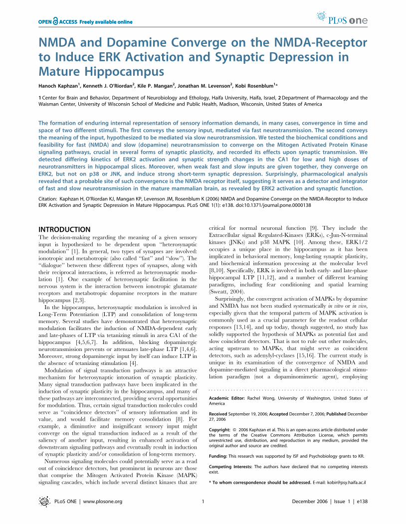
separately and together. Co-application induced a significantly
stronger activation of ERK2 than that induced by 10 mM
dopamine or 10 mM NMDA alone. ERK2 activation by 10 mM
NMDA was weak but significantly increased compare with the
control (1.1860.04, p,0.05, n = 6 and 1.060.02, n = 6, re-
spectively). ERK2 activation by 10 mM dopamine was also slightly
greater than that in the control, but significant (1.2060.07,
Figure 1. Time dependency of ERK2 activation by NMDA or dopamine. Western blot analysis of the time dependency of ERK2 activation, asmeasured by the ratio of pERK2/ERK2, in response to NMDA or dopamine application to hippocampal slices. NMDA was co-applied with 10 mMglycine, and dopamine was co-applied with 1 mM ascorbic acid within various time frames. Hippocampal slices were harvested in the course of thevarious time frames, snap frozen on dry ice, homogenized in SDS sample buffer, and subjected to Western blot analysis of the different specimens.Antibodies against the phosphorylated ERK2 were directed to phosphorylation sites Thr183/Tyr185 (here and below). Each specimen comprised twohippocampal slices combined (i.e., 2 slices is n = 1) (this applies to all the following figure captions). A. NMDA was co-applied with glycine, each ata concentration of 10 mM (n = 6 for each time frame). B. NMDA was co-applied with glycine at concentrations of 100 and 10 mM respectively (n = 6 foreach time frame). C. dopamine at a concentration 10 mM was co-applied with 1 mM ascorbic acid (n = 6 for each time frame). D. Dopamine ata concentration 100 mM was co-applied with 1 mM ascorbic acid (n = 6 for each time frame).Here and after representative immunoblots are depicted above the graphs.doi:10.1371/journal.pone.0000138.g001
Heterosynaptic Modulation
PLoS ONE | www.plosone.org 3 December 2006 | Issue 1 | e138

p,0.05, n = 6 and 1.060.02, n = 6, respectively) (Fig. 3A). ERK2
activation by the co-application of NMDA+dopamine, each at
10 mM was strong and significantly greater than that in the control
(1.5860.06, p,0.0001, n = 6 and 160.02, n = 6, respectively)
(Fig. 3A). Moreover, it was also significantly greater than that
caused by either 10 mM NMDA alone or 10 mM dopamine alone
(p,0.0001 in both comparisons) (Fig. 3A). The MAPK signaling
cascades include other members than ERK that are known to play
roles in synaptic plasticity and long-term memory [22,23,24,25,26].
The question that was addressed in the present study was whether
this pattern of signal transduction convergence is unique to ERK2,
or whether it is common to other members of the MAPK signaling
cascades, i.e., JNK and p38. Therefore, the same specimens that had
been used in looking for convergence of the two agents in ERK2
activation were also analyzed for p38 and JNK activation.
Significant activation of p38 within 10 min was observed in
response to separate application of either 10 mM NMDA or
10 mM dopamine (1.2660.05, p,0.001, n = 6; 1.3360.05,
p,0.0005, n = 6, respectively), compared with that in the control
(1.060.01, n = 6) (Fig. 3B). The p38 activation within 10 min of
co-application was also significant in comparison with that in the
control (1.4060.08, p,0.0005, n = 6; and 1.060.01, n = 6,
respectively) (Fig. 3B), but no significant convergence was observed
in comparison with either agent alone (p = 0.18 vs. NMDA,
p = 0.48 vs. dopamine) (Fig. 3B).
Analysis of the same specimens for JNK activation yielded
similar results to those for p38 activation. Both 10 mM NMDA
and 10 mM dopamine, acting separately, induced significant
activation compared with that in the control within 10 min of
application: (1.3760.07, p,0.001, n = 6; 1.4260.13, p,0.01,
n = 6; and 1.060.02 n = 6, respectively) (Fig. 3C). Co-application
of NMDA and dopamine induced a significant activation of JNK
compared to control (1.5460.19, p,0.05, n = 6), but no significant
further augmentation was detected as a result of such co-
application (p = 0.44 vs. NMDA, p = 0.63 vs. dopamine). Hence,
no significant convergence of the effects of NMDA and dopamine
on the activation of JNK was observed.
Dopamine activation of ERK2 is NMDA receptor
dependent, but NMDA activation of ERK2 is
dopamine receptor independentThus far, our data indicate that ERK2 activation serves as a point
of convergence readout for NMDA and dopaminergic neuro-
transmission. However, in order to better identify the upstream
convergence site, we tested the hypothesis that the NMDA
receptor itself serves as a coincident-detector of NMDA and
dopamine neurotransmission. Several previous studies indicate
that the effects of dopamine or dopamine agonists in vitro and in vivo
require NMDA-mediated synaptic transmission [27,28,29,30].
The NMDA-receptor serves as a classic example of convergence in
the nervous system whereby NMDA-receptor—mediated synaptic
transmission only occurs after removal of Mg2+ from its ion
channel. Therefore, we tested the possibility that the NMDA-R
itself can also act as a point of convergence with dopaminergic
neurotransmission.
In order to get a significant and definite response with the
various stimulations (since 10 mM is a threshold concentration for
stimulation) we employed concentrations of 20 mM NMDA and
dopamine each. Previous experiments observed that these
concentrations are sufficient to yield maximal ERK2 activation.
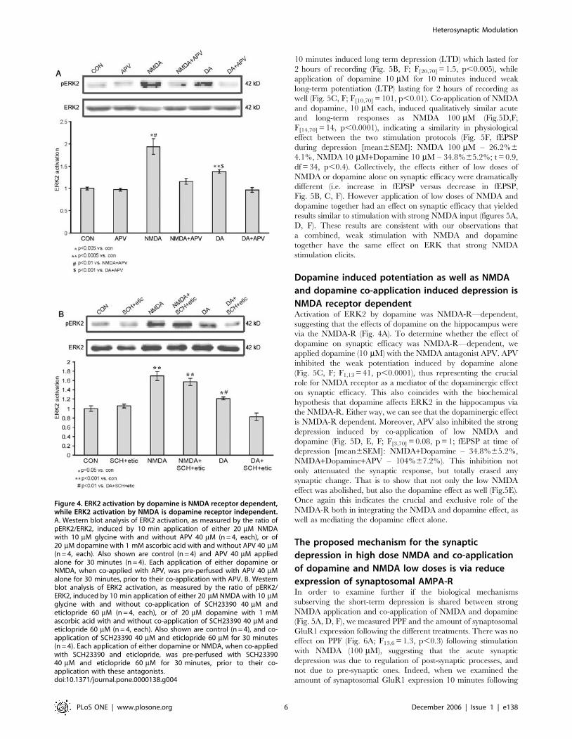
Both NMDA 20 mM and Dopamine 20 mM, applied separately,
induced significant activation of ERK2 compared to control
(1.9360.17, p,0.005, n = 4; 1.3860.04, p,0.0005, n = 4;
160.03, n = 6, respectively). Blocking the NMDA receptor with
D-APV (D-2-amino-5-phosphonovaleric acid) 40 mM during
NMDA 20 mM or dopamine 20 mM stimulations yields similar
outcomes. While APV (40 mM) abolished the NMDA induced
ERK2 activation as expected (1.1660.07, n = 4, p,0.01 vs.
NMDA, p = 0.09 vs. cont), it also completely abolished any
dopamine induced ERK2 activation as well (0.9760.05, n = 4,
p,0.001 vs. dopamine, p = 0.61 vs. cont) (Fig. 4A). In order to
examine a possible reciprocal effect of NMDA on dopamine
receptors, we stimulated with NMDA and blocked dopamine
receptors with a combination of SCH23390 (40 mM, D1/5
antagonist) co-applied with eticlopride (60 mM, D2 antagonist).
Once again we employed concentrations of 20 mM NMDA and
dopamine each, in order to get a significant response with the
various stimulations. While dopamine 20 mM induced a significant
ERK2 activation compared to control (1.2260.03, p,0.05 n = 4;
160.06, n = 4, respectively), SCH23390 and eticlopride abolished
the dopamine induced ERK2 activation as expected (0.8360.08,
n = 4; p,0.005 vs. dopamine; p = 0.13 vs. control). There was no
effect of the SCH23390 and eticlopride on NMDA-induced
activation of ERK. NMDA 20 mM induced a significant ERK2
activation compared to control (1.7060.10, p,0.001 n = 4; 160.06,
n = 4, respectively). SCH23390 co-applied with eticlopride during
NMDA 20 mM stimulation induced significant ERK2 activation
compared to control, which was not significantly different from
activation of ERK2 in response to NMDA alone (1.5760.08, n = 4;
p,0.001 vs. control; p = 0.35 vs. NMDA), (Fig. 4B).
Figure 2. Time dependency of ERK2 activation following co-application of low doses of dopamine and NMDA. Western blotanalysis of time dependency of ERK2 activation, as measured by theratio of pERK2/ERK2, in response to co-application of dopamine andNMDA to hippocampal slices. Dopamine at 10 mM was co-applied with1 mM ascorbic acid, 10 mM NMDA and 10 mM glycine for the differenttime frames (n = 6 for each time frame).doi:10.1371/journal.pone.0000138.g002
Heterosynaptic Modulation
PLoS ONE | www.plosone.org 4 December 2006 | Issue 1 | e138
Co-application of low doses of NMDA and dopamine
has similar functional properties as strong NMDA
activation on synaptic plasticityThe results above demonstrated that the molecular kinetics of
ERK activation is similar between strong NMDA and co-
application of weak NMDA and dopamine. We thus tested
another hypothesis that strong activation of the NMDA-R will
have similar functional effects on synaptic efficacy in the
hippocampus as with co-application of low doses of NMDA and
dopamine. Towards that end, we examined the effect of the
various chemical stimulation protocols on synaptic efficacy at
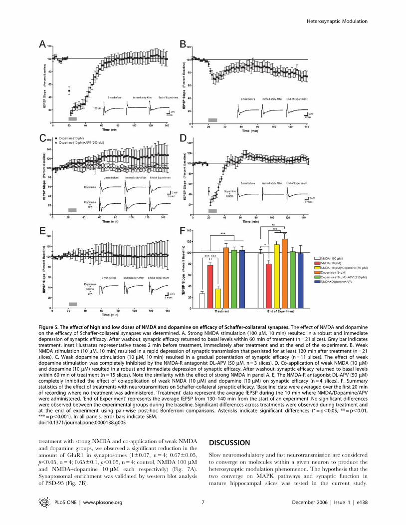
Schaffer-collateral synapses. Application of 100 mM NMDA for
10 minutes induced a sharp and transient fEPSP depression with
a gradual return to baseline within 40 minutes (Fig. 5A, F;
F[20,70] = 13, p,0.0001). Application of NMDA 10 mM for
Figure 3. ERK2 but neither p38 nor JNK serves as a coincidence detector of dopamine and NMDA application. Western blot analysis of MAPKsactivation, as measured by the ratio of phospho-MAPK/total MAPK, induced by 10 min application of either 10 mM NMDA with 10 mM glycine (n = 6),or of 10 mM dopamine with 1 mM ascorbic acid (n = 6), or co-application of 10 mM dopamine, 1 mM ascorbic acid, 10 mM NMDA and 10 mM glycine(n = 6), or control (n = 6). A. ERK2 activation; B. p38 activation. Antibodies against the phosphorylated p38 were directed to phosphorylation sitesThr180/Tyr182; C. JNK activation. Antibodies against the phosphorylated JNK were directed to phosphorylation sites Thr183/Tyr185.doi:10.1371/journal.pone.0000138.g003
Heterosynaptic Modulation
PLoS ONE | www.plosone.org 5 December 2006 | Issue 1 | e138
10 minutes induced long term depression (LTD) which lasted for
2 hours of recording (Fig. 5B, F; F[20,70] = 1.5, p,0.005), while
application of dopamine 10 mM for 10 minutes induced weak
long-term potentiation (LTP) lasting for 2 hours of recording as
well (Fig. 5C, F; F[10,70] = 101, p,0.01). Co-application of NMDA
and dopamine, 10 mM each, induced qualitatively similar acute
and long-term responses as NMDA 100 mM (Fig.5D,F;
F[14,70] = 14, p,0.0001), indicating a similarity in physiological
effect between the two stimulation protocols (Fig. 5F, fEPSP
during depression [mean6SEM]: NMDA 100 mM – 26.2%6
4.1%, NMDA 10 mM+Dopamine 10 mM – 34.8%65.2%; t = 0.9,
df = 34, p,0.4). Collectively, the effects either of low doses of
NMDA or dopamine alone on synaptic efficacy were dramatically
different (i.e. increase in fEPSP versus decrease in fEPSP,
Fig. 5B, C, F). However application of low doses of NMDA and
dopamine together had an effect on synaptic efficacy that yielded
results similar to stimulation with strong NMDA input (figures 5A,
D, F). These results are consistent with our observations that
a combined, weak stimulation with NMDA and dopamine
together have the same effect on ERK that strong NMDA
stimulation elicits.
Dopamine induced potentiation as well as NMDA
and dopamine co-application induced depression is
NMDA receptor dependentActivation of ERK2 by dopamine was NMDA-R—dependent,
suggesting that the effects of dopamine on the hippocampus were
via the NMDA-R (Fig. 4A). To determine whether the effect of
dopamine on synaptic efficacy was NMDA-R—dependent, we
applied dopamine (10 mM) with the NMDA antagonist APV. APV
inhibited the weak potentiation induced by dopamine alone
(Fig. 5C, F; F1,13 = 41, p,0.0001), thus representing the crucial
role for NMDA receptor as a mediator of the dopaminergic effect
on synaptic efficacy. This also coincides with the biochemical
hypothesis that dopamine affects ERK2 in the hippocampus via
the NMDA-R. Either way, we can see that the dopaminergic effect
is NMDA-R dependent. Moreover, APV also inhibited the strong
depression induced by co-application of low NMDA and
dopamine (Fig. 5D, E, F; F[3,70] = 0.08, p = 1; fEPSP at time of
depression [mean6SEM]: NMDA+Dopamine – 34.8%65.2%,
NMDA+Dopamine+APV – 104%67.2%). This inhibition not
only attenuated the synaptic response, but totally erased any
synaptic change. That is to show that not only the low NMDA
effect was abolished, but also the dopamine effect as well (Fig.5E).
Once again this indicates the crucial and exclusive role of the
NMDA-R both in integrating the NMDA and dopamine effect, as
well as mediating the dopamine effect alone.
The proposed mechanism for the synaptic
depression in high dose NMDA and co-application
of dopamine and NMDA low doses is via reduce
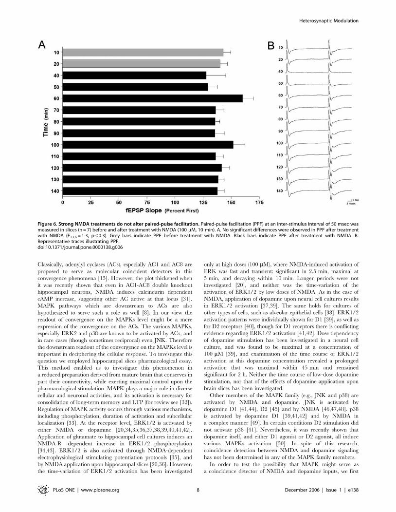
expression of synaptosomal AMPA-RIn order to examine further if the biological mechanisms
subserving the short-term depression is shared between strong
NMDA application and co-application of NMDA and dopamine
(Fig. 5A, D, F), we measured PPF and the amount of synaptosomal
GluR1 expression following the different treatments. There was no
effect on PPF (Fig. 6A; F13,6 = 1.3, p,0.3) following stimulation
with NMDA (100 mM), suggesting that the acute synaptic
depression was due to regulation of post-synaptic processes, and
not due to pre-synaptic ones. Indeed, when we examined the
amount of synaptosomal GluR1 expression 10 minutes following
Figure 4. ERK2 activation by dopamine is NMDA receptor dependent,while ERK2 activation by NMDA is dopamine receptor independent.A. Western blot analysis of ERK2 activation, as measured by the ratio ofpERK2/ERK2, induced by 10 min application of either 20 mM NMDAwith 10 mM glycine with and without APV 40 mM (n = 4, each), or of20 mM dopamine with 1 mM ascorbic acid with and without APV 40 mM(n = 4, each). Also shown are control (n = 4) and APV 40 mM appliedalone for 30 minutes (n = 4). Each application of either dopamine orNMDA, when co-applied with APV, was pre-perfused with APV 40 mMalone for 30 minutes, prior to their co-application with APV. B. Westernblot analysis of ERK2 activation, as measured by the ratio of pERK2/ERK2, induced by 10 min application of either 20 mM NMDA with 10 mMglycine with and without co-application of SCH23390 40 mM andeticlopride 60 mM (n = 4, each), or of 20 mM dopamine with 1 mMascorbic acid with and without co-application of SCH23390 40 mM andeticlopride 60 mM (n = 4, each). Also shown are control (n = 4), and co-application of SCH23390 40 mM and eticlopride 60 mM for 30 minutes(n = 4). Each application of either dopamine or NMDA, when co-appliedwith SCH23390 and eticlopride, was pre-perfused with SCH2339040 mM and eticlopride 60 mM for 30 minutes, prior to their co-application with these antagonists.doi:10.1371/journal.pone.0000138.g004
Heterosynaptic Modulation
PLoS ONE | www.plosone.org 6 December 2006 | Issue 1 | e138
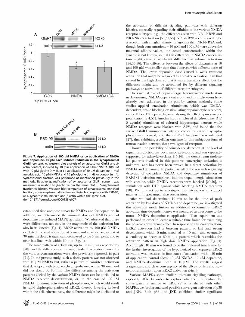
treatment with strong NMDA and co-application of weak NMDA
and dopamine groups, we observed a significant reduction in the
amount of GluR1 in synaptosomes (160.07, n = 4; 0.6760.05,
p,0.05, n = 4; 0.6560.1, p,0.05, n = 4; control, NMDA 100 mM
and NMDA+dopamine 10 mM each respectively) (Fig. 7A).
Synaptosomal enrichment was validated by western blot analysis
of PSD-95 (Fig. 7B).
DISCUSSION
Slow neuromodulatory and fast neurotransmission are considered
to converge on molecules within a given neuron to produce the
heterosynaptic modulation phenomenon. The hypothesis that the
two converge on MAPK pathways and synaptic function in
mature hippocampal slices was tested in the current study.
Figure 5. The effect of high and low doses of NMDA and dopamine on efficacy of Schaffer-collateral synapses. The effect of NMDA and dopamineon the efficacy of Schaffer-collateral synapses was determined. A. Strong NMDA stimulation (100 mM, 10 min) resulted in a robust and immediatedepression of synaptic efficacy. After washout, synaptic efficacy returned to basal levels within 60 min of treatment (n = 21 slices). Grey bar indicatestreatment. Inset illustrates representative traces 2 min before treatment, immediately after treatment and at the end of the experiment. B. WeakNMDA stimulation (10 mM, 10 min) resulted in a rapid depression of synaptic transmission that persisted for at least 120 min after treatment (n = 21slices). C. Weak dopamine stimulation (10 mM, 10 min) resulted in a gradual potentiation of synaptic efficacy (n = 11 slices). The effect of weakdopamine stimulation was completely inhibited by the NMDA-R antagonist DL-APV (50 mM, n = 3 slices). D. Co-application of weak NMDA (10 mM)and dopamine (10 mM) resulted in a robust and immediate depression of synaptic efficacy. After washout, synaptic efficacy returned to basal levelswithin 60 min of treatment (n = 15 slices). Note the similarity with the effect of strong NMDA in panel A. E. The NMDA-R antagonist DL-APV (50 mM)completely inhibited the effect of co-application of weak NMDA (10 mM) and dopamine (10 mM) on synaptic efficacy (n = 4 slices). F. Summarystatistics of the effect of treatments with neurotransmitters on Schaffer-collateral synaptic efficacy. ‘Baseline’ data were averaged over the first 20 minof recording where no treatment was administered. ‘Treatment’ data represent the average fEPSP during the 10 min where NMDA/Dopamine/APVwere administered. ‘End of Experiment’ represents the average fEPSP from 130–140 min from the start of an experiment. No significant differenceswere observed between the experimental groups during the baseline. Significant differences across treatments were observed during treatment andat the end of experiment using pair-wise post-hoc Bonferroni comparisons. Asterisks indicate significant differences (* = p,0.05, ** = p,0.01,*** = p,0.001). In all panels, error bars indicate SEM.doi:10.1371/journal.pone.0000138.g005
Heterosynaptic Modulation
PLoS ONE | www.plosone.org 7 December 2006 | Issue 1 | e138
Classically, adenylyl cyclases (ACs), especially AC1 and AC8 are
proposed to serve as molecular coincident detectors in this
convergence phenomena [15]. However, the plot thickened when
it was recently shown that even in AC1-AC8 double knockout
hippocampal neurons, NMDA induces calcineurin dependent
cAMP increase, suggesting other AC active at that locus [31].
MAPK pathways which are downstream to ACs are also
hypothesized to serve such a role as well [8]. In our view the
readout of convergence on the MAPKs level might be a mere
expression of the convergence on the ACs. The various MAPKs,
especially ERK2 and p38 are known to be activated by ACs, and
in rare cases (though sometimes reciprocal) even JNK. Therefore
the downstream readout of the convergence on the MAPKs level is
important in deciphering the cellular response. To investigate this
question we employed hippocampal slices pharmacological essay.
This method enabled us to investigate this phenomenon in
a reduced preparation derived from mature brain that conserves in
part their connectivity, while exerting maximal control upon the
pharmacological stimulation. MAPK plays a major role in diverse
cellular and neuronal activities, and its activation is necessary for
consolidation of long-term memory and LTP (for review see [32]).
Regulation of MAPK activity occurs through various mechanisms,
including phosphorylation, duration of activation and subcellular
localization [33]. At the receptor level, ERK1/2 is activated by
either NMDA or dopamine [20,34,35,36,37,38,39,40,41,42].
Application of glutamate to hippocampal cell cultures induces an
NMDA-R -dependent increase in ERK1/2 phosphorylation
[34,43]. ERK1/2 is also activated through NMDA-dependent
electrophysiological stimulating potentiation protocols [35], and
by NMDA application upon hippocampal slices [20,36]. However,
the time-variation of ERK1/2 activation has been investigated
only at high doses (100 mM), where NMDA-induced activation of
ERK was fast and transient: significant in 2.5 min, maximal at
5 min, and decaying within 10 min. Longer periods were not
investigated [20], and neither was the time-variation of the
activation of ERK1/2 by low doses of NMDA. As in the case of
NMDA, application of dopamine upon neural cell cultures results
in ERK1/2 activation [37,39]. The same holds for cultures of
other types of cells, such as alveolar epithelial cells [38]. ERK1/2
activation patterns were individually shown for D1 [39], as well as
for D2 receptors [40], though for D1 receptors there is conflicting
evidence regarding ERK1/2 activation [41,42]. Dose dependency
of dopamine stimulation has been investigated in a neural cell
culture, and was found to be maximal at a concentration of
100 mM [39], and examination of the time course of ERK1/2
activation at this dopamine concentration revealed a prolonged
activation that was maximal within 45 min and remained
significant for 2 h. Neither the time course of low-dose dopamine
stimulation, nor that of the effects of dopamine application upon
brain slices has been investigated.
Other members of the MAPK family (e.g., JNK and p38) are
activated by NMDA and dopamine. JNK is activated by
dopamine D1 [41,44], D2 [45] and by NMDA [46,47,48]. p38
is activated by dopamine D1 [39,41,42] and by NMDA in
a complex manner [49]. In certain conditions D2 stimulation did
not activate p38 [41]. Nevertheless, it was recently shown that
dopamine itself, and either D1 agonist or D2 agonist, all induce
various MAPKs activation [50]. In spite of this research,
coincidence detection between NMDA and dopamine signaling
has not been determined in any of the MAPK family members.
In order to test the possibility that MAPK might serve as
a coincidence detector of NMDA and dopamine inputs, we first
Figure 6. Strong NMDA treatments do not alter paired-pulse facilitation. Paired-pulse facilitation (PPF) at an inter-stimulus interval of 50 msec wasmeasured in slices (n = 7) before and after treatment with NMDA (100 mM, 10 min). A. No significant differences were observed in PPF after treatmentwith NMDA (F13,6 = 1.3, p,0.3). Grey bars indicate PPF before treatment with NMDA. Black bars indicate PPF after treatment with NMDA. B.Representative traces illustrating PPF.doi:10.1371/journal.pone.0000138.g006
Heterosynaptic Modulation
PLoS ONE | www.plosone.org 8 December 2006 | Issue 1 | e138
established time and dose curves for NMDA and for dopamine. In
addition, we determined the minimal doses of NMDA and of
dopamine that induced MAPK activation. We observed that there
were differences, not only in the magnitude of the activation but
also in its kinetics (Fig. 1). ERK2 activation by 100 mM NMDA
exhibited maximal activation at 5 min, and a fast decay, so that at
30 min the decay is significant compared to the 5 min peak, and to
near baseline levels within 60 min (Fig. 1).
The same pattern of activation, up to 10 min, was reported by
[20], and the differences in the magnitude of activation caused by
the various concentrations were also previously reported, in part
[21]. In the present study, such a decay pattern was not observed
with 10 mM NMDA but, rather a pattern of consistent activation
that developed with time, reached significance within 30 min, and
did not decay by 60 min. The difference among the activation
patterns elicited by the various NMDA doses can be attributed to
NMDA receptor desensitization, or, in the case of 100 mM
NMDA, to strong activation of phosphatases, which would result
in rapid dephosphorylation of ERK2, thereby lowering its level
within the cell. Alternatively, the difference might be attributed to
the activation of different signaling pathways with differing
kinetics, especially regarding their affinities to the various NMDA
receptor subtypes, e.g., the differences seen with NR1-NR2B and
NR1-NR2A activation [51,52,53]. NR1-NR2B is considered to be
a receptor with a higher affinity for agonists than NRI-NR2A and,
though both concentrations – 10 mM and 100 mM – are above the
maximal affinity values, the actual concentration within the
synapse is not known, so that this difference in NMDA concentra-
tion might cause a significant difference in subunit activation
[54,55,56]. The difference between the effects of dopamine at 10
and 100 mM was smaller than that observed with different doses of
NMDA. The lower dopamine dose caused a weak transient
activation that might be regarded as a weaker activation than that
caused by the high dose, so that it was a transitory effect, but the
difference might also be accounted for by different signaling
pathways or activation of different receptor subtypes.
The essential role of dopaminergic heterosynaptic modulation
in determining NMDA-dependent input, and its implications have
already been addressed in the past by various methods. Some
studies applied tetanization stimulation, which was NMDA-
dependent, while blocking or stimulating dopaminergic receptors,
either D1 or D2 separately, in analyzing the effect upon synaptic
potentiation [2,4,57]. Another study employed dihydrexidine (D1/
5 agonist) stimulation of cultured hippocampal neurons while
NMDA receptors were blocked with APV, and found that the
surface GluR1 immunoreactivity and colocalization with synapto-
physin was reduced, and the mEPSC frequency was inhibited
[27], thus exhibiting a cellular outcome for this ambiguous form of
transactivation between these two types of receptors.
Though, the possibility of coincidence detection at the level of
signal transduction has been raised previously, and was especially
supported for adenylyl-cyclases [15,16], the downstream molecu-
lar patterns involved in this putative converging activation is
unknown, and has never been proven in a direct activation by
NMDA and dopamine. In particular, all of the research regarding
detection of coincident NMDA and dopamine stimulation of
ERK1/2 activation employed indirect dopaminergic stimulation
with cocaine, while NMDA receptors were blocked [29,30], or
stimulation with D1R agonist while blocking NMDA receptors
[28]. We thus set up to investigate this interaction in a direct
manner in hippocampal slice preparation.
After we had determined 10 min to be the time of peak
activation by low doses of NMDA and dopamine, we investigated
this activation mode further in additional experiments. ERK2
activation time dependent curve was measured as a response to the
mutual NMDA+dopamine co-application. That experiment was
performed in order to locate a suitable time frame for examining
the possible convergence effect. In response to this co-application,
ERK2 activation had a bursting pattern of fast and strong
development within 5 min, maximal at 10 min, and eventually
a tendency to decay at 60 min, a pattern which resembles the
activation pattern in high dose NMDA application (Fig. 3).
Accordingly, 10 min was found to be the preferred time frame for
the further investigation of the hypothesized convergence. ERK2
activation was measured in four states of activation, within 10 min
of application: control slices, 10 mM NMDA, 10 mM dopamine,
and NMDA+dopamine, both at 10 mM. The results suggest
a significant and clear convergence of the effects of fast and slow
neurotransmission upon ERK2 activation (Fig. 4).
Various MAPKs share similar upstream signaling pathways,
especially ACs. In order to explore whether this readout for
convergence is unique to ERK1/2 or is shared with other
MAPKs, we further analyzed possible convergent activation of p38
and JNK. Both p38 and JNK exhibited similar significant
Figure 7. Application of 100 mM NMDA or co application of NMDAand dopamine, 10 mM each induces reduction in the synaptosomalGluR1 content. A. Western blot analysis of synaptosomal GluR1 and b-actin content, induced by 10 min application of either NMDA 100 mMwith 10 mM glycine (n = 4), or co-application of 10 mM dopamine, 1 mMascorbic acid, 10 mM NMDA and 10 mM glycine (n = 4), or control (n = 4).Synaptosomal fraction was performed as mentioned previously in themethods. Relative quantification of synaptosomal GluR1 content wasmeasured in relation to b-actin within the same blot. B. Synaptosomalfraction validation. Western blot comparison of synaptosomal enrichedfraction, non-synaptosomal fraction and total homogenate with PSD-95,as a synaptosomal marker, and b-actin within the same blot.doi:10.1371/journal.pone.0000138.g007
Heterosynaptic Modulation
PLoS ONE | www.plosone.org 9 December 2006 | Issue 1 | e138
activations following application of either NMDA or dopamine
separately. However, the impressive converging effect that was
found with ERK2, was not detected with the other MAPK’s.
Thus, the converging signaling pathways are unique to each
pattern of activation and are not general. A similar pattern of
differential activation of the various MAPK cascades in response
to other types of stimulations (Gi- and Gq-coupled receptors) has
been detected in neuronal cell culture [42].
As consequence to these findings, the question raised is where,
or on which molecule/s, the convergence is taking place. Previous
research has shown that there are known interactions between
NMDA and dopamine receptors [58,59], which makes such
a convergence at the receptor level feasible.
Hence, we conducted the dopamine and NMDA applications
(each agent alone) with antagonists, while using the ERK2
activation as metabotropic readout. Surprisingly, we found that
dopamine induced ERK2 activation is dependent upon the
NMDA receptor but not vice versa (Fig. 4A, B). In other words,
dopamine activates ERK2 through the NMDA receptor.
This finding coincides with other publications [28,29,30], that
suggest that the actual converging site is the NMDA receptor, and
among the various MAPK cascades, ERK2 is the molecule that
expresses this convergence, though other signaling cascades can
not be excluded. Another supporting evidence for such converging
interaction upon the NMDA receptor can be drawn from the
resemblance of the two ERK2 activation kinetics patterns: the low
doses co-application of NMDA and dopamine and that of the
NMDA high dose application. Both applications resulted in
a similar ERK2 activation as previously reported. The actual
mechanism of that converging interaction needs further in-
vestigation, and might be mediated directly at the level of the
receptors.
Due to our intent to explore the question of signals convergence
as close as possible to the physiological state, we did not
differentiate in our current study between the D1 and the D2
dopamine receptors, and such differentiation should be further
investigated.
In order to further explore the resemblance between strong
NMDA and co-application of weak NMDA and dopamine inputs
on cellular measurements, we have analyzed electrophysiologically
the fEPSP following these chemical stimulations. In accordance
with the biochemical findings, similar results were shown by the
electrophysiological essay. The effect of low dose dopamine
application resulting with a weak and sustained LTP is totally
inhibited by employing APV concurrently (Fig. 5C). Hence,
suggesting that the dopaminergic effect upon synaptic transmission
is conveyed through the NMDA receptor. Indeed, strong and
transient depression was observed following application of high
dose of NMDA (Fig. 5A) and co-application of low doses of
NMDA and dopamine (Fig. 5D) but not with low doses of either
NMDA (Fig. 5B) or dopamine (Fig. 5C) alone. This strong short-
term depression is totally NMDA dependent, as APV application
inhibits any change in synaptic response. Furthermore, not only
the dopamine ‘‘enhancing’’ effect of the depression is abolished by
APV, but neither low dose NMDA nor low dose dopamine effects
are present. Once again, the erase of even the potentiative
dopamine alone effect, strongly support the notion that it is
mediated via the NMDA-R.
These results, of a fast short term depression of the fEPSP with
a gradual return to baseline, coincide with former findings
reported with NMDA application on hippocampal cell culture
[60] [61]. These protocols induced a rapid and transient
internalization of a-amino-5-hydroxy-3-methyl-4-isoxazole pro-
pionic acid (AMPA) receptors, which is NMDA dependent, quite
similar to the electrophysiological kinetics received in our
experiments (Fig. 5A). Our findings correspond with these results,
also in the proposed molecular mechanism for the short-term
depression, considering that the major component of the fEPSP is
contributed by the AMPA receptors. As shown in our results, both
high dose of NMDA and co-application of low doses of NMDA
and dopamine induced GluR1 withdrawal from the synaptosomal
fraction (Fig. 7), suggesting for a resemblance also in the molecular
mechanisms involved for this synaptic depression. The AMPA-R
internalization, observed in hippocampal cell culture is calcineurin
dependent [61]. Interestingly, it was recently shown that AC (not
AC1 or 8) is activated by the same calcineurin [31]. Furthermore,
NMDA induces this particular AC activation via calcineurin, and
APV inhibits its induction. It is not clear currently, what are the
relationships between these two NMDA and calcineurin de-
pendent processes?
In the present study we demonstrated that among MAPKs,
ERK2 serves as coincidence biochemical readout of fast and slow
neurotransmission of information in the mature brain. Thus
ERK2 has the potential to compute information from fast and
slow neurotransmissions. Nevertheless, other molecules might be
good candidates for conveying similar coincident detection
readout. Moreover, this metabotropic effect of both NMDA and
dopamine are dependent on the NMDA receptor itself, and the
receptor itself serves as the point for convergence and integration
of the different stimuli. In a similar manner to the biochemical
results, we observed parallel synaptic modifications following
strong NMDA application and co-application of dopamine and
NMDA. We proposed that long term neuronal modifications,
manifested by ERK activation, GluR1 internalization or synaptic
depression, can be achieved via two different inputs. The first one
is a prolonged and strong NMDA activation and the second one is
prolonged and weak co-activation of NMDA and dopamine (Fig 8).
These neuronal mechanisms may underlay heterosynaptic me-
chanisms subserving learning process (Fig. 8). Moreover, they give
new molecular framework to the proposed malfunction in
dopaminergic and glutamatergic neurotransmissions and the
interactions between the two, underlying schizophrenia, especially
the negative symptoms and the cognitive deficits involved. It is
conceptualized that the negative symptoms and cognitive deficits
in schizophrenia are related to either hypodopaminergic trans-
mission in the frontal cortex [62], or to a decreased NMDA
receptor functioning [63], and treatments tailored in that direction
are becoming favorable [64]. The molecular findings discussed
here point to the converging interaction between the two
pathways, and might play a role in the understanding of the
schizophrenic pathophysiology and treatment.
Further research is needed to identify the subtypes of dopamine
receptors involved in the process, the inter-receptorial mechanism
of such convergence, the spatial distribution within individual
neurons, and the neuronal events determined by such conver-
gence.
MATERIALS AND METHODS
SubjectsAdult male Sprague-Dawley rats, weighing 300–350 g (Harlan,
Jerusalem, Israel), were housed individually and maintained on
a 12/12-h light/dark cycle. The experiments were approved by
the Institutional Animal Care and Use Committee (Haifa
University and University of Wisconsin), and adequate measures
were taken to minimize pain or discomfort, in accordance with the
guidelines laid down by the NIH, regarding the care and the use of
animals for experimental procedures.
Heterosynaptic Modulation
PLoS ONE | www.plosone.org 10 December 2006 | Issue 1 | e138

Hippocampal slice preparationAfter decapitation the brain was immediately immersed in cold
(4uC) carboxygenated (95% O2, 5% CO2) Artificial Cerebro
Spinal Fluid (ACSF [in mM]: 124 NaCl, 5 KCl, 1.2 MgSO4, 1.2
NaH2PO4, 26 NaHCO3, 10 D-glucose, 2.4 CaCl), and after about
120 s both hippocampi were dissected out in a plate filled with
cold (4uC) ACSF on ice. The hippocampi were put on a cooled
stand of a McIlwain tissue chopper TC752 (Campden Instruments
Ltd, UK), cut into 400-mm slices, and then put back into a chamber
filled with carboxygenated cold (4uC) ACSF.
The slices were transferred to a holding chamber for about 20–
30 min, to reach room temperature, and were then transferred to
a six-chamber pharmacological instrument, designed to our
specifications by Scientific Systems Design Company (Ontario,
Canada). All of the slices tested in any one experiment (i.e., in all
six chambers) were produced by the same procedure from the
same rat.
ReagentsN-methyl-D-Aspartate (NMDA, 10 mM and 100 mM) was pur-
chased from Tocris (Ellisville, Mo, USA). Dopamine (10 mM and
100 mM), ascorbic acid (free acid) (1 mM), glycine (10 mM), TTX
(1 mM), NMDA antagonist - 2-amino-5-phosphonovalerate (APV,
50 mM), MEK inhibitor U0126 (30 mM) were all from Sigma
(Steinheim, Germany).
NMDA (10 mM and 100 mM) was always co-applied with
10 mM glycine, and dopamine (10 mM and 100 mM) was always
co-applied with 1 mM ascorbic acid. This was done to ensure the
conservation of dopamine activity in the course of the experiment,
and also to protect the hippocampal slices against the possible
cytotoxic effect of dopamine. Ascorbic acid at 1 mM has been
shown not to affect MAPK activity [19,65].
p44/42 MAP kinase antibody (1:1000), rabbit polyclonal;
phospho-p44/42 MAP kinase (Thr202/Tyr204) antibody
(1:500), rabbit polyclonal; p38 antibody (1:500), rabbit polyclonal;
phopho-p38 (Thr180/Tyr182) antibody (1:500), rabbit polyclonal;
Jnk antibody (1:500), rabbit polyclonal; phospho-Jnk (Thr183/
Tyr185) antibody (1:500), rabbit polyclonal were purchased from
Cell Signaling (Beverly, MA, USA). b-actin antibody (1:3000), goat
polyclonal; PSD95 antibody (1:500), mouse monoclonal were
purchased from Santa-Cruz Biotechnology (Santa Cruz, CA, USA).
Goat anti-mouse (IgG) horseradish peroxidase (HRP) conjugat-
ed was from Jackson Immunoresearch (West Grove, PA, USA).
Goat anti-rabbit, (IgG) HRP conjugated and the Enhanced
Chemiluminescence (ECL+) Kit were from Amersham (Piscat-
away, NJ, USA).
Pharmacological chamber handling and
manipulationThe hippocampal slices were heated to 32uC and were kept in the
chamber for 5 h before any pharmacological intervention. Each
chamber contained four slices. The slices were perfused with
heated and carboxygenated ACSF via a Model MP3 peristaltic
pump (Gilson, France), at a rate of ,2 mL min21. The chamber
space was carboxygenated and humidified. The chamber was an
interface type, and the slices were placed upon a lens paper.
Within each experiment, one chamber was used as a ‘‘positive
control’’ for the quality of the slices by infusing the chamber with
NMDA 100 mM together with glycine 10 mM, which is known to
induce a strong ERKII activation. Only experiments that showed
an arbitrarily chosen ERKII activation level of at least 25% were
considered. Moreover, viability of the slices after this prolonged
incubation period was occasionally tested by electrophysiological
Figure 8. An illustration of possible fast and slow neurotransmissionconvergence. A. shows that a strong input of fast neurotransmission(which means either large magnitude, long duration, or optimalspacing) causes GluR1 insertion and strong activation of ERK1/2, whichmay lead to increased protein synthesis and long-term changes (e.g.,Late-LTP or Long Term Memory). B. shows that a weak input of fastneurotransmission (weak means small magnitude, too short duration,or non-optimal spacing) would lead to weak ERK1/2 activation, andwould result in only short-term changes if any. C. shows that thoughthe fast neurotransmission input is weak (as most ordinary dailyphysiological input is), the convergence of a slow neurotransmission(e.g., dopamine) upon the fast neurotransmission receptor inducesGluR1 insertion and strong and fast ERK2 activation, thus gives itanother meaning, and consequently causing long-term changes. Theprocess is NMDA-R dependent and can be mediated via the activationof different types of AC’s.doi:10.1371/journal.pone.0000138.g008
Heterosynaptic Modulation
PLoS ONE | www.plosone.org 11 December 2006 | Issue 1 | e138
means: slices were transferred to an electrophysiological chamber,
and the response from the stratum-radiatum in the CA1 region
was recorded, while the Schaffer collateral pathway was
stimulated. Viability was verified by the presence of a fEPSP,
and facilitation of fEPSP.
There was no pooling of results from different comparing
protocols experiments which shared a part of the same stimulation
protocol. Each of the comparative experiments and analysis used
a new batch of slices that came from the same rat via the same
preparation process.
Preparation of samplesAt several time points following the pharmacological manipula-
tion, hippocampal slices were removed from the pharmacological
chamber and snap frozen on dry ice. After freezing, slices were
homogenized in SDS sample buffer as previously described [66].
Four slices from each chamber were combined as two pairs and
the two slices of each pair were homogenized as a single sample, so
that each chamber yielded two samples (n = 2).
Preparation of Synaptoneurosomal fractionThe protocol was adopted from Quinlan [67] and is similar to the
protocol used in [68]. In brief: four hippocampal slices (from each
chamber) were snap-frozen on dry ice, homogenized in a teflon/
glass 2.5 ml tissue grinder, with 1.5 ml of homogenization buffer
[10 mM HEPES, 2 mM EDTA, 2 mM EGTA, 0.5 mM DTT,
1% phosphatase inhibitor cocktail (Sigma) and 1% protease
inhibitor cocktail (Sigma)]. An aliquot of the homogenized tissue
(50 ml) was retained and mixed with 50 ml SDS sample buffer 62
(Total fraction). The remaining material was passed once through
a 100 mm filter and once through a 5 mm filter (Millipore),
attached to a 2 ml syringe. The homogenized tissue was
centrifuged at 3200 rpm for 10 min at 4uC. The pellet contained
the synaptoneurosome fraction, while non-specific material
remained in supernatant. SDS sample buffer 62 was added to
both the pellet and supernatant fractions, the samples were boiled
for 5 min, and stored at 220uC.
QuantificationQuantification of immunoblots was performed with a CCD
camera, Model XRS (Biorad, Italy). Each sample was measured
relative to the background. Phosphorylation levels were calculated
as the ratio between the readings from the antibody directed
against the phospho-proteins and those from the antibody directed
against the phosphorylation-state-independent form of the pro-
teins. Phosphorylation of a protein (phospho-protein/protein) is
expressed as the ratio between the readings from the pharmaco-
logically manipulated slices and those from the control slices
within the same batch of slices in the same experiment. A value of
1 indicates no difference in protein phosphorylation, whereas
values different from 1 indicate an increase or decrease in
phosphorylation. The value multiplied by 100 represents the
percentage change in protein phosphorylation.
ElectrophysiologyTransverse hippocampal slices (400 mm) were isolated with
a Vibratome (Vibratome, St. Louis, MO) in ice cold cutting
saline (CS [in mM]: 110 Sucrose, 60 NaCl, 3 KCl, 1.25
NaH2PO4, 28 NaHCO3, 0.5 CaCl2, 7 MgCl2, 5 Glucose, 0.6
Ascorbate). Slices were allowed to recover for 45 min at room
temperature in 50:50 CS:ACSF. After initial recovery, slices were
placed in an interface chamber (Fine Science Tools, Foster City,
CA) and maintained at 30uC in ACSF (1 mL/min). Slices were
allowed to recover for an additional 60 min on the electrophys-
iology rig prior to experimentation. Bipolar stimulating electrodes
(92:8 Pt:Y) were placed at the border of Area CA3 and Area CA1
along the Schaffer-Collateral pathway. ACSF-filled glass recording
electrodes (1–3 MV) were placed in stratum radiatum of Area
CA1. Basal synaptic transmission was assessed for each slice by
applying gradually increasing stimuli (0.05–1.5 mA, 0.5–15 V, A-
M Systems model 2200 stimulus isolator, Carlsborg, WA) and
determining the input:output relationship. All subsequent stimuli
applied to slices was equivalent to the level necessary to evoke
a fEPSP that had a left slope which was 50% of the maximal left
slope that could be evoked. Synaptic efficacy in response to
treatment of slices with various neurotransmitters and drugs was
continuously monitored (0.05 Hz). Sweeps were averaged together
every 2 min. In experiments where paired-pulse facilitation was
measured, two stimuli administered 50 msec apart were applied
every 10 min while monitoring synaptic efficacy. fEPSPs were
amplified (A-M Systems Model 1800) and digitized (Digidata
1322B, Molecular Devices, Sunnyvale, CA) prior to analysis
(pClamp, Molecular Devices).
Statistical AnalysisThe results are expressed as means6SEM, and the number of
samples. For statistical analysis one-way ANOVA was applied. For
post-hoc comparisons, Fisher’s LSD test was used. Significance for
all tests was set at an a level of 0.05. Analysis of electrophysiology
was performed using either a 1-way (Fig. 5 A,B,D,E) or 2-way
ANOVA (between: treatment, within: time; Fig.5C,F). Post-hoc
analysis was performed using the method of Bonferroni. Paired-
pulse facilitation (Supp Fig. 1) was analyzed using a 1-way
ANOVA. Significance for all tests was set at p#0.05.
ACKNOWLEDGMENTS
Author Contributions
Conceived and designed the experiments: KR HK JL. Performed the
experiments: HK KO KM. Analyzed the data: KR HK JL. Wrote the
paper: KR HK.
REFERENCES1. Bailey CH, Giustetto M, Huang YY, Hawkins RD, Kandel ER (2000) Is
heterosynaptic modulation essential for stabilizing Hebbian plasticity and
memory? Nat Rev Neurosci 1: 11–20.
2. Gurden H, Takita M, Jay TM (2000) Essential role of D1 but not D2
receptors in the NMDA receptor-dependent long-term potentiation at
hippocampal-prefrontal cortex synapses in vivo. J Neurosci 20:
RC106.
3. Jay TM (2003) Dopamine: a potential substrate for synaptic plasticity and
memory mechanisms. Prog Neurobiol 69: 375–390.
4. Huang YY, Kandel ER (1995) D1/D5 receptor agonists induce a protein
synthesis-dependent late potentiation in the CA1 region of the hippocampus.
Proc Natl Acad Sci U S A 92: 2446–2450.
5. Matthies H, Becker A, Schroeder H, Kraus J, Hollt V, et al. (1997) Dopamine
D1-deficient mutant mice do not express the late phase of hippocampal long-
term potentiation. Neuroreport 8: 3533–3535.
6. Frey U, Schroeder H, Matthies H (1990) Dopaminergic antagonists prevent
long-term maintenance of posttetanic LTP in the CA1 region of rat
hippocampal slices. Brain Res 522: 69–75.
Heterosynaptic Modulation
PLoS ONE | www.plosone.org 12 December 2006 | Issue 1 | e138
7. Frey U, Matthies H, Reymann KG, Matthies H (1991) The effect ofdopaminergic D1 receptor blockade during tetanization on the expression of
long-term potentiation in the rat CA1 region in vitro. Neurosci Lett 129:111–114.
8. Sweatt JD (2001) The neuronal MAP kinase cascade: a biochemical signal
integration system subserving synaptic plasticity and memory. J Neurochem 76:1–10.
9. Impey S, Obrietan K, Storm DR (1999) Making new connections: role of ERK/MAP kinase signaling in neuronal plasticity. Neuron 23: 11–14.
10. Derkinderen P, Enslen H, Girault JA (1999) The ERK/MAP-kinases cascade inthe nervous system. Neuroreport 10: R24–34.
11. Adams JP, Roberson ED, English JD, Selcher JC, Sweatt JD (2000) MAPK
regulation of gene expression in the central nervous system. Acta Neurobiol Exp(Wars) 60: 377–394.
12. Rosenblum K, Futter M, Voss K, Erent M, Skehel PA, et al. (2002) The role ofextracellular regulated kinases I/II in late-phase long-term potentiation.
J Neurosci 22: 5432–5441.
13. Ajay SM, Bhalla US (2004) A role for ERKII in synaptic pattern selectivity on
the time-scale of minutes. Eur J Neurosci 20: 2671–2680.
14. Zhao M, Adams JP, Dudek SM (2005) Pattern-dependent role of NMDAreceptors in action potential generation: consequences on extracellular signal-
regulated kinase activation. J Neurosci 25: 7032–7039.
15. Wang H, Storm DR (2003) Calmodulin-regulated adenylyl cyclases: cross-talk
and plasticity in the central nervous system. Mol Pharmacol 63: 463–468.
16. Wang H, Ferguson GD, Pineda VV, Cundiff PE, Storm DR (2004)Overexpression of type-1 adenylyl cyclase in mouse forebrain enhances
recognition memory and LTP. Nat Neurosci 7: 635–642.
17. Belelovsky K, Elkobi A, Kaphzan H, Nairn AC, Rosenblum K (2005) A
molecular switch for translational control in taste memory consolidation.Eur J Neurosci 22: 2560–2568.
18. Ho OH, Delgado JY, O’Dell TJ (2004) Phosphorylation of proteins involved in
activity-dependent forms of synaptic plasticity is altered in hippocampal slicesmaintained in vitro. J Neurochem 91: 1344–1357.
19. Sajikumar S, Frey JU (2004) Late-associativity, synaptic tagging, and the role ofdopamine during LTP and LTD. Neurobiol Learn Mem 82: 12–25.
20. Komiyama NH, Watabe AM, Carlisle HJ, Porter K, Charlesworth P, et al.(2002) SynGAP regulates ERK/MAPK signaling, synaptic plasticity, and
learning in the complex with postsynaptic density 95 and NMDA receptor.
J Neurosci 22: 9721–9732.
21. Opazo P, Watabe AM, Grant SG, O’Dell TJ (2003) Phosphatidylinositol 3-
kinase regulates the induction of long-term potentiation through extracellularsignal-related kinase-independent mechanisms. J Neurosci 23: 3679–3688.
22. Berman DE, Hazvi S, Rosenblum K, Seger R, Dudai Y (1998) Specific anddifferential activation of mitogen-activated protein kinase cascades by unfamiliar
taste in the insular cortex of the behaving rat. J Neurosci 18: 10037–10044.
23. Zhen X, Du W, Romano AG, Friedman E, Harvey JA (2001) The p38 mitogen-activated protein kinase is involved in associative learning in rabbits. J Neurosci
21: 5513–5519.
24. Gisabella B, Rowan MJ, Anwyl R (2003) Mechanisms underlying the inhibition
of long-term potentiation by preconditioning stimulation in the hippocampus invitro. Neuroscience 121: 297–305.
25. Gerdjikov TV, Ross GM, Beninger RJ (2004) Place preference induced by
nucleus accumbens amphetamine is impaired by antagonists of ERK or p38MAP kinases in rats. Behav Neurosci 118: 740–750.
26. Zhu Y, Pak D, Qin Y, McCormack SG, Kim MJ, et al. (2005) Rap2-JNKremoves synaptic AMPA receptors during depotentiation. Neuron 46: 905–916.
27. Smith WB, Starck SR, Roberts RW, Schuman EM (2005) Dopaminergicstimulation of local protein synthesis enhances surface expression of GluR1 and
synaptic transmission in hippocampal neurons. Neuron 45: 765–779.
28. Papadeas ST, Blake BL, Knapp DJ, Breese GR (2004) Sustained extracellularsignal-regulated kinase 1/2 phosphorylation in neonate 6-hydroxydopamine-
lesioned rats after repeated D1-dopamine receptor agonist administration:implications for NMDA receptor involvement. J Neurosci 24: 5863–5876.
29. Valjent E, Corvol JC, Pages C, Besson MJ, Maldonado R, et al. (2000)
Involvement of the extracellular signal-regulated kinase cascade for cocaine-rewarding properties. J Neurosci 20: 8701–8709.
30. Valjent E, Pascoli V, Svenningsson P, Paul S, Enslen H, et al. (2005) Regulationof a protein phosphatase cascade allows convergent dopamine and glutamate
signals to activate ERK in the striatum. Proc Natl Acad Sci U S A 102: 491–496.
31. Chan GC, Tonegawa S, Storm DR (2005) Hippocampal neurons express
a calcineurin-activated adenylyl cyclase. J Neurosci 25: 9913–9918.
32. Sweatt JD (2004) Mitogen-activated protein kinases in synaptic plasticity andmemory. Curr Opin Neurobiol 14: 311–317.
33. Rubinfeld H, Seger R (2004) The ERK cascade as a prototype of MAPKsignaling pathways. Methods Mol Biol 250: 1–28.
34. Bading H, Greenberg ME (1991) Stimulation of protein tyrosine phosphoryla-tion by NMDA receptor activation. Science 253: 912–914.
35. Huang YY, Martin KC, Kandel ER (2000) Both protein kinase A and mitogen-
activated protein kinase are required in the amygdala for the macromolecularsynthesis-dependent late phase of long-term potentiation. J Neurosci 20:
6317–6325.
36. Banko JL, Hou L, Klann E (2004) NMDA receptor activation results in PKA-
and ERK-dependent Mnk1 activation and increased eIF4E phosphorylation inhippocampal area CA1. J Neurochem 91: 462–470.
37. Ambrosini A, Tininini S, Barassi A, Racagni G, Sturani E, et al. (2000) cAMPcascade leads to Ras activation in cortical neurons. Brain Res Mol Brain Res 75:
54–60.
38. Guerrero C, Pesce L, Lecuona E, Ridge KM, Sznajder JI (2002) Dopamine
activates ERKs in alveolar epithelial cells via Ras-PKC-dependent and Grb2/Sos-independent mechanisms. Am J Physiol Lung Cell Mol Physiol 282:
L1099–1107.
39. Chen J, Rusnak M, Luedtke RR, Sidhu A (2004) D1 dopamine receptormediates dopamine-induced cytotoxicity via the ERK signal cascade. J Biol
Chem 279: 39317–39330.
40. Yan Z, Feng J, Fienberg AA, Greengard P (1999) D(2) dopamine receptorsinduce mitogen-activated protein kinase and cAMP response element-binding
protein phosphorylation in neurons. Proc Natl Acad Sci U S A 96:11607–11612.
41. Zhen X, Uryu K, Wang HY, Friedman E (1998) D1 dopamine receptor agonists
mediate activation of p38 mitogen-activated protein kinase and c-Jun amino-
terminal kinase by a protein kinase A-dependent mechanism in SK-N-MChuman neuroblastoma cells. Mol Pharmacol 54: 453–458.
42. Chan AS, Yeung WW, Wong YH (2005) Integration of G protein signals by
extracellular signal-regulated protein kinases in SK-N-MC neuroepitheliomacells. J Neurochem 94: 1457–1470.
43. Mao L, Tang Q, Samdani S, Liu Z, Wang JQ (2004) Regulation of MAPK/
ERK phosphorylation via ionotropic glutamate receptors in cultured rat striatalneurons. Eur J Neurosci 19: 1207–1216.
44. Chan AS, Wong YH (2005) Gq-mediated activation of JNK by the GRP
receptor is inhibited upon co-stimulation of the Gs-coupled dopamine D1receptor in Cos-7 cells. Mol Pharmacol.
45. Luo Y, Kokkonen GC, Wang X, Neve KA, Roth GS (1998) D2 dopamine
receptors stimulate mitogenesis through pertussis toxin-sensitive G proteins andRas-involved ERK and SAP/JNK pathways in rat C6-D2L glioma cells.
J Neurochem 71: 980–990.
46. Ko HW, Park KY, Kim H, Han PL, Kim YU, et al. (1998) Ca2+-mediatedactivation of c-Jun N-terminal kinase and nuclear factor kappa B by NMDA in
cortical cell cultures. J Neurochem 71: 1390–1395.
47. Borsello T, Croquelois K, Hornung JP, Clarke PG (2003) N-methyl-d-aspartate-triggered neuronal death in organotypic hippocampal cultures is endocytic,
autophagic and mediated by the c-Jun N-terminal kinase pathway. Eur J Neurosci
18: 473–485.
48. Borsello T, Bonny C (2004) Use of cell-permeable peptides to prevent neuronaldegeneration. Trends Mol Med 10: 239–244.
49. Waxman EA, Lynch DR (2005) N-methyl-D-aspartate receptor subtype
mediated bidirectional control of p38 mitogen-activated protein kinase. J BiolChem 280: 29322–29333.
50. Lee MY, Heo JS, Han HJ (2006) Dopamine regulates cell cycle regulatory
proteins via cAMP, Ca(2+)/PKC, MAPKs, and NF-kappaB in mouseembryonic stem cells. J Cell Physiol 208: 399–406.
51. Kim MJ, Dunah AW, Wang YT, Sheng M (2005) Differential roles of NR2A-
and NR2B-containing NMDA receptors in Ras-ERK signaling and AMPAreceptor trafficking. Neuron 46: 745–760.
52. Krapivinsky G, Krapivinsky L, Manasian Y, Ivanov A, Tyzio R, et al. (2003)
The NMDA receptor is coupled to the ERK pathway by a direct interactionbetween NR2B and RasGRF1. Neuron 40: 775–784.
53. Erreger K, Dravid SM, Banke TG, Wyllie DJ, Traynelis SF (2005) Subunit-
specific gating controls rat NR1/NR2A and NR1/NR2B NMDA channelkinetics and synaptic signalling profiles. J Physiol 563: 345–358.
54. Laurie DJ, Seeburg PH (1994) Ligand affinities at recombinant N-methyl-D-
aspartate receptors depend on subunit composition. Eur J Pharmacol 268:
335–345.
55. Priestley T, Laughton P, Myers J, Le Bourdelles B, Kerby J, et al. (1995)
Pharmacological properties of recombinant human N-methyl-D-aspartate
receptors comprising NR1a/NR2A and NR1a/NR2B subunit assembliesexpressed in permanently transfected mouse fibroblast cells. Mol Pharmacol
48: 841–848.
56. Buller AL, Larson HC, Schneider BE, Beaton JA, Morrisett RA, et al. (1994)The molecular basis of NMDA receptor subtypes: native receptor diversity is
predicted by subunit composition. J Neurosci 14: 5471–5484.
57. Li S, Cullen WK, Anwyl R, Rowan MJ (2003) Dopamine-dependent facilitationof LTP induction in hippocampal CA1 by exposure to spatial novelty. Nat
Neurosci 6: 526–531.
58. Scott L, Kruse MS, Forssberg H, Brismar H, Greengard P, et al. (2002) Selectiveup-regulation of dopamine D1 receptors in dendritic spines by NMDA receptor
activation. Proc Natl Acad Sci U S A 99: 1661–1664.
59. Scott L, Zelenin S, Malmersjo S, Kowalewski JM, Markus EZ, et al. (2006)Allosteric changes of the NMDA receptor trap diffusible dopamine 1 receptors in
spines. Proc Natl Acad Sci U S A 103: 762–767.
60. Ehlers MD (2000) Reinsertion or degradation of AMPA receptors determined byactivity-dependent endocytic sorting. Neuron 28: 511–525.
61. Beattie EC, Carroll RC, Yu X, Morishita W, Yasuda H, et al. (2000) Regulation
of AMPA receptor endocytosis by a signaling mechanism shared with LTD. Nat
Neurosci 3: 1291–1300.
62. Davis KL, Kahn RS, Ko G, Davidson M (1991) Dopamine in schizophrenia:
a review and reconceptualization. Am J Psychiatry 148: 1474–1486.
63. Coyle JT (2006) Glutamate and Schizophrenia: Beyond the Dopamine
Hypothesis. Cell Mol Neurobiol. .
Heterosynaptic Modulation
PLoS ONE | www.plosone.org 13 December 2006 | Issue 1 | e138

64. Murphy BP, Chung YC, Park TW, McGorry PD (2006) Pharmacological
treatment of primary negative symptoms in schizophrenia: A systematic review.Schizophr Res.
65. Gomez-Santos C, Ferrer I, Santidrian AF, Barrachina M, Gil J, et al. (2003)
Dopamine induces autophagic cell death and alpha-synuclein increase in humanneuroblastoma SH-SY5Y cells. J Neurosci Res 73: 341–350.
66. Rosenblum K, Berman DE, Hazvi S, Lamprecht R, Dudai Y (1997) NMDAreceptor and the tyrosine phosphorylation of its 2B subunit in taste learning in
the rat insular cortex. J Neurosci 17: 5129–5135.
67. Quinlan EM, Philpot BD, Huganir RL, Bear MF (1999) Rapid, experience-
dependent expression of synaptic NMDA receptors in visual cortex in vivo. Nat
Neurosci 2: 352–357.
68. Kelleher RJ 3rd, Govindarajan A, Jung HY, Kang H, Tonegawa S (2004)
Translational control by MAPK signaling in long-term synaptic plasticity and
memory. Cell 116: 467–479.
Heterosynaptic Modulation
PLoS ONE | www.plosone.org 14 December 2006 | Issue 1 | e138