HNE increases HO1 through activation of the ERK pathway in pulmonary epithelial cells
22
HNE increases HO-1 through activation of the ERK pathway in pulmonary epithelial cells Karen E. Iles a,b,1 , Dale A. Dickinson a,b,1 , Amanda F. Wigley a , Nathan E. Welty a , Volker Blank c , and Henry Jay Forman d,* a Department of Environmental Health Sciences, University of Alabama at Birmingham, Birmingham, AL 35294, USA b Center for Free Radical Biology, University of Alabama at Birmingham, Birmingham, AL 35294, USA c Department of Medicine, McGill University, Jewish General Hospital, Montréal, QC, Canada H3T 1E2 d School of Natural Sciences, University of California at Merced, P.O. Box 2039, Merced, CA 95344, USA Abstract Heme oxygenase-1 (HO-1) is a key cytoprotective enzyme and an established marker of oxidative stress. Increased HO-1 expression has been found in the resident macrophages in the alveolar spaces of smokers. The lipid peroxidation product 4-hydroxynonenal (HNE) is also increased in the bronchial and alveolar epithelium in response to cigarette smoke. This suggests a link between a chronic environmental stress, HNE formation, and HO-1 induction. HNE is both an agent of oxidative stress in vivo and a potent cell signaling molecule. We hypothesize that HNE acts as an endogenously produced pulmonary signaling molecule that elicits an adaptive response culminating in the induction of HO-1. Here we demonstrate that HNE increases HO-1 mRNA, protein, and activity in pulmonary epithelial cells and identify ERK as a key pathway involved. Treatment with HNE increased ERK phosphorylation, c-Fos protein, JNK phosphorylation, c-Jun phosphorylation, and AP-1 binding. Whereas inhibiting the ERK pathway with the MEK inhibitor PD98059 significantly decreased HNE- mediated ERK phosphorylation, c-Fos protein induction, AP-1 binding, and HO-1 protein induction, inhibition of the ERK pathway had no effect on HNE-induced HO-1 mRNA. This suggests that ERK is involved in the increase in HO-1 through regulation of translation rather than transcription. Keywords 4-Hydroxynonenal; Oxidative stress; HO-1; MAPK signaling; Free radicals Heme oxygenase-1 (HO-1) is an important cytoprotective enzyme and widely accepted marker of oxidative stress. HO-1 catalyzes the first and rate-limiting step in the catabolism of heme, which generates equimolar amounts of biliverdin, ferrous iron, and carbon monoxide [1–3]. Although heme is the major substrate of HO-1, a variety of non-heme-containing agents are also strong inducers of HO-1. HO-1 has been shown to respond to a number of proinflammatory cytokines, including TNF-α, IL-1, and IL-11 [4]. HO-1 is also induced by a variety of agents © 2005 Elsevier Inc. All rights reserved. *Corresponding author. Fax: (208) 498 7635., [email protected] (H.J. Forman). 1 These authors contributed equally to this paper. NIH Public Access Author Manuscript Free Radic Biol Med. Author manuscript; available in PMC 2009 December 28. Published in final edited form as: Free Radic Biol Med. 2005 August 1; 39(3): 355–364. doi:10.1016/j.freeradbiomed.2005.03.026. NIH-PA Author Manuscript NIH-PA Author Manuscript NIH-PA Author Manuscript
-
Upload
ua-birmingham -
Category
Documents
-
view
1 -
download
0
Transcript of HNE increases HO1 through activation of the ERK pathway in pulmonary epithelial cells
HNE increases HO-1 through activation of the ERK pathway inpulmonary epithelial cells
Karen E. Ilesa,b,1, Dale A. Dickinsona,b,1, Amanda F. Wigleya, Nathan E. Weltya, VolkerBlankc, and Henry Jay Formand,*aDepartment of Environmental Health Sciences, University of Alabama at Birmingham, Birmingham,AL 35294, USAbCenter for Free Radical Biology, University of Alabama at Birmingham, Birmingham, AL 35294,USAcDepartment of Medicine, McGill University, Jewish General Hospital, Montréal, QC, Canada H3T1E2dSchool of Natural Sciences, University of California at Merced, P.O. Box 2039, Merced, CA 95344,USA
AbstractHeme oxygenase-1 (HO-1) is a key cytoprotective enzyme and an established marker of oxidativestress. Increased HO-1 expression has been found in the resident macrophages in the alveolar spacesof smokers. The lipid peroxidation product 4-hydroxynonenal (HNE) is also increased in thebronchial and alveolar epithelium in response to cigarette smoke. This suggests a link between achronic environmental stress, HNE formation, and HO-1 induction. HNE is both an agent of oxidativestress in vivo and a potent cell signaling molecule. We hypothesize that HNE acts as an endogenouslyproduced pulmonary signaling molecule that elicits an adaptive response culminating in the inductionof HO-1. Here we demonstrate that HNE increases HO-1 mRNA, protein, and activity in pulmonaryepithelial cells and identify ERK as a key pathway involved. Treatment with HNE increased ERKphosphorylation, c-Fos protein, JNK phosphorylation, c-Jun phosphorylation, and AP-1 binding.Whereas inhibiting the ERK pathway with the MEK inhibitor PD98059 significantly decreased HNE-mediated ERK phosphorylation, c-Fos protein induction, AP-1 binding, and HO-1 protein induction,inhibition of the ERK pathway had no effect on HNE-induced HO-1 mRNA. This suggests that ERKis involved in the increase in HO-1 through regulation of translation rather than transcription.
Keywords4-Hydroxynonenal; Oxidative stress; HO-1; MAPK signaling; Free radicals
Heme oxygenase-1 (HO-1) is an important cytoprotective enzyme and widely accepted markerof oxidative stress. HO-1 catalyzes the first and rate-limiting step in the catabolism of heme,which generates equimolar amounts of biliverdin, ferrous iron, and carbon monoxide [1–3].Although heme is the major substrate of HO-1, a variety of non-heme-containing agents arealso strong inducers of HO-1. HO-1 has been shown to respond to a number of proinflammatorycytokines, including TNF-α, IL-1, and IL-11 [4]. HO-1 is also induced by a variety of agents
© 2005 Elsevier Inc. All rights reserved.*Corresponding author. Fax: (208) 498 7635., [email protected] (H.J. Forman).1These authors contributed equally to this paper.
NIH Public AccessAuthor ManuscriptFree Radic Biol Med. Author manuscript; available in PMC 2009 December 28.
Published in final edited form as:Free Radic Biol Med. 2005 August 1; 39(3): 355–364. doi:10.1016/j.freeradbiomed.2005.03.026.
NIH
-PA Author Manuscript
NIH
-PA Author Manuscript
NIH
-PA Author Manuscript
that cause oxidative stress and/or in response to environmental stress [5–8]. For example,increased HO-1 expression has recently been found in the alveolar spaces in the residentmacrophages of smokers [9]. Once induced, HO-1 provides protection against multiple typesof tissue injury [10–13]; specific to the lung, overexpression of HO-1 in pulmonary epithelialcells has been shown to protect against oxygen toxicity [14]. In contrast, HO-1 deficiency inmice (HO-1−/−) increases susceptibility to inflammatory lung injury [15], as does HO-1deficiency in humans [16]. Although the precise mechanisms by which HO-1 exerts itscytoprotective effects are not known, there is mounting evidence that carbon monoxide playsa key role in this process. For thorough reviews of the recent literature see [17,18].
Another characteristic of oxidative stress is the formation of the lipid peroxidation product 4-hydroxy-2-nonenal (HNE). HNE has been shown to increase HO-1 in the cell [19,20], but themechanism(s) has not been clearly defined. HNE is an α,β-unsaturated aldehyde that is formedfrom the reaction of oxygen species with arachidonate in cellular membranes during manyforms of environmental stress, including exposure to cigarette smoke [9,21,22]. HNE is knownto impact the cell in many ways, which include inactivation of enzymes, depletion ofintracellular glutathione, and inhibition of DNA and protein synthesis [23–25]. Recent datasuggest that HNE may also modulate biological responses by triggering intracellular signaltransduction pathways [26–28].
The mitogen-activated protein kinases (MAPK) are among the signal transduction pathwaysactivated by HNE. As such, the MAPK pathways may play a significant role in mediating themany cellular functions that are affected by HNE. The MAPK are activated by dualphosphorylation on specific tyrosine and threonine residues [29]. The MAPK include theextracellular signal-regulated kinases 1 and 2 (ERK1/2), p38 MAP kinases, c-Jun N-terminalkinases (JNK), and the big MAPK. The MAPK are activated in response to various stressors,including oxidative stress. They phosphorylate and activate key transcription factors, therebyregulating the transcription of many genes. For a comprehensive review of MAPK-redoxsignaling see [29]. Members of the MAPK family are critical regulators of the transcriptionfactor complex AP-1 (activator protein-1). AP-1 consists of proteins of the Jun, Fos, and ATFfamilies that can form homo- and heterodimers. Activation of AP-1 involves expression ofAP-1-driven genes that include c-Jun. c-Jun may also form part of the stress response (StRE)and electrophile response elements (EpRE), also key regulators of redox-sensitive genes [30,31].
HO-1 gene and protein expression is also modulated by MAPK activation. Seminal studiessupport a role for these kinases as mediators of HO-1 induction in a broad range of tissues andcell types. Studies have shown that HO-1 is induced via the ERK pathway by LPS + IFN-γ[32], through p38 MAPK in murine macrophages in response to IL-1β, and in human lungepithelial cells by TGF-β1 [33,34]. However, care must be taken not to generalize, as thesignaling pathways that are activated are both agonist-and species-specific.
In this study, we set out to determine if HNE induces HO-1 in lung pulmonary epithelial cells,and, if so, the pathway or pathways involved. As HNE and HO-1 are increased in the bronchialand alveolar epithelium in response to chronic environmental insults such as cigarette smoke,mechanistically, this raises the possibility that such insults increase HNE, which in turn inducesHO-1. We tested this hypothesis in vitro, by treating lung epithelial cells with HNE, assayingthe effects on HO-1 induction, and then determining which of the MAPK pathways areinvolved.
Iles et al. Page 2
Free Radic Biol Med. Author manuscript; available in PMC 2009 December 28.
NIH
-PA Author Manuscript
NIH
-PA Author Manuscript
NIH
-PA Author Manuscript

Materials and methodsMaterials
HNE was purchased from Cayman Chemical (Ann Arbor, MI, USA). SB202190 (SB),PD98059 (PD), SP600125 (SP), and the JNK inhibitor peptide (JNKi) were purchased fromCalbiochem. TRIzol reagent was purchased from Life Technologies (Grand Island, NY, USA).The antiserum to the small Maf proteins was a gift from Volker Blank (McGill University,Montréal, QC, Canada). All other antibodies were obtained from Santa Cruz Biotechnology(Santa Cruz, CA, USA). All chemicals were at least of analytical grade and were purchasedfrom Sigma–Aldrich unless otherwise noted.
Cell culture and treatmentsL2 cells are a rat alveolar epithelial type II-like cell line that was purchased from the ATCC.Cultures were maintained in F12-K nutrient mixture supplemented with 1% penicillin/streptomycin and 10% BSA in a humidified incubator at 37°C with 5% CO2. Cells were grownto near confluence and then treated as outlined below.
HNE was dissolved in ethanol; the inhibitors SB, PD, and SP were dissolved in dimethylsulfoxide (DMSO); and the JNKi was dissolved in water. The final concentrations of ethanoland DMSO were 0.05 and 0.1%, respectively. Treatments were performed when the cellsreached 90–95% confluence. For inhibitor studies cells were pretreated with 25 µM SB, 50µM PD, 20 µM SP, 20 µM JNKi, or the dilution buffer (containing DMSO) for 30 min andthen incubated with 20 µM HNE for increasing lengths of time as outlined under Results. Aftertreatments, cells were washed once with 1 × phosphate-buffered saline (PBS) and harvestedwith a cell scraper in 1 × PBS. Cells were centrifuged at 500 g for 5 min, trace PBS was removed,and the pellets were either processed immediately or stored at −80°C.
Western blotting analysisWestern blotting was done as described previously [35,36]. Briefly, cell lysates were preparedusing the MPER reagent (Pierce, Rockford, IL, USA) following the manufacturer’sinstructions. Between 20 and 50 µg of protein was electrophoresed under denaturing conditionson a 10% Tris–glycine acrylamide gel (Invitrogen), transferred to a polyvinylidene difluoridemembrane (Immobilon P; Millipore), and then blocked in nonfat dry milk (NFDM) at roomtemperature for 1 h. Membranes were then incubated overnight at 4°C with the appropriateprimary antibody in 5% NFDM in Tris-buffered saline with 0.05% Tween 20 (T-TBS). Afterrigorous washes with T-TBS, the membranes were incubated with the appropriate HRP-coupled secondary antibody at room temperature for 2 h. After repeated rigorous washes withT-TBS membranes were incubated with ECL Plus reagent (Amersham). Blots were exposedto ECL Hyperfilm (Amersham), and quantitation was performed by densitometric analysis ofthe exposed films using SigmaScan software.
RNA quantificationThe content of HO-1 message was determined using real-time PCR on a Cepheid SmartCycler1.2 with Gapdh message content as an active control, using the threshold cycle (CT) method.Briefly, total RNA was isolated using TRIzol reagent (Invitrogen), following themanufacturer’s recommendations, and then treated with DNA-free (Ambion) at 37°C for 30min to remove any contaminating DNA. The remaining RNA was quantitatedspectrophotometrically at 260 nm. Reverse transcriptase was performed using TaqMan randomhexamers (Applied Biosystems). OligoPerfect was used to design gene-specific primers for ratHO-1 (sense, L2 5′-GAAGAAGATTGCACAGAAGG-3′, and antisense, 5′-GAAGGCGGTCTTAGCCTCTT-3′) and Gapdh (sense, 5′-
Iles et al. Page 3
Free Radic Biol Med. Author manuscript; available in PMC 2009 December 28.
NIH
-PA Author Manuscript
NIH
-PA Author Manuscript
NIH
-PA Author Manuscript
ACCCCAATGTATCCGTTGT-3′, and antisense, 5′-TACTCCTTGGAGGCCATGT-3′),which were used to amplify a segment of reverse-transcribed mRNA using SYBR Green PCRMasterMix (Applied Biosystems). The annealing temperature was the same for each pair ofgene-specific primers, and the amplicon sizes were similar. Each mRNA species was measuredfour times.
HO-1 activity assayHO-1 activity was measured as described by Visner et al. [37]. L2 cells were grown toconfluence in 100-mm tissue culture dishes. After treatment, cells were rinsed twice with 1 ×PBS, scraped with a rubber policeman, and pelleted at 2000g for 10 min. The resulting pelletwas resuspended in 0.1 mol/L KPO4 and 2 mmol/L MgCl2 and frozen (−80°C) and thawedthree times to break the cell membrane. The samples were sonicated on ice, and an aliquot wasreserved for protein determination. Proteins were measured using a modification of theBradford assay. The remaining sample was centrifuged at 12,000 g at 4°C for 20 min, and thesupernatant was added to the reaction mixture containing 3 mg rat liver cytosol, 20 µM hemin,2 mM glucose 6-phosphate, 0.2 units glucose-6-phosphate dehydrogenase, and 0.8 mM β-NADPH and incubated at 37°C for 60 min in the dark. One milliliter of chloroform was addedto the extract, and the change in optical density at 464–530 nm was measured. Theconcentration of bilirubin produced in 60 min was calculated using the extinction coefficient,40 mM−1 cm−1 for bilirubin per milligram of protein.
Electrophoretic mobility shift assay (EMSA)Nuclear extracts were prepared using the NE-PER kit (Pierce) following the manufacturer’sinstructions. Protein concentrations were determined with Protein Assay Dye (Bio-Rad), usinga modification of the Bradford method. Nuclear extracts were either quick frozen in liquidnitrogen and reserved at −80°C or used immediately. EMSAs were done as previouslydescribed [36,38,39]. Briefly, AP-1 and EpRE probes were made from AP-1 oligonucleotide(5′CGCTTGATGAGTCAGCCGGAA-3′) or EpRE oligonucleotide (5′-TCAACTAGAGTCACAGTGACTTGGCAAAAT-3′) hybridized to its complementary DNAsequence. Nuclear protein (2 µg) was preincubated with 5× binding buffer for 15 min at 37°C, followed by incubation with double-stranded 32P-labeled AP-1 or EpRE probe for 20 minat room temperature. For supershift experiments, this was followed by an additional incubationwith 1 µg of the appropriate antibody for 5–45 min at room temperature. Samples wereelectrophoresed at 150 V for 3 h. Gels were dried and imaged via electronic autoradiographyon a Packard Instant Imager (Canberra Co.).
Statistical analysisStatistical analysis was done using a Student t test or a one-way ANOVA where appropriatewith a Tukey’s test post hoc. Differences were considered statistically significant at p < 0.05.
ResultsAs HNE-mediated induction of HO-1 protein has been reported in alveolar macrophages[40], but not in other lung cell types, we first sought to determine if HNE induced HO-1 inpulmonary epithelial cells. L2 cells treated with 20 µM HNE for 5 h exhibited an ~ 7-foldincrease in HO-1 protein versus controls (Fig. 1). HNE was dissolved in ethanol, yielding afinal concentration of ethanol in the HNE-treated samples of 0.05%. This concentration ofethanol does not generally affect cell viability; however, as ethanol is a stress that can alter cellsignaling, gene expression, and protein synthesis, initial experiments included both anuntreated control and a 0.05% ethanol control. There was no difference in HO-1 protein in theuntreated cells and the cells exposed to 0.05% ethanol (data not shown), with HO-1 beingalmost undetectable in both. As the vehicle did not affect the endpoint, only the vehicle control
Iles et al. Page 4
Free Radic Biol Med. Author manuscript; available in PMC 2009 December 28.
NIH
-PA Author Manuscript
NIH
-PA Author Manuscript
NIH
-PA Author Manuscript

was used in subsequent experiments. To determine the mechanism of the HNE-mediatedincrease in HO-1 (i.e., transcriptional versus translational control) the change in HO-1 mRNAwith HNE treatment was measured. Results from replicate real-time PCR assays showed thatHNE increased HO-1 mRNA ~ 20- fold by 2 h postincubation (Fig. 2). This increase wasblocked by the mRNA synthesis inhibitor actinomycin D, suggesting transcriptional controlof HO-1 protein.
As HO-1 requires posttranslational modification for activity [41], HO-1 activity was alsomeasured. An approximately threefold increase in HO-1 activity was observed 6 hpostincubation (Fig. 3). This confirmed that the increase in HO-1 mRNA and protein isaccompanied by a functional change in HO-1 in the cell.
Having established that HNE increases HO-1 mRNA, protein, and activity, we sought todetermine if MAPK activation is required for HO-1 induction in pulmonary epithelial cells.MAPK activation by HNE was assayed by Western blot with phosphospecific antibodies foreach of the respective MAPK (JNK, ERK, and p38). A 5-h time course compared thephosphorylation and activation of each of the three major MAPK under identical experimentalconditions. Phosphorylation of all three MAPK increased during this time, with ERK MAPKshowing the most profound increase (Fig. 4). Downstream indicators of ERK activation suchas c-Fos (Fig. 4) and p-Elk were also increased (data not shown). c-Fos protein was markedlyincreased 1 h after HNE exposure, suggesting that ERK pathway activation occurred evenearlier. Addition of the ERK pathway inhibitor PD98059 blocked the increase in c-Fos andconfirmed that the increase in c-Fos protein was via activation of the ERK pathway and not byeither mRNA or c-Fos protein stabilization.
In contrast to the modest increase in pJNK that was observed, there was a dramatic increasein the phosphorylation of c-Jun (Fig. 5). JNK phosphorylation is often rapid and transient,whereas phospho-c-Jun can persist in the cell for some time depending on the experimentalconditions (K. Iles, unpublished data). This may explain the apparent discrepancy between themagnitudes of JNK and c-Jun phosphorylation. Alternatively, c-Jun may also bephosphorylated by an HNE-driven JNK-independent mechanism [42]. Phosphorylation of p38in response to HNE was also significantly increased (Fig. 6); there was no increase in thephosphorylation of a primary downstream target of p38, ATF-2 (data not shown).
A common downstream consequence of MAPK activation is increased transcription of AP-1-mediated genes. Next we sought to determine the effect of treatment with HNE on AP-1binding. HNE caused a significant increase in AP-1 binding ( p < 0.05%) that could be blockedby the addition of MAPK pathway inhibitors (Fig. 7). The HNE-mediated increase in AP-1binding was partially blocked by the p38 pathway inhibitor SB202190 and completely blockedby the ERK pathway inhibitor PD98059. Supershift assays confirmed that c-Jun is present inthe HNE-induced AP-1 binding complex (data not shown).
Next we investigated other potential mechanisms for HNE-mediated MAPK activation leadingto increased HO-1 protein expression. It has been shown that activation of the ERK pathwaymay be required for Nrf2 nuclear translocation [43]. Nrf2 is a transcription factor that is partof the StRE and EpRE binding complexes, which may also be involved in HO-1 gene regulation[31,44,45]. No change in total EpRE binding was observed after treatment with HNE (data notshown). Even though HNE did not increase total EpRE binding, EpRE-regulated transcriptioncan also be increased by alterations in the composition of the EpRE binding complex [39]. L2cells were treated with 20 µM HNE from 1 to 5 h, nuclear extracts were generated as describedunder Materials and methods, and the nuclear Nrf2 content was assayed by Western blotting.HNE caused a small but rapid increase in nuclear Nrf2 that was seen as early as 1 h
Iles et al. Page 5
Free Radic Biol Med. Author manuscript; available in PMC 2009 December 28.
NIH
-PA Author Manuscript
NIH
-PA Author Manuscript
NIH
-PA Author Manuscript

postincubation and reached significance 3 h postincubation (Fig. 9). There was a rapid turn-on/turn-off of this signal, with nuclear Nrf2 levels returning to baseline by 4 h postincubation.
MafG has also been reported to be part of the StRE and EpRE binding complexes [31,39,46,47]. In some reports it has been identified as an inhibitor [46] and in other reports as a positiveregulator of transcription [47]. MafG content was assayed by Western blotting in L2 cells asdescribed above (Fig. 10). The MafG antibody used was designed to bind to MafG, but doescross-react with the other small Maf proteins. As the antibody produced multiple bands, thesmall Maf proteins were identified based on molecular weight. Two bands corresponding tothe correct molecular weight were seen, and the intensity of these two bands was determinedusing an Alpha-Innotech. The intensity of the top band gradually increased, peaked at 4 hpostincubation, and then returned to baseline. The bottom band increased abruptly at 4 h andalso returned rapidly to baseline levels. This increase in the small Maf proteins follows theincrease in Nrf2 in the nucleus by 1 h, and they may be functioning as inhibitors to turn off aNrf-2-containing positive regulatory binding complex. Alternatively, there may be a briefwindow of overlap in the nucleus when they function coordinately to increase HO-1transcription.
It is possible that HNE-mediated increases in AP-1 binding via activation of the MAPKpathways are unrelated to the subsequent downstream increases in HO-1 mRNA and protein.To address this possibility, L2 cells were pretreated with the ERK pathway inhibitor PD98059,the p38 pathway inhibitor SB202190, or both, and then treated with 20 µM HNE for 5 h. HO-1protein was then assayed by Western blotting (Fig. 8). Pretreatment with the p38 pathwayinhibitor SB202190 had no significant effect on HO-1 protein levels. In contrast, pretreatmentwith the ERK pathway inhibitor PD98059 partially blocked HO-1 protein induction.Pretreatment with both inhibitors does not cause a statistically significant decrease versuspretreatment with either inhibitor alone. Inhibitor studies were repeated with SB, PD, and JNKi,and HO-1 mRNA was assayed. Surprisingly, none of these inhibitors blocked the HNE-inducedincrease in HO-1 mRNA (data not shown). Collectively these data suggest that the HNE-mediated increase in p38 phosphorylation is unrelated to the downstream increases in HO-1protein and activity. Furthermore, as inhibition of the ERK pathway with PD98059 completelyinhibited the HNE-induced increase in AP-1 binding but had no effect upon HNE-inducedHO-1 mRNA expression, it is likely that AP-1 activation is unrelated to HO-1 induction byHNE. Mechanistically, this confirms that even though the ERK pathway activation is requiredfor induction of HO-1 protein, it is not involved in transcription.
Inhibitor studies were also conducted with the JNK inhibitors SP600125 and JNKi, a peptideJNK inhibitor. Although SP600125 is not specific for JNK, it is a potent inhibitor (Ki = 40 nMfor both JNK1 and JNK 2) [48]. Although SP600125 did not block the HNE-mediated increasein HO-1 protein, it also failed to block the HNE-mediated increase in c-Jun phosphorylation(data not shown). Thus, there was a question of whether this agent was effective in L2 cells.Regardless, there were two reasons for not pursuing further investigation with SP600125. First,a rigorous study of the specificity of 28 commercially available inhibitors has identifiedmultiple cellular effects of SP600125 and has brought its specificity into question [49].Nonspecific effects of SP600125 have also been reported by others [50,51]. Second, acommercially available peptide inhibitor of JNK (JNKi) with greater specificity was available.Nonetheless, pretreatment with JNKi also did not block the HNE-mediated increase in HO-1protein or c-Jun phosphorylation. Collectively, these data support the conclusion that HNE-induced c-Jun phosphorylation occurs via a JNK-independent mechanism such as throughactivation of the COP9 signalosome.
Iles et al. Page 6
Free Radic Biol Med. Author manuscript; available in PMC 2009 December 28.
NIH
-PA Author Manuscript
NIH
-PA Author Manuscript
NIH
-PA Author Manuscript

DiscussionThe link between chronic environmental insults such as cigarette smoke and HNE formationand HO-1 induction in the lung is still being elucidated. In this report we show for the firsttime that HNE induces HO-1 mRNA, protein, and activity in pulmonary epithelial cells. HNEis not only a marker of oxidative stress that is induced by environmental exposure to“pollutants” and in diseases and disorders of the lung with an inflammatory component; it isalso a mediator of both injury and adaptive responses [22,24,52–55]. The induction of HO-1by HNE was both rapid and dramatic. HO-1 induction via most characterized agonists doesnot peak until 8 or even 12 h postincubation [6,56–58], and often no change was seen at thetime points assayed in this study. The rapid induction of HO-1 mRNA also distinguishes ourfindings from those of other reports in the literature. Interestingly, one notable exception wasthe rapid response to TGF-β reported by Ning et al. in pulmonary epithelial cells [34]. In thisstudy, HO-1 mRNA increased at 1 h and protein at 3 h postincubation. This raises the possibilitythat the mechanism of HO-1 induction by HNE in pulmonary epithelial cells is different fromother systems. As the lung is the “first line of defense,” it is not surprising that pulmonaryepithelial cells may have evolved mechanisms to rapidly respond to cellular insults.
It is well established in the literature that HNE activates some, or even all, of the MAPK inother cell systems [38,59–62]. We hypothesized that HNE-mediated activation of MAPKsignaling pathways culminated in phosphorylation and/or nuclear translocation of transcriptionfactors critical to the induction of HO-1 mRNA synthesis and was thus the mechanism forincreased HO-1 protein and activity. Each of the three main MAPK (ERK, JNK, and p38) wasphosphorylated in response to exposure to HNE, but when the p38 pathway was inhibited bySB202190, it did not block the HNE-mediated increase in HO-1 protein. This is consistentwith p38 pathway activation not being required for induction of HO-1 mRNA and protein inthis system. In contrast, we have seen that HNE-mediated induction of GGT occurs via ERKand p38 in the same cell type [63]. This is consistent with downstream differences in theregulation of these two genes.
The increase in JNK phosphorylation after HNE exposure was consistent with the JNK pathwaybeing required for the HNE-mediated increase in HO-1 protein. HNE increased JNK and c-Jun phosphorylation, and c-Jun was present in the HNE-activated AP-1 binding complex.However, neither chemical nor peptide inhibitors of JNK had any effect on HNE-mediated c-Jun phosphorylation. It should also be noted an alternate mechanism of c-Jun phosphorylationby the COP9 signalosome has been reported. This has been shown using in vitro kinase activityassays [42] and may explain the JNK-independent c-Jun phosphorylation observed in responseto HNE in this study.
The remaining data confirm that activation of the ERK MAPK pathway is required for HNE-mediated induction of HO-1. Not only did HNE increase ERK phosphorylation and c-Fosprotein, but c-Fos was also found in the HNE-activated AP-1 binding complex. Furthermore,AP-1 binding was blocked by the MEK inhibitor PD, indicating a requirement for ERKactivation for the HNE-mediated increase in AP-1 binding. However, the most criticalobservation is that the HNE-mediated increase in HO-1 protein was decreased by inhibitingthe ERK pathway. As already noted, it did not completely block the increase, suggesting thatother mechanisms are also involved.
Studies with the transcriptional inhibitor actinomycin D provided clues to the mechanism ofHO-1 induction by HNE. The HNE-mediated increase in HO-1 mRNA was completely blockedby actinomycin D, suggesting that transcription is required for the up-regulation of HO-1 byHNE in pulmonary epithelial cells. Given that the MEK inhibitor PD98059 blocked HNE-mediated induction of HO-1 protein, we expected that it would also block the HNE-mediated
Iles et al. Page 7
Free Radic Biol Med. Author manuscript; available in PMC 2009 December 28.
NIH
-PA Author Manuscript
NIH
-PA Author Manuscript
NIH
-PA Author Manuscript

increase in HO-1 mRNA. Surprisingly, pretreatment with the MEK inhibitor had no effect onHO-1 message. Together these data are consistent with the conclusion that the increase in HO-1message is via a MAPK-independent mechanism. However, as the increase in HO-1 proteinwas dependent on ERK activation this suggests that there may be at least two complementarymechanisms producing the profound increase in HO-1 protein that is observed with HNE: anERK-independent activation of HO-1 transcription and an ERK-dependent effect on translationof HO-1 mRNA.
The surprising but not unprecedented finding of JNK-independent c-Jun phosphorylation inthese cells seems to be a novel mechanism of HNE activation of gene expression. c-Jun canform part of the AP-1, StRE [31], and EpRE binding complexes [64] and further, StRE- andEpRE-mediated induction of HO-1 has been shown previously in rat cell lines [31,64,65].Although no change in total EpRE binding was observed in response to HNE, the EpRE and/or StRE enhancer elements could still be involved in the increase in HO-1 mRNA viatranscription factor complex remodeling [39], specifically through activated c-Jun joining thecomplex and potentially displacing an inhibitor [66].
It has also been shown that Nrf-2 nuclear translocation can be dependent on ERKphosphorylation [43], but other protein kinases may act in place of ERK in Nrf-2 nucleartranslocation. The Western data (Fig. 9 and Fig. 10) are consistent with the conclusion thatHNE causes Nrf2 nuclear translocation which is later followed by increased small Maf proteinsin the nucleus. As the studies with the MEK inhibitor showed that increased HO-1 mRNA didnot require ERK activation, the nuclear translocation of Nrf-2 and subsequent activation ofEpRE-mediated transcription can account for the increased HO-1 mRNA only if this isoccurring through an ERK-independent mechanism.
There are conflicting reports that the small Maf proteins can be both positive and negativeregulators of transcription. In our system, Nrf-2 nuclear translocation and HO-1 proteininduction occurred sequentially. This was followed by a rapid drop in nuclear Nrf-2, whichcoincided with a spike in the nuclear content of the small Maf proteins. Although it is possiblethat a small, critical window of overlap of these two proteins occurred in the nucleus, it is moreplausible that the small Maf proteins are acting as inhibitors of HO-1 in these cells.
In summary, we have found that HNE induces HO-1 mRNA and protein in rat pulmonaryepithelial cells and that ERK activation is critical to this process. The data are also consistentwith the increases in HO-1 mRNA occurring through a c-Jun phosphorylation/MAPK-independent mechanism. However, there is still a key role for MAPK-mediated signaling inthis process as the increase in HO-1 protein can be partially blocked by inhibiting the ERKpathway. There is further evidence that the increase in HO-1 protein is not mediated throughAP-1 because PD completely inhibited HNE-induced AP-1 binding but had no effect upon theHNE-induced increase in HO-1 mRNA. Finally, whereas previous studies have suggested thatHO-1 induction is an adaptive response to oxidative stress, the increase in HO-1 in lungepithelium induced by HNE may be a mechanism for that increase as HNE is produced by theoxidative stress caused by environmental or inflammatory challenges.
Abbreviations
AP-1 activator protein-1
EMSA electrophoretic mobility shift assay
EpRE electrophile response element
ERK extracellular signal-regulated kinase
Iles et al. Page 8
Free Radic Biol Med. Author manuscript; available in PMC 2009 December 28.
NIH
-PA Author Manuscript
NIH
-PA Author Manuscript
NIH
-PA Author Manuscript

HNE 4-hydroxynonenal
HO-1 heme oxygenase-1
HRP horseradish peroxidase
JNK c-Jun N-terminal kinase
MAPK mitogen-activated protein kinase
NFDM nonfat dry milk
PD PD98059
SB SB202190
SP SP600125
StRE stress response element
T-TBS Tris-buffered saline, with 0.05% Tween 20
AcknowledgmentsThis work was supported by NIEHS ES05511 to H.J.F., by a Parker B. Francis Fellowship to K.E.I., and by a grantfrom the Canadian Institutes of Health Research to V.B. The authors thank Drs. Rui-Ming Liu and Jinah Choi formany helpful comments and suggestions.
References1. Tenhunen R, Marver HS, Schmidt R. The enzymatic catabolism of hemoglobin: stimulation of
microsomal heme oxygenase by hemin. J. Lab. Clin. Med 1970;76:410–421. [PubMed: 4392030]2. Yoshida T, Kikuchi G. Sequence of the reaction of heme catabolism catalyzed by the microsomal heme
oxygenase system. FEBS Lett 1974;48:256–261. [PubMed: 4154870]3. Yoshida T, Noguchi M, Kikuchi G. A new intermediate of heme degradation catalyzed by the heme
oxygenase system. J. Biochem. (Tokyo) 1980;88:557–563. [PubMed: 6774971]4. Fernandez P, Guillen MI, Gomar F, Alcaraz MJ. Expression of heme oxygenase-1 and regulation by
cytokines in human osteoarthritic chondrocytes. Biochem. Pharmacol 2003;66:2049–2052. [PubMed:14599563]
5. Grasso S, Scifo C, Cardile V, Gulino R, Renis M. Adaptive responses to the stress induced byhyperthermia or hydrogen peroxide in human fibroblasts. Exp. Biol. Med. (Maywood) 2003;228:491–498. [PubMed: 12709575]
6. Hartsfield CL, Alam J, Choi AM. Transcriptional regulation of the heme oxygenase 1 gene bypyrrolidine dithiocarbamate. FASEB J 1998;12:1675–1682. [PubMed: 9837857]
7. Inoue S, Suzuki M, Nagashima Y, Suzuki S, Hashiba T, Tsuburai T, Ikehara K, Matsuse T, IshigatsuboY. Transfer of heme oxygenase 1 cDNA by a replication-deficient adenovirus enhances interleukin10 production from alveolar macrophages that attenuates lipopolysaccharide-induced acute lung injuryin mice. Hum. Gene Ther 2001;12:967–979. [PubMed: 11387061]
8. Oguro T, Hayashi M, Numazawa S, Asakawa K, Yoshida T. Heme oxygenase-1 gene expression bya glutathione depletor, phorone, mediated through AP-1 activation in rats. Biochem. Biophys. Res.Commun 2003;221:259–265. [PubMed: 8619843]
9. Maestrelli P, El Messlemani AH, De Fina O, Nowicki Y, Aetta M, App C, Abbri LM. Increasedexpression of heme oxygenase (HO)-1 in alveolar spaces and HO-2 in alveolar walls of smokers. Am.J. Respir. Crit. Care Med 2001;164:1508–1513. [PubMed: 11704604]
10. Dennery PA, Visner G, Weng YH, Nguyen X, Lu F, Zander D, Yang G. Resistance to hyperoxia withheme oxygenase-1 disruption: role of iron. Free Radic. Biol. Med 2003;34:124–133. [PubMed:12498987]
Iles et al. Page 9
Free Radic Biol Med. Author manuscript; available in PMC 2009 December 28.
NIH
-PA Author Manuscript
NIH
-PA Author Manuscript
NIH
-PA Author Manuscript

11. Lu F, Zander DS, Visner GA. Increased expression of heme oxygenase-1 in human lungtransplantation. J. Heart Lung Transplant 2002;21:1120–1126. [PubMed: 12398878]
12. Suzuki H, Tashiro S, Sun J, Doi H, Satomi S, Igarashi K. Cadmium induces nuclear export of Bach1,a transcriptional repressor of heme oxygenase-1 gene. J. Biol. Chem 2003;278:49246–49253.[PubMed: 14504288]
13. Zhou J, Zhu X, Zhang G, Ling T. Protective effect of hemoglobin-induced heme oxygenase-1 oninjured lungs caused by limb ischemia-reperfusion in rats. Chin. J. Traumatol 2002;5:86–91.[PubMed: 11904069]
14. Lee PJ, Alam J, Wiegand GW, Choi AM. Overexpression of heme oxygenase-1 in human pulmonaryepithelial cells results in cell growth arrest and increased resistance to hyperoxia. Proc. Natl. Acad.Sci. USA 1996;93:10393–10398. [PubMed: 8816811]
15. Poss KD, Tonegawa S. Heme oxygenase 1 is required for mammalian iron reutilization. Proc. Natl.Acad. Sci. USA 1997;94:10919–10924. [PubMed: 9380735]
16. Kawashima A, Oda Y, Yachie A, Koizumi S, Nakanishi I. Heme oxygenase-1 deficiency: the firstautopsy case. Hum. Pathol 2002;33:125–130. [PubMed: 11823983]
17. Ryter SW, Otterbein LE, Morse D, Choi AM. Heme oxygenase/carbon monoxide signaling pathways:regulation and functional significance. Mol. Cell. Biochem 2002;234– 235:249–263.
18. Sikorski EM, Hock T, Hill-Kapturczak N, Agarwal A. The story so far: molecular regulation of theheme oxygenase-1 gene in renal injury. Am. J. Physiol. Renal Physiol 2004;286:F425–F441.[PubMed: 14761930]
19. Ishii T, Itoh K, Ruiz E, Leake DS, Unoki H, Yamamoto M, Mann GE. Role of Nrf2 in the regulationof CD36 and stress protein expression in murine macrophages: activation by oxidatively modifiedLDL and 4-hydroxynonenal. Circ. Res 2004;94:609–616. [PubMed: 14752028]
20. Numazawa S, Ishikawa M, Yoshida A, Tanaka S, Yoshida T. Atypical protein kinase C mediatesactivation of NF-E2-related factor 2 in response to oxidative stress. Am. J. Physiol. Cell Physiol2003;285:C334–C342. [PubMed: 12700136]
21. Hamilton RF, Li L, Eschenbacher WL, Szweda I, Holian A. Potential involvement of 4-hydroxynonenal in the response of human lung cells to ozone. Am. J. Physiol 1998;274:L8–L16.[PubMed: 9458795]
22. Robison TW, Forman HJ, Thomas MJ. Release of aldehydes from rat alveolar macrophages exposedin vitro to low concentrations of nitrogen dioxide. Biochim. Biophys. Acta 1995;1256:334–340.[PubMed: 7786896]
23. Tjalkens RB, Cook LW, Petersen DR. Formation and export of the glutathione conjugate of 4-hydroxy-2, 3-E-nonenal (4-HNE) in hepatoma cells. Arch. Biochem. Biophys 1999;361:113–119.[PubMed: 9882435]
24. Poli G, Schaur RJ. 4-Hydroxynonenal in the pathomechanisms of oxidative stress. IUBMB Life2000;50:315–321. [PubMed: 11327326]
25. Parola M, Bellomo G, Robino G, Barrera G, Dianzani MU. 4-Hydroxynonenal as a biological signal:molecular basis and pathophysiological implications. Antioxid. Redox Signaling 1999;1:255–284.
26. Rinaldi M, Barrera G, Aquino A, Spinsanti P, Pizzimenti S, Farace MG, Dianzani MU, Fazio VM.4-Hydroxynonenal-induced MEL cell differentiation involves PKC activity translocation. Biochem.Biophys. Res. Commun 2000;272:75–80. [PubMed: 10872805]
27. Calonghi N, Boga C, Cappadone C, Pagnotta E, Bertucci C, Fiori J, Masotti L. Cytotoxic and cytostaticeffects induced by 4- hydroxynonenal in human osteosarcoma cells. Biochem. Biophys. Res.Commun 2002;293:1502–1507. [PubMed: 12054686]
28. Yang Y, Sharma A, Sharma R, Patrick B, Singhal SS, Zimniak P, Awasthi S, Awasthi YC. Cellspreconditioned with mild, transient UVA irradiation acquire resistance to oxidative stress and UVA-induced apoptosis: role of 4-hydroxynonenal in UVA-mediated signaling for apoptosis. J. Biol. Chem2003;278:41380–41388. [PubMed: 12888579]
29. Torres M, Forman HJ. Redox signaling and the MAP kinase pathways. BioFactors 2003;17:287–296.[PubMed: 12897450]
30. Friling RS, Bergelson S, Daniel V. Two adjacent AP-1-like binding sites form the electrophile-responsive element of the murine glutathione S-transferase Ya subunit gene. Proc. Natl. Acad. Sci.USA 1992;89:668–672. [PubMed: 1731339]
Iles et al. Page 10
Free Radic Biol Med. Author manuscript; available in PMC 2009 December 28.
NIH
-PA Author Manuscript
NIH
-PA Author Manuscript
NIH
-PA Author Manuscript

31. Alam J, Killeen E, Gong P, Naquin R, Hu B, Stewart D, Ingelfinger JR, Nath KA. Heme activatesthe heme oxygenase-1 gene in renal epithelial cells by stabilizing Nrf2. Am. J. Physiol. Renal Physiol2003;284:F743–F752. [PubMed: 12453873]
32. Chen YC, Shen SC, Lee WR, Lin HY, Ko CH, Lee TJ. Nitric oxide and prostaglandin E2 participatein lipopolysaccharide/interferon-gamma-induced heme oxygenase 1 and prevent RAW264.7macrophages from UV-irradiation-induced cell death. J. Cell. Biochem 2002;86:331–339. [PubMed:12112002]
33. Lee TS, Chau LY. Heme oxygenase-1 mediates the antiinflammatory effect of interleukin-10 in mice.Nat. Med 2002;8:240–246. [PubMed: 11875494]
34. Ning W, Song R, Li C, Park E, Mohsenin A, Choi AM, Choi M. TGF-beta1 stimulates HO-1 via thep38 mitogen-activated protein kinase in A549 pulmonary epithelial cells. Am. J. Physiol. Lung Cell.Mol. Physiol 2002;283:L1094–L1102. [PubMed: 12376363]
35. Iles KE, Nagy LE. Chronic ethanol feeding increases the quantity of G alpha S-protein in rat liverplasma membranes. Hepatology 1995;21:1154–1160. [PubMed: 7705791]
36. Iles KE, Dickinson DA, Watanabe N, Iwamoto T, Forman HJ. AP-1 activation through endogenousH2O2 generation by alveolar macrophages. Free Radic. Biol. Med 2002;32:1304–1313. [PubMed:12057768]
37. Visner GA, Lu F, Zhou H, Liu J, Kazemfar K, Agarwal A. Rapamycin induces heme oxygenase-1 inhuman pulmonary vascular cells: implications in the antiproliferative response to rapamycin.Circulation 2003;107:911–916. [PubMed: 12591764]
38. Dickinson DA, Iles KE, Watanbe N, Iwamoto T, Zhang H, Krzywanski DM, Forman HJ. 4-Hydroxynonenal induces glutamate cysteine ligase through JNK in HBE1 cells. Free Radic. Biol.Med 2002;33:974–987. [PubMed: 12361807]
39. Dickinson DA, Iles KE, Zhang H, Blank V, Forman HJ. Curcumin alters EpRE and AP-1 bindingcomplexes and elevates glutamate– cysteine ligase gene expression. FASEB J 2003;17:473–475.[PubMed: 12514113]
40. Li L, Hamilton RFJ, Kirichenko A, Holian A. 4-Hydroxynonenal-induced cell death in murinealveolar macrophages. Toxicol. Appl. Pharmacol 1996;139:135–143. [PubMed: 8685896]
41. Salinas M, Wang J, Rosa de Sagarra M, Martin D, Rojo A, Martin-Perez J, Ortiz de Montellano P,Cuadrado A. Protein kinase Akt/PKB phosphorylates heme oxygenase-1 in vitro and in vivo. FEBSLett 2004;578:90–94. [PubMed: 15581622]
42. Naumann M, Bech-Otschir D, Huang X, Ferrell K, Dubiel W. COP9 signalosome-directed c-Junactivation/stabilization is independent of JNK. J. Biol. Chem 1999;274:35297–35300. [PubMed:10585392]
43. Zipper LM, Mulcahy RT. Erk activation is required for Nrf2 nuclear localization during pyrrolidinedithiocarbamate induction of glutamate cysteine ligase modulatory gene expression in HepG2 cells.Toxicol. Sci 2003;73:124–134. [PubMed: 12657749]
44. Moinova HR, Mulcahy RT. Up-regulation of the human gamma-glutamylcysteine synthetaseregulatory subunit gene involves binding of Nrf-2 to an electrophile responsive element. Biochem.Biophys. Res. Commun 1999;261:661–668. [PubMed: 10441483]
45. He CH, Gong P, Hu B, Stewart D, Choi ME, Choi AMK, Alam J. Identification of activatingtranscription factor 4 (ATF4) as an Nrf-2-interacting protein. J. Biol. Chem 2001;276:20858–20865.[PubMed: 11274184]
46. Kataoka K, Igarashi K, Itoh K, Fujiwara KT, Noda M, Yamamoto M, Nishizawa M. Small Mafproteins heterodimerize with Fos and may act as competitive repressors of the NF-E2 transcriptionfactor. Mol. Cell. Biochem 1995;15:2180–2190.
47. Gong P, Hu B, Stewart D, Ellerbe M, Figueroa YG, Blank V, Beckman BS, Alam J. Cobalt inducesheme oxygenase-1 expression by a hypoxia-inducible factor-independent mechanism in Chinesehamster ovary cells: regulation by Nrf2 and MafG transcription factors. J. Biol. Chem2001;276:27018–27025. [PubMed: 11356853]
48. Bogoyevitch M, Boehm I, Oakley A, Ketterman A, Barr R. Targeting the JNK MAPK cascade forinhibition: basic science and therapeutic potential. Biochim. Biophys. Acta 2004;1697:89–101.[PubMed: 15023353]
Iles et al. Page 11
Free Radic Biol Med. Author manuscript; available in PMC 2009 December 28.
NIH
-PA Author Manuscript
NIH
-PA Author Manuscript
NIH
-PA Author Manuscript
49. Bain J, McLauchlan H, Elliott M, Cohen P. The specificities of protein kinase inhibitors: an update.Biochem. J 2005;371:199–204. [PubMed: 12534346]
50. Vaishnav D, Jambal P, Reusch JE, Pugazhenthi S. SP600125, an inhibitor of c-jun N-terminal kinase,activates CREB by a p38 MAPK-mediated pathway. Biochem. Biophys. Res. Commun2003;307:855–860. [PubMed: 12878189]
51. Minutoli L, Altavilla D, Marini H, Passaniti M, Bitto A, Seminara P, Venuti FS, Famulari C, MacriA, Versaci A, Squadrito F. Protective effects of SP600125 a new inhibitor of c-jun N-terminal kinase(JNK) and extracellular-regulated kinase (ERK1/2) in an experimental model of cerulein-inducedpancreatitis. Life Sci 2004;75:2853–2866. [PubMed: 15454338]
52. Rahman I, Van Schadewijk AA, Crowther AJ, Hiemstra PS, Stolk J, MacNee W, De Boer WI. 4-Hydroxy-2-nonenal, a specific lipid peroxidation product, is elevated in lungs of patients with chronicobstructive pulmonary disease. Am. J. Respir. Crit. Care Med 2002;166:490–495. [PubMed:12186826]
53. Oudijk EJ, Lammers JW, Koenderman L. Systemic inflammation in chronic obstructive pulmonarydisease. Eur. Respir. J. Suppl 2003;46:5s–13s. [PubMed: 14621102]
54. Dickinson DA, Forman HJ. Cellular glutathione and thiols metabolism. Biochem. Pharmacol2002;64:1019–1026. [PubMed: 12213601]
55. Dickinson DA, Moellering DR, Iles KE, Patel RP, Levonen AL, Wigley A, Darley-Usmar VM,Forman HJ. Cytoprotection against oxidative stress and the regulation of glutathione synthesis. Biol.Chem 2003;384:527–537. [PubMed: 12751783]
56. Oberle S, Abate A, Grosser N, Vreman HJ, Dennery PA, Schneider HT, Stalleicken D, Schroder H.Heme oxygenase-1 induction may explain the antioxidant profile of pentaerythrityl trinitrate.Biochem. Biophys. Res. Commun 2002;290:1539–1544. [PubMed: 11820797]
57. Hill-Kapturczak N, Thamilselvan V, Liu F, Nick HS, Agarwal A. Mechanism of heme oxygenase-1gene induction by curcumin in human renal proximal tubule cells. Am. J. Physiol. Renal Physiol2001;281:F851–F859. [PubMed: 11592943]
58. Hill-Kapturczak N, Sikorski E, Voakes C, Garcia J, Nick HS, Agarwal A. An internal enhancerregulates heme- and cadmium-mediated induction of human heme oxygenase-1. Am. J. Physiol.Renal Physiol 2003;285:F515–F523. [PubMed: 12783778]
59. Usatyuk PV, Natarajan V. Role of mitogen-activated protein kinases in 4-hydroxy-2-nonenal-inducedactin remodeling and barrier function in endothelial cells. J. Biol. Chem 2004;279:11789–11797.[PubMed: 14699126]
60. Uchida K. 4-Hydroxy-2-nonenal: a product and mediator of oxidative stress. Prog. Lipid Res2003;42:318–343. [PubMed: 12689622]
61. Parola M, Robino G, Marra F, Pinzani M, Bellomo G, Leonarduzzi G, Chiarugi P, Camandola P, PoliG, Warg G, Gentilini P, Dianzani MU. HNE interacts with JNK isoforms in human hepatic stellatecells. J. Clin. Invest 1998;102:1942–1950. [PubMed: 9835619]
62. Tamagno E, Robino G, Obbili A, Bardini P, Aragno M, Parola M, Danni O. H2O2 and 4-hydroxynonenal mediate amyloid beta-induced neuronal apoptosis by activating JNKs andp38MAPK. Exp. Neurol 2003;180:144–155. [PubMed: 12684028]
63. Zhang H, Dickinson D, Liu R, Forman H. 4-Hydroxynonenal increases g-glutamyl transpeptidasegene expression through mitogen- activated protein kinase pathways. Free Radic. Biol. Med2005;38:463–471. [PubMed: 15649648]
64. Liu X, Peyton K, Ensenat D, Wang H, Schafer A, Alam J. Endoplasmic reticulum stress stimulatesheme oxygenase-1 gene expression in vascular smooth muscle: role in cell survival. J. Biol. Chem2005;280:872–877. [PubMed: 15546873]
65. He C, Gong P, Hu B, Stewart D, Choi M, Choi A, Alam J. Identification of activating transcriptionfactor 4 (ATF4) as an Nrf2- interacting protein: implication for heme oxygenase-1 gene regulation.J. Biol. Chem 2001;276:20858–20865. [PubMed: 11274184]
66. Jaiswal A. Nrf2 signaling in coordinated activation of antioxidant gene expression. Free Radic. Biol.Med 2004;36:1199–1207. [PubMed: 15110384]
Iles et al. Page 12
Free Radic Biol Med. Author manuscript; available in PMC 2009 December 28.
NIH
-PA Author Manuscript
NIH
-PA Author Manuscript
NIH
-PA Author Manuscript

Fig. 1.HNE induces HO-1 protein in pulmonary epithelial cells. Rat L2 cells were grown to 95–100%confluence and then treated with 20 µM HNE for 5 h. The experiment was repeated three times,and a representative blot is shown. For quantification gels were overexposed and relativedensitometry was performed. Data are expressed as a percentage of control with control HO-1arbitrarily being set at 100%. There was a rapid and robust (700%+) increase in HO-1 proteinin cells treated with HNE ( p < 0.001).
Iles et al. Page 13
Free Radic Biol Med. Author manuscript; available in PMC 2009 December 28.
NIH
-PA Author Manuscript
NIH
-PA Author Manuscript
NIH
-PA Author Manuscript

Fig. 2.HNE increases HO-1 mRNA in pulmonary epithelial cells. L2 cells were pretreated with andwithout actinomycin D for 30 min and then treated with or without 20 µM HNE for 2 h. EachmRNA species was measured four times. Data are expressed as fold increase in HO-1 mRNAversus control. There was an ~ 20-fold increase in HO-1 mRNA in only 2 h ( p < 0.001).Actinomycin D, an inhibitor of transcription, completely blocked the HNE-mediated increasein HO-1 mRNA ( p < 0.001). Different lowercase letters (a, b, c) denote a statistically significantdifference in HO-1 mRNA.
Iles et al. Page 14
Free Radic Biol Med. Author manuscript; available in PMC 2009 December 28.
NIH
-PA Author Manuscript
NIH
-PA Author Manuscript
NIH
-PA Author Manuscript

Fig. 3.HNE increases HO-1 activity in pulmonary epithelial cells. L2 cells were treated with 20 µMHNE for 8 h. Activity was measured four times in each treatment group. Data are expressedas pmol bilirubin formed/mg protein/h. HNE caused a significant ( p < 0.01) increase in HO-1activity versus control.
Iles et al. Page 15
Free Radic Biol Med. Author manuscript; available in PMC 2009 December 28.
NIH
-PA Author Manuscript
NIH
-PA Author Manuscript
NIH
-PA Author Manuscript

Fig. 4.HNE activates the ERK pathway and increases c-Fos protein. (A) Cells were treated with 20µM HNE for 0 –5 h, producing a significant increase in ERK phosphorylation. (B) The sameblot was stripped and reprobed with a pan-ERK antibody to show equal loading. (C) Cells weretreated with and without the ERK pathway inhibitor PD98059 (50 µM) for 30 min and thenexposed to 20 µM HNE. HNE increased c-Fos protein, and this increase was blocked byinhibiting the ERK pathway with PD98059.
Iles et al. Page 16
Free Radic Biol Med. Author manuscript; available in PMC 2009 December 28.
NIH
-PA Author Manuscript
NIH
-PA Author Manuscript
NIH
-PA Author Manuscript

Fig. 5.HNE activates the JNK pathway and increases c-Jun phosphorylation. (A) Cells were treatedwith 20 µM HNE for 0 – 5 h and a marked increase in JNK phosphorylation was observed. (B)The same blot was stripped and reprobed with a pan-JNK antibody to show equal loading. (C)Phosphorylated c-Jun also increased significantly when the cells were treated with HNE.
Iles et al. Page 17
Free Radic Biol Med. Author manuscript; available in PMC 2009 December 28.
NIH
-PA Author Manuscript
NIH
-PA Author Manuscript
NIH
-PA Author Manuscript

Fig. 6.HNE increases the phosphorylation of p38. (A) Cells were treated with 20 µM HNE or 0.05%EtOH (control) for 0 –5 h and a marked increase in p38 phosphorylation was observed. (B)The same blot was stripped and reprobed with a pan-p38 antibody to show equal loading. Nochange in the phosphorylation of ATF-2 (a target protein of p38 phosphorylation) was observed(data not shown).
Iles et al. Page 18
Free Radic Biol Med. Author manuscript; available in PMC 2009 December 28.
NIH
-PA Author Manuscript
NIH
-PA Author Manuscript
NIH
-PA Author Manuscript

Fig. 7.HNE-mediated increase in AP-1 binding is via the ERK and p38 pathways. Cells werepretreated for 30 min with and without PD98059 (50 µM) and SB202190 (25 µM) to inhibitthe ERK and p38 pathways, respectively. Data are expressed as a percentage of control AP-1binding, with the control being arbitrarily set at 100%. The different experimental treatmentgroups were repeated at least four times and a representative EMSA is shown. HNE caused asignificant increase in AP-1 binding that was blocked by both SB and PD.
Iles et al. Page 19
Free Radic Biol Med. Author manuscript; available in PMC 2009 December 28.
NIH
-PA Author Manuscript
NIH
-PA Author Manuscript
NIH
-PA Author Manuscript
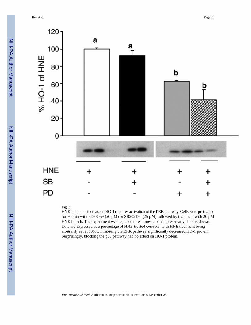
Fig. 8.HNE-mediated increase in HO-1 requires activation of the ERK pathway. Cells were pretreatedfor 30 min with PD98059 (50 µM) or SB202190 (25 µM) followed by treatment with 20 µMHNE for 5 h. The experiment was repeated three times, and a representative blot is shown.Data are expressed as a percentage of HNE-treated controls, with HNE treatment beingarbitrarily set at 100%. Inhibiting the ERK pathway significantly decreased HO-1 protein.Surprisingly, blocking the p38 pathway had no effect on HO-1 protein.
Iles et al. Page 20
Free Radic Biol Med. Author manuscript; available in PMC 2009 December 28.
NIH
-PA Author Manuscript
NIH
-PA Author Manuscript
NIH
-PA Author Manuscript

Fig. 9.HNE causes Nrf-2 nuclear translocation after ERK pathway activation. Cells were treated with20 µM 4HNE for 1 – 5 h. Nuclear lysates were generated using the NE-PER kit as outlinedunder Materials and methods. The experiment was repeated two times, and a representativeblot is shown. Data are expressed as a percentage of control (untreated at time 0) with controlNrf-2 arbitrarily being set at 100%. At 3 h there was a significant increase in Nrf-2 in thenucleus in response to HNE, which had returned to baseline levels by 4 h postincubation.
Iles et al. Page 21
Free Radic Biol Med. Author manuscript; available in PMC 2009 December 28.
NIH
-PA Author Manuscript
NIH
-PA Author Manuscript
NIH
-PA Author Manuscript

Fig. 10.HNE increases the nuclear content of the small Maf proteins. Cells were treated with 20 µM4HNE for 1 – 5 h. Nuclear lysates were generated using the NE-PER kit as outlined underMaterials and methods. The experiment was repeated, and a representative blot is shown. Dataare expressed as a percent of control with control Maf arbitrarily being set at 100%. Theantibody detects all three of the small Maf proteins. As multiple bands were obtained, the smallMafs were identified based on molecular weight markers. At 4 h there was a significant increasein small Maf proteins in the nucleus in response to HNE. This decreased to baseline by 5 hpostincubation.
Iles et al. Page 22
Free Radic Biol Med. Author manuscript; available in PMC 2009 December 28.
NIH
-PA Author Manuscript
NIH
-PA Author Manuscript
NIH
-PA Author Manuscript