Absence of T Cells Confers Increased Pulmonary Arterial Hypertension and Vascular Remodeling
TtpP(
FUMCSCPRM*UMbPnMMoCIe
Journal of the American College of Cardiology Vol. 54, No. 1, Suppl S, 2009© 2009 by the American College of Cardiology Foundation ISSN 0735-1097/09/$36.00P
Updated Clinical Classification of Pulmonary Hypertension
Gérald Simonneau, MD,* Ivan M. Robbins, MD,† Maurice Beghetti, MD,‡Richard N. Channick, MD,§ Marion Delcroix, MD, PHD,� Christopher P. Denton, MD, PHD,¶C. Gregory Elliott, MD,# Sean P. Gaine, MD, PHD,** Mark T. Gladwin, MD,††Zhi-Cheng Jing, MD,‡‡ Michael J. Krowka, MD,§§ David Langleben, MD,��Norifumi Nakanishi, MD, PHD,¶¶ Rogério Souza, MD##
Clamart, France; Nashville, Tennessee; Geneva, Switzerland; La Jolla, California; Leuven, Belgium; London,United Kingdom; Salt Lake City, Utah; Dublin, Ireland; Pittsburgh, Pennsylvania; Shanghai, China;Rochester, Minnesota; Montréal, Québec, Canada; Osaka, Japan; and São Paulo, Brazil
The aim of a clinical classification of pulmonary hypertension (PH) is to group together different manifestationsof disease sharing similarities in pathophysiologic mechanisms, clinical presentation, and therapeutic ap-proaches. In 2003, during the 3rd World Symposium on Pulmonary Hypertension, the clinical classification of PHinitially adopted in 1998 during the 2nd World Symposium was slightly modified. During the 4th World Sympo-sium held in 2008, it was decided to maintain the general architecture and philosophy of the previous clinicalclassifications. The modifications adopted during this meeting principally concern Group 1, pulmonary arterialhypertension (PAH). This subgroup includes patients with PAH with a family history or patients with idiopathicPAH with germline mutations (e.g., bone morphogenetic protein receptor-2, activin receptor-like kinase type 1,and endoglin). In the new classification, schistosomiasis and chronic hemolytic anemia appear as separate enti-ties in the subgroup of PAH associated with identified diseases. Finally, it was decided to place pulmonary veno-occlusive disease and pulmonary capillary hemangiomatosis in a separate group, distinct from but very close toGroup 1 (now called Group 1=). Thus, Group 1 of PAH is now more homogeneous. (J Am Coll Cardiol 2009;54:S43–54) © 2009 by the American College of Cardiology Foundation
ublished by Elsevier Inc. doi:10.1016/j.jacc.2009.04.012
PayATPsmadgat
ItrbiPiit
he classification of pulmonary hypertension (PH) has gonehrough a series of changes since the first classification wasroposed in 1973 at an international conference on primaryH (PPH) endorsed by the World Health Organization
1,2). The initial classification designated only 2 categories,
rom the *Centre National de Référence des Maladies Vasculaires Pulmonaires,niversité Paris-Sud Hôpital Antoine Béclère, Clamart, France; †Department ofedicine, Vanderbilt University Medical Center, Nashville, Tennessee; ‡Pediatricardiology Unit, Hôpital des Enfants, University Hospital of Geneva, Geneva,witzerland; §Division of Pulmonary and Critical Care Medicine, UCSD Medicalenter, La Jolla, California; �Center for Pulmonary Vascular Disease, Department ofneumology, Gasthuisberg University Hospital, Leuven, Belgium; ¶Centre forheumatology, Royal Free Hospital, London, United Kingdom; #Department ofedicine, Intermountain Medical Center, University of Utah, Salt Lake City, Utah;
*Department of Respiratory Medicine, Mater Misericordiae University Hospital,niversity College Dublin, Dublin, Ireland; ††Pulmonary, Allergy, and Critical Careedicine, Hemostasis and Vascular Biology Research Institute, University of Pitts-
urgh, Pittsburgh, Pennsylvania; ‡‡Department of Pulmonary Circulation, Shanghaiulmonary Hospital, Tongji University, Shanghai, China; §§Department of Pulmo-ary and Critical Care Medicine, Division of Gastroenterology and Hepatology,ayo Clinic, Rochester, Minnesota; � �Center for Pulmonary Vascular Disease, Sirortimer B. Davis Jewish General Hospital, Montréal, Québec, Canada; ¶¶Division
f Cardiology and Pulmonary Circulation, Department of Internal Medicine Nationalardiovascular Center, Osaka, Japan; and the ##Pulmonary Department, Heart
nstitute, University of São Paulo Medical School, São Paulo, Brazil. Please see the
Dnd of this article for each author’s conflict of interest information.
Manuscript received February 6, 2009; accepted April 15, 2009.
PH or secondary PH, depending on the presence orbsence of identifiable causes or risk factors. Twenty-fiveears later, the 2nd World Symposium on Pulmonaryrterial Hypertension (PAH) was held in Evian, France.he “Evian classification” attempted to create categories ofH that shared pathologic and clinical features as well asimilar therapeutic options (3). This was a much broader,ore encompassing classification, with 5 major categories; it
llowed investigators to conduct clinical trials in a well-efined group of patients with a shared underlying patho-enesis. This has led to multiple clinical trials and thepproval of 8 different medications worldwide for thereatment of PAH.
The 3rd World Symposium on PAH was held in Venice,taly, 5 years after the Evian conference. At this conference,he impact and usefulness of the “Evian classification” waseviewed, and modest changes were made. The most nota-le change was to abandon the term PPH in favor ofdiopathic pulmonary arterial hypertension (IPAH); familialAH if there is a family history of PAH; or associated PAH
f another cause, such as connective tissue disease or humanmmunodeficiency virus (HIV), is present. Although theerm PPH had become well ingrained in the literature after
resdale first used it in 1951 (4), it had become clear that
firwDc
G
Pcnhw1rdsh
ommfM1fwoprww
1hfBpmnPwcP
Vo
S44 Simonneau et al. JACC Vol. 54, No. 1, Suppl S, 2009Classification and Epidemiology June 30, 2009:S43–54
the pathologic changes and re-sponse to therapy were similar inseveral other conditions or dis-eases. The term “secondary PH”had been abandoned at the Evianmeeting because it was confusingand did not help with diagnosis orin directing treatment (5) (Table1). The other prominent changemade at the Venice meeting was tomove pulmonary veno-occlusivedisease (PVOD) and pulmonarycapillary hemangiomatosis (PCH)from separate categories into a sin-gle subcategory of PAH. These 2entities have many similaritieswith each other, which will bediscussed later in this article, aswell as some similarities withPAH. The 2008 4th World Sym-posium on PH held in DanaPoint, California, provided the op-portunity to slightly modify theprevious clinical classifications.
Dana Point Classification
During the 4th World Sympo-sium on PH held in 2008 inDana Point, California, the con-sensus of an international groupof experts was to maintain thegeneral philosophy and organiza-tion of the Evian-Venice classi-fications. However, in responseto a questionnaire regarding theprevious classification, a majorityof experts (63%) felt that modi-
cation of the Venice classification was required to accu-ately reflect information published over the past 5 years, asell as to clarify some areas that were unclear. The currentana Point classification is listed in Table 2, with major
hanges highlighted.
roup 1: PAH
ulmonary arterial hypertension has been the focus of thelassification of PH since the first classification in 1973. Theomenclature of the subgroups and associated conditionsas evolved since that time, and additional modificationsere made in the Dana Point classification..1./1.2. Idiopathic and heritable PAH. Pulmonary arte-ial hypertension may occur in different clinical conditionsepending on associated diseases. Idiopathic PAH corre-ponds to sporadic disease in which there is neither a family
Abbreviationsand Acronyms
BMPR2 � bonemorphogenetic proteinreceptor type 2
CHD � congenital heartdisease
CTEPH � chronicthromboembolic pulmonaryhypertension
ESRD � end-stage renaldisease
HIV � humanimmunodeficiency virus
IPAH � idiopathicpulmonary arterialhypertension
OR � odds ratio
PAH � pulmonary arterialhypertension
PAP � pulmonary arterialpressure
PCH � pulmonary capillaryhemangiomatosis
PH � pulmonaryhypertension
POPH � portopulmonaryhypertension
PPH � primary pulmonaryhypertension
PVOD � pulmonary veno-occlusive disease
PVR � pulmonary vascularresistance
SCD � sickle cell disease
TRV � tricuspidregurgitation jet velocity
istory of PAH nor an identified risk factor. When PAH l
ccurs in a familial context, germline mutations in the boneorphogenetic protein receptor type 2 (BMPR2) gene, aember of the transforming growth factor � signaling
amily, can be detected in approximately 70% of cases (6,7).ore rarely, mutations in activin receptor-like kinase type
, or endoglin, also members of the transforming growthactor � signaling family, have been identified in patientsith PAH, predominantly with coexistent hereditary hem-rrhagic telangiectasia. Recently, it has been suggested thatatients with PAH associated with BMPR2 mutations mayepresent a subgroup of patients with more severe diseaseho are less likely to demonstrate vasoreactivity than thoseith IPAH without BMPR2 mutations (8–10).Because BMPR2 mutations have also been detected in
1% to 40% of apparently idiopathic cases with no familyistory (11,12), the distinction between idiopathic andamilial BMPR2 mutations is artificial. All patients withMPR2 mutations have heritable disease, whether theatient is the first identified case, possibly with a de novoutation, or other family members were previously diag-
osed with PAH. In addition, in 30% or fewer families withAH, no BMPR2 mutation has been identified. Thus, itas decided to abandon the term “familial PAH” in the new
lassification and to replace it with the term “heritableAH.” Heritable forms of PAH include IPAH with germ-
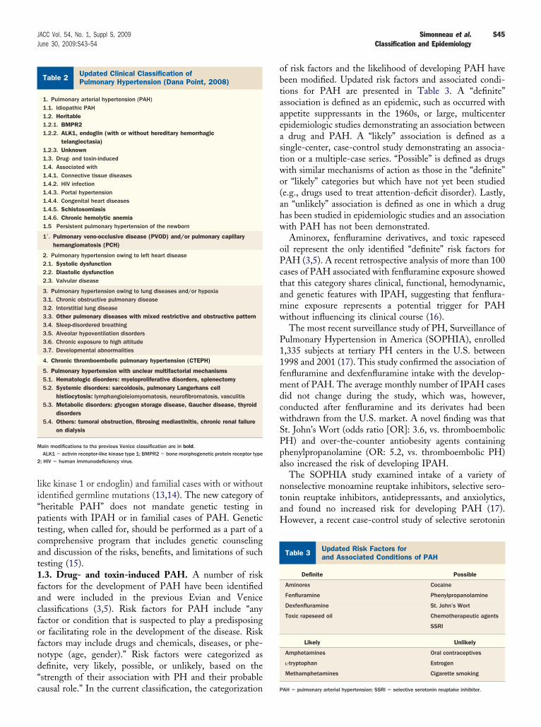
enice Clinical Classificationf Pulmonary Hypertension (2003)
Table 1 Venice Clinical Classificationof Pulmonary Hypertension (2003)
1. Pulmonary arterial hypertension (PAH)1.1. Idiopathic (IPAH)1.2. Familial (FPAH)1.3. Associated with (APAH)1.3.1. Collagen vascular disease1.3.2. Congenital systemic-to-pulmonary shunts1.3.3. Portal hypertension1.3.4. HIV infection1.3.5. Drugs and toxins1.3.6. Other (thyroid disorders, glycogen storage disease, Gaucher disease,
hereditary hemorrhagic telangiectasia, hemoglobinopathies,myeloproliferative disorders, splenectomy)
1.4. Associated with significant venous or capillary involvement1.4.1. Pulmonary veno-occlusive disease (PVOD)1.4.2. Pulmonary capillary hemangiomatosis (PCH)1.5. Persistent pulmonary hypertension of the newborn
2. Pulmonary hypertension with left heart disease2.1. Left-sided atrial or ventricular heart disease2.2. Left-sided valvular heart disease
3. Pulmonary hypertension associated with lung diseases and/or hypoxemia3.1. Chronic obstructive pulmonary disease3.2. Interstitial lung disease3.3. Sleep-disordered breathing3.4. Alveolar hypoventilation disorders3.5. Chronic exposure to high altitude3.6. Developmental abnormalities
4. Pulmonary hypertension owing to chronic thrombotic and/or embolic disease4.1. Thromboembolic obstruction of proximal pulmonary arteries4.2. Thromboembolic obstruction of distal pulmonary arteries4.3. Nonthrombotic pulmonary embolism (tumor, parasites, foreign material)
5. Miscellaneous
Sarcoidosis, histiocytosis X, lymphangiomatosis, compression of pulmonaryvessels (adenopathy, tumor, fibrosing mediastinitis)
ine mutations (mainly BMPR2 but also activin receptor-
li“ptcat1facfofnd“c
obtaaeastwo(ahw
oPctamw
P11fmdcwSPpa
ntaH
Ua
UP
M
2
S45JACC Vol. 54, No. 1, Suppl S, 2009 Simonneau et al.June 30, 2009:S43–54 Classification and Epidemiology
ike kinase 1 or endoglin) and familial cases with or withoutdentified germline mutations (13,14). The new category ofheritable PAH” does not mandate genetic testing inatients with IPAH or in familial cases of PAH. Geneticesting, when called for, should be performed as a part of aomprehensive program that includes genetic counselingnd discussion of the risks, benefits, and limitations of suchesting (15)..3. Drug- and toxin-induced PAH. A number of riskactors for the development of PAH have been identifiednd were included in the previous Evian and Venicelassifications (3,5). Risk factors for PAH include “anyactor or condition that is suspected to play a predisposingr facilitating role in the development of the disease. Riskactors may include drugs and chemicals, diseases, or phe-otype (age, gender).” Risk factors were categorized asefinite, very likely, possible, or unlikely, based on thestrength of their association with PH and their probable
pdated Clinical Classification ofulmonary Hypertension (Dana Point, 2008)
Table 2 Updated Clinical Classification ofPulmonary Hypertension (Dana Point, 2008)
1. Pulmonary arterial hypertension (PAH)1.1. Idiopathic PAH1.2. Heritable1.2.1. BMPR21.2.2. ALK1, endoglin (with or without hereditary hemorrhagic
telangiectasia)1.2.3. Unknown1.3. Drug- and toxin-induced1.4. Associated with1.4.1. Connective tissue diseases1.4.2. HIV infection1.4.3. Portal hypertension1.4.4. Congenital heart diseases1.4.5. Schistosomiasis1.4.6. Chronic hemolytic anemia1.5 Persistent pulmonary hypertension of the newborn
1=. Pulmonary veno-occlusive disease (PVOD) and/or pulmonary capillaryhemangiomatosis (PCH)
2. Pulmonary hypertension owing to left heart disease2.1. Systolic dysfunction2.2. Diastolic dysfunction2.3. Valvular disease
3. Pulmonary hypertension owing to lung diseases and/or hypoxia3.1. Chronic obstructive pulmonary disease3.2. Interstitial lung disease3.3. Other pulmonary diseases with mixed restrictive and obstructive pattern3.4. Sleep-disordered breathing3.5. Alveolar hypoventilation disorders3.6. Chronic exposure to high altitude3.7. Developmental abnormalities
4. Chronic thromboembolic pulmonary hypertension (CTEPH)
5. Pulmonary hypertension with unclear multifactorial mechanisms5.1. Hematologic disorders: myeloproliferative disorders, splenectomy5.2. Systemic disorders: sarcoidosis, pulmonary Langerhans cell
histiocytosis: lymphangioleiomyomatosis, neurofibromatosis, vasculitis5.3. Metabolic disorders: glycogen storage disease, Gaucher disease, thyroid
disorders5.4. Others: tumoral obstruction, fibrosing mediastinitis, chronic renal failure
on dialysis
ain modifications to the previous Venice classification are in bold.ALK1 � activin receptor-like kinase type 1; BMPR2 � bone morphogenetic protein receptor type
; HIV � human immunodeficiency virus.
ausal role.” In the current classification, the categorization P
f risk factors and the likelihood of developing PAH haveeen modified. Updated risk factors and associated condi-ions for PAH are presented in Table 3. A “definite”ssociation is defined as an epidemic, such as occurred withppetite suppressants in the 1960s, or large, multicenterpidemiologic studies demonstrating an association betweendrug and PAH. A “likely” association is defined as a
ingle-center, case-control study demonstrating an associa-ion or a multiple-case series. “Possible” is defined as drugsith similar mechanisms of action as those in the “definite”r “likely” categories but which have not yet been studiede.g., drugs used to treat attention-deficit disorder). Lastly,n “unlikely” association is defined as one in which a drugas been studied in epidemiologic studies and an associationith PAH has not been demonstrated.Aminorex, fenfluramine derivatives, and toxic rapeseed
il represent the only identified “definite” risk factors forAH (3,5). A recent retrospective analysis of more than 100ases of PAH associated with fenfluramine exposure showedhat this category shares clinical, functional, hemodynamic,nd genetic features with IPAH, suggesting that fenflura-ine exposure represents a potential trigger for PAHithout influencing its clinical course (16).The most recent surveillance study of PH, Surveillance of
ulmonary Hypertension in America (SOPHIA), enrolled,335 subjects at tertiary PH centers in the U.S. between998 and 2001 (17). This study confirmed the association ofenfluramine and dexfenfluramine intake with the develop-ent of PAH. The average monthly number of IPAH cases
id not change during the study, which was, however,onducted after fenfluramine and its derivates had beenithdrawn from the U.S. market. A novel finding was thatt. John’s Wort (odds ratio [OR]: 3.6, vs. thromboembolicH) and over-the-counter antiobesity agents containinghenylpropanolamine (OR: 5.2, vs. thromboembolic PH)lso increased the risk of developing IPAH.
The SOPHIA study examined intake of a variety ofonselective monoamine reuptake inhibitors, selective sero-onin reuptake inhibitors, antidepressants, and anxiolytics,nd found no increased risk for developing PAH (17).owever, a recent case-control study of selective serotonin
pdated Risk Factors fornd Associated Conditions of PAH
Table 3 Updated Risk Factors forand Associated Conditions of PAH
Definite Possible
Aminorex Cocaine
Fenfluramine Phenylpropanolamine
Dexfenfluramine St. John’s Wort
Toxic rapeseed oil Chemotherapeutic agents
SSRI
Likely Unlikely
Amphetamines Oral contraceptives
L-tryptophan Estrogen
Methamphetamines Cigarette smoking
AH � pulmonary arterial hypertension; SSRI � selective serotonin reuptake inhibitor.
rgsorPt
Parrs(pfaeaian1PabpmfSpmt
Pldcousce
nrsPt(1r(tai
astPidvtmv
Htsvc1oc(prhwcdpvhcdisrtiwsrc1oudpiaiCtPcmhw
S46 Simonneau et al. JACC Vol. 54, No. 1, Suppl S, 2009Classification and Epidemiology June 30, 2009:S43–54
euptake inhibitor use in pregnant women after 20 weeks ofestation showed an increased risk (OR: 6.1) in the off-pring of developing persistent PH of the newborn, a formf PAH (18). Based on this study, selective serotonineuptake inhibitors may play a role in the development ofH, at least in association with pregnancy, and therefore
hey have been reclassified in the “possible” category.Amphetamine use represents a “likely” risk factor for
AH, although they are rarely taken as a single agent andre frequently used in combination with fenfluramine. Aecent comprehensive retrospective study suggested a strongelationship with the use of methamphetamine (inhaled,moked, oral, or intravenous) and the occurrence of IPAH19). Based primarily on the results of this study, metham-hetamine use is now considered a “very likely” risk factoror the development of PAH. Additional changes in drug-nd toxin-induced PAH will be discussed later. With thexception of hereditary hemorrhagic telangiectasia associ-ted with PAH, the first 3 subcategories of Group 1,diopathic, heritable, and drug- and toxin-induced PAH,re all associated with the development of isolated pulmo-ary arterial diseases..4.1. PAH associated with connective tissue diseases.AH associated with connective tissue diseases representsn important clinical subgroup. The prevalence of PAH haseen well established only for systemic sclerosis. Two recentrospective studies using echocardiography as a screeningethod and right heart catheterization for confirmation
ound a prevalence of PAH of between 7% and 12% (20,21).everal long-term studies suggest that the outcome ofatients with PAH associated with systemic sclerosis isarkedly worse than that of patients with IPAH, despite
he use of modern therapies.Importantly, PAH does not represent the only cause of
H in systemic sclerosis. Pulmonary hypertension owing toung fibrosis is also frequent (22), and diastolic left heartysfunction is not uncommon (23). There is also primaryardiac involvement in the disease process (24). Thesebservations emphasize the importance of a complete eval-ation when PH is suspected in patients with systemicclerosis and the need for right heart catheterization toonfirm the diagnosis of PH and to accurately classify itstiology to determine appropriate treatment.
In systemic lupus erythematosis (25,26) and mixed con-ective tissue disease (27,28), the prevalence of PAHemains unknown but likely occurs less frequently than inystemic sclerosis. In the absence of fibrotic lung disease,AH has been reported infrequently in other connective
issue diseases such as Sjögren syndrome (29), polymyositis30), or rheumatoid arthritis (31)..4.2. HIV infection. Pulmonary arterial hypertension is aare but well-established complication of HIV infection32,33). Epidemiologic data in the early 1990s, a time whenherapy with highly active antiretroviral therapy was not yetvailable, indicated a prevalence of 0.5% (95% confidence
nterval: 0.10% to 0.50%) (34). The prevalence of HIV- pssociated PAH was evaluated more recently and showed atable prevalence of 0.46% (95% confidence interval: 0.32%o 0.64%) (35). Human immunodeficiency virus-associatedAH has clinical, hemodynamic, and histologic character-
stics similar to those seen in IPAH. The mechanism for theevelopment of PH remains unclear. Because neither theirus nor viral DNA has been found in pulmonary endo-helial cells, an indirect action of virus through secondaryessengers such as cytokines, growth factors, endothelin, or
iral proteins is strongly suspected.Uncontrolled studies suggest that patients with severeIV-associated PAH could benefit from bosentan or long-
erm infusion of epoprostenol (36,37). Interestingly, in aubstantial number of cases, normalization of pulmonaryascular hemodynamics can be obtained with therapy indi-ated for PAH; this is very rarely seen in IPAH (38)..4.3. Portopulmonary hypertension. The developmentf PAH in association with elevated pressure in the portalirculation is known as portopulmonary hypertensionPOPH) (39,40). Portal hypertension, rather than theresence of underlying liver disease, is the main determiningisk factor for the development of POPH. Prospectiveemodynamic studies have shown that 2% to 6% of patientsith portal hypertension have PH (41,42). Right heart
atheterization is absolutely mandatory for the definitiveiagnosis of POPH because several factors may increaseulmonary arterial pressure (PAP) in the setting of ad-anced liver disease (e.g., high flow associated with theyperdynamic circulatory state and increased pulmonaryapillary wedge pressure owing to fluid overload and/oriastolic dysfunction). Pulmonary vascular resistance (PVR)s usually normal in these cases. Pathologic changes in themall arteries appear identical to those seen in IPAH. Aecent multicenter case-control study identified 2 risk fac-ors for the development of POPH: female sex and auto-mmune hepatitis (43). Interestingly, hepatitis C infectionas associated with a decreased risk. A recent, large cohort
tudy of POPH showed that long-term prognosis waselated to the presence and severity of cirrhosis and toardiac function (44)..4.4. Congenital heart diseases. A significant proportionf patients with congenital heart disease (CHD), in partic-lar those with relevant systemic-to-pulmonary shunts, willevelop PAH if left untreated. Persistent exposure of theulmonary vasculature to increased blood flow, as well asncreased pressure, may result in pulmonary obstructiverteriopathy, which leads to increased PVR that will resultn shunt reversal. Eisenmenger syndrome is defined asHD with an initial large systemic-to-pulmonary shunt
hat induces progressive pulmonary vascular disease andAH, with resultant reversal of the shunt and centralyanosis (45,46). Eisenmenger syndrome represents theost advanced form of PAH associated with CHD. The
istopathologic and pathobiologic changes seen in patientsith PAH associated with congenital systemic-to-
ulmonary shunts (e.g., endothelial dysfunction of the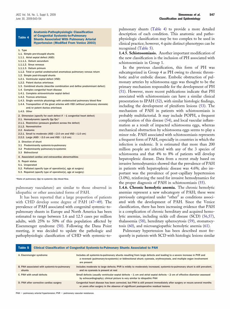
pi
wppeaEmp
pdpcr1ts
sbmp(apimpcmmmaimshiip(t1apaciltt
q
AoSH
*
C
P
S47JACC Vol. 54, No. 1, Suppl S, 2009 Simonneau et al.June 30, 2009:S43–54 Classification and Epidemiology
ulmonary vasculature) are similar to those observed indiopathic or other associated forms of PAH.
It has been reported that a large proportion of patientsith CHD develop some degree of PAH (47–49). Therevalence of PAH associated with congenital systemic-to-ulmonary shunts in Europe and North America has beenstimated to range between 1.6 and 12.5 cases per milliondults, with 25% to 50% of this population affected byisenmenger syndrome (50). Following the Dana Pointeeting, it was decided to update the pathologic and
athophysiologic classification of CHD with systemic-to-
natomic-Pathophysiologic Classificationf Congenital Systemic-to-Pulmonaryhunts Associated With Pulmonary Arterialypertension (Modified From Venice 2003)
Table 4
Anatomic-Pathophysiologic Classificationof Congenital Systemic-to-PulmonaryShunts Associated With Pulmonary ArterialHypertension (Modified From Venice 2003)
1. Type1.1. Simple pre-tricuspid shunts1.1.1. Atrial septal defect (ASD)1.1.1.1. Ostium secundum1.1.1.2. Sinus venosus1.1.1.3. Ostium primum1.1.2. Total or partial unobstructed anomalous pulmonary venous return1.2. Simple post-tricuspid shunts1.2.1. Ventricular septal defect (VSD)1.2.2. Patent ductus arteriosus1.3. Combined shunts (describe combination and define predominant defect)1.4. Complex congenital heart disease1.4.1. Complete atrioventricular septal defect1.4.2. Truncus arteriosus1.4.3. Single ventricle physiology with unobstructed pulmonary blood flow1.4.4. Transposition of the great arteries with VSD (without pulmonary stenosis)
and/or patent ductus arteriosus1.4.5. Other
2. Dimension (specify for each defect if �1 congenital heart defect)2.1. Hemodynamic (specify Qp/Qs)*2.1.1. Restrictive (pressure gradient across the defect)2.1.2. Nonrestrictive2.2. Anatomic2.2.1. Small to moderate (ASD �2.0 cm and VSD �1.0 cm)2.2.2. Large (ASD �2.0 cm and VSD �1.0 cm)
3. Direction of shunt3.1 Predominantly systemic-to-pulmonary3.2 Predominantly pulmonary-to-systemic3.3 Bidirectional
4. Associated cardiac and extracardiac abnormalities
5. Repair status5.1. Unoperated5.2. Palliated (specify type of operation[s], age at surgery)5.3. Repaired (specify type of operation[s], age at surgery)
Ratio of pulmonary (Qp) to systemic (Qs) blood flow.
linical Classification of Congenital Systemic-to-Pulmonary Shunts
Table 5 Clinical Classification of Congenital Systemic-to-Pulmo
A. Eisenmenger syndrome Includes all systemic-to-pulmonaa reversed (pulmonary-to-systare present
B. PAH associated with systemic-to-pulmonaryshunts
Includes moderate to large defeand no cyanosis is present at
C. PAH with small defects Small defects (usually ventriculaby echocardiography); clinica
D. PAH after corrective cardiac surgery Congenital heart disease has beor years after surgery in the a
AH � pulmonary arterial hypertension; PVR � pulmonary vascular resistance.
ulmonary shunts (Table 4) to provide a more detailedescription of each condition. This anatomic and patho-hysiologic classification may be too complex to be used inlinical practice; however, 4 quite distinct phenotypes can beecognized (Table 5)..4.5. Schistosomiasis. Another important modification ofhe new classification is the inclusion of PH associated withchistosomiasis in Group 1.
In the previous classification, this form of PH wasubcategorized in Group 4 as PH owing to chronic throm-otic and/or embolic disease. Embolic obstruction of pul-onary arteries by schistosoma eggs was thought to be the
rimary mechanism responsible for the development of PH51). However, more recent publications indicate that PHssociated with schistosomiasis can have a similar clinicalresentation to IPAH (52), with similar histologic findings,ncluding the development of plexiform lesions (53). The
echanism of PAH in patients with schistosomiasis isrobably multifactorial. It may include POPH, a frequentomplication of this disease (54), and local vascular inflam-ation as a result of impacted schistosoma eggs, whereasechanical obstruction by schistosoma eggs seems to play ainor role. PAH associated with schistosomiasis representsfrequent form of PAH, especially in countries in which the
nfection is endemic. It is estimated that more than 200illion people are infected with any of the 3 species of
chistosoma and that 4% to 8% of patients will developepatosplenic disease. Data from a recent study based on
nvasive hemodynamics showed that the prevalence of PAHn patients with hepatosplenic disease was 4.6%; also im-ortant was the prevalence of post-capillary hypertension3.0%), reinforcing the need for invasive hemodynamics forhe proper diagnosis of PAH in schistosomiasis (55)..4.6. Chronic hemolytic anemia. The chronic hemolyticnemias represent a new subcategory of PAH; these werereviously categorized under “other” as conditions associ-ted with the development of PAH. Since the Venicelassification, there has been increasing evidence that PAHs a complication of chronic hereditary and acquired hemo-ytic anemias, including sickle cell disease (SCD) (56,57),halassemia (58), hereditary spherocytosis (59), stomatocy-osis (60), and microangiopathic hemolytic anemia (61).
Pulmonary hypertension has been described most fre-uently in patients with SCD with histologic lesions similar
ciated to PAH
Shunts Associated to PAH
nts resulting from large defects and leading to a severe increase in PVR andor bidirectional shunt; cyanosis, erythrocytosis, and multiple organ involvement
R is mildly to moderately increased, systemic-to-pulmonary shunt is still prevalent,
al defects �1 cm and atrial septal defects �2 cm of effective diameter assessede is very similar to idiopathic PAH
rected, but PAH is still present immediately after surgery or recurs several monthse of significant postoperative residual lesions
Asso
nary
ry shuemic)
cts; PVrest
r septl pictur
en corbsenc
tciSpmucnasciswpphPnnwSeaciSAhosd
G
TtEdiadtPptmhpcw
o
naPuPdtdrLfwPs
io1phiepPtca
tpdcttbdPmdpPfp(tbbP
bnicf
S48 Simonneau et al. JACC Vol. 54, No. 1, Suppl S, 2009Classification and Epidemiology June 30, 2009:S43–54
o those found in IPAH, including plexiform lesions in 1ase series (62). However, the prevalence of PAH in SCDs not clearly established. The largest study of patients withCD, which defined PH echocardiographically by theresence of tricuspid regurgitation jet velocity (TRV) �2.5/s, found that 32% of patients had PH (57). However,
sing a TRV �2.5 m/s on echocardiography to define PHan lead to a substantial number of false positive cases of PHot confirmed by right heart catheterization (35,63). WhenTRV �3.0 m/s was used, corresponding to an estimated
ystolic PAP of �41 mm Hg, only 9% of the cohort met theriteria for PH. Right heart catheterization was carried outn only 18 of 63 patients with TRV �2.5 m/s. In thisubpopulation, PH defined by a mean PAP �25 mm Hgas confirmed in 17 patients; however, pulmonary wedgeressure was elevated in some patients. A substantial pro-ortion of patients with SCD have pulmonary venousypertension: 46% in 1 study of 26 patients with SCD andH (64). In addition, some patients present with a hyperki-etic state with moderate elevation in mean PAP andormal PVR. Thus, although it appears that some patientsith SCD do develop PAH, the prevalence of PAH inCD is undoubtedly much lower than 32%. Prospectivepidemiologic studies using echocardiographic screeningnd direct hemodynamic confirmation with right heartatheterization in all patients with suspected PH are ongo-ng and will evaluate the precise prevalence of PAH inCD. The mechanism of PAH in SCD remains uncertain.
probable hypothesis is that chronic hemolysis results inigh rates of nitric oxide consumption and produces a statef resistance to nitric oxide bioactivity (65). Consequently,mooth muscle guanosine monophosphate, a potent vaso-ilator/antiproliferative mediator, is not activated (66).
roup 1=: PVOD and/or PCH
he conditions of PVOD and PCH are uncommon, buthey are increasingly recognized as causes of PH (67). In thevian classification, these 2 entities were placed in 2ifferent groups, both distinct from PAH: PVOD was
ncluded in the pulmonary venous hypertension category,nd PCH was included in the heterogeneous group ofisorders believed to directly affect the pulmonary vascula-ure. Pathologic studies indicate, however, that PVOD andCH are often quite similar in terms of changes in theulmonary parenchyma (i.e., pulmonary hemosiderosis, in-erstitial edema, and lymphatic dilation) and the develop-ent of pulmonary arterial intimal fibrosis and medial
ypertrophy. Similarities in pathologic features and clinicalresentation suggest that these disorders may overlap. Ac-ordingly, in the Venice classification, PVOD and PCHere placed together as a subgroup of PAH.PVOD and PCH were included in Group 1 for a number
f reasons. First, the histologic changes in the small pulmo- n
ary arteries (i.e., intimal fibrosis and medial hypertrophy)re also found in PAH. Second, the clinical presentations ofVOD/PCH and PAH are often indistinguishable andnrecognized antemortem (5). Third, PVOD/PCH andAH share similar risk factors. These include the sclero-erma spectrum of disease (68), HIV infection (69,70), andhe use of anorexigens. Fourth, in addition to the well-escribed familial association with PAH, familial occur-ence has been reported with both PVOD and PCH (67).astly, mutations in BMPR2, the gene associated with
amilial PAH and IPAH, have been documented in patientsith PVOD (71,72). These findings suggest that PVOD,CH, and PAH may represent different components of aingle spectrum of disease.
The present decision to leave PVOD and PCH togethern the same subgroup is supported by a recent clinicopath-logic study (73) analyzing 38 specimens (autopsies [n �5], surgical biopsies [n � 15], explants [n � 7], andneumonectomy [n � 1]) from 35 patients diagnosed asaving either PVOD (n � 30) or PCH (n � 5). PCH was
dentified in 24 (73%) patients diagnosed as having PVOD,ither as perivenular foci or diffuse involvement of theulmonary parenchyma. In 5 patients diagnosed withCH, 4 showed the venous and arterial changes charac-
eristic of PVOD. These findings suggest that PCHould be an angioproliferative process frequently associ-ted with PVOD.
Recent evidence supports leaving PVOD and PCHogether; however, it is apparent that although they mayresent similarly to IPAH, there are a number of importantifferences. These include the presence of crackles andlubbing on examination, ground glass opacities, septalhickening, mediastinal adenopathy on chest computedomography (74–76), hemosiderin-laden macrophages onronchoalveolar lavage (77), and a lower carbon monoxideiffusing capacity and PaO2 in patients with PVOD orCH (71). In addition, the management, response toedical therapy, and prognosis of PVOD/PCH are quite
ifferent than that of PAH. A recent study compared 24atients with histologic evidence of PVOD with or withoutCH and 24 randomly selected patients with idiopathic,
amilial, or anorexigen-associated PAH (71). Among the 16atients with PVOD who received PAH-specific therapy, 743.8%) developed pulmonary edema. These patients werereated mainly with continuous intravenous epoprostenol,ut also with oral therapies, bosentan, and calcium channellockers. Clinical outcomes were worse in patients withVOD than those with PAH.PVOD/PCH remains a difficult disorder to categorize
ecause it shares characteristics with IPAH but also has aumber of distinct differences. Given the current evidence,
t was decided that PVOD/PCH should be a distinctategory but not completely separated from PAH. There-ore, in the current classification, PVOD/PCH is desig-
ated as 1=.
G
LcmbPWa�eovdtTddiphlt
etduwpdoeplppr
GL
IhbPttaTcabsfis
st
ai(pcIoc(upavPial
G
IatiauhT4cdtgesuCdfom
wwtedddatP
S49JACC Vol. 54, No. 1, Suppl S, 2009 Simonneau et al.June 30, 2009:S43–54 Classification and Epidemiology
roup 2: PH Owing to Left Heart Disease
eft heart disease probably represents the most frequentause of PH (78). Left-sided ventricular or valvular diseasesay produce an increase in left atrial pressure, with passive
ackward transmission of the pressure leading to increasedAP. In this situation, PVR is normal or near normal (�3.0ood units) and there is no gradient between mean PAP
nd pulmonary wedge pressure (transpulmonary gradient12 mm Hg). In the Venice classification, 2 broad subcat-
gories were recognized in this group based on the presencer absence of valvular disease: PH owing to left atrial orentricular disease and PH owing to left-sided valvularisease (mitral and/or aortic). In the Venice classification,his category was referred to as PH with left heart disease.he new heading for this classification more appropriatelyenotes cause and effect for this group of heterogeneousisorders on the development of PH. In addition, with
ncreasing recognition of left-sided heart dysfunction withreserved ejection fraction, the subcategories of Group 2ave been modified and now include 3 distinct etiologies:
eft heart systolic dysfunction, left heart diastolic dysfunc-ion, and left heart valvular disease.
Importantly, in some patients with left heart disease, thelevation of PAP is out of proportion to that expected fromhe elevation of left arterial pressure (transpulmonary gra-ient �12 mm Hg) and PVR is increased to �3.0 Woodnits. In patients referred to cardiac transplant clinics, PHith PVR �3.0 Wood units is reported in 19% to 35% ofatients (78,79). Some patients with left heart valvularisease or even left heart dysfunction can develop severe PHf the same magnitude as that seen in PAH (80–82). Thelevation of PAP and PVR is due to either the increase ofulmonary artery vasomotor tone and/or pulmonary vascu-
ar remodeling (83,84). No studies using medications ap-roved for PAH have been performed in this patientopulation, and the efficacy and safety of PAH medicationsemain unknown.
roup 3: PH Owing toung Diseases and/or Hypoxia
n this category, the predominant cause of PH is alveolarypoxia as a result of lung disease, impaired control ofreathing, or residence at high altitude. The prevalence ofH in all of these conditions remains largely unknown. In
he present classification, the structure of this group was forhe most part unchanged. The heading has been modified,gain to denote cause and effect on the development of PH.he primary modification in this group was to add a
ategory of lung disease characterized by a mixed obstructivend restrictive pattern. This new subgroup includes chronicronchiectasis, cystic fibrosis (85), and a newly identifiedyndrome characterized by the combination of pulmonarybrosis, mainly of the lower zones of the lung, and emphy-
ema, mainly of the upper zones of the lung (86). In the fyndrome of combined pulmonary fibrosis and emphysema,he prevalence of PH is almost 50% (86).
In patients with parenchymal lung disease, PH is gener-lly modest (mean PAP 25 to 35 mm Hg) (87). However,n some patients, PAP elevations can be more substantialmean PAP 35 to 50 mm Hg) (88). In such patients,articularly those with only moderate pulmonary mechani-al impairment, this is considered “out-of-proportion” PH.n a recent retrospective study of 998 patients with chronicbstructive pulmonary disease who underwent right heartatheterization, only 1% had severe pulmonary hypertensionmean PAP �40 mm Hg) (89). The authors described annusual pattern of cardiopulmonary abnormalities in theatients with more severe PH, including mild to moderateirway obstruction, severe hypoxemia, hypocapnia, and aery low diffusing capacity for carbon monoxide. As withH out of proportion to left heart disease, large random-
zed, controlled studies of medications approved for PAHre not available for PH out of proportion for parenchymaung disease.
roup 4: Chronic Thromboembolic PH
n the Venice classification, Group 4 was very heterogeneousnd included obstruction of pulmonary arterial vessels byhromboemboli, tumors, or foreign bodies. However, depend-ng on the origin of the obstruction, the clinical presentationnd radiologic findings are often different, and management isnique to each etiology. Chronic thromboembolic pulmonaryypertension (CTEPH) represents a frequent cause of PH.he incidence of CTEPH is uncertain, but it occurs in up to% of patients after an acute pulmonary embolism (90,91). Inontrast, other obstructive etiologies are very rare. It wasecided, therefore, to maintain only CTEPH in Group 4. Inhe Venice classification, CTEPH was divided into 2 sub-roups: proximal CTEPH, accessible to pulmonary thrombo-ndarterectomy, and distal CTEPH, which is not accessible tourgery. In practice, however, this distinction may be quitenclear, and it may vary depending on individual centers.urrently there is no consensus among experts about theefinitions of proximal and distal CTEPH (92). Thus, it wasurther decided to maintain in Group 4 only a single categoryf CTEPH and not to attempt to distinguish between proxi-al and distal forms.Importantly, it is strongly recommended that patients
ith suspected or confirmed CTEPH be referred to a centerith expertise in the management of this disease to consider
he feasibility of performing pulmonary thromboendarter-ctomy, currently the only curative treatment. The decisionepends on the location of the obstruction (central vs. moreistal pulmonary arteries), the correlation between hemo-ynamic findings and the degree of mechanical obstructionssessed by angiography, comorbidities, the willingness ofhe patient, and the experience of the surgeon (93,94).atients who are not candidates for surgery may benefit
rom PAH-specific medical therapy (95,96); however, the
uu
Go
Ge5odtpmdmhtiatms5a(dcldatditnautMs
ccfcrmsmdicc
d
ltppp
hbbctkSsmtfi
atd5hddapprp
dacawesse(p
inupowpfig
S50 Simonneau et al. JACC Vol. 54, No. 1, Suppl S, 2009Classification and Epidemiology June 30, 2009:S43–54
se of these medications in CTEPH requires further eval-ation in randomized, controlled trials (97).
roup 5: PH With Unclearr Multifactorial Etiologies
roup 5 consists of several forms of PH for which thetiology is unclear or multifactorial..1. The first subgroup comprises several hematologic dis-rders. PH has been reported in chronic myeloproliferativeisorders including polycythemia vera, essential thrombocy-hemia, and chronic myeloid leukemia (98). Chronic myelo-roliferative disorders can cause PH by various potentialechanisms. High cardiac output, auto or surgical asplenia,
irect obstruction of pulmonary arteries by circulatingegakaryocytes (99), CTEPH (100), POPH, and congestive
eart failure may all play a role. Splenectomy as a result ofrauma or as a treatment for hematologic disorders mayncrease the risk of developing PH (101). CTEPH (102,103)nd several cases of PAH (102,104) with medial hyper-rophy, internal fibrosis, and plexiform lesions in the pul-onary vasculature have been reported in association with
plenectomy..2. The second subgroup includes systemic disorders thatre associated with an increased risk of developing PH105). Sarcoidosis is a common systemic granulomatousisease of unknown origin. PH is an increasingly recognizedomplication of sarcoidosis (106), with a reported preva-ence of 1% to 28% (107). PH is often attributed to theestruction of the capillary bed by the fibrotic processnd/or to the resultant chronic hypoxemia (108). However,he severity of PH does not always correlate well with theegree of parenchymal lung disease and blood gas abnormal-
ties, suggesting that other mechanisms may be contributing tohe development of PH (105). In this setting, such mecha-isms could include extrinsic compression of large pulmonaryrteries by mediastinal and hilar adenopathy, and direct gran-lomatous infiltration of the pulmonary vasculature, especiallyhe pulmonary veins, which sometimes mimic PVOD (109).
ore rarely, POPH secondary to hepatic involvement witharcoidosis can be associated with the pathogenesis.
Pulmonary Langerhans cell histiocytosis is an uncommonause of infiltrative lung disease associated with destructivehanges in the lung parenchyma. Severe PH is a commoneature in patients with end-stage pulmonary Langerhansells histiocytosis (110). In some patients, PH is probablyelated to chronic hypoxemia and/or abnormal pulmonaryechanics; in others, especially those patients with more
evere elevation of PAP, PH is unrelated to lung parenchy-al injury. Histopathologic examination has shown severe
iffuse pulmonary vasculopathy involving predominantlyntralobular pulmonary veins and, to a lesser extent, mus-ular pulmonary arteries (111). These vascular changesonsist of medial hypertrophy and intimal fibrosis.
Lymphangioleiomyomatosis is a rare multisystem disor-
er predominantly affecting women, characterized by cystic iung destruction, lymphatic abnormalities, and abdominalumors. PH is relatively uncommon in patients with lym-hangioleiomyomatosis (112,113). Chronic hypoxemia andulmonary capillary destruction caused by cystic lung lesionsrobably represent the predominant causes of PH.Neurofibromatosis type 1, also known as von Reckling-
ausen disease, is an autosomal dominant disease that cane recognized by characteristic “café au lait” skin lesions andy cutaneous fibromas. The disease is occasionally compli-ated by systemic vasculopathy. Recently it was reportedhat the neurofibromatosis type 1 gene modulates proteininase B, an important regulator of cell proliferation.everal cases of PH have recently been reported in theetting of von Recklinghausen disease (114–117). Theechanism of PH is unclear. Lung fibrosis and CTEPH are
hought to play a role. In rare cases, histologic examinationound both arteries and veins narrowed by medial and/orntimal hypertrophy and fibrosis (117,118).
Lastly, some rare cases of PH have been observed inntineutrophil cytoplasmic antibodies-associated vasculitis;he clinical presentation is similar to PAH, but histologicata are not available (119)..3. The third subgroup comprises metabolic disorders. PHas been reported in a few cases of type Ia glycogen storageisease, a rare autosomal recessive disorder caused by aeficiency of glucose-6-phosphatase (120–122). The mech-nism of PH is uncertain but has been associated withortocaval shunts, atrial septal defects, or severe restrictiveulmonary function defects. Thrombosis may also play aole in this setting. In 1 case, autopsy findings revealed theresence of plexiform lesions (123).Gaucher disease is a rare disorder characterized by a
eficiency of lysosomal B glucosidase, which results in anccumulation of glucocerebroside in reticuloendothelialells. Typical manifestations include hepatosplenomegalynd bone marrow infiltration. In a study of 134 patientsith Gaucher disease who were systematically screened by
chocardiography, PH was not uncommon (124). In thisetting, several potential mechanisms for PH have beenuggested, including interstitial lung disease, chronic hypox-mia, capillary plugging by Gaucher cells, and splenectomy124,125). One case of histologic findings similar to idio-athic PAH has been reported (126).The association between thyroid diseases (hypothyroid-
sm and hyperthyroidism) and PH has been reported in aumber of studies (127,128). In a recent prospective studysing echocardiographic evaluation, more than 40% ofatients with thyroid diseases had PH (129). One instancef PVOD confirmed by histology was observed in a patientith Hashimoto thyroiditis (130). Interestingly, a recentrospective study of 63 consecutive adult patients with PAHound a 49% prevalence of autoimmune thyroid disease,ncluding both hypothyroidism and hyperthyroidism, sug-esting that these conditions may be linked by a common
mmunogenetic susceptibility (131).
5mgaopawmut
e(orsgfpti
Pvpstafw
ryP(dmtirwh
C
Ifitcc
A
PfU
sTcLfUamLSArVrEmEfwsURMaraGraaUchBdRWAsL
RCnA(E
R
S51JACC Vol. 54, No. 1, Suppl S, 2009 Simonneau et al.June 30, 2009:S43–54 Classification and Epidemiology
.4. The last subgroup in category 5 includes a number ofiscellaneous conditions. In tumor obstruction, a tumor
rows into the central pulmonary arteries, with additionalppositional thrombosis leading to a progressive obstructionf proximal pulmonary arteries and PH. Such cases are duerincipally to pulmonary artery sarcomas, occur rarely, andre usually rapidly fatal (132,133). The differential diagnosisith CTEPH can be difficult. Computed tomography andagnetic resonance imaging angiography are the most
seful diagnostic modalities for differentiation betweenumor and thrombotic material (132,134,135).
Occlusion of the microvasculature by metastatic tumormboli represents another rare cause of rapidly progressive PH136). The initial laboratory evaluation shows hypoxemia,ften severe, with a clear lung field (137). Computed tomog-aphy scanning does not show proximal thrombi but oftenhows thickening of septa. In contrast, the V/Q lung scan isenerally abnormal with multiple subsegmental perfusion de-ects. Pulmonary microvascular cytology sampling through aulmonary artery catheter in the wedge position is an impor-ant diagnostic tool (137). The majority of reported cases occurn association with breast, lung, or gastric carcinomas.
Patients with mediastinal fibrosis may present with severeH owing to compression of both pulmonary arteries andeins (138,139). V/Q scan, computed tomography, andulmonary angiography are very useful for accurate diagno-is, but findings can mimic proximal thrombotic obstruc-ion. The predominant etiology is histoplasmosis (139),lthough mediastinal fibrosis has been reported with otherungal organisms, with tuberculosis (140), and in patientsith sarcoidosis.Lastly, PH has been reported in patients with end-stage
enal disease (ESRD) maintained on long-term hemodial-sis. Based on echocardiographic studies, the prevalence ofH in this patient population is estimated at up to 40%
141). There are several potential explanations for theevelopment of PH in patients with ESRD. Hormonal andetabolic derangement associated with ESRD might lead
o pulmonary vascular constriction. The PAP may also bencreased by high cardiac output (resulting from the arte-iovenous access itself and often concomitant anemia) asell as fluid overload. In addition, diastolic and systolic lefteart dysfunctions are frequent in this setting (142).
onclusions
n updating the classification of PH, we incorporated recentndings and sought to clarify areas of ambiguity. We believehat this version is both more comprehensive and moreomprehensible and hope that it will also be more useful tolinicians, pending further research into this diverse disease.
uthor Disclosures
rof. Simonneau has received honoraria and research grantsrom Actelion, Bayer Schering, GlaxoSmithKline, Pfizer, and
nited Therapeutics. Dr. Robbins has received grant/researchupport from Actelion, BioMarin, Gilead, Pfizer, and Unitedherapeutics; and has served on advisory boards, steering
ommittees, and/or speaker’s bureaus for Actelion, Gilead, andung Rx. Dr. Channick has received consulting and speaker
ees and research grants from Actelion, Gilead, Pfizer, andnited Therapeutics. Dr. Delcroix has received fees for serving
s investigator, speaker, consultant, or steering committeeember from Actelion, Aventis Pharmaceuticals, Bayer, Eliilly, Encysive, Gilead (Myogen), GlaxoSmithKline, Pfizer,chering, and United Therapeutics; and research grants fromctelion, Encysive, and GlaxoSmithKline. Dr. Denton has
eceived honoraria and consultancy fees from Actelion, Bio-itrum, Encysive, Pfizer, and Genzyme Therapeutics, and
esearch funding from Actelion, Aspreva Pharmaceuticals,ncysive, and Genzyme. Dr. Elliott is employed by Inter-ountain Healthcare. Intermountain Healthcare, with Dr.lliott as the inventor, has filed a patent application “method
or determining vasoreactivity.” Intermountain Healthcare,ith Dr. Elliott as Principal Investigator, has received grant
upport from Actelion, Pfizer, Encysive, CoTherix, andnited Therapeutics. Dr. Elliott serves as a member of theegistry to Evaluate Early and Long Term PAH Diseaseanagement sponsored by Actelion. Dr. Gaine has served on
dvisory boards for Actelion, GlaxoSmithKline, and Pfizer;eceived honoraria from Actelion, GlaxoSmithKline, Lung Rx,nd Pfizer; and conducted clinical trials supported by Actelion,ilead, Bayer, and United Therapeutics. Dr. Gladwin has
eceived grant support in the form of a Collaborative Researchnd Development Agreement between the U.S. Governmentnd INO Therapeutics. He is also listed as a co-inventor on a.S. Government Patent for the use of nitrite salts for
ardiovascular indications. Dr. Jing has received travel support,onoraria for speaking, and consulting fees from Actelion andayer Schering. Dr. Nakanishi has received a grant for Car-iovascular Disease and a grant to the Respiratory Failureesearch Group from the Ministry of Health, Labour andelfare of Japan. Dr. Souza has received consulting fees from
ctelion; lecture fees from Actelion and Pfizer; and travelupport from Gilead (Myogen). Drs. Beghetti, Krowka, andangleben report no conflicts of interest.
eprint requests and correspondence: Dr. Gérald Simonneau,entre National de Référence des Maladies Vasculaires Pulmo-aires, Service de Pneumologie, Hôpital Antoine Béclère,ssistance Publique, Hôpitaux de Paris, Université Paris-Sud
XI), 157, rue de la Porte de Trivaux, 92140 Clamart, France.-mail: [email protected].
EFERENCES
1. Hatano S, Strasser T. Primary Pulmonary Hypertension. Report ona WHO Meeting. October 15–17, 1973, Geneva: World HealthOrganization, 1975.
2. Wagenvoort CA, Wagenvoort N. Primary pulmonary hypertension:a pathologic study of the lung vessels in 156 clinically diagnosedcases. Circulation 1970;42:1163–84.
3. Fishman AP. Clinical classification of pulmonary hypertension. ClinChest Med 2001;22:385–91.
S52 Simonneau et al. JACC Vol. 54, No. 1, Suppl S, 2009Classification and Epidemiology June 30, 2009:S43–54
4. Dresdale DT, Schultz M, Michtom RJ. Primary pulmonary hyperten-sion clinical and hemodynamic study. Am J Med 1951;11:686–705.
5. Simonneau G, Galiè N, Rubin LJ, et al. Clinical classification ofpulmonary hypertension. J Am Coll Cardiol 2004;43:5S–12S.
6. Cogan JD, Pauciulo MW, Batchman AP, et al. High frequency ofBMPR2 exonic deletions/duplications in familial pulmonary arterialhypertension. Am J Respir Crit Care Med 2006;174:590–8.
7. Aldred MA, Vijayakrishnan J, James V, et al. BMPR2 gene rear-rangements account for a significant proportion of mutations infamilial and idiopathic pulmonary arterial hypertension. Hum Mutat2006;27:212–3.
8. Sztrymf B, Coulet F, Girerd B, et al. Clinical outcomes of pulmonaryarterial hypertension in carriers of BMPR2 mutation. Am J RespirCrit Care Med 2008;177:1377–83.
9. Elliott CG, Glissmeyer EW, Havlena GT, et al. Relationship ofBMPR2 mutations to vasoreactivity in pulmonary arterial hyperten-sion. Circulation 2006;113:2509–15.
10. Rosenzweig EB, Morse JH, Knowles JA, et al. Clinical implicationsof determining BMPR2 mutation status in a large cohort of childrenand adults with pulmonary arterial hypertension. J Heart LungTransplant 2008;27:668–74.
11. Machado RD, Aldred MA, James V, et al. Mutations of the TGF-�type II receptor BMPR2 in pulmonary arterial hypertension. HumMutat 2006;27:121–32.
12. Thomson JR, Machado RD, Pauciulo MW, et al. Sporadic primarypulmonary hypertension is associated with germline mutations of thegene encoding BMPR-II, a receptor member of the TGF-� family.J Med Genet 2000;37:741–5.
13. Chaouat A, Coulet F, Favre C, et al. Endoglin germline mutation ina patient with hereditary haemorrhagic telangiectasia and dexfenflu-ramine associated pulmonary arterial hypertension. Thorax 2004;59:446–8.
14. Trembath RC, Thomson JR, Machado RD, et al. Clinical andmolecular genetic features of pulmonary hypertension in patientswith hereditary hemorrhagic telangiectasia. N Engl J Med 2001;345:325–34.
15. McGoon M, Gutterman D, Steen V, et al. Screening, early detection,and diagnosis of pulmonary arterial hypertension: ACCP evidence-based clinical practice guidelines. Chest 2004;126:14S–34S.
16. Souza R, Humbert M, Sztrymf B, et al. Pulmonary arterial hyper-tension associated with fenfluramine exposure: report of 109 cases.Eur Respir J 2008;31:343–8.
17. Walker AM, Langleben D, Korelitz JJ, et al. Temporal trends anddrug exposures in pulmonary hypertension: an American experience.Am Heart J 2006;152:521–6.
18. Chambers CD, Hernandez-Diaz S, Van Marter LJ, et al. Selectiveserotonin-reuptake inhibitors and risk of persistent pulmonary hy-pertension of the newborn. N Engl J Med 2006;354:579–87.
19. Chin KM, Channick RN, Rubin LJ. Is methamphetamine useassociated with idiopathic pulmonary arterial hypertension? Chest2006;130:1657–63.
20. Hachulla E, Gressin V, Guillevin L, et al. Early detection ofpulmonary arterial hypertension in systemic sclerosis: a Frenchnationwide prospective multicenter study. Arthritis Rheum 2005;52:3792–800.
21. Mukerjee D, St George D, Coleiro B, et al. Prevalence andoutcome in systemic sclerosis associated pulmonary arterial hyper-tension: application of a registry approach. Ann Rheum Dis2003;62:1088 –93.
22. Launay D, Mouthon L, Hachulla E, et al. Prevalence and character-istics of moderate to severe pulmonary hypertension in systemicsclerosis with and without interstitial lung disease. J Rheumatol2007;34:1005–11.
23. de Groote P, Gressin V, Hachulla E, et al. Evaluation of cardiacabnormalities by Doppler echocardiography in a large nationwidemulticentric cohort of patients with systemic sclerosis. Ann RheumDis 2008;67:31–6.
24. Meune C, Avouac J, Wahbi K, et al. Cardiac involvement in systemicsclerosis assessed by tissue-doppler echocardiography during routinecare: a controlled study of 100 consecutive patients. Arthritis Rheum2008;58:1803–9.
25. Tanaka E, Harigai M, Tanaka M, Kawaguchi Y, Hara M, KamataniN. Pulmonary hypertension in systemic lupus erythematosus: evalu-
ation of clinical characteristics and response to immunosuppressivetreatment. J Rheumatol 2002;29:282–7.
26. Asherson RA, Higenbottam TW, Dinh Xuan AT, Khamashta MA,Hughes GR. Pulmonary hypertension in a lupus clinic: experiencewith twenty-four patients. J Rheumatol 1990;17:1292–8.
27. Burdt MA, Hoffman RW, Deutscher SL, Wang GS, Johnson JC,Sharp GC. Long-term outcome in mixed connective tissue disease:longitudinal clinical and serologic findings. Arthritis Rheum 1999;42:899–909.
28. Jaïs X, Launay D, Yaici A, et al. Immunosuppressive therapy inlupus- and mixed connective tissue disease-associated pulmonaryarterial hypertension: a retrospective analysis of twenty-three cases.Arthritis Rheum 2008;58:521–31.
29. Launay D, Hachulla E, Hatron PY, Jaïs X, Simonneau G, HumbertM. Pulmonary arterial hypertension: a rare complication of primarySjögren syndrome: report of 9 new cases and review of the literature.Medicine (Baltimore) 2007;86:299–315.
30. Bunch TW, Tancredi RG, Lie JT. Pulmonary hypertension inpolymyositis. Chest 1981;79:105–7.
31. Dawson JK, Goodson NG, Graham DR, Lynch MP. Raised pul-monary artery pressures measured with Doppler echocardiography inrheumatoid arthritis patients. Rheumatology (Oxford) 2000;39:1320–5.
32. Kim KK, Factor SM. Membranoproliferative glomerulonephritis andplexogenic pulmonary arteriopathy in a homosexual man with ac-quired immunodeficiency syndrome. Hum Pathol 1987;18:1293–6.
33. Mehta NJ, Khan IA, Mehta RN, Sepkowitz DA. HIV-relatedpulmonary hypertension: analytic review of 131 cases. Chest 2000;118:1133–41.
34. Opravil M, Pechère M, Speich R, et al. HIV-associated primarypulmonary hypertension: a case control study. Swiss HIV CohortStudy. Am J Respir Crit Care Med 1997;155:990–5.
35. Sitbon O, Lascoux-Combe C, Delfraissy JF, et al. Prevalence ofHIV-related pulmonary arterial hypertension in the current antiret-roviral therapy era. Am J Respir Crit Care Med 2008;177:108–13.
36. Nunes H, Humbert M, Sitbon O, et al. Prognostic factors for survivalin human immunodeficiency virus-associated pulmonary arterial hy-pertension. Am J Respir Crit Care Med 2003;167:1433–9.
37. Sitbon O, Gressin V, Speich R, et al. Bosentan for the treatment ofhuman immunodeficiency virus-associated pulmonary arterial hyper-tension. Am J Respir Crit Care Med 2004;170:1212–7.
38. Degano B, Yaïci A, Le Pavec J, et al. Long-term effects of bosentanin patients with HIV-associated pulmonary arterial hypertension. EurRespir J 2008;33:92–8.
39. Hervé P, Lebrec D, Brenot F, et al. Pulmonary vascular disorders inportal hypertension. Eur Respir J 1998;11:1153–66.
40. Rodrıguez-Roisin R, Krowka MJ, Hervé P, Fallon MB, on behalf ofthe ERS Task Force Pulmonary-Hepatic Vascular Disorders Scien-tific Committee. Pulmonary-hepatic vascular disorders. Eur Respir J2004;24:861–80.
41. Hadengue A, Benhayoun MK, Lebrec D, Benhamou JP. Pulmonaryhypertension complicating portal hypertension: prevalence and rela-tion to splanchnic hemodynamics. Gastroenterology 1991;100:520–8.
42. Krowka MJ, Swanson KL, Frantz RP, McGoon MD, Wiesner RH.Portopulmonary hypertension: results from a 10-year screening algo-rithm. Hepatology 2006;44:1502–10.
43. Kawut SM, Krowka MJ, Trotter JF, et al. Clinical risk factors forportopulmonary hypertension. Hepatology 2008;48:196–203.
44. Le Pavec J, Souza R, Herve P, et al. Portopulmonary hypertension:survival and prognostic factors. Am J Respir Crit Care Med 2008;178:637–43.
45. Eisenmenger V. Die angeborene defecte der kammersheidewand dasherzen. Z Klin Med 1897;132:131.
46. Wood P. The Eisenmenger syndrome or pulmonary hypertensionwith reversed central shunt. Br Med J 1958;2:701–9.
47. Daliento L, Somerville J, Presbitero P, et al. Eisenmenger syndrome:factors relating to deterioration and death. Eur Heart J 1998;19:1845–55.
48. Besterman E. Atrial septal defect with pulmonary hypertension. BrHeart J 1961;23:587–598.
49. Hoffman JI, Rudolph AM. The natural history of ventricular septaldefects in infancy. Am J Cardiol 1965;16:634–53.
S53JACC Vol. 54, No. 1, Suppl S, 2009 Simonneau et al.June 30, 2009:S43–54 Classification and Epidemiology
50. Galiè N, Manes A, Palazzini M, et al. Management of pulmonaryarterial hypertension associated with congenital systemic-to-pulmonary shunts and Eisenmenger’s syndrome. Drugs 2008;68:1049–66.
51. Shaw AP, Ghareed A. The pathogenesis of pulmonary schistosomi-asis in Egypt with special reference to Ayerza’s disease. J PatholBacteriol 1938;46:401–24.
52. Lapa MS, Ferreira EV, Jardim C, Martins Bdo C, Arakaki JS, SouzaR. [Clinical characteristics of pulmonary hypertension patients in tworeference centers in the city of Sao Paulo]. Rev Assoc Med Bras2006;52:139–43.
53. Chaves E. The pathology of the arterial pulmonary vasculature inManson’s schistosomiasis. Chest 1966;50:72–7.
54. de Cleva R, Herman P, Pugliese V, et al. Prevalence of pulmonaryhypertension in patients with hepatosplenic Mansonic schistosomia-sis—prospective study. Hepatogastroenterology 2003;50:2028–30.
55. Lapa M, Dias B, Jardim C, et al. Cardio-pulmonary manifestationsof hepatosplenic schistosomiasis. Circulation 2009;119:1518–23.
56. Castro O, Hoque M, Brown BD. Pulmonary hypertension in sicklecell disease: cardiac catheterization results and survival. Blood 2003;101:1257–61.
57. Gladwin MT, Sachdev V, Jison ML, et al. Pulmonary hypertensionas a risk factor for death in patients with sickle cell disease. N EnglJ Med 2004;350:886–95.
58. Aessopos A, Stamatelos G, Skoumas V, Vassilopoulos G, Mantzou-rani M, Loukopoulos D. Pulmonary hypertension and right heartfailure in patients with �-thalassemia intermedia. Chest 1995;107:50–3.
59. Smedema JP, Louw VJ. Pulmonary arterial hypertension after sple-nectomy for hereditary spherocytosis. Cardiovasc J Afr 2007;18:84–9.
60. Jaïs X, Till SJ, Cynober T, et al. An extreme consequence ofsplenectomy in dehydrated hereditary stomatocytosis: gradualthrombo-embolic pulmonary hypertension and lung-heart transplan-tation. Hemoglobin 2003;27:139–47.
61. Stuard ID, Heusinkveld RS, Moss AJ. Microangiopathic hemolyticanemia and thrombocytopenia in primary pulmonary hypertension.N Engl J Med 1972;287:869–70.
62. Haque AK, Gokhale S, Rampy BA, Adegboyega P, Duarte A,Saldana MJ. Pulmonary hypertension in sickle cell hemoglobinopa-thy: a clinicopathologic study of 20 cases. Hum Pathol 2002;33:1037–43.
63. Parent F, Egels S, Stzrymf B, et al. Haemodynamic characteristics ofpatients with sickle cell disease and suspected pulmonary hyperten-sion on the basis of a tricuspid regurgitation jet velocity �2.5 m/s ondoppler echocardiography. Eur Respir J 2006;28:544s.
64. Anthi A, Machado RF, Jison ML, et al. Hemodynamic and func-tional assessment of patients with sickle cell disease and pulmonaryhypertension. Am J Respir Crit Care Med 2007;175:1272–9.
65. Reiter CD, Wang X, Tanus-Santos JE, et al. Cell-free hemoglobinlimits nitric oxide bioavailability in sickle-cell disease. Nat Med2002;8:1383–9.
66. Gladwin MT, Lancaster JR Jr., Freeman BA, Schechter AN. Nitricoxide’s reactions with hemoglobin: a view through the SNO-storm.Nat Med 2003;9:496–500.
67. Montani D, Price LC, Dorfmüller P, et al. Pulmonary veno-occlusivedisease. Eur Respir J 2009;33:189–200.
68. Dorfmüller P, Humbert M, Perros F, et al. Fibrous remodeling of thepulmonary venous system in pulmonary arterial hypertension associ-ated with connective tissue diseases. Hum Pathol 2007;38:893–902.
69. Escamilla R, Hermant C, Berjaud J, Mazerolles C, Daussy X.Pulmonary veno-occlusive disease in a HIV-infected intravenousdrug abuser. Eur Respir J 1995;8:1982–4.
70. Ruchelli ED, Nojadera G, Rutstein RM, Rudy B. Pulmonaryveno-occlusive disease: another vascular disorder associated withhuman immunodeficiency virus infection? Arch Pathol Lab Med1994;118:664–6.
71. Montani D, Achouh L, Dorfmüller P, et al. Pulmonary veno-occlusive disease: clinical, functional, radiologic, and hemodynamiccharacteristics and outcome of 24 cases confirmed by histology.Medicine (Baltimore) 2008;87:220–33.
72. Runo JR, Vnencak-Jones CL, Prince M, et al. Pulmonary veno-occlusive disease caused by an inherited mutation in bone morpho-
genetic protein receptor II. Am J Respir Crit Care Med 2003;167:889–94.
73. Lantuejoul S, Sheppard MN, Corrin B, Burke MM, Nicholson AG.Pulmonary veno-occlusive disease and pulmonary capillary hemangi-omatosis: a clinicopathologic study of 35 cases. Am J Surg Pathol2006;30:850–7.
74. Resten A, Maitre S, Humbert M, et al. Pulmonary hypertension: CTof the chest in pulmonary venoocclusive disease. AJR Am J Roent-genol 2004;183:65–70.
75. Holcomb BW Jr,. Loyd JE, Ely EW, Johnson J, Robbins IM.Pulmonary veno-occlusive disease: a case series and new observations.Chest 2000;118:1671–9.
76. Dufour B, Maitre S, Humbert M, Capron F, Simonneau G, MussetD. High-resolution CT of the chest in four patients with pulmonarycapillary hemangiomatosis or pulmonary venoocclusive disease. AJRAm J Roentgenol 1998;171:1321–4.
77. Rabiller A, Jaïs X, Hamid A, et al. Occult alveolar haemorrhage inpulmonary veno-occlusive disease. Eur Respir J 2006;27:108–13.
78. Oudiz RJ. Pulmonary hypertension associated with left-sided heartdisease. Clin Chest Med 2007;28:233–41.
79. Costard-Jackle A, Fowler MB. Influence of preoperative pulmo-nary artery pressure on mortality after heart transplantation:testing of potential reversibility of pulmonary hypertension withnitroprusside is useful in defining a high risk group. J Am CollCardiol 1992;19:48 –54.
80. Abramson SV, Burke JF, Kelly JJ Jr., et al. Pulmonary hypertensionpredicts mortality and morbidity in patients with dilated cardiomy-opathy. Ann Intern Med 1992;116:888–95.
81. Zener JC, Hancock EW, Shumway NE, Harrison DC. Regression ofextreme pulmonary hypertension after mitral valve surgery. Am JCardiol 1972;30:820–6.
82. Braunwald E, Braunwald NS, Ross J Jr., Morrow AG. Effects ofmitral-valve replacement on the pulmonary vascular dynamics of patientswith pulmonary hypertension. N Engl J Med 1965;273:509–14.
83. Delgado JF, Conde E, Sanchez V, et al. Pulmonary vascular remod-eling in pulmonary hypertension due to chronic heart failure. EurJ Heart Fail 2005;7:1011–6.
84. Moraes DL, Colucci WS, Givertz MM. Secondary pulmonaryhypertension in chronic heart failure: the role of the endothelium inpathophysiology and management. Circulation 2000;102:1718–23.
85. Fraser KL, Tullis DE, Sasson Z, Hyland RH, Thornley KS, HanlyPJ. Pulmonary hypertension and cardiac function in adult cysticfibrosis: role of hypoxemia. Chest 1999;115:1321–8.
86. Cottin V, Nunes H, Brillet PY, et al. Combined pulmonary fibrosisand emphysema: a distinct underrecognised entity. Eur Respir J2005;26:586–93.
87. Weitzenblum E, Hirth C, Ducolone A, Mirhom R, Rasaholinjana-hary J, Ehrhart M. Prognostic value of pulmonary artery pressure inchronic obstructive pulmonary disease. Thorax 1981;36:752–8.
88. Thabut G, Dauriat G, Stern JB, et al. Pulmonary hemodynamics inadvanced COPD candidates for lung volume reduction surgery orlung transplantation. Chest 2005;127:1531–6.
89. Chaouat A, Bugnet AS, Kadaoui N, et al. Severe pulmonaryhypertension and chronic obstructive pulmonary disease. Am J RespirCrit Care Med 2005;172:189–94.
90. Tapson VF, Humbert M. Incidence and prevalence of chronicthromboembolic pulmonary hypertension: from acute to chronicpulmonary embolism. Proc Am Thorac Soc 2006;3:564–7.
91. Pengo V, Lensing AW, Prins MH, et al. Incidence of chronicthromboembolic pulmonary hypertension after pulmonary embolism.N Engl J Med 2004;350:2257–64.
92. Kim NH. Assessment of operability in chronic thromboembolicpulmonary hypertension. Proc Am Thorac Soc 2006;3:584–8.
93. Dartevelle P, Fadel E, Mussot S, et al. Chronic thromboembolicpulmonary hypertension. Eur Respir J 2004;23:637–48.
94. Jamieson SW, Kapelanski DP, Sakakibara N, et al. Pulmonaryendarterectomy: experience and lessons learned in 1,500 cases. AnnThorac Surg 2003;76:1457–62.
95. Suntharalingam J, Treacy CM, Doughty NJ, et al. Long-term use ofsildenafil in inoperable chronic thromboembolic pulmonary hyper-tension. Chest 2008;134:229–36.
96. Jaïs X, D’Armini AM, Jansa P, et al. Bosentan for treatment ofinoperable chronic thromboembolic pulmonary hypertension; BEN-
EFiT (bosentan effects in inoperable forms of chronic thromboem-
1
1
1
1
1
1
1
1
1
1
1
1
1
1
1
1
1
1
1
1
1
1
1
1
1
1
1
1
1
1
1
1
1
1
1
1
1
1
1
1
1
1
1
K
S54 Simonneau et al. JACC Vol. 54, No. 1, Suppl S, 2009Classification and Epidemiology June 30, 2009:S43–54
bolic pulmonary hypertension), a randomized, placebo-controlledtrial. J Am Coll Cardiol 2008:16:2127–34.
97. Rubin LJ, Hoeper MM, Klepetko W, Galiè N, Lang IM, SimonneauG. Current and future management of chronic thromboembolicpulmonary hypertension: from diagnosis to treatment responses. ProcAm Thorac Soc 2006;3:601–7.
98. Dingli D, Utz JP, Krowka MJ, Oberg AL, Tefferi A. Unexplainedpulmonary hypertension in chronic myeloproliferative disorders.Chest 2001;120:801–8.
99. Marvin KS, Spellberg RD. Pulmonary hypertension secondary tothrombocytosis in a patient with myeloid metaplasia. Chest 1993;103:642–4.
00. Nand S, Orfei E. Pulmonary hypertension in polycythemia vera.Am J Hematol 1994;47:242–4.
01. Peacock AJ. Pulmonary hypertension after splenectomy: a conse-quence of loss of the splenic filter or is there something more? Thorax2005;60:983–4.
02. Guilpain P, Montani D, Damaj G, et al. Pulmonary hypertensionassociated with myeloproliferative disorders: a retrospective study often cases. Respiration 2008;76:295–302.
03. Jaïs X, Ioos V, Jardim C, et al. Splenectomy and chronic thrombo-embolic pulmonary hypertension. Thorax 2005;60:1031–4.
04. Hoeper MM, Niedermeyer J, Hoffmeyer F, Flemming P, Fabel H.Pulmonary hypertension after splenectomy? Ann Intern Med 1999;130:506–9.
05. Handa T, Nagai S, Miki S, et al. Incidence of pulmonary hyperten-sion and its clinical relevance in patients with sarcoidosis. Chest2006;129:1246–52.
06. Gluskowski J, Hawrylkiewicz I, Zych D, Wojtczak A, Zielinski J.Pulmonary haemodynamics at rest and during exercise in patientswith sarcoidosis. Respiration 1984;46:26–32.
07. Shorr AF, Helman DL, Davies DB, Nathan SD. Pulmonary hyper-tension in advanced sarcoidosis: epidemiology and clinical character-istics. Eur Respir J 2005;25:783–8.
08. Bourbonnais JM, Samavati L. Clinical predictors of pulmonaryhypertension in sarcoidosis. Eur Respir J 2008;32:296–302.
09. Nunes H, Humbert M, Capron F, et al. Pulmonary hypertensionassociated with sarcoidosis: mechanisms, haemodynamics and prog-nosis. Thorax 2006;61:68–74.
10. Dauriat G, Mal H, Thabut G, et al. Lung transplantation forpulmonary Langerhans’ cell histiocytosis: a multicenter analysis.Transplantation 2006;81:746–50.
11. Fartoukh M, Humbert M, Capron F, et al. Severe pulmonaryhypertension in histiocytosis X. Am J Respir Crit Care Med2000;161:216–23.
12. Taveira-DaSilva AM, Hathaway OM, Sachdev V, Shizukuda Y,Birdsall CW, Moss J. Pulmonary artery pressure in lymphangio-leiomyomatosis: an echocardiographic study. Chest 2007;132:1573–8.
13. Harari S, Simonneau G, De Juli E, et al. Prognostic value ofpulmonary hypertension in patients with chronic interstitial lungdisease referred for lung or heart-lung transplantation. J Heart LungTransplant 1997;16:460–3.
14. Simeoni S, Puccetti A, Chilosi M, et al. Type 1 neurofibromatosiscomplicated by pulmonary artery hypertension: a case report. J MedInvest 2007;54:354–8.
15. Engel PJ, Baughman RP, Menon SG, Kereiakes DJ, Taylor L, ScottM. Pulmonary hypertension in neurofibromatosis. Am J Cardiol2007;99:1177–8.
16. Aoki Y, Kodama M, Mezaki T, et al. von Recklinghausen diseasecomplicated by pulmonary hypertension. Chest 2001;119:1606–8.
17. Samuels N, Berkman N, Milgalter E, Bar-Ziv J, Amir G, KramerMR. Pulmonary hypertension secondary to neurofibromatosis: inti-mal fibrosis versus thromboembolism. Thorax 1999;54:858–9.
18. Stewart DR, Cogan JD, Kramer MR, et al. Is pulmonary arterialhypertension in neurofibromatosis type 1 secondary to a plexogenic
arteriopathy? Chest 2007;132:798–808. p19. Launay D, Souza R, Guillevin L, et al. Pulmonary arterial hyperten-sion in ANCA-associated vasculitis. Sarcoidosis Vasc Diffuse LungDis 2006;23:223–8.
20. Hamaoka K, Nakagawa M, Furukawa N, Sawada T. Pulmonaryhypertension in type I glycogen storage disease. Pediatr Cardiol1990;11:54–6.
21. Inoue S, Nakamura T, Hasegawa K, et al. [Pulmonary hypertensiondue to glycogen storage disease type II (Pompe’s disease): a casereport]. J Cardiol 1989;19:323–32.
22. Humbert M, Labrune P, Sitbon O, et al. Pulmonary arterialhypertension and type-I glycogen-storage disease: the serotoninhypothesis. Eur Respir J 2002;20:59–65.
23. Pizzo CJ. Type I glycogen storage disease with focal nodularhyperplasia of the liver and vasoconstrictive pulmonary hypertension.Pediatrics 1980;65:341–3.
24. Elstein D, Klutstein MW, Lahad A, Abrahamov A, Hadas-HalpernI, Zimran A. Echocardiographic assessment of pulmonary hyperten-sion in Gaucher’s disease. Lancet 1998;351:1544–6.
25. Lee R, Yousem S. The frequency and type of lung involvement inpatients with Gaucher’s disease. Lab Invest 1998;58:54A.
26. Theise ND, Ursell PC. Pulmonary hypertension and Gaucher’sdisease: logical association or mere coincidence? Am J PediatrHematol Oncol 1990;12:74–6.
27. Li JH, Safford RE, Aduen JF, Heckman MG, Crook JE, Burger CD.Pulmonary hypertension and thyroid disease. Chest 2007;132:793–7.
28. Ferris A, Jacobs T, Widlitz A, Barst RJ, Morse JH. Pulmonaryarterial hypertension and thyroid disease. Chest 2001;119:1980–1.
29. Mercé J, Ferras S, Oltra C, et al. Cardiovascular abnormalities inhyperthyroidism: a prospective Doppler echocardiographic study.Am J Med 2005;118:126–31.
30. Kokturk N, Demir N, Demircan S, et al. Pulmonary veno-occlusivedisease in a patient with a history of Hashimoto’s thyroiditis. IndianJ Chest Dis Allied Sci 2005;47:289–92.
31. Chu JW, Kao PN, Faul JL, Doyle RL. High prevalence of autoim-mune thyroid disease in pulmonary arterial hypertension. Chest2002;122:1668–73.
32. Mayer E, Kriegsmann J, Gaumann A, et al. Surgical treatment ofpulmonary artery sarcoma. J Thorac Cardiovasc Surg 2001;121:77– 82.
33. Anderson MB, Kriett JM, Kapelanski DP, Tarazi R, Jamieson SW.Primary pulmonary artery sarcoma: a report of six cases. Ann ThoracSurg 1995;59:1487–90.
34. Kim HK, Choi YS, Kim K, et al. Surgical treatment for pulmonaryartery sarcoma. Eur J Cardiothorac Surg 2008;33:712–6.
35. Ishiguro T, Kasahara K, Matsumoto I, et al. Primary pulmonaryartery sarcoma detected with a pulmonary infarction. Intern Med2007;46:601–4.
36. Roberts KE, Hamele-Bena D, Saqi A, Stein CA, Cole RP. Pulmo-nary tumor embolism: a review of the literature. Am J Med2003;115:228–32.
37. Dot JM, Sztrymf B, Yaici A, et al. [Pulmonary arterial hypertensiondue to tumor emboli]. Rev Mal Respir 2007;24:359–66.
38. Davis AM, Pierson RN, Loyd JE. Mediastinal fibrosis. Semin RespirInfect 2001;16:119–30.
39. Loyd JE, Tillman BF, Atkinson JB, Des Prez RM. Mediastinalfibrosis complicating histoplasmosis. Medicine (Baltimore) 1988;67:295–310.
40. Goodwin RA, Nickell JA, Des Prez RM. Mediastinal fibrosiscomplicating healed primary histoplasmosis and tuberculosis. Medi-cine (Baltimore) 1972;51:227–46.
41. Yigla M, Nakhoul F, Sabag A, et al. Pulmonary hypertension inpatients with end-stage renal disease. Chest 2003;123:1577–82.
42. Nakhoul F, Yigla M, Gilman R, Reisner SA, Abassi Z. Thepathogenesis of pulmonary hypertension in haemodialysis patients viaarterio-venous access. Nephrol Dial Transplant 2005;20:1686–92.
ey Words: pulmonary hypertension y clinical classification y
ulmonary arterial hypertension.Copyright © 2022 FDOKUMEN