Immune Dysregulation and Endothelial Dysfunction in Pulmonary Arterial Hypertension: A Complex...
10
Dorfmüller, Christophe Guignabert and Marc Humbert Alice Huertas, Frédéric Perros, Ly Tu, Sylvia Cohen-Kaminsky, David Montani, Peter A Complex Interplay Immune Dysregulation and Endothelial Dysfunction in Pulmonary Arterial Hypertension: Print ISSN: 0009-7322. Online ISSN: 1524-4539 Copyright © 2014 American Heart Association, Inc. All rights reserved. is published by the American Heart Association, 7272 Greenville Avenue, Dallas, TX 75231 Circulation doi: 10.1161/CIRCULATIONAHA.113.004555 2014;129:1332-1340 Circulation. http://circ.ahajournals.org/content/129/12/1332 World Wide Web at: The online version of this article, along with updated information and services, is located on the http://circ.ahajournals.org//subscriptions/ is online at: Circulation Information about subscribing to Subscriptions: http://www.lww.com/reprints Information about reprints can be found online at: Reprints: document. Permissions and Rights Question and Answer this process is available in the click Request Permissions in the middle column of the Web page under Services. Further information about Office. Once the online version of the published article for which permission is being requested is located, can be obtained via RightsLink, a service of the Copyright Clearance Center, not the Editorial Circulation in Requests for permissions to reproduce figures, tables, or portions of articles originally published Permissions: at AP HP-Assistance Publique Hopitaux de Paris on March 28, 2014 http://circ.ahajournals.org/ Downloaded from at AP HP-Assistance Publique Hopitaux de Paris on March 28, 2014 http://circ.ahajournals.org/ Downloaded from
-
Upload
independent -
Category
Documents
-
view
0 -
download
0
Transcript of Immune Dysregulation and Endothelial Dysfunction in Pulmonary Arterial Hypertension: A Complex...

Dorfmüller, Christophe Guignabert and Marc HumbertAlice Huertas, Frédéric Perros, Ly Tu, Sylvia Cohen-Kaminsky, David Montani, Peter
A Complex InterplayImmune Dysregulation and Endothelial Dysfunction in Pulmonary Arterial Hypertension:
Print ISSN: 0009-7322. Online ISSN: 1524-4539 Copyright © 2014 American Heart Association, Inc. All rights reserved.
is published by the American Heart Association, 7272 Greenville Avenue, Dallas, TX 75231Circulation doi: 10.1161/CIRCULATIONAHA.113.004555
2014;129:1332-1340Circulation.
http://circ.ahajournals.org/content/129/12/1332World Wide Web at:
The online version of this article, along with updated information and services, is located on the
http://circ.ahajournals.org//subscriptions/
is online at: Circulation Information about subscribing to Subscriptions:
http://www.lww.com/reprints Information about reprints can be found online at: Reprints:
document. Permissions and Rights Question and Answer this process is available in the
click Request Permissions in the middle column of the Web page under Services. Further information aboutOffice. Once the online version of the published article for which permission is being requested is located,
can be obtained via RightsLink, a service of the Copyright Clearance Center, not the EditorialCirculationin Requests for permissions to reproduce figures, tables, or portions of articles originally publishedPermissions:
at AP HP-Assistance Publique Hopitaux de Paris on March 28, 2014http://circ.ahajournals.org/Downloaded from at AP HP-Assistance Publique Hopitaux de Paris on March 28, 2014http://circ.ahajournals.org/Downloaded from
1332
Pulmonary arterial hypertension (PAH) corresponds to a heterogeneous group of severe clinical conditions char-
acterized by precapillary pulmonary hypertension (PH) diag-nosed when mean pulmonary artery pressure equals or exceeds 25 mm Hg at rest with normal pulmonary artery wedge pres-sure (≤15 mm Hg). According to the current clinical classifica-tion, PAH can be idiopathic (IPAH), heritable, drug or toxin induced, or associated with other diseases (eg, connective tis-sue diseases [CTDs], congenital heart diseases, HIV infection, and portal hypertension).1 PAH has a complex and multifacto-rial pathogenesis in which excessive migration and prolifera-tion of pulmonary vascular cells (ie, endothelial cells [ECs] and smooth muscle cells [SMCs]) and dysregulated immune responses are critical contributors to the inappropriate pulmo-nary vascular remodeling. PAH is a fatal condition leading to right heart failure and death within 2 to 3 years after diagnosis if left untreated. During the last decade, therapeutic options for the treatment of this disease have improved exercise capac-ity, quality of life, and long-term outcomes. However, there is currently no cure available, and further insight into the disease pathophysiology is needed to advance drug development and to improve patient management.
It is now widely accepted that altered immune mechanisms play a significant role in PAH by recruiting inflammatory cells, remodeling the pulmonary vasculature, and promoting autoimmune responses.2–4 Inflammation is a general term for the local accumulation of fluid, plasma proteins, and white blood cells that is initiated by physical injury, infection, or a local immune response. These phenomena represent the innate immune response. The innate immune response contributes to the activation of adaptive immunity, which is the response of antigen-specific lymphocytes to antigen, including the development of immunologic memory. The unique features of adaptive immunity, based on clonal selection of lympho-cytes bearing antigen-specific receptors, provide the ability to recognize all pathogens specifically and enhanced protec-tion against reinfection. When adaptive immunity attacks the
self, because of a breakdown of self-tolerance, an autoimmune response develops (ie, directed against self-antigens) that can give rise to an autoimmune disease. The pathophysiology of autoimmune diseases involves an aberrant interplay between the innate and adaptive immune systems, culminating in a loss of self-tolerance. Dysregulation of these processes results in T cell–mediated autoimmune responses and autoantibody formation. Normal inflammatory/immune responses proceed through initiation, effector, and resolution phases. In contrast to normal responses, there is sustained cellular activation in autoimmune diseases, resulting in chronic inflammation with concomitant tissue damage and remodeling.
It currently remains unclear how altered immune responses may contribute to PAH initiation, perpetuation, and worsen-ing. This review highlights the central role of dysregulated immune mechanisms in PAH pathogenesis.
Clinical Evidence for Altered Immune Responses in PAH
Innate ResponsesIt is now well established that a wide array of inflammatory markers are increased in the serum and lungs of patients with PAH. Circulating levels of cytokines, including interleukin (IL)-1β and IL-6,5 and chemokines such as CC chemokine ligand 2/monocyte chemotactic protein-1,6 CC chemokine ligand 5/RANTES (regulated on activation, normal T cell expressed and secreted),7 and CX(3)C fractalkine8 are ele-vated in IPAH and CTD- and HIV-associated PAH patients.9 Increasing data indicate that inflammation is involved in PAH pathophysiology, and it has been shown that circulating levels of inflammatory mediators correlate with patient survival.10,11 These inflammatory mediators play a role in leukocyte recruit-ment and trafficking and are produced predominantly by inflammatory cells of the innate immune system, but they can also be produced by any of the cellular components of the vascular wall or adventitia.12 Classic arteritic PAH lesions
(Circulation. 2014;129:1332-1340.)© 2014 American Heart Association, Inc.
Circulation is available at http://circ.ahajournals.org DOI: 10.1161/CIRCULATIONAHA.113.004555
From the Univ. Paris–Sud, Faculté de Médecine, Le Kremlin Bicêtre, F-94270 (A.H., F.P., L.T., S.C.-K., D.M., P.D., C.G., M.H.); AP-HP, Centre de Référence de l’Hypertension Pulmonaire Sévère, Département Hospitalo-Universitaire (DHU) Thorax Innovation (TORINO), Service de Pneumologie, Hôpital de Bicêtre, Le Kremlin Bicêtre, F-94270 (A.H., F.P., L.T., S.C.-K., D.M., P.D., C.G., M.H.); UMR_S 999, Univ. Paris–Sud; INSERM; Laboratoire d’Excellence (LabEx) en Recherche sur le Médicament et l’Innovation Thérapeutique (LERMIT), Centre Chirurgical Marie Lannelongue, Le Plessis Robinson, F-92350 (A.H., F.P., L.T., S.C.-K., D.M., P.D., C.G., M.H.), France.
Correspondence to Marc Humbert, MD, PhD, Service de Pneumologie, Hôpital Bicêtre, 78, Rue du Général Leclerc, 94270 Le Kremlin-Bicêtre, France. E-mail [email protected]
Immune Dysregulation and Endothelial Dysfunction in Pulmonary Arterial Hypertension
A Complex Interplay
Alice Huertas, MD, PhD; Frédéric Perros, PhD; Ly Tu, PhD; Sylvia Cohen-Kaminsky, PhD; David Montani, MD, PhD; Peter Dorfmüller, MD, PhD; Christophe Guignabert, PhD;
Marc Humbert, MD, PhD
Basic Science for Clinicians
at AP HP-Assistance Publique Hopitaux de Paris on March 28, 2014http://circ.ahajournals.org/Downloaded from

Huertas et al Immune/Endothelial Dysregulation in PAH 1333
comprise pulmonary transmural inflammatory cell infiltrates with focal vessel wall necrosis and fibrinoid insudation, a his-tological pattern that has been etiologically linked with par-ticularly severe forms of PAH. The histological “inflammatory mark,” which is much more frequent, corresponds to pulmo-nary perivascular inflammatory infiltrates, made up primar-ily of T and B lymphocytes, mast cells, dendritic cells (DCs), and macrophages13–15 (Figure 1). Interestingly, it has recently been shown, in an analysis of 62 PAH explanted lungs, that marked perivascular inflammation is present in a high number of PAH lungs and correlates with intima and media remodel-ing.16 Despite these data, whether such inflammatory infiltrates are involved in the pathobiology of PAH or whether they are only epiphenomena linked to other pathological mechanisms leading to pulmonary vascular remodeling is still unclear. However, according to the experience gained in our national PH reference center, which gives us access to a large collection of pulmonary samples from severe PAH, inflammatory lesions seem more often to be associated with active and cellular arte-rial remodeling rather than cicatricial-like fibrotic lesions.12
Innate responses and effectors such as natural killer (NK) cells may also play an important role in PAH pathogenesis. It has been shown in IPAH and heritable PAH, as well as in ani-mal PH models, that NK cells display an altered and impaired phenotype.17 Furthermore, cytotoxic T, NK, and NK T cells
may contribute to vascular remodeling in different physiologi-cal and pathological conditions. We have recently shown that immune regulation of cytotoxic, NK, and NK T cells could contribute differently to the pathophysiology of pulmonary veno-occlusive disease (PVOD) and PAH. We found that in PVOD there was a decrease in populations and subpopulations of cytotoxic and NK T cells but an increase in NK populations. We assessed their function through their capacity to produce granulysin and found that peripheral blood mononuclear cells and explanted lungs display lower levels of GNLY demeth-ylation in PVOD compared with PAH patients. Furthermore, despite the reduced granulysin-containing cells in patients with PVOD, granulysin serum levels were higher, suggesting that these cells were secreting their content.18 These results suggest that pulmonary vascular remodeling in PAH or PVOD might be linked to alterations of the circulating and pulmonary compartments of immune cells.
Although increased inflammatory mediators and cell infil-trates represent a common feature of various forms of PAH, only a small percentage of patients with established PAH respond to anti-inflammatory drugs. Complete reversibility of PAH is rare and appears mostly in the setting of active autoim-mune diseases treated with corticosteroids or immunosuppres-sive agents. In a retrospective study, Sanchez and coworkers19 were able to distinguish a small subgroup of patients with
Figure 1. Inflammation in pulmonary arterial hypertension. A, Representative CD3 immunohistochemistry staining of a human distal remodeled pulmonary artery. B, Representative hematoxylin and eosin staining of a distal remodeled pulmonary artery associated with a lymphoid infiltration. C through E, Representative immunostaining of precapillary pulmonary arteries showing progressive occlusion of the arteries (α-smooth muscle actin–positive cells in white) associated with arterial infiltration/accumulation of CD11c+ dendritic cells (in red; C), lymphoid neogenesis characterized by periarterial lymphatic dilation (D), and tertiary lymphoid follicle formation (E) associated with c-kit+ cells (in green in D and E) possibly supplied by Lyve-1+ lymphatic vessels (in red in D and E). Cells were counterstained with DAPI.
at AP HP-Assistance Publique Hopitaux de Paris on March 28, 2014http://circ.ahajournals.org/Downloaded from

1334 Circulation March 25, 2014
CTD-associated PAH who responded to anti-inflammatory therapies with hemodynamic improvement (8 patients of 28 and none with systemic sclerosis [SSc]). Another study per-formed by our group showed that only 50% of CTD-associated PAH patients treated with immunosuppressants (cyclophos-phamide and glucocorticoids) had significant improvement. Interestingly, this study highlighted that the patients who could benefit from immunosuppressive therapy could be those who have less severe disease at baseline.20 Furthermore, Montani et al21 reported that a patient displaying PAH associated with multicentric Castleman disease, HIV-1, and human herpesvi-rus-8 infections responded dramatically to anti-inflammatory treatment with complete reversibility of PAH, allowing wean-ing of continuous specific PAH therapy.
Adaptive ResponsesFine targeted immune mechanisms are characterized by a spe-cific response to an antigen; they are favored by an inflamma-tory background and refer to adaptive immunity. In the last decade, increasing evidence has highlighted the presence of dysregulated immune mechanisms in PAH patients. A role for autoimmune processes was first proposed in the context of CTD-associated PAH, in particular in patients with SSc. In the French PAH registry, CTDs (represented mainly by SSc) account for 15.3% of PAH cases, whereas the prevalence of PAH in SSc ranges between 7.8% and 12%.22,23 It is well known that PAH-SSc patients display circulating autoantibodies, but interestingly, it has been reported that a subset of patients with IPAH also display circulating immunoglobulin G–type auto-antibodies directed against different components of the vascu-lar wall, ECs, fibroblasts, and SMCs.24–27 A recent study has confirmed the prevalence of anti-EC autoantibodies (AECAs) that recognize cell surface components in patients with IPAH (62% prevalence) or associated PAH (78% prevalence).28 It is also estimated that 30% to 40% of patients with IPAH present antinuclear antibodies and that 10% to 15% of these patients have antiphospholipid antibodies.29 Interestingly, these antiphospholipid antibodies are found more frequently in chronic thromboembolic PH patients, a feature that could be ascribed to the thrombotic risk factor of the disease.30
The main effectors of the adaptive immune response are the T and B lymphocytes, which are selected in the thymus and in the bone marrow, respectively, to react against the nonself and are activated only in the presence of foreign antigens from pathogens. The self-tolerance is controlled in the periphery by a particular population of T lymphocytes called regulatory T lymphocytes (Treg), which develops in the thymus and plays a role in the pathogenesis of several inflammatory and autoimmune diseases. Tregs are involved in the feedback control of the immune response and in the return to homeostasis. They are also known to dampen auto-reactive responses and may delay the onset and progression of autoimmune disorders.31 Reduced Treg count or defec-tive suppressor function has been observed in humans dis-playing several autoimmune diseases such as SSc.32 Little is known about the role of Tregs in pulmonary diseases, particularly PAH. Two studies showed an increase in Treg number in peripheral blood in PAH patients,33,34 whereas we recently reported, with more specific markers, that IPAH and
PAH-SSc patients display a normal Treg cell count but with an altered function.35
Another key cell type is represented by DCs. Their func-tion is not primarily to destroy pathogens but to carry pathogen antigens to peripheral lymphoid organs and pres-ent them to T lymphocytes. DCs represent not only simple antigen-presenting cells responsible for the initiation of inflammatory responses but also key modulators of the whole immune process. Hence, DCs are highly specialized immune cells that induce both T-cell immunity and tolerance. It has recently been shown that these 2 seemingly opposite func-tions are played by 2 distinct DC activation stages.36 Mature DCs are considered to be immunogenic and potent induc-ers of T-cell immunity through their upregulation of major histocompatibility complex class II and costimulatory mol-ecules. Mature DCs are indeed able to efficiently process and present antigens to T cells, leading to active effector T-cell responses.36 On the other hand, the immature subset of DCs express a small amount of major histocompatibility complex class II and costimulatory molecules and are able to maintain peripheral tolerance to self-antigens.36 It has also been shown that different stimuli can induce or inhibit the DC maturation level. Proinflammatory cytokines will promote DC maturation and induce T helper 1 responses, whereas anti-inflammatory molecules such as IL-10 will inhibit DC maturation, promot-ing T helper 2 responses or Treg activation.37 In IPAH and in PH animal models, we showed that immature DCs accumulate in remodeled pulmonary vessels and hence could be involved in the pathobiology of PAH.14
In vascular lesions, all effectors of a local immune response are present around the remodeled vessels in patients with IPAH: macrophages, monocytes, mast cells, DCs, CD3+ T lymphocytes, CD8+ cytotoxic T cells, CD4+ T helper cells, and the few CD20+ B lymphocytes that could contribute to disease pathobiology.38 It is important to point out that patients with an associated immune disorder present pulmonary lesions that cannot be discriminated from those encountered in patients with IPAH.
We have recently shown that tertiary (ectopic) lymphoid tis-sues accumulate along the remodeled pulmonary vasculature of IPAH patients (Figure 1), suggesting the presence of local autoimmune responses in IPAH.15 Lymphoid neogenesis in the target organ is considered to be a major element of auto-immune diseases,39 and it has been suggested to correlate with local autoantibody production (Table).
Despite increasing clinical evidence, it is still unclear how immune dysregulation could contribute to PAH pathogenesis. According to current knowledge, it is particularly difficult to assess whether altered immune responses represent a cause or an effect of the disease.
Contribution of Dysregulated Immunity to PAH Pathogenesis
Endothelial Dysfunction Leading to Vascular Remodeling as a Result of Altered ImmunityRecently, there has been increasing evidence for inflammatory involvement in the initiation of pulmonary vascular remod-eling in PH animal models. Transgenic mice overexpressing
at AP HP-Assistance Publique Hopitaux de Paris on March 28, 2014http://circ.ahajournals.org/Downloaded from
Huertas et al Immune/Endothelial Dysregulation in PAH 1335
the proinflammatory cytokine IL-6 specifically in the lung spontaneously develop PH, whereas IL-6 knockout mice are protected against PH.55,56 In a rat model of PH induced by monocrotaline, a number of immunosuppressive and anti-inflammatory approaches have been successful in treat-ing or preventing the development of the disease.57,58
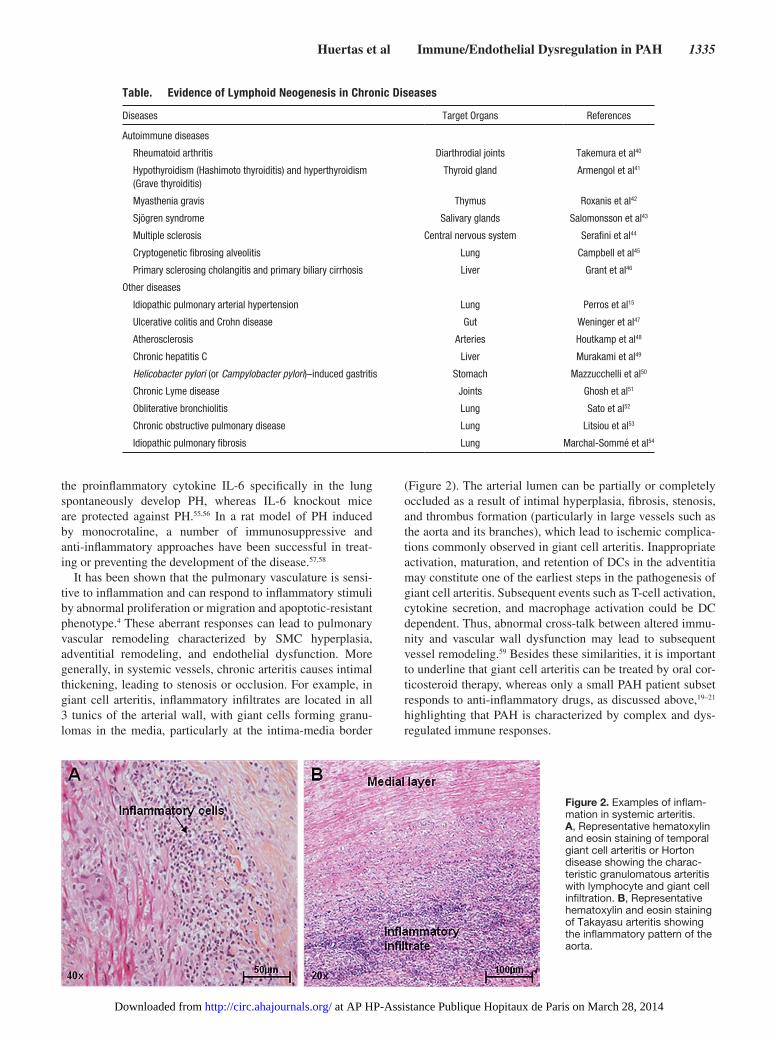
It has been shown that the pulmonary vasculature is sensi-tive to inflammation and can respond to inflammatory stimuli by abnormal proliferation or migration and apoptotic-resistant phenotype.4 These aberrant responses can lead to pulmonary vascular remodeling characterized by SMC hyperplasia, adventitial remodeling, and endothelial dysfunction. More generally, in systemic vessels, chronic arteritis causes intimal thickening, leading to stenosis or occlusion. For example, in giant cell arteritis, inflammatory infiltrates are located in all 3 tunics of the arterial wall, with giant cells forming granu-lomas in the media, particularly at the intima-media border
(Figure 2). The arterial lumen can be partially or completely occluded as a result of intimal hyperplasia, fibrosis, stenosis, and thrombus formation (particularly in large vessels such as the aorta and its branches), which lead to ischemic complica-tions commonly observed in giant cell arteritis. Inappropriate activation, maturation, and retention of DCs in the adventitia may constitute one of the earliest steps in the pathogenesis of giant cell arteritis. Subsequent events such as T-cell activation, cytokine secretion, and macrophage activation could be DC dependent. Thus, abnormal cross-talk between altered immu-nity and vascular wall dysfunction may lead to subsequent vessel remodeling.59 Besides these similarities, it is important to underline that giant cell arteritis can be treated by oral cor-ticosteroid therapy, whereas only a small PAH patient subset responds to anti-inflammatory drugs, as discussed above,19–21 highlighting that PAH is characterized by complex and dys-regulated immune responses.
Figure 2. Examples of inflam-mation in systemic arteritis. A, Representative hematoxylin and eosin staining of temporal giant cell arteritis or Horton disease showing the charac-teristic granulomatous arteritis with lymphocyte and giant cell infiltration. B, Representative hematoxylin and eosin staining of Takayasu arteritis showing the inflammatory pattern of the aorta.
Table. Evidence of Lymphoid Neogenesis in Chronic Diseases
Diseases Target Organs References
Autoimmune diseases
Rheumatoid arthritis Diarthrodial joints Takemura et al40
Hypothyroidism (Hashimoto thyroiditis) and hyperthyroidism (Grave thyroiditis)
Thyroid gland Armengol et al41
Myasthenia gravis Thymus Roxanis et al42
Sjögren syndrome Salivary glands Salomonsson et al43
Multiple sclerosis Central nervous system Serafini et al44
Cryptogenetic fibrosing alveolitis Lung Campbell et al45
Primary sclerosing cholangitis and primary biliary cirrhosis Liver Grant et al46
Other diseases
Idiopathic pulmonary arterial hypertension Lung Perros et al15
Ulcerative colitis and Crohn disease Gut Weninger et al47
Atherosclerosis Arteries Houtkamp et al48
Chronic hepatitis C Liver Murakami et al49
Helicobacter pylori (or Campylobacter pylori)–induced gastritis Stomach Mazzucchelli et al50
Chronic Lyme disease Joints Ghosh et al51
Obliterative bronchiolitis Lung Sato et al52
Chronic obstructive pulmonary disease Lung Litsiou et al53
Idiopathic pulmonary fibrosis Lung Marchal-Sommé et al54
at AP HP-Assistance Publique Hopitaux de Paris on March 28, 2014http://circ.ahajournals.org/Downloaded from

1336 Circulation March 25, 2014
It has been shown that pulmonary ECs and SMCs pro-liferate or migrate in vitro in response to proinflammatory cytokines/chemokines and growth factors such as serotonin, platelet-derived growth factor, epidermal growth factor, vascular endothelial growth factor, and fibroblast growth factor-2.2,60 We recently showed that platelet-derived growth factor, fibroblast growth factor-2, and platelet-derived growth factor signaling pathways converge to p130Cas, an adaptor pro-tein modulating several signaling pathways that controls cell migration and proliferation and acts as an amplifier of down-stream signals.61 We showed that p130Cas is increased in IPAH (in serum, in human isolated pulmonary arteries, and in cul-tured pulmonary ECs and SMCs) and in experimental models of PH. We also reported that p130Cas deficiency by RNA inter-ference causes attenuated extracellular signal-regulated kinase 1/2 activation and normalized migration and proliferation of pulmonary SMCs and ECs derived from IPAH.61
An important player in the innate immunity is represented by the complement system, constituted by 3 distinct pathways that all converge to C3 activation. Interestingly, it has been shown that the complement system, in particular C3, plays a role in the development of PH in a mouse model, promoting pulmonary vascular remodeling and a prothrombotic pheno-type. Furthermore, increased C3 deposition was observed in human lung sections compared with controls.62 Because the complement system is a major component of the innate immu-nity and can be brought into action by the adaptive immune system, these results highlight the possible link between innate and adaptive altered responses in PH pathogenesis.
It has been proposed that antibodies directed to vascular endothelium could promote EC apoptosis and that endothe-lium aggression could initiate a dysfunction leading to uncon-trolled proliferation.63 Using a proteomic approach, Dib and coworkers25 were recently able to identify target antigens for AECAs in IPAH, which include lamin A/C and tubulin β-chain, major components of the inner nuclear membrane and microtubules, respectively. These proteins identified as targets of AECAs are ubiquitous and play key roles in dif-ferent cell types in that they are involved in cell metabolism, morphology, and protein folding, but the exact role of AECAs in patients with IPAH remains unclear. It is known that AECAs can activate ECs and induce apoptosis in SSc patients,64 but it needs further investigation in IPAH. Terrier et al27 also showed that the target antigens recognized by antifibroblast antibod-ies present in IPAH are ubiquitous proteins involved in the main cellular systems: regulation of cytoskeletal organization (vimentin, calumenin, and phosphatidylinositol 3-kinase), cell contraction (tropomyosin 1), oxidative stress (heat shock protein-27 and -70 and glucose-6-phosphate dehydrogenase), and protein metabolism. Of note, although several potentially important target antigens have been identified, the techni-cal approach used does not allow the identification of target antigens at the cell membrane, and such cell membrane tar-gets may be of importance in the immunopathology of PAH. Nevertheless, the identification of target antigens suggests that they may contribute to EC apoptosis and fibroblast dysfunc-tion in IPAH patients. Functional analyses are still needed to better define the potential role of AECAs and antifibroblast antibodies in IPAH. Recently, the presence of autoantibodies
against vascular SMCs has also been demonstrated in IPAH.24 Interestingly, these antibodies can modulate SMC contraction, and they bind mainly to 2 target antigens involved in vascular remodeling: stress-induced phosphoprotein 1 and α-enolase. Although this is the first evidence of a clear functional role of autoantibodies in IPAH, further investigations are needed to identify the potential pathogenic contribution of autoantibod-ies to IPAH.
Among all the autoantibody targets identified, it remains difficult to define which ones are recognized by pathogenic antibodies that would influence vascular dysfunction or play a role in remodeling. One may hypothesize that autoantibod-ies directed against cytoplasmic or nuclear components could emerge after initial EC aggression, inducing EC apoptosis and neoantigen exposure. Thus, these autoantibodies argue in favor of humoral mechanisms in the pathogenesis of PAH. Taken together, these data suggest that altered immunity may initiate or contribute to endothelial dysfunction in PAH, lead-ing to pulmonary vascular wall remodeling, the hallmark of the disease.
Endothelial Dysfunction Perpetuates Altered Immune ResponsesIt is also known that genetic predisposition and environmental factors such as hypoxia may trigger or contribute to endothelial aberrant immune responses. In particular, it has recently been shown that cellular microparticles contribute to the endothe-lial production of various proinflammatory cytokines and che-mokines such as IL-1β, IL-6, and CC chemokine ligand 2 and to the expression of intercellular adhesion molecule-1, vascu-lar cell adhesion molecule-1, and E-selectin.65 Microparticles are plasma membrane vesicle fragments released from various cell types during activation by agonists or physical or chemi-cal stress. Interestingly, their role is a function of the parent cell from which they stem, as well as the stimulus used for their generation. Furthermore, erythrocytes are responsible for pulmonary endothelial proinflammatory transcriptional responses to hypoxia such as endothelial nuclear factor-κB activation and hypoxia-inducible factor-1α stabilization, lead-ing to upregulation of endothelial leukocyte adhesion recep-tors, E-selectin, and intercellular adhesion molecule-1.66 It is also known that this PAH proinflammatory environment, with increased production of IL-1 and IL-6,5,11 represents a condition favorable to activation, proliferation, and differen-tiation of B lymphocytes.67 Local autoimmunity is also sug-gested by the presence of mast cells in and around vascular lesions as a source of IL-4 needed for local B-cell expan-sion. This cytokine production represents another important link between innate and adaptive immune responses within the context of vascular lesions. Mast cells are bone marrow–derived cells that contain large granules rich in histamine and heparin and that are resident in many tissues. These cells are important in hypersensitivity reactions, wound healing, and defending against pathogens. Mast cell accumulation has been described in several types of PAH and an animal model.68,69 Our group has recently identified an increase in c-kit–positive cells (including mast cells) in remodeled vessels and mobili-zation of bone marrow–derived circulating progenitor cells.70 How mast cells may contribute to PAH pathophysiology is
at AP HP-Assistance Publique Hopitaux de Paris on March 28, 2014http://circ.ahajournals.org/Downloaded from

Huertas et al Immune/Endothelial Dysregulation in PAH 1337
not clear, but proposed mechanisms include direct vasoactive effects and stimulation of remodeling by increased production of matrix metalloproteinases.71
More proof of altered immune responses in PAH comes from the potential role of T lymphocytes, particularly Tregs, in PAH pathogenesis. Taraseviciene-Stewart and coauthors72 showed that, in contrast to vascular endothelial growth factor receptor blocker SU5416–treated euthymic rats that develop severe PH only in combination with chronic hypoxia, athy-mic nude rats developed severe PH and pulmonary vascular remodeling at normoxic conditions. It is therefore legitimate to hypothesize that altered immune responses may indeed play a role in PAH. Interestingly, Tamosiuniene and coworkers73 have shown a link between immune dysregulation, resulting from T-cell absence, and vascular injury. They demonstrated that immune reconstitution, performed by intravenously injecting T cells in athymic rats, attenuates early inflamma-tion induced by vascular endothelial growth factor receptor 2
inhibitor–dependent vascular injury. These data suggest that in normal conditions, T cells play an important role in pro-tecting against vascular inflammation secondary to a vascular injury and possibly in preventing the development of PAH.
Thus, identifying the functional status of Treg cells, key players in the autoimmunity onset, could help us better under-stand the potential mechanisms leading to the development and progression of PAH. As described above, we recently showed that IPAH patients display normal Treg cell count but with an altered function and demonstrated that Treg cell function is inhibited in a leptin-dependent manner in IPAH.35 Interestingly, the increased levels of circulating leptin in IPAH are endothelium derived. Importantly, the observed high levels of leptinemia and leptin receptor expression in IPAH patients are independent of the patient’s body mass index.35 Therefore, obesity should not be considered a risk factor for IPAH devel-opment. These results not only suggest a possible role for leptin and its receptor in PAH immunopathogenesis but also
Figure 3. Schematic overview of the cross-talk between immune system cells and pulmonary arterial wall components in pulmonary arterial hypertension pathogenesis. Abnormal innate, adaptive, and autoimmune responses can interact with pulmonary arterial wall com-ponents through soluble factors, microparticles, and local infiltration, contributing to pulmonary arterial altered functions, leading to endo-thelial dysfunction, smooth muscle cell hyperplasia, and adventitial remodeling. Pulmonary arterial wall remodeling may also perpetuate altered immune responses through immune cell recruitment and feedback regulation. Environmental factors such as hypoxia and genetic predisposition may trigger or contribute to immune dysregulation and pulmonary arterial remodeling. All of these mechanisms lead to an inappropriate interplay between the immune system and pulmonary arterial wall components, in particular the endothelium. Nevertheless, whether immune dysregulation represents a cause or an effect of pulmonary arterial hypertension onset is still unknown. A better under-standing of the cross-talk between immune mediators and the components of the pulmonary vascular wall could help to identify novel therapeutic targets in pulmonary arterial hypertension.
at AP HP-Assistance Publique Hopitaux de Paris on March 28, 2014http://circ.ahajournals.org/Downloaded from
1338 Circulation March 25, 2014
highlight the existing link between immune dysregulation and endothelial dysfunction in IPAH.
ConclusionsThere is strong evidence that immune dysregulation plays an important role in the pathogenesis and progression of PAH, promoting inflammatory cell recruitment, stimulating autoan-tibody production, and triggering vascular wall remodeling, leading to inappropriate interplay between the immune system and the pulmonary endothelium (Figure 3). However, the exact mechanisms are still unclear, and several questions remain unanswered. Whether immune dysregulation represents a cause or an effect of PAH onset is still unknown. Although the patho-genesis of PAH shares several characteristics with systemic autoimmune diseases, why PAH represents a lung-specific dis-ease has not yet been elucidated. This review clearly shows that altered immune responses in PAH represent an important contribution to its pathogenesis. Further studies are clearly needed to assess the inadequate cross-talk between immune mediators and the components of the pulmonary vascular wall. Identification of these factors could lead to novel therapeutic targets. The current PAH therapies are essentially focused on decreasing pulmonary vascular resistance by stimulating pul-monary vasodilation (prostacyclin analogs, phosphodiesterase type 5 inhibitors, and endothelin receptor antagonists).74 These agents have some antiremodeling properties, but there is no current antiremodeling strategy approved for PAH.75 Of note, some hemodynamic and clinical effects of the tyrosine kinase inhibitor imatinib have been reported in severe PAH, but at the expense of severe side effects.76,77 Because survival remains poor in the modern management era,78 new treatments target-ing other PAH pathomechanisms would be useful to slow, stop, or even reverse disease progression.
Therefore, more data are needed on novel agents with anti-inflammatory or immunomodulatory properties. In this context, a randomized, clinical trial testing the safety and effi-cacy of the monoclonal antibody anti-CD20, a B lymphocyte protein, is currently in phase II in patients with SSc-PAH. Unfortunately, such a therapeutic strategy is not yet appli-cable in IPAH. Additional efforts should be made to further our understanding of the complex interplay between immune dysregulation and endothelial dysfunction in IPAH to develop new and more powerful drugs that could restore immune responses and Treg function.
AcknowledgmentsM.R. Ghigna assisted in the preparation of the manuscript.
Sources of FundingDr Huertas is supported by the Josso Award 2010 from the French Medical Research Foundation and by the French National Agency for Research (grant ANR_12_JSV1_0004_01). Dr Perros is sup-ported by the French National Agency for Research (grant ANR_13_JSV1_0011_01).
DisclosuresDr Montani has relationships with drug companies, including Actelion, AstraZeneca, Bayer Schering, GSK, Lilly, Novartis, and Pfizer. In addition to being an investigator in trials involv-ing these companies, relationships include consultancy service and
membership of scientific advisory boards. Dr Humbert has relation-ships with drug companies, including Actelion, Bayer Schering, GSK, Lilly, Novartis, Pfizer, and United Therapeutics. In addition to being an investigator in trials involving these companies, relation-ships include consultancy service and membership of scientific advi-sory boards. The other authors report no conflicts.
References 1. Simonneau G, Robbins IM, Beghetti M, Channick RN, Delcroix M,
Denton CP, Elliott CG, Gaine SP, Gladwin MT, Jing ZC, Krowka MJ, Langleben D, Nakanishi N, Souza R. Updated clinical classification of pulmonary hypertension. J Am Coll Cardiol. 2009;54(suppl):S43–S54.
2. Hassoun PM, Mouthon L, Barberà JA, Eddahibi S, Flores SC, Grimminger F, Jones PL, Maitland ML, Michelakis ED, Morrell NW, Newman JH, Rabinovitch M, Schermuly R, Stenmark KR, Voelkel NF, Yuan JX, Humbert M. Inflammation, growth factors, and pulmonary vascular remodeling. J Am Coll Cardiol. 2009;54(suppl):S10–S19.
3. Tamosiuniene R, Nicolls MR. Regulatory T cells and pulmonary hyper-tension. Trends Cardiovasc Med. 2011;21:166–171.
4. Voelkel NF, Gomez-Arroyo J, Abbate A, Bogaard HJ, Nicolls MR. Pathobiology of pulmonary arterial hypertension and right ventricular failure. Eur Respir J. 2012;40:1555–1565.
5. Humbert M, Monti G, Brenot F, Sitbon O, Portier A, Grangeot-Keros L, Duroux P, Galanaud P, Simonneau G, Emilie D. Increased interleukin-1 and interleukin-6 serum concentrations in severe primary pulmonary hypertension. Am J Respir Crit Care Med. 1995;151:1628–1631.
6. Sanchez O, Marcos E, Perros F, Fadel E, Tu L, Humbert M, Dartevelle P, Simonneau G, Adnot S, Eddahibi S. Role of endothelium-derived CC chemokine ligand 2 in idiopathic pulmonary arterial hypertension. Am J Respir Crit Care Med. 2007;176:1041–1047.
7. Dorfmüller P, Zarka V, Durand-Gasselin I, Monti G, Balabanian K, Garcia G, Capron F, Coulomb-Lherminé A, Marfaing-Koka A, Simonneau G, Emilie D, Humbert M. Chemokine RANTES in severe pulmonary arterial hypertension. Am J Respir Crit Care Med. 2002;165:534–539.
8. Balabanian K, Foussat A, Dorfmüller P, Durand-Gasselin I, Capel F, Bouchet-Delbos L, Portier A, Marfaing-Koka A, Krzysiek R, Rimaniol AC, Simonneau G, Emilie D, Humbert M. CX(3)C chemokine fractal-kine in pulmonary arterial hypertension. Am J Respir Crit Care Med. 2002;165:1419–1425.
9. El Chami H, Hassoun PM. Immune and inflammatory mechanisms in pul-monary arterial hypertension. Prog Cardiovasc Dis. 2012;55:218–228.
10. Quarck R, Nawrot T, Meyns B, Delcroix M. C-reactive protein: a new predictor of adverse outcome in pulmonary arterial hypertension. J Am Coll Cardiol. 2009;53:1211–1218.
11. Soon E, Holmes AM, Treacy CM, Doughty NJ, Southgate L, Machado RD, Trembath RC, Jennings S, Barker L, Nicklin P, Walker C, Budd DC, Pepke-Zaba J, Morrell NW. Elevated levels of inflammatory cytokines predict survival in idiopathic and familial pulmonary arterial hyperten-sion. Circulation. 2010;122:920–927.
12. Dorfmüller P, Humbert M. Progress in pulmonary arterial hypertension pathology: relighting a torch inside the tunnel. Am J Respir Crit Care Med. 2012;186:210–212.
13. Dorfmüller P, Perros F, Balabanian K, Humbert M. Inflammation in pul-monary arterial hypertension. Eur Respir J. 2003;22:358–363.
14. Perros F, Dorfmüller P, Souza R, Durand-Gasselin I, Mussot S, Mazmanian M, Hervé P, Emilie D, Simonneau G, Humbert M. Dendritic cell recruit-ment in lesions of human and experimental pulmonary hypertension. Eur Respir J. 2007;29:462–468.
15. Perros F, Dorfmüller P, Montani D, Hammad H, Waelput W, Girerd B, Raymond N, Mercier O, Mussot S, Cohen-Kaminsky S, Humbert M, Lambrecht BN. Pulmonary lymphoid neogenesis in idiopathic pulmonary arterial hypertension. Am J Respir Crit Care Med. 2012;185:311–321.
16. Stacher E, Graham BB, Hunt JM, Gandjeva A, Groshong SD, McLaughlin VV, Jessup M, Grizzle WE, Aldred MA, Cool CD, Tuder RM. Modern age pathology of pulmonary arterial hypertension. Am J Respir Crit Care Med. 2012;186:261–272.
17. Ormiston ML, Chang C, Long LL, Soon E, Jones D, Machado R, Treacy C, Toshner MR, Campbell K, Riding A, Southwood M, Pepke-Zaba J, Exley A, Trembath RC, Colucci F, Wills M, Trowsdale J, Morrell NW. Impaired natural killer cell phenotype and function in idiopathic and heri-table pulmonary arterial hypertension. Circulation. 2012;126:1099–1109.
18. Perros F, Cohen-Kaminsky S, Gambaryan N, Girerd B, Raymond N, Klingelschmitt I, Huertas A, Mercier O, Fadel E, Simonneau G, Humbert M, Dorfmüller P, Montani D. Cytotoxic cells and granulysin in pulmonary
at AP HP-Assistance Publique Hopitaux de Paris on March 28, 2014http://circ.ahajournals.org/Downloaded from

Huertas et al Immune/Endothelial Dysregulation in PAH 1339
arterial hypertension and pulmonary veno-occlusive disease. Am J Respir Crit Care Med. 2013;187:189–196.
19. Sanchez O, Sitbon O, Jaïs X, Simonneau G, Humbert M. Immunosuppressive therapy in connective tissue diseases-associated pul-monary arterial hypertension. Chest. 2006;130:182–189.
20. Jais X, Launay D, Yaici A, Le Pavec J, Tchérakian C, Sitbon O, Simonneau G, Humbert M. Immunosuppressive therapy in lupus- and mixed connec-tive tissue disease-associated pulmonary arterial hypertension: a retrospec-tive analysis of twenty-three cases. Arthritis Rheum. 2008;58:521–531.
21. Montani D, Achouh L, Marcelin AG, Viard JP, Hermine O, Canioni D, Sitbon O, Simonneau G, Humbert M. Reversibility of pulmonary arterial hypertension in HIV/HHV8-associated Castleman’s disease. Eur Respir J. 2005;26:969–972.
22. Humbert M, Sitbon O, Chaouat A, Bertocchi M, Habib G, Gressin V, Yaici A, Weitzenblum E, Cordier JF, Chabot F, Dromer C, Pison C, Reynaud-Gaubert M, Haloun A, Laurent M, Hachulla E, Simonneau G. Pulmonary arterial hypertension in France: results from a national registry. Am J Respir Crit Care Med. 2006;173:1023–1030.
23. Hachulla E, Gressin V, Guillevin L, Carpentier P, Diot E, Sibilia J, Kahan A, Cabane J, Francès C, Launay D, Mouthon L, Allanore Y, Tiev KP, Clerson P, de Groote P, Humbert M. Early detection of pulmonary arte-rial hypertension in systemic sclerosis: a French nationwide prospective multicenter study. Arthritis Rheum. 2005;52:3792–3800.
24. Bussone G, Tamby MC, Calzas C, Kherbeck N, Sahbatou Y, Sanson C, Ghazal K, Dib H, Weksler BB, Broussard C, Verrecchia F, Yaici A, Witko-Sarsat V, Simonneau G, Guillevin L, Humbert M, Mouthon L. IgG from patients with pulmonary arterial hypertension and/or systemic scle-rosis binds to vascular smooth muscle cells and induces cell contraction. Ann Rheum Dis. 2012;71:596–605.
25. Dib H, Tamby MC, Bussone G, Regent A, Berezné A, Lafine C, Broussard C, Simonneau G, Guillevin L, Witko-Sarsat V, Humbert M, Mouthon L. Targets of anti-endothelial cell antibodies in pulmonary hypertension and scleroderma. Eur Respir J. 2012;39:1405–1414.
26. Tamby MC, Chanseaud Y, Humbert M, Fermanian J, Guilpain P, Garcia-de-la-Peña-Lefebvre P, Brunet S, Servettaz A, Weill B, Simonneau G, Guillevin L, Boissier MC, Mouthon L. Anti-endothelial cell antibodies in idiopathic and systemic sclerosis associated pulmonary arterial hyper-tension. Thorax. 2005;60:765–772.
27. Terrier B, Tamby MC, Camoin L, Guilpain P, Broussard C, Bussone G, Yaïci A, Hotellier F, Simonneau G, Guillevin L, Humbert M, Mouthon L. Identification of target antigens of antifibroblast antibodies in pulmonary arterial hypertension. Am J Respir Crit Care Med. 2008;177:1128–1134.
28. Arends SJ, Damoiseaux J, Duijvestijn A, Debrus-Palmans L, Boomars K, Broers B, Tervaert JW, van Paassen P. Prevalence of anti-endothelial cell antibodies in idiopathic pulmonary arterial hypertension. Eur Respir J. 2010;35:923–925.
29. Karmochkine M, Cacoub P, Dorent R, Laroche P, Nataf P, Piette JC, Boffa MC, Gandjbakhch I. High prevalence of antiphospholipid antibodies in precapillary pulmonary hypertension. J Rheumatol. 1996;23:286–290.
30. Wolf M, Boyer-Neumann C, Parent F, Eschwege V, Jaillet H, Meyer D, Simonneau G. Thrombotic risk factors in pulmonary hypertension. Eur Respir J. 2000;15:395–399.
31. Wing K, Sakaguchi S. Regulatory T cells exert checks and balances on self tolerance and autoimmunity. Nat Immunol. 2010;11:7–13.
32. Viglietta V, Baecher-Allan C, Weiner HL, Hafler DA. Loss of functional suppression by CD4+CD25+ regulatory T cells in patients with multiple sclerosis. J Exp Med. 2004;199:971–979.
33. Austin ED, Rock MT, Mosse CA, Vnencak-Jones CL, Yoder SM, Robbins IM, Loyd JE, Meyrick BO. T lymphocyte subset abnormalities in the blood and lung in pulmonary arterial hypertension. Respir Med. 2010;104:454–462.
34. Ulrich S, Nicolls MR, Taraseviciene L, Speich R, Voelkel N. Increased regulatory and decreased CD8+ cytotoxic T cells in the blood of patients with idiopathic pulmonary arterial hypertension. Respiration. 2008;75:272–280.
35. Huertas A, Tu L, Gambaryan N, Girerd B, Perros F, Montani D, Fabre D, Fadel E, Eddahibi S, Cohen-Kaminsky S, Guignabert C, Humbert M. Leptin and regulatory T-lymphocytes in idiopathic pulmonary arterial hypertension. Eur Respir J. 2012;40:895–904.
36. Cools N, Ponsaerts P, Van Tendeloo VF, Berneman ZN. Balancing between immunity and tolerance: an interplay between dendritic cells, regulatory T cells, and effector T cells. J Leukoc Biol. 2007;82:1365–1374.
37. Akbari O, DeKruyff RH, Umetsu DT. Pulmonary dendritic cells produc-ing IL-10 mediate tolerance induced by respiratory exposure to antigen. Nat Immunol. 2001;2:725–731.
38. Savai R, Pullamsetti SS, Kolbe J, Bieniek E, Voswinckel R, Fink L, Scheed A, Ritter C, Dahal BK, Vater A, Klussmann S, Ghofrani HA, Weissmann N, Klepetko W, Banat GA, Seeger W, Grimminger F, Schermuly RT. Immune and inflammatory cell involvement in the pathology of idio-pathic pulmonary arterial hypertension. Am J Respir Crit Care Med. 2012;186:897–908.
39. Aloisi F, Pujol-Borrell R. Lymphoid neogenesis in chronic inflammatory diseases. Nat Rev Immunol. 2006;6:205–217.
40. Takemura S, Braun A, Crowson C, Kurtin PJ, Cofield RH, O’Fallon WM, Goronzy JJ, Weyand CM. Lymphoid neogenesis in rheumatoid synovitis. J Immunol. 2001;167:1072–1080.
41. Armengol MP, Juan M, Lucas-Martín A, Fernández-Figueras MT, Jaraquemada D, Gallart T, Pujol-Borrell R. Thyroid autoim-mune disease: demonstration of thyroid antigen-specific B cells and recombination-activating gene expression in chemokine-containing active intrathyroidal germinal centers. Am J Pathol. 2001;159:861–873.
42. Roxanis I, Micklem K, McConville J, Newsom-Davis J, Willcox N. Thymic myoid cells and germinal center formation in myasthenia gravis; possible roles in pathogenesis. J Neuroimmunol. 2002;125:185–197.
43. Salomonsson S, Jonsson MV, Skarstein K, Brokstad KA, Hjelmström P, Wahren-Herlenius M, Jonsson R. Cellular basis of ectopic germinal center formation and autoantibody production in the target organ of patients with Sjögren’s syndrome. Arthritis Rheum. 2003;48:3187–3201.
44. Serafini B, Rosicarelli B, Magliozzi R, Stigliano E, Aloisi F. Detection of ectopic B-cell follicles with germinal centers in the meninges of patients with secondary progressive multiple sclerosis. Brain Pathol. 2004;14:164–174.
45. Campbell DA, Poulter LW, Janossy G, du Bois RM. Immunohistological analysis of lung tissue from patients with cryptogenic fibrosing alveo-litis suggesting local expression of immune hypersensitivity. Thorax. 1985;40:405–411.
46. Grant AJ, Goddard S, Ahmed-Choudhury J, Reynolds G, Jackson DG, Briskin M, Wu L, Hübscher SG, Adams DH. Hepatic expression of secondary lymphoid chemokine (CCL21) promotes the development of portal-associated lymphoid tissue in chronic inflammatory liver disease. Am J Pathol. 2002;160:1445–1455.
47. Weninger W, Carlsen HS, Goodarzi M, Moazed F, Crowley MA, Baekkevold ES, Cavanagh LL, von Andrian UH. Naive T cell recruitment to nonlymphoid tissues: a role for endothelium-expressed CC chemokine ligand 21 in autoimmune disease and lymphoid neogenesis. J Immunol. 2003;170:4638–4648.
48. Houtkamp MA, de Boer OJ, van der Loos CM, van der Wal AC, Becker AE. Adventitial infiltrates associated with advanced atherosclerotic plaques: structural organization suggests generation of local humoral immune responses. J Pathol. 2001;193:263–269.
49. Murakami J, Shimizu Y, Kashii Y, Kato T, Minemura M, Okada K, Nambu S, Takahara T, Higuchi K, Maeda Y, Kumada T, Watanabe A. Functional B-cell response in intrahepatic lymphoid follicles in chronic hepatitis C. Hepatology. 1999;30:143–150.
50. Mazzucchelli L, Blaser A, Kappeler A, Schärli P, Laissue JA, Baggiolini M, Uguccioni M. BCA-1 is highly expressed in Helicobacter pylori-induced mucosa-associated lymphoid tissue and gastric lymphoma. J Clin Invest. 1999;104:R49–R54.
51. Ghosh S, Steere AC, Stollar BD, Huber BT. In situ diversification of the antibody repertoire in chronic Lyme arthritis synovium. J Immunol. 2005;174:2860–2869.
52. Sato M, Hirayama S, Hwang DM, Lara-Guerra H, Wagnetz D, Waddell TK, Liu M, Keshavjee S. The role of intrapulmonary de novo lymphoid tissue in obliterative bronchiolitis after lung transplantation. J Immunol. 2009;182:7307–7316.
53. Litsiou E, Semitekolou M, Galani IE, Morianos I, Tsoutsa A, Kara P, Rontogianni D, Bellenis I, Konstantinou M, Potaris K, Andreakos E, Sideras P, Zakynthinos S, Tsoumakidou M. CXCL13 production in B cells via Toll-like receptor/lymphotoxin receptor signaling is involved in lym-phoid neogenesis in chronic obstructive pulmonary disease. Am J Respir Crit Care Med. 2013;187:1194–1202.
54. Marchal-Sommé J, Uzunhan Y, Marchand-Adam S, Valeyre D, Soumelis V, Crestani B, Soler P. Cutting edge: nonproliferating mature immune cells form a novel type of organized lymphoid structure in idiopathic pul-monary fibrosis. J Immunol. 2006;176:5735–5739.
55. Steiner MK, Syrkina OL, Kolliputi N, Mark EJ, Hales CA, Waxman AB. Interleukin-6 overexpression induces pulmonary hypertension. Circ Res. 2009;104:236–244.
56. Savale L, Tu L, Rideau D, Izziki M, Maitre B, Adnot S, Eddahibi S. Impact of interleukin-6 on hypoxia-induced pulmonary hypertension and lung inflammation in mice. Respir Res. 2009;10:6.
at AP HP-Assistance Publique Hopitaux de Paris on March 28, 2014http://circ.ahajournals.org/Downloaded from

1340 Circulation March 25, 2014
57. Price LC, Montani D, Tcherakian C, Dorfmüller P, Souza R, Gambaryan N, Chaumais MC, Shao DM, Simonneau G, Howard LS, Adcock IM, Wort SJ, Humbert M, Perros F. Dexamethasone reverses monocrotaline- induced pulmonary arterial hypertension in rats. Eur Respir J. 2011; 37:813–822.
58. Voelkel NF, Tuder RM, Bridges J, Arend WP. Interleukin-1 receptor antagonist treatment reduces pulmonary hypertension generated in rats by monocrotaline. Am J Respir Cell Mol Biol. 1994;11:664–675.
59. Ma-Krupa W, Jeon MS, Spoerl S, Tedder TF, Goronzy JJ, Weyand CM. Activation of arterial wall dendritic cells and breakdown of self-tolerance in giant cell arteritis. J Exp Med. 2004;199:173–183.
60. Tu L, Dewachter L, Gore B, Fadel E, Dartevelle P, Simonneau G, Humbert M, Eddahibi S, Guignabert C. Autocrine fibroblast growth factor-2 signal-ing contributes to altered endothelial phenotype in pulmonary hyperten-sion. Am J Respir Cell Mol Biol. 2011;45:311–322.
61. Tu L, De Man FS, Girerd B, Huertas A, Chaumais MC, Lecerf F, François C, Perros F, Dorfmüller P, Fadel E, Montani D, Eddahibi S, Humbert M, Guignabert C. A critical role for p130Cas in the progression of pulmo-nary hypertension in humans and rodents. Am J Respir Crit Care Med. 2012;186:666–676.
62. Bauer EM, Zheng H, Comhair S, Erzurum S, Billiar TR, Bauer PM. Complement C3 deficiency attenuates chronic hypoxia-induced pulmo-nary hypertension in mice. PLoS One. 2011;6:e28578.
63. Nicolls MR, Taraseviciene-Stewart L, Rai PR, Badesch DB, Voelkel NF. Autoimmunity and pulmonary hypertension: a perspective. Eur Respir J. 2005;26:1110–1118.
64. Pignone A, Scaletti C, Matucci-Cerinic M, Vázquez-Abad D, Meroni PL, Del Papa N, Falcini F, Generini S, Rothfield N, Cagnoni M. Anti-endothelial cell antibodies in systemic sclerosis: significant asso-ciation with vascular involvement and alveolo-capillary impairment. Clin Exp Rheumatol. 1998;16:527–532.
65. Amabile N, Guignabert C, Montani D, Yeghiazarians Y, Boulanger CM, Humbert M. Cellular microparticles in the pathogenesis of pulmonary hypertension. Eur Respir J. 2013;42:272–279.
66. Huertas A, Das SR, Emin M, Sun L, Rifkind JM, Bhattacharya J, Bhattacharya S. Erythrocytes induce proinflammatory endothelial activa-tion in hypoxia. Am J Respir Cell Mol Biol. 2013;48:78–86.
67. Nurieva RI, Chung Y, Hwang D, Yang XO, Kang HS, Ma L, Wang YH, Watowich SS, Jetten AM, Tian Q, Dong C. Generation of T follicular helper cells is mediated by interleukin-21 but independent of T helper 1, 2, or 17 cell lineages. Immunity. 2008;29:138–149.
68. Dahal BK, Kosanovic D, Kaulen C, Cornitescu T, Savai R, Hoffmann J, Reiss I, Ghofrani HA, Weissmann N, Kuebler WM, Seeger W, Grimminger
F, Schermuly RT. Involvement of mast cells in monocrotaline-induced pulmonary hypertension in rats. Respir Res. 2011;12:60.
69. Mitani Y, Ueda M, Maruyama K, Shimpo H, Kojima A, Matsumura M, Aoki K, Sakurai M. Mast cell chymase in pulmonary hypertension. Thorax. 1999;54:88–90.
70. Montani D, Perros F, Gambaryan N, Girerd B, Dorfmuller P, Price LC, Huertas A, Hammad H, Lambrecht B, Simonneau G, Launay JM, Cohen-Kaminsky S, Humbert M. C-kit-positive cells accumulate in remodeled vessels of idiopathic pulmonary arterial hypertension. Am J Respir Crit Care Med. 2011;184:116–123.
71. Vajner L, Vytásek R, Lachmanová V, Uhlík J, Konrádová V, Novotná J, Hampl V, Herget J. Acute and chronic hypoxia as well as 7-day recovery from chronic hypoxia affects the distribution of pulmonary mast cells and their MMP-13 expression in rats. Int J Exp Pathol. 2006;87:383–391.
72. Taraseviciene-Stewart L, Nicolls MR, Kraskauskas D, Scerbavicius R, Burns N, Cool C, Wood K, Parr JE, Boackle SA, Voelkel NF. Absence of T cells confers increased pulmonary arterial hypertension and vascular remodeling. Am J Respir Crit Care Med. 2007;175:1280–1289.
73. Tamosiuniene R, Tian W, Dhillon G, Wang L, Sung YK, Gera L, Patterson AJ, Agrawal R, Rabinovitch M, Ambler K, Long CS, Voelkel NF, Nicolls MR. Regulatory T cells limit vascular endothelial injury and prevent pul-monary hypertension. Circ Res. 2011;109:867–879.
74. Humbert M, Sitbon O, Simonneau G. Treatment of pulmonary arterial hypertension. N Engl J Med. 2004;351:1425–1436.
75. O’Callaghan DS, Savale L, Montani D, Jaïs X, Sitbon O, Simonneau G, Humbert M. Treatment of pulmonary arterial hypertension with targeted therapies. Nat Rev Cardiol. 2011;8:526–538.
76. Hoeper MM, Barst RJ, Bourge RC, Feldman J, Frost AE, Galié N, Gómez-Sánchez MA, Grimminger F, Grünig E, Hassoun PM, Morrell NW, Peacock AJ, Satoh T, Simonneau G, Tapson VF, Torres F, Lawrence D, Quinn DA, Ghofrani HA. Imatinib mesylate as add-on therapy for pul-monary arterial hypertension: results of the randomized IMPRES study. Circulation. 2013;127:1128–1138.
77. Humbert M. Impression, sunset. Circulation. 2013;127:1098–1100. 78. Humbert M, Sitbon O, Chaouat A, Bertocchi M, Habib G, Gressin
V, Yaïci A, Weitzenblum E, Cordier JF, Chabot F, Dromer C, Pison C, Reynaud-Gaubert M, Haloun A, Laurent M, Hachulla E, Cottin V, Degano B, Jaïs X, Montani D, Souza R, Simonneau G. Survival in patients with idiopathic, familial, and anorexigen-associated pulmonary arterial hyper-tension in the modern management era. Circulation. 2010;122:156–163.
KEY WORDS: autoimmunity ◼ endothelium ◼ hypertension, pulmonary ◼ immune system
at AP HP-Assistance Publique Hopitaux de Paris on March 28, 2014http://circ.ahajournals.org/Downloaded from