THE INTERPLAY BETWEEN CHROMOSOME STRUCTURE ...
77
From the Department of Cell and Molecular Biology Karolinska Institutet, Stockholm, Sweden THE INTERPLAY BETWEEN CHROMOSOME STRUCTURE AND MEIOTIC INTEGRITY Ingrid Lilienthal Stockholm 2014
-
Upload
khangminh22 -
Category
Documents
-
view
0 -
download
0
Transcript of THE INTERPLAY BETWEEN CHROMOSOME STRUCTURE ...
From the Department of Cell and Molecular Biology Karolinska Institutet, Stockholm, Sweden
THE INTERPLAY BETWEEN CHROMOSOME STRUCTURE AND
MEIOTIC INTEGRITY
Ingrid Lilienthal
Stockholm 2014
All previously published papers were reproduced with permission from the publisher. Published by Karolinska Institutet. Printed by Åtta.45 Tryckeri © Ingrid Lilienthal, 2014 ISBN 978-91-7549-646-7
The interplay between chromosome structure and meiotic integrity
THESIS FOR DOCTORAL DEGREE (Ph.D.)
By
Ingrid Lilienthal
Principal Supervisor: Professor Camilla Sjögren Department of Cell and Molecular Biology Karolinska Institutet Co-supervisor(s): Professor Christer Höög Department of Cell and Molecular Biology Karolinska Institutet
Opponent: Andreas Hochwagen, Ph.D. Department of Biology New York University Examination Board: Docent Pernilla Bjerling Department of Medicinal Biochemistry and Microbiology Uppsala Universitet Docent Herwig Schüler Department of Biochemistry and Biophysics Karolinska Institutet Docent Rickard Sandberg Department of Cell and Molecular Biology Karolinska Institutet


To my mother,
In honor of the sacrifices you have made for me.


Just keep swimming.
And if all else fails…
Do.
Or do not.
There is no try.
~Yoda
ABSTRACT Sexually reproducing organisms employ a specialized cell division called meiosis to form unique, haploid gametes from a diploid precursor cell. Upon fertilization, two opposite-sex gametes fuse to create a zygote that will develop into the offspring. Fundamental to meiosis is the formation and repair of programmed DNA double-strand breaks (DSBs) during prophase. These DSBs are repaired using the homologous chromosome (homolog) as a template for homologous recombination, which creates linkages between the homologs that are essential for faithful segregation. Following prophase, two successive rounds of chromosome segregation ensue. The first, meiosis I (MI), segregates homologs and the second, meiosis II (MII), segregates sister chromatids. Recombination is dependent on chromosome structure, and events that alter the DNA landscape will impact meiotic fidelity and, in turn, genomic integrity in gametes and future offspring. Thus understanding the relationship between chromosome structure and meiosis can help gain insight into human fertility and underlying causes of genetic disorders. It was the goal of this thesis to investigate factors known to regulate chromosome structure and determine their influence on meiosis in the budding yeast Saccharomyces cerevisiae and mice. In addition, we tested a novel method to search for new mammalian meiotic factors that may impact fertility.
In Paper I, we identified a role for the conserved Structural Maintenance of Chromosomes (SMC) 5/6 complex during meiotic recombination in budding yeast. smc5/6 complex mutants experienced a DSB-dependent segregation block, suggesting that the defect was caused by recombination. Consistent with this notion, Smc6-deficient cells accumulated high levels of recombination intermediates, particularly between sister chromatids, which is normally not seen in the wild type. Return-to-function studies indicated that the Smc5/6 complex was most crucial during resolution of recombination intermediates. These results suggest that the Smc5/6 complex works primarily in the resolution of recombination structures formed outside of homolog-directed pathways during meiosis.
We characterized a role for DNA topoisomerases Top2 and Top3 during meiosis in S. cerevisiae by using meiosis-specific mutants in Paper II. Cells deficient for either Top2 or Top3 experienced a segregation block. While top3 cells were rescued completely by removing recombination, the top2 mutant was only partially rescued. This suggests that Top3 mainly functions during meiotic recombination. In contrast, the data indicates that
Top2 has a role outside of recombination. In line with this idea, some of the segregation defects in cells lacking Top2 seemed to arise from break-independent sister entanglements. Since Top2 is known to be important in resolving sister chromatid intertwinings during mitosis to facilitate proper segregation, it is likely that it plays a similar role during meiosis.
The CCCTC-binding factor (CCTF) is an architectural protein essential for proper genome structure and function in higher eukaryotes. In Paper III, we created a testes-specific ctcf mouse mutant strain (cctf-cKO) in order to study the function of CTCF during gamete (sperm in males) formation in mice. CTCF-deficient mice completed meiosis and sperm specialization without any major abnormalities, though mice were infertile and had low sperm counts. Sperm from the ctcf-cKO had chromatin compaction defects, most likely due to lack of sperm-specific compaction factors. These findings indicate that CTCF is essential for proper chromatin organization during spermiogenesis and suggest that infertility in ctcf-cKO mice was a result of the chromatin defects in the sperm.
Using a method called phylogenetic profiling in Paper IV, we showed that new meiotic factors can be discovered by clustering proteins according to their function.
LIST OF SCIENTIFIC PAPERS This thesis is based on the following articles and manuscripts. In the text, they will be referred to by their Roman numerals.
I. Lilienthal, I., Kanno, T., Sjögren, C. (2013). “Inhibition of the Smc5/6 complex during meiosis perturbs joint molecule formation and resolution without significantly changing crossover or non-crossover levels.” PLoS Genetics 9(11):e1003898.
II. Lilienthal, I. and Sjögren, C. “Uncovering the role for topoisomerases during meiosis.” Manuscript
III. Hernández-Hernández, A., Lilienthal, I., Fukuda, N., Galjart, N., Höög, C. “Multiple roles of CTCF during spermatogenesis.” Manuscript
IV. Tabach, Y., Golan, T., Hernández-Hernández, A., Messer, A.R., Fukuda, T., Kouznetsova, A., Liu, JG., Lilienthal, I., Levy, C., Ruvkun, G. (2013). “Human disease locus discovery and mapping to molecular pathways through phylogenetic profiling.” Molecular Systems Biology 9:692.
TABLE OF CONTENTS INTRODUCTION AND AIMS ...................................................................................... 1
MEIOSIS .......................................................................................................................... 2 General overview ......................................................................................................... 2 Meiotic recombination ................................................................................................ 4 The synaptonemal complex ........................................................................................ 8 Inter-homolog vs. inter-sister recombination ......................................................... 10 Meiotic segregation ................................................................................................... 11
MAMMALIAN GERM CELL FORMATION ......................................................... 12 Spermatogenesis ........................................................................................................ 12 Oogenesis .................................................................................................................... 16
PROTEINS THAT SHAPE CHROMOSOMES ....................................................... 16 Topoisomerases .......................................................................................................... 17 Top3 ......................................................................................................... 18 Top1 ......................................................................................................... 20 Top2 ......................................................................................................... 20 SMC protein complexes ............................................................................................ 23
Condensin ................................................................................................ 23 Cohesin .................................................................................................... 24 The Smc5/6 complex ............................................................................... 26 CTCF .......................................................................................................................... 30
FINDING NOVEL MEIOTIC FACTORS ................................................................ 32
CONCLUSIONS ............................................................................................................ 35
REFERENCES .............................................................................................................. 38
ACKNOWLEDGEMENTS .......................................................................................... 55

LIST OF ABBREVIATIONS AE Axial element of the SC
CE Central element of the SC
CO Crossover
CR
D-loop
dHJ
DSB
IH
IS
JM
LE
MI
MII
mn
NCO
PAS
SC
SCC
SCI
SMC
SSB
ssDNA
TF
Central region of the SC (yeast)
Displacement loop
Double-Holliday junction
Double-strand break
Inter-homolog
Inter-sister
Joint molecule
Lateral element of the SC
Meiosis I
Meiosis II
Meiotic null
Non-crossover
Periodic acid-Schiff’s
Synaptonemal complex
Sister chromatid cohesion
Sister chromatid intertwining
Structural maintenance of chromosomes
Single-strand break
Single-stranded DNA
Transverse filament of the SC


Introduction and Aims
1
INTRODUCTION Formation of healthy gametes is essential for the propagation of sexually reproducing
organisms, as it is gamete fusion that creates the offspring. Central to gamete production
is a specialized cell division called meiosis, which is designed to create genetic diversity
and preserve the chromosome number in a species. These features rely on chromosome
structure for their accurate execution. If chromosome structure is abnormal, meiosis is
defective and can lead to aneuploidy. In humans, aneuploidy is the leading cause of
infertility and aneuploidy in females increases with age. The reasons for this high rate of
aneuploidy are not well understood, but studying meiosis has allowed researches to gain
some insight into infertility and fertility mechanisms. The overall aim of this thesis
was to investigate the interplay between chromosome structure and meiotic
integrity in order to better understand factors underlying gamete formation and
fertility.
SPECIFIC AIMS OF THIS THESIS:
◊ In Paper I, to elucidate the function of the Smc5/6 complex during meiosis in
the budding yeast Saccharomyces cerevisiae.
◊ In Paper II, to investigate the meiotic roles of topoisomerases Top2 and Top3
in the budding yeast Saccharomyces cerevisiae.
◊ In Paper III, to use a conditional knockout mouse to determine if CTCF is
crucial during spermatogenesis.
◊ In Paper IV, to use phylogenetic profiling to find new meiosis-specific genes.
Meiosis
2
MEIOSIS Survival of all organisms depends on their ability to grow and reproduce. Growth is
achieved by cell division following DNA replication to produce two daughter cells from
a single parent. This process, called mitosis, produces cells that are both identical to one
another and to the precursor cell (Figure 1). Reproduction, on the other hand, can be
achieved via several different mechanisms depending on the organism. Most eukaryotes
use sexual reproduction to proliferate, which fuses the genetic material from two, often
opposite-sex, individuals to create unique offspring. In order to preserve the
chromosome number through generations, sexually reproducing organisms must first
form gametes containing half the chromosome number as the rest of the organism. The
reductive division used for gamete formation is called meiosis. In contrast to mitosis,
meiosis produces cells that are haploid and genetically distinct from both the parent cell
and one another (Figure 1). Central to this process is recombination, which is necessary
for proper chromosome segregation in budding yeast and mice and also creates genetic
diversity within a species. In this section, meiosis will be presented in the context of
events that occur in the budding yeast Saccharomyces cerevisiae and mice. Gene and
protein names are given for S. cerevisiae and are the same in mice unless otherwise
specified.
General overview of meiosis
The regulatory pathways dictating the transition from the mitotic to the meiotic
program are poorly conserved among species. In diploid yeast, starvation leads to
irreversible entry into meiosis due to expression of the master regulator protein Ime1
(Kassir, Granot et al. 1988, Simchen and Kassir 1989, Mandel, Robzyk et al. 1994),
which in turn activates a cascade of early meiotic genes. In the continued absence of
nutrients, cells will then express middle and late meiotic genes to complete the meiotic
program (Winter 2012). In mice, meiotic induction depends on the Stra8 (Stimulated by
retinoic acid gene 8) gene in both males and females (Anderson, Baltus et al. 2008).
Meiosis
3
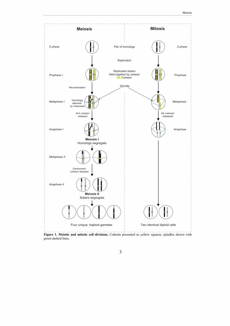
Figure 1. Meiotic and mitotic cell divisions. Cohesin presented as yellow squares, spindles shown with green dashed lines.
Replication
Pair of homologs
Replicated sisters
Held together by cohesin
Cohesin
Recombination
Meiosis IHomologs segregate
Meiosis IISisters segregate
Prophase I
Anaphase I
Metaphase I
Metaphase II
Anaphase II
S phase
Homologs
attached
by chiasmata
Arm cohesin
released
Centromeric
cohesin released
Four unique, haploid gametes
Prophase
Anaphase
Metaphase
S phase
All cohesin
released
Two identical diploid cells
Meiosis Mitosis
Spindle
Meiosis
4
Following meiotic induction, meiotic S phase replicates homologous parental
chromosomes (homologs) into a pair of sister chromatids and loads a complex called
cohesin between sisters to form sister chromatid cohesion (SCC) (Figure 1).
Programmed double-strand breaks (DSBs) are then induced that are repaired via
homologous recombination during prophase. Whereas mitotic cells prefer the sister
chromatid as a repair template, meiotic recombination favors the homolog. After
prophase, two consecutive nuclear divisions ensue. The first, meiosis I (MI), segregates
homologs, while the second, meiosis II (MII), segregates sister chromatids. The final
meiotic products are packaged into spores in yeast while the gametes formed during
mammalian meiosis are stored in the gonads. In budding yeast and mice, recombination
is essential for proper homolog segregation at MI because it creates stable chromosomal
connections between the homologs. These attachments, together with cohesin, ensure
proper chromosome orientation at the first meiotic spindle (reviewed in Handel and
Schimenti 2010).
Meiotic recombination
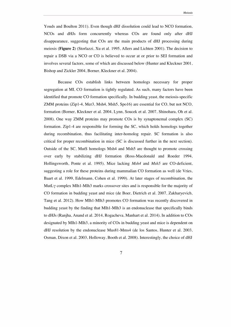
Meiotic recombination is the key feature of meiosis. It is initiated by the
formation of programmed DNA DSBs (reviewed in (Keeney 2001). Meiotic DSBs are
catalyzed by the conserved Spo11 protein in both yeast and mice (Keeney, Giroux et al.
1997, Romanienko and Camerini-Otero 2000, Baudat and Keeney 2001), which cleaves
DNA via a type II topoisomerase-like reaction (for more on topoisomerases, please see
the following section) to generate a transient, Spo11-bound DNA intermediate
(Bergerat, de Massy et al. 1997, Keeney 2008). After break induction, Spo11 is removed
and the 5’ ends are resected to generate 3’ DNA overhangs (Figure 2). To facilitate
strand exchange, the 3’ overhangs are coated with two conserved RecA-like
recombinases. The first, Rad51, is a major repair protein in vegetative cells that favors
sister-directed repair, while the second, Dmc1, is meiosis-specific and promotes the
homolog as a repair template (Bishop, Park et al. 1992, Shinohara, Ogawa et al. 1992,
Barlow, Benson et al. 1997, Yoshida, Kondoh et al. 1998). Despite these differences in

Meiosis
5
Spo11 removal
End resection
Single-end invasion
D-loop formation
Spo11 binding &
DSB induction
SDSA
A
Double-Holliday junction (dHJ)
(stable JM)
Crossover
dHJ/JM resolution
B SEI
(unstable JM)
Non-crossover
synthesis
Initial joint molecule (JM)
dHJ formation
3’5’
DNA
Figure 2. Schematic representation of meiotic recombination. Meiotic recombination is initiated by Spo11-catalyzed DNA double-strand breaks (DSBs). Spo11 is removed from the DNA in the form of Spo11-oligonucleotide complexes, allowing the 5’ ends of the DSB to be resected to generate 3’ single-stranded overhangs coated by Rad51 and Dmc1 (not shown) that can invade a homologous strand for repair. Strand invasion gives rise to a D-loop, forming an initial joint molecule (JM) intermediate. Following stabilization and DNA synthesis, the initial JM gives rise to another transient JM species called the single-end invasion (SEI). (A) The SEI can be quickly dissociated to re-ligate the newly synthesized DNA end to the complementary free break end in a process called synthesis-dependent strand annealing (SDSA). Additional DNA synthesis and ligation yields a mature non-crossover product. (B) Alternatively, the SEI can be stabilized to facilitate capture of the second 3’ DSB end via engagement of the intact homologous strand. Further processing yields gives rise to a stable JM intermediate known as a double-Holliday junction (dHJ). Resolution of the dHJ yields crossover products.
Meiosis
6
template preferance, yeast and mice require both Rad51 and Dmc1 for proper homolog-
directed repair during meiosis (Bishop 1994, Shinohara, Gasior et al. 1997, Tarsounas,
Morita et al. 1999).
Once the broken DNA is coated with Rad51 and Dmc1, it invades an intact
homologous sequence from the homolog, forming a displacement loop (D-loop) and
initial DNA joint molecule (JM). After stabilization and DNA synthesis, this structure
gives rise to an unstable type of JM called a single-end invasion (SEI) (Figure 2)
(Hunter and Kleckner 2001). Further processing of the SEI in subsequent repair steps
can give rise to two classes of products. The first, called non-crossovers (NCOs), repair
without mutual exchange of flanking sequences, whereas the second, known as
crossovers (COs), repair by mutually exchanging DNA between the homologs in order
to physically attach them to one another (Mimitou and Symington 2008, Schwartz and
Heyer 2011).
The majority, if not all, of meiotic NCOs are formed via the so-called synthesis-
dependent strand-annealing (SDSA) pathway (Figure 2A) (Paques and Haber 1999,
Allers and Lichten 2001, Hunter and Kleckner 2001). In SDSA, the invading strand is
displaced from the D-loop to dissolve the SEI. Then, the displaced strand anneals to
complementary DNA sequences on the other end of the DSB, which, after additional
DNA synthesis and ligation, reseals the break to form a NCO. The Blooms helicase
ortholog Sgs1 has been proposed to promote NCO formation during yeast meiosis by
facilitating unwinding of the SEI (De Muyt, Jessop et al. 2012, Zakharyevich, Tang et
al. 2012). Unlike NCO formation, which goes via transient JMs, CO formation involves
the formation of stable JM intermediates. In the CO-forming pathway, the SEI is
stabilized and the second DSB end is captured via the D-loop to form a stable JM called
a double-Holliday junction (dHJ) (Figure 2B) (Schwacha and Kleckner 1995). In order
to be processed into its products, the dHJ must be resolved. This can be achieved by
either symmetric DNA cleavage by endonucleases (dHJ resolution) or by combined
activity of a helicase and topoisomerase (dHJ dissolution) (Schwartz and Heyer 2011,
Meiosis
7
Youds and Boulton 2011). Even though dHJ dissolution could lead to NCO formation,
NCOs and dHJs form concurrently whereas COs are found only after dHJ
disappearance, suggesting that COs are the main products of dHJ processing during
meiosis (Figure 2) (Storlazzi, Xu et al. 1995, Allers and Lichten 2001). The decision to
repair a DSB via a NCO or CO is believed to occur at or prior to SEI formation and
involves several factors, some of which are discussed below (Hunter and Kleckner 2001,
Bishop and Zickler 2004, Borner, Kleckner et al. 2004).
Because COs establish links between homologs necessary for proper
segregation at MI, CO formation is tightly regulated. As such, many factors have been
identified that promote CO formation specifically. In budding yeast, the meiosis-specific
ZMM proteins (Zip1-4, Mer3, Msh4, Msh5, Spo16) are essential for CO, but not NCO,
formation (Borner, Kleckner et al. 2004, Lynn, Soucek et al. 2007, Shinohara, Oh et al.
2008). One way ZMM proteins may promote COs is by synaptonemal complex (SC)
formation. Zip1-4 are responsible for forming the SC, which holds homologs together
during recombination, thus facilitating inter-homolog repair. SC formation is also
critical for proper recombination in mice (SC is discussed further in the next section).
Outside of the SC, MutS homologs Msh4 and Msh5 are thought to promote crossing
over early by stabilizing dHJ formation (Ross-Macdonald and Roeder 1994,
Hollingsworth, Ponte et al. 1995). Mice lacking Msh4 and Msh5 are CO-deficient,
suggesting a role for these proteins during mammalian CO formation as well (de Vries,
Baart et al. 1999, Edelmann, Cohen et al. 1999). At later stages of recombination, the
MutLγ complex Mlh1-Mlh3 marks crossover sites and is responsible for the majority of
CO formation in budding yeast and mice (de Boer, Dietrich et al. 2007, Zakharyevich,
Tang et al. 2012). How Mlh1-Mlh3 promotes CO formation was recently discovered in
budding yeast by the finding that Mlh1-Mlh3 is an endonuclease that specifically binds
to dHJs (Ranjha, Anand et al. 2014, Rogacheva, Manhart et al. 2014). In addition to COs
designated by Mlh1-Mlh3, a minority of COs in budding yeast and mice is dependent on
dHJ resolution by the endonuclease Mus81-Mms4 (de los Santos, Hunter et al. 2003,
Osman, Dixon et al. 2003, Holloway, Booth et al. 2008). Interestingly, the choice of dHJ
Meiosis
8
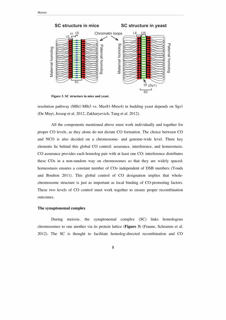
Figure 3. SC structure in mice and yeast.
resolution pathway (Mlh1-Mlh3 vs. Mus81-Mms4) in budding yeast depends on Sgs1
(De Muyt, Jessop et al. 2012, Zakharyevich, Tang et al. 2012).
All the components mentioned above must work individually and together for
proper CO levels, as they alone do not dictate CO formation. The choice between CO
and NCO is also decided on a chromosome- and genome-wide level. Three key
elements lie behind this global CO control: assurance, interference, and homeostasis.
CO assurance provides each homolog pair with at least one CO; interference distributes
these COs in a non-random way on chromosomes so that they are widely spaced;
homeostasis ensures a constant number of COs independent of DSB numbers (Youds
and Boulton 2011). This global control of CO designation implies that whole-
chromosome structure is just as important as local binding of CO-promoting factors.
These two levels of CO control must work together to ensure proper recombination
outcomes.
The synaptonemal complex
During meiosis, the synaptonemal complex (SC) links homologous
chromosomes to one another via its protein lattice (Figure 3) (Fraune, Schramm et al.
2012). The SC is thought to facilitate homolog-directed recombination and CO
SC structure in yeastLE CR
TF (Zip1)
SC
Mat
erna
l hom
olog
Paternal hom
olog
Chromatin loopsLE
TF CE
SC
SC structure in mice
Mat
erna
l hom
olog
Paternal hom
olog
Meiosis
9
formation by holding homologs in close proximity during DSB repair. In line with this
hypothesis, Drosophila males, which do not perform meiotic recombination, lack an SC
(Zickler 1999). Proper SC formation in budding yeast and mice is dependent on DSB
formation (Agarwal and Roeder 2000, Baudat, Manova et al. 2000, Romanienko and
Camerini-Otero 2000), and meiosis in these organisms depends on SC formation, as
mutating any SC components leads to CO loss and abnormal meiosis.
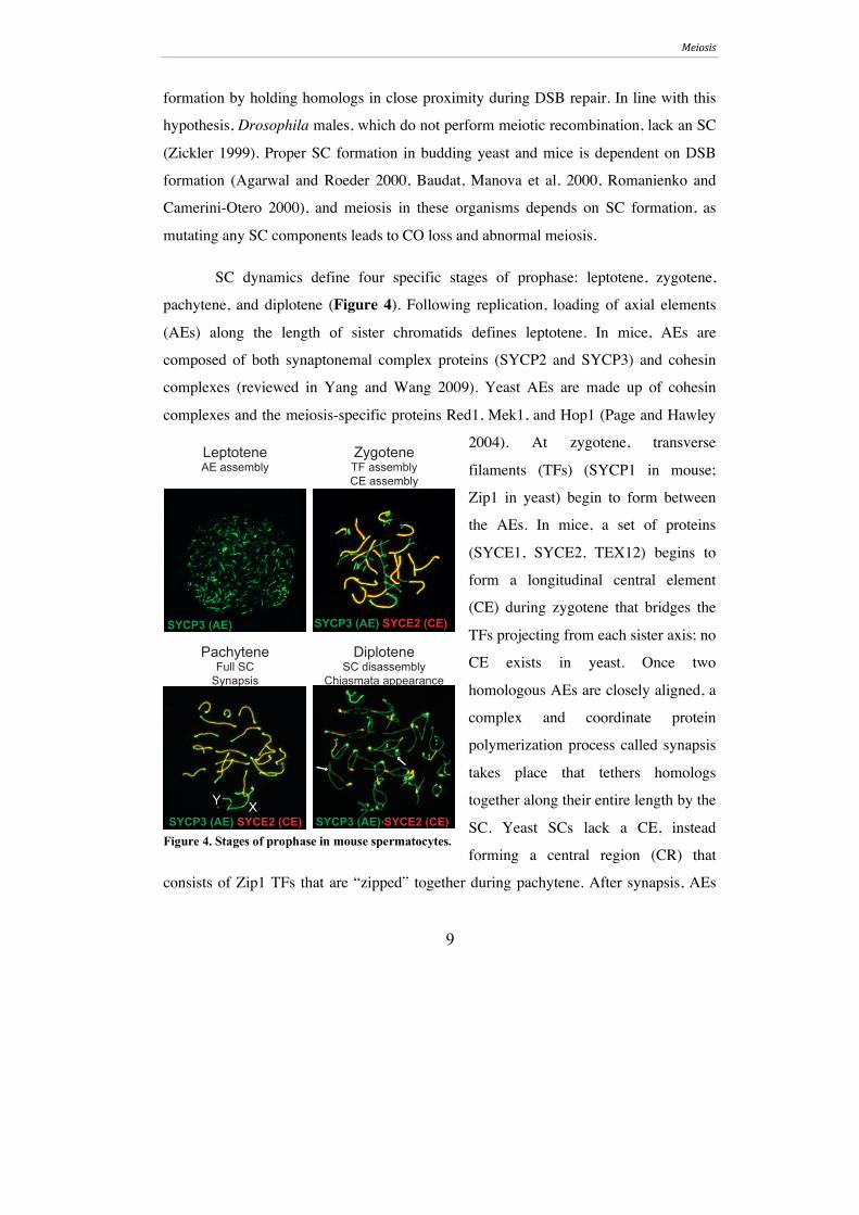
SC dynamics define four specific stages of prophase: leptotene, zygotene,
pachytene, and diplotene (Figure 4). Following replication, loading of axial elements
(AEs) along the length of sister chromatids defines leptotene. In mice, AEs are
composed of both synaptonemal complex proteins (SYCP2 and SYCP3) and cohesin
complexes (reviewed in Yang and Wang 2009). Yeast AEs are made up of cohesin
complexes and the meiosis-specific proteins Red1, Mek1, and Hop1 (Page and Hawley
2004). At zygotene, transverse
filaments (TFs) (SYCP1 in mouse;
Zip1 in yeast) begin to form between
the AEs. In mice, a set of proteins
(SYCE1, SYCE2, TEX12) begins to
form a longitudinal central element
(CE) during zygotene that bridges the
TFs projecting from each sister axis; no
CE exists in yeast. Once two
homologous AEs are closely aligned, a
complex and coordinate protein
polymerization process called synapsis
takes place that tethers homologs
together along their entire length by the
SC. Yeast SCs lack a CE, instead
forming a central region (CR) that
consists of Zip1 TFs that are “zipped” together during pachytene. After synapsis, AEs
PachyteneFull SC
Synapsis
SYCP3 (AE) SYCE2 (CE)
SYCP3 (AE) SYCE2 (CE)XY
SYCP3 (AE) SYCE2 (CE)
DiploteneSC disassembly
Chiasmata appearance
ZygoteneTF assemblyCE assembly
LeptoteneAE assembly
SYCP3 (AE)
Figure 4. Stages of prophase in mouse spermatocytes.
Meiosis
10
are referred to as lateral elements (LEs). SC disassembly begins during diplotene, the
final stage of prophase. By the end of diplotene, all LEs have disappeared and homologs
are connected by chiasmata, the physical manifestations of crossovers at the
chromosomal level.
Inter-homolog vs. inter-sister recombination in Saccharomyces cerevisae
Meiotic chromosomes are organized into a loop-axis configuration (Figure 3).
It is believed that DSB formation occurs at the top of the chromatin loops while
recombination is carried out at the axis. Components that shape this loop-axis structure
are important for the integrity of meiotic recombination.
Unlike mitotic recombination, which favors the sister chromatid, meiotic
recombination prefers the homologous chromosome as a repair template (Schwacha and
Kleckner 1997). In addition to the SC’s role in homolog synapsis, inter-homolog bias is
due to combined efforts of mechanisms that promote homolog strand invasion, such as
Dmc1, and meiotic axis components such as Red1, Hop1, and Mek1 that hinder use of
the sister as a repair template by inhibiting recruitment of sister-promoting
recombination factors (Bishop, Park et al. 1992, Hollingsworth and Ponte 1997,
Schwacha and Kleckner 1997, Shinohara, Gasior et al. 1997, Niu, Wan et al. 2005,
Carballo, Johnson et al. 2008). The meiotic axis includes the cohesin subunit Rec8
(Revenkova and Jessberger 2006). Cohesin is responsible for sister chromatid cohesion,
and it is required for inter-sister recombination during DSB repair in vegetative cells
(more on cohesin in following section). During meiosis, Rec8 is required for proper axis
configuration, and its absence leads to altered DSB distribution thought to be a result of
abnormal tethering of chromatin loops (Kugou, Fukuda et al. 2009, Kim, Weiner et al.
2010). In line with the idea that cohesin is needed to properly tether chromatin loops,
mice that lack Rec8 have shortened axes (Bannister, Reinholdt et al. 2004); this
phenotype is also observed in the absence of another meiosis-specific murine cohesin
component, SMC1β, which was shown to have longer chromatin loops tethered to the
shortened axis (Revenkova, Eijpe et al. 2004). Interestingly, Rec8 is needed to maintain
Meiosis
11
inter-homolog bias during the SEI-to-dHJ transition, even though Rec8 traditionally aids
in inter-sister recombination (Kim, Weiner et al. 2010). Homolog-promoting
components of the meiotic axis counteract the inter-sister bias created by Rec8, allowing
inter-homolog events to dominate (Kim, Weiner et al. 2010). Concurrent activities of
these mechanisms establish a bias for inter-homolog recombination but do not eliminate
inter-sister recombination altogether, with the possibility that as many as one-third of
meiotic repair is directed to the sister, at least in yeast (Goldfarb and Lichten 2010, Kim,
Weiner et al. 2010). Whether between sisters or homologs, all DNA linkages (i.e. JMs)
must be properly resolved for accurate chromosome segregation.
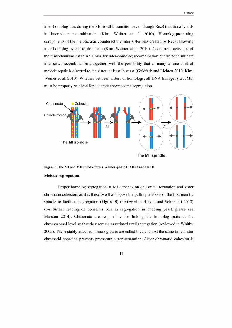
Figure 5. The MI and MII spindle forces. AI=Anaphase I; AII=Anaphase II
Meiotic segregation
Proper homolog segregation at MI depends on chiasmata formation and sister
chromatin cohesion, as it is these two that oppose the pulling tensions of the first meiotic
spindle to facilitate segregation (Figure 5) (reviewed in Handel and Schimenti 2010)
(for further reading on cohesin’s role in segregation in budding yeast, please see
Marston 2014). Chiasmata are responsible for linking the homolog pairs at the
chromosomal level so that they remain associated until segregation (reviewed in Whitby
2005). These stably attached homolog pairs are called bivalents. At the same time, sister
chromatid cohesion prevents premature sister separation. Sister chromatid cohesion is
Cohesin
Spindle forces
The MI spindle
Chiasmata
The MII spindle
AI AII

Meiosis
12
also necessary for bivalent maintenance, as early loss of cohesion on the telomere-
proximal side of chiasmata can lead to untimely chiasmata dissolution. Once chiasmata
and sister chromatid cohesion are properly in place, homologs can bi-orient, meaning
that non-sister kinetochores orient to opposite spindle poles. Homolog bi-orientation at
metaphase I, and subsequent segregation at anaphase I, is achieved by the specific loss
of arm cohesion, which facilitates chiasmata resolution, and maintenance of centromeric
cohesion, which prevents sister chromatid separation until MII. This is different from
mitosis when all cohesion is lost at the metaphase-anaphase transition (Figure 1). At
MII, cohesion is lost at the centromeres, allowing sister segregation (this process is
reviewed in Wassmann 2013). Step-wise loss of cohesion is a hallmark of meiotic
segregation and is mainly achieved by specialization of meiosis-specific cohesin
complexes (reviewed in McNicoll, Stevense et al. 2013). Overall, proper segregation at
MI and MII is crucial for proper gamete formation and reproductive success of an
organism.
MAMMALIAN GERM CELL FORMATION
In the gonads of mammalian organisms, diploid germ cells produce haploid gametes
that, after fusion with another gamete from the opposite sex, will produce the offspring.
The collective process of gametogenesis (gamete formation) is known as
spermatogenesis in males and oogenesis in females, and takes place in the testes and the
ovaries, respectively.
Spermatogenesis
Sperm, also known as mature spermatozoa, are the final products of
spermatogenesis and are the gametes used for sexual reproduction. Failure to complete
spermatogenesis or the generation of improper sperm can lead to infertility or genetic
disorders, making it important to understand the steps of this intricate process.
Spermatogenesis takes place in the seminiferous tubules of the testes. The seminiferous
Mammalian germ cell formation
13
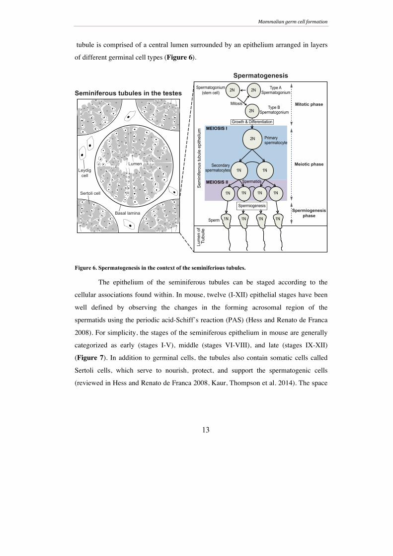
tubule is comprised of a central lumen surrounded by an epithelium arranged in layers
of different germinal cell types (Figure 6).
Figure 6. Spermatogenesis in the context of the seminiferious tubules.
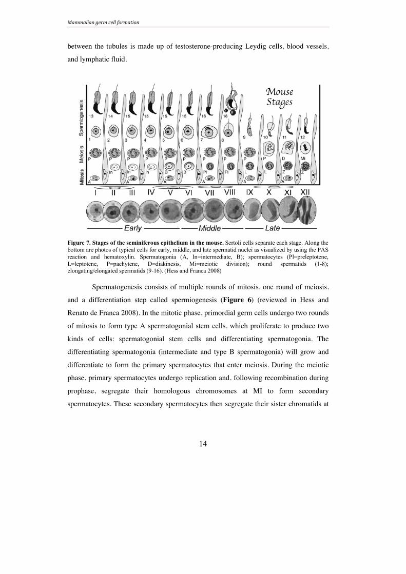
The epithelium of the seminiferous tubules can be staged according to the
cellular associations found within. In mouse, twelve (I-XII) epithelial stages have been
well defined by observing the changes in the forming acrosomal region of the
spermatids using the periodic acid-Schiff’s reaction (PAS) (Hess and Renato de Franca
2008). For simplicity, the stages of the seminiferous epithelium in mouse are generally
categorized as early (stages I-V), middle (stages VI-VIII), and late (stages IX-XII)
(Figure 7). In addition to germinal cells, the tubules also contain somatic cells called
Sertoli cells, which serve to nourish, protect, and support the spermatogenic cells
(reviewed in Hess and Renato de Franca 2008, Kaur, Thompson et al. 2014). The space
Spermatogonium(stem cell)
Type A Spermatogonium
Type B Spermatogonium
Primary spermatocyte
Spermatids
Mitosis
Growth & Differentiation
2N 2N
2N
2N
Spermiogenesis
MEIOSIS I
MEIOSIS II
Sem
inife
rous
tubu
le e
pith
eliu
m
Secondary spermatocytes 1N 1N
1N 1N 1N 1N
1N 1N 1N 1N Sperm
Lum
en o
f Tu
bule
Sertoli cell
LumenLeydig
cell
Basal lamina
Seminiferous tubules in the testes
Spermatogenesis
Mitotic phase
Meiotic phase
Spermiogenesis
phase
Mammalian germ cell formation
14
between the tubules is made up of testosterone-producing Leydig cells, blood vessels,
and lymphatic fluid.
Spermatogenesis consists of multiple rounds of mitosis, one round of meiosis,
and a differentiation step called spermiogenesis (Figure 6) (reviewed in Hess and
Renato de Franca 2008). In the mitotic phase, primordial germ cells undergo two rounds
of mitosis to form type A spermatogonial stem cells, which proliferate to produce two
kinds of cells: spermatogonial stem cells and differentiating spermatogonia. The
differentiating spermatogonia (intermediate and type B spermatogonia) will grow and
differentiate to form the primary spermatocytes that enter meiosis. During the meiotic
phase, primary spermatocytes undergo replication and, following recombination during
prophase, segregate their homologous chromosomes at MI to form secondary
spermatocytes. These secondary spermatocytes then segregate their sister chromatids at
Figure 7. Stages of the seminiferous epithelium in the mouse. Sertoli cells separate each stage. Along the bottom are photos of typical cells for early, middle, and late spermatid nuclei as visualized by using the PAS reaction and hematoxylin. Spermatogonia (A, In=intermediate, B); spermatocytes (Pl=preleptotene, L=leptotene, P=pachytene, D=diakinesis, Mi=meiotic division); round spermatids (1-8); elongating/elongated spermatids (9-16). (Hess and Franca 2008)

Mammalian germ cell formation
15
MII to generate haploid spermatids. The spermatids then enter the final phase of
spermatogenesis, spermiogenesis.
A series of morphological changes occur during spermiogenesis that transform
round spermatids into elongated, compact sperm over the course of sixteen steps
(Figure 7). There are three types of developing spermatids in the seminiferous
epithelium: early spermatids with round nuclei, intermediate spermatids with elongating
nuclei, and mature spermatids with condensed nuclei (reviewed in Dadoune 2003).
Central to spermatid differentiation is the attachment of specialized structures and
compaction of the spermatid head, both of which allow sperm to carry out their function
during fertilization. In early spermiogenesis, specialized structures called an acrosome
and axoneme begin to develop at opposing poles of the round spermatids (reviewed in
O'Donnell and O'Bryan 2014). The acrosome, which eventually caps the entire sperm
head, arises from the Golgi apparatus and facilitates fertilization by the secretion of
enzymes that digest the outer surface of the female gamete to allow nuclear fusion. The
axoneme is a microtubule-based structure that is equally as crucial to fertilization, as it is
the core structure of the flagellum that mobilizes sperm. Though initially formed in early
stages, the acrosome and axoneme continue to develop throughout spermiogenesis
parallel to nuclear reshaping.
Compaction of the spermatid nucleus begins approximately halfway through
spermiogenesis with the formation of a microtubule-based organelle called the
manchette. The manchette encompasses the nucleus directly below the acrosome in
stage 7-8 spermatids, sliding down the nucleus in subsequent elongation steps before
being dismantled in step 14 spermatids (reviewed in O'Donnell and O'Bryan 2014). This
movement of the manchette facilitates nuclear compaction by altering the shape of the
nucleus. DNA remodeling allows for nuclear compaction as well. During step 10 of
spermiogenesis, histones begin to be replaced with protamines, which allow for tight
DNA packaging due to their affinity for the minor groove of DNA (reviewed in Braun
2001). Mice have two protamines, protamine 1 (PRM1) and protamine (PRM2), both of
Mammalian germ cell formation
16
which are required for proper sperm formation (Cho, Willis et al. 2001, Cho, Jung-Ha et
al. 2003). It is unclear why a nuclear compaction is essential for sperm formation, but it
is proposed that the small head protects sperm from damage and facilitates in motility
during fertilization (reviewed in Carrell, Emery et al. 2007).
Oogenesis
Unlike males, who can produce millions of new gametes each day from a
replenishing pool of spermatogonial germ cells, females are born with a fixed number of
cells with the potential to become mature gametes. These cells, called primary oocytes,
are housed in individual primordial follicles and are arrested at a diplotene-like dicyte
stage at the end of meiotic prophase at birth. Once the animal reaches sexual maturity, a
hormone surge triggers the development of a subset of primordial follicles and
subsequent maturation of the primary oocytes within. The oocytes complete meiosis to
form mature ovum, also referred to as eggs, which are the gametes with the potential to
be fertilized. The process of oocyte maturation and egg release, called menstruation,
occurs once a month until menopause when menstruation ceases. (reviewed in Pepling
2006)
PROTEINS THAT SHAPE CHROMOSOMES
Though visualized under the microscope earlier, chromosomes were not linked to
heredity until the turn of the 20th century, and it took scientists almost fifty more years to
identify DNA as the basic unit of heredity. After Watson and Crick described the DNA
double helix in 1953, research in the field boomed and provided evidence that made it
impossible to deny the importance of DNA for life. We now know that proper
organization of DNA into chromosomes, via the action of DNA-altering proteins, is just
as important as DNA sequence itself. There are a multitude of proteins and protein
families that influence overall chromosome structure and function. This section will
Proteins that shape chromosomes
17
focus on the proteins and protein complexes most relevant to this thesis: topoisomerases,
SMC complexes, and CTCF, with an emphasis on their functions in budding yeast and
mice. Where relevant, findings from Papers I-III will be presented and discussed.
Topoisomerases
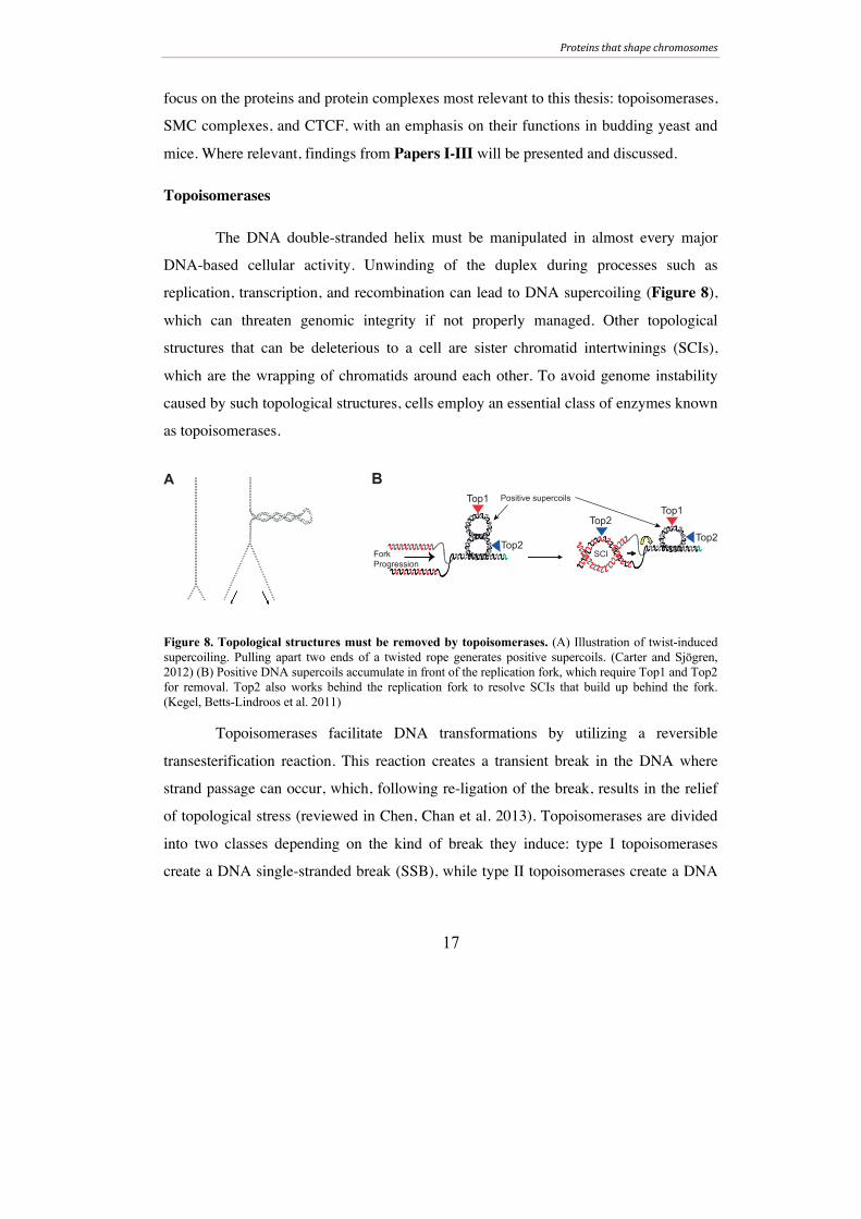
The DNA double-stranded helix must be manipulated in almost every major
DNA-based cellular activity. Unwinding of the duplex during processes such as
replication, transcription, and recombination can lead to DNA supercoiling (Figure 8),
which can threaten genomic integrity if not properly managed. Other topological
structures that can be deleterious to a cell are sister chromatid intertwinings (SCIs),
which are the wrapping of chromatids around each other. To avoid genome instability
caused by such topological structures, cells employ an essential class of enzymes known
as topoisomerases.
Figure 8. Topological structures must be removed by topoisomerases. (A) Illustration of twist-induced supercoiling. Pulling apart two ends of a twisted rope generates positive supercoils. (Carter and Sjögren, 2012) (B) Positive DNA supercoils accumulate in front of the replication fork, which require Top1 and Top2 for removal. Top2 also works behind the replication fork to resolve SCIs that build up behind the fork. (Kegel, Betts-Lindroos et al. 2011)
Topoisomerases facilitate DNA transformations by utilizing a reversible
transesterification reaction. This reaction creates a transient break in the DNA where
strand passage can occur, which, following re-ligation of the break, results in the relief
of topological stress (reviewed in Chen, Chan et al. 2013). Topoisomerases are divided
into two classes depending on the kind of break they induce: type I topoisomerases
create a DNA single-stranded break (SSB), while type II topoisomerases create a DNA
A BTop1
Top2
Top2Top1
Top2ForkProgression
Positive supercoils
SCI
Proteins that shape chromosomes
18
double-stranded break (DSB) (reviewed in Wang 1996). These classes are further
categorized into subfamilies A, B, or C based on amino acid sequence and reaction
mechanism (reviewed in Schoeffler and Berger 2008). The topoisomerases at work in
eukaryotic cells are type IA (yeast Top3), type IB (yeast Top1), and type IIA (yeast
Top2). The most relevant functions of these topoisomerases as they relate to this thesis
will be described for budding yeast below. For further reading on topoisomerases, please
consult (Chen, Chan et al. 2013), (Wang 2002), and references therein.
Top3 (Paper II)
Top3 is the DNA-cleaving component of the Top3-Sgs1-Rmi1 complex.
Though this complex has roles during replication, its best characterized function is
during recombination in vegetative cells due to its ability to resolve double-Holliday
junctions (dHJs) (reviewed in Cejka, Plank et al. 2012). A study in meiosis suggests that
Top3 functions during meiotic recombination as well, since top3 mutants experienced a
recombination-dependent segregation block at MI (Gangloff, de Massy et al. 1999).
In Paper II, we further explored the meiotic function of Top3 by using a
meiotic null (mn) allele of TOP3, top3-mn. The meiotic null allele was constructed by
replacing the endogenous promoter of TOP3 with the mitosis-specific CLB2 promoter
(Lee and Amon 2003), which is down regulated after meiotic S phase (Grandin and
Reed 1993, Dahmann and Futcher 1995). In this way, replication defects are avoided
using this system. Despite incomplete reduction in Top3 protein levels in the top3-mn,
40% of cells were unable to complete meiotic divisions and instead formed cells with
one nuclear mass outside of four empty spores. This illustrates the necessity of Top3 for
meiosis, as even a small loss in Top3 levels causes a phenotype. Consistent with spore
formation in these cells, which indicates completion of the meiotic program in S.
cerevisiae (Neiman 2011), Top3-deficient cells had normal spindle and synaptonemal
complex (SC) dynamics.
Proteins that shape chromosomes
19
The phenotype in top3-mn cells is reminiscent of that observed in smc6 mutants
(Copsey, Tang et al. 2013, Lilienthal, Kanno et al. 2013, Xaver, Huang et al. 2013)
(Paper I) and mus81 sgs1 and mms4 sgs1 double mutants (Jessop and Lichten 2008,
Oh, Lao et al. 2008), which accumulate recombination intermediates. Accordingly, we
found that the segregation block in top3-mn was rescued when DSBs were eliminated.
Given the role of Sgs1 and Top3 in mitotic recombination, it is very likely that they
work in conjunction during meiosis as well. Indeed, rescue of the top3 meiotic
segregation in an sgs1 background suggests interplay between Sgs1 and Top3 in meiosis
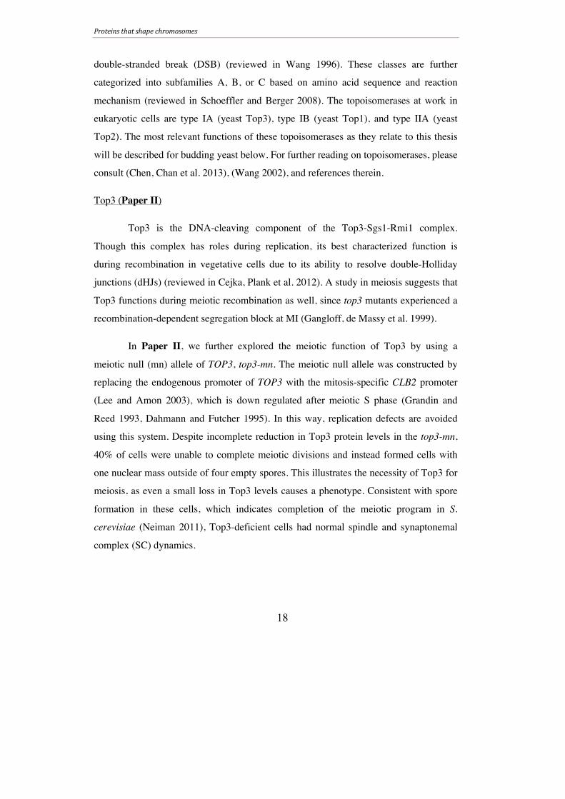
(Gangloff, de Massy et al. 1999). However,
in meiosis, accumulation of unresolved
recombination intermediates and subsequent
segregation block is only achieved when
SGS1 is removed along with an
endonuclease component (MUS81/MMS4)
(Jessop and Lichten 2008, Oh, Lao et al.
2008). This indicates that Top3’s meiotic
function depends on more than just Sgs1.
Recent data suggests that Smc6 binding in
mitosis partially depends on Top3 (Berta et
al., unpublished). Given the recently defined
role for Smc6 during meiotic recombination
(Paper I), it may be that the phenotype in
top3-mn cells is due to loss of Smc6 on chromosomes. However, no detectable changes
in Smc6 loading onto meiotic chromosomes were detected in Top3-deficient cells by
immunofluorescence. This may indicate that Top3 does not regulate meiotic Smc6
binding. Alternatively, Top3 may influence small changes in Smc6 loading. The Top3-
dependent changes in mitotic Smc6 binding were detected using the more sensitive
chromatin immunoprecipitation technique, making it possible that our
immunofluorescence analysis was not sensitive enough to detect changes in Smc6
X X
Mainly Mlh1-Mlh3facilitate resolution Unknown factors
promote resolution
Top3??
Unresolved recombination intermediatesthat can lead to segregation block at MI
Chiasmata
Figure 9. Possible role for Top3 in the removal of inter-sister dHJs.
Proteins that shape chromosomes
20
loading. In conclusion, Paper II proposes a role for Top3 during meiotic recombination
(Figure 9), and it will be interesting to explore this role in the future. Critical to future
endeavors is the creation of a meiosis-specific TOP3 allele that effectively reduces Top3
levels, as the top3-mn used in this investigation only reduced protein levels slightly. If
an efficient mutant is created, it will be interesting to explore what, if any, role Top3
plays during meiotic recombination. Two lines of evidence suggest that any Top3-
mediated dHJ processing would be outside of established CO-forming meiotic
pathways. First, the Top3-Sgs1-Rmi1 complex dissolves dHJs into NCO products,
which are not products of meiotic dHJ resolution. Second, the factors responsible for
virtually all inter-homolog dHJ resolution during meiosis have been identified
(Zakharyevich, Tang et al. 2012). Instead, Top3 may be needed to process inter-sister
dHJs. Inter-sister DSB repair during meiosis is not well understood, but resolution of
inter-sister recombination intermediates is required for proper segregation.
Top1
Top1 is the most important factor for removal of supercoil-induced stress in the
cell (Wang 2002). Together with Top2 (see below), it works to release supercoils ahead
of the replication fork (Bermejo, Doksani et al. 2007). Due to its overlapping function
with Top2, Top1 is non-essential in yeast (Chen, Chan et al. 2013). The function of
Top1 during meiosis was not investigated in this thesis, and to our knowledge no
meiotic function has been reported.
Top2 (Paper II)
Chromosome replication creates two topological structures which must be
handled by topoisomerases: supercoils ahead of the replication fork and SCIs behind it
(Figure 8B). While both Top1 and Top2 can remove supercoils, elimination of SCIs
relies almost solely on Top2 (Figure 8C), and cells lacking Top2 complete replication
but are unable to segregate their chromosomes (Holm, Goto et al. 1985, Bermejo,
Doksani et al. 2007, Baxter and Diffley 2008). A catalytically dead top2 mutant,
Proteins that shape chromosomes
21
however, leads to a checkpoint-induced replication arrest (Baxter and Diffley 2008).
During mitosis, newly replicated sister chromatids must be held until their timely
segregation at the metaphase-anaphase transition (Figure 1B). If sisters are not
connected, not only will they fall apart prior to anaphase, they will also be unable to
satisfy the spindle checkpoint due to lack of tension, causing spindle assembly
checkpoint-mediated arrest. SCIs were originally proposed to be the factor linking sister
chromatids, but this model was disfavored upon the discovery of proteins that
functioned in the regulation of sister cohesion (Guacci, Koshland et al. 1997, Michaelis,
Ciosk et al. 1997), which are know known components of the cohesin complex. More
recently, cohesin was proposed to protect SCIs from Top2, suggesting once again that
SCIs may play a role in keeping sisters together (Farcas, Uluocak et al. 2011). Together
with the condensin complex (discussed further below), Top2 also aids in proper
chromosome condensation and axis organization in vegetative cells (Koshland and
Strunnikov 1996). During meiosis, Top2 binds to the meiotic chromosome axis (Klein,
Laroche et al. 1992), and mutations in top2 during meiosis lead to lack of chromosome
condensation and a segregation block that is partially dependent on recombination
(Rose, Thomas et al. 1990, Rose and Holm 1993). Recently, Kleckner and colleagues
also reported that the structural role of Top2 makes it a key factor in meiotic crossover
interference (Zhang, Wang et al. 2014).
In Paper II, we further characterized the meiotic role of Top2 by using the
meiosis-specific mutant top2-mn. (This mutant was constructed as described for top3-
mn.) The top2-mn displayed a mixture of two phenotypes: cells that accumulated as
uninucleates (40%) and cells that formed 1-3 empty spores with one nuclear mass
outside (60%). Since certain top2 mutants experience replication defects during mitosis
(Baxter and Diffley 2008), the uninucleate population in top2-mn could be blocked due
to problems during pre-meiotic S phase. While we believe that replication defects were
avoided using the meiotic null, we cannot completely exclude this possibility.
Proteins that shape chromosomes
22
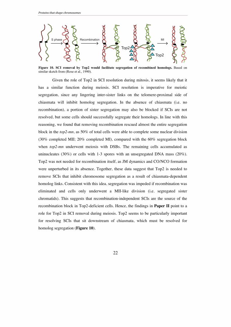
Given the role of Top2 in SCI resolution during mitosis, it seems likely that it
has a similar function during meiosis. SCI resolution is imperative for meiotic
segregation, since any lingering inter-sister links on the telomere-proximal side of
chiasmata will inhibit homolog segregation. In the absence of chiasmata (i.e. no
recombination), a portion of sister segregation may also be blocked if SCIs are not
resolved, but some cells should successfully segregate their homologs. In line with this
reasoning, we found that removing recombination rescued almost the entire segregation
block in the top2-mn, as 50% of total cells were able to complete some nuclear division
(30% completed MII; 20% completed MI), compared with the 60% segregation block
when top2-mn underwent meiosis with DSBs. The remaining cells accumulated as
uninucleates (30%) or cells with 1-3 spores with an unsegregated DNA mass (20%).
Top2 was not needed for recombination itself, as JM dynamics and CO/NCO formation
were unperturbed in its absence. Together, these data suggest that Top2 is needed to
remove SCIs that inhibit chromosome segregation as a result of chiasmata-dependent
homolog links. Consistent with this idea, segregation was impeded if recombination was
eliminated and cells only underwent a MII-like division (i.e. segregated sister
chromatids). This suggests that recombination-independent SCIs are the source of the
recombination block in Top2-deficient cells. Hence, the findings in Paper II point to a
role for Top2 in SCI removal during meiosis. Top2 seems to be particularly important
for resolving SCIs that sit downstream of chiasmata, which must be resolved for
homolog segregation (Figure 10).
Top2
Top2
S phase Recombination MI
Figure 10. SCI removal by Top2 would facilitate segregation of recombined homologs. Based on similar sketch from (Rose et al., 1990).
Proteins that shape chromosomes
23
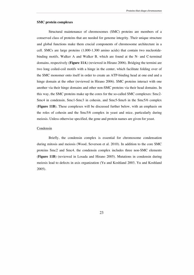
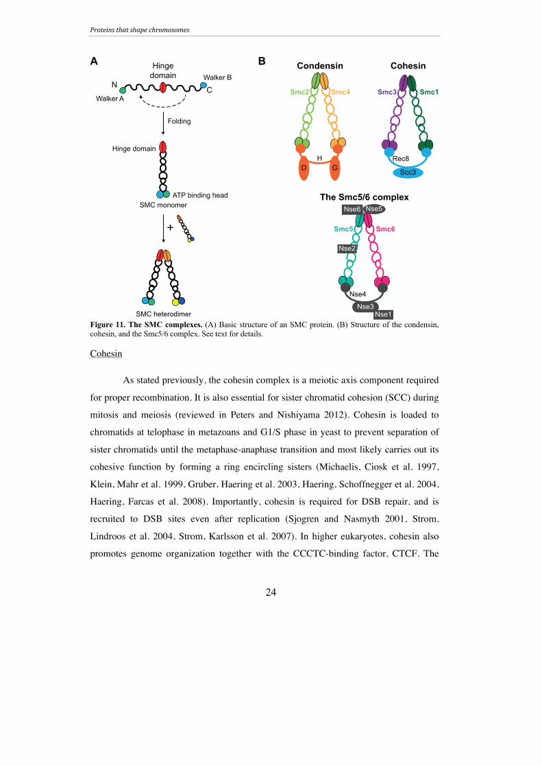
SMC protein complexes
Structural maintenance of chromosomes (SMC) proteins are members of a
conserved class of proteins that are needed for genome integrity. Their unique structure
and global functions make them crucial components of chromosome architecture in a
cell. SMCs are large proteins (1,000-1,300 amino acids) that contain two nucleotide-
binding motifs, Walker A and Walker B, which are found at the N- and C-terminal
domains, respectively (Figure 11A) (reviewed in Hirano 2006). Bridging the termini are
two long coiled-coil motifs with a hinge in the center, which facilitate folding over of
the SMC monomer onto itself in order to create an ATP-binding head at one end and a
hinge domain at the other (reviewed in Hirano 2006). SMC proteins interact with one
another via their hinge domains and other non-SMC proteins via their head domains. In
this way, the SMC proteins make up the cores for the so-called SMC complexes: Smc2-
Smc4 in condensin, Smc1-Smc3 in cohesin, and Smc5-Smc6 in the Smc5/6 complex
(Figure 11B). These complexes will be discussed further below, with an emphasis on
the roles of cohesin and the Smc5/6 complex in yeast and mice, particularly during
meiosis. Unless otherwise specified, the gene and protein names are given for yeast.
Condensin
Briefly, the condensin complex is essential for chromosome condensation
during mitosis and meiosis (Wood, Severson et al. 2010). In addition to the core SMC
proteins Smc2 and Smc4, the condensin complex includes three non-SMC elements
(Figure 11B) (reviewed in Losada and Hirano 2005). Mutations in condensin during
meiosis lead to defects in axis organization (Yu and Koshland 2003, Yu and Koshland
2005).
Proteins that shape chromosomes
24
Figure 11. The SMC complexes. (A) Basic structure of an SMC protein. (B) Structure of the condensin, cohesin, and the Smc5/6 complex. See text for details.
Cohesin
As stated previously, the cohesin complex is a meiotic axis component required
for proper recombination. It is also essential for sister chromatid cohesion (SCC) during
mitosis and meiosis (reviewed in Peters and Nishiyama 2012). Cohesin is loaded to
chromatids at telophase in metazoans and G1/S phase in yeast to prevent separation of
sister chromatids until the metaphase-anaphase transition and most likely carries out its
cohesive function by forming a ring encircling sisters (Michaelis, Ciosk et al. 1997,
Klein, Mahr et al. 1999, Gruber, Haering et al. 2003, Haering, Schoffnegger et al. 2004,
Haering, Farcas et al. 2008). Importantly, cohesin is required for DSB repair, and is
recruited to DSB sites even after replication (Sjogren and Nasmyth 2001, Strom,
Lindroos et al. 2004, Strom, Karlsson et al. 2007). In higher eukaryotes, cohesin also
promotes genome organization together with the CCCTC-binding factor, CTCF. The
N C
Hinge
domain
Walker A
Walker B
Folding
ATP binding head
Hinge domain
SMC heterodimer
+
SMC monomer
A B
Nse4
Nse3
Nse1
Nse6 Nse5
Nse2
Smc5 Smc6
Smc2 Smc4
H
D G
Rec8
Scc3
Smc3 Smc1
Condensin Cohesin
The Smc5/6 complex

Proteins that shape chromosomes
25
interplay between CTCF and cohesin is crucial for mammalian genome function (CTCF
will be discussed more in the next section).
The cohesin complex consists of a heterodimer between Smc1 (SMC1α in
mice) and Smc3 (SMC3 in mice) bridged by an α-kleisin subunit, Scc1/Mcd1 (RAD21
in mice), to which an additional non-SMC subunit binds: sister chromatid cohesion 3
(Scc3, called SA or STAG in mice) (Figure 11B) (yeast review in Haering, Lowe et al.
2002; mammalian review in McNicoll, Stevense et al. 2013). Vertebrates have two SA
proteins, SA1 and SA2, that give rise to two functionally different mitotic cohesin
complexes (reviewed in Canudas and Smith 2009). During meiosis, the kleisin subunit
of cohesin is replaced with Rec8 in yeast and REC8 or RAD21L in vertebrates (Klein,
Mahr et al. 1999, Bannister, Reinholdt et al. 2004, Lee and Hirano 2011, Polakova,
Cipak et al. 2011). Mice have two additional meiosis-specific components for the SMC3
and SA subunits: SMC3β and STAG3, respectively (Prieto, Suja et al. 2001,
Revenkova, Eijpe et al. 2001).
As mentioned earlier, cohesin is removed stepwise in meiosis (Figures 1, 5). At
MI, arm cohesin is cleaved away in order to allow homolog segregation; centromeric
cohesin is maintained until MII when sister chromatids are segregated. In addition to its
role in SCC, cohesin is necessary for proper architecture of the meiotic axis (for more on
the meiotic axis, please see the Meiosis section in this thesis) as well as SC formation
and DSB repair in yeast and mice (Klein, Mahr et al. 1999, Pelttari, Hoja et al. 2001,
Llano, Herran et al. 2012). Studies have found both unique and overlapping functions
for the different meiotic subunits in mice, suggesting interplay between at least six
different cohesin complexes during meiosis (Ishiguro, Kim et al. 2011, Jessberger 2011,
Lee and Hirano 2011, Fukuda, Fukuda et al. 2014, Hopkins, Hwang et al. 2014,
Ishiguro, Kim et al. 2014).

Proteins that shape chromosomes
26
The Smc5/6 complex (Paper I)
The most enigmatic of the SMC complexes is the Smc5/6 complex. The
complex consists of eight subunits, all of which are essential in yeast: Smc5, Smc6,
Nse1, Mms21 (Nse2) and Nse3-6 (Figure 11B). These components were originally
discovered in fission yeast as part of the mitotic homologous recombination repair
pathway (Lehmann, Walicka et al. 1995, Fousteri and Lehmann 2000, Morikawa,
Morishita et al. 2004, Andrews, Palecek et al. 2005), and was later found to function in
the resolution of recombination intermediates that accumulate following damage during
mitotic S phase (Branzei, Sollier et al. 2006, Sollier, Driscoll et al. 2009, Chavez,
George et al. 2010). The Smc5/6 complex in budding yeast is crucial for DNA repair,
and evidence suggests that one of its repair function may be due to the complex’s ability
to recruit cohesin genome-wide following DNA damage (Onoda, Takeda et al. 2004,
Zhao and Blobel 2005, Cost and Cozzarelli 2006, De Piccoli, Cortes-Ledesma et al.
2006, Lindroos, Strom et al. 2006). Mutating genes involved in the removal of aberrant
recombination structures at blocked replication forks worsens the phenotype in smc5/6
mutants (Morikawa, Morishita et al. 2004, Hwang, Smith et al. 2008, Chen, Choi et al.
2009). Homologous recombination is not essential in yeast, however, indicating that the
Smc5/6 complex, while important for recombination, plays another vital role. One
possibility is that Smc5/6’s crucial function is during replication, where it has been
shown to reduce topological stress in vegetative cells (Kegel, Betts-Lindroos et al.
2011). The finding that removing Top2, which increases the amount of SCIs, increases
the amount of Smc6 binding supports a role for Smc6 in DNA topology (Kegel, Betts-
Lindroos et al. 2011). In contrast, recent studies indicate that the Smc5/6 complex’s vital
meiotic function is during recombination.
In C. elegans, Smc5 and Smc6 are required to process recombination structures
in germ line cells (Bickel, Chen et al. 2010). Fission nse1-3 are needed for proper
meiotic chromosome segregation (Pebernard, McDonald et al. 2004, Wehrkamp-
Richter, Hyppa et al. 2012), and mutations in nse6 lead to the accumulation of meiotic
Proteins that shape chromosomes
27
JMs in the form of single HJs that resemble those found in cells lacking the
endonuclease Mus81 (Wehrkamp-Richter, Hyppa et al. 2012). These HJs were DSB-
dependent, but the nse6 mutant used in this study was not meiosis-specific and
accumulated recombination intermediates already in mitosis and pre-meiotic S phase.
Similarly, the authors of a study in budding yeast attributed the meiotic segregation
defect in their smc6 mutant to defects prior to meiotic recombination (Farmer, San-
Segundo et al. 2011).
In Paper I (Lilienthal, Kanno et al. 2013), we identified a recombination-
specific role for the Smc5/6 complex during meiosis in budding yeast. Unlike the studies
described above, the mutants used in our investigation were meiosis-specific, either by
conditional gene depletion by promoter replacement (using so-called meiotic nulls (mn):
smc5-mn, nse4-mn, nse2-mn) or by temperature-dependent protein inactivation after
meiotic S phase completion (using the smc6-56 allele). This allowed us to study meiotic
functions of the Smc5/6 complex exclusively. Cells deficient for Smc5/6 complex
components in meiosis were unable to segregate their DNA, instead forming only cells
with one DNA mass outside of four empty spores. However, when the temperature-
sensitive mutant smc6-56 underwent meiosis at restrictive temperature from the time
nutrient depletion began (i.e. upon meiotic induction prior to S phase), 20% of cells
remained uninucleate, suggesting a role for the Smc5/6 in meiotic DNA replication. We
chose not to pursue this phenotype here, though it will be of interest to characterize this
defect further in future studies. Since spores in S. cerevisiae form around duplicated
spindle pole bodies (reviewed in Neiman 2011), the “one DNA mass outside of four
spores” phenotype indicates that smc5/6 mutants complete the meiotic program despite
unsegregated DNA. In line with this, spindle duplication and elongation were normal in
these mutants. In addition, SC and cohesin dynamics were unperturbed, supporting the
notion that the Smc5/6 complex is needed for chromosome segregation but not meiotic
progression.
Proteins that shape chromosomes
28
Knowing the relationship between the Smc5/6 complex and the cohesin
complex during mitosis, we were curious as to whether they had related functions during
meiosis as well. Cohesin loading and removal at the chromosomal level was not
inhibited in the smc6-56 mutant, suggesting that the Smc5/6 complex does not regulate
cohesin. In addition, the dynamics of sister chromatid cohesion were normal in these
cells, further supporting the notion that the Smc5/6 complex does not affect cohesin and
its functions. To determine whether the nuclear division failure was due to break-
independent sister entanglements and to further explore the possibility that defective
cohesin may block chromosome segregation in Smc6-deficient cells, we monitored
nuclear division in cells that were forced to undergo a single mitosis-like division (i.e.
segregating sisters) in the absence of recombination. In this background, the smc6-56
mutant was able to complete nuclear divisions, indicating that the Smc5/6 complex is
not needed to process recombination-independent chromatid entanglements and further
supports the notion that the block in Smc6-deficient cells is a result of cohesion-
independent mechanisms. In contrast to these findings, another study found that the
segregation block in smc5-mn cells was rescued when the cohesin component Rec8 was
artificially cleaved from chromosomes (Copsey, Tang et al. 2013). It may be that
different subunits of the Smc5/6 complex function in different pathways during meiosis.
Alternatively, cohesin removal, through elimination of SCC, may reduce the likelihood
of repairing DSBs via the sister. Given that the Smc5/6 complex is required to remove
such structures in mitosis, it may be that it is needed for resolution of inter-sister
recombination intermediates during meiosis as well. We favor this explanation since we
find that smc6 mutants accumulate inter-sister repair intermediates that depend on Smc6
function for their removal (see below). Moreover, we found that Smc6 localization on
meiotic chromosomes depended on Rec8, suggesting that Smc6 is not needed when
SCC is absent. Reduction in Smc6 binding is also observed in mitotic cells when
mutating the cohesin component SCC1, which leads to loss of cohesion (Jeppsson et al.,
unpublished). On the other hand, this may indicate that Smc6 loading is dependent on
meiotic axis organization.
Proteins that shape chromosomes
29
The segregation block in smc6-56, smc5-mn, nse2-mn, and nse4-mn was
rescued when DSBs were eliminated, indicating that the block is recombination-
dependent. Despite this, DSB repair was complete and efficient, suggesting that the
accumulation of recombination intermediates may cause the problem in Smc6-deficient
cells. Using return-to-function studies, we determined that the Smc5/6 complex
performs its most critical functions during resolution of recombination intermediates. In
line with this, we detected accumulation of 2.5-3 fold higher levels of total joint
molecules (JMs) when Smc6 was dysfunctional throughout meiosis. NCO formation
was not affected, but the smc6 mutant had a higher ratio of IS-JMs to total JMs,
suggesting a role for Smc6 in the prevention and/or resolution of IS-JMs. JMs persisted
at later time points and although some IH-JMs remained, many lingering species seemed
to be IS-JMs. Surprisingly, CO formation was normal in smc6-56 cells despite
remaining IH-JMs. It may be that the IH-JMs that persist are not enough to cause a
significant change in CO levels. On the other hand, additional IH-JMs might be forming
from extra DSBs. Steady-state DSB levels were slightly higher in the smc6 mutant at
one break site, which may account for extra recombination events.
To further characterize the role of Smc6
in recombination, we investigated JM dynamics
in cells that had functional Smc6 during JM
formation but not resolution. Under these
conditions, segregation was completely blocked.
Even though JMs formed at normal levels and
ratios, cells were not able to resolve JMs in
Smc6’s absence. The remaining JMs were
between both homologs and sisters, but the IS-
JM: total JM ratio increased when Smc6 was
inactivated, suggesting that IH-JMs, but not IS-
JMs, can be resolved. Again CO and NCO levels were unaffected. When the scenario
was reversed and Smc6 was functional during resolution but not formation of JMs, all
X X
Smc6
Chiasmata
Aids in prevention
Promotes
resolution
IH-JM IS-JM
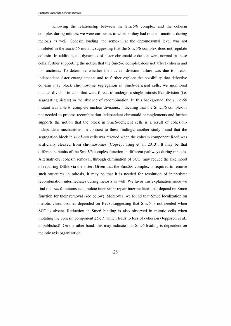
Figure 12. Possible role for Smc6 in the resolution of recombination intermediates.
Proteins that shape chromosomes
30
JMs could be resolved despite higher and altered levels of JM species during formation.
In line with the notion that JMs are the culprits of the segregation block, these cells
completed nuclear divisions. These data suggest that the Smc5/6 complex is responsible
for IS-JM resolution and is important in resolving a subset of non-CO forming JMs
(Figure 12) and leads us to propose that the Smc5/6 complex is pivotal in resolving
recombination intermediates that form outside of canonical meiotic pathways.
CTCF (Paper III)
Genome organization in eukaryotes requires extensive packaging of the
chromatin into the fixed, confined space of the nucleus. It has been proposed that
specific factors exist that ensure proper chromosome architecture and function by
mediating intra- and inter-chromosomal contacts. One such candidate is the essential
CCCTC-binding factor (CTCF) found in vertebrates. CTCF is a huge protein that
contains a conserved DNA-binding motif consisting of eleven zinc finger domains
(Klenova, Nicolas et al. 1993), which make it possible for CTCF to bind multiple DNA
regions at the same time. CTCF was initially characterized as a transcription factor
capable of both activating and repressing gene expression in heterologous reporter
assays (Baniahmad, Steiner et al. 1990, Lobanenkov, Nicolas et al. 1990, Filippova,
Fagerlie et al. 1996). Using transgenic assays, CTCF was later found to be the major
DNA-binding component that establishes insulators, meaning that it buffers regions of
defined gene expression and/or chromatin state (Bell, West et al. 1999). The insulator
properties exhibited by CTCF were thought to be a result of its ability to modify
chromosome structure. This idea is supported by the finding that many CTCF sites
overlap with cohesin (Kim, Abdullaev et al. 2007), which, in addition to its roles in
sister chromatid cohesion, regulates gene expression (reviewed in Merkenschlager and
Odom 2013). Not long after the discovery of CTCF-cohesin binding sites, it was shown
that both CTCF and cohesin are required for long-range genome interactions (Bowers,
Mirabella et al. 2009, Hadjur, Williams et al. 2009), and recent evidence has further
implicated the coordinated roles of CTCF and cohesin in setting up proper chromosome
Proteins that shape chromosomes
31
structure (reviewed in Merkenschlager and Odom 2013). In addition, the recent ability
to study the three-dimensional structure of genomes has generated more evidence
supporting the notion that CTCF is a key architectural protein (reviewed in Ong and
Corces 2014).
Although the majority of investigations into CTCF’s function have focused on
vegetative cells, studies suggest that it works during gametogenesis as well. In females,
CTCF is required for proper meiosis and oocyte growth, and fertilized CTCF-deficient
oocytes lead to offspring with defects in early embryonic development (Fedoriw, Stein
et al. 2004, Wan, Pan et al. 2008). CTCF is expressed in all cell types of the
seminiferous epithelium in the testes, suggesting that it may also play a role during
spermatogenesis (Sleutels, Soochit et al. 2012) Though these studies hint to a role for
CTCF in these processes, the function of CTCF during gametogenesis has been difficult
to study due to the lack of a viable knockout mouse model.
In Paper III, we created a testes-specific ctcf knockout mouse strain (ctcf-cKO)
to elucidate CTCF function during spermatogenesis. Excision of ctcf in the ctcf-cKO
was germ-line specific and occurred from pre-leptotene cells and onward, and mice
exhibited seminiferous tubule atrophy along with low sperm counts and infertility. To
explore the reasons behind the low sperm counts, we analyzed CTCF function in
meiosis.
Early in meiosis, γH2AX marks DSB sites. It is removed from autosomal
chromosome axes concurrent with DSB repair, but a subset remains associated with the
XY body, which is silenced during meiosis due to the inability for the non-homologous
X- and Y-chromosomes to synapse (Hunter, Borner et al. 2001, Fernandez-Capetillo,
Mahadevaiah et al. 2003, Inselman, Eaker et al. 2003). CTCF bound to meiotic
chromosomes with a strong preference for the XY body, suggesting that it may play a
role in meiosis, particularly at the XY body. However, ctcf-cKO mice had no major
defects in cohesin loading, DSB formation, SC dynamics, chromosome pairing, XY
body formation or recombination, suggesting a non-essential role for CTCF during
Proteins that shape chromosomes
32
meiosis. Because the ctcf-cKO did not fully excise Ctcf, we cannot rule out the
possibility that CTCF plays a role during meiosis. However, despite a normal meiosis,
Ctcf-deficient mice had low sperm counts and were infertile, implying a role for CTCF
during post-meiotic stages of spermatogenesis.
In line with this notion, elongating spermatids were apoptotic in the ctcf-cKO
and mature ctcf-cKO sperm showed structural aberrations. Stage 8-10 elongating
spermatids lacked a manchette, while stage 12-14 spermatids had an abnormal
manchette. Later-stage ctcf-cKO sperm exhibited abnormal head structure and
inconsistent DNA compaction, both of which may be related to the manchette
abnormalities in earlier stages. Mature sperm from ctcf-cKO mice had less-elongated
heads with visible regions of decondensed chromatin, suggesting a role for CTCF in
chromatin compaction. Tail morphology was also irregular in the ctcf-cKO, most likely
due to abnormal head shape. Consistent with a role for CTCF in chromatin compaction
during spermiogenesis, CTCF-deficient mice were unable to incorporate the sperm-
specific compaction factor protamine 1 (PRM1), which disrupts the protamine balance
known to be crucial for sperm compaction (Cho, Willis et al. 2001, Cho, Jung-Ha et al.
2003). Thus, infertility in ctcf-cKO mice is likely caused by chromatin compaction
defects in sperm, which result from improper protamine incorporation during
spermiogenesis.
FINDING NOVEL MEIOTIC FACTORS
In 1859, Charles Darwin published his revolutionary work On the Origin of
Species by Means of Natural Selection, which laid the foundation of evolutionary
biology. Though controversial when first presented, researchers have transformed
Darwin’s theory of evolution into a fundamental biological dogma. Now the pressing
question for many biologists is not if or why evolution happened, but rather what can we

Finding novel meiotic factors
33
gain from it. Namely, how can we utilize evolutionary relationships to gain insights into
human health and disease mechanisms?
One way to extract information from evolution is by studying co-evolution,
which is a pivotal aspect of evolutionary theory. Co-evolution is defined as the
interdependence between the evolutionary changes of two interacting entities (Ehrlich
and Raven 1964). Co-evolution was originally studied at the species level, but recent
advances in technology and genome sequence availability have made it possible to
examine co-evolution at the molecular level as well (reviewed in de Juan, Pazos et al.
2013). Of most interest in modern studies is protein co-evolution, which detects the
relationship between interacting and functionally related proteins.
The co-evolution search at the protein level often makes use of phylogenetic
trees, which are visual representations of the evolutionary relationships between a set of
biological factors (in this case, proteins) (reviewed in Ochoa and Pazos 2014). It has
been shown that structurally and functionally interacting proteins have similar
phylogenetic tree patterns. That observation has led to the development of methods that
use sequence input to predict protein interactions (structural and functional) based on
phylogenetic tree similarity (Ochoa and Pazos 2014). Phylogenetic profiling, however,
predicts protein interactions based on the joint presence or absence of proteins across
species. This method is based on the notion that co-evolving populations will lose or
maintain biological entities, such as proteins, as evolution, via natural selection, in that
environment deems necessary (Pellegrini, Marcotte et al. 1999). Since proteins in the
same phylogenetic cluster are related based on functional evolutionary conservation or
divergence, rather than sequence homology, proteins that lack sequence conservation
but function in the same pathway can be discovered. This feature of phylogenetic
profiling make it of particular use in studying new potential meiosis-specific proteins,
which are conserved on a functional but not sequence level, making them difficult to
study using other established screens (reviewed in Bolcun-Filas and Schimenti 2012).
Finding novel meiotic factors
34
Since proteins that make up specialized complexes are expected to have a
similar phylogenetic profile, it may be possible to use phylogenetic profiling to predict
novel protein components of such complexes. In Paper IV (Tabach, Golan et al. 2013),
we used phylogenetic profiling to predict protein members of the meiosis-specific
synaptonemal complex (SC). As predicted, known proteins of the SC clustered strongly
with other meiosis-specific proteins, demonstrating the ability of phylogenetic profiling
to cluster proteins that work in the same complex. The SC proteins also shared the
phylogenetic profile with other uncharacterized proteins, many of which contained a
predicted coiled-coil structure that is characteristic of many SC proteins (Costa and
Cooke 2007), suggesting new components of the SC. To test if these uncharacterized
proteins were meiosis-specific, as would be expected for SC proteins, we analyzed the
mRNA expression patterns in various mouse tissues. Several were highly expressed in
the ovary and/or testes. One gene, Ccdc105 (coiled-coiled domain containing gene 105),
encoded a protein most similar phylogenetic profile to SC proteins and was expressed in
testes only. Using an antibody raised against CCDC105, we found that CCDC105
resides in the nuclear fraction in testes by western blotting. CCDC105 localized to the
heterochromatic centromeres in in spermatocytes in late diplotene, and was also found in
heterochromatic regions in post-meiotic round spermatids. These data suggest that
CCDC105 may play a role in late stages of meiosis and spermiogenesis. Together, the
results in Paper IV indicate that phylogenetic profiling can be used to find new genes
that impact meiosis. Since meiosis is key for gametogenesis and reproduction, finding
new meiotic factors can help researchers find ways to treat and perhaps prevent
infertility.
Conclusions
35
CONCLUSIONS
The goal of this thesis was to explore the relationship between proteins that
dictate chromosome structure and their function in meiosis.
In Paper I, we showed that the Smc5/6 complex is imperative for proper
meiotic recombination in budding yeast. Using meiosis-specific alleles, we found that in
the absence of Smc5/6 complex components, cells were unable to divide their nuclei due
to the accumulation of recombination intermediates. Many of these intermediates were
formed between sister chromatids, a situation very rarely observed in wild-type cells
most likely due to the transiency of meiotic inter-sister repair. This suggests that Smc6,
and the complex as a whole, facilitates proper recombination by promoting the
prevention and/or resolution of inter-sister repair intermediates. In support of the latter
notion, all recombination intermediates were resolved and nuclear divisions were
restored when Smc6 function was turned on at the resolution step of recombination.
Also in support of a sister-specific function for Smc6 during recombination were the
normal levels of crossover formation in the smc6 mutant, despite accumulating
recombination structures. We therefore propose that the main function of the Smc5/6
complex during meiosis is to aid in the resolution of inter-sister recombination
intermediates.
In Paper II, we utilized meiosis-specific mutants for topoisomerases Top2 and
Top3 and found that both are necessary for proper meiotic segregation in budding yeast.
In the absence of Top2, cells experienced a partially recombination-dependent
segregation block. This block was not due to the accumulation of recombination
intermediates as the timing of intermediate formation and removal, as well as crossover
and non-crossover formation, was normal. Top2-deficient cells were not able to
segregate their sisters when undergoing a meiosis II-like division in the absence of
recombination, suggesting that it is recombination-independent structures which block
top2 cells. Based on these data, we conclude that Top2 is needed for the segregation of
Conclusions
36
recombined chromosomes during meiosis due to its ability to resolve sister chromatid
intertwinings. The segregation block in the top3 mutant was fully recombination-
dependent. Given its role in removal of recombination intermediates in mitosis, we
suggest that it works during meiosis in the processing of recombination structures as
well.
We defined a role for the CCCTC-binding factor CTCF during spermatogenesis
in mice in Paper III by creating a testes-specific knockout mouse for Ctcf (ctcf-cKO).
The ctcf-cKO was able to complete meiosis normally, but had a very low sperm count
and formed aberrant sperm. On closer examination, sperm from ctcf-cKO mice did not
compact chromatin correctly, leading to head abnormalities and aberrant tail attachment,
which most likely contributed to the infertility in these mice. In line with the lack of
chromatin compaction, CTCF-deficient sperm were not able to load sperm-specific
chromatin compaction factors, suggesting a role for CTCF in their recruitment and/or
loading. Together, these results implicate CTCF as an important factor for proper
chromatin compaction during spermiogenesis.
Finally, in Paper IV, we used phylogenetic profiling to discover a meiosis-
specific, previously uncharacterized protein called CCDC105. CCDC105 showed an
evolutionary clustering with known components of the synaptonemal complex,
suggesting that it plays a role in organizing chromosome structure during meiosis. In
line with this, we found that CCDC105 localized to heterochromatic regions on meiotic
chromosomes in late prophase. These findings indicate that phylogenetic profiling can
be used to find novel factors in pathways known to involve specific protein complexes.
In conclusion, this thesis focused on factors that uphold the integrity of gamete
formation. While CTCF seems to be important for differentiation of male gametes, and
the role of CCDC105 is uncharacterized as of yet, the Smc5/6 complex, Top2, and Top3
are imperative for proper meiotic segregation. Mistakes in chromosome segregation can
lead to aneuploidy, which is a hallmark of tumor cells. When aneuploidy occurs in the
germ line, it can lead to genetic disorders such as trisomy of chromosome 21, more

Conclusions
37
commonly known as Down’s syndrome. Moreover, aneuploidy is the leading cause of
infertility and spontaneous abortions in humans. Due to the increasing use of in vitro
fertilization, researchers have been able to study aneuploidy in the germ line and its
embryonic consequences more closely. This has led to a striking observation: as
maternal age increases, so does aneuploidy and, in turn, maternally-contributed
infertility and trisomy. The exact reason for this correlation is not yet clear, but it has
been suggested that the integrity of meiotic chromosomal features, such as cohesin and
chiasmata, are key predictors of aneuploidy. Since female gametes are arrested at
meiosis I at birth, it may be that these factors degenerate with time. Understanding more
about how chromosome structure impacts meiotic integrity is the key to treating, and
perhaps preventing, infertility and genetic disorders in the future.
References
38
REFERENCES
Agarwal, S. and G. S. Roeder (2000). "Zip3 provides a link between recombination enzymes and synaptonemal complex proteins." Cell 102(2): 245-255.
Allers, T. and M. Lichten (2001). "Differential timing and control of noncrossover and crossover recombination during meiosis." Cell 106(1): 47-57.
Anderson, E. L., A. E. Baltus, H. L. Roepers-Gajadien, T. J. Hassold, D. G. de Rooij, A. M. van Pelt and D. C. Page (2008). "Stra8 and its inducer, retinoic acid, regulate meiotic initiation in both spermatogenesis and oogenesis in mice." Proc Natl Acad Sci U S A 105(39): 14976-14980.
Andrews, E. A., J. Palecek, J. Sergeant, E. Taylor, A. R. Lehmann and F. Z. Watts (2005). "Nse2, a component of the Smc5-6 complex, is a SUMO ligase required for the response to DNA damage." Mol Cell Biol 25(1): 185-196.
Baniahmad, A., C. Steiner, A. C. Kohne and R. Renkawitz (1990). "Modular structure of a chicken lysozyme silencer: involvement of an unusual thyroid hormone receptor binding site." Cell 61(3): 505-514.
Bannister, L. A., L. G. Reinholdt, R. J. Munroe and J. C. Schimenti (2004). "Positional cloning and characterization of mouse mei8, a disrupted allelle of the meiotic cohesin Rec8." Genesis 40(3): 184-194.
Barlow, A. L., F. E. Benson, S. C. West and M. A. Hulten (1997). "Distribution of the Rad51 recombinase in human and mouse spermatocytes." EMBO J 16(17): 5207-5215.
Baudat, F. and S. Keeney (2001). "Meiotic recombination: Making and breaking go hand in hand." Curr Biol 11(2): R45-48.
Baudat, F., K. Manova, J. P. Yuen, M. Jasin and S. Keeney (2000). "Chromosome synapsis defects and sexually dimorphic meiotic progression in mice lacking Spo11." Mol Cell 6(5): 989-998.
Baxter, J. and J. F. Diffley (2008). "Topoisomerase II inactivation prevents the completion of DNA replication in budding yeast." Mol Cell 30(6): 790-802.

References
39
Bell, A. C., A. G. West and G. Felsenfeld (1999). "The protein CTCF is required for the enhancer blocking activity of vertebrate insulators." Cell 98(3): 387-396.
Bergerat, A., B. de Massy, D. Gadelle, P. C. Varoutas, A. Nicolas and P. Forterre (1997). "An atypical topoisomerase II from Archaea with implications for meiotic recombination." Nature 386(6623): 414-417.
Bermejo, R., Y. Doksani, T. Capra, Y. M. Katou, H. Tanaka, K. Shirahige and M. Foiani (2007). "Top1- and Top2-mediated topological transitions at replication forks ensure fork progression and stability and prevent DNA damage checkpoint activation." Genes Dev 21(15): 1921-1936.
Bickel, J. S., L. Chen, J. Hayward, S. L. Yeap, A. E. Alkers and R. C. Chan (2010). "Structural maintenance of chromosomes (SMC) proteins promote homolog-independent recombination repair in meiosis crucial for germ cell genomic stability." PLoS Genet 6(7): e1001028.
Bishop, D. K. (1994). "RecA homologs Dmc1 and Rad51 interact to form multiple nuclear complexes prior to meiotic chromosome synapsis." Cell 79(6): 1081-1092.
Bishop, D. K., D. Park, L. Xu and N. Kleckner (1992). "DMC1: a meiosis-specific yeast homolog of E. coli recA required for recombination, synaptonemal complex formation, and cell cycle progression." Cell 69(3): 439-456.
Bishop, D. K. and D. Zickler (2004). "Early decision; meiotic crossover interference prior to stable strand exchange and synapsis." Cell 117(1): 9-15.
Borner, G. V., N. Kleckner and N. Hunter (2004). "Crossover/noncrossover differentiation, synaptonemal complex formation, and regulatory surveillance at the leptotene/zygotene transition of meiosis." Cell 117(1): 29-45.
Bowers, S. R., F. Mirabella, F. J. Calero-Nieto, S. Valeaux, S. Hadjur, E. W. Baxter, M. Merkenschlager and P. N. Cockerill (2009). "A conserved insulator that recruits CTCF and cohesin exists between the closely related but divergently regulated interleukin-3 and granulocyte-macrophage colony-stimulating factor genes." Mol Cell Biol 29(7): 1682-1693.
References
40
Branzei, D., J. Sollier, G. Liberi, X. Zhao, D. Maeda, M. Seki, T. Enomoto, K. Ohta and M. Foiani (2006). "Ubc9- and mms21-mediated sumoylation counteracts recombinogenic events at damaged replication forks." Cell 127(3): 509-522.
Braun, R. E. (2001). "Packaging paternal chromosomes with protamine." Nat Genet 28(1): 10-12.
Bolcun-Filas, E. and J.C. Schimenti (2012). “Genetics of meiosis and recombination in mice.” Int Rev Cell Mol Biol 298: 179-227.
Canudas, S. and S. Smith (2009). "Differential regulation of telomere and centromere cohesion by the Scc3 homologues SA1 and SA2, respectively, in human cells." J Cell Biol 187(2): 165-173.
Carballo, J. A., A. L. Johnson, S. G. Sedgwick and R. S. Cha (2008). "Phosphorylation of the axial element protein Hop1 by Mec1/Tel1 ensures meiotic interhomolog recombination." Cell 132(5): 758-770.
Carrell, D. T., B. R. Emery and S. Hammoud (2007). "Altered protamine expression and diminished spermatogenesis: what is the link?" Hum Reprod Update 13(3): 313-327.
Cejka, P., J. L. Plank, C. C. Dombrowski and S. C. Kowalczykowski (2012). "Decatenation of DNA by the S. cerevisiae Sgs1-Top3-Rmi1 and RPA complex: a mechanism for disentangling chromosomes." Mol Cell 47(6): 886-896.
Chavez, A., V. George, V. Agrawal and F. B. Johnson (2010). "Sumoylation and the structural maintenance of chromosomes (Smc) 5/6 complex slow senescence through recombination intermediate resolution." J Biol Chem 285(16): 11922-11930.
Chen, S. H., N. L. Chan and T. S. Hsieh (2013). "New mechanistic and functional insights into DNA topoisomerases." Annu Rev Biochem 82: 139-170.
Chen, Y. H., K. Choi, B. Szakal, J. Arenz, X. Duan, H. Ye, D. Branzei and X. Zhao (2009). "Interplay between the Smc5/6 complex and the Mph1 helicase in recombinational repair." Proc Natl Acad Sci U S A 106(50): 21252-21257.
References
41
Cho, C., H. Jung-Ha, W. D. Willis, E. H. Goulding, P. Stein, Z. Xu, R. M. Schultz, N. B. Hecht and E. M. Eddy (2003). "Protamine 2 deficiency leads to sperm DNA damage and embryo death in mice." Biol Reprod 69(1): 211-217.
Cho, C., W. D. Willis, E. H. Goulding, H. Jung-Ha, Y. C. Choi, N. B. Hecht and E. M. Eddy (2001). "Haploinsufficiency of protamine-1 or -2 causes infertility in mice." Nat Genet 28(1): 82-86.
Cho, C., W. D. Willis, E. H. Goulding, H. Jung-Ha, Y. C. Choi, N. B. Hecht and E. M. Eddy (2001). "Haploinsufficiency of protamine-1 or -2 causes infertility in mice." Nature Genetics 28(1): 82-86.
Copsey, A., S. Tang, P. W. Jordan, H. G. Blitzblau, S. Newcombe, A. C. Chan, L. Newnham, Z. Li, S. Gray, A. D. Herbert, P. Arumugam, A. Hochwagen, N. Hunter and E. Hoffmann (2013). "Smc5/6 coordinates formation and resolution of joint molecules with chromosome morphology to ensure meiotic divisions." PLoS Genet 9(12): e1004071.
Cost, G. J. and N. R. Cozzarelli (2006). "Smc5p promotes faithful chromosome transmission and DNA repair in Saccharomyces cerevisiae." Genetics 172(4): 2185-2200.
Costa, Y. and H. J. Cooke (2007). "Dissecting the mammalian synaptonemal complex using targeted mutations." Chromosome Res 15(5): 579-589.
Dadoune, J. P. (2003). "Expression of mammalian spermatozoal nucleoproteins." Microsc Res Tech 61(1): 56-75.
Dahmann, C. and B. Futcher (1995). "Specialization of B-type cyclins for mitosis or meiosis in S. cerevisiae." Genetics 140(3): 957-963.
de Boer, E., A. J. Dietrich, C. Hoog, P. Stam and C. Heyting (2007). "Meiotic interference among MLH1 foci requires neither an intact axial element structure nor full synapsis." J Cell Sci 120(Pt 5): 731-736.
de Juan, D., F. Pazos and A. Valencia (2013). "Emerging methods in protein co-evolution." Nat Rev Genet 14(4): 249-261.
References
42
de los Santos, T., N. Hunter, C. Lee, B. Larkin, J. Loidl and N. M. Hollingsworth (2003). "The Mus81/Mms4 endonuclease acts independently of double-Holliday junction resolution to promote a distinct subset of crossovers during meiosis in budding yeast." Genetics 164(1): 81-94.
De Muyt, A., L. Jessop, E. Kolar, A. Sourirajan, J. Chen, Y. Dayani and M. Lichten (2012). "BLM helicase ortholog Sgs1 is a central regulator of meiotic recombination intermediate metabolism." Mol Cell 46(1): 43-53.
De Piccoli, G., F. Cortes-Ledesma, G. Ira, J. Torres-Rosell, S. Uhle, S. Farmer, J. Y. Hwang, F. Machin, A. Ceschia, A. McAleenan, V. Cordon-Preciado, A. Clemente-Blanco, F. Vilella-Mitjana, P. Ullal, A. Jarmuz, B. Leitao, D. Bressan, F. Dotiwala, A. Papusha, X. Zhao, K. Myung, J. E. Haber, A. Aguilera and L. Aragon (2006). "Smc5-Smc6 mediate DNA double-strand-break repair by promoting sister-chromatid recombination." Nat Cell Biol 8(9): 1032-1034.
de Vries, S. S., E. B. Baart, M. Dekker, A. Siezen, D. G. de Rooij, P. de Boer and H. te Riele (1999). "Mouse MutS-like protein Msh5 is required for proper chromosome synapsis in male and female meiosis." Genes Dev 13(5): 523-531.
Edelmann, W., P. E. Cohen, B. Kneitz, N. Winand, M. Lia, J. Heyer, R. Kolodner, J. W. Pollard and R. Kucherlapati (1999). "Mammalian MutS homologue 5 is required for chromosome pairing in meiosis." Nat Genet 21(1): 123-127.
Ehrlich, P. R. and P. H. Raven (1964). "Butterflies and plants: a study in coevolution." Evolution 18: 586-608.
Farcas, A. M., P. Uluocak, W. Helmhart and K. Nasmyth (2011). "Cohesin's concatenation of sister DNAs maintains their intertwining." Mol Cell 44(1): 97-107.
Farmer, S., P. A. San-Segundo and L. Aragon (2011). "The Smc5-Smc6 complex is required to remove chromosome junctions in meiosis." PLoS One 6(6): e20948.
Fedoriw, A. M., P. Stein, P. Svoboda, R. M. Schultz and M. S. Bartolomei (2004). "Transgenic RNAi reveals essential function for CTCF in H19 gene imprinting." Science 303(5655): 238-240.
References
43
Fernandez-Capetillo, O., S. K. Mahadevaiah, A. Celeste, P. J. Romanienko, R. D. Camerini-Otero, W. M. Bonner, K. Manova, P. Burgoyne and A. Nussenzweig (2003). "H2AX is required for chromatin remodeling and inactivation of sex chromosomes in male mouse meiosis." Dev Cell 4(4): 497-508.
Filippova, G. N., S. Fagerlie, E. M. Klenova, C. Myers, Y. Dehner, G. Goodwin, P. E. Neiman, S. J. Collins and V. V. Lobanenkov (1996). "An exceptionally conserved transcriptional repressor, CTCF, employs different combinations of zinc fingers to bind diverged promoter sequences of avian and mammalian c-myc oncogenes." Mol Cell Biol 16(6): 2802-2813.
Fousteri, M. I. and A. R. Lehmann (2000). "A novel SMC protein complex in Schizosaccharomyces pombe contains the Rad18 DNA repair protein." EMBO J 19(7): 1691-1702.
Fraune, J., S. Schramm, M. Alsheimer and R. Benavente (2012). "The mammalian synaptonemal complex: protein components, assembly and role in meiotic recombination." Exp Cell Res 318(12): 1340-1346.
Fukuda, T., N. Fukuda, A. Agostinho, A. Hernandez-Hernandez, A. Kouznetsova and C. Hoog (2014). "STAG3-mediated stabilization of REC8 cohesin complexes promotes chromosome synapsis during meiosis." EMBO J 33(11): 1243-1255.
Gangloff, S., B. de Massy, L. Arthur, R. Rothstein and F. Fabre (1999). "The essential role of yeast topoisomerase III in meiosis depends on recombination." EMBO J 18(6): 1701-1711.
Goldfarb, T. and M. Lichten (2010). "Frequent and efficient use of the sister chromatid for DNA double-strand break repair during budding yeast meiosis." PLoS Biol 8(10): e1000520.
Grandin, N. and S. I. Reed (1993). "Differential function and expression of Saccharomyces cerevisiae B-type cyclins in mitosis and meiosis." Mol Cell Biol 13(4): 2113-2125.
Gruber, S., C. H. Haering and K. Nasmyth (2003). "Chromosomal cohesin forms a ring." Cell 112(6): 765-777.
References
44
Guacci, V., D. Koshland and A. Strunnikov (1997). "A direct link between sister chromatid cohesion and chromosome condensation revealed through the analysis of MCD1 in S. cerevisiae." Cell 91(1): 47-57.
Hadjur, S., L. M. Williams, N. K. Ryan, B. S. Cobb, T. Sexton, P. Fraser, A. G. Fisher and M. Merkenschlager (2009). "Cohesins form chromosomal cis-interactions at the developmentally regulated IFNG locus." Nature 460(7253): 410-413.
Haering, C. H., A. M. Farcas, P. Arumugam, J. Metson and K. Nasmyth (2008). "The cohesin ring concatenates sister DNA molecules." Nature 454(7202): 297-301.
Haering, C. H., J. Lowe, A. Hochwagen and K. Nasmyth (2002). "Molecular architecture of SMC proteins and the yeast cohesin complex." Mol Cell 9(4): 773-788.
Haering, C. H., D. Schoffnegger, T. Nishino, W. Helmhart, K. Nasmyth and J. Lowe (2004). "Structure and stability of cohesin's Smc1-kleisin interaction." Mol Cell 15(6): 951-964.
Handel, M. A. and J. C. Schimenti (2010). "Genetics of mammalian meiosis: regulation, dynamics and impact on fertility." Nat Rev Genet 11(2): 124-136.
Hess, R. A. and L. Renato de Franca (2008). "Spermatogenesis and cycle of the seminiferous epithelium." Adv Exp Med Biol 636: 1-15.
Hirano, T. (2006). "At the heart of the chromosome: SMC proteins in action." Nat Rev Mol Cell Biol 7(5): 311-322.
Hollingsworth, N. M. and L. Ponte (1997). "Genetic interactions between HOP1, RED1 and MEK1 suggest that MEK1 regulates assembly of axial element components during meiosis in the yeast Saccharomyces cerevisiae." Genetics 147(1): 33-42.
Hollingsworth, N. M., L. Ponte and C. Halsey (1995). "MSH5, a novel MutS homolog, facilitates meiotic reciprocal recombination between homologs in Saccharomyces cerevisiae but not mismatch repair." Genes Dev 9(14): 1728-1739.
Holloway, J. K., J. Booth, W. Edelmann, C. H. McGowan and P. E. Cohen (2008). "MUS81 generates a subset of MLH1-MLH3-independent crossovers in mammalian meiosis." PLoS Genet 4(9): e1000186.
References
45
Holm, C., T. Goto, J. C. Wang and D. Botstein (1985). "DNA topoisomerase II is required at the time of mitosis in yeast." Cell 41(2): 553-563.
Hopkins, J., G. Hwang, J. Jacob, N. Sapp, R. Bedigian, K. Oka, P. Overbeek, S. Murray and P. W. Jordan (2014). "Meiosis-Specific Cohesin Component, Stag3 Is Essential for Maintaining Centromere Chromatid Cohesion, and Required for DNA Repair and Synapsis between Homologous Chromosomes." PLoS Genet 10(7): e1004413.
Hunter, N., G. V. Borner, M. Lichten and N. Kleckner (2001). "Gamma-H2AX illuminates meiosis." Nat Genet 27(3): 236-238.
Hunter, N. and N. Kleckner (2001). "The single-end invasion: an asymmetric intermediate at the double-strand break to double-holliday junction transition of meiotic recombination." Cell 106(1): 59-70.
Hwang, J. Y., S. Smith, A. Ceschia, J. Torres-Rosell, L. Aragon and K. Myung (2008). "Smc5-Smc6 complex suppresses gross chromosomal rearrangements mediated by break-induced replications." DNA Repair (Amst) 7(9): 1426-1436.
Inselman, A., S. Eaker and M. A. Handel (2003). "Temporal expression of cell cycle-related proteins during spermatogenesis: establishing a timeline for onset of the meiotic divisions." Cytogenet Genome Res 103(3-4): 277-284.
Ishiguro, K., J. Kim, S. Fujiyama-Nakamura, S. Kato and Y. Watanabe (2011). "A new meiosis-specific cohesin complex implicated in the cohesin code for homologous pairing." EMBO Rep 12(3): 267-275.
Ishiguro, K., J. Kim, H. Shibuya, A. Hernandez-Hernandez, A. Suzuki, T. Fukagawa, G. Shioi, H. Kiyonari, X. C. Li, J. Schimenti, C. Hoog and Y. Watanabe (2014). "Meiosis-specific cohesin mediates homolog recognition in mouse spermatocytes." Genes Dev 28(6): 594-607.
Jessberger, R. (2011). "Cohesin complexes get more complex: the novel kleisin RAD21L." Cell Cycle 10(13): 2053-2054.
References
46
Jessop, L. and M. Lichten (2008). "Mus81/Mms4 endonuclease and Sgs1 helicase collaborate to ensure proper recombination intermediate metabolism during meiosis." Mol Cell 31(3): 313-323.
Kassir, Y., D. Granot and G. Simchen (1988). "IME1, a positive regulator gene of meiosis in S. cerevisiae." Cell 52(6): 853-862.
Kaur, G., L. A. Thompson and J. M. Dufour (2014). "Sertoli cells--immunological sentinels of spermatogenesis." Semin Cell Dev Biol 30: 36-44.
Keeney, S. (2001). "Mechanism and control of meiotic recombination initiation." Curr Top Dev Biol 52: 1-53.
Keeney, S. (2008). "Spo11 and the Formation of DNA Double-Strand Breaks in Meiosis." Genome Dyn Stab 2: 81-123.
Keeney, S., C. N. Giroux and N. Kleckner (1997). "Meiosis-specific DNA double-strand breaks are catalyzed by Spo11, a member of a widely conserved protein family." Cell 88(3): 375-384.
Kegel, A., H. Betts-Lindroos, T. Kanno, K. Jeppsson, L. Strom, Y. Katou, T. Itoh, K. Shirahige and C. Sjogren (2011). "Chromosome length influences replication-induced topological stress." Nature 471(7338): 392-396.
Kim, K. P., B. M. Weiner, L. Zhang, A. Jordan, J. Dekker and N. Kleckner (2010). "Sister cohesion and structural axis components mediate homolog bias of meiotic recombination." Cell 143(6): 924-937.
Kim, T. H., Z. K. Abdullaev, A. D. Smith, K. A. Ching, D. I. Loukinov, R. D. Green, M. Q. Zhang, V. V. Lobanenkov and B. Ren (2007). "Analysis of the vertebrate insulator protein CTCF-binding sites in the human genome." Cell 128(6): 1231-1245.
Klein, F., T. Laroche, M. E. Cardenas, J. F. Hofmann, D. Schweizer and S. M. Gasser (1992). "Localization of RAP1 and topoisomerase II in nuclei and meiotic chromosomes of yeast." J Cell Biol 117(5): 935-948.
References
47
Klein, F., P. Mahr, M. Galova, S. B. Buonomo, C. Michaelis, K. Nairz and K. Nasmyth (1999). "A central role for cohesins in sister chromatid cohesion, formation of axial elements, and recombination during yeast meiosis." Cell 98(1): 91-103.
Klenova, E. M., R. H. Nicolas, H. F. Paterson, A. F. Carne, C. M. Heath, G. H. Goodwin, P. E. Neiman and V. V. Lobanenkov (1993). "CTCF, a conserved nuclear factor required for optimal transcriptional activity of the chicken c-myc gene, is an 11-Zn-finger protein differentially expressed in multiple forms." Mol Cell Biol 13(12): 7612-7624.
Koshland, D. and A. Strunnikov (1996). "Mitotic chromosome condensation." Annu Rev Cell Dev Biol 12: 305-333.
Kugou, K., T. Fukuda, S. Yamada, M. Ito, H. Sasanuma, S. Mori, Y. Katou, T. Itoh, K. Matsumoto, T. Shibata, K. Shirahige and K. Ohta (2009). "Rec8 guides canonical Spo11 distribution along yeast meiotic chromosomes." Mol Biol Cell 20(13): 3064-3076.
Lee, B. H. and A. Amon (2003). "Role of Polo-like kinase CDC5 in programming meiosis I chromosome segregation." Science 300(5618): 482-486.
Lee, J. and T. Hirano (2011). "RAD21L, a novel cohesin subunit implicated in linking homologous chromosomes in mammalian meiosis." J Cell Biol 192(2): 263-276.
Lehmann, A. R., M. Walicka, D. J. Griffiths, J. M. Murray, F. Z. Watts, S. McCready and A. M. Carr (1995). "The rad18 gene of Schizosaccharomyces pombe defines a new subgroup of the SMC superfamily involved in DNA repair." Mol Cell Biol 15(12): 7067-7080.
Lilienthal, I., T. Kanno and C. Sjogren (2013). "Inhibition of the Smc5/6 complex during meiosis perturbs joint molecule formation and resolution without significantly changing crossover or non-crossover levels." PLoS Genet 9(11): e1003898.
Lindroos, H. B., L. Strom, T. Itoh, Y. Katou, K. Shirahige and C. Sjogren (2006). "Chromosomal association of the Smc5/6 complex reveals that it functions in differently regulated pathways." Mol Cell 22(6): 755-767.
References
48
Llano, E., Y. Herran, I. Garcia-Tunon, C. Gutierrez-Caballero, E. de Alava, J. L. Barbero, J. Schimenti, D. G. de Rooij, M. Sanchez-Martin and A. M. Pendas (2012). "Meiotic cohesin complexes are essential for the formation of the axial element in mice." J Cell Biol 197(7): 877-885.
Lobanenkov, V. V., R. H. Nicolas, V. V. Adler, H. Paterson, E. M. Klenova, A. V. Polotskaja and G. H. Goodwin (1990). "A novel sequence-specific DNA binding protein which interacts with three regularly spaced direct repeats of the CCCTC-motif in the 5'-flanking sequence of the chicken c-myc gene." Oncogene 5(12): 1743-1753.
Losada, A. and T. Hirano (2005). "Dynamic molecular linkers of the genome: the first decade of SMC proteins." Genes Dev 19(11): 1269-1287.
Lynn, A., R. Soucek and G. V. Borner (2007). "ZMM proteins during meiosis: crossover artists at work." Chromosome Res 15(5): 591-605.
Mandel, S., K. Robzyk and Y. Kassir (1994). "IME1 gene encodes a transcription factor which is required to induce meiosis in Saccharomyces cerevisiae." Dev Genet 15(2): 139-147.
Marston, A. L. (2014). "Chromosome segregation in budding yeast: sister chromatid cohesion and related mechanisms." Genetics 196(1): 31-63.
McNicoll, F., M. Stevense and R. Jessberger (2013). "Cohesin in gametogenesis." Curr Top Dev Biol 102: 1-34.
Merkenschlager, M. and D. T. Odom (2013). "CTCF and cohesin: linking gene regulatory elements with their targets." Cell 152(6): 1285-1297.
Michaelis, C., R. Ciosk and K. Nasmyth (1997). "Cohesins: chromosomal proteins that prevent premature separation of sister chromatids." Cell 91(1): 35-45.
Mimitou, E. P. and L. S. Symington (2008). "Sae2, Exo1 and Sgs1 collaborate in DNA double-strand break processing." Nature 455(7214): 770-774.
Morikawa, H., T. Morishita, S. Kawane, H. Iwasaki, A. M. Carr and H. Shinagawa (2004). "Rad62 protein functionally and physically associates with the smc5/smc6
References
49
protein complex and is required for chromosome integrity and recombination repair in fission yeast." Mol Cell Biol 24(21): 9401-9413.
Neiman, A. M. (2011). "Sporulation in the budding yeast Saccharomyces cerevisiae." Genetics 189(3): 737-765.
Niu, H., L. Wan, B. Baumgartner, D. Schaefer, J. Loidl and N. M. Hollingsworth (2005). "Partner choice during meiosis is regulated by Hop1-promoted dimerization of Mek1." Mol Biol Cell 16(12): 5804-5818.
O'Donnell, L. and M. K. O'Bryan (2014). "Microtubules and spermatogenesis." Semin Cell Dev Biol 30: 45-54.
Ochoa, D. and F. Pazos (2014). "Practical aspects of protein co-evolution." Frontiers in Cell and Developmental Biology 2(14).
Oh, S. D., J. P. Lao, A. F. Taylor, G. R. Smith and N. Hunter (2008). "RecQ helicase, Sgs1, and XPF family endonuclease, Mus81-Mms4, resolve aberrant joint molecules during meiotic recombination." Mol Cell 31(3): 324-336.
Ong, C. T. and V. G. Corces (2014). "CTCF: an architectural protein bridging genome topology and function." Nat Rev Genet 15(4): 234-246.
Onoda, F., M. Takeda, M. Seki, D. Maeda, J. Tajima, A. Ui, H. Yagi and T. Enomoto (2004). "SMC6 is required for MMS-induced interchromosomal and sister chromatid recombinations in Saccharomyces cerevisiae." DNA Repair (Amst) 3(4): 429-439.
Osman, F., J. Dixon, C. L. Doe and M. C. Whitby (2003). "Generating crossovers by resolution of nicked Holliday junctions: a role for Mus81-Eme1 in meiosis." Mol Cell 12(3): 761-774.
Page, S. L. and R. S. Hawley (2004). "The genetics and molecular biology of the synaptonemal complex." Annu Rev Cell Dev Biol 20: 525-558.
Paques, F. and J. E. Haber (1999). "Multiple pathways of recombination induced by double-strand breaks in Saccharomyces cerevisiae." Microbiol Mol Biol Rev 63(2): 349-404.
References
50
Pebernard, S., W. H. McDonald, Y. Pavlova, J. R. Yates, 3rd and M. N. Boddy (2004). "Nse1, Nse2, and a novel subunit of the Smc5-Smc6 complex, Nse3, play a crucial role in meiosis." Mol Biol Cell 15(11): 4866-4876.
Pellegrini, M., E. M. Marcotte, M. J. Thompson, D. Eisenberg and T. O. Yeates (1999). "Assigning protein functions by comparative genome analysis: protein phylogenetic profiles." Proc Natl Acad Sci U S A 96(8): 4285-4288.
Pelttari, J., M. R. Hoja, L. Yuan, J. G. Liu, E. Brundell, P. Moens, S. Santucci-Darmanin, R. Jessberger, J. L. Barbero, C. Heyting and C. Hoog (2001). "A meiotic chromosomal core consisting of cohesin complex proteins recruits DNA recombination proteins and promotes synapsis in the absence of an axial element in mammalian meiotic cells." Mol Cell Biol 21(16): 5667-5677.
Pepling, M. E. (2006). "From primordial germ cell to primordial follicle: mammalian female germ cell development." Genesis 44(12): 622-632.
Peters, J. M. and T. Nishiyama (2012). "Sister chromatid cohesion." Cold Spring Harb Perspect Biol 4(11).
Polakova, S., L. Cipak and J. Gregan (2011). "RAD21L is a novel kleisin subunit of the cohesin complex." Cell Cycle 10(12): 1892-1893.
Prieto, I., J. A. Suja, N. Pezzi, L. Kremer, A. C. Martinez, J. S. Rufas and J. L. Barbero (2001). "Mammalian STAG3 is a cohesin specific to sister chromatid arms in meiosis I." Nat Cell Biol 3(8): 761-766.
Ranjha, L., R. Anand and P. Cejka (2014). "The Saccharomyces cerevisiae Mlh1-Mlh3 heterodimer is an endonuclease that preferentially binds to Holliday junctions." J Biol Chem 289(9): 5674-5686.
Revenkova, E., M. Eijpe, C. Heyting, B. Gross and R. Jessberger (2001). "Novel meiosis-specific isoform of mammalian SMC1." Mol Cell Biol 21(20): 6984-6998.
Revenkova, E., M. Eijpe, C. Heyting, C. A. Hodges, P. A. Hunt, B. Liebe, H. Scherthan and R. Jessberger (2004). "Cohesin SMC1 beta is required for meiotic chromosome
References
51
dynamics, sister chromatid cohesion and DNA recombination." Nat Cell Biol 6(6): 555-562.
Revenkova, E. and R. Jessberger (2006). "Shaping meiotic prophase chromosomes: cohesins and synaptonemal complex proteins." Chromosoma 115(3): 235-240.
Rogacheva, M. V., C. M. Manhart, C. Chen, A. Guarne, J. Surtees and E. Alani (2014). "Mlh1-Mlh3, a meiotic crossover and DNA mismatch repair factor, is a Msh2-Msh3-stimulated endonuclease." J Biol Chem 289(9): 5664-5673.
Romanienko, P. J. and R. D. Camerini-Otero (2000). "The mouse Spo11 gene is required for meiotic chromosome synapsis." Mol Cell 6(5): 975-987.
Rose, D. and C. Holm (1993). "Meiosis-specific arrest revealed in DNA topoisomerase II mutants." Mol Cell Biol 13(6): 3445-3455.
Rose, D., W. Thomas and C. Holm (1990). "Segregation of recombined chromosomes in meiosis I requires DNA topoisomerase II." Cell 60(6): 1009-1017.
Ross-Macdonald, P. and G. S. Roeder (1994). "Mutation of a meiosis-specific MutS homolog decreases crossing over but not mismatch correction." Cell 79(6): 1069-1080.
Schoeffler, A. J. and J. M. Berger (2008). "DNA topoisomerases: harnessing and constraining energy to govern chromosome topology." Q Rev Biophys 41(1): 41-101.
Schwacha, A. and N. Kleckner (1995). "Identification of double Holliday junctions as intermediates in meiotic recombination." Cell 83(5): 783-791.
Schwacha, A. and N. Kleckner (1997). "Interhomolog bias during meiotic recombination: meiotic functions promote a highly differentiated interhomolog-only pathway." Cell 90(6): 1123-1135.
Schwartz, E. K. and W. D. Heyer (2011). "Processing of joint molecule intermediates by structure-selective endonucleases during homologous recombination in eukaryotes." Chromosoma 120(2): 109-127.
References
52
Shinohara, A., S. Gasior, T. Ogawa, N. Kleckner and D. K. Bishop (1997). "Saccharomyces cerevisiae recA homologues RAD51 and DMC1 have both distinct and overlapping roles in meiotic recombination." Genes Cells 2(10): 615-629.
Shinohara, A., H. Ogawa and T. Ogawa (1992). "Rad51 protein involved in repair and recombination in S. cerevisiae is a RecA-like protein." Cell 69(3): 457-470.
Shinohara, M., S. D. Oh, N. Hunter and A. Shinohara (2008). "Crossover assurance and crossover interference are distinctly regulated by the ZMM proteins during yeast meiosis." Nat Genet 40(3): 299-309.
Simchen, G. and Y. Kassir (1989). "Genetic regulation of differentiation towards meiosis in the yeast Saccharomyces cerevisiae." Genome 31(1): 95-99.
Sjogren, C. and K. Nasmyth (2001). "Sister chromatid cohesion is required for postreplicative double-strand break repair in Saccharomyces cerevisiae." Curr Biol 11(12): 991-995.
Sleutels, F., W. Soochit, M. Bartkuhn, H. Heath, S. Dienstbach, P. Bergmaier, V. Franke, M. Rosa-Garrido, S. van de Nobelen, L. Caesar, M. van der Reijden, J. C. Bryne, W. van Ijcken, J. A. Grootegoed, M. D. Delgado, B. Lenhard, R. Renkawitz, F. Grosveld and N. Galjart (2012). "The male germ cell gene regulator CTCFL is functionally different from CTCF and binds CTCF-like consensus sites in a nucleosome composition-dependent manner." Epigenetics Chromatin 5(1): 8.
Sollier, J., R. Driscoll, F. Castellucci, M. Foiani, S. P. Jackson and D. Branzei (2009). "The Saccharomyces cerevisiae Esc2 and Smc5-6 proteins promote sister chromatid junction-mediated intra-S repair." Mol Biol Cell 20(6): 1671-1682.
Storlazzi, A., L. Xu, L. Cao and N. Kleckner (1995). "Crossover and noncrossover recombination during meiosis: timing and pathway relationships." Proc Natl Acad Sci U S A 92(18): 8512-8516.
Strom, L., C. Karlsson, H. B. Lindroos, S. Wedahl, Y. Katou, K. Shirahige and C. Sjogren (2007). "Postreplicative formation of cohesion is required for repair and induced by a single DNA break." Science 317(5835): 242-245.
References
53
Strom, L., H. B. Lindroos, K. Shirahige and C. Sjogren (2004). "Postreplicative recruitment of cohesin to double-strand breaks is required for DNA repair." Mol Cell 16(6): 1003-1015.
Tabach, Y., T. Golan, A. Hernandez-Hernandez, A. R. Messer, T. Fukuda, A. Kouznetsova, J. G. Liu, I. Lilienthal, C. Levy and G. Ruvkun (2013). "Human disease locus discovery and mapping to molecular pathways through phylogenetic profiling." Mol Syst Biol 9: 692.
Tarsounas, M., T. Morita, R. E. Pearlman and P. B. Moens (1999). "RAD51 and DMC1 form mixed complexes associated with mouse meiotic chromosome cores and synaptonemal complexes." J Cell Biol 147(2): 207-220.
Wan, L. B., H. Pan, S. Hannenhalli, Y. Cheng, J. Ma, A. Fedoriw, V. Lobanenkov, K. E. Latham, R. M. Schultz and M. S. Bartolomei (2008). "Maternal depletion of CTCF reveals multiple functions during oocyte and preimplantation embryo development." Development 135(16): 2729-2738.
Wang, J. C. (1996). "DNA topoisomerases." Annu Rev Biochem 65: 635-692.
Wang, J. C. (2002). "Cellular roles of DNA topoisomerases: a molecular perspective." Nat Rev Mol Cell Biol 3(6): 430-440.
Wassmann, K. (2013). "Sister chromatid segregation in meiosis II: Deprotection through phosphorylation." Cell Cycle 12(9): 1352-1359.
Wehrkamp-Richter, S., R. W. Hyppa, J. Prudden, G. R. Smith and M. N. Boddy (2012). "Meiotic DNA joint molecule resolution depends on Nse5-Nse6 of the Smc5-Smc6 holocomplex." Nucleic Acids Res 40(19): 9633-9646.
Whitby, M. C. (2005). "Making crossovers during meiosis." Biochem Soc Trans 33(Pt 6): 1451-1455.
Winter, E. (2012). "The Sum1/Ndt80 transcriptional switch and commitment to meiosis in Saccharomyces cerevisiae." Microbiol Mol Biol Rev 76(1): 1-15.
Wood, A. J., A. F. Severson and B. J. Meyer (2010). "Condensin and cohesin complexity: the expanding repertoire of functions." Nat Rev Genet 11(6): 391-404.
References
54
Xaver, M., L. Huang, D. Chen and F. Klein (2013). "Smc5/6-mms21 prevents and eliminates inappropriate recombination intermediates in meiosis." PLoS Genet 9(12): e1004067.
Yang, F. and P. J. Wang (2009). "The Mammalian synaptonemal complex: a scaffold and beyond." Genome Dyn 5: 69-80.
Yoshida, K., G. Kondoh, Y. Matsuda, T. Habu, Y. Nishimune and T. Morita (1998). "The mouse RecA-like gene Dmc1 is required for homologous chromosome synapsis during meiosis." Mol Cell 1(5): 707-718.
Youds, J. L. and S. J. Boulton (2011). "The choice in meiosis - defining the factors that influence crossover or non-crossover formation." J Cell Sci 124(Pt 4): 501-513.
Yu, H. G. and D. Koshland (2005). "Chromosome morphogenesis: condensin-dependent cohesin removal during meiosis." Cell 123(3): 397-407.
Yu, H. G. and D. E. Koshland (2003). "Meiotic condensin is required for proper chromosome compaction, SC assembly, and resolution of recombination-dependent chromosome linkages." J Cell Biol 163(5): 937-947.
Zakharyevich, K., S. Tang, Y. Ma and N. Hunter (2012). "Delineation of joint molecule resolution pathways in meiosis identifies a crossover-specific resolvase." Cell 149(2): 334-347.
Zhang, L., S. Wang, S. Yin, S. Hong, K. P. Kim and N. Kleckner (2014). "Topoisomerase II mediates meiotic crossover interference." Nature 511(7511): 551-556.
Zhao, X. and G. Blobel (2005). "A SUMO ligase is part of a nuclear multiprotein complex that affects DNA repair and chromosomal organization." Proc Natl Acad Sci U S A 102(13): 4777-4782.
Zickler, D. (1999). "[The synaptonemal complex: a structure necessary for pairing, recombination or organization of the meiotic chromosome?]." J Soc Biol 193(1): 17-22.

Acknowledgements
55
ACKNOWLEDGEMENTS
I am blessed to be surrounded by many wonderful people every day. I could write a whole second book just thanking all of you! While this space can hardly do any of you justice, I would like to express my sincere gratitude to the following people who, in one way or another, made this thesis possible.
First and foremost, to my fantastic supervisor Camilla. Few are as lucky as I to have a supervisor as kind and thoughtful as you are, and it has truly been a joy to be a member of your group. Your drive, energy, and love for science inspire me continuously to dig deeper, and your dedication has shown me the power of humbly believing in my ideas and myself. I will never look at hair ties the same way again! Thank you for pushing me when I needed it while also allowing me to be independent, even when that meant that I spent time on projects unrelated to your interests, and I am eternally grateful for your generosity. Though some of the situations during my PhD were not easy, you made it easy for me to never give up, and showed me the importance of focusing on the science instead of the drama (even though I did dwell on the drama for a while). Thank you for always believing in me! In you I have not only a supervisor, but also a friend. From the bottom of my heart: TACK.
My co-supervisor, Christer. Thank you for supporting my endeavors in the meiosis field over these years, and especially for taking a chance on me and allowing me to do mouse work in your lab. I have learned an extraordinary amount from the opportunity and appreciate it so very much.
Lena, from the agony of failed experiments to that involved in finding an apartment, thank you for always having your door open and lending a listening ear and friendly and scientific advice. And thank you for always letting me use your Mat på Jobbet card – it saved me so many times!
To everyone in our corridor, thank you for creating such a fun working! I say without a doubt that we are the best of the best at KI.
Andreas darling, you are the second person I met on my first day in the lab, and since then I have felt like your little sidekick. Thanks for teaching me everything I know about yeast work! And thank you for your humor and advice in all of our little chats about everything and nothing. I don’t know what I would do without you!
Acknowledgements
56
Takaharu, thank you for your quirky sense of humor, kindness, and honesty. If I want to know the truth I go to you! And thanks for the in-lab, You-Tube-assisted ballet lessons. Special thanks for helping with the 2D gels for my PLoS Genetics paper – couldn’t have done it without you!
My beer pong partner Kristian C, you always know when I need to be cheered up or just need someone to sing fabulously ridiculous songs with. Thank you for our dance parties, rap-offs, and deep talks, and the friendship we have built. And thanks for taking care of me when I get in over my head.
Sidney, our early mornings in the lab are some of my favorite times. You have helped me find a balance between the different parts of me. Thank you for believing in me, encouraging me to stand up for myself, all the great discussions, and for being such a good friend to me in and out of the lab. And it is so nice to have someone who understands all of my American references!
Krisitan J, thank you for keeping me sharp with your great science questions and for our little adventures, like the diving course and the FASEB conference. Thanks for being a caring friend I know I can always talk to.
Davide, the lab got a little brighter when you joined! Thanks for spreading your positive energy – and for letting Andreas and I steal your cookies and candy.
Martin, thank you for keeping me company in the office at odd hours, especially during my thesis writing. It has been fun to get to know you better these last couple of weeks. And thank you for the candy!
Tatjana, I knew we would be friends when you said you watched Veronica Mars. Thanks for your kindness, support, and thoughtfulness. I wish you didn’t have to leave!
Anna, my one consistent officemate all of these years! Thank you for all the help and advice during my PhD, especially for encouraging and supporting me extra during the little Austrian incident. And thanks for a great vacation in Italy together!
Tomoyuki, my walking encyclopedia of meiosis information. Thank you so very much for all of your help; I don’t think I would have gotten through the first couple of years without your advice and kindness. I know that you will do great things in Japan, but we miss you in Sweden!
Acknowledgements
57
Liu, thank you for always taking the time to help me, even with stupid questions.
Abrahan, thank you for giving me the opportunity to work on the CTCF project with you. I have learned so much! Thanks for always listening to me and for cheering me on even when I am at my breaking point. I really appreciate the friendship we have developed, and it has been fun to share in your journey with Charlotte and now little Alvaro. Looking forward to visiting you and the family in Mexico sometime soon!
Sonata, thank you for your superb baking skills, our various interesting discussions and for sharing my love of Celestial Seasonings tea!
Ana, thanks for organizing after-work drinks! We have become a bit more social and pub-centered since you came, though I know you still need to push a bit. Thanks for good lunch conversations and your great humor as well!
Emma, my partner in this journey they call getting a PhD. Solbacka, Friskis classes, Pride parade, Härnösand, more glasses of wine than either of us can (literally) recall… we have done and been through a lot together! Through it all, crazy good and crazy bad, having you there has made it all better. Thank you for being such a loyal friend and for your big heart. Special thanks for helping me with all the last-minute thesis stuff! It’s hard for me to imagine being here without you. Kram!
Elin, things move a bit more slowly without you wandering the halls. Thank you for all the coffees and lunches and for your kind, encouraging words. I know you will do great in Italy!
My training partner Fosco, my little bello de katso (I am sure I spelled that totally wrong). Thank you for being like a big brother to me and for always making me smile, and for being such a kind, fun (and funny) friend!
Chris, thanks for your dry English humor and for teaching how to make a proper cup of tea. And special thanks for proofreading my thesis!
Other former corridor members Hanna, Maartje, Pierre, Joanna, Nanaho, and all the others in between – thanks for contributing to a fun lab environment and for ice cream trips.
Acknowledgements
58
Other CMBers…
Evan, my pseudo-little brother, thank you for being such a wonderful, shiny person. I am so happy that you came to CMB and that we have become such good friends. Vållö ties us forever!
Hanna, your energy and enthusiasm are inspiring! Thanks for making everything fun. I know you will rock it in San Fransisco!
Thanks to the Texiera lab for semi-adopting me at various points. Especially thanks to Vanessa and Richie for all the comfy chair coffees and laughs.
Fina Vanessa, I am so glad that we found each other! Nothing would have been as good without you in my life. Thank you for being supportive and understanding and that you “älskar mig mest när jag förtjänar det minst.” You are so special to me. Thank you for being you. Puss!
Everyone in the CMB Pub Crew, past and present members: Pedro, Micha, Suzy, Jeff, Isabelle, Helena C (special thanks for your optimism), Vanessa, Jens, Thibaud, Shahul, Tiago, Martin, Davide, Fosco… and the others whose names I cannot recall at the moment. Thank you for making our pubs legendary! It was so fun to be part of it and I will definitely miss it and all of you.
Matti, thank you for everything you have done for me, I really should get you that superhero cape! Needless to say, none of this would be possible without you pulling the strings behind the scenes. Special thanks for all the encouragement you have given me when discussing my future.
Everyone who keeps CMB up and running… Zdravko (for always being so cheerful among other things!), Irene, Micke, Lina, Christine, Riita, Linda (special thanks for not getting too mad at me when I forget meetings). Thank you to the kitchen ladies Elisabeth, Veronica, and Rosa-Amanda for your smiles and our chats and for of course doing a great job.
To everyone else at CMB I failed to mention by name, thank you for the fun parties, lunchtime chats, and coffee machine small talk. And thanks for always making Solbacka fun and exciting!
Acknowledgements
59
To my other friends from KI…
My bubbly Valentina, my experience in Stockholm would be so much different without you. You saved me when you moved in!! Thank you for our adventures in an out of Sweden, and for becoming a life-long friend.
Ida, thank you for helping me get through the animal course and for the bond we share over Marc Jacobs.
My friends from the horse world…
Emma P & Martina, you guys (and Anders!) were the best part of those long Wednesday night treks out to Bålsta! Thank you for all the laughs and helping me survive all the pendeltåg- and horse-induced drama we shared on those journeys. Thank you for even becoming great friends outside of riding. Special big thanks to you guys – Tabberas – for helping with my party – I would have never been able to do it without you! Vänner för livet!
Anders, thank you for chauffeuring Emma, Martina, and I around Bålsta, and for your wonderfully entertaining stories along the way. Wednesdays are not the same without you!
Everyone from Äppelviken, I wish I could thank each of you individually! You are all such amazing people, and I am so happy that I found you. Thank you Andy and Vavva for running such a great program and for giving me the opportunity be a part of it. Ulrika, I would be lost in the Stockholm horse scene without you. Thank you for taking care of me and sharing my passion for showjumping, and also for always making me laugh. Thank you to Julia, Lisa, Ankan, Cissi, Felicia, Katti W, Lotta, Clara, Maria, Amanda, Kattis M, Matilda V and everyone else that contributes to a fun, social environment both in and out of the saddle. Ni är grymma!
My non-science, non-horse Stockholm friends…
Charlotte, my first friend in Sweden. Thank you for always being there for me. You are my rock. Thank you for always listening and understanding and for always telling me the truth –even when I don’t want to hear it! I am so excited for the new phase of your
Acknowledgements
60
life with little Alvaro and of course Abrahan. No matter where life takes us, we will always share a special bond. KRAM
Söta Tove, I am so grateful that our friendship has grown since you left CMB. Thank you for being so sweet and for always being a positive, happy influence, and for listening when I need it. Looking forward to see you grow as a mother to your new little one. Puss!
Guilia, it takes a special girl to be with Fosco… thanks for being so sweet and the after-work drinks we have shared.
Hannah, Laura, Anneka, and Cheryl, thank you so much for all of your encouragement and for being interested in my projects. Your guys’ faith in me has brightened even my darkest science days, and I know that we will remain friends no matter what part of the world we are in. Hannah, I bet that no one could have predicted that Words-With-Friends could bring people so close! Thanks for everything; I know I can always count on you and vice versa. Laura, I maintain that you were/are the best part of netball for me. Thanks for your good spirit, support, love, and Irish dancing. Anneka, you have such a big heart. Thanks for sharing the magic of Frozen with me and for your kindness. I miss having you in Stockholm but can’t wait to visit you in Hong Kong! Cheryl, thanks for always being able to make me smile and for being a great listener. Special thanks for always letting me crash at yours when we partied until dawn!
My brilliant family in Sweden…
First, to my second set of parents, Irene and Jan. You are the kindest people I know. Thank you for taking me in from day one, and for all of the food, help, comfort, and laughter you have provided over the years. You have done everything for me. Together with my little princesses Ellen and Mathilda you have given me a home in Stockholm. Words cannot express how much “our” little family means to me… I would be completely lost and unhappy without you. Jag älskar er. Ellen och Mathilda, tack för glädjen ni sprider och för att ni alltid sätter perspektiv på saker. <3
Till gammal moster Christina, tack för att du alltid håller mig på tårna. Och för att du lagar världens godaste mat!
Gänget i Dalsfällan, Catta, Niclas och Jan-Christer. Tack för att jag alltid har nånstans att gå när storstadslivet blir för mycket. Jag har många fina minne av allt kul vi har haft! Catta, tack för att vi får dela det bästa som finns – vår passion för hästar.
Acknowledgements
61
Go’ast Kiirsti och Gustav, tänk att jag har en sån fantastisk gudmor och kusin! Tack för att ni alltid peppar mig och påminner mig om vad som egentligen är viktigt.
Marcus, världens bästa bonuskusin! Tack för allt roligt, särskilt Jul 2012 på Vållö.
Andreas, Frida, Julian, Clara, Dexter och lille Ellion. Ni är så fina allihopa, tack för att jag vet att ni finns där alltid.
Och till Gunilla, Ulrika, Victoria och Phillip – tack för ert stöd.
Sist men absolut inte minst, till Mormor och Morfar… tack för allt. Ni betyder så mycket för mig. Jag älskar er.
All of my fantastic friends across the pond who are never than a phone or Skype call away…
My darling Rachel, thank you for always understanding me, no matter how crazy I may seem to the outside world. I would never have made it through these years in Sweden without our near-daily e-mails, Gchats, texts, and BuzzFeed quizzes/facts. Thanks for your support, sarcasm and wit, and for believing in me when I don’t. Our friendship will most definitely make it into the beyond. #ghostfriends all the way! <3
Erica, my better half, to think it has been 10 years since we moved away to college together! A LOT has changed since then, but we have not. No matter how long we go without talking, we can always pick up where we left off. Thank you for being such a good friend, especially when I didn’t deserve it. I am not me without you! Love you a million white gummy bears!
Jessie, no city is safe when we are in it together! From secret bathroom exits to random alleyways, we do know how to keep things exciting. Thank you for always making me laugh and letting me be my goofy self.
Lisa, my travel buddy, the Frodo to my Froda. From spontaneous trips to the Cape; to romantic ferry rides; to sundresses in snowstorms, we always find a way to laugh a lot together! Thanks for all the marathon Skype dates and real-life adventures. One word sums up our friendship: success.
My sparkly Hannah, I wish we could see each other more often. Thank you for pushing me out of my comfort zone and for encouraging me to think outside of the box. No
Acknowledgements
62
matter what happens, you will always be like a sister to me. In your own words… “we have our whole lives to talk, so a break here and there is no big deal.” <3
My “framly,” Marie, Tonya, Charles, Austin, Grant, Carole, and Larry. Thank you for your love and support through all of these years, and for always making me feel like I never left. Thank you especially Marie for your steadfast faith in my future and me.
To my other state-side anchors, the Herbst, Juett, and Malloure families, thank you for being extra families to me. Special thanks to Liz Herbst for storing the contents of my childhood in her basement while I am off gallivanting in Sweden!
My family in the States…
Dad, I am happy and thankful that we have begun a new chapter.
To my extended family, the Ingrams. Bill, Pat, Tom, and Ben, you guys have been a part of my life as long as I can remember. Thank you for all of your love and encouragement, and for everything you have done and do for my family.
To my awesome brothers, two of my favorite people in the world. Thank you for putting up with my craziness and hangriness and snapping me out of it. No matter what happens, it’s us against the world. Chris, my partner in crime, I am incomplete without you in Stockholm with me. Thank you for your support, your humor, and for always putting me in my place – even from across an ocean. And for fixing all my technical and not-so-technical problems! Jag älskar dig. Tim, not many brothers would have slept on an antique park bench just to visit their sister in a jungle! Thanks for coming to see me and for all the fun little adventures we have shared (Gröna Lund, Cedar Point, Helsingborg, college visits… pig summer homes). And thanks for encouraging me when I need it! I know we give you a hard time but I am so proud of you! Älskar dig, flodis.
Bästa Mamma. You are my biggest inspiration and role model. You are living proof it is never too late to pursue your dreams and that with work and perseverance anything is possible. Your love and encouragement have shaped me, and I would not be where or who I am today without you. Thank you for your sense of humor and silliness – even though it can be embarrassing at times, I love it! There are not enough words to describe how much you mean to me. I am so very proud of you for going back to school to fulfill your dreams – you’re next! Jag älskar dig med hela mitt hjärta, och jag är så otroligt stolt över att få vara din dotter. <3
Acknowledgements
63
And thank YOU for reading my thesis – or at least the Acknowledgements! Cheers! J
You have brains in your head.
You have feet in your shoes.
You can steer yourself
Any direction you choose.
You’re on your own. And you know what you know.
And YOU are the guy who’ll decide where to go.
-- Dr. Suess