Chromosome-level genome of Schistosoma haematobium ...
27
Himmelfarb Health Sciences Library, The George Washington University Himmelfarb Health Sciences Library, The George Washington University Health Sciences Research Commons Health Sciences Research Commons Microbiology, Immunology, and Tropical Medicine Faculty Publications Microbiology, Immunology, and Tropical Medicine 2-1-2022 Chromosome-level genome of Schistosoma haematobium Chromosome-level genome of Schistosoma haematobium underpins genome-wide explorations of molecular variation. underpins genome-wide explorations of molecular variation. Andreas J Stroehlein Pasi K Korhonen V Vern Lee Stuart A Ralph Margaret Mentink-Kane See next page for additional authors Follow this and additional works at: https://hsrc.himmelfarb.gwu.edu/smhs_microbio_facpubs Part of the Medical Immunology Commons, Medical Microbiology Commons, and the Tropical Medicine Commons
-
Upload
khangminh22 -
Category
Documents
-
view
0 -
download
0
Transcript of Chromosome-level genome of Schistosoma haematobium ...

Himmelfarb Health Sciences Library, The George Washington University Himmelfarb Health Sciences Library, The George Washington University
Health Sciences Research Commons Health Sciences Research Commons
Microbiology, Immunology, and Tropical Medicine Faculty Publications
Microbiology, Immunology, and Tropical Medicine
2-1-2022
Chromosome-level genome of Schistosoma haematobium Chromosome-level genome of Schistosoma haematobium
underpins genome-wide explorations of molecular variation. underpins genome-wide explorations of molecular variation.
Andreas J Stroehlein
Pasi K Korhonen
V Vern Lee
Stuart A Ralph
Margaret Mentink-Kane
See next page for additional authors
Follow this and additional works at: https://hsrc.himmelfarb.gwu.edu/smhs_microbio_facpubs
Part of the Medical Immunology Commons, Medical Microbiology Commons, and the Tropical
Medicine Commons

Authors Authors Andreas J Stroehlein, Pasi K Korhonen, V Vern Lee, Stuart A Ralph, Margaret Mentink-Kane, Hong You, Donald P McManus, Louis-Albert Tchuem Tchuenté, J Russell Stothard, Parwinder Kaur, Olga Dudchenko, Erez Lieberman Aiden, Bicheng Yang, Huanming Yang, Aidan M Emery, Bonnie L Webster, Paul J. Brindley, David Rollinson, Bill C H Chang, Robin B Gasser, and Neil D Young

RESEARCH ARTICLE
Chromosome-level genome of Schistosoma
haematobium underpins genome-wide
explorations of molecular variation
Andreas J. Stroehlein1, Pasi K. Korhonen1, V. Vern Lee2, Stuart A. Ralph2,
Margaret Mentink-Kane3, Hong You4, Donald P. McManus4, Louis-Albert
Tchuem Tchuente5,6, J. Russell Stothard6, Parwinder Kaur7, Olga Dudchenko8,9, Erez
Lieberman Aiden7,8,9,10,11, Bicheng Yang12, Huanming Yang13,14, Aidan M. Emery15,16,
Bonnie L. Webster15,16, Paul J. Brindley17, David Rollinson15,16, Bill C. H. Chang1, Robin
B. Gasser1*, Neil D. YoungID1*
1 Faculty of Veterinary and Agricultural Sciences, The University of Melbourne, Parkville, Victoria, Australia,
2 Department of Biochemistry and Pharmacology, Bio21 Molecular Science and Biotechnology Institute, The
University of Melbourne, Parkville, Australia, 3 NIH-NIAID Schistosomiasis Resource Center, Biomedical
Research Institute, Rockville, Maryland, United States of America, 4 Immunology Department, QIMR
Berghofer Medical Research Institute, Brisbane, Queensland, Australia, 5 Faculty of Sciences, University of
Yaounde I, Yaounde, Cameroon, 6 Department of Parasitology, Liverpool School of Tropical Medicine,
Liverpool, United Kingdom, 7 UWA School of Agriculture and Environment, The University of Western
Australia, Perth, Western Australia, Australia, 8 The Center for Genome Architecture, Department of
Molecular and Human Genetics, Baylor College of Medicine, Houston, Texas, United States of America,
9 Center for Theoretical Biological Physics, Rice University, Houston, Texas, United States of America,
10 Shanghai Institute for Advanced Immunochemical Studies, ShanghaiTech, Pudong, China, 11 Institute of
MIT and Harvard, Cambridge, Massachusetts, United States of America, 12 BGI Australia, Oceania, BGI
Group, CBCRB Building, Herston, Queensland, Australia, 13 BGI-Shenzhen, Shenzhen, China,
14 Shenzhen Key Laboratory of Unknown Pathogen Identification, BGI-Shenzhen, Shenzhen, China,
15 Parasites and Vectors Division, The Natural History Museum, London, United Kingdom, 16 London
Centre for Neglected Tropical Disease Research (LCNTDR), London, United Kingdom, 17 School of
Medicine & Health Sciences, Department of Microbiology, Immunology & Tropical Medicine, George
Washington University, Washington DC, United States of America
* [email protected] (RBG); [email protected] (NDY)
Abstract
Urogenital schistosomiasis is caused by the blood fluke Schistosoma haematobium and is
one of the most neglected tropical diseases worldwide, afflicting > 100 million people. It is
characterised by granulomata, fibrosis and calcification in urogenital tissues, and can lead
to increased susceptibility to HIV/AIDS and squamous cell carcinoma of the bladder. To
complement available treatment programs and break the transmission of disease, sound
knowledge and understanding of the biology and ecology of S. haematobium is required.
Hybridisation/introgression events and molecular variation among members of the S. hae-
matobium-group might effect important biological and/or disease traits as well as the mor-
bidity of disease and the effectiveness of control programs including mass drug
administration. Here we report the first chromosome-contiguous genome for a well-defined
laboratory line of this blood fluke. An exploration of this genome using transcriptomic data
for all key developmental stages allowed us to refine gene models (including non-coding ele-
ments) and annotations, discover ‘new’ genes and transcription profiles for these stages,
likely linked to development and/or pathogenesis. Molecular variation within S.
PLOS PATHOGENS
PLOS Pathogens | https://doi.org/10.1371/journal.ppat.1010288 February 15, 2022 1 / 25
a1111111111
a1111111111
a1111111111
a1111111111
a1111111111
OPEN ACCESS
Citation: Stroehlein AJ, Korhonen PK, Lee VV,
Ralph SA, Mentink-Kane M, You H, et al. (2022)
Chromosome-level genome of Schistosoma
haematobium underpins genome-wide
explorations of molecular variation. PLoS Pathog
18(2): e1010288. https://doi.org/10.1371/journal.
ppat.1010288
Editor: Mostafa Zamanian, University of
Wisconsin-Madison, UNITED STATES
Received: October 28, 2021
Accepted: January 19, 2022
Published: February 15, 2022
Peer Review History: PLOS recognizes the
benefits of transparency in the peer review
process; therefore, we enable the publication of
all of the content of peer review and author
responses alongside final, published articles. The
editorial history of this article is available here:
https://doi.org/10.1371/journal.ppat.1010288
Copyright: This is an open access article, free of all
copyright, and may be freely reproduced,
distributed, transmitted, modified, built upon, or
otherwise used by anyone for any lawful purpose.
The work is made available under the Creative
Commons CC0 public domain dedication.
Data Availability Statement: The sequence data
generated here have been deposited in the
Sequence Read Archive (SRA) under BioProject

haematobium among some geographical locations in Africa revealed unique genomic ‘sig-
natures’ that matched species other than S. haematobium, indicating the occurrence of
introgression events. The present reference genome (designated Shae.V3) and the findings
from this study solidly underpin future functional genomic and molecular investigations of S.
haematobium and accelerate systematic, large-scale population genomics investigations,
with a focus on improved and sustained control of urogenital schistosomiasis.
Author summary
More than 100 million people are infected with the carcinogenic blood fluke Schistosomahaematobium, the aetiological agent of urogenital schistosomiasis—a neglected tropical
disease (NTD). In spite of its major significance, little is known about this fluke, its inter-
actions with the human and snail intermediate hosts and the pathogenesis of the urogeni-
tal form of schistosomiasis at the molecular and biochemical levels. To enable research in
these areas, we report the first chromosome-level genome and markedly enhanced gene
models for S. haematobium. Comparative genomic analyses also reveal evidence of past
introgression events between or among closely related schistosome species. This present
reference genome for S. haematobium and the findings from this study should underpin
future functional genomic and molecular investigations of S. haematobium and accelerate
systematic, large-scale population genomics investigations, with a focus on improved con-
trol of urogenital schistosomiasis.
Introduction
Urogenital schistosomiasis, caused by the blood fluke Schistosoma haematobium, is one of the
most neglected tropical diseases (NTDs) worldwide, afflicting more than 100 million people,
particularly in Africa and the Middle East [1,2]. This disease is transmitted to humans viaaquatic snails (intermediate hosts) typically of the genus Bulinus [3] and is characterised by
granulomata, fibrosis and calcification in the urinary bladder wall and other parts of the uro-
genital tract [4,5], with complications including increased susceptibility to HIV/AIDS [6] and
squamous cell carcinoma of the urinary bladder [7,8].
Although no vaccine is available to prevent urogenital schistosomiasis, affected people can
be treated with the anthelminthic drug, called praziquantel. However, treatment efficacy with
this drug can be variable [9–13], such that mass treatment alone might not achieve a sustain-
able control of this disease. Effective control is achieved by breaking the transmission of infec-
tion/disease, which requires sound knowledge and understanding of the biology and ecology
of S. haematobium.
A number of studies have shown marked molecular genetic variation within S. haemato-bium [14–17], and some have provided evidence of hybridisation and/or introgression events
occurring between members of the S. haematobium-group (e.g., S. haematobium, S. bovis, S.
curassoni, S. guineensis, S. intercalatum and S. mattheei) in regions of sympatry in continental
Africa and, more recently, in France (Corsica) [18–21]. Most studies utilised nuclear ribosomal
or mitochondrial DNA, or biochemical markers, and genome-wide investigations are starting
to be employed [22]. Thus, it would be highly beneficial to conduct genome-wide analyses of
genetic variation within the species currently recognised as S. haematobium and closely related
species (i.e. of the “S. haematobium-group”) [23,24].
PLOS PATHOGENS Chromosome-level genome of Schistosoma haematobium
PLOS Pathogens | https://doi.org/10.1371/journal.ppat.1010288 February 15, 2022 2 / 25
PRJNA78265 (accession numbers SRR15400745-
SRR15400772) and PRJNA512907 (DNAzoo
repository). Code used for the analysis of data or to
create figures is available at https://gitlab.unimelb.
edu.au/bioscience/s_haematobium_v3. All other
data used are referred to in this article and its
supplementary files.
Funding: Funding from the Australian Research
Council (ARC; LP180101334 to N.D.Y. and P.K.K.;
LP180101085 to R.B.G. and B.C.H.C.; and
DP160100389 to S.A.R.), BGI and Yourgene
Singapore supported this project. The research
was supported by the LIEF HPC-GPGPU facility
hosted at the University of Melbourne, was
established with the assistance of ARC LIEF grant
LE170100200. P.K. is supported by the University
of Western Australia. E.L.A. was supported by the
Welch Foundation (Q-1866), a McNair Medical
Institute Scholar Award, an NIH Encyclopedia of
DNA Elements Mapping Center Award
(UM1HG009375), a US-Israel Binational Science
Foundation Award (2019276), the Behavioral
Plasticity Research Institute (NSF DBI-2021795),
NSF Physics Frontiers Center Award (NSF PHY-
2019745), and an NIH CEGS (RM1HG011016-
01A1). The funders had no role in study design,
data collection and analysis, decision to publish, or
preparation of the manuscript.
Competing interests: The authors have declared
that no competing interests exist.

Central to such expanded analyses will be the availability of a high-quality genome for a
well-defined line of S. haematobium. Although progress has been made in this direction
[25,26], draft genomes for S. haematobium remain fragmented and their gene annotations
incomplete, compromising comprehensive analyses of molecular variation. Here we use a
combination of Hi-C sequencing, and long-read nanopore and PacBio data to produce and
annotate the first chromosome-level genome for a well-defined laboratory line of S. haemato-bium, and explore the nature and extent of molecular variation within S. haematobium at dif-
ferent stages of development from distinct hosts and from multiple geographic locations in
Africa. We discuss the implications of this work for future, large-scale population genetic
investigations and for the exploration of hybridisation and introgression events in natural
schistosome populations.
Results
Reference genome (Shae.V3)
We assembled a chromosome-level reference genome (Shae.V3) for an Egyptian strain of S.
haematobium [27] from 30,735,883 paired-end Hi-C reads, 4,532,276 Oxford Nanopore long-
reads (S1 Table) as well as mate-pair and PacBio data sets (NCBI accession number
PRJNA78265) available from previous studies [25,26]. Shae.V3 was assembled into 163 scaf-
folds, estimated at 400.27 Mb, 98% of which were represented in eight chromosomes. These
inferred chromosomes have high synteny to those of S. mansoni via 380 linked, syntenic blocks
of genes representing 96% of the S. mansoni genome (Fig 1A). One of the linked blocks repre-
sented a rearrangement between chromosomes 2 and 3 (S2 Table). A comparison of Shae.V3
with assemblies for S. japonicum and S. bovis (Figs 1B and 1C) showed similar levels of synteny
(502 and 339 syntenic blocks, respectively), but a lower percentage of linked scaffolds (83.0%
and 33.8%, respectively). For S. japonicum, eight rearrangements were evident, whereas rear-
rangements were not detected for S. bovis. A comparison of genome Shae.V3 with a previous
assembly for S. haematobium (i.e. Shae.V2 with 130 linked scaffolds) [26] (Table 1; Fig 1D)
showed that Shae.V3 is substantially more contiguous.
Gene models and annotation
We transferred 6277 high-confidence gene models (i.e. 67.4%) from Shae.V2 to Shae.V3, and
inferred 3154 more genes based on evidence from mapped long and short RNA sequence
reads (~ 20.5 million and ~ 277.9 million, respectively) from all key developmental stages and
both sexes of S. haematobium. All 9431 gene models were encoded on 155 scaffolds, with most
(n = 9182; 97.4%) located on the eight scaffolds representing all chromosomes of S.
haematobium.
Of all 9431 genes, 9246 (98%) had orthologs in one or more of three other schistosome spe-
cies (S. mansoni: n = 8462; S. japonicum: 7953; S. bovis: 9246), which clustered into 8370
ortho-groups (S3 Table). All 14,700 isoforms predicted for the 9431 genes were supported by
RNA-Seq data (S4 Table). Short and long-read transcript data also provided support for 12,563
5’- and 12,888 3’-untranslated regions (UTRs).
The gene set inferred for Shae.V3 is superior to that of Shae.V2, achieving a higher overall
BUSCO score, with fewer fragmented or missing BUSCOs (Table 2). It also contains novel
genes, inferred using RNA-Seq evidence for the sporocyst (n = 19), cercaria (9), schistosomule
(2) stages or eggs from urine (15) (S4 Table). Of all 14,700 conceptually translated protein
sequences, 13,649 (92.9%) were annotated using one or more databases (S5–S7 Tables), includ-
ing InterProScan (n = 13,220), eggNOG (12,273) and Kyoto Encyclopedia of Genes and
Genomes (KEGG; 9768).
PLOS PATHOGENS Chromosome-level genome of Schistosoma haematobium
PLOS Pathogens | https://doi.org/10.1371/journal.ppat.1010288 February 15, 2022 3 / 25

Fig 1. Synteny and contiguity of the Schistosoma haematobium reference genome. Comparisons are shown with genomes of A, S. mansoni, B, S. japonicum,
C, S. bovis and D, the published draft genome of S. haematobium (Shae.V2). The eight chromosomes are represented as bars in a circular fashion, are distinctly-
coloured in a dark shade and named according to the S. mansoni chromosomes. Syntenic blocks containing five or more single-copy orthologs (SCOs) between
S. haematobium and the respective other species are shown as ‘links’ and are coloured, in a lighter shade, based on the link that spans the largest portion of the
linked reference scaffold/chromosome. The number of SCOs, syntenic blocks, and linked scaffolds, as well as the percentage of the genome assembly that they
represent are shown for each panel.
https://doi.org/10.1371/journal.ppat.1010288.g001
PLOS PATHOGENS Chromosome-level genome of Schistosoma haematobium
PLOS Pathogens | https://doi.org/10.1371/journal.ppat.1010288 February 15, 2022 4 / 25

Table 1. Key metrics of the Schistosoma haematobium Shae.V3 assembly and comparison with assemblies for other key schistosome species.
Metric S. haematobium V3 S. haematobium V2 S. mansonib S. bovisb S. japonicumb
N50 48,328,128 4,779,868 50,458,499 202,989 1,093,989
L50 3 26 3 498 94
N90 22,148,653 1,076,958 24,989,083 30,057 238,898
L90 7 88 7 2299 348
Longest scaffold 93,306,550 14,276,808 88,881,357 1,115,616 6,264,197
Shortest scaffold 2000 518 1307 2009 1019
Number of scaffolds 163 666 320 4774 1789
Genome size 400,271,889 371,394,055 409,579,008 373,478,075 369,900,518
Number of Ns 23,062 (0.01%) 951,002 (0.26%) 9,332,694 (2.28%) 12,677,721 (3.39%) 26,673 (0.01%)
Number of gaps 45 3128 282 16,814 319
Repeat content 54.3795 53.39 49.23 50.9114 46.87
GC content 35.2 34.4 34.7 33.2 33.8
Complete BUSCOsa 211 (82.7%) 195 (76.5%) 216 (84.7%) 203 (79.6%) 201 (78.8%)
Complete and single-copy BUSCOs 208 (81.6%) 193 (75.7%) 211 (82.7%) 198 (77.6%) 200 (78.4%)
Complete and duplicated BUSCOs 3 (1.2%) 2 (0.8%) 5 (2.0%) 5 (2.0%) 1 (0.4%)
Fragmented BUSCOs 22 (8.6%) 32 (12.5%) 13 (5.1%) 26 (10.2%) 20 (7.8%)
Missing BUSCOs 22 (8.6%) 28 (11.0%) 26 (10.2%) 26 (10.2%) 34 (13.3%)
a Number of Benchmarking Universal Single-Copy Orthologs (BUSCOs) identified (genome mode), and percentage of the 255 genes within the Eukaryota data set.b NCBI accession numbers: PRJEA36577, PRJNA520774 and PRJNA451066. Data sets were obtained from WormBase Parasite (release WBPS15).
https://doi.org/10.1371/journal.ppat.1010288.t001
Table 2. Features of the gene and protein sets for S. haematobium V3, V2 and other key schistosome species
Feature S. haematobium V3 S. haematobium V2 S. mansonic S. bovisc S. japonicumc
Number of genes/mRNA 9431/14,700 9314/9314 10,172/14,499 11,576/11,576 10,089/16,936
Gene lengtha 23,252 ± 25,748 18,333 ± 20,681 21,682 ± 24,112 12,618 ± 16,045 18,366 ± 21,336
mRNA length 3892 ± 3651 2195 ± 1978 2794 ± 2266 1458 ± 1501 2578 ± 2068
Coding domain length 1600 ± 1659 2004 ± 1881 1775 ± 1895 1458 ± 1501 1537 ± 1498
Exon length 487 ± 1118 263 ± 343 320 ± 468 259 ± 314 333 ± 540
Protein length 532 ± 553 666 ± 625 591 ± 632 485 ± 500 512 ± 499
Number of 5’ UTRs 12,563 3097 14,157 n/ad 12,421
Number of 3’ UTRs 12,888 2935 14,171 n/a 12,503
Complete BUSCOsb 736 (77.1%) 639 (67.0%) 752 (78.8%) 577 (60.5%) 688 (72.1%)
Complete and single-copy BUSCOs 582 (61.0%) 628 (65.8%) 607 (63.6%) 548 (57.4%) 386 (40.5%)
Complete and duplicated BUSCOs 154 (16.1%) 11 (1.2%) 145 (15.2%) 29 (3.0%) 302 (31.7%)
Fragmented BUSCOs 26 (2.7%) 53 (5.6%) 24 (2.5%) 114 (11.9%) 43 (4.5%)
Missing BUSCOs 192 (20.1%) 262 (27.5%) 178 (18.7%) 263 (27.6%) 223 (23.4%)
a Lengths presented as mean ± standard deviation.b Number of Benchmarking Universal Single-Copy Orthologs (BUSCOs) identified (protein mode), and percentage of the 954 genes for the Metazoa data set.c NCBI accession numbers: PRJEA36577, PRJNA520774 and PRJNA451066. Data sets were obtained from WormBase Parasite (release WBPS15).d not available.
https://doi.org/10.1371/journal.ppat.1010288.t002
PLOS PATHOGENS Chromosome-level genome of Schistosoma haematobium
PLOS Pathogens | https://doi.org/10.1371/journal.ppat.1010288 February 15, 2022 5 / 25

Molecular variation among individual S. haematobium worms from
distinct geographic locations
Using genome Shae.V3 representing the Egyptian strain of S. haematobium [27], we assessed
the nature and extent of genetic variation between individual male S. haematobium from dis-
tinct geographic locations (Fig 2 and Table 3 and S1–S4 Datasets). Compared with this refer-
ence strain, we identified 1.4 to 2.0 million SNPs, a marked proportion (29.5–54.5%) of which
represented fixed (i.e. unequivocally homozygous) SNPs (Table 3). Of all fixed SNPs, ~ 6%
were within protein-coding regions of Shae.V3, with a notable percentage (12.1 to 36.7%)
being uniquely present in individual samples (Table 3 and Fig 2A). Taken together, fixed SNPs
from all four samples were found in 9129 (96.8%) of all protein-coding genes, and between
9783 (Mali-sample) and 13,564 (Zambia-sample) SNPs were inferred to have a moderate or
high impact on the encoded protein (including a loss of a start codon, gain of a stop codon, or
a non-synonymous alteration).
Fig 2. Analysis of single nucleotide polymorphisms (SNPs) of four individual male Schistosoma haematobium worms from distinct geographic location.
A Intersections of unique or shared, fixed SNPs within the predicted coding regions for isolates from Zambia, Senegal, Mauritius or Mali. Total numbers of
SNPs within individual samples are indicated by distinctly-coloured bars (bottom left). B For all samples, density and localisation of SNPs in the S.
haematobium reference genome are shown as histograms in the same colour. Gene densities are shown in a histogram on the innermost track, divided into
1Mb sections along each chromosome. For each sample, SNP-rich regions of which> 20% resembled a genomic reference other than S. haematobium are
labelled (i-xi), and the distribution of matches against the genome of other schistosome species is displayed as a pie chart.
https://doi.org/10.1371/journal.ppat.1010288.g002
Table 3. Summary of the single nucleotide polymorphisms (SNPs) predicted in four representative Schistosoma haematobium males from Zambia, Senegal, Mauri-
tius or Mali.
Geographic location Total SNPs Fixed SNPs (GN = 1/1) Fixed SNPs in protein-coding regions Unique, fixed SNPs in
protein-coding regions
Zambia 1,415,223 771,957 47,491 11,651
Senegal 1,617,886 696,405 42,238 11,600
Mauritius 1,539,711 603,944 37,754 4568
Mali 2,081,064 613,253 36,854 13,516
https://doi.org/10.1371/journal.ppat.1010288.t003
PLOS PATHOGENS Chromosome-level genome of Schistosoma haematobium
PLOS Pathogens | https://doi.org/10.1371/journal.ppat.1010288 February 15, 2022 6 / 25
Across the genome, SNP density was low and did not correlate with gene density; 67.6% to
80% of all SNPs per sample (individual worm) were concentrated in SNP-dense regions, col-
lectively representing ~ 20% of the genome for each sample. Eleven of these SNP-dense regions
(designated i–xi in Figs 2B and S1) contained substantial portions (> 20%) that were most
similar (at the nucleotide level) to genomes of Schistosoma species other than S. haematobium.
Between one and five of these regions were located on chromosomes 3, 4, 5 or 7; their location
differed among samples from different geographic origins (Figs 2B and S1), with the exception
of one region located on chromosome 5 (3–4 Mb) that was detected in three of the four sam-
ples (from Zambia, Mauritius and Mali). Of all samples, three SNP-dense regions identified in
the Mali-sample (ii, iii and vi) and two identified in the Zambia-sample (x and xi) showed the
greatest resemblance to those in the genomes of species other than S. haematobium but within
the S. haematobium group; 20.6–33.9% of the SNP-dense regions ii, iii and vi matched those in
S. bovis and 24.1–25.8% of regions x and xi matched those in S. matthei. For regions iv and v
(Senegal-sample), as well as regions i and viii (Mali-sample), significant portions (11.1–17.6%)
matched those in S. curassoni.
Variations in the transcriptome among developmental stages and sexes of
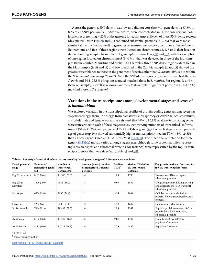
S. haematobiumWe explored variation in the transcriptional profiles of protein-coding genes among seven key
stages/sexes: eggs from urine; eggs from hamster tissues; sporocysts; cercariae; schistosomules;
and adult male and female worms. We showed that 69% to 86.8% of all protein-coding genes
were transcribed in each of these stages/sexes, with varying numbers of transcribed isoforms
overall (54.4–81.2%), and per gene (1.2–1.4) (Tables 4 and S4). For each stage, a small percent-
age of genes (top 1%) showed substantially higher transcription (median TPM: 1391–2435)
than all other genes (median TPM: 5.74–26.3) (Table 4). The functional annotation for these
genes (S8 Table) mostly varied among stages/sexes, although some protein families (represent-
ing RNA transport and ribosomal proteins, for instance) were represented by the top 1% tran-
scripts in more than one stage/sex (Tables 4 and S8).
Table 4. Summary of transcription levels across seven key developmental stages of Schistosoma haematobium.
Developmental
stage
Number of
transcribed genesa
(%)
Number of
transcribed
isoforms (%)
Average (mean) number
of transcribed isoforms
per gene
Median
TPMbMedian TPM of top
1% transcribed
isoforms
Key protein/pathway functions for
top 1% transcribed isoforms
Egg (from urine) 8153 (86.4) 11,106 (75.6) 1.4 15.0 1790 Translation; RNA transport;
ribosomal proteins
Egg (from
hamster)
7446 (79.0) 9584 (65.2) 1.3 9.95 1592 Ubiquitin; protein folding, sorting
and degradation; RNA transport;
ribosomal proteins
Sporocyst 6506 (69.0) 7990 (54.4) 1.2 5.95 1894 Cellular nucleic acid-binding
protein; RNA transport; ribosomal
proteins
Cercaria 7202 (76.4) 9280 (63.1) 1.3 5.74 1607 Calmodulin; cytochrome cSchistosomule 7696 (81.6) 10,657 (72.5) 1.4 26.3 1391 Peptidyl-prolyl isomerase; 14-3-3
protein beta; RNA transport,
ribosomal proteins
Adult male 8182 (86.8) 11,935 (81.2) 1.5 9.81 1702 Glutathione S-transferase;
peptidases/proteases
Adult female 8112 (86.0) 11,714 (79.7) 1.4 7.79 2435 Peptidases/proteases
a TPM > 0.5b transcripts per million
https://doi.org/10.1371/journal.ppat.1010288.t004
PLOS PATHOGENS Chromosome-level genome of Schistosoma haematobium
PLOS Pathogens | https://doi.org/10.1371/journal.ppat.1010288 February 15, 2022 7 / 25

Genes with similar transcription profiles and functions among
developmental stages
We hypothesised that transcription profiles that correlated among different developmental
stages would link to common pathways or signalling networks. We established seven distinct
clusters, each containing 817 to 4301 transcripts with similar transcription profiles (Fig 3A
and S4 Table). Each of the seven clusters contained transcripts predominantly transcribed in
one of the seven key developmental stages. For clusters 1 and 5, marked co-transcription was
seen among two to three different stages. By contrast, 22, 3 and 3 transcripts were uniquely
transcribed in the sporocyst, cercaria and schistosomula stages, respectively (clusters 3–5, S4
Table). In the sporocyst stage, unique transcripts encoded peptidases/proteases, CAP domain-
containing proteins (including “venom allergen-like” or SmVAL-like proteins) and a heat
shock protein-associated CDC37 homolog (MS3_00009347.2). CAP protein-encoding
(MS3_00004475.1) and sodium channel-encoding (MS3_00007597.2) transcripts were unique
Fig 3. Analysis of transcription for key developmental stages of Schistosoma haematobium. A Transcription profiles of transcript isoforms across seven
developmental stages/sexes, clustered (Ward; k = 7) by similarity of Z-score-normalised TPM (transcripts per million) values. Key, enriched (q< 0.05)
pathways and/or protein functions are shown to the left of each cluster. Numbers of molecules in round parentheses. B Pairwise comparison of differential
(DE; fold change (FC)> 2, false discovery rate (FDR)< 0.05) transcription between male (blue) and female (red) samples, displayed as a ‘volcano’ plot. Key
pathways and/or protein functions enriched in DE subsets are highlighted. c Percentage of DE transcripts encoded on each chromosome/scaffold for males
(blue) and females (red), respectively; chromosomes/scaffolds enriched (q< 0.05) for male or female DE genes are marked with an asterisk.
https://doi.org/10.1371/journal.ppat.1010288.g003
PLOS PATHOGENS Chromosome-level genome of Schistosoma haematobium
PLOS Pathogens | https://doi.org/10.1371/journal.ppat.1010288 February 15, 2022 8 / 25

to the cercarial stage, and a transcript exclusive to the schistosomula stage encoded a “sperm-
tail PG-rich repeat” protein (MS3_00007199.1).
Overall, all seven clusters showed significant enrichment for protein families (n = 35) and/
or pathways (47) (Fig 3A and S9 Table), including those linked to transport and catabolism in
both adult stages (clusters 6 and 7), as well as molecules related to the exosome in the cercaria
stage and both adult sexes (clusters 4, 6 and 7). Additionally, peptidases and peptidase inhibi-
tors were enriched in clusters 3 (sporocyst) and 7 (adult female).
Distinct isoform usage in different developmental stages
Next, we investigated genes that encoded distinct isoforms in multiple transcription-clusters,
reflecting variation in isoform usage for different developmental stages/sexes. We identified
2648 of such genes with distinct isoforms present into two (n = 2102) to six (1) clusters (S4
Table). We hypothesised that this isoform switching is driven by alternative splicing and is
either facilitated by genes encoding small exons (microexons of� 54 nt) or many exons.
Although there was no overall correlation between the number of isoforms in distinct clusters
and the number of microexons (Pearson’s R = 0.23) or exons (R = 0.3) per gene, we did find
evidence of genes encoding microexons and multiple isoforms in distinct clusters. For exam-
ple, of the genes containing inferred microexons (n = 2996), two genes encoded isoforms that
were present in five of the seven clusters, and were comprised of 7–10 exons and 0–2 microex-
ons. These isoforms were assigned to clusters 2–6 (MS3_00008061) and 3–7 (MS3_00004678),
and encoded a MYND-type zinc finger domain-containing protein (IPR002893) and a small
GTPase (IPR001806), respectively. We provided evidence for the differential usage of two iso-
forms transcribed predominantly in males (cluster 6) or females (cluster 7), respectively, by
mapping transcripts assembled from mixed-sex, long-reads to the genomic region encoding
MS3_00004678 (chromosome ZW, positions 87,941,969 to 87,954,222; Fig 4).
Sex-linked transcription
A comparison of transcription levels in the male and female adult stages of S. haematobiumshowed that 1512 transcripts were significantly upregulated in female compared with male
Fig 4. Long-read, full-length transcripts supporting differential isoform usage in male and female Schistosoma haematobium. The gene model
MS3_00004678 encodes a small GTPase on chromosome ZW. Exons are depicted as blocks and introns as arrowed lines, indicating the coding strand.
Reference transcripts are shown at the bottom in red (female; MS3_00004678.7, transcription cluster 7) and blue (male; MS3_00004678.1; transcription cluster
6) with narrow blocks at the end of the gene models representing untranslated regions (UTRs). Full-length, long-read transcripts that matched the intron-exon
structure of the isoforms inferred to be transcribed in the male and female adult stage, respectively, are coloured accordingly. Transcripts that support distinct,
alternative exon-intron boundaries are shown in black.
https://doi.org/10.1371/journal.ppat.1010288.g004
PLOS PATHOGENS Chromosome-level genome of Schistosoma haematobium
PLOS Pathogens | https://doi.org/10.1371/journal.ppat.1010288 February 15, 2022 9 / 25

worms (Fig 3B and S10 Table), and that the genes encoding these transcripts were over-repre-
sented (Fisher’s exact test, adjusted p-value < 0.05) on chromosomes 1 and 3 and on the larg-
est, “unplaced” scaffold (no. 194) of Shae.V3 (Fig 3C). None of the 10 genes encoded in this
scaffold were upregulated in males, and all had very low transcription levels (mean TPM
of� 0.57) in males.
In Schistosoma, sex is determined by a ZW chromosomal system, whereby maleness is con-
ferred by a ZZ composition and the absence of the female-specific W chromosome. Unlike in
most XY systems, where double-dosage of X transcripts is prevented by transcriptional sup-
pression in XX individuals, schistosomes have a limited suppression of Z chromosomes [28].
Consistent with a ZW chromosomal system, genes located on the S. haematobium ZW chro-
mosome encoded markedly more of the 1963 sexually-upregulated transcripts, with 670
(34.1%) upregulated in males and 279 (18.5%) in females (Fig 3B and 3C and S10 and
S11 Tables).
Of the transcripts upregulated in females, there was significant enrichment of those encod-
ing proteins linked to progesterone-mediated oocyte maturation, cell cycle and ribosome, and
proteins involved in DNA replication and chromosome-related functions (S12 Table). Pro-
teins encoded by transcripts upregulated in male worms were significantly enriched for roles
in 50 different pathways, including those involved in signal transduction associated with envi-
ronmental information processing (329 proteins), and endocrine (251), nervous (148) and
digestive (106) systems linked to 5 to 11 pathways. Enriched protein families included those
related to the cytoskeleton (n = 99), transport (membrane trafficking: n = 195; exosome: 184;
transport system: 95) and signalling (ion channels: n = 45; G protein-coupled receptors: 31)
(S12 Table).
Distinctive transcription in eggs, depending on host origin
We hypothesised that the transcription in S. haematobium eggs derived from human urine
would differ from eggs isolated from hamster livers. A comparison revealed 1143 transcripts
that were unique to eggs from urine, including a hepatotoxic ribonuclease omega-1 (UniProt
identifier: Q2Y2H5) homolog (69% amino acid similarity (BLAST); MS3_00010006.1;
TPM = 11.0–111.9). To investigate whether this homolog was structurally similar to the S.
mansoni omega-1 protein, we predicted the structures of all seven S. haematobium omega-1homologs using AlphaFold [29]. The alignment of these predicted structures using TM-align
[30] showed that six of these homologs (including MS3_00010006.1) aligned well (RMSD:
1.15–2.2Å; TM-score: 0.69–0.85) with 74.7–90.2% of the S. mansoni omega-1 structure, despite
limited overall sequence identity (33.5%-50.6%, based on structural alignment) (S13 Table).
Additional 4965 transcripts had substantially higher TPMs (> 112) in eggs from human
urine, including an “interleukin (IL)-4-inducing principle of S. mansoni eggs” (M-IPSE/alpha-1; UniProt identifier: Q869D4) homolog (H-IPSE; MS3_00010265.1). The proteins encoded
by 6108 transcripts linked predominantly to spliceosome (n = 89 proteins), membrane traffick-
ing (221), as well as transferase (537) and hydrolase (443) activities. Enriched were protein
families that related to chromosomes (n = 358) and mRNA biogenesis (n = 180). By contrast, a
kappa-5 (UniProt identifier: Q2KMJ3) homolog (MS3_00010619.1) had a higher level of tran-
scription (TPMs > 112) in eggs from hamster liver than from human urine.
Discussion
The assembly of the chromosome-contiguous reference genome (Shae.V3) for a well-defined
Egyptian strain [27] of S. haematobium has underpinned an exploration of molecular variation
within S. haematobium at key stages of development from different hosts and from multiple
PLOS PATHOGENS Chromosome-level genome of Schistosoma haematobium
PLOS Pathogens | https://doi.org/10.1371/journal.ppat.1010288 February 15, 2022 10 / 25

geographic localities in Africa, with important implications for investigating natural schistosome
populations as well as urogenital schistosomiasis and associated bladder cancer in humans.
The substantial genetic variation observed among four S. haematobium samples from four
disparate locations in Africa (Zambia, Senegal, Mauritius and Mali) was associated with
unique genomic ‘signatures’ matching species other than S. haematobium. This finding sup-
ports the proposal that schistosome species within the S. haematobium-group form a complex
genetic landscape, resulting from genomic admixture and introgression upon hybridisation
[21,31]. The presence of such hybridisation/introgression events raises the importance of
exploring natural populations of members of this group and establishing their biological traits
in relation to host affiliations/range, pathogenicity, susceptibility to praziquantel and, particu-
larly, carcinogenicity. In this context, the fragmented nature of the existing assemblies for
some members of the S. haematobium-group and the lack of draft or reference genomes for S.
guineensis, S. intercalatum and S. leiperi represents a hurdle to more detailed explorations of
the extent and size of such introgression events. Clearly, future genome sequencing efforts
should place emphasis on creating reference genomes for all other members of the S. haemato-bium group, to complement the S. haematobium reference genome (Shae.V3).
The present genome, comprehensive transcriptomic profiling and long-read evidence
allowed us to refine gene models and annotations, discover ‘new’ genes (n = 45) and define
UTRs, which will enable further molecular explorations of S. haematobium. Variation in the
transcription profiles of genes likely relate to molecular alterations during developmental,
infection and/or disease processes. For instance, genes exclusively transcribed in the sporocyst,
cercaria and schistosomule stages encoding peptidases/proteases (including leishmanolysins,
metalloendopeptidases and trypsins) or SCP/TAPS superfamily members (e.g. venom-allergen
like proteins, VALs [32]) likely play roles in egress, invasion, digestive processes and/or
immune evasion in the molluscan or vertebrate hosts [33–35]. Sex-specific molecules identi-
fied likely associate with roles in development and/or reproduction in the female, and signal-
ling, transport and catabolism in the male [25,36–38]. It is noteworthy that many genes on the
Z chromosome are upregulated in ZZ males, consistent with a lack of widespread transcrip-
tional dosage compensation of the Z chromosome [28]. The lack of transcription in ‘male’
genes encoded on the largest “unplaced” scaffold (no. 194) of Shae.V3 suggests that this scaf-
fold represents a female-specific portion of the W chromosome. However, the complete W-
specific region (WSR) is likely much larger based on evidence for S. mansoni, whose highly-
repetitive WSR is estimated at 18–46 Mb [39]. Future work is warranted to fully resolve the sex
chromosomes of S. haematobium using long-read data from individual worms (females and
males) as a foundation for detailed explorations of sex-determining genes and sex- and devel-
opmentally-regulated gene expression.
We propose that variation in transcription levels between eggs from hamster liver and
those from human urine relate to differences in host-parasite relationship and to the ability of
eggs to induce immunopathological changes and disease (which is pronounced in humans,
but not in the hamster), including the presence of S. mansoni homologs of IPSE/alpha-1[40,41], kappa-5 [42] and ribonuclease omega-1 [43]. Intriguingly, as omega-1 was not detected
previously in proteomic investigations of egg-secreted antigens (ESAs) of S. haematobium[44,45], or known to induce a humoral antibody response in people and not detected in the
urine from S. haematobium-infected people [44], this ESA was considered as S. mansoni-spe-
cific [40]. However, to some extent, structural modelling supports the presence of those mole-
cules in S. haematobium eggs from human urine. Whether the transcripts of these homologs
are specifically transcribed in the eggs from urine from infected human patients and encode
immunogens that involved in the egg-directed immune responses in the human host warrants
investigation. In this context, the enriched transferase-encoding transcripts in urine-derived S.
PLOS PATHOGENS Chromosome-level genome of Schistosoma haematobium
PLOS Pathogens | https://doi.org/10.1371/journal.ppat.1010288 February 15, 2022 11 / 25

haematobium eggs might relate to roles in glycosylation of immunomodulatory glycoproteins
such as omega-1 and kappa-5, likely required for protein function, as described for S. mansoni[46,47]. The findings from this study lay a critical foundation for investigation of ESAs in S.
haematobium and can complement efforts to understand the pathogenesis of urogenital schis-
tosomiasis [48–50].
The chromosome-level genome assembly for an Egyptian strain of S. haematobium adds
important resources to the schistosome ‘-omics’ reference toolkit. For example, this genome
should accelerate large-scale population investigations and provide a unique opportunity to
study the implications of genomic admixture, including its effect on biological and/or disease
traits, morbidity and/or the effectiveness of control programs [51,52], including mass drug
administration (MDA) [53]. The present resource should also enable future functional geno-
mics investigations of S. haematobium [54–56] and facilitate investigations of the fundamental
pathobiology of this important parasite using an integrative proteomic, glycomic and lipido-
mic approach. Insights into these areas could significantly assist in ongoing control and elimi-
nation efforts of urogenital schistosomiasis. We expect that the long-read sequencing
technologies used herein will facilitate future investigation of schistosome chromosomes and
transcriptomes, particularly differential isoform transcription and alternative splicing in sex
determination, development and reproduction.
Methods
Ethics statement
Approval to maintain the life cycle of S. haematobium using Mesocricetus auratus (hamster;
mammalian definitive host) and Bulinus truncatus as the snail intermediate host at the Bio-
medical Research Institute (BRI), Rockville, Maryland, USA was obtained from the NIH Office
of Laboratory Animal Welfare [OLAW]: D16-00046 (A3080-01). Ethics approval for the col-
lection of blood fluke parasite materials for the Schistosomiasis Collection at the Natural His-
tory Museum (SCAN) was obtained from the Home Office, project license number 70/4687
[14]. Approval to collect urine from schoolchildren was obtained from the administrative
authorities, school inspectors, directors and teachers. The objectives of the study were
explained to schoolchildren and their parents or guardians, and to participants from whom
written informed consent was obtained. The study was also approved by the National Ethics
Committee (Nr 2016/11/833/CE/CNERSH/SP) and the Ministries of Health and Education of
Cameroon, and from the Liverpool School of Tropical Medicine Research Ethics Committee
(M1516-18 and M1516-06).
Parasite material
Different developmental stages of S. haematobium were obtained from experimental and natu-
ral hosts and distinct geographical regions. Adult, egg and schistosomule stages of S. haemato-bium originating from Egypt) [27] and maintained routinely in M. auratus (hamster;
mammalian definitive host) using B. truncatus as the snail intermediate host at the BRI, Rock-
ville, Maryland, USA. Hamsters exposed to 1,000 cercariae in pond water (200 ml) were eutha-
nised after 90 days of infection. Paired S. haematobium adults were perfused from the
mesenteric/intestinal vessels with physiological saline (37˚C) using an established method
[25]. Schistosomules were prepared by mechanical transformation [57] of ~10,500 cercariae
shed from infected B. truncatus, followed by culture for 24 h [58]. All of these developmental
stages were prepared and stored at -80˚C or -196˚C.
Single adult males of S. haematobium from four disparate geographic locations in Africa
(Zambia, Senegal, Mauritius and Mali) were obtained via SCAN [59]. Adult worms were
PLOS PATHOGENS Chromosome-level genome of Schistosoma haematobium
PLOS Pathogens | https://doi.org/10.1371/journal.ppat.1010288 February 15, 2022 12 / 25

perfused at 90 days from M. auratus infected in the laboratory with from individual Bulinuswrighti snails infected with miracidia from eggs from urine samples from individual patients
(n = 3), or from hamsters infected with cercariae from naturally infected snails (B. truncatus)(n = 1) (S14 Table). These worms were frozen in liquid nitrogen until use.
Eggs were collected from the urine from ~6 to 10 year-old children attending schools near
Loum, Cameroon, with approval from the administrative authorities, school inspectors, direc-
tors and teachers. Individual eggs were isolated microscopically and stored in RNAlater at 4˚C
(Thermo Fisher Scientific, Waltham, MA, USA).
DNA sequencing and genome assembly
The S. haematobium reference genome (designated Shae.V3 – representing the Egyptian refer-
ence strain [27], maintained at BRI) was assembled from data produced by Oxford Nanopore
long-read and Hi-C sequencing and from previous short-read data sets produced using Illu-
mina or Dovetail technology [25,26] using the following approach:
First, long-read data (SRA accession numbers SRR15400746 and SRR15400747; via Oxford
Nanopore technology [60]) were used for initial contig assembly employing the program Canu
v.1.9 [61], setting a genome size estimate of 400 Mb. Errors in these data were corrected using
medaka_consensus in the Medaka package v.1.0.3 (https://github.com/nanoporetech/
medaka). Redundancy was removed using purge_haplotigs v.1.1.1 [62] and using depths of 8,
35 and 100 reads (low, medium and high, respectively). Contigs were first scaffolded using
available short-read and mate-pair library data [25] using Platanus-allee v.2.0.2 [63], using a
minimum of 15 links to join contigs into contiguous scaffolds. Further scaffolding was done
using long-range, paired-read data (‘Dovetail’) using the HiRise pipeline v.2.0.5 [64], as
described earlier [26]. Then, scaffolds were polished using available short-read and mate-pair
library data employing pilon v.1.23 [65].
Next, in situ Hi-C sequencing was performed as described previously [66]. High molecular
weight DNA from 100 S. haematobium adults was restriction-digested with equal concentra-
tions of CviAII and MseI (New England Biolabs); the library was constructed and then
sequenced using the NextSeq550 platform (Illumina, San Diego, CA, USA). Scaffolds were
combined with the in situ Hi-C data using Juicer v.1.6 [67], 3D-DNA v.180922 [68] and
Juicebox Assembly Tools v.1.9.8 [69] to scaffold, inspect and manually curate results to achieve
chromosome-length scaffolds. The sequence data are available via the DNA Zoo SRA reposi-
tory (PRJNA512907); interactive Hi-C contact maps before and after the Hi-C-guided assem-
bly are available on the DNA Zoo website (https://www.dnazoo.org/assemblies/Schistosoma_
haematobium). Gaps in scaffolds were closed using long-reads that had been error-corrected
using the -correct and -trim steps within the program Canu employing the program TGS-Gap-
Closer v.1.0.3 (https://github.com/BGI-Qingdao/TGS-GapCloser). The gap-closed scaffolds
were then polished employing published data sets (produced from 500-bp and 800-bp librar-
ies) [25] and the error-corrected long-reads using the software HyPo v.1.0.3 [70].
Repeats in the final, gap-closed and polished assembly were identified and masked using
RepeatMasker v.4.1 (http://www.repeatmasker.org) employing the DFAM v.3.1 library and a
published S. haematobium repeat [25].
Synteny analysis
Genome-wide synteny between the repeat-masked Shae.V3 genome and the repeat-masked
scaffolds or chromosomes of other schistosome species was assessed by linking single-copy
orthologs (SCOs) (for each species-pair). Coordinates of SCOs were used as links between
scaffolds and were bundled using bundlelinks in circos tools v.0.23 [71], setting the minimum
PLOS PATHOGENS Chromosome-level genome of Schistosoma haematobium
PLOS Pathogens | https://doi.org/10.1371/journal.ppat.1010288 February 15, 2022 13 / 25
bundle size at 10,000 nt, with� 5 SCOs per bundle, and allowing the gap between members of
the same bundle to be at most 100,000 nt. Scaffolds were ordered and displayed using circos
v.0.69–8 [71].
RNA sequencing and data sets
Total RNA samples were isolated from (i) adult worms (50 worm pairs; three biological repli-
cates), (ii) individual male and female worms separated from pairs (six biological replicates for
each sex), (iii) cercariae and (iv) mechanically-transformed schistosomules of S. haematobiumusing the TriPure Isolation Reagent (Sigma Aldrich, St. Louis, MO, USA). Each RNA sample
was treated with DNase (TURBO DNA-freeTM kit, Thermo Fisher Scientific, Waltham, MA,
USA) and messenger RNA (mRNA) was purified (Dynabeads mRNA purification kit, Thermo
Fisher Scientific, Waltham, MA, USA). The size, integrity (i.e. RNA integrity number, RIN)
and concentration of RNA were estimated using a 4200 TapeStation System RNA ScreenTape
Assay (Agilent Technologies, Waldbronn, Germany) and a Qubit 3.0 Flourometer RNA High
Sensitivity Assay (Life Technologies, Carlsbad, CA, USA). TruSeq Stranded mRNA (Illumina,
San Diego, CA, USA) short-read libraries (150 bp, paired-end) were prepared from from indi-
vidual mRNA samples, according to the manufacturers’ instructions and sequenced on an Illu-
mina NextSeq 500 instrument.
Total RNA samples were also prepared from S. haematobium eggs (~ 500 to 1000 each), iso-
lated from urine samples from three different individuals, using the TRIzol Plus RNA purifica-
tion kit (Thermo Fisher Scientific, Waltham, MA, USA) and non-stranded, paired-end
libraries (145 bp) were constructed (TruSeq Non-Stranded Kit, Illumina, San Diego, CA,
USA) and sequenced on an Illumina HiSeq 4000 platform at BGI International (Shenzhen,
China).
All short-read data produced here were filtered for quality and adapters removed using the
program fastp v.0.20.1 [72]. Then, reads representing technical artefacts (including PCR dupli-
cates) or contamination were removed by mapping all quality-filtered and trimmed reads to
published genome scaffolds [26] using HISAT2 v.2.1.0 [73] with the options-fr for upstream/
downstream mate orientations for Illumina paired-end sequencing and-dta (“downstream
transcriptome analysis”). Mapped reads were then retained by filtering sam files using the -F4
flag in samtools v.1.9 [74] and the remaining reads were separated into files with mapped,
paired reads and mapped, unpaired reads using the options -f1 and -F1, respectively. The pro-
gram centrifuge v.1.0.4 [75] was then used to confirm no contamination was present.
Publicly-available short-read data sets for (i) S. haematobium eggs isolated from hamster
liver, pooled adult female or male worms of S. haematobium and pooled sporocysts produced
previously [25,76] were obtained from the Short Read Archive (SRA; accession nos.
SRR6655493, SRR6655495, SRR6655497 and SRR13147979).
Long-read RNA sequence data were produced from mRNA from pooled adult worms
(both sexes) using Oxford Nanopore technologies (Oxford, UK). Two direct RNA-sequencing
libraries using the SQK-RNA002 kit (which selects for full-length mRNAs with polyA tails),
and one cDNA-PCR long-read sequencing library using the SQK-PCS109 kit were con-
structed. PCR-amplification (SQK-PCS109) was conducted for 14 cycles using an extension
time of 3 min. All libraries were sequenced using a MinION device for 48–72 h using an
EXP-FLP002 flow cell priming kit and three R9.4.1 flow cells (FLO-MIN106). Reads were
obtained from raw fast5 files using a GPU-enabled version of the program Guppy v.3.2.4, pro-
viding the configuration file rna_r9.4.1_70bps_hac.cfg (for SQK-RNA002) or
dna_r9.4.1_450bps_hac.cfg (for SQK-PCS109). Reads that did not meet the quality required
(Q� 7) by Guppy were removed.
PLOS PATHOGENS Chromosome-level genome of Schistosoma haematobium
PLOS Pathogens | https://doi.org/10.1371/journal.ppat.1010288 February 15, 2022 14 / 25

Prediction of protein-coding genes
Gene models predicted for the S. haematobium Shae.V2 draft genome [26] were transferred to
the new genome assembly (Shae.V3) using liftOver (release 8 April 2020; [77]). First, a chain
file was created using the published [26] and new genome assemblies and using the doSame-
SpeciesLiftOver.pl script. Next, Shae.V2 gene models were transformed from genome feature
format (GFF) to gene prediction (GP) format and transferred to the Shae.V3 genome using the
liftOver chain file.
For gene prediction, quality-filtered and mapped paired-end reads from all 24 short-read
libraries were combined and supplied to the programs StringTie v.2.1.4 [78] and TransDeco-
der v.5.5.0 [79]. Then, to infer transcripts from long-reads, long-reads were mapped to the ref-
erence genome using minimap2 v.2.17-r941[80] employing the options -ax splice, -uf and -k14.
The program FLAIR (release Oct 2020) [81] was subsequently employed to correct splice junc-
tions created by mapped long-reads using high-quality, mapped short-reads and to collapse
mapped long-reads into transcripts using the-stringent option.
Gene models transferred from Shae.V2 and those inferred based on short- and long-read
RNA-Seq evidence were merged using StringTie with the-merge option and were used as
‘hints’ for gene prediction using the software AUGUSTUS v.3.4.0 [82]. Next, to create a train-
ing set for AUGUSTUS, redundant, duplicate, and incomplete gene models and transcript iso-
forms were removed, retaining only the most highly transcribed isoform per gene and those
that had a transcripts per million (TPM) value of� 1 and were covered by mapped reads
across their entire length. Additionally, for each gene the isoform with highest sequence iden-
tity to a S. mansoni transcript sequence was also retained. Gene models that did not pass the
NCBI quality checks using the program table2asn v.25.8 (https://www.ncbi.nlm.nih.gov/
genbank/tbl2asn2/) were removed.
Genes were predicted using AUGUSTUS with the- species schistosoma2 option and were
subsequently refined by adding UTRs and transcript isoforms using the program PASA
(docker image 8b604b34971f) [83] employing long-read transcripts as evidence. All non-
redundant, complete gene models from the initial StringTie predictions and the AUGUSTUS/
PASA predictions were retained as the final gene set. The completeness of the gene set was
assessed using the program BUSCO v.4.0.6 [84] using the -l metazoa_odb10 (release 10 Sept
2020) and- update-data options and was compared to published gene sets of S. haematobium,
S. mansoni, S. japonicum and S. bovis.
Functional annotation of inferred proteins
Protein sequences conceptually translated from predicted gene models were functionally
annotated using an established approach [85]. In brief, protein sequences were assessed for
conserved protein domains using InterProScan v.5.44–79.0 [86] employing default settings.
Next, using the program diamond v.0.9.24.125 (E-value� 10–8), amino acid sequences were
searched against the Kyoto Encyclopedia of Genes and Genomes (KEGG) database [87] to
infer pathway associations, and against Swiss-Prot within UniProtKB [88] to infer homologs.
Additionally, EggNOG mapper v.5.0 [89] was used to name protein sequences based on their
closest match to the EggNOG database [90].
Orthologs between the inferred proteome for Shae.V3 and available proteomes of
S. mansoni [91]; NCBI accession number PRJEA36577), S. japonicum [92]; NCBI accession
number PRJNA520774) and S. bovis [31]; NCBI accession number PRJNA451066) down-
loaded from WormBase Parasite (release WBPS15; [93]) were determined using OrthoFinder
v.2.5.2 [94].
PLOS PATHOGENS Chromosome-level genome of Schistosoma haematobium
PLOS Pathogens | https://doi.org/10.1371/journal.ppat.1010288 February 15, 2022 15 / 25

Analysis of genetic variation within S. haematobium among disparate
geographic locations
High molecular weight genomic DNA was isolated from single adult males of S. haematobiumfrom four distinct geographic locations (Zambia, Senegal, Mauritius and Mali) using the Che-
magic STAR DNA Tissue kit (Perkin Elmer, Waltham, MA, USA). The DNA yield was esti-
mated spectrophotometrically using the Qubit 3.0 Flourometer dsDNA HS kit (Life
Technologies, Carlsbad, CA, USA), and DNA integrity was assessed by agarose-gel electropho-
resis and then using a Bioanalyzer 2100 (Agilent Technologies, Waldbronn, Germany). High-
quality genomic DNA was used to construct short-insert libraries (500 bp) using a TruSeq
DNA library construction kit (Illumina, San Diego, CA, USA) and paired-end sequenced as
100 nt reads using the HiSeq-2500 platform (Illumina, San Diego, CA, USA).
Low-quality bases (Phred quality: < 20), adapters and reads of< 70 nt in length were
removed using Trimmomatic v.0.32 [95], and sequence quality was confirmed using FastQC
v.0.11.2 (http://www.bioinformatics.babraham.ac.uk/projects/fastqc/). Subsequently, high-
quality reads were mapped to scaffolds of the Shae.V3 genome using Bowtie2 v.2.4.2 [96], and
read alignments were stored in the BAM format. The mapped data were then used to record
single nucleotide polymorphisms (SNPs) at individual positions in relation to the references
using the Genome Analysis Toolkit (GATK v.4.0.8.1; [97]). In brief, base-quality scores of
‘raw’, aligned read data were re-calibrated twice based on predicted variants; subsequently,
SNP sites were identified for each sample using the GATK HaplotypeCaller [97] and merged
into one ‘variant call format’ (VCF) file listing all variable sites for all samples using GATK
CombineGVCFs and GenotypeGVCFs. Raw SNP sites were filtered for quality using GATK
VariantFiltration and following GATK best-practice guidelines. Specifically, SNP sites were
selected if read mapping depth (DP) was> 10, variant confidence (QD) > 2.0, strand bias (FS)
< 60.0, mapping quality (MQ) > 40.0, mapping quality (MQRankSum) > -12.5 and read posi-
tion bias (ReadPosRankSum) > -8.0. VCF files for reported SNPs in each sample were anno-
tated based on their genomic locations and predicted coding effects using snpEff v.5.0e [98]
and a GFF annotation file for the reference genome. Descriptive statistics were obtained from
snpEff output and using bcftools v.1.11 [74] and filtered VCF files.
The fixed SNPs (genotype call = 1/1) for each individual male of S. haematobium were
selected and transferred onto the reference sequence using FastaAlternateReferenceMaker in
GATK v.4.2.0.0. The genomic locations of fixed SNPs in coding regions were then compared
within and among the four individuals, and were displayed using the UpSet v.1.4.0 package in
R [99]. For each sample and each S. haematobium chromosome, the number of SNPs per 1Mb
non-overlapping region was determined, and regions with equal or more SNPs than 80% of all
1Mb regions per chromosome (80th percentile) were selected as ‘SNP-dense’ locations. Each
chromosome was then fragmented into 2000 nt sections and nucleotide similarity searches
were undertaken using minimap2 (-x asm20 -N 5- secondary = no) and a nucleotide database
of schistosome genomes, which consisted of the available genomes of key members of the S.
haematobium group (S. haematobium (Shae.V3, this study), S. bovis (PRJNA451066), S. curas-soni (PRJEB519), S. mattheei (PRJEB523) and S. margrebowiei (PRJEB522) as well as S. man-soni (PRJEA36577), S. rodhaini (PRJEB526) and S. japonicum (PRJNA520774).
The number of unique SNPs in coding regions within 2000 nucleotide regions along each
chromosome were then plotted and labelled according to the species with the greatest nucleo-
tide sequence homology match (requiring > 90% query coverage) using ggplot2 in R. SNP-
dense locations for which > 20% of the 2000 nt sections (i.e. > 100 sections) matched those of
a species other than S. haematobium, were considered to have a ‘non-S. haematobium SNP sig-
nature’. For these regions, the number of matches against each species in the database was
PLOS PATHOGENS Chromosome-level genome of Schistosoma haematobium
PLOS Pathogens | https://doi.org/10.1371/journal.ppat.1010288 February 15, 2022 16 / 25

represented in a pie chart. To assess the extent of false-positive species signatures in SNP-
dense regions, sequence regions were also subjected to homology searches against a reference
with no mutations (identical to the Shae.V3 genome sequence) and against one containing
random mutations introduced at the rate of 1934 nucleotide mutations per 1 Mb of genome
scaffold using msbar in the emboss package v.6.6.0.0 [100].
Analysis of transcription
For each developmental stage, we aligned length- and quality-filtered, short-read data to the
Shae.V3 genome using HISAT2, and inferred the transcription level for each transcript
employing StringTie2 and the Shae.V3 gene set GFF file. Transcripts were clustered employing
the Ward clustering method based on the Euclidian distance of their TPM values, that were Z-
score-normalised across seven developmental stages. For stages with multiple samples, the
median TPM was employed. TPM values were then ordered according to their cluster mem-
bership and displayed in a heatmap using the tidyheatmap package (https://github.com/
jbengler/tidyheatmap) in R.
Differential transcription analysis for libraries derived from individual male and female
worms (six biological replicates each) was conducted using Ballgown v.2.22.0 [101], employing
a two-group comparison and performing library size adjustment by using the sum of the log
non-zero expression measurements for each sample, up to the 75th percentile of those mea-
surements. Transcripts with a false discovery rate of< 0.05 and a fold-change (FC) of� 2
were considered differentially transcribed (i.e. upregulated). Individual stages/clusters were
tested for enrichment of KEGG pathways and KEGG BRITE terms (requiring a minimum
BRITE protein family size of 10), using Fisher’s exact test and correcting for multiple testing
by calculating the q-value and applying a cut-off of< 0.05.
Supporting information
S1 Fig. Analysis of Schistosoma haematobium genome regions of four individual male
worms from distinct geographic locations. For isolates from Zambia, Senegal, Mauritius or
Mali, density and localisation of SNPs in the S. haematobium reference genome are shown in 2
kb non-overlapping regions, with each point coloured by the species with the closest nucleo-
tide sequence homology. For each sample, SNP-rich regions (light green blocks) of
which> 20% resembled a genomic reference other than S. haematobium are labelled (i-xi).
(TIFF)
S1 Dataset. Variant call format (VCF) file, including single nucleotide polymorphisms
(SNPs) reported in an individual male Schistosoma haematobium worm from Mali (Mi).
(ZIP)
S2 Dataset. Variant call format (VCF) file, including single nucleotide polymorphisms
(SNPs) reported in an individual male Schistosoma haematobium worm from Mauritius
(Ms).
(ZIP)
S3 Dataset. Variant call format (VCF) file, including single nucleotide polymorphisms
(SNPs) reported in an individual male Schistosoma haematobium worm from Senegal (S1).
(ZIP)
S4 Dataset. Variant call format (VCF) file, including single nucleotide polymorphisms
(SNPs) reported in an individual male Schistosoma haematobium worm from Zambia
PLOS PATHOGENS Chromosome-level genome of Schistosoma haematobium
PLOS Pathogens | https://doi.org/10.1371/journal.ppat.1010288 February 15, 2022 17 / 25

(Z1).
(ZIP)
S1 Table. New Schistosoma haematobium sequence data produced in this study and linked
to NCBI sequence read archive submission details.
(XLSX)
S2 Table. Synteny and contiguity of the Schistosoma haematobium reference genome
(Shae.V3), compared with that of other schistosomes.
(XLSX)
S3 Table. Orthologs of Schistosoma haematobium in S. mansoni, S. japonicum and S. bovisinferred using OrthoFinder.
(XLSX)
S4 Table. Transcription levels for Schistosoma haematobium genes, determined using
StringTie2.
(XLSX)
S5 Table. Annotation of inferred Schistosoma haematobium proteins using InterProScan.
(XLSX)
S6 Table. Annotation of inferred Schistosoma haematobium proteins based on matches to
the EggNOG database.
(XLSX)
S7 Table. Kyoto Encyclopedia of Genes and Genomes (KEGG) orthology, pathway annota-
tion, BRITE and enzyme classification for Schistosoma haematobium proteins.
(XLSX)
S8 Table. Functional annotation of the five most highly transcribed sequences in seven key
developmental stages of Schistosoma haematobium.
(XLSX)
S9 Table. KEGG terms significantly enriched (q-value < 0.05) in clusters of distinct tran-
scription profiles for Schistosoma haematobium.
(XLSX)
S10 Table. Differentially transcribed isoforms in adult female Schistosoma haematobium,
compared with adult males. Fold change, q-value and TPM (transcript per million) for each
library are shown.
(XLSX)
S11 Table. Differentially transcribed isoforms in adult male Schistosoma haematobium,
compared with adult females. Fold change, q-value and TPM (transcript per million) for
each library are shown.
(XLSX)
S12 Table. KEGG terms significantly enriched (q-value < 0.05) among transcripts differ-
entially expressed in adult male or female Schistosoma haematobium.
(XLSX)
S13 Table. Modelling of the predicted structures of seven Schistosoma haematobiumomega-1 homologs using AlphaFold and alignment employing TM-align.
(XLSX)
PLOS PATHOGENS Chromosome-level genome of Schistosoma haematobium
PLOS Pathogens | https://doi.org/10.1371/journal.ppat.1010288 February 15, 2022 18 / 25

S14 Table. Information on collection site, host, year and Natural History Museum refer-
ence code for four isolates of adult male Schistosoma haematobium from Africa.
(XLSX)
Acknowledgments
Parasite materials (cercariae, adult male and female worms of Schistosoma haematobium,
Egyptian strain) were provided by the NIAID Schistosomiasis Resource Center for distribution
through BEI Resources, NIAID, NIH. We are grateful to Ashling Charles from the DNA Zoo
Australia team for support in the routine processing of data.
Author Contributions
Conceptualization: Andreas J. Stroehlein, Robin B. Gasser, Neil D. Young.
Data curation: Andreas J. Stroehlein, Pasi K. Korhonen, Aidan M. Emery, Neil D. Young.
Formal analysis: Andreas J. Stroehlein, Parwinder Kaur, Olga Dudchenko, Neil D. Young.
Funding acquisition: Pasi K. Korhonen, Bicheng Yang, Bill C. H. Chang, Robin B. Gasser,
Neil D. Young.
Investigation: Andreas J. Stroehlein, Louis-Albert Tchuem Tchuente, J. Russell Stothard, Par-
winder Kaur, David Rollinson, Robin B. Gasser, Neil D. Young.
Methodology: Andreas J. Stroehlein, Stuart A. Ralph, Parwinder Kaur, Olga Dudchenko, Bon-
nie L. Webster, Robin B. Gasser, Neil D. Young.
Project administration: Neil D. Young.
Resources: Andreas J. Stroehlein, Pasi K. Korhonen, Margaret Mentink-Kane, Hong You,
Donald P. McManus, Louis-Albert Tchuem Tchuente, J. Russell Stothard, Olga Dud-
chenko, Bicheng Yang, Aidan M. Emery, Bonnie L. Webster, Paul J. Brindley, David Rollin-
son, Robin B. Gasser.
Software: Andreas J. Stroehlein.
Supervision: Robin B. Gasser, Neil D. Young.
Validation: Andreas J. Stroehlein.
Visualization: Andreas J. Stroehlein, Neil D. Young.
Writing – original draft: Andreas J. Stroehlein, Neil D. Young.
Writing – review & editing: Andreas J. Stroehlein, V. Vern Lee, Stuart A. Ralph, Margaret
Mentink-Kane, Hong You, Donald P. McManus, Louis-Albert Tchuem Tchuente, J. Russell
Stothard, Parwinder Kaur, Erez Lieberman Aiden, Huanming Yang, Aidan M. Emery, Bon-
nie L. Webster, Paul J. Brindley, David Rollinson, Bill C. H. Chang, Robin B. Gasser, Neil
D. Young.
References
1. Colley DG, Bustinduy AL, Secor WE, King CH. Human schistosomiasis. Lancet. 2014; 383
(9936):2253–64. https://doi.org/10.1016/S0140-6736(13)61949-2 PMID: 24698483
2. McManus DP, Dunne DW, Sacko M, Utzinger J, Vennervald BJ, Zhou XN. Schistosomiasis. Nat Rev
Dis Primers. 2018; 4(1):13. https://doi.org/10.1038/s41572-018-0013-8 PMID: 30093684
PLOS PATHOGENS Chromosome-level genome of Schistosoma haematobium
PLOS Pathogens | https://doi.org/10.1371/journal.ppat.1010288 February 15, 2022 19 / 25

3. Rollinson D, Stothard JR, Southgate VR. Interactions between intermediate snail hosts of the genus
Bulinus and schistosomes of the Schistosoma haematobium group. Parasitology. 2001; 123 Suppl:
S245–60. https://doi.org/10.1017/s0031182001008046 PMID: 11769287
4. Burke ML, Jones MK, Gobert GN, Li YS, Ellis MK, McManus DP. Immunopathogenesis of human
schistosomiasis. Parasite Immunol. 2009; 31(4):163–76. https://doi.org/10.1111/j.1365-3024.2009.
01098.x PMID: 19292768
5. Brindley PJ, Hotez PJ. Break out: urogenital schistosomiasis and Schistosoma haematobium infection
in the post-genomic era. PLoS Negl Trop Dis. 2013; 7(3):e1961. https://doi.org/10.1371/journal.pntd.
0001961 PMID: 23556007
6. Patel P, Rose CE, Kjetland EF, Downs JA, Mbabazi PS, Sabin K, et al. Association of schistosomiasis
and HIV infections: A systematic review and meta-analysis. Int J Infect Dis. 2021; 102:544–53. https://
doi.org/10.1016/j.ijid.2020.10.088 PMID: 33157296
7. Palumbo E. Association between schistosomiasis and cancer: a review. Infect Dis Clin Pract. 2007; 15
(3):145–8.
8. Brindley PJ, da Costa JM, Sripa B. Why does infection with some helminths cause cancer? Trends
Cancer. 2015; 1(3):174–82. https://doi.org/https%3A//doi.org/10.1016/j.trecan.2015.08.011 PMID:
26618199
9. Melman SD, Steinauer ML, Cunningham C, Kubatko LS, Mwangi IN, Wynn NB, et al. Reduced sus-
ceptibility to praziquantel among naturally occurring Kenyan isolates of Schistosoma mansoni. PLoS
Negl Trop Dis. 2009; 3(8):e504. https://doi.org/10.1371/journal.pntd.0000504 PMID: 19688043
10. Crellen T, Walker M, Lamberton PH, Kabatereine NB, Tukahebwa EM, Cotton JA, et al. Reduced effi-
cacy of praziquantel against Schistosoma mansoni is associated with multiple rounds of mass drug
administration. Clin Infect Dis. 2016; 63(9):1151–9. https://doi.org/10.1093/cid/ciw506 PMID:
27470241
11. Levecke B, Vlaminck J, Andriamaro L, Ame S, Belizario V, Degarege A, et al. Evaluation of the thera-
peutic efficacy of praziquantel against schistosomes in seven countries with ongoing large-scale
deworming programs. Int J Parasitol Drugs Drug Resist. 2020; 14:183–7. https://doi.org/10.1016/j.
ijpddr.2020.10.003 PMID: 33125936
12. Aula OP, McManus DP, Jones MK, Gordon CA. Schistosomiasis with a focus on Africa. Trop Med
Infect Dis. 2021; 6(3). https://doi.org/10.3390/tropicalmed6030109 PMID: 34206495
13. Knopp S, Ame SM, Person B, Hattendorf J, Rabone M, Juma S, et al. A 5-Year intervention study on
elimination of urogenital schistosomiasis in Zanzibar: Parasitological results of annual cross-sectional
surveys. PLoS Negl Trop Dis. 2019; 13(5):e0007268. https://doi.org/10.1371/journal.pntd.0007268
PMID: 31059495
14. Webster BL, Emery AM, Webster JP, Gouvras A, Garba A, Diaw O, et al. Genetic diversity within
Schistosoma haematobium: DNA barcoding reveals two distinct groups. PLoS Negl Trop Dis. 2012; 6
(10):e1882. https://doi.org/10.1371/journal.pntd.0001882 PMID: 23145200
15. Leger E, Webster JP. Hybridizations within the genus Schistosoma: implications for evolution, epide-
miology and control. Parasitology. 2017; 144(1):65–80. https://doi.org/10.1017/S0031182016001190
PMID: 27572906
16. Leger E, Borlase A, Fall CB, Diouf ND, Diop SD, Yasenev L, et al. Prevalence and distribution of schis-
tosomiasis in human, livestock, and snail populations in northern Senegal: a One Health epidemiologi-
cal study of a multi-host system. Lancet Planet Health. 2020; 4(8):e330–e42. https://doi.org/10.1016/
S2542-5196(20)30129-7 PMID: 32800151
17. Rey O, Webster BL, Huyse T, Rollinson D, Van den Broeck F, Kincaid-Smith J, et al. Population genet-
ics of African Schistosoma species. Infect Genet Evol. 2021; 89:104727. https://doi.org/10.1016/j.
meegid.2021.104727 PMID: 33486128
18. Webster BL, Diaw OT, Seye MM, Webster JP, Rollinson D. Introgressive hybridization of Schistosoma
haematobium group species in Senegal: species barrier break down between ruminant and human
schistosomes. PLoS Negl Trop Dis. 2013; 7(4):e2110. https://doi.org/10.1371/journal.pntd.0002110
PMID: 23593513
19. Webster BL, Alharbi MH, Kayuni S, Makaula P, Halstead F, Christiansen R, et al. Schistosome interac-
tions within the Schistosoma haematobium group, Malawi. Emerg Infect Dis. 2019; 25(6):1245–7.
https://doi.org/10.3201/eid2506.190020 PMID: 31107237
20. Pennance T, Allan F, Emery A, Rabone M, Cable J, Garba AD, et al. Interactions between Schisto-
soma haematobium group species and their Bulinus spp. intermediate hosts along the Niger River Val-
ley. Parasit Vectors. 2020; 13(1):268. https://doi.org/10.1186/s13071-020-04136-9 PMID: 32448268
21. Rey O, Toulza E, Chaparro C, Allienne JF, Kincaid-Smith J, Mathieu-Begne E, et al. Diverging patterns
of introgression from Schistosoma bovis across S. haematobium African lineages. PLoS Pathog.
2021; 17(2):e1009313. https://doi.org/10.1371/journal.ppat.1009313 PMID: 33544762
PLOS PATHOGENS Chromosome-level genome of Schistosoma haematobium
PLOS Pathogens | https://doi.org/10.1371/journal.ppat.1010288 February 15, 2022 20 / 25

22. Platt RN, McDew-White M, Le Clec’h W, Chevalier FD, Allan F, Emery AM, et al. Ancient hybridization
and adaptive introgression of an invadolysin gene in schistosome parasites. Mol Biol Evol. 2019; 36
(10):2127–42. https://doi.org/10.1093/molbev/msz154 PMID: 31251352
23. Webster BL, Southgate VR, Littlewood DT. A revision of the interrelationships of Schistosoma includ-
ing the recently described Schistosoma guineensis. Int J Parasitol. 2006; 36(8):947–55. https://doi.
org/10.1016/j.ijpara.2006.03.005 PMID: 16730013
24. Stothard JR, Kayuni SA, Al-Harbi MH, Musaya J, Webster BL. Future schistosome hybridizations: Will
all Schistosoma haematobium hybrids please stand-up! PLoS Negl Trop Dis. 2020; 14(7):e0008201.
https://doi.org/10.1371/journal.pntd.0008201 PMID: 32614820
25. Young ND, Jex AR, Li B, Liu S, Yang L, Xiong Z, et al. Whole-genome sequence of Schistosoma hae-
matobium. Nat Genet. 2012; 44(2):221–5. https://doi.org/10.1038/ng.1065 PMID: 22246508
26. Stroehlein AJ, Korhonen PK, Chong TM, Lim YL, Chan KG, Webster B, et al. High-quality Schisto-
soma haematobium genome achieved by single-molecule and long-range sequencing. GigaScience.
2019; 8(9):gniz108. https://doi.org/10.1093/gigascience/giz108 PMID: 31494670
27. Lewis FA, Liang YS, Raghavan N, Knight M. The NIH-NIAID Schistosomiasis Resource Center. PLoS
Negl Trop Dis. 2008; 2(7):e267. https://doi.org/10.1371/journal.pntd.0000267 PMID: 18665228
28. Picard MAL, Cosseau C, Ferre S, Quack T, Grevelding CG, Coute Y, et al. Evolution of gene dosage
on the Z-chromosome of schistosome parasites. eLife. 2018;7. https://doi.org/10.7554/eLife.35684
PMID: 30044216
29. Jumper J, Evans R, Pritzel A, Green T, Figurnov M, Ronneberger O, et al. Highly accurate protein
structure prediction with AlphaFold. Nature 2021; 596(7873):583–9. https://doi.org/10.1038/s41586-
021-03819-2 PMID: 34265844
30. Zhang Y, Skolnick J. TM-align: a protein structure alignment algorithm based on the TM-score. Nucleic
Acids Res. 2005; 33(7):2302–9. https://doi.org/10.1093/nar/gki524 PMID: 15849316
31. Oey H, Zakrzewski M, Gravermann K, Young ND, Korhonen PK, Gobert GN, et al. Whole-genome
sequence of the bovine blood fluke Schistosoma bovis supports interspecific hybridization with S. hae-
matobium. PLoS Pathog. 2019; 15(1):e1007513. https://doi.org/10.1371/journal.ppat.1007513 PMID:
30673782
32. Chalmers IW, Hoffmann KF. Platyhelminth Venom Allergen-Like (VAL) proteins: revealing structural
diversity, class-specific features and biological associations across the phylum. Parasitology. 2012;
139(10):1231–45. https://doi.org/10.1017/S0031182012000704 PMID: 22717097
33. Parker-Manuel SJ, Ivens AC, Dillon GP, Wilson RA. Gene expression patterns in larval Schistosoma
mansoni associated with infection of the mammalian host. PLoS Negl Trop Dis. 2011; 5(8):e1274.
https://doi.org/10.1371/journal.pntd.0001274 PMID: 21912711
34. Buddenborg SK, Kamel B, Hanelt B, Bu L, Zhang SM, Mkoji GM, et al. The in vivo transcriptome of
Schistosoma mansoni in the prominent vector species Biomphalaria pfeifferi with supporting observa-
tions from Biomphalaria glabrata. PLoS Negl Trop Dis. 2019; 13(9):e0007013. https://doi.org/10.1371/
journal.pntd.0007013 PMID: 31568484
35. Ingram JR, Rafi SB, Eroy-Reveles AA, Ray M, Lambeth L, Hsieh I, et al. Investigation of the proteolytic
functions of an expanded cercarial elastase gene family in Schistosoma mansoni. PLoS Negl Trop
Dis. 2012; 6(4):e1589. https://doi.org/10.1371/journal.pntd.0001589 PMID: 22509414
36. Anderson L, Amaral MS, Beckedorff F, Silva LF, Dazzani B, Oliveira KC, et al. Schistosoma mansoni
egg, adult male and female comparative gene expression analysis and identification of novel genes by
RNA-Seq. PLoS Negl Trop Dis. 2015; 9(12):e0004334. https://doi.org/10.1371/journal.pntd.0004334
PMID: 26719891
37. Picard MA, Boissier J, Roquis D, Grunau C, Allienne JF, Duval D, et al. Sex-biased transcriptome of
Schistosoma mansoni: host-parasite interaction, genetic determinants and epigenetic regulators are
associated with sexual differentiation. PLoS Negl Trop Dis. 2016; 10(9):e0004930. https://doi.org/10.
1371/journal.pntd.0004930 PMID: 27677173
38. Lu Z, Sessler F, Holroyd N, Hahnel S, Quack T, Berriman M, et al. Schistosome sex matters: a deep
view into gonad-specific and pairing-dependent transcriptomes reveals a complex gender interplay.
Sci Rep. 2016; 6:31150. https://doi.org/10.1038/srep31150 PMID: 27499125
39. Buddenborg SK, Tracey A, Berger DJ, Lu Z, Doyle SR, Fu B, et al. Assembled chromosomes of the
blood fluke Schistosoma mansoni provide insight into the evolution of its ZW sex-determination sys-
tem. bioRxiv. 2021. https://doi.org/https://doi.org/10.1101/2021.08.13.456314
40. Dunne DW, Jones FM, Doenhoff MJ. The purification, characterization, serological activity and hepa-
totoxic properties of two cationic glycoproteins (alpha 1 and omega 1) from Schistosoma mansoni
eggs. Parasitology. 1991; 103 Pt 2:225–36. https://doi.org/10.1017/s0031182000059503 PMID:
1745548
PLOS PATHOGENS Chromosome-level genome of Schistosoma haematobium
PLOS Pathogens | https://doi.org/10.1371/journal.ppat.1010288 February 15, 2022 21 / 25

41. Pennington LF, Alouffi A, Mbanefo EC, Ray D, Heery DM, Jardetzky TS, et al. H-IPSE is a pathogen-
secreted host nucleus-infiltrating protein (infiltrin) expressed exclusively by the Schistosoma haemato-
bium egg stage. Infect Immun. 2017; 85(12). https://doi.org/10.1128/IAI.00301-17 PMID: 28923894
42. Schramm G, Hamilton JV, Balog CI, Wuhrer M, Gronow A, Beckmann S, et al. Molecular characterisa-
tion of kappa-5, a major antigenic glycoprotein from Schistosoma mansoni eggs. Mol Biochem Parasi-
tol. 2009; 166(1):4–14. https://doi.org/10.1016/j.molbiopara.2009.02.003 PMID: 19428667
43. Fitzsimmons CM, Schramm G, Jones FM, Chalmers IW, Hoffmann KF, Grevelding CG, et al. Molecu-
lar characterization of omega-1: a hepatotoxic ribonuclease from Schistosoma mansoni eggs. Mol Bio-
chem Parasitol. 2005; 144(1):123–7. https://doi.org/10.1016/j.molbiopara.2005.08.003 PMID:
16143411
44. Sotillo J, Pearson MS, Becker L, Mekonnen GG, Amoah AS, van Dam G, et al. In-depth proteomic
characterization of Schistosoma haematobium: Towards the development of new tools for elimination.
PLoS Negl Trop Dis. 2019; 13(5):e0007362. https://doi.org/10.1371/journal.pntd.0007362 PMID:
31091291
45. Carson JP, Robinson MW, Hsieh MH, Cody J, Le L, You H, et al. A comparative proteomics analysis
of the egg secretions of three major schistosome species. Mol Biochem Parasitol. 2020; 240:111322.
https://doi.org/10.1016/j.molbiopara.2020.111322 PMID: 32961206
46. Wilbers RH, Westerhof LB, van Noort K, Obieglo K, Driessen NN, Everts B, et al. Production and
glyco-engineering of immunomodulatory helminth glycoproteins in plants. Sci Rep. 2017; 7:45910.
https://doi.org/10.1038/srep45910 PMID: 28393916
47. van Noort K, Nguyen DL, Kriechbaumer V, Hawes C, Hokke CH, Schots A, et al. Functional characteri-
zation of Schistosoma mansoni fucosyltransferases in Nicotiana benthamiana plants. Sci Rep. 2020;
10(1):18528. https://doi.org/10.1038/s41598-020-74485-z PMID: 33116178
48. Fu CL, Odegaard JI, Herbert DR, Hsieh MH. A novel mouse model of Schistosoma haematobium egg-
induced immunopathology. PLoS Pathog. 2012; 8(3):e1002605. https://doi.org/10.1371/journal.ppat.
1002605 PMID: 22479181
49. Ray D, Nelson TA, Fu CL, Patel S, Gong DN, Odegaard JI, et al. Transcriptional profiling of the bladder
in urogenital schistosomiasis reveals pathways of inflammatory fibrosis and urothelial compromise.
PLoS Negl Trop Dis. 2012; 6(11):e1912. https://doi.org/10.1371/journal.pntd.0001912 PMID:
23209855
50. Ishida K, Hsieh MH. Understanding urogenital schistosomiasis-related bladder cancer: an update.
Front Med. 2018; 5:223. https://doi.org/10.3389/fmed.2018.00223 PMID: 30159314
51. Webster JP, Neves MI, Webster BL, Pennance T, Rabone M, Gouvras AN, et al. Parasite population
genetic contributions to the Schistosomiasis Consortium for Operational Research and Evaluation
within sub-Saharan Africa. Am J Trop Med Hyg. 2020; 103(1_Suppl):80–91. https://doi.org/10.4269/
ajtmh.19-0827 PMID: 32400355
52. Fall CB, Lambert S, Leger E, Yasenev L, Garba AD, Diop SD, et al. Hybridized zoonotic Schistosoma
infections result in hybridized morbidity profiles: A clinical morbidity study amongst co-infected human
populations of Senegal. Microorganisms. 2021; 9(8). https://doi.org/10.3390/microorganisms9081776
PMID: 34442855
53. Berger DJ, Crellen T, Lamberton PHL, Allan F, Tracey A, Noonan JD, et al. Whole-genome sequenc-
ing of Schistosoma mansoni reveals extensive diversity with limited selection despite mass drug
administration. Nat Commun. 2021; 12(1):4776. https://doi.org/10.1038/s41467-021-24958-0 PMID:
34362894
54. Hagen J, Scheerlinck JP, Gasser RB. Knocking down schistosomes—promise for lentiviral transduc-
tion in parasites. Trends Parasitol. 2015; 31(7):324–32. https://doi.org/10.1016/j.pt.2015.03.009
PMID: 25933926
55. Rinaldi G, Young ND, Honeycutt JD, Brindley PJ, Gasser RB, Hsieh MH. New research tools for uro-
genital schistosomiasis. J Infect Dis. 2015; 211(6):861–9. https://doi.org/10.1093/infdis/jiu527 PMID:
25240172
56. Ittiprasert W, Mann VH, Karinshak SE, Coghlan A, Rinaldi G, Sankaranarayanan G, et al. Pro-
grammed genome editing of the omega-1 ribonuclease of the blood fluke, Schistosoma mansoni.
eLife. 2019;8. https://doi.org/10.7554/eLife.41337 PMID: 30644357
57. Coultas KA, Zhang SM. In vitro cercariae transformation: comparison of mechanical and nonmechani-
cal methods and observation of morphological changes of detached cercariae tails. J. Parasitol. 2012;
98(6):1257–61. https://doi.org/10.1645/GE-3072.1 PMID: 22519732
58. You H, Gobert GN, Du X, Pali G, Cai P, Jones MK, et al. Functional characterisation of Schistosoma
japonicum acetylcholinesterase. Parasit Vectors. 2016; 9(1):328. https://doi.org/10.1186/s13071-016-
1615-1 PMID: 27283196
PLOS PATHOGENS Chromosome-level genome of Schistosoma haematobium
PLOS Pathogens | https://doi.org/10.1371/journal.ppat.1010288 February 15, 2022 22 / 25

59. Emery AM, Allan FE, Rabone ME, Rollinson D. Schistosomiasis collection at NHM (SCAN). Parasit
Vectors. 2012; 5:185. https://doi.org/10.1186/1756-3305-5-185 PMID: 22943137
60. Kinkar L, Gasser RB, Webster BL, Rollinson D, Littlewood DTJ, Chang BCH, et al. Nanopore sequenc-
ing resolves elusive long tandem-repeat regions in mitochondrial genomes. Int J Mol Sci. 2021; 22(4).
https://doi.org/10.3390/ijms22041811 PMID: 33670420
61. Koren S, Walenz BP, Berlin K, Miller JR, Bergman NH, Phillippy AM. Canu: scalable and accurate
long-read assembly via adaptive k-mer weighting and repeat separation. Genome Res. 2017; 27
(5):722–36. https://doi.org/10.1101/gr.215087.116 PMID: 28298431
62. Roach MJ, Schmidt SA, Borneman AR. Purge Haplotigs: allelic contig reassignment for third-gen dip-
loid genome assemblies. BMC Bioinformatics. 2018; 19(1):460. https://doi.org/10.1186/s12859-018-
2485-7 PMID: 30497373
63. Kajitani R, Yoshimura D, Okuno M, Minakuchi Y, Kagoshima H, Fujiyama A, et al. Platanus-allee is a
de novo haplotype assembler enabling a comprehensive access to divergent heterozygous regions.
Nat Commun. 2019; 10(1):1702. https://doi.org/10.1038/s41467-019-09575-2 PMID: 30979905
64. Putnam NH, O’Connell BL, Stites JC, Rice BJ, Blanchette M, Calef R, et al. Chromosome-scale shot-
gun assembly using an in vitro method for long-range linkage. Genome Res. 2016; 26(3):342–50.
https://doi.org/10.1101/gr.193474.115 PMID: 26848124
65. Walker BJ, Abeel T, Shea T, Priest M, Abouelliel A, Sakthikumar S, et al. Pilon: an integrated tool for
comprehensive microbial variant detection and genome assembly improvement. PLoS One. 2014; 9
(11):e112963. https://doi.org/10.1371/journal.pone.0112963 PMID: 25409509
66. Rao SS, Huntley MH, Durand NC, Stamenova EK, Bochkov ID, Robinson JT, et al. A 3D map of the
human genome at kilobase resolution reveals principles of chromatin looping. Cell. 2014; 159
(7):1665–80. https://doi.org/10.1016/j.cell.2014.11.021 PMID: 25497547
67. Durand NC, Shamim MS, Machol I, Rao SS, Huntley MH, Lander ES, et al. Juicer provides a one-click
system for analyzing loop-resolution Hi-C experiments. Cell Syst. 2016; 3(1):95–8. https://doi.org/10.
1016/j.cels.2016.07.002 PMID: 27467249
68. Dudchenko O, Batra SS, Omer AD, Nyquist SK, Hoeger M, Durand NC, et al. De novo assembly of the
Aedes aegypti genome using Hi-C yields chromosome-length scaffolds. Science. 2017; 356
(6333):92–5. https://doi.org/10.1126/science.aal3327 PMID: 28336562
69. Dudchenko O, Shamim MS, Batra SS, Durand NC, Musial NT, Mostofa R, et al. The
Juicebox Assembly Tools module facilitates de novo assembly of mammalian genomes with chromo-
some-length scaffolds for under $1000. bioRxiv. 2018:254797. https://doi.org/10.1101/254797
70. Kundu R, Casey J, Sung W-K. HyPo: super fast and accurate polisher for long read genome assem-
blies. bioRxiv. 2019:2019.12.19.882506. https://doi.org/10.1101/2019.12.19.882506
71. Krzywinski M, Schein J, Birol I, Connors J, Gascoyne R, Horsman D, et al. Circos: an information aes-
thetic for comparative genomics. Genome Res. 2009; 19(9):1639–45. https://doi.org/10.1101/gr.
092759.109 PMID: 19541911
72. Chen S, Zhou Y, Chen Y, Gu J. fastp: an ultra-fast all-in-one FASTQ preprocessor. Bioinformatics.
2018; 34(17):i884–i90. https://doi.org/10.1093/bioinformatics/bty560 PMID: 30423086
73. Kim D, Paggi JM, Park C, Bennett C, Salzberg SL. Graph-based genome alignment and genotyping
with HISAT2 and HISAT-genotype. Nat Biotechnol. 2019; 37(8):907–15. https://doi.org/10.1038/
s41587-019-0201-4 PMID: 31375807
74. Danecek P, Bonfield JK, Liddle J, Marshall J, Ohan V, Pollard MO, et al. Twelve years of SAMtools
and BCFtools. Gigascience. 2021; 10(2). https://doi.org/10.1093/gigascience/giab008 PMID:
33590861
75. Kim D, Song L, Breitwieser FP, Salzberg SL. Centrifuge: rapid and sensitive classification of metage-
nomic sequences. Genome Res. 2016; 26(12):1721–9. https://doi.org/10.1101/gr.210641.116 PMID:
27852649
76. Stroehlein AJ, Korhonen PK, Rollinson D, Stothard JR, Hall RS, Gasser RB, et al. Bulinus truncatus
transcriptome–a resource to enable molecular studies of snail and schistosome biology. Current
Research in Parasitology and Vector-Borne Diseases. 2021; 1:100015.
77. Haeussler M, Zweig AS, Tyner C, Speir ML, Rosenbloom KR, Raney BJ, et al. The UCSC Genome
Browser database: 2019 update. Nucleic Acids Res. 2019; 47(D1):D853–D8. https://doi.org/10.1093/
nar/gky1095 PMID: 30407534
78. Kovaka S, Zimin AV, Pertea GM, Razaghi R, Salzberg SL, Pertea M. Transcriptome assembly from
long-read RNA-seq alignments with StringTie2. Genome Biol. 2019; 20(1):278. https://doi.org/10.
1186/s13059-019-1910-1 PMID: 31842956
PLOS PATHOGENS Chromosome-level genome of Schistosoma haematobium
PLOS Pathogens | https://doi.org/10.1371/journal.ppat.1010288 February 15, 2022 23 / 25

79. Haas BJ, Papanicolaou A, Yassour M, Grabherr M, Blood PD, Bowden J, et al. De novo transcript
sequence reconstruction from RNA-seq using the Trinity platform for reference generation and analy-
sis. Nat Protoc. 2013; 8(8):1494–512. https://doi.org/10.1038/nprot.2013.084 PMID: 23845962
80. Li H. Minimap2: pairwise alignment for nucleotide sequences. Bioinformatics. 2018; 34(18):3094–100.
https://doi.org/10.1093/bioinformatics/bty191 PMID: 29750242
81. Tang AD, Soulette CM, van Baren MJ, Hart K, Hrabeta-Robinson E, Wu CJ, et al. Full-length transcript
characterization of SF3B1 mutation in chronic lymphocytic leukemia reveals downregulation of
retained introns. Nat Commun. 2020; 11(1):1438. https://doi.org/10.1038/s41467-020-15171-6 PMID:
32188845
82. Hoff KJ, Stanke M. Predicting genes in single genomes with AUGUSTUS. Curr Protoc Bioinformatics.
2019; 65(1):e57. https://doi.org/10.1002/cpbi.57 PMID: 30466165
83. Haas BJ, Salzberg SL, Zhu W, Pertea M, Allen JE, Orvis J, et al. Automated eukaryotic gene structure
annotation using EVidenceModeler and the Program to Assemble Spliced Alignments. Genome Biol.
2008; 9(1):R7. https://doi.org/10.1186/gb-2008-9-1-r7 PMID: 18190707
84. Waterhouse RM, Seppey M, Simao FA, Manni M, Ioannidis P, Klioutchnikov G, et al. BUSCO applica-
tions from quality assessments to gene prediction and phylogenomics. Mol Biol Evol. 2017; 35
(3):543–8. https://doi.org/10.1093/molbev/msx319 PMID: 29220515
85. Korhonen PK, Young ND, Gasser RB. Making sense of genomes of parasitic worms: Tackling bioinfor-
matic challenges. Biotechnol Adv. 2016; 34:663–86. https://doi.org/10.1016/j.biotechadv.2016.03.001
PMID: 26956711
86. Mitchell AL, Attwood TK, Babbitt PC, Blum M, Bork P, Bridge A, et al. InterPro in 2019: improving cov-
erage, classification and access to protein sequence annotations. Nucleic Acids Res. 2019; 47(D1):
D351–D60. https://doi.org/10.1093/nar/gky1100 PMID: 30398656
87. Kanehisa M, Sato Y, Kawashima M, Furumichi M, Tanabe M. KEGG as a reference resource for gene
and protein annotation. Nucleic Acids Res. 2016; 44(D1):D457–D62. https://doi.org/10.1093/nar/
gkv1070 PMID: 26476454
88. Boutet E, Lieberherr D, Tognolli M, Schneider M, Bansal P, Bridge AJ, et al. UniProtKB/Swiss-Prot,
the Manually Annotated Section of the UniProt KnowledgeBase: How to Use the Entry View. Methods
Mol Biol. 2016; 1374:23–54. https://doi.org/10.1007/978-1-4939-3167-5_2 PMID: 26519399
89. Huerta-Cepas J, Forslund K, Coelho LP, Szklarczyk D, Jensen LJ, von Mering C, et al. Fast genome-
wide functional annotation through orthology assignment by eggNOG-mapper. Mol Biol Evol. 2017; 34
(8):2115–22. https://doi.org/10.1093/molbev/msx148 PMID: 28460117
90. Huerta-Cepas J, Szklarczyk D, Heller D, Hernandez-Plaza A, Forslund SK, Cook H, et al. eggNOG
5.0: a hierarchical, functionally and phylogenetically annotated orthology resource based on 5090
organisms and 2502 viruses. Nucleic Acids Res. 2019; 47(D1):D309–D14. https://doi.org/10.1093/
nar/gky1085 PMID: 30418610
91. Protasio AV, Tsai IJ, Babbage A, Nichol S, Hunt M, Aslett MA, et al. A systematically improved high
quality genome and transcriptome of the human blood fluke Schistosoma mansoni. PLoS Negl Trop
Dis. 2012; 6(1):e1455. https://doi.org/10.1371/journal.pntd.0001455 PMID: 22253936
92. Luo F, Yin M, Mo X, Sun C, Wu Q, Zhu B, et al. An improved genome assembly of the fluke Schisto-
soma japonicum. PLoS Negl Trop Dis. 2019; 13(8):e0007612. https://doi.org/10.1371/journal.pntd.
0007612 PMID: 31390359
93. Howe KL, Bolt BJ, Shafie M, Kersey P, Berriman M. WormBase ParaSite—a comprehensive resource
for helminth genomics. Mol Biochem Parasitol. 2017; 215:2–10. https://doi.org/10.1016/j.molbiopara.
2016.11.005 PMID: 27899279
94. Emms DM, Kelly S. OrthoFinder: phylogenetic orthology inference for comparative genomics.
Genome Biol. 2019; 20(1):238. https://doi.org/10.1186/s13059-019-1832-y PMID: 31727128
95. Bolger AM, Lohse M, Usadel B. Trimmomatic: a flexible trimmer for Illumina sequence data. Bioinfor-
matics. 2014; 30(15):2114–20. https://doi.org/10.1093/bioinformatics/btu170 PMID: 24695404
96. Langmead B, Salzberg SL. Fast gapped-read alignment with Bowtie 2. Nat Methods. 2012; 9(4):357–
9. https://doi.org/10.1038/nmeth.1923 PMID: 22388286
97. McKenna A, Hanna M, Banks E, Sivachenko A, Cibulskis K, Kernytsky A, et al. The Genome Analysis
Toolkit: a MapReduce framework for analyzing next-generation DNA sequencing data. Genome Res.
2010; 20(9):1297–303. https://doi.org/10.1101/gr.107524.110 PMID: 20644199
98. Cingolani P, Platts A, Wang le L, Coon M, Nguyen T, Wang L, et al. A program for annotating and pre-
dicting the effects of single nucleotide polymorphisms, SnpEff: SNPs in the genome of Drosophila mel-
anogaster strain w1118; iso-2; iso-3. Fly. 2012; 6(2):80–92. https://doi.org/10.4161/fly.19695 PMID:
22728672
PLOS PATHOGENS Chromosome-level genome of Schistosoma haematobium
PLOS Pathogens | https://doi.org/10.1371/journal.ppat.1010288 February 15, 2022 24 / 25

99. Conway JR, Lex A, Gehlenborg N. UpSetR: an R package for the visualization of intersecting sets and
their properties. Bioinformatics. 2017; 33(18):2938–40. https://doi.org/10.1093/bioinformatics/btx364
PMID: 28645171
100. Rice P, Longden I, Bleasby A. EMBOSS: the European Molecular Biology Open Software Suite.
Trends Genet. 2000; 16(6):276–7. https://doi.org/10.1016/s0168-9525(00)02024-2 PMID: 10827456
101. Pertea M, Kim D, Pertea GM, Leek JT, Salzberg SL. Transcript-level expression analysis of RNA-seq
experiments with HISAT, StringTie and Ballgown. Nat Protoc. 2016; 11(9):1650–67. https://doi.org/10.
1038/nprot.2016.095 PMID: 27560171
PLOS PATHOGENS Chromosome-level genome of Schistosoma haematobium
PLOS Pathogens | https://doi.org/10.1371/journal.ppat.1010288 February 15, 2022 25 / 25