Patterns of Gene Expression in Schistosoma mansoni larvae ...
184
1 Patterns of Gene Expression in Schistosoma mansoni larvae associated with Infection of the Mammalian Host Sophia J Manuel Submitted for PhD University of York Department of Biology February 2010
-
Upload
khangminh22 -
Category
Documents
-
view
0 -
download
0
Transcript of Patterns of Gene Expression in Schistosoma mansoni larvae ...

1
Patterns of Gene Expression in Schistosoma mansoni larvae
associated with Infection of the Mammalian Host
Sophia J Manuel
Submitted for
PhD
University of York
Department of Biology
February 2010

2
Abstract
Larval schistosomes infect the human host by penetration of unbroken skin, before gaining
access to a blood vessel and beginning their intravascular life. The work in this thesis focuses
on the mammalian infection process, as it would be the ideal point to interrupt the life cycle. A
whole organism approach was taken, and the life cycle stages before, during and after skin
penetration were studied, namely the intra-molluscan germ ball (embryonic cercaria), infective
cercaria, and the in vitro cultured day 3 schistosomulum (equivalent to skin stage larva).
Confocal microscopy was used for a morphological survey of these life cycle stages,
establishing the timeline of cercarial embryogenesis and documenting the impressive changes
they undergo. The temporal and spatial gene expression patterns underlying the changes were
investigated using the first genome-wide microarray for S. mansoni for transcriptional
profiling of the three stages. The known repertoire of molecules likely to be secreted during
host entry was greatly expanded, particularly the proteases and venom allergen-like proteins.
Genes involved in energy production and conservation were up-regulated in the cercaria, as
were several genes encoding proteins deployed immediately on arrival in the skin.
Additionally, micro exon genes (MEGs) encoding variant secreted proteins were highly up-
regulated in the schistosomulum, emphasising their likely role after entry into the mammalian
host. The transcription of many tegument and gut-associated genes was also increased in the
schistosomulum; cathepsins were particularly notable, even in the cercaria, implying that the
larval gut becomes active long before blood feeding begins. The first application of whole
mount in situ hybridisation to germ balls confirmed localisation of invadolysin and VAL-10 to
the nascent acetabular glands. However, SmKK7 and Sm16 were not expressed in these
glands, questioning their putative roles in immunomodulation. Finally, three MEGs were
revealed to be expressed in tissues at the host-parasite interface.

3
Table of Contents
List of Abbreviations................................................................................................................. 11
1. Introduction ....................................................................................................................... 14
1.1. Part 1: Mammalian infection by larval schistosomes ............................................... 15
1.1.1. Schistosomiasis ................................................................................................. 15
1.1.2. Life cycle........................................................................................................... 15
1.1.3. Gross anatomical features of adult schistosomes.............................................. 17
Tegument....................................................................................................................... 17
Nerves ........................................................................................................................... 19
Protonephridial system.................................................................................................. 19
Gut and oesophageal gland ........................................................................................... 19
Reproductive system ..................................................................................................... 20
Musculature................................................................................................................... 20
1.1.4. Larval morphology............................................................................................ 20
Daughter sporocyst morphology................................................................................... 21
Germ ball development ................................................................................................. 21
Morphology of the cercaria........................................................................................... 23
Skin stage morphology.................................................................................................. 26
1.1.5. Physiological changes ....................................................................................... 27
1.2. Part 2: Gene expression............................................................................................. 28
1.2.1. The history of S. mansoni sequencing efforts ................................................... 29
1.2.2. The Transcriptome ............................................................................................ 30
1.2.3. The Genome...................................................................................................... 30
1.2.4. Genetic manipulation of schistosomes.............................................................. 32
1.2.5. Transgenesis...................................................................................................... 33
1.2.6. Global transcription profiling by microarray .................................................... 34
1.2.7. Previous applications of microarray technology to the study of schistosomes. 36
1.2.8. Molecules associated with the infection process .............................................. 39
1.2.9. Localisation ....................................................................................................... 41
1.3. Aims of the thesis...................................................................................................... 43
2. Morphological study ......................................................................................................... 44
2.1. Introduction ............................................................................................................... 45

4
2.2. Methods:.................................................................................................................... 47
2.2.1. Parasite material ................................................................................................ 47
2.2.2. Langeron’s Carmine staining ............................................................................ 48
2.2.3. Phalloidin and DAPI staining ........................................................................... 48
2.2.4. Confocal microscopy ........................................................................................ 48
2.3. Results ....................................................................................................................... 49
2.3.1. Daughter Sporocyst ........................................................................................... 49
2.3.2. Germ ball development ..................................................................................... 52
2.3.3. The Cercaria ...................................................................................................... 61
2.3.4. Skin stage schistosomulum ............................................................................... 67
2.4. Discussion ................................................................................................................. 71
2.4.1. Daughter sporocysts .......................................................................................... 71
2.4.2. Germ ball development ..................................................................................... 72
2.4.3. Cercariae ........................................................................................................... 74
2.4.4. Skin stage schistosomula .................................................................................. 74
2.4.5. Final thoughts.................................................................................................... 75
3. Microarray analysis of the germ ball, cercaria and day 3 schistosomulum ...................... 77
3.1. Introduction ............................................................................................................... 78
3.2. Methods..................................................................................................................... 80
3.2.1. Array design ...................................................................................................... 80
3.2.2. Biological material ............................................................................................ 80
3.2.3. Hybridisation..................................................................................................... 80
3.2.4. Statistical analyses ............................................................................................ 81
3.3. Results ....................................................................................................................... 82
3.3.1. Novel array........................................................................................................ 82
3.3.2. Contrasts summary............................................................................................ 83
3.3.3. GO analysis summary ....................................................................................... 83
3.3.4. DNA replication and Cell division.................................................................... 84
3.3.5. Translation......................................................................................................... 86
3.3.6. Development ..................................................................................................... 89
3.3.7. Proteases............................................................................................................ 91
3.3.8. Serine proteases................................................................................................. 91
3.3.9. Metalloproteases ............................................................................................... 93

5
3.3.10. Cysteine proteases............................................................................................. 95
3.3.11. Aspartic proteases ............................................................................................. 99
3.3.12. Membrane proteins............................................................................................ 99
3.3.13. Transporters..................................................................................................... 100
3.3.14. Membrane Channels........................................................................................ 102
3.3.15. Receptors......................................................................................................... 103
3.3.16. Others .............................................................................................................. 104
3.3.17. Lipid metabolism ............................................................................................ 106
3.3.18. Energy metabolism.......................................................................................... 108
3.3.19. Custom categories ........................................................................................... 109
3.3.20. Tegument......................................................................................................... 110
3.3.21. Gut-associated ................................................................................................. 112
3.3.22. Stress-related genes......................................................................................... 113
3.3.23. Micro exon genes ............................................................................................ 114
3.3.24. Venom allergen-like proteins.......................................................................... 116
3.4. Discussion ............................................................................................................... 118
3.4.1. Design and application of the first genome wide microarray for S. mansoni. 118
3.4.2. Germ ball......................................................................................................... 119
3.4.3. Cercaria ........................................................................................................... 120
3.4.4. Day 3 schistosomulum .................................................................................... 120
3.4.5. Cell proliferation ............................................................................................. 121
3.4.6. Protein synthesis genes are down-regulated in the cercaria............................ 121
3.4.7. Development ................................................................................................... 122
3.4.8. Proteases.......................................................................................................... 123
Serine proteases........................................................................................................... 123
Metalloproteases ......................................................................................................... 123
Cysteine proteases....................................................................................................... 124
Aspartyl proteases ....................................................................................................... 125
3.4.9. Energy production........................................................................................... 126
3.4.10. Lipid metabolism ............................................................................................ 126
3.4.11. Membrane proteins.......................................................................................... 126
3.4.12. Tegument......................................................................................................... 128
3.4.13. Micro Exon Genes........................................................................................... 130

6
3.4.14. Venom Allergen Like proteins........................................................................ 130
3.4.15. Stress ............................................................................................................... 131
3.4.16. Gut................................................................................................................... 132
4. Spatial Expression Patterns of Selected Genes ............................................................... 134
4.1. Introduction ............................................................................................................. 135
4.2. Methods:.................................................................................................................. 137
4.2.1. Biological material .......................................................................................... 137
4.2.2. PCR ................................................................................................................. 138
4.2.3. Cloning............................................................................................................ 138
4.2.4. Probe synthesis................................................................................................ 139
4.2.5. Whole mount in situ hybridisation.................................................................. 139
4.2.6. Immunohistochemistry (IHC) ......................................................................... 140
4.3. Results ..................................................................................................................... 141
4.3.1. Cercarial Elastase ............................................................................................ 141
4.3.2. Invadolysin...................................................................................................... 142
4.3.3. VAL-10 ........................................................................................................... 144
4.3.4. Sm16 ............................................................................................................... 145
4.3.5. SmKK7 expression in larvae........................................................................... 146
4.3.6. KK7 transcript localises to numerous cell bodies in adults ............................ 154
4.3.7. Microexon genes ............................................................................................. 156
4.4. Discussion ............................................................................................................... 158
4.4.1. Methods........................................................................................................... 158
4.4.2. Cercarial elastase 1a........................................................................................ 159
4.4.3. Invadolysin...................................................................................................... 159
4.4.4. VAL-10 ........................................................................................................... 160
4.4.5. Sm16 ............................................................................................................... 160
4.4.6. SmKK7............................................................................................................ 161
4.4.7. MEGs .............................................................................................................. 163
5. Final Discussion .............................................................................................................. 165
6. List of References ........................................................................................................... 173

7
List of Figures
Figure 1-1 Life cycle of schistosomes. ..................................................................................... 17
Figure 1-2 Diagram of the adult tegument. ............................................................................... 18
Figure 1-3 Diagram of a cercaria showing position of glands (not to scale). ........................... 24
Figure 2-1 a and b Migratory daughter sporocysts stained with Alexafluor 488-conjugated
phalloidin .................................................................................................................................. 50
Figure 2-2 Migratory daughter sporocysts stained with Langeron’s carmine. ......................... 51
Figure 2-3 Projection of a 38μm deep z-stack of a daughter sporocyst .................................... 52
Figure 2-4 a and b. Optical sections of germ balls stained with Langeron’s carmine. ............. 54
Figure 2-5 a and b. Young germ ball stained with Langeron’s carmine................................... 55
Figure 2-6 Developing germ ball. ............................................................................................. 56
Figure 2-7 Outer epithelium of germ balls................................................................................ 58
Figure 2-8 Possible subtegumental cells ................................................................................... 59
Figure 2-9 a and b Germ balls with stubby tails. ...................................................................... 60
Figure 2-10 Muscle development in a maturing germ ball ....................................................... 61
Figure 2-11 Cercaria stained with phalloidin (green) and DAPI (blue).................................... 63
Figure 2-12 Cercaria stained with phalloidin............................................................................ 64
Figure 2-13 Cercariae stained with Langeron’s carmine .......................................................... 65
Figure 2-14 Selected optical sections of a schistosomulum stained with phalloidin................ 69
Figure 2-15 Skin stage schistosomulum ................................................................................... 70
Figure 2-16 Schistosomulum stained with Langeron’s carmine............................................... 71
Figure 4-1 Transcript for cercarial elastase 1a localised in germ balls................................... 142
Figure 4-2 Invadolysin transcript localized in germ balls by WISH. ..................................... 143
Figure 4-3 Invadolysin protein localizes to the acetabular glands.......................................... 144
Figure 4-4 VAL 10 transcript localised in germ balls by WISH. ........................................... 145
Figure 4-5 Expression of Sm16 was localised in germ balls by WISH. ................................. 146
Figure 4-6 SmKK7 transcript was localised in larvae by WISH ............................................ 148
Figure 4-7 SmKK7 protein distribution in cercariae............................................................... 149
Figure 4-8 SmKK7 positive protrusions at anterior of cercaria.............................................. 150
Figure 4-9 SmKK7 protein in a cercaria alongside an electron micrograph of the anterior ... 152
Figure 4-10 Distribution of SmKK7 protein in schistosomula ............................................... 153
Figure 4-11 SmKK7 transcript localised in whole mount adult worms. ................................ 155
Figure 4-12 Distribution of SmKK7 protein in adult worms.................................................. 156

8
Figure 4-13 Distribution of MEG transcripts in larvae........................................................... 157
Figure 4-14 MEG3.2 (Smp_138070) transcript locaslied in a Day 10 schistosomulum ........ 158

9
Acknowledgements
I am very grateful to Alan for his excellent supervision, encouragement, clarity of thought and
for constructive criticism. I am also thankful for his careful help with writing, and for teaching
me what semicolons are for.
Thanks are also due to all members of the schistosome research group at York, past and
present, for their encouragement, friendship, fun, and help with the work: Adrian, Adam, Ann,
Bill, Fabienne, Gavin, Jenny, Joe, Pat, Pete, Rachel, Ross, Sarah, Shobana, Simon, Steph, and
William. In particular, Gary taught me the techniques I needed in the laboratory, and was a
great bench mate. In the Technology Facility: Graeme Park, Karen Chance and Meg Stark
helped with microscopy. Celina Whalley and Naveed Aziz were always available with
invaluable help and support. Also at York, I’m grateful to Professors Deborah Smith and
Jenny Southgate for their advice and support as my training committee.
I am thankful to Dr Alasdair Ivens for all his work on the microarray project.
On a personal note I would like to thank my friends for keeping me sane, and sharing the good
times and not-so-good times: Natalie Russell; Rachel, Sarah and Esther Hagger-Holt; Tom
Selmes, Charlie Forman, Debbie Coldwell, Pete Morrison, Harry Saffin, Jerome Jackson,
Silvia Nulissi, Francesca Vattari, Andreas Biternas, Charlotta Salmi, Mike Bright, Susanna
Brown, Tom Bartlett and Clare Gladding, Anna Semlyen, Hannah and Norna, Susan Collier,
and the lovely folks at St. Luke’s Burton Stone Lane.
Last but not least I would like to thank my family, my fiancé Rich and his family for always
cheering me on.
Thanks are due to the BBSRC, the NIH, and the Pathogen Sequencing Advisory Group at the
Wellcome Trust Sanger Institute for funding.
10
Declaration
All of the following work is my own with the following exceptions:
Chapter 1:
Figure 1 was reproduced by kind permission of Prof RA Wilson
Chapter 3:
RNA was run on Bioanalyser gels by Mrs. Celina Whalley. The microarray
design and statistical analysis was carried out by Dr A Ivens. Roche-
NimbleGen synthesised the microarrays and carried out the hybridisations.
Chapter 4:
The clone of VAL-10 and antisera for SmKK7 and invadolysin were the kind
gifts of Dr S Prasad at Cape Western University, Ohio, USA
Ms Jenny Middleton cloned SmKK7, and carried out WISH on larvae under
my supervision as part of her undergraduate research project.
The negative control sense chorion probe used in WISH was kindly provided
by Dr Gary P Dillon.
Electron micrograph of the anterior end of a cercaria kindly provided by
Stephanie Hopkins and Prof Jim McKerrow, University of California, San
Fransisco.
Perfusion of adult worms Prof Alan Wilson and Dr William C Borges
Electron microscopy of adult worms carried out by Ms Meg Stark

11
List of Abbreviations
Abbreviation
20x SSC 0.3M trisodium citrate and 3.0M sodium chloride
2D Two dimensional
3D Three dimensional
5-HT 5-hydroxytryptamine
A Acetabulum
AbD Antibody diluent
AChE Acetylcholine esterase
ADAM A disintegrin and a metalloprotease
adjP Adjusted P value
AFA Fixative containing ethanol, formalin, and acetic acid
AG Acetabular glands
ATP Adenine triphosphate
BLAST Basic local alignment search tool
BSA Bovine serum albumin
C cercaria
cDNA Complementary deoxyribonucleic acid
CE Cercarial elastase
CHAPS 3-[(3-cholamidopropyl)dimethylammonio]-1-propanesulfonate
CM Circular muscle
D Acetabular gland duct bundle
D3 Day 3 in vitro cultured schistosomulum
DALY Disability adjusted life year
DAPI 4',6' diamino-2-phenylindole
DIC differential interference contrast image
DIG digoxigenin
DM Diagonal muscle
DNA deoxyribonucleic acid
DPX Mounting media containing distyrene, a plasticizer, and xylene
dscDNA Double-stranded complementary deoxyribonucleic acid
dsRNA Double-stranded RNA

12
EDTA ethylenediaminetetraacetic acid
EFGP Enhanced green fluorescent protein
EP Excretory pore
ER Endoplasmic reticulum
EST Expressed sequence tag
f-actin Filamentous actin (as opposed to globular)
GB Germ ball
GFP green fluorescent protein
GO Gene ontology
GPCR G-protein coupled receptor
HDL High density lipoprotein
HG Head gland
HSP Heat shock protein
ID identity
IHC immunohistochemistry
ISH In situ hybridisation
kDa Kilo Daltons
LC Langeron’s carmine
LM Longitudinal muscle
LMWP Low molecular weight protein Smp_194860
LSM Laser scanning microscope
M169 Parasite culture medium
MCM Minichromosome maintenance proteins
MDR Multidrug resistance protein
MEG Micro-exon gene
mm Mismatch
mRNA Messenger ribonucleic acid
NG Neural ganglia
ORESTES Open reading frame expressed sequence tag - method for sequencing
PBS Phosphate buffered saline
PBSAT PBS and 0.1% Tween 20
PCNA Proliferating cell nuclear antigen
PGN Permeablising fluid (PBS with gelatin, saponin, and sodium azide)

13
PI Propidium iodide
PIPLC phosphatidylinositol-specific phospholipase C
pm Perfect match
PZQ Praziquantel
qPCR Quantitative or real time PCR
RM Radial muscle
RNA ribonucleic acid
RNAi RNA interference
RPMI Roswell Park Memorial Institute culture medium
RT-PCR Reverse transcriptase polymerase chain reaction
SCP Sperm Coating Protein domain found in venom allergen-like
proteins
SDS Sodium dodecyl sulphate
SGTP1 Schistosoma glucose transporter protein 1
SGTP4 Schistosoma glucose transporter protein 4
Sm16 Smp_113760
SmKK7 Smp_194830
Smp S. mansoni gene prediction identifier number from GeneDB
SmPepM8 Schistosoma mansoni metalloprotease eight (Smp_090100)
TBST Tris buffered saline and tween
TIGR The Institute for Genome Research
tRNA Transfer RNA
TWIK weakly inward rectifying potassium channels with a tandem of p
domains
U Undifferentiated cells
VAL Venom allergen-like protein (containing sperm coating protein
domain)
WHO World Health Organisation
WISH Whole mount in situ hybridisation
wk Week(s) old
WTSI Wellcome Trust Sanger Institute

14
1. Introduction

15
1.1. Part 1: Mammalian infection by larval schistosomes
1.1.1. Schistosomiasis
Schistosomiasis is a disease caused by blood flukes of the genus Schistosoma, affecting more
than 200 million people in 74 countries [1]. An estimated 779 million people are ‘at risk’ of
infection [2] and the annual number of deaths attributable to S. mansoni and S. haematobium
is approximately 130,000 and 150,000 respectively, in Sub-Saharan Africa alone [3]. The
symptoms are caused by the immune reaction to parasite eggs in the host tissue. The morbidity
associated with infection by schistosomes was reassessed in 2005; it was suggested that the
disability adjusted life years (DALYs) due to the disease should be increased from 0.5%
(equivalent to a birth mark on the face, which is clearly absurd) to 2-15% [1]. Presently there
is only one drug in use to control schistosomiasis: praziquantel (PZQ) and no vaccine
available yet. It has been recognised, in the light of potential resistance, that novel drugs are
needed, and should be used in combination therapy to extend the useful life of PZQ. New drug
targets are being identified [4] and chemical libraries have been screened against schistosomes
[5]. At present the WHO recommends focussing on morbidity reduction through mass
chemotherapy [6]. Re-infection occurs rapidly after treatment, so a vaccine is an important
goal of research. A combination of interventions, ideally including a vaccine, chemotherapy,
safe water sources and improved sanitation, will be necessary to eliminate schistosomiasis [7].
As noted by Gryseels et al [8], schistosomiasis will be eradicated only when its underlying
cause of poverty is no more.
1.1.2. Life cycle
Schistosomes have a two host life cycle, including a snail host and a mammalian host (Figure
1). Adult S. mansoni live paired in the blood vessels draining the intestines. The females lay
up to 350 eggs per day [9], which either cross the intestine wall and exit in the faeces, or are
washed back with the blood flow and become lodged in the liver. On contact with fresh water
the egg hatches, releasing a miracidium larva; this will infect a snail of the genus
Biomphalaria. There are two rounds of asexual reproduction which is important for life cycle
maintenance. The miracidium penetrates the head-foot of the intermediate host and transforms
into a mother sporocyst in which daughter sporocysts form. They leave the mother sporocyst

16
10-14 days after snail infection [10, 11] whereupon they enter the snail’s circulatory system.
The daughter sporocysts are carried by the flow of haemolymph via the heart to the digestive
gland (hepatopancreas). On arrival here they grow quickly and lose their ability to migrate
further. This is probably due to the availability of nutrients there. The daughter sporocysts
settle and intertwine with the snail tissue. They can reproduce two ways: by sporocystogenesis
producing a second generation of daughter sporocysts, or by cercariogenesis when cercarial
embryos called germ balls grow and develop within the daughter sporocyst [12]. Mature
cercariae leave the snail and enter fresh water. Here the larvae repeatedly swim to the surface
and sink down again until they find a mammalian host, which they infect, by penetration of
unbroken skin, using secretions from acetabular glands. On entry to the skin, the cercaria
transforms into a schistosomulum larva, which exits the skin via a blood or lymph vessel and
reaches the lungs four to six days after infection [13]. On arrival in the lungs the
schistosomulum becomes longer and thinner to enable further migration through the narrow
capillaries [13]. Migration through the lung takes an average of 30-35 hours in the mouse [14].
The schistosomulum leaves the lungs via the pulmonary vein and travels via the left side of the
heart to the systemic organs [15]; it arrives at the liver between 6 and 21 days later [14]. Most
schistosomula arrive at the hepatic portal system directly from the lungs and they remain
there. However, if they are carried by the circulation to another site, they return to the lungs
and complete further circuits until they arrive at the hepatic portal system [14]. It takes no
more than 2-3 such circuits to recruit all the schistosomes that will mature successfully to the
liver; those which do not arrive there will die in the other tissues [14].

17
Figure 1-1 Life cycle of schistosomes.
Source: Centers for Disease Control, Division of Parasitic Diseases
(http://www.dpd.cdc.gov/dpdx/HTML/Schistosomiasis.htm).
1.1.3. Gross anatomical features of adult schistosomes
Schistosomes are triploblastic acoelomates; they have no body cavities and three germ layers.
Many cell types have a cell body with projections going elsewhere; examples include
tegument cell bodies, nerve cells and muscle cells. Schistosomes have tissues and organs, but
they cannot readily be dissected [16]. It has been suggested that laser microdissection could be
used to cirvcumvent this problem [17]. However, cell bodies for various tissues can not be
distinguished using light microscopy, and therefore would be difficult to target [17].
Tegument
The surface of schistosomes is an anucleate syncytium termed the tegument [18]. In intra-
mammalian stages it is bounded apically by two lipid bilayers; the outer membranocalyx, and

18
the inner plasma membrane (see Figure 1-2). McLaren and Hockley noted in 1977 that this is
a common feature of blood flukes, whereas flukes residing in the intestine or associated body
cavities have a single outer membrane [19]. It was suggested that the membranocalyx may be
useful for evading the immune system. The syncytium contains various inclusion bodies and is
bounded basally by another membrane, below which lie the layers of circular and longitudinal
muscle. The tegument also contains spines which protrude from the surface of the worm, but
are covered apically by the membranocalyx. The spine’s bases rest on the tegument basement
membrane. Tegument cell bodies are situated underneath the two muscle layers and are joined
to the syncytium by narrow microtubule-lined connections [20]. Everything that comprises the
syncytial layer and its outer membrane originate in these cell bodies and is trafficked to the
surface. The literature contains widely variable estimates of the turnover rate of the adult
tegument, ranging from 20 minutes to 2 weeks [21]. Saunders et al carried out in vivo
experiments and reported a half life of 5.4 days [22].
Figure 1-2 Diagram of the adult tegument.
Note the two apical membrane bilayers, the basement membrane (arrow), the muscle layers, and a tegument cell
body with vesicles (reproduced courtesy of Prof RA Wilson).

19
Nerves
Schistosomes have an extensive nervous system. The brain [23] consists of a pair of anteriorly
located central ganglia joined medially by a commisure. Longitudinal nerve trunks extend
anterior and posterior from the brain. There is an extensive peripheral nerve net. The
neurotransmitter 5-hydoxytryptamine (5-HT) has been shown in the peripheral nerve net by
immunolocalisation, and in extensions to protrusions at the surface of adult males [24]. These
protrusions are unicilliated sensory structures. A bulb filled with vesicles and containing nerve
tissue is embedded in the tegument, but demarcated from it by a membrane [25]. The apical
cilium is covered by the membranocalyx [25]. Unicillated sensory endings are said to be the
only type of sensory ending in the adult [26]. The anterior ganglia, lateral nerve chords, and
peripheral nerves are readily visualised by immunohistochemistry for the neuropeptide
SALMFamide [27].
Protonephridial system
The protonephridial system consists of flame cells joined to collecting tubules; these lead to
the bladder which is situated at the posterior of the worm and ends in the excretory pore [28].
The beating of the flagella gives the flame cell its flickering appearance, and also provides
hydrostatic pressure to propel liquid down the tubules [29]. So called flame cells are made up
of two cells, one flagella bearing cap cell which has a large nucleus, and second, a flattened
cell which forms the tubule. The two cells are joined by interdigitations [28]. The
ultrastructure of flame cells is the same in the cercaria, schistosomulum and adult worm [30].
The protonephridium is thought to function to remove organic metabolites from the deep
tissues of the worm [28]. It has been suggested more recently that it may play a role in drug
excretion, although the authors note that more experimental data are required to support this
hypothesis [31].
Gut and oesophageal gland
In both male and female worms the mouth opens into the oesophagus which is lined by
inturned tegument surrounded by circular and longitudinal muscle [32]. The gut, by contrast,
is surrounded by circular muscle fibres only; these are thicker and more widely spaced than
those of the oesophagus [32]. The oesophageal gland lies ventrally to the oesophagus at its

20
posterior end, just posterior to the ventral sucker and anterior to the gut [16]. The oesophagus
is joined to the gut which splits into two lateral caecae which run either side of the genitalia,
rejoining posterior to them and continuing to the posterior of the body. The gut is blind-ended
and so excretion occurs by regurgitation.
Reproductive system
Adult male schistosomes have a gynaecophoric canal where the female resides; this begins
just posterior to the ventral sucker. The genital pore is situated at the anterior end of the
gynaecophoric canal and is connected to the four of five pairs of dorsal testes by a seminal
vesicle and defferent duct [33] (p 54). Machado Silva propose that these are in fact a multi-
lobed testis [34]. Female worms have a rather more complex reproductive system. The uterus
opens via a genital pore just posterior to the ventral sucker and leads to the ootype or egg
chamber. The ootype is connected both to the oviduct (leading to the centrally situated ovary)
and the vitteline duct (leading to the vittelaria which continue to the posterior of the body)
[33].
Musculature
Schistosomes are very muscular creatures. In adults there are three subtegumental muscle
layers: outer circular, inner longitudinal, innermost are the diagonal fibres which are often
paired [32].The oral and ventral suckers have radial fibres in addition to longitudinal and
circular fibres [32]. Psuedo-striated muscle fibres have been described in the tail of cercariae
[35]. The alimentary canal and female reproductive structures are also highly muscular as
noted above.
1.1.4. Larval morphology
This thesis is concerned with mammalian infection by larval schistosomes. The larvae undergo
considerable development as they progress from the snail, into fresh water and finally enter
the mammalian host. Morphological studies have shed light on how each stage is adapted to its
particular niche. The life cycle stages from daughter sporocyst to skin schistosomulum will
now be described.
21
Daughter sporocyst morphology
Migratory daughter sporocysts are vermiform larvae approximately 250μm in length, and
15μm diameter [11]. They are covered in posterior facing spines at the anterior end, with
fewer spines in the middle and none at the posterior end [36]. They are capable of moving at
up to 2290μm per hour in vitro [10]. As they develop further, dilated zones appear, joined by
narrow isthmuses [37]. Within these ‘dilated zones’ of mature daughter sporocysts, cercarial
embryos (called germ balls) grow and develop [37].
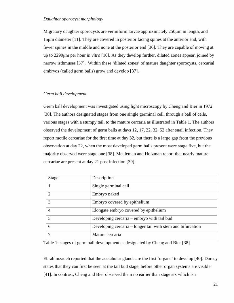
Germ ball development
Germ ball development was investigated using light microscopy by Cheng and Bier in 1972
[38]. The authors designated stages from one single germinal cell, through a ball of cells,
various stages with a stumpy tail, to the mature cercaria as illustrated in Table 1. The authors
observed the development of germ balls at days 12, 17, 22, 32, 52 after snail infection. They
report motile cercariae for the first time at day 32, but there is a large gap from the previous
observation at day 22, when the most developed germ balls present were stage five, but the
majority observed were stage one [38]. Meuleman and Holzman report that nearly mature
cercariae are present at day 21 post infection [39].
Stage Description
1 Single germinal cell
2 Embryo naked
3 Embryo covered by epithelium
4 Elongate embryo covered by epithelium
5 Developing cercaria – embryo with tail bud
6 Developing cercaria – longer tail with stem and bifurcation
7 Mature cercaria
Table 1: stages of germ ball development as designated by Cheng and Bier [38]
Ebrahimzadeh reported that the acetabular glands are the first ‘organs’ to develop [40]. Dorsey
states that they can first be seen at the tail bud stage, before other organ systems are visible
[41]. In contrast, Cheng and Bier observed them no earlier than stage six which is a

22
‘developing cercaria’, approximately 300μm long with the tail and body roughly equal in
length [38]. The developing glands can be recognised by their size as they are larger than the
other cells, and they are positioned in the centre towards the posterior end of the body [41].
The prominence of their nuclei is also noted. Dorsey uses the ratio of secretory granules to
golgi apparatus and ER as a measure of maturity. Early on, machinery for protein synthesis is
present, but secretory granules are yet to be formed; when the glands are mature the fundi and
ducts are packed with vesicles containing the cercarial secretions [41]. In addition, he
described the gland fundi and their ducts separately as it was not possible to reconstruct an
image of the entire cells from the sections taken. Dorsey did not attempt a ‘strict chronological
study’, as cercariae from different snails with the same post exposure date developed at
different rates [41]. This is likely to be due to the different times taken by daughter sporocysts
to migrate to the hepatopancreas, and any secondary daughter sporocysts arising from the first
generation.
The development of the cercarial tegument has been observed using electron microscopy by
Hockley [20] and Meuleman and Holzmann [39] and using conventional light microscopy by
Cheng and Bier [38]. They each chose different time points to study; this fact, along with the
different methods used and the lack of more general descriptions of the germ balls
morphology means that these three studies are difficult to synthesise into one coherent
description of germ ball development. The two electron microscopical studies describe a
primitive epithelium that surrounds embryonic cercariae before their true tegument forms [20,
39]. Cheng and Bier did not observe this, hence their designation of stage two as a naked cell
aggregate (Table 1) [38]. Hockley states that the primitive epithelium probably derives from
the germ ball, and serves to protect it until the formation of the true tegument [20]. In contrast,
Meuleman and Holzmann show evidence that it is formed from extensions of the daughter
sporocyst’s tegument, and suggest that it may provide the developing embryo with nutrients
from the snail haemolymph [39]. The two studies agree that the primitive epithelium is a
nucleated syncytium, underneath which peripheral germ ball cells expand and coalesce to
form the true tegument; the primitive epithelium is then shed [20, 39] and is not present 21
days post-infection [39]. It is also agreed that the subtegumental cell bodies arise
independently of the tegument syncytium and form connections to it later, i.e. no evidence was
found of nuclei from the syncytium sinking into the parenchyma [20, 39].

23
Morphology of the cercaria
Cercariae have been the subject of extensive ultrastructural studies. As a result much is known
about cercarial anatomy [42]. The cercaria is a complex creature that comprises two main
parts: the body and the tail. The tegument is covered in a single outer membrane, which is in
turn enveloped/coated in a 1μm thick glycocalyx [20] which probably has a role in enabling
this stage to be water-impermeable [43]. The ventral sucker or acetabulum is situated two
thirds down the body. Schistosomes are very muscular, and the cercaria is no exception. There
are two layers of muscle underlying the tegument surrounding the parasite; outermost is the
circular layer, which encloses the inner longitudinal layer [35]. Approximately half to two
thirds of the body volume is taken up by the ten large unicellular acetabular glands; two pairs
of pre-acetabular glands lie anterior, and three pairs of post-acetabular glands posterior to the
acetabulum (Figure 1.3). Post-acetabular glands contain homogenously granular secretory
granules, and granules with electron dense bodies. Pre-acetabular glands contain granules
with homogenous dense matrix and some with a less dense matrix containing electron lucid
bodies [41]. When the glands are mature, the fundi and ducts are packed with vesicles
containing the cercarial secretions, ready for action when they find a host. The contents of
these gland cells are secreted during penetration of host skin via long ducts extending to the
anterior tip of the cercaria. The ducts run in two lateral bundles, with two pre- and three post-
acetabular in each. The bundles are surrounded by circular muscle fibres [44], and split as they
reach the anterior end of the parasite so that each duct opens into the outside individually [44].
The anterior section of the body contains a thick muscular head capsule. The mouth lies on the
ventral side of the head capsule, and opens into the oesophagus which is surrounded by two
layers of muscle, outer circular and inner longitudinal [45]. The oesophagus runs through the
posterior boundary of the head capsule, continues ventral to the brain [45], to the blind bifid
caecum or gut, just anterior and dorsal to the acetabulum. The luminal surface of the gut bears
many plates rather than villi. The gut is innervated by external neuromuscular junctions [45].
The gut is not active at this stage, the cercaria survives by using up a store of glycogen, more
than half of which is in the tail [46].
The head capsule surrounds the head gland (Figure 1.3). This is a large purportedly unicellular
gland which lies in the anterior area of the head capsule dorsal to the oesophagus and
acetabular gland ducts [47]. There are many narrow microtubule-lined ducts which extend

24
from the fundus and open into the tegument – not to the exterior of the larva. There are three
morphologically distinct types of vesicle present; in the cercaria these are found only in the
fundus, not the ducts [47]. Dorsey suggests that the head gland secretions may repair the
tegument at the anterior of the cercaria after damage occurs during penetration [47]. Crabtree
later postulated that the head gland contents may be a source of ‘lytic secretions’ used by the
schistosomulum to cross the epidermal basement membrane as the acetabular glands were
empty at that stage [48].
Figure 1-3 Diagram of a cercaria showing position of glands (not to scale).
The osmoregulatory or excretory system is made up of flame cells which are connected by
tubules to the body tail junction. The tubules extend down the tail to the tip of each furcus
where they end in excretory pores. There is confusion in the literature as to the number of
flame cells in the cercaria. There are reports of six pairs in the body and one situated
proximally in the tail. However, a diagram reproduced from Gordon et al by Stirewalt [49]
shows four pairs in the body and one in the tail. This diagram also shows ciliated areas in the
collecting tubules draining the flame cells. It is now acknowledged that cercariae have four
pairs of flame cells in the body and one pair in the tail [42, 50].
The nervous system in the cercaria has been visualised by staining for acetylcholine esterase
activity [51], neuropeptides [52], and by electron microscopy [53, 54]. The neurotransmitter 5-
Acetabular glands
Head gland
Acetabulum
Head capsule

25
HT was not detected by immunohistochemistry in the cercaria [52]. The ultrastructure of the
internal nervous system has been described [54]. The bilobed brain measures 15μm at its
widest point, and lies between the pre-acetabular gland fundi, and the head capsule. It lies
dorsal to the oesophagus and acetabular gland duct bundles [54]. Sixteen nerve trunks extend
from the brain, four pairs anterior, and four pairs posterior. In each direction there is one pair
of central nerve trunks and three pairs peripheral: lateral, dorsal and ventral. The anterior
central nerve trunks follow the acetabular gland ducts into the head capsule and continue to
the anterior tip of the cercaria. The peripheral nerves also enter the head capsule and branch in
order to innervate the body wall muscle [54]. The posterior nerves extend to the body tail
junction. It has been reported by Cousin and Dorsey that they contribute to the dorsal and
ventral nerve chords that extend the length of the tail stem [54]; however, Skuce et al report no
connection between the nerves in the tail and those in the body [52]. When the cercaria was
stained for AChE activity, a pair of ‘anterior ganglia’ was observed inside the head capsule,
with nerves extending anteriorly to the tip and posteriorly to the brain [51]. Reactivity was
also shown at the surface in structures which resembled “minute volcanic craters” [51]. These
may correspond to the ciliated pits of Nuttman [53]. Just under the surface, a network made up
of longitudinal and circular fibres, stained positive for AChE. The longitudinal fibres were
spaced 3 – 4μm apart, and the circular fibres were arranged closer together, 1.5 – 2μm apart
[51].
Three types of sensory ending have been described on the surface of the cercaria: 1) ciliated
cavities, 2) unsheathed uniciliated bulbs, and 3) sheathed uniciliated bulbs [53]. Ciliated
cavities are found on the cercarial body, adjacent to lateral nerve chords, but not on the tail.
They form a bulb shaped cavity approximately 1.5μm wide which may be open to the
environment. The pits contain five or six cilia and are packed with small dense and large clear
vesicles which are extracellular [53]. It was suggested that these ciliated cavities could
function as chemoreceptors as they are open to the environment [53].
Unsheathed uniciliated endings are found all over the cercaria including the tail and furcae
[53]. They consist of a bulb embedded in the tegument, from which a cilium extends up to
7μm. The cilium is not covered by the tegument. The bulb is packed with large clear vesicles
and microtubules which are continuous with those of the axon [53].

26
Sheathed uniciliated nerve endings are found only at the anterior tip of the cercaria [53, 55].
The cilium is shorter, only projecting 0.8- 2.5μm [55] or 1-2μm [53] and is surrounded by
tegument apart from at its apex [53]. The basal bulb of these structures is raised from the
tegument [55]. The acetabular gland duct openings are arranged laterally in two cresents.
They are surrounded by tegumentary folds which vary in height from 0.5μm at the inner,
concave edge, to 2.5μm at the outer, convex edge. The outer convex edge is reported to
support five [53] or seven [55] sheathed unicilated nerve endings.
Skin stage morphology
Many important changes take place on entry to the mammalian host. These include loss of the
tail, secretion of the acetabular glands contents and the swift transition to the double outer
membrane. First the cercarial glycocalyx is shed, then the cercarial surface is lost by the
formation and shedding of microvilli [56] and a new double outer membrane is formed.
Skelly et al cloned, identified and sequenced 3 new S. mansoni glucose transporters (SGTP1,
2 and 4) and confirmed their function by expression studies in Xenopus oocytes [57]. Skelly
and Shoemaker showed that SGTP4 protein is present in adults, and schistosomula (1hr post
transformation), but not in eggs, cercariae, or the intramolluscan stages [58]. Jiang et al
showed that SGTP4 is present in both the membranocalyx and plasma membrane, but not the
tegument basement membrane of adult worms and schistosomula [59]. Thus SGTP4 can be
used as a very effective marker for the outer tegumental membranes in intramammalian
schistosomes. Indeed, Skelly and Shoemaker used immunostaining of SGTP4 to track the
formation of the new tegument during transformation induced by incubation of cercariae in
various different media [60]. SGTP4 appeared at the surface 30 minutes into transformation,
and covered the surface by 3 hours [60]. These results show that the new tegument surface is
formed rapidly on transformation.
As early as 30 minutes after penetration into mouse skin the head gland ducts have become
wider and secretory vesicles can be seen in them and in the tegument [47]. It has been
suggested that the material from the head gland, from the multilamellar vesicles in particular,
is used to repair damage of the tegument sustained whilst migrating through mouse skin [47].
Dorsey suggests that the head gland is a specialised type of tegumental cell body [47].
Contrary to this, the authors of an investigation into penetration of the skin and entry into a

27
vein, using the hamster cheek pouch model, report that head gland secretions are likely to be
involved in blood vessel penetration [48]. Vesicles of head gland type were observed in the
host, outside the parasite [48]. Forty hours after initial penetration the contents of the
acetabular glands had been secreted, whereas the head gland was still visible. Vesicles of the
type from the head gland were seen in the tegument at the anterior of the head capsule as early
as 30 minutes after entry to host skin [48]. Another study, which observed cercariae incubated
in different media to encourage transformation to schistosomula, reported that although the
acetabular glands were empty after 30 hours the head gland still contained secretory vesicles
and was easier to see than in freshly emerged cercariae [40]. By day two the schistosomulum
starts to elongate, and fewer spines are present between the mouth and the ventral sucker [61].
The cercarial ciliated papillae are lost; however, some sensory endings remain throughout
migration [61]. By day three all the midbody spines had disappeared, but spines remain at the
anterior and posterior ends, probably to be used as anchors during migration [61]. An
investigation into schistosomula collected from lung 4 to 7 days after infection, showed that
the head capsule musculature was no longer present.
The organisation of the flame cells in the parasite body does not change from cercaria to
schistosomulum. The functional excretory pore is at the posterior end, where the body/tail
junction had been, rather than at the tips of the tail furcae. There have been reports of the
excretory pore serving as point for large molecules to enter the schistosomulum as it
penetrates the mammalian host [62]. There is some discussion in the literature as to when the
schistosome gut becomes active on entering the mammalian host [49]. It has been suggested
that early schistosomula may ingest media [33]. However, in skin and lung stage
schistosomula, electron microscopy shows that the oesophagus is too narrow to admit
erythrocytes [48]. Crabtree noted that even if the oesophagus was large enough to
accommodate cells, the mouth is pressed up against the blood vessel wall in migrating
parasites, making ingestion of cells very difficult [63].
1.1.5. Physiological changes
Along with the morphological changes discussed above, there are several physiological
changes that take place on entry to the mammalian host. The cercaria survives by using a store
of glycogen as the substrate for aerobic metabolism. Lawson and Wilson noted that this would
be used up after 24 hours in vivo so the schistosomulum must rely on the host as a source of

28
metabolites shortly after penetration [46]. This is borne out by the observation described
earlier that the glucose transporter SGTP4 appears at the surface of the schistosomulum as
early as 30 minutes after transformation [60].
Harrop and Wilson investigated the rate of protein synthesis and release by in vitro cultured
schistosomula and found that much protein was released in the first 3 hours after
transformation [64]. Hardly any translation occurred during the first 24 hours; after this point
the rate steadily increased to a peak at day 8, then decreased for two days before rising again
at day 11. An important finding of these authors was that in vitro cultured schistosomula
would mature normally if transferred to a host intravenously [64]. In addition it was shown
that schistosomula release proteins into the medium between 24 hours and 7 days after
transformation. It was suggested that these proteins may originate in the head gland, as the
acetabular glands were no longer present [64]. It is worth noting that the lack of translation in
the first 24 hours is not due to a lack of mRNA. Blanton and Licate showed that post-
transcriptional control is exerted during this period [65].
Metabolic function from cercaria to adult was investigated by Skelly and Shoemaker using
genes involved in glucose uptake, phosphorylation, glycolysis, the Kreb’s cycle, and the
electron transport chain to probe northern and dot blots [66]. They discovered that cercarial
tails have the highest level of transcripts involved in aerobic respiration, and schistosomula
have the lowest. Adult worms had the highest levels of glucose transport transcripts, and the
transcripts encoding aerobic respiration enzymes were increased, but not to the level of the
cercarial tail [66]. It is worth noting that transcripts for SGTP1 and 4 were low and
undetectable in cercariae and cercarial tails respectively [66]. This raises a question as to how
SGTP4 protein arrives so quickly at the tegument of the schistosomulum.
1.2. Part 2: Gene expression
Having observed the morphological developments that take place during the infection process,
the second part of this chapter deals with the molecular biology of schistosomes. First, the
sequencing projects will be described, followed by the progress in and difficulties associated
with genetic manipulation of schistosomes. The use of microarrays for gene expression
profiling generally and in schistosomes specifically will be introduced. In addition to
transcriptional profiling, the availability of sequence data has allowed proteomic studies to be

29
carried out. Our knowledge of proteins involved in the infection process will be presented.
Finally, the importance of spatial expression patterns will be highlighted, and the methods
available to elucidate them will be described.
1.2.1. The history of S. mansoni sequencing efforts
Simpson et al stated that understanding the genome and its expression would provide a new
approach to discovering gene products of immunological or pharmalogical interest [67]. They
estimated that the S. mansoni genome was 270 megabases in size in 1982 [67]. Ten years later
the Schistosome Genome Project (including S. mansoni and S. japonicum) was launched by
the World Health Organisation. The gene discovery programme began around the same time,
using a directionally cloned adult worm cDNA library. The ends of these cDNAs were
sequenced to produce expressed sequence tags (ESTs) [68]. This project resulted in 607 ESTs
from 429 clones; 16% matched known S. mansoni genes, 22% matched genes from other
organisms, and 33% had no significant matches to any sequences deposited in GenBank.
Clustering revealed that the ESTs represented 169 genes, 154 of which were newly identified.
Another study branched out from using adult worms only, and included cDNA libraries
synthesised from eggs, cercariae and lung stage schistosomula [69]. The authors noted that
larval stages can not be overlooked in the search for drug targets and vaccine candidates [69].
The resulting 1401 ESTs clustered together represented 466 unique genes, of which 8%
matched known S. mansoni genes, 20% matched sequences from other organisms, but the vast
majority 71.5% were new genes with no significant matches in GenBank [69]. This last
category may represent schistosome specific genes, or simply genes which have not been
sequenced from other organisms yet.
A new method called open reading frame expressed sequence tags or ORESTEs was
developed to enable sequencing when starting material is limiting (ng of mRNA). It involves
using arbitrary primers in conjunction with low-stringency RT-PCR. This method was
reported to be three times more efficient than using directional cDNA libraries [70]. Other
advantages of this method are firstly that it tends to sample the middle region of a cDNA
rather than being biased to either end, and secondly, it normalises and samples rare transcripts
very efficiently [71]. However, the direction of the sequences is unknown. In addition, the

30
relative expression of transcripts can not be estimated using this technique, as the most
abundant transcripts are excluded before sequencing.
In 1999 Santos et al published a study of genes expressed by cercariae, having sequenced two
cDNA libraries from this stage. Its purpose was to understand the biology of the host parasite
relationship, and to identify novel gene products which could be useful as drug targets or
vaccine candidates [72]. 64% of the ESTs discovered in this study had no matches at
GenBank. The total 859 ESTs represented 453 genes. Those that could be putatively identified
based on homology with other organisms were mostly involved in energy metabolism, gene
expression, and regulation and signalling.
1.2.2. The Transcriptome
As a culmination of the sequencing efforts described above, a detailed and comprehensive
analysis of the S. mansoni transcriptome was published in 2003 [73]. Sequences from a
normalised adult worm cDNA library were added to ORESTES reads from eggs, miracidia,
germ balls, cercariae, and schistosomula. Altogether, 163,000 ESTs were sequenced and
compiled to form 31,000 assembled sequences (contigs). The authors estimated that this
represented a 92% sampling of the probable 14,000 genes encoded by the genome [73]. A
very high proportion of the sequences (77%) were new S. mansoni gene fragments, and 55%
had no significant match to anything at GenBank. The presence of many genes encoding cell
or tissue adhesion molecules was noted. Dicer and Piwi/Argonaute were also identified; these
proteins form part of the RNA interference pathway. It was observed that no highly variable
gene families were found, so evidence for antigenic variation was lacking at that time.
1.2.3. The Genome
The availability of extensive transcript data made gene finding possible, as the contig
sequences could be used to train gene finding algorithms [74]. The genome of S. mansoni has
been published recently alongside that of S. japonicum; these are the first Platyhelminthes to
be fully sequenced [75]. In contrast to earlier estimates, it was found to be 363 mega bases (cf
[67]), with at least 11, 092 genes. This is likely to be an underestimate, as more than 7,000
contigs are yet to be mapped to the genome [74]. Genomic DNA was extracted from cercariae
31
and libraries of varying sized inserts were sequenced randomly from either end using the
Sanger method. Several gene-finding algorithms were trained using a set of 409 manually-
curated genes before they were used on the sequence data. In addition to these automatically
annotated genes, 958 genes were manually curated. All data are available at
www.geneDB.org. Gene predictions are designated an identifier in the format Smp_xxxxxx;
ESTs or contigs are named in the format Smxxxxx.
An exciting discovery reported by Berriman et al in the genome paper is a group of Micro-
exon gene (MEG) families [75]. There were 14 families with 1 to 28 member genes in each; a
total of 45 MEGs were reported. These genes have signal peptides, and, as their name
suggests, very small exons and large introns. Not only are the exons very small, the transcripts
display high splice variability. Indeed cDNA clones have been sequenced that reveal copies of
MEG 1 with each of the exons missing [75]. The families are grouped together based on
sequence similarity; there is no homology between families. It is also worth noting that these
genes are represented in ESTs from intramammalian life cycle stages and germ balls, but were
not found in miracidia [75].
The genome publication also illustrates that schistosomes lack certain genes involved in lipid
metabolism and are reliant on the host for inositol [75].
The gene complement of S. mansoni was compared with that of the sea anemone Nematostella
victensis to identify genes which may be needed for a parasitic life style, a third germ layer,
and the formation of tissues into organs [75]. Tetraspannins, invadolysins and cathepsins were
highlighted as important for parasitism. Genes encoding cadherins (cell-cell adhesion),
Notch/Delta signalling (tissue patterning), and histone modification were all expanded
compared to Nematostella. It was also noted that schistosomes have tools required for
neurogenesis, axonguidance, and migration of neural cells. Many neuropeptides have been
discovered in schistosomes, some of which appear to be platyhelminth-specific [75, 76].
Another area where schistosomes are different is in their complement of G protein-coupled
receptors (GPCRs) and gated ion channels. No voltage-gated sodium channels were found, but
many voltage-gated potassium channels are present [75].

32
Bioinformatic analyses were performed in order to identify potential new drug targets. One
approach used was to build up a picture of the metabolic pathways and to highlight ‘choke
points’ which could be targeted. A second approach was to search for schistosome orthologues
of genes known to be targeted by drugs currently in use to treat human disease – either
targeting a human protein, or a protein belonging to a human-infective pathogen. The rationale
for this approach was to enable ‘piggy-backing’ of a current chemotherapy, potentially saving
much time and money, as the current drugs have already passed the stringent safety testing
required. However, this approach does not take into account the selective toxicity required to
target a pathogen specifically, rather than the host.
The publication of the S. mansoni genome, 27 years after the first investigations into its size, is
finally realising the aim stated by Simpson et al of providing a new approach to identify gene
products of immunological or pharmacological interest [67]. Indeed, Caffrey et al carried out a
comparative genomics analysis with C. elegans and D. melanogaster [4]. Genes which led to
lethality, paralysis, or reduced motility when knocked out in the model organisms were
identified and their orthologues in S. mansoni found, resulting in 72 possible leads. This set
was filtered to exclude redundant genes, and those with no intramammalian expression
pattern, leaving 57 genes. Of this set, 35 were found to be ‘druggable’ and the structure of 18
of these had been solved. The final 18 genes are the subject of further investigations by
Caffrey et al [4].
1.2.4. Genetic manipulation of schistosomes
Progress has been made both in gene knockdown by RNA interference (RNAi), and in the
application of transgenesis techniques. Genes encoding proteins in the RNAi pathway (Dicer
and Piwi/argonaute) were discovered in the S. mansoni transcriptome [73]. Since then there
have been several reports of successful, specific gene knock down in schistosomula [77, 78],
eggs [79], and intra-molluscan stages [80]. On the basis of this, Brindley and Pearce state that
“RNAi works powerfully in schistosomes and we see no reason why this approach should not
become routine in laboratories studying gene function in schistosomes.” [81]. It is worth
noting that the genes for which a positive effect was observed are expressed either in the gut,
or in tissues close to the surface. Both of these sites are readily accessible to the culture
medium. Issues around delivery of dsRNA to deeper tissues must be addressed. A contrasting
view was taken by Geldhof et al who reviewed the use of RNAi in parasitic helminths more

33
broadly [82]. They reported that non-specific effects, limited efficiency and reproducibility are
problems shared by many species. They are more cautious than Brindley and Pearce, and say
that the technique is in need of some development before it can be relied upon as a screen for
functional genomics. Indeed, in 2009, de Moraes Maurão et al chose 32 genes known to be
expressed by mother sporocysts, and carried out a phenotypic screen using RNAi [83].
Miracidia were allowed to transform in vitro into mother sporocysts in the presence of
dsRNA. qPCR was carried out to measure transcript levels after seven days of culture, and any
phenotypes were noted. Only 11 of the 32 treatments resulted in any phenotype – all of these
were a reduction in parasite length. Transcript levels were reduced in 6/11 with decreased
length, and 6/12 with no observed phenotype [83]. The authors note that gene specific effects
must be controlled and experiments optimised carefully. Hence their suggestion that RNAi
techniques should be developed further, before they can be used to screen for gene function
[83].
1.2.5. Transgenesis
Heyers et al state that there are at least two obstacles for a reliable, tractable transgenesis
system in schistosomes: 1) the development of constructs that allow stable integration into the
genome, and 2) the demonstration that genetically modified parasites can complete the life
cycle [84]. They report the use of particle bombardment to deliver a plasmid into miracidia
and the subsequent infection of snails by transformed specimens. The plasmid used in this
experiment encoded enhanced green fluorescent protein (EGFP), flanked by the S. mansoni
heat shock protein 70 (HSP70) promoter and terminator. The construct was not designed to
integrate into the genome. Transcript (but not protein) was demonstrated in infected snails
several days after infection, and gold particles were evident inside germ balls within mother
sporocysts. The development of mother sporocysts containing germ balls was taken as
evidence that the miracidia were ‘completely vital’; snails were killed before the infection was
patent, so there is no evidence as to whether cercariae would be produced from such an
infection [84].
In 2002 Wippersteg et al used virions to deliver GFP under the control of the schistosome
HSP70 promoter. They showed virions fusing with the worm tegument, and integration into
the genome by southern blot [85]. However, this technique failed to result in delivery of the
transgene to germ cells buried deep within the worm. They later used particle bombardment to

34
deliver GFP flanked by the promoter and termination regions of the cysteine protease ER60
[86]. This was carried out in order to determine the tissue specificity of ER60. RT-PCR and
western blot confirmed expression of GFP. Although particles were distributed throughout the
worm body, GFP was only detected in the excretory secretory system, confirming earlier
results gained by conventional methods.
A promising result was described by Beckman et al, who used particle bombardment to
deliver a plasmid encoding GFP under the control of the S. mansoni actin promoter to
miracidia before allowing them to infect snails [87]. The resulting cercariae were collected and
some from each snail were taken for molecular analysis, and the others used to infect mice.
The resulting adult worms were also analysed by PCR. Confocal microscopy was carried out
48 hours post bombardment, and fluorescence was noted in a ‘mosaic’ like pattern on the
tegument. This was taken as evidence that the DNA remained episomal rather than integrating
into the genome [87]. Transcription levels of the transgene showed that the transgene was
present in the first whole life cycle, but not in later rounds. Kines et al acheived incorporation
of the firefly luciferase gene into the S. mansoni genome using a pseudotyped murine
leukaemia virus [88]. Luciferase protein was detected by immunohistochemistry. However, it
was not demonstrated that transduced schistosomula would mature and complete the life cycle
if injected into mice [88].
Taken together, the techniques described above show great promise for the heritable
introduction of a transgene into schistosomes; but substantial hurdles remain. If a construct for
genome integration could be delivered to germ cells, by targeting an appropriate larval stage
with particle bombardment, the transformed larvae could then be re-introduced to the life
cycle and the resulting generations examined.
1.2.6. Global transcription profiling by microarray
Microarrays are a widely used tool for comparing transcription levels between different
biological samples. This is achieved by hybridising fluorescently labelled cDNA from the
samples under investigation to an array of cDNA or oligonucleotide probes immobilised on a
solid substrate. After hybridisation, the array is washed under stringent conditions, and the
fluorescence intensity level of each probe is recorded. The brighter the signal from a probe,
the more cDNA was present in the test sample. It is important to note that information can

35
only be obtained for sequences which are represented on the array. In order to get statistically
significant results, at least three biological replicates (i.e. material from separate organisms)
must be carried out for each test.
Arrays can be made by printing cDNAs from a library onto glass slides, or by synthesising
oligonucleotides in situ resulting in high-density formats. Many different platforms are
commercially available. Affymetrix for example, use 20nt long probes perfect match (pm)
paired with mis-matches (mm) where a single base is changed; 10 probes per gene are made,
and the expression of a gene is calculated using the mismatch as a background control. Roche-
NimbleGen use a digital micro-mirror array which enables them so synthesise 60mer probes,
making mismatches unnecessary [89]. This technological advance allows very high density
arrays to be synthesised. Whole genome arrays are available from a range of companies for
various organisms, meaning that the expression of their entire genomes can be investigated
under different conditions.
Data analysis from microarray experiments is no small task. First the fluorescence data must
be quality assessed, and any aberrant arrays excluded. Background fluorescence must be
accounted for, and the data must be normalised. Finally statistical analysis can be carried out
to determine which genes are differentially expressed. It is usual to express the difference in
gene expression in log2 fold change. Often a cut-off of fold change of 2 is applied. In addition,
a statistical significance cut-off is used. The P-value is adjusted for multiple testing. Another
statistic that is used is the B value, or log odds. This is the log (odds that a gene is
differentially expressed). For example if B = 3, the odds that a gene is expressed is e3 = 20, or
1 in 20, corresponding to a probability of 95%. If B = 0, the likelihood of differential
expression is 50% [90].
There are many software packages available for microarray data analysis; one option is
Bioconductor, an open source package of programmes in the statistical language R [91]. It can
be used to perform a wide range of analyses from raw fluorescence data to meaningful
biological information. Pair wise comparisons can be made using the limma package to
produce top tables of the fold change in expression of each gene between samples [90].The
Gene Ontology [92] is a controlled vocabulary of terms that describe gene products across
species. There are three independent ontologies: molecular function, biological process and

36
cellular compartment. The ontologies are structured, so that root or parent terms are general
and their descendants are more specific. One gene may be annotated to several GO terms,
belonging to one or more ontologies. The GOstats package
(http://bioconductor.org/packages/bioc/html/GOstats.html) can be used to determine whether
particular groups of genes are enriched in one sample compared to another. Another
possibility is to cluster the genes based on expression pattern. This can allow hypotheses to be
made regarding the function of an unannotated gene if it clusters with genes whose products
have been characterised.
Although microarrays offer a very sensitive method to characterise transcript levels in a
sample, caution should be applied when inferring conclusions regarding the presence of a
particular protein, as post-transcriptional control should be taken into account.
1.2.7. Previous applications of microarray technology to the study of schistosomes
The use of microarrays is a well-established tool for investigating gene expression patterns in
schistosomes. Various different arrays have been designed and used to answer separate
biological questions. As noted above, information about a particular sequence can only be
gained if it is represented on the array. Microarrays have been used to address differences in
gene expression between female and male adult worms [93, 94]; the rationale for this is that an
insight into sexual development could lead to interventions to interfere with egg production,
thereby reducing disease and transmission. Most recently this has been carried out using laser-
capture micro-dissection of gastrodermis versus vitellaria [95]. Another popular approach is
to measure gene expression between life cycle stages [96-99]. Here the focus is on identifying
transcripts related to biological processes occurring in a particular life cycle stage. Even the
effect of host gender on parasite gene expression profile has been investigated [100].
There are various different arrays in use by the community. They range from cDNA made
exclusively from lung stage sequences [98, 101], a 44k oligo array [102], other oligo arrays
based on ESTs available at TIGR [97] or WTSI [99], to an array with sequences from both S.
mansoni and S. japonicum [103]. These different arrays have arisen as the sequence data
available have increased over time, and/or to ensure that the genes relevant to the biological
question under consideration were included.

37
Of particular relevance to this thesis are three published studies undertaking life cycle based
experiments. The first, by Dillon et al reports the use of a custom made cDNA array
comprising 6000 features representing ~3088 contigs and singlets from the lung stage larva
[98, 101]. The authors state that this equates to approximately half of the 7,000 genes expected
to be expressed by any one life cycle stage. This array was used to identify transcripts
enriched at the lung stage compared to eggs, germ balls (dissected from snails 21 days post
infection), day 2 and day 7 in vitro cultured schistosomula, and worms obtained from mice 21
days and 7 weeks post infection. The authors report a decrease in abundance of transcripts
involved in aerobic metabolism and chromatin remodelling in schistosomula (both day 2 and
day 7), compared to the reference sample. This supports the switch in larval metabolism
described by Lawson and Wilson [104]. GOminer analysis revealed that genes involved in
energy metabolism, cytoskeletal organisation, protease activity and chromosome remodelling
showed the largest changes between lung schistosomula and adjacent life cycle stages. This
array was designed specifically to show only lung stage transcripts, so it cannot answer
questions regarding transcripts specific to the other life cycle stages.
The second, by Jolly et al, used an array comprising 12000 45-50mer probes representing
contigs from the S. mansoni Genome Index at The Institute for Genome Research (TIGR).
They compared daughter sporocysts (whole infected hepatopancreas six weeks post infection),
cercariae and adult worms. Uninfected hepatopancreas was used as a control for the snail
contribution to the daughter sporocyst sample. They report that, although the data set used to
design the probes was of unknown orientation, >9,700 of the probes gave some signal, and
therefore were likely to have been printed in the correct direction. Furthermore, only two
probes gave a signal when hybridised with the snail only control. Jolly et al report that
daughter sporocysts have high levels of transcripts encoding proteins involved in translation
and quality control, as would be expected for this cercaria-producing stage. Transcripts
involved in cell death and ubiquitination were also up-regulated; these may play an important
part in morphological development. Genes highly expressed in cercariae were mainly those
involved in mitochondrial function, enabling the energy production necessary for swimming.
However, this stage was less transcriptionally active than the other stages studied. In contrast,
adults produced a much wider range of transcripts; they were involved in egg production,
immune evasion, and energy metabolism. Overall, 1,154 genes were differentially expressed
in at least one contrast, most of these were enriched in the adult. 406 of the differentially

38
expressed genes had no known function. Interestingly, the hepatopancreas used was collected
6 weeks post infection, which the authors state is before cercariae were released. In our
laboratory snails are shed routinely at five weeks post infection, so mature cercariae would
have been present in the sample called daughter sporocyst. In spite of this, it was noted that
the gene expression profiles of infected hepatopancreas and mature cercariae were different.
The third study was carried out by Fitzpatrick et al, using an array comprising 37,632 50mer
probes [99]. The array was based on the sequence data available at GeneDB.org in 2005 [105].
They hybridised material from an impressive 15 separate life cycle stages: eggs; miracidia;
mother sporocysts; daughter sporocysts (migratory stage obtained from snails 15 days after
infection); cercariae; 3hr, 24hr, 3 day, 6 day in vitro cultured schistosomula; and worms
obtained from mice at the following times post infection: 2wk, 3wk, 5wk, 7wk males, 7wk
females. The authors analysed the data in several ways to get the most information from it.
They carried out pair wise statistical analyses, a network analysis, GO category enrichment
analysis, and focused on specific families of interest. Statistical analysis identified 973 non-
redundant genes which were differentially expressed in at least one comparison, 448
transcripts exhibited a difference of more than 32 fold. Network analysis showed that 73% of
the dataset were differentially expressed.
One finding Fitzpatrick et al highlighted was that the minichromosome maintenance (MCM)
heterohexamer which is involved in licensing DNA replication is upregulated in the mother
sporocyst and 3 week worm. This supports the observation by Clegg that cell division does
not occur in migrating schistosomula, but remodelling is achieved by existing cells migrating
[106]. Another discovery was that cercariae are not as transcriptionally inactive as previously
thought. The authors focused on three gene families with may have value as intervention
targets. Fucosyl transferases, tetraspanins, and G protein coupled receptors (GPCRs). The first
group were examined as they are responsible for post translational modifications which may
contribute to immunomodulation by the invading parasite. Tetraspanins are membrane
proteins which are interesting not only because they have been identified on the surface of
adult worms [107], but also because it has been shown that people with reduced susceptibility
to re-infection by schistosomes have antibodies to them [108]. The array experiment showed
that two tetraspanins (Sm04463 (best blast hit Smp_194970) and Smp_140000) were up-
regulated in stages from cercaria to adult. Unfortunately only 19 of the 65 putative GPCRs

39
predicted in the genome were present on the array used by Fitzpartrick et al; however, they
discovered that Smp_149770, Smp_152540 and Smp_127310 are enriched in the
schistosomulum. Also highlighted as potential drug targets are Leishmanolysin, netrin and the
netrin receptor.
Another approach Fitzpatrick et al took was to identify constitutively expressed genes which
may be necessary for each life cycle stage. They take the view that platyhelminth-specific
genes represent important genes to investigate for interventions – in contrast to the approach
taken by Caffrey et al [4]. It could be a long and arduous task to discover more about the 294
platyhelminth-specific genes expressed in either cercaria to schistosomulum or later intra-
mammalian stages and their products but one with rich rewards.
1.2.8. Molecules associated with the infection process
Long before microarrays were invented it was known that secretions from the acetabular
glands of Schistosoma cercariae play an important role in host invasion. Gordon and Griffiths
noted in 1951 that secretions from these glands separated layers of the stratum corneum and
postulated that post-acetabular gland secretions may have a lytic effect on host cells [109]. In
1977, Dresden et al., demonstrated that the proteins from the cercaria had proteolytic activity
against keratin and basement membrane proteins, but not collagen [110]. McKerrow et al
showed that a 30kDa protease purified from cercarial secretions by chromatography could
degrade type IV and VIII collagens as well as laminin and fibronectin [111]. Marikovsky et al
immunolocalised a 28kDa protease to the pre- and post-acetabular glands, as well as the
surface [112]. However, Salter et al stated that invasion is mediated by a single serine protease
[113]. The first cercarial protease to be cloned was cercarial elastase (CE) in 1988 [114].
Newport et al demonstrated mRNA encoding CE in the post acetabular glands of developing
cercariae by in situ hybridisation of a radiolabelled cDNA probe to sections of infected snail
hepatopancreas. The transcript was not present in mature cercariae. Northern blots with the
same probe showed the transcript in infected snails, but not in uninfected snails or adult
schistosomes [114]. This work showed that there was transcriptional regulation both at the
level of developmental stage and cell type. In 2002 Salter et al showed that there are five
genes encoding cercarial elastase in the S. mansoni genome and stated that two of them
account for more than 90% of the activity and protein released [115]. They also showed that

40
these genes are conserved between S. mansoni and S. haematobium and can also be identified
in S. douthitti.
With the advent of both transcriptomic data and proteomic techniques, several studies of
cercarial skin penetration have been carried out [116-119]. Curwen et al collected the proteins
secreted by cercariae in the first three hours after mechanical transformation and separated
them using 2D gel electrophoresis before identifying the spots by mass spectrometry.
Cercarial elastase was found, along with SmPepM8 (a metalloprotease with homology to
leishmanolysin), and a dipeptidyl peptidase IV. Curwen also reported several putative
immunomodulatory proteins; these included 3 containing Sperm Coating Protein (SCP)
domains (named SCP a, c, and d), a novel protein named SmKK7 with homology to BmKK7,
(a potassium channel blocker found in scorpion venom), and Sm16 which had been identified
by Rao and Ramaswamy [120]. This work suggested that the process of skin invasion by
cercariae is more complex than had been previously acknowledged.
Proteins containing SCP domains are found throughout biology and have been identified in
several helminths including hookworms Ancylostoma duodenale and Necator americanus
[121], Brugia malayi [122] and Onchocerca volvulus [123]. Studies on the Brugia SCP (VAL-
1) could not conclude whether it was secreted or not [122]. When jirds were vaccinated with
Bm VAL-1 the average worm burden was not reduced [122]. An Onchocerca SCP has a role
in promoting angiogenesis [123]. Ancylostoma secreted protein (ASP) contains the SCP
domain; it is the largest constituent of the proteins secreted by infective L3 larvae. Anti-sera to
the N. americanus ASP-2 inhibits larval migration in-vitro. Humans with anti-ASP-2 have
lower average worm burdens [121]. This protein has undergone phase 1 clinical trails in naïve
adults in non endemic region with some success; the vaccine was tolerated, and induced
elevated antibody titres [124]. It remains to be seen whether there is any effect on worm
burden.
A family of 28 S. mansoni genes encoding SCP domain containing proteins has been
described recently by Chalmers et al [125]. They were named SmVAL (venom allergen like)
1-28. Expression of VALs 1-13 throughout the life cycle was investigated using qPCR. VALs
1, 4, 6, 7 and 10 were upregulated in cercariae relative to the other stages (Curwen’s SCP a, c
and d correspond to VALs 4, 10 and 18, respectively). VALs 8 and 12 were restricted to liver

41
stage worms, and VALs 2, 3, 5, and 9 were expressed by eggs, miracidia and mother
sporocysts.
SmKK7 was named after the scorpion toxin BmKK7, the activity of which is dependent on the
position of several key amino acids within a three-dimensional structure that is stabilized by
three disulfide bridges [126]. The level of conservation between these important residues in
the schistosome and scorpion proteins suggests a similar function for the schistosome
molecule. Because T-cell activation is known to be regulated by potassium channels [127], it
is plausible that the invading schistosome modulates the immune response of the host by this
mechanism.
1.2.9. Localisation
A common theme in all of the S. mansoni sequencing projects to date has been the high
proportion of ‘schistosome unique’ sequences obtained. Indeed, according to Wilson et al,
informative identity may be as low as 26% [128]. The schistosome only genes are attractive as
vaccine candidates and/or drug targets because of their lack of identity with host proteins
[129]. Nevertheless, functional annotation of the remaining 74% of S. mansoni genes
represents a large challenge. As discussed in Part 1, schistosomes have a complex body plan
with tissues and organs. Therefore localisation of a specific gene can facilitate hypotheses as
to its function. An example given by Dillon et al is the case of a protease: if localised to the
gut it is likely to play a role in digestion, whereas if it is seen in the acetabular glands it is
probably involved in host invasion [16]. An important fact to take into account when
searching for vaccine candidates or drug targets is whether the target is accessible to the
immune system or the potential drug. In the case of schistosomes, the host-parasite interface
comprises the tegument, the gut, the secretory glands, and potentially the excretory system.
Wilson and Coulson note that the most of the vaccine candidates tested in trials carried out by
the WHO and other labs were cytosolic or cytoskeletal and therefore unlikely to be available
to the host whilst the parasite is intact [130]. This fact led Curwen et al to state “it seems
counterintuitive that such proteins would form the basis of an effective vaccine” [131], an
opinion also held by DeMarco and Verjovski-Almeida [129], who also point out that in silico
analyses can only be taken so far; a protein which is secreted may still be located deep within
the body rather than exposed at the host-parasite interface [129]. There is no substitution for

42
experimental localisation data; this should be obtained early in the pipeline for both drug
target and vaccine candidate validation.
Classically localisation studies are carried out by immunolocalisation of proteins. This is a
long, difficult and costly process involving two particularly hard steps. Firstly the gene of
interest must be cloned and expressed, or a peptide chosen and synthesised; secondly an
animal is vaccinated with the recombinant protein to elicit an immune response. The serum is
then collected and tested for reactivity to the protein in question. Finally the antiserum can be
used to localise the protein, either in sectioned or whole mount worms, the latter is a long
protocol in itself [32].
In order to allow higher throughput localisation studies, Dillon et al adapted whole-mount in
situ hybridisation (WISH) for use in schistosomes. WISH has long been used in development
to discover where specific transcripts are expressed during embryogenesis. This is the first
application to a fully differentiated organism. A part of the gene of interest must be cloned (at
least 250bp) and anti-sense digoxigenin (DIG)-labelled RNA probes made. These probes are
hybridised to fixed, permeabilised whole mount parasites, and detected using an alkaline
phosphatase-conjugated anti-DIG antibody in conjunction with a colourimetric or fluorimetric
substrate. Not only is this technique higher through-put than immunohistochemistry (IHC), it
also reduces the use of laboratory animals, and circumvents the often problematic step of
protein expression. An example of this is antigen 10.3 (MEG 4.1) – a highly repetitive protein
which was difficult to express in vitro. Dillon et al were able to synthesise probes and
localised the transcript to the oesophageal gland of adult worms. The protocol described by
these authors can be used for any of the intra-mammalian life cycle stages [16].
The availability of sequence data has enabled transcriptomic and proteomic experiments to be
carried out, and so some of the molecules associated with infection by larval schistosomes
have been identified. However, previous microarray studies have been hampered by the
incomplete nature of the microarray platforms available and the exclusion of germ balls. A
time line for germ ball development is lacking in the literature. Techniques for gene knock
down and transgenesis are not yet routine in schistosomes. However, WISH can be used as a
screen to discover which tissue(s) a gene is expressed in.

43
1.3. Aims of the thesis
The aim of this thesis is to investigate the gene expression patterns underlying infection of the
mammalian host by larval schistosomes. This can be broken down further into two questions:
1) Which genes are differentially expressed from germ ball to cercaria to skin
schistosomulum?
2) Where are these genes expressed?
In order to answer these questions a prerequisite is an in depth knowledge of the morphology
of the life cycle stages involved. This is addressed in Chapter 2. The next part of the story was
explored using a gene expression microarray experiment which is described in Chapter 3.
Chapter 4 deals with localisation of some of the gene products highlighted in Chapter 3 and in
the literature concerning the infection process.

44
2. Morphological study
45
2.1. Introduction
Schistosomes undergo two transitions in the part of the life cycle under investigation; firstly
from the snail intermediate host to fresh water, and secondly, from fresh water to the
mammalian definitive host. These three contrasting environments present different challenges
for the parasite. Considerable morphological and physiological changes take place as the larva
adapts to each niche. An understanding of these morphological changes will provide the
context for investigations into the gene expression patterns associated with infection by larval
schistosomes.
Morphological studies have been carried out on these life cycle stages. Cercariae have been
studied more extensively than either germ balls or skin stage schistosomula; a situation which
has not changed since a review published in 1974 [49]. The early work on schistosome larval
morphology was carried out using conventional light [38], or electron microscopy [39, 41, 42,
44, 47, 132]. These methods enabled broad observations of body structure or detailed studies
of ultrastructure, respectively, to be made. As a result, much has been revealed about the
morphology of these important life cycle stages; however, several questions remain. No
definitive timeline has been established for germ ball development. This is partly due to the
asynchronous nature of development, but also to a lack of regular sampling [38] and the
difficulty with reconstructing images from sections of infected snail hepatopancreas [41].
Another question which remains is the cellular make up and function of the head gland. In
addition the skin stage schistosomulum has not been well studied.
More recently the advent of confocal microscopy has enabled detailed studies to be made of
internal structures using whole mount worms. This technique allows pictures to be taken with
a restricted focal depth, so that ‘optical sections’ may be imaged. A series of optical sections,
called a z-stack, can be easily built up into a 3-D representation of the specimen using
specialised software. This technique has several advantages over conventional and electron
microscopy; the technically difficult process of sectioning small specimens is unnecessary,
and the orientation is always known. This aids interpretation of the resulting images.

46
New staining methods have become available for use in conjunction with confocal
microscopy. Langeron’s carmine is a general protein stain which is visible by both light and
fluorescence microscopy. It has been used successfully with confocal microscopy to provide
novel insights into the anatomy of adult schistosomes [34]. It has not yet been applied to
larvae. Phalloidin is a toxin from the ‘Death Cap’ mushroom (Amanita phalloides) which
binds specifically to filamentous actin (f-actin). It can be conjugated to various fluorophores
and used to examine musculature. Studies of adults [32], cercariae [35], miracidia and mother
sporocysts [133] have been carried out using this method and have revealed that the outer
muscle layers, oesophageal and gut musculature, acetabulum, and flame cells all stain well.
This makes phalloidin an attractive stain for use with larval schistosomes. Nuclei may also be
stained using propidium iodide (PI) and 4',6-diamidino-2-phenylindole (DAPI). These are
aqueous stains which bind to DNA and fluoresce when excited by a 543nm (PI) or 405nm
(DAPI) laser. These stains are ideal as a counterstain with phalloidin, but they cannot be used
with the ethanolic Langeron’s Carmine.
These techniques will be applied for the first time to daughter sporocysts, germ balls, and skin
stage schistosomula. Although cercariae have been studied using phalloidin, this represents the
first use of Langeron’s carmine with this stage.
Aims
The aims of this study are:
To discover when important cercarial structures, such as the secretory glands, are
developing in the germ ball. The information gained will be used to ensure that germ
balls are sampled appropriately for the gene expression study (Chapter 3).
To describe the anatomy of the daughter sporocyst, cercaria, and skin stage
schistosomulum. This knowledge will be necessary to interpret the data gained in the
following chapters, particularly the localisation studies (Chapters 4).

47
2.2. Methods:
2.2.1. Parasite material
All parasite material was from a Puerto Rican isolate maintained at the University of York by
passage through NMRI mice and albino Biomphalaria glabrata snails. To obtain daughter
sporocysts and their developing germ balls, snails were infected with 40 miracidia each and
dissected carefully in filter-sterilised 50% phosphate buffered saline (PBS; 3.2 mM Na2HPO4,
0.5 mM KH2PO4, 1.3 mM KCl, 135 mM NaCl, pH 7.4.) 23 – 27 days later. Obvious snail
material was removed with a finely drawn-out Pasteur pipette. Daughter sporocysts and germ
balls were harvested separately using a clean drawn out pipette and kept on ice until use.
Cercariae were collected from snails infected with 10 miracidia each. Five weeks after
infection the snails were placed in the dark for two days and then placed under a bright light in
approximately 10mls aerated tap water (pond water) for two hours to induce shedding of
cercariae. The resulting cercariae were concentrated by cooling on ice for one hour to prevent
swimming.
To obtain skin stage schistosomula, cercariae were obtained as above, and transformed
mechanically before culturing for three days in vitro [134]. After concentration on ice and two
brief washes in cold RPMI media (Invitrogen Gibco), the cercariae were transferred to 3mls
M169 media (see below) and vortexed at full speed for 90 seconds to shear the bodies from
their tails. They were then separated on a discontinous percoll gradient (70% and 45% percoll
in RPMI). The bodies were recovered from the interface with a pastette, taking care to avoid
the tails in the upper layer. Bodies were washed extensively in RPMI and then plated out into
24 well plates in M169 (BME (Eagle) liquid medium (Gibco) containing: 0.1% Lactalbumin
hydrolysate, 0.1% Glucose, 0.5μM Hypoxanthine, 1μM Serotonin, 1μM Hydrocortisone,
0.2μM Triiodothyronine, 0.5% MEM vitamins, 5% Schneider’s drosophila medium, 1%
Penicillin/streptomycin) and incubated at 37°C with 5% CO2 for three days. They were then
recovered and washed twice more in RPMI [64].

48
2.2.2. Langeron’s Carmine staining
Parasites were fixed in AFA (22% formalin, 33% 95% ethanol, 11.5% glacial acetic acid,
33.5% water) overnight, and then stored in 70% Ethanol until use (at least 10 minutes if they
were used immediately). The ethanol was removed and replaced with a few drops of
Langeron’s Carmine (LC) and incubated at room temperature for 50 minutes. Excess stain was
removed by washing the parasites several times with 70% ethanol. The parasites were then
dehydrated through 90% ethanol and two changes of absolute ethanol (10 minute incubations
at RTP). They were cleared for 15 minutes in Histoclear (Fisher Chemicals), mounted onto
slides with DPX (BDH Chemicals), allowed to dry overnight and stored at room temperature.
2.2.3. Phalloidin and DAPI staining
Daughter sporocysts, germ balls, cercariae and skin stage schistosomula were stained with
Alexafluor 488-conjugated phalloidin (Invitrogen, Molecular Probes) to visualize f-actin, and
counterstained with DAPI according to Bahia et al [133]. Specimens were fixed for one hour
in 4% paraformaldehyde in PBS on ice and then permeablised for an hour in PBS, 0.2%
gelatin, 0.1% saponin, 0.1% NaN3 (PGN) at room temperature, before addition of phalloidin
(0.0165μM, 20 minutes) and/or DAPI (0.01μM, 30 minutes) in PGN at room temperature.
Specimens were washed several times in PBS before they were viewed with the Zeiss invert
confocal microscope.
2.2.4. Confocal microscopy
Confocal microscopy was carried out using a Zeiss LSM 510 meta on an Axioplan 2M
(upright), or a Zeiss LSM 510 meta on an Axiovert 200M (invert).
Pictures were taken with a Plan-Aprochromat 63x/1.4 oil DIC under oil immersion.
Stain Laser Beam splitter Filter
Langeron’s Carmine 543nm HFT 488/543 Long Pass 585nm
Phalloidin 488nm 405/488/543 Band Pass 505-530nm
DAPI 405nm 405/488/543 Band Pass 420-480nm
PI 543nm Long Pass 585nm
Table 2.1 Confocal microscope settings

49
2.3. Results
2.3.1. Daughter Sporocyst
Migratory (Fig. 2.1) and mature daughter sporocysts (Fig. 2.3), were stained with phalloidin to
visualise f-actin. Note coverage of anterior end with posterior-facing, actin-rich spines
highlighted by phalloidin staining (Fig. 2.1). Spine coverage is sparse in the mid region of the
sporocyst with no spines at the posterior end (Fig. 2.1). There is a protrusion at the anterior
end (Fig. 2.1b). Circular and longitudinal muscle fibres can be seen, and are arranged in a
pattern reminiscent of a loose weave material. Four flame cells are visible along with their
collecting tubules (Fig. 2.1b).
Langeron’s Carmine staining reveals the cellular make up of the migratory daughter sporocyst
(Fig. 2.3). Most cells are irregularly shaped. The outer edges of the body comprise small cells
(approximately 4-5μm in diameter) with fewer large cells (8-10μm diameter) comprising the
internal core of the body. These core cells may be germ cells which will develop into germ
balls. Extensive punctate staining in the nucleus of the majority of large cells provides
evidence of cell division, the cytosol of these cells is full of protein (Fig. 2.2).
When the daughter sporocyst is further developed it is no longer vermiform (Fig. 2.3).
Bulbous regions of the body are connected by actin-rich isthmuses. The surface spines are still
in evidence, with the same distribution as in the younger specimens (Fig.s 2.1 and 2.2). The
spines are most numerous on the very anterior bulb where they are paired; fewer can be seen
on the second bulb. The PI staining reveals many cells packed into the bulbs. These are the
‘brood chambers’ where the germ balls will develop into cercariae. The outer muscle layers of
these brood chambers form a ‘loose weave’.

Figure 2-1 a and b Migratory daughter sporocysts stained with Alexafluor 488-conjugated phalloidin
a) single slice b) 3D projection of 17.47μm deep z-stack. Note spines at anterior end (arrowhead), lon
(LM) and circular muscle (CM), and four flame cells (arrows) with the ducts (chevrons) connecting t
is a protrusion at the anterior end (star).
LM
CM
a
50
gitudinal
hem. There
b

51
Figure 2-2 Migratory daughter sporocysts stained with Langeron’s carmine.
Parasites were recovered from snails 21 days post-infection and fixed overnight in AFA and stored in 70%
ethanol. They were and viewed using an upright confocal microscope using the 543nm laser. This specimen is
~180μm long. The optical section shown is 14μm into the 23μm z-stack. The core of the body consists of large
cells (arrow heads) with smaller cells arranged along the periphery (arrows). Inset are enlargements of cells. Note
prominent nucleolus (N), punctate staining in the nucleus (P), and staining throughout the cytosol (C) scale bar
5μm.
P
N
C

52
Figure 2-3 Projection of a 38μm deep z-stack of a daughter sporocyst
This specimen was stained with alexafluor 488-conjugated phalloidin (green) as above, with the addition of a 5
minute incubation with PI (red) before viewing by confocal microscope using 488nm and 543nm lasers. Note
brood chambers (chevrons), narrow actin-rich constrictions (large arrows). Surface spines (arrows) are paired on
the most anterior bulb and are distributed singly on the second bulb.
2.3.2. Germ ball development
The first germ ball documented is the single germ cell inside the daughter sporocyst. Next is a
loosely connected ball of cells which are not uniform in size or shape (Fig. 2.4a).The cells

53
vary in diameter from 2-9μm, they have prominent nucleoli, and there is no staining in the
cytoplasm. Some cells with two brightly staining spots can be seen indicating that they are
about to divide (not shown). The next stage in development is that the germ ball elongates
(Fig. 2.4b). At this point there are some prominent large (7μm diameter) cells at its edge. The
remaining cells average 4-5μm in diameter. At this stage it is difficult to distinguish the
anterior end from the posterior, and the germ ball does not have an outer epithelium. The cells
are tightly packed together, in contrast to the earlier stage.
By the time the germ ball is ~50μm long, an epithelium surrounds the body (Fig 2.5). There
are cells contiguous with this epithelium (Fig.s 2.5b and 2.7), which I will refer to as
contributing cells. The posterior end of the germ ball is flattened (Fig. 2.5) and towards its
anterior there is a group of cells arranged in an arc which may be the developing head capsule
or the developing acetabular gland cells (Fig. 2.5a). The appearance of the majority of cells
has changed. Unlike those in the early round germ ball (Fig. 2.4a), they stain positive for
protein in the cytoplasm, indicative of protein synthesis.
When the germ ball is approximately 75μm in length (Fig. 2.6) the epithelium and some
contributing cells are still clearly visible. In addition to this, bridges of protein stain are now
evident, which may link internal cell bodies with the outer epithelium (Fig. 2.8). The head
capsule area at the anterior end of the body is also more developed with cells elongated along
the antero-posterior axis (Fig. 2.6). Phalloidin staining at this stage (Fig. 2.6c) shows that
circular muscle is present, and although some strands of longitudinal muscle are evident, they
are far fewer in number. There is a protrusion at the anterior end which may be developing
sensory endings (Fig. 2.6b).
By the time the germ ball has a tail (body ~80μm, tail ~50μm, Fig. 2.9) its cells are well
organised and some organ systems are recognisable. The body may be described in quarters
from anterior to posterior. The first, anterior quarter, is made up of cells that will become the
head capsule. They are well ordered, but there are no visible structures. The second quarter
contains the neural ganglia. The third quarter comprises all 10 acetabular gland cells in the
centre and three clearly separate bands or layers of cells on each side. The posterior quarter
contains undifferentiated cells. The cells in the tail are neatly arranged in rows along the side
and the tail is bifurcated from this early stage (Fig. 2.9a). A phalloidin stained germ ball of

54
approximately this age has two visible flame cells (Fig. 2.9B), and the outer muscle layers are
denser in the region of the head capsule than across the rest of the body. Fibres of actin can be
seen in squares on the tail which has two little knobs where the furcae are starting to grow.
Phalloidin staining of an older germ ball (Fig. 2.10) (tail detached during fixation) shows that
the circular muscle layer is formed first, with the longitudinal muscles following. The latter
are formed from bands of muscle cells in tiers around the body after the head capsule muscle
boundary has formed. Diagonal muscle strands are also visible at this stage.
In order to discover the time line of development, snails were infected with 40 miracidia each
and dissected daily starting 21 days later. At this early time point I noted that only small round
germ balls were present (Fig. 2.4a – Cheng and Bier stage 2). Motile cercariae were observed
for the first time at day 27 post- infection. At days 24 and 25, there was a range of germ balls
from small round ones, through various stages with tails (Cheng and Bier stages 5 and 6) but
not including motile cercariae.
Figure 2-4 a and b. Optical sections of germ balls stained with Langeron’s carmine.
Germ balls were recovered from snails at daily intervals from 21-27 days after infection with 40 miracidia and
fixed in AFA. They were stained, dehydrated and viewed with a confocal microscope with a 543nm laser at 63x.
Scale bars 50μm. a) cells vary widely in size and have prominent nucleoli (arrow). Protein staining is absent in
the cytosol. Small cell highlighted by arrowhead. b) an elongated germ ball, anterior and posterior ends are
indistinguishable. Cells are still variable sizes.
a b

55
Figure 2-5 a and b. Young germ ball stained with Langeron’s carmine
Selected optical sections, 23μm and 10μm through a 27μm deep z-stack, are shown. Note that the posterior end is
flattened (stars). The body is more organised; towards the anterior end there is a group of cells arranged in an arc
shape (asterix). Very thin Epithelium covers the body (block arrow), and cells contributing to it can be seen (thin
arrows). Scale bar 50μm.
*
a
b

56
Figure 2-6 Developing germ ball.
*
EC
a
*
CM
LMc
G
*
b

57
Figure 2-6a) and b) optical sections, 24μm and 19μm through a 38μm deep z-stack, of a germ ball approximately
75μm long. Gland cells are appearing (G), the epithelium (block arrows) and contributing cells are still present
(arrow). Connections between parechymal cells and the epithelium are evident (chevrons). The anterior end is
organising into the head capsule cells are elongating along the anterioposterior axis (EC). There is a protrusion at
the anterior end (arrow head). c) Germ ball of approximately the same developmental stage stained with
phalloidin to visualize F-actin. Circular muscle is well developed (CM), and the longitudinal muscle is
developing (LM), note that posterior end is flattened (asterix).

58
Figure 2-7 Outer epithelium of germ balls.
Areas cropped from germ balls stained with Langeron’s carmine shown in figures 2.5 and 2.6. Cells forming the
epithelium are shown (arrows).

59
Figure 2-8 Possible subtegumental cells
Areas cropped from germ ball shown in Figure 2.6 highlighting protein ‘bridges’ which may connect cells inside
the body to the epithelium (arrows). These cells may be the first sub-tegumental cell bodies. Scale bars 10μm.

Figure 2-9 a and b Germ balls with stubby tails.
a) Optical section of a parasite stained with Langeron’s
(HC), acetabular glands (AG) and tail (T) are visible. b)
ball stained with phalloidin, note two flame cells in the
end (chevron). Scale bar 50μm
AG
HCT
a
b
AG
HCT
NG
carmi
Opti
centre
FF
60
ne. Putative neural ganglia (NG), head capsule
cal section (1.46μm into a 10.2μm z-stack) of a germ
(F) and the well developed muscle at the anterior

61
Figure 2-10 Muscle development in a maturing germ ball
Optical section of a more developed germ ball stained with phalloidin (green) and DAPI. The tail detached during
fixation. Note acetabulum (A), the circular muscle is formed (CM), longitudinal muscle is developing (LM), head
capsule musculature (H) more developed than that of the body.
2.3.3. The Cercaria
The fully developed cercarial body measures 190μm in length and 40μm in width. Posterior
facing spines cover the body and tail. Most striking at this stage are the 10 acetabular gland
cells which comprise approximately one third to half of the cercarial body volume (Fig.
2.11b). These cells have large nuclei and nucleoli (Fig. 2.13a and b). Their ducts form two
bundles which are surrounded by muscle (Fig. 2.11a), and extend to the anterior of the head
capsule, where they reach to the exterior of the larva through the tegument. There are
protrusions from the surface in this area shown by LC staining (Figs 2.13a and b). The
muscles surrounding the two bundles of acetabular gland ducts are stained by phalloidin, and
can be seen on either side of the body just posterior to the head capsule boundary and also
within the head capsule (Fig. 2.11a). This stucture extends posteriorly and is bounded by two
layers of muscle: circular and longitudinal. At the anterior end of the body there are also
LM
A
CM
LM
LM
H

diagonal muscles, crossing from anterior to posterior (Fig. 2.11d). The head gland can be seen
in Figs 2.13b and c, lying anterior and dorsal to the oesophagus, extending from the junction
between the tegument and the head capsule muscle boundary, to the anterior end of the body.
This gland is similar in appearance to the acetabular glands when stained with LC in that there
is very little staining in the cytoplasm. There are two prominent nucleoli (Fig. 2.13b and c).
The acetabulum or ventral sucker is positioned two thirds down the length of the body. It is
supported by eight lengths of muscle anchored dorso-laterally (Fig. 2.11a). The acetabulum is
made up of many cells. This is highlighted by DAPI staining (Fig. 2.11a), whereas phalloidin
staining shows concentric rings of actin fibres and strands of muscle extending radially from
the centre to the edge (Fig. 2.12a). Pseudo-striated muscle can be seen in the tail (Fig. 2.13b).
There are 8 flame cells in the cercarial body, and two at the proximal of the tail; they are
arranged in pairs laterally. There are two pairs at the posterior of the body between the
acetabulum and the body/tail joint, one pair lies dorsal, the other ventral (Figs 2.12 a and d,
respectively). The middle pair lies in the same plane as the caecum, just posterior to it (Fig.
2.12b). The anteriormost pair is dorsally situated, between the caecum and the head capsule
boundary (Fig. 2.12c). In the area between the acetabular glands and the head capsule, the
cells are small and tightly packed. DAPI staining also shows a group of undifferentiated cells,
approximately the same size as the acetabulum, on the ventral aspect, posterior to the
acetabulum, between post -acetabular glands (Fig. 2.11a).
62
U

63
Figure 2-11 Cercaria stained with phalloidin (green) and DAPI (blue).
Optical sections shown are a) 3.78μm, b)4.91μm, and d) 6.23μm from the ventral surface. a) Note acetabulum
(A) with supporting muscles (S), large acetabular glands with ducts (D), head capsule, oesophagus (O). A group
of undifferentiated cells (U) can be seen at the posterior end. b) Area occupied by acetabular glands is shown. d)
The outer muscle layers are visible: circular (CM) Longitudinal (LM) and diagonal (DM). c) area from a)
enlarged to show oesophagus (O), position of the gut (G), and an acetabular gland duct bundle (D) in more detail.
The oesophagus measures 4μm across, the gut branches are 7μm wide and 12μm long. Scale bar represents
10μm.
MD
A
O
O
G
S
D
DM
LM
CM
AG
U
dc
ba

64
Figure 2-12 Cercaria stained with phalloidin
This is the same specimen as in figure 2.11, but Z sections, starting ventral (a) and moving dorsal (d), were
selected to show the position of the flame cells (arrows), and DAPI is not shown. The two posterior pairs are
shown: ventral (a; 2.64μm into the z-stack), and dorsal (d; 6.04μm). The middle pair (b; 4.15μm) are more ventral
than the anterior pair (c; 5.48μm). There are no flame cells in the head capsule. Radial muscle fibres (RM) can be
seen in the acetabulum.
a b
c d
RM

65
Figure 2-13 Cercariae stained with Langeron’s carmine
AG
A
HG
H
TM
*
a
b
c
U
AG
HDA
T

66
Figure 2-13a) The body tail junction is visible (T) and the acetabular gland cells (AG) are prominent. The group
of undifferentiated cells can be seen (U) just posterior to the acetabulum (A). Muscle boundary of the head
capsule is stained (H), the acetabular gland ducts (D) wind through the head capsule, the putative sensory endings
are indicated at the anterior (arrow heads). Scale bar represents 100μm. c) is a z section 2.59μm from b), showing
a second brightly staining spot visible in the head gland (asterix). Scale bar 10μm.

67
2.3.4. Skin stage schistosomulum
The body of the skin schistosomulum is approximately 140μm long, and 55μm wide. Posterior
oriented spines are prominent at either end, but almost absent from the middle third of the
body (Fig. 2.15b). The two layers of muscle (circular and longitudinal), and the supporting
diagonal strands are well developed and neatly arranged (Fig. 2.15b). Examination of the
entire z stack reveals 8 flame cells, positioned as in the cercaria (Fig. 2.14). The acetabular
glands and their ducts are no longer present; however, the head capsule with its strong muscle
boundary is still intact (Fig. 2.14b) with the head gland still in evidence (Fig. 2.15a). It is
different morphologically to the cercarial head gland in that it contains spherical vesicles (Fig.
2.15a), but as in the cercaria, there are many extensions which seem to be continuous with the
tegument. The oesophagus is 3-4μm in diameter. The gut is larger and more prominent, and
positioned dorsally just anterior to the acetabulum (Fig. 2.14b); the lumen stains positive for
protein (Fig. 2.16). The neural ganglia are discernible by LC staining (Fig. 2.16), along with
lateral nerve chords extending from them down the length of the body (Fig. 2.16). The
acetabulum is prominent, as are the muscles supporting it (Fig. 2.14a). As in the cercaria, these
are anchored dorso-laterally (Fig. 2.14a). The cells throughout the body are notably different
morphologically from those in the cercaria and early germ ball, but similar to those of the later
germ balls and daughter sporocyst (Fig. 2.2). There is distinct punctate staining in the nucleus
of the vast majority of cells. The sensory endings that were present at the anterior end of the
cercaria are no longer present.

*
A
H
O
a b
68
SM
DM
EP
Sc d
RM

69
Figure 2-14 Selected optical sections of a schistosomulum stained with phalloidin.
Sections shown in a) to d) are 4.46μm, 5.95μm, 7.43μm and 8.18μm, respectively, through an 11.52μm deep z-
stack. a) Acetabulum (A) with its retractor muscles (RM) are highlighted, and flame cells are indicated (arrows).
b) Head capsule muscle boundary is visible (H), with the oesophagus (O) passing through it, and leading to the
dorsally situated blind gut (asterix). The oesophagus is 4μm wide, the gut sacs are 16μm long and 10μm wide.
The excretory pore (EP) is seen at the posterior. c) ventral middle and posterior flame cells are shown (arrows).
d) diagonal muscle (DM) and posterior dorsal and ventral flame cells (arrows) can be seen.

70
Figure 2-15 Skin stage schistosomulum
a) Optical section of a parasite stained with Langeron’s carmine. Note head gland (HG) within head capsule (H),
lack of acetabular glands and thick tegument (T). The oesophagus is also seen (O). b) Skin stage schistosomulum
stained with phalloidin. Note anterior and posterior surface spines (S), and muscle layers: circular (CM),
longitudinal (LM) and diagonal (DM). Scale bars 50μm.
T
H
O
HG
a b
CM
DM
LM
S
S

F
S
s
p
2
S
a
F
c
s
a
B
2
T
P
a
a
c
igure 2-16 Schistosomulum stained
elected optical sections (a) 2.92μm
tack. Note acetabulum (A), lateral n
resent in the gut lumen (G), head c
.4. Discussion
chistosomes undergo impress
pproximately 7 days from ge
amiliarity with the anatomy o
ome and put the infection pro
chistosome. The advantages o
nimal is always known, and i
oth these facts make images
.4.1. Daughter sporocysts
o my knowledge this is the fi
revious studies have describe
ppearance of brood chambers
s revealed by phalloidin stain
ells.
A
N
N
a
with Langeron’s carmine.
(ventral), (b) 5.85μm, (c) 11.69μm
erve chords (N), oesophagus (O), n
apsule (H) is still in evidence.
ive and rapid morphological c
rm ball to cercaria, and a furth
f these lifecycle stages will fa
cess in context of considerabl
f using whole mount specime
mages do not have to be ‘reco
easier to interpret.
rst application of confocal mi
d the ‘vermiform’ nature of th
[10, 36, 37]. Novel insights i
ing, and the proportion of the
O
NG
b
71
(central), taken from a 22.73μm deep z-
eural ganglia (NG), protein staining is
hanges in a brief time,
er 3 days to skin worm.
cilitate interpretation of results to
e developmental changes for the
ns are that the orientation of the
nstituted’ from serial sections.
croscopy to daughter sporocysts.
is stage, the spines, and the
nclude the arrangement of muscle
body core made up of large germ
H
G
c

72
In both migratory and developed daughter sporocysts the outer muscle layers are arranged as
in a loose basket weave conformation. This is particularly evident in the brood chambers. It is
likely that this loose weave allows the brood chambers to expand in order to accommodate the
growing germ balls. Phalloidin staining also enables visualisation of the protonephridia; four
flame cells and their two connecting tubules can be seen, strongly suggesting the presence of
actin in the tubule walls. This is the only stage in which the tubules are visible.
Langeron’s carmine staining highlights the cellular make up of the daughter sporocyst. The
majority of cells comprising the core of the body are large and may represent the germ cells
which will develop into germ balls. Previous attempts at germ line transformation in
schistosomes have focussed on larval stages such as miracidia or schistosomula which can
easily be re-introduced to the lifecycle. It has been shown that daughter sporocysts can be
successfully transplanted into snails and produce cercariae [135]. The prominence of germ
cells and ‘loose weave’ muscle configuration revealed here, suggest that the migratory
daughter sporocyst may be a prime target for transgenesis. DNA constructs could be delivered
reliably to germ cells, and the transduced daughter sporocysts returned to snails to continue
the life cycle.
2.4.2. Germ ball development
This is the first application of confocal microscopy to the study of germ ball development;
previous studies have used light microscopy [38] or electron microscopy [41]. It is also the
first time that this process has been observed daily from 21-27 days post-infection, thus filling
the gap in the report of Cheng and Bier, who did not observe germ ball between days 22 and
32 post infection [38]. The findings are in agreement with both Cheng and Bier and Dorsey’s
observations that germ balls from snails infected on the same day, and indeed germ balls from
the same daughter sporocyst develop at different rates. However, in contrast to Cheng and
Bier’s findings, only small round germ balls were present at day 21, germ balls with stubby
tails were present at days 24 and 25. Motile cercariae were observed for the first time at day
27.
Germ balls corresponding to Cheng and Bier stages 2-7 were documented. Although these
designated stages are helpful, there are some differences in the specimens observed presently.
The authors show diagrams of germ balls with a spherical tail bud joined by a constriction to

73
the germ ball body. Novel observations in this study show that the posterior end of the germ
ball first flattens and then extends, becoming bifurcated very early on. Cheng and Bier [38]
show the acetabular gland ducts separately extending to the anterior of the germ ball. Later
ultrastructural studies have shown that the ducts exist in a bundle surrounded by muscle fibres
[44]. I have not observed the ‘primitive epithelium’ [20, 39]. However, this was reportedly
lost at day 21 post infection [39], the time at which my observations began. The present data
show that the tegument is joined by ‘protein bridges’ to cells in the body before the nuclei in
the tegument are lost. This confirms the observation of Hockley, who reported that the
tegument cell bodies arose independently of the tegument syncytium and that processes from
these cells migrate towards the tegument and become continuous with it [20].
The circular muscle layer develops first, followed by the longitudinal layer, and then the
diagonal strands appear last of all. The longitudinal fibres begin as short separate segments,
which must coalesce to form the complete strand. It is possible that each cluster of fibres
originates from an individual cell. The head capsule muscle is well developed by the time the
tail is elongating; it can be seen as more developed than the body musculature at the ‘stubby-
tailed’ stage. The acetabulum can be seen at the ‘elongating-tail’ stage. Flame cells can be
seen first when there is a stubby tail. Nerve system development is not easy to follow with the
stains chosen; however, the brain is discernable at the ‘stubby-tailed’ stage.
The contents of the cercarial glands are known to play an important role in host invasion. In
order to include the transcripts for these proteins for the microarray experiment described in
Chapter 3, it was necessary to sample germ balls whose glands (both acetabular and head)
were developing. Although the development of cercarial glands has been described in some
detail, no timescale was established. When the germ ball is 50μm long, the primordial
acetabular gland cells can be seen towards the anterior of the body. The glands are prominent
when the stubby tail is seen, and they develop further as the tail elongates. Tail length may be
taken as a proxy for the appearance of acetabular glands. Ebrahimzadeh reported that the head
gland develops later than the other cercarial glands [40]. The current results agree; I cannot
discern this gland before the cercaria is mature. Taken together, these observations show that
germ balls must be collected from the ‘flat-bottomed’ stage through the ‘stubby-tailed’ stage,
but not including motile cercaria for the gene expression experiments. Thus, snails will be
dissected between 22 and 26 days after infection.

74
2.4.3. Cercariae
Many features of cercariae have been examined in detail using electron microscopy.
Phalloidin has also been used to describe the muscle structure. The present study used whole
mount preparations, and the stains phalloidin, DAPI and Langeron’s carmine in order to draw
the anatomical information into an overall picture.
Acetabular glands are the most prominent feature of cercariae, taking up approximately ½ of
the body volume. They failed to stain with either the general protein stain Langeron’s
Carmine, or the specific f-actin stain phalloidin. This indicates that the contents are not chiefly
protein. Phalloidin staining shows clearly that the gut is found just anterior to the acetabular
glands. The group of undifferentiated cells at the posterior of the cercaria have been described
previously as ‘genital anlagen’, the implication being that these cells will in time develop into
the adult reproductive system, which is the only anatomical system lacking in the cercaria. In
agreement with Stirewalt et al [49], the current study shows four pairs of flame cells in the
body and one pair located proximally in the tail. However, the ciliated tubules are not visible.
The other uncertainty in the literature is the cellular make up of the head gland. I have
observed two nucleoli by LC staining, suggesting that this gland comprises two cells rather
than one. This grasp of cercarial anatomy at the whole mount level will be essential for
interpretation of localisation experiments in Chapter 4.
2.4.4. Skin stage schistosomula
Presented here is the first investigation into the morphology of skin stage schistosomula using
confocal microscopy. The stains Langeron’s carmine and phalloidin were used and show a
detailed picture of the anatomy of this early intra-mammalian stage.
The developing schistosomulum is not larger than the cercaria, and has a similar range of sizes
from 110μm when contracted to 170μm when elongated. The tegumental spines are retained at
the anterior and posterior ends, and may help with migration, but there are no spines on the
mid-body section. The acetabular glands have been emptied and are no longer present.
However, the head capsule remains, indicating that this stage is indeed most similar to a ‘skin

75
stage’ schistosomulum as larvae recovered from lungs are reported to have lost the head
capsule [13]. In addition, the head gland persists, but it is morphologically different to the
cercarial head gland. Langeron’s carmine staining shows spherical vesicles which may
indicate that the gland is actively secreting at this stage. This finding is in agreement with
Crabtree and Wilson who state that the head gland plays an important role in crossing the
basal membrane of the epidermis to reach the dermis, and invasion of blood vessels once the
larva is inside the skin [48].
The nervous system undergoes some remodelling between the cercaria and the skin stage
larva, particularly the loss of longer sensory endings at the anterior end. The brain is more
prominent and lateral nerve chords can be seen extending down the length of the body. These
were not visible in the cercaria by Langeron’s carmine staining which suggests that a degree
of neural development occurs on entry to the host.
It has been shown that schistosomula are capable of taking in glucose through their tegument
shortly after arrival in the skin. However, there is some discussion in the literature as to when
the gut becomes active. The extra space available in the body once the acetabular glands have
disappeared allows the gut to expand. The gut pouches had elongated and widened and were
located more posteriorly, just anterior to the acetabulum. The oesophagus remains 4μm wide,
but has elongated. Novel evidence that the gut is now ‘active’ is the protein staining in the
lumen. This may be due to secretion of gut enzymes, in preparation for digestion, or the
ingestion of media containing protein, or both. Although the oesophagus is still not large
enough to admit erythrocytes, serum may well be consumed.
The excretory system is arranged much as in the cercaria; the flame cells are in the same
positions. However, the bladder is more obvious along with a muscular pore which is
surrounded by circular and radial fibres.
2.4.5. Final thoughts
The data presented in this chapter show the anatomical changes that occur during the
mammalian infection process. This lays the ground work for understanding the underlying
gene expression patterns which will be investigated in the coming chapters. The discovery that
germ balls with developing glands can be recovered from snails 22 to 26 days after infection

76
will allow appropriate sampling for the microarray experiment. In addition, the broad picture
of morphology gained will ensure well informed interpretation of localisation studies.

77
3. Microarray analysis of the germ ball, cercaria and day 3
schistosomulum

78
3.1. Introduction
The previous chapter described the morphological changes that occur in larval schistosomes
during the mammalian infection process. The next task is to investigate the gene expression
patterns underlying these developments. Several studies have used proteomics and
transcriptomics to examine this process at the molecular level [97, 99, 116, 119].
Recently two microarray experiments have been published which address the transition from
intermediate to definitive host, neither of which included all three of the life cycle stages
analysed here (germ balls, cercariae, day 3 schistosomula). Jolly et al compared infected
hepatopancreas with free cercariae and adult worms; schistosomula were not included [97].
Fitzpatrick et al omitted germ balls: their daughter sporocysts were migratory stage and did
not contain developing cercariae [99]. It is also important to recognise that these studies were
carried out using arrays designed before the full genome of S. mansoni was available. The
array used by Jolly et al comprised 12,000 45-50mer probes representing contigs from the S.
mansoni Genome Index at The Institute for Genome Research (TIGR) [97]. This dataset
comprises the ORESTES-generated sequences described by Verjovsky-Almeida et al in 2003
[73]. The ORESTES method uses random primers with low stringency PCR, therefore
direction of the resulting sequences is unknown [73]. Jolly et al report that, despite the
unidentified orientation of the probes, >9,700 of them gave some signal, and therefore were
likely to have been printed in the correct direction [97]. However, based on probability, it
would be expected that approximately half of the probes would be in the wrong direction.
Fitzpatrick et al used an array comprising 37,632 50mer probes, which was designed using the
sequence data available at www.GeneDB.org in 2005 [99]. Although the authors went to
great efforts to collect biological material from 15 separate life cycle stages, they were unable
to carry out one of their chosen analyses (expression of G Protein-coupled receptors (GPCRs)
throughout the lifecycle) as the array they used did not have unambiguous probes for the genes
they were interested in [99].
Array construction technology has improved radically, such that extremely high-density arrays
can be made. Combined with the availability of gene predictions encoded by the S. mansoni
genome and cDNA libraries from the lifecycle stages involved in infection, this made it
possible to design the first genome-wide microarray. This suggested that a new microarray

79
study of the mammalian infection process was due, as much new information should be gained
by using a comprehensive array. In particular, knowledge about transformation and the genes
expressed by skin stage schistosomula was lacking.
It has been shown that the transcripts encoding proteins secreted during cercarial
transformation are present only in the intra-molluscan germ ball [116]; therefore, it was
necessary to include this life-cycle stage in studies addressing infection by larval
schistosomes. The germ balls used here were dissected from snails 22-26 days post infection,
when their gland cells were developing (see Chapter 2). In order to discover the suite of genes
required by the schistosomula to survive and thrive in the mammalian host, three-day in vitro
cultured schistosomula were included. This stage corresponds to the skin stage larva (Chapter
2). Schistosomes transformed and cultured by the methods used in the current study are able to
mature if transferred to the mouse as a definitive host [64], they are biologically comparable to
ex vivo worms and can be produced in the large quantities needed to obtain enough RNA for
hybridisation without amplification.
In this chapter, the design and use of the first genome-wide microarray platform for S.
mansoni is described, and the insights gained into mammalian infection by larval schistosomes
are presented.
Aims
The aims of this study are:
To hybridise the novel S. mansoni microarray with cDNA from germ balls, cercariae
and day 3 schistosomula
Interrogate the resulting data to build a picture of the molecular processes
underpinning infection by larval schistosomes
Highlight novel genes involved in skin invasion and transformation

80
3.2. Methods
3.2.1. Array design
The array was designed by Dr A Ivens of Fios Genomics. All S. mansoni ESTs available at
GeneDB.org whose direction was known were compiled using phrap (Phil Green at Genome
Sciences Department, University of Washington, US;
http://www.phrap.org/phredphrapconsed.html). These formed the input for the array design
along with the predicted genes from version E of the S. mansoni genome (www.GeneDB.org).
The input data were broken up into sequential 50mers and redundant sequences were removed
using FAlite.pm (Ian Korf; http://homepage.mac.com/iankorf/) and associated Perl scripts.
The unique sequences were mapped back to the genome assembly using exonerate
(http://www.ebi.ac.uk/~guy/exonerate/). From a map ordered list, every 13th 50mer was
chosen as a probe. No selection was made for the number of probes per predicted transcript.
The design was sent to Roche-NimbleGen, who made some minor refinements for ease of
synthesis and constructed the arrays using digital micromirror technology [89].
3.2.2. Biological material
Parasites were obtained as described in section 2.2.1. Germ balls were recovered from snails
22-26 days post-infection with 40 miracidia each. RNA was extracted from the larvae with
TRIzol (Invitrogen, Paisley, UK) according to the manufacturer’s instructions. Double
stranded cDNA was synthesised using SuperScript II double-stranded cDNA synthesis kit
(Invitrogen).
3.2.3. Hybridisation
NimbleGen were supplied with at least 2.7μg of double stranded cDNA for three biological
replicates each from germ balls, cercariae, and day 3 schistosomula. Each replicate was
necessarily made up of pooled cDNA from multiple syntheses. Care was taken to ensure that
separate biological replicates were obtained i.e. no parasite homogenates were split across
replicates. Each biological replicate of cDNA was labelled with Cy3 and hybridised to an
array for 16 to 20 hours at 42°C. Slides were washed, and dried before fluorescence data was
read using a NimbleGen MS 2000 Scanner with NimbleScan software.

81
3.2.4. Statistical analyses
NimbleGen supplied background corrected data. All subsequent statistical analysis was carried
out by Dr A Ivens using programmes from the Bioconductor suite [91]. The data were quality-
assessed by visual inspection of graphical representations of the raw probe level data. Box
plots were drawn using the boxplot function from the graphics package. Correlation data were
calculated using the cor function from the stats package and heatmaps were made by calling
the heatmap.2 function in the gplots package. All arrays passed the quality assessment. Next
the data were quantile normalised using the normalizeBetweenArrays function in the limma
package. This ensures identical distributions of the data [136]. Following normalisation, the
probe level data were summarised to yield ‘gene level’ data. The probes were re-mapped to
the S. mansoni gene predictions at www.geneDB.org (version F) using exonerate. If a probe
matched an Smp locus with an e value <1-05 by both nucleotide and protein BLAST, the probe
was annotated to that Smp locus. The intensity value for each locus was the mean intensity of
all the probes by which it was represented. The resulting gene level data were the input for the
differential expression analysis, which was carried out using the limma package. First a linear
model fit was performed. This reduced the data for each gene to a mean value from each of the
life cycle stages. Next, differential expression data were obtained by performing a contrast
analysis. This compares the expression level of each gene in the following contrasts:
germ ball : cercaria
germ ball : day 3 schistosomulum
day 3 schistosomulum : cercaria
Multiple testing was corrected for using the eBayes function which employs the method of
Benjamini and Hochberg [137]. This gives an adjusted P value (adjP). Genes which were
differentially expressed with an adjP<0.0001 were chosen for further analysis. Finally a
hypergeometric test was carried out using the Category package to discover whether particular
GO terms were over-represented in the differentially expressed genes compared to the ‘gene
universe’ of Smps on the array.

82
3.3. Results
The availability of sequence data for S. mansoni allowed the design of the first genome-wide
array for this medically important species. The construction of the array will be described,
followed by an overview of the contrasts made. The differentially expressed genes will be
reported in groups based on functions.
3.3.1. Novel array
This is the most comprehensive array that has been constructed for S. mansoni. There are
385K features on the array comprising 377,598 sequences representing 16,222 loci from the S.
mansoni compilation and 11,613 random sequences for hybridisation controls.
Of the loci represented on the array, 84.4% correspond to a gene prediction (Smp) or can be
mapped to a gene prediction at GeneDB.org by BLAST, with an E value below a threshold of
1e-05. Gene predictions were classed as unannotated if they had no name, PFam ID, or Interpro
ID. Loci with no annotation (ie Smp no annotation + EST with no matches to an Smp) make
up 42.9% of the array.
Loci Number Percentage
(of total)
locus designated by Smp
of which have an annotation
of which have no annotation
11,585
7,160
4,425
71.4%
44.1%
27.3%
locus designated by EST
of which hit Smp by BLAST nucleotide (n) or
translated DNA versus protein (tblastx)
of which no BLAST hit to Smp
4,637
2,103
2,536
28.6%
13%
15.6%
Total loci 16,222 100%
Table 3.1 Summary of array features
Annotation is classed as having a protein name, Pfam ID or Interpro ID.
Of the 11,585 Smps, 4,425 had no annotation at all (38%).

83
3.3.2. Contrasts summary
There were large numbers of differentially regulated loci in each comparison made. The
fewest differences were observed in the day 3 – cercaria comparison. The comparison with the
highest number of differentially expressed genes was germ ball – cercaria. Before further
analysis, two filters were applied; loci with B≥3 which corresponds to 95% confidence value
(Chapter 1.2.6) and a fold change of ≥2.
Contrast GB vs C GB vs D3 D3 vs C
No. of loci with B≥3 1,731 1,431 1,066
Table 3.2 Summary of contrasts at gene level
3.3.3. GO analysis summary
GO analysis was carried out on the loci which matched Smps. Loci with no GO term
annotation were automatically excluded from this analysis. Table 3.3.3 below shows a
summary of the categories of genes enriched in each stage relative to the others.
Comparison Some categories highlighted by GO analysis
GB > C DNA replication, DNA repair, translation, Wnt signalling,
proteolysis, macromolecule biosynthesis, metabolism
GB > D3 DNA replication, proteolysis, microtubule cytoskeketon, ATPase,
biosynthesis, transferase activity, metabolism
C > GB Neurotransmitter transport, proteolysis, carbohydrate biosynthesis,
kinase activity, ion transmembrane transport, cytoskeleton
C > D3 Biosynthesis, cytoskeleton, catalytic activity, proteolysis, energy
production
D3 > C Membrane, pepsin A, lipid metabolism, transferase activity, calcium
ion binding
D3 > GB Proteolysis, enzyme regulation, neurotransmitter transport, ion
transmembrane transport, membrane, transferase activity, lipid
metabolism, hydrolase activity, calcium ion binding
Table 3.3.3 Examples of GO terms enriched at each stage relative to the others.

84
GO analysis reveals that DNA replication and proteolysis genes are up-regulated in the germ
ball compared to the other two stages. The developmental patterning Wnt genes are also up-
regulated in the germ ball along with genes involved in macromolecule biosynthesis and
metabolism. Several categories are enriched in the cercaria including proteolysis, biosynthesis,
cytoskeleton, neurotransmitter transport and energy production. Proteolysis and
neurotransmitter transport are also enriched in the schistosomulum compared to the germ ball.
Categories up-regulated exclusively in the schistosomulum include lipid metabolism and
calcium ion binding.
In addition to GO category analysis, categories based on those presented by Berriman et al in
the genome paper [75], and custom groups based on proteomic and/or localisation evidence
were also analysed. They are presented below in separate groups based on function. The bar
charts show the relative fold change for each gene. Each bar chart has a key table showing the
gene annotations in the order they appear in the chart. The baseline for each gene is the lowest
level of expression for that gene in the life cycle stages studied; it is set to unity. Therefore, the
actual baseline expression values may be different for each gene, and comparisons of absolute
expression levels between genes, based on the charts shown here, are not valid. However, the
patterns of gene expression may be compared between genes.
3.3.4. DNA replication and Cell division
DNA replication and cell division are important functions for an organism which is growing.
The genes included in the chart below were highlighted by the GOstats analysis. All of the
differentially regulated genes encoding proteins involved in DNA replication and cell division
were up-regulated in the germ ball compared to the cercaria. The fold changes in this category
ranged from 2 to 22 fold compared to the cercaria. The 22 fold change is exhibited by the
mitotic cyclin B. This is followed by histone H2A, which is expressed 18 fold higher than in
the cercaria. The DNA repair helicase, rad 25, is up-regulated approximately 2-fold in both the
germ ball and the day 3 schistosomulum compared to the cercaria.
Seven DNA polymerase subunits are enriched in the germ ball along with DNA primase,
DNA ligase, proliferating cell nuclear antigen (PCNA), and replication factors. Also
noteworthy are the minichromosome maintenance (MCM) proteins with are thought to unwind
DNA ahead of the replication fork; all six of the subunits which form the heterohexamer

85
(MCM2-7) are up-regulated in the germ ball with fold changes greater than five. Geminin
(involved in loading MCMs onto DNA), GINS, and CDC45 also form part of the
‘unwindosome’ and are highly expressed in the germ ball.
DNA replication and Cell division
0 5 10 15 20 25
Smp_082490Smp_086860Smp_172530Smp_176260Smp_054840Smp_153660Smp_143490Smp_094140Smp_079050.1Smp_037590Smp_079560Smp_067260Smp_002930Smp_047610Smp_032500Smp_033700Smp_113350Smp_069600Smp_096360Smp_124120Smp_046500.1Smp_047620Smp_178260Smp_100380Smp_075110Smp_124230Smp_040020Smp_186980Smp_035980Smp_172020Smp_046500.2Smp_106040Smp_151560Smp_141430Smp_066260Smp_012480Smp_169120Smp_076050Smp_171710Smp_076660Smp_055500Smp_009030Smp_121800Smp_087010Smp_161380Smp_019840.2Smp_151510Smp_136180Smp_051320Smp_165580
Gen
eID
Relative expression (lowest = 1)
GB
C
D3

86
ID Annotation ID Annotation
Smp_082490 cyclin B Smp_124230 DNA repair protein RAD51
Smp_086860 histone H2A Smp_040020 centromere protein A
Smp_172530 MCM4 Smp_186980 flap endonuclease-1
Smp_176260.2 Geminin Smp_035980 histone H2A
Smp_054840 MCM2 Smp_172020 conserved hypothetical protein
Smp_153660 cyclin d Smp_046500.2 proliferating cell nuclear antigen
Smp_143490 MCM5 Smp_106040 DNA damage repair
Smp_094140 MCM6 Smp_151560 MCM1
Smp_079050.1 DNA primase large subunit Smp_141430 hypothetical protein
Smp_037590 MCM3 Smp_066260 replication factor C
Smp_079560 MCM2 Smp_012480 exonuclease
Smp_067260transforming growth factor-βreceptor type I Smp_169120 DNA mismatch repair
Smp_002930 histone H2A Smp_076050 Telomere binding protein
Smp_047610cell division control protein45-related Smp_171710
structural maintenance ofchromosomes smc2
Smp_032500 MCM7 Smp_076660 DNA polymerase delta small subunit
Smp_033700 DNA photolyase Smp_055500 DNA polymerase I
Smp_113350 MCM3 Smp_009030ribonucleoside-diphosphatereductase, alpha subunit
Smp_069600 Ribosome biogenesis Smp_121800 replication factor A 1, rfa1
Smp_096360 replication factor C Smp_087010DNA polymerase delta catalyticsubunit
Smp_124120 DNA polymerase ε subunit B Smp_161380 DNA polymerase gamma
Smp_046500.1proliferating cell nuclearantigen Smp_019840.2 DNA ligase I
Smp_047620 cyclin B3 Smp_151510 DNA repair nuclease
Smp_178260DNA polymerase alphacatalytic subunit Smp_136180 DNA polymerase eta
Smp_100380 GINS Smp_051320 ap endonuclease
Smp_075110rad1 DNA damage checkpointprotein Smp_165580 rad25/xp-B DNA repair helicase
3.3.5. Translation
Translation or protein synthesis is a basic activity of cells, especially those which are dividing
or differentiating. The GOStats analysis revealed that 32 genes of the 780 (4%) S. mansoni
genes annotated to the GO term ‘translation’ are differentially regulated in the comparisons
made. This includes genes encoding ribosomal proteins (six 60S, and one 28S, two S2, and
one S11), ten tRNA synthases, five initiation factors and two elongation factors. None of the
fold changes are exceptional; most are between 2-3 fold, the highest being 5.2. As figure 1
shows, the vast majority of these genes are most highly expressed in the germ ball. The
exceptions are three genes which are up-regulated in the cercaria compared to the other two

87
life cycle stages. These are: glutamine synthetase, glycyl-tRNA synthetase, and eukaryotic
translation initiation factor 2c.
There are five genes which are expressed at least three-fold higher in the germ ball compared
to either the cercaria or the day 3 schistosomulum; these encode eukaryotic translation
initiation factor 2c and elongation factor 2 kinase, lysyl-tRNA synthetase, ribosomal protein
S11, and alanyl-tRNA synthetase.
Several genes are up-regulated more than 2 fold in both the germ ball and day 3
schistosomulum compared to the cercaria; these encode: eukaryotic translation elongation
factor 2 kinase, lysyl-tRNA synthetase, tyrosyl-tRNA synthetase, ribosomal protein S2, and
eukaryotic translation initiation factor 3 subunit 5.

88
Translation
0 1 2 3 4 5 6
Smp_179320Smp_072980Smp_104470Smp_032760Smp_038240Smp_179180Smp_135810Smp_145780Smp_081960Smp_057230Smp_002960Smp_078320Smp_148050Smp_095290Smp_191990Smp_040770Smp_083410Smp_041450Smp_155500Smp_009690Smp_177210Smp_153790Smp_000480Smp_129210Smp_053610Smp_138930Smp_047200Smp_047200Smp_022640Smp_091460Smp_040800Smp_102690
Gen
eID
Relative fold change (lowest = 1)
GB
C
D3
Key
ID Annotation ID Annotation
Smp_179320 translation initiation factor 2c Smp_083410 elongation factor 1-gamma
Smp_072980 elongation factor 2 kinase Smp_041450 aspartyl-tRNA synthetase
Smp_104470 lysyl-tRNA synthetase Smp_155500 DNA polymerase theta
Smp_032760 ribosomal protein S11 Smp_009690 60S acidic ribosomal protein P0
Smp_038240 alanyl-tRNA synthetase Smp_177210 60S ribosomal protein L7a
Smp_179180 ribosomal protein S2 Smp_153790 60S ribosomal protein L4
Smp_135810 expressed protein Smp_000480 initiation factor 3 subunit 8
Smp_145780 28S ribosomal protein S5 Smp_129210 tyrosyl-tRNA synthetase
Smp_081960 ribosomal protein S2 Smp_053610 seryl-tRNA synthetase
Smp_057230 seryl-tRNA synthetase Smp_138930 glutamyl-tRNA synthetase
Smp_002960 pelota Smp_047200 60S ribosomal protein L3
Smp_078320 initiation factor 3 subunit 5 Smp_047200 60S ribosomal protein L3
Smp_148050 glutaminyl-tRNA synthetase Smp_022640 60S ribosomal protein L13
Smp_095290 zinc finger protein Smp_091460 glutamine synthetase 1, 2
Smp_191990eukaryotic translation initiationfactor 3 subunit (eif-3) Smp_040800 glycyl-tRNA synthetase
Smp_040770 methionine-tRNA synthetase Smp_102690 initiation factor 2c

89
3.3.6. Development
As has been noted in the previous two chapters, much development occurs during the
transitions from the snail intermediate host to the mammalian definitive host. The expression
patterns of the 62 genes described by Berriman et al [75] as involved in neural development
were investigated. Nine of them (15%) were found to be differentially expressed. In addition,
components of the Wnt signaling pathway, cadherins, homeobox proteins, and other
transcription factors were highlighted by the GOstats analysis. The resulting 29 differentially
expressed developmental genes are described below.
Whilst the majority of the genes in the ‘development’ category (16/29) were up-regulated in
the germ ball, several are up-regulated in the cercaria (7/29) or day 3 schistosomulum (6/29).
Of the nine differentially regulated neural development genes, four were up-regulated in the
germ ball, with two expressed at the highest level in the cercaria and three in the day 3
schistosomulum. In the germ ball, the following early neural patterning genes are up-
regulated: a ‘sox-like’ transcription factor, a maternal embryonic leucine zipper kinase,
‘single-minded’ and ‘neurogenin’. The polarity complex component ‘Mbt/PAK’ is up-
regulated in the cercaria; ‘stardust’ is enriched in the day 3 schistosomulum along with two
‘notch’ genes.
The four genes that are at least 2 fold up-regulated in both the germ ball and day 3
schistosomulum, compared to the cercaria, are an embryonic ectoderm development protein, a
Wnt growth factor (Smp_167140) and two other genes involved in Wnt signaling
(Smp_062560 and Smp_068680). Another Wnt gene is up-regulated solely in the germ ball
(Smp_156540). A gene encoding Hox 8 is up-regulated in both the germ ball and cercaria
compared to the day 3 schistosomulum.
Cadherins are proteins involved in cell-cell adhesion; 26 are present in the S. mansoni genome.
Two cadherin transcripts are up-regulated in the germ ball, and three different cadherin
transcripts are enriched in the day 3 schistosomulum. Different homeobox genes are up-
regulated in each life-cycle stage.
The seven cercaria-enriched genes also include one which is involved in controlling neural
differentiation and one which may play a role in photoreceptor cell morphogenesis.

90
Development
0 1 2 3 4 5 6 7 8
Smp_076600Smp_099440Smp_139940Smp_179750Smp_165220Smp_156540Smp_167140Smp_062560
Smp_166150Smp_045470Smp_068680Smp_146840Smp_129830
Smp_136900Smp_178780Smp_125400Smp_150270Smp_169270Smp_054170Smp_064040Smp_180820Smp_104070Smp_002640Smp_013630Smp_128230Smp_132320Smp_162790Smp_176540Smp_174150
Smp_050520
Gene
ID
Relative fold change (lowest = 1)
GB
C
D3
ID Annotation
Smp_076600 sox transcription factor Smp_178780single-minded, meso-ectoderm geneexpression control protein
Smp_099440bone morphogenetic proteinantagonist noggin Smp_125400
neurogenin, bHLH transcriptionfactor
Smp_139940pou6f1/brn-5 transcriptionfactor Smp_150270 Cullin (ubiquitin ligase)
Smp_179750 cadherin-related Smp_169270 homeobox protein six-related
Smp_165220embryonic ectodermdevelopment protein Smp_054170
Neural proliferation differentiationcontrol-1 protein (NPDC1)
Smp_156540 wnt related Smp_064040.1cell polarity protein, regulator ofphotoreceptor cell morphogenesis
Smp_167140 wnt related Smp_180820 sox transcription factor
Smp_062560 wnt inhibitor frzb2 Smp_104070 SmHox8 (fragment)
Smp_166150maternal embryonic leucinezipper kinase Smp_002640 hox protein Smox1
Smp_045470 homeobox protein prosperoSmp_013630 cadherin-relatedSmp_128230 Delta/Serrate/lag-2 (DSL) protein
Smp_068680 secreted frizzled-related protein Smp_132320 cadherin
Smp_146840 netrin Smp_176540 cadherin
Smp_129830 cadherin Smp_174150 homeobox protein meis
Smp_136900homeobox protein distal-lessdlx Smp_050520 notch

91
3.3.7. Proteases
All of the 335 proteases annotated in the genome paper were investigated as it has long been
known that these proteins play a large role in skin invasion by larval schistosomes [109]. They
are described in four groups based on their functional classes as assigned by the MEROPS
protease database [138]. No proteases which belong to the classes G (glutamic acid) or U
(Unknown catalytic site) proteases and only two T (threonine) proteases are differentially
regulated in the contrasts studied. One is a proteasome subunit (Smp_070930) which is up-
regulated in the germ ball; the other (Smp_173480) is involved in degradation of
glycoproteins and is up-regulated in both the germ ball and day 3 schistosomulum compared
to the cercaria. The expression patterns of the differentially expressed proteases belonging to
the remaining classes (serine, metallo, cysteine, and aspartic) will be reported in detail.
3.3.8. Serine proteases
Of the 78 serine proteases listed by Berriman et al [75], 27 (35%) were differentially
expressed. This class of proteases encompasses the 11 cercarial elastase genes. Although the
proteins they encode are secreted by cercariae, the transcripts are most highly expressed in the
germ ball; in some cases they are between 10 and 65 fold higher in this stage compared to the
day 3 schistosomulum. Also up-regulated in the germ ball are three family S09 esterases (non-
peptidase homologues), one family S09 carboxypeptidase, gliotactin and a rhomboid protease
(Smp_129230).
Five serine proteases are up-regulated in the cercaria; their fold changes are not as extreme as
those of the germ ball-enriched transcripts (ranging from two to five fold). Two may have a
role in cell differentiation (S33 family), one is a subtilase, and the remaining two belong to the
S01 family (trypsin/chymotrypsin).
The remaining five differentially expressed serine proteases are up-regulated in the
schistosomulum compared to the germ ball. This group includes two genes which are
expressed at a similar level in the cercaria and day 3 schistosomulum; they belong to groups
S08 (subtilase) and S09 (esterases). Two of the three which are up-regulated in the day 3
schistosomulum alone are trypsin/chymotrypsin-like (family S1A); the other is a
carboxypeptidase (family S10).

92
Serine protease genes
0 10 20 30 40 50 60 70
Smp_112090Smp_185190Smp_006510Smp_006520Smp_119130Smp_115980Smp_194890Smp_185150Smp_056680Smp_106910Smp_187200Smp_163970Smp_138190Smp_155100Smp_151390Smp_074940Smp_136690Smp_131250Smp_129230Smp_194720Smp_149400Smp_103680Smp_077980Smp_010280Smp_194090Smp_172590Smp_002150
Gen
eID
Relative fold change (lowest = 1)
GB
C
D3
Gene ID Annotation Gene ID Annotation
Smp_112090 cercarial elastase Smp_155100 family S54, rhomboid
Smp_185190 cercarial elastase Smp_151390 family S9 esterase
Smp_006510 cercarial elastase Smp_074940 family S9 esterase
Smp_006520 cercarial elastase Smp_136690 family S9 esterase
Smp_119130 cercarial elastase Smp_131250 NDRG4 protein (S33 family)
Smp_115980 cercarial elastase Smp_129230 subfamily S1A unassigned peptidase
Smp_194890subfamily S1A non-peptidase homologue Smp_194720 family S33 non-peptidase homologue
Smp_185150 cercarial elastase Smp_149400 subfamily S8B unassigned peptidase
Smp_056680.2 cercarial elastase Smp_103680 subfamily S1A unassigned peptidase
Smp_106910 cercarial elastase Smp_077980 subfamily S8B non-peptidase homologue
Smp_187200 cercarial elastase Smp_010280family S9 unassigned peptidase (S09family)
Smp_163970family S10 non-peptidasehomologue Smp_194090 subfamily S1A unassigned peptidase
Smp_138190gliotactin, septate junctionprotein, cholinesterase
Smp_172590 family S10 unassigned peptidase
Smp_002150 subfamily S1A unassigned peptidase

93
3.3.9. Metalloproteases
Of the 117 metalloproteases described in the genome paper, 38 are differentially expressed
during the germ ball to day 3 schistosomulum transitions (33%). The majority (25/38) of these
are enriched in the germ ball. The five most highly expressed germ ball metalloproteases are
invadolysins. A sixth invadolysin was expressed 2.2 fold higher in the germ ball compared to
the cercaria, and another one was enriched in the cercaria compared to the other two stages
(Smp_135530). The remaining germ ball-enriched metalloproteases are involved in translation
and cell proliferation.
The metalloproteases up-regulated in the cercaria are two mitochondrial processing
peptidases, two carboxypeptidases, an ADAM protease (a disintegrin and metalloprotease), a
matrix metalloprotease and a proteasome regulatory subunit, which is expressed 14 fold higher
than in the germ ball. Five metalloproteases are up-regulated in the day 3 schistosomulum.
These include tolloid-like protease, a leucine aminopeptidase, which plays a role in protein
turnover, and a glutamine hydrolase.

94
Metalloproteases
0 5 10 15 20 25 30
Smp_090110
Smp_090100
Smp_153930
Smp_167090
Smp_171320
Smp_171100
Smp_174540
Smp_082620
Smp_142010.2
Smp_146010
Smp_157400
Smp_026630
Smp_019010
Smp_155220
Smp_029500
Smp_094050.1
Smp_019630
Smp_173030
Smp_127030
Smp_091470
Smp_078320
Smp_094050.2
Smp_165550
Smp_150690.1
Smp_088280
Smp_009650.1
Smp_079450
Smp_142190
Smp_167640
Smp_175340.1
Smp_145930
Smp_135530
Smp_044250
Smp_134430
Smp_106150
Smp_000755
Smp_030000
Smp_007550
Gen
eID
Relative fold change (lowest = 1)
GB
C
D3

95
Gene ID Annotation Gene ID Annotation
Smp_090110 invadolysin (M08 family) Smp_091470cytosol alanyl aminopeptidase (M01family)
Smp_090100 invadolysin (M08 family) Smp_078320eukaryotic translation initiationfactor 3 subunit 5 (M67 family)
Smp_153930 invadolysin (M08 family) Smp_094050.2
mitochondrial processing peptidasenon-peptidase alpha subunit (M16family)
Smp_167090 invadolysin (M08 family) Smp_165550 Afg3-like protein 2 (M41 family)
Smp_171320 invadolysin (M08 family) Smp_150690.1proliferation-associated protein2G4, 38kDa (M24 family)
Smp_171100 neprilysin-2 (M13 family) Smp_088280
chromatin-specific transcriptionelongation factor 140 kDa subunit(M24 family)
Smp_174540family M1 non-peptidasehomologue (M01 family) Smp_009650.1
mitochondrial processing peptidasebeta-subunit (M16 family)
Smp_082620farnesylated-protein convertingenzyme 1 (M48 family) Smp_079450
subfamily M16B non-peptidasehomologue (M16 family)
Smp_142010.2methionyl aminopeptidase 1(M24 family) Smp_142190 carboxypeptidase N (M14 family)
Smp_146010 insulysin unit 3 (M16 family) Smp_167640Zinc carboxypeptidase (M14family)
Smp_157400family M13 unassignedpeptidase (M13 family) Smp_175340.1 ADAM17 peptidase (M12 family)
Smp_026630
26S proteasome non-ATPaseregulatory subunit 7 (M67family) Smp_145930
matrix metallopeptidase-7 (M10family)
Smp_019010dipeptidyl-peptidase III (M49family) Smp_135530 leishmanolysin-2 (M08 family)
Smp_155220family M16 unassignedpeptidase (M16 family) Smp_044250
Proteasome regulatory subunit(M67 family)
Smp_029500thimet oligopeptidase (M03family) Smp_134430 Tolloid-like peptidase (M12 family)
Smp_094050.1
mitochondrial processingpeptidase non-peptidase alphasubunit (M16 family) Smp_106150
aspartatecarbamoyltransferase,(glutamine-hydrolysing)
Smp_019630aminoacylase(M20 family) Smp_000755
family M13 non-peptidasehomologue (M13 family)
Smp_173030aminopeptidase A(M01 family) Smp_030000
leucine aminopeptidase (M17family)
Smp_127030 invadolysin (M08 family) Smp_007550leukotriene A4 hydrolase (M01family)
3.3.10. Cysteine proteases
Of the 97 cysteine proteases annotated in the genome paper, 35 were differentially expressed
in the contrasts studied (36%). In contrast to the metallo- and serine- proteases, the cysteine
proteases are mostly up-regulated in the cercaria and/or day 3 schistosomulum, the highest
fold changes being in the day 3 schistosomulum. The fold changes of the seven cysteine
proteases up-regulated in the germ ball are relatively low, levels ranging from 2 to 7.5 fold
greater than the cercaria. Caspases account for three of the seven. These proteins are involved

96
in programmed cell death and are likely to be important during embryogenesis. A ubiquitin-
specific protease involved in protein turnover is up-regulated in both the germ ball and the day
3 schistosomulum compared to the cercaria. The remaining germ ball-enriched transcripts
encode a separase (caspase-like protein involved in cell division), GMP synthase, and a
phytochelatin synthase which is involved in the synthesis of a heavy metal binding protein.
Fourteen genes in the Cysteine protease category are most highly expressed in the cercaria. Of
these, five are calpains and four are cathepsin B-like. In addition, two deubiquitinating
enzymes, two genes with homology to the Drosophila ovarian tumour gene and an autophagin
are up-regulated at this stage.
With the exception of two calpains, all of the fourteen genes which are most highly expressed
in the schistosomulum are annotated as cathepsin or asparaginyl endopeptidase. The increases
are quite striking, ranging from 2 to 50 fold, with seven genes being up at least 10 fold. The
majority are also up-regulated in the cercaria compared to the germ ball; their general pattern
is one of increasing expression from germ ball to day 3 schistosomulum. However, one
cathepsin B-like gene (Smp_141610) is up-regulated in both the germ ball and day 3
schistosomulum compared to the cercaria.

97
Cysteine proteases
0 5 10 15 20 25 30 35 40 45 50
Smp_074200.2
Smp_172010
Smp_167810
Smp_032000
Smp_072740
Smp_141270
Smp_063830
Smp_159120
Smp_003980
Smp_093950
Smp_039820.2
Smp_083530.2
Smp_137410
Smp_158420
Smp_158410
Smp_089460.2
Smp_179950
Smp_089460.1
Smp_105370
Smp_175520
Smp_052350
Smp_083530.1
Smp_187140
Smp_139240
Smp_000960
Smp_085010
Smp_157500
Smp_141610
Smp_103610
Smp_067060
Smp_075800.1
Smp_179170
Smp_139160
Smp_075800.2
Smp_193000
Ge
ne
ID
Relative fold change (lowest = 1)
GB
C
D3

98
Key
Gene ID Annotation
Smp_074200.2 ubiquitin-specific peptidase 45 (C19 family)Smp_172010 caspase-7 (C14 family)Smp_167810 separase non-peptidase homologue (C50 family)Smp_032000 caspase-7 (C14 family)Smp_072740 Phytochelatin synthase (C83 family)Smp_141270 Caspase (C14 family)Smp_063830 GMP synthetase (C26 family)
Smp_159120 Deubiquitinating protease (C48 family)Smp_003980 Calpain (C02 family)Smp_093950 Ovarian tumour-like (C85 family)Smp_039820 autophagin-1 (C54 family)Smp_083530.2 calpain-7 (C02 family)Smp_137410 calpain C (C02 family)Smp_158420 cathepsin B-like peptidase (C01 family)Smp_158410 cathepsin B-like peptidase (C01 family)Smp_089460.2 Calpain (C02 family)Smp_179950 cathepsin B-like peptidase (C01 family)Smp_089460.1 Calpain (C02 family)Smp_105370 cathepsin B-like peptidase (C01 family)Smp_175520 ubiquitin-specific peptidase 2 (C19 family)Smp_052350 Ovarian tumour-like (C64 family)
Smp_083530.1 calpain-7 (C02 family)Smp_187140 papain (C01 family)Smp_139240 cathepsin S (C01 family)Smp_000960 cathepsin B-like peptidase (C01 family)Smp_085010 cathepsin B-like peptidase (C01 family)Smp_157500 calpain (C02 family)Smp_141610 SmCB2 peptidase (C01 family)Smp_103610 cathepsin B-like peptidase (C01 family)Smp_067060 cathepsin B-like peptidase (C01 family)Smp_075800.1 asparaginyl endopeptidase (C13 family)Smp_179170 asparaginyl endopeptidase (C13 family)Smp_139160 SmCL2-like peptidase (C01 family)Smp_075800.2 asparaginyl endopeptidase (C13 family)Smp_193000 SmCL2-like peptidase (C01 family)

99
3.3.11. Aspartic proteases
These proteins have homology to the acidic digestive enzymes Cathepsin D and pepsin. They
are exclusively up-regulated in the day 3 schistosomulum compared to the other two stages
with fold changes ranging from 2 to 15 fold compared to the germ ball. In this group,the
expression of three genes increases from germ ball to cercaria and then again in the day 3
schistosomulum. These are Smp_132470, Smp_132480, and Smp_136830.2.
Aspartic proteases
0 2 4 6 8 10 12 14 16
Smp_136730
Smp_018800
Smp_136720
Smp_136830.1
Smp_136840
Smp_136820
Smp_132470
Smp_132480
Smp_136830.2
Gen
eID
Relative fold change (lowest = 1)
GB
C
D3
Key
All of the genes in the above graph are annotated as subfamily A1A unassigned peptidase.
3.3.12. Membrane proteins
Transmembrane proteins (in particular, G –protein coupled receptors and channels) are the
targets of many drugs currently in use. If expressed on cell surfaces or indeed on the tegument
surface, they may represent novel intervention targets. As such, the differentially expressed
membrane proteins are described in detail below. The GOstats analysis highlighted membrane
proteins up-regulated in the cercariae and day 3 schistosomulum, but not in the germ ball.
They are grouped together by putative function. The genes encoding membrane proteins that
have been identified in the tegument are also included in the separate ‘tegument’ category.

100
3.3.13. Transporters
In the cercaria, three sodium-neurotransmitter symporters are up-regulated along with another
sodium dependent transporter and a monocarboxylate transporter. The day 3 schistosomulum
expresses a different suite of transporters; of these, a cationic amino acid transporter has the
highest fold change (19 fold increase compared to the germ ball), followed closely by a
glucose transporter (16 fold). Also enriched in the day 3 schistosomulum are two multidrug
resistance pumps, three zinc transporters, two noradrenaline/adrenaline transporters, a copper
uptake protein, a mitochondrial glutamate carrier and glycerol-3-phosphate transporter. A
transcript encoding an intriguing protein called stomatin is also up-regulated in the
schistosomulum; it was first described in the surface membrane of erythrocytes [139].

101
Transporters
0 2 4 6 8 10 12 14 16 18 20
Smp_129900
Smp_129920
Smp_129910
Smp_131350
Smp_151010
Smp_130890
Smp_084600.1
Smp_044690
Smp_055780
Smp_013440
Smp_167630
Smp_176100
Smp_157430
Smp_048230
Smp_157420
Smp_129540
Smp_135490
Smp_003440
Smp_131890
Smp_146830
Smp_123010
Smp_105410
Smp_176930
Gen
eID
Relative fold change (lowest = 1)
GB
C
D3
Gene ID Annotation
Smp_129900 sodium-dependent neurotransmitter transporter
Smp_129920 sodium-dependent neurotransmitter transporter
Smp_129910 sodium-dependent neurotransmitter transporter
Smp_131350 Sodium-solute cotransporter related
Smp_151010 monocarboxylate transporter
Smp_130890 transporter
Smp_084600.1 solute carrier family 37 member 2 (glycerol-3-phosphate transporter
Smp_044690 mitochondrial glutamate carrier protein
Smp_055780 smdr2
Smp_013440 solute carrier protein
Smp_167630 solute carrier family
Smp_176100 cation efflux protein/ zinc transporter
Smp_157430 norepinephrine/norepinephrine transporter
Smp_048230 high-affinity copper uptake protein
Smp_157420 norepinephrine/norepinephrine transporter
Smp_129540 sugar transporter
Smp_135490 multidrug resistance pump
Smp_003440 stomatin-related
Smp_131890 sodium/chloride dependent transporter
Smp_146830 monocarboxylate porter
Smp_123010 cationic amino acid transporter
Smp_105410 glucose transport protein
Smp_176930 cationic amino acid transporter

102
3.3.14. Membrane Channels
Channels are important for allowing substrates to pass into and out of cells and organelles, as
such they may be expressed at the host-parasite interface. An amiloride sensitive sodium
channel is expressed 8 fold higher in the cercaria; compared to the day 3 schistosomulum.
Potassium channels are up-regulated in both the cercaria and the day 3 schistosomulum
compared to the germ ball. Those enriched in cercariae are weakly inward rectifying
potassium channels (TWIK family), which maintain membrane potentials. In contrast, voltage
gated potassium channels are up-regulated in the day 3 schistosomulum. The most highly up-
regulated channel in the day 3 schistosomulum is aquaporin; it is ~19 fold higher, compared to
the germ ball. Also up-regulated in the day 3 schistosomulum is a porin (Smp_042560) which
forms channels in the mitochondrial outer membrane.
Transmembrane channels
0 2 4 6 8 10 12 14 16 18 20
Smp_093210
Smp_165660
Smp_034850
Smp_141570
Smp_180950
Smp_161140
Smp_194710
Smp_136030
Smp_042560
Smp_160780
Smp_094560
Smp_121190
Smp_005740
Gen
eID
Relative fold change (lowest = 1)
GB
C
D3
Gene ID Annotation
Smp_093210 amiloride-sensitive sodium channel-related
Smp_165660 Polycystin cation channel
Smp_034850 twik family of potassium channels-related
Smp_141570 twik family of potassium channels-related
Smp_180950 anion exchange protein
Smp_161140 voltage-gated potassium channel
Smp_194710 voltage-gated potassium channelSmp_136030 anion exchange proteinSmp_042560 Porin3, Eukaryotic porin family
Smp_160780 voltage-gated potassium channel
Smp_094560 voltage-gated potassium channel
Smp_121190 voltage-gated potassium channel
Smp_005740 aquaporin-3

103
3.3.15. Receptors
These genes are almost exclusively up-regulated in the day 3 schistosomulum. However, an
opsin-like receptor is more than nine fold up-regulated in the germ ball compared to the day 3
schistosomulum. In addition, a muscarinic acetyl choline receptor and an orphan G-protein
coupled receptor, are enriched in the cercaria. In the day 3 schistosomulum, three G-protein
coupled receptors, an adiponectin receptor, a progestin/adipoQ receptor, a mitochondrial
import receptor, along with receptors for biogenic amines, purines, and glutamate are up-
regulated. The gene exhibiting the largest fold change in the day 3 schistosomulum is a CD-36
like scavenger receptor.
Receptors
0 1 2 3 4 5 6 7 8 9 10
Smp_164140
Smp_145540
Smp_099670
Smp_086190
Smp_072450
Smp_152540
Smp_132220
Smp_127310
Smp_179310
Smp_045410
Smp_153780
Smp_011680.1
Gen
eID
Relative fold change (lowest = 1)
GB
C
D3
Gene ID Annotation
Smp_164140 opsin-like receptor
Smp_145540 muscarinic acetylcholine (GAR) receptor
Smp_099670 GPCR
Smp_086190 progestin and adipoq receptor family member VI
Smp_072450 rhodopsin-like orphan GPCR
Smp_152540 G-protein coupled receptor fragment
Smp_132220 G-protein coupled receptor fragment
Smp_127310 biogenic amine (dopamine) receptor
Smp_179310 P2X purine receptor subunit
Smp_045410 adiponectin receptor
Smp_153780 glutamate receptor, kainate
Smp_011680.1 CD36-like class B scavenger receptor

104
3.3.16. Others
The majority of the remaining differentially expressed transcripts encoding membrane proteins
are up-regulated in the schistosomulum. Exceptions to this are a fucosyl-transferase, and three
ATPases (two of which are calcium-transporting sarcoplasmic reticulum type) which are up-
regulated in the cercaria. A group of eight tetraspanins are most highly expressed in the
schistosomulum along with three innexins and one integrin. Various transferases, a peptidyl-
glycine alpha-amidating monooxygenase, an ATPase and a ferlin are also enriched in the day
3 schistosomulum.
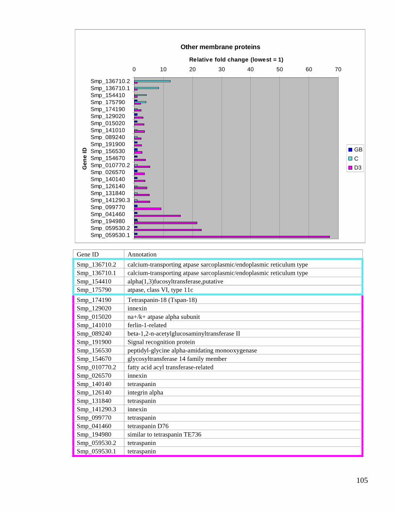
105
Other membrane proteins
0 10 20 30 40 50 60 70
Smp_136710.2Smp_136710.1Smp_154410Smp_175790Smp_174190Smp_129020Smp_015020Smp_141010Smp_089240Smp_191900Smp_156530Smp_154670Smp_010770.2Smp_026570Smp_140140Smp_126140Smp_131840Smp_141290.3Smp_099770Smp_041460Smp_194980Smp_059530.2Smp_059530.1
Ge
ne
ID
Relative fold change (lowest = 1)
GB
C
D3
Gene ID Annotation
Smp_136710.2 calcium-transporting atpase sarcoplasmic/endoplasmic reticulum type
Smp_136710.1 calcium-transporting atpase sarcoplasmic/endoplasmic reticulum type
Smp_154410 alpha(1,3)fucosyltransferase,putative
Smp_175790 atpase, class VI, type 11c
Smp_174190 Tetraspanin-18 (Tspan-18)
Smp_129020 innexin
Smp_015020 na+/k+ atpase alpha subunit
Smp_141010 ferlin-1-related
Smp_089240 beta-1,2-n-acetylglucosaminyltransferase II
Smp_191900 Signal recognition protein
Smp_156530 peptidyl-glycine alpha-amidating monooxygenase
Smp_154670 glycosyltransferase 14 family member
Smp_010770.2 fatty acid acyl transferase-related
Smp_026570 innexin
Smp_140140 tetraspanin
Smp_126140 integrin alpha
Smp_131840 tetraspanin
Smp_141290.3 innexin
Smp_099770 tetraspanin
Smp_041460 tetraspanin D76
Smp_194980 similar to tetraspanin TE736
Smp_059530.2 tetraspanin
Smp_059530.1 tetraspanin

106
3.3.17. Lipid metabolism
Lipid metabolism was singled out in the genome paper as being an area where schistosomes
may be vulnerable to interventions [75]. Therefore, the expression patterns of those genes
were investigated. Of the 102 genes described as playing a role in lipid synthesis, transport or
degradation 23 were differentially expressed (22.5%). The five genes enriched in the germ ball
are involved in lipid synthesis; they are up-regulated three to six fold compared to the cercaria.
There is a switch in expression as genes involved in lipid uptake and breakdown are enriched
in the cercaria and day 3 schistosomulum.
Twelve of the 23 lipid metabolism genes were up-regulated in the cercaria. The most highly
expressed cercaria-enriched transcript encodes ceramide synthase, which is 14 fold up
compared to the day 3 schistosomulum. The single gene which is up-regulated in both the
cercaria and schistosomulum compared to the germ ball is a saposin. The genes most highly
expressed in the day 3 schistosomulum include an inositol transporter and two phospholipases
and a sterol O-acyltransferase.

107
Lipid Metabolism
0 2 4 6 8 10 12 14
Smp_051810
Smp_042980
Smp_130370
Smp_079380
Smp_124300
Smp_095000
Smp_123710
Smp_039130
Smp_000130
Smp_157100
Smp_146180
Smp_117370
Smp_015050
Smp_143830
Smp_014570
Smp_042440
Smp_127280
Smp_141720
Smp_155270
Smp_134080
Smp_151420
Smp_134390
Smp_031180
Gene
ID
Relative fold change (lowest = 1)
GB
C
D3
Gene ID Annotation
Smp_051810 elongation of fatty acids protein 1
Smp_042980 mevalonate kinase
Smp_130370 elongation of fatty acids protein 1
Smp_079380 serine palmitoyltransferase 2
Smp_124300 ERG4/ERG24 ergosterol biosynthesis protein (ERG24)
Smp_095000 inositol monophosphatase
Smp_123710 acetyl-CoA carboxylase
Smp_039130 niemann-pick C1 (NPC1)
Smp_000130 hormone-sensitive lipase (S09 family)
Smp_157100 sphingoid long chain base kinase
Smp_146180 lipase 1,sterol esterase 1,sterol esterase 2,lysosomal acid lipase-related
Smp_117370 sphingosine kinase A, B
Smp_015050 choline kinase
Smp_143830 FFA transport protein
Smp_014570 saposin containing protein
Smp_042440 (dihydro)ceramide Synthase (LAG1)
Smp_127280 diacylglycerol O-acyltransferase 1
Smp_141720 3-keto-dihydrosphingosinereductase
Smp_155270 HMG-CoA synthase,hydroxymethylglutaryl-CoAsynthase
Smp_134080 inositol transporter
Smp_151420 phospholipase D
Smp_134390 sterol O-acyltransferase 1
Smp_031180 Phospholipase A

108
3.3.18. Energy metabolism
The life-cycle stages under investigation in the current study have different energy
requirements and sources. Genes involved in these processes were highlighted by the GOstats
analysis in each of the comparisons made. Of the 30 S. mansoni genes whose products are
involved in glycolysis, six were found to be differentially expressed (20%). The highest fold
change (14) was in the day 3 schistosomulum-enriched gene lactate dehydrogenase. The
differentially expressed genes of the citric acid cycle (aerobic respiration) were all up-
regulated in the cercaria. Likewise, a large proportion of ‘electron transport’-related genes are
enriched in the cercaria. All of these contribute to oxidative phosphorylation. The five genes
involved in electron transport, which are up-regulated in the germ ball, are involved in
mitochondrial transport and fatty acid metabolism, whereas choline dehydrogenase and
peroxidasin are enriched in the schistosomulum.
Glycolysis
0 2 4 6 8 10 12 14 16
Smp_022400
Smp_043670.2
Smp_047370
Smp_043030
Smp_043670.1
Smp_033040
Gen
eID
Relative fold change (lowest = 1)
GB
C
D3
Gene ID Annotation
Smp_022400 glucose-6-phosphate isomerase
Smp_043670.2 6-phosphofructokinase
Smp_047370 malate dehydrogenase
Smp_043030 hexokinase
Smp_043670.1 6-phosphofructokinase
Smp_033040 L-lactate dehydrogenase

109
Aerobic metabolism and electron transfer
0 1 2 3 4 5 6 7 8 9 10
Smp_082370
Smp_147970.1
Smp_122930
Smp_153460
Smp_114440
Smp_063090
Smp_018680
Smp_056200
Smp_005290
Smp_061870
Smp_134150
Smp_021730.2
Smp_038870.1
Smp_123650
Smp_094500
Gen
eID
Relative fold change (lowest = 1)
GB
C
D3
Gene ID Annotation
Smp_082370 NADP transhydrogenase
Smp_147970.1 kif1
Smp_122930 acyl-CoA dehydrogenase
Smp_153460 monoxygenase
Smp_114440 acyl-CoA dehydrogenase
Smp_063090 aconitate hydratase
Smp_018680 isocitrate dehydrogenase
Smp_056200 Isocitrate dehydrogenase [NAD] subunit gamma, mitochondrial
Smp_005290 cytochrome C1
Smp_061870 ubiquinol--cytochrome C reductase
Smp_134150 glucose-methanol-choline (gmc) oxidoreductase
Smp_021730.2 cytochrome c oxidase
Smp_038870.1 NADH-ubiquinone oxidoreductase
Smp_123650 peroxidasin
Smp_094500 choline dehydrogenase
3.3.19. Custom categories
In addition to the GOstats analysis and categories based on genome annotations, the
expression patterns of custom gene categories were investigated. These are based on
proteomic or localisation data, and are linked with a tissue (tegument, gut) or condition such

110
as stress. Some of these genes are also included elsewhere (e.g. membrane and protease
categories).
3.3.20. Tegument
The tegument undergoes substantial remodeling on entry to the mammalian host, and is a large
component of the host-parasite interface. Of the 65 genes in this category, only three are most
highly expressed in the germ ball. They are a voltage-dependent anion-selective channel, an
amino acid transporter, and acetylcholinesterase.
In the cercaria, three closely related annexins, alkaline phosphatase, a cation channel and a
cationic amino acid transporter are most highly expressed. One of the annexins (Smp_045550)
is expressed ~15 fold higher, compared to the germ ball. The majority of protein encoding-
genes of the tegument are up-regulated in the day 3 schistosomulum compared to the germ
ball. Five of these encode copies of a putative complement inhibitor CD59, one of which
(Smp_166340) is ~70 fold higher than in the germ ball. Another of them (Smp_081900) is up-
regulated in both the day 3 schistosomulum and the cercaria compared to the germ ball. Other
genes that share this pattern are a calpain, the glucose transporter SGTP4, an aquaporin, an
ectonucleotide-pyrophosphatase and a protein of unknown function, which was discovered on
the schistosome surface by Braschi et al [140]. Transcripts which are up-regulated solely in
the day 3 schistosomulum include otoferlin, ferlin, a copper transporter, and a sodium/chloride
dependent transporter. Sm8.7 (low molecular weight secreted protein) is striking; it is up-
regulated 40 fold compared to the germ ball.

111
Tegument
0 10 20 30 40 50 60 70
Smp_091240Smp_147070Smp_136690Smp_155890Smp_045500Smp_045560Smp_147140Smp_176940Smp_045550Smp_163750Smp_048230Smp_141010Smp_105220Smp_042020Smp_131890Smp_131840Smp_157500Smp_019350Smp_081920Smp_081900Smp_105410Smp_077720Smp_005740Smp_153390.Smp_074450Smp_194970Smp_194860Smp_166340
Gen
eID
Relative fold change (lowest = 1)
GB
C
D3
Gene ID Annotation Gene ID Annotation
Smp_091240voltage-dependent anion-selectivechannel Smp_131890
sodium/chloride dependenttransporter
Smp_147070 amino acid transporter Smp_131840 tetraspanin
Smp_136690 Acetylcholinesterase Smp_157500 calpain (C02 family)
Smp_155890 alkaline phosphatase Smp_019350 CD59
Smp_045500 annexin Smp_081920 CD59
Smp_045560 annexin Smp_081900 CD59
Smp_147140transient receptor potential cationchannel Braschi
Smp_105410 glucose transport protein SGTP4
Smp_077720 annexin
Smp_176940 cationic amino acid transporter Smp_005740 aquaporin-3
Smp_045550 annexin Smp_153390 Ectonucleotide-pyrophosphatase
Smp_163750 otoferlin Smp_074450 expressed protein (Braschi)
Smp_048230 high-affinity copper uptake protein Smp_194970 similar to tetraspanin TE736
Smp_141010 ferlin-1-related Smp_194860Sm8.7 Low molecular weightprotein
Smp_105220 CD59 Smp_166340 CD59

112
3.3.21. Gut-associated
The expression patterns of genes encoding gut-associated proteins were investigated to shed
light on the issue of when the larval gut becomes active. This category comprises genes
encoding proteins that were identified in adult worm vomitus by Hall et al [141]. Of the 32
genes in this custom category, ten (31%) are differentially expressed. They include cathepsins,
saposins, an asparaginyl endopeptidase, and a calpain. All of them are up-regulated in the day
3 schistosomulum compared to the germ ball. Most are also enriched in the cercaria; the
exceptions are a glycosyl hydrolase, cathepsin S, and a saposin.
Gut
0 10 20 30 40 50 60
Smp_014570
Smp_157090
Smp_043390
Smp_139240
Smp_157500
Smp_103610
Smp_105450
Smp_067060
Smp_075800
Smp_130100
Smp_193000
Gen
eID
Relative fold change (lowest = 1)
GB
C
D3
Gene ID Annotation
Smp_014570 saposin containing protein
Smp_157090 SmCL3 (C01 family)
Smp_043390 Glycosyl hydrolase
Smp_139240 cathepsin S (C01 family)
Smp_157500 calpain (C02 family)
Smp_103610 cathepsin B-like peptidase (C01 family) Sm31
Smp_105450 saposin containing protein
Smp_067060 cathepsin B-like peptidase (C01 family)
Smp_075800 asparaginyl endopeptidase Sm32Smp_130100 saposin containing proteinSmp_193000 SmCL2-like peptidase (C01 family)

113
3.3.22. Stress-related genes
To gauge the response to stress presumed to be experienced by larval schistosomes during the
mammalian infection process; the expression patterns of stress-related genes were
investigated. This custom category, based on literature surveys, comprises 29 genes, only six
of which were differentially expressed (21%). None of the stress related genes were germ ball-
enriched. Genes encoding universal stress protein, glutathione peroxidase and a multidrug
resistance pump (MDR) are up-regulated in the cercaria. Notably up-regulated at this stage is a
small heat shock protein, which is 18 fold higher than in the other two life cycle stages. A
different MDR is up-regulated in the day 3 schistosomulum, along with a superoxide
dismutase and a thioredoxin peroxidase. The latter gene shows the largest difference; it is 37
fold higher in the day 3 schistosomulum than in the germ ball.
Stress
0 5 10 15 20 25 30 35 40
Smp_043120
Smp_058690
Smp_151290
Smp_049230
Smp_135490
Smp_174810
Smp_059480
Gen
eID
Relative fold change (lowest = 1)
GB
C
D3
Gene ID Annotation
Smp_043120 universal stress protein
Smp_058690 glutathione peroxidaseSmp_151290 multidrug resistance pumpSmp_049230 Sm-p40 (small heat shock protein)
Smp_135490 multidrug resistance pump
Smp_174810 cu/zn superoxide dismutase
Smp_059480 thioredoxin peroxidase

114
3.3.23. Micro exon genes
The micro exon genes were briefly described by Berriman et al [75] and a detailed account has
been given by DeMarco et al [142]. They are hypothesised to represent a mechanism for
generating variant proteins by schistosomes. The MEGs show the highest fold changes
encountered in this entire study, with eight of the 25 having values greater than 40. Of the 14
MEG families described previously [75] all but three were found to be highly expressed in
either the germ ball, cercaria or day 3 schistosomulum. Only MEGs 1 and 11 showed no
differential expression during the transition studied. Fluorescence data across life cycle stages
revealed that MEG 1 exhibits a consistent low level of expression, whereas MEG 6, which has
small fold changes (up two fold in both cercaria and day 3 schistosomulum compared to germ
ball) was highly expressed at each stage. No data could be gleaned for MEG 11.
Due to the very high fold changes exhibited by many of the MEGs, gene models of transcripts
annotated as ‘hypothetical protein’ or ‘expressed protein’ that were highly expressed in one
stage or another in my study were manually inspected. This led to the identification of four
new MEGs (Smp_010550, Smp_158890, Smp_180620, Smp_032560). Further investigation
revealed that the newly identified MEGs have no homology to previously identified MEG
families, or to each other; they represent novel families, and have been named MEGs 15-18
respectively.
The majority of MEGs are up-regulated in the day 3 schistosomulum compared to the other
two stages; however, MEGs 7, 10 and 18 are enriched in the germ ball, and MEGs 4.2, 13 and
6 are most highly expressed in the cercaria. Although MEGs 4.1, 8, 12, 14, 15, 16, and 17 are
most highly expressed in the day 3 schistosomulum, they are also up-regulated in the cercaria
compared to the germ ball. MEGs 2, 3, 5, 8 and 9 are up-regulated in the day 3
schistosomulum compared to both germ balls and cercariae.

115
MEGs
0 20 40 60 80 100 120
Smp_165050
Smp_152590
Smp_032560
Smp_085840
Smp_127990
Smp_163710
Smp_159800
Smp_138060
Smp_180340
Smp_159830
Smp_172180
Smp_180620
Smp_152580
Smp_158890
Smp_181510
Smp_138090
Smp_138080
Smp_010550
Smp_138070
Smp_159810
Smp_152630
Smp_163630
Smp_125320
Smp_171190
Smp_124000
Gen
eID
Relative fold change (lowest = 1)
GB
C
D3
Gene ID Annotation Gene ID Annotation
Smp_165050 MEG 7 Smp_158890 MEG 16
Smp_152590 MEG 10 Smp_181510 MEG 2
Smp_032560 MEG 18 Smp_138090 MEG 3
Smp_085840 MEG 4 Smp_138080 MEG 3
Smp_127990 MEG 13 Smp_010550 MEG 15
Smp_163710 MEG 6 Smp_138070 MEG 3
Smp_159800 MEG 2 Smp_159810 MEG 2
Smp_138060 MEG 3 Smp_152630 MEG 12
Smp_180340 MEG 2 Smp_163630 MEG 4
Smp_159830 MEG 2 Smp_125320 MEG 9
Smp_172180 MEG 8 Smp_171190 MEG 8
Smp_180620 MEG 17 Smp_124000 MEG 14
Smp_152580 MEG 5

116
3.3.24. Venom allergen-like proteins
VALs have been identified in secretions of schistosomes and parasitic nematodes. They are
proposed to play a role in immune evasion [116]. Of the 28 VAL genes described by Chalmers
et al [125], 13 are differentially expressed during the germ ball to day 3 schistosomulum
transitions (46%). Most are enriched in the germ ball and/or cercaria compared to the day 3
schistosomulum. VALs 4, 18, 19, 20, 24 and 25 are most highly expressed in the germ ball
compared to the day 3 schistosomulum. Of these, VALs 18 and 25 are also enriched in the
cercaria. VALs 1, 2, 16, 17 and 21 are most highly expressed in the cercaria compared to the
day 3 schistosomulum; of these, VALs 1, 2, and 21 are also up-regulated in the germ ball.
Only two VALs (7 and 13) are most highly expressed in the schistosomulum. VAL 7 has the
highest fold change of any of the VALs: it is ~47fold higher in the day 3 schistosomulum than
the germ ball. This is noteworthy as none of the other VAL-encoding genes exhibit fold
changes greater than 15.

117
VALs
0 5 10 15 20 25 30 35 40 45 50
Smp_002070
Smp_001890
Smp_141550
Smp_141560
Smp_127130
Smp_123090
Smp_120240
Smp_193680
Smp_159290
Smp_124070
Smp_002630
Smp_163400
Smp_124060
Smp_070240
Gen
eID
Relative Fold Change
GB
C
D3
Gene ID Annotation
Smp_002070 VAL 04
Smp_001890 VAL 18
Smp_141550 VAL 24
Smp_141560 VAL 25
Smp_127130 VAL 20
Smp_123090 VAL 19
Smp_120240 VAL 01
Smp_193680 VAL 01Smp_159290 VAL 21
Smp_124070 VAL 16
Smp_002630 VAL 02
Smp_163400 VAL 17
Smp_124060 VAL 13
Smp_070240 VAL 07

118
3.4. Discussion
3.4.1. Design and application of the first genome wide microarray for S. mansoni
The publication of the S. mansoni genome in 2009 [75] represents the culmination of nearly
three decades of sequencing work (see Chapter 1.2). The availability of the gene predictions
and many ESTs at GeneDB have enabled the design of the first genome wide array for S.
mansoni. The ESTs were compiled with the gene predictions and 45mer probes were
designed to the resulting contigs. Each locus is represented by 1-300 non-redundant probes
ensuring both diversity and coverage. 15.6% of loci represented on the array do not have
significant matches to gene predictions when blasted against the genome either by BLASTn or
tBLASTx. There are two explanations for this: a) this sequence is not encoded by the S.
mansoni genome b) the sequence is present in the S. mansoni genome, but has not been
identified by gene finding software. The sequences which are not truly S. mansoni are likely to
be contaminating sequences of host origin, (this is most likely for germ ball, or ex vivo
intramammalian library sequences). Host sequences have been mistaken previously for novel
schistosome genes [143]. The ESTs were included in the compilation for the array to reduce
the chance of excluding bonafide schistosome genes that were not called by gene finding
software. However, although some of the non-Smps represented on the array may be new
uncalled schistosome-specific genes, they were filtered out before further analysis as there was
no annotation available, and hypotheses would be difficult to make about their function.
An important caveat is that some of the gene models in GeneDB, on which the array is based,
were faulty. For example, a gene encoding a secreted protein named LMWP (now
Smp_194860) was concatenated with that for a histone (now Smp_194870), resulting in the
erroneous annotation of a secreted histone (Smp_053310 in version 3.1). In the present study,
such a gene model would be assigned an expression level based on the probes representing
both the LMWP and histone portions.
Of the loci represented on the array, 42.9% have no annotation available. This figure is in
keeping with previous sequencing projects using ESTs; analysis of the transcriptome showed
that ~50% of the sequences available had no homology to anything at GenBank [73].

119
The expression patterns are presented as relative fold changes; for each gene, the baseline is
the expression level of the lowest stage and is set to 1. Comparisons have not been made on an
absolute basis. Low fold changes indicate only that the expression of a gene does not vary; it
may be consistently low or consistently high across the stages. An example of the latter case is
MEG 6 (see Micro-exon genes section in Results). Genes which were not differentially
expressed were not reported, as the focus here is on the biological differences between the life
cycle stages.
In the contrasts carried out here, the cercaria and day 3 schistosomulum were most similar to
each other, and the germ ball and cercaria most different. The germ ball is an outlier. This can
be explained by taking into account the environments and developments taking place. As we
will see later, many of the genes required by the migrating schistosomulum are up-regulated in
the cercariae in preparation for host entry. Both protein synthesis and cell division are
processes highly up-regulated in the germ ball compared to the other stages. Furthermore, the
genes needed specifically for intra-molluscan life and preparation for host entry are not
transcribed at the later stages, a fact which also contributes to the high contrasts observed
between this stage and the other two.
An overview of the functional categories whose member genes are particularly enriched in
each life cycle stage will be given, followed by an in depth consideration of each category by
named gene.
3.4.2. Germ ball
As we saw in Chapter 2, the term ‘germ ball’ encompasses embryonic cercariae at all stages of
development, from a single cell, up to, but not including, the mature cercaria. The latter
comprises approximately 1000 cells [42], which form tissues and organs. Although only 5% of
the total S. mansoni genes involved in DNA replication are differentially regulated in the
transitions studied here, it is noteworthy that all of them are up-regulated in the germ ball,
reflecting the obvious occurrence of cell division. The majority of differentially expressed
translation-associated genes are also up-regulated in the germ ball, but the fold changes are not
as large; the maximum is 4.5. Both DNA replication and translation are processes necessary
for the development of germ balls into cercariae. Likewise, several genes encoding
morphogens such as homeobox and Wnt proteins are most highly expressed in the germ ball.

120
Again, the fold changes are not exceptional, the maximum being up-regulated seven fold
compared to the day 3 schistosomulum. These genes are necessary for dorso-ventral and
anterio-posterior patterning, essential processes in embryogenesis.
The expression of genes encoding serine and metallo-proteases was somewhat more dramatic,
with 33 and 35% respectively of the S. mansoni gene complement differentially expressed.
The vast majority in both cases were up-regulated in the germ ball with maximum fold
changes of 65 and 27 respectively. In addition, several VAL-encoding genes were up-
regulated in the germ ball. Proteases (cercarial elastases in particular) are known to be secreted
during penetration of skin, enabling the parasite to gain access to the host [115, 116, 144].
VAL-proteins have been identified in cercarial secretions; the majority of transcripts encoding
these putative immunomodulators are up-regulated in the germ ball and/or cercaria. These will
be discussed in more detail in the relevant sections below.
3.4.3. Cercaria
The free-living cercaria swims actively in order to find a host, which it must do before it uses
up its supply of glycogen. Thus, energy metabolism and neural functions are salient features of
this highly motile life cycle stage. Genes that were most highly expressed in the cercaria
belong to a wide range of groups. The most striking were those involved in lipid and energy
metabolism. In each case, more than 50% of the differentially expressed genes are up-
regulated in the cercaria; the highest fold changes in the two groups are 14 and 5, respectively.
Genes encoding various membrane proteins involved in neural activity, including
neurotransmitter transporters, are also up-regulated at this stage, with expression levels up to
14 fold that in the germ ball. In addition, many genes encoding proteins required by the
schistosomulum immediately on entry to the host are already up-regulated in the cercaria
compared to the germ ball. This includes gut-associated cysteine proteases and the tegumental
aquaporin.
3.4.4. Day 3 schistosomulum
Inspection of the day 3 schistosomulum-enriched genes reveals a marked activation of genes
across a wide range of categories on entry to the mammalian host. There is a switch from the
serine and metalloproteases associated with skin penetration to aspartlyl and cysteine
proteases involved in feeding. This is highlighted by the up-regulation of genes in the ‘gut’

121
category. The majority of genes encoding known tegument proteins were also up-regulated in
the day 3 schistosomulum, reflecting the new surface that is required for survival in the
mammalian host [145]. In particular, a CD59 gene was up-regulated 70 fold compared to the
germ ball. In fact, the vast majority of the membrane proteins were most highly expressed in
the day 3 schistosomulum, with increases up to 60 fold compared to the germ ball,
emphasising the dramatic changes required for adaptation to the mammalian environment.
However, the most striking category was the MEGs (de Marco et al, submitted). 76% of these
genes were up-regulated in the day 3 schistosomulum, most being expressed at least 20 fold
higher than the germ ball. MEG 14 exhibited the highest fold change in the entire study. It was
expressed 110 fold higher compared to the germ ball. This indicates an important role for
these genes in establishing the parasitic state.
3.4.5. Cell proliferation
All of the differentially expressed genes involved in cell proliferation are up-regulated solely
in the germ ball. This includes DNA polymerases and Cyclins B and D, which control cell
cycle progression. Pollok et al showed that CDC45 is rate limiting for DNA replication
licensing in human cells; up-regulation of this important transcript in germ balls is in
accordance with their rate of cell proliferation [146]. The results presented here are in keeping
with those of earlier studies into larval maturation; although cell proliferation is necessary for
cercariogenesis, Lawson and Wilson found no evidence that development occuring in the
migrating parasite prior to arrival at the liver is due to cell proliferation; rather they suggest it
is achieved through tissue reorganisation [104].
3.4.6. Protein synthesis genes are down-regulated in the cercaria
Only one RNA polymerase is differentially expressed in the transitions studied. Smp_004640
is up-regulated 2.66 fold in the germ ball compared to the cercaria. This suggests that each of
the stages under investigation carry out transcription at a similar rate. However, transcripts
encoding ribosomal proteins, tRNA synthetases, and translation initiation and elongation
factors were all up-regulated in the germ ball compared to the cercaria and day 3
schistosomulum. Many of these genes were also up-regulated at day 3 compared to the
cercaria. This pattern suggests the germ ball is the most translationally active, the cercaria the
least. Blanton and Licate reported that protein synthesis occurs in the absence of a significant
increase in transcription during the first 24 hours after transformation, indicating that post
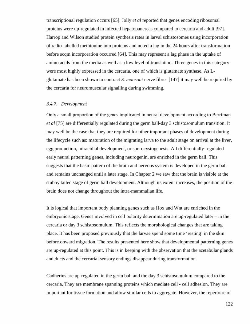
122
transcriptional regulation occurs [65]. Jolly et al reported that genes encoding ribosomal
proteins were up-regulated in infected hepatopancreas compared to cercaria and adult [97].
Harrop and Wilson studied protein synthesis rates in larval schistosomes using incorporation
of radio-labelled methionine into proteins and noted a lag in the 24 hours after transformation
before scqm incorporation occurred [64]. This may represent a lag phase in the uptake of
amino acids from the media as well as a low level of translation. Three genes in this category
were most highly expressed in the cercaria, one of which is glutamate synthase. As L-
glutamate has been shown to contract S. mansoni nerve fibres [147] it may well be required by
the cercaria for neuromuscular signalling during swimming.
3.4.7. Development
Only a small proportion of the genes implicated in neural development according to Berriman
et al [75] are differentially regulated during the germ ball-day 3 schistosomulum transition. It
may well be the case that they are required for other important phases of development during
the lifecycle such as: maturation of the migrating larva to the adult stage on arrival at the liver,
egg production, miracidial development, or sporocystogenesis. All differentially-regulated
early neural patterning genes, including neurogenin, are enriched in the germ ball. This
suggests that the basic pattern of the brain and nervous system is developed in the germ ball
and remains unchanged until a later stage. In Chapter 2 we saw that the brain is visible at the
stubby tailed stage of germ ball development. Although its extent increases, the position of the
brain does not change throughout the intra-mammalian life.
It is logical that important body planning genes such as Hox and Wnt are enriched in the
embryonic stage. Genes involved in cell polarity determination are up-regulated later – in the
cercaria or day 3 schistosomulum. This reflects the morphological changes that are taking
place. It has been proposed previously that the larvae spend some time ‘resting’ in the skin
before onward migration. The results presented here show that developmental patterning genes
are up-regulated at this point. This is in keeping with the observation that the acetabular glands
and ducts and the cercarial sensory endings disappear during transformation.
Cadherins are up-regulated in the germ ball and the day 3 schistosomulum compared to the
cercaria. They are membrane spanning proteins which mediate cell - cell adhesion. They are
important for tissue formation and allow similar cells to aggregate. However, the repertoire of

123
cadherins expressed by the germ ball is different to that of the day 3 schistosomulum,
suggesting that associations between different cell types form at these two distinct life cycle
stages.
3.4.8. Proteases
The proteases will be discussed by catalytic type.
Serine proteases
The most striking group of serine proteases are the cercarial elastases (MEROPS class S01).
Previously, Salter et al described five cercarial elastase genes, stating that two of them
accounted for 90% of the protein present [115]. The data presented here show that the 11
genes encoding this enzyme are transcribed at high levels in the germ ball; all of them are
expressed at least 10 fold higher than in the schistosomulum. This shows that skin invasion is
more complex than had been thought previously. Also enriched in the germ ball is a rhomboid
protease. Transmembrane proteases of this type have been implicated in cell invasion by
Toxoplasma [148] and malaria parasites [149], immune evasion by Entamoeba histolytica
[150], and in cell signalling in Drosophila [151]. Freeman notes that work is needed to clarify
whether inhibition of rhomboid proteases prevents invasion by parasites, and furthermore
whether they are suitable as drug targets [151]. The localisation of the germ ball-enriched
rhomboid protease would help decipher its role in schistosomes, as both cell signalling and
host interaction are plausible functions for this stage. The family S33 serine proteases up-
regulated in the cercaria may play roles in development rather than skin invasion.
Metalloproteases
Most of the metalloproteases which are differentially expressed during the germ ball to day 3
schistosomulum transitions are up-regulated in the germ ball. The leishmanolysin identified in
cercarial secretions by Curwen et al [116] (actually an invadolysin), was shown to be highly
up-regulated at this stage, in addition to five other invadolysins. The number of these and their
high expression (ranging from 2-28 fold higher in the germ ball than the other stages)
indicates that they are all important during skin invasion. This along with the high expression
of the previously unreported elastase genes greatly expands the known repertoire of proteases

124
involved in infection by larval schistosomes. The other germ ball-enriched metalloproteases
include proliferation-associated protein (Smp_150690.1) and several peptidases involved in
protein turnover, highlighting the importance of cell proliferation and protein synthesis for the
embryonic cercaria.
An invadolysin, an ADAM protease, and a matrix metalloprotease are up-regulated in the
cercaria. The two latter enzymes can be involved in tissue remodelling so they may play a role
in parasite morphogenesis on entry to the host. However, ADAMs are found in snake venom
where they inhibit platelet aggregation; this would be a useful function for a secreted protein
to perform during blood vessel entry. Likewise, breakdown of the extracellular matrix in the
skin would facilitate host invasion by schistosomes. Localisation of these proteases would
increase our understanding of their function and aid decisions about whether they represent
targets for intervention.
In the day 3 schistosomulum, a membrane metalloprotease and another M12 family protease
(Smp_134430) are up-regulated. The latter has high homology to tolloid which is a dorso-
ventral patterning protein in Drosophila. Together with the up-regulation of various cadherins,
these may be the involved in the remodelling that occurs in this life cycle stage. Also up-
regulated in the day 3 schistosomulum is a gene encoding leucine aminopeptidase which is a
cytosolic enzyme involved in protein turnover. This protein has been localised to gastrodermal
cells in adult worms, and is hypothesised to break down the peptides resulting from
haemoglobin degradation in the gut to single amino acids [152].
Cysteine proteases
The gene expression patterns in this category reveal much about the biology of larval
schistosomes. Whilst the majority of cysteine proteases are most highly expressed in the
cercaria or day 3 schistosomulum, the up-regulation of caspases and a separase is consistent
with cell death and division during cercariogenesis. Bos et al recently described S. mansoni
proteases, and carried out a phylogeny of caspases [153]. They noted that Smp_172010 was in
a clade with effector caspases from mice and humans, and Smp_141270 was closer to initiator
caspases [153]. Both of these genes are up-regulated in the germ ball. It is tempting to

125
speculate that apoptosis is involved in the bifurcation of the tail, after the posterior of the germ
ball has flattened and extended.
The cercaria expresses a de-ubiquitinating protease; this enzyme may serve to help avoid
proteasomal degradation of proteins, which is costly in terms of ATP. In addition, two
developmental proteases are up-regulated in the cercaria: an ovarian tumour-like protein, and
autophagin. It is likely that these are both needed for transformation. The presence of
autophagin, a protease involved in autophagy [154], indicates that this process may be used to
destroy the acetabular glands once they are no longer needed. Calpains are also up-regulated
in the cercaria. They are calcium-dependent proteases with diverse functions including signal
transduction [155]. It is plausible that they are involved in neuromuscular signalling during
swimming.
The remaining cysteine proteases are cathepsins and asparaginyl endopeptidases. Some of
these are included in the ‘gut’ category as they have been localised to that tissue, or identified
in adult worm vomit. It is likely that the additional cathepsins also have a role in digestion.
The known digestive enzymes will be discussed in the ‘gut’ category.
Aspartyl proteases
Compared to the cathepsins, the function of these aspartyl proteases is poorly characterised in
schistosomes. This is the first report on their expression. All of the differentially regulated
enzymes in this class follow a pattern of increasing expression from germ ball to day 3
schistosomulum. They have homology to the acidic digestive enzymes pepsin and cathepsin
D. The cathepsin D identified by Wong et al [156] is Smp_013040, which is not differentially
expressed during the mammalian infection process. However, knock down studies using RNAi
showed that this enzyme was required for schistosomula to grow [157]. Based on the
similarity of their expression patterns with those of known gut proteases, it is likely that the
aspartyl proteases included here are also involved in gut function. However, localisation and
biochemical studies would be needed to confirm this hypothesis.

126
3.4.9. Energy production
With the exception of lactate dehydrogenase (up-regulated in the schistosomulum), the genes
encoding glycolytic enzymes did not vary greatly in the germ ball to day 3 schistosomulum
transitions. However, transcripts encoding citric acid cycle enzymes were consistently up-
regulated in the cercaria. This is consistent with the findings of Skelly et al who showed that
cercariae use aerobic metabolism, whereas schistosomula rely on glycolysis to supply energy
[66, 158]. The cercaria requires energy to swim, and as it does not feed, it relies on a store of
glycogen. It is logical that the cercaria uses aerobic metabolism to yield the maximum possible
molecules of ATP from the limited resources available to it.
3.4.10. Lipid metabolism
Genes involved in lipid metabolism up-regulated in the day 3 schistosomulum include
phospholipases; it is not clear in this context whether they are acting as digestive enzymes, or
to generate signalling molecules. It is possible that both functions are undertaken, as both
signalling and digestion are important for the day 3 schistosomulum as illustrated by the up-
regulation of other gut-associated enzymes and neurotransmitter transporters. CD36 is also up-
regulated in the day 3 schistosomulum. It is thought to scavenge oxidised low density
lipoprotein and long chain fatty acids, and is also an adhesion molecule; in humans it is
expressed on monocytes, macrophages, platelets, microvascular endothelial cells and adipose
tissue. It suggests that the schistosomulum takes up lipid as well glucose from the host at this
early larval stage. Verjovsky-Almeida et al included CD36 in their table of possible vaccine
candidates [73]. Berriman et al noted that schistosomes must rely on the host for a source of
inositol [75]. The current microarray data show that a putative inositol transporter
(Smp_134080) is up-regulated in the day 3 larva compared to the germ ball. This should be
investigated as an intervention target. The gene with the highest fold change at day 3 is
lecithin:cholesterol acyltransferase. This is a secreted enzyme which metabolises plasma
lipoproteins. Taken together these results imply that schistosomula are capable of ingesting
and digesting plasma.
3.4.11. Membrane proteins
This category contains a wide range of proteins with various functions. Fitzpatrick et al note
that GPCRs and ion channels are the target of a large proportion of drugs currently in use, and
for that reason should be investigated thoroughly in schistosomes [99]. Efforts should be

127
concentrated on those which are up-regulated in the intra-mammalian stages. An important
task is to narrow down the large repertoire of channels and receptors encoded in the S.
mansoni genome, to those highly expressed in intra-mammalian stages. Therefore special
attention will be given to the subset of membrane proteins enriched in the schistosomulum
compared to the other two stages studied. Many of the membrane proteins have been
identified as potential intervention targets. Those included in the ‘tegument’ category will be
discussed later (section 3.4.12 below).
The increase in transcription of neurotransmitter-sodium symporters in the cercaria probably
reflects the muscle activity required for swimming. Likewise, channels of the TWIK family
(Tandem of P domains in a weakly inward rectifying K+ channel) are enriched in the cercaria.
TWIK channels are reported to be involved in maintaining the background membrane
potential [159]. These proteins are also thought to be non-inactivating and so they may play an
important role in maintaining the membrane potential in neuromuscular tissue in the cercarial
tail enabling swimming. In contrast, voltage-gated potassium channels are up-regulated in the
schistosomulum.
Only a small proportion of the predicted GPCRs are differentially regulated during the
transition studied here. Although the GPCR up-regulated in the germ ball was assigned no
specific annotation in the genome, BLASTp against NCBI shows highest similarity to opsin
from Branchiostoma belcheri (37% identitiy, 52% positive), and to retinal pigment derived
rhodopsin from house mouse (31% identity, 48% positive). It was highlighted in the
transcriptome by Verjovsky-Almeida et al [73]. Although no eye spot-like morphological
structure has yet been identified in the cercaria, this life cycle stage is known to be responsive
to light. The opsin-like receptor, along with the cercaria-enriched photoreceptor cell
development protein, is strongly indicative of pathways required for light detection.
Stomatin or erythrocyte band 7 protein was suggested by Verjovsky-Almeida et al as a gene
that should be investigated as a vaccine candidate. It has been ascribed many roles including
mechanoreception, HDL binding, phospholipid uptake, calcium transport, and interaction with
antimalarial drugs [73]. Although its site of expression is not known, the current results reveal
that it is expressed in the intra-mammalian larva, and as such shows promise as an intervention
target.

128
3.4.12. Tegument
The dramatic change in tegument structure on entry to the mammalian host is one of the
defining aspects of transformation. The genes included in this category are derived from
proteomic studies and other published reports in which the gene product is localised to the
tegument. Skelly and Shoemaker showed that the glucose transporter SGTP4 appears at the
surface one hour into transformation induced by incubation in RPMI-rich media [60]. Thus the
newly parasitic schistosomulum is able to acquire glucose from the host. This is important as
the cercarial glycogen store is depleted after 24 hours in vivo [46]. In another study, Skelly and
Shoemaker investigated the expression of SGTP4 using western blots: protein was not
detectable in eggs, sporocysts, or cercariae, but was found in adult worms [58]. Furthermore,
SGTP4 was not detected in protein extracted from schistosomula immediately after
transformation, but was clearly visible 1 hour afterwards. In the present study, transcription of
SGTP4 was lowest in the germ ball, increased in the cercaria, and reached a peak in the day 3
schistosomulum. The up-regulation of SGTP4 in the cercaria indicates that the transcript must
be present and ready, allowing the schistosomulum to translate and traffic it to the surface
immediately on entry to the host. As this protein has been shown on the apical surface of both
schistosomula and adult worms, it represents a potential target for intervention. This would
require either the design of a suitable inhibitor that binds with high specificity for the parasite
channel, or an immune response to be elicited to an epitope not shared by the host homologue.
Although these goals are not trivial, they may be possible, and would provide a treatment with
potential against both larvae and adults.
The other known tegument surface proteins, aquaporin 3 [160], and nucleotide
pyrophosphatase [161], share SGTP4’s expression pattern. The aquaporin has been
characterized recently by Skelly et al [160]. In addition to controlling osmoregulation, it has
been ascribed an important role in allowing drugs to enter the parasite [160]. The nucleotide
pyrophosphatase/ phosphodiesterase has been identified in tegument preparations by
proteomics [107, 162, 163]. It has been demonstrated at the surface of adult worms by Rofatto
et al using imunohistochemistry [161]. Western blots revealed that the protein was present in 7
day schistosomula. The authors established that the phosphodiesterase is active at the surface
of live schistosomes and that the activity could be reduced with anti-sera raised against it
[161].

129
CD59 is a complement inhibitory protein expressed on the surface of human cells [164]. It
blocks incorporation of C9 into the C5b-9 complex, so that the membrane pore does not form,
and cell lysis is avoided [165]. CD59 proteins have been discovered at the surface of adult
schistosomes by proteomics (Braschi and Castro Borges et al unpublished). The current study
shows that five genes encoding schistosome CD59 are up-regulated on entry to the
mammalian host, indicating that this important defense strategy is employed throughout the
intra-mammalian life. However, the most highly up-regulated CD59-encoding transcript has
not been found by proteomics; this is the first evidence for its expression. It is possible that
this is specific to the schistosomulum.
Tegument proteins should also be investigated as targets as they are accessible to the host,
unlike cytosolic proteins [166]. It may be easier to achieve selective toxicity for schistosome
specific tegument proteins rather than those with human orthologues, but these will need to be
identified and localized first. Braschi et al used proteomic techniques to investigate the adult
worm tegument and identified several Schistosoma specific proteins in the membrane fraction
of the preparation [140]. One of these, Smp_074450, shares the expression pattern of SGTP4,
implying that it may be present at the surface shortly after transformation, and remain
throughout the intra-mammalian life. As such, this protein should be investigated as a vaccine
target. Tran et al reported that two tetraspanins, found on the adult tegument, showed
promising results when mice were vaccinated with the recombinant protein [167]. The genes
encoding these particular proteins were not up-regulated in the schistosomulum in the current
study. However, ten different tetraspanins were, and only two of these had previously been
found on the surface on adult worms. Localisation studies would be required to discover
whether the eight remaining schistosomulum-enriched tetraspanins are novel tegument
proteins. The two day 3 schistosomulum-enriched tetraspanins that have been found in the
adult tegument are worth investigating as they may be expressed on the surface of the worm
throughout the intra-mammalian life, and as such may represent new vaccine targets.
Bhardwaj and Skelly propose that the expression of alkaline phosphatase and nucleotide
pyrophosphatase at the tegument modulate the immune response by catabolizing free ATP
which is a potent inflammatory mediator [168]. Taken with the expression of the anti-
complement protein CD59 on the surface of adult worms (Castro Borges et al, unpublished),

130
this shows that the tegument has mechanisms for active defence; it is not simply an inert
barrier.
Three of the genes included in the tegument category were shown to be up-regulated in the
germ ball (a voltage-dependent anion-selective channel, an amino acid transporter, and
acetylcholinesterase). This raises doubts regarding their expression in the intra-mammalian
stages. As such these should not be considered as intervention targets.
3.4.13. Micro Exon Genes
MEGs are highly differentially regulated during the germ ball to day 3 schistosomulum
transitions. The majority are up-regulated in the day 3 schistosomulum compared to the other
two stages. Visual inspection of the gene models for some of the unannotated genes highly
expressed at each stage enabled the identification of four new MEGs in addition to the 14
families reported in the genome paper. These represent new MEG families as they have no
homology either to each other, or to the other 14 MEG families. These genes demonstrate
considerable alternative splicing and exon skipping. Another important point to note is that
they are almost all predicted to have signal peptides, but no transmembrane domains, so they
are likely to be secreted [142]. No mechanism for antigenic variation had been found in
schistosomes when the transcriptome was published [73]. The MEGs may represent a
schistosome mechanism for protein variation [142]. Antigenic variation is an important feature
of how other blood borne parasites survive (Plasmodium [169] and Trypanosoma [170]). In
order to discover whether MEGs play a role in presenting variant proteins to the host, it will be
necessary to discover whether these genes are expressed at the host parasite interface – either
by secretion from the gut or glands, or expression on the tegument surface. Further study into
the expression of proteins encoded by MEGs may shed light on the way that schistosomes are
able to survive in the blood stream for decades. Arguably the mechanisms employed by
schistosomes for immune evasion must be more effective than those used by the shorter lived
unicellular intravascular parasites as schistosomes live in the bloodstream for up to 30 years.
3.4.14. Venom Allergen Like proteins
Another interesting group of genes that have been described recently are the VALs. These are
not schistosome specific and have been implicated in angiogenesis [123] and skin invasion by
parasitic nematodes [171]. Curwen et al identified VALs 4, 10 and 18 in cercarial secretions

131
by proteomics [116], the data here show that the transcripts for VAL 18 and VALs 1, 2, 19,
20, 21, 24, and 25 are all up-regulated in the germ ball. Chalmers et al investigated the
expression of VALs 1-13 across various lifecycle stages using RT PCR and showed that VALs
1, 4, and 10 are up-regulated in the cercaria [125]. In addition to VAL 1, the present study
showed VALs 2, 16, 17, 21 and 25 to be enriched at this stage compared to the day 3
schistosomulum.
Only two VALs were most highly expressed in the day 3, which is in contrast to the dominant
expression pattern for MEGs. This suggests that these proteins play a role earlier in infection.
Since other VALs are highly expressed in the intramolluscan stages, it may be the case that
these proteins are important for germ balls and cercariae while they are in the intermediate
host. However, expression of VAL 7 in the schistosomulum is more than 45 fold higher than
that in the germ ball. A VAL in Onchocerca is reported to induce angiogenesis [123]. If
SmVAL 7 protein were to interact with blood vessels to cause vasodilatation it would facilitate
skin exit. The current study confirms that this family of proteins warrants further attention. In
particular, VAL 7 should be prioritised as it is the most highly expressed VAL gene in the
early intra-mammalian larva.
3.4.15. Stress
Only 21% of stress-related genes were differentially regulated in the contrasts studied here.
This is somewhat surprising as the parasite must adjust to wildly different environments
during this portion of the life cycle. Of those that were differentially expressed, none were
enriched in the germ ball. This may be because the germ ball is shielded from the host tissue
by the daughter sporocyst. Certain heat shock proteins are up-regulated in the cercaria. These
proteins are chaperones which help keep proteins properly folded. Sm-p40 is noteworthy as
Mathieson discovered that it is up-regulated in the miracidium [172]. He reported that this
small heat shock protein acts to prevent irreversible aggregation of denatured proteins without
using up precious ATP [172]. It is logical that the non-feeding cercaria expresses this gene as
it too must conserve as much energy as possible. A thioredoxin peroxidise was up-regulated in
the schistosomulum, suggesting involvement in defence against hydrogen peroxide. This gene
was cloned and characterised by Kwatia et al in 2000, who found that the protein was active in
adults [173]. This important defence protein is very likely to be expressed throughout the

132
intra-mammalian life-cycle stages, and should be investigated as an intervention target.
Indeed, a new group of drug leads has recently been identified that inhibit thioredoxin
glutathione reductase (Smp_048430) [174], highlighting the importance of investigating the
genes in this category.
3.4.16. Gut
Proteins involved in the digestion of the blood meal have been studied extensively, as they are
appealing targets for interventions (reviewed in [175-177]). Sm31 and Sm32 were the first to
be discovered [178]. The former is cathepsin B, the latter was first named haemoglobinase.
However, Dalton and Brindley noted that Sm32 has homology to legumain, an asparaginyl
protease from legumes and suggested that it may activate cathepsins in the worm gut [179].
Sm32 has since been shown to self-activate at acidic pH [180] and to activate cathepsin B
(Sm31) [181]. Given the large number of cathepsins in the schistosome gut, Dalton and
Brindley suggest that it may be wiser to target Sm32; it is known to activate at least one of the
cathepsins, and so it may be possible to target blood feeding by inhibiting that one enzyme.
Suppression of cathepsin B by RNAi did not inhibit haematin formation, but led to stunting of
treated worms [77].
In addition to the cathepsins, saposins are included in the ‘gut’ category. Sapsosins have been
suggested to lyse blood cells on entry to the gut [182], and although Smp_105450 was
localised to the gut of adult schistosomes, it was ineffective as a vaccine in the mouse model
[182].
There has been some discussion in the literature as to when the larval gut becomes active.
Crabtree states that lung schistosomula are unlikely to feed on plasma or cells during
migration as their mouths are pressed up against blood vessel wall [63] and that the larval
oesophagus is too narrow to admit red blood cells. Protein staining was observed in the larval
gut in Chapter 2. The results presented in this chapter show that transcripts encoding many
cathepsins are already up-regulated in the cercaria, with expression increased further at Day 3.
This is consistent with the immunolocalisation of Sm31 and Sm32 to the cercarial gut reported
by Skelly [183], and suggests that gut-specific proteases are translated and secreted early in

133
infection. In addition, Dillon et al localised cathepsin L mRNA to the gut of lung stage
schistosomula [16]. Nevertheless, the question of what the larva ingests at this stage remains
unanswered. The finding that there are important digestive proteases up-regulated throughout
the intra-mammalian stages could mean that an intervention which targeted them would be
effective against both larval and adult worms. This would be a useful feature of any new drug
against schistosomes. The current treatment is only effective against adults; any migratory
larvae present at the time of treatment mature unless follow-up treatment is administered.

134
4. Spatial Expression Patterns of Selected Genes

135
4.1. Introduction
The availability of genomic sequence data for S. mansoni has enabled transcriptomic and
proteomic experiments to be carried out. Various approaches have been taken, some focusing
on the differences between life cycle stages, others on the proteins expressed in certain tissues.
Of particular relevance to the current study are those which tackle the infection process.
Throughout all of the sequencing programmes for schistosomes, a common theme has been
the large proportion of S. mansoni genes with no significant matches to any sequences
available at GenBank. Indeed, Wilson et al noted that as few as 26% of S. mansoni sequences
may have informative homology [128]. In order to assign functional annotation to the
remaining 75%, an important early step is localisation of the gene product. Forming
hypotheses regarding the function of a gene product is greatly aided by knowledge of where in
the organism the gene is expressed. Localisation is also necessary when considering whether
the gene under investigation could serve as a vaccine candidate, as such gene products must be
available to the host. As several workers point out [129-131], cytosolic and cytoskeletal
proteins are extremely unlikely to be of use as intervention targets and should be avoided in
future. Localisation studies reveal whether a potential target is expressed at the host-parasite
interface (tegument, gut, secretory glands, or excretory system); this enables researchers to
disregard those which are found to be expressed internally with little likelihood of secretion.
A great step forward was achieved when Dillon et al adapted whole mount in situ
hybridisation (WISH) for use in schistosomes, such that transcripts of interest could be
localised in intra-mammalian larvae and adult worms [16]. The whole mount technique is
particularly useful as small cells or tissues may be missed in the sectioning process for
conventional in situ hybridisation (ISH) [16]. In addition, the life cycle stages under
investigation in the present study are very small, making sectioning technically difficult and
interpretation of images problematic. The method described by Dillon et al utilizes proteinase
K to permeabilise the worms to allow the DIG-labelled RNA probes and detection antibodies
to enter the parasite and reach their target mRNA. This treatment proved too harsh for germ
balls to withstand. However, Dr Jean Illes of Sheffield University kindly provided an
alternative protocol that uses methanol for permeabilisation [184]. Thus the first successful
application of WISH to germ balls is described in this chapter.

136
I chose to localize transcripts encoding proteins previously identified in cercarial secretions
and hypothesized by Curwen et al to play a role in infection, either by proteolysis or
immunomodulation [116]. Of these, only one cercarial elastase had previously been localized.
Newport et al carried out ISH on sections of infected snail hepatopancreas and showed that
transcripts encoding cercarial elastase 1a (Smp_119130) were present in acetabular glands of
intra-molluscan germ balls [114]. Thus Smp_119130 was used as a positive control in the
current study. Expression of Curwen’s SmPepM8 (invadolysin, Smp_090100), SCPc (VAL
10, Smp_002060), SmKK7 (Smp_194830) and Sm16 (Smp_113760) was localized in germ
balls. Anti-sera for SmKK7 and invadolysin were also used to localise these proteins by
immunohistochemistry.
As well as being identified in cercarial secretions, SmKK7 was discovered by mass
spectrometry in the soluble fraction of the supernatant resulting from treatment of adult worms
with trypsin and PIPLC, suggesting that it may be associated with the tegument (Dr W. C.
Borges, personal communication). This led to the investigation of SmKK7 expression (both
transcript and protein) in adult worms in addition to germ balls, cercariae, and schistosomula.
This was carried out as adults are larger and better described, so it may be easier to determine
which tissue SmKK7 was expressed in at that stage.
Sm16 was first identified by Ramaswamy et al in secretions from schistosomula, and was
reported to have immunomodulatory properties [185]. Sm16 was cloned and shown not to be
stage-specific [120]. More recently Sm16 protein has been expressed and its effect on human
cells investigated [186, 187]. Transcript encoding this protein has been localized in the germ
ball for the first time.
In addition, the microarray experiment described in the previous chapter revealed that MEGs
are the most highly up-regulated genes in the early intra-mammalian larva. This coupled with
the observation that MEGs encode highly variable secreted proteins with no homology to
known proteins from any other organism, made them perfect targets for WISH. Three MEGS
were chosen for localisation studies: MEG 4.2 (Smp_085840.2) was most highly expressed in
the cercaria, whilst MEGs 3 (Smp_138070) and 14 (Smp_124000) were up-regulated in the
D3 schistosomulum. MEG 3 protein was first identified in the 3hr to 3 day secretions of in

137
vitro cultured schistosomula [142]. These MEGs were localised to discover whether they are
expressed at the host-parasite interface.
Aims:
Confirm localisation of cercarial elastase transcript to acetabular glands using
methanol protocol for WISH in germ balls
Use WISH to localize chosen target transcripts in appropriate life cycle stages
Interpret images and discuss implications for existing hypotheses regarding the
function of the gene products under investigation.
4.2. Methods:
4.2.1. Biological material
Larval parasites were obtained as described in Chapter 2. In addition, day 10 schistosomula
were obtained by transforming cercariae and were cultured using the same method as day 3
schistosomula, but for a further seven days. Adult worms were obtained by portal perfusion of
mice up to seven weeks post-infection with 180 cercariae. Subsequently cercariae and
schistosomula were fixed in Carnoy’s fixative (Ethanol, chloroform, glacial acetic acid 6:3:1)
for two hours on ice with shaking, followed by two five minute washes in 100% EtOH.
Following this they were fixed for 1 hour in MEMFA (0.1M MOPS, 2mM EGTA,
1mMMgSO4, 3.7% formaldehyde in water) at room temperature, then washed and stored in
100% ethanol at -20°C until use.
Germ balls were fixed in 4% paraformaldehyde at 4°C overnight with shaking, then washed
twice for 5 minutes in PBS and stored in PBS at 4°C. To permeabilise the worms were
dehydrated through 10 minute washes in a graded series of PBSAT plus 25%, 50%, and 75%
MeOH at room temperature, before two washes in 100% MeOH. Parasites were stored in
100% MeOH at 20°C until use, or for at least 30 minutes if used immediately.

138
4.2.2. PCR
RNA was extracted from germ balls as described above. A 2μg sample of RNA was reverse
transcribed in a 20μl reaction using Omniscript RT (Qiagen) with an oligo dT primer
(Promega). Genes chosen for WISH were amplified by PCR using the primers in Table 4.1
and germ ball cDNA. Primers were designed using sequences available at www.geneDB.org.
VAL 10 was subcloned from a clone kindly provided by Dr S Prasad of the Department of
Molecular Medicine at Cape Western Reserve University, Cleveland, Ohio, USA, using the
pENTR primer pair. SmKK7 was cloned by Ms Jenny Middleton.
Primer Gene name and ID Sequence
Sm09202 3F Cercarial elastase 1a CTG TCA TCG CAT TCT TAA CGA C
Sm09202 3R Smp_119130 CGT TAT CAT CCC TTC CAT AAC C
Smp_090100 1 F Invadolysin (SmPepM8) ATG ATA CCC TGT TCA AGA AAT CTC TT
Smp_090100 1 R Smp_090100 CCT ATC AGT TGT AGG ATG CAT TTC
pENTR 1 F VAL 10 (for subcloning) GCC TTG TTT AAC TTT AAG AAG GAG C
pENTR 1 R Smp_002060 ATA ATG ACT TTG TAC AAG AAA GC
Sm16 1 F Sm16 ATG ACA TTG ATC ACA GCT ACA ACG
Sm16 1 R Smp_113760 CAT CAT CTT ATC CAG TTT CTT CGC
KK7 F SmKK7 GAA TTC AAA CCT GGC CGA GTC AAG TGC AGCG
KK7 R Smp_194830 TCT AGA TCA TGC AAT TTA TGT TCA TCA TAG G
Smp_124000 F MEG 14 ATG AAT AGG TTC TTT TGG ACT GTC A
Smp_124000 R Smp_124000 TAC GAT AGG GAC AGC CGC
Smp_085840.2 F MEG 4.2 ATG AAT TTC TTG ACA CTT TAC GTA ACT
Smp_085840.2 R Smp_085840.2 TGA AGT AAT ATG ATA TAG CTC TTG GAA
Sm12949 F MEG 3.2 TTAATTATAGTTAACAAACAGCCAAGA
Sm12949 R Smp_138070 TCGACTGTGTATTCACAGCTCG
Table 4.1 Primers used to amplify gene portions for WISH
4.2.3. Cloning
PCR products were ligated into PGEM-T easy (Promega) according to the manufacturer’s
instructions. The reaction was allowed to proceed for 1hr at room temperature followed by
overnight incubation at 4°C. A 2μl aliquot of the resulting ligation was transformed into
chemically competent DH5α E. coli (Invitrogen). The bacteria were spread onto LB agar
plates with 0.1mg/ml Ampicillin and 40μg/ml X-gal for blue/white selection. White colonies
were picked and grown in 10ml LB cultures with 40μg/ml ampicillin overnight at 37°C with
shaking. Plasmid DNA was extracted using Qiagen miniprep kit according to the
manufacturer’s instructions. Plasmid was sequenced by the in house Technology Facility using

139
T7 primer in order to check the quality and direction of the insert. Sequences were checked by
BLAST analysis against the S. mansoni genome at www.genedb.org using the default settings.
4.2.4. Probe synthesis
DIG-labelled probes were synthesised using the method described by Dillon et al [16].
Briefly, plasmids containing schistosome gene portions were linearised, by cutting at the 5’
end of the insert with an appropriate restriction enzyme (NcoI or NdeI, New England Biolabs).
In vitro transcription was carried out using T7 or S6 polymerase (Promega) incorporating
DIG-labelled dUTP (Roche). Probes longer than 600bp were hydrolysed to yield probes
between 250 and 600bp as described by Dillon et al [16].
4.2.5. Whole mount in situ hybridisation
Cercariae, schistosomula and adults were treated according to the method described by Dillon
et al [16]. The more delicate germ balls do not withstand proteinase K treatment, and so they
were permeablised in methanol according a protocol kindly supplied by Dr J Illes of Sheffield
University. Germ balls were rehydrated through 10 minute washes at room temperature in a
graded series 100%, 75%, 50%, and 25% MeOH in phosphate buffered saline with 0.1%
Tween 20 (PBSAT), followed by two washes in PBSAT. Following rehydration, germ balls
were incubated in pre-hybridisation solution (50% formamide, 5X SSC (pH7), 2% blocking
powder (Boeringer Mannheim),1% Triton X-100, 0.5% CHAPS, 100μg Yeast RNA, 50μM
EDTA, and 50μg/ml heparin) for 1 hour at 65°C. After this step the prehybrisation buffer was
replaced with fresh hybridisation solution containing 2μl/ml digoxygenin (DIG)-labelled RNA
probe and the germ balls were incubated overnight at 65°C with constant rocking.
Hybridisation solution was removed and germ balls were washed twice 30 minutes each in
pre-warmed Solution 1 (50% formamide, 5X SSC pH 4.5, 1%SDS) at 65°C, followed by two
30 minute washes in Solution 2 (50% formamide, 2X SSC pH4.5, 1% tween 20) at 65°C. Next
the germ balls were washed three times in tris-buffered saline and tween (TBST; 0.14M NaCl.
2.7mM KCl, 25mM Tris HCl pH7.5, 0.1% tween) for five minutes each at room temperature,
this step was followed by a blocking step: 90 minute incubation at room temperature in TBST
with 10% heat inactivated sheep serum. Germ balls were incubated in fresh blocking solution
containing alkaline phosphatase-conjugated anti-DIG FAb fragments (Roche) (1:2000

140
dilution) overnight at 4°C. The following morning, antibody solution was removed and the
germ balls were subjected to three 5 minute washes, and four 1 hour washes in TBST at room
temperature. A final TBST wash was applied overnight at 4°C. Colour was developed by
addition of BM purple (Roche) or Fast Red fluorescent substrate (Sigma). When signal had
developed sufficiently, germ balls were washed twice in PBSAT and stored in 10% formalin
in PBSAT. Negative controls were always included, using DIG-labelled sense probe for the S.
mansoni chorion gene. This probe was the kind gift of Dr GP Dillon, University of York.
When Fast Red substrate was used, the worms were counterstained with DAPI as in Chapter 2.
Parasites were imaged using a Leica DM2500 microscope attached to an 18.2 colour mosaic
camera (Diagnostic Instruments) with SPOT Advanced software (SPOT Imaging Solutions).
Confocal microscopy was carried out using a Zeiss LSM 510 meta on an Axiovert 200M with
a 543nm laser and a 560-615nm filter. To visualise DAPI, a 405nm laser was used with a 420-
480nm band pass filter.
4.2.6. Immunohistochemistry (IHC)
Antisera for SmKK7 and Leishmanolysin were a kind gift of Dr S Prasad at Cape Western
University, Ohio, USA. IHC was carried out on adult worms according to the protocol
described by Mair et al [32]. Larvae were fixed overnight in 4% paraformaldehyde at 4°C
with shaking. The following morning they were washed twice for at least 5 minutes in PBS
and stored at 4°C until use. All of the following steps were carried out at 4°C with shaking.
Bodies were washed three times in permeablising fluid (PBS, 1% triton x-100, 0.1% SDS,
10% naïve sera from secondary Ab species, 0.1% NaN3), 30 minutes. This fluid was replaced
with Antibody Diluent (AbD) PBS with 0.5 % Triton X-100, 0.1%BSA, 10% naïve sera from
secondary Ab species, and 0.1% NaN3 and 1:200 antiserum to recombinant antigen. Parasites
were incubated for 4 days at 4°C with shaking. Non – specifically bound antisera were
removed by 3 washes in AbD over 24hrs at 4°C with shaking. Secondary goat anti-rat
antibody was added at 1:200 in AbD and incubated for 48 hours. In adults, the secondary
antibody was conjugated to Alexafluor 488, but for larvae the fluorophore was Alexafluor 647
(Invitrogen). Excess secondary antibody was removed by 3 – 4 washes in AbD over 24 hours.
Larvae were counterstained with Alexafluor 488-conjugated phalliodin by incubating for 2-3
days in AbD. After two brief washes in AbD, larvae were mounted on slides and viewed with
a confocal microscope. Lasers used were 488nm and 633nm with long pass filters of 505 and
650nm, respectively.

141
4.3. Results
4.3.1. Cercarial Elastase
Newport’s elastase 1a (Smp_119130) was used as a positive control for germ ball WISH as it
had been shown previously in the acetabular glands of embryonic cercariae [114]. This is the
first time that transcript encoding this important protease has been shown in whole mount
germ balls. Figure 4.1 shows germ balls of various stages of development. Only the young
round germ ball did not stain positive for elastase 1a mRNA (Fig.4.1A). Figure 4.1B shows
positive staining in a young germ ball, approximately 110μm in length with a flattened
posterior end; elastase 1a transcript is clearly discernable in the cytoplasm of acetabular gland
cells towards the anterior of the body (Fig. 4.1B). In a slightly older germ ball, with an
extended posterior, elastase 1a transcript is present in several cells – some are more posterior
and some more anteriorly located (Fig. 4.1C). The early ‘stubby-tailed’ stage (Fig.4.1D)
exhibits the strongest staining. Here transcript is seen in a band across the middle of the body.
The same pattern is seen in a ~100μm long germ ball at the ‘elongating-tail’ stage (Fig.4.1F).
By the time the germ ball is approaching maturity (body ~125μm long, Fig.4.1E), staining is
less bright and can be seen in a narrow band across the body at the anterior of the acetabular
gland fundi.

Figure 4-1 Tra
Staining can be
round germ ba
continually unt
4.3.2. Invad
Invadolysin
various stage
posterior’ sta
(Fig.4.2D an
Invadolysin
cells, which
C
E
nscript for cercarial elastase 1a localised
seen clearly in the acetabular glands o
lls (A) It is first visible in young germ b
il the germ ball is almost mature (C, D,
olysin
(Smp_090100) was localized in g
s of development from a small r
ge (Fig.4.2B), a stubby tailed sp
d E). Positive staining is only see
transcript is detected at the anteri
are well developed by this stage.
A
D
F
B
142
in germ balls
f developing germ balls. It is not transcribed in young
alls with a flattened posterior (B), and is expressed
E, F). Scale bars 50μm.
erm balls. Figure 4.2 shows germ balls at
ound germ ball (Fig.4.2A), one at the ‘flattened-
ecimen (Fig.4.2C), and two with elongating tails
n in the most mature germ ball (Fig.4.2F).
or and lateral periphery of the acetabular gland

143
Invadolysin protein was visualized in the cercaria by IHC. Figure 4.3 shows two optical
sections of a cercaria showing presence of invadolysin in the acetabular gland fundi (Fig. 4.3A
and B) and ducts, extending into the head capsule (Fig. 4.3A). It is unclear whether expression
of this protein is limited to the pre-acetabular glands, or whether the staining visible in Fig.
4.3B is in the post-acetabular glands.
Figure 4-2 Invadolysin transcript localized in germ balls by WISH.
Transcript is not visible in young round (A), ‘flattened-posterior’ (B), ‘stubby-tailed’ (C), or young ‘elongating-
tail’ (D and E) germ balls. A germ ball nearing maturity (F) stains positive for invadolysin mRNA at the
periphery of the acetabular gland fundi. Scale bar 50μm
A B
C D
E F

Fig
Sel
No
duc
20μ
4.3
Tr
ap
can
4.4
*
A
144
ure 4-3 Invadolysin protein localizes to the acetabular glands
ected optical sections of a cercaria after immunohistochemistry for detection of invadolysin (Smp_090100).
te acetabulum (asterisk), invadolysin protein immunolocalised to the acetabular glands (arrows) and their
ts (block arrows). A) section 10.47μm from the ventral surface B) 17.47μm from the ventral surface. Scale bar
m.
.3. VAL-10
anscript encoding VAL-10 was localised in germ balls. Expression of this gene was not
parent in small round (Fig. 4.4A) or ‘stubby-tailed’ germ balls (Fig. 4.4B and C). Staining
be seen only at the elongating-tail stage (Fig. 4.4 D) and in nearly mature germ balls (Fig.
E and F). The expression pattern of VAL-10 is similar to that of invadolysin described
B
B

145
above. It is visible in a band across the middle of the body along the anterior and lateral edges
of the acetabular gland cell fundi (Fig. 4.4D).
Figure 4-4 VAL 10 transcript localised in germ balls by WISH.
Transcript is present at the anterior of the acetabular glands. No staining is visible in younger germ balls (A, B,
C); expression is restricted to germ balls nearing maturity (D, E, F). Scale bars 50μm
4.3.4. Sm16
In the present study Sm16 mRNA was localised in germ balls. A selection of images is shown
in figure 4.5. In stark contrast to the circumscribed patterns reported above for elastase 1a,
invadolysin and VAL-10, Sm16 is distributed throughout the body. It is first transcribed in
germ balls with flattened posteriors (Fig. 4.5A). Images of stubby-tailed germ balls reveal that
Sm16 expression is not limited to any particular tissue; staining is seen throughout the body
including the tail (Fig. 4.5 B and C).However, there are patches that lack staining (Fig. 4.5 A,
C and D). These correspond to the location of the acetabular gland fundi and ducts. This is
A B
C D
E F
C D
EF

146
most clearly evident in the ‘elongating-tail’ stage (Fig. 4.5D), but is also apparent in the
‘flattened-posterior’ and ‘stubby-tailed’ stages (Fig. 4.4A and C).
Figure 4-5 Expression of Sm16 was localised in germ balls by WISH.
Note the presence of transcript in the tail (B and C), and clear spots corresponding to the acetabular glands and
ducts (A, C, D arrows). Scale bars 50μm
4.3.5. SmKK7 expression in larvae
SmKK7 transcript was localised in the germ ball, cercaria and schistosomulum (Fig. 4.6). In
the cercaria, SmKK7 is transcribed in six cells in the body (Fig.4.6 B) and two towards the
posterior of the tail, just anterior to the bifurcation (Fig. 4.5A). The same pattern is seen in the
schistosomulum (Fig. 4.5C). The KK7-producing cells are arranged in pairs laterally; the
anterior pair is within the head capsule, one pair is situated in the middle of the body, and the
third pair is located towards the posterior of the body. WISH in germ balls revealed that the
middle and posterior cells transcribe SmKK7 at the ‘elongating-tail’ stage (Fig. 4.6 D). This is
followed by the appearance of the anterior (Fig. 4.6E) and tail pairs (Fig.4.6F) as the germ ball
develops further.
A B
C D

147
Immunohistochemistry revealed the presence of KK7 in a network of fine processes within the
head capsule and throughout the body (Fig. 4.7A), and on the external surface of the cercarial
head capsule (Fig.4.7 B). There are also KK7-containing knobs at the very anterior of the
cercaria, external to the phalloidin staining. Examination of the entire z stack shows these
protuberances are ventral, and arranged in three pairs laterally (Fig.4.8). Figure 4.9 shows a
3D projection of a cercaria stained for SmKK7 protein (A), with an electron micrograph of the
anterior end of a cercaria kindly provided by Stephanie Hopkins and Prof Jim McKerrow,
University of California, San Fransisco (C). The ‘buckyball’-like protrusions are the contents
of the acetabular glands exiting the ducts. Figure 4.9 B is a diagrammatic representation of the
sensory endings, illustrating the three different types observed. The nerve endings are arranged
in pairs laterally. The ventral-most pair has a raised base that is wider in diameter than the thin
cilium that protrudes from it. The next pair also has a raised base, but the cilium is wider. The
most striking pair is the middle pair. The base is comprised of two concentric, raised
tegumentary folds; the cilium is thin and much longer than the others (~3.5μm). Dorsal to the
pair of sensory endings with long cilia is another pair with a raised base and a short thin
cilium. Finally, the most dorsal pair is similar to the second ventral pair with a raised base and
a short, wide cilium. This shows that although there are clearly more sensory endings than
SmKK7 positive protrusions (Fig. 4.9), the KK7 staining pattern is consistent with that of the
nerve endings. It is possible that the sensory structures with the short, wide cilium are those
that stain positive for SmKK7 protein.
The pattern revealed by immunohistochemistry in the schistosomulum is striking (Fig. 4.10).
The same six cells which were identified by WISH are highlighted, and the protein is
distributed in a cyton network. Phalloidin counter-staining reveals that the middle pair of
SmKK7-positive cells lies just posterior and dorsal to the gut sacs (Fig. 4.10C). Two processes
from each of these cells extends to the anterior, the medial pair meet and join dorsal to the
oesophagus (Figure 4.10C). There are six ventral strands (Fig.4.10A) and six dorsal strands
(Fig.4.10 D) extending from the posterior of the worm towards the anterior, they connect with
lateral processes just posterior to the acetabulum. SmKK7 staining is not discernible on the
surface of the schistosomulum, and the anterior knobs are no longer visible.

148
Figure 4-6 SmKK7 transcript was localised in larvae by WISH
KK7 transcript is localised in two cells at the posterior of the cercarial tail stem (A), six distinct cells in the
cercarial body (B) and schistosomulum (C). SmKK7 transcript can be seen in four cells in the body at first (D),
appearing in the head (E) and tail as the larva develops (F, arrows). Scale bars 50μm.
A D
B
C
E
F

Figure 4-7 SmKK7 protein distribution in
SmKK7 (pink) was localised by immuno
counterstain. Optical sections of two typi
also extensive staining external to the the
20μm.
cercariae
histochemistry using Al
cal specimens are shown
head capsule musculatu
A
149
exafluor 488-phalloidin (green) as a
. Note internal network staining (A). There is
re and within the head capsule (B). Scale bars
B

150
Figure 4-8 SmKK7 positive protrusions at anterior of cercaria
Selected optical sections of the head capsule of a cercaria stained with anti-SmKK7 antisera (pink) counterstained
with phalloidin (green). Sections are presented ventral (A) to dorsal (D) and are 3.35μm, 5.95μm, 8.18μm and
11.53μm through a 19.71μm deep z-stack. Note positively staining protrusions from anterior end of head capsule
(arrows), and cell bodies (block arrows). Head capsule muscle boundary (HC) and acetabular gland ducts (AD)
are highlighted by phalloidin staining. Scale bar 20μm.
A B
C D
HC
AD

151
Dorsal
Ventral
*
**
B
C
A

152
Figure 4-9 SmKK7 protein in a cercaria alongside an electron micrograph of the anterior
A) 3D projection of a cercaria (the specimen shown in Figure 4.8) highlighting the position of SmKK7 positive
protrusions (arrows). Scale bar represents 10μm. B) Diagram showing position of anterior cercarial sensory
endings as revealed by electron microscopy (C; arrows). Secretions can be seen exiting the acetabular glands
(asterisks). Electron micrograph kindly provided by Stephanie Hopkins and Prof Jim McKerrow, University of
California, San Fransisco. Dorso-ventral axis is shown (dotted lines), and sensory endings are indicated (arrows).
Scale bar 1μm.

153
Figure 4-10 Distribution of SmKK7 protein in schistosomula
C D
BA
A

154
Figure 4-10 Selected optical sections of day 3 schistosomulum after IHC for SmKK7 (red) counterstained with
Alexafluor 488-phalliodin (green). The sections shown are 3.71μm, 4.83μm, 5.94μm, and 7.43μm (A to D,
respectively) through a 10.03μm deep z-stack. Note ventral (A) and dorsal (D) network, and lateral chords (arrow
head, B) stained positive for KK7. The middle pair of cells is most prominent (D). A pair of processes meets
dorsal to the oesophagus (block arrow, C).
4.3.6. KK7 transcript localises to numerous cell bodies in adults
In adults, KK7 was localised using a fluorescent substrate for WISH allowing visualisation by
confocal microscopy. Many more KK7-producing cells were seen in this stage. Particularly
striking were those around the oral sucker and near the surface of the body (Fig. 4.11).
Following the positive staining for transcript in adults, IHC was carried out on cryo-sections
of adults. Figure 4.12 shows electron micrographs of the tegument of an adult male (A, B)
alongside a projection of a z-stack taken of a section of an adult male worm after IHC
(Fig.4.12 C). SmKK7 protein is visible in a distinct and complex network, which protrudes
into selected dorsal tubercles. In contrast, no staining is seen in the tegument on the ventral
side (in contact with the female). The electron micrographs reveal the presence of sensory
endings on dorsal tubercles. Figures 4.12 A and B show the same area of a male worm at
different magnifications. Tubercles covered in spines alternate with tubercles with many fewer
spines (Fig 4.12A). The sensory structures are present on the tubercles with fewer spines
(Fig.4.12 B).

155
Figure 4-11 SmKK7 transcript localised in whole mount adult worms.
Projection of a z-stack of adult male anterior after WISH. Fast Red was the substrate. Cell bodies expressing
SmKK7 (white) are located around the oral sucker (arrow) and laterally on the body.
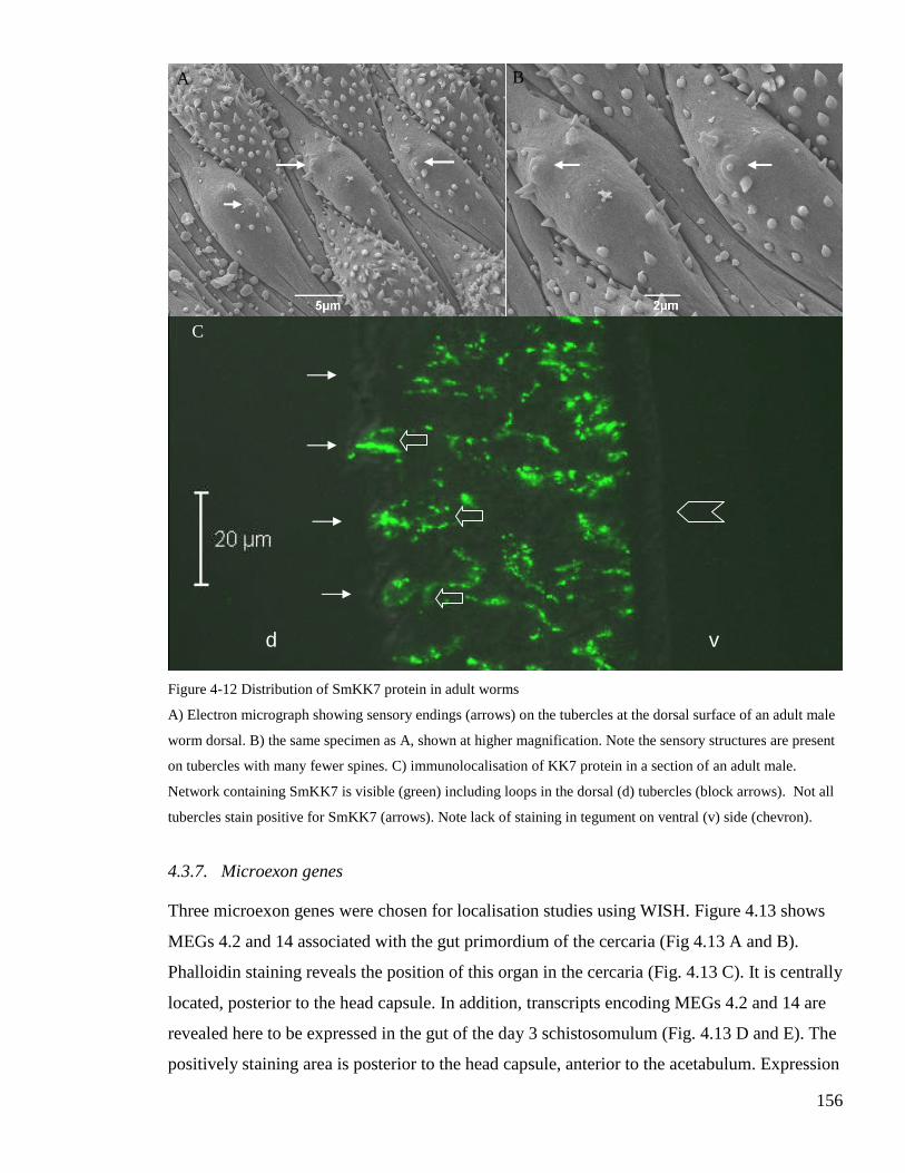
Fig
A)
wo
on
Ne
tub
4.3
Th
M
Ph
loc
rev
po
A B
ure 4-12 Dis
Electron mi
rm dorsal. B
tubercles wi
twork contai
ercles stain
.7. Micro
ree microe
EGs 4.2 an
alloidin st
ated, post
ealed here
sitively sta
C
tribution of SmKK7 protein in adult worms
crograph showing sensory endings (arrows) on the tubercles at the dorsal su
) the same specimen as A, shown at higher magnification. Note the sensory
th many fewer spines. C) immunolocalisation of KK7 protein in a section o
ning SmKK7 is visible (green) including loops in the dorsal (d) tubercles (b
positive for SmKK7 (arrows). Note lack of staining in tegument on ventral
exon genes
xon genes were chosen for localisation studies using WISH. F
d 14 associated with the gut primordium of the cercaria (Fig 4
aining reveals the position of this organ in the cercaria (Fig. 4.1
erior to the head capsule. In addition, transcripts encoding ME
to be expressed in the gut of the day 3 schistosomulum (Fig.
ining area is posterior to the head capsule, anterior to the aceta
d
156
rface of an adult male
structures are present
f an adult male.
lock arrows). Not all
(v) side (chevron).
igure 4.13 shows
.13 A and B).
3 C). It is centrally
Gs 4.2 and 14 are
4.13 D and E). The
bulum. Expression
v

157
is increased at this stage as evidenced by the darker staining. Visualisation of f-actin shows
minimal expansion of the oesophagus and gut at this stage (Fig. 4.13F). Expression of MEG
3.2 was shown to be sharply demarcated in the head gland of the schistosomulum (Figure
4.14). It is the first transcript to be localised to this gland. The positive staining is seen at the
very anterior of the worm, within the head capsule. The morphology of the larva is highlighted
by counterstaining with DAPI and simultaneous collection of a differential interference
contrast image. This image highlights the absence of nuclei in the tegument.
Figure 4-13 Distribution of MEG transcripts in larvae
WISH reveals transcripts encoding MEGs 4.2 (A) and MEG 14 (B) in the cercarial gut. Gut (g) and oesophagus
(o) revealed by phalloidin staining of actin in surrounding muscle cells in the cercaria (C) and day 3
schistosomulum (F). MEG 4.2 (D) and MEG 14 (E) transcript is also expressed in the gut in the day 3
schistosomulum. Scale bars A, B, D, E 20μm; C and F 50μm.

158
Figure 4-14 MEG3.2 (Smp_138070) transcript locaslied in a Day 10 schistosomulum
Fast Red (Sigma) was the substrate for alkaline phosphatase, thus MEG 3 transcript is highlighted by the red stain
in the head gland. Parasites were counterstained with DAPI to visualise nuclei (blue). The mouth (M), head
capsule muscle boundary (HC), gut (G), and acetabulum (A) are visible due to counterstaining with DAPI and
collection of a non-confocal differential interference contrast image (grey).
4.4. Discussion
4.4.1. Methods
The use of methanol for permeabilisation, instead of proteinase Ktreatment, has resulted in the
first successful application of WISH to germ balls. As we saw in Chapter two, germ balls are
fragile, it seems they cannot withstand proteinase Ktreatment. However, this harsher
permeablisation method is necessary for cercariae and all other lifecycle stages [16]. DNA is
preserved during WISH as the technique was developed to localise nucleic acids. This makes
DAPI an ideal counterstain for WISH when Fast Red is used as the alkaline phosphate
substrate for visualisation by confocal microscopy.
A
G
MEG 3
M
HC

159
4.4.2. Cercarial elastase 1a
Cercarial elastase 1a transcript was chosen as a positive control both for WISH in germ balls
generally, and for localisation to the acetabular glands in particular, as Newport et al showed
that cercarial elastase 1a was produced in these cells [114]. The results of the current study
reveal that cercarial elastase mRNA is abundant in young germ balls at the flattened posterior
stage, and expression continues until the germ ball is nearly mature, although the staining is
not so intense. This pattern allows the morphogenesis of the acetabular glands to be tracked
throughout germ ball development. It confirms that the large prominent cells observed towards
the anterior of young germ balls in Chapter 2 are the primordial acetabular glands. This is in
keeping with statements by Ebrahimzadeh and Dorsey that the acetabular glands are the first
tissues that are recognisable in very young germ balls [40, 41]. In addition to identifying novel
proteins in cercarial secretions, Curwen et al investigated the temporal expression pattern
using pulse labelling with radio-labelled methionine [116]. Infected snails were incubated
with [35S] methionine, and the post transformation secretions were collected from cercariae
shed three, five, seven and nine days later. The results showed that the bulk of cercarial
elastase was translated seven days prior to shedding [116].This is consistent with the present
observation that transcript encoding this important enzyme is transcribed in very young germ
balls.
4.4.3. Invadolysin
Following identification of invadolysin (SmPepM8) in cercarial secretions, it was
hypothesised by Curwen et al to be secreted during host invasion, and to play a role in
allowing the parasite to travel through the extracellular matrix in the skin and across the
basement membrane [116]. In the current study, the presence of invadolysin has been verified
in the acetabular glands by both WISH and IHC. This confirms the earlier observation that it is
secreted during entry into the skin. The authors also showed that the bulk of this enzyme was
synthesised three days prior to exit from the snail. The results detailed here also show that
invadolysin is expressed later than cercarial elastase, as transcript was detected only in more
mature germ balls.

160
4.4.4. VAL-10
VALs 4, 10 and 18 were identified in cercarial secretions by Curwen et al [116], and although
the family of S. mansoni VAL-protein encoding genes was described by Chalmers et al
recently [125], no function has yet been ascribed to these proteins. We saw in Chapter 3 that
just under half of the VAL genes are differentially expressed during the infection process. The
majority of these are up-regulated in the germ ball or the cercaria. In this chapter, transcript
encoding VAL-10 was localised to the acetabular glands. This confirms the likely secretion of
VAL-10 protein in the early stages of infection. The peripheral nature of the staining pattern
exhibited by acetabular gland associated transcripts in well-developed germ balls is in keeping
with the observation of Dorsey that the protein synthesis machinery is pushed to the edge of
the acetabular gland fundi as these cells develop and fill with secretory vesicles [41].
4.4.5. Sm16
Sm16 has been variously described as an immunomodulator and stathmin-like. Stathmin is a
protein with microtubule regulating activity [188]. It binds to free tubulin monomers and
inhibits microtubule formation. Valle et al reported that Sm16 was stage specific, it was found
in sporocysts but was not detectable in schistosomula 48 hours post infection [189]. The
authors suggested that Sm16 plays a role in regulating development at specific points in the
life cycle. Sm16 was found in the proteins released by cercariae in the first three hours after
mechanical transformation [116] and reported to be translated by both cercariae and
schistosomula. Rao et al cloned and expressed Sm16 and suggested that it is secreted by
schistosomula whilst they are in the skin to down regulate the host’s inflammatory response
[120].
Subsequent work has failed to confirm either that Sm16 is stathmin-like, or that it has a potent
immunomodulatory effect. Holmfeldt et al showed that recombinant Sm16 did not inhibit
microtubule formation in vitro [186]. They found that the presence of Sm16 in the cytosol of
human cells led to apoptosis. However, this was achieved by expressing Sm16 in human cells.
It was found that when Sm16 is added to human cells, it binds to the surface, is endocytosed
and degraded. In this case, the cells do not apoptose [186]. Later, Brannstrom et al expressed
Sm16 in Pichia pastoris and discovered that it had not cause T-lymphocytes to proliferate;
neither did it lead to a change in the cytokine production of whole human blood or monocytes

161
[187]. However, Sm16 did inhibit the response of various cells to lipopolysaccharide and poly
(I:C) [187]. In an attempt to better understand this controversial protein, mRNA encoding
Sm16 was localised in germ balls.
WISH results presented here show that the Sm16 transcript has an expression pattern quite
distinct from those of elastase 1a, invadolysin and VAL-10. This indicates that Sm16 is not
expressed in cells normally expected to secrete their contents into the host during infection.
Due to the widespread expression of Sm16 throughout the tissues of the germ ball, including
the tail (but excluding the acetabular glands), it is plausible that Sm16 would be released as
the tail detaches, rather than as a result of holocrine secretion from the acetabular glands.
However, the current results are in agreement with the timescale of expression detailed by
Curwen et al. The authors state that Sm16 was translated between seven to three days prior to
exit from the snail [116]; here Sm16 transcript is evident in germ balls from the flattened-
posterior to the nearly mature stage.
The discovery that Sm16 is not expressed in secretory glands underlines the importance of
localisation studies for confirmation that a particular gene is expressed at the host parasite
interface. Such studies should be carried out prior to further studies aimed at discovering how
the protein of interest interacts with the host.
4.4.6. SmKK7
SmKK7 was first identified in cercarial secretions. However, it has also been discovered in the
soluble fractions of supernatants when the tegument of adult worms is perturbed. In the light
of these findings, it is perhaps not surprising that SmKK7 is not expressed in the acetabular
glands. SmKK7 was annotated because of homology to the potassium channel blocker
BmKK7 from venom of the scorpion Mesobuthus martensii [116]. The two proteins are 30%
identical, with conserved cysteine residues. It was hypothesised by Curwen et al to play a part
in immunomodulation by interacting with T cells [116].
The transcript for SmKK7 was localised to eight cells in the germ ball and cercaria, and six
cells in the day 3 schistosomulum. At first glance these cells look similar in distribution to the
flame cells; however, more careful observation reveals that they are different. In the tail, flame

162
cells are proximal, whereas the KK7 producing cells are distally located, just before the tail
bifurcates. In addition, the anterior KK7-positive cells are situated in the head capsule,
whereas the most anterior flame cells are posterior to the head capsule muscle boundary (see
phalloidin staining of larvae in section 2.3.2). Confirmation that the KK7-producing cells are
not flame cells is the lack of colocalistaion of KK7 staining with the flame cells as highlighted
by phalloidin counterstaining with IHC [133].
It is rather impressive that the extensive network stained positive for KK7 protein arises solely
from the six cells identified by WISH. The anterior pair of cells may be the pair with
extensions to the surface, that was highlighted by Bruckner and Voge as staining for
acetylcholine esterase activity [51]. The varied morphology of the sensory structures at the
anterior of the cercaria suggests that they serve different functions. The shape of the KK7-
positive protrusions indicates that they may be the sensory endings that have a short wide
cilium, rather than either of the types with a narrow cilium. Robson and Erasmus studied the
anterior of the cercaria using electron microscopy. They showed the five sensory structures
detailed in the present study with the addition of two at the dorsal side of the crescents that
surround the acetabular duct openings [55]. Nuttman showed that the axon and raised bulb
(but not the cilium) of the anterior sensory endings were filled with vesicles [190]. Based on
the current observations, it is possible that SmKK7 is within these vesicles. However,
immunohistochemistry in conjunction with electron microscopy would have to be carried out
to confirm this.
The expression of SmKK7 has also been investigated in adult worms. The KK7-producing
cells are far more numerous in the adult, and visualisation of the protein reveals an extensive
net there, with processes entering tubercles on the adult male dorsal surface. Staining was
restricted to the area under the two muscle layers on the ventral side. Neuropeptide S1
(KYSALMF-NH2) has previously been localised with a pattern similar to KK7; it was present
in the peripheral nerve net including occasional dorsal tubercles [27]. In order to demonstrate
that KK7 enters the tubercles with fewer spines, counterstaining with an appropriately labelled
phalloidin would be necessary. It seems likely that these are ‘touch’ receptors, as the lack of
spines would aid perception. SmKK7 staining could be used to track the development of the
schistosome peripheral nervous system from comprising only six cells in the larvae to the fully
developed state in the adult.

163
The neural location of SmKK7, revealed here, seems at odds with its appearance in the 0-3
hour released proteins collected when cercariae are mechanically transformed. The simplest
explanation is that KK7 is expressed in the peripheral nerves in the worm where it functions as
a modulator of the action potential through inhibition of potassium channels. In this scenario,
SmKK7 is not expected to come into contact with the host under normal circumstances and so
the presence of KK7 in the released proteins is likely to be an artefact of mechanical
transformation (incubation on ice followed by vortexing), or in the case of the adult by
deliberate damage to the tegument. Conversely, it is known that the anterior cercarial nerve
endings disappear during transformation. It may be the case that SmKK7 is released into the
host as part of that process, whereupon it acts on host potassium channels. To investigate the
possible effect of KK7 on host blood vessels, the protein was expressed in Pichia pastoris by
Dr GP Dillon, and applied to mouse blood vessels. However, no effect was observed. SmKK7
has also been the subject of a knock-down study using RNAi in schistosomula (Dr P
McVeigh, personal communication). Again, no phenotype was observed. This is perhaps not
surprising as it may take a long time for all of the existing KK7 protein to be turned over. This
would have to happen before the knockdown of the mRNA would have an effect.
Co-localisation with known neural markers may give extra information of the type of nerves in
which KK7 is expressed, and shed some light on its function. Although many questions
remain regarding its role in schistosome biology, it is clear from the current study that it
expression is not limited to the cercaria.
4.4.7. MEGs
The remarkable up-regulation of MEG expression is the day 3 schistosomulum documented in
chapter three, leads to the conclusion that whatever their function, the proteins encoded by
these genes are important for the parasite during early intra-mammalian life. Although genes
containing single micro-exons have been described in other species, DeMarco et al report that
MEGs (comprising mostly micro-exons) are only found in schistosomes [142]. The authors
state that MEGs seem to have appeared and expanded rapidly in number in an ancient
schistosome ancestor. It is likely that this was an adaption to parasitism, specifically to life in
the bloodstream [142].

164
Three MEGs have been localised here for the first time in cercariae and/or schistosomula.
They are without exception expressed in tissues that form part of the host-parasite interface.
MEGs 4.2 and 14 are transcribed in the larval gut; Dillon et al have previously shown that
MEG 4.1 is expressed in the oesophageal gland of adult worms [16]. It is difficult to identify
this tissue in cercariae and schistosomula. Nevertheless, the localisation of these transcripts to
the alimentary tract confirms that they could come into contact with the host; schistosomes
have a blind ending gut therefore waste products are ejected by vomit. DeMarco et al postulate
that protein variation arising from MEGs is likely to be part of the immune evasion strategy
employed by schistosomes, but exactly how this is of benefit to the parasite is under
investigation [142]. It is also more evidence, taken with the up-regulation of gut proteases, that
the gut is transcriptionally active at this stage. MEG 3.2 stands out as it is the first gene known
to be expressed in the head gland. This is noteworthy as the contents of this gland are secreted
during skin invasion [48].
Summary
The localisation of novel schistosome gene products reveals unexpected patterns, and
highlights the importance of WISH both as a screen for selection of genes expressed at the
host-parasite interface, and in its role in hypothesis forming for functional annotation.

165
5. Final Discussion

166
The work presented in this thesis focuses on the S. mansoni larval stages before, during, and
after infection of the mammalian host. This encompasses many varied developmental changes
for the parasite: embryogenesis within the daughter sporocyst, exit from the snail, and survival
in fresh water with only a store of glycogen for nourishment. Once the cercaria has found a
suitable host, it enters by penetration of unbroken skin. The newly parasitic schistosomulum
undergoes rapid morphological changes that allow it, not only successfully to evade the host
immune system, but also to thrive. The next challenges the parasite must overcome are two
barriers: the epidermal basement membrane, and the epithelium of a blood vessel. The
schistosome persists in the vascular system for the rest of its life, which could be up to 30
years.
An intervention directed at larvae is attractive as it would interrupt the life cycle prior to the
onset of egg deposition. This is important for two reasons: firstly, pathology would be
prevented as it is the host response to the egg that causes it; secondly, transmission would be
blocked as this requires eggs to escape in the faeces, allowing miracidia to hatch and infect a
snail intermediate host. Therefore, a more thorough understanding of the infection process was
sought, with the aim of discovering new ways to target larvae early during infection.
The current study took a whole organism approach. First, the morphology of the relevant life
cycle stages was characterised. This was followed by a whole genome microarray experiment
to discover the gene expression underlying the impressive morphological and physiological
changes that take place. Lastly, the expression of selected genes was localised, putting them
into the context of the whole organism. The new methods used in this study will be described.
I will then draw together the novel findings discovered through the application of these
techniques and discuss how our knowledge of each life cycle stage and the infection process
has been furthered.
The cercaria had previously been studied extensively using electron microscopy but the
development of germ balls and schistosomula had been less well characterised. A large hurdle
for the study of germ balls was the use of sectioned hepatopancreas specimens. This made
interpretation of images difficult, as the orientation was hard to determine, and images had to
be reconstructed from a series of sections. The first application of confocal microscopy to
whole mount daughter sporocysts, germ balls, and schistosomula, presented here, allowed a

167
detailed study of the morphological changes that take place throughout the infection process.
This enabled the information gained in the later chapters to be related to the big picture.
The data that can be gleaned from a microarray experiment are limited to the sequences
represented on the array. When undertaking such an experiment, one must ensure that genes of
interest are included. This has led to the design and use of a variety of different microarray
platforms for S. mansoni, each of which was tailor-made to address the biological questions
under consideration at the time. All of these previous arrays have included only part of the
transcriptome; many comprised sequences derived from one life cycle stage only. With the
availability of a draft genome sequence and EST libraries from many life cycle stages, an
opportunity arose to design the first comprehensive microarray for this medically important
species. This was carried out in collaboration with the Wellcome Trust Sanger Institute and
the array was synthesised by Roche-NimbleGen. The design will be made freely available, and
should be useful for the research community, as it could be applied to any gene expression
question with any life cycle stage(s). It could also be re-annotated as the genome assembly
improves and the number of un-annotated genes is reduced.
The other novel aspect of the microarray experiment detailed in this thesis is the hybridisation
of germ balls to an array comprising sequences expressed at that stage. It has been shown that
the proteins secreted by cercariae during skin penetration are synthesised whilst germ balls are
in the snail. Therefore, gaining knowledge of this life cycle stage is an integral part of
investigating the mammalian infection process. The only other investigators to hybridise germ
ball material to an array were Dillon et al who used a microarray based entirely on lung
schistosomulum sequences to identify transcripts preferentially expressed in the
schistosomulum [98]. However, germ balls have not been included before in microarray
studies aimed at investigating the infection process. Other investigators have included
daughter sporocysts or infected hepatopancreas instead.
In order to relate the temporal expression pattern to the biology of schistosomes, a small group
of genes was chosen for localisation studies. The whole mount in situ hybridisation technique
developed by Dillon et al was applied to cercariae, schistosomula and adult worms. In order to
successfully localise transcripts in germ balls, an alternative permeablisation method was

168
employed. This allowed genes of interest to be localised in each of the life cycle stages under
investigation. It was the first application of WISH to germ balls.
The morphological study revealed that migratory daughter sporocysts have large germ cells
beneath a relatively thin layer of muscles. These cells could be readily targeted by particle
bombardment, and may solve the problem of delivering DNA to germ cells. It is worth noting
that daughter sporocysts can be delivered into snails where they continue the life cycle and
could produce transgenic cercariae.
In order for a germ ball to become a cercaria, much cell division, translation, and organisation
of cells into tissues must take place. Daily sampling in the morphological analysis revealed
that germ balls develop between 22-26 days after infection, and mature cercariae are present at
day 27. This was an important finding, as it allowed germ balls of all stages to be sampled in
the absence of cercariae, so that their gene expression profile could be compared with
cercariae and day 3 schistosomula. I have confirmed that the germ ball tegument forms as cells
at the surface coalesce, and that connections are made between these contributing cells and
cells further inside the body before the nuclei in the tegument are lost. Thus the epithelial
origins of the adult tegument syncytium are confirmed in the germ ball.
Results from the morphological study indicated that the acetabular glands begin to develop
very early. This was confirmed by the first application of WISH to germ balls; it was apparent,
following the localisation of cercarial elastase mRNA, that the acetabular glands take longer to
mature than any other tissue. They begin developing as soon as the anterior-posterior axis is
discernable by the flattened posterior, and transcripts encoding secreted proteins are still
present in nearly mature specimens. The duration of expression noted for cercarial elastase is
in stark contrast to the brief appearance of transcripts encoding both SmPepM8 and VAL-10,
which are only expressed in nearly mature germ balls.
The microarray study detailed in Chapter 3 shed light on the gene expression patterns
underlying cercarial embryogenesis. As well as DNA polymerase and genes whose products
are required for cell cycle progression, it was revealed that a subset of cadherins, caspases,
homeobox and Wnt genes are up-regulated at this stage. Cadherins are adhesion molecules
which regulate cell-cell interactions. It is likely that the four cadherins expressed in the germ

169
ball are involved in sorting tissues. Caspases are involved in programmed cell death, which is
also important in embryogenesis. Although much remains to be learnt regarding the complex
process of cercariogenesis, WISH may be used to localise genes shown in this study to be
enriched in germ balls, enabling hypotheses to be generated regarding their precise functions.
Five cercarial elastase genes had been described previously by Salter et al [115]. The current
thesis provides the first evidence that 11 cercarial elastase genes are up-regulated in the germ
ball and five invadolysins additional to Curwen’s SmPepM8 are also germ ball-enriched. The
results reveal that the infection process is more complex than had been thought. It is certainly
not the case that cercarial entry into the host is mediated by a single protease class. This is the
first report of VAL expression in germ balls. Again, the known repertoire was expanded; six
VALs were shown to be up-regulated at this stage. The up-regulation of VALs in germ balls
and/or cercariae suggests that their functions are related to the early stages of infection.
Transcripts encoding VAL 10 and invadolsysin (SmPepM8) were localised to the acetabular
glands, confirming their importance as secreted proteins involved in host invasion.
More surprising results were obtained when Sm16 and SmKK7 were localised. Both of these
proteins, Sm16 in particular, had been suggested to function as immunomodulators during
early infection. A prerequisite for such a function is that the protein must come into contact
with the host. However, transcripts encoding both of these proteins were localised to tissues
not expected to secrete their contents to the exterior of the worm. This could be taken as a
salutary tale; caution should be taken when assigning putative functions to novel proteins.
Even their presence in preparations enriched for secretions is not conclusive proof that such
proteins are, in fact, secreted by the worm. Localisation studies can be used to confirm
hypotheses prior to further research. In this case, the putative functions of both SmKK7 and
Sm16 can now be reappraised in the light of their localisation patterns. SmKK7 is present in
the peripheral nerves. The simplest explanation is that SmKK7 is involved in neural function
within the worm. Many questions remain. Assuming it is a potassium channel blocker, which
potassium channels does it affect and which signals does it respond to? It is very difficult to
make any hypotheses regarding the function of Sm16 based on its expression throughout the
body. However, it is not transcribed in the acetabular glands and as such, despite several
reports that it is found in preparations enriched for secreted proteins; it does not seem to be
secreted ‘on purpose’ by the worm.

170
The cercaria has been thought of as quite transcriptionally inactive. It is a complex organism
adapted for its short stint as a free-living, non-feeding, aquatic creature. As previous studies
have shown, the cercaria uses aerobic metabolism in order to make the most of its glycogen
store. The free-living, snail-infective miracidium also uses aerobic metabolism. Another
similarity with the miracidium is the up-regulation of the small heat shock protein Sm-p40,
which conserves energy by avoiding proteasomal degradation of denatured proteins. A novel
finding of the current study is that the cercaria transcribes genes whose products are needed on
entry to the host. Indeed, ‘planning ahead’ seems to be a salient feature of schistosome
biology. The contents of the acetabular glands are synthesised even before the germ ball has a
tail and tegument- and gut-associated protein encoding genes are up-regulated in the cercaria
which does not feed, and whose tegument has no use for glucose transporters or complement
inhibitors.
Cercariae are known to emerge from snails when it is light. Although the morphological study
did not pin-point the light sensitive organ, the microarray experiment revealed that an opsin-
like GPCR is up-regulated in the germ ball. There is also a light sensitive cell development
protein whose transcript is up-regulated in the cercaria. WISH could be used to localise the
expression of either of these genes in the germ ball, potentially leading to the identification of
the cercarial light sensing organ. The cercaria also relies on neural signalling for swimming
and to find a host. This is reflected in the up-regulation of neurotransmitter transporters at this
life cycle stage and the conspicuous nerve endings at the anterior, some of which contain large
amounts of the novel neural protein SmKK7.
The head gland, whilst known to be important in the infection process is an enigmatic organ. It
was not known whether it was unicellular like the acetabular glands; it was shown in Chapter
2 that it contains two nuclei. It was also revealed in this study that MEG 3 is transcribed in this
gland.
Considerable remodelling occurs in the three days after transformation. It was noted in
Chapter 2 that the anterior sensory endings and acetabular glands and ducts are no longer
visible in the day 3 schistosomulum, and that the lateral nerve chords can be distinguished at
this stage. Cadherins and various other morphogens are implicated in this developmental

171
phase by virtue of their increased expression in the day 3 schistosomulum. It is worth noting
that the cadherins expressed at this stage are different to those up-regulated in the germ ball.
The morphological study of the schistosomula revealed the presence of protein in the larval
gut and the microarray study demonstrated that gut-related proteases and transport proteins are
most highly expressed at this stage compared to the other two. It remains unclear whether the
protein observed in the gut is synthesised or ingested by the schistosomulum. This question
could be addressed by incubating schistosomula in medium containing fluorescently labelled
proteins and subsequently viewing them by confocal microscopy. It seems likely that early
larvae do ingest plasma, as nutrition is necessary for the onward migration. However, at this
stage the oesophagus is still too narrow to admit red blood cells.
Another finding of the current study was the marked up-regulation of certain tegument
protein-encoding genes in the day 3 schistosomulum. The tegument is the ideal target for a
vaccine, as it is accessible to the host. Those proteins expressed at the surface throughout
intra-mammalian life may prove to be successful candidates, as they are by implication
necessary for survival in the host, and may enable the host to target the parasite before it
reaches maturity and causes pathology. Tegument proteins linked to immune evasion by adult
worms are up-regulated in the day 3 schistosomulum. Of particular note are a group of CD59-
encoding genes. In humans, CD59 is a complement inhibitor. The schistosome genome
contains seven of these paralogues, four of which have been identified on the surface of adult
worms. This is the first report showing their up-regulation in the schistosomulum. Functional
studies are yet to be carried out on schistosome CD59. These would be needed as well as
localisation studies in the larva to determine its role in complement inhibition and immune
evasion.
An intriguing part of the infection process is the appearance of micro exon genes in the early
intra-mammalian larva. Four new MEGs were identified in the current study. The highest fold
change observed in the study was exhibited by a MEG. Not only are the majority of MEGs up-
regulated in the schistosomulum, three of them have been localised to tissues at the host
parasite interface. The extreme splice-variability of these transcripts, coupled with their
expression at the host parasite interface begs the question: what is their primary function? It
seems likely that the variants are generated to bamboozle the host immune response. Whether

172
that is in order to mask an important function carried out by a specific variant, or rather to
allow the parasite as a whole to avoid immune attack remains to be discovered.
A large proportion of the genes in the S. mansoni genome have no homology to any other
known sequences. Due to time constraints, these have not been investigated. However, the
discovery of four novel MEGs among the un-annotated genes points likely presence of other
novel schistosome genes that may shed light on how these worms gain entry to the host and
remain there.

173
6. List of References
1. King CH, Dickman K, Tisch DJ: Reassessment of the cost of chronic helminticinfection: a meta-analysis of disability-related outcomes in endemicschistosomiasis. Lancet 2005, 365(9470):1561-1569.
2. Steinmann P, Keiser J, Bos R, Tanner M, Utzinger J: Schistosomiasis and waterresources development: systematic review, meta-analysis, and estimates of peopleat risk. Lancet Infect Dis 2006, 6(7):411-425.
3. van der Werf MJ, de Vlas SJ, Brooker S, Looman CW, Nagelkerke NJ, Habbema JD,Engels D: Quantification of clinical morbidity associated with schistosomeinfection in sub-Saharan Africa. Acta Trop 2003, 86(2-3):125-139.
4. Caffrey CR, Rohwer A, Oellien F, Marhofer RJ, Braschi S, Oliveira G, McKerrow JH,Selzer PM: A comparative chemogenomics strategy to predict potential drugtargets in the metazoan pathogen, Schistosoma mansoni. PLoS ONE 2009,4(2):e4413.
5. Abdulla MH, Ruelas DS, Wolff B, Snedecor J, Lim KC, Xu F, Renslo AR, Williams J,McKerrow JH, Caffrey CR: Drug discovery for schistosomiasis: hit and leadcompounds identified in a library of known drugs by medium-throughputphenotypic screening. PLoS Negl Trop Dis 2009, 3(7):e478.
6. Prevention and control of schistosomiasis and soil-transmitted helminthiasis.World Health Organ Tech Rep Ser 2002, 912:i-vi, 1-57, back cover.
7. Bethony JM, Diemert DJ, Oliveira SC, Loukas A: Can schistosomiasis really beconsigned to history without a vaccine? Vaccine 2008, 26(27-28):3373-3376.
8. Gryseels B, Polman K, Clerinx J, Kestens L: Human schistosomiasis. Lancet 2006,368(9541):1106-1118.
9. Cheever AW, Macedonia JG, Mosimann JE, Cheever EA: Kinetics of egg productionand egg excretion by Schistosoma mansoni and S. japonicum in mice infectedwith a single pair of worms. Am J Trop Med Hyg 1994, 50(3):281-295.
10. Becker W: [Studies on the migrating daughter sporocysts of Schistosoma mansoni.II. The migration to the hepatopancreas]. Z Parasitenkd 1970, 34(3):226-241.
11. Meuleman EA, Holzmann PJ, Peet RC: Development Of Daughter SporocystsInside The Mother Sporocyst Of Schistosoma-Mansoni With Special ReferenceTo The Ultrastructure Of The Body Wall. Zeitschrift Fur Parasitenkunde-Parasitology Research 1980, 61(3):201-212.
12. Jourdane J, Theron A, Combes C: Demonstration of several sporocysts generationsas a normal pattern of reproduction of Schistosoma mansoni. Acta Trop 1980,37(2):177-182.
13. Crabtree JE, Wilson RA: Schistosoma mansoni: an ultrastructural examination ofpulmonary migration. Parasitology 1986, 92 (Pt 2):343-354.
14. Wilson RA, Coulson PS: Schistosoma mansoni: dynamics of migration through thevascular system of the mouse. Parasitology 1986, 92 (Pt 1):83-100.
15. Miller P, Wilson RA: Migration of the schistosomula of Schistosoma mansoni fromthe lungs to the hepatic portal system. Parasitology 1980, 80(2):267-288.
16. Dillon GP, Illes JC, Isaacs HV, Wilson RA: Patterns of gene expression inschistosomes: localization by whole mount in situ hybridization. Parasitology2007, 134(Pt 11):1589-1597.

174
17. Jones MK, Higgins T, Stenzel DJ, Gobert GN: Towards tissue specifictranscriptomics and expression pattern analysis in schistosomes using lasermicrodissection microscopy. Exp Parasitol 2007, 117(3):259-266.
18. Morris GP, Threadgold LT: Ultrastructure of the tegument of adult Schistosomamansoni. J Parasitol 1968, 54(1):15-27.
19. McLaren DJ, Hockley DJ: Blood flukes have a double outer membrane. Nature1977, 269(5624):147-149.
20. Hockley DJ: Schistosoma mansoni: the development of the cercarial tegument.Parasitology 1972, 64(2):245-252.
21. Skelly PJ, Alan Wilson R: Making sense of the schistosome surface. Adv Parasitol2006, 63:185-284.
22. Saunders N, Wilson RA, Coulson PS: The outer bilayer of the adult schistosometegument surface has a low turnover rate in vitro and in vivo. Mol BiochemParasitol 1987, 25(2):123-131.
23. Halton DW, Gustafsson, M. K. S.: Functional morphology of the platyhelminthnervous system. Parasitology 1996, 113:S47-S72.
24. Gustafsson MK: Immunocytochemical demonstration of neuropeptides andserotonin in the nervous systems of adult Schistosoma mansoni. Parasitol Res1987, 74(2):168-174.
25. Silk MH, Spence IM: Ultrastructural studies of the blood fluke--schistosomamansoni. 3. The nerve tissue and sensory structures. S Afr J Med Sci 1969,34(4):93-104.
26. Hockley DJ: Ultrastructure of the tegument of Schistosoma. Adv Parasitol 1973,11(0):233-305.
27. Brownlee DJ, Fairweather I, Johnston CF, Thorndyke MC, Skuce PJ:Immunocytochemical demonstration of a SALMFamide-like neuropeptide in thenervous system of adult and larval stages of the human blood fluke, Schistosomamansoni. Parasitology 1995, 110 (Pt 2):143-153.
28. Wilson RA, Webster LA: Protonephridia. Biol Rev Camb Philos Soc 1974,49(2):127-160.
29. Wilson RA: A physiological study of the development of the egg of Fasciolahepatica L., the common liver fluke. Comp Biochem Physiol 1967, 21(2):307-320.
30. Bogers JJ, Nibbeling HA, Van Marck EA, Deelder AM: Immunoelectronmicroscopical localization of a circulating antigen in the excretory system ofSchistosoma mansoni. Ultrastructural localization studies of the excretory systemof S. mansoni. Parasitol Res 1995, 81(5):375-381.
31. Kusel JR, McVeigh P, Thornhill JA: The schistosome excretory system: a key toregulation of metabolism, drug excretion and host interaction. Trends Parasitol2009, 25(8):353-358.
32. Mair GR, Maule AG, Day TA, Halton DW: A confocal microscopical study of themusculature of adult Schistosoma mansoni. Parasitology 2000, 121 (Pt 2):163-170.
33. The biology of schistsosomes from genes to latrines.34. Machado-Silva JR, Pelajo-Machado M, Lenzi HL, Gomes DC: Morphological study
of adult male worms of Schistosoma mansoni Sambon, 1907 by confocal laserscanning microscopy. Mem Inst Oswaldo Cruz 1998, 93 Suppl 1:303-307.
35. Mair GR, Maule AG, Fried B, Day TA, Halton DW: Organization of themusculature of schistosome cercariae. J Parasitol 2003, 89(3):623-625.
36. Hansen E, Perez-Mendez G: Scanning electron microscopy of Schistosoma mansonidaughter sporocysts. Int J Parasitol 1972, 2(1):174.

175
37. Fournier A, Theron A: [Morpho-anatomic and functional sectorization ofSchistosoma mansoni daughter sporocysts]. Z Parasitenkd 1985, 71(3):325-336.
38. Cheng TC, Bier JW: Studies on molluscan schistosomiasis: an analysis of thedevelopment of the cercaria of Schistosoma mansoni. Parasitology 1972,64(1):129-141.
39. Meuleman EA, Holzmann PJ: Development Of Primitive Epithelium And TrueTegument In Cercaria Of Schistosoma-Mansoni. Zeitschrift Fur Parasitenkunde-Parasitology Research 1975, 45(4):307-318.
40. Ebrahimzadeh A: [Development, histology and histochemistry of the glandularsystem of the cercaria of Schistosoma mansoni Sambon (1907)]. Z Parasitenkd1970, 34(4):319-342.
41. Dorsey CH: Schistosoma mansoni: development of acetabular glands of cercariaat ultrastructural level. Exp Parasitol 1975, 37(1):37-59.
42. Dorsey CH, Cousin CE, Lewis FA, Stirewalt MA: Ultrastructure of the Schistosomamansoni cercaria. Micron 2002, 33(3):279-323.
43. Stirewalt MA: Cercaria vs. schistosomule (Schistosoma mansoni): absence of thepericercarial envelope in vivo and the early physiological and histologicalmetamorphosis of the parasite. Exp Parasitol 1963, 13:395-406.
44. Dorsey CH, Stirewalt MA: Schistosoma mansoni: fine structure of cercarialacetabular glands. Exp Parasitol 1971, 30(2):199-214.
45. Ebrahimzadeh A, Kraft M: [Ultrastructural studies on the morphology of thecercariae Schistosoma mansoni. I. The alimentary tract]. Z Parasitenkd 1969,32(2):157-175.
46. Lawson JR, Wilson RA: The survival of the cercariae of Schistosoma mansoni inrelation to water temperature and glycogen utilization. Parasitology 1980,81(2):337-348.
47. Dorsey CH: Schistosoma mansoni: description of the head gland of cercariae andschistosomules at the ultrastructural level. Exp Parasitol 1976, 39(3):444-459.
48. Crabtree JE, Wilson RA: Schistosoma mansoni: an ultrastructural examination ofskin migration in the hamster cheek pouch. Parasitology 1985, 91 (Pt 1):111-120.
49. Stirewalt MA: Schistosoma mansoni: cercaria to schistosomule. Adv Parasitol 1974,12:115-182.
50. Mecozzi B, Rossi A, Lazzaretti P, Kady M, Kaiser S, Valle C, Cioli D, Klinkert MQ:Molecular cloning of Schistosoma mansoni calcineurin subunits andimmunolocalization to the excretory system. Mol Biochem Parasitol 2000,110(2):333-343.
51. Bruckner DA, Voge M: The nervous system of larval Schistosoma mansoni asrevealed by acetylcholinesterase staining. J Parasitol 1974, 60(3):437-446.
52. Skuce PJ, Johnston CF, Fairweather I, Halton DW, Shaw C: A confocal scanninglaser microscope study of the peptidergic and serotoninergic components of thenervous system in larval Schistosoma mansoni. Parasitology 1990, 101 Pt 2:227-234.
53. Nuttman CJ: The fine structure of ciliated nerve endings in the cercariae ofSchistosoma mansoni. The Journal of Parasitology 1971, 57):855-859.
54. Cousin CE, Dorsey CH: Nervous system of Schistosoma mansoni cercaria:organization and fine structure. Parasitol Res 1991, 77(2):132-141.
55. Robson RT, Erasmus DA: The ultrastructure, based on stereoscan observations, ofthe oral sucker of the cercaria of Schistosoma mansoni with special reference topenetration. Z Parasitenkd 1970, 35(1):76-86.

176
56. McLaren DJ, Hockley DJ: Schistosoma mansoni: the occurrence of microvilli onthe surface of the tegument during transformation from cercaria toschistosomulum. Parasitology 1976, 73(2):169-187.
57. Skelly PJ, Kim JW, Cunningham J, Shoemaker CB: Cloning, characterization, andfunctional expression of cDNAs encoding glucose transporter proteins from thehuman parasite Schistosoma mansoni. J Biol Chem 1994, 269(6):4247-4253.
58. Skelly PJ, Shoemaker CB: Rapid appearance and asymmetric distribution ofglucose transporter SGTP4 at the apical surface of intramammalian-stageSchistosoma mansoni. Proc Natl Acad Sci U S A 1996, 93(8):3642-3646.
59. Jiang J, Skelly PJ, Shoemaker CB, Caulfield JP: Schistosoma mansoni: the glucosetransport protein SGTP4 is present in tegumental multilamellar bodies, discoidbodies, and the surface lipid bilayers. Exp Parasitol 1996, 82(2):201-210.
60. Skelly PJ, Shoemaker CB: Induction cues for tegument formation during thetransformation of Schistosoma mansoni cercariae. Int J Parasitol 2000, 30(5):625-631.
61. Crabtree JE, Wilson RA: Schistosoma mansoni: a scanning electron microscopestudy of the developing schistosomulum. Parasitology 1980, 81(Pt 3):553-564.
62. Thornhill J, Coelho PM, McVeigh P, Maule A, Jurberg AD, Kusel JR: Schistosomamansoni cercariae experience influx of macromolecules during skin penetration.Parasitology 2009, 136(11):1257-1267.
63. Crabtree JE: An ultrastructural study of the migrating schistosomulum ofSchistosoma mansoni. D Phil thesis, University of York 1982.
64. Harrop R, Wilson RA: Protein synthesis and release by cultured schistosomula ofSchistosoma mansoni. Parasitology 1993, 107 (Pt 3):265-274.
65. Blanton RE, Licate LS: Developmental regulation of protein synthesis inschistosomes. Mol Biochem Parasitol 1992, 51(2):201-208.
66. Skelly PJ, Shoemaker CB: A molecular genetic study of the variations in metabolicfunction during schistosome development. Mem Inst Oswaldo Cruz 1995, 90(2):281-284.
67. Simpson AJ, Sher A, McCutchan TF: The genome of Schistosoma mansoni:isolation of DNA, its size, bases and repetitive sequences. Mol Biochem Parasitol1982, 6(2):125-137.
68. Franco GR, Adams MD, Soares MB, Simpson AJ, Venter JC, Pena SD: Identificationof new Schistosoma mansoni genes by the EST strategy using a directional cDNAlibrary. Gene 1995, 152(2):141-147.
69. Franco GR, Rabelo EM, Azevedo V, Pena HB, Ortega JM, Santos TM, Meira WS,Rodrigues NA, Dias CM, Harrop R et al: Evaluation of cDNA libraries fromdifferent developmental stages of Schistosoma mansoni for production ofexpressed sequence tags (ESTs). DNA Res 1997, 4(3):231-240.
70. Dias Neto E, Harrop R, Correa-Oliveira R, Wilson RA, Pena SD, Simpson AJ:Minilibraries constructed from cDNA generated by arbitrarily primed RT-PCR:an alternative to normalized libraries for the generation of ESTs from nanogramquantities of mRNA. Gene 1997, 186(1):135-142.
71. Dias Neto E, Correa RG, Verjovski-Almeida S, Briones MR, Nagai MA, da Silva W,Jr., Zago MA, Bordin S, Costa FF, Goldman GH et al: Shotgun sequencing of thehuman transcriptome with ORF expressed sequence tags. Proc Natl Acad Sci U SA 2000, 97(7):3491-3496.
72. Santos TM, Johnston DA, Azevedo V, Ridgers IL, Martinez MF, Marotta GB, SantosRL, Fonseca SJ, Ortega JM, Rabelo EM et al: Analysis of the gene expression profile

177
of Schistosoma mansoni cercariae using the expressed sequence tag approach.Mol Biochem Parasitol 1999, 103(1):79-97.
73. Verjovski-Almeida S, DeMarco R, Martins EA, Guimaraes PE, Ojopi EP, Paquola AC,Piazza JP, Nishiyama MY, Jr., Kitajima JP, Adamson RE et al: Transcriptomeanalysis of the acoelomate human parasite Schistosoma mansoni. Nat Genet 2003,35(2):148-157.
74. Haas BJ, Berriman M, Hirai H, Cerqueira GG, Loverde PT, El-Sayed NM:Schistosoma mansoni genome: closing in on a final gene set. Exp Parasitol 2007,117(3):225-228.
75. Berriman M, Haas BJ, LoVerde PT, Wilson RA, Dillon GP, Cerqueira GC,Mashiyama ST, Al-Lazikani B, Andrade LF, Ashton PD et al: The genome of theblood fluke Schistosoma mansoni. Nature 2009, 460(7253):352-358.
76. McVeigh P, Mair GR, Atkinson L, Ladurner P, Zamanian M, Novozhilova E, MarksNJ, Day TA, Maule AG: Discovery of multiple neuropeptide families in the phylumPlatyhelminthes. Int J Parasitol 2009.
77. Correnti JM, Brindley PJ, Pearce EJ: Long-term suppression of cathepsin B levelsby RNA interference retards schistosome growth. Mol Biochem Parasitol 2005,143(2):209-215.
78. Skelly PJ, Da'dara A, Harn DA: Suppression of cathepsin B expression inSchistosoma mansoni by RNA interference. Int J Parasitol 2003, 33(4):363-369.
79. Rinaldi G, Morales ME, Alrefaei YN, Cancela M, Castillo E, Dalton JP, Tort JF,Brindley PJ: RNA interference targeting leucine aminopeptidase blocks hatchingof Schistosoma mansoni eggs. Mol Biochem Parasitol 2009, 167(2):118-126.
80. Boyle JP, Wu XJ, Shoemaker CB, Yoshino TP: Using RNA interference tomanipulate endogenous gene expression in Schistosoma mansoni sporocysts. MolBiochem Parasitol 2003, 128(2):205-215.
81. Brindley PJ, Pearce EJ: Genetic manipulation of schistosomes. Int J Parasitol 2007,37(5):465-473.
82. Geldhof P, Visser A, Clark D, Saunders G, Britton C, Gilleard J, Berriman M, Knox D:RNA interference in parasitic helminths: current situation, potential pitfalls andfuture prospects. Parasitology 2007, 134(Pt 5):609-619.
83. de Moraes Mourao M, Dinguirard N, Franco GR, Yoshino TP: Phenotypic Screen ofEarly-Developing Larvae of the Blood Fluke, Schistosoma mansoni, using RNAInterference. PLoS Negl Trop Dis 2009, 3(8):e502.
84. Heyers O, Walduck AK, Brindley PJ, Bleiss W, Lucius R, Dorbic T, Wittig B, KalinnaBH: Schistosoma mansoni miracidia transformed by particle bombardment infectBiomphalaria glabrata snails and develop into transgenic sporocysts. ExpParasitol 2003, 105(2):174-178.
85. Wippersteg V, Kapp K, Kunz W, Jackstadt WP, Zahner H, Grevelding CG: HSP70-controlled GFP expression in transiently transformed schistosomes. Mol BiochemParasitol 2002, 120(1):141-150.
86. Wippersteg V, Kapp K, Kunz W, Grevelding CG: Characterisation of the cysteineprotease ER60 in transgenic Schistosoma mansoni larvae. Int J Parasitol 2002,32(10):1219-1224.
87. Beckmann S, Wippersteg V, El-Bahay A, Hirzmann J, Oliveira G, Grevelding CG:Schistosoma mansoni: germ-line transformation approaches and actin-promoteranalysis. Exp Parasitol 2007, 117(3):292-303.
88. Kines KJ, Morales ME, Mann VH, Gobert GN, Brindley PJ: Integration of reportertransgenes into Schistosoma mansoni chromosomes mediated by pseudotypedmurine leukemia virus. Faseb J 2008, 22(8):2936-2948.

178
89. Singh-Gasson S, Green RD, Yue Y, Nelson C, Blattner F, Sussman MR, Cerrina F:Maskless fabrication of light-directed oligonucleotide microarrays using a digitalmicromirror array. Nat Biotechnol 1999, 17(10):974-978.
90. Smyth GK: Linear models and empirical bayes methods for assessing differentialexpression in microarray experiments. Stat Appl Genet Mol Biol 2004, 3:Article3.
91. Gentleman RC, Carey VJ, Bates DM, Bolstad B, Dettling M, Dudoit S, Ellis B,Gautier L, Ge Y, Gentry J et al: Bioconductor: open software development forcomputational biology and bioinformatics. Genome Biol 2004, 5(10):R80.
92. Ashburner M, Ball CA, Blake JA, Botstein D, Butler H, Cherry JM, Davis AP,Dolinski K, Dwight SS, Eppig JT et al: Gene ontology: tool for the unification ofbiology. The Gene Ontology Consortium. Nat Genet 2000, 25(1):25-29.
93. Fitzpatrick JM, Johnston DA, Williams GW, Williams DJ, Freeman TC, Dunne DW,Hoffmann KF: An oligonucleotide microarray for transcriptome analysis ofSchistosoma mansoni and its application/use to investigate gender-associated geneexpression. Mol Biochem Parasitol 2005, 141(1):1-13.
94. Hoffmann KF, Johnston DA, Dunne DW: Identification of Schistosoma mansonigender-associated gene transcripts by cDNA microarray profiling. Genome Biol2002, 3(8):RESEARCH0041.
95. Gobert GN, McManus DP, Nawaratna S, Moertel L, Mulvenna J, Jones MK: TissueSpecific Profiling of Females of Schistosoma japonicum by Integrated LaserMicrodissection Microscopy and Microarray Analysis. PLoS Negl Trop Dis 2009,3(6):e469.
96. Vermeire JJ, Taft AS, Hoffmann KF, Fitzpatrick JM, Yoshino TP: Schistosomamansoni: DNA microarray gene expression profiling during the miracidium-to-mother sporocyst transformation. Mol Biochem Parasitol 2006, 147(1):39-47.
97. Jolly ER, Chin CS, Miller S, Bahgat MM, Lim KC, DeRisi J, McKerrow JH: Geneexpression patterns during adaptation of a helminth parasite to differentenvironmental niches. Genome Biol 2007, 8(4):R65.
98. Dillon GP, Feltwell T, Skelton JP, Ashton PD, Coulson PS, Quail MA, Nikolaidou-Katsaridou N, Wilson RA, Ivens AC: Microarray analysis identifies genespreferentially expressed in the lung schistosomulum of Schistosoma mansoni. Int JParasitol 2006, 36(1):1-8.
99. Fitzpatrick JM, Peak E, Perally S, Chalmers IW, Barrett J, Yoshino TP, Ivens AC,Hoffmann KF: Anti-schistosomal Intervention Targets Identified by LifecycleTranscriptomic Analyses. PLoS Negl Trop Dis 2009, 3(11):e543.
100. Waisberg M, Lobo FP, Cerqueira GC, Passos LK, Carvalho OS, El-Sayed NM, FrancoGR: Schistosoma mansoni: Microarray analysis of gene expression induced byhost sex. Exp Parasitol 2008, 120(4):357-363.
101. Dillon GP, Feltwell T, Skelton J, Coulson PS, Wilson RA, Ivens AC: Altered patternsof gene expression underlying the enhanced immunogenicity of radiation-attenuated schistosomes. PLoS Negl Trop Dis 2008, 2(5):e240.
102. Verjovski-Almeida S, Venancio TM, Oliveira KC, Almeida GT, DeMarco R: Use of a44k oligoarray to explore the transcriptome of Schistosoma mansoni adult worms.Exp Parasitol 2007, 117(3):236-245.
103. Gobert GN, McInnes R, Moertel L, Nelson C, Jones MK, Hu W, McManus DP:Transcriptomics tool for the human Schistosoma blood flukes using microarraygene expression profiling. Exp Parasitol 2006, 114(3):160-172.
104. Lawson JR, Wilson RA: Metabolic changes associated with the migration of theschistosomulum of Schistosoma mansoni in the mammal host. Parasitology 1980,81(2):325-336.

179
105. Fitzpatrick JM, Protasio AV, McArdle AJ, Williams GA, Johnston DA, Hoffmann KF:Use of Genomic DNA as an Indirect Reference for Identifying Gender-AssociatedTranscripts in Morphologically Identical, but Chromosomally Distinct,Schistosoma mansoni Cercariae. PLoS Negl Trop Dis 2008, 2(10):e323.
106. Clegg JA: In Vitro Cultivation Of Schistosoma Mansoni. Exp Parasitol 1965,16:133-147.
107. Braschi S, Wilson RA: Proteins exposed at the adult schistosome surface revealedby biotinylation. Mol Cell Proteomics 2006, 5(2):347-356.
108. Webster M, Fulford AJ, Braun G, Ouma JH, Kariuki HC, Havercroft JC, Gachuhi K,Sturrock RF, Butterworth AE, Dunne DW: Human immunoglobulin E responses toa recombinant 22.6-kilodalton antigen from Schistosoma mansoni adult wormsare associated with low intensities of reinfection after treatment. Infect Immun1996, 64(10):4042-4046.
109. Gordon RM, Griffiths RB: Observations on the means by which the cercariae ofSchistosoma mansoni penetrate mammalian skin, together with an account ofcertain morphological changes observed in the newly penetrated larvae. Ann TropMed Parasitol 1951, 45(3-4):227-243.
110. Dresden MH, Lewis JC, Krisko I: Proteolytic action of Schistosoma mansonicercarial proteases on keratin and basement membrane proteins. J Parasitol 1977,63(5):941-943.
111. McKerrow JH, Jones P, Sage H, Pino-Heiss S: Proteinases from invasive larvae ofthe trematode parasite Schistosoma mansoni degrade connective-tissue andbasement-membrane macromolecules. Biochem J 1985, 231(1):47-51.
112. Marikovsky M, Arnon R, Fishelson Z: Schistosoma mansoni: localization of the 28kDa secreted protease in cercaria. Parasite Immunol 1990, 12(4):389-401.
113. Salter JP, Lim KC, Hansell E, Hsieh I, McKerrow JH: Schistosome invasion ofhuman skin and degradation of dermal elastin are mediated by a single serineprotease. J Biol Chem 2000, 275(49):38667-38673.
114. Newport GR, McKerrow JH, Hedstrom R, Petitt M, McGarrigle L, Barr PJ, AgabianN: Cloning of the proteinase that facilitates infection by schistosome parasites. JBiol Chem 1988, 263(26):13179-13184.
115. Salter JP, Choe Y, Albrecht H, Franklin C, Lim KC, Craik CS, McKerrow JH:Cercarial elastase is encoded by a functionally conserved gene family acrossmultiple species of schistosomes. J Biol Chem 2002, 277(27):24618-24624.
116. Curwen RS, Ashton PD, Sundaralingam S, Wilson RA: Identification of novelproteases and immunomodulators in the secretions of schistosome cercariae thatfacilitate host entry. Mol Cell Proteomics 2006, 5(5):835-844.
117. Dvorak J, Mashiyama ST, Braschi S, Sajid M, Knudsen GM, Hansell E, Lim KC,Hsieh I, Bahgat M, Mackenzie B et al: Differential use of protease families forinvasion by schistosome cercariae. Biochimie 2008, 90(2):345-358.
118. Hansell E, Braschi S, Medzihradszky KF, Sajid M, Debnath M, Ingram J, Lim KC,McKerrow JH: Proteomic analysis of skin invasion by blood fluke larvae. PLoSNegl Trop Dis 2008, 2(7):e262.
119. Knudsen GM, Medzihradszky KF, Lim KC, Hansell E, McKerrow JH: Proteomicanalysis of Schistosoma mansoni cercarial secretions. Mol Cell Proteomics 2005,4(12):1862-1875.
120. Rao KV, Ramaswamy K: Cloning and expression of a gene encoding Sm16, ananti-inflammatory protein from Schistosoma mansoni. Mol Biochem Parasitol2000, 108(1):101-108.

180
121. Asojo OA, Goud G, Dhar K, Loukas A, Zhan B, Deumic V, Liu S, Borgstahl GE,Hotez PJ: X-ray structure of Na-ASP-2, a pathogenesis-related-1 protein from thenematode parasite, Necator americanus, and a vaccine antigen for humanhookworm infection. J Mol Biol 2005, 346(3):801-814.
122. Murray J, Gregory WF, Gomez-Escobar N, Atmadja AK, Maizels RM: Expressionand immune recognition of Brugia malayi VAL-1, a homologue of vespid venomallergens and Ancylostoma secreted proteins. Mol Biochem Parasitol 2001,118(1):89-96.
123. Tawe W, Pearlman E, Unnasch TR, Lustigman S: Angiogenic activity of Onchocercavolvulus recombinant proteins similar to vespid venom antigen 5. Mol BiochemParasitol 2000, 109(2):91-99.
124. Bethony JM, Simon G, Diemert DJ, Parenti D, Desrosiers A, Schuck S, Fujiwara R,Santiago H, Hotez PJ: Randomized, placebo-controlled, double-blind trial of theNa-ASP-2 hookworm vaccine in unexposed adults. Vaccine 2008, 26(19):2408-2417.
125. Chalmers IW, McArdle AJ, Coulson RM, Wagner MA, Schmid R, Hirai H, HoffmannKF: Developmentally regulated expression, alternative splicing and distinct sub-groupings in members of the Schistosoma mansoni venom allergen-like (SmVAL)gene family. BMC Genomics 2008, 9:89.
126. Korolkova YV, Bocharov EV, Angelo K, Maslennikov IV, Grinenko OV, Lipkin AV,Nosyreva ED, Pluzhnikov KA, Olesen SP, Arseniev AS et al: New binding site oncommon molecular scaffold provides HERG channel specificity of scorpion toxinBeKm-1. J Biol Chem 2002, 277(45):43104-43109.
127. Panyi G, Varga Z, Gaspar R: Ion channels and lymphocyte activation. Immunol Lett2004, 92(1-2):55-66.
128. Wilson RA, Ashton PD, Braschi S, Dillon GP, Berriman M, Ivens A: 'Oming in onschistosomes: prospects and limitations for post-genomics. Trends Parasitol 2007,23(1):14-20.
129. DeMarco R, Verjovski-Almeida S: Schistosomes--proteomics studies for potentialnovel vaccines and drug targets. Drug Discov Today 2009, 14(9-10):472-478.
130. Wilson RA, Coulson PS: Schistosome vaccines: a critical appraisal. Mem InstOswaldo Cruz 2006, 101 Suppl 1:13-20.
131. Curwen RS, Ashton PD, Johnston DA, Wilson RA: The Schistosoma mansonisoluble proteome: a comparison across four life-cycle stages. Mol BiochemParasitol 2004, 138(1):57-66.
132. Meuleman EA, Holzmann PJ, Peet RC: The development of daughter sporocystsinside the mother sporocyst of Schistosoma mansoni with special reference to theultrastructure of the body wall. Z Parasitenkd 1980, 61(3):201-212.
133. Bahia D, Avelar LG, Vigorosi F, Cioli D, Oliveira GC, Mortara RA: The distributionof motor proteins in the muscles and flame cells of the Schistosoma mansonimiracidium and primary sporocyst. Parasitology 2006, 133(Pt 3):321-329.
134. Ramalho-Pinto FJ, Gazzinelli G, Howells RE, Mota-Santos TA, Figueiredo EA,Pellegrino J: Schistosoma mansoni: defined system for stepwise transformation ofcercaria to schistosomule in vitro. Exp Parasitol 1974, 36(3):360-372.
135. Jourdane J, Theron A: Schistosoma mansoni: cloning by microsurgicaltransplantation of sporocysts. Exp Parasitol 1980, 50(3):349-357.
136. Bolstad BM, Irizarry RA, Astrand M, Speed TP: A comparison of normalizationmethods for high density oligonucleotide array data based on variance and bias.Bioinformatics 2003, 19(2):185-193.

181
137. Hochberg Y, Benjamini Y: More powerful procedures for multiple significancetesting. Stat Med 1990, 9(7):811-818.
138. Rawlings ND, Morton FR, Kok CY, Kong J, Barrett AJ: MEROPS: the peptidasedatabase. Nucleic Acids Res 2008, 36(Database issue):D320-325.
139. Stewart GW, Hepworth-Jones BE, Keen JN, Dash BC, Argent AC, Casimir CM:Isolation of cDNA coding for an ubiquitous membrane protein deficient in highNa+, low K+ stomatocytic erythrocytes. Blood 1992, 79(6):1593-1601.
140. Braschi S, Curwen RS, Ashton PD, Verjovski-Almeida S, Wilson A: The tegumentsurface membranes of the human blood parasite Schistosoma mansoni: aproteomic analysis after differential extraction. Proteomics 2006, 6(5):1471-1482.
141. Hall SL: The use of proteomic techniques to identify antibody targets inschistosomiasis. University of York; 2005.
142. DeMarco R, Mathieson W, Manuel SJ, Dillon GP, Curwen RS, Ashton PD, Ivens A,Berriman M, Verjovski-Almeida S, Wilson RA: Protein variation in blood-dwellingschistosome worms generated by differential splicing of micro-exon genetranscripts. Submitted 2009.
143. DeMarco R, Mathieson W, Dillon GP, Wilson RA: Schistosome albumin is of host,not parasite, origin. Int J Parasitol 2007, 37(11):1201-1208.
144. Newport G, McKerrow J, Hedstrom R, Culpepper J, McGarrigle L, Agabian N:Schistosome elastases: biological importance, structure, function and stage-specific expression. Biochem Soc Symp 1987, 53:115-121.
145. Hockley DJ, McLaren DJ: Schistosoma mansoni: changes in the outer membraneof the tegument during development from cercaria to adult worm. Int J Parasitol1973, 3(1):13-25.
146. Pollok S, Bauerschmidt C, Sanger J, Nasheuer HP, Grosse F: Human Cdc45 is aproliferation-associated antigen. Febs J 2007, 274(14):3669-3684.
147. Miller CL, Day TA, Bennett JL, Pax RA: Schistosoma mansoni: L-glutamate-induced contractions in isolated muscle fibers; evidence for a glutamatetransporter. Exp Parasitol 1996, 84(3):410-419.
148. Brossier F, Starnes GL, Beatty WL, Sibley LD: Microneme rhomboid proteaseTgROM1 is required for efficient intracellular growth of Toxoplasma gondii.Eukaryot Cell 2008, 7(4):664-674.
149. Dowse TJ, Pascall JC, Brown KD, Soldati D: Apicomplexan rhomboids have apotential role in microneme protein cleavage during host cell invasion. Int JParasitol 2005, 35(7):747-756.
150. Baxt LA, Baker RP, Singh U, Urban S: An Entamoeba histolytica rhomboidprotease with atypical specificity cleaves a surface lectin involved in phagocytosisand immune evasion. Genes Dev 2008, 22(12):1636-1646.
151. Freeman M: Rhomboids: 7 years of a new protease family. Semin Cell Dev Biol2009, 20(2):231-239.
152. McCarthy E, Stack C, Donnelly SM, Doyle S, Mann VH, Brindley PJ, Stewart M, DayTA, Maule AG, Dalton JP: Leucine aminopeptidase of the human blood flukes,Schistosoma mansoni and Schistosoma japonicum. Int J Parasitol 2004, 34(6):703-714.
153. Bos DH, Mayfield C, Minchella DJ: Analysis of regulatory protease sequencesidentified through bioinformatic data mining of the Schistosoma mansonigenome. BMC Genomics 2009, 10:488.
154. Marino G, Uria JA, Puente XS, Quesada V, Bordallo J, Lopez-Otin C: Humanautophagins, a family of cysteine proteinases potentially implicated in celldegradation by autophagy. J Biol Chem 2003, 278(6):3671-3678.

182
155. Sorimachi H, Ishiura S, Suzuki K: Structure and physiological function of calpains.Biochem J 1997, 328 (Pt 3):721-732.
156. Wong JY, Harrop SA, Day SR, Brindley PJ: Schistosomes express two forms ofcathepsin D. Biochim Biophys Acta 1997, 1338(2):156-160.
157. Morales ME, Rinaldi G, Gobert GN, Kines KJ, Tort JF, Brindley PJ: RNAinterference of Schistosoma mansoni cathepsin D, the apical enzyme of thehemoglobin proteolysis cascade. Mol Biochem Parasitol 2008, 157(2):160-168.
158. Skelly PJ, Stein LD, Shoemaker CB: Expression of Schistosoma mansoni genesinvolved in anaerobic and oxidative glucose metabolism during the cercaria toadult transformation. Mol Biochem Parasitol 1993, 60(1):93-104.
159. Lesage F, Guillemare E, Fink M, Duprat F, Lazdunski M, Romey G, Barhanin J:TWIK-1, a ubiquitous human weakly inward rectifying K+ channel with a novelstructure. Embo J 1996, 15(5):1004-1011.
160. Faghiri Z, Skelly PJ: The role of tegumental aquaporin from the human parasiticworm, Schistosoma mansoni, in osmoregulation and drug uptake. Faseb J 2009,23(8):2780-2789.
161. Rofatto HK, Tararam CA, Borges WC, Wilson RA, Leite LC, Farias LP:Characterization of phosphodiesterase-5 as a surface protein in the tegument ofSchistosoma mansoni. Mol Biochem Parasitol 2009, 166(1):32-41.
162. Braschi S, Borges WC, Wilson RA: Proteomic analysis of the schistosome tegumentand its surface membranes. Mem Inst Oswaldo Cruz 2006, 101 Suppl 1:205-212.
163. van Balkom BW, van Gestel RA, Brouwers JF, Krijgsveld J, Tielens AG, Heck AJ,van Hellemond JJ: Mass spectrometric analysis of the Schistosoma mansonitegumental sub-proteome. J Proteome Res 2005, 4(3):958-966.
164. Davies A, Simmons DL, Hale G, Harrison RA, Tighe H, Lachmann PJ, Waldmann H:CD59, an LY-6-like protein expressed in human lymphoid cells, regulates theaction of the complement membrane attack complex on homologous cells. J ExpMed 1989, 170(3):637-654.
165. Rollins SA, Sims PJ: The complement-inhibitory activity of CD59 resides in itscapacity to block incorporation of C9 into membrane C5b-9. J Immunol 1990,144(9):3478-3483.
166. Loukas A, Tran M, Pearson MS: Schistosome membrane proteins as vaccines. Int JParasitol 2007, 37(3-4):257-263.
167. Tran MH, Pearson MS, Bethony JM, Smyth DJ, Jones MK, Duke M, Don TA,McManus DP, Correa-Oliveira R, Loukas A: Tetraspanins on the surface ofSchistosoma mansoni are protective antigens against schistosomiasis. Nat Med2006, 12(7):835-840.
168. Bhardwaj R, Skelly PJ: Purinergic signaling and immune modulation at theschistosome surface? Trends Parasitol 2009, 25(6):256-260.
169. Kyes SA, Kraemer SM, Smith JD: Antigenic variation in Plasmodium falciparum:gene organization and regulation of the var multigene family. Eukaryot Cell 2007,6(9):1511-1520.
170. Taylor JE, Rudenko G: Switching trypanosome coats: what's in the wardrobe?Trends Genet 2006, 22(11):614-620.
171. Hawdon JM, Jones BF, Hoffman DR, Hotez PJ: Cloning and characterization ofAncylostoma-secreted protein. A novel protein associated with the transition toparasitism by infective hookworm larvae. J Biol Chem 1996, 271(12):6672-6678.
172. Mathieson W, Wilson RA: A comparative proteomic study of the undeveloped anddeveloped Schistosoma mansoni egg and its contents: The miracidium, hatch fluidand secretions. Int J Parasitol 2009.

183
173. Kwatia MA, Botkin DJ, Williams DL: Molecular and enzymatic characterization ofSchistosoma mansoni thioredoxin peroxidase. J Parasitol 2000, 86(5):908-915.
174. Sayed AA, Simeonov A, Thomas CJ, Inglese J, Austin CP, Williams DL:Identification of oxadiazoles as new drug leads for the control of schistosomiasis.Nat Med 2008, 14(4):407-412.
175. Dalton JP, Smith AM, Clough KA, Brindley PJ: Digestion of haemoglobin byschistosomes: 35 years on. Parasitol Today 1995, 11(8):299-303.
176. Brindley PJ, Kalinna BH, Dalton JP, Day SR, Wong JY, Smythe ML, McManus DP:Proteolytic degradation of host hemoglobin by schistosomes. Mol BiochemParasitol 1997, 89(1):1-9.
177. Caffrey CR, McKerrow JH, Salter JP, Sajid M: Blood 'n' guts: an update onschistosome digestive peptidases. Trends Parasitol 2004, 20(5):241-248.
178. Ruppel A, Diesfeld HJ, Rother U: Immunoblot analysis of Schistosoma mansoniantigens with sera of schistosomiasis patients: diagnostic potential of an adultschistosome polypeptide. Clin Exp Immunol 1985, 62(3):499-506.
179. Dalton JP, Brindley PJ: Schistosome asparaginyl endopeptidase SM32 inhemoglobin digestion. Parasitol Today 1996, 12(3):125.
180. Caffrey CR, Mathieu MA, Gaffney AM, Salter JP, Sajid M, Lucas KD, Franklin C,Bogyo M, McKerrow JH: Identification of a cDNA encoding an active asparaginylendopeptidase of Schistosoma mansoni and its expression in Pichia pastoris. FEBSLett 2000, 466(2-3):244-248.
181. Sajid M, McKerrow JH, Hansell E, Mathieu MA, Lucas KD, Hsieh I, Greenbaum D,Bogyo M, Salter JP, Lim KC et al: Functional expression and characterization ofSchistosoma mansoni cathepsin B and its trans-activation by an endogenousasparaginyl endopeptidase. Mol Biochem Parasitol 2003, 131(1):65-75.
182. Don TA, Bethony JM, Loukas A: Saposin-like proteins are expressed in thegastrodermis of Schistosoma mansoni and are immunogenic in natural infections.Int J Infect Dis 2008, 12(6):e39-47.
183. Skelly PJ, Shoemaker CB: Schistosoma mansoni proteases Sm31 (cathepsin B) andSm32 (legumain) are expressed in the cecum and protonephridia of cercariae. JParasitol 2001, 87(5):1218-1221.
184. Vesque C, Ellis S, Lee A, Szabo M, Thomas P, Beddington R, Placzek M:Development of chick axial mesoderm: specification of prechordal mesoderm byanterior endoderm-derived TGFbeta family signalling. Development 2000,127(13):2795-2809.
185. Ramaswamy K, Salafsky B, Potluri S, He YX, Li JW, Shibuya T: Secretion of ananti-inflammatory, immunomodulatory factor by Schistosomulae of Schistosomamansoni. J Inflamm 1995, 46(1):13-22.
186. Holmfeldt P, Brannstrom K, Sellin ME, Segerman B, Carlsson SR, Gullberg M: TheSchistosoma mansoni protein Sm16/SmSLP/SmSPO-1 is a membrane-bindingprotein that lacks the proposed microtubule-regulatory activity. Mol BiochemParasitol 2007, 156(2):225-234.
187. Brannstrom K, Sellin ME, Holmfeldt P, Brattsand M, Gullberg M: The Schistosomamansoni protein Sm16/SmSLP/SmSPO-1 assembles into a nine-subunit oligomerwith potential To inhibit Toll-like receptor signaling. Infect Immun 2009,77(3):1144-1154.
188. Cassimeris L: The oncoprotein 18/stathmin family of microtubule destabilizers.Curr Opin Cell Biol 2002, 14(1):18-24.

184
189. Valle C, Festucci A, Calogero A, Macri P, Mecozzi B, Liberti P, Cioli D: Stage-specific expression of a Schistosoma mansoni polypeptide similar to the vertebrateregulatory protein stathmin. J Biol Chem 1999, 274(48):33869-33874.
190. Nuttman CJ: The stucture and behaviour of the cercaria of Schistosoma mansoni.University of York; 1975.
![Characterization of glycosphingolipids from Schistosoma mansoni eggs carrying Fuc(��1-3)GalNAc, GalNAc(��1-4)[Fuc(��1-3)]GlcNAc- and Gal(��1-4)[Fuc(��1-3)]GlcNAc-](https://static.fdokumen.com/doc/165x107/63176e09b6c3e3926d0ddfa3/characterization-of-glycosphingolipids-from-schistosoma-mansoni-eggs-carrying-fuc1-3galnac.jpg)