SmCB2, a novel tegumental cathepsin B from adult Schistosoma mansoni
Upload
independentCategory
view
0download
0
864
online | memorias.ioc.fiocruz.br
Mem Inst Oswaldo Cruz, Rio de Janeiro, Vol. 106(7): 864-877, November 2011
Schistosomiasis, which is caused by different species from the Schistosoma genus, remains one of the most prevalent tropical neglected diseases, affects 210 million people worldwide, and is responsible for at least 280,000 deaths every year (van der Werf et al. 2003, Steinmann et al. 2006, Han et al. 2009). Schistosoma mansoni is one of the three major species that infect humans and is the causative agent of intestinal and hepatic schistosomiasis mainly in Africa and South America (Han et al. 2009). Measures to control schistosomiasis rely almost entirely on praziquantel®, which is the only drug available for mass chemotherapy. Despite the effectiveness of this treatment, re-infection is common and drug-resistant parasites have been found in the laboratory and in the field, which demonstrate the urgent need to develop ad-ditional chemotherapeutic agents and effective vaccines (Liang et al. 2003, Pica-Mattoccia & Cioli 2004, Botros & Bennett 2007, Melman et al. 2009).
Over the past several years, advances in the molecu-lar analysis of major parasites have identified some key factors involved in parasitic diseases and peptidases as one of the major factors of pathogenicity (McKerrow et al. 2006, Kasný et al. 2009). These enzymes have been implicated in processes that are crucial to the develop-ment and survival of helminth parasites, including diges-
and evasion of the host immune system (McKerrow et al. 2006, Kasný et al. 2009).
Peptidases (also termed proteases, proteinases or proteolytic enzymes) are hydrolytic enzymes that cleave peptide bonds in proteins. Endopeptidases cleave inter-nal peptide bonds, whereas exopeptidases hydrolyse the amino terminus (aminopeptidases) or carboxy terminus (carboxypeptidases) of different proteins. Enzymatic specificity is determined based on the chemical groups responsible for catalysis in the peptide’s active site. Thus, peptidases are classified into one of the following classes: asparagine, aspartic, cysteine, glutamic, metal-lo, serine, threonine and unknown peptidases (Rawlings & Barrett 1993, Rawlings et al. 2010).
Asparagine peptidases are enzymes that have active sites composed of an aspartic acid and an asparagine, the latter being the P1 residue, the amino acid or molecule, which can be found at a specific location in the cleavage site (Rawlings et al. 2010). In turn, aspartic peptidases have their catalytic centres formed by two aspartate residues that activate a water molecule that mediates the nucleo-philic attack on the peptide bond (James 2004, Rawlings
Financial support: NIH/FIC (TW007012 to GO), CNPq (CNPq Re-search Fellowship 306879/2009-3 and INCT-DT 573839/2008-5 to GO, CNPq-Universal 476036/2010-0 to LAN), MICINN (BFU2009-09168 to TG), FAPEMIG (CBB-1181/08 and PPM-00439-10 to GO)+ Corresponding author: [email protected] 20 April 2011Accepted 9 August 2011
Evolutionary histories of expanded peptidase families in Schistosoma mansoni
Larissa Lopes Silva1,2,3, Marina Marcet-Houben4, Adhemar Zerlotini1,2,Toni Gabaldón4, Guilherme Oliveira1,2, Laila Alves Nahum1,2/+
1Grupo de Genômica e Biologia Computacional, Instituto de Pesquisas René Rachou,
Instituto Nacional de Ciência e Tecnologia em Doenças Tropicais 2Centro de Excelência em Bioinformática-Fiocruz,
Belo Horizonte, MG, Brasil 3Instituto de Ciências Biológicas, Universidade Federal de Minas Gerais, Belo Horizonte, MG, Brasil
4Bioinformatics and Genomics Programme, Centre de Regulació Genòmica, Universitat Pompeu Fabra, Barcelona, Spain
Schistosoma mansoni is one of the three main causative agents of human schistosomiasis, a major health prob-lem with a vast socio-economic impact. Recent advances in the proteomic analysis of schistosomes have revealed that peptidases are the main virulence factors involved in the pathogenesis of this disease. In this context, evolution-ary studies can be applied to identify peptidase families that have been expanded in genomes over time in response to different selection pressures. Using a phylogenomic approach, we searched for expanded endopeptidase families in the S. mansoni predicted proteome with the aim of contributing to the knowledge of such enzymes as potential thera-peutic targets. We found three endopeptidase families that comprise leishmanolysins (metallopeptidase M8 family), cercarial elastases (serine peptidase S1 family) and cathepsin D proteins (aspartic peptidase A1 family). Our results suggest that the Schistosoma members of these families originated from successive gene duplication events in the parasite lineage after its diversification from other metazoans. Overall, critical residues are conserved among the duplicated genes/proteins. Furthermore, each protein family displays a distinct evolutionary history. Altogether, this work provides an evolutionary view of three S. mansoni peptidase families, which allows for a deeper understanding of the genomic complexity and lineage-specific adaptations potentially related to the parasitic lifestyle.
Key words: phylogenomics - maximum likelihood analysis - homology prediction - functional annotation - proteases - paralogous families - parasite genomics

S. mansoni 865
et al. 2010). In general, cysteine peptidases have cysteine and histidine residues forming their “catalytic dyad”. Meanwhile, other active site residues have been found. Glutamic peptidases have glutamic acid residues as their primary catalytic residues, which are probably the nucleo-philic attack mediators involved in the catalysis (Fujinaga et al. 2004, Rawlings et al. 2010). In metallopeptidases, the catalytic mechanism usually involves a single catalyt-ic zinc ion tetrahedrally coordinated by one glutamate and two histidine residues (Rawlings et al. 2010). Serine pep-tidases have serine residues at their active sites, which to-gether with two other variable amino acids constitute the “catalytic triad” (Hedstrom 2002, Rawlings et al. 2010). Threonine peptidases have threonine residues as their nu-cleophiles during catalysis. For unknown peptidases, the active site residues have not yet been determined.
Evolutionary analyses have been applied to a broad range of studies, which include the identification of gene/protein families that have expanded in a specific lineage over evolutionary time and possibly indicate the existence of selective pressure (Irving et al. 2003, Sargeant et al. 2006, Nahum & Pereira 2008, Robinson et al. 2008, Wu et al. 2009, Huzurbazar et al. 2010). The availability of faster and more powerful computers com-bined with the development of automated pipelines has enabled the investigation of such evolutionary processes through the reconstruction of phylogenetic trees for the complete set of proteins encoded in a genome (known as phylome). The results obtained by this analysis provide a broad view of the evolution of an organism’s genome and proteome, which allows for a deeper understanding of genomic complexity and lineage-specific adaptations (Huerta-Cepas et al. 2007, 2010b).
In a previous study, we described the reconstruc- tion of the S. mansoni phylome to improve gene/protein functional annotation and provide insights into parasite’s biology (phylomedb.org). By applying an automated pipeline, we also identified lineage-specific gene dupli-cations, which may have led to a potential diversification of several protein families that are relevant for host-par-asite interactions, such as tetraspanins, fucosyltrans-ferases and sperm-coating protein-like proteins. Here, we explore the S. mansoni phylome data to analyse three endopeptidase families that expanded in this lineage since its diversification from 15 other metazoan species with the aim of contributing to the available knowledge of parasite biology and host-parasite interactions from an evolutionary perspective. The members of these fam- ilies include leishmanolysins (metallopeptidase M8 fam-ily), cercarial elastases (serine peptidase S1 family) and cathepsin D proteins (aspartic peptidase A1 family).
The present paper is centred on two main research questions: (i) Did any peptidase families expand in the S. mansoni genome/proteome and if so, which ones? (ii) What are the evolutionary histories of these peptidase families? To address these questions, we used a so-called species-overlap algorithm (Huerta-Cepas et al. 2007) to detect lineage-specific duplications that occurred during the evolution of the parasite’s genome. We also integrated information on sequence alignments, phylogenetic trees, protein architecture and the conservation of critical resi-
dues to characterise these proteins. Our results indicate that each peptidase family has a unique evolutionary his-tory within/across the analysed species. Furthermore, our data support the hypothesis that gene duplication events followed by divergence is the main mechanism shaping the evolution of S. mansoni-specific paralogous groups.
The analysis of the evolutionary histories of these three S. mansoni families is relevant to functional ge-nomics, evolutionary biology, medicine and biotechnol-ogy, especially taking into account the importance of S. mansoni peptidases in the development of schistosomia-sis and that they have been described as promising vac-cine and drug targets (McKerrow et al. 2006, Abdulla et al. 2007, Kasný et al. 2009).
MATERIALS AND METHODS
Organisms and sequence data - The dataset of species selected for analysis includes eight invertebrates (Nema-tostella vectensis, Caenorhabditis elegans, Caenorhab-ditis briggsae, S. mansoni, Drosophila melanogaster, Anopheles gambiae, Bombyx mori and Strongylocen-trotus purpuratus), one tunicate (Ciona intestinalis), one cephalochordate (Branchiostoma floridae), three verte-brates (Danio rerio, Mus musculus and Homo sapiens), three fungi (Neurospora crassa, Saccharomyces cerevi-siae and Ustilago maydis) and one plant (Arabidopsis thaliana). Information on the selected taxa is provided as Supplementary data.
This dataset is particularly rich in metazoans (76% of the selected species) that cover important evolution-ary innovations, for example, the origin of bilateral symmetry, the third germ layer, the development of or-gans, systems, complex patterns of communication and the emergence of the adaptive immune system, which makes it especially suitable for addressing the evolution-ary innovations in S. mansoni in comparison with other metazoan species (phylomedb.org).
The S. mansoni predicted proteome dataset was downloaded from SchistoDB version 2.0 (schistodb.net) (Zerlotini et al. 2009). Proteomes derived from the 16 fully sequenced genomes were downloaded from the Broad Institute Ustilago maydis Database, Ensembl, In-tergr8, JGI Genome Projects, National Center for Bio-technology Information Genome Database and SilkDB, which can be collectively accessed through the Genomes OnLine Database (genomesonline.org).
Endopeptidase protein families - Peptidases are hy-drolases that act on peptide bonds [Enzyme Commission (EC) 3.4]. Three endopeptidase families were selected and analysed in detail in the present work. They include the metallopeptidase M8 family (EC 3.4.24.-), serine pep-tidase S1 family (EC 3.4.21.-) and aspartic peptidase A1 family (EC 3.4.23.-) members and belong to three pepti-dase clans (MA, PA and AA, respectively), as described in the MEROPS database (Rawlings et al. 2010).
Information on enzymes was collected from the lit-erature and database references and included in the Sup- plementary data. The EC numbers were collected from the Nomenclature Committee of the International Union of Biochemistry and Molecular Biology database, which is available online (chem.qmul.ac.uk/iubmb/enzyme/).

866 Mem Inst Oswaldo Cruz, Rio de Janeiro, Vol. 106(7), November 2011
Alignments and phylogenetic trees - Sequence alignments and phylogenetic trees of the endopeptidase families selected for analysis were retrieved from the S. mansoni phylome data, which were reconstructed through a comparative analysis among all proteins en-coded by the parasite genome and their potential ho-mologs in 16 other eukaryotic species (phylomedb.org) (Huerta-Cepas et al. 2011).
Briefly, the S. mansoni phylome was reconstructed using each protein encoded in the S. mansoni genome (“seed” proteins) and the potential homologs identified through similarity-based searches (Smith & Waterman 1981) against the dataset of selected proteome data de-scribed above. The groups of homologous sequences were aligned using MUSCLE v3.6 (Edgar 2004) and gap-rich columns were filtered using trimAl (Capel-la-Gutiérrez et al. 2009). Phylogenetic analyses were performed using the neighbour-joining and maximum likelihood (ML) methods, as implemented in PhyML (Guindon & Gascuel 2003).
For the phylogenetic reconstruction of each “seed protein”, we tested four different evolutionary mod-els (JTT, WAG, BLOSUM62 and VT). In all cases, a discrete gamma-distribution model with four rate cat-egories plus invariant positions was assumed with the gamma parameter and the fraction of invariant posi-tions estimated from the data. Tree support values were computed using the approximate likelihood ratio test as implemented in PhyML (Guindon & Gascuel 2003, Anisimova & Gascuel 2006). The evolutionary model best fitting the data was determined by comparing the likelihood of the used models according to the Akaike Information Criterion (Akaike 1973). The resulting alignments, phylogenies and homology prediction can be accessed at PhylomeDB (phylomedb.org) (Huerta-Cepas et al. 2011) through protein sequence identifiers (e.g., UniProt: C4PZH6; SchistoDB: Smp_127030; Phy-lomeDB: Phy000V7EC_SCHMA).
To integrate information from SchistoDB (Zerlotini et al. 2009) and PhylomeDB (Huerta-Cepas et al. 2011), we built a local relational database, named SchistoPhy-lomeSQL, which allowed us to extract and interpret the large amount of data in this work (Fig. 1). Access to this local database was implemented using DbVisualizer ver-sion 7.0.5 (dbvis.com). The SchistoPhylomeSQL data-base was the main resource for data mining in this work. In-house Perl scripts and Structured Query Language queries were used to parse data files during the database building and searching processes.
Paralogy and orthology relationships - To derive a complete catalogue of the paralogy and orthology rela-tionships between S. mansoni proteins and those from other eukaryotic proteomes, we applied a “species-over-lap” algorithm, as previously described (Huerta-Cepas et al. 2007). This algorithm uses the level of species overlap between the two daughter partitions of a given node to
rise to paralogs and orthologs, respectively. Once all the nodes have been classified, the algorithm establishes the paralogy and orthology relationships between the “seed
protein” and other proteins included in the phylogenetic
(Fitch 1970, Gabaldón 2008).Lineage-specific duplications - Using a python Envi-
ronment for Tree Exploration (Huerta-Cepas et al. 2010a), we analysed the S. mansoni phylome data (phylomedb.org) to identify protein families that were specifically expanded in the S. mansoni lineage since its diversifica-tion from the other selected taxa (Supplementary data). The duplication events defined by the “species-overlap” algorithm that only comprised paralogs from S. mansoni were considered lineage-specific duplications. In cases where more than one phylogenetic tree contained the same paralogous proteins, by changing only the “seed” protein position, the data were filtered to obtain a non-redundant list of in-paralogs.
Protein architecture and critical residues - In this study, we used the Pfam database (Finn et al. 2010) to identify the presence and organisation of protein se-quence domains as well as critical residues present in the three S. mansoni endopeptidase families. Pfam is a large and widely used database of protein domains families. This database contains multiple sequence alignments and profile hidden Markov models (profile HMMs) for each protein family. Pfam-A entries are derived from the underlying sequence database, which is termed Pfam-seq. This database is built from the most recent release of UniProtKB at a given time point (Finn et al. 2010, Ap-weiler et al. 2011). To predict active sites in new sequenc-es, Pfam uses the information available in UniProtKB for homologous proteins, whose catalytic residues have been experimentally characterized (Mistry et al. 2007). Based on Pfam information, the illustrations of the S. mansoni protein domain architectures were generated using DOG 2.0 (Ren et al. 2009).
Fig. 1: flowchart of the applied methodology. The Schistosoma man-soni proteome data was retrieved from SchistoDB and each protein was used as “seed” to reconstruct the S. mansoni phylome. The result-ing alignments, phylogenies, and homology predictions are available at PhylomeDB. To integrate information from SchistoDB and Phy-lomeDB, we built the SchistoPhylomeSQL, a local relational database as the main resource for data mining in this work.

S. mansoni 867
RESULTS
Comparative genomics has revealed a great deal of se-quence and/or functional diversity within and across or-ganisms with respect to gene/protein family size, compo-sition and the relatedness of their members (Huerta-Cepas et al. 2007, 2010b, Nahum et al. 2009, Andrade et al. 2011, Avelar et al. 2011). The rationale underlying the present work is that lineage-specific duplications may reflect mo-lecular biodiversity and that the adaptation of organisms to different environments may ultimately help to identify potential therapeutic targets against parasitic diseases.
Our previous work identified lineage-specific gene duplications that led to the diversification of several families in S. mansoni (phylomedb.org). Furthermore, recent advances in proteomic analyses of schistosomes have revealed that peptidases are one of the main viru-lence factors involved in the pathogenesis of schistoso-miasis (McKerrow et al. 2006, Kasný et al. 2009). In this work, we performed a phylogenomic analysis to address the two main questions of (i) whether peptidase fam- ilies are expanded in the S. mansoni proteome and (ii) whether they share similar evolutionary histories.
Endopeptidase family members are duplicated in S. mansoni - To investigate which peptidase families are expanded in the S. mansoni genome, we explored the parasite phylome data available at PhylomeDB (Huerta-Cepas et al. 2011). Phylogenetic analyses were performed using an automated pipeline and a complete list of the paralogy relationships among the S. mansoni proteins was retrieved using a “species-overlap” algorithm that identifies family members originated by lineage-specif-ic duplication events (Huerta-Cepas et al. 2007).
Based on the functional annotation available from the SchistoDB (Zerlotini et al. 2009) and UniProt (Ap-weiler et al. 2011) databases, the results revealed that the most significant peptidase expansions in the S. mansoni proteome corresponded to endopeptidases such as leish-manolysins, cercarial elastases and cathepsin D proteins. These enzymes belong to three distinct endopeptidase families, metallopeptidase M8 family (EC 3.4.24.-), serine peptidase S1 family (EC 3.4.21.-) and aspartic peptidase A1 family (EC 3.4.23.-), as described in the MEROPS database (Rawlings et al. 2010) and represent promising targets for vaccine and drug development.
In total, we identified 12 leishmanolysins, 13 cercar-ial elastases (Supplementary data) and 11 cathepsin D proteins (Supplementary data) in the predicted S. man-soni proteome. These proteins vary in length and se-quence composition, but they are highly conserved with respect to the presence of a conserved sequence domain, which is distinct for each protein family as defined by the Pfam database (see details below). Currently, no crystal structure has been obtained for the S. mansoni peptidases described here.
Leishmanolysin (also called invadolysin) is a major surface peptidase member of the metallopeptidase M8 family. Leishmanolysins are believed to share the same mechanism used by the other zinc metalloproteinases, such as thermolysin. The conserved glutamate residue in the catalytic site acts in conjunction with a zinc ion
to deprotonate and activate a water molecule. In turn, the activated water molecule acts as a nucleophile to attack the carbonyl of the peptide bond of a variety of substrates (Macdonald et al. 1995, Schlagenhauf et al. 1998). In Leishmania, these proteins are involved in dif-ferent types of processes, such as the inhibition or per-turbations of host cell interactions and the degradation of the extracellular matrix (Fitzpatrick et al. 2009). These proteins may have similar activities in schistosomes. In-deed, the S. mansoni protein, SmPepM8 (Smp_090100), is the second most abundant constituent in cercarial se-cretions, which provides insight on how it may contrib-ute to tissue invasion by schistosomes and suggests this protein as a potential anti-parasitic target (Curwen et al. 2006, Fitzpatrick et al. 2009).
The catalytic triad of serine, histidine and aspartate residues is conserved in members of the serine protease family (Wilmouth et al. 2001, Hajjar et al. 2010). In elastases, this triad and an essential water molecule are involved in the catalysis. The peptide to be cleaved is bound noncovalently in the enzyme near the catalytic triad. In the first reaction step, the hydroxyl of the serine residue performs a nucleophilic attack on the substrate amide bond to form an ester. The amino terminus of the substrate is then covalently bound to the enzyme. The histidine residue abstracts a proton from a water mol-ecule, which then attaches to the ester carbon to give rise to an oxyanion intermediate. Cercarial elastases play a key role in the penetration by the cercariae of mammalian skin to initiate infection and recent studies have revealed that these peptidases are also employed by the schistosomes to overcome or evade the host immune response (Salter et al. 2002, Aslam et al. 2008).
Cathepsin D is a member of the aspartic protease fam-ily. The active site of cathepsin D contains two aspartate residues, which perform an acid-base catalysis. This en-zymatic mechanism involves the deprotonation of water by an ionised aspartate residue. This water molecule at-tacks the peptide carbonyl and there is a simultaneous protonation of the carbonyl oxygen by the other aspartate residue (e.g., Northrop 2001). Schistosome cathepsin D is involved in haemoglobin digestion, a process that provides the parasite with its main source of amino acid nutrients and that is essential for its development, growth and re-production (Brindley et al. 2001, Caffrey et al. 2004, Del-croix et al. 2006). Given the essential function of cathep-sin D in parasite nutrition and the ability of recombinant forms to cleave human immunoglobulin G, this protein is considered a potential target for novel anti-parasitic inter-ventions (Verity et al. 2001, Morales et al. 2008).
The phylogenetic relationships of each endopepti-dase family (Figs 2-4) are shown with protein sequences represented by identifiers in PhylomeDB (phylomedb.org) (Huerta-Cepas et al. 2011), UniProt (uniprot.org) (Apweiler et al. 2011) and/or SchistoDB (schistodb.net) (Zerlotini et al. 2009). In each phylogenetic tree, the S. mansoni endopeptidases form a well-supported clade of closely related proteins.
Together, the analysis of the S. mansoni proteome through an evolutionary approach identified endopepti-dase family members that arose by gene duplication after

868 Mem Inst Oswaldo Cruz, Rio de Janeiro, Vol. 106(7), November 2011
the divergence of this parasite from the other eukaryotic species studied in this work. These lineage-specific dupli-cations are related to the parasite’s biology and evolution.
Leishmanolysins (metallopeptidase M8 family) - Our pipeline identified 12 S. mansoni leishmanolysins (Sup-plementary data). Proteins Smp_171330 and Smp_171340 are located in the same genomic region of Smp_090100 and Smp_090110, respectively, and could not be retrieved from the UniProt (Apweiler et al. 2011) and GeneDB (genedb.org) databases, which suggests that these genes were incorrectly annotated and probably deleted from these databases. Similar findings were obtained in two previous studies (Berriman et al. 2009, Bos et al. 2009).
To reconstruct the evolutionary history of S. man-soni leishmanolysins and their homologs in selected taxa, we performed a sequence alignment of 32 pro-tein sequences identified as potential homologs by our pipeline. The trimmed alignment contained 1,822 sites, which cover most of the conserved protein domain iden-tified in these proteins.
By analysing the phylogenetic tree (Fig. 2), it is pos-sible to demonstrate that S. mansoni leishmanolysins have homologs in most species analysed in the present work, with the exception of C. intestinalis (tunicata) and fungi. However, this result does not completely discard the presence of homologous proteins in other organisms because they may be very divergent from the others in the database and therefore be missed by the pipeline search. The same is true for the other protein families mentioned in this paper.
Based on the information available in the literature and curated databases, three leishmanolysin homologs have been experimentally confirmed in D. melano-gaster, M. musculus and H. sapiens and their function is related to the coordination of mitotic progression and cell migration (for details see Supplementary data). Al-though predicted functions or experimental evidence are not yet available, the metallopeptidase M8 family is also expanded in the sea anemone (N. vectensis) and sea ur-chin (S. purpuratus). The metallopeptidase M8 family also has more paralogs in the schistosomes (12 proteins)
Fig. 2: phylogenetic relationships of the S. mansoni leishmanolysins and their homologs in selected taxa. Analysis was performed with trimmed sequence alignment by using the maximum likelihood method as implemented in PhyML. Best fit model (WAG) and support values for each node were estimated by the Akaike Likelihood Ratio Test (aLRT). Sequence labels follow the PhylomeDB internal identifier. For details, see Supplementary data.

S. mansoni 869
compared to H. sapiens (4 proteins), which is in contrast with what is normally observed for the human peptidase families (Berriman et al. 2009).
A conserved protein domain (Pfam: PF01457), which characterises members of the metallopeptidase M8 fam-ily, was identified in all S. mansoni proteins analysed here (Fig. 5). Length variation and conservation of active sites were also observed. According to the Pfam profile HMMs, truncated domains were identified in all pro-teins, which possibly reflects the presence of different protein isoforms, as has been described elsewhere (Floris et al. 2008). The truncated domains could also indicate that parts of the sequences are missing at the N-terminal, C-terminal regions, or both due to annotation issues.
The data also reveals that the protein domain is du-plicated in Smp_167090, Smp_167120 and Smp_135530. Seven S. mansoni proteins (Smp_090100, Smp_090110, Smp_127030, Smp_135530, Smp_153930, Smp_167090 and Smp_173070) were identified as active due to the presence of expected active site residues and metal li-gand sites in the correct positions based on alignments with reference sequences, as previously described (Ber-riman et al. 2009).
Cercarial elastases (serine peptidase S1 family) - Our analysis identified a total of 13 cercarial elastases encoded in the S. mansoni genome (Supplementary data). Similar results were obtained by Berriman et al. (2009). However, with TreeFam (Ruan et al. 2008), Berriman et al. (2009) also
Fig. 3: conserved protein domain architecture of the Schistosoma mansoni leishmanolysins. Protein identifiers were assigned in SchistoDB. The conserved protein domain according to Pfam (PF01457) is present in all proteins. Truncated regions (yellow block) are indicated. Sequence length, domain limits, and active sites are also shown.

870 Mem Inst Oswaldo Cruz, Rio de Janeiro, Vol. 106(7), November 2011
identified the Smp_192850 protein, which is annotated as a hypothetical protein and only contains 69 amino acids.
Two proteins, Smp_152560.2 and Smp_056680.2, are encoded in the same genomic location and could not be recovered in UniProt (Apweiler et al. 2011). Searches for the former protein in GeneDB (genedb.org) retrieved only the latter (Smp_056680), which indicated that the Smp_152560.2 gene was improperly annotated and thus was eliminated from both databases. In the original ver-sion of the S. mansoni genome, some sequences were interpreted as isoforms and different gene models were constructed. However, further studies indicated that these were actually mistakes in the genome assembly/annotation due to low sequence coverage. In the new version of the parasite genome, which is to be released by the Wellcome Trust Sanger Institute (sanger.ac.uk), many of these sequences have been collapsed.
Whole amino acid sequences from 35 proteins were aligned and filtered to remove gap-rich columns as pre-viously described. The trimmed alignment contains 583 sites, which cover the conserved protein domain.
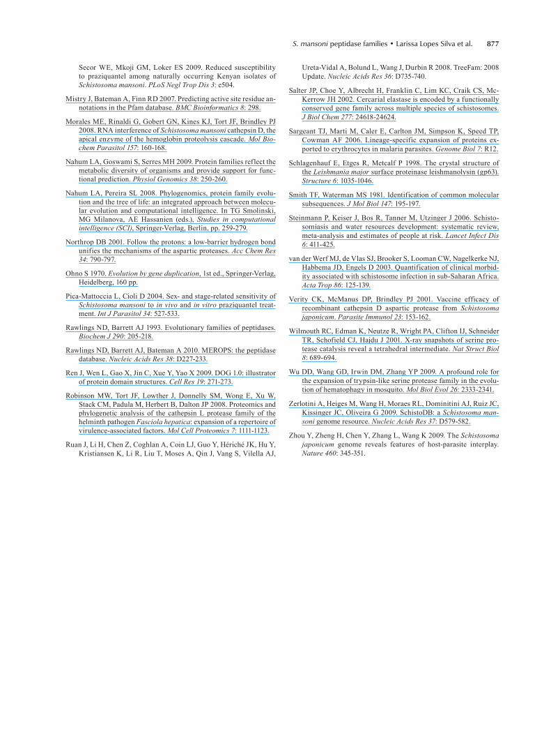
The phylogenetic analysis of the S. mansoni elastases and their homologs in the other species included in this work was performed as already described. The parasite elastases form a well-supported monophyletic clade, which suggests that these proteins originated from a common ancestor by gene duplication events followed by divergence in the Schistosoma lineage.
In observing the resulting phylogeny (Fig. 3), it is possible to demonstrate that S. mansoni elastases have homologs in six of the 16 other species considered in this analysis (N. vectensis, D. melanogaster, An. gambiae, B. floridae, M. musculus and H. sapiens). The serine peptidase S1 family is also expanded in all of these spe-cies except for one, D. melanogaster. According to the information available in UniProt (Apweiler et al. 2011), seven homologs have been experimentally confirmed in D. melanogaster, M. musculus and H. sapiens, and their function is related to a digestive process and immune response (Supplementary data). It is believed that simi-lar activities are performed by elastases in schistosomes (Salter et al. 2002, Aslam et al. 2008).
Fig. 4: phylogenetic relationships of the Schistosoma mansoni elastases and their homologs in selected taxa. Analysis was performed with trimmed sequence alignment by using the maximum likelihood method as implemented in PhyML. Best fit model (WAG) and support values for each node were estimated by the Akaike Likelihood Ratio Test (aLRT). Sequence labels follow the PhylomeDB internal identifier. For details, see Supplementary data.

S. mansoni 871
A conserved protein sequence domain (Pfam: PF00089), which is found in all characterised mem-bers of the serine peptidase S1 family, was identified in the S. mansoni elastases and ranges in length from 141-265 amino acids (Fig. 6). The catalytic triad of his-tidine, aspartate and serine residues is present in most of these proteins. Based on profile HMMs available in Pfam, truncated regions were assigned to all 12 of these elastases, perhaps reflecting their degree of divergence in relation to other proteins in the database. Meanwhile, it is important to emphasise that protein databases do not cover all of the existing diversity in nature.
Together, these results indicate that the correct num-ber of cercarial elastases encoded in the S. mansoni genome is 12 and not 13 as described before. However, only Smp_006510, Smp_006520 and Smp_141450 were previously predicted as active proteins (Berriman et al. 2009). Smp_194800 has a much shorter domain com-pared to others. This difference could reflect either the presence of an elastase pseudogene in the parasite ge-nome or that the sequence was incorrectly annotated due to an error in the gene model. Considering that the first-pass annotation of the S. mansoni genome was produced by a combination of gene-finding algorithms (Augustus,
Fig. 5: conserved protein domain architecture of the Schistosoma mansoni elastases. Protein identifiers were assigned in SchistoDB. The con-served protein domain according to Pfam (PF00089) is present in all proteins. Truncated regions (yellow block) are indicated. Sequence length, domain limits, and the catalytic triad of histidine (H), aspartate (D), and serine (S) are also shown.

872 Mem Inst Oswaldo Cruz, Rio de Janeiro, Vol. 106(7), November 2011
Fig. 6: phylogenetic relationships of the Schistosoma mansoni cathepsins D and their homologs in selected taxa. Analysis was performed with trimmed sequence alignment by using the maximum likelihood method as implemented in PhyML. Best fit model (WAG) and support values for each node were estimated by the Akaike Likelihood Ratio Test (aLRT). Sequence labels follow the PhylomeDB internal identifier. For details, see Supplementary data.

S. mansoni 873
Twinscan and GlimmerHMM) (Berriman et al. 2009), this genome has not received extensive manual cura-tion and therefore, many gene models will be refined in the future. Furthermore, EVidenceModeler (Haas et al. 2008) has also been used to incorporate expressed se-quence tag (EST) evidence into the data.
Cathepsin D proteins (aspartic peptidase A1 family) - Our pipeline identified 11 S. mansoni cathepsin D proteins (Supplementary data) that were duplicated after the diver-gence of S. mansoni from the other metazoans analysed here. The evolutionary history of cathepsin D proteins was reconstructed from the sequence alignment of 111 protein sequences from S. mansoni and the selected taxa. The final trimmed alignment contained 1,676 sites, which covered most of the conserved protein domain (Pfam: PF00026). Two S. mansoni proteins corresponded to alternative splic-ing products (Smp_136830.2 and Smp_013040.2). Similar results were found by Berriman et al. (2009).
The phylogenetic tree indicates that the S. mansoni cathepsin D proteins have homologs in all but one spe-cies (S. purpuratus) analysed in this work (Fig. 4). The aspartic peptidase A1 family has also been expanded in 12 of the 15 species in which homologous proteins were identified (A. thaliana, U. maydis, S. cerevisiae, N. cras-sa, C. elegans, C. briggsae, D. melanogaster, C. intesti-nalis, B. floridae, D. rerio, M. musculus and H. sapiens). The number of paralogous proteins ranges from two-17 and includes different aspartic peptidases, such as pep-sins, renins, gastricsin and cathepsin D proteins. Based on the information available in the literature and curated databases, these homologous proteins are involved in di-gestion and protein degradation (Supplementary data). In schistosomes, cathepsin D proteins play an integral role in haemoglobin proteolysis (Brindley et al. 2001, Caffrey et al. 2004, Delcroix et al. 2006).
To predict the protein domain architecture of S. man-soni cathepsin D proteins, we applied the same meth-odology as previously described. The conserved domain (Pfam: PF00026), which has been found in all charac-terised aspartic peptidase A1 family members, was also identified in the S. mansoni proteins with sequence lengths ranging from 94-430 amino acids (Fig. 7). Ac-tive sites are also indicated. Based on the profile HMMs available in Pfam, truncated regions were observed in the N-terminal, C-terminal or both regions. The data also indicate that an additional short sequence domain (Pfam: PF07966), which is known as the A1 propeptide domain, is present at the N-terminal region of two S. mansoni proteins, Smp_013040.1 and Smp_013040.2. Smp_136840 has a much shorter domain compared to other proteins in the same family.
In a previous study, four S. mansoni cathepsin D proteins (Smp_013040.1, Smp_013040.2, Smp_136730 and Smp_136830.2) were identified as active proteins (Berriman et al. 2009), but the variation in the domain architecture and its implications in functional complex-ity were not investigated. One interesting study would be to analyse the functional properties of Smp_013040.1 and Smp_013040.2, which contain the A1 propeptide domain (PF07966).
DISCUSSION
We found that three endopeptidase families are ex-panded in the helminth parasite S. mansoni, which include members of the metallopeptidases (M8 family), serine peptidases (S1 family) and aspartic peptidases (A1 family). In this work, a comparative analysis of these three protein families in S. mansoni and 16 other eukaryotic proteomes revealed their distinct evolutionary histories and provided further information with respect to the sequence and func-tional features of the parasite family members.
Based on the S. mansoni genomic data, 335 peptidas-es were identified, which comprise 2.5% of the predicted proteome (Berriman et al. 2009). They include members of five major classes of peptidases (aspartic, cysteine, metallo, serine and threonine). Of the 61 peptidase fami-lies, 44 are expanded in this parasite and the number of paralogous proteins range from two-26.
Using a computational approach, Bos et al. (2009) analysed all putative peptidases encoded in the parasite’s genome in addition to using EST data, which is simi-lar to work by Berriman et al. (2009). After removing redundant sequences, inactive homologs, likely pseudo-genes and sequences smaller than 100 amino acids from the dataset, they identified a total of 255 peptidase se-quences from the five catalytic classes.
Our results are not fully comparable to those ob-tained by Bos et al. (2009) with respect to elastases and cathepsin D proteins. However, it is worth noting that the phylogenetic analysis of the serine peptidase S1 family performed by these authors also indicated a well-sup-ported clade of four S. mansoni elastases, which are cor-roborated by our findings. The other homologs with high similarities to the cercarial elastases were likely pseudo-genes and, for this reason, they were excluded from the analysis by Bos et al. (2009).
Our results suggest that Schistosoma members of these endopeptidase families originated from successive gene duplication events in the parasite lineage after its diver-sification from the other metazoans analysed here. These results were corroborated by previous proteomic and phy-logenetic analyses on Fasciola hepatica peptidases, which showed that the repertoire of virulence-associated cathe-psin L proteins was established by a series of gene dupli-cation events (Irving et al. 2003, Robinson et al. 2008). These studies also indicate that the gene duplications were followed by active site residue refinements, which inter-fere with the substrate specificity of the F. hepatica cathe-psin L proteins. Whether the S. mansoni proteins share a similar refinement remains to be established.
Gene duplication followed by divergence is known to be the most predominant mechanism of molecular evolution and represents the main source of raw material for the gen-eration of new genes and proteins through the processes of neo and sub-functionalisation (Ohno 1970, Conant & Wolfe 2008, Nahum & Pereira 2008, Hamilton et al. 2009). Al-though in some cases sequences have diverged to the extent that it is impossible to recognise homologous relationships, different proteins that arose by gene duplication may be related at distinct levels, such as sequence, structure, func-tion or a combination of these features and can be grouped into families and superfamilies (Nahum & Pereira 2008).

874 Mem Inst Oswaldo Cruz, Rio de Janeiro, Vol. 106(7), November 2011
Gene fusion, gene fission and domain shuffling were not observed as mechanisms shaping the evolution of the S. mansoni endopeptidase families analysed in this work. Whether gene fusion/fission also plays a role in the evolu-tion of the S. mansoni genome will be a subject of a future work. Our previous study indicated that domain shuffling is one of the main evolutionary forces driving the sequence and functional diversification of the protein kinases of this parasite (Andrade et al. 2011, Avelar et al. 2011).
Peptidases have been implicated in various processes that are crucial to the development and survival of para-sites, including host invasion, degradation of haemoglo-bin in blood feeding, immune evasion and activation of
Experimental work suggests that the SmPepM8 met-allopeptidase (leishmanolysin) may contribute to tissue invasion by schistosome cercariae. This peptidase was the second most abundant protein released during the transformation of S. mansoni cercariae into schistoso-mula (Curwen et al. 2006). Leishmanolysins are a major surface peptidase member of the metallopeptidase M8 family, which in leishmaniasis are involved in different types of processes, such as the inhibition or perturbation of host cell interactions and the degradation of the extra-cellular matrix (Fitzpatrick et al. 2009). It is speculated that these proteins could perform similar activities in schistosomes during host-parasite interactions (Curwen et al. 2006, Fitzpatrick et al. 2009).
Fig. 7: conserved protein domain architecture of the S. mansoni cathepsin Ds. Protein identifiers were assigned in SchistoDB. The conserved protein domain according to Pfam (PF00026) is present in all proteins. The PF07966 additional N-terminal domain was identified in two pro-teins. Truncated regions (yellow block) are indicated. Sequence length, domain limits, and active sites are also shown.

S. mansoni 875
Invasion of host skin is the initial event in establish-ing an infection in mammalian hosts. Considering the complexity of host skin barriers that the cercariae must go through during invasion, it has been suggested that multi-ple enzyme activities are required for this process (Salter et al. 2002). However, only one peptidase (cercarial elastase) has been identified as a major secretory product released during skin penetration (Knudsen et al. 2005, Hansell et al. 2008). These proteins may also be involved in eliminat-ing the outer layer of the cercariae during transformation. Although cercarial elastases were named based on their ability to degrade insoluble elastin, numerous substrates
-lagen, keratin and extracellular matrix proteins (Salter et al. 2002, McKerrow 2003, Knudsen et al. 2005).
Orthologous genes encoding elastase proteins were found in Schistosoma haematobium, Schistosoma japoni-cum and Schistosoma douthitti (Salter et al. 2002, Zhou et al. 2009). The expression of S. japonicum cercarial elastases was confirmed in both the sporocyst and cercar-ial stages and evidence that this peptidase is released by the parasite during the invasion of mammalian skin was obtained by anti-recombinant SjCE antibodies in infected mouse skin (Zhou et al. 2009). However, orthologous pep-tidases to S. mansoni cercarial elastases were not detected in the acetabular secretions of S. japonicum (Dvorák et al. 2008). Furthermore, the faster penetration by S. japoni-cum into the host skin may reflect the differential use of proteolytic enzymes in addition to those characterised in S. mansoni or even involve new peptidases not yet charac-terised (Chlichlia et al. 2005, He et al. 2005). Recent stud-ies have also demonstrated that S. mansoni elastases are capable of cleaving IgE molecules from human, mouse and rat, indicating that the parasite may be able to over-come or evade the IgE response (Aslam et al. 2008). How-ever, this subject remains controversial.
The biological complexity of S. mansoni is related to evolutionary innovations that took place before and after its diversification from other metazoans. Because duplicat- ed genes are important substrates for improving an organ-ism’s adaptation to its environment, understanding how members of protein families evolved may link evolution-ary studies to parasite biology. In turn, this knowledge will provide insights into host-parasite relationships and accel-erate the identification of novel vaccine and drug targets aimed at the treatment and eradication of schistosomiasis.
In conclusion, this paper provides an evolutionary view of three S. mansoni peptidase families, thus allow-ing for a deeper understanding of the genomic complexi-ty and lineage-specific adaptations potentially related to the parasitic lifestyle. In the future, our results obtained using a systemic approach (proteome-wide analyses) may accelerate the understanding of schistosomiasis, its etiologic agents and host-parasite interactions and opti-mise the discovery of therapeutic targets for the develop-ment of new drugs and vaccines.
ACKNOWLEDGEMENTS
To the use of the computing resources of CRG (Spain) and CEBio-Fiocruz (Brazil), to Jaime Huerta-Cepas (CRG), Eric Aguiar and Francislon Silva (CEBio), for bioinformatics tech-
nical support, to Mariana de Oliveira (CEBio), for help with il-lustrations, and to the two anonymous reviewers, for the valu-able suggestions that improved this paper.
REFERENCES
Abdulla MH, Lim KC, Sajid M, McKerrow JH, Caffrey CR 2007. Schistosomiasis mansoni: novel chemotherapy using a cysteine protease inhibitor. PLoS Med 4: e14.
Akaike H 1973. Information theory and an extension of the maximum likelihood principle. In BN Petrov, F Csaki (eds.), Second Inter-national Symposium on Information Theory, Academiai Kiado, Budapest, p. 267-281.
Andrade LF, Nahum LA, Avelar LG, Silva LL, Zerlotini A, Ruiz JC, Oliveira G 2011. Eukaryotic protein kinases (ePKs) of the helminth parasite Schistosoma mansoni. BMC Genomics 12: 215.
Anisimova M, Gascuel O 2006. Approximate likelihood-ratio test for branches: a fast, accurate and powerful alternative. Syst Biol 55: 539-552.
Apweiler R, Martin MJ, O’Donovan C, Magrane M, Alam-Faruque Y, Antunes R, Barrell D, Bely B, Bingley M, Binns D, Bower L, Browne P, Chan WM, Dimmer E, Eberhardt R, Fazzini F, Fedotov A, Foulger R, Garavelli J, Castro LG, Huntley R, Jacobsen J, Kleen M, Laiho K, Legge D, Lin Q, Liu W, Luo J, Orchard S, Patient S, Pichler K, Poggioli D, Pontikos N, Pruess M, Rosanoff S, Sawford T, Sehra H, Turner E, Corbett M, Donnelly M, van Rensburg P, Xe-narios I, Bougueleret L, Auchincloss A, Argoud-Puy G, Axelsen K, Bairoch A, Baratin D, Blatter MC, Boeckmann B, Bolleman J, Bollondi L, Boutet E, Quintaje SB, Breuza L, Bridge A, deCastro E, Coudert E, Cusin I, Doche M, Dornevil D, Duvaud S, Estre-icher A, Famiglietti L, Feuermann M, Gehant S, Ferro S, Gasteiger E, Gateau A, Gerritsen V, Gos A, Gruaz-Gumowski N, Hinz U, Hulo C, Hulo N, James J, Jimenez S, Jungo F, Kappler T, Keller G, Lara V, Lemercier P, Lieberherr D, Martin X, Masson P, Moinat M, Morgat A, Paesano S, Pedruzzi I, Pilbout S, Poux S, Pozzato M, Redaschi N, Rivoire C, Roechert B, Schneider M, Sigrist C, Sonesson K, Staehli S, Stanley E, Stutz A, Sundaram S, Tognolli M, Verbregue L, Veuthey AL, Wu CH, Arighi CN, Arminski L, Barker WC, Chen C, Chen Y, Dubey P, Huang H, Mazumder R, McGarvey P, Natale DA, Natarajan TG, Nchoutmboube J, Roberts NV, Suzek BE, Ugochukwu U, Vinayaka CR, Wang Q, Wang Y, Yeh LS, Zhang J 2011. Ongoing and future developments at the Universal Protein Resource. Nucleic Acids Res 39: D214-219.
Aslam A, Quinn P, McIntosh RS, Shi J, Ghumra A, McKerrow JH, Bunting KA, Dunne DW, Doenhoff MJ, Morrison SL, Zhang K, Pleass RJ 2008. Proteases from Schistosoma mansoni cercariae cleave IgE at solvent exposed interdomain regions. Mol Immu-nol 45: 567-574.
Avelar LG, Nahum LA, Andrade LF, Oliveira G 2011. Functional diversity of the Schistosoma mansoni tyrosine kinases. J Signal Transduct: 603290.
Berriman M, Haas BJ, LoVerde PT, Wilson RA, Dillon GP, Cerqueira GC, Mashiyama ST, Al-Lazikani B, Andrade LF, Ashton PD, As-lett MA, Bartholomeu DC, Blandin G, Caffrey CR, Coghlan A, Coulson R, Day TA, Delcher A, DeMarco R, Djikeng A, Eyre T, Gamble JA, Ghedin E, Gu Y, Hertz-Fowler C, Hirai H, Hirai Y, Houston R, Ivens A, Johnston DA, Lacerda D, Macedo CD, McVeigh P, Ning Z, Oliveira G, Overington JP, Parkhill J, Pertea M, Pierce RJ, Protasio AV, Quail MA, Rajandream MA, Rogers J, Sajid M, Salzberg SL, Stanke M, Tivey AR, White O, Williams DL, Wortman J, Wu W, Zamanian M, Zerlotini A, Fraser-Liggett CM, Barrell BG, El-Sayed NM 2009. The genome of the blood fluke Schistosoma mansoni. Nature 460: 352-358.
Bos DH, Mayfield C, Minchella DJ 2009. Analysis of regulatory pro-tease sequences identified through bioinformatic data mining of the Schistosoma mansoni genome. BMC Genomics 10: 488.

876 Mem Inst Oswaldo Cruz, Rio de Janeiro, Vol. 106(7), November 2011
Botros SS, Bennett JL 2007. Praziquantel resistance. Expert Opin Drug Discov 2 (Suppl. 1): 35-40.
Brindley PJ, Kalinna BH, Wong JY, Bogitsh BJ, King LT, Smyth DJ, Verity CK, Abbenante G, Brinkworth RI, Fairlie DP, Smythe ML, Milburn PJ, Bielefeldt-Ohmann H, Zheng Y, McManus DP 2001. Proteolysis of human hemoglobin by schistosome cathepsin D. Mol Biochem Parasitol 112: 103-112.
Caffrey CR, McKerrow JH, Salter JP, Sajid M 2004. Blood ‘n’ guts: an update on schistosome digestive peptidases. Trends Parasi-tol 20: 241-248.
Capella-Gutiérrez S, Silla-Martínez JM, Gabaldón T 2009. trimAl: a tool for automated alignment trimming in large-scale phyloge-netic analyses. Bioinformatics 25: 1972-1973.
Chlichlia K, Schauwienold B, Kirsten C, Doenhoff MJ, Fishelson Z, Ruppel A 2005. Schistosoma japonicum reveals distinct reactiv-ity with antisera directed to proteases mediating host infection and invasion by cercariae of S. mansoni or S. haematobium. Par-asite Immunol 27: 97-102.
Conant GC, Wolfe KH 2008. Turning a hobby into a job: how dupli-cated genes find new functions. Nat Rev Genet 9: 938-950.
Curwen RS, Ashton PD, Sundaralingam S, Wilson RA 2006. Identi-fication of novel proteases and immunomodulators in the secre-tions of schistosome cercariae that facilitate host entry. Mol Cell Proteomics 5: 835-844.
Delcroix M, Sajid M, Caffrey CR, Lim KC, Dvorák J, Hsieh I, Bah-gat M, Dissous C, McKerrow JH 2006. A multienzyme network functions in intestinal protein digestion by a platyhelminth para-site. J Biol Chem 281: 39316-39329.
Dvorák J, Mashiyama ST, Braschi S, Sajid M, Knudsen GM, Hansell E, Lim KC, Hsieh I, Bahgat M, Mackenzie B, Medzihradszky KF, Babbitt PC, Caffrey CR, McKerrow JH 2008. Differential use of protease families for invasion by schistosome cercariae. Biochimie 90: 345-358.
Edgar RC 2004. MUSCLE: multiple sequence alignment with high accuracy and high throughput. Nucleic Acids Res 32: 1792-1797.
Finn RD, Mistry J, Tate J, Coggill P, Heger A, Pollington JE, Gavin OL, Gunasekaran P, Ceric G, Forslund K, Holm L, Sonnhammer EL, Eddy SR, Bateman A 2010. The Pfam protein families data-base. Nucleic Acids Res 38: D211-222.
Fitch WM 1970. Distinguishing homologous from analogous pro-teins. Syst Zool 19: 99-113.
Fitzpatrick JM, Peak E, Perally S, Chalmers IW, Barrett J, Yoshino TP, Ivens AC, Hoffmann KF 2009. Anti-schistosomal interven-tion targets identified by lifecycle transcriptomic analyses. PLoS Negl Trop Dis 3: e543.
Floris M, Orsini M, Thanaraj TA 2008. Splice-mediated variants of proteins (SpliVaP) - data and characterization of changes in sig-natures among protein isoforms due to alternative splicing. BMC Genomics 9: 453.
Fujinaga M, Cherney MM, Oyama H, Oda K, James MN 2004. The molecular structure and catalytic mechanism of a novel car-boxyl peptidase from Scytalidium lignicolum. Proc Natl Acad Sci USA 101: 3364-3369.
Gabaldón T 2008. Large-scale assignment of orthology: back to phy-logenetics? Genome Biol 9: 235.
Guindon S, Gascuel O 2003. A simple, fast, and accurate algorithm to estimate large phylogenies by maximum likelihood. Syst Biol 52: 696-704.
Haas BJ, Salzberg SL, Zhu W, Pertea M, Allen JE, Orvis J, White O, Buell CR, Wortman JR 2008. Automated eukaryotic gene struc-
ture annotation using EVidenceModeler and the Program to As-semble Spliced Alignments. Genome Biol 9: R7.
Hajjar E, Broemstrup T, Kantari C, Witko-Sarsat V, Reuter N 2010. Structures of human proteinase 3 and neutrophil elastase - so similar yet so different. FEBS J 277: 2238-2254.
Hamilton PB, Adams ER, Njiokou F, Gibson WC, Cuny G, Herder S 2009. Phylogenetic analysis reveals the presence of the Trypano-soma cruzi clade in African terrestrial mammals. Infect Genet Evol 9: 81-86.
Han ZG, Brindley PJ, Wang SY, Chen Z 2009. Schistosoma genomics: new perspectives on schistosome biology and host-parasite inter-action. Annu Rev Genomics Hum Genet 10: 211-240.
Hansell E, Braschi S, Medzihradszky KF, Sajid M, Debnath M, In-gram J, Lim KC, McKerrow JH 2008. Proteomic analysis of skin invasion by blood fluke larvae. PLoS Negl Trop Dis 2: e262.
He YX, Salafsky B, Ramaswamy K 2005. Comparison of skin inva-sion among three major species of Schistosoma. Trends Parasi-tol 21: 201-203.
Hedstrom L 2002. Serine protease mechanism and specificity. Chem Rev 102: 4501-4524.
Huerta-Cepas J, Capella-Gutierrez S, Pryszcz LP, Denisov I, Kormes D, Marcet-Houben M, Gabaldón T 2011. PhylomeDB v3.0: an expanding repository of genome-wide collections of trees, align-ments and phylogeny-based orthology and paralogy predictions. Nucleic Acids Res 39: D556-560.
Huerta-Cepas J, Dopazo H, Dopazo J, Gabaldón T 2007. The human phylome. Genome Biol 8: R109.
Huerta-Cepas J, Dopazo J, Gabaldón T 2010a. ETE: a python envi-ronment for tree exploration. BMC Bioinformatics 11: 24.
Huerta-Cepas J, Marcet-Houben M, Pignatelli M, Moya A, Gabaldón T 2010b. The pea aphid phylome: a complete catalogue of evolution-ary histories and arthropod orthology and paralogy relationships for Acyrthosiphon pisum genes. Insect Mol Biol 19 (Suppl. 2): 13-21.
Huzurbazar S, Kolesov G, Massey SE, Harris KC, Churbanov A, Liberles DA 2010. Lineage-specific differences in the amino acid substitution process. J Mol Biol 396: 1410-1421.
Irving JA, Spithill TW, Pike RN, Whisstock JC, Smooker PM 2003. The evolution of enzyme specificity in Fasciola spp. J Mol Evol 57: 1-15.
James MNG 2004. Catalytic pathway of aspartic peptidases. In AJ Barrett, ND Rawlings, JF Woessner (eds.), Handbook of prote-olytic enzymes, Elsevier Science, London, p. 12-19.
Kasný M, Mikes L, Hampl V, Dvorák J, Caffrey CR, Dalton JP, Horák P 2009. Chapter 4. Peptidases of trematodes. Adv Para-sitol 69: 205-297.
Knudsen GM, Medzihradszky KF, Lim KC, Hansell E, McKerrow JH 2005. Proteomic analysis of Schistosoma mansoni cercarial secretions. Mol Cell Proteomics 4: 1862-1875.
Liang YS, Dai JR, Zhu YC, Coles GC, Doenhoff MJ 2003. Genetic analysis of praziquantel resistance in Schistosoma mansoni. Southeast Asian J Trop Med Public Health 34: 274-280.
Macdonald MH, Morrison CJ, McMaster WR 1995. Analysis of the active site and activation mechanism of the Leishmania surface metalloproteinase GP63. Biochim Biophys Acta 1253: 199-207.
McKerrow JH, Caffrey C, Kelly B, Loke P, Sajid M 2006. Proteases in parasitic diseases. Annu Rev Pathol 1: 497-536.
McKerrow JJ 2003. Invasion of skin by schistosome cercariae: some neglected facts - Response. Trends Parasitol 19: 63-66.
Melman SD, Steinauer ML, Cunningham C, Kubatko LS, Mwangi IN, Wynn NB, Mutuku MW, Karanja DM, Colley DG, Black CL,
S. mansoni 877
Secor WE, Mkoji GM, Loker ES 2009. Reduced susceptibility to praziquantel among naturally occurring Kenyan isolates of Schistosoma mansoni. PLoS Negl Trop Dis 3: e504.
Mistry J, Bateman A, Finn RD 2007. Predicting active site residue an-notations in the Pfam database. BMC Bioinformatics 8: 298.
Morales ME, Rinaldi G, Gobert GN, Kines KJ, Tort JF, Brindley PJ 2008. RNA interference of Schistosoma mansoni cathepsin D, the apical enzyme of the hemoglobin proteolysis cascade. Mol Bio-chem Parasitol 157: 160-168.
Nahum LA, Goswami S, Serres MH 2009. Protein families reflect the metabolic diversity of organisms and provide support for func-tional prediction. Physiol Genomics 38: 250-260.
Nahum LA, Pereira SL 2008. Phylogenomics, protein family evolu-tion and the tree of life: an integrated approach between molecu-lar evolution and computational intelligence. In TG Smolinski, MG Milanova, AE Hassanien (eds.), Studies in computational intelligence (SCI), Springer-Verlag, Berlin, pp. 259-279.
Northrop DB 2001. Follow the protons: a low-barrier hydrogen bond unifies the mechanisms of the aspartic proteases. Acc Chem Res 34: 790-797.
Ohno S 1970. Evolution by gene duplication, 1st ed., Springer-Verlag, Heidelberg, 160 pp.
Pica-Mattoccia L, Cioli D 2004. Sex- and stage-related sensitivity of Schistosoma mansoni to in vivo and in vitro praziquantel treat-ment. Int J Parasitol 34: 527-533.
Rawlings ND, Barrett AJ 1993. Evolutionary families of peptidases. Biochem J 290: 205-218.
Rawlings ND, Barrett AJ, Bateman A 2010. MEROPS: the peptidase database. Nucleic Acids Res 38: D227-233.
Ren J, Wen L, Gao X, Jin C, Xue Y, Yao X 2009. DOG 1.0: illustrator of protein domain structures. Cell Res 19: 271-273.
Robinson MW, Tort JF, Lowther J, Donnelly SM, Wong E, Xu W, Stack CM, Padula M, Herbert B, Dalton JP 2008. Proteomics and phylogenetic analysis of the cathepsin L protease family of the helminth pathogen Fasciola hepatica: expansion of a repertoire of virulence-associated factors. Mol Cell Proteomics 7: 1111-1123.
Ruan J, Li H, Chen Z, Coghlan A, Coin LJ, Guo Y, Hériché JK, Hu Y, Kristiansen K, Li R, Liu T, Moses A, Qin J, Vang S, Vilella AJ,
Ureta-Vidal A, Bolund L, Wang J, Durbin R 2008. TreeFam: 2008 Update. Nucleic Acids Res 36: D735-740.
Salter JP, Choe Y, Albrecht H, Franklin C, Lim KC, Craik CS, Mc- Kerrow JH 2002. Cercarial elastase is encoded by a functionally conserved gene family across multiple species of schistosomes. J Biol Chem 277: 24618-24624.
Sargeant TJ, Marti M, Caler E, Carlton JM, Simpson K, Speed TP, Cowman AF 2006. Lineage-specific expansion of proteins ex-ported to erythrocytes in malaria parasites. Genome Biol 7: R12.
Schlagenhauf E, Etges R, Metcalf P 1998. The crystal structure of the Leishmania major surface proteinase leishmanolysin (gp63). Structure 6: 1035-1046.
Smith TF, Waterman MS 1981. Identification of common molecular subsequences. J Mol Biol 147: 195-197.
Steinmann P, Keiser J, Bos R, Tanner M, Utzinger J 2006. Schisto-somiasis and water resources development: systematic review, meta-analysis and estimates of people at risk. Lancet Infect Dis 6: 411-425.
van der Werf MJ, de Vlas SJ, Brooker S, Looman CW, Nagelkerke NJ, Habbema JD, Engels D 2003. Quantification of clinical morbid-ity associated with schistosome infection in sub-Saharan Africa. Acta Trop 86: 125-139.
Verity CK, McManus DP, Brindley PJ 2001. Vaccine efficacy of recombinant cathepsin D aspartic protease from Schistosoma japonicum. Parasite Immunol 23: 153-162.
Wilmouth RC, Edman K, Neutze R, Wright PA, Clifton IJ, Schneider TR, Schofield CJ, Hajdu J 2001. X-ray snapshots of serine pro-tease catalysis reveal a tetrahedral intermediate. Nat Struct Biol 8: 689-694.
Wu DD, Wang GD, Irwin DM, Zhang YP 2009. A profound role for the expansion of trypsin-like serine protease family in the evolu-tion of hematophagy in mosquito. Mol Biol Evol 26: 2333-2341.
Zerlotini A, Heiges M, Wang H, Moraes RL, Dominitini AJ, Ruiz JC, Kissinger JC, Oliveira G 2009. SchistoDB: a Schistosoma man-soni genome resource. Nucleic Acids Res 37: D579-582.
Zhou Y, Zheng H, Chen Y, Zhang L, Wang K 2009. The Schistosoma japonicum genome reveals features of host-parasite interplay. Nature 460: 345-351.
Copyright © 2022 FDOKUMEN