Quantum Mechanics-Based Scoring Rationalizes the Irreversible Inactivation of Parasitic Schistosoma...
10
Quantum Mechanics-Based Scoring Rationalizes the Irreversible Inactivation of Parasitic Schistosoma mansoni Cysteine Peptidase by Vinyl Sulfone Inhibitors Jindr ̌ ich Fanfrlík, † Pathik S Brahmkshatriya, † Jan R ̌ eza ́ č , † Ade ́ la Jílkova ́ , † Martin Horn, † Michael Mares ̌ , † Pavel Hobza,* ,†,‡ and Martin Leps ̌ ík* ,† † Institute of Organic Chemistry and Biochemistry, v.v.i., and Gilead Sciences and IOCB Research Center, Academy of Sciences of the Czech Republic, Flemingovo na ́ m. 2, 166 10 Prague 6, Czech Republic ‡ Regional Center of Advanced Technologies and Materials, Department of Physical Chemistry, Palacky ́ University, Olomouc, 771 46 Olomouc, Czech Republic * S Supporting Information ABSTRACT: The quantum mechanics (QM)-based scoring function that we previously developed for the description of noncovalent binding in protein-ligand complexes has been modified and extended to treat covalent binding of inhibitory ligands. The enhancements are (i) the description of the covalent bond breakage and formation using hybrid QM/ semiempirical QM (QM/SQM) restrained optimizations and (ii) the addition of the new ΔG cov ′ term to the noncovalent score, describing the “free” energy difference between the covalent and noncovalent complexes. This enhanced QM-based scoring function is applied to a series of 20 vinyl sulfone-based inhibitory compounds inactivating the cysteine peptidase cathepsin B1 of the Schistosoma mansoni parasite (SmCB1). The available X-ray structure of the SmCB1 in complex with a potent vinyl sulfone inhibitor K11017 is used as a template to build the other covalently bound complexes and to model the derived noncovalent complexes. We present the correlation of the covalent score and its constituents with the experimental binding data. Four outliers are identified. They contain bulky R 1 ′ substituents structurally divergent from the template, which might induce larger protein rearrangements than could be accurately modeled. In summary, we propose a new computational approach and an optimal protocol for the rapid evaluation and prospective design of covalent inhibitors with a conserved binding mode. 1. INTRODUCTION The covalent inactivation of biomolecular targets is an alternative strategy in pharmaceutical research which yielded several clinical drugs, was later forsaken for potential safety issues but has recently been revived, 1-3 as exemplified by second-generation covalent proteasome inhibitors for the treatment of myeloma. 4 Computational tools for the design of covalent inhibitors are far less developed than for their noncovalent counterparts. A few examples could be algorithms for covalent docking, 5,6 analysis of noncovalent complexes, 7 covalent QSAR descriptors, 8 or enzyme mechanism-based method (EMBM). 9 However, there is still an urgent need to develop robust and efficient protocols for covalent inhibitor computational design. The molecular processes of the covalent binding of ligands such as enzyme inhibitors can be notionally divided into two steps (eq 1). First, the inhibitor (I) binds noncovalently to the target enzyme (E); this phenomenon is characterized by the K i value or by its constituent on- and off-rates, k on and k off , respectively (eq 1a). This leads to the formation of a noncovalent complex (E-I), in which the moderately reactive electrophilic group of I approaches the catalytic nucleophile of E (e.g., cysteine or serine residue). Second, a chemical reaction occurs (characterized by k inact ), and the resulting covalent-bond formation gives rise to a covalently inactivated complex (EI). The overall thermodynamics and kinetics can be described by the k 2nd parameter (eq 1b). + −⎯→ ⎯⎯ HI oo E I E I EI k k k off on inact (1) = K k k / i off on (1a) = k k K / 2nd inact i (1b) The thermodynamics of the first noncovalent binding step can be described by standard computational tools based on molecular mechanics (MM), such as free-energy perturbation, linear interaction energy, MM-GBSA, or scoring functions. 10 The quantum mechanical (QM) description should also be used where the MM description fails (e.g., in the case of metals 11,12 or halogen bonds 13,14 in the E-I complexes) for an enhanced reliability of the scoring. 15 The second step, the Received: September 26, 2013 Revised: November 3, 2013 Published: November 6, 2013 Article pubs.acs.org/JPCB © 2013 American Chemical Society 14973 dx.doi.org/10.1021/jp409604n | J. Phys. Chem. B 2013, 117, 14973-14982
-
Upload
independent -
Category
Documents
-
view
0 -
download
0
Transcript of Quantum Mechanics-Based Scoring Rationalizes the Irreversible Inactivation of Parasitic Schistosoma...
Quantum Mechanics-Based Scoring Rationalizes the IrreversibleInactivation of Parasitic Schistosoma mansoni Cysteine Peptidase byVinyl Sulfone InhibitorsJindrich Fanfrlík,† Pathik S Brahmkshatriya,† Jan Rezac,† Adela Jílkova,† Martin Horn,† Michael Mares,†
Pavel Hobza,*,†,‡ and Martin Lepsík*,†
†Institute of Organic Chemistry and Biochemistry, v.v.i., and Gilead Sciences and IOCB Research Center, Academy of Sciences of theCzech Republic, Flemingovo nam. 2, 166 10 Prague 6, Czech Republic‡Regional Center of Advanced Technologies and Materials, Department of Physical Chemistry, Palacky University, Olomouc, 771 46Olomouc, Czech Republic
*S Supporting Information
ABSTRACT: The quantum mechanics (QM)-based scoring function that we previouslydeveloped for the description of noncovalent binding in protein−ligand complexes has beenmodified and extended to treat covalent binding of inhibitory ligands. The enhancements are(i) the description of the covalent bond breakage and formation using hybrid QM/semiempirical QM (QM/SQM) restrained optimizations and (ii) the addition of the newΔGcov′ term to the noncovalent score, describing the “free” energy difference between thecovalent and noncovalent complexes. This enhanced QM-based scoring function is applied toa series of 20 vinyl sulfone-based inhibitory compounds inactivating the cysteine peptidasecathepsin B1 of the Schistosoma mansoni parasite (SmCB1). The available X-ray structure ofthe SmCB1 in complex with a potent vinyl sulfone inhibitor K11017 is used as a template tobuild the other covalently bound complexes and to model the derived noncovalentcomplexes. We present the correlation of the covalent score and its constituents with theexperimental binding data. Four outliers are identified. They contain bulky R1′ substituentsstructurally divergent from the template, which might induce larger protein rearrangementsthan could be accurately modeled. In summary, we propose a new computational approach and an optimal protocol for the rapidevaluation and prospective design of covalent inhibitors with a conserved binding mode.
1. INTRODUCTION
The covalent inactivation of biomolecular targets is analternative strategy in pharmaceutical research which yieldedseveral clinical drugs, was later forsaken for potential safetyissues but has recently been revived,1−3 as exemplified bysecond-generation covalent proteasome inhibitors for thetreatment of myeloma.4 Computational tools for the designof covalent inhibitors are far less developed than for theirnoncovalent counterparts. A few examples could be algorithmsfor covalent docking,5,6 analysis of noncovalent complexes,7
covalent QSAR descriptors,8 or enzyme mechanism-basedmethod (EMBM).9 However, there is still an urgent need todevelop robust and efficient protocols for covalent inhibitorcomputational design.The molecular processes of the covalent binding of ligands
such as enzyme inhibitors can be notionally divided into twosteps (eq 1). First, the inhibitor (I) binds noncovalently to thetarget enzyme (E); this phenomenon is characterized by the Kivalue or by its constituent on- and off-rates, kon and koff,respectively (eq 1a). This leads to the formation of anoncovalent complex (E−I), in which the moderately reactiveelectrophilic group of I approaches the catalytic nucleophile ofE (e.g., cysteine or serine residue). Second, a chemical reaction
occurs (characterized by kinact), and the resulting covalent-bondformation gives rise to a covalently inactivated complex (EI).The overall thermodynamics and kinetics can be described bythe k2nd parameter (eq 1b).
+ − ⎯ →⎯⎯H IooE I E I EIk
k k
off
on inact
(1)
=K k k/i off on (1a)
=k k K/2nd inact i (1b)
The thermodynamics of the first noncovalent binding stepcan be described by standard computational tools based onmolecular mechanics (MM), such as free-energy perturbation,linear interaction energy, MM-GBSA, or scoring functions.10
The quantum mechanical (QM) description should also beused where the MM description fails (e.g., in the case ofmetals11,12 or halogen bonds13,14 in the E−I complexes) for anenhanced reliability of the scoring.15 The second step, the
Received: September 26, 2013Revised: November 3, 2013Published: November 6, 2013
Article
pubs.acs.org/JPCB
© 2013 American Chemical Society 14973 dx.doi.org/10.1021/jp409604n | J. Phys. Chem. B 2013, 117, 14973−14982

covalent-bond formation, requires the use of QM methods toexplore the reaction mechanism and to search for the transitionstates and intermediates. This is done either in a model systemusing QM calculation in the gas phase or implicit solvent16 or inthe full protein−ligand system using a hybrid QM/MM setup.17
It should be mentioned here that the use of MM in this step isgenerally excluded because the MM cannot describe theformation and breaking of a covalent bond unless specificallytailored to this aim.18
For cysteine peptidases, much computational work has beendone in the group of Bernd Engels.19−25 Importantly, thestabilization of the zwitterionic thiolate-imidazolium pair of thecatalytic Cys-His dyad has been attributed to a hydrogen-bondnetwork and 2−4 water molecules.21 The reactivity of thethiolate is increased when the substrate binds and disrupts thishydrogen-bond network.21 The stereo- and regiospecificities ofepoxide and aziridine-based covalent inhibitors of cysteinepeptidases have been elucidated using QM/MM calcula-tions.22,23 On the basis of the knowledge gained fromcomputations, new inhibitors have been rationally designedand synthesized and their high potency has been demonstratedexperimentally.24,25
In our laboratory, we have been developing semiempiricalQM (SQM) methods for a reliable and accurate description ofall types of noncovalent interactions26−28 (reviewed in ref 29).Indeed, the use of the corrections for dispersion (D) andhydrogen (H) and halogen (X) bonding with the PM6 SQMmethod (designated as PM6-D3H4X) results in a high degreeof accuracy, which is close to the much more costly DFT-D.27
Moreover, the linear-scaling algorithm MOZYME30,31 makesthis SQM method so fast that systems of up to 10 000 atomscan be optimized. An important feature of the method is thereliable description of the solvent using the implicit COSMOmodel.32
Armed with these powerful corrected SQM methods, wehave designed a QM-based scoring function and successfullyapplied it to two iconic examples of pharmaceutically relevantE−I complexes, namely, the HIV-1 protease and proteinkinases, namely, cyclin-dependent kinase 2 (CDK2) and caseinkinase (CK2).13,33,34 Recently, we have implemented the triple-layer QM/SQM/MM methodology and applied it to anotherknown series of CDK2 inhibitors35 and also to a designed seriesof aldose reductase inhibitors.14 In this work, we aim atextending our QM-based scoring function to be applicable indescribing the process of the covalent inactivation of enzymesby inhibitory ligands.Other laboratories have also used corrected SQM methods
for drug design purposes.29,36−38 The corrected PM6 methodhas been applied to evaluate the binding to a cucurbit[7]urilhost,39 vitamin D receptor,40 NADH-dependent enoyl-acylcarrier protein reductase,41 DNA containing zinc-fingerprotein,42 avidin, factor Xa, and ferritin.43
Schistosomiasis, caused by a parasitic blood fluke of thegenus Schistosoma, afflicts over 250 million people worldwide.Schistosoma mansoni cathepsin B1 (SmCB1) is the mostabundant cysteine peptidase in the parasite gut and is necessaryfor normal parasite growth.44−46 SmCB1 is a molecular targetfor the cure of schistosomiasis in a mouse model using the vinylsulfone-based inhibitor K11777. Recently, we have reportedthree crystal structures of SmCB1 complexed with threepeptidomimetic covalent inhibitors containing a vinyl sulfoneor epoxide reactive moiety.47 We have also evaluated theinteractions of these inhibitors with the individual subsites of
the SmCB1 active-site cleft by QM calculations.47 These dataand the inhibition profiling with a panel of 20 vinyl sulfonederivatives have identified the key binding interactions andprovided initial insights into the specificity of SmCB1inhibition.47 Here, we investigate this panel of vinyl sulfonederivatives using a computational approach.In this study, we show for the first time that the QM-based
scoring function originally designed for noncovalent protein−ligand binding33 can be readily extended to the inhibitorsbinding covalently to their targets. The QM character of thescoring function enables us to reliably describe both steps of theligand binding (i.e., the formation of the noncovalent complexand the formation of the covalent bond) by the samemethodology. As a proof of concept, we demonstrate thisprocedure here with a series of vinyl sulfone-based inhibitors ofthe drug-target peptidase SmCB1,47 taking advantage of (i) thehigh-resolution crystal structure of SmCB1 in complex with thepotent vinyl sulfone inhibitor K11017,47 (ii) the structuraldiversity of the investigated vinyl sulfone inhibitors, and (iii)the broad range of their inhibition potency (IC50 from 0.61 to11849 nM, spanning 5 orders of magnitude). Finally, weevaluate the correlation between the calculated and exper-imental inhibition data and discuss the potential applicationsand limitations of the developed QM-based procedure in theanalysis of covalent protein−ligand complexes.
2. METHODS2.1. Vinyl Sulfone Inhibitor Series. Table 1 presents 20
vinyl sulfone inhibitors studied previously biochemically47 andcomputationally in this work. They are defined by thecompound backbone and various substituents at the R3, R2,R1, and R1′ positions (see Table 1). The inhibitors are orderedaccording to their IC50 values against SmCB1. Among theseries, X-ray crystal structures have been reported forcompounds K11017 and K11777.47
2.2. Enzyme Kinetics. An Analysis of Inhibition Kinetics.An inhibition assay of vinyl sulfone inhibitors and SmCB1 wasperformed as described previously.47 SmCB1 (40 pM) wasadded to a mixture of fluorogenic substrate Cbz-Phe-Arg-AMC(16 μM) and an inhibitor (2 nM to 20 μM) in the assay buffer(0.1 M Na-acetate pH 5.5, containing 3 mM DTT and 0.125%PEG 1500); the hydrolysis of the substrate was monitored for30 min. An observed rate constant, kobs, was calculated at eachinhibitor concentration by fitting the progress curve to theequation P = vi/kobs(1 − e(−kobst)) where P is product formation,vi initial velocity, and t reaction time. The second-order rateconstant, ksecond, was determined depending on the kineticbehavior of the enzyme. In theory, two situations mayoccur:48,49 (a) If kobs varies linearly with the inhibitorconcentration, then ksecond can be determined by fitting to thelinear equation kobs = (ksecond[I])/(1 + [S]/Km); however, thiskinetic mechanism does not allow the determination of theindividual kinact and Ki parameters. (b) If kobs varies hyperboli-cally with [I], then ksecond = kinact/Ki, and kinact and Ki can bedetermined by nonlinear regression using the equation kobs =kinact [I]/([I] + Ki(1+[S]/Km)). The measurement of inhibitionkinetics was performed with five model inhibitors (K11017,K11777, WRR-145, WRR-185, and WRR-347, see Table 1),and all of them were found to follow the kinetic mechanism (a)(see Figure S1 of the Supporting Information). The linear plotof kobs as a function of [I] (Figure S1 of the SupportingInformation) shows that the dependence of kobs on theinhibitor concentration is nonsaturating. On the basis of this,
The Journal of Physical Chemistry B Article
dx.doi.org/10.1021/jp409604n | J. Phys. Chem. B 2013, 117, 14973−1498214974
we assume that the identified interaction scheme is general forvinyl sulfone inhibitors binding to the SmCB1 active site; thesame scheme was previously reported for the interaction ofvinyl sulfone inhibitors with several other cysteine pepti-dases.48,50,51 Furthermore, the plot of vi as a function of [I](Figure S1 of the Supporting Information), showing decreasingvi with [I], indicates a two-step reaction scheme.52
An Analysis of Irreversibility. To demonstrate theirreversibility of vinyl sulfone inhibitors, we investigated tworepresentative compounds, K11777 and WRR-359 (the modelinhibitors of S and R configurations, respectively), using adilution experiment.53 SmCB1 (400 pM) was preincubated (10min at 37 °C) with a vinyl sulfone inhibitor and the assay bufferin a 20 μL volume; the inhibitor concentration was set to theIC50 or 10-times the IC50 level. Afterward, the mixture wasdiluted 10-fold with the assay buffer with an inhibitoryconcentration of IC50 (for IC50 preincubation concentration)or without an inhibitor (for 10× IC50 preincubationconcentration) and was incubated for 10 min at 37 °C. Thefinal activity was measured with a Cbz-Phe-Arg-AMC substrateas described in ref 47. This analysis showed that the activity ofSmCB1 is controlled by the initial concentration of theinhibitors in the preincubation step and that the extent of
inhibition is not reduced by the subsequent dilution (as knownfor reversible inhibitors).53 The experiment clearly demon-strated the irreversibility of the inhibition of the inhibitorstested.The inhibition constant values presented in Table 1 are in
the form of IC50, which represents the inhibitor concentrationnecessary to cause 50% inhibition of the enzyme under theconditions of the enzyme assay. The following relationsbetween IC50 and kobs hold for an irreversible inhibitor:54
= =k t t0.693/ 0.693/obs 1/2 assay
=k t/[I] 0.693/[ IC ]obs assay 50
Under the assumption that IC50 is the inhibitor concen-tration necessary to reduce the enzyme activity by 50% duringthe time of incubation, the assay time (tassay) is equal to the half-life t1/2 for the kobs rate constant.
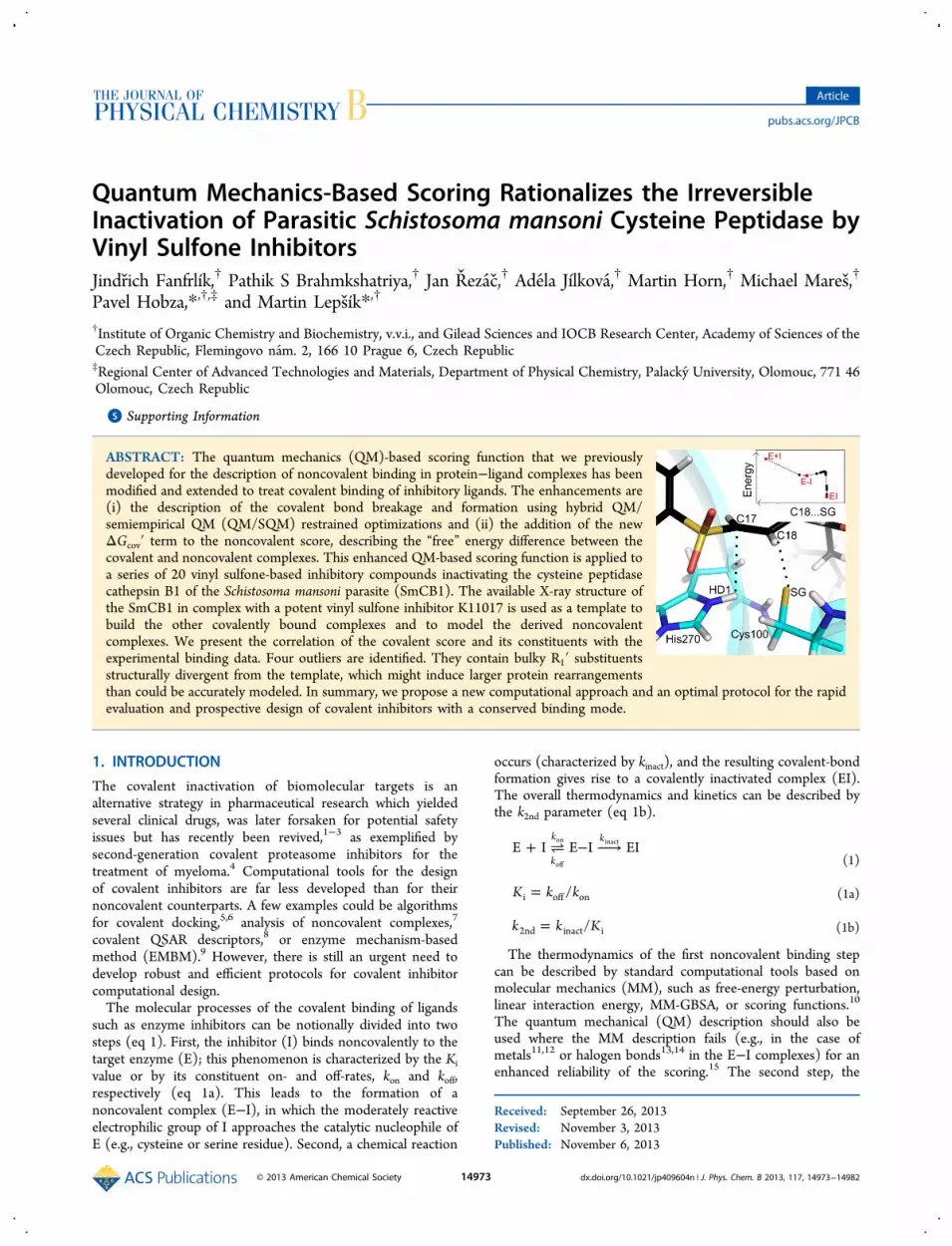
2.3. Setup of the Calculations. Hydrogens were added tothe protein using the Reduce program55 and to the ligandsusing Chimera, ver. 1.5.356 to correspond to the experimentalpH of 5.5.47 This means that both the N- and C-termini, all theaspartates, glutamates, lysines, arginines, and all the histidineresidues (except a neutral Nε-protonated His270 in thecovalent EI complex, Figure 1B) were charged. Special care
was taken to treat the catalytic residues Cys100 and His270. Aswas found earlier,21 the protein frame provides the stabilizationof the zwitterionic state, and these two residues were thusmodeled as Cys100:thiolate and doubly protonated His270 inthe noncovalent E−I complex, Figure 1A.Our previous setup was a full SQM optimization of the
protein−ligand complexes.13,33,34 In a subsequent work weutilized a hybrid SQM/MM setup with a generalized Born(GB) solvent to speed up the calculations; a three-layer QM/SQM/MM-GB setup was also implemented for morequantitative results.14,35 Herein, we have made further progressin using a hybrid QM/SQM methodology in that we coupled itwith the COSMO implicit solvent model,32 which has recentlybeen shown to be more reliable than GB.57
Like in our previous works,14,35 the coupling between theQM and SQM parts was done with an in-house program(CUBY3) using a subtractive scheme of an ONIOM type.58,59
It calls Turbomole60 for QM and MOPAC30 for SQM. TheQM part was treated by DFT with empirical dispersion (D foroptimizations,61 D3 for single points62). The B-LYP/SVP
Table 1. Structures of the SmCB1 Inhibitors Used in ThisStudy along with Their Experimental IC50 Values (Adaptedfrom Ref 47)a
substituent position
compoundIC50(nM) R3 R2 R1 R1′
WRR-286 0.6 Mpip Phe Hph NH−O−CH2−PhK11017 1.7 Mu Leu Hph PhK11002 1.7 Mu Phe Hph PhWRR-282 2.0 Mpip Phe Hph CH2−PhK11777 2.1 Mpip Phe Hph PhWRR-285 2.1 Mpip Phe Hph NH−PhK11747 2.5 Mpip 2-Np-Ala Hph NpAR-198048
6.0 Mpip Phe-3-CH3
Hph Ph
WRR-284 7.8 Mpip Phe Hph O−PhAR-198049
10.5 Mpip Phe-3-CF3
Hph Ph
WRR-145 22.9 Cbz Phe Ala PhWRR-499 29.5 Mpip His Hph PhK11006 33.2 Mu Phe Lys PhWRR-483 48.9 Mpip Arg Hph PhWRR-359 114 Mpip Pheb Hphb O−PhWRR-185 126 Cbz Phe Alab NH−CH(Bz)−
COO−CH3
WRR-283 192 Mpip Phe Hph O−CH2−CH3
WRR-200 247 Cbz Phe Alab NH−CH2−Ph-4-CF3WRR-347 747 Mpip Phe Hph O−Ph-4-O−CH3
WRR-453 11849 Mu Phe Tyr PhaWe use the following abbreviations for the substituents: N-methylpiperazinylcarbonyl (Mpip), morpholinylcarbonyl (Mu), ben-zyloxycarbonyl (Cbz), phenyl (Ph), benzyl (Bz), naphtyl (Np), andthe standard three-letter codes for amino-acid side chains (Phe, Leu,His, Ala, Hph (homophenylalanine)). bThese residues are in an Rconfiguration; all other residues are in an S configuration. Figure 1. Scheme of noncovalent (E−I, panel A) and covalent (EI,
panel B) SmCB-vinyl sulfone inhibitor complexes. The dotted lines in(A) represent the nucleophilic attack of Cys100:SG by C18 of theinhibitor and the associated proton transfer (HD1 from His270 to C17of the inhibitor).
The Journal of Physical Chemistry B Article
dx.doi.org/10.1021/jp409604n | J. Phys. Chem. B 2013, 117, 14973−1498214975

functional/basis set combination was used for optimizationsand the TPSS/TZVPP combination for single-point energies.The QM region comprised the inhibitors and residues Cys100,Gln94, and His270 (146 atoms altogether). The SQM part wastreated by PM6-D3H4X. The PM6-D3H4X method (thesemiempirical PM6 method31 corrected for dispersion inter-actions as well as hydrogen- and halogen-bonding26−28) hasbeen shown to provide accurate interaction energies for modelcomplexes27 and for the evaluation of protein−ligandinteractions in QM-based scoring.14 For single-point calcu-lations, whole complexes were considered (about 4 000 atoms).For geometry optimization, only residues within 10 Å of theinhibitor were considered (about 1 600 atoms). Residuesfarther than 8 Å from the inhibitor were frozen during theoptimization. The protein surrounding was modeled using theCOSMO implicit solvent model.32
The QM/SQM optimizations were performed using theFIRE optimization algorithm63 until the energy and gradientconvergence criteria (ΔE = 0.005 kcal mol−1, maximumgradient of 1 kcal mol−1 Å−1, root-mean-square value of thegradient of 0.5 kcal mol−1 Å−1) were met.2.4. Generating Covalent Complex Structures. Co-
valently bound complexes were built based on the SmCB1-K11017 crystal structure (PDB entry 3S3Q).47 The guanidi-nium side chain of compound WRR-483, the aminobutyl sidechain of K11006, and the N-methylpiperazine ring at R3 andthe imidazole ring of theWRR-499 compound were consideredin the cationic form because of their pKa values.The compounds WRR-359, WRR-185, and WRR-200
differed in their stereochemistry at the carbons bearing theR2 and R1 substitutions (R isomers) (Table 1). We could notbuild these complexes from the SmCB1-K11017 complexbecause of steric clashes. The modeling of these compoundswould require large changes in the protein conformation, whichis beyond the limits of the presented methodology.The built inhibitor modifications were relaxed by annealing
from 300 to 0 K at the MM level in AMBER. The parm03 forcefield was used for the protein and the GAFF force field for theligands.64 The atomic charges for the ligands were calculatedusing the RESP procedure at the HF/6-31G* level asrecommended.65 The cooling runs were 1 ps, and we usedthe Berendsen thermostat, 1 fs time step, GB solvent model(Bondi radii and igb = 7 sander option). Three levels ofrelaxation were explored: first, only the ligand modificationswere treated as flexible; second, His181 and Glu313 were alsotreated as flexible; third, all the amino acids within 6 Å of R1′ ofthe K11017 were treated as flexible. Four runs of annealingwere performed for each level of relaxation. All the generatedstructures were optimized using the QM/SQM methodology.The most stable complex of each ligand was selected andfurther used.2.5. Generating Noncovalent Complex Structures. For
the generation of noncovalent complexes, it was necessary tobreak the covalent bond between the C18 atom of the inhibitorand the SG atom of Cys100 and transfer the HD1 atom of theinhibitor to His270, according to an assumed reactionmechanism (see also Figure 1).21 To this aim, we essayedthree types of bond breaking using harmonic restraints andrelaxed scans: (i) the C18-SG bond, (ii) the C17-HD1 bond,and (iii) both C18-SG and C17-HD1 bonds. The distanceranges were 1.9−3.4 and 1.0−3.0 Å for the C18-SG and C17-HD1 bonds, respectively. The obtained noncovalent complexes(E−I) were reoptimized without any restraint. To localize the
noncovalent intermediate E−I, we further extended the C18−SG distance from 2.4 to 3.4 Å using a relaxed scan. Such anapproach gave us the “free” energy difference between thecovalent and noncovalent complexes (ΔG′cov) and an estimatefor the height of the reaction barrier (Ea).
2.6. QM/SQM Scoring. The noncovalent complexes werescored using our standard QM-based scoring function33 (QM/SQM in this case). The binding free energy of the noncovalentprotein−ligand complex is approximated by the noncovalentscore (scorenc) expressed by eq 5 (ref 33; a new consistentnotation in ref 29).
= Δ + ΔΔ + Δ ′ + Δ ′
− Δ
E G G G
T S
score (P) (L)nc int solv confw
confw
int (5)
where
ΔΔ = ΔΔ + Δ − ΔG G G G( (L) (L))solv int ,solvlow
solvhigh
solv
(5a)
The individual terms describe the gas-phase interactionenergy (ΔEint), interaction desolvation free energy (ΔΔGsolv),change of the conformational free energy of the ligand andprotein (ΔG′confw(P,L)), and the entropy change upon binding(TΔSint). The ΔEint was calculated using the QM/SQMmethod on the whole complexes (optimized in waterenvironment) by subtracting the energies of the protein andligand alone. The ΔΔGint,solv was determined by the COSMOsolvent model32 implemented in MOPAC. The desolvation freeenergy of the bare inhibitor ΔGsolv(L) was evaluated by a moredemanding but also more reliable57 SMD model66 at the HF/6-31G* level of theory implemented in Gaussian09.67 This gaverise to the correction for ligand desolvation (ΔGlow
solv(L) −ΔGhigh
solv(L)), which is especially important for a correctdescription of charged inhibitors.33
The TΔSint term was estimated by assigning a penalty of 1kcal/mol to each rotatable bond of the ligand that becamehindered upon complex formation, which is similar to otherscoring functions.68,69 As an alternative, we applied a 1 kcal/mol penalty for the total number of rotatable bonds in theligand and also for the rotatable bonds of the protein sidechains that became hindered upon complex formation.70
The ΔG′confw(L) term represents the difference between theconformation free energy of the ligand in the conformationrestrained by the protein surroundings and that of the freeligand in water. ΔG′confw(L) is the sum of the vacuum andsolvation parts. ΔEdef(L) was calculated at the DFT-D3 level(see above), and ΔΔGconf,solv(L) using SMD at the HF/6-31G*level. The ligand conformation restrained by the proteinsurroundings was taken from the optimized E−I complex. Theenergy of the ligand conformation in water was obtained byusing a quenching technique (DFT-D B-LYP/SVP for MD andgradient optimization followed by DFT-D3 TPSS/TZVPPcalculation combined with the COSMO solvent model; a 50 psMD simulation in 300 K, snapshots were taken everypicosecond and optimized, and the obtained energies wereaveraged).The ΔG′confw(P) term is the sum of the vacuum and
solvation parts of the protein in the E−I complex and of theisolated optimized protein (taken from the SmCB1/K11017crystal structure). This simple approach can be used because allthe complexes have been modeled using the same crystalstructure. This term is calculated at the QM/SQM level.
The Journal of Physical Chemistry B Article
dx.doi.org/10.1021/jp409604n | J. Phys. Chem. B 2013, 117, 14973−1498214976
In addition to these standard scoring terms for treatingnoncovalent protein−ligand complexes, which had beenapplied in several studies from our laboratory (reviewed inref 29), the total covalent score in this work was constructed asa sum of scorenc and ΔG′cov. The latter term was calculated asthe “free” energy (i.e. the vacuum energy plus solvation freeenergy) difference between the covalent and noncovalentcomplexes.The calculated total scores were compared to the
experimental binding data in the form of RTln(IC50) usingthe coefficient of determination (R2) and the predictive index(PI).71 While R2 reflects the variability of the data by the least-squares linear fit, the PI represents a measure of the rank-orderprediction: 1 stands for an always correct prediction, 0 for arandom prediction, and −1 for an always incorrect prediction.71
3. RESULTS AND DISCUSSION
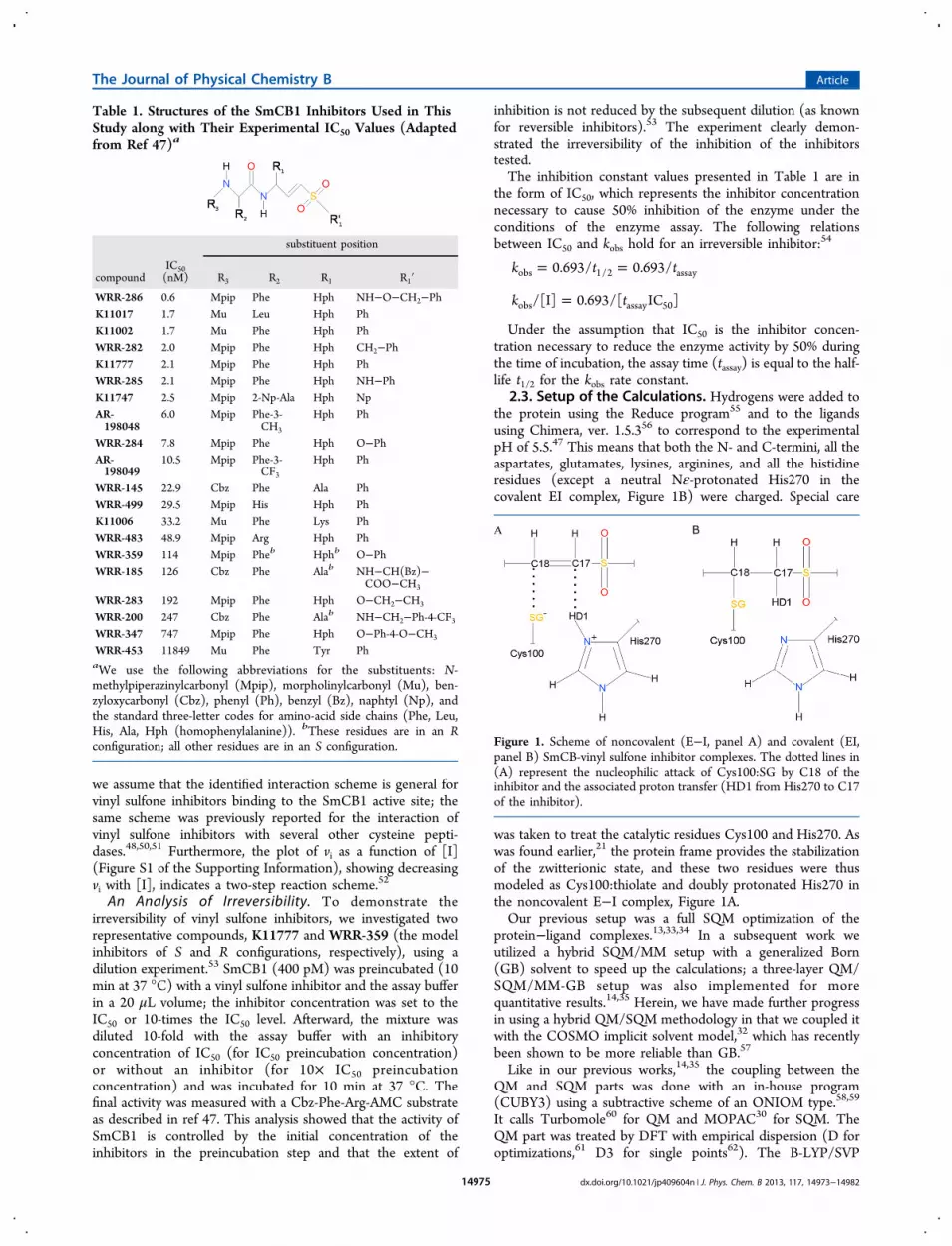
3.1. Covalent Complexes of SmCB1 and Vinyl SulfoneInhibitors. The series of 20 vinyl sulfone inhibitors (Table 1)was modeled in the active site of SmCB1 based on the crystalstructure of the SmCB1/K11017 covalent complex. Fourcomplexes (WRR-359, WRR-185, WRR-200, and WRR-453)could not be built because of steric clashes with the protein. Forthree of these inhibitors (WRR-359, WRR-185, and WRR-200), the simple structural reason was the opposite stereo-chemistry at the carbons having R2 and R1 substitutions (Risomers) as compared to the parental K11017. This findingqualitatively indicates that these compounds require largechanges of the protein conformation, the modeling of which isbeyond our current approach. This may also in part explain thepoor activity of these compounds in the experiment (IC50 from114 to 11849 nM, see Table 1).The binding mode of the covalently bound inhibitors in the
modeled complexes is shown in Figure 2. Nine of the studiedinhibitors have the phenylsulfonyl moiety in the P1′ position.This chemical group can adopt two distinct conformations inthe active site of SmCB1 and related cysteine pepti-dases.47,48,72−74 In our approximate dynamics-based approach
with the limited treatment of protein flexibility, we found thesame conformation of the phenylsulfonyl moiety in P1′ in all ofthese complexes (see Figure 2A). It might be the case thatallowing extended protein flexibility and longer simulationtimes would also yield different phenylsulfonyl conformations.However, we had previously found that the alternativeconformation of the phenylsulfonyl moiety in P1′ had asmaller stabilization energy,47 which is consistent with thecurrent results.The studied inhibitors had similar overall binding modes, and
the R3, R2, R1, and R1′ substituents of the inhibitor bound tothe corresponding S3, S2, S1, and S1′ binding subsites ofSmCB1. Of special interest was the structure of the SmCB1/WRR-145 covalent complex. The benzyloxycarbonyl R3substituent folded back onto the hydrophobic R1 substituent(Figure 2A, the inhibitor is show in the light gray color). Thismay be caused by the higher hydrophobicity of the R3substituent (benzyloxycarbonyl vs N-methylpiperazinylcarbon-yl) and the smaller size of the R1 substituent (Ala vs Hph) ofWRR-145.
3.2. Noncovalent Complexes of SmCB1 and VinylSulfone Inhibitors. The protocol to model the noncovalentE−I complexes was worked out on the crystal structure of theSmCB1/K11017 covalent complex (PDB entry 3S3Q).47
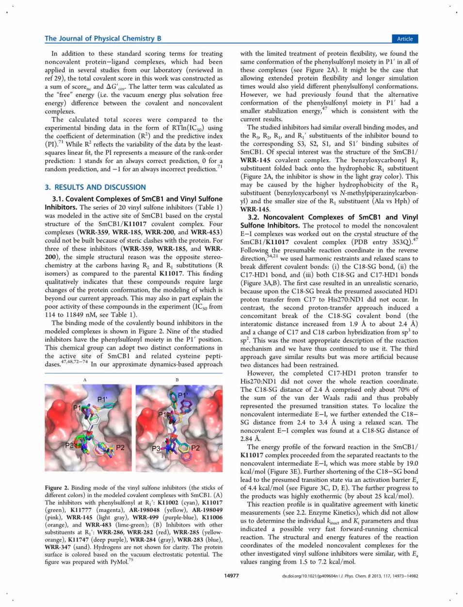
Following the presumable reaction coordinate in the reversedirection,54,21 we used harmonic restraints and relaxed scans tobreak different covalent bonds: (i) the C18-SG bond, (ii) theC17-HD1 bond, and (iii) both C18-SG and C17-HD1 bonds(Figure 3A,B). The first case resulted in an unrealistic scenario,because upon the C18-SG break the presumed associated HD1proton transfer from C17 to His270:ND1 did not occur. Incontrast, the second proton-transfer approach induced aconcomitant break of the C18-SG covalent bond (theinteratomic distance increased from 1.9 Å to about 2.4 Å)and a change of C17 and C18 carbon hybridization from sp3 tosp2. This was the most appropriate description of the reactionmechanism and we have thus continued to use it. The thirdapproach gave similar results but was more artificial becausetwo distances had been restrained.However, the completed C17-HD1 proton transfer to
His270:ND1 did not cover the whole reaction coordinate.The C18-SG distance of 2.4 Å comprised only about 70% ofthe sum of the van der Waals radii and thus probablyrepresented the presumed transition states. To localize thenoncovalent intermediate E−I, we further extended the C18−SG distance from 2.4 to 3.4 Å using a relaxed scan. Thenoncovalent E−I complex was found at a C18-SG distance of2.84 Å.The energy profile of the forward reaction in the SmCB1/
K11017 complex proceeded from the separated reactants to thenoncovalent intermediate E−I, which was more stable by 19.0kcal/mol (Figure 3E). Further shortening of the C18−SG bondlead to the presumed transition state via an activation barrier Eaof 4.4 kcal/mol (see Figure 3C, D, E). The further progress tothe products was highly exothermic (by about 25 kcal/mol).This reaction profile is in qualitative agreement with kinetic
measurements (see 2.2. Enzyme Kinetics), which did not allowus to determine the individual kinact and Ki parameters and thusindicated a possible very fast forward-running chemicalreaction. The structural and energy features of the reactioncoordinates of the modeled noncovalent complexes for theother investigated vinyl sulfone inhibitors were similar, with Eavalues ranging from 1.5 to 7.2 kcal/mol.
Figure 2. Binding mode of the vinyl sulfone inhibitors (the sticks ofdifferent colors) in the modeled covalent complexes with SmCB1. (A)The inhibitors with phenylsulfonyl at R1′: K11002 (cyan), K11017(green), K11777 (magenta), AR-198048 (yellow), AR-198049(pink), WRR-145 (light gray), WRR-499 (purple-blue), K11006(orange), and WRR-483 (lime-green); (B) Inhibitors with othersubstituents at R1′: WRR-286, WRR-282 (red), WRR-285 (yellow-orange), K11747 (deep purple), WRR-284 (gray), WRR-283 (blue),WRR-347 (sand). Hydrogens are not shown for clarity. The proteinsurface is colored based on the vacuum electrostatic potential. Thefigure was prepared with PyMol.75
The Journal of Physical Chemistry B Article
dx.doi.org/10.1021/jp409604n | J. Phys. Chem. B 2013, 117, 14973−1498214977

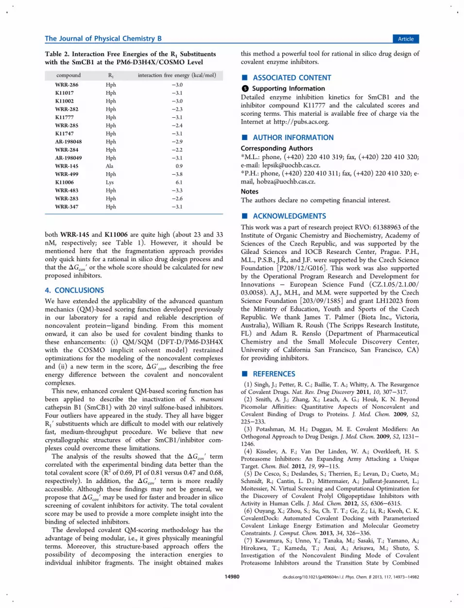
3.3. QM-Based Scoring of SmCB1/Vinyl SulfoneInhibitor Complexes. The calculated estimates of the bindingaffinities (scores) for the modeled vinyl sulfone inhibitors toSmCB1 were compared to the experimental inhibition IC50values available. The IC50 is a suitable parameter for theenzymological evaluation of the inhibitors studied because weexperimentally demonstrated that they are irreversible ligandsand the kinetic measurements did not allow us to determinetheir individual kinact and Ki parameters (see 2.2. Enzyme
Kinetics and Figure S1 of the Supporting Information). Thecomputationally estimated values of the reaction barrier Ea werelow (ranging from 2 to 7 kcal/mol), which agreed with thekinetic measurements of the fast forward-running chemicalreaction. We thus assumed that the conversion of noncovalentcomplexes to covalent is very fast for all the studied inhibitorsand the kinact rate should have a comparable impact on themeasured IC50. The estimates of Ea energies were thus neitherincluded in the scores nor correlated with the experimental
Figure 3. (A) Structural details of the modeled noncovalent SmCB1/K11017 complex, intermediate (E−I), with the reaction coordinates, the C17−HD1 and C18−SG distances, marked by dotted lines. (B) Covalent SmCB1/K11017 complex, the experimentally determined final product of theinhibition reaction (PDB entry 3S3Q). The color coding is as follows: red, oxygen; blue, nitrogen; white, hydrogen; yellow, sulfur; cyan, carbonatoms of SmCB1; black, carbon atoms of K11017. (C) Relative energy (compared to the energy of the separated reactants) plotted against theC17−HD1 distance. (D) C18−SG distance plotted against the C17−HD1 distance. (E) Relative energy plotted against the C18−SG distance. Alldistances are in angstroms, and energies are in kilocalories per mole.
Figure 4. Correlation between the calculated and experimental binding data of the vinyl sulfone inhibitors in the modeled complexes with SmCB1.The calculated terms used for the correlation are as follows: (A) The total covalent score, where the coefficient of determination (R2) is 0.47 andpredictive index (PI) is 0.68. The outliers (WRR-286, cyan; WRR-285, green; WRR-282, yellow; K11747, red) are not considered for R2 and PIdetermination. (B) The noncovalent score (scorenc), where R2 is 0.27 and PI is 0.49. (C) The free energy difference between covalent andnoncovalent complexes (ΔG′cov), where R2 is 0.69 and PI is 0.81. All the energies are in kilocalories per mole.
The Journal of Physical Chemistry B Article
dx.doi.org/10.1021/jp409604n | J. Phys. Chem. B 2013, 117, 14973−1498214978

data. The experimental binding values (from Table 1) werecompared to the total covalent score values and theircomponents which are defined in Section 2.6. QM/SQMScoring of Methods.The calculated scores and their terms are summarized in
Table S1 of the Supporting Information and are plotted againstthe experimental binding data in Figure 4. The correlation forthe whole series of compounds was insignificant (the predictiveindex, PI, ranged from −0.05 to 0.29, and R2 from 0.02 to0.12). However, a visual examination of the graphs showed thatfour outliers deteriorated the correlation. Upon exclusion ofthese four outliers (WRR-286, WRR-282, WRR-285, andK11747), the coefficient of determination (R2) equaled 0.47and PI equaled 0.68 (Figure 4A). The affinities of theseinhibitors tend to be underestimated (see Figure 4). Astructural analysis revealed that the common feature of thesecompounds was a more bulky substituent R1′ (Table 1) ascompared to the template inhibitor K11017 used for themodeling. We hypothesize that these structurally divergentsubstituents in the R1′ position induce larger conformationalchanges in the S1′ pocket of SmCB1, which cannot beaccurately described with the limited treatment of the proteinflexibility in the methodology applied (see Methods). However,a more adequate treatment of the protein flexibility wouldsignificantly increase the cost of the calculations, which wouldbe impractical for high-throughput screening.The correlation of the ΔG′cov term with the experimental
data was significantly higher than that of the total covalentscore (compare Figures 4A and 4C). The number of outliersdecreased from four to three for the correlation of the ΔG′covterm. Upon exclusion of the three remaining outliers (WRR-286, WRR-282, and WRR-285), the R2 equaled 0.69 and PIwas 0.81. The ΔG′cov term was thus the most important term ofthe total covalent score. When the ΔG′cov term was neglected(i.e., scorenc alone considered) there was only a weakcorrelation (R2 = 0.27 and PI = 0.49, see Figure 4B).The other terms were less important. The omission of
ΔG′confw(P) from the total score had a negligible effect (R2 =0.46 and PI = 0.67). The exclusion of ΔG′confw(L) slightlydecreased the correlation (R2 = 0.40 and PI = 0.62). Similarly,when both ΔG′confw(L) and ΔG′confw(P) were excluded, thecorrelation slightly decreased (R2 = 0.41 and PI = 0.64, seeGraph S1 of the Supporting Information)The entropy change of the ligand upon binding was
estimated by the number of rotatable bonds of the ligandhindered upon complex formation. The omission of this termslightly deteriorated the correlation (R2 of 0.40 and PI of 0.66as compared to the original R2 of 0.47 and PI of 0.68 for thetotal covalent score including the entropy term). We also triedto estimate this term by the total number of rotatable bonds inthe ligand. This approach gave very similar correlation of thetotal covalent score (R2 = 0.46 and PI = 0.68, see also Graph S1of the Supporting Information). Additionally, we also applied 1kcal/mol penalty for the rotatable bonds from the protein sidechains which became hindered upon binding. The effect on thescore correlation was also quite small, R2 equaled 0.45 and PIwas 0.70.3.4. Limitations in Scoring Terms. The correlation of the
ΔG′cov term with the experimental binding data wasconsiderably higher than that of the total covalent score. Thismay stem from the fact that the ΔE, ΔΔGsolv, and ΔG′cov termsare described at a significantly higher level than the otherterms.34 These approximations in our scoring procedure can
deteriorate the correlation as shown previously.13,34 Forexample, the lack of dynamical treatment of the ligands maylead in the case of large flexible HIV protease inhibitors touncertainties in the computed energies of 2.5 kcal/mol.76 Evenworse, to obtain converged values for the conformational freeenergy changes of a protein in the binding process, an extensivesampling on the microsecond time scale is needed (see, forexample, ref 77). Much simpler and faster protocols utilizingoptimizations of a limited surroundings of the protein activesite have thus been proposed.78 Also, the use of the ligand-bound form of the protein instead of the apoprotein cancircumvent the problem and yield reliable relative scores.33
In this study, the TΔSint term was estimated by assigning anenergy penalty to each hindered rotatable bond in the ligandand optionally also to the hindered amino acid side chains ofthe protein. The changes in the vibrational entropy and zero-point vibrational energy (ZPVE) upon binding are very difficultto address accurately using computations.79,80 In our previousstudies, we used vibrational analysis at the MM level33,34 butonly with a limited success because of the inability of theempirical potential in describing the quantum phenomena, suchas halogen bonds.13 Making use of this knowledge, we did notopt for the vibrational analysis at the MM level because of thebreaking of covalent bonds and proton transfer in the systemsstudied here. In addition, the QM/SQM methodology that isused for other terms of the covalent score is impractical forvibrational analysis and optimization to very tight convergencecriteria in extended P−L complexes.ΔEint is usually negative and thus makes a favorable
contribution to binding, whereas ΔΔGsolv usually disfavorsbinding.29,33 The former term reflects the strength of theinteraction between the ligand and protein. The latter term canbe interpreted as the free energy needed to desolvate the ligandand binding site of the protein. It can also be viewed as an effectof the solvent on the first term. In the case of charged ligandsand proteins, these two terms may be large and their balancecan become the most important part of the score. However,when a ligand and its protein surroundings have a charge of thesame sign, ΔEint can become repulsive. In such a case, theΔΔGsolv term may lower the repulsion and have a negative sign,i.e., favor the ligand binding. This is the case of K1006 bindingto SmCB1, where the solvent screens the repulsion of its lysineR1 side chain with the positively charged His180 and His181side chains of SmCB1 (Table S1).
3.5. Interaction Energy Decomposition. To demon-strate the power of the QM-based scoring for rational drugdesign, we have fragmented the vinyl sulfone SmCB1 inhibitorsand calculated the interaction free energies of the R1substituents with the SmCB1 active site at the PM6-D3H4X/COSMO level. For the purpose of fragmentation, the Cα−Cβbonds of the inhibitors were cut and capped by hydrogens.The results of this analysis are summarized in Table 2. It is
shown that chemically identical substituents have similarinteraction energies, and the most important finding is asfollows. The parental compound K11017 as well as a majorityof the other inhibitors have homophenylalanine (Hph) as R1;WRR-145 has Ala and K11006 has a charged Lys in R1. Theinteraction free energies of Hph in R1 were calculated to rangefrom −2.2 to −3.8 kcal/mol for the inhibitors studied. WhenHph was changed to Ala, the interactions were lost (aninteraction energy of 0.9 kcal/mol) and the charged Lys in thehydrophobic S1 pocket had a repulsive character (6.1 kcal/mol). These calculations quantitatively explain why the IC50 of
The Journal of Physical Chemistry B Article
dx.doi.org/10.1021/jp409604n | J. Phys. Chem. B 2013, 117, 14973−1498214979
both WRR-145 and K11006 are quite high (about 23 and 33nM, respectively; see Table 1). However, it should bementioned here that the fragmentation approach providesonly quick hints for a rational in silico drug design process andthat the ΔGcov′ or the whole score should be calculated for newproposed inhibitors.
4. CONCLUSIONSWe have extended the applicability of the advanced quantummechanics (QM)-based scoring function developed previouslyin our laboratory for a rapid and reliable description ofnoncovalent protein−ligand binding. From this momentonward, it can also be used for covalent binding thanks tothese enhancements: (i) QM/SQM (DFT-D/PM6-D3H4Xwith the COSMO implicit solvent model) restrainedoptimizations for the modeling of the noncovalent complexesand (ii) a new term in the score, ΔG′cov, describing the freeenergy difference between the covalent and noncovalentcomplexes.This new, enhanced covalent QM-based scoring function has
been applied to describe the inactivation of S. mansonicathepsin B1 (SmCB1) with 20 vinyl sulfone-based inhibitors.Four outliers have appeared in the study. They all have biggerR1′ substituents which are difficult to model with our relativelyfast, medium-throughput procedure. We believe that newcrystallographic structures of other SmCB1/inhibitor com-plexes could overcome these limitations.The analysis of the results showed that the ΔGcov′ term
correlated with the experimental binding data better than thetotal covalent score (R2 of 0.69, PI of 0.81 versus 0.47 and 0.68,respectively). In addition, the ΔGcov′ term is more readilyaccessible. Although these findings may not be general, wepropose that ΔGcov′ may be used for faster and broader in silicoscreening of covalent inhibitors for activity. The total covalentscore may be used to provide a more complete insight into thebinding of selected inhibitors.The developed covalent QM-scoring methodology has the
advantage of being modular, i.e., it gives physically meaningfulterms. Moreover, this structure-based approach offers thepossibility of decomposing the interaction energies toindividual inhibitor fragments. The insight obtained makes
this method a powerful tool for rational in silico drug design ofcovalent enzyme inhibitors.
■ ASSOCIATED CONTENT*S Supporting InformationDetailed enzyme inhibition kinetics for SmCB1 and theinhibitor compound K11777 and the calculated scores andscoring terms. This material is available free of charge via theInternet at http://pubs.acs.org.
■ AUTHOR INFORMATIONCorresponding Authors*M.L.: phone, (+420) 220 410 319; fax, (+420) 220 410 320;e-mail: [email protected].*P.H.: phone, (+420) 220 410 311; fax, (+420) 220 410 320; e-mail, [email protected] authors declare no competing financial interest.
■ ACKNOWLEDGMENTSThis work was a part of research project RVO: 61388963 of theInstitute of Organic Chemistry and Biochemistry, Academy ofSciences of the Czech Republic, and was supported by theGilead Sciences and IOCB Research Center, Prague. P.H.,M.L., P.S.B., J.R., and J.F. were supported by the Czech ScienceFoundation [P208/12/G016]. This work was also supportedby the Operational Program Research and Development forInnovations − European Science Fund (CZ.1.05/2.1.00/03.0058). A.J., M.H., and M.M. were supported by the CzechScience Foundation [203/09/1585] and grant LH12023 fromthe Ministry of Education, Youth and Sports of the CzechRepublic. We thank James T. Palmer (Biota Inc., Victoria,Australia), William R. Roush (The Scripps Research Institute,FL) and Adam R. Renslo (Department of PharmaceuticalChemistry and the Small Molecule Discovery Center,University of California San Francisco, San Francisco, CA)for providing inhibitors.
■ REFERENCES(1) Singh, J.; Petter, R. C.; Baillie, T. A.; Whitty, A. The Resurgenceof Covalent Drugs. Nat. Rev. Drug Discovery 2011, 10, 307−317.(2) Smith, A. J.; Zhang, X.; Leach, A. G.; Houk, K. N. BeyondPicomolar Affinities: Quantitative Aspects of Noncovalent andCovalent Binding of Drugs to Proteins. J. Med. Chem. 2009, 52,225−233.(3) Potashman, M. H.; Duggan, M. E. Covalent Modifiers: AnOrthogonal Approach to Drug Design. J. Med. Chem. 2009, 52, 1231−1246.(4) Kisselev, A. F.; Van Der Linden, W. A.; Overkleeft, H. S.Proteasome Inhibitors: An Expanding Army Attacking a UniqueTarget. Chem. Biol. 2012, 19, 99−115.(5) De Cesco, S.; Deslandes, S.; Therrien, E.; Levan, D.; Cueto, M.;Schmidt, R.; Cantin, L. D.; Mittermaier, A.; Juillerat-Jeanneret, L.;Moitessier, N. Virtual Screening and Computational Optimization forthe Discovery of Covalent Prolyl Oligopeptidase Inhibitors withActivity in Human Cells. J. Med. Chem. 2012, 55, 6306−6315.(6) Ouyang, X.; Zhou, S.; Su, Ch. T. T.; Ge, Z.; Li, R.; Kwoh, C. K.CovalentDock: Automated Covalent Docking with ParameterizedCovalent Linkage Energy Estimation and Molecular GeometryConstraints. J. Comput. Chem. 2013, 34, 326−336.(7) Kawamura, S.; Unno, Y.; Tanaka, M.; Sasaki, T.; Yamano, A.;Hirokawa, T.; Kameda, T.; Asai, A.; Arisawa, M.; Shuto, S.Investigation of the Noncovalent Binding Mode of CovalentProteasome Inhibitors around the Transition State by Combined
Table 2. Interaction Free Energies of the R1 Substituentswith the SmCB1 at the PM6-D3H4X/COSMO Level
compound R1 interaction free energy (kcal/mol)
WRR-286 Hph −3.0K11017 Hph −3.1K11002 Hph −3.0WRR-282 Hph −2.3K11777 Hph −3.1WRR-285 Hph −2.4K11747 Hph −3.1AR-198048 Hph −2.9WRR-284 Hph −2.2AR-198049 Hph −3.1WRR-145 Ala 0.9WRR-499 Hph −3.8K11006 Lys 6.1WRR-483 Hph −3.3WRR-283 Hph −2.6WRR-347 Hph −3.1
The Journal of Physical Chemistry B Article
dx.doi.org/10.1021/jp409604n | J. Phys. Chem. B 2013, 117, 14973−1498214980
Use of Cyclopropylic Strain-Based Conformational Restriction andComputational Modeling. J. Med. Chem. 2013, 56, 5829−5842.(8) Shokhen, M.; Traube, T.; Vijayakumar, S.; Hirsch, M.; Uritsky,N.; Albeck, A. Differentiating Serine and Cysteine ProteasesMechanism by New Covalent QSAR Descriptors. ChemBioChem2011, 12, 1023−1026.(9) Traube, T.; Vijayakumar, S.; Hirsch, M.; Uritsky, N.; Shokhen,M.; Albeck, A. EMBM - A New Enzyme Mechanism-Based Methodfor Rational Design of Chemical Sites of Covalent Inhibitors. J. Chem.Inf. Model. 2010, 50, 2256−2265.(10) Wang, W.; Donini, O.; Reyes, C. M.; Kollman, P. A.Biomolecular Simulations: Recent Developments in Force Fields,Simulations of Enzyme Catalysis, Protein-Ligand, Protein-Protein, andProtein-Nucleic Acid Noncovalent Interactions. Annu. Rev. Biophys.Biomol. Struct. 2001, 30, 211−43.(11) Raha, K.; Merz, K. M. A Quantum Mechanics-Based ScoringFunction: Study of Zinc Ion-Mediated Ligand Binding. J. Am. Chem.Soc. 2004, 126, 1020−1021.(12) Ciancetta, A.; Genheden, S.; Ryde, U. A QM/MM Study of theBinding of RAPTA Ligands to Cathepsin B. J. Comput. Aided Mol. Des.2011, 25, 729−742.(13) Dobes, P.; Rezac, J.; Fanfrlík, J.; Otyepka, M.; Hobza, P.Semiempirical Quantum Mechanical Method PM6-DH2X Describesthe Geometry and Energetics of CK2-Inhibitor Complexes InvolvingHalogen Bonds Well, While the Empirical Potential Fails. J. Phys.Chem. B 2011, 115, 8581−8589.(14) Fanfrlik, J.; Kolar, M.; Kamlar, M.; Hurny, D.; Ruiz, F. X.;Cousido-Siah, A.; Mitschler, A.; Rezac, J.; Munusamy, E.; Lepsík, M.;et al. The Modulation of Aldose Reductase Inhibition by HalogenBond Tuning. ACS Chem. Biol., in press. DOI: 10.1021/Cb400526n.(15) Soderhjelm, P.; Kongsted, J.; Ryde, U. Ligand AffinitiesEstimated by Quantum Chemical Calculations. J. Chem. TheoryComput. 2010, 6, 1726−1737.(16) Siegbahn, P. E. M.; Himo, F. Recent Developments of theQuantum Chemical Cluster Approach for Modeling EnzymeReactions. J. Biol. Inorg. Chem. 2009, 14, 643−651.(17) Senn, H. M.; Thiel, W. QM/MM Methods for BiomolecularSystems. Angew. Chem., Int. Ed. 2009, 48, 1198−1229.(18) van Duin, A. C. T.; Dasgupta, S.; Lorant, F.; Goddard, W. A.ReaxFF: A Reactive Force Field for Hydrocarbons. J. Phys. Chem. A2001, 105, 9396−9409.(19) Helten, H.; Schirmeister, T.; Engels, B. Model Calculationsabout the Influence of Protic Environments on the Alkylation Step ofEpoxide, Aziridine and Thiirane Based Cysteine Protease Inhibitors. J.Phys. Chem. A 2004, 108, 7691−7701.(20) Paasche, A.; Schirmeister, T.; Engels, B. Benchmark Study forthe Cysteine−Histidine Proton Transfer Reaction in a ProteinEnvironment: Gas Phase, COSMO, QM/MM Approaches. J. Chem.Theory Comput. 2013, 9, 1311−1319.(21) Mladenovic, M.; Fink, R. F.; Thiel, W.; Schirmeister, T.; Engels,B. On the Origin of Stabilization of the Zwitterionic Resting State ofCysteine Proteases: A Theoretical Study. J. Am. Chem. Soc. 2008, 130,8696−8705.(22) Mladenovic, M.; Ansorg, K.; Fink, R. F.; Thiel, W.; Schirmeister,T.; Engels, B. Atomistic Insights into the Inhibition of CysteineProteases: First QM/MM Calculations Clarifying the Stereoselectivityof Epoxide-Based Inhibitors. J. Phys. Chem. B 2008, 112, 11798−11808.(23) Mladenovic, M.; Junold, K.; Fink, R. F.; Thiel, W.; Schirmeister,T.; Engels, B. Atomistic Insights into the Inhibition of CysteineProteases: First QM/MM Calculations Clarifying the Regiospecificityand the Inhibition Potency of Epoxide- and Aziridine-Based Inhibitors.J. Phys. Chem. B 2008, 112, 5458−5469.(24) Vicik, V.; Helten, H.; Schirmeister, T.; Engels, B. RationalDesign of Aziridine-Containing Cysteine Protease Inhibitors withImproved Potency: Studies on Inhibition Mechanism. ChemMedChem2006, 1, 1021−1028.(25) Mladenovic, M.; Arnone, M.; Fink, R. F.; Engels, B.Environmental Effects on Charge Densities of Biologically Active
Molecules: Do Molecule Crystal Environments Indeed ApproximateProtein Surroundings? J. Phys. Chem. B 2009, 113, 5072−5082.(26) Rezac, J.; Fanfrlik, J.; Salahub, D.; Hobza, P. SemiempiricalQuantum Chemical PM6 Method Augmented by Dispersion and H-Bonding Correction Terms Reliably Describes Various Types ofNoncovalent Complexes. J. Chem. Theory Comput. 2009, 5, 1749−1760.(27) Rezac, J.; Hobza, P. Advanced Corrections of HydrogenBonding and Dispersion for Semiempirical Quantum MechanicalMethods. J. Chem. Theory Comput. 2012, 8, 141−151.(28) Rezac, J.; Hobza, P. A Halogen-Bonding Correction for theSemiempirical PM6 Method. Chem. Phys. Lett. 2011, 506, 286−289.(29) Lepsík, M.; Rezac, J.; Kolar, M.; Pecina, A.; Hobza, P.; Fanfrlík,J. The Semiempirical Quantum Mechanical Scoring Function for In-Silico Drug Design. ChemPlusChem 2013, 78, 921−931.(30) Stewart, J. J. P. MOPAC2009; Stewart ComputationalChemistry: Colorado Springs, CO; http://OpenMOPAC.net.(31) Stewart, J. J. P. Optimization of Parameters for SemiempiricalMethods V: Modification of NDDO Approximations and Applicationto 70 Elements. J. Mol. Model. 2007, 13, 1173−1213.(32) Klamt, A.; Schuurmann, G. COSMO: A New Approach toDielectric Screening in Solvents with Explicit Expressions for theScreening Energy and its Gradient. J. Chem. Soc., Perkin Trans. 1993, 2,799−805.(33) Fanfrlík, J.; Bronowska, A. K.; Rezac, J.; Prenosil, O.;Konvalinka, J.; Hobza, P. A Reliable Docking/Scoring Scheme Basedon the Semiempirical Quantum Mechanical PM6-DH2 MethodAccurately Covering Dispersion and H-Bonding: HIV-1 Proteasewith 22 Ligands. J. Phys. Chem. B 2010, 114, 12666−12678.(34) Dobes, P.; Fanfrlík, J.; Rezac, J.; Otyepka, M.; Hobza, P.Transferable Scoring Function Based on Semiempirical QuantumMechanical PM6-DH2 Method: CDK2 with 15 Structurally DiverseInhibitors. J. Comput.-Aid. Mol. Des. 2011, 25, 223−235.(35) Brahmkshatriya, P.; Dobes, P.; Fanfrlík, J.; Rezac, J.; Paruch, K.;Bronowska, A. K.; Lepsík, M.; Hobza, P. Quantum MechanicalScoring: Structural and Energetic Insights into Cyclin-DependentKinase 2 Inhibition by Pyrazolo(1,5-a)Pyrimidines. Curr. Comput.Aided Drug Des. 2012, 9, 118−129.(36) Peters, M. B.; Raha, K.; Merz, K. M. Quantum Mechanics inStructure-Based Drug Design. Curr. Opin. Drug Discovery Dev. 2006, 9,370−379.(37) Raha, K.; Peters, M. B.; Wang, B.; Yu, N.; Wollacott, A. M.;Westerhoff, L. M.; Merz, K. M. The Role of Quantum Mechanics inStructure-Based Drug Design. Drug Discovery Today 2007, 12, 725−731.(38) Mucs, D.; Bryce, R. A. The Application of Quantum Mechanicsin Structure-Based Drug Design. Expert Opin. Drug Discovery 2013, 8,263−276.(39) Muddana, H. S.; Gilson, M. K. Calculation of Host-GuestBinding Affinities using a Quantum-Mechanical Energy Model. J.Chem Theory. Comput. 2012, 8, 2023−2033.(40) Kamel, K.; Kolinski, A. Assessment of the Free Binding Energyof 1,25-Dihydroxyvitamin D3 and its Analogs with the Human VDRReceptor Model. Acta Biochim. Pol. 2012, 59, 653−660.(41) Stigliani, J. L.; Bernardes-Genisson, V.; Bernadou, J.; Pratviel, G.Cross-Docking Study on InhA Inhibitors: A Combination of AutodockVina and PM6-DH2 Simulations to Retrieve Bioactive Conformations.Org. Biomol. Chem. 2012, 10, 6341−6349.(42) Nagy, G.; Gyurcsik, B.; Hoffmann, E. A.; Koertvelyesi, T.Theoretical Design of a Specific DNA-Zinc-Finger Protein Interactionwith Semi-Empirical Quantum Chemical Methods. J. Mol. Graph.Model. 2011, 29, 928−934.(43) Mikulskis, P.; Genheden, S.; Wichmann, K.; Ryde, U. ASemiempirical Approach to Ligand-Binding Affinities: Dependence onthe Hamiltonian and Corrections. J. Comput. Chem. 2012, 33, 1179−1189.(44) Correnti, J. M.; Brindley, P. J.; Pearce, E. J. Long-TermSuppression of Cathepsin B Levels by RNA Interference RetardsSchistosome Growth. Mol. Biochem. Parasitol. 2005, 143, 209−215.
The Journal of Physical Chemistry B Article
dx.doi.org/10.1021/jp409604n | J. Phys. Chem. B 2013, 117, 14973−1498214981
(45) Sajid, M.; McKerrow, J. H.; Hansell, E.; Mathieu, M. A.; Lucas,K. D.; Hsieh, I.; Greenbaum, D.; Bogyo, M.; Salter, J. P.; Lim, K. C.Functional Expression and Characterization of Schistosoma MansoniCathepsin B and its Trans-Activation by an Endogenous AsparaginylEndopeptidase. Mol. Biochem. Parasitol. 2003, 131, 65−75.(46) Horn, M.; Jılkova, A.; Vondrasek, J.; Maresova, L.; Caffrey, C.R.; Mares, M. Mapping the Pro-Peptide of the Schistosoma MansoniCathepsin B1 Drug Target: Modulation of Inhibition by Heparin andDesign of Mimetic Inhibitors. ACS Chem. Biol. 2011, 6, 609−617.(47) Jílkova, A.; Rezacova, P.; Lepsík, M.; Horn, M.; Vachova, J.;Fanfrlík, J.; Brynda, J.; McKerrow, J. H.; Caffrey, C. R.; Mares, M.Structural Basis for Inhibition of Cathepsin B Drug Target from theHuman Blood Fluke, Schistosoma mansoni. J. Biol. Chem. 2011, 286,35770−35781.(48) Kerr, I. D.; Lee, J. H.; Farady, C. J.; Marion, R.; Rickert, M.;Sajid, M.; Pandey, K. C.; Caffrey, C. R.; Legac, J.; Hansell, E. VinylSulfones As Antiparasitic Agents and a Structural Basis for DrugDesign. J. Biol. Chem. 2009, 284, 25697−25703.(49) Bieth, J.G. Theoretical and Practical Aspects of ProteinaseInhibition Kinetics. Methods Enzymol. 1995, 248, 59−84.(50) Caffrey, C. R.; Hansell, E.; Lucas, K. D.; Brinen, L. S.; AlvarezHernandez, A.; Cheng, J.; Gwaltney, S. L.; Roush, W. R.; Stierhof, Y.-D.; Bogyo, M. Active Site Mapping, Biochemical Properties andSubcellular Localization of Rhodesain, the Major Cysteine Protease ofTrypanosoma Brucei Rhodesiense. Mol. Biochem. Parasitol. 2001, 118,61−73.(51) Roush, W. R.; Gwaltney, S. L.; Cheng, J. M.; Scheidt, K. A.;McKerrow, J. H.; Hansell, E. Vinyl Sulfonate Esters and VinylSulfonamides: Potent, Irreversible Inhibitors of Cysteine Proteases. J.Am. Chem. Soc. 1998, 120, 10994−10995.(52) Baici, A.; Schenker, P.; Wachter, M.; Ruedi, P. 3-Fluoro-2,4-dioxa-3-phosphadecalins as Inhibitors of Acetylcholinesterase. AReappraisal of Kinetic Mechanisms and Diagnostic Methods. Chem.Biodiversity 2009, 6, 261−282.(53) Horn, M.; Pavlık, M.; Doleckova, L.; Baudys, M.; Mares, M.Arginine-Based Structures are Specific Inhibitors of Cathepsin C.Application of Peptide Combinatorial Libraries. Eur. J. Biochem. 2000,267, 3330−3336.(54) Powers, J. C.; Asgian, J. L.; Ekici, O. D.; James, K. E. IrreversibleInhibitors of Serine, Cysteine, and Threonine Proteases. Chem. Rev.2002, 102, 4639−4750.(55) Case, D. A.; Darden, T. A.; Cheatham, T. E., III; Simmerling, C.L.; Wang, J.; Duke, R. E.; Luo, R.; Crowley, M.; Walker, R. C.; Zhang,W.; et al. AMBER 10; University of California: San Francisco, CA,2008.(56) Pettersen, E. F.; Goddard, T. D.; Huang, C. C.; Couch, G. S.;Greenblatt, D. M.; Meng, E. C.; Ferrin, T. E. UCSF Chimera − AVisualization System for Exploratory Research and Analysis. J. Comput.Chem. 2004, 25, 1605−12.(57) Kolar, M.; Fanfrlík, J.; Lepsík, M.; Forti, F.; Luque, F. J.; Hobza,P. Assessing the Accuracy and Performance of Implicit Solvent Modelsfor Drug Molecules: Conformational Ensemble Approaches. J. Phys.Chem. B 2013, 117, 5950−5962.(58) Svensson, M.; Humbel, S.; Froese, R. D. J.; Matsubara, T.;Sieber, S.; Morokuma, K. ONIOM: A Multilayered Integrated MO +MM Method for Geometry Optimizations and Single Point EnergyPredictions. A Test for Diels−Alder Reactions and Pt(P(t-Bu)3)2 + H2
Oxidative Addition. J. Phys. Chem. 1996, 100, 19357−19363.(59) Dapprich, S.; Komaromi, I.; Suzie Byun, K.; Morokuma, K.;Frisch, M. J. A New ONIOM Implementation in Gaussian98. Part I.The Calculation of Energies, Gradients, Vibrational Frequencies andElectric Field Derivatives. J. Mol. Struct. Theochem. 1999, 461−462, 1−21.(60) Ahlrichs, R.; Bar, M.; Haser, M.; Horn, H.; Kolmel, C.Electronic Structure Calculations on Workstation Computers: TheProgram System Turbomole. Chem. Phys. Lett. 1989, 162, 165−169.(61) Jurecka, P.; Cerny, J.; Hobza, P.; Salahub, D. Density FunctionalTheory Augmented with an Empirical Dispersion Term. InteractionEnergies and Geometries of 80 Noncovalent Complexes Compared
with Ab Initio Quantum Mechanics Calculations. J. Comput. Chem.2007, 28, 555−569.(62) Ehrlich, S.; Moellmann, J.; Reckien, W.; Bredow, T.; Grimme, S.System-Dependent Dispersion Coefficients for the DFT-D3 Treat-ment of Adsorption Processes on Ionic Surfaces. ChemPhysChem2011, 12, 3414−3420.(63) Bitzek, E.; Koskinen, P.; Gahler, F.; Moseler, M.; Gumbsch, P.Structural Relaxation Made Simple. Phys. Rev. Lett. 2006, 97, 170201−170205.(64) Wang, J.; Wolf, R. M.; Caldwell, J. W.; Kollman, P. A.; Case, D.A. Development and Testing of a General AMBER Force Field. J.Comput. Chem. 2004, 25, 1157−1174.(65) Bayly, C. I.; Cieplak, P.; Cornell, W.; Kollman, P. A. A Well-Behaved Electrostatic Potential Based Method Using ChargeRestraints for Deriving Atomic Charges: The RESP Model. J. Phys.Chem. 1993, 97, 10269−10280.(66) Marenich, A. V.; Cramer, C. J.; Truhlar, D. G. UniversalSolvation Model Based on Solute Electron Density and on aContinuum Model of the Solvent Defined by the Bulk DielectricConstant and Atomic Surface Tensions. J. Phys. Chem. B 2009, 113,6378−6396.(67) Frisch, M. J.; Trucks, G. W.; Schlegel, H. B.; Scuseria, G. E.;Robb, M. A.; Cheeseman, J. R.; Scalmani, G.; Barone, V.; Mennucci,B.; Petersson, G. A.; et al. Gaussian 09; Gaussian, Inc.: Wallingford,CT, 2009.(68) Hayik, S. A.; Dunbrack, R., Jr.; Merz, K. M. A., Jr. Mixed QM/MM Scoring Function to Predict Protein-Ligand Binding Affinity. JChem. Theory Comput. 2010, 6, 3079−3091.(69) Ishchenko, A. V.; Shakhnovich, E. I. Small Molecule Growth2001 (SMoG2001): An Improved Knowledge-Based Scoring Functionfor Protein-Ligand Interactions. J. Med. Chem. 2002, 45, 2770−2780.(70) Raha, K.; Merz, K. M. Large-Scale Validation of a QuantumMechanics Based Scoring Function: Predicting the Binding Affinityand the Binding Mode of a Diverse Set of Protein−Ligand Complexes.J. Med. Chem. 2005, 48, 4558−4575.(71) Pearlman, D. A.; Charifson, P. S. Are Free Energy CalculationsUseful in Practice? A Comparison with Rapid Scoring Functions forthe p38 MAP Kinase Protein System. J. Med. Chem. 2001, 44, 3417−3423.(72) Chen, Y. T.; Brinen, L. S.; Kerr, I. D.; Hansell, E.; Doyle, P. S.;McKerrow, J. H.; Roush, W. R. In Vitro and In Vivo Studies of theTrypanocidal Properties of WRR-483 against Trypanosoma cruzi.PLOS Neglected Trop. Dis. 2010, 4, 825e.(73) Brinen, L. S.; Hansell, E.; Cheng, J.; Roush, W. R.; McKerrow, J.H.; Fletterick, R. J. A Target within the Target: Probing Cruzain’s P1′Site to Define Structural Determinants for the Chagas’ DiseaseProtease. Structure 2000, 8, 831−840.(74) Kerr, I. D.; Wu, P.; Marion-Tsukamaki, R.; Mackey, Z. B.;Brinen, L. S. Crystal Structures of TbCatB and Rhodesain, PotentialChemotherapeutic Targets and Major Cysteine Proteases ofTrypanosoma Brucei. PLOS. Neglected Trop. Dis. 2010, 4, e701.(75) PyMOL Molecular Graphics System, version 1.5.0.4. Schrodinger,LLC.(76) Kolar, M.; Fanfrlík, J.; Hobza, P. Ligand Conformational andSolvation/Desolvation Free Energy in Protein−Ligand ComplexFormation. J. Phys. Chem. B 2011, 115, 4718−4724.(77) Sadiq, S. K.; De Fabritiis, G. Explicit Solvent Dynamics andEnergetics of HIV-1 Protease Flap Opening and Closing. Proteins2010, 78, 2873−85.(78) Pecina, A.; Prenosil, O.; Fanfrlík, J.; Rezac, J.; Granatier, J.;Hobza, P.; Lepsík, M. On the Reliability of the CorrectedSemiempirical Quantum Chemical Method (PM6-DH2) for Assigningthe Protonation States in HIV-1 Protease/Inhibitor Complexes.Collect. Czech. Chem. Commun. 2011, 76, 457−479.(79) Zhou, H.-X.; Gilson, M. K. Theory of Free Energy and Entropyin Noncovalent Binding. Chem. Rev. 2009, 109, 4092−4107.(80) Grimme, S. Supramolecular Binding Thermodynamics byDispersion-Corrected Density Functional Theory. Chem.Eur. J.2012, 18, 9955−9964.
The Journal of Physical Chemistry B Article
dx.doi.org/10.1021/jp409604n | J. Phys. Chem. B 2013, 117, 14973−1498214982