MARVEL: A wireless Miniature Anchored Robotic Videoscope for Expedited Laparoscopy

Carboxypeptidase O Is a Glycosylphosphatidylinositol-anchored Intestinal Peptidase with Acidic Amino AcidSpecificity*□S
Received for publication, May 27, 2011, and in revised form, September 6, 2011 Published, JBC Papers in Press, September 15, 2011, DOI 10.1074/jbc.M111.265819
Peter J. Lyons and Lloyd D. Fricker1
From the Department of Molecular Pharmacology, Albert Einstein College of Medicine, Bronx, New York 10461
Background:All previously characterizedmetallocarboxypeptidases of the A/B subfamily are secreted enzymes that cleavealiphatic or basic residues and are initially produced as inactive proenzymes.Results: Carboxypeptidase O is a membrane-anchored intestinal enzyme that cleaves acidic residues and is not made from aproenzyme.Conclusion: Carboxypeptidase O is distinct from other metallocarboxypeptidases.Significance: Carboxypeptidase O plays a unique physiological role in the intestinal release of acidic amino acids from dietarypeptides and proteins.
The firstmetallocarboxypeptidase (CP) was identified in pan-creatic extracts more than 80 years ago and named carboxypep-tidase A (CPA; now known as CPA1). Since that time, sevenadditional mammalian members of the CPA subfamily havebeen described, all of which are initially produced as proen-zymes, are activated by endoproteases, and remove either C-ter-minal hydrophobic or basic amino acids from peptides. Here wedescribe the enzymatic and structural properties of carboxypep-tidase O (CPO), a previously uncharacterized and uniquemem-ber of the CPA subfamily. Whereas all other members of theCPA subfamily contain an N-terminal prodomain necessary forfolding, bioinformatics and expression of both human andzebrafish CPO orthologs revealed that CPO does not require aprodomain. CPO was purified by affinity chromatography, andthe purified enzyme was able to cleave proteins and syntheticpeptides with greatest activity toward acidic C-terminal aminoacids unlike other CPA-like enzymes. CPO displayed a neutralpHoptimumandwas inhibited by commonmetallocarboxypep-tidase inhibitors as well as citrate. CPOwas modified by attach-ment of a glycosylphosphatidylinositolmembrane anchor to theC terminus of the protein. Immunocytochemistry of Madin-Darby canine kidney cells stably expressing CPO showed local-ization to vesicular membranes in subconfluent cells and to theplasma membrane in differentiated cells. CPO is highlyexpressed in intestinal epithelial cells in both zebrafish andhuman. These results suggest that CPO cleaves acidic aminoacids from dietary proteins and peptides, thus complementingthe actions of well known digestive carboxypeptidases CPA andCPB.
TheM14 family ofmetallocarboxypeptidases (CPs)2 andCP-like proteins consists of 25 members in humans (1, 2). Themajor function of these enzymes is the removal of C-terminalamino acids from peptides and proteins during maturationand/or degradation (3, 4). One important area of carboxypep-tidase function is in the digestion of dietary proteins and pep-tides to release amino acids that are able to be absorbed in theintestinal tract (5). This activity was identified more than 80years ago when the first CP, originally called simply “carboxy-peptidase,” was found to be produced in large quantities by thepancreas and secreted into the intestine for dietary proteindigestion (6). A second pancreatic carboxypeptidase was sub-sequently identified and namedCPB for its substrate specificitytoward basic C-terminal amino acids (7), and the first carboxy-peptidase was renamed CPA for its aliphatic/aromatic aminoacid specificity. This enzyme is now known as CPA1 because anadditional pancreatic enzymewas discovered and namedCPA2(8). However, none of these enzymes cleave acidic C-terminalamino acids, raising the question of how peptides and proteinscontaining C-terminal aspartate and glutamate are digested inthe intestine.In addition to the three digestive carboxypeptidases identi-
fied in the exocrine pancreas, a number of carboxypeptidaseshave been identified in non-pancreatic tissues. Examplesinclude CPN, which processes circulating bioactive peptides(9); CPEwith a prominent role in neuropeptidematuration (10,11); and many others (4, 12–17). The 25 members of the M14carboxypeptidase family have been grouped into four subfam-ilies based on sequence and structural similarities (Fig. 1A). Allsubfamilies share a carboxypeptidase domain with criticalactive site and substrate-binding residues generally conservedbetween subfamilies (18). All characterized N/E members thatare enzymatically active have substrate specificity toward basic* This work was supported, in whole or in part, by National Institutes of Health
Grants DA-004494 (to L. D. F.) and EY-194332 (to P. J. L.).□S The on-line version of this article (available at http://www.jbc.org) contains
supplemental Figs. S1 and S2.1 To whom correspondence should be addressed: Dept. of Molecular Phar-
macology, Albert Einstein College of Medicine, 1300 Morris Park Ave.,Bronx, NY 10461. Tel.: 718-430-4225; Fax: 718-430-8922; E-mail: [email protected].
2 The abbreviations used are: CP, metallocarboxypeptidase; MDCK, Madin-Darby canine kidney; CCP, cytosolic carboxypeptidase; GPI, glycosylphos-phatidylinositol; hCPO, human CPO; zCPO, zebrafish CPO; dpf, day(s) post-fertilization; fa, 3-(2-furyl)acryloyl; PI-PLC, phosphatidylinositol-specificphospholipase C.
THE JOURNAL OF BIOLOGICAL CHEMISTRY VOL. 286, NO. 45, pp. 39023–39032, November 11, 2011© 2011 by The American Society for Biochemistry and Molecular Biology, Inc. Printed in the U.S.A.
NOVEMBER 11, 2011 • VOLUME 286 • NUMBER 45 JOURNAL OF BIOLOGICAL CHEMISTRY 39023
by guest on June 18, 2016http://w
ww
.jbc.org/D
ownloaded from
by guest on June 18, 2016
http://ww
w.jbc.org/
Dow
nloaded from
by guest on June 18, 2016http://w
ww
.jbc.org/D
ownloaded from
by guest on June 18, 2016
http://ww
w.jbc.org/
Dow
nloaded from

C-terminal amino acids, and characterized A/B membersexhibit aliphatic or basic amino acid specificity. Although nopreviously characterized mammalian A/B or N/E CPs havebeen shown to cleave acidic residues, a number of members ofthe cytosolic carboxypeptidase (CCP) (19) and aminoacylase(20) subfamilies are able to cleave substrates with C-terminalaspartate or glutamate residues. All members of the A/B andN/E subfamilies contain N-terminal signal peptides that directthe proteins into the endoplasmic reticulum and secretorypathway, whereas all members of the CCP and aminoacylasesubfamilies lack signal peptides and are expressed in the cyto-sol. TwoCPs are known to bemembrane-bound; CPD containsa transmembrane domain (21), and CPM is membrane-boundthrough a glycosylphosphatidylinositol (GPI) anchor (22).Several years ago, a new member of the A/B subfamily was
identified through a bioinformatics analysis of the humangenome and named carboxypeptidase O (CPO) (15). Modelingpredicted CPO to have specificity for acidic C-terminal aminoacids. However, the N terminus of the protein was not identi-fied, and it was thought that CPO might be a pseudogene. Toaddress this, we used a bioinformatics approach to identify full-length CPO from a number of species; all showed the presenceof a signal peptide but either no prodomain (zebrafish) or a veryshort N-terminal extension (mammals; Fig. 1B). Because thedogma in the field was that the prodomain was essential forfolding of proteins in this subfamily, CPO was predicted to beinactive.We show in this report that CPO is functional withoutthe presence of a prodomain and is enzymatically active withacidic amino acid specificity. CPO is membrane-attached via aGPI anchor and is found on the apical surface of intestinal epi-thelial cells. We propose that CPO completes the complementof digestive enzymes within the intestine: CPA1 and CPA2cleave aliphatic/aromatic amino acids, CPB1 cleaves basicamino acids, and CPO cleaves acidic amino acids.
EXPERIMENTAL PROCEDURES
Plasmids—The human CPO cDNA was purchased fromOpen Biosystems (clone ID 8327546; GenBankTM accessionnumber BC112078). Human CPO was tagged on the C termi-nus with the HA or His6 epitopes by PCR with the Pfu Ultra IIpolymerase (Stratagene) and subcloned into pcDNA3.1(�) formammalian cell expression. Human CPO with the C-terminalHis6 tag (hCPO-His6) was subcloned into the pVL1393 plasmidfor baculovirus expression. The zebrafish CPO cDNA wasamplified from cDNA made from 5-day postfertilization (dpf)zebrafish, tagged with the His6 epitope as above, and subcloned
into the pCRII and pVL1393 plasmids. All cDNAs subcloned byPCR were verified by sequencing.Cell Culture andTransfection—MDCKcells were cultured in
Dulbecco’s modified Eagle’s medium (DMEM) supplementedwith 10% fetal bovine serum (FBS) and penicillin/streptomycinat 37 °C and 5% CO2. Transfection was performed with Lipo-fectamine 2000 (Invitrogen) according to the manufacturer’sinstructions. Stably expressing clones were selected with 1mg/ml Geneticin. Sf9 cells were grown in suspension inSf-900III serum-free medium (Invitrogen) at 27 °C with shak-ing at 130 rpm and transfected using the BaculoGold transfec-tion kit (BD Biosciences) according to the manufacturer’sinstructions.Zebrafish Care—Zebrafish were maintained under standard
conditions as described previously (23). Embryos were main-tained at 28.5 °C in egg water (24). All experiments were per-formed in strict accordance to standard guidelines for zebrafishwork and approved by the Animal Institute Committee atAlbert Einstein College of Medicine.Protein Purification—Sf9 insect cells (200ml at 2� 106 cells/
ml) were infected with high titer recombinant baculovirus.Cellswere grown for 2 days before centrifugation and resuspen-sion of cells in 30 ml of lysis buffer consisting of 50 mM sodiumphosphate, pH 7.0, 500 mM NaCl, 1% Nonidet P-40 alternative(Calbiochem), and Complete EDTA-free protease inhibitormixture (Roche Applied Science). Lysate was sonicated andcentrifuged to remove cell debris. One milliliter of potato car-boxypeptidase inhibitor-Sepharose resin, a generous gift fromProf. F. Xavier Avilés, was washed with lysis buffer before add-ing it to clarified lysate and incubating batchwise at room tem-perature for 30 min. Resin was transferred to a column forwashing with lysis buffer followed by Nonidet P-40-free lysisbuffer. CPO was eluted with 10–15 ml of elution buffer (100mMNa2CO3, pH 11.2, 500mMNaCl), dripping directly into 1 M
sodium acetate, pH 5.0 to immediately neutralize the eluate.Protein concentration was measured by Bradford assay.Carboxypeptidase Assays—The 3-(2-furyl)acryloyl (fa)-pep-
tide substrates (Bachem)were dissolved in 50mMTris-HCl, pH7.5, 150 mM NaCl to a concentration of 0.5 mM. Enzymaticcleavage of substrates was measured by a decrease in absorb-ance at 340 nm at 25 °C. To determine enzyme pH optimum,substrate was dissolved in 50mMTris acetate buffer containing150 mM NaCl at the indicated pH values. For kinetic constantdetermination, the initial reaction rate was determined using arange of substrate concentrations from 30 �M to 1 mM and
FIGURE 1. Comparison of domain structure of CP subfamilies. A, all M14 CPs have a 300-residue catalytic domain (light gray). Members of the A/B andcytosolic CCP subfamilies usually have an N-terminal �-sheet-rich domain (dark gray), whereas members of the N/E and aminoacylase (AA) subfamilies have aC-terminal �-sheet-rich domain (dark gray). Some CPs have signal peptides (black) and additional domains with uncharacterized structure (white). The CCPsvary in length (indicated by “ . . . ”). B, CPO consists of an N-terminal signal peptide, a catalytic domain, and a short C-terminal sequence. In mammals and birds,CPO also has a short peptide between the signal peptide and the CP domain; this sequence is not present in fish CPO.
Carboxypeptidase O
39024 JOURNAL OF BIOLOGICAL CHEMISTRY VOLUME 286 • NUMBER 45 • NOVEMBER 11, 2011
by guest on June 18, 2016http://w
ww
.jbc.org/D
ownloaded from

enzyme concentrations from 0.3–50 ng/�l, depending on thesubstrate, followed by nonlinear regression analysis usingGraphPad Prism. All inhibitors were dissolved in water andpreincubated with enzyme for 1 h prior to the addition of sub-strate (0.5 mM fa-EE at pH 7.5). Inhibition experiments and pHand substrate optimum experiments were performed withzCPO enzyme at a concentration of 0.4 ng/�l and hCPOenzyme at 4.0 ng/�l. Purified porcine tubulin was obtainedfrom Cytoskeleton, Inc.Western Blotting—Proteins were resolved by SDS-PAGE on
10 or 4–15% SDS-polyacrylamide gels (Bio-Rad) and trans-ferred to nitrocellulose. Western blotting was performedaccording to a standard protocol with the following antibodies:rabbit RP1-CPO, RP2-CPO, and RP3-CPO (Triple Point Bio-logics; 1:1000 dilution); �-tubulin (clone DM1A, Sigma; 1:5000dilution); tyrosinated tubulin (mAb 1864, Millipore; 1:5000);detyrosinated (Glu-) tubulin (Ab 3201, Millipore; 1:500); �2tubulin (Ab 3203, Millipore; 1:500); poly(Glu) (a kind gift fromDr. Martin Gorovsky; 1:1500), B3 polyglutamylated tubulin(Sigma; 1:2000); GT335 polyglutamylated tubulin (Enzo LifeSciences; 1:3000), and IRDye 800-congugated secondary anti-bodies (Rockland; 1:3000 dilution). Images were obtained andquantified using the LiCor Odyssey system.Characterization of CPO Post-translational Modifications—
Separation of CPO into Triton X-114 (Sigma) detergent andaqueous phases as well as digestion with phosphatidylinositol-specific phospholipase C (PI-PLC; Sigma) was performed asdescribed (25). PeptideN-glycosidase F (New England Biolabs)digestions were performed according to the manufacturer’sprotocol. Edman degradation of purified human and zebrafishCPOwas performed by the ProteinMicroanalytical Laboratoryat the University of Pittsburgh (Dr. John Hempel, director).Immunofluorescence—MDCK cells were cultured on cham-
ber slides (Lab-Tek). Cells were washed with DMEM followedby PBS, fixed in 4% paraformaldehyde for 15 min, and thenpermeabilized for 15min in 0.1%TritonX-100 in PBS. After 1 hof blocking in 5% BSA, cells were immunostained for 1 h withrabbit RP3-CPO (Triple Point Biologics; 1:1000 dilution),mouse anti-EEA1 (BDBiosciences; 1:500 dilution), mouse anti-LAMP2 (Lifespan Biosciences; 1:500 dilution), andmouse anti-Na�/K�-ATPase � subunit (Affinity Bioreagents; 1:500 dilu-tion) antibodies. The cells were washed three times with 0.2%Tween 20 in PBS and then incubated with Cy2- (1:100 dilution)or Cy3 (1:800 dilution)-conjugated secondary antibodies (Jack-son ImmunoResearch Laboratories) for 1 h. After five washeswith 0.2% Tween 20 in PBS, cells were mounted in a smallamount of ProlongGold antifade reagentwith 4�,6�-diamidino-2-phenylidole (DAPI; Molecular Probes).Immunohistochemistry—Frozen human ileum sections (Zya-
gen) were fixed with 4% paraformaldehyde for 20 min. Afterblocking in PBS containing 5% BSA and 0.5% Triton X-100 for2 h at room temperature, sections were incubated with primaryantibody RP1-CPO or RP3-CPO (Triple Point Biologics)diluted 1:500 in blocking solution for 20 h at 4 °C. To control fornonspecific binding, an antibody raised against a 10-residuepeptide corresponding to a Drosophila-specific form of car-boxypeptidase Dwas used; no homologous protein exists in thehuman non-redundant protein database. Sections were washed
several times with PBS containing 0.2% Triton X-100 and incu-bated with Cy3-conjugated anti-rabbit secondary antibody(1:800; Jackson ImmunoResearch Laboratories) for another20 h at 4 °C. After washing, sections were mounted with Pro-long Gold antifade reagent containing DAPI (MolecularProbes). Images were obtained on a Nikon Eclipse microscopesupplemented with a cooled charge-coupled device camera(Roper Scientific).RT-PCR—RNA was extracted from zebrafish embryos using
the RNeasy minikit (Qiagen). RNA quality was assessed byformaldehyde gel electrophoresis. First strand cDNA synthesiswas performed using the Superscript III First Strand SynthesisSystem (Invitrogen).Quantitative real timePCRwas performedon an ABI 7900 using Power SYBR� Green PCR Master Mix(Applied Biosystems). Data were normalized to �-actin expres-sion and shown as relative expression using the 2���CT
method.In Situ Hybridization—A probe for in situ hybridization of
CPO mRNA was generated by in vitro transcription from lin-earized pCRII-zCPO plasmid using a DIG (digoxigenin) RNAlabeling kit (RocheApplied Science)withT3orT7RNApolym-erase according to the manufacturer’s protocol. The lithiumchloride-precipitated RNA probe was dissolved in water, andquality was assessed by formaldehyde gel electrophoresis.Zebrafish embryos were fixed in 4% paraformaldehyde in PBSovernight at 4 °C and processed for in situ hybridizationaccording to standard protocols (26) using a probe hybridiza-tion temperature of 70 °C. Staining reactions were performedwith the alkaline phosphatase substrate BM purple (RocheApplied Science).
RESULTS
CPO Is a Conserved Enzyme with Unique Structure—Theonly published report on CPO describes a partial sequence thatlacks the initiationmethionine, a signal peptide, andmost of theproregion (15). In the decade since this publication, severalsequences for CPO have appeared in various internet data-bases, some of which contain an initiation methionine and sig-nal peptide but not an extended prodomain that is expected tobe required for enzyme folding based on related A/B subfamilypeptidases. To explore this in more detail and to see whetherthe unique features of CPO were conserved, CPO orthologsfrom the genomes of a wide range of vertebrate species wereidentified (Fig. 2). CPO was present in most vertebrate speciesexamined, including mammals, birds, and fish. Exceptionsincluded the mouse and rat; only a pseudogene could be foundin the mouse genome, and no ortholog was identified in the ratgenome. All CPO orthologs exhibited strict conservation ofcritical active site residues necessary for zinc coordination (His-69, Glu-72, and His-196; bovine CPA numbering according toconvention), catalysis (Glu-270), and C-terminal specificity(Arg-127and Arg-145; Fig. 2). In addition, residue 255, knownto impart amino acid specificity within the CPA/B subfamily, isa conservedArg in all CPOorthologs. This is a unique feature ofCPO that predicts specificity for C-terminal acidic amino acids.All orthologs of CPO contained anN-terminal signal peptide
and a catalytic CP domain (Figs. 1B and 2). Mammalianorthologs contained a short �20-amino acid N-terminal
Carboxypeptidase O
NOVEMBER 11, 2011 • VOLUME 286 • NUMBER 45 JOURNAL OF BIOLOGICAL CHEMISTRY 39025
by guest on June 18, 2016http://w
ww
.jbc.org/D
ownloaded from

domain with some homology to the equivalent portion of theCPA1 prodomain (Fig. 2). This N-terminal domain was slightlylonger in birds (chicken and finch; 27–28 amino acids) andabsent in fish (zebrafish and stickleback; Figs. 1B and 2). AllCPO orthologs also contained a short C-terminal domain(25–27 amino acids in length) that was absent from all othermembers of the A/B subfamily (Figs. 1B and 2).Purification of Human and Zebrafish CPO—Both human
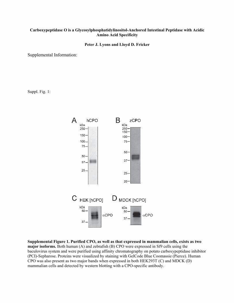
CPOand zebrafishCPOwere expressed in Sf9 cells andpurifiedusing a potato carboxypeptidase inhibitor-Sepharose affinitychromatography approach. This one-step purification resulted
in the complete purification of CPO, which was detected as adoublet by Coomassie or silver staining (supplemental Fig. S1,A and B). This doublet was also seen byWestern blotting usingan antibody to a C-terminal region of CPO (RP3-CPO) whenCPO was transiently expressed in HEK293T cells (supplemen-tal Fig. S1C) and stably expressed inMDCK cells (supplementalFig. S1D). Additional Western blot analyses performed withTriple Point Biologics antibodies to the N-terminal regions ofCPO (RP1-CPO and RP2-CPO) also showed doublets of thesame size (not shown). None of these antibodies cross-reactedwith zebrafish CPO.
FIGURE 2. CPO protein sequences from representative species. Genomic and cDNA databases (Ensembl and NCBI) were searched for CPO sequences, whichwere aligned with each other and with human CPA1 using ClustalW. Signal peptide, N-terminal domain/prodomain, and C-terminal domain sequences areindicated as well as all critical active site residues necessary for zinc binding (His-69, Glu-72, and His-196), substrate binding (Arg-127, Arg-145, Tyr-248, andArg/Ile-255), and catalytic activity (Glu-270; numbering system based on the position of residues in bovine CPA by convention). Greater sequencesimilarity is indicated by darker shading. Sequences shown are from the following database entries: human CPO, ENSP00000272852; cow CPO, ENS-BTAP00000025539; dog CPO, ENSCAFP00000019833; rabbit CPO, ENSOCUP00000008449; chicken CPO, gi�118093691:4613825– 4669825; finch CPO,NW_002198919.1�Tgu7_WGA826_1; zebrafish CPO, NM_001145629; stickleback CPO, ENSGACP00000018671; and human CPA1, NP_001859.
Carboxypeptidase O
39026 JOURNAL OF BIOLOGICAL CHEMISTRY VOLUME 286 • NUMBER 45 • NOVEMBER 11, 2011
by guest on June 18, 2016http://w
ww
.jbc.org/D
ownloaded from

To determine whether the CPO doublet was due toN-glyco-sylation, purified protein was digested with peptide N-glycosi-dase F. This digestion resulted in an increase inmobility of bothhuman and zebrafish CPO, indicating that the protein wasN-glycosylated (supplemental Fig. S2). Because CPO remainedas a doublet after peptide N-glycosidase F treatment, N-linkedglycosylation was not responsible for the difference betweenthe two bands. Similar results were found when extracts frommammalian cells transfected with plasmids expressing CPOwere treated with peptide N-glycosidase F (not shown).The CPO doublet could also be a result of partial processing
of either the N-terminal or C-terminal domains. To determinethe N-terminal sequence, Edman degradation was performed.The N-terminal amino acid sequence of zebrafish CPO(LEHKS) and human CPO (YDRSL) both immediately followthe predicted signal peptide cleavage sites (see Fig. 2). No otherN-terminal sequence was detected, indicating that the shortN-terminal domain present in humanCPO remains attached tothe protein and is not cleaved like the prodomains of other CPs.Enzymatic Characteristics of Purified Human and Zebrafish
CPO—The enzymatic characteristics of purified CPO weredetermined. ZebrafishCPOwas testedwith a panel of commer-cially available CP substrates at pH 7.5 to determine its sub-strate specificity. Consistent with its predicted specificity foracidic amino acids, the substrate containing a C-terminal glu-tamate (fa-EE) was cleaved much more rapidly than substratescontaining hydrophobic or basic C-terminal amino acids (Fig.3A). In addition, the fa-EE substrate was used to assess the pHoptimum for zebrafish CPO. The optimal pH for this enzymewas found to be 6.5–7.5, although CPO retained greater than70% of maximal activity across a wide pH range from 5.5 to 8.5(Fig. 3B).Kinetic parameters were determined for both human and
zebrafish CPO at pH 7.5 with the substrate fa-EE and threeother substrates showing detectable cleavage (Table 1). Bothhuman and zebrafish CPO exhibited greatest activity (Kcat)toward fa-EE followed by decreasing activity in the orderfa-FA � fa-KA � fa-FF. This indicates that C-terminal acidicamino acids are the best substrates for CPO. However, CPOwas also able to cleave C-terminal hydrophobic amino acidswith small residues (alanine) being preferred over large residues(phenylalanine). Both human and zebrafish CPO exhibited Km
values toward substrates with C-terminal alanine that wereabout double the Km value toward C-terminal glutamate andphenylalanine.Kcat/Km values obtained for zebrafish CPOweretypically 5–20-fold greater than those obtained for humanCPO, but the overall specificity of CPO appears similar forthese two species.CPO Is Inhibited byCommonCP Inhibitors asWell as Citrate—
A number of standard metallocarboxypeptidase inhibitorswere incubated with CPO to determine their inhibitory activity(Table 2). Consistent with the acidic amino acid specificity ofCPO, benzylsuccinic acid was a poor inhibitor of this enzyme.In contrast, potato carboxypeptidase inhibitor, which does notrely primarily on interactions with the C-terminal amino acid,was a potent inhibitor with an IC50 of�20–30 nM.Metal chela-tors EDTA and 1,10-phenanthroline were also effective inhibi-tors of CPO activity when used in the low millimolar range. Anumber of amino acids were tested to learn more about thespecificity of this enzyme through product inhibition. Even at10 mM concentrations, all amino acids tested resulted in rela-tively modest inhibition of CPO activity from 10 to 55%. How-ever, as the best substrates and amino acid inhibitors of CPOwere consistently dicarboxylic acids, we tested other com-pounds with multiple carboxylic acid groups. Succinate provedto be a modest inhibitor of CPO with an IC50 in the low milli-molar range. Citrate, which contains three carboxylic acidgroups, virtually eliminated CPO activity at 10 mM. Furtheranalysis of the inhibitory ability of citrate revealed an IC50 of 3�M toward zebrafish CPO and 200 �M for human CPO (Fig.3C). No inhibitory activity was found for citrate concentrationsof up to 1 mM toward bovine CPA1 (not shown), an enzyme
FIGURE 3. Specificity and inhibition of CPO enzyme activity. A, zebrafish CPO (0.4 ng/�l) was incubated with a variety of commercially available syntheticsubstrates at 0.5 mM concentrations. Reaction rates were determined by measuring the change in absorption at 340 nm. B, zebrafish CPO (0.4 ng/�l) wasincubated with its optimal substrate, fa-EE, at a concentration of 0.5 mM and at a range of pH values to determine the pH optimum of CPO. C, citrate was foundto inhibit the enzymatic activity of CPO assayed with 0.5 mM fa-EE, pH 7.5, although inhibition was less potent toward the human enzyme than the zebrafishenzyme. Human enzyme was incubated at a concentration of 4.0 ng/�l, whereas zebrafish CPO was incubated at 0.4 ng/�l. For all panels, error bars show S.E.for triplicate determinations.
TABLE 1Kinetic constants for human and zebrafish CPO
Substrate Kcat �S.E. Km �S.E. Kcat/Km �S.E.
s�1 �m mm�1 s�1
Human CPOfa-EE 8.6� 0.5 325� 49 26.5� 7.5fa-FF 0.27�0.02 284� 41 0.95�0.15fa-FA 1.7� 0.2 549�148 3.1� 0.9fa-KA 0.60�0.03 614� 71 0.98�0.12
Zebrafish CPOfa-EE 178� 10 346� 47 514� 75fa-FF 1.6� 0.1 324� 37 4.9� 0.6fa-FA 14.6� 1.2 794�114 18.4� 3.0fa-KA 3.9� 0.2 743� 80 5.2� 0.6
Carboxypeptidase O
NOVEMBER 11, 2011 • VOLUME 286 • NUMBER 45 JOURNAL OF BIOLOGICAL CHEMISTRY 39027
by guest on June 18, 2016http://w
ww
.jbc.org/D
ownloaded from

with specificity for hydrophobic amino acids, suggesting thatthe inhibition ofCPOwas due to specific active site interactionsunique to CPO. This is consistent with a recent report of thecrystal structure of CPA1 in complex with citrate indicating arelatively weak competitive inhibition with a Ki of 5 mM (27).CPOCanCleave Large Proteins asWell as Peptides—In addi-
tion to the cleavage of small synthetic substrates by CPO, theability of CPO to cleave larger proteins was investigated usingtubulin as a model substrate. Tubulin normally undergoes anumber of cleavages within the cytosol in which glutamates areremoved both from the C terminus and from polyglutamatesadded to the �-carboxyl group of glutamates near the C termi-nus. A number of antibodies are available that are specific foreach of these modifications (Fig. 4B). Human and zebrafishCPO enzymes were incubated with purified porcine tubulin,which was then analyzed by Western blotting for C-terminaltubulin modifications. A decrease in signal was observed formost tubulin glutamate modifications even when tubulin wasincubatedwith only 50 ng of CPOenzyme (Fig. 4A). In contrast,no large change was observed in the amount of tyrosinatedtubulin, confirming that C-terminal tyrosine is not a good sub-strate for CPO. A dramatic increase in GT335 antibody signalwas observed upon CPO incubation. The GT335 antibody isspecific for the branch point glutamate formed through a �linkage on a glutamate side chain (Fig. 4B) (28). It is most likelythat long side-chain polyglutamates interfere sterically with thebinding of the GT335 antibody to its epitope, the branch pointglutamate. Removal of these interfering glutamates as seenwiththe poly(Glu) and B3 antibodies but not the branch point glu-tamate would result in a dramatic increase in the GT335 signalas seen upon incubation with CPO enzyme. It is interesting tonote that CPO was able to decrease quantities of immunoreac-tive detyrosinated tubulin as well as �2 tubulin, likely making a�3 tubulin in which the C-terminal residue is a glycine. This
suggests that, unlike many related CPs, CPO readily cleavessubstrates with glycine in the penultimate position.CPO Is Membrane-bound via GPI Anchor—All CPO
orthologs are predicted to have a functional N-terminal signalpeptide directing them into the secretory pathway (Fig. 2). Inaddition, all CPOorthologs contain aC-terminal domainwith apreponderance of hydrophobic residues. This C-terminalregion fits the general consensus site for attachment of GPI.Utilizing the big-PI on-line program,CPOenzymes frommam-mals and chicken were predicted to be GPI-modified (Fig. 5A).CPO enzymes from finch, zebrafish, and stickleback fish werescored with lower probability for GPI modification, receivingscores that were just outside of the cutoff for likely modifica-tion. However, these scores were similar to that of carboxypep-tidase M, which has been shown experimentally to be GPI-modified (22) despite the modest score from the big-PIprogram. Therefore, it is likely that CPO from most if not allspecies is GPI-modified.To confirm that human CPO is GPI-anchored, it was stably
expressed inMDCKcells, the cells were differentiated, and pro-teins were extracted into Triton X-114 buffer. This detergentenables the separation of detergent-soluble (membrane-bound) proteins from water-soluble proteins. Analysis of sev-eral different clones of stably transfected cells indicated that�90% of human CPO was present in the detergent phase andtherefore likely to be membrane-bound (Fig. 5B). Detergent-soluble MDCK proteins were digested with PI-PLC, which is
TABLE 2Inhibition of human and zebrafish CPO activity
InhibitorMaximal activity �S.E.hCPO zCPO
%Benzylsuccinic acid0.1 mM 92.5�3.2 93.6�6.41 mM 87.8�3.7 65.6�7.6
Potato carboxypeptidase inhibitor10 nM 92.2�3.1 71.6�7.9100 nM 15.5�1.3 7.6�0.6
EDTA1 mM 27.1�1.3 16.7�1.510 mM 4.3�0.4 9.1�1.3
1,10-Phenanthroline (1 mM) 10.0�0.4 35.7�2.3Glycine (10 mM) 77.1�1.3 84.4�9.0Asparagine (10 mM) 85.6�8.8 82.9�5.9Glutamine (10 mM) 79.2�3.2 75.8�2.1Aspartate (10 mM) 57.3�3.4 43.8�1.7Glutamate (10 mM) 62.2�5.2 56.7�6.6Succinate1 mM 68.7�5.6 60.1�4.010 mM 16.9�0.4 11.3�0.4
Citrate1 mM 33.5�1.9 3.8�0.410 mM 12.1�1.1 1.1�0.5
FIGURE 4. CPO cleaves C-terminal glutamate from protein substrates.Purified human and zebrafish CPO enzymes were incubated with 5 �g ofpurified porcine tubulin. Proteins were resolved by SDS-PAGE, and tubulinC-terminal and side-chain modifications were analyzed by Western blotting.All bands were quantified, normalized to total �-tubulin, and expressed as apercentage of tubulin not incubated with CPO enzyme. B, schematic showingthe C terminus of �-tubulin and the antibodies that recognize the variousmodifications. Tyr, tyrosinated; DeTyr, detyrosinated.
Carboxypeptidase O
39028 JOURNAL OF BIOLOGICAL CHEMISTRY VOLUME 286 • NUMBER 45 • NOVEMBER 11, 2011
by guest on June 18, 2016http://w
ww
.jbc.org/D
ownloaded from
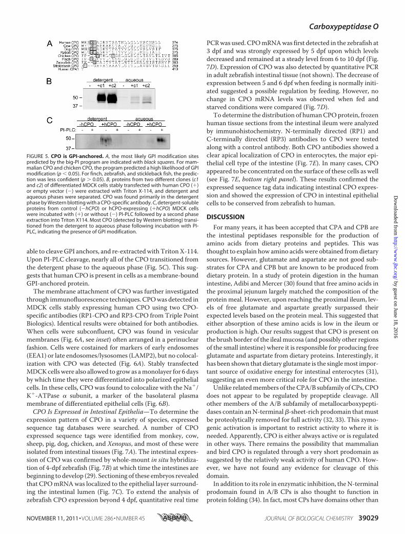
able to cleaveGPI anchors, and re-extractedwithTritonX-114.Upon PI-PLC cleavage, nearly all of the CPO transitioned fromthe detergent phase to the aqueous phase (Fig. 5C). This sug-gests that human CPO is present in cells as amembrane-boundGPI-anchored protein.The membrane attachment of CPO was further investigated
through immunofluorescence techniques. CPOwas detected inMDCK cells stably expressing human CPO using two CPO-specific antibodies (RP1-CPO and RP3-CPO from Triple PointBiologics). Identical results were obtained for both antibodies.When cells were subconfluent, CPO was found in vesicularmembranes (Fig. 6A, see inset) often arranged in a perinuclearfashion. Cells were costained for markers of early endosomes(EEA1) or late endosomes/lysosomes (LAMP2), but no colocal-ization with CPO was detected (Fig. 6A). Stably transfectedMDCKcells were also allowed to grow as amonolayer for 6 daysby which time they were differentiated into polarized epithelialcells. In these cells, CPO was found to colocalize with the Na�/K�-ATPase � subunit, a marker of the basolateral plasmamembrane of differentiated epithelial cells (Fig. 6B).CPO Is Expressed in Intestinal Epithelia—To determine the
expression pattern of CPO in a variety of species, expressedsequence tag databases were searched. A number of CPOexpressed sequence tags were identified from monkey, cow,sheep, pig, dog, chicken, and Xenopus, and most of these wereisolated from intestinal tissues (Fig. 7A). The intestinal expres-sion of CPO was confirmed by whole-mount in situ hybridiza-tion of 4-dpf zebrafish (Fig. 7B) at which time the intestines arebeginning to develop (29). Sectioning of these embryos revealedthat CPOmRNAwas localized to the epithelial layer surround-ing the intestinal lumen (Fig. 7C). To extend the analysis ofzebrafish CPO expression beyond 4 dpf, quantitative real time
PCRwas used. CPOmRNAwas first detected in the zebrafish at3 dpf and was strongly expressed by 5 dpf upon which levelsdecreased and remained at a steady level from 6 to 10 dpf (Fig.7D). Expression of CPO was also detected by quantitative PCRin adult zebrafish intestinal tissue (not shown). The decrease ofexpression between 5 and 6 dpf when feeding is normally initi-ated suggested a possible regulation by feeding. However, nochange in CPO mRNA levels was observed when fed andstarved conditions were compared (Fig. 7D).To determine the distribution of humanCPOprotein, frozen
human tissue sections from the intestinal ileum were analyzedby immunohistochemistry. N-terminally directed (RP1) andC-terminally directed (RP3) antibodies to CPO were testedalong with a control antibody. Both CPO antibodies showed aclear apical localization of CPO in enterocytes, the major epi-thelial cell type of the intestine (Fig. 7E). In many cases, CPOappeared to be concentrated on the surface of these cells as well(see Fig. 7E, bottom right panel). These results confirmed theexpressed sequence tag data indicating intestinal CPO expres-sion and showed the expression of CPO in intestinal epithelialcells to be conserved from zebrafish to human.
DISCUSSION
For many years, it has been accepted that CPA and CPB arethe intestinal peptidases responsible for the production ofamino acids from dietary proteins and peptides. This wasthought to explain how amino acids were obtained fromdietarysources. However, glutamate and aspartate are not good sub-strates for CPA and CPB but are known to be produced fromdietary protein. In a study of protein digestion in the humanintestine, Adibi andMercer (30) found that free amino acids inthe proximal jejunum largely matched the composition of theprotein meal. However, upon reaching the proximal ileum, lev-els of free glutamate and aspartate greatly surpassed theirexpected levels based on the protein meal. This suggested thateither absorption of these amino acids is low in the ileum orproduction is high. Our results suggest that CPO is present onthe brush border of the ilealmucosa (and possibly other regionsof the small intestine) where it is responsible for producing freeglutamate and aspartate from dietary proteins. Interestingly, ithas been shown that dietary glutamate is the singlemost impor-tant source of oxidative energy for intestinal enterocytes (31),suggesting an even more critical role for CPO in the intestine.Unlike relatedmembers of theCPA/B subfamily ofCPs, CPO
does not appear to be regulated by propeptide cleavage. Allother members of the A/B subfamily of metallocarboxypepti-dases contain anN-terminal�-sheet-rich prodomain thatmustbe proteolytically removed for full activity (32, 33). This zymo-genic activation is important to restrict activity to where it isneeded. Apparently, CPO is either always active or is regulatedin other ways. There remains the possibility that mammalianand bird CPO is regulated through a very short prodomain assuggested by the relatively weak activity of human CPO. How-ever, we have not found any evidence for cleavage of thisdomain.In addition to its role in enzymatic inhibition, theN-terminal
prodomain found in A/B CPs is also thought to function inprotein folding (34). In fact, most CPs have domains other than
FIGURE 5. CPO is GPI-anchored. A, the most likely GPI modification sitespredicted by the big-PI program are indicated with black squares. For mam-malian CPO and chicken CPO, the program predicted a high likelihood of GPImodification (p 0.05). For finch, zebrafish, and stickleback fish, the predic-tion was less confident (p � 0.05). B, proteins from two different clones (c1and c2) of differentiated MDCK cells stably transfected with human CPO (�)or empty vector (�) were extracted with Triton X-114, and detergent andaqueous phases were separated. CPO was found primarily in the detergentphase by Western blotting with a CPO-specific antibody. C, detergent-solubleproteins from control (�hCPO) or hCPO-expressing (�hCPO) MDCK cellswere incubated with (�) or without (�) PI-PLC followed by a second phaseextraction into Triton X114. Most CPO (detected by Western blotting) transi-tioned from the detergent to aqueous phase following incubation with PI-PLC, indicating the presence of GPI modification.
Carboxypeptidase O
NOVEMBER 11, 2011 • VOLUME 286 • NUMBER 45 JOURNAL OF BIOLOGICAL CHEMISTRY 39029
by guest on June 18, 2016http://w
ww
.jbc.org/D
ownloaded from

the CP domain that are thought to function in folding: N/Emembers have a C-terminal �-sheet-rich transthyretin-likedomain (35, 36), CCPs all have an N-terminal �-sheet-richdomain (16, 17), and the aminoacylases have a C-terminal�-sheet-rich domain (20). With the characterization of CPOpresented here, it is now apparent that not all CPs requireanother large domain to fold and function properly. It is possi-ble that another protein co-expressed with CPO performs thisfunction, although expression of CPO in cells not normallyexpressing this protein resulted in active enzyme.The selectivity of CPO for acidic amino acids is unique among
the mammalian A/B subfamily of CPs and was predicted becauseof the presence of an Arg at the position equivalent to residue 255in bovine CPA. Currently, there are five known mammalian CPswith acidic amino acid specificity. Aminoacylase 2 (also calledaspartoacylase) functions in the deacetylationofN-acetyl-L-aspar-tate through a CP-like mechanism (20). This is in contrast to itsclose relative, aminoacylase 3, which preferentially deacetylatesN-acetylated aromatic amino acids (37). Mutations in aminoacy-lase 2 cause Canavan disease, a fatal and progressive neurodegen-erative disease, due to greatly elevated levels of N-acetyl-L-aspar-tate (38). Four members of the CCP subfamily have also beenidentified to have specificity for acidic amino acids (19, 39). Likeaminoacylase 2, the CCPs are cytosolic enzymes (16). The CCPshavebeenproposed toplay a role in removal of glutamate fromtheC termini of �-tubulin (19, 39). One of these enzymes, CCP1, ismutated in a classical mouse mutant, Purkinje cell degeneration(pcd), resulting in degeneration of Purkinje cells and several otherneuronal cell types (40–42).In addition to those mentioned above, several other CPs that
cleave acidic amino acids have been identified in non-mamma-
lian species; like CPO, these other enzymes are members of theA/B subfamily of CPs. One of these enzymes was purified fromthe marine annelid Sabellastarte magnifica and was found tocleave both hydrophobic and acidic amino acids (43). Anotherenzyme was identified in the Aedes aegypti mosquito and waspredicted to have acidic amino acid specificity (44). Finally, aglutamate-specific CP was identified and characterized fromthe insect pest corn earwormHelicoverpa armigera (45). Othernon-M14 enzymeswith acidic amino acid-specific carboxypep-tidase activity exist, including glutamate carboxypeptidase II,an extracellular enzyme that hydrolyzes the neuropeptideN-acetylaspartylglutamate (46), and �-glutamyl hydrolase, alysosomal enzyme that hydrolyzes folate polyglutamates (47).Many of the above mentioned CPs are known to play regula-
tory roles through their substrate cleavages. In addition to itsdigestive role, CPOmight also regulate the activity of bioactiveproteins and peptides. When expressed in MDCK cells, CPOwas found localized to the basolateral membrane, suggesting arole in processing extracellular adhesion and signaling proteins.Although CPO appeared to be predominantly apical in humanenterocytes, a role on the basolateral membrane or within thecell during vesicular trafficking cannot be ruled out. The activ-ity of CPO is close to maximal even at the low pH values foundwithin the secretory system, and because activation by removalof a prodomain is not necessary, the enzyme is likely to be fullyactive upon folding within the endoplasmic reticulum.Although CPO was found largely in enterocytes, some colo-
calizationwas observedwith chromograninAwithin enteroen-docrine cells (data not shown), further suggesting a potentialrole in intracellular peptide maturation and regulation. Severalpeptides that contain acidic C-terminal amino acids are pro-
FIGURE 6. Stably expressed CPO is localized to intracellular vesicular membranes as well as plasma membrane of MDCK cells. A, MDCK cells stablyexpressing human CPO were fixed at a subconfluent state. CPO was identified by immunocytochemistry in vesicular structures (see inset). No colocalization ofCPO was found with markers of either early (EEA1) or late (LAMP2) endosomes. Nuclei (blue) were stained with DAPI. B, when the above MDCK cells weredifferentiated into a polarized epithelial monolayer, CPO was more prominently seen at the plasma membrane as indicated by colocalization with Na�/K�-ATPase, a marker of the basolateral membrane. Scale bars, 10 �m.
Carboxypeptidase O
39030 JOURNAL OF BIOLOGICAL CHEMISTRY VOLUME 286 • NUMBER 45 • NOVEMBER 11, 2011
by guest on June 18, 2016http://w
ww
.jbc.org/D
ownloaded from

duced in intestinal cells. GLP-2 is a 33-residue proglucagon-derived peptide containing a C-terminal aspartate that issecreted from the L cells of the distal small intestine and colon(48). GLP-2 is responsible for the stimulation of growth of cryptcells in the intestinal jejunum and ileum (49) and has also beenreported to play a role in gastric emptying (50) and intestinalepithelial barrier function (51). However, to our knowledge, a32-residue GLP-2 lacking the C-terminal aspartate has notbeen reported. Chromogranin A is a well known marker ofenteroendocrine cells of the intestine (52) that produces a num-ber of peptides having acidic C-terminal amino acids (53).Another well known intestinal proprotein, procholecystokinin,is also known to produce peptides with acidic C termini (53).However, the exact functions of these peptides are not known.In conclusion, we have characterized the activity of a unique
metallocarboxypeptidase, CPO, which is translated and foldedas an active enzyme without the need for a typical prodomain.CPOhas substrate selectivity forC-terminal acidic amino acids,is expressed predominantly in the epithelial cells of the intes-tine, and is GPI-anchored and enzymatically active in the extra-
cellular space where it presumably processes dietary proteinand peptides. The high degree of conservation of CPO in mostvertebrates from fish to humans argues for an important func-tion, although the absence of CPO in rat and mouse suggests itis not essential for life.
Acknowledgments—We thank Prof. F. Xavier Avilés (AutonomousUniversity of Barcelona, Barcelona, Spain) for the generous gift ofpotato carboxypeptidase inhibitor-Sepharose resin, Prof. MartinGorovsky (University of Rochester, Rochester, NY) for the generous giftof antiserum to the tubulin poly(Glu) side chain, andDr. AnneMüsch(Albert Einstein College of Medicine, Bronx, NY) for the MDCK cells.In addition, we gratefully acknowledge the helpful advice provided byan anonymous reviewer of a previous draft of this manuscript whorecognized the GPI motif in CPO and recommended the GPI modifi-cation prediction program. We give special thanks to the Einsteinhistotechnology and comparative pathology facility for histology ser-vices and Prof. Jon Backer (Department of Molecular Pharmacology)for generous use of a microscope.
FIGURE 7. CPO is expressed in intestinal epithelial cells. A, a database search of expressed sequence tags (ESTs) for CPO mRNA from all species revealed 19sequences from intestinal tissues and four or fewer from other tissues. B, in situ hybridization (ISH) of 4-dpf zebrafish indicated that CPO mRNA was foundpredominantly in intestinal tissues. Both lateral and dorsal views are shown. C, the above fish stained for CPO by in situ hybridization were paraffin-embedded,sectioned sagittally, and counterstained with methyl green. Blue in situ hybridization stain showed epithelial expression of CPO mRNA. D, CPO mRNA abun-dance was determined by quantitative PCR, which showed expression after 3 dpf in both fed and starved states. Error bars indicate S.E. for three biologicalreplicates, each assayed in triplicate. E, frozen sections of human ileum were stained by immunohistochemical techniques with an unrelated control antibodyand two different antibodies, one raised against the N-terminal region of CPO and the other raised against the C-terminal region of CPO (shown in “red”). Nucleiwere stained with DAPI (blue). Both low (left) and high (right) magnification indicated CPO staining in the apical region of enterocytes. Scale bars in E, 100 �m(left) and 10 �m (right).
Carboxypeptidase O
NOVEMBER 11, 2011 • VOLUME 286 • NUMBER 45 JOURNAL OF BIOLOGICAL CHEMISTRY 39031
by guest on June 18, 2016http://w
ww
.jbc.org/D
ownloaded from

REFERENCES1. Arolas, J. L., Vendrell, J., Aviles, F. X., and Fricker, L. D. (2007) Curr.
Pharm. Des. 13, 349–3662. Fernández, D., Pallarès, I., Vendrell, J., and Avilés, F. X. (2010) Biochimie
92, 1484–15003. Vendrell, J., Querol, E., and Avilés, F. X. (2000) Biochim. Biophys. Acta
1477, 284–2984. Reznik, S. E., and Fricker, L. D. (2001) Cell. Mol. Life Sci. 58, 1790–18045. Beck, I. T. (1973) Am. J. Clin. Nutr. 26, 311–3256. Waldschmidt-Leitz, E., and Purr, A. (1929) Ber. Dtsch. Chem. Ges. 62,
2217–22267. Folk, J. E. (1956) J. Am. Chem. Soc. 78, 3541–35428. Gardell, S. J., Craik, C. S., Clauser, E., Goldsmith, E. J., Stewart, C. B., Graf,
M., and Rutter, W. J. (1988) J. Biol. Chem. 263, 17828–178369. Erdos, E. G., and Sloane, E. M. (1962) Biochem. Pharmacol. 11, 585–59210. Fricker, L. D., and Snyder, S. H. (1982) Proc. Natl. Acad. Sci. U.S.A. 79,
3886–389011. Fricker, L. D. (1988) Annu. Rev. Physiol. 50, 309–32112. Song, L., and Fricker, L. D. (1995) J. Biol. Chem. 270, 25007–2501313. Song, L., and Fricker, L. D. (1997) J. Biol. Chem. 272, 10543–1055014. Lei, Y., Xin, X.,Morgan, D., Pintar, J. E., and Fricker, L. D. (1999)DNACell
Biol. 18, 175–18515. Wei, S., Segura, S., Vendrell, J., Aviles, F. X., Lanoue, E., Day, R., Feng, Y.,
and Fricker, L. D. (2002) J. Biol. Chem. 277, 14954–1496416. Kalinina, E., Biswas, R., Berezniuk, I., Hermoso, A., Aviles, F. X., and Fric-
ker, L. D. (2007) FASEB J. 21, 836–85017. Rodriguez de la Vega, M., Sevilla, R. G., Hermoso, A., Lorenzo, J., Tanco,
S., Diez, A., Fricker, L. D., Bautista, J. M., and Avilés, F. X. (2007) FASEB J.21, 851–865
18. Gomis-Rüth, F. X. (2008) Crit. Rev. Biochem. Mol. Biol. 43, 319–34519. Rogowski, K., van Dijk, J., Magiera, M. M., Bosc, C., Deloulme, J. C.,
Bosson, A., Peris, L., Gold, N. D., Lacroix, B., Grau, M. B., Bec, N., Lar-roque, C., Desagher, S., Holzer,M., Andrieux, A.,Moutin,M. J., and Janke,C. (2010) Cell 143, 564–578
20. Bitto, E., Bingman, C. A., Wesenberg, G. E., McCoy, J. G., and Phillips,G. N., Jr. (2007) Proc. Natl. Acad. Sci. U.S.A. 104, 456–461
21. Kalinina, E. V., and Fricker, L. D. (2003) J. Biol. Chem. 278, 9244–924922. Deddish, P. A., Skidgel, R. A., Kriho, V. B., Li, X. Y., Becker, R. P., and Erdös,
E. G. (1990) J. Biol. Chem. 265, 15083–1508923. Kimmel, C. B., Ballard, W. W., Kimmel, S. R., Ullmann, B., and Schilling,
T. F. (1995) Dev. Dyn. 203, 253–31024. Westerfield,M. (2007)TheZebrafish Book. AGuide for the LaboratoryUse
of Zebrafish (Danio rerio), 5th Ed., University ofOregon Press, Eugene,OR25. Rosenberg, I. M. (2005) Protein Analysis and Purification: Benchtop Tech-
niques, 2nd Ed., pp. 308–313, Birkhauser, Boston26. Thisse, C., and Thisse, B. (2008) Nat. Protoc. 3, 59–6927. Fernández, D., Boix, E., Pallarès, I., Avilés, F. X., and Vendrell, J. (2011)
Enzyme Res. 2011, 12867628. Wolff, A., de Néchaud, B., Chillet, D., Mazarguil, H., Desbruyères, E.,
Audebert, S., Eddé, B., Gros, F., andDenoulet, P. (1992)Eur. J. Cell Biol. 59,425–432
29. Wallace, K. N., Akhter, S., Smith, E. M., Lorent, K., and Pack, M. (2005)Mech. Dev. 122, 157–173
30. Adibi, S. A., and Mercer, D. W. (1973) J. Clin. Investig. 52, 1586–159431. Stoll, B., Burrin, D. G., Henry, J., Yu, H., Jahoor, F., and Reeds, P. J. (1999)
Am. J. Physiol. Endocrinol. Metab. 277, E168–E17532. Guasch, A., Coll, M., Avilés, F. X., and Huber, R. (1992) J. Mol. Biol. 224,
141–15733. Coll, M., Guasch, A., Avilés, F. X., and Huber, R. (1991) EMBO J. 10, 1–934. Phillips, M. A., and Rutter, W. J. (1996) Biochemistry 35, 6771–677635. Gomis-Rüth, F. X., Companys, V., Qian, Y., Fricker, L. D., Vendrell, J.,
Avilés, F. X., and Coll, M. (1999) EMBO J. 18, 5817–582636. Reverter, D.,Maskos, K., Tan, F., Skidgel, R. A., and Bode,W. (2004) J.Mol.
Biol. 338, 257–26937. Hsieh, J. M., Tsirulnikov, K., Sawaya, M. R., Magilnick, N., Abuladze, N.,
Kurtz, I., Abramson, J., and Pushkin, A. (2010) Proc. Natl. Acad. Sci. U.S.A.107, 17962–17967
38. Namboodiri, A. M., Peethambaran, A., Mathew, R., Sambhu, P. A., Her-shfield, J., Moffett, J. R., andMadhavarao, C. N. (2006)Mol. Cell. Endocri-nol. 252, 216–223
39. Kimura, Y., Kurabe, N., Ikegami, K., Tsutsumi, K., Konishi, Y., Kaplan,O. I., Kunitomo, H., Iino, Y., Blacque, O. E., and Setou, M. (2010) J. Biol.Chem. 285, 22936–22941
40. Fernandez-Gonzalez, A., La Spada, A. R., Treadaway, J., Higdon, J. C.,Harris, B. S., Sidman, R. L., Morgan, J. I., and Zuo, J. (2002) Science 295,1904–1906
41. Mullen, R. J., Eicher, E. M., and Sidman, R. L. (1976) Proc. Natl. Acad. Sci.U.S.A. 73, 208–212
42. Wang, T., Parris, J., Li, L., and Morgan, J. I. (2006)Mol. Cell. Neurosci. 33,200–213
43. Alonso-del-Rivero, M., Trejo, S. A., Rodríguez de la Vega, M., González,Y., Bronsoms, S., Canals, F., Delfín, J., Diaz, J., Aviles, F. X., and Chávez,M. A. (2009) FEBS J. 276, 4875–4890
44. Isoe, J., Zamora, J., andMiesfeld, R. L. (2009) Insect Biochem.Mol. Biol. 39,68–73
45. Bown, D. P., and Gatehouse, J. A. (2004) Eur. J. Biochem. 271, 2000–201146. Thomas, A. G., Wozniak, K. M., Tsukamoto, T., Calvin, D., Wu, Y., Rojas,
C., Vornov, J., and Slusher, B. S. (2006)Adv. Exp.Med. Biol. 576, 327–337;discussion 361–363
47. Schneider, E., and Ryan, T. J. (2006) Clin. Chim. Acta 374, 25–3248. Drozdowski, L., and Thomson, A. B. (2009) World J. Gastroenterol. 15,
385–40649. Drucker, D. J., Erlich, P., Asa, S. L., and Brubaker, P. L. (1996) Proc. Natl.
Acad. Sci. U.S.A. 93, 7911–791650. Nagell, C. F.,Wettergren, A., Pedersen, J. F., Mortensen, D., and Holst, J. J.
(2004) Scand. J. Gastroenterol. 39, 353–35851. Benjamin, M. A., McKay, D. M., Yang, P. C., Cameron, H., and Perdue,
M. H. (2000) Gut 47, 112–11952. Facer, P., Bishop, A. E., Lloyd, R. V., Wilson, B. S., Hennessy, R. J., and
Polak, J. M. (1985) Gastroenterology 89, 1366–137353. Zhang, X., Che, F. Y., Berezniuk, I., Sonmez, K., Toll, L., and Fricker, L. D.
(2008) J. Neurochem. 107, 1596–1613
Carboxypeptidase O
39032 JOURNAL OF BIOLOGICAL CHEMISTRY VOLUME 286 • NUMBER 45 • NOVEMBER 11, 2011
by guest on June 18, 2016http://w
ww
.jbc.org/D
ownloaded from
Carboxypeptidase O is a Glycosylphosphatidylinositol-Anchored Intestinal Peptidase with Acidic Amino Acid Specificity
Peter J. Lyons and Lloyd D. Fricker
Supplemental Information: Suppl. Fig. 1:
Supplemental Figure 1. Purified CPO, as well as that expressed in mammalian cells, exists as two major isoforms. Both human (A) and zebrafish (B) CPO were expressed in Sf9 cells using the baculovirus system and were purified using affinity chromatography on potato carboxypeptidase inhibitor (PCI)-Sepharose. Proteins were visualized by staining with GelCode Blue Coomassie (Pierce). Human CPO was also present as two major bands when expressed in both HEK293T (C) and MDCK (D) mammalian cells and detected by western blotting with a CPO-specific antibody.

Suppl. Fig. 2:
Supplemental Figure 2. CPO is N-glycosylated. Equal amounts of purified human and zebrafish CPO were untreated (lane 1), incubated for 1 hour at 37°C with buffers only (lane 2), or incubated for 1 hour at 37°C with PNGase F glysosidase (lane 3). Protein was resolved by SDS-PAGE and visualized by either silver staining or by western blotting with an antibody for human CPO raised against the C-terminal region of the carboxypeptidase domain. The lower band detected in lanes 3 of the silver stained samples is the PNGase F enzyme (apparent molecular weight 36 kDa).

Peter J. Lyons and Lloyd D. FrickerPeptidase with Acidic Amino Acid Specificity
Carboxypeptidase O Is a Glycosylphosphatidylinositol-anchored Intestinal
doi: 10.1074/jbc.M111.265819 originally published online September 15, 20112011, 286:39023-39032.J. Biol. Chem.
10.1074/jbc.M111.265819Access the most updated version of this article at doi:
Alerts:
When a correction for this article is posted•
When this article is cited•
to choose from all of JBC's e-mail alertsClick here
Supplemental material:
http://www.jbc.org/content/suppl/2011/09/15/M111.265819.DC1.html
http://www.jbc.org/content/286/45/39023.full.html#ref-list-1
This article cites 51 references, 18 of which can be accessed free at
by guest on June 18, 2016http://w
ww
.jbc.org/D
ownloaded from
Copyright © 2022 FDOKUMEN