Ca2+/calcineurin-dependent inactivation of neuronal L-type Ca2+ channels requires priming by...
11
Cell Reports Report Ca 2+ /Calcineurin-Dependent Inactivation of Neuronal L-Type Ca 2+ Channels Requires Priming by AKAP-Anchored Protein Kinase A Philip J. Dittmer, 1 Mark L. Dell’Acqua, 1 and William A. Sather 1, * 1 Department of Pharmacology, University of Colorado School of Medicine, Mail Stop 8315, 12800 East 19 th Avenue, Aurora, CO 80045, USA *Correspondence: [email protected] http://dx.doi.org/10.1016/j.celrep.2014.04.039 This is an open access article under the CC BY-NC-ND license (http://creativecommons.org/licenses/by-nc-nd/3.0/). SUMMARY Within neurons, Ca 2+ -dependent inactivation (CDI) of voltage-gated L-type Ca 2+ channels shapes cyto- plasmic Ca 2+ signals. CDI is initiated by Ca 2+ binding to channel-associated calmodulin and subsequent Ca 2+ /calmodulin activation of the Ca 2+ -dependent phosphatase, calcineurin (CaN), which is targeted to L channels by the A-kinase-anchoring protein AKAP79/150. Here, we report that CDI of neuronal L channels was abolished by inhibition of PKA acti- vity or PKA anchoring to AKAP79/150 and that CDI was also suppressed by stimulation of PKA activity. Although CDI was reduced by positive or negative manipulation of PKA, interference with PKA anchoring or activity lowered Ca 2+ current density whereas stimulation of PKA activity elevated it. In contrast, inhibition of CaN reduced CDI but had no effect on current density. These results suggest a model wherein PKA-dependent phosphorylation enhances neuronal L current, thereby priming chan- nels to undergo CDI, and Ca 2+ /calmodulin-activated CaN actuates CDI by reversing PKA-mediated enhancement of channel activity. INTRODUCTION Voltage-gated Ca 2+ channels convert patterns of electrical acti- vity on the neuronal surface membrane into signals that can initiate intracellular signaling: rises in cytoplasmic Ca 2+ . Within neurons, Ca 2+ can trigger release of neurotransmitter and changes in gene expression that contribute to modification of cell morphology and synaptic plasticity (Catterall, 2011). Residing at the interface between electrical and chemical signaling, Ca 2+ channels represent natural points for regulation, with up- and downmodulation of channel activity providing precise spatiotemporal control of cytoplasmic Ca 2+ signals that specify which of various Ca 2+ -dependent processes are activated and how strongly. Curbing Ca 2+ channel activity is also critical in avoiding cytotoxicity arising from Ca 2+ overload (Choi, 1994; Na ¨ gerl et al., 2000). One important mechanism that has evolved to limit Ca 2+ entry via Ca 2+ channels is Ca 2+ - dependent inactivation (CDI) (Tillotson, 1979; Budde et al., 2002). Calmodulin (CaM) has been identified as the Ca 2+ sensor that initiates CDI (Zu ¨ hlke et al., 1999; Peterson et al., 1999), and in the CaM-actuated model of CDI, Ca 2+ ions entering the cytoplasm bind to calmodulin docked on the channel through which they have just passed, Ca 2+ /CaM undergoes a confor- mational change that is sensed by its associated channel, and the channel is nudged into an inactivated conformation inca- pable of conducting Ca 2+ (Erickson et al., 2003). Despite the elegance of studies aimed at elucidating the mechanism of CaM-actuated CDI, they generally have had the major drawback of relying upon heterologous expression of voltage-gated Ca 2+ channels in cells that naturally lack these channels and are also deficient in the scaffolding proteins that target enzymes like PKA and calcineurin (CaN) to channels. Using a more intact and physiologically relevant system of cultured hippocampal neurons, we recently described experimental results strongly suggesting that Ca 2+ /CaM initiates CDI largely through acti- vation of the natural Ca 2+ /CaM substrate, CaN (Oliveria et al., 2012). We found that CaN, anchored to Ca V 1.2 by the A-kinase-anchoring protein AKAP79/150 (human/rodent), was essential for CDI of pharmacologically isolated L-type Ca 2+ cur- rent in hippocampal neurons. Disruption of this anchoring protein prevented enhancement by PKA of L-current amplitude in cultured neurons, raising the possibility that PKA might enhance L current by opposing CaM/CaN-mediated CDI. Modulation of Ca V 1.2 by PKA is one of the best-described forms of ion channel modulation and has been identified in a variety of excitable cell types (Bean et al., 1984; Kalman et al., 1988; Hadley and Lederer, 1991; Rankovic et al., 2011). Here, we report evidence from hippocampal neurons indi- cating that impairment of PKA anchoring or activity decreases L-type Ca 2+ current density and abolishes CDI of these channels. Furthermore, neurons in which PKA activity was stimulated exhibited concomitant enhancement of current and diminution of CDI. These experimental results can be explained by a simple model of inverse control by PKA and CaN of L-channel current and kinetics: PKA-dependent phosphorylation enhances L-channel opening probability and primes channels for CDI, and Ca 2+ /CaM-activated CaN actuates CDI by reversing PKA- mediated enhancement. This mechanism readily accommo- dates the experimental observations that interference with the 1410 Cell Reports 7, 1410–1416, June 12, 2014 ª2014 The Authors
-
Upload
dailycamera -
Category
Documents
-
view
2 -
download
0
Transcript of Ca2+/calcineurin-dependent inactivation of neuronal L-type Ca2+ channels requires priming by...

Cell Reports
Report
Ca2+/Calcineurin-Dependent Inactivationof Neuronal L-Type Ca2+ Channels RequiresPriming by AKAP-Anchored Protein Kinase APhilip J. Dittmer,1 Mark L. Dell’Acqua,1 and William A. Sather1,*1Department of Pharmacology, University of Colorado School of Medicine, Mail Stop 8315, 12800 East 19th Avenue, Aurora, CO 80045, USA*Correspondence: [email protected]
http://dx.doi.org/10.1016/j.celrep.2014.04.039
This is an open access article under the CC BY-NC-ND license (http://creativecommons.org/licenses/by-nc-nd/3.0/).
SUMMARY
Within neurons, Ca2+-dependent inactivation (CDI) ofvoltage-gated L-type Ca2+ channels shapes cyto-plasmic Ca2+ signals. CDI is initiated by Ca2+ bindingto channel-associated calmodulin and subsequentCa2+/calmodulin activation of the Ca2+-dependentphosphatase, calcineurin (CaN), which is targetedto L channels by the A-kinase-anchoring proteinAKAP79/150. Here, we report that CDI of neuronalL channels was abolished by inhibition of PKA acti-vity or PKA anchoring to AKAP79/150 and that CDIwas also suppressed by stimulation of PKA activity.Although CDI was reduced by positive or negativemanipulation of PKA, interference with PKAanchoring or activity lowered Ca2+ current densitywhereas stimulation of PKA activity elevated it. Incontrast, inhibition of CaN reduced CDI but had noeffect on current density. These results suggest amodel wherein PKA-dependent phosphorylationenhances neuronal L current, thereby priming chan-nels to undergo CDI, and Ca2+/calmodulin-activatedCaN actuates CDI by reversing PKA-mediatedenhancement of channel activity.
INTRODUCTION
Voltage-gated Ca2+ channels convert patterns of electrical acti-
vity on the neuronal surface membrane into signals that can
initiate intracellular signaling: rises in cytoplasmic Ca2+. Within
neurons, Ca2+ can trigger release of neurotransmitter and
changes in gene expression that contribute to modification of
cell morphology and synaptic plasticity (Catterall, 2011).
Residing at the interface between electrical and chemical
signaling, Ca2+ channels represent natural points for regulation,
with up- and downmodulation of channel activity providing
precise spatiotemporal control of cytoplasmic Ca2+ signals
that specify which of various Ca2+-dependent processes are
activated and how strongly. Curbing Ca2+ channel activity is
also critical in avoiding cytotoxicity arising from Ca2+ overload
(Choi, 1994; Nagerl et al., 2000). One important mechanism
1410 Cell Reports 7, 1410–1416, June 12, 2014 ª2014 The Authors
that has evolved to limit Ca2+ entry via Ca2+ channels is Ca2+-
dependent inactivation (CDI) (Tillotson, 1979; Budde et al., 2002).
Calmodulin (CaM) has been identified as the Ca2+ sensor
that initiates CDI (Zuhlke et al., 1999; Peterson et al., 1999),
and in the CaM-actuated model of CDI, Ca2+ ions entering the
cytoplasm bind to calmodulin docked on the channel through
which they have just passed, Ca2+/CaM undergoes a confor-
mational change that is sensed by its associated channel, and
the channel is nudged into an inactivated conformation inca-
pable of conducting Ca2+ (Erickson et al., 2003). Despite the
elegance of studies aimed at elucidating the mechanism of
CaM-actuated CDI, they generally have had the major drawback
of relying upon heterologous expression of voltage-gated Ca2+
channels in cells that naturally lack these channels and are
also deficient in the scaffolding proteins that target enzymes
like PKA and calcineurin (CaN) to channels. Using a more intact
and physiologically relevant system of cultured hippocampal
neurons, we recently described experimental results strongly
suggesting that Ca2+/CaM initiates CDI largely through acti-
vation of the natural Ca2+/CaM substrate, CaN (Oliveria et al.,
2012). We found that CaN, anchored to CaV1.2 by the
A-kinase-anchoring protein AKAP79/150 (human/rodent), was
essential for CDI of pharmacologically isolated L-type Ca2+ cur-
rent in hippocampal neurons.
Disruption of this anchoring protein prevented enhancement
by PKA of L-current amplitude in cultured neurons, raising the
possibility that PKA might enhance L current by opposing
CaM/CaN-mediated CDI. Modulation of CaV1.2 by PKA is one
of the best-described forms of ion channel modulation and has
been identified in a variety of excitable cell types (Bean et al.,
1984; Kalman et al., 1988; Hadley and Lederer, 1991; Rankovic
et al., 2011).
Here, we report evidence from hippocampal neurons indi-
cating that impairment of PKA anchoring or activity decreases
L-typeCa2+ current density and abolishes CDI of these channels.
Furthermore, neurons in which PKA activity was stimulated
exhibited concomitant enhancement of current and diminution
of CDI. These experimental results can be explained by a simple
model of inverse control by PKA and CaN of L-channel current
and kinetics: PKA-dependent phosphorylation enhances
L-channel opening probability and primes channels for CDI,
and Ca2+/CaM-activated CaN actuates CDI by reversing PKA-
mediated enhancement. This mechanism readily accommo-
dates the experimental observations that interference with the

Figure 1. CDI of Neuronal L-type Ca2+ Channels Requires AKAP79/150-Anchored PKA
(A) Left, top three images, fluorescence images of cultured rat neurons transfected with 150RNAi and: wild-type AKAP79 (79 WT), AKAP79DPKA (79DPKA), or
AKAP79Pro2 (79Pro2). Bottom three images: differential interference contrast images of short-term cultured neurons from wild-type mice, AKAP150DPKAmice,
and AKAP150/ mice. (Center) Superimposed, normalized records of pharmacologically isolated L-channel Ca2+ (red) or Ba2+ (black) currents. Right, mean
inactivation rates (1/t). Number of cells recorded per condition (n) is noted on each bar.
(B) CDI index, calculated as the difference between r400 in Ba2+ and r400 in Ca2+. Black bars represent knockdown/replacement experiments in rat neurons and
gray bars represent knockin mice in all four panels. Fractional depression of divalent charge influx as a consequence of CDI, calculated as the difference between
peak-normalized Ba2+ charge influx and peak-normalized Ca2+ charge influx during 500 ms depolarizations.
(C) Peak Ca2+ current density and Ca2+ charge density for each of the experimental manipulations in (B). Dividing peak Ca2+ current (ICa; pA) by a measure of
membrane surface area, cell capacitance (Cm; pF), yields the density of Ca2+ current (pA/pF). Ca2+ charge density (pC/pF) was calculated as the integrated Ca2+
current during 500 ms depolarization (pC) divided by cell capacitance (pF).
(D) For 150RNAi-transfected rat neurons, voltage dependence of Ca2+ current density for 79WT (C) or 79DPKA (:) cotransfected neurons compared with the
voltage dependence of inactivation rate for Ca2+ current for 79WT (B) or 79DPKA (6) cotransfected neurons. Error bars indicate SEM. Mean values were
compared using ANOVA with a post hoc Bonferroni correction. Statistical significance marked as *p = 0.05, **p = 0.01, ***p = 0.005, and ****p = 0.001.
action of either PKA or CaN obstructs the normal process of
CDI. More generally, these results expand the repertoire of
L-channel-complexed proteins known to modulate Ca2+ signals
in postsynaptic regions: channel-bound CaM and AKAP79/150-
anchored CaN and PKA function coordinately to tune Ca2+
signals that regulate neuronal gene expression, as further
explored in a paper published in this issue of Cell Reports (Mur-
phy et al., 2014).
C
RESULTS
Channel-Localized PKA Enhances Current Density andPrimes Channels for CDIIn rodent hippocampal pyramidal neurons grown in culture for
up to 5 days, Ca2+ current carried by L-type channels exhibited
two components of inactivation: fast, Ca2+-dependent inacti-
vation (1/t = 40.6 ± 2.1 s1 in mice; Figure 1A, red bars;
ell Reports 7, 1410–1416, June 12, 2014 ª2014 The Authors 1411

42.9 ± 2.0 s1 in rats; Oliveria et al., 2012) and slow, voltage-
dependent inactivation that remains present when Ba2+ (black
bars) is substituted for Ca2+ in the extracellular solution. The
fast component—CDI—was virtually eliminated in AKAP150-
knockout mice (AKAP150/; Figure 1A), consistent with previ-
ously reported results with RNAi-mediated knockdown of
AKAP150 (150RNAi) in rat neurons (Oliveria et al., 2007, 2012).
CDI was fully restored in 150RNAi knockdown neurons that
were cotransfected with the 150RNAi-insensitive human ortho-
log of AKAP150, AKAP79 (79WT + 150RNAi), confirming the
importance of AKAP79/150 in L-channel CDI.
Knockdown or knockout of AKAP150 delocalizes both CaN
and PKA from the L-channel nanoenvironment, which precludes
identification of CaN- or PKA-specific effects on channel func-
tion. To probe more specifically whether AKAP-anchored PKA
is involved in CDI, L currents were recorded from neurons that
express an AKAP79/150 mutant incapable of anchoring PKA,
owing to a selective deletion within the PKA-RII-subunit-binding
domain (residues 391–400; designated 79DPKA; Carr et al.,
1992; Oliveria et al., 2003; Murphy et al., 2014). Rat neurons
cotransfected with AKAP79DPKA (79DPKA) and 150RNAi to
knock down native AKAP150 exhibited greatly slowed CDI (Fig-
ure 1A, row 2). Neurons cultured from AKAP150DPKA knockin
mice (Murphy et al., 2014) showed similarly slowed CDI (Fig-
ure 1A, row 5). CDI was also significantly slowed in neurons
expressing 150RNAi and 79pro2 (Figure 1A, row 3), an
AKAP79 mutant bearing two proline substitutions (A396P and
I404P) that disrupt the helical structure of the PKA-binding site
(Carr et al., 1992; Oliveria et al., 2007).
The fraction of peak current remaining 400 ms after the onset
of step depolarization (r400) provides another view of inactivation,
and the difference between Ba2+ and Ca2+ in fractional current
remaining 400 ms after the onset of depolarization (r400,Ba –
r400,Ca) provides a second useful index of CDI (Figure 1B, top;
Kim et al., 2004; Oz et al., 2011). A third CDI metric estimates
the fraction of Ca2+ prevented from entering the neuron as a
consequence of CDI: peak Ba2+ and Ca2+ current amplitudes
were normalized because Ba2+ permeates the channel more
readily and then the peak-normalized Ba2+ and Ca2+ currents
were integrated to obtain relative charge transfer, and the differ-
ence between these (ABa – ACa) approximates the fractional
depression of Ca2+ entry by CDI (Figure 1B, bottom). For the
AKAP manipulations studied, the patterns for both CDI index
and depression of Ca2+ entry (ABa – ACa) mirrored that for rate
of CDI (1/t) in Figure 1A.
Deficits in AKAP79/150 anchoring of PKA (DPKA and pro2)
that reduced CDI also reduced peak Ca2+ current density (Fig-
ure 1C, top). In contrast, current density was not decreased in
cells expressing mutant AKAP79/150 in which the CaN-binding
PxIxIT-like motif (DPIX mutants) was deleted. The overall pattern
for integrated Ca2+ current density, i.e., Ca2+ charge density
(Figure 1C, bottom), is similar to the peak current density pattern
but less pronounced: disruption of PKA anchoring moderately
reduced charge density, whereas disruption of CaN anchoring
increased L-channel charge density, albeit modestly. Peak
current density and charge density provide different views of
channel activity, but together they suggest the idea that
anchored PKA supports a basal degree of enhancement of
1412 Cell Reports 7, 1410–1416, June 12, 2014 ª2014 The Authors
L-channel activity that is limited by anchored CaN. Using Ca2+
imaging and phosphoimmunoblotting, our companion paper in
this issue (Murphy et al., 2014) provides additional evidence
that disruption of AKAP79/150-PKA anchoring leads to substan-
tial decreases in basal L-channel phosphorylation and Ca2+
signaling in hippocampal neurons.
Inward Ca2+ current increases with increasing depolarization
and then declines to zero as membrane potential approaches
the reversal potential for L channels; as a Ca2+-dependent
process, the degree of CDI reflects the trajectory of this ICa-VM
curve (Figure 1D). L currents measured from 150RNAi/79DPKA
neurons were smaller and inactivated more slowly than in
150RNAi/79WT control neurons, but the voltage dependence
of both current density and 1/t for CDI showed the same form
as for control, indicating that the decreased current density
caused by disruption of PKA anchoring cannot be attributed to
a shift in the channel’s voltage dependence of gating. Further,
the large reduction in CDI caused by disruption of PKA anchoring
cannot be explained by the more modest reduction in current
density that was caused by this anchoring disruption.
If our explanation of the effects of PKA on CDI was correct,
we predicted that pharmacological antagonism of either PKA
anchoring to the AKAP or of PKA enzymatic activity would
diminish Ca2+ current density and CDI. To test this prediction,
Ca2+ currents were measured when PKA anchoring was antag-
onized by Ht31, an AKAP-derived peptide that binds the RII
regulatory subunit of PKA with nanomolar affinity and thereby
competes with AKAPs for binding to RII (Carr et al., 1992). By
incorporating Ht31 into the whole-cell pipet, we found that
Ht31 dose-dependently reduced peak current density and CDI,
with an IC50 of 5–10 mM (Figures 2A and 2B). Current density
and CDI were insensitive to a high concentration of a proline
substitution mutant of Ht31 (Ht31-Pro) that cannot properly
bind to PKA RII (Figure 2A). In another set of experiments, PKA
activity was inhibited by loading the PKA inhibitory peptide,
PKI, into the whole-cell recording pipets. Inhibition of PKA acti-
vity also depressed Ca2+ current density and eliminated CDI
(Figure 2C). Thus, competitive inhibitors of PKA anchoring and
activity acted much like the genetic manipulations that impaired
PKA anchoring, and as predicted, the competitive inhibitors
reduced current density and attenuated CDI.
PKA versus CaN in CDITo examine the connection between PKA and CaN in CDI of
neuronal L channels, we tested the effects on whole-cell Ca2+
currents of fast application of membrane-permeant inhibitors
of PKA (H89) or CaN (FK506). After establishing a baseline for
peak inward Ca2+ current density, the solution flowing over the
neuron was quickly changed from control to 500 nM H89 (Ki =
135 nM; Davies et al., 2000). H89 application lowered both
peak current density and CDI rate to new steady levels in control
neurons (Figure 3A, left), but not in 79DPKA neurons (Figure 3A,
right). The low density of current in 150RNAi/79DPKA neurons,
near absence of CDI, and lack of effect of H89 are consistent
with results in Figures 1 and 2: in 79DPKA neurons, channels
are already largely de-enhanced and inactivated. Antagonism
of CaN via fast application of 5 mM FK506 to control neurons
slowed CDI but had no significant effect on peak current density

Figure 2. CDI of Neuronal L-type Ca2+
Channels Is Suppressed by Acute Inhibition
of PKA Anchoring or Activity
(A) Ht31 peptide, a competitive inhibitor of PKA
anchoring to AKAP150, decreased CDI rate and
current density of L-type Ca2+ channels in short-
term cultured hippocampal neurons. Ht31 peptide
was perfused into the cell through the patch pipet.
Left, normalized Ca2+ (red) and Ba2+ currents
(black), recorded from the same cell, were evoked
by 500 ms step depolarization from 60 to 0 mV.
Right, inactivation rates for Ca2+ current (red bars)
and Ba2+ current (black bars). Control rate mea-
sured with 0.1% DMSO in the patch incorporation
of Ht31-Pro in place of Ht31 in the patch pipet had
no effect on current density or CDI.
(B) Dose-response relationships for neurons
treated with Ht31. C, current density; 6, 1/t.
(C) Inhibition of PKA activity by internal perfusion
with 5 mM PKI suppresses CDI and lowers current
density. Error bars indicate SEM. Mean values
were compared using ANOVA with a post hoc
Bonferroni correction. Statistical significance
marked as ****p = 0.001.
(Figure 3B, left). In 150RNAi/79DPIX neurons, which have disrup-
ted CaN anchoring, FK506 exerted no obvious effect on current
density or rate of CDI (Figure 3B, right). But in this case, current
density remained high, whereas CDI was low, as though chan-
nels were in an enhanced state but no longer subject to CDI. In
both situations, the experimental results with drugs that altered
PKA or CaN activity agreed with results obtained with AKAP
mutants bearing binding site deletions for PKA or CaN.
Elevation of PKA Activity Suppresses CDICompared to nonstimulated control neurons, forskolin-
stimulated neurons with concomitant elevated PKA activity ex-
hibited significantly less CDI and enhanced L current density
(Figure 4A). If attenuation of CDI reflected the ability of an
elevated level of PKA activity to overcome CaN action, then
other means of stimulating PKA were predicted to reduce CDI
as well. We therefore tested the effects on L current of fast appli-
cation of a potent and membrane-permeant cyclic AMP (cAMP)
analog, Sp-5,6-dichloro-1-beta-D-ribofuranosylbenzimidazole-
30,50-monophosphorothioate (cBIMPS) (Sandberg et al., 1991).
At 100 mM, but not 10 mM, the cAMP analog significantly
increased peak L current density (23%) and reduced CDI, as
predicted (Figure 4B).
DISCUSSION
For endogenous L-channel complexes in neurons, we have
found that Ca2+/CaN-mediated CDI requires priming of L chan-
nels by PKA. A PKA-dependence for CDI was revealed by loss
of CDI when either PKA activity was pharmacologically inhibited
or PKA anchoring to AKAP79/150 was prevented. Three distinct
experimental approaches to disrupt PKA anchoring—(1)
150DPKA knockin, (2) 150RNAi knockdown and replacement
by PKA-binding-defective AKAP79DPKA or AKAP79pro2, or (3)
competitive inhibition of PKA anchoring by Ht31—abolished
CDI, and thus we conclude that PKA anchoring by AKAP79/
C
150 is a prerequisite for CDI (Figures 1, 2A, and 2B). Whether
determined from rate of CDI, CDI index, or fractional depression
of Ca2+ entry, all anchoring interventions strongly reduced CDI in
cultured neurons.
Kinase activity of PKA is also a prerequisite for CDI of
L channels in neurons: CDI was prevented either by a synthetic
mimic of a highly selective natural inhibitor of free PKA catalytic
subunits, PKI-(6-22)-amide (Figure 2C), or by a low concentra-
tion of a competitive inhibitor of ATP binding in the enzyme’s
catalytic site, 500 nM H89 (Figure 3A; Murray, 2008). H89 was
without effect in knockdown/rescue experiments in which PKA
anchoring was disrupted, indicating that H89 suppression of
CDI is attributable specifically to antagonism of PKA that is
anchored in the channel-AKAP complex.
CDI was efficaciously suppressed by the calcineurin inhibitor
FK506 (Figure 3B), directly in line with the idea that CDI of
neuronal L channels is carried out by calcineurin (Chad and
Eckert, 1986; Armstrong and Eckert, 1987; Oliveria et al., 2007,
2012). In calcineurin-anchoring-defective 79DPIX neurons, the
absence of calcineurin from L-channel complexes resulted in
large L-channel Ca2+ currents with little CDI. FK506 had no effect
on the (already diminished) CDI of L channels in 79DPIX neurons,
presumably because calcineurin, the target of FK506 action, was
not present in the AKAP/L-channel complex.
Channel-Localized PKA Activity Supports CaN-Mediated CDIExperimental manipulations that impaired either anchoring
(DPKA or Ht31) or activity (PKI or H89) of PKA virtually abolished
CDI, but they also decreased current density (from 18–20 pA/pF
to 10–14 pA/pF). Was loss of CDI in these cases simply a conse-
quence of smaller Ca2+ influx rather than decreased phosphory-
lation by PKA? Apparently not, because strong CDI was evident
in wild-type (WT) currents recorded at10mV or +30mV, and at
these potentials, WT currents were similar in amplitude to those
of CDI-lacking 79DPKA currents recorded at the peak (+10 mV)
ell Reports 7, 1410–1416, June 12, 2014 ª2014 The Authors 1413

Figure 3. Time Course of Changes in
Current Density and Inactivation Rate
Induced by Inhibition of PKA or CaN
(A) Response to fast perfusion of H89 (500 nM).
Left, superimposed Ca2+ current records before
(black) and during (gray) H89 application. Middle,
time course of H89 action on peak inward currents
recorded from untransfected rat neurons. Right,
rat neurons expressing 79DPKA.
(B) Response to fast application of FK506 (5 mM).
Left, representative Ca2+ current records before
(black) and during (gray) FK506 application. Mid-
dle, time course of FK506 action for untransfected
rat neurons. Right, time course of FK506 action for
rat neurons expressing 79DPIX. Error bars repre-
sent standard error of the mean.
of the current-voltage relationship (Figure 1D). Therefore, loss
of CDI upon disruption of PKA anchoring or block of PKA activity
apparently arose as a direct consequence of the loss of channel-
localized PKA activity.
PKI and disruption of PKA localization were very similar to
one another in the size of their effects on current density
(30%–40% depression) and CDI (abolishment). This similarity
indicates that an intact pool of PKA anchored by AKAP79/150
to L channels has a basal level of activity in neurons, that this
pool of PKA enhances L current, and that this degree of PKA-
dependent enhancement is sufficient to support subsequent
CaN-mediated CDI. For Ht31 treatment, de-enhancement of
current and suppression of CDI displayed overlapping concen-
tration-response relationships, suggesting that these two ef-
fects are directly related to one another (Figure 2B). Regulation
of L channels by basal activity of AKAP79/150-anchored PKA
may be attributable to cAMP-independent kinase activity of
catalytic subunits in type II PKA holoenzymes or to activity of
catalytic subunits that may remain associated with RII subunits
in cAMP-activated PKA holoenzymes (Yang et al., 1995; Smith
et al., 2013).
Phosphorylation Is a Prerequisite for CaN-Mediated,Dephosphorylation-Dependent CDIWe have found that stimulation of PKA activity (forskolin [FSK]
and cBIMPS) in hippocampal neurons reduced CDI (Figure 4).
Similarly, other groups have found that the beta agonist isopro-
terenol, forskolin, mimics of cAMP, or intracellular perfusion with
PKA reduced CDI of voltage-gated Ca2+ channels in neurons
(Kalman et al., 1988; You et al., 1995). Yet we have found that
interference with PKA action (delocalization; block) also reduced
CDI, much like stimulation of PKA did. Thus, CDI was reduced
by either positive or negative manipulation of PKA action. In
contrast, the effects of PKA manipulation on current density
were straightforward: interference with PKA localization or acti-
vity reduced current density whereas stimulation of PKA (by
forskolin or cBIMPS) increased current density. How might the
effects of PKA manipulation upon CDI and enhancement of
1414 Cell Reports 7, 1410–1416, June 12, 2014 ª2014 The Authors
L current be reconciled, and in particular,
how might interference with localized
PKA action reduce CDI?
Evidently, PKA-mediated enhancement and CaN-mediated
CDI function as inverse operations at L channels: PKA enhances
via phosphorylation and CaN de-enhances—initiates CDI—via
dephosphorylation. A natural conclusion is that PKA-dependent
phosphorylation primes channels to undergo CDI and Ca2+/
CaM-activated CaN actuates CDI by reversing PKA-mediated
enhancement. In this model, elevated PKA activity (FSK and
cBIMPS) overcomes CaN phosphatase action and thereby
reduces CDI and enhances current. Conversely, when channel-
localized PKA action is impeded (DPKA, Pro2, Ht31, H89, and
PKI), the diminution of current density and of CDI reflect the facts
that channel opening probability is reduced and fewer channels
are phosphorylated and thus available to undergo CaN-depen-
dent CDI. In essence, impediment of PKA action allows the CDI
process to chronically dominate channel gating, an increased
fraction of channels resides in aCa2+-inactivated state, and fewer
open channels remain available to undergo CDI. More-complex
alternative explanations are possible as well. The time course of
the effects of H89 on L current particularly supports our simple
interpretation (Figure 3A). The time course of FK506 action can
also be accommodated by this model: during FK506 inhibition
of Ca2+/CaM-activated CaN, L-channel current density was
maintained and presumably the number of phosphorylated,
active L channels was preserved as well; at the same time, the
Ca2+/CaN-mediated dephosphorylation of channels that sub-
serves CDI was prevented, thus reducing rate and degree of CDI.
In summary, our work addresses the control of postsynaptic
L-type Ca2+ signals and particularly the mechanism of CDI in
cultured hippocampal neurons. In these neurons, CDI requires
prior priming of channels by PKA, with the actual process of
CDI mediated by CaM and CaN (Chad and Eckert, 1986;
Armstrong, 1989; Oliveria et al., 2007, 2012). In the PKA-CaN
network that regulates L channel activity, PKA may be activated
by an upstream input, for example by b2 adrenergic receptors
(Gray and Johnston, 1987; Davare et al., 2001; Hoogland and
Saggau, 2004), which provides a positive modulatory influence
on L channels. PKA might also be activated by Ca2+/CaM-stim-
ulated adenylyl cyclase activity, which would provide positive

Figure 4. CDI of Neuronal L-Type Ca2+ Channels Is Reduced by PKA
Pathway Activation
(A) Internal perfusion with forskolin slows Ca2+-dependent inactivation of
L-type current and increases current density. Peak Ca2+ current amplitude
(red) was normalized to peak Ba2+ current (black) amplitude. Bar graphs
present inactivation rates for Ca2+ and Ba2+ currents; number of individual
cells recorded (n) is marked on each bar.
(B) Bath application of 100 mM cAMP analog (Sp-5,6-dichloro-cBIMPS;
bottom), but not 10 mM Sp-5,6-dichloro-cBIMPS (top), enhances current
density and slows Ca2+-dependent inactivation. Error bars indicate SEM.
Mean values were compared using ANOVA with a Bonferroni post hoc
correction. Statistical significance marked as *p = 0.05, ****p = 0.001.
autoregulatory feedback onto L channels. Negative autoregula-
tory feedback is provided by Ca2+/CaM-activated CaN, in the
form of CDI. Such integrated control by PKA and CaN of L-chan-
nel Ca2+ signals is also critical in regulation of gene expression in
neurons. For example, a companion paper in this issue (Murphy
et al., 2014) demonstrates that anchoring of PKA within the
AKAP/L channel complex is necessary to balance the inhibitory
action on L channels of coanchored CaN and thereby sustain
proper neuronal excitation-transcription coupling.
C
EXPERIMENTAL PROCEDURES
All animal experiments were completed in accordance with guidelines
established by the Institutional Animal Care and Use Committee of the Univer-
sity of Colorado Denver. Hippocampal pyramidal neurons obtained from
neonatal rats and mice were cultured for up to 5 days in order to reduce the
more egregious loss of space clamp that occurs in longer-term cultured
neurons exhibiting extensive neuritic branching. Compared to older cultures,
the short-term cultured neurons exhibited faster activation and deactivation
of L-type currents, facilitating measurement of inactivation kinetics. Details
regarding preparation of short-term cultured hippocampal pyramidal neurons,
patch-clamp recording, and data analysis are described in the Supplemental
Experimental Procedures.
SUPPLEMENTAL INFORMATION
Supplemental Information includes Supplemental Experimental Procedures
and can be found with this article online at http://dx.doi.org/10.1016/j.
celrep.2014.04.039.
AUTHOR CONTRIBUTIONS
P.J.D., M.L.D., and W.A.S. designed experiments; P.J.D. carried out experi-
ments; and P.J.D., M.L.D., and W.A.S. wrote the manuscript.
ACKNOWLEDGMENTS
Support was provided by NIH grants T32-AA007464, R01-MH080291, and
R01-HL088548. AKAP150/ mice were kindly provided by John D. Scott
(University of Washington). We thank John H. Caldwell for advice on the
manuscript.
Received: January 26, 2014
Revised: March 21, 2014
Accepted: April 25, 2014
Published: May 15, 2014
REFERENCES
Armstrong, D.L. (1989). Calcium channel regulation by calcineurin, a Ca2+-acti-
vated phosphatase in mammalian brain. Trends Neurosci. 12, 117–122.
Armstrong, D., and Eckert, R. (1987). Voltage-activated calcium channels that
must be phosphorylated to respond to membrane depolarization. Proc. Natl.
Acad. Sci. USA 84, 2518–2522.
Bean, B.P., Nowycky, M.C., and Tsien, R.W. (1984). b-adrenergic modulation
of calcium channels in frog ventricular heart cells. Nature 307, 371–375.
Budde, T., Meuth, S., and Pape, H.C. (2002). Calcium-dependent inactivation
of neuronal calcium channels. Nat. Rev. Neurosci. 3, 873–883.
Carr, D.W., Hausken, Z.E., Fraser, I.D., Stofko-Hahn, R.E., and Scott, J.D.
(1992). Association of the type II cAMP-dependent protein kinase with a
human thyroid RII-anchoring protein. Cloning and characterization of the RII-
binding domain. J. Biol. Chem. 267, 13376–13382.
Catterall, W.A. (2011). Voltage-gated calcium channels. Cold Spring Harb.
Perspect. Biol. 3, a003947.
Chad, J.E., and Eckert, R. (1986). An enzymaticmechanism for calcium current
inactivation in dialysed Helix neurones. J. Physiol. 378, 31–51.
Choi, D.W. (1994). Calcium and excitotoxic neuronal injury. Ann. N Y Acad. Sci.
747, 162–171.
Davare, M.A., Avdonin, V., Hall, D.D., Peden, E.M., Burette, A., Weinberg, R.J.,
Horne, M.C., Hoshi, T., and Hell, J.W. (2001). A b2 adrenergic receptor
signaling complex assembled with the Ca2+ channel Cav1.2. Science 293,
98–101.
Davies, S.P., Reddy, H., Caivano, M., and Cohen, P. (2000). Specificity and
mechanism of action of some commonly used protein kinase inhibitors.
Biochem. J. 351, 95–105.
ell Reports 7, 1410–1416, June 12, 2014 ª2014 The Authors 1415

Erickson, M.G., Liang, H., Mori, M.X., and Yue, D.T. (2003). FRET two-hybrid
mapping reveals function and location of L-type Ca2+ channel CaM preasso-
ciation. Neuron 39, 97–107.
Gray, R., and Johnston, D. (1987). Noradrenaline and b-adrenoceptor agonists
increase activity of voltage-dependent calcium channels in hippocampal
neurons. Nature 327, 620–622.
Hadley, R.W., and Lederer, W.J. (1991). Ca2+ and voltage inactivate Ca2+
channels in guinea-pig ventricular myocytes through independent mecha-
nisms. J. Physiol. 444, 257–268.
Hoogland, T.M., and Saggau, P. (2004). Facilitation of L-type Ca2+ channels in
dendritic spines by activation of b2 adrenergic receptors. J. Neurosci. 24,
8416–8427.
Kalman, D., O’Lague, P.H., Erxleben, C., and Armstrong, D.L. (1988). Calcium-
dependent inactivation of the dihydropyridine-sensitive calcium channels in
GH3 cells. J. Gen. Physiol. 92, 531–548.
Kim, J., Ghosh, S., Nunziato, D.A., and Pitt, G.S. (2004). Identification of the
components controlling inactivation of voltage-gated Ca2+ channels. Neuron
41, 745–754.
Murphy, J.G., Sanderson, J.L., Gorski, J.A., Scott, J.D., Catterall, W.A., Sather,
W.A., and Dell’Acqua, M.L. (2014). AKAP-anchored PKA maintains neuronal
L-type calcium channel activity and NFAT transcriptional signaling. Cell Rep.
7, this issue, 1577–1588.
Murray, A.J. (2008). Pharmacological PKA inhibition: all may not be what it
seems. Sci. Signal. 1, re4.
Nagerl, U.V., Mody, I., Jeub, M., Lie, A.A., Elger, C.E., and Beck, H. (2000).
Surviving granule cells of the sclerotic human hippocampus have reduced
Ca(2+) influx because of a loss of calbindin-D(28k) in temporal lobe epilepsy.
J. Neurosci. 20, 1831–1836.
Oliveria, S.F., Gomez, L.L., and Dell’Acqua, M.L. (2003). Imaging kinase—
AKAP79—phosphatase scaffold complexes at the plasma membrane in living
cells using FRET microscopy. J. Cell Biol. 160, 101–112.
Oliveria, S.F., Dell’Acqua, M.L., and Sather, W.A. (2007). AKAP79/150
anchoring of calcineurin controls neuronal L-type Ca2+ channel activity and
nuclear signaling. Neuron 55, 261–275.
1416 Cell Reports 7, 1410–1416, June 12, 2014 ª2014 The Authors
Oliveria, S.F., Dittmer, P.J., Youn, D.H., Dell’Acqua, M.L., and Sather, W.A.
(2012). Localized calcineurin confers Ca2+-dependent inactivation on neuronal
L-type Ca2+ channels. J. Neurosci. 32, 15328–15337.
Oz, S., Tsemakhovich, V., Christel, C.J., Lee, A., and Dascal, N. (2011). CaBP1
regulates voltage-dependent inactivation and activation of Ca(V)1.2 (L-type)
calcium channels. J. Biol. Chem. 286, 13945–13953.
Peterson, B.Z., DeMaria, C.D., Adelman, J.P., and Yue, D.T. (1999). Calmod-
ulin is the Ca2+ sensor for Ca2+ -dependent inactivation of L-type calcium
channels. Neuron 22, 549–558.
Rankovic, V., Landgraf, P., Kanyshkova, T., Ehling, P., Meuth, S.G., Kreutz,
M.R., Budde, T., and Munsch, T. (2011). Modulation of calcium-dependent
inactivation of L-type Ca2+ channels via b-adrenergic signaling in thalamo-
cortical relay neurons. PLoS ONE 6, e27474.
Sandberg, M., Butt, E., Nolte, C., Fischer, L., Halbrugge, M., Beltman, J., Jahn-
sen, T., Genieser, H.G., Jastorff, B., and Walter, U. (1991). Characterization of
Sp-5,6-dichloro-1-beta-D-ribofuranosylbenzimidazole- 30,50-monophosphor-
othioate (Sp-5,6-DCl-cBiMPS) as a potent and specific activator of cyclic-
AMP-dependent protein kinase in cell extracts and intact cells. Biochem. J.
279, 521–527.
Smith, F.D., Reichow, S.L., Esseltine, J.L., Shi, D., Langeberg, L.K., Scott, J.D.,
and Gonen, T. (2013). Intrinsic disorder within an AKAP-protein kinase A
complex guides local substrate phosphorylation. eLife 2, e01319.
Tillotson, D. (1979). Inactivation of Ca conductance dependent on entry of Ca
ions in molluscan neurons. Proc. Natl. Acad. Sci. USA 76, 1497–1500.
Yang, S., Fletcher, W.H., and Johnson, D.A. (1995). Regulation of cAMP-
dependent protein kinase: enzyme activation without dissociation. Biochem-
istry 34, 6267–6271.
You, Y., Pelzer, D.J., and Pelzer, S. (1995). Trypsin and forskolin decrease the
sensitivity of L-type calcium current to inhibition by cytoplasmic free calcium in
guinea pig heart muscle cells. Biophys. J. 69, 1838–1846.
Zuhlke, R.D., Pitt, G.S., Deisseroth, K., Tsien, R.W., and Reuter, H. (1999).
Calmodulin supports both inactivation and facilitation of L-type calcium chan-
nels. Nature 399, 159–162.

Cell Reports, Volume 7
Supplemental Information
Ca2+
/Calcineurin-Dependent Inactivation
of Neuronal L-Type Ca2+
Channels Requires
Priming by AKAP-Anchored Protein Kinase A
Philip J. Dittmer, Mark L. Dell’Acqua, and William A. Sather
SUPPLEMENTAL EXPERIMENTAL PROCEDURES
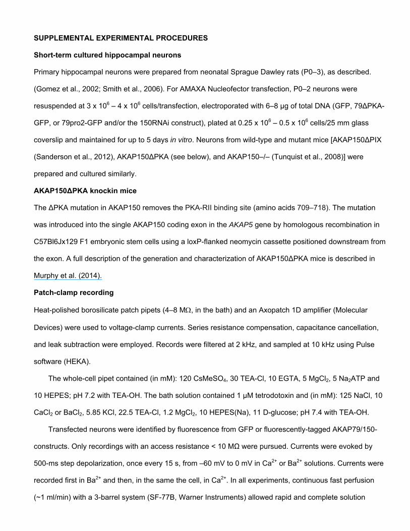
Short-term cultured hippocampal neurons
Primary hippocampal neurons were prepared from neonatal Sprague Dawley rats (P0–3), as described.
(Gomez et al., 2002; Smith et al., 2006). For AMAXA Nucleofector transfection, P0–2 neurons were
resuspended at 3 x 10⁶ – 4 x 10⁶ cells/transfection, electroporated with 6–8 μg of total DNA (GFP, 79∆PKA-
GFP, or 79pro2-GFP and/or the 150RNAi construct), plated at 0.25 x 10⁶ – 0.5 x 10⁶ cells/25 mm glass
coverslip and maintained for up to 5 days in vitro. Neurons from wild-type and mutant mice [AKAP150∆PIX
(Sanderson et al., 2012), AKAP150∆PKA (see below), and AKAP150–/– (Tunquist et al., 2008)] were
prepared and cultured similarly.
AKAP150∆PKA knockin mice
The ∆PKA mutation in AKAP150 removes the PKA-RII binding site (amino acids 709–718). The mutation
was introduced into the single AKAP150 coding exon in the AKAP5 gene by homologous recombination in
C57Bl6Jx129 F1 embryonic stem cells using a loxP-flanked neomycin cassette positioned downstream from
the exon. A full description of the generation and characterization of AKAP150∆PKA mice is described in
Murphy et al. (2014).
Patch-clamp recording
Heat-polished borosilicate patch pipets (4–8 M, in the bath) and an Axopatch 1D amplifier (Molecular
Devices) were used to voltage-clamp currents. Series resistance compensation, capacitance cancellation,
and leak subtraction were employed. Records were filtered at 2 kHz, and sampled at 10 kHz using Pulse
software (HEKA).
The whole-cell pipet contained (in mM): 120 CsMeSO4, 30 TEA-Cl, 10 EGTA, 5 MgCl2, 5 Na2ATP and
10 HEPES; pH 7.2 with TEA-OH. The bath solution contained 1 µM tetrodotoxin and (in mM): 125 NaCl, 10
CaCl2 or BaCl2, 5.85 KCl, 22.5 TEA-Cl, 1.2 MgCl2, 10 HEPES(Na), 11 D-glucose; pH 7.4 with TEA-OH.
Transfected neurons were identified by fluorescence from GFP or fluorescently-tagged AKAP79/150-
constructs. Only recordings with an access resistance < 10 MΩ were pursued. Currents were evoked by
500-ms step depolarization, once every 15 s, from –60 mV to 0 mV in Ca2+ or Ba2+ solutions. Currents were
recorded first in Ba2+ and then, in the same the cell, in Ca2+. In all experiments, continuous fast perfusion
(~1 ml/min) with a 3-barrel system (SF-77B, Warner Instruments) allowed rapid and complete solution

exchanges. L-type currents were isolated from other Ca2+ current types by treating neurons for 30 minutes
with the long-duration blockers of N- and P/Q-type channels, ω-CTx-GVIA (1 μM) and ω-CTx-MVIIC (5 μM),
and by holding neurons at –60 mV during recordings to inactivate R-type Ca2+ channels (Sochivko et al.,
2003; Tavalin et al., 2004; Oliveria et al., 2007). Because it was not practical to include N- and P/Q-channel
blockers in the fast perfusate owing to the excessive blocker quantities that would be required, neurons
were used for <1 hr after blocker treatment to minimize adulteration of L current as N- and P/Q-type
channels became unblocked (McCleskey et al., 1987; Sather et al., 1993; Oliveria et al., 2012).
Forskolin (5 µM), PKA inhibitor peptide 6-22 (PKI; 5 µM), or Ht31 peptide (0.5 – 100 µM) were applied
via the patch pipet solution. The acute effects of FK506 (5 μM), H89 (500 nM), and Sp-5,6-DCl-cBiMPS (10
or 100 μM) were examined by fast bath application. Forskolin, PKI, and FK506 were obtained from Sigma-
Aldrich, Sp-5,6-DCl-cBiMPS was obtained from ENZO Life Science, and Ht31 was obtained from
ResGen/Invitrogen.
Data analysis
To facilitate comparison of inactivation rate, Ca2+ and Ba2+ current records obtained from the same neuron
were normalized by peak inward amplitude. Inactivation time course was best-fit with a double exponential
function (PulseFit software; HEKA).The faster component represents CDI, and the slower corresponds to
voltage-dependent inaction. Statistical analyses (ANOVA, Bonferroni post-test) were carried out with
SigmaPlot 11 (Systat).

SUPPLEMENTAL REFERENCES Gomez, L.L., Alam, S., Smith, K.E., Horne, E., and Dell'Acqua, M.L. (2002). Regulation of A-kinase
anchoring protein 79/150-cAMP-dependent protein kinase postsynaptic targeting by NMDA receptor
activation of calcineurin and remodeling of dendritic actin. J. Neurosci. 22, 7027-7044.
McCleskey, E.W., Fox, A.P., Feldman, D.H., Cruz, L.J., Olivera, B.M., Tsien, R.W., and Yoshikami, D.
(1987). -Conotoxin: direct and persistent blockade of specific types of calcium channels in neurons
but not muscle. Proc. Natl. Acad. Sci. USA 84, 4327-4331.
Murphy, J.G., Sanderson, J.L., Gorski, J.A., Scott, J.D., Catterall, W.A., Sather, W.A., and Dell’Acqua, M.L.
(2014). AKAP-anchored PKA maintains neuronal L-type calcium channel activity and NFAT
transcriptional signaling. (Submitted to Cell Reports).
Sanderson, J.L., Gorski, J.A., Gibson, E.S., Lam, P., Freund, R.K., Chick, W.S., and Dell'Acqua, M.L.
(2012). AKAP150-anchored calcineurin regulates synaptic plasticity by limiting synaptic
incorporation of Ca2+-permeable AMPA receptors. J. Neurosci. 32, 15036-15052.
Sather, W.A., Tanabe, T., Zhang, J.F., Mori, Y., Adams, M.E., and Tsien, R.W. (1993). Distinctive
biophysical and pharmacological properties of class A (BI) calcium channel 1 subunits. Neuron 11,
291-303.
Smith, K.E., Gibson, E.S., and Dell'Acqua, M.L. (2006). cAMP-dependent protein kinase postsynaptic
localization regulated by NMDA receptor activation through translocation of an A-kinase anchoring
protein scaffold protein. J. Neurosci. 26, 2391-2402.
Sochivko, D., Chen, J., Becker, A., and Beck, H. (2003). Blocker-resistant Ca2+ currents in rat CA1
hippocampal pyramidal neurons. Neuroscience 116, 629-638.
Tavalin, S.J., Shepherd, D., Cloues, R.K., Bowden, S.E., and Marrion, N.V. (2004). Modulation of single
channels underlying hippocampal L-type current enhancement by agonists depends on the
permeant ion. J. Neurophysiol. 92, 824-837.
Tunquist, B.J., Hoshi, N., Guire, E.S., Zhang, F., Mullendorff, K., Langeberg, L.K., Raber, J., and Scott, J.D.
(2008). Loss of AKAP150 perturbs distinct neuronal processes in mice. Proc. Natl. Acad. Sci. USA
105, 12557-12562.


![Near-Membrane [Ca2+] Transients Resolved Using the Ca2+ Indicator FFP18](https://static.fdokumen.com/doc/165x107/631286873ed465f0570a4533/near-membrane-ca2-transients-resolved-using-the-ca2-indicator-ffp18.jpg)