Tetrodotoxin blocks L-type Ca2+ channels in canine ventricular cardiomyocytes
Upload
independentCategory
view
0download
0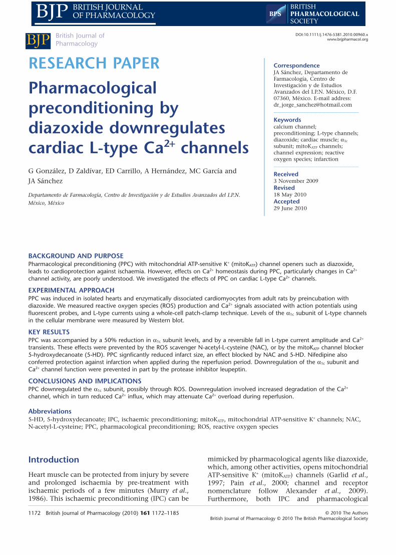
RESEARCH PAPERbph_960 1172..1185
Pharmacologicalpreconditioning bydiazoxide downregulatescardiac L-type Ca2+ channelsG González, D Zaldívar, ED Carrillo, A Hernández, MC García and
JA Sánchez
Departamento de Farmacología, Centro de Investigación y de Estudios Avanzados del I.P.N.
México, México
CorrespondenceJA Sánchez, Departamento deFarmacología, Centro deInvestigación y de EstudiosAvanzados del I.P.N. México, D.F.07360, México. E-mail address:dr_jorge_sanchez@hotmail.com----------------------------------------------------------------
Keywordscalcium channel;preconditioning; L-type channels;diazoxide; cardiac muscle; a1c
subunit; mitoKATP channels;channel expression; reactiveoxygen species; infarction----------------------------------------------------------------
Received3 November 2009Revised18 May 2010Accepted29 June 2010
BACKGROUND AND PURPOSEPharmacological preconditioning (PPC) with mitochondrial ATP-sensitive K+ (mitoKATP) channel openers such as diazoxide,leads to cardioprotection against ischaemia. However, effects on Ca2+ homeostasis during PPC, particularly changes in Ca2+
channel activity, are poorly understood. We investigated the effects of PPC on cardiac L-type Ca2+ channels.
EXPERIMENTAL APPROACHPPC was induced in isolated hearts and enzymatically dissociated cardiomyocytes from adult rats by preincubation withdiazoxide. We measured reactive oxygen species (ROS) production and Ca2+ signals associated with action potentials usingfluorescent probes, and L-type currents using a whole-cell patch-clamp technique. Levels of the a1c subunit of L-type channelsin the cellular membrane were measured by Western blot.
KEY RESULTSPPC was accompanied by a 50% reduction in a1c subunit levels, and by a reversible fall in L-type current amplitude and Ca2+
transients. These effects were prevented by the ROS scavenger N-acetyl-L-cysteine (NAC), or by the mitoKATP channel blocker5-hydroxydecanoate (5-HD). PPC signficantly reduced infarct size, an effect blocked by NAC and 5-HD. Nifedipine alsoconferred protection against infarction when applied during the reperfusion period. Downregulation of the a1c subunit andCa2+ channel function were prevented in part by the protease inhibitor leupeptin.
CONCLUSIONS AND IMPLICATIONSPPC downregulated the a1c subunit, possibly through ROS. Downregulation involved increased degradation of the Ca2+
channel, which in turn reduced Ca2+ influx, which may attenuate Ca2+ overload during reperfusion.
Abbreviations5-HD, 5-hydroxydecanoate; IPC, ischaemic preconditioning; mitoKATP, mitochondrial ATP-sensitive K+ channels; NAC,N-acetyl-L-cysteine; PPC, pharmacological preconditioning; ROS, reactive oxygen species
Introduction
Heart muscle can be protected from injury by severeand prolonged ischaemia by pre-treatment withischaemic periods of a few minutes (Murry et al.,1986). This ischaemic preconditioning (IPC) can be
mimicked by pharmacological agents like diazoxide,which, among other activities, opens mitochondrialATP-sensitive K+ (mitoKATP) channels (Garlid et al.,1997; Pain et al., 2000; channel and receptornomenclature follow Alexander et al., 2009).Furthermore, both IPC and pharmacological
BJP British Journal ofPharmacology
DOI:10.1111/j.1476-5381.2010.00960.xwww.brjpharmacol.org
1172 British Journal of Pharmacology (2010) 161 1172–1185 © 2010 The AuthorsBritish Journal of Pharmacology © 2010 The British Pharmacological Society
preconditioning (PPC) can be antagonized bymitoKATP channel blockers (Ardehali and O’Rourke,2005; Halestrap et al., 2007). Based on this pharma-cological evidence, mitoKATP channels are proposedto be central in protection of the heart muscle byIPC and PPC (Ardehali and O’Rourke, 2005; Hal-estrap et al., 2007). However, the possible involve-ment in IPC and PPC of other ion channels, such asthe L-type Ca2+ channel, a key element inexcitation–contraction coupling and Ca2+ homeo-stasis in the heart and other tissues (Bers, 2002), islargely unexplored. In smooth muscle, L-type chan-nels are involved in refilling intracellular stores, andthis is inhibited by high concentrations of diazoxide(Dessy and Godfraind, 1996). Recent evidence sug-gests that L-type channels may contribute to pacing-induced protection against anoxia-reoxygenation inthe embryonic heart (Bruchez et al., 2008).
In this study, we tested the hypothesis thatL-type Ca2+ channels are regulated during PPC. Wefound that preincubation with diazoxide drasticallyreduced the magnitude of L-type Ca2+ currents andintracellular Ca2+ transients. PPC also downregu-lated a1c, the principal subunit of the cardiac L-typeCa2+ channel (Catterall, 2000; Lacinová, 2005). Wepropose that PPC produced by diazoxide involves anincrease in reactive oxygen species (ROS) produc-tion that is involved in downregulation of the a1c
subunit via a protease-dependent process.
Methods
Preparation of heartsAll animal care and experimental procedures con-formed to protocols approved by the Division ofLaboratory Animal Units, Cinvestav-IPN, in compli-ance with federal law, federal statute and ConsejoNacional de Ciencia y Tecnología (CONACYT) regu-lations. Male Wistar rats (300–350 g) were anaesthe-tized with 50 mg·kg-1 of pentobarbital sodiuminjected intraperitoneally. A 500 U·kg-1 heparinsodium were also administered intraperitoneally.Hearts (1.5–1.7 g) were rapidly excised, arrested inmodified Krebs-Henseleit buffer (in mM): 117.8NaCl, 1.2 NaH2PO4, 6.0 KCl, 24.3 NaHCO3, 1.2MgSO4, 0.027 EDTA, 5.1 glucose and 1.6 CaCl2,gassed with 95% O2, 5% CO2 at pH 7.4), and per-fused in a Langendorff apparatus with an aorticcannula delivering 37 � 0.5°C buffer at 66 mmHg.Care was taken to avoid damage to the aorta. Thewhole procedure took ~ 30 s. Left ventricle (LV) pres-sure was continuously monitored and recorded byAxotape software (Axon Instruments, Foster City,CA, USA). The following parameters of cardiac func-tion were assessed: Left ventricle developed pressure
(LVDP) calculated as the difference between the sys-tolic and diastolic pressure of the LV, LV end-diastolic pressure (LVEDP), and the maximal (dP/dtmax) and minimal (dP/dtmin) value of the firstderivative of LV pressure. LVDP was monitored witha water-filled balloon inserted into the left ventricle,set to deliver diastolic pressure of 5–10 mmHg. Theresulting coronary flow was in the range of 8.7–14 ml·min-1·g-1. Diazoxide was added from a 1 Mstock solution in dimethyl sulphoxide (DMSO) to afinal DMSO concentration of less than 0.01%.
Isolation of ventricular myocytesVentricular myocytes were isolated as described pre-viously (Sánchez et al., 2001), with slight modifica-tions. In brief, hearts were perfused for 5 min at37°C with Ca2+-free Tyrode solution containing (inmM): 136 NaCl, 5.4 KCl, 1 MgCl2, 10 HEPES, and 11glucose plus heparin (10 U·ml-1, Sigma). Hearts wererecirculated for ~30 min with Tyrode solution with20–40 mM CaCl2, 0.1% bovine serum albumin (BSA),collagenase (Worthington, Lakewood, NJ, USA, typeII, 70 U·ml-1), and protease type XIV (Sigma,0.5 mg/100 ml). Ventricles were minced and shaken2–3 times at 45 r.p.m. for 7 min in the same solu-tion. Cells were filtered through a cell strainer(100 mm nylon BD Falcon) and centrifuged at 700¥ gfor 2 min. The pellet was resuspended for 90 min inTyrode solution containing (in mM): 136 NaCl, 5.4KCl, 1 CaCl2, 2 MgCl2, 10 HEPES, and 11 glucose,plus 1% BSA in control experiments, or in an iden-tical solution containing diazoxide (100 mM),5-hydroxydecanoate (5-HD; 100 mM), N-acetyl-L-cysteine (NAC; 2 or 4 mM) or leupeptin (100 mM)for the indicated time periods. Myocytes were usedimmediately.
Electrophysiological methodsL-type currents were recorded in dissociated adultventricular myocytes using a whole-cell patch-clamp technique as described elsewhere (Sánchezet al., 2001). Currents were recorded with an Axo-patch 200-A amplifier (Axon Instruments) using thepulse protocol shown in Figure 1A. Two consecutive-10 mV hyperpolarizing pulses, lasting 150 ms,were followed by a 150 ms depolarizing pulse ofpreselected amplitude. The sequence was repeated10 times, increasing the amplitude of the depolariz-ing pulse by +10 mV steps from a holding potentialof -40 mV. Hyperpolarizing currents were used tosubtract linear currents, and to measure membranecapacitance. Current records were digitized with aDigidata interface (Axon Instruments, Foster City,CA, USA) at 16 bit resolution. Data were analysedusing pCLAMP 8.0 (Axon Instruments) andin-house software.
BJPDiazoxide and Ca2+ channels
British Journal of Pharmacology (2010) 161 1172–1185 1173

Peak Ca2+ channel current values were fitted toequation (1) to describe current-voltage relation-ships of L-type currents.
I G V V V km m rev m= ⋅ −( ) + − )[ ]{ }max exp1 V (1)
Gmax is the maximum conductance, Vrev is the rever-sal potential, Vm is the membrane potential, V is thepotential for which G = 0.5 Gmax, and k is a measureof the steepness of the curve. Numerical formulaewere fitted to experimental data using a non-linear,least-squares algorithm.
The pipette solution contained (in mM): 100cesium aspartate, 20 CsCl, 20 TEACl, 2 Mg-ATP, 1.8MgCl2, 0.05 EGTA, and 5 HEPES, pH 7.2. The stan-dard bath saline was Tyrode solution plus 1% BSAfor stability of the electrophysiological recordings.We confirmed that BSA has no significant effects onL-type currents. In Tyrode solution with no BSA, theaverage value of peak currents at +10 mV was -4.37� 0.03 pA·pF-1 (n = 7). In the presence of BSA, at thesame potential, it was -4.61 � 0.58 pA·pF-1 (n = 38).No shifts in the current-voltage (I-V) curve wereobserved.
To characterize the effects of PPC on L-type cur-rents, intact cardiac myocytes were incubated incontrol solution or in solutions containing differentdrugs (indicated in the figures and legends) for thestated time period. After the incubation period wasterminated, cardiomyocytes were transferred to therecording chamber where a selected myocyte waspatch-clamped to record whole-cell membranecurrents. Experiments were performed at roomtemperature (23°C).
Measurement of Ca2+ transientsFluo-3 AM (Molecular Probes) was used to monitorthe levels of intracellular Ca2+. This dye undergoeslarge fluorescence changes upon Ca2+ binding, has alarge dynamic range and low compartmentalization(Thomas et al., 2000), and was previously used torecord Ca2+ signals in ventricular myocytes (Vicen-cio et al., 2006). Adult cardiomyocytes were loadedwith approximately 5 mM Fluo-3 AM in standardTyrode solution for 40 min at room temperature.After loading, cells were incubated in dye-free solu-tion for over 30 min to allow the conversion of thedye to its Ca2+-sensitive, free acid form. Dye-loadedcells were placed on a laminin-coated coverslip atthe bottom of a chamber on the stage of anOptiphot microscope (Nikon, Tokyo, Japan). Fluo-rescence emitted by stained myocytes, illuminatedepiscopically with monochromatic light at 485 nm,was filtered with a high-pass barrier filter (cut-offwavelength 535 nm) and detected with a low-noisephotodiode in a photovoltaic configuration.
Figure 1PPC reduces L-type currents. A, pulse protocol. B–D, experimentalprotocol and representative recordings of L-type Ca2+ currents at0 mV. B, representative L-type Ca2+ current after 90 min in controlsolution. C, myocyte after 90 min in diazoxide (100 mM). D, myocytepreincubated for 30 min in 5-HD (100 mM), then 90 min in 5-HDplus diazoxide (100 mM). E and subsequent panels show mean � SE.E, I-V relationship of peak L-type currents. At the indicated potentials,peak currents are shown from controls (Cont; n = 38); fromdiazoxide-incubated myocytes (Dzx; n = 29) and from 5-HD plusdiazoxide pretreated myocytes (Dzx + 5HD; n = 14). Differencesbetween diazoxide only and control data sets were statistically sig-nificant between -20 mV and +30 mV, and at +50 mV (P < 0.05). F:relationship between incubation time and peak L-type Ca2+ currents(�): peak L-type currents as a function of time in control solution (n= 15–38). Corresponding mean values from diazoxide-treated myo-cytes (n = 10–14); data were significantly different from control datafor t >10 min. Dotted line: mean amplitude of control L-type Ca2+
currents at t = 0. Solid line: best fit of a single exponential fordiazoxide-treated myocytes with t = 13.2 min. G, representativerecording of L-type Ba2+ currents at 0 mV after 90 min in controlsolution. H, myocyte after 90 min in diazoxide (100 mM). I, normal-ized recordings from G (black) and H (red). J, relationship betweenthe time constant of inactivation of L-type Ba2+ currents and voltageunder control (Cont) conditions and after diazoxide (Dzx). Valueswere significantly different at all potentials (n = 4–8).
BJP G González et al.
1174 British Journal of Pharmacology (2010) 161 1172–1185
Analogue signals were digitized as described aboveand sampled every 60 ms for 40 s. The basal fluores-cence (F) of the myocyte was recorded, and its meanvalue during a 300 ms interval prior to electricalstimulation was used to calculate Ca2+ signals asDF/F. No absolute value measurements were madefor intracellular Ca2+ concentration, as this studywas designed to detect temporal and relative pat-terns of Ca2+ transients. To induce Ca2+ transientsduring action potentials, myocytes were electricallystimulated with two extracellular platinum elec-trodes at a frequency of 0.3 Hz. Effects of PPC onCa2+ transients were determined with an experimen-tal procedure similar to the one used for electro-physiological experiments.
Measurement of ROS productionROS was measured using the cell-permeantfluorescent probe, 5-(and-6)-chloromethyl-2′,7′-dichlorodihydrofluorescein diacetate, acetyl ester(CM-H2DCFDA, Molecular Probes). Inside cells, thediacetate group of CM-H2DCFDA is cleaved by intra-cellular esterases, trapping the non-fluorescentproduct CM-H2DCFFH, which is oxidized to fluores-cent CM-H2DCF upon exposure to H2O2 or ·OH(Rijstenbil et al., 2000). Intensity is dependent onthe amount of generated ROS. The probe has excel-lent retention in live cells and is used to measureintracellular ROS in cardiomyocytes and other cells(Bodi et al., 2005). Fluorescence in arbitrary units at470 nm excitation and 515 nm emission was mea-sured for 200 ms in user-defined segments of cardi-omyocytes. The intensity of the emitted light wasdetected as described above and sampled at a fre-quency of 17 kHz.
Cardiomyocytes were loaded with 10 mMCM-H2DCFDA by incubation in the dark for 30 minat room temperature. The dye was added from astock solution in DMSO to result in a final DMSOconcentration of less than 0.1%. At this concentra-tion, fluorescence signals associated with ROS pro-duction are unaffected by the vehicle (Pasdois et al.,2008). Loaded myocytes were incubated in dye-freesolution for over 30 min to allow conversion of thedye into its ROS-sensitive form. At the end of theexperiments, dye-loaded myocytes were exposed toconstant excitation for 3–6 min to induce photooxi-dation of CM-H2DCFDA. Fluorescence valuesincreased with light exposure to a plateau represent-ing the total amount of CM-H2DCFDA available foroxidation within the exposed area of the myocyte.Immediately after dye loading, ROS production wasmeasured every 5 or 10 min for 30 min, either inmyocytes in Tyrode solution, with or without diaz-oxide (100 mM). For normalization, ROS fluores-
cence data are expressed relative to meanfluorescence at the beginning of the experiment.
Isolated heartsHearts were subjected to the following treatments:The control group was perfused for 90 min withKrebs-Henseleit buffer. The diazoxide-treated groupwas perfused for 90 min in Krebs-Henseleit buffercontaining 100 mM diazoxide. For the NAC plus dia-zoxide group, the ROS scavenger NAC was added toKrebs-Henseleit buffer at 4 mM, and hearts wereperfused for 15 min, then perfused for 90 min in thesame buffer with diazoxide. The NAC-only groupwas perfused for 90 min with Krebs-Henseleit bufferplus NAC. For the 5-HD plus diazoxide group, themitoKATP channel inhibitor 5-HD was added toKrebs-Henseleit buffer at 100 mM, and hearts wereperfused for 15 min, then perfused for 90 min in thesame buffer with diazoxide. The 5-HD-only groupwas perfused for 90 min with Krebs-Henseleit bufferplus 5-HD.
To assess protection against ischaemia, heartspreviously perfused for 90 min in Krebs-Henseleitbuffer or in solutions containing diazoxide and/orthe inhibitors as described above, and were sub-jected to global ischaemia for 30 min, followed by2 h of reperfusion with control solution. To assessthe effect of nifedipine on infarction, a group ofhearts was perfused for 60 min with Krebs-Henseleitbuffer, then perfused for 30 min in the same bufferwith nifedipine (30 nM). This was followed byglobal ischaemia for 30 min, then hearts were rep-erfused with the same buffer for 30 min and reper-fusion continued with Krebs-Henseleit buffer for90 min. Thereafter, hearts from all groups were dyedwith 2,3,5-triphenyltetrazolium chloride (TTC, at1%) for 10 min fixed overnight with paraformalde-hyde (4%), cut into 500 mm slices and photographedwith a digital camera attached to a microscope(Olympus). TTC produces coloured precipitates inthe presence of intact dehydrogenase enzymesystems, whereas necrotic areas lacking dehydroge-nase activity or sufficient concentrations of cofac-tors (e.g. NADH) fail to stain (Klein et al., 1981). TTCis widely used for estimating the size of myocardialinfarction areas (e.g. Garcia-Dorado et al., 1987). Inf-arcted areas were measured by the Analysis SoftImaging System (Olympus).
Measurement of LDHLactate dehydrogenase (LDH), an indicator of myo-cardial tissue injury, was assayed in coronary efflu-ent using a coupled spectrophotometric techniquefrom a kit (Cayman Chemical Co., USA) accordingto the manufacturer’s instructions. LDH activity wasmeasured with a coupled two-step reaction, the
BJPDiazoxide and Ca2+ channels
British Journal of Pharmacology (2010) 161 1172–1185 1175
second step of which was the reduction of a tetra-zolium salt to formazan, which absorbs at 490 nm.The amount of LDH released in the first 15 min ofreperfusion was calculated by integrating the areaunderneath individual time course curves. Tocompare data from different experiments, normal-ization used the ratio of the LDH value from PPC- ornifedipine-treated ischaemic hearts to LDH fromcontrol ischaemic hearts.
Western blottingMembrane fractions were obtained as described(Saada et al., 2005) by Dounce homogenization of-80°C frozen pulverized rat ventricles in 10 volumesof homogenization medium containing (in mM):250 sucrose, 50 MOPS, and 2 EGTA, at pH 7.4, towhich protease inhibitors 1 mM benzamidine, 1 mMleupeptin, 0.1 mM aprotinin, 1 mM pepstatin A,0.2 mM PMSF, and 0.4 mM pefabloc were added.Homogenates were centrifuged at 1000¥ g for10 min, then at 5000¥ g for 10 min, and finally at100 000¥ g for 1 h. The final pellet was resuspendedin homogenization medium and stored at -80°C.Protein content was measured with the Bradfordassay.
In some experiments, dissociated myocytes wereused for Western blotting by resuspension in lysisbuffer containing: 20 mM Tris (pH 7.5), 100 mMNaCl, 1% Triton X-100 and 1 mM PMSF with pro-tease inhibitors 1 mM benzamidine, 1 mM leupep-tin, 0.1 mM aprotinin, 1 mM pepstatin A, and0.4 mM pefabloc. Lysis was achieved by continuousagitation for 60 min at 4°C followed by five cycles ofsonication. Samples were centrifuged at 13 000¥ gfor 10 min at 4°C and the soluble fraction used forWestern blots. Protein content was measured withan RC DC Protein Assay kit (Bio-Rad) following themanufacturer’s instructions.
Western blotting used 50 mg of membrane prepa-rations (or 100 mg when myocytes were used)applied to 7.5% dodecyl sulphate-polyacrylamide(SDS-PAGE) gels to identify a1c subunits, and 30 mg(or 40 mg when myocytes were used) on 13.5% gel toidentify a-actin. Loading buffer and gels for a1c
subunit immunoblots contained 8 M urea. Proteinswere transferred to nitrocellulose membranes andblocked with 4.5% nonfat dried milk in phosphatebuffered saline (PBS), before probing with primaryanti-a1c antibodies (ACC-003, Alomone Laborato-ries, Jerusalem, Israel) at 1:100 dilution. Monoclonalantibody (a gift from Dr J.M. Hernández, from theDepartment of Cell Biology, Cinvestav) againsta-actin (42 kDa) was also used at 1:100 (or 1:200when myocytes were used). After washing, mem-branes were incubated with an anti-rabbit (1:8000)or an anti-mouse (1:8000) horseradish peroxidase
conjugate (Zymed) as secondary antibodies anddetected by enhanced chemiluminescence using aSuper Signal West Pico Chemiluminescent substrate(Pierce). To measure the densities of the a1c subunitand a-actin bands, film was scanned at 300 dpi, andimages were inverted. The mean value of the inten-sity of each band and its corresponding area weremeasured with Photoshop (CS3), and absolute den-sities were calculated as the product of these twoparameters. For normalization, the absolute densityof a1c subunit bands was divided by the density forcorresponding a-actin bands. Relative densities werethe ratio between the density of the a1c subunitband from a treated heart, and the density of the a1c
subunit band from a control heart obtained in par-allel experiments.
Data analysisData are expressed as the mean � standard error (s.e.mean). Analysis used independent t-tests or ANOVA,and Tukey’s or Dunnett’s post-test, as appropriate. AP < 0.05 was considered to be statistically significant.
MaterialsDiazoxide, NAC, 5HD, nifedipine, TTC, proteaseinhibitors, Tiron and other chemicals were suppliedby Sigma Aldrich (USA) and leupeptin was fromRoche (USA).
Results
Effect of diazoxide on L-type Ca2+
channel currentsIncubation with diazoxide led to a significant reduc-tion in Ca2+-current amplitude. Figure 1B–D showsrepresentative recordings during step depolariza-tions to 0 mV. To allow comparisons among experi-ments, membrane current values were normalizedto unit capacitance. L-type Ca2+ currents undercontrol conditions reached a peak of ~ -5 pA·pF-1,approximately 10 ms after pulse onset. The currentwas not sustained but inactivated during the pulse.These characteristics are typical of L-type Ca2+ cur-rents of adult rat cardiac myocytes (Gomez et al.,1994; Striessnig, 1999; Bodi et al., 2005). Diazoxideproduced a significant decrease in current amplitudeto approximately 30% of the value observed in con-trols. The effects of diazoxide on L-type Ca2+ cur-rents developed within minutes, leading to aprogressive decline in current magnitude that wasalmost complete after 30 min (Figure 1F). Diazoxideeffects were reversible after 60 min of wash-out withcontrol solution (data not shown). At the concen-tration used, diazoxide is a selective opener of ATP-sensitive K+ channels in the inner mitochondrial
BJP G González et al.
1176 British Journal of Pharmacology (2010) 161 1172–1185
membrane (Garlid et al., 1996), so we tested theeffects of the selective mitoKATP channel blocker5-HD (Hu et al., 1999) on the diazoxide effect. The5-HD completely antagonized the reduction inL-type Ca2+ currents produced by diazoxide, at allpotentials tested (Figure 1E).
The decrease in current amplitude might involvean increase in Ca2+-dependent inactivation of L-typechannels by diazoxide. To explore this possibility,we replaced Ca2+ with an equimolar amount of Ba2+
as the charge carrier. As expected, these currentswere larger than Ca2+ currents because the channelsare more permeable to Ba2+ (Figure 1G). In the pres-ence of diazoxide, Ba2+ peak currents were greatlydecreased (Figure 1H), with a drop in current mag-nitude that was similar to that seen with Ca2+ as thecharge carrier, indicating that Ca2+-dependent inac-tivation appears to play no role in the effects ofdiazoxide on the Ca2+ currents. On the other hand,Ba2+ current recordings revealed an increase involtage-dependent inactivation of L-type currentsby diazoxide. This was manifested as a significantacceleration in Ba2+ current decay, seen whencurrent traces were normalized (Figure 1I). To esti-mate current time courses, the inactivation phase ofthe currents was fitted to a single exponential. Thetime constant (t) at different potentials was dis-tinctly smaller in diazoxide-treated myocytes(Figure 1J).
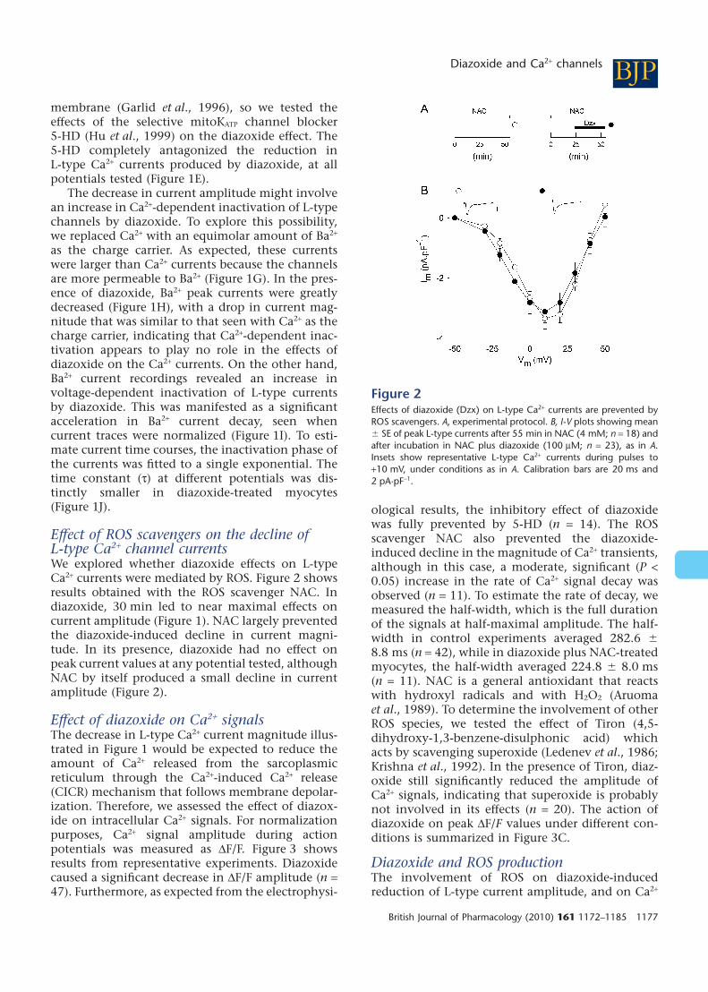
Effect of ROS scavengers on the decline ofL-type Ca2+ channel currentsWe explored whether diazoxide effects on L-typeCa2+ currents were mediated by ROS. Figure 2 showsresults obtained with the ROS scavenger NAC. Indiazoxide, 30 min led to near maximal effects oncurrent amplitude (Figure 1). NAC largely preventedthe diazoxide-induced decline in current magni-tude. In its presence, diazoxide had no effect onpeak current values at any potential tested, althoughNAC by itself produced a small decline in currentamplitude (Figure 2).
Effect of diazoxide on Ca2+ signalsThe decrease in L-type Ca2+ current magnitude illus-trated in Figure 1 would be expected to reduce theamount of Ca2+ released from the sarcoplasmicreticulum through the Ca2+-induced Ca2+ release(CICR) mechanism that follows membrane depolar-ization. Therefore, we assessed the effect of diazox-ide on intracellular Ca2+ signals. For normalizationpurposes, Ca2+ signal amplitude during actionpotentials was measured as DF/F. Figure 3 showsresults from representative experiments. Diazoxidecaused a significant decrease in DF/F amplitude (n =47). Furthermore, as expected from the electrophysi-
ological results, the inhibitory effect of diazoxidewas fully prevented by 5-HD (n = 14). The ROSscavenger NAC also prevented the diazoxide-induced decline in the magnitude of Ca2+ transients,although in this case, a moderate, significant (P <0.05) increase in the rate of Ca2+ signal decay wasobserved (n = 11). To estimate the rate of decay, wemeasured the half-width, which is the full durationof the signals at half-maximal amplitude. The half-width in control experiments averaged 282.6 �8.8 ms (n = 42), while in diazoxide plus NAC-treatedmyocytes, the half-width averaged 224.8 � 8.0 ms(n = 11). NAC is a general antioxidant that reactswith hydroxyl radicals and with H2O2 (Aruomaet al., 1989). To determine the involvement of otherROS species, we tested the effect of Tiron (4,5-dihydroxy-1,3-benzene-disulphonic acid) whichacts by scavenging superoxide (Ledenev et al., 1986;Krishna et al., 1992). In the presence of Tiron, diaz-oxide still significantly reduced the amplitude ofCa2+ signals, indicating that superoxide is probablynot involved in its effects (n = 20). The action ofdiazoxide on peak DF/F values under different con-ditions is summarized in Figure 3C.
Diazoxide and ROS productionThe involvement of ROS on diazoxide-inducedreduction of L-type current amplitude, and on Ca2+
Figure 2Effects of diazoxide (Dzx) on L-type Ca2+ currents are prevented byROS scavengers. A, experimental protocol. B, I-V plots showing mean� SE of peak L-type currents after 55 min in NAC (4 mM; n = 18) andafter incubation in NAC plus diazoxide (100 mM; n = 23), as in A.Insets show representative L-type Ca2+ currents during pulses to+10 mV, under conditions as in A. Calibration bars are 20 ms and2 pA·pF-1.
BJPDiazoxide and Ca2+ channels
British Journal of Pharmacology (2010) 161 1172–1185 1177
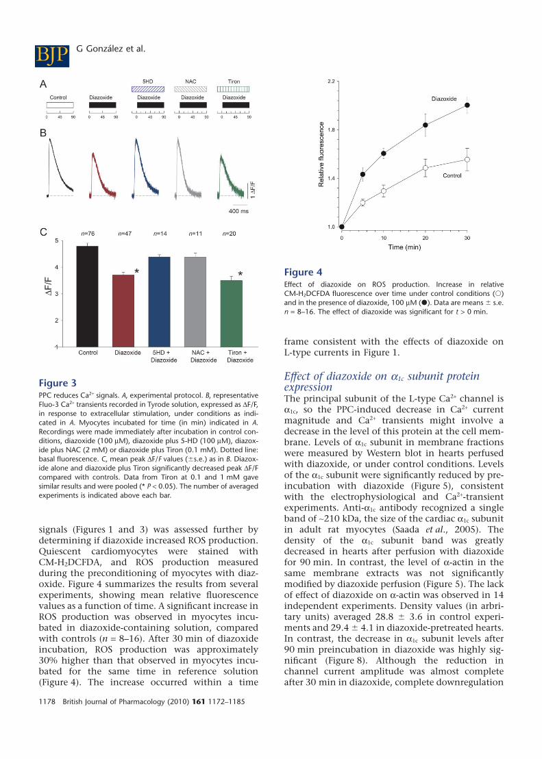
signals (Figures 1 and 3) was assessed further bydetermining if diazoxide increased ROS production.Quiescent cardiomyocytes were stained withCM-H2DCFDA, and ROS production measuredduring the preconditioning of myocytes with diaz-oxide. Figure 4 summarizes the results from severalexperiments, showing mean relative fluorescencevalues as a function of time. A significant increase inROS production was observed in myocytes incu-bated in diazoxide-containing solution, comparedwith controls (n = 8–16). After 30 min of diazoxideincubation, ROS production was approximately30% higher than that observed in myocytes incu-bated for the same time in reference solution(Figure 4). The increase occurred within a time
frame consistent with the effects of diazoxide onL-type currents in Figure 1.
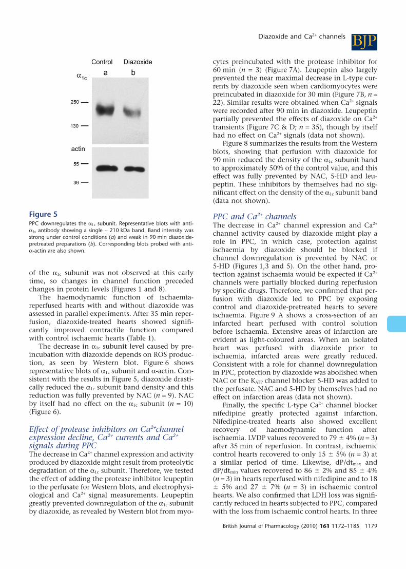
Effect of diazoxide on a1c subunit proteinexpressionThe principal subunit of the L-type Ca2+ channel isa1c, so the PPC-induced decrease in Ca2+ currentmagnitude and Ca2+ transients might involve adecrease in the level of this protein at the cell mem-brane. Levels of a1c subunit in membrane fractionswere measured by Western blot in hearts perfusedwith diazoxide, or under control conditions. Levelsof the a1c subunit were significantly reduced by pre-incubation with diazoxide (Figure 5), consistentwith the electrophysiological and Ca2+-transientexperiments. Anti-a1c antibody recognized a singleband of ~210 kDa, the size of the cardiac a1c subunitin adult rat myocytes (Saada et al., 2005). Thedensity of the a1c subunit band was greatlydecreased in hearts after perfusion with diazoxidefor 90 min. In contrast, the level of a-actin in thesame membrane extracts was not significantlymodified by diazoxide perfusion (Figure 5). The lackof effect of diazoxide on a-actin was observed in 14independent experiments. Density values (in arbri-tary units) averaged 28.8 � 3.6 in control experi-ments and 29.4 � 4.1 in diazoxide-pretreated hearts.In contrast, the decrease in a1c subunit levels after90 min preincubation in diazoxide was highly sig-nificant (Figure 8). Although the reduction inchannel current amplitude was almost completeafter 30 min in diazoxide, complete downregulation
Figure 3PPC reduces Ca2+ signals. A, experimental protocol. B, representativeFluo-3 Ca2+ transients recorded in Tyrode solution, expressed as DF/F,in response to extracellular stimulation, under conditions as indi-cated in A. Myocytes incubated for time (in min) indicated in A.Recordings were made immediately after incubation in control con-ditions, diazoxide (100 mM), diazoxide plus 5-HD (100 mM), diazox-ide plus NAC (2 mM) or diazoxide plus Tiron (0.1 mM). Dotted line:basal fluorescence. C, mean peak DF/F values (�s.e.) as in B. Diazox-ide alone and diazoxide plus Tiron significantly decreased peak DF/Fcompared with controls. Data from Tiron at 0.1 and 1 mM gavesimilar results and were pooled (* P < 0.05). The number of averagedexperiments is indicated above each bar.
Figure 4Effect of diazoxide on ROS production. Increase in relativeCM-H2DCFDA fluorescence over time under control conditions (�)and in the presence of diazoxide, 100 mM (�). Data are means � s.e.n = 8–16. The effect of diazoxide was significant for t > 0 min.
BJP G González et al.
1178 British Journal of Pharmacology (2010) 161 1172–1185
of the a1c subunit was not observed at this earlytime, so changes in channel function precededchanges in protein levels (Figures 1 and 8).
The haemodynamic function of ischaemia-reperfused hearts with and without diazoxide wasassessed in parallel experiments. After 35 min reper-fusion, diazoxide-treated hearts showed signifi-cantly improved contractile function comparedwith control ischaemic hearts (Table 1).
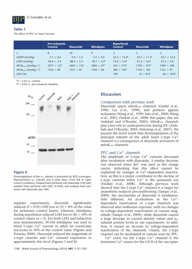
The decrease in a1c subunit level caused by pre-incubation with diazoxide depends on ROS produc-tion, as seen by Western blot. Figure 6 showsrepresentative blots of a1c subunit and a-actin. Con-sistent with the results in Figure 5, diazoxide drasti-cally reduced the a1c subunit band density and thisreduction was fully prevented by NAC (n = 9). NACby itself had no effect on the a1c subunit (n = 10)(Figure 6).
Effect of protease inhibitors on Ca2+channelexpression decline, Ca2+ currents and Ca2+
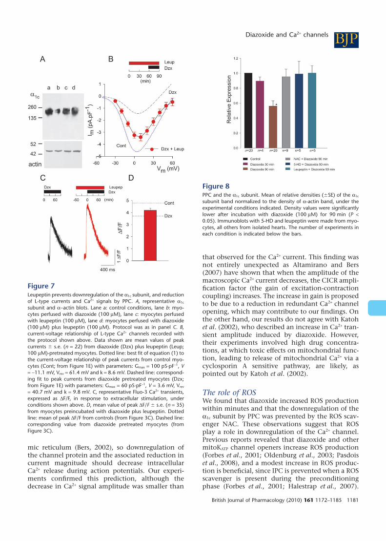
signals during PPCThe decrease in Ca2+ channel expression and activityproduced by diazoxide might result from proteolyticdegradation of the a1c subunit. Therefore, we testedthe effect of adding the protease inhibitor leupeptinto the perfusate for Western blots, and electrophysi-ological and Ca2+ signal measurements. Leupeptingreatly prevented downregulation of the a1c subunitby diazoxide, as revealed by Western blot from myo-
cytes preincubated with the protease inhibitor for60 min (n = 3) (Figure 7A). Leupeptin also largelyprevented the near maximal decrease in L-type cur-rents by diazoxide seen when cardiomyocytes werepreincubated in diazoxide for 30 min (Figure 7B, n =22). Similar results were obtained when Ca2+ signalswere recorded after 90 min in diazoxide. Leupeptinpartially prevented the effects of diazoxide on Ca2+
transients (Figure 7C & D; n = 35), though by itselfhad no effect on Ca2+ signals (data not shown).
Figure 8 summarizes the results from the Westernblots, showing that perfusion with diazoxide for90 min reduced the density of the a1c subunit bandto approximately 50% of the control value, and thiseffect was fully prevented by NAC, 5-HD and leu-peptin. These inhibitors by themselves had no sig-nificant effect on the density of the a1c subunit band(data not shown).
PPC and Ca2+ channelsThe decrease in Ca2+ channel expression and Ca2+
channel activity caused by diazoxide might play arole in PPC, in which case, protection againstischaemia by diazoxide should be blocked ifchannel downregulation is prevented by NAC or5-HD (Figures 1,3 and 5). On the other hand, pro-tection against ischaemia would be expected if Ca2+
channels were partially blocked during reperfusionby specific drugs. Therefore, we confirmed that per-fusion with diazoxide led to PPC by exposingcontrol and diazoxide-pretreated hearts to severeischaemia. Figure 9 A shows a cross-section of aninfarcted heart perfused with control solutionbefore ischaemia. Extensive areas of infarction areevident as light-coloured areas. When an isolatedheart was perfused with diazoxide prior toischaemia, infarcted areas were greatly reduced.Consistent with a role for channel downregulationin PPC, protection by diazoxide was abolished whenNAC or the KATP channel blocker 5-HD was added tothe perfusate. NAC and 5-HD by themselves had noeffect on infarction areas (data not shown).
Finally, the specific L-type Ca2+ channel blockernifedipine greatly protected against infarction.Nifedipine-treated hearts also showed excellentrecovery of haemodynamic function afterischaemia. LVDP values recovered to 79 � 4% (n = 3)after 35 min of reperfusion. In contrast, ischaemiccontrol hearts recovered to only 15 � 5% (n = 3) ata similar period of time. Likewise, dP/dtmax anddP/dtmin values recovered to 86 � 2% and 85 � 4%(n = 3) in hearts reperfused with nifedipine and to 18� 5% and 27 � 7% (n = 3) in ischaemic controlhearts. We also confirmed that LDH loss was signifi-cantly reduced in hearts subjected to PPC, comparedwith the loss from ischaemic control hearts. In three
Figure 5PPC downregulates the a1c subunit. Representative blots with anti-a1c antibody showing a single ~ 210 kDa band. Band intensity wasstrong under control conditions (a) and weak in 90 min diazoxide-pretreated preparations (b). Corresponding blots probed with anti-a-actin are also shown.
BJPDiazoxide and Ca2+ channels
British Journal of Pharmacology (2010) 161 1172–1185 1179
separate experiments, diazoxide significantlyreduced (P < 0.05) LDH loss to 33 � 8% of the valuefor ischaemic control hearts. Likewise, nifedipineduring reperfusion reduced LDH loss to 46 � 10% ofcontrol values (n = 3). For both LDH and infarctionarea measurements, 30 nM nifedipine was used toblock L-type Ca2+ current in adult rat ventricularmyocytes to 50% of the control value (Pignier andPotreau, 2000). Diazoxide reduced the magnitude ofL-type currents and Ca2+ channel expression toapproximately this level (Figures 1 and 8).
Discussion
Comparison with previous workDiazoxide opens mitoKATP channels (Garlid et al.,1996; Liu et al., 1998), and protects againstischaemia (Wang et al., 1999; Sato et al., 2000; Wanget al., 2001; Pasdois et al., 2008; this paper; also seeArdehali and O’Rourke, 2005). MitoKATP channelsplay a key role in cardioprotection during IPC (Arde-hali and O’Rourke, 2005; Halestrap et al., 2007). Wepresent the novel result that downregulation of theprincipal subunit of the adult heart L-type Ca2+
channel is a consequence of diazoxide activation ofmitoKATP channels.
PPC and Ca2+ channelsThe amplitude of L-type Ca2+ currents decreasedafter incubation with diazoxide. A similar decreasewas observed when Ba2+ was used as the chargecarrier, indicating that this effect cannot beexplained by changes in Ca2+-dependent inactiva-tion, as this is a major contributor to the decline ofL-type currents when Ca2+ is the permeant ion(Findlay et al., 2008). Although previous workshowed that the L-type Ca2+ channel is a target foranaesthetic-induced preconditioning (Tampo et al.,2009), the mechanism of diazoxide is fundamen-tally different. An acceleration in the Ca2+-dependent inactivation of L-type channels wasobserved after isoflurane exposure, with no changesin voltage-dependent inactivation or current mag-nitude (Tampo et al., 2009), while diazoxide causeda large decrease in current density values and a1c
subunit protein levels in our experiments. In addi-tion, it caused an increase in voltage-dependentinactivation of the channels. Clearly, the L-typechannel can be modulated in various ways by PPC.
Ca2+ entry via the L-type Ca2+ channel is thedominant Ca2+ source for the CICR of the sarcoplas-
Table 1The effect of PPC on heart function
Pre-ischaemic ReperfusedControl Diazoxide Nifedipine Control Diazoxide Nifedipine
n 8 10 9 3 3 3
LVEDP (mmHg) 2.1 � 0.6 3.9 � 1.2 3.5 � 6.0 52.5 � 12.6* 25.5 � 11.5* 24.1 � 12.2
LVDP (mmHg) 96.6 � 3.4 88.5 � 5.3 78.7 � 4.5# 14.9 � 5.6* 61.2 � 4.6* 57.2 � 4.9
dP/dtmax (mmHg·s-1) 2477 � 127 2467 � 132 1805 � 67# 454 � 174* 1720 � 357* 1494 � 100
dP/dtmin (mmHg·s-1) 1532 � 89 1531 � 81 1305 � 64 384 � 124* 1164 � 182 1131 � 97
LDH (%) 100 33 � 8.0# 46 � 10.0#
#P < 0.05 vs. control.*P < 0.05 vs. pre-ischaemic baseline.
Figure 6Downregulation of the a1c subunit is prevented by ROS scavengers.Representative a1c subunit and a-actin blots. From left to right:control conditions, isolated heart perfused with diazoxide (100 mM),isolated heart perfused with NAC (4 mM), and isolated heart per-fused with diazoxide plus NAC.
BJP G González et al.
1180 British Journal of Pharmacology (2010) 161 1172–1185
mic reticulum (Bers, 2002), so downregulation ofthe channel protein and the associated reduction incurrent magnitude should decrease intracellularCa2+ release during action potentials. Our experi-ments confirmed this prediction, although thedecrease in Ca2+ signal amplitude was smaller than
that observed for the Ca2+ current. This finding wasnot entirely unexpected as Altamirano and Bers(2007) have shown that when the amplitude of themacroscopic Ca2+ current decreases, the CICR ampli-fication factor (the gain of excitation-contractioncoupling) increases. The increase in gain is proposedto be due to a reduction in redundant Ca2+ channelopening, which may contribute to our findings. Onthe other hand, our results do not agree with Katohet al. (2002), who described an increase in Ca2+ tran-sient amplitude induced by diazoxide. However,their experiments involved high drug concentra-tions, at which toxic effects on mitochondrial func-tion, leading to release of mitochondrial Ca2+ via acyclosporin A sensitive pathway, are likely, aspointed out by Katoh et al. (2002).
The role of ROSWe found that diazoxide increased ROS productionwithin minutes and that the downregulation of thea1c subunit by PPC was prevented by the ROS scav-enger NAC. These observations suggest that ROSplay a role in downregulation of the Ca2+ channel.Previous reports revealed that diazoxide and othermitoKATP channel openers increase ROS production(Forbes et al., 2001; Oldenburg et al., 2003; Pasdoiset al., 2008), and a modest increase in ROS produc-tion is beneficial, since IPC is prevented when a ROSscavenger is present during the preconditioningphase (Forbes et al., 2001; Halestrap et al., 2007).
Figure 7Leupeptin prevents downregulation of the a1c subunit, and reductionof L-type currents and Ca2+ signals by PPC. A, representative a1c
subunit and a-actin blots. Lane a: control conditions, lane b: myo-cytes perfused with diazoxide (100 mM), lane c: myocytes perfusedwith leupeptin (100 mM), lane d: myocytes perfused with diazoxide(100 mM) plus leupeptin (100 mM). Protocol was as in panel C. B,current-voltage relationship of L-type Ca2+ channels recorded withthe protocol shown above. Data shown are mean values of peakcurrents � s.e. (n = 22) from diazoxide (Dzx) plus leupeptin (Leup;100 mM)-pretreated myocytes. Dotted line: best fit of equation (1) tothe current-voltage relationship of peak currents from control myo-cytes (Cont; from Figure 1E) with parameters: Gmax = 100 pS·pF-1, V= -11.1 mV, Vrev = 61.4 mV and k = 8.6 mV. Dashed line: correspond-ing fit to peak currents from diazoxide pretreated myocytes (Dzx;from Figure 1E) with parameters: Gmax = 60 pS·pF-1, V = 3.6 mV, Vrev
= 40.7 mV and k = 9.8 mV. C, representative Fluo-3 Ca2+ transients,expressed as DF/F, in response to extracellular stimulation, underconditions shown above. D, mean value of peak DF/F � s.e. (n = 35)from myocytes preincubated with diazoxide plus leupeptin. Dottedline: mean of peak DF/F from controls (from Figure 3C). Dashed line:corresponding value from diazoxide pretreated myocytes (fromFigure 3C).
Figure 8PPC and the a1c subunit. Mean of relative densities (�SE) of the a1c
subunit band normalized to the density of a-actin band, under theexperimental conditions indicated. Density values were significantlylower after incubation with diazoxide (100 mM) for 90 min (P <0.05). Immunoblots with 5-HD and leupeptin were made from myo-cytes, all others from isolated hearts. The number of experiments ineach condition is indicated below the bars.
BJPDiazoxide and Ca2+ channels
British Journal of Pharmacology (2010) 161 1172–1185 1181

The production of free radicals after the opening ofmitoKATP channels is proposed to trigger the precon-ditioned state (Pain et al., 2000). However, theincrease in ROS production by diazoxide is not nec-essarily mediated by mitoKATP channel opening, asdescribed by Dröse et al. (2006). The targets for ROShave not been fully identified, but oxygen radicalsare proposed to contribute to IPC by activatingprotein kinase C (Baines et al., 1997). ROS are likelyto have several targets in preconditioning, includingthe Ca2+ channel, according to our results. In agree-ment, free radicals decrease the number of
[H3]-nitrendipine binding sites in isolated heartmembranes (Kaneko et al., 1989).
Physiological significance of channeldownregulationLong periods of ischaemia result in large increases incytosolic Ca2+ concentration during reperfusion (Leeand Allen, 1992) and this rise is generally thought toprecede muscle damage. Furthermore, reduction inthis increase improves myocardial survival (seeMurphy and Steenbergen, 2008). Diazoxide-induceddecrease in Ca2+ influx and subsequent downregula-tion of the a1c subunit may, therefore, contribute tocardioprotection. Normally, Ca2+ efflux in heart cellsis predominantly by the Na+/Ca2+ exchangemechanism, with a minor contribution fromthe sarcolemmal Ca2+ pump (Bers, 2002). Duringischaemia-reperfusion, the deleterious increase inintracellular Ca2+ concentration appears to resultfrom the reverse operation of the Na+/Ca2+ exchangemechanism associated with intracellular acidosisand membrane depolarization (Murphy and Steen-bergen, 2008). Factors that diminish the increase inintracellular Ca2+ would be expected to attenuatemyocardial injury, so downregulation of Ca2+ chan-nels should be beneficial since, even if the Na+/Ca2+
exchange mechanism is not extruding Ca2+ as effi-ciently as under normal conditions, reduced Ca2+
channel influx would decrease Ca2+ overload. Ourresults agree with this as we observed protectionagainst infarction with nifedipine, a selectiveblocker of L-type Ca2+ channels. Furthermore, pre-venting channel downregulation with NAC or 5-HDresulted in lack of protection.
The role of L-type Ca2+ channels in altering Ca2+
influx as a mechanism of infarct size reductionduring preconditioning is unclear. Wallbridge et al.(1996) described no changes in infarct size whennisoldipine was administered to pigs by intravenousinfusion through the ischaemic period. Likewise,LDH levels in the coronary effluent of Langendorff-perfused rat hearts subjected to global ischaemia didnot change when Ca2+ channels were blocked byinfusion with verapamil, with washing before theischaemic-reperfusion period (Miyawaki et al.,1996). Mocanu et al. (1999) found that amlodipinedoes not reduce infarct size when administered inthe perfusate until just after coronary occlusion. Inall these examples, Ca2+ channel blockers werewashed out before reperfusion, so Ca2+ influx, andconsequently Ca2+ overload, remained unalteredduring this period. In contrast, we infused nife-dipine up to 30 minutes after the start of reperfu-sion, to reduce the influx of Ca2+ when Ca2+ overloadtakes place. A relevant finding is that verapamil andthe Ca2+ channel blocker Ro-40-5967 significantly
Figure 9PPC and infarction areas. A–E, infarction areas (light areas) aftersevere ischaemia in cross-sections of isolated ventricles under condi-tions as indicated above. Numbers are time in minutes. Concentra-tions were 100 mM diazoxide, 4 mM NAC, 100 mM 5-HD, and 30 nMnifedipine. Ventricles were dyed with 2,3,5-triphenyltetrazoliumchloride. F, infarction areas under indicated conditions correspond-ing to panels A–E. Data are the mean relative infarcted area (�s.e.)with the number of experiments indicated below. Infarcted areavalues from hearts perfused with diazoxide and nifedipine weresignificantly lower than those from control hearts (*P < 0.05).
BJP G González et al.
1182 British Journal of Pharmacology (2010) 161 1172–1185

reduced infarct size after local ischaemia in dogs,when the drugs were administered before andduring ischaemia, and through the first 25 min ofreperfusion (Vander Heide et al., 1994). Finally, con-tinuous administration of nifedipine and other Ca2+
channel blockers improved postischaemic recoveryof isolated guinea pig hearts (Becker and Möbert,1999).
MitoKATP channels and L-type Ca2+ channelsOur experiments indicated that downregulation ofthe Ca2+ channel followed the opening of mitoKATP
channels. MitoKATP channels are suggested to bedownstream regulators of myocardial protectionwith beneficial effects lasting into the reperfusionperiod (Fryer et al., 2001). After the ischaemicperiod, the increase in intracellular Ca2+ concentra-tion is generally thought to be an early event inreperfusion (Murphy and Steenbergen, 2008). Thisdoes not necessarily argue against downregulationof the Ca2+ channel by diazoxide as a factor in Ca2+
overload reduction, since we found that onlyminutes are required for the reduction in Ca2+
channel current amplitude and channel downregu-lation. Therefore, a significant drop in the fractionof active channels may have already occurred at thebeginning of reperfusion after PPC, thus reducingthe Ca2+ overload.
Changes in channel surface expressionOur results provide evidence for the involvement ofa protease in downregulation of cardiac L-typechannels during PPC. A single band correspondingto the intact a1c subunit decreased in density in ourimmunoblots, with no cleaved proteolytic productscontaining the epitope apparent in the membranefraction. This does not rule out the possibility thatmultiple regions of the a1c subunit are cleaved byPPC. In fact, we observed changes in channel activ-ity at times earlier than those observed by immuno-blot, suggesting that the integrity of the channel isalready being compromised. The particular proteaseinvolved and the exact mechanism of proteolysisremain to be determined. A possible candidate iscalpain, a Ca2+-dependent protease. The 10a isoformis the most abundant calpain, and is expressed inheart (Goll et al., 2003). Calpain is fully inhibited byleupeptin at 100 mM, and is activated during inhibi-tion of metabolism in cardiomyocytes (Atsma et al.,1995). Furthermore, calpain-dependent proteolyticevents take place in rat hearts after brief ischaemicinsults (Yoshida et al., 1995) and calpains areinvolved in degradation of ryanodine receptorsduring cardiac ischaemic reperfusion (Pedrozo et al.,2010).
Rapid degradation of proteins follows oxidativestress by ROS and oxidative modifications areaccompanied by protein malfunction, possiblythrough direct modification in one of the proteindomains required for function (Jung et al., 2007).Slightly oxidized proteins first show decreasedactivity and, after being subsequently defolded,they become ideal targets for degradation (Junget al., 2007). The observation that the decrease inthe amplitude of L-type currents preceded thedecrease in a1c subunit expression is consistentwith this view. The a1c subunit of the L-type Ca2+
channel contains many cysteine residues (Hool,2008), likely to be targets of redox modification ofproteins, as free thiols can easily react with ROS(Hool, 2008). Furthermore, we observed that cur-rents not only decreased in size by diazoxide asexpected by downregulation of the a1c subunit butalso showed an enhanced voltage-dependent inac-tivation indicating alterations in channel function.Increased inactivation is expected to reducecurrent size which may explain the fact that thedecrease in current magnitude was distinctly largerthan the decrease in a1c subunit expression. Thepossibility that changes in channel function and inthe level of the a1c subunit by PP have a commonorigin or alternatively, that diazoxide has addi-tional effects on L-type currents would requirefurther experimentation.
In conclusion, our results indicate that PPCreduces Ca2+ influx during reperfusion via increasedL-type channel degradation, which protects theheart from injury caused by Ca2+ overload.
Acknowledgements
We thank J.M. Galindo for advice and O. Ramirezand E. Delgado for technical assistance. G. Gonzalezwas supported by a fellowship from CONACYT,Mexico. We thank Dr Ariel Escobar for relevant dis-cussions. This work was supported in part byCONACYT, grant number: UI60880.
Conflicts of interest
None.
References
Alexander SPH, Mathie A, Peters JA (2009). Guide toreceptors and channels (GRAC). 4th edn. Br J Pharmacol158 (Suppl. 1): S1–S254.
BJPDiazoxide and Ca2+ channels
British Journal of Pharmacology (2010) 161 1172–1185 1183

Altamirano J, Bers DM (2007). Voltage dependence ofcardiac excitation-contraction coupling: unitary Ca2+
current amplitude and open channel probability. CircRes 101: 590–597.
Ardehali H, O’Rourke B (2005). Mitochondrial KATP
channels in cell survival and death. J Mol Cell Cardiol39: 7–16.
Aruoma OI, Halliwell B, Hoey BM, Butler J (1989). Theantioxidant action of N-acetylcysteine: its reaction withhydrogen peroxide, hydroxyl radical, superoxide, andhypochlorous acid. Free Radic Biol Med 6: 593–597.
Atsma DE, Bastiaanse EML, Jerzewski A,Van der Valk LJM, Van der Laarse A (1995). Role ofcalcium-activated neutral protease (Calpain) in celldeath in cultured neonatal rat cardiomyocytes duringmetabolic inhibition. Circ Res 76: 1071–1078.
Baines CP, Goto M, Downey JM (1997). Oxygen radicalsreleased during ischemic preconditioning contribute tocardioprotection in the rabbit myocardium. J Mol CellCardiol 29: 207–216.
Becker BF, Möbert J (1999). Low-dose calciumantagonists reduce energy demand and cellular damageof isolated hearts during both ischemia and reperfusion.Naunyn Schmiedebergs Arch Pharmacol 360: 287–294.
Bers DM (2002). Cardiac excitation–contractioncoupling. Nature 415: 198–205.
Bodi I, Mikala G, Koch SE, Akhter SA, Sch A (2005). TheL-type calcium channel in the heart: the beat goes on. JClin Invest 115: 3306–3317.
Bruchez P, Sarre A, Kappenberger L, Raddatz E (2008).The L-type Ca2+ and KATP channels may contribute topacing-induced protection against anoxia-reoxygenationin the embryonic heart model. J CardiovascElectrophysiol 19: 1196–1202.
Catterall WA (2000). Structure and regulation ofvoltage-gated Ca2+ channels. Annu Rev Cell Dev Biol 16:521–555.
Dessy C, Godfraind T (1996). The effect of L-typecalcium channel modulators on the mobilization ofintracellular calcium stores in guinea-pig intestinalsmooth muscle. Br J Pharmacol 119: 142–148.
Dröse S, Brandt U, Hanley PJ (2006). K+-independentactions of diazoxide question the role of innermembrane KATP channels in mitochondrialcytoprotective signaling. J Biol Chem 281: 23733–23739.
Findlay I, Suzukib S, Murakami S, Kurachi Y (2008).Physiological modulation of voltage-dependentinactivation in the cardiac muscle L-type calciumchannel: A modelling study. Prog Biophys Mol Biol 96:482–498.
Forbes RA, Steenbergen C, Murphy E (2001).Diazoxide-induced cardioprotection requires signalingthrough a redox-sensitive mechanism. Circ Res 88:802–809.
Fryer RM, Hsu AK, Gross GJ (2001). Mitochondrial KATP
channel opening is important during index ischemiaand following myocardial reperfusion in ischemicpreconditioned rat hearts. J Mol Cell Cardiol 33:831–834.
Garcia-Dorado D, Theroux P, Elizaga J, Galinanes M,Solares J, Riesgo M et al. (1987). Myocardial reperfusionin the pig heart model: infarct size and duration andcoronary occlusion. Cardiovasc Res 21: 537–544.
Garlid KD, Paucek P, Yarov-Yarovoy V, Sun X,Schindler PA (1996). The mitochondrial KATP channel asreceptor for potassium channel openers. J Biol Chem271: 8796–8799.
Garlid KD, Paucek P, Yarov-Yarovoy V, Murray HN,Darbenzio RB, D’Alonzo AJ et al. (1997).Cardioprotective effect of diazoxide and its interactionwith mitochondrial ATP-sensitive K+ channels. Circ Res81: 1072–1082.
Goll DE, Thompson VF, Li H, Wei W (2003). TheCalpain System. Physiol Rev 83: 731–801.
Gomez JP, Potreau D, Branka JE, Raymond G (1994).Developmental changes in Ca2+ currents from newbornrat cardiomyocytes in primary culture. Pflugers Arch428: 241- 249.
Halestrap AP, Clarke SJ, Khaliulin I (2007). The role ofmitochondria in protection of the heart bypreconditioning. Biochim Biophys Acta 1767:1007–1031.
Hool LC (2008). Evidence for the regulation of L-typeCa2+ channels in the heart by reactive oxygen species:mechanism for mediating pathology. Clin ExpPharmacol Physiol 35: 229–234.
Hu H, Sato T, Seharaseyon J, Liu Y, Johns DC,O’Rourke B et al. (1999). Pharmacological andhistochemical distinctions between molecularly definedsarcolemmal KATP channels and native cardiacmitochondrial KATP channels. Mol Pharmacol 55:1000–1005.
Jung T, Bader N, Grune T (2007). Oxidized proteins:Intracellular distribution and recognition by theproteasome. Arch Biochem Biophys 462: 231–237.
Kaneko M, Lee SL, Wolf CM, Dhalla NS (1989).Reduction of calcium channel antagonist binding sitesby oxygen free radicals in rat heart. J Mol Cell Cardiol21: 935–943.
Katoh H, Nishigaki N, Hayashi H (2002). Diazoxideopens the mitochondrial permeability transition poreand alters Ca2+ transients in rat ventricular myocytes.Circulation 105: 2666–2671.
Klein HH, Puschmann S, Schaper J, Schaper W (1981).The mechanism of the tetrazolium reaction inidentifying experimental myocardiai infarction.Virchows Arch 393: 287–297.
Krishna CM, Libmann JE, Kaufman D, DeGraff W,Hahn SM, McMurry T et al. (1992). The catecholic metalsequestering agent 1,2-dihydroxybenzene-3,5-disulfonate
BJP G González et al.
1184 British Journal of Pharmacology (2010) 161 1172–1185

confers protection against oxidative cell damage. ArchBiochem Biophys 294: 98–106.
Lacinová L (2005). Voltage-dependent calcium channels.Gen Physiol Biophys 24 (Suppl 1): 1–78.
Ledenev AN, Konstantinov AA, Popova E, Ruuge EK(1986). A simple assay of the superoxide generation ratewith Tiron as an EPR-visible radical scavenger. BiochemInt 13: 391–396.
Lee JA, Allen DG (1992). Changes in intracellular freecalcium concentration during long exposures tosimulated ischemia in isolated mammalian ventricularmuscle. Circ Res 71: 58–69.
Liu Y, Sato T, O’Rourke B, Marban E (1998).Mitochondrial ATP-dependent potassium channels:Novel effectors of cardioprotection? Circulation 97:2463–2469.
Miyawaki H, Zhou X, Ashraf M (1996). Calciumpreconditioning elicits strong protection againstischemic injury via protein kinase C Signaling Pathway.Circ Res 79: 137–146.
Mocanu MM, Gadgil S, Yellon DM, Baxter GF (1999).Mibefradil, a T-type and L-type calcium channel blocker,limits infarct size through a Glibenclamide-sensitivemechanism. Cardiovasc Drugs Ther 13: 115–122.
Murphy E, Steenbergen C (2008). Mechanismsunderlying acute protection from cardiacischemia-reperfusion injury. Physiol Rev 88: 581–609.
Murry CE, Jennings RB, Reimer KA (1986).Preconditioning with ischemia: a delay of lethal cellinjury in ischemic myocardium. Circulation 74:1124–1136.
Oldenburg O, Yang XM, Krieg T, Garlid KD, Cohen MV,Grover GJ et al. (2003). P1075 opens mitochondrial KATP
channels and generates reactive oxygen species resultingin cardioprotection of rabbir hearts. J Mol Cell Cardiol35: 1035–1042.
Pain T, Yang XM, Critz SD, Yue Y, Nakano A, Liu GSet al. (2000). Opening of mitochondrial KATP channelstriggers the preconditioned state by generating freeradicals. Circ Res 87: 460–466.
Pasdois P, Beauvoit B, Tariosse L, Vinassa B,Bonoron-Adele S, Dos Santos P (2008). Effect ofdiazoxide on flavoprotein oxidation and reactive oxygenspecies generation during ischemia-reperfusion: a studyon Langendorff-perfused rat hearts using optic fibers.Am J Physiol Heart Circ Physiol 294: H2088–H2097.
Pedrozo Z, Sánchez G, Torrealba N, Valenzuela R,Fernández C, Hidalgo C et al. (2010). Calpains andproteasomes mediate degradation of ryanodine receptorsin a model of ischemic reperfusion. Biochem BiophysActa. 1802: 356–362.
Pignier C, Potreau D (2000). Characterization ofnifedipine-resistant calcium current in neonatal ratventricular cardiomyocytes. Am J Physiol Heart CircPhysiol 279: H2259–H2268.
Rijstenbil JW, Coelho SM, Eijsackers M (2000). Amethod for the assessment of light-induced oxidativestress in embryos of fucoid algae via confocal laserscanmicroscopy. Mar Biol 137: 763–774.
Saada N, Carrillo E, Bosong D, Wang W, Dettbarn C,Sanchez JA et al. (2005). Expression of multiple Cav1.2transcripts in rat tissues mediated by differentpromoters. Cell Calcium 37: 301–309.
Sánchez JA, García MC, Sharma VK, Young KC,Matlib MA, Sheu SS (2001). Mitochondria regulateinactivation of L-type Ca2+ channels in rat heart. JPhysiol 536: 387–396.
Sato T, Sasaki N, Seharaseyon J, O’Rourke B, Marbán E(2000). Selective pharmacological agents implicatemitochondrial but not sarcolemmal KATP channels inischemic cardioprotection. Circulation 101: 2418–2423.
Striessnig J (1999). Pharmacology, structure andfunction of cardiac L-type Ca2+ channels. Cell PhysiolBiochem 9: 242–269.
Tampo A, Hogan CS, Sedlic F, Bosnjak ZJ, Kwok WM(2009). Accelerated inactivation of cardiac L-typecalcium channels triggered by anesthetic-inducedpreconditioning. Br J Pharmacol 156: 432–443.
Thomas D, Tovey SC, Collins TJ, Bootman MD,Berridge MJ, Lipp P (2000). A comparison of fluorescentCa2+ indicator properties and their use in measuringelementary and global Ca2+ signals. Cell Calcium 28:213–233.
Vander Heide RS, Schwartz LM, Reimer KA (1994). Thenovel calcium antagonist Ro 40-5967 limits myocardialinfarct size in the dog. Cardiovasc Res 28: 1526–1532.
Vicencio JM, Ibarra C, Estrada M, Chiong M, Soto D,Parra V et al. (2006). Testosterone induces anintracellular calcium increase by a nongenomicmechanism in cultured rat cardiac myocytes.Endocrinology 147: 1386–1395.
Wallbridge DR, Schulz R, Braun C, Post H, Heusch G(1996). No attenuation of ischemic preconditioning bythe calcium antagonist nisoldipine. J Mol Cell Cardiol28: 1801–1810.
Wang S, Cone J, Liu Y (2001). Dual roles ofmitochondrial KATP channels in diazoxide-mediatedprotection in isolated rabbit hearts. Am J Physiol HeartCirc Physiol 280: 246–255.
Wang Y, Hirai K, Ashraf M (1999). Activation ofmitochondria ATP-sensitive K+ channels for cardiacprotection against ischemic injury is dependent onprotein kinase C activity. Circ Res 85: 731–741.
Yoshida K, Inui M, Harada K, Saido TC, Sorimachi Y,Ishihara T et al. (1995). Reperfusion of rat heart afterbrief ischemia induces proteolysis of calspectin(nonerythroid spectrin or fodrin) by Calpain. Circ Res77: 603–610.
BJPDiazoxide and Ca2+ channels
British Journal of Pharmacology (2010) 161 1172–1185 1185
Copyright © 2022 FDOKUMEN