Plasmalemmal Na+/Ca2+ exchanger modulates Ca2+-dependent exocytotic release of glutamate from rat...
13
Plasmalemmal Na + /Ca 2+ exchanger modulates Ca 2+ -dependent exocytotic release of glutamate from rat cortical astrocytes Reno C Reyes* ,{ , Alexei Verkhratsky {,1 and Vladimir Parpura* ,1,"1 *Department of Neurobiology, Center for Glial Biology in Medicine, Atomic Force Microscopy and Nanotechnology Laboratories, Civitan International Research Center, Evelyn F. McKnight Brain Institute, University of Alabama, Birmingham, AL 35294, U.S.A. { Department of Neurology, University of California, San Francisco and Veterans Affairs Medical Center, San Francisco, CA 94121, U.S.A. { Faculty of Life Sciences, The University of Manchester, Manchester, M13 9PT, U.K. 1 IKERBASQUE, Basque Foundation for Science, 48011, Bilbao, Spain " School of Medicine, University of Split, 21000 Split, Croatia Cite this article as: Reyes RC, Verkhratsky A and Parpura V (2012) Plasmalemmal Na + /Ca 2+ exchanger modulates Ca 2+ -dependent exocytotic release of glutamate from rat cortical astrocytes. ASN NEURO 4(1):art:e00075.doi:10.1042/AN20110059 ABSTRACT Astroglial excitability operates through increases in Ca 2+ cyt (cytosolic Ca 2+ ), which can lead to glutamatergic glio- transmission. In parallel fluctuations in astrocytic Na + cyt (cytosolic Na + ) control metabolic neuronal-glial signalling, most notably through stimulation of lactate production, which on release from astrocytes can be taken up and utilized by nearby neurons, a process referred to as lactate shuttle. Both gliotransmission and lactate shuttle play a role in modulation of synaptic transmission and plasticity. Consequently, we studied the role of the PMCA (plasma membrane Ca 2+ -ATPase), NCX (plasma membrane Na + /Ca 2+ exchanger) and NKA (Na + /K + -ATPase) in complex and coordinated regulation of Ca 2+ cyt and Na + cyt in astrocytes at rest and upon mechanical stimulation. Our data support the notion that NKA and PMCA are the major Na + and Ca 2+ extruders in resting astrocytes. Surprisingly, the blockade of NKA or PMCA appeared less important during times of Ca 2+ and Na + cytosolic loads caused by mechanical stimulation. Unexpectedly, NCX in reverse mode appeared as a major contributor to overall Ca 2+ and Na + homoeostasis in astrocytes both at rest and when these glial cells were mechanically stimulated. In addition, NCX facilitated me- chanically induced Ca 2+ -dependent exocytotic release of glutamate from astrocytes. These findings help better understanding of astrocyte-neuron bidirectional signalling at the tripartite synapse and/or microvasculature. We pro- pose that NCX operating in reverse mode could be involved in fast and spatially localized Ca 2+ -dependent gliotrans- mission, that would operate in parallel to a slower and more widely distributed gliotransmission pathway that requires metabotropically controlled Ca 2+ release from the ER (endoplasmic reticulum). Key words: astrocyte, calcium, calcium signalling, glutam- ate release, sodium, sodium-calcium exchanger. INTRODUCTION Multiple pathways are utilized for bi-directional astrocyte- neuron signalling in various regions of the CNS (central nervous system) particularly at the synaptic level (Araque et al., 1999a; Haydon and Carmignoto, 2006; Ni et al., 2007; Theodosis et al., 2008; Perea and Araque, 2009). It is at these locations that astrocytes by using their ionotropic and metabotropic receptors listen to neurotransmission. Here, activation of astrocytic receptors leads to dynamic changes in Ca 2+ cyt and Na + cyt (cytosolic Ca 2+ and cytosolic Na + ) (Lalo et al., 2011). It is also at the tripartite synapse that astrocytes, via 1 To whom correspondence should be addressed (email [email protected]). Abbreviations: AMPA, a-amino-3-hydroxy-5-methylisoxazole-4-propionic acid; BAPTA-AM, 1,2-bis-(o-aminophenoxy)ethane-N,N,N9,N9-tetra-acetic acid tetrakis-acetoxymethyl ester; Ca 2+ cyt , cytosolic Ca 2+ ; [Ca 2+ ] cyt , Ca 2+ cyt , concentration; CNS, central nervous system; ECS, extracellular space; ER, endoplasmic reticulum; GDH, glutamate dehydrogenase; InsP 3 , inositol 1,4,5-trisphosphate; KB-R7943, 2-[2-[4-(4- nitrobenzyloxy)phenyl]ethyl]isothiourea methane sulfonate; LSD, least significant difference; mGluR, metabotropic glutamate receptor; Na + cyt , cytosolic Na + ; [Na + ] cyt , Na + cyt , concentration; NCX, Na + /Ca 2+ exchanger; NCKX, K + -dependent NCX; NKA Na + /K + -ATPase; PEI, polyethyleneimine; PMCA, plasma membrane Ca 2+ -ATPase; SERCA, sarcoplasmic/endoplasmic reticulum Ca 2+ -ATPase; TRPC, canonical transient receptor potential. E 2012 The Author(s) This is an Open Access article distributed under the terms of the Creative Commons Attribution Non-Commercial Licence (http:// creativecommons.org/licenses/by-nc/2.5/) which permits unrestricted non-commercial use, distribution and reproduction in any medium, provided the original work is properly cited. RESEARCH ARTICLE ASN NEURO 4(1):art:e00075.doi:10.1042/AN20110059 asnneuro.org / Volume 4 (1) / art:e00075 33
Transcript of Plasmalemmal Na+/Ca2+ exchanger modulates Ca2+-dependent exocytotic release of glutamate from rat...
Plasmalemmal Na+/Ca2+ exchangermodulates Ca2+-dependent exocytoticrelease of glutamate from rat corticalastrocytesReno C Reyes*,{, Alexei Verkhratsky{,1 and Vladimir Parpura*,1,"1
*Department of Neurobiology, Center for Glial Biology in Medicine, Atomic Force Microscopy and Nanotechnology Laboratories, Civitan InternationalResearch Center, Evelyn F. McKnight Brain Institute, University of Alabama, Birmingham, AL 35294, U.S.A.{Department of Neurology, University of California, San Francisco and Veterans Affairs Medical Center, San Francisco, CA 94121, U.S.A.{Faculty of Life Sciences, The University of Manchester, Manchester, M13 9PT, U.K.1IKERBASQUE, Basque Foundation for Science, 48011, Bilbao, Spain"School of Medicine, University of Split, 21000 Split, Croatia
Cite this article as: Reyes RC, Verkhratsky A and Parpura V (2012) Plasmalemmal Na+/Ca2+ exchanger modulates Ca2+-dependent exocytotic release ofglutamate from rat cortical astrocytes. ASN NEURO 4(1):art:e00075.doi:10.1042/AN20110059
ABSTRACT
Astroglial excitability operates through increases in Ca2+cyt
(cytosolic Ca2+), which can lead to glutamatergic glio-transmission. In parallel fluctuations in astrocytic Na+
cyt
(cytosolic Na+) control metabolic neuronal-glial signalling,most notably through stimulation of lactate production,which on release from astrocytes can be taken up andutilized by nearby neurons, a process referred to as lactateshuttle. Both gliotransmission and lactate shuttle play arole in modulation of synaptic transmission and plasticity.Consequently, we studied the role of the PMCA (plasmamembrane Ca2+-ATPase), NCX (plasma membrane Na+/Ca2+
exchanger) and NKA (Na+/K+-ATPase) in complex andcoordinated regulation of Ca2+
cyt and Na+cyt in astrocytes
at rest and upon mechanical stimulation. Our data supportthe notion that NKA and PMCA are the major Na+ and Ca2+
extruders in resting astrocytes. Surprisingly, the blockade ofNKA or PMCA appeared less important during times of Ca2+
and Na+ cytosolic loads caused by mechanical stimulation.Unexpectedly, NCX in reverse mode appeared as a majorcontributor to overall Ca2+ and Na+ homoeostasis inastrocytes both at rest and when these glial cells weremechanically stimulated. In addition, NCX facilitated me-chanically induced Ca2+-dependent exocytotic release ofglutamate from astrocytes. These findings help better
understanding of astrocyte-neuron bidirectional signallingat the tripartite synapse and/or microvasculature. We pro-pose that NCX operating in reverse mode could be involvedin fast and spatially localized Ca2+-dependent gliotrans-mission, that would operate in parallel to a slower andmore widely distributed gliotransmission pathway thatrequires metabotropically controlled Ca2+ release from theER (endoplasmic reticulum).
Key words: astrocyte, calcium, calcium signalling, glutam-ate release, sodium, sodium-calcium exchanger.
INTRODUCTION
Multiple pathways are utilized for bi-directional astrocyte-
neuron signalling in various regions of the CNS (central
nervous system) particularly at the synaptic level (Araque
et al., 1999a; Haydon and Carmignoto, 2006; Ni et al., 2007;
Theodosis et al., 2008; Perea and Araque, 2009). It is at
these locations that astrocytes by using their ionotropic and
metabotropic receptors listen to neurotransmission. Here,
activation of astrocytic receptors leads to dynamic changes in
Ca2+cyt and Na+
cyt (cytosolic Ca2+and cytosolic Na+) (Lalo et al.,
2011). It is also at the tripartite synapse that astrocytes, via
1 To whom correspondence should be addressed (email [email protected]).Abbreviations: AMPA, a-amino-3-hydroxy-5-methylisoxazole-4-propionic acid; BAPTA-AM, 1,2-bis-(o-aminophenoxy)ethane-N,N,N9,N9-tetra-acetic acidtetrakis-acetoxymethyl ester; Ca2+
cyt, cytosolic Ca2+; [Ca2+]cyt, Ca2+cyt, concentration; CNS, central nervous system; ECS, extracellular space; ER, endoplasmic reticulum; GDH,
glutamate dehydrogenase; InsP3, inositol 1,4,5-trisphosphate; KB-R7943, 2-[2-[4-(4- nitrobenzyloxy)phenyl]ethyl]isothiourea methane sulfonate; LSD, least significantdifference; mGluR, metabotropic glutamate receptor; Na+
cyt, cytosolic Na+; [Na+]cyt, Na+cyt, concentration; NCX, Na+/Ca2+ exchanger; NCKX, K+-dependent NCX; NKA
Na+/K+-ATPase; PEI, polyethyleneimine; PMCA, plasma membrane Ca2+-ATPase; SERCA, sarcoplasmic/endoplasmic reticulum Ca2+-ATPase; TRPC, canonical transient receptorpotential.E 2012 The Author(s) This is an Open Access article distributed under the terms of the Creative Commons Attribution Non-Commercial Licence (http://creativecommons.org/licenses/by-nc/2.5/) which permits unrestricted non-commercial use, distribution and reproduction in any medium, provided the original work isproperly cited.
RESEARCH ARTICLEASN NEURO 4(1):art:e00075.doi:10.1042/AN20110059
asnneuro.org / Volume 4 (1) / art:e00075 33
their Na+-dependent metabolic changes (Magistretti, 2006)
and by utilizing their Ca2+-dependent exocytotic machinery
(Parpura et al., 1995; Mothet et al., 2005; Parpura and Zorec,
2010), metabolically support and signal to neurons. Hence, the
release of gliotransmitters, such as glutamate and metabolic
products, such as lactate, can lead to modulation of synaptic
transmission and plasticity (Araque et al., 1999a, 1999b;
Perea and Araque, 2007; Suzuki et al., 2011). Thus, studying
Ca2+cyt and Na+
cyt dynamics is important for understanding
the role of astrocytes in physiology of the CNS.
The vast majority of astrocytes in the brain grey matter,
together with neurons, endothelial cells and pericytes,
represent the neurovascular unit. It is at this interface with
blood vessels, which dynamically change their diameter, that
astrocytes undergo large morphological changes and mech-
anical strains associated with changes in their Ca2+cyt (Zonta
et al., 2003; Filosa et al., 2004; Mulligan and MacVicar, 2004).
Indeed, mechanical stimulation of astrocytes leads to an in-
crease of Ca2+cyt and subsequent release of glutamate (Inno-
centi et al., 2000; Hua et al., 2004; Montana et al., 2004).
Sources of Ca2+ for this mechanically induced exocytosis of
glutamate from astrocytes have been recently reviewed
(Parpura et al., 2011). Briefly, the major source of Ca2+ in this
process resides within the ER (endoplasmic reticulum)
endowed with InsP3 (inositol 1,4,5-trisphosphate) and
ryanodine receptors. Activation of these receptors provides
a conduit for the release of Ca2+ into the cytosol. The ER store
depletion and refilling, the latter accomplished by the activity
of the store-specific Ca2+-ATPase of SERCA (sarcoplasmic/
endoplasmic reticulum Ca2+-ATPase) type, additionally
draws Ca2+ from the ECS (extracellular space) via store-
operated Ca2+ entry. An alternative, albeit less studied conduit
for Ca2+ entry is associated with the plasma membrane NCX
(Na+/Ca2+ exchanger). Activation of ionotropic receptors
leading to increases in Na+cyt and depolarization was reported
to stimulate Ca2+ entry through the reverse mode of the NCX
(Kirischuk et al., 1997). Mild depolarization of astrocytes
isolated from adult, but not neonatal, brains led to the
reverse mode of NCX operation causing Ca2+ entry into
astrocytic cytosol and consequential glutamate release
(Paluzzi et al., 2007). In this process, however, neither
Na+cyt changes have been investigated, nor the ability of
neonatal astrocytes to utilize the reverse mode of NCX due
to mechanical stimulation identified.
Astrocytes express the PMCA (plasma membrane: Ca2+-
ATPase), which extrudes Ca2+cyt; NCX which depending of ion
concentrations and the plasma membrane potential either
extrudes or delivers Ca2+ and Na+ to the cytosol; and NKA
(Na+/K+-ATPase) that extrudes Na+cyt. PMCAs are expressed
throughout the plasma membrane of astrocytes (Mata and
Fink, 1989; Blaustein et al., 2002), while NCXs are enriched at
distal processes of astrocytes surrounding synapses (Minelli
et al., 2007). A subtype of NKA (type a2) colocalizes with NCX
in astrocytes at plasma membrane–ER junctions, a site of
’sodium microdomains’ (Juhaszova and Blaustein, 1997;
Blaustein et al., 2002).
We studied the contribution of PMCA, NCX and NKA
to Ca2+ and Na+ homoeostasis in astrocytes isolated from
neonatal rat visual cortex, both at rest and when these cells
were mechanically stimulated. Our data reveal a complex
interplay between these Ca2+-handling proteins, with NKA
being the major Na+ extruder and PMCA the major Ca2+
extruder from astrocytes at rest. Surprisingly, the blockade of
these pumps had minor effects on Ca2+cyt and Na+
cyt levels in
mechanically stimulated cells. Rather, NCX in the reverse
mode was a major contributor to Ca2+ and Na+ homoeostasis
in mechanically stimulated astrocytes, although it operated
also in resting cells. In addition, NCX facilitated mechanically
induced Ca2+-dependent exocytotic release of glutamate
from astrocytes. We propose that the NCX reverse mode, due
to location and turnover rate of this transporter, could be
linked to the activation of plasmalemmal ionotropic glutam-
ate receptors and glutamate transporters at the tripartite
synapse to accomplish fast and spatially localized gliotrans-
mission. Naturally, such intercellular signalling pathway
would operate in parallel to a slower and more widely
distributed pathway that requires activation of mGluR
(metabotropic glutamate receptor) and Ca2+ release from
the ER in perisynaptic astroglial compartments. Some of these
data have appeared in preliminary form (Reyes and Parpura,
2009).
MATERIALS AND METHODS
Astrocyte culturesWe grew solitary astrocytes (individual astrocytes devoid of
contact with other astrocytes) from visual cortices of 1- to
2- day-old Sprague Dawley rats as previously described (Hua
et al., 2004; Reyes and Parpura, 2008). Briefly, visual cortices
were dissected and enzymatically treated with papain (20 IU/
ml, 1 h at 37 C̊) in the presence of L-cysteine (0.2 mg/ml);
digestion was terminated by trypsin inhibitor (10 mg/ml; type
II-O; 5 min at room temperature). Tissue was mechanically
dissociated and neural cells were seeded into culture flasks
containing culture medium composed of a-MEM (a-minimum
essential medium, without phenol red; Invitrogen) supple-
mented with fetal bovine serum (10% (v/v); Thermo Scientific
HyClone), glucose (20 mM), L-glutamine (2 mM), sodium
pyruvate (1 mM), sodium bicarbonate (14 mM), penicillin
(100 IU/ml), and streptomycin (100 mg/ml) (pH 7.35). After
allowing cells to adhere to the bottom of the flasks for 1 h,
they were washed and provided with fresh medium. Cells
were then maintained at 37 C̊ in a 95% air/ 5% CO2 incubator
for 5–7 days to obtain cell growth and proliferation to
approx. 60% confluency. At that juncture, the cell cultures
were purified for astrocytes using a previously described
procedure (McCarthy and de Vellis, 1980). Purified astro-
cytes were detached from the flasks using trypsin (10,000
R. C. Reyes and others
34 E 2012 The Author(s) This is an Open Access article distributed under the terms of the Creative Commons Attribution Non-Commercial Licence (http://creativecommons.org/licenses/by-nc/2.5/)which permits unrestricted non-commercial use, distribution and reproduction in any medium, provided the original work is properly cited.

Na-benzoyl-arginine ethyl ester hydrochloride units/ml;
Sigma–Aldrich). After inhibition of trypsin activity by
addition of complete culture medium, cells were pelleted
using centrifugation (1006g for 10 min), resuspended and
plated onto round (12 mm in diameter) glass coverslips
(Fisher Scientific, cat. no. 12-545-82-12CIR-1D) pre-coated
with PEI (polyethyleneimine, 1 mg/ml; Sigma). Purified
astrocytes were kept in culture medium at 37 C̊ in a 95%
air/5% CO2 incubator for 5–8 days when used in experiments.
The purity of astrocytic culture (.99%) was confirmed: (i) by
indirect immunocytochemistry using anti-glial fibrillary
acidic protein antibody (Supplementary Figure S1 at http://
www.asnneuro.org/an/004/an004e075add.htm) and (ii) by
visualization of accumulation of a dipeptide, b-Ala-Lys,
conjugated to 7-amino-4-methylcoumarin-3-acetic acid as
previously described (Hua et al., 2004; Montana et al., 2004;
Malarkey et al., 2008). Astrocytes in our culture system are
flat polygonal cells having simplified morphological appear-
ance when compared with astrocytes in situ (Supplementary
Figure S1) (Hua et al., 2004; Montana et al., 2004; Malarkey
et al., 2008).
Pharmacological agentsConcentration and pre-incubation times for each pharma-
cological agent were adapted from literature (Goldman
et al., 1994; Chaudhary et al., 2001; Benz et al., 2004; Rojas et al.,
2007): caloxin 2A1 (peptide sequence VSNSNWPSFPSSGGG-
NH2; 2 mM, 5 min; Synthetic Biomolecules), benzamil hy-
drochloride (benzamil; 100 mM, 5 min; Alexis Biochemical),
KB-R7943 (2-[2-[4-(4- nitrobenzyloxy)phenyl]ethyl]i-
sothiourea methane sulfonate; 30 mM, 10 min; EMD Che-
micals, Inc.), and ouabain (1 mM, 10 min; Sigma). These
agents were delivered to astrocytes in external solution (pH
7.35) containing (in mM): sodium chloride (140), potassium
chloride (5), calcium chloride (2), magnesium chloride (2),
HEPES (10) and glucose (5). In experiments using mech-
anical stimulation, and prior to imaging, astrocytes were
pre-incubated with solutions containing a pharmacological
agent or a combination of them at room temperature (22–
25 C̊) at prescribed times above, and were then kept bathed
in the agent(s) during the entire imaging procedure lasting
approx. 250 s for ion measurements; for glutamate mea-
surements, inherent to a dual run approach (see below), the
imaging procedure lasted twice as long.
Intracellular Ca2+ imagingCa2+
cyt levels in somata of cultured solitary astrocytes were
assessed using the Ca2+ indicator fluo-3 as described earlier
(Hua et al., 2004; Montana et al., 2004; Lee et al., 2008).
Briefly, astrocytes were loaded with AM (acetoxymethyl)
ester of fluo-3 (10 mg/ml; Invitrogen) in external solution
containing pluronic acid (0.025% (w/v)) for 30 min at
room temperature. To allow de-esterification of fluo-3 AM,
cells were subsequently kept in external solution for 30 min
at room temperature. Cells were then incubated with
pharmacological agents described above. Coverslips were mount-
ed onto a recording chamber, and astrocytes were visualized with
a standard FITC filter set (Chroma). Fluorescence intensities
obtained from somata of indicator-loaded astrocytes were
corrected (digital subtraction) for the background fluor-
escence measured from regions of coverslips containing no
cells. Fluorescence data were expressed as DF/Fo (%) with
the cell baseline fluorescence (Fo) representing the average
of the first five images before mechanical stimulation,
while DF represents the change in fluorescence emission.
The [Ca2+]cyt (Ca2+cyt concentration) was determined using
calibration of fluo-3 as described elsewhere (Parpura and
Haydon, 2000; Malarkey et al., 2008). In experiments
studying the effect of agents on basal Ca2+cyt, we expressed
fluorescence emission as a ratio of the average background
subtracted fluorescence at testing time [F; five consecutive
images obtained at the end of the incubation time with the
agent(s)] over the average baseline fluorescence of cells at
rest [Fo; five consecutive images obtained just prior to the
addition of the agent(s)].
Intracellular Na+ imagingNa+
cyt levels in somata of cultured solitary astrocytes were
assessed using the Na+ indicator CoroNaTMGreen AM
(10 mM; Invitrogen) (Meier et al., 2006). Astrocytes were
loaded with the indicator, imaged, and data collected and
processed as described above for Ca2+ imaging. Because
CoroNaTMGreen tends to leak out of the cell, its intracellular
fluorescence intensity substantially decays over time (Meier
et al., 2006). Consequently, using a linear regression and
extrapolation of the baseline fluorescence of individual
traces, we corrected them for the leak of the dye. To
estimate the concentration of intracellular Na+ concentra-
tion, we used a calibration protocol in situ. Here, astrocytes
were imaged in external solution containing various
concentrations of Na+ (in mM: 140, 100, 50 or 10).
Following approx. 4 min exposure to a particular extra-
cellular Na+ concentration, astrocytes were treated with the
Na+ ionophore gramicidin (50 mM; Sigma). On equilibration
of the intracellular milieu with the extracellular Na+
concentration by using the peak of CoroNaTMGreen DF/Fo
and by applying an exponential fit (r5 0.97) to the data, we
obtained the relationship between [Na+]cyt, (Na+cyt, concen-
tration) and CoroNaTMGreen DF/Fo, which can be formulated
as: [Na+]cyt516.597 mM 6 e (0.01316DF/Fo).
Buffering of intracellular Ca2+
To buffer Ca2+cyt in astrocytes, during the loading procedure
with either fluo-3 AM or CoroNaTMGreen AM, we co-loaded
astrocytes with the Ca2+ chelator BAPTA-AM [1,2-bis-(o-
aminophenoxy)ethane-N,N,N9,N9-tetra-acetic acid tetrakis-
acetoxymethyl ester]; 100 mM; Invitrogen, followed by a
30 min de-esterification period.
Plasmalemmal Na+/Ca2+ transport in astrocytes
E 2012 The Author(s) This is an Open Access article distributed under the terms of the Creative Commons Attribution Non-Commercial Licence (http://creativecommons.org/licenses/by-nc/2.5/)which permits unrestricted non-commercial use, distribution and reproduction in any medium, provided the original work is properly cited.
35
Extracellular glutamate imagingCa2+-dependent glutamate release from cultured solitary
astrocytes was measured using the L-GDH (glutamate
dehydrogenase)-linked assay as previously described (Hua
et al., 2004; Montana et al., 2004; Lee et al., 2008). PEI-coated
coverslips containing cultured astrocytes were mounted onto
a recording chamber and bathed in external solution. A set of
images containing the cell of interest were taken in a sham
run and used to correct for reduction of fluorescence due to
photobleaching in the follow-up experimental run, for which
the external solution was exchanged with fresh external
solution supplemented with 1 mM of NAD+ (Sigma) and 55–
61 IU/ml of GDH (Sigma). Visualization was achieved using a
standard DAPI (496-diamidino-2-phenylindole) filter set
(Nikon). When released, glutamate is oxidized to a-ketoglu-
tarate by GDH, while bath-supplied NAD+ is reduced to
NADH, a fluorescent product when excited by UV light.
Fluorescence data were expressed as DF/Fo (%) with the base-
line fluorescence (Fo) being the fluorescence of the medium
surrounding the solitary astrocyte, immediately and laterally
of its soma, before mechanical stimulation. To account for
the possibility that KB-R7943 has an effect on the activity
of the GDH in the medium surrounding the astrocytes, a
spectrophotometer assay (Genequant Pro) was performed
using NADH absorbance as a measure of GDH activity (Reyes
and Parpura, 2008; Reyes et al., 2011). The assay solution
contained NAD+ (1 mM), glutamate (100 mM), and approx.
59 IU/ml of GDH. The NADH produced from a 5-min reaction
was monitored by its absorbance at 320 nm. There was no
significant difference (Student’s t-test, P50.09) in NADH
absorbance in assay solution either containing KB-R7943
(30 mM; n57; 0.35¡0.03) or lacking this agent [control;
n57; 0.43¡0.03 (mean¡SEM)]. It should be noted that due
to its fluorescence when excited with UV light, benzamil is
not amenable for use in our glutamate release assay.
Image acquisition and processingAn inverted microscope (TE 300; Nikon), equipped with DIC
(differential interference contrast), wide-field fluorescence
illumination and oil-immersion objectives, was used in all
experiments. For glutamate imaging we used a 406 SFluor
objective (1.3 NA; Nikon), whereas all other experiments were
done using a 606 Plan Apo objective (1.4 NA; Nikon). Images
were acquired using a CoolSNAP-HQ cooled CCD (charge-
coupled device) camera (Roper Scientific Inc.) driven by V++imaging software (Digital Optics Ltd.). All raw data/images
had their pixel intensities within the camera’s dynamic range
(0–4095). For glutamate release analysis, the DF/Fo of the
treatment groups were ranked and normalized to accom-
modate for variations in enzyme-based method and culture
conditions, and to allow comparisons between experimental
batches, as we previously described (Reyes and Parpura,
2008). Similar rankings of intracellular Na+ and Ca2+
dynamics were done for consistency.
Mechanical stimulationTo stimulate a solitary astrocyte of interest, we employed
mechanical contact using a glass pipette filled with external
solution as we described in detail elsewhere (Hua et al., 2004).
Briefly, this approach allows spatio-temporal control of the
stimulus application without affecting plasma membrane
integrity. The establishment of the patch pipette contact with
the plasma membrane was determined by an increase in
pipette resistance monitored using a patch-clamp amplifier
(PC-ONE) that delivered –20 mV, 10 ms square pulses at
50 Hz. Once established, cell contact was maintained for
approx. 1 s. The strength of the stimulus, expressed as DR/Ro
(%), where Ro represents the pipette resistance (1.4–9.3 MV)
prior to the establishment of a pipette-astrocyte contact, and
DR represents the increase in the resistance (0.02–1.3 MV)
during the contact, had comparable intensities, under all
conditions tested (Mann–Whitney U-test, P50.092–0.934).
Statistical analysisAll reported effects were tested using data originating from
at least three independent cultures. The comparison of the
pipette resistance increases (DR/Ro) in different conditions
were tested by Mann–Whitney U-test. Effects of the
pharmacological agents on basal and mechanically evoked
intracellular Ca2+ and Na+ levels were tested using one-way
ANOVA, followed by Fisher’s LSD (least significant difference)
test. Effects of mechanical stimulation on intracellular Ca2+
and Na+ levels were assessed with paired t-tests. Effects of
BAPTA/AM on mechanically induced intracellular Ca2+ and
Na+ load, as well as that of KB-R7943 on Ca2+ accumulation
and glutamate release were tested using Mann–Whitney
U-test. Data are expressed as means¡SEM.
RESULTS
PMCA, NCX and NKA regulate basal Ca2+cyt and
Na+ levels in cultured cortical astrocytesPMCA, NCX and NKA have been shown to jointly regulate
Ca2+cyt and Na+
cyt homoeostasis at neuronal presynaptic
terminals (Regehr, 1997; Zhong et al., 2001). Since astrocytes
exhibit spatio-temporal changes in [Ca2+]cyt (Verkhratsky
et al., 1998) and [Na+]cyt (Kirischuk et al., 1997; Rose and
Ransom, 1997; Kirischuk et al., 2007), it has been proposed
that combined PMCA, NCX and NKA also contribute to the
regulation of astrocytic [Ca2+]cyt and [Na+]cyt (Goldman et al.,
1994; Blaustein et al., 2002). Consequently, we evaluated the
contribution of PMCA, NCX and NKA in maintaining the basal
Ca2+cyt and Na+
cyt levels in cultured astrocytes using specific
pharmacological inhibitors previously tested in non-neuronal/
glial cell types.
R. C. Reyes and others
36 E 2012 The Author(s) This is an Open Access article distributed under the terms of the Creative Commons Attribution Non-Commercial Licence (http://creativecommons.org/licenses/by-nc/2.5/)which permits unrestricted non-commercial use, distribution and reproduction in any medium, provided the original work is properly cited.

We tested the role of the PMCA in preserving basal [Ca2+]cyt
by incubating cortical astrocytes with caloxin 2A1 (2 mM;
5 min), a peptide blocker that has affinity for the second
extracellular domain sequence of the PMCA (Chaudhary et al.,
2001; De Luisi and Hofer, 2003; Kawano et al., 2003). To assess
the NCX contribution to basal Na+cyt and Ca2+
cyt regulation, we
incubated astrocytes with either the general NCX blocker,
benzamil (100 mM; 5 min) or the NCX reverse mode block-
er KB-R7943 (30 mM; 10 min) (Benz et al., 2004; Rojas et al.,
2004; Rojas et al., 2007). To test the role of NKA in the
regulation of basal [Na+]cyt we used the NKA blocker ouabain
(1 mM; 10 min) (Goldman et al., 1994).
We monitored fluo-3 fluorescence to assess changes in
basal [Ca2+]cyt in solitary astrocytes. We acquired the resting
Ca2+cyt level from astrocytes bathed in external solution,
which was then replaced by a pharmacological agent(s)
containing solution. In control, astrocytes were sham treated
by exchanging the plain external solution. We found that
astrocytes treated with caloxin 2A1 showed a significant
increase in the basal fluo-3 fluorescence when compared
with control, sham-treated, astrocytes (Figure 1A; one-way
ANOVA, followed by Fisher’s LSD test; P,0.01). In contrast
astrocytes treated with KB-R7943 or ouabain showed a
significant decrease in fluo-3 fluorescence (Figure 1A; one-
way ANOVA, followed by Fisher’s LSD test; P,0.01). These
changes in fluo-3 fluorescence corresponded to rather subtle
variations in calculated [Ca2+]cyt from approx. 73 nM at rest,
before the treatment with an agent, to approx. 85 nM in
caloxin 2A1, approx. 61 nM in KB-R7943 and approx. 58 nM
in ouabain; the exchange of external solution alone in sham-
treated astrocytes resulted in basal [Ca2+]cyt of approx. 77 nM,
which was essentially unchanged from the resting level. In
addition, astrocytes treated with benzamil did not exhibit
significant change in fluorescence when compared with
control. Furthermore, we incubated astrocytes with various
agents in tandem. We found that astrocytes treated with the
caloxin 2A1/benzamil or benzamil/ouabain combination did
not exhibit significant changes in fluo-3 fluorescence, while
astrocytes treated with caloxin 2A1/ouabain showed a
significant increase in fluorescence, corresponding to
[Ca2+]cyt of approx. 82 nM, when compared with control
(Figure 1A; one-way ANOVA, followed by Fisher’s LSD test;
P,0.05).
Plasmalemmal NCX regulates both [Ca2+]cyt and [Na+]cyt
depending on the relative concentration of these two ions and
the plasma membrane potential (Goldman et al., 1994; Rojas
et al., 2007). Similar to their differential effect on [Ca2+]cyt,
benzamil and KB-R7943 had a differential effect on the
[Na+]cyt. Astrocytes treated with benzamil did not show a
significant difference in CoroNaTMGreen fluorescence, corres-
ponding to approx. 19.0 mM of [Na+]cyt, when compared with
levels (approx.16.6 mM) recorded in astrocytes at rest or sham-
treated. Treatment of these cells with KB-R7943 caused a
decrease in CoroNaTMGreen fluorescence corresponding to
approx. 11.3 mM of [Na+]cyt (Figure 1B; one-way ANOVA,
followed by Fisher’s LSD test; P,0.01). Since PMCA does not
transport Na+, as predicted, astrocytes treated with caloxin
2A1 did not exhibit a change in [Na+]cyt, as evident from the
lack of change in CoroNaTMGreen fluorescence compared with
the sham-treated control. Furthermore, since the NKA trans-
ports Na+ extracellularly, astrocytes treated with ouabain
exhibited significant increase in [Na+]cyt, corresponding to
approx. 24.1 mM, when compared with control (Figure 1B;
one-way ANOVA, followed by Fisher’s LSD test; P,0.01).
Astrocytes treated with benzamil in conjunction with ouabain
exhibited a significant increase in CoroNaTMGreen fluorescence
(corresponding to approx. 34.7 mM of [Na+]cyt), while astrocytes
treated with caloxin 2A1 in conjunction with ouabain exhibited
a significant decrease in fluorescence (corresponding to approx.
8.3 mM of [Na+]cyt) when compared with control (Figure 1B;
one-way ANOVA, followed by Fisher’s LSD test; P,0.01).
Taken together, at resting conditions it appears that the
PMCA is the primary astrocytic Ca2+ extruder, since blockage
Figure 1 PMCA, NCX and NKA modulate basal Ca2+cyt and Na+
cyt levels inneonatal solitary cortical astrocytes at rest(A) Normalized fluo-3 fluorescence, reporting on Ca2+
cyt levels, in astrocytesat rest. Astrocytes treated with caloxin 2A1 (PMCA blocker) alone or inconjunction with ouabain (NKA blocker) showed a significant increasein basal Ca2+
cyt levels, which were decreased when cells were treated withKB-R7943 (NCX reverse mode blocker) or ouabain. (B) NormalizedCoroNaTMGreen fluorescence, reporting on Na+
cyt levels, in astrocytes atrest. A significant increase in basal Na+
cyt levels was recorded from astrocytestreated with ouabain alone or in combination with benzamil (general NCXblocker), while astrocytes treated with KB-R7943 or caloxin 2A1/ouabaincombination exhibited a decrease in basal Na+
cyt levels. Bars representaverage (¡SEM) of fluorescence ratio before (Fo) and after (F) incubation ofastrocytes in various agents. Statistical comparisons were made betweentime-matched sham treated (control) and agent treated astrocytes (one-wayANOVA, followed by Fisher’s LSD test; *P,0.05, **P,0.01), whose numbers ineach group are given below abscissas.
Plasmalemmal Na+/Ca2+ transport in astrocytes
E 2012 The Author(s) This is an Open Access article distributed under the terms of the Creative Commons Attribution Non-Commercial Licence (http://creativecommons.org/licenses/by-nc/2.5/)which permits unrestricted non-commercial use, distribution and reproduction in any medium, provided the original work is properly cited.
37
by caloxin 2A1 caused a significant increase in [Ca2+]cyt. In
contrast, at resting conditions the NCX appears to facilitate
Ca2+ entry to the astrocytic cytosol, thus operating in the
reverse mode, detected as the reduction of fluo-3 fluor-
escence in astrocytes treated with KB-R7943. The primary
astrocytic Na+ extruder appears to be NKA, since its inhibition
by ouabain caused significant increase in astrocytic [Na+]cyt.
Mechanical stimulation causes both Ca2+ andNa+ cytosolic increases in astrocytesSolitary astrocytes are known to respond to mechanical
stimulation, which does not compromise the plasma mem-
brane integrity, by increasing their intracellular Ca2+ levels
(Hua et al., 2004). The majority of mechanically induced
Ca2+cyt accumulation originates from the ER internal store,
although plasmalemmal Ca2+ entry contributes to this
excitability and is ultimately required for the (re)filling of
ER Ca2+ stores, a process that involves store-operated Ca2+
entry via TRPC (canonical transient receptor potential) 1
channels (Hua et al., 2004; Malarkey et al., 2008); these chan-
nels are permeable for both Ca2+ and Na+, albeit with much
higher Ca2+ permeability (Clapham, 2003; Rychkov and
Barritt, 2007). Additionally, mitochondria modulate the
magnitude of this mechanically induced excitability (Reyes
and Parpura, 2008). However, whether mechanical stimulus
can also affect Na+cyt loads that should be inherently linked
to Ca2+cyt changes has not been determined yet.
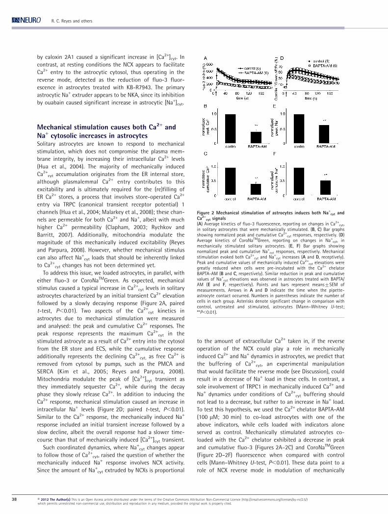
To address this issue, we loaded astrocytes, in parallel, with
either fluo-3 or CoroNaTMGreen. As expected, mechanical
stimulus caused a typical increase in Ca2+cyt levels in solitary
astrocytes characterized by an initial transient Ca2+ elevation
followed by a slowly decaying response (Figure 2A, paired
t-test, P,0.01). Two aspects of the Ca2+cyt kinetics in
astrocytes due to mechanical stimulation were measured
and analysed: the peak and cumulative Ca2+ responses. The
peak response represents the maximum Ca2+cyt in the
stimulated astrocyte as a result of Ca2+ entry into the cytosol
from the ER store and ECS, while the cumulative response
additionally represents the declining Ca2+cyt, as free Ca2+ is
removed from cytosol by pumps, such as the PMCA and
SERCA (Kim et al., 2005; Reyes and Parpura, 2008).
Mitochondria modulate the peak of [Ca2+]cyt transient as
they immediately sequester Ca2+, while during the decay
phase they slowly release Ca2+. In addition to inducing the
Ca2+ response, mechanical stimulation caused an increase in
intracellular Na+ levels (Figure 2D; paired t-test, P,0.01).
Similar to the Ca2+ response, the mechanically induced Na+
response included an initial transient increase followed by a
slow decline, albeit the overall response had a slower time-
course than that of mechanically induced [Ca2+]cyt transient.
Such coordinated dynamics, where Na+cyt changes appear
to follow those of Ca2+cyt, raised the question of whether the
mechanically induced Na+ response involves NCX activity.
Since the amount of Na+cyt extruded by NCXs is proportional
to the amount of extracellular Ca2+ taken in, if the reverse
operation of the NCX could play a role in mechanically
induced Ca2+ and Na+ dynamics in astrocytes, we predict that
the buffering of Ca2+cyt, an experimental manipulation
that would facilitate the reverse mode (see Discussion), could
result in a decrease of Na+ load in these cells. In contrast, a
sole involvement of TRPC1 in mechanically induced Ca2+ and
Na+ dynamics under conditions of Ca2+cyt buffering should
not lead to a decrease, but rather to an increase in Na+ load.
To test this hypothesis, we used the Ca2+ chelator BAPTA-AM
(100 mM; 30 min) to co-load astrocytes with one of the
above indicators, while cells loaded with indicators alone
served as control. Mechanically stimulated astrocytes co-
loaded with the Ca2+ chelator exhibited a decrease in peak
and cumulative fluo-3 (Figures 2A–2C) and CoroNaTMGreen
(Figure 2D–2F) fluorescence when compared with control
cells (Mann–Whitney U-test, P,0.01). These data point to a
role of NCX reverse mode in modulation of mechanically
Figure 2 Mechanical stimulation of astrocytes induces both Na+cyt and
Ca2+cyt signals
(A) Average kinetics of fluo-3 fluorescence, reporting on changes in Ca2+cyt,
in solitary astrocytes that were mechanically stimulated. (B, C) Bar graphsshowing normalized peak and cumulative Ca2+
cyt responses, respectively. (D)Average kinetics of CoroNaTMGreen, reporting on changes in Na+
cyt, inmechanically stimulated solitary astrocytes. (E, F) Bar graphs showingnormalized peak and cumulative Na+
cyt responses, respectively. Mechanicalstimulation evoked both Ca2+
cyt and Na+cyt increases (A and D, receptively).
Peak and cumulative values of mechanically induced Ca2+cyt elevations were
greatly reduced when cells were pre-incubated with the Ca2+ chelatorBAPTA-AM (B and C, respectively). Similar reduction in peak and cumulativevalues of Na+
cyt elevations was observed in astrocytes treated with BAPTA/AM (E and F, respectively). Points and bars represent means¡SEM ofmeasurements. Arrows in A and D indicate the time when the pipette-astrocyte contact occurred. Numbers in parentheses indicate the number ofcells in each group. Asterisks denote significant change in comparison withcontrol, untreated and stimulated, astrocytes (Mann–Whitney U-test;**P,0.01).
R. C. Reyes and others
38 E 2012 The Author(s) This is an Open Access article distributed under the terms of the Creative Commons Attribution Non-Commercial Licence (http://creativecommons.org/licenses/by-nc/2.5/)which permits unrestricted non-commercial use, distribution and reproduction in any medium, provided the original work is properly cited.

induced Ca2+ and Na+ dynamics in astrocytes, which we
further studied using a pharmacological approach below.
PMCA, NCX and NKA modulate mechanicallyinduced Ca2+
cyt accumulations in astrocytesHaving determined effects of pharmacological blockers of
PMCA, NCX and NKA on Ca2+ and Na+ cytosolic levels of cells
at rest along with finding that mechanical stimulation
induced both Ca2+cyt and Na+
cyt elevations, we studied the
role of these pumps and the exchanger on cytosolic Ca2+
dynamics of mechanically stimulated astrocytes. As we
described above, mechanical stimulation of control astrocytes
caused a robust increase in [Ca2+]cyt, corresponding to approx.
3.8 mM (Figure 3A; paired t-test, P,0.01). We found that the
treatment of astrocytes with either caloxin 2A1 (2 mM;
5 min) to block PMCAs, benzamil (100 mM; 5 min) to block
NCX, or ouabain (1 mM; 10 min) to block NKA modulated the
mechanically induced Ca2+ response as evident in the average
kinetics of the fluorescence intensity (Figure 3A). We should
be reminded that pharmacological agents affect resting
Ca2+cyt levels bringing them to various post-treatment
baselines (Figure 1A). Since we were interested in the
astrocytic ability to handle Ca2+cyt after the treatments, we
used post-treatment Ca2+cyt levels as a baseline (Fo) for
further quantitative analysis. Here, pretreatment of astro-
cytes with either caloxin 2A1, benzamil or ouabain reduced
the peak of mechanically induced Ca2+ response, when com-
pared with control (Figure 3B; one-way ANOVA, followed
by Fisher’s LSD test; P,0.01 for caloxin 2A1 and benzamil,
while P,0.05 for ouabain). In contrast, astrocytes treated
with caloxin 2A1 did not exhibit significant difference in
cumulative response, while cells treated with benzamil or
ouabain exhibited attenuated or enhanced cumulative
responses respectively (Figure 3C, one-way ANOVA, followed
by Fisher’s LSD test; P,0.01).
Taken together it appears that PMCA, NCX and NKA all play
a role in modulating the mechanically induced Ca2+ entry
into the cytosol since these agents attenuated the peak Ca2+
response. In addition, it also appears that NCX and NKA play
additional, but opposing roles in the removal of Ca2+ from the
cytosol.
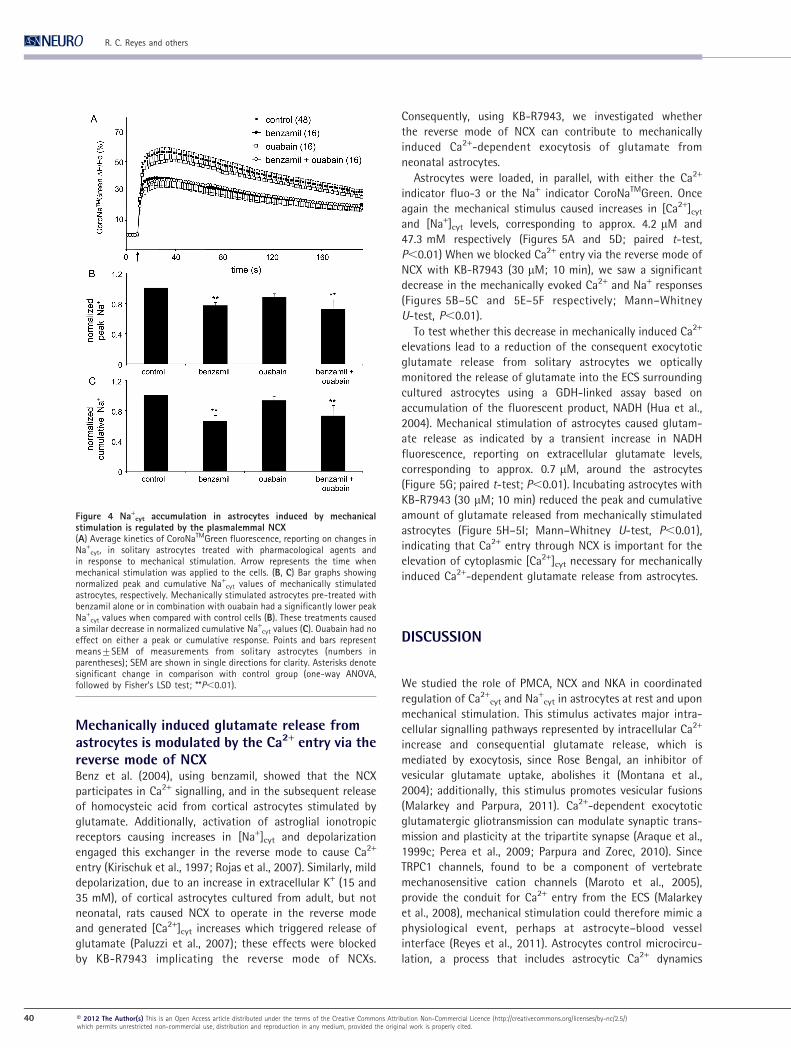
Na+cyt load induced by mechanical stimulation is
mediated by NCXMechanical stimulation of control astrocytes, in addition to
Ca2+cyt response, caused an increase in [Na+]cyt corresponding
to ,36.8 mM (Figure 4A; paired t-test, P,0.01), which is
about the same [Na+]cyt reached upon treatment of astrocytes
at rest with the NKA blocker (Figure 1B). We tested whether
mechanically induced Na+ load in astrocytes can be
modulated by NCX and NKA. We loaded cortical astrocytes
with CoroNaTMGreen, and then treated them with either
benzamil (100 mM; 5 min) to block NCX, or ouabain (1 mM;
10 min) to block NKA. We were interested in the astrocytic
ability to handle Na+cyt following pharmacological treat-
ments, which affected resting [Na+]cyt levels bringing them to
various post-treatment baselines (Figure 1B). Thus, we used
post-treatment [Na+]cyt baselines for further quantitative
analysis. Astrocytes pretreated with benzamil showed a
reduced mechanically induced peak and cumulative
CoroNaTMGreen fluorescence (Figures 4A and 4B; one-way
ANOVA, followed by Fisher’s LSD test; P,0.01), while ouabain
did not significantly affect these parameters by comparison
to control astrocytes. Additionally, when ouabain was added
in combination with benzamil, it did not alter the effect
caused by benzamil (Figures 4B–4C).
Thus, it appears that the NCX may contribute to Na+ entry
into cytosol of mechanically stimulated astrocytes since its
blockade with benzamil reduced the Na+ load in stimulated
astrocytes. In contrast, the NKA appears to be a minor player
in the extrusion of Na+ in mechanically stimulated astrocytes.
Figure 3 Ca2+cyt accumulation in astrocytes induced by mechanical
stimulation is modulated by the plasmalemmal PMCA, NCX, and NKA(A) Average kinetics of fluo-3 fluorescence, reporting on changes in Ca2+
cyt,in solitary astrocytes treated with pharmacological agents and in response tomechanical stimulation. Arrow represents the time when mechanicalstimulation was applied to the cells. (B,C) Bar graphs showing normalizedpeak and cumulative Ca2+
cyt responses, respectively. When treated withcaloxin 2A1, benzamil and ouabain, mechanically stimulated astrocytes had adecrease in the peak Ca2+
cyt response (B). Benzamil led to a decrease, whileouabain to an increase in the normalized cumulative Ca2+
cyt responseobtained from mechanically stimulated astrocytes (C). Points and barsrepresent means¡SEM of measurements from solitary astrocytes (numbersin parentheses); SEM are shown in single directions for clarity. Asterisksdenote significant change in comparison with control group (one-wayANOVA, followed by Fisher’s LSD test; *P,0.05, **P,0.01).
Plasmalemmal Na+/Ca2+ transport in astrocytes
E 2012 The Author(s) This is an Open Access article distributed under the terms of the Creative Commons Attribution Non-Commercial Licence (http://creativecommons.org/licenses/by-nc/2.5/)which permits unrestricted non-commercial use, distribution and reproduction in any medium, provided the original work is properly cited.
39
Mechanically induced glutamate release fromastrocytes is modulated by the Ca2+ entry via thereverse mode of NCXBenz et al. (2004), using benzamil, showed that the NCX
participates in Ca2+ signalling, and in the subsequent release
of homocysteic acid from cortical astrocytes stimulated by
glutamate. Additionally, activation of astroglial ionotropic
receptors causing increases in [Na+]cyt and depolarization
engaged this exchanger in the reverse mode to cause Ca2+
entry (Kirischuk et al., 1997; Rojas et al., 2007). Similarly, mild
depolarization, due to an increase in extracellular K+ (15 and
35 mM), of cortical astrocytes cultured from adult, but not
neonatal, rats caused NCX to operate in the reverse mode
and generated [Ca2+]cyt increases which triggered release of
glutamate (Paluzzi et al., 2007); these effects were blocked
by KB-R7943 implicating the reverse mode of NCXs.
Consequently, using KB-R7943, we investigated whether
the reverse mode of NCX can contribute to mechanically
induced Ca2+-dependent exocytosis of glutamate from
neonatal astrocytes.
Astrocytes were loaded, in parallel, with either the Ca2+
indicator fluo-3 or the Na+ indicator CoroNaTMGreen. Once
again the mechanical stimulus caused increases in [Ca2+]cyt
and [Na+]cyt levels, corresponding to approx. 4.2 mM and
47.3 mM respectively (Figures 5A and 5D; paired t-test,
P,0.01) When we blocked Ca2+ entry via the reverse mode of
NCX with KB-R7943 (30 mM; 10 min), we saw a significant
decrease in the mechanically evoked Ca2+ and Na+ responses
(Figures 5B–5C and 5E–5F respectively; Mann–Whitney
U-test, P,0.01).
To test whether this decrease in mechanically induced Ca2+
elevations lead to a reduction of the consequent exocytotic
glutamate release from solitary astrocytes we optically
monitored the release of glutamate into the ECS surrounding
cultured astrocytes using a GDH-linked assay based on
accumulation of the fluorescent product, NADH (Hua et al.,
2004). Mechanical stimulation of astrocytes caused glutam-
ate release as indicated by a transient increase in NADH
fluorescence, reporting on extracellular glutamate levels,
corresponding to approx. 0.7 mM, around the astrocytes
(Figure 5G; paired t-test; P,0.01). Incubating astrocytes with
KB-R7943 (30 mM; 10 min) reduced the peak and cumulative
amount of glutamate released from mechanically stimulated
astrocytes (Figure 5H–5I; Mann–Whitney U-test, P,0.01),
indicating that Ca2+ entry through NCX is important for the
elevation of cytoplasmic [Ca2+]cyt necessary for mechanically
induced Ca2+-dependent glutamate release from astrocytes.
DISCUSSION
We studied the role of PMCA, NCX and NKA in coordinated
regulation of Ca2+cyt and Na+
cyt in astrocytes at rest and upon
mechanical stimulation. This stimulus activates major intra-
cellular signalling pathways represented by intracellular Ca2+
increase and consequential glutamate release, which is
mediated by exocytosis, since Rose Bengal, an inhibitor of
vesicular glutamate uptake, abolishes it (Montana et al.,
2004); additionally, this stimulus promotes vesicular fusions
(Malarkey and Parpura, 2011). Ca2+-dependent exocytotic
glutamatergic gliotransmission can modulate synaptic trans-
mission and plasticity at the tripartite synapse (Araque et al.,
1999c; Perea et al., 2009; Parpura and Zorec, 2010). Since
TRPC1 channels, found to be a component of vertebrate
mechanosensitive cation channels (Maroto et al., 2005),
provide the conduit for Ca2+ entry from the ECS (Malarkey
et al., 2008), mechanical stimulation could therefore mimic a
physiological event, perhaps at astrocyte–blood vessel
interface (Reyes et al., 2011). Astrocytes control microcircu-
lation, a process that includes astrocytic Ca2+ dynamics
Figure 4 Na+cyt accumulation in astrocytes induced by mechanical
stimulation is regulated by the plasmalemmal NCX(A) Average kinetics of CoroNaTMGreen fluorescence, reporting on changes inNa+
cyt, in solitary astrocytes treated with pharmacological agents andin response to mechanical stimulation. Arrow represents the time whenmechanical stimulation was applied to the cells. (B, C) Bar graphs showingnormalized peak and cumulative Na+
cyt values of mechanically stimulatedastrocytes, respectively. Mechanically stimulated astrocytes pre-treated withbenzamil alone or in combination with ouabain had a significantly lower peakNa+
cyt values when compared with control cells (B). These treatments causeda similar decrease in normalized cumulative Na+
cyt values (C). Ouabain had noeffect on either a peak or cumulative response. Points and bars representmeans¡SEM of measurements from solitary astrocytes (numbers inparentheses); SEM are shown in single directions for clarity. Asterisks denotesignificant change in comparison with control group (one-way ANOVA,followed by Fisher’s LSD test; **P,0.01).
R. C. Reyes and others
40 E 2012 The Author(s) This is an Open Access article distributed under the terms of the Creative Commons Attribution Non-Commercial Licence (http://creativecommons.org/licenses/by-nc/2.5/)which permits unrestricted non-commercial use, distribution and reproduction in any medium, provided the original work is properly cited.

occurring at their end feet and bodies (Gordon et al., 2007); it
is at this interface that astrocytes undergo large mechanical
dynamics.
We used a culturing method yielding solitary astrocytes
which appear as flat polygonal cells, having less complex
morphological features than astrocytes in situ which display
elaborate processes and are in intimate contact with other
neural cell types. While this system minimizes effects of
intercellular astrocyte–astrocyte communications and thus
eases the interpretation of our measurements, it brings some
limitations. Hence, we were not able to study: (i) the
difference in measurements at the somatic region compared
with peripheral astrocytic processes, and (ii) the effects that
the presence of other cell types, e.g., neurons, would have on
our measurements. Nonetheless, using solitary astrocytes we
are poised to study in detail spatio-temporal characteristics
of Ca2+ and Na+ fluxes at mitochondrial, ER and plasma
membranes, as well as to assess concentrations of these ions
within mitochondria and ER. Indeed, some future studies
would also need to cross-correlate our findings in culture
with experiments done in more intact systems.
Our finding that mechanical stimulus leads to activation of
another intracellular signalling pathway, an increase in
[Na+]cyt, has important consequences to astrocytic metabol-
ism. Na+ entry drives glutamate uptake through the plasma
membrane glutamate transporters, and is used to counter
transport H+ out of the cytosol via the Na+/H+ exchanger to
regulate cytosolic pH (Deitmer, 2004). As such, maintenance
of the Na+ gradient via the NKA is a major ATP expenditure in
astrocytes. Na+cyt increases leading to depolarization due to
activity of plasma membrane glutamate transporters (Rojas
et al., 2007) and ionotropic glutamate receptors and/or
purinoceptors (Benz et al., 2004; Lalo et al., 2006; Lalo et al.,
2008) can trigger intracellular Ca2+ signalling mediated via
NCX. Additionally, increases in [Na+]cyt can trigger aerobic
glycolysis leading to lactate production (Magistretti, 2006),
which shuttled to neurons appears to be important for
synaptic transmission and plasticity (Suzuki et al., 2011).
Inhibition of the PMCA with caloxin 2A1 raised basal
[Ca2+]cyt in cortical astrocytes (Figure 1A). This observation
further supports the notion that the PMCA is an important
extrusion mechanism to maintain resting levels of Ca2+ in
Figure 5 Mechanically induced glutamate release from astrocytes is modulated by the Ca2+ entry via the reverse mode of theplasmalemmal NCX(A) Average kinetics of fluo-3 fluorescence, reporting on changes in Ca2+
cyt, in solitary astrocytes treated with KB-R7943 in responseto mechanical stimulation. (B, C) Bar graphs showing normalized peak and cumulative Ca2+
cyt values of mechanically stimulatedastrocytes, respectively. When treated with KB-R7943, astrocytes had significantly lower peak and cumulative Ca2+
cyt responses tomechanical stimulation than those recorded in control. (D) Average kinetics of CoroNaTMGreen (CoroNaTMG) fluorescence, reportingon changes in Na+
cyt, in solitary astrocytes treated with KB-R7943 in response to mechanical stimulation. (E, F) Bar graphs showingnormalized peak and cumulative Na+
cyt values of mechanically stimulated astrocytes, respectively. When treated with KB-R7943,astrocytes had significantly lower peak and cumulative Na+
cyt responses to mechanical stimulation than those recorded in control.(H) Time lapse of extracellular NADH fluorescence, reporting on glutamate levels in the ECS surrounding somata of solitaryastrocytes. Mechanical stimulation of solitary astrocytes caused glutamate release, which was affected by KB-R7943. (H, I)Normalized peak and cumulative extracellular glutamate (Glut) values of mechanically stimulated astrocytes, respectively. Whentreated with KB-R7943, astrocytes had both parameters significantly reduced. Points and bars represent means¡SEM ofmeasurements from solitary astrocytes (numbers in parentheses); SEM in A are shown in single directions for clarity. Arrows in A, Dand G represent the time when mechanical stimulation was applied to the cells. Asterisks denote significant change in comparisonwith control group (Mann–Whitney U-test, ** P,0.01).
Plasmalemmal Na+/Ca2+ transport in astrocytes
E 2012 The Author(s) This is an Open Access article distributed under the terms of the Creative Commons Attribution Non-Commercial Licence (http://creativecommons.org/licenses/by-nc/2.5/)which permits unrestricted non-commercial use, distribution and reproduction in any medium, provided the original work is properly cited.
41

cortical astrocytes, consistent with findings in other cell
types, e.g. in mammalian photoreceptors (Morgans et al.,
1998) and the calyx of Held (Kim et al., 2005), as well as an
invertebrate model of squid giant axons (DiPolo and Beauge,
1979). The relative contribution of PMCA isoforms 1, 2 and 4,
expressed by cortical astrocytes (Fresu et al., 1999), in this
process is not presently understood and it should be
investigated in future. Nonetheless, at stimulated conditions
when the [Ca2+]cyt is increased due to Ca2+ mobilization from
the ER and ECS, inhibition of the PMCA resulted in a sig-
nificant reduction in the peak and with no effect on the
cumulative Ca2+ response (Figures 3A and 3B). This apparent
lack of the PMCA activity in the removal of Ca2+cyt during
times of high [Ca2+]cyt indicates that this task is accomplished
by other extrusion systems, perhaps the ER store-specific
Ca2+-ATPase (Hua et al., 2004).
Astrocytes at rest treated with benzamil alone showed only
a trend in a [Ca2+]cyt decrease, while application of this
general NCX blocker in conjunction with caloxin 2A1
occluded the action of this PMCA blocker, as evidenced by
the absence of significant change in [Ca2+]cyt from controls
(Figure 1A). This finding suggests that the Ca2+ influx via NCX
is opposed/balanced by the Ca2+ efflux via the PMCA
(Figure 1A). This inference is supported by experiments using
the specific antagonist of the NCX reverse mode, KB-R7943,
which significantly decreased [Ca2+]cyt in cortical astrocytes
(Figure 1A), confirming the apparent Ca2+ entry via NCX in
astrocytes at rest. Additionally, KB-R7943 significantly
decreased basal [Na+]cyt, which is likely an indirect effect
due to an increased NKA activity. These data also imply that
the resting membrane potential in our cultured cortical
astrocytes is slightly depolarized from the equilibrium
potential for the NCX (ENCX), as supported by the previously
published work and the calculated ENCX. Hence, the mem-
brane potential of cortical astrocytes was reported to have a
bimodal distribution with peaks at 268 mV and 241 mV [see
Figure 2A of (Kucheryavykh et al., 2007)]. Using our recorded
[Na+]cyt of 16.6 mM and [Ca2+]cyt of 73 nM in astrocytes at
rest, together with concentration of these ions in the external
solution and presumed NCX 3:1 stoichiometry, we calculated
ENCX to be approx.298 mV at 25 C̊. Thus, the vast majority or
astrocytes at rest should display the reverse mode of NCX
operation in our experimental conditions. It should be noted
that there are three NCX isoforms (1–3), with NCX1 being a
predominant isoform, having three splice variants, in primary
cultures of rat cortical astrocytes (He et al., 1998). Future
experiments are needed to address relative contribution of
each of these splice variants in Ca2+cyt regulation.
When cortical astrocytes were mechanically stimulated to
raise [Ca2+]cyt, this stimulus also caused large increases in
[Na+]cyt. As above by using our recorded peak [Na+]cyt of
36.8 mM and peak [Ca2+]cyt of 4 mM due to mechanical
stimulation, we calculated ENCX to be approx.257 mV at
25 C̊. Such shift in ENCX would be less favourable for the
reverse mode of NCX operation in astrocytes. Consequently,
to initially test a possible involvement of NCX in [Ca2+]cyt and
[Na+]cyt regulation at elevated levels of these ions, we used
BAPTA-AM to clamp the Ca2+cyt increase due to mechanical
stimulation, corresponding to approx. 614 nM, which would
substantially shift ENCX to hyperpolarization at approx.
2105 mV assuming unchanged peak [Na+]cyt of 36.8 mM.
Such experimental conditions would result in bettering of
NCX activity that will be seen as a reduction of [Na+]cyt in
astrocytes. Indeed, in BAPTA-AM treated and mechanically
stimulated astrocytes we recorded a significantly lower
increase of [Na+]cyt, approx. 26.8 mM, when compared with
controls, at which juncture cells would settle for their ENCX at
approx. 280 mV (Figure 2). Of course, large Ca2+ entry
associated with the reverse mode of NCX operation was
clamped down by the fast buffering capabilities of BAPTA,
resulting in reduced Ca2+cyt levels. We tested this immediate
conclusion based on thermodynamics considerations further
by using NCX pharmacological blockers.
When cortical astrocytes were mechanically stimulated to
raise [Ca2+]cyt, inhibition of the NCX with benzamil resulted in
a significant reduction in peak and cumulative Ca2+cyt
accumulation (Figure 3). This observation suggests that the
NCX mediates Ca2+ entry and promotes Ca2+ excitability in
astrocytes, consistent with earlier studies (Kirischuk et al.,
1997; Benz et al., 2004; Rojas et al., 2007). Since calculated
ENCX is approx.257 mV during the initial phase of mechanical
stimulation, depolarization of the majority of astrocytes
would be required for the reverse mode of NCX operation.
This seems a plausible scenario because mechanical stimu-
lation leads to rather large Na+cyt load, which would lead to
depolarization of astrocytes. Perhaps as in basal conditions, a
decrease in [Na+]cyt that was recorded from mechanically
stimulated astrocytes exposed to benzamil could be due to
increased activity of NKA (Figure 4), although another
alternative is possible (see below). Ca2+ entry through the
NCX may modulate the InsP3 receptor-gated channel
activation, since the NCX1 isoform can associate with plasma
membrane–ER junctions, microdomains containing InsP3
receptors (Lencesova et al., 2004). Whether this notion could
extend to a possible interplay between the Ca2+ entry via NCX
and activity of ryanodine/caffeine-sensitive receptors of the
ER is not clear due to lack of evidence for spatial association
of these proteins; additionally, the role of ryanodine
receptors in astrocytic Ca2+ excitability remains debatable
(Parpura et al., 2011).
Unlike previous studies demonstrating that inhibition of
NKA increased basal [Ca2+]cyt in cortical and in cerebellar type
1 astrocytes (Rojas et al., 2004), our treatment with ouabain
significantly reduced the basal [Ca2+]cyt in cortical astrocytes
(Figure 1A). This observation cannot be explained by an
inadequate inhibition of the NKA since in parallel experi-
ments ouabain significantly increased Na+cyt accumulation
(Figure 1B). The likely explanation for these seemingly
disparate findings might be lower temperature in our
experiments. Namely, while we recorded at room temperature
(22–25 C̊), above mentioned studies have done so at higher
temperatures [32–34 C̊ (Goldman et al., 1994) or 35–37 C̊
R. C. Reyes and others
42 E 2012 The Author(s) This is an Open Access article distributed under the terms of the Creative Commons Attribution Non-Commercial Licence (http://creativecommons.org/licenses/by-nc/2.5/)which permits unrestricted non-commercial use, distribution and reproduction in any medium, provided the original work is properly cited.
(Rojas et al., 2004)]. If we compare calculated ENCX using our
ionic conditions at 25 C̊ (298 mV) compared with 37 C̊
(2123 mV), we find ENCX to be shifted towards less
hyperpolarizing values at room temperature, which would
lessen the operation of NCX in the reverse mode. Indeed, it
has been reported that NCX activity is greatly inhibited in
cultured Purkinje neurons at room temperature (Rojas et al.,
2003). Thus, it is possible that reduction in Ca2+cyt during NKA
blockade points to restrained operation of NCX in the reverse
mode at room temperature, while PMCA activity would
remain grossly uninterrupted, as one could expect from
difference in turnover rates between the pump and the
transporter; PMCA turnover rate is 30–250/s, while that of
NCX is 2000–5000/s (Blaustein and Lederer, 1999). In
addition, NCX activity requires Ca2+cyt to bind internal
regulatory sites, which at the low [Ca2+]cyt and relatively
low temperature conditions we tested, may not be optimally
engaged (Blaustein and Lederer, 1999). Indeed, when Ca2+cyt
was increased by addition of caloxin 2A1, which inhibited the
PMCA along with the NKA blockade, the [Na+]cyt was greatly
reduced revealing the operation of NCX in the reverse mode
to extrude Na+cyt while facilitating Ca2+ entry (Figure 1).
Furthermore, a decrease in basal [Ca2+]cyt due to ouabain
treatment and via NCX modulation was occluded when
astrocytes were co-treated by benzamil (Figure 1A).
At elevated [Ca2+]cyt induced by mechanical stimulation,
the NKA indirectly affected Ca2+ homoeostasis as well.
Inhibition of NKA decreased the peak but increased
cumulative Ca2+ accumulation in response to mechanical
stimulation (Figure 3). The reduced Ca2+cyt peak response
suggests that elevated Na+cyt initially reduces the activity of
the NCX (Blaustein and Lederer, 1999). However, as the NCX
activity ramped up, the elimination of the NKA activity as
the primary reducer of Na+cyt significantly increased the
cumulative accumulation of Ca2+cyt (Figure 3C). This obser-
vation suggests that Na+ extrusion at elevated mechanically
induced Na+cyt is in part mediated by the NCX in exchange for
Ca2+ entry. It is possible that Na+ extrusion is also mediated
by other Na+ extrusion systems, such as the NCKX (K+-
dependent NCX) (Kim et al., 2005; Visser and Lytton, 2007),
although neurons, but not astrocytes seem to preferentially
express NCKX (Kiedrowski et al., 2002). Hence, treatment with
ouabain alone or in combination with benzamil did not cause
increase in peak and cumulative Na+cyt accumulation in
mechanically stimulated astrocytes, but rather a significant
decrease reaching levels similar to that seen in the stimulated
astrocytes pre-treated with benzamil alone (Figure 4). This
contradicts our previous consideration that decrease in
[Na+]cyt seen in stimulated astrocytes pretreated with
benzamil could be due to the NKA activity and might
imply an involvement of NCKX in this process. Conse-
quently, although a minor contribution of NCKX is
expected (Kiedrowski et al., 2002), its possible role in the
regulation of the Na+cyt and Ca2+
cyt in astrocytes needs to be
evaluated as it has been done in the calyx of Held (Kim et al.,
2005).
The complex interplay between Na+cyt and Ca2+
cyt
dynamics and signalling in astrocytes warrants some further
considerations. The NKA (type a2) have been found to
colocalize with NCX in cortical astrocytes at plasma
membrane–ER junctions where tightly regulated ’sodium
microdomains’ may occur (Juhaszova and Blaustein, 1997;
Blaustein et al., 2002). Inhibition of NKA has been shown to
generate Ca2+ oscillations in cultured hippocampal astrocytes
(Liu et al., 2007). In addition, subsets of mitochondria have
been shown to interact with the ER (Rizzuto et al., 1998;
Csordas et al., 2006). At higher [Na+]cyt near mitochondria–ER
junctions, the driving force for Ca2+ efflux from mitochondria
via the mitochondrial NCX would increase. Hypothetically,
this Ca2+ efflux could in turn increase [Ca2+]cyt to activate
ryanodine receptor of the ER. Indeed, it appears that the
mitochondrial NCX plays a role in Ca2+cyt oscillations
(Hernandez-SanMiguel et al., 2006).
Previous work showed that mild depolarization of cortical
astrocytes cultured from adult, but not neonatal, rats caused
NCX to operate in the reverse mode leading to [Ca2+]cyt
increases and consequential exocytotic glutamate release
(Paluzzi et al., 2007). Our data are consistent with this
finding, yet they add an additional possibility: the involve-
ment of the reverse mode of NCX in mechanically induced
glutamate release from neonatal astrocytes. Mechanical
stimulation drives Ca2+-dependent regulated exocytosis in
astrocytes (Hua et al., 2004). The contribution of the NCX
in these processes was seen as reduced peak and cumulative
Ca2+cyt accumulations (Figures 5A–5C), as well as reduction in
peak and cumulative release of glutamate from astrocytes to
the ECS (Figures 5G–5I). It should be noted that, consistent
with our calculated ENCX at rest and during mechanical
stimulation, KB-R7943 blockade of Ca2+ entry via the reverse
mode of NCX upon mechanical stimulation appeared smaller
(85 and 90% of peak and cumulative response of control,
respectively) than its effect on reduction of Ca2+ entry in
astrocytes at rest (72% of basal level) (compare Figure 5A and
Figure 1A). Additionally, KB-R7943 significantly reduced peak
and cumulative Na+cyt accumulations (Figures 5D–5F), an
indirect effect, likely due to an increased NKA activity, which
is in agreement with data obtained using this blocker on
astrocytes at rest.
The cellular location of the NCX favours its role in the
modulation of Ca2+-dependent glutamate release. NCXs are
located on plasma membrane–ER junctions and are ex-
pressed near InsP3 receptor-gated channels (Lencesova et al.,
2004) and NKA (Juhaszova and Blaustein, 1997). Additionally,
NCX is co-localized with plasma membrane glutamate
transporters in perisynaptic processes (Minelli et al., 2007).
Thus, in terms of glutamatergic synaptic transmission, as
glutamate is released from neurons, it acts upon the plasma
membrane glutamate transporters and ionotropic receptors
on astrocytes, which leads to an increase in astrocytic [Na+]cyt
(Langer and Rose, 2009; Lalo et al., 2011). Such Na+cyt
dynamics should lead to depolarization causing NCX to
operate in reverse and allow Ca2+ entry from the ECS
Plasmalemmal Na+/Ca2+ transport in astrocytes
E 2012 The Author(s) This is an Open Access article distributed under the terms of the Creative Commons Attribution Non-Commercial Licence (http://creativecommons.org/licenses/by-nc/2.5/)which permits unrestricted non-commercial use, distribution and reproduction in any medium, provided the original work is properly cited.
43

subsequently stimulating glutamatergic gliotransmission.
However, the role of plasma membrane glutamate ionotropic
receptors and transporters in exocytotic glutamate release
from astrocytes is speculative at the moment. Exposure of
cultured cortical astrocytes to AMPA (a-amino-3-hydroxy-5-
methylisoxazole-4-propionic acid) did not induce glutamate
release, unless it was co-applied with an mGluR agonist (Bezzi
et al., 1998); co-application had much greater effect than
that of an mGluR agonist alone. While this finding is
consistent with the expression of GluR2 subunit in astrocytes,
leading to low Ca2+ permeability through their AMPA
channels, it is at odds with findings that depolarization due
to increases in Na+cyt following activation of kainate
receptors was reported to stimulate Ca2+ entry through the
reverse mode of the NCX in specialized astrocytes, the Berg-
mann glia, in acute slice (Kirischuk et al 1997). These
ostensibly incongruent findings could perhaps be due to
different spatial associations between NCX, glutamate
receptor and secretory machinery in different regions of
the brain (cortex against cerebellum), choice of the agonist
(AMPA against kainate) and preparation (culture against
acute slice). Nonetheless, when operable, this NCX-linked
pathway should be faster and spatially more confined, than
the parallel pathway engaging astrocytic mGluRs tapping
mainly into recruitment of Ca 2+ from the ER store to cause
glutamatergic gliotransmission. Of course, this possibility
would need to be corroborated morphologically by studying
the spatial relationship between exocytotic proteins, NCX,
glutamate receptors and transporters, as well as using
electrophysiological recordings from neurons while specif-
ically manipulating NCX in nearby astrocytes.
ACKNOWLEDGEMENTS
We dedicate this paper to the late Professor Dale J. Benos. We
thank Manoj K. Gottipati for comments on a previous version
of this paper.
FUNDING
This work was supported by the National Institute of Mental
Health [grant number MH 069791] and the National Science
Foundation [grant number CBET 0943343].
REFERENCES
Araque A, Sanzgiri RP, Parpura V, Haydon PG (1999a) Astrocyte-inducedmodulation of synaptic transmission. Can. J. Physiol. Pharmacol. 77:699–706.
Araque A, Sanzgiri RP, Parpura V, Haydon PG (1999b) Calcium elevation inastrocytes causes an NMDA receptor-dependent increase in thefrequency of miniature synaptic currents in cultured hippocampalneurons. J. Neurosci. 18:6822–6829.
Araque A, Parpura V, Sanzgiri RP, Haydon PG (1999c) Tripartite synapses: glia,the unacknowledged partner. Trends Neurosci. 22:208–215.
Benz B, Grima G, Do KQ (2004) Glutamate-induced homocysteic acid releasefrom astrocytes: possible implication in glia-neuron signaling.Neuroscience 124:377–386.
Bezzi P, Carmignoto G, Pasti L, Vesce S, Rossi D, Rizzini BL, Pozzan T, VolterraA (1998) Prostaglandins stimulate calcium-dependent glutamate releasein astrocytes. Nature 391:281–285.
Blaustein MP, Lederer WJ (1999) Sodium/calcium exchange: its physiologicalimplications. Physiol. Rev. 79:763–854.
Blaustein MP, Juhaszova M, Golovina VA, Church PJ, Stanley EF (2002) Na/Caexchanger and PMCA localization in neurons and astrocytes: functionalimplications. Ann. NY Acad. Sci. 976:356–366.
Chaudhary J, Walia M, Matharu J, Escher E, Grover AK (2001) Caloxin: a novelplasma membrane Ca2+ pump inhibitor. Am. J. Physiol. Cell Physiol.280:C1027–1030.
Clapham DE (2003) TRP channels as cellular sensors. Nature 426:517–524.Csordas G, Renken C, Varnai P, Walter L, Weaver D, Buttle KF, Balla T,
Mannella CA, Hajnoczky G (2006) Structural and functional features andsignificance of the physical linkage between ER and mitochondria. J. Cell.Biol. 174:915–921.
De Luisi A, Hofer AM (2003) Evidence that Ca(2+) cycling by the plasmamembrane Ca(2+)-ATPase increases the ‘excitability’ of the extracellularCa(2+)-sensing receptor. J. Cell. Sci. 116:1527–1538.
Deitmer JW (2004) pH regulation and acid/base-mediated transport in glialcells. In: Glial-Neuronal Signaling (Hatton GI, Parpura V, eds.), pp. 263–277. Boston, MA: Kluwer Academic Publishers.
DiPolo R, Beauge L (1979) Physiological role of ATP-driven calcium pump insquid axon. Nature 278:271–273.
Filosa JA, Bonev AD, Nelson MT (2004) Calcium dynamics in corticalastrocytes and arterioles during neurovascular coupling. Circ. Res.95:e73-81.
Fresu L, Dehpour A, Genazzani AA, Carafoli E, Guerini D (1999) Plasmamembrane calcium ATPase isoforms in astrocytes. Glia 28:150–155.
Goldman WF, Yarowsky PJ, Juhaszova M, Krueger BK, Blaustein MP (1994)Sodium/calcium exchange in rat cortical astrocytes. J. Neurosci.14:5834–5843.
Gordon GR, Mulligan SJ, MacVicar BA (2007) Astrocyte control of thecerebrovasculature. Glia 55:1214–1221.
Haydon PG, Carmignoto G (2006) Astrocyte control of synaptic transmissionand neurovascular coupling. Physiol. Rev. 86:1009–1031.
He S, Ruknudin A, Bambrick LL, Lederer WJ, Schulze DH (1998) Isoform-specific regulation of the Na+/Ca2+ exchanger in rat astrocytes andneurons by PKA. J. Neurosci. 18:4833–4841.
Hernandez-SanMiguel E, Vay L, Santo-Domingo J, Lobaton CD, Moreno A,Montero M, Alvarez J (2006) The mitochondrial Na+/Ca2+ exchanger playsa key role in the control of cytosolic Ca2+ oscillations. Cell Calcium40:53–61.
Hua X, Malarkey EB, Sunjara V, Rosenwald SE, Li WH, Parpura V (2004)Ca2+-dependent glutamate release involves two classes of endoplasmicreticulum Ca2+ stores in astrocytes. J. Neurosci. Res. 76:86–97.
Innocenti B, Parpura V, Haydon PG (2000) Imaging extracellular waves ofglutamate during calcium signaling in cultured astrocytes. J. Neurosci.20:1800–1808.
Juhaszova M, Blaustein MP (1997) Na+ pump low and high ouabain affinityalpha subunit isoforms are differently distributed in cells. Proc. Natl.Acad. Sci. U.S.A. 94:1800–1805.
Kawano S, Otsu K, Shoji S, Yamagata K, Hiraoka M (2003) Ca(2+) oscillationsregulated by Na(+)-Ca(2+) exchanger and plasma membrane Ca(2+) pumpinduce fluctuations of membrane currents and potentials in humanmesenchymal stem cells. Cell Calcium 34:145–156.
Kiedrowski L, Czyz A, Li XF, Lytton J (2002) Preferential expression ofplasmalemmal K-dependent Na+/Ca2+ exchangers in neurons versusastrocytes. Neuroreport 13:1529–1532.
Kim MH, Korogod N, Schneggenburger R, Ho WK, Lee SH (2005) Interplaybetween Na+/Ca2+ exchangers and mitochondria in Ca2+ clearance atthe calyx of Held. J. Neurosci. 25:6057–6065.
Kirischuk S, Kettenmann H, Verkhratsky A (1997) Na+/Ca2+ exchangermodulates kainate-triggered Ca2+ signaling in Bergmann glial cells insitu. FASEB J. 11:566–572.
Kirischuk S, Kettenmann H, Verkhratsky A (2007) Membrane currents andcytoplasmic sodium transients generated by glutamate transport inBergmann glial cells. Pflugers Arch. 454:245–252.
Kucheryavykh YV, Kucheryavykh LY, Nichols CG, Maldonado HM, Baksi K,Reichenbach A, Skatchkov SN, Eaton MJ (2007) Downregulation of Kir4.1inward rectifying potassium channel subunits by RNAi impairs potassiumtransfer and glutamate uptake by cultured cortical astrocytes. Glia55:274–281.
Lalo U, Pankratov Y, Parpura V, Verkhratsky A (2011) Ionotropic receptors inneuronal-astroglial signalling: what is the role of ’excitable’ moleculesin non-excitable cells. Biochim. Biophys. Acta 1813:992–1002.
R. C. Reyes and others
44 E 2012 The Author(s) This is an Open Access article distributed under the terms of the Creative Commons Attribution Non-Commercial Licence (http://creativecommons.org/licenses/by-nc/2.5/)which permits unrestricted non-commercial use, distribution and reproduction in any medium, provided the original work is properly cited.
Lalo U, Pankratov Y, Kirchhoff F, North RA, Verkhratsky A (2006) NMDAreceptors mediate neuron-to-glia signaling in mouse cortical astrocytes.J. Neurosci. 26:2673–2683.
Lalo U, Pankratov Y, Wichert SP, Rossner MJ, North RA, Kirchhoff F,Verkhratsky A (2008) P261 and P265 subunits form the functional P2Xreceptor in mouse cortical astrocytes. J. Neurosci. 28:5473–5480.
Langer J, Rose CR (2009) Synaptically induced sodium signals in hippocampalastrocytes in situ. J. Physiol. 587:5859–5877.
Lee W, Malarkey EB, Reyes RC, Parpura V (2008) Micropit: a new cellculturing approach for characterization of solitary astrocytes andsmall networks of these Glial cells. Front Neuroeng. 1:2.
Lencesova L, O’Neill A, Resneck WG, Bloch RJ, Blaustein MP (2004) Plasmamembrane–cytoskeleton–endoplasmic reticulum complexes in neuronsand astrocytes. J. Biol. Chem. 279:2885–2893.
Liu XL, Miyakawa A, Aperia A, Krieger P (2007) Na,K-ATPase generatescalcium oscillations in hippocampal astrocytes. Neuroreport 18:597–600.
Magistretti PJ (2006) Neuron-glia metabolic coupling and plasticity. J. Exp.Biol. 209:2304–2311.
Malarkey EB, Parpura V (2011) Temporal characteristics of vesicular fusionin astrocytes: examination of synaptobrevin 2-laden vesicles at singlevesicle resolution. J. Physiol. 589:4271–4300.
Malarkey EB, Ni Y, Parpura V (2008) Ca2+ entry through TRPC1 channelscontributes to intracellular Ca2+ dynamics and consequent glutamaterelease from rat astrocytes. Glia 56:821–835.
Maroto R, Raso A, Wood TG, Kurosky A, Martinac B, Hamill OP (2005) TRPC1forms the stretch-activated cation channel in vertebrate cells. Nat. CellBiol. 7:179–185.
Mata M, Fink DJ (1989) Ca++-ATPase in the central nervous system: an EMcytochemical study. J. Histochem. Cytochem. 37:971–980.
McCarthy KD, de Vellis J (1980) Preparation of separate astroglial andoligodendroglial cell cultures from rat cerebral tissue. J. Cell. Biol.85:890–902.
Meier SD, Kovalchuk Y, Rose CR (2006) Properties of the new fluorescent Na+indicator CoroNa Green: comparison with SBFI and confocal Na+ imaging.J. Neurosci. Methods 155:251–259.
Minelli A, Castaldo P, Gobbi P, Salucci S, Magi S, Amoroso S (2007) Cellularand subcellular localization of Na+–Ca2+ exchanger protein isoforms,NCX1, NCX2, and NCX3 in cerebral cortex and hippocampus of adult rat.Cell Calcium 41:221–234.
Montana V, Ni Y, Sunjara V, Hua X, Parpura V (2004) Vesicular glutamatetransporter-dependent glutamate release from astrocytes. J. Neurosci.24:2633–2642.
Morgans CW, El Far O, Berntson A, Wassle H, Taylor WR (1998) Calciumextrusion from mammalian photoreceptor terminals. J. Neurosci.18:2467–2474.
Mothet JP, Pollegioni L, Ouanounou G, Martineau M, Fossier P, Baux G (2005)Glutamate receptor activation triggers a calcium-dependent and SNAREprotein-dependent release of the gliotransmitter D-serine. Proc. Natl.Acad. Sci. U.S.A. 102:5606–5611.
Mulligan SJ, MacVicar BA (2004) Calcium transients in astrocyte endfeetcause cerebrovascular constrictions. Nature 431:195–199.
Ni Y, Malarkey EB, Parpura V (2007) Vesicular release of glutamate mediatesbidirectional signaling between astrocytes and neurons. J. Neurochem.103:1273–1284.
Paluzzi S, Alloisio S, Zappettini S, Milanese M, Raiteri L, Nobile M, Bonanno G(2007) Adult astroglia is competent for Na+/Ca2+ exchanger-operatedexocytotic glutamate release triggered by mild depolarization. J.Neurochem. 103:1196–1207.
Parpura V, Haydon PG (2000) Physiological astrocytic calcium levels stimulateglutamate release to modulate adjacent neurons. Proc. Natl. Acad. Sci.U.S.A. 97:8629–8634.
Parpura V, Zorec R (2010) Gliotransmission: exocytotic release fromastrocytes. Brain Res. Rev. 63:83–92.
Parpura V, Grubisic V, Verkhratsky A (2011) Ca2+ sources for the exocytoticrelease of glutamate from astrocytes. Biochim. Biophys. Acta 1813:984–991.
Parpura V, Fang Y, Basarsky T, Jahn R, Haydon PG (1995) Expression ofsynaptobrevin II, cellubrevin and syntaxin but not SNAP-25 in culturedastrocytes. FEBS Lett. 377:489–492.
Perea G, Araque A (2007) Astrocytes potentiate transmitter release at singlehippocampal synapses. Science 317:1083–1086.
Perea G, Araque A (2009) Synaptic information processing by astrocytes. In:Astrocytes in (patho)physiology of the Nervous System (Parpura V,Haydon PG, eds.), pp. 287–300. New York, NY: Springer.
Perea G, Navarrete M, Araque A (2009) Tripartite synapses: astrocytes processand control synaptic information. Trends Neurosci. 32:421–431.
Regehr WG (1997) Interplay between sodium and calcium dynamics ingranule cell presynaptic terminals. Biophys. J. 73:2476–2488.
Reyes RC, Parpura V (2008) Mitochondria modulate Ca2+-dependentglutamate release from rat cortical astrocytes. J. Neurosci. 28:9682–9691.
Reyes RC, Parpura V (2009) Modulation of Ca2+-dependent glutamate releasein rat cortical astrocytes by the Na+/Ca2+ exchanger. J. Neurochem.108:116–117.
Reyes RC, Perry G, Lesort M, Parpura V (2011) Immunophilin deficiencyaugments Ca2+-dependent glutamate release from mouse corticalastrocytes. Cell Calcium 49:23–34.
Rizzuto R, Pinton P, Carrington W, Fay FS, Fogarty KE, Lifshitz LM, Tuft RA,Pozzan T (1998) Close contacts with the endoplasmic reticulum asdeterminants of mitochondrial Ca2+ responses. Science 280:1763–1766.
Rojas H, Ramos M, Dipolo R (2004) A genistein-sensitive Na+/Ca2+ exchangeis responsible for the resting [Ca2+]i and most of the Ca2+ plasmamembrane fluxes in stimulated rat cerebellar type 1 astrocytes. Japan. J.Physiol. 54:249–262.
Rojas H, Ramos M, Mijares A, DiPolo R (2003) Role of Na+/Ca2+ exchange in[Ca2+]i clearance in rat culture Purkinje neurons requires reevaluation.Japan. J. Physiol. 53:259–269.
Rojas H, Colina C, Ramos M, Benaim G, Jaffe EH, Caputo C, DiPolo R (2007)Na+ entry via glutamate transporter activates the reverse Na+/Ca2+
exchange and triggers Ca(i)2+-induced Ca2+ release in rat cerebellarType-1 astrocytes. J. Neurochem. 100:1188–1202.
Rose CR, Ransom BR (1997) Gap junctions equalize intracellular Na+
concentration in astrocytes. Glia 20:299–307.Rychkov G, Barritt GJ (2007) TRPC1 Ca(2+)-permeable channels in animal
cells. Handb. Exp. Pharmacol. 179:23–52.Suzuki A, Stern SA, Bozdagi O, Huntley GW, Walker RH, Magistretti PJ,
Alberini CM (2011) Astrocyte-neuron lactate transport is required forlong-term memory formation. Cell 144:810–823.
Theodosis DT, Poulain DA, Oliet SH (2008) Activity-dependent structural andfunctional plasticity of astrocyte-neuron interactions. Physiol. Rev.88:983–1008.
Verkhratsky A, Orkand RK, Kettenmann H (1998) Glial calcium: homeostasisand signaling function. Physiol. Rev. 78:99–141.
Visser F, Lytton J (2007) K+ -dependent Na+/Ca2+ exchangers: keycontributors to Ca2+ signaling. Physiology (Bethesda) 22:185–192.
Zhong N, Beaumont V, Zucker RS (2001) Roles for mitochondrial and reversemode Na+/Ca2+ exchange and the plasmalemma Ca2+ ATPase in post-tetanic potentiation at crayfish neuromuscular junctions. J. Neurosci.21:9598–9607.
Zonta M, Angulo MC, Gobbo S, Rosengarten B, Hossmann KA, Pozzan T,Carmignoto G (2003) Neuron-to-astrocyte signalling is central to thedynamic control of brain microcirculation. Nat. Neurosci. 6:43–50.
Received 5 December 2011/18 January 2012; accepted 20 January 2012
Published as Immediate Publication 23 January 2012, doi 10.1042/AN20110059
Plasmalemmal Na+/Ca2+ transport in astrocytes
E 2012 The Author(s) This is an Open Access article distributed under the terms of the Creative Commons Attribution Non-Commercial Licence (http://creativecommons.org/licenses/by-nc/2.5/)which permits unrestricted non-commercial use, distribution and reproduction in any medium, provided the original work is properly cited.
45


![Near-Membrane [Ca2+] Transients Resolved Using the Ca2+ Indicator FFP18](https://static.fdokumen.com/doc/165x107/631286873ed465f0570a4533/near-membrane-ca2-transients-resolved-using-the-ca2-indicator-ffp18.jpg)