Mitochondrial exchanger NCLX plays a major role in the intracellular Ca2+ signaling,...
14
Cellular/Molecular Mitochondrial Exchanger NCLX Plays a Major Role in the Intracellular Ca 2 Signaling, Gliotransmission, and Proliferation of Astrocytes Julia Parnis, 1 Vedrana Montana, 3 Ignacio Delgado-Martinez, 1,5,6 Vitali Matyash, 1 Vladimir Parpura, 3,4 Helmut Kettenmann, 1 Israel Sekler, 2 and Christiane Nolte 1 1 Cellular Neurosciences, Max Delbru ¨ck Centre for Molecular Medicine, Berlin 13092, Germany, 2 Ben Gurion University, Beer-Sheva 84105, Israel, 3 Department of Neurobiology, University of Alabama, Birmingham, Alabama 35294, 4 Department of Biotechnology, University of Rijeka, 51000 Rijeka, Croatia, and 5 Department of Neurosurgery, Charite ´ University Hospital, Berlin 10117, Germany, and 6 Singapore Institute of Neurotechnology (SINAPSE), Singapore 117456 Mitochondria not only provide cells with energy, but are central to Ca 2 signaling. Powered by the mitochondrial membrane potential, Ca 2 enters the mitochondria and is released into the cytosol through a mitochondrial Na /Ca 2 exchanger. We established that NCLX, a newly discovered mitochondrial Na /Ca 2 exchanger, is expressed in astrocytes isolated from mice of either sex. Immunoblot analysis of organellar fractions showed that the location of NCLX is confined to mitochondria. Using pericam-based mitochondrial Ca 2 imaging and NCLX inhibition either by siRNA or by the pharmacological blocker CGP37157, we demonstrated that NCLX is responsible for mitochondrial Ca 2 extrusion. Suppression of NCLX function altered cytosolic Ca 2 dynamics in astrocytes and this was mediated by a strong effect of NCLX activity on Ca 2 influx via store-operated entry. Furthermore, Ca 2 influx through the store-operated Ca 2 entry triggered strong, whereas ER Ca 2 release triggered only modest mitochondrial Ca 2 transients, indicating that the functional cross talk between the plasma membrane and mitochondrial domains is particularly strong in astrocytes. Finally, silencing of NCLX expression significantly reduced Ca 2 -dependent processes in astrocytes (i.e., exocytotic glutamate release, in vitro wound closure, and prolifera- tion), whereas Ca 2 wave propagation was not affected. Therefore, NCLX, by meditating astrocytic mitochondrial Na /Ca 2 exchange, links between mitochondria and plasma membrane Ca 2 signaling, thereby modulating cytoplasmic Ca 2 transients required to control a diverse array of astrocyte functions. Introduction Ca 2 signaling is central for the regulation of astrocyte functions and for interastrocytic and astrocyte–neuron communication. Neuronal activity, via the activation of metabotropic receptors, triggers transient increases in the cytosolic Ca 2 concentration in astrocytes that leads to the release of gliotransmitters such as ATP and glutamate, which can signal to adjacent neurons (Agulhon et al., 2008; Perea et al., 2009). Astrocytic Ca 2 transients after metabotropic receptor activation are initiated by release of Ca 2 from the ER stores and by the entry from the extracellular space through store-operated Ca 2 channels. The spatiotemporal pat- tern of cytoplasmic Ca 2 signals is dynamically organized and allows for a great complexity of astrocytic Ca 2 responses. Mitochondria, in addition to their metabolic role, participate in intracellular Ca 2 signaling via dynamic buffering and shut- tling cytosolic Ca 2 . Powered by the mitochondrial membrane potential, Ca 2 enters mitochondria via the mitochondrial uni- porter (Baughman et al., 2011; De Stefani et al., 2011; Pizzo et al., 2012) and is extruded from mitochondria via Na -dependent or -independent pathways (Drago et al., 2011). Mitochondria rap- idly sense cellular Ca 2 signals and act as local Ca 2 buffers in the vicinity of Ca 2 release sites such as the ER or plasma membrane Ca 2 channels. By buffering and shuttling Ca 2 , they modulate local and bulk cytoplasmic Ca 2 changes (Szabadkai and Duchen, 2008; Contreras et al., 2010), thereby controlling cell- type-specific functions. Different cell types may vary with regard to mitochondrial Ca 2 handling, in particular Ca 2 extrusion mechanisms (Pizzo et al., 2012). At the neuronal synapse, for example, the mitochondrial Na /Ca 2 exchanger participates in shaping Ca 2 signals, thus modulating neuronal activity and syn- aptic plasticity (Kann and Kova ´cs, 2007; Pizzo et al., 2012). Ac- tivity of the mitochondrial exchanger has also been documented in astrocytes, where it is closely linked to exocytotic release of Received Dec. 11, 2012; revised Feb. 20, 2013; accepted Feb. 25, 2013. Author contributions: V.P., H.K., I.S., and C.N. designed research; J.P., V. Montana, and I.S. performed research; I.D.-M. and V. Matyash contributed unpublished reagents/analytic tools; J.P., V. Montana, I.D.-M., V. Matyash, V.P., H.K., and I.S. analyzed data; J.P., V. Montana, V.P., H.K., I.S., and C.N. wrote the paper. This work was supported by the German Israeli Foundation (Grant #917/2006 to I.S., C.N., and H.K.) and by the Hezneq faculty grant to I.S. I.D.M. was supported by the NeuroCure Cluster of Excellence (Charite ´ University Hospital, Berlin). V.P. is supported by the National Science Foundation (Grant #CBET 0943343). We thank Prof. Michal Hersh- finkel for helpful discussion and Regina Piske and Irene Haupt for excellent technical assistance. The authors declare no competing financial interests. Correspondence should be addressed to either of the following: Christiane Nolte, PhD, Cellular Neurosciences, Max Delbrueck Center for Molecular Medicine, Robert-Roessle-Strasse 10, 13125 Berlin, Germany, E-mail: [email protected]; or Israel Sekler, PhD, Department of Physiology, Ben Gurion University, P.O. Box 653 Beer Sheva, 84105 Israel, E-mail: [email protected]. DOI:10.1523/JNEUROSCI.5721-12.2013 Copyright © 2013 the authors 0270-6474/13/337206-14$15.00/0 7206 • The Journal of Neuroscience, April 24, 2013 • 33(17):7206 –7219
-
Upload
independent -
Category
Documents
-
view
2 -
download
0
Transcript of Mitochondrial exchanger NCLX plays a major role in the intracellular Ca2+ signaling,...

Cellular/Molecular
Mitochondrial Exchanger NCLX Plays a Major Role in theIntracellular Ca2� Signaling, Gliotransmission, andProliferation of Astrocytes
Julia Parnis,1 Vedrana Montana,3 Ignacio Delgado-Martinez,1,5,6 Vitali Matyash,1 Vladimir Parpura,3,4
Helmut Kettenmann,1 Israel Sekler,2 and Christiane Nolte1
1Cellular Neurosciences, Max Delbruck Centre for Molecular Medicine, Berlin 13092, Germany, 2Ben Gurion University, Beer-Sheva 84105, Israel,3Department of Neurobiology, University of Alabama, Birmingham, Alabama 35294, 4Department of Biotechnology, University of Rijeka, 51000 Rijeka,Croatia, and 5Department of Neurosurgery, Charite University Hospital, Berlin 10117, Germany, and 6Singapore Institute of Neurotechnology (SINAPSE),Singapore 117456
Mitochondria not only provide cells with energy, but are central to Ca 2� signaling. Powered by the mitochondrial membrane potential,Ca 2� enters the mitochondria and is released into the cytosol through a mitochondrial Na �/Ca 2� exchanger. We established that NCLX,a newly discovered mitochondrial Na �/Ca 2� exchanger, is expressed in astrocytes isolated from mice of either sex. Immunoblot analysisof organellar fractions showed that the location of NCLX is confined to mitochondria. Using pericam-based mitochondrial Ca 2� imagingand NCLX inhibition either by siRNA or by the pharmacological blocker CGP37157, we demonstrated that NCLX is responsible formitochondrial Ca 2� extrusion. Suppression of NCLX function altered cytosolic Ca 2� dynamics in astrocytes and this was mediated by astrong effect of NCLX activity on Ca 2� influx via store-operated entry. Furthermore, Ca 2� influx through the store-operated Ca 2� entrytriggered strong, whereas ER Ca 2� release triggered only modest mitochondrial Ca 2� transients, indicating that the functional cross talkbetween the plasma membrane and mitochondrial domains is particularly strong in astrocytes. Finally, silencing of NCLX expressionsignificantly reduced Ca 2�-dependent processes in astrocytes (i.e., exocytotic glutamate release, in vitro wound closure, and prolifera-tion), whereas Ca 2� wave propagation was not affected. Therefore, NCLX, by meditating astrocytic mitochondrial Na �/Ca 2� exchange,links between mitochondria and plasma membrane Ca 2� signaling, thereby modulating cytoplasmic Ca 2� transients required to controla diverse array of astrocyte functions.
IntroductionCa 2� signaling is central for the regulation of astrocyte functionsand for interastrocytic and astrocyte–neuron communication.Neuronal activity, via the activation of metabotropic receptors,triggers transient increases in the cytosolic Ca 2� concentration inastrocytes that leads to the release of gliotransmitters such as ATPand glutamate, which can signal to adjacent neurons (Agulhon etal., 2008; Perea et al., 2009). Astrocytic Ca 2� transients aftermetabotropic receptor activation are initiated by release of Ca 2�
from the ER stores and by the entry from the extracellular space
through store-operated Ca 2� channels. The spatiotemporal pat-tern of cytoplasmic Ca 2� signals is dynamically organized andallows for a great complexity of astrocytic Ca 2� responses.
Mitochondria, in addition to their metabolic role, participatein intracellular Ca 2� signaling via dynamic buffering and shut-tling cytosolic Ca 2�. Powered by the mitochondrial membranepotential, Ca 2� enters mitochondria via the mitochondrial uni-porter (Baughman et al., 2011; De Stefani et al., 2011; Pizzo et al.,2012) and is extruded from mitochondria via Na�-dependent or-independent pathways (Drago et al., 2011). Mitochondria rap-idly sense cellular Ca 2� signals and act as local Ca 2� buffers in thevicinity of Ca 2� release sites such as the ER or plasma membraneCa 2� channels. By buffering and shuttling Ca 2�, they modulatelocal and bulk cytoplasmic Ca 2� changes (Szabadkai andDuchen, 2008; Contreras et al., 2010), thereby controlling cell-type-specific functions. Different cell types may vary with regardto mitochondrial Ca 2� handling, in particular Ca 2� extrusionmechanisms (Pizzo et al., 2012). At the neuronal synapse, forexample, the mitochondrial Na�/Ca 2� exchanger participates inshaping Ca 2� signals, thus modulating neuronal activity and syn-aptic plasticity (Kann and Kovacs, 2007; Pizzo et al., 2012). Ac-tivity of the mitochondrial exchanger has also been documentedin astrocytes, where it is closely linked to exocytotic release of
Received Dec. 11, 2012; revised Feb. 20, 2013; accepted Feb. 25, 2013.Author contributions: V.P., H.K., I.S., and C.N. designed research; J.P., V. Montana, and I.S. performed research;
I.D.-M. and V. Matyash contributed unpublished reagents/analytic tools; J.P., V. Montana, I.D.-M., V. Matyash, V.P.,H.K., and I.S. analyzed data; J.P., V. Montana, V.P., H.K., I.S., and C.N. wrote the paper.
This work was supported by the German Israeli Foundation (Grant #917/2006 to I.S., C.N., and H.K.) and by theHezneq faculty grant to I.S. I.D.M. was supported by the NeuroCure Cluster of Excellence (Charite University Hospital,Berlin). V.P. is supported by the National Science Foundation (Grant #CBET 0943343). We thank Prof. Michal Hersh-finkel for helpful discussion and Regina Piske and Irene Haupt for excellent technical assistance.
The authors declare no competing financial interests.Correspondence should be addressed to either of the following: Christiane Nolte, PhD, Cellular Neurosciences,
Max Delbrueck Center for Molecular Medicine, Robert-Roessle-Strasse 10, 13125 Berlin, Germany, E-mail:[email protected]; or Israel Sekler, PhD, Department of Physiology, Ben Gurion University, P.O. Box 653 BeerSheva, 84105 Israel, E-mail: [email protected].
DOI:10.1523/JNEUROSCI.5721-12.2013Copyright © 2013 the authors 0270-6474/13/337206-14$15.00/0
7206 • The Journal of Neuroscience, April 24, 2013 • 33(17):7206 –7219
glutamate and mediates a robust cytosolic and mitochondrialNa� transport (Bernardinelli et al., 2006; Reyes and Parpura,2008; Verkhratsky et al., 2012).
The benzothiazepine CGP37157 effectively blocks the activityof the mitochondrial exchanger. However, CGP37157, like otherbenzothiazepines, also modulates the activity of other Ca 2�
channels and transporters that participate in glial Ca 2� signaling,among them SERCA, L-type, and the store-operated channels(Czyz and Kiedrowski, 2003; Thu le et al., 2006; Neumann et al.,2011). Molecular tools that selectively control the exchanger’sactivity or expression were not available because the identity ofthe mitochondrial exchanger gene was unknown. We have re-cently found that mitochondrial Na�/Ca 2� exchange can be me-diated by a member of the Na�/Ca 2� exchanger superfamily,NCLX (Palty et al., 2010), and devised siRNA-based tools thatcontrol NCLX activity and thus can demonstrate its impact onmitochondrial and global cellular Ca 2� dynamics.
In the present study, we show that NCLX is the mitochondrialexchanger in astrocytes and plays a distinct role in controlling theER- versus store-operated channel-dependent Ca 2� signals inastrocytes. By differentially controlling these Ca 2� signalingpathways, NCLX plays an essential role in facilitating a diversearray of astrocytic cellular activities ranging from release of glu-tamate to wound healing and proliferation.
Materials and MethodsAstrocyte cell cultureProcedures for animal work were approved by the Federal Ministry ofBerlin (Landesamt fur Gesundheit und Soziales) or the University ofAlabama-Birmingham institutional animal care and use committee.
Enriched astrocyte cultures were prepared from cortices of 0- to 2-d-old newborn Naval Medical Research Institute mice or C57BL/6 mice ofeither sex, as described previously (Lyons and Kettenmann, 1998).Briefly, mice were killed by decapitation and cortical tissue was carefullyfreed from blood vessels and meninges, trypsinized, and gently trituratedwith a fire-polished pipette in the presence of 0.05% DNase (Worthing-ton Biochem). After two washes, cells were cultured in DMEM with 10%fetal calf serum in Petri dishes (10 cm in diameter), in 25 cm 2 flasks, or onpoly-L-lysine-coated glass coverslips at 37°C in a humidified 5% CO2/95% air atmosphere. After 1 d, cells were washed twice with HBSS toremove cellular debris.
For glutamate release experiments and the associated subset ofcalcium-imaging experiments, astrocyte cultures were prepared fromvisual cortices of 0- to 2-d-old C57BL/6 mice of either sex as describedpreviously (Reyes et al., 2011). Astrocytes were grown in culture mediumcontaining �-MEM without phenol red (Invitrogen) supplemented withfetal bovine serum (10% v/v; Thermo Scientific Hyclone), L-glutamine (2mM), D-glucose (20 mM), sodium pyruvate (1 mM), penicillin (100 I.U./ml), streptomycin (100 �g/ml), and sodium bicarbonate (14 mM), pH7.35. After 7–18 d in culture, cells were purified for astrocytes (�99% forthe astrocytes from visual cortices). In some cases, after the purificationprocedure, astrocytes were returned to the incubator up to 1 d beforetransfection.
Reagents and plasmidsCGP37157 (7-chloro-5-(2-chlorophenyl)-1,5-dihydro-4,1-benzothiaz-epin-2( 3H )-one; Ascent Scientific) was freshly prepared before each ex-periment at a stock concentration of 40 mM in DMSO and was used at afinal concentration of 20 �M. ATP and all other reagents were obtainedfrom Sigma-Aldrich.
The plasmid expressing mitochondrial-targeted ratiometric-pericam(pcDNA3.1 �-mtRP) was kindly provided by Atsushi Miyawaki (Wako,Japan). Double-stranded ON-TARGETplus SMARTpool siRNAs, usedto silence NCLX expression, and siGLO RISC-Free siRNA were obtainedfrom Dharmacon or Thermo Fisher Scientific.
Transfection proceduresFor silencing NCLX expression, astrocytes were transfected with an ON-TARGET mixture of siRNAs (siNCLX) or a pool of control ON-TARGETplus nontargeting siRNAs (siControl) at a final concentrationof 10 nM siRNA. As additional controls, in some experiments, cells weretreated only with a transfection reagent (mock-treated) or untreated.Astrocytes were analyzed 3 d after the transfection. LipofectamineRNAiMAX (1 �l/1200 �l of medium; Invitrogen) or TransIT-TKO (6�l/flask containing 4 ml of medium;Mirus) was used to transfect astro-cytes with siRNA. The fluorescent transfection marker siGLO RISC-FreesiRNA was used in the cytoplasmic Ca 2�-imaging experiments and glu-tamate release experiments to identify siRNA-transfected cells. Analysisof siGLO fluorescence, visualized using a standard tetramethylrhod-amine isothiocyanate filter set (Chroma Technology), indicated thatsiRNA was delivered to all astrocytes and retained intracellularlythroughout the duration of experiments.
To determine mitochondrial Ca 2� responses after knock-down ofNCLX expression in astrocytes, the siNCLX or control siRNAs (siCon-trol) were cotransfected with pcDna3.1 �-mtRP (1 �g) using Lipo-fectamine 2000 (1 �l/each 300 �l of medium; Invitrogen) according tothe manufacturer’s protocol. Transfection efficiency was �1–5%. Mito-chondrial expression of the pericam sensor mtRP was documented in atwo-photon laser scanning microscope (Till Photonics) equipped with awater-immersion objective (40�, numerical aperture [NA] 0.8; Olym-pus). mtRP was excited by a Chameleon Ultra II laser (Coherent) set to awavelength of 920 nm, and z-stacks of 150 � 150 �m images with a stepsize of 2 �m were acquired.
Cell fractionation and Western blot analysisCell/tissue lysis. Astrocytic monolayers or homogenized total brains werelysed with RIPA buffer (Sigma-Aldrich) supplemented with protease in-hibitors (Roche Diagnostics), agitated at 4°C for 30 min, and centrifugedfor 20 min at 14,000 rpm. The supernatants were then collected andfrozen at �70°C until use.
Subcellular fractionation. ER, cytosol, and mitochondria-enrichedfractions from primary astroglia cultures were obtained as described pre-viously (Bozidis et al., 2007). Briefly, �4 –5 � 10 7 cells were washed oncewith PBS, suspended in MTE solution (270 mM D-mannitol, 10 mM Tris,0.1 mM EDTA, pH 7.4), and then lysed by sonication. The homogenatewas centrifuged at 1400 � g for 10 min and the supernatant (total frac-tion) was recovered and further centrifuged for 10 min at 15,000 � g. Theresulting pellet (crude mitochondria) and supernatant (crude ER) wereseparated for further purification, loaded on the top of a sucrose gradi-ent, centrifuged, isolated, and washed once. Pellets were resuspended inPBS and frozen at �70°C until use.
The plasma-membrane-enriched fraction was purified using a cell sur-face protein isolation kit (Pierce/Thermo Fisher Scientific) according tothe manufacturer’s instructions.
Protein quantification and immunoblotting. Protein concentration wasdetermined using the BCA assay (Pierce/Thermo Fisher Scientific). First,the plasma-membrane-enriched fraction was dialyzed 3 times in 2 L ofPBS and then assayed using the BCA method. Extracted proteins (20�g/lane) were separated on 10% or 12% SDS-PAGE and transferredonto polyvinylidene difluoride membrane (GE Healthcare Europe).Membranes were probed using the following antibodies: polyclonalanti-NCLX (1:1000; Palty et al., 2004), anti-GAPDH (1:10,000; NewEngland Biolabs), anti-ANT (1:100; Santa Cruz Biotechnology), anti-Sec62 (1:1000; kindly provided by Prof. Dr. T. Sommer, Max Del-bruck Center for Molecular Medicine, Berlin), and anti-N-cadherin(1:1000; BD Biosciences).
Real-time quantitative RT-PCREfficacy of silencing was determined by real-time quantitative RT-PCRanalysis, which was performed 3 d after the delivery of siRNA (as de-scribed above). Astrocytes were harvested and total RNA was extractedwith the InviTrap Spin universal mini kit (Invitek) according to themanufacturer’s instructions. This was followed by first-strand cDNAsynthesis using the SuperScript II reverse transcriptase enzyme (Invitro-gen) with 1 �g of total RNA and oligo-dT primers. Quantitative RT-PCR
Parnis et al. • NCLX in Astrocytes J. Neurosci., April 24, 2013 • 33(17):7206 –7219 • 7207

was performed with gene-specific assays purchased from Dharmacon/Thermo Fisher Scientific) according to the manufacturer’s instructions.GAPDH served as the internal control to quantify relative changes ingene expression.
Glutamate measurements in stimulated solitary astrocytesStimulation of astrocytes. To evoke an increase in cytosolic Ca 2� of soli-tary astrocytes and consequential exocytotic glutamate release, we me-chanically stimulated astrocytes using glass pipettes filled with externalsolution as described in detail previously (Hua et al., 2004). This ap-proach has physiological relevance and allows spatiotemporal control ofthe stimulus application without affecting plasma membrane integrity(Hua et al., 2004; Malarkey and Parpura, 2011). To control for the con-tact between the pipette and the solitary astrocyte, we monitored pipetteresistance using a patch-clamp amplifier (PC-ONE; Dagan). Thestrength of the stimulus, measured as the increase in the pipette resis-tance upon establishment of a pipette-astrocyte contact, was comparableunder all conditions tested (Mann–Whitney U test, p � 0.27– 0.34).
Glutamate measurements. Ca 2�-dependent glutamate release fromcultured solitary astrocytes was measured using the L-glutamate dehy-drogenase (GDH)-linked assay as described previously (Hua et al., 2004;Montana et al., 2004; Lee et al., 2008). Astrocytes were bathed in anenzymatic assay solution containing external solution supplementedwith NAD � (1 mM, catalog #N6522; Sigma-Aldrich) and GDH (�53–137 IU/ml, catalog #G2626; Sigma-Aldrich, pH � 7.4). External solutioncontained the following (in mM): 140 NaCl, 5 KCl, 2 CaCl2, 2 MgCl2, 5glucose, and 10 HEPES, pH 7.4. When released to the extracellular space,glutamate gets converted by GDH to �-ketoglutarate with the concomi-tant reduction of the bath supplied coenzyme NAD � to NADH, thelatter being a fluorescent product when excited by UV light. Visualizationwas achieved at room temperature (20 –24°C) using a standard DAPIfilter set (Nikon). Every experiment was preceded by a sham run onastrocytes bathed in solution lacking GDH and NAD � for photobleach-ing and background subtraction calculation. After correction, all imag-ing data were expressed as dF/F0 (%), where dF represents the change offluorescence and F0 represents the background fluorescence level sur-rounding the solitary astrocyte, immediately and laterally of its soma,before mechanical stimulation. Imaging acquisition for these experi-ments is described below as Ca 2� measurements in solitary astrocytes.
Fluorimetric measurements of cytosolic and mitochondrial Ca 2�
Cytosolic Ca 2� levels in astrocytes were recorded using the Ca 2� indi-cators fura-2 AM or fluo-3 AM as described previously (Hua et al., 2004;Montana et al., 2004; Lee et al., 2008). Subconfluent astrocytic monolay-ers on coverslips were loaded for 30 min at room temperature with 2.5�M fura-2 AM (Invitrogen) in external solution (HEPES buffer; see be-low), followed by at least a 20 min wash to allow de-esterification. Alter-natively, solitary astrocytes were loaded with fluo-3 AM (1 �g/ml;Invitrogen) in external solution containing pluronic acid (0.025% w/v;Invitrogen) for 20 min at room temperature, followed by washing inexternal solution for 20 min at room temperature. Mitochondrial Ca 2�
levels were monitored in astrocytes transiently expressing mtRP. AllCa 2�-imaging experiments were performed at room temperature. Cellswere transferred to the stage of an Axiovert 135 inverted microscope(Carl Zeiss) equipped with a cooled CCD camera (PCO Imaging) and aPolychrome V monochromator (TILL Photonics) and superfused with3.5– 4.0 ml/min Ca 2�-full HEPES buffer containing the following (inmM): NaCl 150.0, KCl 5.4, MgCl2 1.0, CaCl2 2.0, HEPES 10.0, and glucose10.0, pH 7.4). In Ca 2�-free HEPES buffer, 2 mM Ca 2� was replaced by 2mM MgCl2 and 0.5 mM EGTA. Images were acquired through a 20�objective for cytoplasmic-imaging experiments using Axon ImagingWorkbench 6 software (INDEC BioSystems) at an acquisition rate of 1frame/1.2 s. Fura-2 AM-loaded cells were excited at wavelengths of 340and 380 nm and the emitted light passed through a long-pass emissionfilter at 510 (� 40) nm.
Mitochondrial Ca 2� levels were monitored in cells transiently ex-pressing mtRP using a 40� objective at an excitation wavelength of 430nm (Ca 2�-sensitive wavelength; Nagai et al., 2001) and 480 nm, pre-sented as F0/F430 or R/R0 (r � F480/F430). In some of the indicated exper-
iments described in the Results section, cell data were taken only at 430nm because of significant fluorescence changes at 480 nm, likely relatedto changes in mitochondrial pHi (Malli et al., 2003). When excited at 480nm, mitochondrial pericam mtRP is strongly affected by pH, and theexcitation of mtRP at 480 nm was in fact previously used effectively formonitoring mitochondrial pH changes (Jiang et al., 2009). Emitted lightof cells excited in either wavelength was collected using a 535 nm band-pass filter. F0 was calculated as the average value obtained during the50 –100 s before the stimulus application.
Cytoplasmic Ca 2� measurements in solitary astrocytes (associatedwith glutamate measurements) were performed on an inverted micro-scope (TE 300; Nikon) equipped with differential interference contrastand wide-field fluorescence illumination (halogen and xenon arc lamps,respectively). Visualization of fluo-3 AM was accomplished using a stan-dard FITC filter set (Chroma Technology). Images were capturedthrough a 40� fluor objective (NA 1.3; Nikon) using a CoolSNAP-HQcooled charge-coupled device camera (Photometrics) driven by V��imaging software (Digital Optics). For time-lapse image acquisition, acamera and an electronic shutter (Vincent Associates) inserted in theexcitation pathway were controlled by the software. All imaging datawere background subtracted using regions of the coverslip field contain-ing no cells. Data are expressed as dF/F0 � SEM (%) in which dF repre-sents the change of fluorescence and F0 represents the fluorescence of thecell soma at rest.
Cell viability and nuclear stainingTo test the effects that transfection agents and downregulation of NCLXmay have on viability of astrocytes, we assessed the ability of these cells toaccumulate the vital stain calcein (Hua et al., 2004). Cultured astrocyteswere incubated with calcein AM (1 �g/ml; Invitrogen) and pluronic acid(0.025% w/v) in complete culture medium at 37°C in a humidified 5%CO2/95% air atmosphere for 10 min. De-esterification of calcein AM waspermitted for 10 min by keeping astrocytes in external solution at roomtemperature. During the last 5 min, nuclei were stained by adding the cellpermeant nuclear stain Hoechst 33342 (5 �g/ml; Invitrogen). Calceinwas visualized using the FITC filter set, and a DAPI filter set was used forvisualization of Hoechst. Image acquisition and processing was done asreported above, except here we used a 60� Plan Apo oil-immersionobjective (1.4 NA; Nikon).
Astrocytic wound-healing assayAstrocytic wound healing was evaluated using a scratch assay describedpreviously (Geback et al., 2009). One day after seeding into 4-well plates,semiconfluent astrocytes were transfected with either 10 nM siNCLX-silencing RNA, siControl or mock-transfected as described, or left un-treated and cultured for 3 d to reach full confluence. At day 4, astrocyticmonolayers were scratched with a 200 �l sterile pipette tip in the shape ofa horizontal double cross (Fig. 7B). After two washes with PBS to removedetached cells and debris, cells were incubated in serum-reduced mediumcontaining 1% FBS. Phase contrast images of the wounds were acquired at0 –72 h after scratching using a 5� magnification objective and framegrabber software (InteQ). At each acquisition time point, the culture dishand the cross-shaped wound area was exactly centered and images wereacquired at identical positions (Fig. 7 A, B). TScratch software (Geback etal., 2009; www.cse-lab.ethz.ch/software) was used to determine the per-centage area without cells at different time points (open wound area).The final time point was defined as the time point when the initial openwound area was closed by at least two-thirds (in untreated control). Atleast six replicates per condition were analyzed and the results are ex-pressed as the percentage open wound area.
Cell proliferation assayAstrocyte proliferation was determined by a colorimetric immunoassaymeasuring 5-bromodeoxyuridine (BrdU) incorporation (Roche Diag-nostics) in proliferating cells. Two days after transfection of astrocyteswith siRNA, cells were trypsinized and seeded into 96-well plates at adensity of 3.5 � 10 3 cells/well. One day after the transfection, mediumwas changed to medium containing 1% FCS and BrdU was added 3 dafter the transfection according to the manufacturer’s instructions. The
7208 • J. Neurosci., April 24, 2013 • 33(17):7206 –7219 Parnis et al. • NCLX in Astrocytes

assay was performed 24 h after the addition of BrdU according to themanufacturer’s instructions with five replicates for each condition.
Astrocytic Ca 2� wavesConfluent astrocytes grown on coverslips were loaded with 5 �m fluo-4AM for 30 min at room temperature. After dye loading and washing, thecoverslips were mounted in the bath containing HEPES buffer, and Ca 2�
waves were evoked mechanically by touching a single astrocyte with amicropipette (Cornell-Bell et al., 1990). The changes in fluo-4 AM fluo-rescence were acquired with 10� Plan objective (Carl Zeiss) at an acqui-sition speed of 1 frame/1.2 s.
Data analysisThe rate of Ca 2� influx (entry) or efflux was obtained by measuring theinitial slope of Ca 2� rise or decline, respectively, as illustrated in Fig. 2D,E, insets. The maximal value of the normalized fluorescence intensity was
taken as the peak amplitude or influx. To eval-uate the response time, we determined thewidth of the peak at its half-amplitude. Thecumulative fluorescence resulted from the areaunder the curve and was derived by integra-tion. Data analysis was performed with MS Ex-cel 2003, Origin 7, KaleidaGraph version 4.1,and ImageJ software.
For the glutamate release analysis, the dF/F0
values of the test group (siNCLX) were rankedand normalized to siControl to allow compar-isons between experimental batches and toaccommodate for variations in GDH concentra-tion and culture conditions. In associated exper-iments, similar ranking of Ca2�
cyt dF/F0 wasdone for consistency. Resulting proportions areexpressed as means � SEM. Data analysis wasperformed with MS Excel 2003, GB-Stat, andV�� imaging software (Digital Optics).
A custom-made algorithm programmed inC�� was used to calculate the Ca2� wave veloc-ity at which the wave reached each cell. For thispurpose, two parameters were determined; (1)the time at which the increase in cytoplasmicCa2� was observed in a given cell and (2) thedistance of this cell from the stimulation point.The first parameter was measured by a peak de-tection algorithm in the derivative of the meanintensity values over time in the labeled regions.Cell detection was done by first obtaining a bi-nary mask of the SD projection of the time stackusing automatic triangle thresholding (Zack etal., 1977). Individual cells were identified thereaf-ter by a two-pass connected-component labeling(Shapiro and Stockman, 2001).
StatisticsAll experiments were done at least three timesusing astrocytic cultures originating from inde-pendent cell preparations. Data are presented asmean with SEM for column graphs, with excep-tion of Figure 3, where we used box plots withmedian, interquartile range (25th and 75th per-centiles), and minimum and maximum values(whiskers). For the parametrical data, the statisti-cal significance was evaluated using Student’s ttest, unpaired and double-tailed. For the ex-periments with more than two test groups tocompare, a multiple parameter one-wayANOVA test was used followed by Bonferroniposttest. For the comparison of nonparametricdata, a Mann–Whitney U test or Kruskall–Wallistest followed by Bonferroni correction and Man-n–Whitney U test pair comparisons were used toevaluate the statistical significance (*p � 0.05;
**p � 0.01; ***p � 0.001).
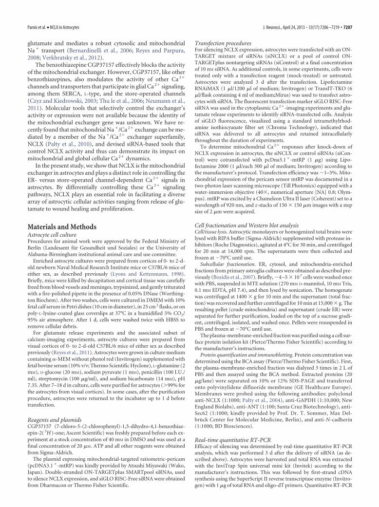
ResultsNCLX is enriched in the mitochondria of astrocytesRecently, NCLX has been identified as the mitochondrial Na�/Ca 2� exchanger in several cell types (Palty et al., 2010; Kim et al.,2012). To determine whether NCLX is the mitochondrial ex-changer in astrocytes, we first evaluated NCLX expression in ly-sates from cultures of cortical astrocytes by Western blot analysis.Consistent with the previous studies (Palty et al., 2010), amajor band of �60 kDa and a fainter band at �100 kDa re-lated to the SDS-stable NCLX dimer were detected. The 60kDa band was also detected in brain lysates from newborn
Figure 1. A, immunoblot analysis of NCLX expression in brain extracts of newborn mice, primary murine astrocytes, and themurine astrocytoma cell line GL261 (top). GAPDH serves as an internal loading control (bottom). B, NCLX is enriched in themitochondria of astrocytes. NCLX expression in indicated cellular compartments: total, crude ER, cytosol, plasma membrane (PM),ER, and mitochondria (Mito)-enriched fractions (top). The membranes were stripped and reprobed for PM (N-cadherin), ER(Sec-62), and mitochondrial (ANT) markers (bottom). C, Astrocytes are viable after 100% efficient siRNA transfection. Top rowshows untreated control astrocytes (no siRNA); bottom row shows astrocytes treated with siGLO and siNCLX (siGLO� siNCLX). Left:Astrocytes accumulated calcein (green), indicating their viability. Nuclei are marked with Hoechst 33342 in blue. Right: Redfluorescence of the same area; untreated astrocytes display dim autofluorecence, whereas siRNA (siGLO � SiNCLX)-treated cellsshow punctate stain, consistent with intracellular accumulation of the transfection marker siGLO. Scale bar, 20 �m. D, QuantitativeRT-PCR analysis after cotransfection with mtRP and siNCLX or siControl, respectively. Untreated and mock-treated (tranfectionagent only) cells were used as additional controls. Experiment was performed four times, each in triplicate. Values are given asmeans with SEM (*p�0.05, Wilcoxon-Mann–Whitney U test). E, Immunoblot analysis showing NCLX expression in untreated andmock-treated astrocytes, or transfected with siControl or siNCLX (top). GAPDH serves as an internal loading control (bottom).
Parnis et al. • NCLX in Astrocytes J. Neurosci., April 24, 2013 • 33(17):7206 –7219 • 7209

mice and in an astrocytoma cell line GL261, whereas the 100kDa band was not detectable in brain homogenate, but waspresent in GL261 cells (Fig. 1A).
To analyze NCLX expression in different cellular compart-ments, we performed subcellular fractionation of cultured astro-cytes and determined NCLX expression using Western blotanalysis. The fractions were counterstained with organelle-specific markers (Fig. 1B) to determine the separation quality ofthe fractions. Total, crude ER, and ER-enriched fractions re-vealed a faint band at �60 kDa, whereas no band was visible inthe plasma membrane or cytosolic fractions. Interestingly, themitochondrial fraction showed a strong NCLX-positive band ofslightly reduced molecular weight (MW), suggesting that the pas-sage of NCLX to the mitochondria may be linked to posttransla-tional proteolysis. Probing with anti-ANT and anti-N-cadherinantibodies showed a good separation of the mitochondrial andplasma membrane fractions, respectively. Some degree of ERcross-contamination was detected in other fractions when prob-ing the ER marker Sec-62, which is likely related to the physicalcross-linking of the ER with other organelles. Nonetheless, thesedata show that the astrocytic Na�/Ca 2� exchanger NCLX is pri-marily located in mitochondria.
Molecular silencing of NCLX in astrocytesTo assess the role of NCLX in modulating astrocytic Ca 2� signal-ing and related functions, the expression of the exchanger wasknocked down with an NCLX-specific mixture of siRNAs(siNCLX). The fluorescently tagged siRNA siGLO was used as acotransfection marker to determine optimal conditions for effec-tive NCLX silencing and to detect transfected cells; calcein livecell staining was applied to assess cell viability (�99%). Cellstreated with siRNA (siGLO � siNCLX) showed punctate redstaining, consistent with intracellular accumulation of the trans-fection marker siGLO (Fig. 1C, lower right). siGLO fluorescencewas visible in all treated viable cells, showing that cells were suc-cessfully transfected. The untreated cells showed only dim auto-fluorescence (Fig. 1C, upper right). All astrocytes showed asimilar accumulation of calcein regardless of whether they weretreated with siRNA or were untreated controls (Fig. 1C, left col-umn), indicating that the siRNA transfection procedure did notaffect cell viability.
The efficiency of NCLX silencing via siRNA was assessed byquantitative RT-PCR. Optimal NCLX gene silencing via siRNAwas achieved after 3 d using 10 nM siNCLX. NCLX expression inthe presence of siNCLX was reduced to 24.9 � 1.3% of the ex-pression level seen in astrocytes transfected with siControl (Fig.1D, n � 4 experiments, *p � 0.05). NCLX mRNA expressionlevels remained almost unchanged in cells treated only with thetransfection agent (mock-treated) and in untreated control cells(84.0 � 13.9% and 101.7 � 4.5%, respectively). Consequently, allexperiments were performed on astrocytic cultures 3 d aftersiRNA treatment.
NCLX silencing on mRNA level also affected the expression ofthe NCLX protein. Western blot analysis of astrocytic extractsshowed a reduction in NCLX expression in astrocytes transfectedwith siNCLX compared with astrocytes transfected with siCon-trol, mock-treated, or untreated cells (Fig. 1E).
NCLX conducts mitochondrial Ca 2� efflux in astrocytesTo monitor mitochondrial Ca 2� responses, primary astrocyteswere transfected with mtRP (Nagai et al., 2001) Expression ofmtRP reached maximal intensity �48 –72 h after transfection.mtRP expression pattern manifested a typical network-like mito-
chondrial distribution consistent with the strict mitochondriallocalization of this Ca 2� reporter (Fig. 2A). Resting mitochon-drial Ca 2� levels in astrocytes transfected with siNCLX werehigher than values obtained from mitochondria of astrocytestreated with control siRNA (Fig. 2B). Application of ATP (100�M) to astrocytes in Ca 2�-free HEPES Ringer’s solution for 100 selicited a fast rise in the mitochondrial Ca 2� corresponding to theCa 2� uptake phase followed by a slower efflux (Fig. 2C, n � 10and 11 experiments for siControl and siNCLX-transfected astro-cytes, respectively). Mitochondrial Ca 2� influx and efflux rateswere determined as described in Materials and Methods and asindicated in Fig. 2D,E, insets. Comparison of the efflux rates ofthe ATP-elicited mitochondrial Ca 2� responses in siNCLX-treated versus siControl-treated cells showed that the mitochon-drial efflux rate was decreased by 60.2% in siNCLX-treated cellscompared with siControl astrocytes (from 15.1 � 1.5 � 10�4/s to6.0 � 0.6 � 10�4/s, n � 10 and 11 experiments and n � 38 and 64regions for siControl and siNCLX conditions, respectively,***p � 0.001; Fig. 2D). Moreover, silencing of NCLX signifi-cantly increased the net mitochondrial Ca 2� influx by 28.1%compared with the siControl condition (from 8.7 � 0.6 �10�2
to 11.1 � 0.7 � 10�2; n � 10 and 11 experiments with siControl-and siNCLX- transfected astrocytes, respectively, *p � 0.05;Fig. 2E).
The effect of NCLX activity on mitochondrial Ca 2� fluxes wasalso evident when we used the pharmacological inhibitor ofmitochondrial Na�/Ca 2� exchange; application of 20 �M
CGP37157 resulted in a 62.4% reduction of the ATP-inducedmitochondrial Ca 2� efflux in treated astrocytes compared withcells treated with vehicle (DMSO) (control, 6.4 � 0.5 � 10�4/s,n � 15 experiments and 106 regions of interest; CGP37157-treated group, 2.5 � 0.5 � 10�4/s, n � 11 experiments and 75regions of interest, ***p � 0.001; Fig. 2F,G). The average netmitochondrial Ca 2� influx was increased from 8.6 � 0.4 �10�2/s to 12.5 � 0.9 �10�2/s in the presence of CGP37157 (n �15 and 11 experiments with control- and CGP37157- treatedastrocytes, respectively, *p � 0.05; Fig. 2H).
NCLX inhibition, either by specific molecular silencing or bythe pharmacological inhibitor, significantly reduced mitochon-drial Ca 2� extrusion, thereby also indirectly increasing the netCa 2� influx. These data indicate that NCLX mediates mitochon-drial Ca 2� efflux in astrocytes and thereby shapes the mitochon-drial Ca 2� transients.
NCLX shapes the ATP-induced cytosolic Ca 2� responsein astrocytesMitochondrial Ca 2� transport participates in intracellular Ca 2�
signaling (Hoth et al., 1997; Hajnoczky et al., 1999; Malli et al.,2003; Parekh, 2008). Our finding that NCLX is the mitochondrialNa�/Ca 2� exchanger in astrocytes provides us with a molecularbasis to evaluate how mitochondria respond to or shape cytosolicCa 2� responses in astrocytes and to determine the role of NCLXin this process. Astrocytes were cotransfected with siControl orsiNCLX together with the transfection marker siGLO red. After3 d, cells were loaded with fura-2 AM and cytoplasmic Ca 2�
signals in response to application of 100 �M ATP were monitoredfrom siGLO-red-positive cells, either in Ca 2�-containing buf-fer (Fig. 3A–D) or Ca 2�-free buffer (Fig. 3E–H ). As describedpreviously (Kresse et al., 2005), application of ATP in Ca2�-containing buffer evoked a rapid increase of cytosolic Ca 2�
mainly due to InsP3-receptor-mediated release of Ca 2� from theER stores, followed by an initial rapid decline and a slower decay-ing phase. The latter “elevated Ca 2� plateau” has been associated
7210 • J. Neurosci., April 24, 2013 • 33(17):7206 –7219 Parnis et al. • NCLX in Astrocytes

with Ca 2� influx from the extracellular space via store-operatedCa 2� entry (SOCE). The averaged Ca 2� transients indicate thatthe amplitude of the elevated Ca 2� plateau is reduced in NCLX-silenced astrocytes (Fig. 3A, n � 12 and 11 experiments for si-Control and siNCLX-transfected astrocytes, respectively). Toquantify the effect of NCLX silencing on the cytosolic Ca 2� tran-
sient, we determined the cumulative fura-2 AM fluorescence,peak amplitude, and half-amplitude time in siControl-treatedversus siNCLX-treated astrocytes. The cumulative fluorescencewas reduced by 39.3% in NCLX-silenced astrocytes (Fig. 3B),whereas the peak amplitude was reduced by 13.4% (Fig. 3C) andthe half-amplitude time of the signal was reduced by 9.8% (Fig.
Figure 2. NCLX mediates mitochondrial Ca 2� efflux in astrocytes. A–E, Primary astrocytes were cotransfected with mtRP and siNCLX or siControl and were analyzed after 3 d. A, Two-photonmicroscopy image of astrocytes transfected with mtRP reveals typical mitochondrial pattern of the fluorescent pericam sensor. Scale bar, 10 �m. B–E, Mitochondrial Ca 2� imaging. B, Restingmitochondrial Ca 2� levels of astrocytes transfected with siNCLX- or control siRNA, R/R0 values were determined from fluorescent mtRP imaging before application of the stimulus and normalizedto the control condition (n � 5 cultures, n � 134 and 118 regions), C, Mitochondrial Ca 2� signals of control and siNCLX-transfected astrocytes; 100 �M ATP in Ca 2�-free HEPES buffer was appliedas indicated, averaged curves � SEM. D–E, Average rates of the mitochondrial Ca 2� efflux (slopes; D) and influx (amplitudes, E) in siControl and siNCLX-transfected astrocytes, comparing n � 10(n � 38 regions) and n � 11 (n � 64 regions) experiments for each condition (***p � 0.001, *p � 0.05, Wilcoxon, Mann–Whitney U test). F–H, Effect of CGP37157 on mitochondrial Ca 2� efflux.F, Mitochondrial Ca 2� signals from control and CGP37157 (CGP)-treated astrocytes. Cells were superfused with Ca 2�-free HEPES buffer � 20 �M CGP37157and 100 �M ATP was applied asindicated; shown are averaged curves � SEM. G–H, average rates of mitochondrial efflux (G) and influx (H ). Values are given as means with SEM for n � 15 (n � 106 regions) and n � 11 (n �75 regions) control and CGP37157 experiments, respectively (***p � 0.001, *p � 0.05, Wilcoxon, Mann–Whitney U test).
Parnis et al. • NCLX in Astrocytes J. Neurosci., April 24, 2013 • 33(17):7206 –7219 • 7211

3D) in NCLX-silenced astrocytes compared with siControl-treated astrocytes (n � 12 and 11 experiments, n � 294 siControland n � 222 siNCLX-transfected astrocytes; ***p � 0.001,**p � 0.01).
To determine the contribution of mitochondrial NCLX to themodulation of Ca 2� signals that originate from ER stores, wesuperfused cells with Ca 2�-free buffer and measured ATP-induced cytosolic Ca 2� signals in NCLX-silenced and siControlastrocytes. ATP triggered a fast rise followed by a fast decline ofthe cytoplasmic Ca 2� signal as described above. The elevatedplateau phase, seen in Ca 2�-containing buffer (above) was, asexpected, reduced under Ca 2�-free conditions. Knock-down ofNCLX revealed a slight but significant reduction of the meancumulative Ca 2� signal in NCLX-silenced astrocytes, with medi-ans shifted from 102.1% in the siControl population to 89.4% inthe NCLX-silenced astrocytes (***p � 0.001; Fig. 3F). The rep-resentation of cumulative Ca 2� responses as median and inter-quartile ranges revealed that NCLX silencing is more pronouncedin the lower quartiles of the data (25 th percentile of the box plot):in those cells with overall lower cumulative Ca 2� responses, theshift was from 79% in the nonsilenced condition to 47.4% afterNCLX knock-down (Fig. 3F).
These results indicate that stimulus-induced cytoplasmicCa 2� signals in astrocytes, in particular those caused by the entryof Ca 2� from the extracellular space versus Ca 2� release from theER store, are distinctly modulated by the activity of the mito-chondrial Na�/Ca 2� exchanger NCLX.
NCLX activity modulates the store-operated Ca 2�entryin astrocytesSOCE is an important Ca 2� influx pathway in nonexcitable cellssuch as astrocytes (Kresse et al., 2005; Pivneva et al., 2008;Verkhratsky et al., 2012). Observations in other cell types show-ing that the mitochondrial Ca 2� shuttling machinery is essentialto sustaining the activity of SOCE (Hoth et al., 1997; Malli et al.,2003; Parekh, 2008, and the reduction of the Ca 2�-elevated pla-teau phase seen in the ATP-induced Ca 2� signal in NCLX-silenced astrocytes (described above) prompted us to determinethe specific influence of mitochondrial NCLX on Ca 2� influxthrough the SOCE pathway in astrocytes.
To quantify SOCE activity, we used a previously describedprotocol. Briefly, ER stores were depleted by application of ATPin the absence of extracellular Ca 2�. Cells were then superfusedwith Ca 2�-containing buffer, which resulted in a Ca 2� influx dueto SOCE activity (Kresse et al., 2005; Fig. 4A). The amplitude ofSOCE response measured at the steady phase was decreased from100.0 � 5.1% to 59.9 � 3.1% after silencing of NCLX (n � 9experiments; n � 175 and 131 cells for siControl and siNCLX-transfected astrocytes, respectively, ***p � 0.001; Fig. 4B). More-over, the SOCE rate (reflected by the slope of the influx phase)was reduced from 100.0 � 3.6% to 68.0 � 3.7% by NCLX silenc-ing (***p � 0.001; Fig. 4C). A similar impairment on SOCE func-tion was found in astrocytes superfused with 20 �M CGP3715.The drug reduced SOCE amplitude by 25.5% and the SOCE rateby 34.2% compared with vehicle-treated control (n � 8 and 7
Figure 3. NCLX modulates ATP-induced Ca 2� responses in astrocytes. Primary astrocytes were cotransfected with siControl or siNCLX and siGLO red indicator. Cells were loaded with fura-2 AMand changes in cytoplasmic Ca 2� signals were monitored from siGLO-red-positive cells. A, E, Recording of average fura-2 AM fluorescence for control and siNCLX-transfected astrocytes inCa 2�-containing buffer (A) and Ca 2�-free buffer (E); 100 �M ATP was added as indicated, averaged curves SEM. B–D, Statistical analysis of the recordings shown in A: cumulative response (B),amplitudes (C), and half-amplitude time (D) of the ATP-evoked Ca 2� responses. Values are given as mean with SEM (n � 12 experiments, n � 294 cells for control, and n � 11 experiments, n �222 cells for siNCLX-transfected astrocytes, respectively; ***p � 0.001, **p � 0.01, Wilcoxon, Mann–Whitney U test). F, Box-plot chart of the cumulative Ca 2� responses corresponding torecordings in E indicating median (dark line), interquartile range (box), and minimum and maximum of the values (whiskers) (***p � 0.001, Wilcoxon, Mann–Whitney U test).
7212 • J. Neurosci., April 24, 2013 • 33(17):7206 –7219 Parnis et al. • NCLX in Astrocytes

experiments, n � 376 and 425 cells for control and CGP37157-treated astrocytes, ***p � 0.001; Fig. 4D–F). Similarly, applica-tion of the protonophore FCCP, which leads to a collapse ofmembrane potential and a diminished calcium-buffering capacity ofmitochondria, caused reductions in both the SOCE amplitudeand rate (Fig. 4D–F). These results suggest that mitochondrialCa 2� shuttling, and in particular NCLX activity, is of major im-portance for the regulation of Ca 2� influx into the cell throughSOCE.
We next investigated whether Ca 2� influx via the SOCE path-way evokes mitochondrial Ca 2� transients and how NCLX activ-ity regulates this cross talk. We adapted the Ca 2� re-admissionprotocol described above to mtRP-expressing astrocytes and de-termined mitochondrial Ca 2� transients. In this set of experi-ments, we measured only the wavelength F430 of the mtRP signalbecause ratiometric measurements of mtRP fluorescence F480/F430 during SOCE for unknown reasons displayed a decrease inthe signal-to-noise ratio. SOCE evoked a fast rise in mitochon-
Figure 4. NCLX controls SOCE in astrocytes. A–C, Imaging of cytosolic Ca 2� in fura-2 AM-loaded primary murine astrocytes. Astrocytes were cotransfected with siControl or siNCLX and siGLO red indicator.Cells were perfused in Ca 2�-free HEPES buffer and 100�M ATP was applied to empty ER Ca 2� stores; then 5 mM Ca 2�was re-added as indicated by the bar to trigger the activation of SOCE. A, Traces of averagefura-2AMfluorescence(normalizedaverages)duringSOCE.B–C,Amplitudes(B)andratesofSOCE(C) insiControl(n�9,n�175)andsiNCLX-transfectedastrocytes(n�9,n�131).Valuesaregivenasmeanwith SEM (***p�0.001, Wilcoxon, Mann–Whitney U test). D–F, Untransfected astrocytes were perfused in Ca 2�-free HEPES buffer without (Control) or with 20�M CGP37157 or with 1�M FCCP. After adding100�M ATP to empty the ER stores, 5 mM Ca 2�was added as indicated. D, Mean Ca 2� response curve of all experiments (normalized averages). E–F, Statistical analysis of the amplitudes (E) and SOCE rates (F )in astrocytes treated with CGP37157(CGP), FCCP, or without any drug (Control). Values are given as mean with SEM summarized from n � 8 (n � 376 cells), n � 7 (n � 425 cells), and n � 5 (n � 168 cells)experiments for control, CGP37157, and FCCP-treated astrocytes, respectively (***p � 0.001, Kruskal-Wallis rank-sum test followed by Bonferoni’s posttest and Wilcoxon test pair comparisons). G–J,SOCE-induced mitochondrial Ca 2� signals in astrocytes cotransfected with mtRP and siControl or siNCLX using the same paradigm described in A. G, Representative curve showing the averaged mitochondrialCa 2� response. H–J,RatesofthemitochondrialCa 2�efflux(H ), influx(I ),andcumulativefluorescence(J ) fromn�9experiments(n�59andn�43regionsof interestforsiControl-andsiNCLX-transfectedastrocytes, respectively; ***p � 0.001, Student’s t test for unpaired samples and two-tailed and Mann–Whitney U test).
Parnis et al. • NCLX in Astrocytes J. Neurosci., April 24, 2013 • 33(17):7206 –7219 • 7213

drial Ca 2� signal followed by a gradual efflux (Fig. 4G). In NCLX-silenced astrocytes, mitochondrial Ca 2� efflux rate was clearlyreduced compared with siControl-transfected cells (1.67 �0.22 � 10�4/s and 0.28 � 0.26 �10�4/s, respectively, n � 9experiments; ***p � 0.001; Fig. 4H). Concomitantly, upon acti-vation of SOCE, the net influx of Ca 2� into the mitochondria wasincreased by 48.8% (***p � 0.001; Fig. 4I) and the cumulativemitochondrial Ca 2� signal was elevated by 60.3% (***p � 0.001;Fig. 4J) in NCLX-silenced astrocytes compared with the nonsi-lenced control.
These results indicate that an active cross talk exists betweenSOCE and mitochondria and that Ca2� extrusion through NCLX iscrucial for maintaining a strong SOCE activity in astrocytes.
NCLX- and Ca 2�-dependent glutamate releasefrom astrocytesTo determine the impact of NCLX on Ca2�-dependent functions ofastrocytes, we studied the secretion of glutamate via the exocytotic/vesicular pathway induced by mechanical stimulation of astrocytes,which involves an increase of cytosolic Ca2� (Innocenti et al., 2000;Hua et al., 2004; Montana et al., 2004). Mitochondria modulate themagnitude of this mechanically induced excitability (Reyes and Par-pura, 2008). Interestingly, the pharmacological inhibitor of the mi-
tochondrial Na�/Ca2� exchanger, CGP37157, has been shownpreviously to reduce mechanically induced Ca2� responses and glu-tamate release from rat solitary astrocytes in vitro (Reyes and Par-pura, 2008). In the present study, we assessed whether the molecularsilencing of NCLX produced similar effects on astrocytic Ca2� ex-citability and gliotransmission.
Astrocytes were transfected with the transfection markersiGLO, along with either siNCLX or a nontargeted siRNA inthe control group and then loaded with fluo-3 AM to measurecytosolic Ca 2�. Similar to the purinergic receptor stimulationdescribed above, mechanical stimulation caused an initial tran-sient Ca 2� elevation (n � 15; peak dF/F0 � 478 � 43%; **p �0.01, paired t test) followed by a slowly decaying response incontrol solitary astrocytes transfected with the nontargetedsiRNA (Fig. 5A). The transient peak was described to indicate theCa 2� entry into the cytosol predominately from the ER store andfrom the extracellular space (Hua et al., 2004; Malarkey et al.,2008). Our data revealed a twofold decrease in both the peak (n �15; peak dF/F0 � 283 � 52%) and a strong, fivefold decrease incumulative cytosolic Ca2� levels in siNCLX-treated cells (**p �0.01, Mann–Whitney U test; Fig. 5A–C). The knock-down of NCLXhad a much larger inhibitory effect on the Ca2� response than the
Figure 5. NCLX mediates increase of cytosolic Ca 2� and exocytotic glutamate release in astrocytes after mechanical stimulation. A–C, Cytosolic Ca 2� measurements. A, Time lapse of averagefluo-3 AM fluorescence, reporting on changes in cytoplasmic Ca 2�, in solitary astrocytes transfected with either siControl or siNCLX in response to mechanical stimulation; all cells were cotransfectedwith the transfection marker siGLO. B, Normalized fluo-3 AM peak. C, Cumulative fluorescence values of mechanically stimulated solitary astrocytes in A. D–F, Extracellular glutamate measurements.D, Average kinetics of extracellular NADH fluorescence showing mechanically induced changes in extracellular glutamate surrounding somata of solitary astrocytes that were transfected as in A. E,F, Normalized peak (E) and cumulative glutamate (Glut; F ) release from mechanically stimulated solitary astrocytes in D. Solitary astrocytes transfected with siNCLX displayed significantly(Mann–Whitney U test, **p � 0.01) reduced increase in mechanically induced Ca 2� signals and glutamate release compared with siControl-transfected cells. The points and bars represent meanswith SEMs of measurements from individual astrocytes (n � 15); Arrows in A and D represent the time when mechanical stimulation was applied to the cells.
7214 • J. Neurosci., April 24, 2013 • 33(17):7206 –7219 Parnis et al. • NCLX in Astrocytes

pharmacological inhibition of this exchanger by CGP37157, as de-scribed previously (Reyes and Parpura, 2008).
We next investigated the role of NCLX in Ca 2�-dependentexocytotic glutamate release from astrocytes. We used a GDH-linked assay based on accumulation of the fluorescent productNADH (Hua et al., 2004; Montana et al., 2004). Mechanical stim-ulation of the siControl-treated, solitary astrocytes evoked gluta-mate release, as indicated by a transient increase in NADHfluorescence (n � 15; peak dF/F0 � 50 � 11%; **p � 0.01, pairedt test; Fig. 5D), corresponding to glutamate surrounding the as-trocytic somata. We observed a significant decrease in both nor-malized peak (n � 15; peak dF/F0 � 47 � 6%) and particularly incumulative fluorescence intensity for glutamate release. Afterstimulation, basal levels of glutamate release was lower, whenNCLX expression was knocked down when compared with thesiControl group (**p � 0.01, Mann–Whitney U test; Fig. 5E,F).These data indicate that NCLX plays an essential role in medi-ating cytosolic Ca 2�responses in astrocytes required for exocy-totic glutamate gliotransmission.
Mechanically induced astrocytic Ca 2�
waves are not affected by the activityof NCLXCa 2� waves in astrocyte networks consti-tute a form of intercellular communica-tion and provide astrocytes with a specificform of excitability. In cultured astro-cytes, Ca 2� signals can spread to neigh-boring cells and can propagate as a wavethrough many cells (Cornell-Bell et al.,1990; Dani et al., 1992; Schipke et al.,2002). Although different mechanismshave been described to account for the in-duction and maintenance of intercellu-lar astroglial Ca 2� waves (Scemes andGiaume, 2006), there is nothing known sofar on the role of mitochondria in themaintenance of the intercellullar astro-cytic Ca 2� waves and whether Ca 2� effluxthrough NCLX contributes to the controlof wave propagation in an astrocyticmonolayer. We therefore measured veloc-ities of Ca 2� waves in fluo-4 AM-loadedastrocytes that had been treated with siN-CLX or control siRNA (Fig. 6A). An astro-cyte within the monolayer was mechanicallystimulated, as described above, and wave ve-locity was calculated for each cell by an algo-rithm that measures the distance from thestimulation point and the time at which thewave reached a given cell. Although the av-eraged velocity of the Ca2� wave was 23.9 �3.3 �m/s for siControl astrocytes, and18.8 � 2.3 �m/s on average for the NCLX-silenced astrocytes, it was not significantlydifferent (Fig. 6B). A wave was initiated as arapid onset, followed by centrifugal propa-gation with almost linear velocity. The aver-aged time course of the waves is shown inFigure 6C. The mean data points were fittedto the following power functions: insiControl-treated astrocytes, distance �82 � 6.7 � time 0.35 � 0.02, R 2 � 0.92,and in NCLX-treated astrocytes, dis-
tance � 74 � 6.6 � time 0.36 � 0.02, R 2 � 0.82). The slight reduc-tion in the Ca 2� wave velocity that we observed in NCLX-silenced astrocytes was not significant, which suggests that theactivity of the mitochondrial exchanger only marginally, if at all,affects the Ca 2� signaling machinery contributing to propaga-tion of intercellular Ca 2� waves in astrocytes.
NCLX participates in control of astrocytic wound healingin vitroAstrocyte migration and proliferation represent important cellu-lar aspects of the brain’s response to injury and during regenera-tion. Intracellular Ca 2� signaling is critical to the regulation ofthese cellular responses (Xu et al., 2004; Valero et al., 2008; Wei etal., 2009; Feldman et al., 2010). Therefore, we investigatedwhether the mitochondrial Na�/Ca 2� exchanger NCLX affectsthese functional parameters in astrocytes.
To assess the effect of NCLX on astrocytic wound healing, weadapted an in vitro assay described previously (Kornyei et al.,2000; Matyash et al., 2002). Monolayers of astrocytes that had
Figure 6. NCLX does not control the velocity of mechanically evoked Ca 2� waves in astrocytes. A, Ca 2� waves were induced bymechanical stimulation in confluent astrocyte cultures, transfected with siControl or siNCLX, and loaded with fluo-4 AM to imagecytosolic Ca 2�. A, Series of subtraction images showing the concentric propagation of a Ca 2� wave as induced by poking anastrocyte with a micropipette (bright field image, top). B, For each experiment, Ca 2� wave propagation velocity was calculated byaveraging the velocities at which the Ca 2� wave reached each of the cells on a coverslip. (Wilcoxon, Mann–Whitney U test, p �0.05, n � 5 astrocytic preparations, n � 20 and 28 coverslips for control and siNCLX, respectively). C, Average time course of thepropagation of the Ca 2� wave. The wave was initiated as a fast burst that propagated centrifugally close to a linear velocity. Meandata points could be fitted to a power function: distance � A � timeb. For siControl-treated astrocytes, A � 82 � 6.7 �m andb � 0.35 � 0.02 (R 2 � 0.92), and for siNCLX-treated astrocytes, A � 74 � 6.64 �m and b � 0.36 � 0.02 (R 2 � 0.82). Thevalues for A and b are given as mean � SD. Values in the graph show mean � SEM. Fitting curves are presented as dashed lines.
Parnis et al. • NCLX in Astrocytes J. Neurosci., April 24, 2013 • 33(17):7206 –7219 • 7215

either been treated with NCLX siRNA orwith control siRNA, as well as mock-transfected and untreated astrocytes, werescratched as indicated in Figure 7B andthe cell-free area was measured immedi-ately after scratching as described byGeback et al. (2009) (Fig. 7A). Thereafter,repopulation was analyzed repeatedly48–72 h after scratching until the woundarea was closed by at least two-thirds in theuntreated control (end point). Molecular si-lencing of NCLX with siRNA significantlyimpaired the astrocytic ability to repopulatethe cell-free area (Fig. 7B,C). Although thescratch was closed by more than two-thirdsafter 48–72 h in siControl cultures, it re-mained much larger when NCLX was in-hibited: the mean open wound area inNCLX-silenced astrocytes was 48.5 � 2.6%on average compared with 24.6 � 2.5% forthe siControl conditions (n � 3 experi-ments, n � 20 scratches, ***p � 0.001; Fig.7C), and these observations were confirmedin cultures treated with 20 �m CGP37157throughout the regrowth phase (data notshown), indicating that NCLX activity iscrucial for astrocytic migration and woundclosure in vitro.
To assess whether NCLX exerts an ef-fect on astrocyte proliferation, we per-formed cell proliferation assays based onBrdU incorporation in noninjured cul-tures. Astrocyte proliferation was de-creased by 38.6% in NCLX-silencedastrocyte cultures compared with astro-cytes transfected with nontargeted siRNA(siControl, n � 3 experiments, n � 15wells, ***p � 0.001; Fig. 7D). A possibleharmful side effect of the transfec-tion procedure can be excluded becausethe proliferation in the siControl andmock-transfected cultures was not im-paired compared with the untreatedcontrol astrocytes.
These findings indicate that the mitochondrial Na�/Ca 2� ex-changer NCLX is involved in the control of astrocytic woundclosure and proliferation.
DiscussionMitochondria are critically involved in Ca2� handling in astrocytes,thereby modifying astrocytic functions (Verkhratsky et al., 2012). Inthe present study, we addressed the specific role of NCLX in shap-ing astrocytic mitochondrial and cytoplasmic Ca 2� signalsand studied its impact on Ca 2�-dependent astrocyte func-tions. We show that NCLX, which is enriched in astrocyticmitochondria, mediates Ca 2� extrusion from mitochondria.In addition, NCLX predominately modulated the SOCE path-way, whereas its effect on the ER-dependent Ca 2� release wasminor. Therefore, NCLX activity has a major impact on modu-lating cytoplasmic Ca 2�signals in astrocytes, as well as their func-tional output, exocytotic glutamate release, wound closure, andproliferation, but does not significantly influence the propaga-
tion of mechanically induced Ca 2� waves in astrocyticmonolayers.
Our immunochemical approach verified that the NCLX proteinis expressed in astrocytes and occurs as an �60 kDa and an �100kDa form that is consistently related to an SDS-resistant NCLXdimer (Palty et al., 2004; Palty et al., 2006). Comparison of NCLXexpression in different organelle-enriched fractions of astrocytes re-vealed a strong enrichment of NCLX in the mitochondria, indicatingthat it is primarily located in these organelles. The MW of mitochon-drial NCLX, interestingly, was slightly reduced to �50 kDa com-pared with the other cellular compartments. Although thephysiological role of such processing is not yet clear and we cannotrule out whether NCLX cleavage occurred during mitochondrialisolation, the reduction in MW is consistent with the sensitivity ofNCLX to mitochondrial proteases such as mitochondrial calpains(Kar et al., 2009; Smith and Schnellmann, 2012). Alternatively, be-cause our antibody is targeted to the C-terminal domain of NCLX,cleavage is likely to occur on the NH3-terminal NCLX domains. TheN-terminal domains usually harbor the mitochondria-targetingpeptide that can be cleaved during mitochondrial insertion (Moss-
Figure 7. NCLX participates in astrocytic in vitro wound healing and proliferation. A, Confluent astrocytic monolayers were scratched asdescribed in the Materials and Methods, and phase contrast images were acquired at 0 h (top row) and after 48 –72 h (bottom row) at lowmagnification. Left column shows raw images and right column shows the same images after analysis with T-scratch software. Dark grayareas correspond to the open wound area; values at the end point are normalized to a 100% area at the starting point (0 h). Scale bar, 100�m. B, Scheme exemplifying how scratches (white lines) were applied to astrocyte monolayers (gray areas). Black frames correspond tothe analyzed areas of interest. C, Percentage of open wound area in monolayers that were untransfected, mock-transfected, or transfectedwith siControl or siNCLX (n � 3 cultures, n � 18, 16, 20, and 20 scratches, respectively). Values are given as means with SEM (***p �0.001, Student’s t test for unpaired samples, two-tailed). D, BrdU incorporation into proliferating astrocytes that were untransfected,mock-transfected,ortransfectedwithsiControlorsiNCLX,givenaspercentageproliferationnormalizedtocontrol(n�3experiments,n�15 wells; ***p � 0.001, Mann–Whitney U test).
7216 • J. Neurosci., April 24, 2013 • 33(17):7206 –7219 Parnis et al. • NCLX in Astrocytes

mann et al., 2012). Although these mechanisms need to be investi-gated further, they raise interesting questions regardingposttranslational regulation of NCLX in mitochondria.
NCLX modulates glial Ca 2� signalingAn important finding of our study is that NCLX activity pro-motes SOCE in astrocytes. Sustained Ca 2� influx through SOCEresults in prolonged intracellular Ca 2� transients that are partic-ularly important in controlling slow cellular processes such assecretory activity and cell proliferation (Hoth et al., 1997;Golovina, 1999; Golovina et al., 2001; Berridge et al., 2003).
Several studies have documented a pivotal role of the mito-chondrial exchanger in the replenishment of the ER Ca 2� stores(Arnaudeau et al., 2001; Malli et al., 2005; Kim et al., 2012). Wefound that ER Ca 2� release, which can be considered an indica-tive value for ER Ca 2� content, was not much altered by theknock-down of NCLX expression. Although the molecular basisfor this relatively minor influence of NCLX on ER Ca 2� needs tobe explored further, it may be related to the major role of SERCAin refilling the astrocytic Ca 2� stores, even under NCLX defi-ciency. In addition, transfer of Ca 2� from the mitochondria tothe ER Ca 2� stores is facilitated by the tight physical interactionsmediated by an ER–mitochondria-tethering protein complexand direct Ca 2� transport through these compartments via thevoltage-dependent anion channels (Szabadkai et al., 2006; Korn-mann et al., 2009; de Brito and Scorrano, 2010). Our resultssuggest that such cross talk between the the ER and mitochondriais relatively weak in astrocytes. This assertion is consistent withour findings that the strong cytosolic Ca 2� response triggered byER Ca 2� release was followed by only a modest mitochondrialCa 2� transient compared with a much stronger mitochondrialCa 2� transient triggered by Ca 2� influx into the cells. The lattereffect suggests a stronger interaction of NCLX with the SOCEpathway. It is not surprising that astrocytic intracellular Ca 2�
dynamics are inherently linked to those of Na� (Kirischuk et al.,2012). Therefore, in addition to obvious exchange of these ionsvia NCLX, cytoplasmic Na� concentrations would also be af-fected by diverse plasma membrane receptors and transportersinvolved in Na� exchange, among them SOCE and/or P2XRs,and the cytosolic Na� load thus affects a multitude of astroglialhomeostatic functions (Kirischuk et al., 2012).
Mitochondrial metabolism is likely to be affected by the activ-ity of NCLX, because Ca 2� activates several enzymes of the Krebscycle (Szabadkai and Duchen, 2008). NCLX, by accelerating mi-tochondrial Ca 2� shuttling, increases the duration of mitochon-drial Ca 2� transients and thereby is likely to enhance ATPproduction.
NCLX activity differentially affects migration/proliferationWe used an in vitro model to study the impact of NCLX functionon the ability of astrocytes to populate a cell-free area as a modelfor wound healing. The repopulation of the cell-free “wound”area is primarily due to astrocyte proliferation, although migra-tion does also contribute to this process (Kornyei et al., 2000). Arole of SOCE in wound closure has been proposed previously(Rao et al., 2006). Our study strongly suggests that mitochondrialCa 2� transfer through NCLX is essential to facilitate SOCE, andtherefore may affect astrocytic proliferation and the regrowth ofthe astrocytes into the denuded areas of the wound. Although thedownstream mitogenic signaling pathways that link the Ca 2�
response to migration and proliferation still need to be investi-gated, earlier studies have highlighted the role of the mitochon-drial exchanger in activating the PI3 kinase pathway by
accelerating SOCE-related cellular Ca 2� rise (Feldman et al.,2010).
NCLX activity does not affect astrocytic Ca 2�
wave propagationPropagating Ca 2� waves are a major communication pathway ofthe astrocytic network and of neuron–astrocyte communication.Different mechanisms account for the induction and mainte-nance of intercellular astroglial Ca 2� waves. These involve therelease of gliotransmitters, as well as direct diffusion of InsP3
through gap junctions into neighboring astrocytes, where it acti-vates InsP3 receptors and subsequent release of Ca 2� from ERstores (Scemes and Giaume, 2006). On a single-cell level (intra-cellular Ca 2� waves), mitochondrial Ca 2� buffering has beendescribed as an important means to attenuate excess Ca 2� levelsat the InsP3R microdomains (Boitier et al., 1999) to keep theamplification working. However, little is known about the role ofmitochondria in the maintenance of the intercellullar astrocyticCa 2� waves and whether Ca 2� efflux through NCLX contributesto the control of wave propagation in an astrocytic monolayer.Our results show that the knock-down of NCLX does not inducea significant change in astrocyte Ca 2� wave propagation velocity.The less dominant role of the mitochondrial exchanger in wavepropagation may be related to at least two issues. First, the rates ofmitochondrial Ca 2� uptake are approximately two orders ofmagnitude faster than mitochondrial Ca 2� efflux, which spansover tens of seconds (Fig. 2C; Palty et al., 2010). However, thepropagation of the cytosolic astrocytic Ca 2� wave is much morerapid, thus crossing a single glial cell within less than a second. Atthis rate, the contribution of the slower mitochondrial exchangerto cytosolic Ca 2� is predicted to be minimal. Second, in addition,Ca 2� release from the ER is the dominant pathway providingCa 2� during wave propagation. Because ER Ca 2� release is notstrongly affected by the knock-down of NCLX, it is thus less likelyto play a dominant role in propagation of the Ca 2� wave.
NCLX is instrumental in increasing cytosolic Ca 2� fortriggering release of gliotransmitter glutamatePrevious work used a pharmacological approach to assess the roleof the mitochondrial Na�/Ca 2� exchanger in cytoplasmic Ca 2�
dynamics in astrocytes: inhibition by CGP37157 resulted in areduction of mechanically induced Ca 2� responses and gluta-mate release recorded from cultured rat solitary astrocytes (Reyesand Parpura, 2008). The molecular identification of NCLX (Paltyet al., 2010) now provides a more specific, siRNA-based molecu-lar tool with which to study its role on mechanically inducedCa 2� responses and glutamate release from astrocytes. Althoughour data are consistent with previous pharmacological data, theyprovide new insight on the role of the exchanger. Most notably,the molecular knock-down of NCLX triggered a more profoundinhibitory effect on mechanically induced increase of cytosolicCa 2�and subsequent exocytotic release of glutamate than thepharmacological approach (Reyes and Parpura, 2008). The rea-son for the differences between the acute pharmacological versusthe siRNA approach could be the multiple sites of CGP37157action, among them Ca 2� transporters. The molecular knock-down approach is specific for NCLX, whereas the pharmacolog-ical approach may partially mask the full effect of themitochondrial exchanger on glutamate secretion due to side ef-fects on other transport molecules. Because vesicular glutamatetransporter 3 and cytoplasmic glutamate levels in astrocytes reg-ulate the magnitude of exocytotic glutamate release from astro-cytes (Ni and Parpura, 2009), a possible explanation for
Parnis et al. • NCLX in Astrocytes J. Neurosci., April 24, 2013 • 33(17):7206 –7219 • 7217

unparalleled changes in cytoplasmic Ca 2� and Ca 2�-dependentglutamate release could be that astrocytes lacking NCLX haveless glutamate available for vesicular storage and release. De novoglutamate synthesis relies on the mitochondrial matrix enzymepyruvate carboxylase (Hertz et al., 1999), which is activated byCa 2�(Civelek et al., 1996). However, our observation that theknock-down of NCLX results in a sustained increase, rather thana decrease, in mitochondrial free Ca 2� would not support such ascenario. Therefore, it is tempting to hypothesize that NCLX, bymodulation of glutamate release from astrocytes, has a role insynaptic transmission and plasticity at the tripartite synapse.
ReferencesAgulhon C, Petravicz J, McMullen AB, Sweger EJ, Minton SK, Taves SR,
Casper KB, Fiacco TA, McCarthy KD (2008) What is the role of astro-cyte calcium in neurophysiology? Neuron 59:932–946. CrossRef Medline
Arnaudeau, S, Kelley WL, Walsh JV, Jr, and Demaurex N. (2001) Mitochon-dria recycle Ca(2�) to the endoplasmic reticulum and prevent the deple-tion of neighboring endoplasmic reticulum regions. J Biol Chem276:29430 –29439.
Baughman JM, Perocchi F, Girgis HS, Plovanich M, Belcher-Timme CA,Sancak Y, Bao XR, Strittmatter L, Goldberger O, Bogorad RL, KotelianskyV, Mootha VK (2011) Integrative genomics identifies MCU as an essen-tial component of the mitochondrial calcium uniporter. Nature 476:341–345. CrossRef Medline
Bernardinelli Y, Azarias G, Chatton JY (2006) In situ fluorescence imagingof glutamate-evoked mitochondrial Na� responses in astrocytes. Glia54:460 – 470. CrossRef Medline
Berridge MJ, Bootman MD, Roderick HL (2003) Calcium signalling: dy-namics, homeostasis and remodelling. Nat Rev Mol Cell Biol 4:517–529.CrossRef Medline
Boitier E, Rea R, Duchen MR (1999) Mitochondria exert a negative feed-back on the propagation of intracellular Ca2� waves in rat cortical astro-cytes. J Cell Biol 145:795– 808. CrossRef Medline
Bozidis P, Williamson CD, Colberg-Poley AM. (2007) Isolation of endo-plasmic reticulum, mitochondria, and mitochondria-associated mem-brane fractions from transfected cells and from human cytomegalovirus-infected primary fibroblasts. Curr Protoc Cell Biol. Chapter 3:Unit 3.27.CrossRef Medline
Civelek VN, Deeney JT, Shalosky NJ, Tornheim K, Hansford RG, Prentki M,Corkey BE (1996) Regulation of pancreatic beta-cell mitochondrial me-tabolism: influence of Ca2�, substrate and ADP. Biochem J 318:615– 621.Medline
Contreras L, Drago I, Zampese E, Pozzan T (2010) Mitochondria: the cal-cium connection. Biochim Biophys Acta 1797:607– 618. CrossRefMedline
Cornell-Bell AH, Finkbeiner SM, Cooper MS, Smith SJ (1990) Glutamateinduces calcium waves in cultured astrocytes: long-range glial signaling.Science 247:470 – 473. CrossRef Medline
Czyz A, Kiedrowski L (2003) Inhibition of plasmalemmal Na(�)/Ca(2�)exchange by mitochondrial Na(�)/Ca(2�) exchange inhibitor 7-chloro-5-(2-chlorophenyl)-1,5-dihydro-4,1-benzothiazepin-2( 3H)-one (CGP-37157) in cerebellar granule cells. Biochem Pharmacol 66:2409 –2411.CrossRef Medline
Dani JW, Chernjavsky A, Smith SJ (1992) Neuronal activity triggers calciumwaves in hippocampal astrocyte networks. Neuron 8:429 – 440. CrossRefMedline
de Brito OM, Scorrano L (2010) An intimate liaison: spatial organization ofthe endoplasmic reticulum-mitochondria relationship. EMBO J 29:2715–2723. CrossRef Medline
De Stefani D, Raffaello A, Teardo E, Szabo I, Rizzuto R (2011) A forty-kilodalton protein of the inner membrane is the mitochondrial calciumuniporter. Nature 476:336 –340. CrossRef Medline
Drago I, Pizzo P, and Pozzan T. (2011) After half a century mitochondrialcalcium in- and efflux machineries reveal themselves. EMBO J 30:4119 –4125. CrossRef Medline
Feldman B, Fedida-Metula S, Nita J, Sekler I, Fishman D (2010) Coupling ofmitochondria to store-operated Ca(2�)-signaling sustains constitutiveactivation of protein kinase B/Akt and augments survival of malignantmelanoma cells. Cell Calcium 47:525–537. CrossRef Medline
Geback T, Schulz MM, Koumoutsakos P, Detmar M (2009) TScratch: a
novel and simple software tool for automated analysis of monolayerwound healing assays. Biotechniques 46:265–274. CrossRef Medline
Golovina VA (1999) Cell proliferation is associated with enhanced capacita-tive Ca(2�) entry in human arterial myocytes. Am J Physiol 277:C343–349. Medline
Golovina VA, Platoshyn O, Bailey CL, Wang J, Limsuwan A, Sweeney M,Rubin LJ, Yuan JX (2001) Upregulated TRP and enhanced capacitativeCa 2� entry in human pulmonary artery myocytes during proliferation.Am J Physiol Heart Circ Physiol 280:H746 –H755. Medline
Hajnoczky G, Hager R, Thomas AP (1999) Mitochondria suppress localfeedback activation of inositol 1,4, 5-trisphosphate receptors by Ca 2�.J Biol Chem 274:14157–14162. CrossRef Medline
Hertz L, Dringen R, Schousboe A, Robinson SR (1999) Astrocytes: gluta-mate producers for neurons. J Neurosci Res 57:417– 428. CrossRefMedline
Hoth M, Fanger CM, Lewis RS (1997) Mitochondrial regulation of store-operated calcium signaling in T lymphocytes. J Cell Biol 137:633– 648.CrossRef Medline
Hua X, Malarkey EB, Sunjara V, Rosenwald SE, Li WH, Parpura V (2004)Ca2�-dependent glutamate release involves two classes of endoplasmicreticulum Ca2� stores in astrocytes. J Neurosci Res 76:86 –97. CrossRefMedline
Innocenti B, Parpura V, Haydon PG (2000) Imaging extracellular waves ofglutamate during calcium signaling in cultured astrocytes. J Neurosci20:1800 –1808. Medline
Jiang D, Zhao L, Clapham DE (2009) Genome-wide RNAi screen identifiesLetm1 as a mitochondrial Ca2�/H� antiporter. Science 326:144 –147.CrossRef Medline
Kann O, Kovacs R (2007) Mitochondria and neuronal activity. Am J PhysiolCell Physiol 292:C641– 657. CrossRef Medline
Kar P, Chakraborti T, Samanta K, Chakraborti S (2009) mu-Calpain medi-ated cleavage of the Na�/Ca2� exchanger in isolated mitochondria un-der A23187 induced Ca2� stimulation. Arch Biochem Biophys 482:66 –76. CrossRef Medline
Kim B, Takeuchi A, Koga O, Hikida M, Matsuoka S (2012a) Pivotal role ofmitochondrial Na �-Ca 2� exchange in antigen receptor mediated Ca 2�
signalling in DT40 and A20 B lymphocytes. J Physiol 590:459 – 474.CrossRef Medline
Kirischuk S, Parpura V, Verkhratsky A (2012) Sodium dynamics: anotherkey to astroglial excitablity? Trends Neurosci 35:497–506. CrossRefMedline
Kornmann B, Currie E, Collins SR, Schuldiner M, Nunnari J, Weissman JS,Walter P (2009) An ER-mitochondria tethering complex revealed by asynthetic biology screen. Science 325:477– 481. CrossRef Medline
Kornyei Z, Czirok A, Vicsek T, Madarasz E (2000) Proliferative and migra-tory responses of astrocytes to in vitro injury. J Neurosci Res 61:421– 429.CrossRef Medline
Kresse W, Sekler I, Hoffmann A, Peters O, Nolte C, Moran A, Kettenmann H(2005) Zinc ions are endogenous modulators of neurotransmitter-stimulated capacitative Ca2� entry in both cultured and in situ mouseastrocytes. Eur J Neurosci 21:1626 –1634. CrossRef Medline
Lee W, Malarkey EB, Reyes RC, Parpura V (2008) Micropit: a new cell cul-turing approach for characterization of solitary astrocytes and small net-works of these glial cells. Front Neuroeng 2008;1:2. CrossRef Medline
Lyons SA, Kettenmann H (1998) Oligodendrocytes and microglia are selec-tively vulnerable to combined hypoxia and hypoglycemia injury in vitro.J Cereb Blood Flow Metab 18:521–530. CrossRef Medline
Malarkey EB, Parpura V (2011) Temporal characteristics of vesicular fusionin astrocytes: examination of synaptobrevin 2-laden vesicles at single ves-icle resolution. J Physiol 589:4271– 4300. CrossRef Medline
Malarkey EB, Ni Y, Parpura V (2008) Ca2� entry through TRPC1 channelscontributes to intracellular Ca2� dynamics and consequent glutamaterelease from rat astrocytes. Glia 56:821– 835. CrossRef Medline
Malli R, Frieden M, Osibow K, Zoratti C, Mayer M, Demaurex N, Graier WF(2003) Sustained Ca2� transfer across mitochondria is Essential for mi-tochondrial Ca2� buffering, sore-operated Ca2� entry, and Ca2� storerefilling. J Biol Chem 278:44769 – 44779. CrossRef Medline
Malli R, Frieden M, Trenker M, Graier WF (2005) The role of mitochondriafor Ca2� refilling of the endoplasmic reticulum. J Biol Chem 280:12114 –12122. CrossRef Medline
Matyash M, Matyash V, Nolte C, Sorrentino V, Kettenmann H (2002) Re-
7218 • J. Neurosci., April 24, 2013 • 33(17):7206 –7219 Parnis et al. • NCLX in Astrocytes

quirement of functional ryanodine receptor type 3 for astrocyte migra-tion. FASEB J 16:84 – 86. CrossRef Medline
Montana V, Ni Y, Sunjara V, Hua X, Parpura V (2004) Vesicular glutamatetransporter-dependent glutamate release from astrocytes. J Neurosci 24:2633–2642. CrossRef Medline
Mossmann D, Meisinger C, Vogtle FN (2012) Processing of mitochondrialpresequences. Biochim Biophys Acta 1819:1098 –1106. CrossRef Medline
Nagai T, Sawano A, Park ES, Miyawaki A (2001) Circularly permuted greenfluorescent proteins engineered to sense Ca2�. Proc Natl Acad Sci U S A98:3197–3202. CrossRef Medline
Neumann JT, Diaz-Sylvester PL, Fleischer S, Copello JA (2011) CGP-37157inhibits the sarcoplasmic reticulum Ca(2)� ATPase and activates ryano-dine receptor channels in striated muscle. Mol Pharmacol 79:141–147.CrossRef Medline
Ni Y, Parpura V (2009) Dual regulation of Ca2�-dependent glutamate re-lease from astrocytes: vesicular glutamate transporters and cytosolic glu-tamate levels. Glia 57:1296 –1305. CrossRef Medline
Palty R, Ohana E, Hershfinkel M, Volokita M, Elgazar V, Beharier O, Silver-man WF, Argaman M, Sekler I (2004) Lithium-calcium exchange is me-diated by a distinct potassium-independent sodium-calcium exchanger.J Biol Chem 279:25234 –25240. CrossRef Medline
Palty R, Hershfinkel M, Yagev O, Saar D, Barkalifa R, Khananshvili D, PeretzA, Grossman Y, Sekler I (2006) Single alpha-domain constructs of theNa�/Ca2� exchanger, NCLX, oligomerize to form a functional ex-changer. Biochemistry 45:11856 –11866. CrossRef Medline
Palty R, Silverman WF, Hershfinkel M, Caporale T, Sensi SL, Parnis J, NolteC, Fishman D, Shoshan-Barmatz V, Herrmann S, Khananshvili D, SeklerI (2010) NCLX is an essential component of mitochondrial Na�/Ca2�exchange. Proc Natl Acad Sci U S A 107:436 – 441. CrossRef Medline
Parekh AB (2008) Mitochondrial regulation of store-operated CRAC chan-nels. Cell Calcium 44:6 –13. CrossRef Medline
Perea G, Navarrete M, Araque A (2009) Tripartite synapses: astrocytes pro-cess and control synaptic information. Trends Neurosci 32:421– 431.CrossRef Medline
Pivneva T, Haas B, Reyes-Haro D, Laube G, Veh RW, Nolte C, Skibo G,Kettenmann H (2008) Store-operated Ca2� entry in astrocytes: differ-ent spatial arrangement of endoplasmic reticulum explains functionaldiversity in vitro and in situ. Cell Calcium 43:591– 601. CrossRef Medline
Pizzo P, Drago I, Filadi R, Pozzan T (2012) Mitochondrial Ca(2�) homeo-stasis: mechanism, role, and tissue specificities. Pflugers Arch 464:3–17.CrossRef Medline
Rao JN, Platoshyn O, Golovina VA, Liu L, Zou T, Marasa BS, Turner DJ, YuanJX, Wang JY (2006) TRPC1 functions as a store-operated Ca2� channelin intestinal epithelial cells and regulates early mucosal restitution afterwounding. Am J Physiol Gastrointest Liver Physiol 290:G782–G792.CrossRef Medline
Reyes RC, Parpura V (2008) Mitochondria modulate Ca2�-dependent glu-tamate release from rat cortical astrocytes. J Neurosci 28:9682–9691.CrossRef Medline
Reyes RC, Perry G, Lesort M, Parpura V (2011) Immunophilin deficiencyaugments Ca2�-dependent glutamate release from mouse cortical astro-cytes. Cell Calcium 49:23–34. CrossRef Medline
Scemes E, Giaume C (2006) Astrocyte calcium waves: what they are andwhat they do. Glia 54:716 –725. CrossRef Medline
Schipke CG, Boucsein C, Ohlemeyer C, Kirchhoff F, Kettenmann H (2002)Astrocyte Ca2� waves trigger responses in microglial cells in brain slices.FASEB J 16:255–257. CrossRef Medline
Shapiro LG, Stockman GC (2001) Computer vision. Upper Saddle River,NJ: Prentice Hall.
Smith MA, Schnellmann RG (2012) Calpains, mitochondria, and apoptosis.Cardiovasc Res 96:32–37. CrossRef Medline
Szabadkai G, Duchen MR (2008) Mitochondria: the hub of cellular Ca2�signaling. Physiology (Bethesda) 23:84 –94. CrossRef Medline
Szabadkai G, Simoni AM, Bianchi K, De Stefani D, Leo S, Wieckowski MR,Rizzuto R (2006) Mitochondrial dynamics and Ca2� signaling.Biochim Biophys Acta 1763:442– 449. CrossRef Medline
Thu le T, Ahn JR, Woo SH (2006) Inhibition of L-type Ca2� channel bymitochondrial Na�-Ca2� exchange inhibitor CGP-37157 in rat atrialmyocytes. Eur J Pharmacol 552:15–19. CrossRef Medline
Valero RA, Senovilla L, Nunez L, Villalobos C (2008) The role of mitochon-drial potential in control of calcium signals involved in cell proliferation.Cell Calcium 44:259 –269. CrossRef Medline
Verkhratsky A, Rodríguez JJ, Parpura V (2012) Calcium signalling in astro-glia. Mol Cell Endocrinol 353:45–56. CrossRef Medline
Wei C, Wang X, Chen M, Ouyang K, Song LS, Cheng H (2009) Calciumflickers steer cell migration. Nature 457:901–905. CrossRef Medline
Xu HT, Yuan XB, Guan CB, Duan S, Wu CP, Feng L (2004) Calcium signal-ing in chemorepellant Slit2-dependent regulation of neuronal migration.Proc Natl Acad Sci U S A 101:4296 – 4301. CrossRef Medline
Zack GW, Rogers WE, Latt SA (1977) Automatic measurement of sisterchromatid exchange frequency. J Histochem Cytochem 25:741–753.CrossRef Medline
Parnis et al. • NCLX in Astrocytes J. Neurosci., April 24, 2013 • 33(17):7206 –7219 • 7219