Increased intracellular Ca2+ and SR Ca2+ load contribute to arrhythmias after acidosis in rat heart....
47
Increased intracellular Ca 2+ and SR Ca 2+ load contribute to arrhythmias after acidosis in rat heart. Role of Ca 2+ –calmodulin dependent protein kinase II Said M 1 , Becerra R 1 , Palomeque J 1 , Rinaldi G 1 , Kaetzel MA 2 , Diaz-Sylvester 3 PL, Copello JA 3 , Dedman JR 2 , Mundiña-Weilenmann C 1 , Vittone L 1 , Mattiazzi A 1 1 Centro de Investigaciones Cardiovasculares, Facultad de Ciencias Médicas,Universidad Nacional de La Plata,60 y 120, 1900 La Plata, Argentina 2 Department of Genome Science. University of Cincinnati College of Medicine, Cincinnati, OH, USA. 3 Department of Pharmacology, Southern Illinois University School of Medicine, Springfield, IL USA. Running title: Arrhythmias, acidosis and CaMKII Corresponding author: Dr. Matilde Said Centro de Investigaciones Cardiovasculares CCT La Plata-CONICET Facultad de Ciencias Médicas 60 y 120, 1900 La Plata Argentina Tel/Fax: 54-221-4834833 Email: [email protected] Articles in PresS. Am J Physiol Heart Circ Physiol (August 22, 2008). doi:10.1152/ajpheart.00010.2008 Copyright © 2008 by the American Physiological Society.
Transcript of Increased intracellular Ca2+ and SR Ca2+ load contribute to arrhythmias after acidosis in rat heart....
Increased intracellular Ca2+
and SR Ca2+
load contribute to arrhythmias after acidosis
in rat heart. Role of Ca2+–calmodulin dependent protein kinase II
Said M1, Becerra R
1, Palomeque J
1, Rinaldi G
1, Kaetzel MA
2, Diaz-Sylvester
3 PL, Copello
JA3, Dedman JR
2, Mundiña-Weilenmann C
1, Vittone L
1, Mattiazzi A
1
1Centro de Investigaciones Cardiovasculares, Facultad de Ciencias Médicas,Universidad
Nacional de La Plata,60 y 120, 1900 La Plata, Argentina
2 Department of Genome Science. University of Cincinnati College of Medicine, Cincinnati,
OH, USA.
3 Department of Pharmacology, Southern Illinois University School of Medicine, Springfield,
IL USA.
Running title: Arrhythmias, acidosis and CaMKII
Corresponding author: Dr. Matilde Said
Centro de Investigaciones Cardiovasculares
CCT La Plata-CONICET
Facultad de Ciencias Médicas
60 y 120, 1900
La Plata Argentina
Tel/Fax: 54-221-4834833
Email: [email protected]
Articles in PresS. Am J Physiol Heart Circ Physiol (August 22, 2008). doi:10.1152/ajpheart.00010.2008
Copyright © 2008 by the American Physiological Society.

2
ABSTRACT
Returning to normal pH after acidosis, similar to reperfusion after ischemia, is prone to
arrhythmias. The type and mechanisms of these arrhythmias have never been explored and
were the aim of the present work. Langendorff perfused rat/mice hearts and rat isolated
myocytes were subjected to respiratory acidosis and then returned to normal pH. Monophasic
action potentials and left ventricular developed pressure were recorded. Removal of acidosis
provoked ectopic beats that were blunted by 1 µM of the CaMKII inhibitor KN-93, 1 µM
thapsigargin, to inhibit SR Ca2+
uptake, and 30 nM ryanodine or 45 µM dantrolene, to inhibit
SR Ca2+
release and were not observed in a transgenic mouse model with inhibition of
CaMKII targeted to the SR. Acidosis increased the phosphorylation of Thr17
site of
phospholamban (PT-PLN) and SR Ca2+
load. Both effects were precluded by KN-93. The
return to normal pH was associated with an increase in SR Ca2+
leak, when compared with
control or with acidosis at the same SR Ca2+
content. Ca2+
leak occurred without changes in
the phosphorylation of ryanodine receptors (RyR2) and was blunted by KN-93. Experiments
in planar lipid bilayers confirmed the reversible inhibitory effect of acidosis on RyR2. Ectopic
activity was triggered by membrane depolarizations (DADs), primarily occurring in
epicardium and were prevented by KN-93. The results reveal that arrhythmias after acidosis
are dependent on CaMKII activation and are associated to an increase in SR Ca2+
load, which
appears to be mainly due to the increase in PT-PLN.
Key words: arrhythmias - acidosis – sarcoplasmic reticulum

3
INTRODUCTION
Cardiac arrhythmias are a leading cause of morbidity and mortality. Despite their importance, a
clear comprehension of the mechanisms underlying life-threatening ventricular
tachyarrhythmias is lacking (6, 23). Different types of evidence indicate that acidosis is able to
generate arrhythmias in the heart (27, 37). This is important in the clinical setting since
substancial changes in extracellular and/or intracellular pH may occur in several disorders of
different origin, like sleep apnea/hypopnea syndrome, diabetic ketoacidosis or in patients on
dialysis, which affect cardiac function (32). Moreover, a marked acidosis occurs during
myocardial ischemia which may play a crucial role in the arrhythmogenesis typical of
ischemia/reperfusion injury (6). Although it is known that acidosis may produce arrhythmias by
its actions either at the single myocyte level or in the conduction pathways within a
multicellular preparation, the molecular mechanism of these arrhythmias remain elusive (6). At
the single cell level, arrhythmias may be produced by changes in automaticity or they can be
triggered either by early afterdepolarizations (EADs) or delayed afterdepolarizations (DADs).
EADs are membrane depolarizations that appeared before the completion of the action
potential (AP). It is generally accepted that they arise from current flowing through L-type Ca2+
channels (2). In contrast to EADs, DADs occur following repolarization of the AP and have
been associated to the higher frequency of sarcoplasmic reticulum (SR) Ca2+
sparks produced
by a Ca2+
overloaded SR. This results in a Ca2+
-activated transient inward current (Iti), which
has been mainly related to the current produced by the electrogenic Na+- Ca
2+ exchanger
(NCX) working in the forward mode (INCX) (41). Whereas EADs have been associated to the
activity of Ca2+
-calmodulin-dependent protein-kinase (CaMKII) (2), this association is not
clear for DADs.
Different laboratories including our own, have shown that the mechanical recovery after an
acid load is primarily dependent on CaMKII activity (13, 34, 36). In particular, the CaMKII-
4
dependent phosphorylation of Thr17
of phospholamban (PLN), the main regulatory protein of
SERCA2a, appears to be important to offset the direct inhibitory effect of acidosis on
SERCA2a and therefore to the recovery of relaxation and SR Ca2+
content during acidosis (13,
31, 34). Under the course of these experiments in perfused rat hearts, we observed arrhythmic
contractions which appeared after approximately 15 min of acidosis in a few preparations, but
were evident in all preparations upon returning to normal pH. A similar pattern was described
in isolated myocytes (36). Interestingly the onset and removal of the acid stimulus have been
associated to the spontaneous SR Ca2+
release in both non-stimulated and electrical stimulated
preparations (37). From these results it is reasonable to expect that the arrhythmias observed
during acidosis and post-acidosis are primarily triggered by a Ca2+
overloaded SR due to
CaMKII activation and PLN phosphorylation. The present experiments were undertaken to test
these hypothesis.

5
METHODS
Animals
Experiments were performed in Wistar male rats (200-300 g body wt) and in transgenic
mice (25-30 g body wt) expressing four concatenated repeats of the CaMKII inhibitory peptide
AIP selectively in the SR membrane (SR-AIP). Age-matched wild type mice (WT) served as
controls. The mouse transgenic model was developed as previously described (21). Animals used
in this study were maintained in accordance with the Guide for the Care and Use of Laboratory
Animals (NIH Publication No.85-23, revised 1996). The protocol was approved by the Ethic
Committee of the Cardiovascular Research Center, National Research Council (CCT-La Plata
CONICET, Argentina).
Intact hearts
Heart perfusions: Isolated hearts were perfused according to Langendorff technique at
constant temperature (37 C) and flow (14 and 4 ml/min for rat and mouse hearts, respectively).
After ablation of the A-V node, heart rate was kept at 240 and 360 beats/min for rat and mouse
hearts, respectively, unless otherwise stated. The composition of the physiological bicarbonate
buffer solution (BBS) was (in mM): 128.3 NaCl, 4.7 KCl, 1.35 CaCl2 (2.5 in the mice), 20.2
NaHCO3, 0.4 NaH2PO4, 1.1 MgCl2, 11.1 glucose and 0.04 Na2EDTA; this solution was
equilibrated with 95% O2-5% CO2 to give a pH of 7.4 (control solution). Mechanical parameters
were obtained by passing into the left ventricle (LV) a latex balloon connected to a pressure
transducer. The balloon was filled with aqueous solution to achieve a left ventricular end-diastolic
pressure of 6-12mmHg (34). Monophasic action potentials (MAPs) were obtained by using a
Ag/AgCl electrode apposed to the epicardial free left ventricular wall, using a DC - coupled
high - input impedance differential amplifier. The MAP electrode was gradually positioned
with the help of a micromanipulator until a gentle but stable contact pressure was achieved (4,
25). Recordings were accepted for analysis if they had a stable baseline, a rapid upstroke with

6
consistent amplitude, a smooth contoured repolarization phase and remain stable throughout
the stabilization period. In most of the experimental series, MAPs were recorded both in the
absence and presence of a latex balloon, in this latter case to allow for simultaneous mechanical
measurements.
Experimental protocol. After stabilization (control solution, pH 7.4), hearts were perfused
with BBS equilibrated with 80% O2-20% CO2 (hypercapnic acidosis, pH 6.8) for 20 min and then
returned to the control solution. Quantification of ectopic activity was accomplished by counting
the number of beats occurring between triggered electrical activity during a period of 3 min (See
Results). A group of hearts was freeze-clamped at different times during acidosis and at 1 and 3
min after the acidosis period- the time at which arrhythmias were more profuse- for biochemical
assays. When drugs were used, they were perfused ten minutes before the beginning of acidosis
and throughout the acidosis and post-acidosis periods, unless otherwise indicated. The
concentration of DMSO used for dilution of drugs failed to affect basal contractility and the
pattern of ectopic activity. In some experiments, a defined pacing protocol with pauses test for
spontaneous activity was performed to detect the possible appearance of DADs: After the usual
protocol of stabilization and acidosis at 240 beats/min, stimulation frequency was stopped to
allow for spontaneous activity at the moment of returning to normal pH. The usual spontaneous
rhythm of the hearts after A-V node blockade was 70-80 beats/min. In some of these
experiments, two electrodes were simultaneously apposed to the endocardial (septum) and the
epicardial surface of the left ventricle.
SR membrane vesicles. SR membrane vesicles were prepared as previously described (16).
Protein was measured by the method of Bradford using bovine serum albumin as standard. The
yield was 1-2 mg membrane vesicles protein/g tissue.
Electrophoresis and Western Blot. For immunological detection of PLN and phosphorylated
PLN, 15 μg of membrane protein were electrophoresed per gel lane in 10% acrylamide gels (34).
7
For immunological detection of RyR2 and phosphorylated RyR2, 50 μg of membrane protein
were electrophoresed per gel lane in 6% acrylamide gels (16). Separated proteins were transferred
to PVDF membranes (Immobilon-P, Millipore) and probed with the following antibodies: PSer16-
PLN (1:5000), PThr17-PLN (1:5000) and RyR2-PS2809 (1:5000) from Badrilla, Leeds, UK;
RyR2-PS2815 (1:1000), kindly provided by X. Wehrens (Houston, TX, USA) and RyR2 (1:2500)
from Affinity Bioreagents Inc. Immunoreactivity was visualized by peroxidase-conjugated
antibodies using a peroxidase-based chemiluminescence detection kit (ECL, Amersham). The
signal intensity of the bands was quantified using ImageJ (NIH). Phosphorylation of PLN was
expressed as percentage of the control values (previous to acidosis) and absence of drugs. RyR2
phosphorylation was normalized by the total RyR2 content and expressed as percentage of control.
Isolated myocytes
Myocyte isolation. Rat myocytes were isolated by enzymatic digestion (38) and kept in a
HEPES buffered solution at room temperature (20-22 ºC), until used. Only rod-shaped myocytes
with clear and distinct striations and an obvious marked shortening and relaxation on stimulation
were used. Experiments were performed at room temperature.
Indo-1 fluorescence and cell shortening measurements. Myocytes were loaded with
Indo-1/AM (17 µM for 9 min) (38). Cells were placed on the stage of an inverted microscope
(Nikon Diaphot 200) adapted for epifluorescence, continuously superfused with BBS (pH 7.4) at
a constant flow of 1 ml/min and field stimulated via two platinum electrodes on either side of the
bath, at 0.5Hz. The ratio of the indo-1 emission (410 and 490 nm) was taken as an index of the
intracellular Ca2+
. Resting cell length and cell shortening were measured by a video-based
motion detector (Crescent electronics, UT, USA). Indo-1 loaded myocytes were subjected to the
protocol of hypercapnic acidosis and then returned to control pH, as described above. SR Ca2+
content and SR Ca2+
leak was assessed at different times during this protocol (See Results). SR
Ca2+
content was determined by rapidly switching from the BBS to one of the same pH,

8
containing 25 mM caffeine to cause SR Ca2+
release. SR Ca2+
leak was studied according to
Shannon et al. (45). In short, the method consists in measuring resting Ca2+
in the presence and
absence of SR Ca2+
channel blockade by tetracaine. At selected times during the protocol,
stimulation was stopped and the myocytes were exposed to 0 Na+- 0 Ca
2+ solution for 60 sec to
block the NCX, - so that little or no Ca2+
can entered or left the resting cell, - in the absence and
presence of tetracaine to block the SR Ca2+
release channel. The difference in diastolic Ca2+
with
and without tetracaine was taken as an estimation of SR Ca2+
leak.
All data (perfused hearts and isolated myocytes) were recorded on a hard disk at a sampling
frequency of 1 kHz by using PowerLab data acquisition software and a personal computer.
Planar lipid bilayers
Isolation of cardiac SR microsomes (CSRM) from rat ventricle and single RyR2 channel
recordings were carried out as described (7, 11). Briefly, CSRM were fused to phosholipid
planar bilayers (5PE:4PS:1PC parts, 50 mg/ml in decane) painted on a 100-µm hole separating
two compartments: CIS or cytosolic (containing 250 mM HEPES / TrisOH, pH 7.9) and TRANS
or lumenal (250 mM HEPES / 52 mM Ca(OH)2, pH 7.4). In all experiments Vm = 0 mV. RyR2
openings are observed as upward deflections of ~3.5 pA (Ca2+
flux: TRANS → CIS).
Recordings were filtered at 1 kHz, digitized at 5 kHz with a Digidata 1360 (Axon Instruments)
and analyzed using pClamp9 and SigmaPlot 9 (Systat Software Inc., San Jose, CA).
The cytosolic pH was decreased in steps (from 7.9 to 6.6) by cumulative addition of
HEPES in two sets of experiments: a) with 2 µM cytosolic free [Ca2+
], where RyR2 are
moderately active, and b) with 200 µM [Ca2+
], where RyR2 are fully-activated (11). As we used
the fairly pH-insensitive Ca2+
chelator Dibromo-BAPTA, only minor adjustment was required at
the most acidic pH. At each pH step, 4-min recordings were taken to estimate the individual open
probability (Po). To test reversibility of the effect of acidity on RyR2 bathed with 2 µM cytosolic

9
free [Ca2+
], pH was changed from 6.6 back to 7.3 by addition of TrisOH. To roughly estimate the
rate of recovery, Po samples were taken every 10 seconds before and after changing the pH.
Statistics. Data are expressed as mean ± SEM. Statistical significance was determined by
Student's t-test for paired or unpaired observations as appropriate, and ANOVA when different
groups were compared. The Newman-Keuls test was used to examine statistical differences
observed with the ANOVA. A P value <0.05 was considered statistically significant.

10
RESULTS
Arrhythmias after acidosis
Figure 1A shows typical records of the time course of left ventricular developed
pressure (LVDP) during acidosis and after returning to normal pH. As already described,
hypercapnic acidosis produced an impairment of contractility followed by a spontaneous
recovery that occurred in spite of the persistent extracellular acidosis. Returning to normal pH
triggers an arrhythmic pattern. Figure 1 B is an enlarged view of LVDP and epicardial MAPs at
different times during the protocol (a to d in Figure 1A). MAP recordings indicated that the
ectopic beats occur after completion of the paced beats. Similar results were obtained in the
absence of the intraventricular balloon to avoid possible irritations (Figure 1C). Figure 1 D
shows the overall results of these experiments.
In an additional group of experiments in which metabolic acidosis was elicited by
decreasing the NaHCO3 concentration of the perfusate (pH 6.8), no mechanical recovery was
observed and the ectopic beats after acidosis were significantly less than with hypercapnic
acidosis (Results not shown).
Role of CaMKII.
a. Perfused rat hearts
To investigate the hypothesis that arrhythmias are favored by CaMKII activation during
acidosis, the same protocol showed in Figure 1 was followed in the presence of 1 µM of the
CaMKII inhibitor KN-93 and of the inactive analogue, KN-92 (Figure 2A and B). Whereas the
mechanical recovery and the arrhythmic activity were greatly reduced in the presence of KN-
93, both persisted in the presence of KN-92. Overall results are depicted in Figure 2C.
In additional experiments it was found that the arrhythmic pattern observed after
acidosis persisted in the presence of 1 µM H-89 and 1 µM chelerythrine used to specifically
11
inhibit protein kinase A and C respectively (9, 19). The number of ectopic beats in a 3 min
period after acidosis was 33 ± 1 and 30 ± 6 , respectively, n = 3 in both cases.
Table 1 shows the lack of effect of the different kinase inhibitors on basal contractility,
relaxation and MAPs duration.
b. Transgenic mice with targeted inhibition of CaMKII in cardiac SR membrane (SR-AIP).
Figure 3A shows that whereas the ectopic activity after acidosis and the mechanical
recovery during acidosis were present in WT mice, both phenomena were significantly
decreased in the SR-AIP mice. A similar arrhythmic pattern was observed when the balloon
was omitted (not shown). Figure 3B shows the overall results of these experiments. Table 1
summarized the basal values of of MAPs duration, contractility and relaxation in WT and SR-
AIP mice.
Arrhythmias upon removal of the acid stimulus are dependent on the SR Ca2+
load and
release
To study whether the arrhythmias observed depend on SR Ca2+
load and release, we
used thapsigargin, to inhibit SR Ca2+
uptake and ryanodine or dantrolene, to inhibit SR Ca2+
release. Figures 4A and 4B depict typical examples of the thapsigargin and ryanodine
experiments. Both drugs produced a significant reduction in the occurrence of ectopic beats
after removal of acidosis. Moreover, the mechanical recovery was virtually abolished in both
cases (Not shown). Similar results were obtained with dantrolene. Overall results of these
experiments are shown in Figure 4 C.
12
SR Ca2+
load is associated with Thr17
phosphorylation of PLN
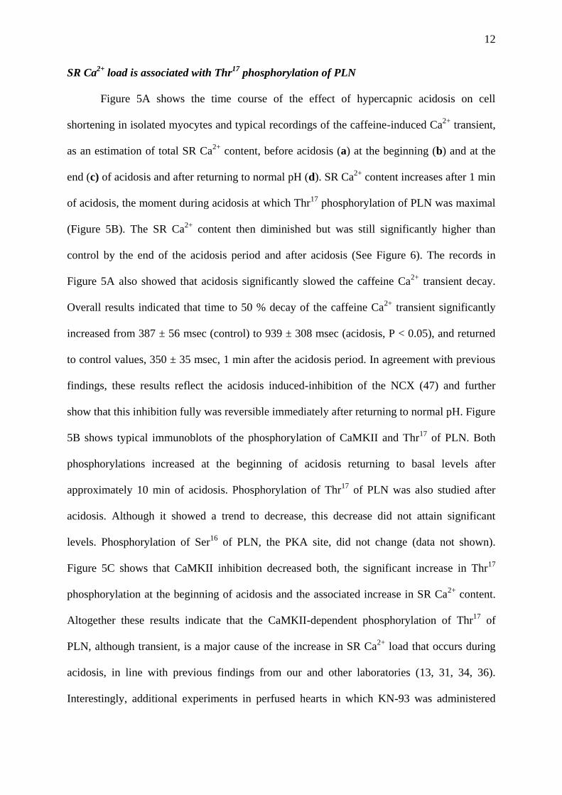
Figure 5A shows the time course of the effect of hypercapnic acidosis on cell
shortening in isolated myocytes and typical recordings of the caffeine-induced Ca2+
transient,
as an estimation of total SR Ca2+
content, before acidosis (a) at the beginning (b) and at the
end (c) of acidosis and after returning to normal pH (d). SR Ca2+
content increases after 1 min
of acidosis, the moment during acidosis at which Thr17
phosphorylation of PLN was maximal
(Figure 5B). The SR Ca2+
content then diminished but was still significantly higher than
control by the end of the acidosis period and after acidosis (See Figure 6). The records in
Figure 5A also showed that acidosis significantly slowed the caffeine Ca2+
transient decay.
Overall results indicated that time to 50 % decay of the caffeine Ca2+
transient significantly
increased from 387 ± 56 msec (control) to 939 ± 308 msec (acidosis, P < 0.05), and returned
to control values, 350 ± 35 msec, 1 min after the acidosis period. In agreement with previous
findings, these results reflect the acidosis induced-inhibition of the NCX (47) and further
show that this inhibition fully was reversible immediately after returning to normal pH. Figure
5B shows typical immunoblots of the phosphorylation of CaMKII and Thr17
of PLN. Both
phosphorylations increased at the beginning of acidosis returning to basal levels after
approximately 10 min of acidosis. Phosphorylation of Thr17
of PLN was also studied after
acidosis. Although it showed a trend to decrease, this decrease did not attain significant
levels. Phosphorylation of Ser16
of PLN, the PKA site, did not change (data not shown).
Figure 5C shows that CaMKII inhibition decreased both, the significant increase in Thr17
phosphorylation at the beginning of acidosis and the associated increase in SR Ca2+
content.
Altogether these results indicate that the CaMKII-dependent phosphorylation of Thr17
of
PLN, although transient, is a major cause of the increase in SR Ca2+
load that occurs during
acidosis, in line with previous findings from our and other laboratories (13, 31, 34, 36).
Interestingly, additional experiments in perfused hearts in which KN-93 was administered
13
during the acidosis period, immediately after the decay of Thr17
phosphorylation, failed to
avoid the arrhythmic pattern: Upon returning to control pH the number of ectopic beats
recorded were 38 ± 7, a figure similar to that observed in the absence of drugs in the same
time period (Figure 1D).
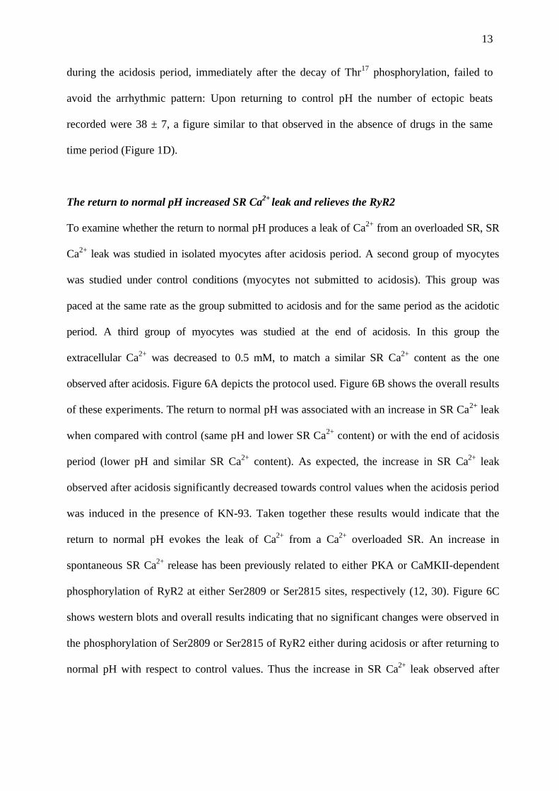
The return to normal pH increased SR Ca2+
leak and relieves the RyR2
To examine whether the return to normal pH produces a leak of Ca2+
from an overloaded SR, SR
Ca2+
leak was studied in isolated myocytes after acidosis period. A second group of myocytes
was studied under control conditions (myocytes not submitted to acidosis). This group was
paced at the same rate as the group submitted to acidosis and for the same period as the acidotic
period. A third group of myocytes was studied at the end of acidosis. In this group the
extracellular Ca2+
was decreased to 0.5 mM, to match a similar SR Ca2+
content as the one
observed after acidosis. Figure 6A depicts the protocol used. Figure 6B shows the overall results
of these experiments. The return to normal pH was associated with an increase in SR Ca2+
leak
when compared with control (same pH and lower SR Ca2+
content) or with the end of acidosis
period (lower pH and similar SR Ca2+
content). As expected, the increase in SR Ca2+
leak
observed after acidosis significantly decreased towards control values when the acidosis period
was induced in the presence of KN-93. Taken together these results would indicate that the
return to normal pH evokes the leak of Ca2+
from a Ca2+
overloaded SR. An increase in
spontaneous SR Ca2+
release has been previously related to either PKA or CaMKII-dependent
phosphorylation of RyR2 at either Ser2809 or Ser2815 sites, respectively (12, 30). Figure 6C
shows western blots and overall results indicating that no significant changes were observed in
the phosphorylation of Ser2809 or Ser2815 of RyR2 either during acidosis or after returning to
normal pH with respect to control values. Thus the increase in SR Ca2+
leak observed after
14
returning to normal pH with respect to that observed during acidosis for a similar SR Ca2+
load,
may be attributed to a reversible inhibition of RyR2 by acidosis.
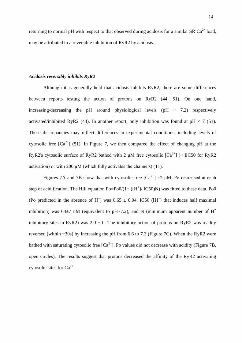
Acidosis reversibly inhibits RyR2
Although it is generally held that acidosis inhibits RyR2, there are some differences
between reports testing the action of protons on RyR2 (44, 51). On one hand,
increasing/decreasing the pH around physiological levels (pH ~ 7.2) respectively
activated/inhibited RyR2 (44). In another report, only inhibition was found at pH < 7 (51).
These discrepancies may reflect differences in experimental conditions, including levels of
cytosolic free [Ca2+
] (51). In Figure 7, we then compared the effect of changing pH at the
RyR2's cytosolic surface of RyR2 bathed with 2 μM free cytosolic [Ca2+
] (~ EC50 for RyR2
activation) or with 200 μM (which fully activates the channels) (11).
Figures 7A and 7B show that with cytosolic free [Ca2+
] ~2 μM, Po decreased at each
step of acidification. The Hill equation Po=Po0/(1+ ([H+]/ IC50)N) was fitted to these data. Po0
(Po predicted in the absence of H+) was 0.65 ± 0.04, IC50 ([H
+] that induces half maximal
inhibition) was 63±7 nM (equivalent to pH~7.2), and N (minimum apparent number of H+
inhibitory sites in RyR2) was 2.0 ± 0. The inhibitory action of protons on RyR2 was readily
reversed (within ~30s) by increasing the pH from 6.6 to 7.3 (Figure 7C). When the RyR2 were
bathed with saturating cytosolic free [Ca2+
], Po values did not decrease with acidity (Figure 7B,
open circles). The results suggest that protons decreased the affinity of the RyR2 activating
cytosolic sites for Ca2+
.

15
CaMKII-dependent DADs can be detected after the acidosis period
The dependence of the arrhythmic pattern described on the SR Ca2+
release and SR Ca2+
content would suggest that arrhythmias may be triggered by DADs. We therefore followed a
defined pacing protocol with pauses test for spontaneous activity, to detect the possible
appearance of DADs (See Methods). Figure 8A shows MAPs recorded from the epicardial wall
after this protocol. After termination of pacing at 240 beats/min and simultaneous return to
normal pH, two membrane depolarizations were observed. Whereas the first was followed by
two spontaneous beats, the second triggers an episode of ventricular arrhythmia. This
behaviour was observed in 5 of 5 experiments of this type and in 0 of 3 experiments when KN-
93 was present (Figure 8B). When MAPs were recorded simultaneously from the endocardial
and epicardial ventricular wall, the number of DADs detected from the endocardial wall was
significantly lower than that detected from the epicardial surface (4 ± 2 vs. 15 ± 4, n = 5) in the
3 first min of normal pH after acidosis). Taken together, these experiments would indicate that
the returning to normal pH evoked membrane depolarizations suggestive of DADs, mainly
detected from the epicardial surface of the ventricular wall, which are able to trigger arrhythmic
episodes and are prevented by CaMKII inhibition.
16
DISCUSSION
In the present study we investigated the cellular mechanisms underlying the arrhythmias that
occur after acidosis, upon returning to normal pH. We observed that arrhythmias are dependent
on the activity of the SR. More importantly, we showed that the ectopic activity is suppressed
by inhibition of the multifunctional Ca2+
-calmodulin-dependent protein kinase, CaMKII and did
not occur in a transgenic mouse model with inhibition of CaMKII targeted to the SR.
Arrhythmias appear to be triggered by slow membrane depolarizations, typical of DADs, that
were also prevented by CaMKII-inhibition. Inhibition of CaMKII also blunted the increase in
phosphorylation of Thr17
of PLN and in SR Ca2+
content that occurred associated with the onset
of acidosis, as well as the increased SR Ca2+
leak that occurred after returning to normal pH.
The results further showed that the return to normal pH reversed the acidosis-induced inhibition
of RyR2 activity and of the NCX. Taken together, these findings indicate a primary role of
CaMKII on the enhancement of the SR Ca2+
content observed during acidosis and suggest that
the phosphorylation of Thr17
of PLN may be possibly involved in this effect, in agreement with
previous findings (13, 34, 36). The results further suggest that the spontaneous activity that
takes place after returning to normal pH is triggered by CaMKII-dependent DADs, which
would be favoured by two concurrent factors: 1. The CaMKII-dependent enhancement of the
SR Ca2+
content that occurred at the beginning of acidosis and still persist after acidosis and 2.
The simultaneous relief of RyR2 and NCX previously inhibited by acidosis.
Monophasic action potentials.
MAPs are extracellularly recorded waveforms that are not identical to true transmembrane
action potential recordings. Nevertheless, they can accurately reproduce the time course of
transmembrane action potentials and are suitable for studying the characteristics of local
myocardial electrophysiology in intact animal preparations and in the clinical setting (5, 8,
14, 17, 33). Indeed, recording MAPs is the only possible method to explore localized
myocardial activation and repolarization in the human heart or in the in vivo animal hearts. In
the present experiments we used MAP recordings to assess the type of arrhythmia that takes
place upon returning to normal pH after acidosis. Although application of this method has
several practical problems such as MAPs instability or registration of artefacts, we were able

17
to minimize the influence of these problems and to obtain stable MAP recordings, that could
be simultaneously assessed in the endocardial and epicardial ventricular walls and could be
suppressed by different interventions.
Arrhythmias after acidosis are suppressed by CaMKII inhibition
The present results showed that the ectopic activity after acidosis was significantly decreased
in transgenic animals with inhibition of CaMKII targeted to the SR, in comparison with the
age-matched WT mice. The arrhythmic pattern was also prevented by the CaMKII inhibitor,
KN-93, but not by its inactive analogue KN-92, in the perfused rat heart. The arrhythmias
appeared to be triggered by membrane depolarizations, typical of DADs that were also
blocked by KN-93. These results indicate that the triggered arrhythmias are dependent on a
CaMKII-phosphorylation which would likely occur at the SR level. Since RyR2 were not
significantly phosphorylated either during or after acidosis, these experiments support the
notion of a major role of PLN phosphorylation, specifically the CaMKII site Thr17
, in the
increase in SR Ca2+
content that occurs at the beginning of acidosis. Recent evidence
indicated that SR-AIP mice show a consistent decrease in CaMKII-dependent facilitation at
the L-type Ca2+
channels level (40). However, acidosis either decreases or does not change L-
type Ca2+
current (20, 26), making it unlikely a possible contribution of this current to the
acidosis-induced increase in SR Ca2+
load. Moreover, if this contribution takes place, it would
not fade the importance of Thr17
phosphorylation on this effect.
It has been previously shown that CaMKII inhibition reduces ICa facilitation, L-type Ca2+
channel opening probability and EADs (2, 15, 49, 50). Taken together, these findings
convincingly showed the link between EADs initiation, L-type Ca2+
current and CaMKII
activation. Moreover, different type of evidence demonstrated that CaMKII may affect Na+
and K+
channels which would be expected to modify action potential duration (APD) (43,

18
48). We did not observe changes in APD after KN-93 administration (Table 1), which would
preclude an effect of either the drug or of CaMKII on these channels under basal conditions.
DADs are not linked to ion channel alterations but rather to conditions that favour SR Ca2+
overload (29). Earlier studies by Wu et al. (49) indicated that when artificially prolonged
action potential waves are used as voltage clamp commands, cell membranes exhibit a
transient inward current that is blocked by dialysis of a CaMKII inhibitory peptide. These
results may suggest that Ca2+
influx during a prolonged action potential is capable of
overloading the SR with Ca2+
, producing currents likely to be responsible for DADs (1).
However, the potential role of CaMKII activity in DADs formation had never been directly
tested. The present results indicate that the arrhythmias observed after acidosis are suppressed
by CaMKII-inhibition and are likely triggered by DADs primarily originated in epicardium.
This mechanism would constitute the main mechanism of triggered arrhythmias after a period
of acidosis. In the context of these results, it is important to mention that although it has been
previously thought that DADs originate mainly in the endocardium (28), recent work
reported DADs and triggered activity associated with spontaneous SR Ca2+
release, that
occurred preferentially near the epicardium in a model of abnormal RyR2 function induced
by FKBP12.6 dissociation and beta-adrenergic stimulation. The authors attributed the higher
ectopy of the epicardium with respect to the endocardium to the faster SR Ca2+
uptake
observed possibly due to a higher expression of SERCA2a compared with that observed in
the endocardium (22, 35).
Mechanism of SR Ca2+
load during acidosis.
Previous experiments causally linked the spontaneous mechanical recovery that occurs during
acidosis to an increase in SR Ca2+
load (18). Earlier evidence indicated that the signalling
cascade involved in this increase was triggered by the activity of the Na+- H
+ exchanger,

19
enhanced by intracellular acidosis, which in turn would increase intracellular Na+ and
intracellular Ca2+
by slowing the forward mode of the NCX and eventually reversing it. It was
hypothesized that the increase in Ca2+
promoted by this pathway would be sufficient to
overcome the direct inhibitory action of acidosis on the activity of SERCA2a (18, 39). More
recent studies indicated however that this cascade of events, although possibly necessary, was
not sufficient by itself to increase the Ca2+
content of the SR. It was shown that the activation
of CaMKII and the phosphorylation of Thr17
of PLN were necessary events underlying the
mechanical recovery (13, 34, 36). In line with these previous findings, the present
experiments also show that acidosis produces an increase in SR Ca2+
load and a mechanical
recovery, both of which were reduced by CaMKII inhibition, as it was the significant increase
in the phosphorylation of Thr17
of PLN. The increase in the phosphorylation of Thr17
occurred at the beginning of acidosis associated to the maximal increase in SR Ca2+
content
(Figure 5). We are aware of the fact that the increase in SR Ca2+
content observed during
acidosis is qualitative and may be distorted due to possible changes in cytoplasmic Ca2+
buffering. However the increase in SR Ca2+
content during acidosis has been validated by
quantitave measurements of SR Ca2+
and Ca2+
buffering and from changes in NCX current on
repolarization (10). The present findings add to these previous results the fact that the
increase in SR Ca2+
content during acidosis is dependent of CaMKII, in agreement with
previous findings in mice by DeSantiago et al. (13). Taken together the results suggest that
the activation of CaMKII seems to be a necessary step required to increase SR Ca2+
load,
possibly through the phosphorylation of Thr17
of PLN (13, 34). In this scenario and relevant to
the present study, it is important to rescue the role of the acidosis-induced inhibition of RyR2
and NCX in favouring SR Ca2+
overload under acidosis conditions (See below).
20
Why is the ectopic activity more closely associated to the return to normal pH?
If the increase in SR Ca2+
content occurs during acidosis, the question is then, why the
arrhythmias are more closely associated to the return to normal pH. The explanation to this
finding may be given by experimental evidence showing the inhibitory effects of acidosis on
RyR2 and on the frequency of Ca2+
spark (3, 10, 44, 46, 51). In agreement with these results,
we showed that acidosis reversibly inhibits RyR2 open probability in planar lipid bilayers at
not saturating cytosolic Ca2+
levels (See Figure 7). Moreover, the SR Ca2 leak observed after
returning to normal pH was not only greater than control, possibly due to a significant
increase in SR Ca2+
content, but also more important than the Ca2 leak observed at the end of
acidosis, for a similar SR Ca2+
load. An increase in SR Ca2+
leak may also be favored by a
CaMKII-dependent phosphorylation of RyR2 (12, 30). However, our results did not detect
any significant increase in the phosphorylation of either Ser2815 or Ser2809 sites, neither
during the acidosis period nor after returning to normal pH. Thus, returning to normal pH
would increase the opening probability of the Ca2+
release channel of a Ca2+
overloaded SR.
Returning to normal pH would also favour the reactivation of NCX inhibited by acidosis. In
agreement with previous findings (47), the present experiments showed indeed that the rate of
Ca2+
decline of caffeine transients was significantly slowed by acidosis, an effect that was
fully reversible upon returning to normal pH. All these mechanisms, acting in concert, would
be responsible for the increase in SR Ca2+
leak and triggered arrhythmias observed.
Figure 9 depicts the proposed mechanism for the arrhythmias triggered after a period of
acidosis.
Clinical Implications
The present experiments indicated that the return to normal pH after a period of hypercanic
acidosis triggered an arrhythmic pattern that is dependent on CaMKII. Intracellular acidosis
seems to be the important change, since metabolic acidosis produced a significant lower
21
number of ectopic beats, after returning to normal pH. Our findings may be of interest in the
clinical setting, since substancial changes in intracellular pH may occur in different clinical
disturbances of the acid –base status, like ischemia/reperfusion injury, the syndrome of sleep
apnea/hypopnea, (32) or in patiens in dialysis (42), that may affect cardiac function. The
present experiments suggest that alterations in intracellular pH associated with all these
pathologies may be the substrate of at least part of the arrhythmias observed in these diseases.
CaMKII has emerged as an important arrhythmogenic signalling molecule in the setting of
LQT syndrome (49), cardiac hypertrophy (50) and cardiomyopathy (24). All these studies
pointed to the crucial role of CaMKII in generating EADs and triggered arrhythmias. The
present results strongly suggest that CaMKII is also responsible for DADs that trigger post-
acidosis arrhythmias. As such, CaMKII may be an antiarrhythmic drug target during this type
of arrhythmias.

22
Acknowledgments
The assistance of Inés Vera in making the illustrations is greatly acknowledged. We also wish
to thank to Ariel Escobar and Daniel Lorenti for helpful comments and assistance in
constructing the set up and electrodes for MAPs acquisition and to the student Nicolás Cédola
for his assistance in the last sets of experiments.

23
Grants
This work was supported by PICT # 14219 (FONCyT, Argentina) to MS and PICT
(FONCyT) # 26117, PIP # 5300 (CONICET, Argentina), Fogarty International Research
Award Grant # R03-TW-07713 (NIH) to AM, and Grant R01 GM078665 (NIH) to JAC and
PLD-S. R. Becerra is a fellow from the Comisión de Investigaciones Científicas, Pcia BA,
Argentina. M. Said, J. Palomeque, G. Rinaldi, C. Mundiña-Weilenmann, L. Vittone and A.
Mattiazzi are established Investigators of CONICET, Argentina.

24
Disclosures
None
25
References
1. Anderson ME. Calmodulin kinase signalling in heart: an intriguing candidate target for
therapy of myocardial dysfunction and arrhythmias. Pharmacology & Therapeutics 106:
39-55, 2005.
2. Anderson ME, Braun AP, Wu Y, Lu T, Wu Y, Schulman H, Sung RJ. KN-93, an
inhibitor of multifunctional Ca++
/calmodulin-dependent protein kinase, decreases early
afterdepolarizations in rabbit heart. J Pharmacol Exp Ther 287: 996-1006, 1998.
3. Balnave CD, Vaughan-Jones RD. Effect of intracellular pH on spontaneous Ca2+
sparks in rat ventricular myocytes. J Physiol 528: 25-37, 2000.
4. Bethell HW, Vanderberg JI, Smith GA, Grace AA. Changes in ventricular
repolarization during acidosis and low-flow ischaemia. Am J Physiol 275: H551-H561,
1998.
5. Brack KE, Patel VH, Coote JH, Ng GA. Nitric oxide mediates the vagal protective
effect on ventricular fibrillation via effects on action potential duration restitution in the
rabbit heart. J Physiol 583: 695-704, 2007.
6. Carmeliet E. Cardiac ionic currents and acute ischemia: from channels to arrhythmias.
(Review) Physiol Rev 79: 917-1017, 1999.
7. Chamberlain BK, Levitsky DO, Fleischer S. Isolation and characterization of canine
cardiac sarcoplasmic reticulum with improved Ca2+
transport properties. J. Biol. Chem
258: 6602-6609, 1983.
8. Chen SA, Chiang CE, Yang CJ, Cheng CC, Wu TJ, Wang SP, Chiang BN, Chang
MS. Sustained atrial tachycardia in adult patients. Electrophysiological characteristics,
pharmacological response, possible mechanisms, and effects of radiofrequency ablation.
Circulation 90: 1262-1278, 1994.
26
9. Chijiwa T, Mishima A, Hagiwara M, Sano M, Hayashi K, Inoue T, Naito K,
Toshioka T, Hidaka H. Inhibition of forskolin-induced neurite outgrowth and protein
phosphorylation by a newly synthesized selective inhibitor of cyclic AMP-dependent
protein kinase, N-[2-(p-bromocinnamylamino)ethyl]-5-isoquinolinesulfonamide (H-89),
of PC12D pheochromocytoma cells. J Biol Chem 265:5267-5272, 1990.
10. Choi HS, Trafford AW, Orchard CH, Eisner DA. The effect of acidosis on systolic
Ca2+
and sarcoplasmic reticulum calcium content in isolated rat ventricular myocytes. J
Physiol 529:661-668, 2000.
11. Copello JA, Barg S, Onoue H, Fleischer S. Heterogeneity of Ca2+
gating of skeletal
muscle and cardiac ryanodine receptors. Biophys J. 73: 141-156, 1997.
12. Curran J, Hinton MJ, Ríos E, Bers DM, Shannon TR. β-Adrenergic enhancement of
sarcoplasmic reticulum calcium leak in cardiac myocytes is mediated by
calcium/calmodulin-dependent protein kinase. Circ Res 100: 391-398, 2007.
13. DeSantiago J, Maier LS, Bers DM. Phospholamban is required for CaMKII-
dependent recovery of Ca transients and SR Ca reuptake during acidosis in cardiac
myocytes. J Mol Cell Cardiol 36: 67-74, 2004.
14. de Groot SH, Vos MA, Gorgels AP, Leunissen JD, van der Steld BJ, Wellens HJ.
Combining monophasic action potential recordings with pacing to demonstrate delayed
afterdepolarizations and triggered arrhythmias in the intact heart. Value of diastolic
slope. Circulation 92: 2697-2704, 1995.
15. Dzhura I, Wu Y, Colbran RJ, Balser JR, Anderson ME. Calmodulin kinase
determines calcium-dependent facilitation of L-type calcium channels. Nat Cell Biol 2:
173-177, 2000.
16. Ferrero P, Said M, Sánchez G, Vittone L, Valverde C, Donoso P, Mattiazzi A,
Mundiña-Weilenmann C. Ca2+
/Calmodulin kinase II increases ryanodine binding and
Ca2+
-induced sarcoplasmic reticulum Ca2+
release kinetics during β-adrenergic
stimulation. J Mol Cel Cardiol 43:281-291, 2007.

27
17. Franz MR. Current status of monophasic action potential recordings: theories,
measurements and interpretations. Cardiovasc Res. 41:25-40, 1999.
18. Harrison SM, Frampton JE, McCall E, Boyett MR, Orchard CH. Contraction and
intracellular Ca2+
, Na+, and H
+ during acidosis in rat ventricular myocytes. Am J Physiol
Cell Physiol 262: C348-C357, 1992.
19. Herbert CJM, Augereaub JM,. Gleyec J, Maffranda JP. Chelerythrine is a potent
and specific inhibitor of protein kinase C. Biochem Biophys Res Commun 172: 993-999,
1990.
20. Irisawa H, Sato R. Intra- and extracellular actions of proton on the calcium current of
isolated guinea pig ventricular cells. Circ Res 59:348-55, 1986.
21. Ji Y, Li B, Reed TD, Lorenz JN, Kaetzel MA, Dedman JR. Targeted inhibition of
Ca2+
/calmodulin-dependent protein kinase II in cardiac longitudinal sarcoplasmic
reticulum results in decreased phospholamban phosphorylation at threonine 17. J Biol
Chem 278: 25063–25071, 2003.
22. Katra RP, Oya T, Hoeker GS, Laurita KR. Ryanodine receptor dysfunction and
triggered activity in the heart. Am J Physiol Heart Circ Physiol 292:H2144-2151, 2007.
23. Keating MT, Sanguinetti MC. Molecular and cellular mechanisms of cardiac
arrhythmias. Cell 104: 569-580, 2001.
24. Khoo MS, Kannankeril PJ, Li J, Zhang R, Kupershmidt S, Zhang W, Atkinson JB,
Colbran RJ, Roden DM, Anderson ME. Calmodulin kinase II activity is required for
normal atrioventricular nodal conduction. Heart Rhythm 2: 634-640, 2005.
25. Knollmann BC, Katchman AN, Franz MR. Monophasic action potential recordings
from intact mouse heart: validation, regional heterogeneity, and relation to
refractoriness. J Cardiovasc Electrophysiol 12: 1286-1294, 2001.
28
26. Komukai K, Pascarel C, Orchard CH. Compensatory role of CaMKII on ICa and SR
function during acidosis in rat ventricular myocytes. Pflugers Arch 442:353-361, 2001.
27. Kurachi Y. The effects of intracellular protons on the electrical activity of single
ventricular cells. Pflugers Arch 394: 264-270, 1982.
28. Laurita KR, Katra RP. Delayed after depolarization-mediated triggered activity
associated with slow calcium sequestration near the endocardium. J Cardiovasc
Electrophysiol 16:418-424, 2005.
29. Lederer WJ, Tsien RW. Transient inward current underlying arrhythmogenic effects
of cardiotonic steroids in Purkinje fibres. J Physiol. 263:73-100, 1976.
30. Marx SO, Reiken S, Hisamatsu Y, Jayaraman T, Burkhoff D, Rosemblit N, Marks
AR. PKA phosphorylation dissociates FKBP12.6 from the calcium release channel
(ryanodine receptor): defective regulation in failing hearts. Cell 101: 365-376, 2000.
31. Mattiazzi A, Vittone L, Mundiña-Weilenmann C. Ca2+
/calmodulin-dependent protein
kinase: A key component in the contractile recovery from acidosis. Cardiovasc Res 73:
648-656, 2007.
32. Mehra R, Benjamin EJ, Shahar E, Gottlieb DJ, Nawabit R, Kirchner HL,
Sahadevan J, Redline S. Association of nocturnal arrhythmias with sleep-disordered
breathing: The Sleep Heart Health Study. Am J Respir Crit Care Med 173: 910-916,
2006
33. Moore HJ, Franz MR. Monophasic action potential recordings in humans. J
Cardiovasc Electrophysiol 18: 787-790, 2007.
34. Mundiña-Weilenmann C, Said M, Ferrero P, Vittone L, Kranias E. Mattiazzi, A.
Role of phosphorylation of Thr17
of phospholamban in the mechanical recovery from
hypercapnic acidosis. Cardiovasc Res 66: 114-122, 2005.
29
35. Nam Gi-Byoung, Burashnikov A, Antzalevich Ch. Cellular mechanisms underlying
the development of catecholaminergic ventricular tachycardia. Circulation 111:2727-
2733, 2005.
36. Nomura N, Satoh H, Terada H, Matsunaga M, Watanabe H, Hayashi H. CaMKII-
dependent reactivation of SR Ca2+
uptake and contractile recovery during intracellular
acidosis. Am J Physiol Heart Circ Physiol 283: H193-H203, 2002.
37. Orchard CH, Houser SR, Kort AA, Bahinski AA, Capogrossi MC, Lakatta EG.
Acidosis facilitates spontaneous sarcoplasmic reticulum Ca2+
release in rat myocardium.
J Gen Physiol 90: 145-165, 1987.
38. Palomeque J, Sapia L, Hajjar R, Mattiazzi A, Vila-Petroff M. Angiotensin-II
induced negative inotropy in rat ventricular myocytes: role of reactive oxygen species
and p38 MAPK. Am J Physiol Heart Circ Physiol 290: H96-H106, 2006.
39. Pérez NG, Mattiazzi AR, Camilión de Hurtado MC, Cingolani HE. Myocardial
contractility recovery during hypercapnic acidosis: its dissociation from recovery in pHi
by ryanodine. Can J Cardiol 11: 553-560, 1995.
40. Picht E, DeSantiago J, Huke S, Kaetzel MA, Dedman JR, Bers DM.CaMKII
inhibition targeted to the sarcoplasmic reticulum inhibits frequency-dependent
acceleration of relaxation and Ca2+
current facilitation. J Mol Cell Cardiol. 42:196-205,
2007.
41. Pogwizd SM, Schlotthauer K, Li L, Yuan W, Bers DM. Arrhythmogenesis and
contractile dysfunction in heart failure: Roles of sodium-calcium exchange, inward
rectifier potassium current, and residual beta-adrenergic responsiveness. Circ Res 88:
1095-1096, 2001.
42. Redaelli B. Hydroelectrolytic equilibrium change in dialysis. J Nephrol 14 Suppl 4:S7-
11, 2001.
30
43. Rezazadeh S, Claydon TW, Fedida D. KN-93 (2-[N-(2-hydroxyethyl)]-N-(4-
methoxybenzenesulfonyl)]amino-N-(4-chlorocinnamyl)-N-methylbenzylamine), a
calcium/ calmodulin-dependent protein kinase II inhibitor, is a direct extracellular
blocker of voltage-gated potassium channels. J Pharmacol Exp Ther 317: 292-299,
2006.
44. Rousseau E, Pinkos J. pH modulates conducting and gating behaviour of single
calcium release channels. Pflugers Arch 415:645-647, 1990.
45. Shannon TR, Ginsburg KS, Bers DM. Quantitative assessment of the SR Ca2+
leak-load
relationship. Circ Res 91: 594-600, 2002.
46. Spacapan de Castuma E, Mattiazzi AR, Cingolani HE. Effect of hypercapnic
acidosis on induction of arrhythmias by catecholamines in cat papillary muscles. Arch
Int Physiol Biochem 85: 509-519, 1977.
47. Terracciano CM, MacLeod KT. Effects of acidosis on Na+/Ca
2+ exchange and
consequences for relaxation in guinea pig cardiac myocytes. Am J Physiol Heart Circ
Physiol 267: H477-487, 1994.
48. Wagner S, Dybkova N, Rasenack EC, Jacobshagen C, Fabritz L, Kirchhof P,
Maier SK, Zhang T, Hasenfuss G, Brown JH, Bers DM, Maier LS. Ca2+
/calmodulin-
dependent protein kinase II regulates cardiac Na+ channels. J Clin Invest 116: 3127-
3138, 2006.
49. Wu Y, MacMillan LB, McNeill RB, Colbran RJ, Anderson ME. CaM kinase
augments cardiac L-type Ca2+
current: a cellular mechanism for long Q-T arrhythmias.
Am J Physiol Heart Circ Physiol 276: H2168- H2178, 1999.
50. Wu Y, Temple J, Zhang R, Dzhura I, Zhang W, Trimble R, Roden DM, Passier R,
Olson EN, Colbran RJ, Anderson ME. Calmodulin kinase II and arrhythmias in a
mouse model of cardiac hypertrophy. Circulation 106: 1288-1293, 2002.
31
51. Xu L, Mann G, Meissner G. Regulation of cardiac Ca2+
release channel (ryanodine
receptor) by Ca2+
, H+, Mg
2+, and adenine nucleotides under normal and simulated
ischemic conditions. Circ Res. 79:1100-1109, 1996.

32
FIGURE LEGENDS
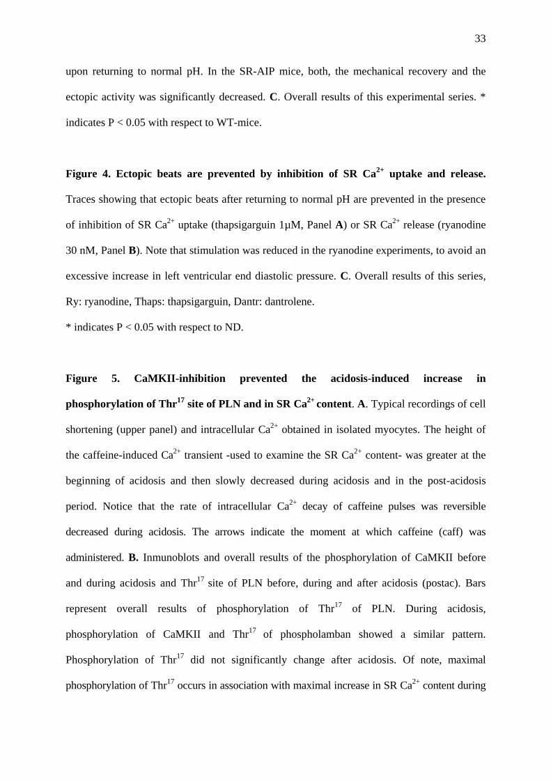
Figure 1. LVDP and epicardial MAPs during acidosis and post-acidosis. A.
Representative mechanical recordings showing the time course of changes in LVDP during
acidosis and upon returning to normal pH (postacidosis). B. Enlarged records of LVDP and
epicardial MAPs, corresponding to the points a to d of the record in A. The figure illustrates
the arrhythmic pattern that occurs when the heart was returned to normal pH. C. Records of
MAPs obtained in the absence of intraventricular balloon. D. Overall results of the
experiments of this series. Each bar represents the mean occurrence of ectopic beats measured
under each condition during a 3 min- period: The last 3 min of the stabilization (C), the last 3
min of the first and second half of the 20 min of acidosis and the first 3 min after returning to
normal pH, in the presence and absence of intraventricular balloon. * indicates P < 0.05 with
respect to control.
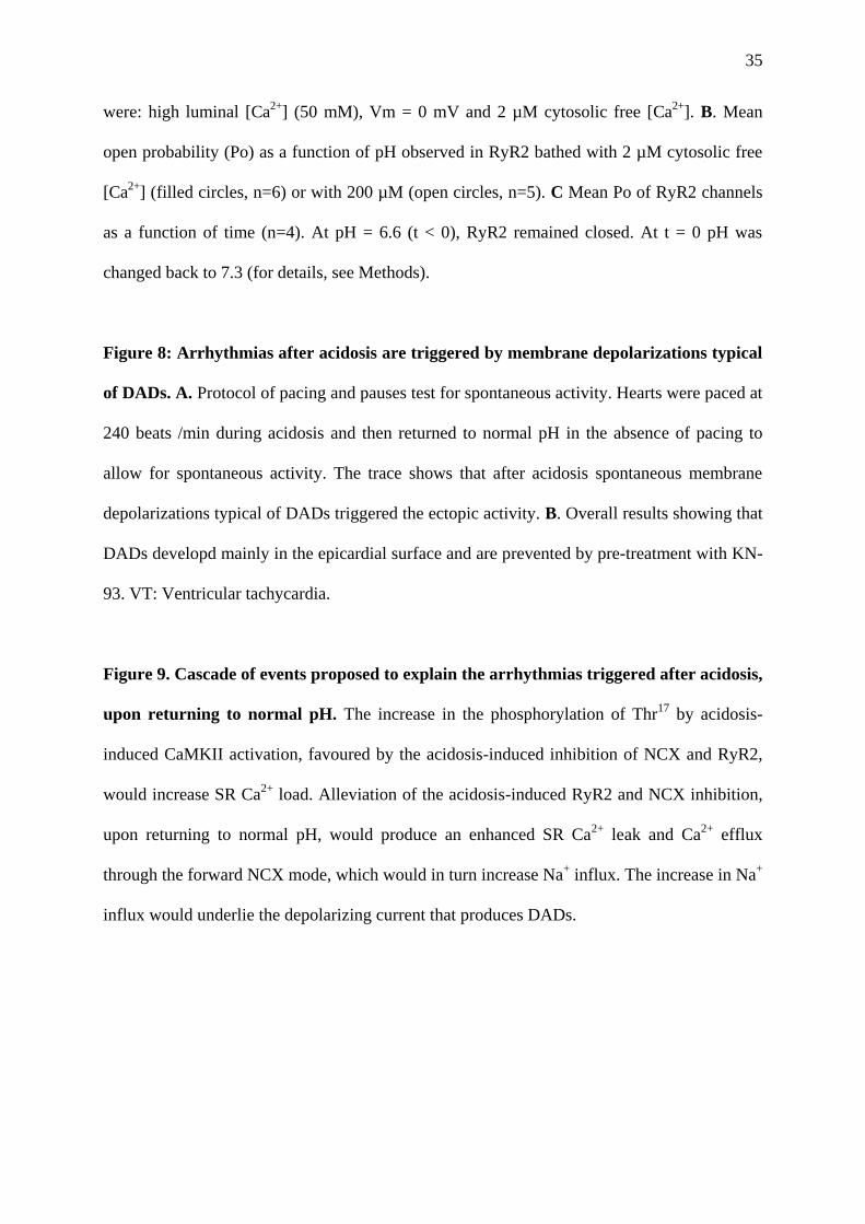
Figure 2. LVDP and epicardial MAPs during acidosis and post-acidosis in the presence
or in the absence of CaMKII inhibitor KN-93 and the inactive analogue, KN-92. A.1µM
KN-93 abolished the ectopic beats that appear immediately after returning to normal pH. B.
Lack of effect of KN-92 (1µM) on the arrhythmias of post-acidosis. C. Overall results of this
series. In this and following figures, each bar represents the mean occurrence of ectopic beats
measured during the first 3 min after returning to normal pH. ND means no drugs added. *
indicates P < 0.05 with respect to ND.
Figure 3. LVDP and epicardial MAPs during acidosis and post-acidosis in WT and SR-
AIP mice. Records of LVDP and epicardial MAPs, corresponding to the control, acidosis and
post- acidosis periods in WT mice (A) and SR-AIP mice (B). In WT mice there was a
mechanical recovery during acidosis, similar to rat hearts, followed by an arrhythmic pattern
33
upon returning to normal pH. In the SR-AIP mice, both, the mechanical recovery and the
ectopic activity was significantly decreased. C. Overall results of this experimental series. *
indicates P < 0.05 with respect to WT-mice.
Figure 4. Ectopic beats are prevented by inhibition of SR Ca2+
uptake and release.
Traces showing that ectopic beats after returning to normal pH are prevented in the presence
of inhibition of SR Ca2+
uptake (thapsigarguin 1µM, Panel A) or SR Ca2+
release (ryanodine
30 nM, Panel B). Note that stimulation was reduced in the ryanodine experiments, to avoid an
excessive increase in left ventricular end diastolic pressure. C. Overall results of this series,
Ry: ryanodine, Thaps: thapsigarguin, Dantr: dantrolene.
* indicates P < 0.05 with respect to ND.
Figure 5. CaMKII-inhibition prevented the acidosis-induced increase in
phosphorylation of Thr17
site of PLN and in SR Ca2+
content. A. Typical recordings of cell
shortening (upper panel) and intracellular Ca2+
obtained in isolated myocytes. The height of
the caffeine-induced Ca2+
transient -used to examine the SR Ca2+
content- was greater at the
beginning of acidosis and then slowly decreased during acidosis and in the post-acidosis
period. Notice that the rate of intracellular Ca2+
decay of caffeine pulses was reversible
decreased during acidosis. The arrows indicate the moment at which caffeine (caff) was
administered. B. Inmunoblots and overall results of the phosphorylation of CaMKII before
and during acidosis and Thr17
site of PLN before, during and after acidosis (postac). Bars
represent overall results of phosphorylation of Thr17
of PLN. During acidosis,
phosphorylation of CaMKII and Thr17
of phospholamban showed a similar pattern.
Phosphorylation of Thr17
did not significantly change after acidosis. Of note, maximal
phosphorylation of Thr17
occurs in association with maximal increase in SR Ca2+
content during

34
acidosis (A). C. The increase in the phosphorylation of Thr17
of PLN and SR Ca2+
content at
the beginning of acidosis were both significantly diminished by pre-treatment with 1 µM KN-
93. n = 3-6. * indicates P < 0.05 with respect to control. # indicates P < 0.05 with respect to
acidosis without KN-93.
Figure 6. CaMKII-inhibition prevented the SR Ca2+
leak observed upon returning to
normal pH. A. Scheme of the protocol used to assess SR Ca2+
leak. In indo-1 loaded
myocytes, stimulation was stopped and the myocytes were exposed to 0 Na+- 0 Ca
2+ solution for
60 sec to block the NCX, - so that little or no Ca2+
can entered or left the resting cell - in the
absence and presence of tetracaine to block the SR Ca2+
release channel. The difference in
diastolic Ca2+
with and without tetracaine was taken as an estimation of SR Ca2+
leak. SR Ca2+
leak was measured in the presence and absence of 1 µM KN-93. B. Comparison of the average
SR Ca2+
load and leak at pH 7.4 (control), at the end of acidosis and at the post acidosis period.
The return to normal pH after acidosis increased SR Ca2+
leak with respect to control myocytes
with a lower SR Ca2+
load and to myocytes submitted to acidosis with a similar SR Ca2+
load
achieved by decreasing extracellular Ca2+
. In the presence of KN-93, SR Ca2+
leak after
acidosis was not significantly different from control. n=3 in each experimental group. C. Left
panel. Immunoblots showing the phosphorylation of Ser2815 and Ser2809 of RyR2 before
acidosis and upon returning to normal pH. Iso represent the phosphorylation of both RyR2
residues observed in hearts perfused with isoproterenol, as a positive control. Right panel.
Overall results of these experiments, n=3-4. No significant alterations in the phosphorylation
of RyR2 were detected. * indicates P<0.05 vs. control. # indicates P<0.05 vs. acidosis.
Figure 7. Cytosolic pH affects RyR2 activity at low (2 µM) cytosolic free Ca2+.
A. Single
channel recordings at pH 7.9 (right panel), 7.1 (center) and 6.6 (left). Recording conditions
35
were: high luminal [Ca2+
] (50 mM), Vm = 0 mV and 2 µM cytosolic free [Ca2+
]. B. Mean
open probability (Po) as a function of pH observed in RyR2 bathed with 2 µM cytosolic free
[Ca2+
] (filled circles, n=6) or with 200 µM (open circles, n=5). C Mean Po of RyR2 channels
as a function of time (n=4). At pH = 6.6 (t < 0), RyR2 remained closed. At t = 0 pH was
changed back to 7.3 (for details, see Methods).
Figure 8: Arrhythmias after acidosis are triggered by membrane depolarizations typical
of DADs. A. Protocol of pacing and pauses test for spontaneous activity. Hearts were paced at
240 beats /min during acidosis and then returned to normal pH in the absence of pacing to
allow for spontaneous activity. The trace shows that after acidosis spontaneous membrane
depolarizations typical of DADs triggered the ectopic activity. B. Overall results showing that
DADs developd mainly in the epicardial surface and are prevented by pre-treatment with KN-
93. VT: Ventricular tachycardia.
Figure 9. Cascade of events proposed to explain the arrhythmias triggered after acidosis,
upon returning to normal pH. The increase in the phosphorylation of Thr17
by acidosis-
induced CaMKII activation, favoured by the acidosis-induced inhibition of NCX and RyR2,
would increase SR Ca2+
load. Alleviation of the acidosis-induced RyR2 and NCX inhibition,
upon returning to normal pH, would produce an enhanced SR Ca2+
leak and Ca2+
efflux
through the forward NCX mode, which would in turn increase Na+ influx. The increase in Na
+
influx would underlie the depolarizing current that produces DADs.

36
TABLE LEGEND.
Table 1. Values (mean ± SEM) were obtained at the end of the stabilization (Control) and at
the end of the 10 min period of drug perfusion, previous to acidosis. LVDP, left ventricular
developed pressure; +dP/dt, maximal rate of pressure development; t1/2, half relaxation time;
MAPD90, monophasic action potential duration at 90% of repolarization. * P < 0.05 vs.
control. ND: no determination.


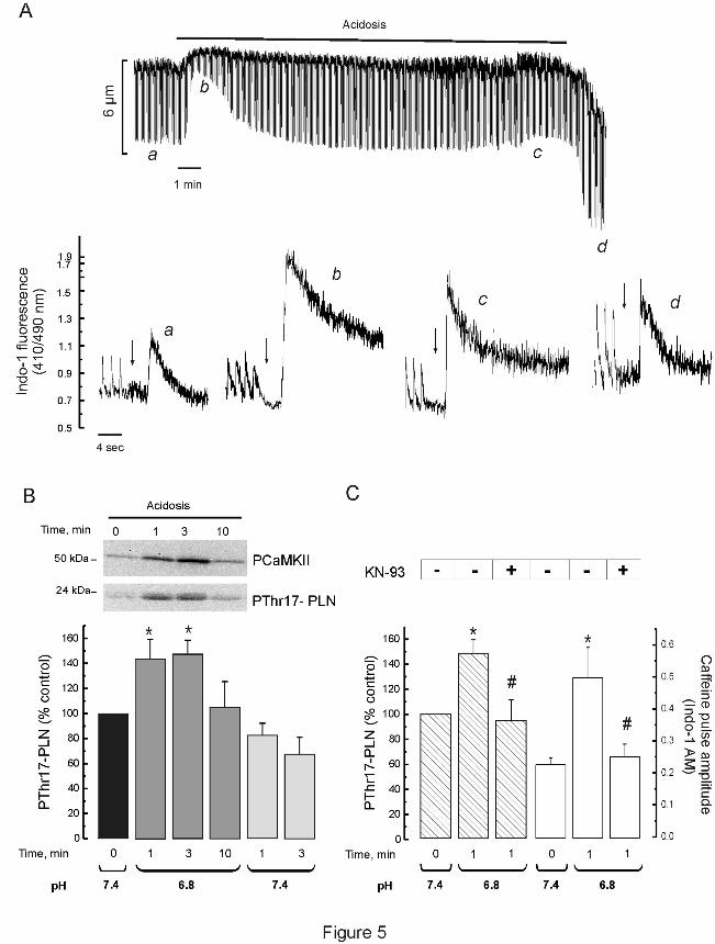

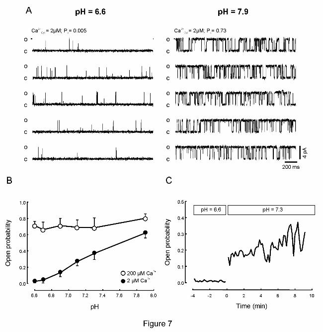
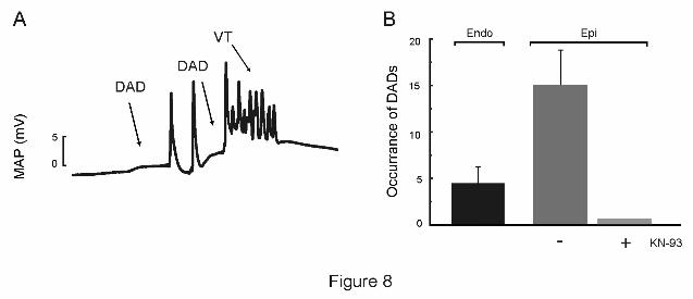


Table 1.
Effects of the different interventions on basal contractility, relaxation and MAPs duration
LVDP +dP/dt t1/2 MAPD90
mmHg mmHg/sec msec msec
Control 101.9 ± 4.8 2799.8 ± 89.0 53.0 ± 1.5 53.3 ± 4.1
KN-93 (1 M) (n=3) 93.2 ± 6.7 2628.7 ± 189.7 54.2 ± 3.1 61.2 ± 6.2
Control 103.5 ± 5.7 2762.5 ± 112.7 54.7 ± 2.6 54.0 ± 2.1
KN-92 (1 M) (n=4) 99.2 ± 5.4 2754.9 ± 173.7 54.7 ± 3.6 50.8 ± 8.5
Control 110.9 ± 11.8 3601.6 ± 411.1 67.7 ± 4.8 68.7 ± 4.1
Ryanodine (30nM) (n=3) 51.4 ± 4.8* 1600.2 ± 169.8* 82.3 ± 6.2* 66.7 ± 2.9
Control 92.9 ± 3.1 2744.0 ± 270.6 55.2 ± 1.7 61.3 ± 6.3
Thapsigarguin (1 M) (n=4) 62.9 ± 5.2* 1411.3 ± 143.7 * 67.5 ± 2.6* 58.7± 4.2
Control ND ND ND 50.5 ± 4.0
Chelerythrine (1 M) (n=3) ND ND ND 55.0 ± 2.9
Control ND ND ND 46.8 ± 6.3
H-89 (1 M) (n=3) ND ND ND 50.3 ± 2.3
Control ND ND ND 57.3 ± 1.8
Dantrolene (1 M) (n=3) ND ND ND 53.0 ± 4.3
WT mice (n=3)
80.3 ± 7.6
3449.7 ± 250.9
35.5 ± 1.3
44.6 ± 3.4
SR-AIP mice (n=3)
75.7 ± 6.7
2682.6 ± 316.6
44.2 ± 1.1*
42.1 ± 2.9