Acute Acquired Metabolic Encephalopathy Based on Diffusion ...

REPORT
Mutations in GTPBP3 Cause a Mitochondrial TranslationDefect Associated with Hypertrophic Cardiomyopathy,Lactic Acidosis, and Encephalopathy
Robert Kopajtich,1,31 Thomas J. Nicholls,2,31 Joanna Rorbach,2,31 Metodi D. Metodiev,3,31
Peter Freisinger,4 Hanna Mandel,5 Arnaud Vanlander,6 Daniele Ghezzi,7 Rosalba Carrozzo,8
Robert W. Taylor,9 Klaus Marquard,10 Kei Murayama,11 Thomas Wieland,1,12 Thomas Schwarzmayr,1,12
Johannes A. Mayr,13 Sarah F. Pearce,2 Christopher A. Powell,2 Ann Saada,14 Akira Ohtake,15
Federica Invernizzi,7 Eleonora Lamantea,7 Ewen W. Sommerville,9 Angela Pyle,16 Patrick F. Chinnery,16
Ellen Crushell,17 Yasushi Okazaki,18,19 Masakazu Kohda,18 Yoshihito Kishita,19 Yoshimi Tokuzawa,19
Zahra Assouline,20 Marlene Rio,20 Francois Feillet,21 Benedict Mousson de Camaret,22
Dominique Chretien,3 Arnold Munnich,3,20 Bjorn Menten,23 Tom Sante,23 Joel Smet,6 Luc Regal,24
Abraham Lorber,25 Asaad Khoury,25 Massimo Zeviani,2,7 Tim M. Strom,1,12 Thomas Meitinger,1,12,26,27,28
Enrico S. Bertini,8 Rudy Van Coster,6 Thomas Klopstock,28,29,30 Agnes Rotig,3 Tobias B. Haack,1,12
Michal Minczuk,2,* and Holger Prokisch1,12,*
Respiratory chain deficiencies exhibit a wide variety of clinical phenotypes resulting from defective mitochondrial energy production
through oxidative phosphorylation. These defects can be caused by either mutations in the mtDNA or mutations in nuclear genes cod-
ing for mitochondrial proteins. The underlying pathomechanisms can affect numerous pathways involved in mitochondrial physi-
ology. By whole-exome and candidate gene sequencing, we identified 11 individuals from 9 families carrying compound heterozygous
or homozygous mutations in GTPBP3, encoding the mitochondrial GTP-binding protein 3. Affected individuals from eight out of nine
families presented with combined respiratory chain complex deficiencies in skeletal muscle. Mutations in GTPBP3 are associated with a
severe mitochondrial translation defect, consistent with the predicted function of the protein in catalyzing the formation of 5-taurino-
methyluridine (tm5U) in the anticodonwobble position of fivemitochondrial tRNAs. All case subjects presented with lactic acidosis and
nine developed hypertrophic cardiomyopathy. In contrast to individuals with mutations in MTO1, the protein product of which is pre-
dicted to participate in the generation of the samemodification,most individuals withGTPBP3mutations developed neurological symp-
toms andMRI involvement of thalamus, putamen, and brainstem resembling Leigh syndrome. Our study of amitochondrial translation
disorder points toward the importance of posttranscriptional modification of mitochondrial tRNAs for proper mitochondrial function.
Defects of the mitochondrial respiratory chain underlie
a diverse group of human disorders characterized
by impaired oxidative phosphorylation (OXPHOS). The
generation of a functional respiratory chain requires the
coordinated expression of both the nuclear genome and
1Institute of Human Genetics, Helmholtz Zentrum Munchen, German Resear
Mitochondrial Biology Unit, Hills Road, Cambridge CB2 0XY, UK; 3INSERM
75015 Paris, France; 4Department of Pediatrics, Klinikum Reutlingen, 72764 R
Care Campus, 31096 Haifa, Israel; 6Department of Pediatric Neurology and M
lecular Neurogenetics, Fondazione IRCCS (Istituto di Ricovero e Cura a Caratte
di Malattie Neuromuscolari e Neurodegenerative, Laboratorio di Medicina Mol
Gesu, 00165 Roma, Italy; 9Wellcome Trust Centre for Mitochondrial Research,
4HH, UK; 10Department of Neuropediatrics, Klinikum Stuttgart, 70176 Stuttgar
266-0007, Japan; 12Institute of Human Genetics, Technische Universitat Mu
Medical University Salzburg, 5020 Salzburg, Austria; 14Monique and Jacques
and Metabolic Diseases, Hadassah-Hebrew University Medical Center, 91120
Medical University, Saitama 350-0495, Japan; 16Wellcome Trust Centre for Mi
Newcastle upon Tyne NE1 3BZ, UK; 17Metabolic Paediatrician, National Centre
pital, Dublin 1, Ireland; 18Department of Translational Research, Research Cen
Japan; 19Department of Functional Genomics & SystemsMedicine, Research Ce
Japan; 20Departments of Pediatrics andGenetics, Hopital Necker-EnfantsMalad
Brabois, CHU de Nancy, 54511 Vandoeuvre-les Nancy, France; 22Service des M23Center for Medical Genetics, Ghent University, Ghent University Hospital, 9
sity Hospital Leuven, 3000 Leuven, Belgium; 25Department of Pediatric Cardio
for Cardiovascular Research), partner site Munich, 81675 Munich, Germany;
Systems Neurology (SyNergy), 80336 Munich, Germany; 29German Research30Department of Neurology, Friedrich-Baur-Institute, Ludwig-Maximilians-Un31These authors contributed equally to this work
*Correspondence: [email protected] (M.M.), prokisch@he
http://dx.doi.org/10.1016/j.ajhg.2014.10.017. �2014 by The American Societ
708 The American Journal of Human Genetics 95, 708–720, Decemb
mitochondrial DNA (mtDNA). Defective translation of
mtDNA-encoded proteins, caused by mutations in either
themitochondrial or nuclear genomes, represents a rapidly
expanding group of human disorders, which often mani-
fest as severe infantile combined OXPHOS deficiencies.1
ch Center for Environmental Health, 85764 Neuherberg, Germany; 2MRC
U1163, Universite Paris Descartes-Sorbonne Paris Cite, Institut Imagine,
eutlingen, Germany; 5Metabolic Unit, Children’s Hospital, Ramban Health
etabolism, University Hospital Ghent, 9000 Ghent, Belgium; 7Unit of Mo-
reScientifico) Istituto Neurologico ‘‘Carlo Besta,’’ 20126 Milan, Italy; 8Unita
ecolare, Dipartimento di Neuroscienze, IRCCS Ospedale Pediatrico Bambino
Institute of Neuroscience, Newcastle University, Newcastle upon Tyne NE2
t, Germany; 11Department of Metabolism, Chiba Children’s Hospital, Chiba
nchen, 81675 Munich, Germany; 13Department of Pediatrics, Paracelsus
Roboh Department of Genetic Research and the Department of Genetics
Jerusalem, Israel; 15Department of Pediatrics, Faculty of Medicine, Saitama
tochondrial Research, Institute of Genetic Medicine, Newcastle University,
for Inherited Metabolic Disorders, Temple Street Children’s University Hos-
ter for Genomic Medicine, Saitama Medical University, Saitama 350-1241,
nter for GenomicMedicine, SaitamaMedical University, Saitama 350-1241,
es, 75015 Paris, France; 21Service demedecine infantile, Hopitald’Enfants de
aladies Hereditaires du Metabolisme, CHU de Lyon, 69677 Bron, France;
000 Ghent, Belgium; 24Department of Pediatrics, Metabolic Center, Univer-
logy, Ramban Medical Center, 31096 Haifa, Israel; 26DZHK (German Centre27Munich Heart Alliance, 80802 Munich, Germany; 28Munich Cluster for
Center for Neurodegenerative Diseases (DZNE), 80336 Munich, Germany;
iversity, 80336 Munich, Germany
lmholtz-muenchen.de (H.P.)
y of Human Genetics. All rights reserved.
er 4, 2014

The mitochondrial genome contains a total of 37 genes,
13 of which encode protein subunits of the respiratory
chain complexes and the ATP synthase. Translation of
these genes is achieved by the organelle’s own protein syn-
thesis machinery, of which only the RNA components
(rRNAs and tRNAs) are encoded bymtDNA. All protein fac-
tors required for mitochondrial translation are encoded in
the nucleus and must be imported after their synthesis in
the cytoplasm. Mitochondrial (mt-) tRNAs require exten-
sive posttranscriptional modifications before achieving
translation competency. Modifications to tRNAs might
contribute to their proper folding, stability, or decoding
capacity. In mitochondria a minimal set of 22 different
tRNAs is used to translate all codons.2 Modifications to
the wobble position of the anticodon loop of mt-tRNAs
play an important role in ensuring correct mRNA-tRNA in-
teractions. In tenmt-tRNA species, all of which correspond
to two codon sets, four different types of modified nucleo-
tides have been identified at the wobble position.3,4 One of
these modifications is 5-taurinomethyluridine (tm5U),
found at position 34 (U34) of mt-tRNAsLeuUUR, Trp, Gln,
Lys, and Glu, which has been suggested to be synthesized
cooperatively by GTPBP3 and MTO1.5 In addition to tm5
U,mt-tRNAs Gln, Lys, and Glu also contain a 2-thiouridine
modification at U34 (s2U), introduced by TRMU (also
known as MTU1). This results in a 5-taurinomethyl-2-
thiouridine (tm5s2U) modification in these mt-tRNA
molecules. Modifications of U34 have been proposed
to modulate either the accuracy or the efficiency of
translation.6,7 Three types of mutations affecting U34
have been associated with human mitochondrial disease:
(1) mutations in the mt-tRNAs;8 (2) mutations in TRMU
(MIM 610230) affecting U34 2-thiouridylation and leading
to acute infantile liver failure resulting from combined
OXPHOS deficiency;9 and (3) more recently, mutations
in MTO1 (MIM 614667) found to underlie cases of hyper-
trophic cardiomyopathy and lactic acidosis, associated
with impaired mitochondrial translation rate and reduced
respiratory chain activities.10,11
Whole-exome sequencing (WES) of 790 individuals with
suspected mitochondriopathy in five centers identified
eight index case subjects (plus two affected siblings) with
homozygous or two heterozygous rare variants (minor
allele frequency < 0.1%) in GTPBP3 (MIM 608536), with
no such case being found in 11,295 control subjects. This
presents a genome-wide significant enrichment in GTPBP3
(RefSeq accession number NM_032620.3) mutation load in
samples from individuals with the clinical diagnosis
‘‘mitochondrial disease’’ (p < 3.2 3 10�10, Fisher exact
test) in comparison to nonmitochondrial disorder samples.
In addition, when filtering for genes coding for mitochon-
drial proteins,12 in several individualsGTPBP3was the only
gene with two mutations. Further evidence for the patho-
genic role of GTPBP3 mutations was derived from follow-
up candidate gene sequencing of 18 individuals with
similar phenotypes, which identified twomore index cases.
Collectively, mutations in GTPBP3 were detected in 12
The American
individuals from 10 families. However, segregation analysis
of a single affected individual (#66654) revealed that the
two identified heterozygous mutations in GTPBP3 affected
the same allele, leaving genetic evidence about 11 individ-
uals from 9 families (Figure 1).
Written informed consent was obtained from all individ-
uals investigated or their guardians, and the ethics com-
mittee of the Technische Universitat Munchen approved
the study.
Individual #49665 (family F1, Figure 1A) is a boy born to
consanguineous parents from the UAE. He presented at the
age of 10 years withmild intellectual disability, fatigability,
mild hypertrophic cardiomyopathy, and visual impair-
ment. At presentation he measured 134 cm with a body
weight of 25 kg. Clinical examination revealed slight
dyspnea when climbing stairs and mild intellectual
disability. Plasma lactate was consistently elevated (3.0 to
7.2 mmol/l, reference < 2.1 mmol/l). Electroencephalo-
gram, hearing test, and visual-evoked potentials showed
no abnormalities. Electrocardiography (ECG) revealed
signs of left ventricular hypertrophy confirmed by echo-
cardiography. There was no obstruction of the left ventric-
ular outflow tract. He had a pale optic disc on both sides
but visual acuity and visual field could not be examined.
Brain MRI was normal, but MR spectroscopy revealed
lactate peaks in the parietal and precentral cortex. Respira-
tory chain (RC) measurement in muscle revealed a sig-
nificant reduction of complex I and IV activities. He
was substituted with CoQ10 (200 mg/day), riboflavin
(400 mg/day), carnitine (1 g/day), and a fat-rich diet
(60% of daily caloric intake). A follow-up examination 1
year after the initial presentation showed no significant
changes of his clinical signs/symptoms.
His 17-year-old elder brother, individual #36349 (family
F1, Figure 1A), had a very similar clinical picture.
Individual #66143 (family F2, Figure 1A), a boy, is the
second child of healthy unrelated parents of Arab-Moslem
origin from Israel. He presented at the age of 2 years with
sudden respiratory failure. Heart ultrasonography indi-
cated a hypertrophic cardiomyopathy and congestive
heart failure. His cardiac symptoms improved on treat-
ment with furosemide, spironolactone, carvedilole, and
digoxin. In addition, a high-dose vitamin treatment
(100 mg/day riboflavin, 100 mg/day vitamin B1, and
60 mg/day CoQ10) was initiated. RC enzyme measurement
inmuscle revealed a significant reduction of complex I and
IV activities. On follow-up examinations (over 3 years), the
child’s psychomotor development is normal and his par-
ents reported that he is active like his peers. Digoxin and
spironolacton treatment was stopped and his recent echo-
cardiography revealed a stable condition of the heart
including normal global function of left ventricle with
no further hypertrophy of interventricular septum and
no pulmonary hypertension.
Individual #72425 (family F3, Figure 1A) was a girl born
to unrelated parents. At 3 months of age, she had feeding
difficulties and failure to thrive. At the age of 7 months,
Journal of Human Genetics 95, 708–720, December 4, 2014 709

F5
II-1
?
I-1c.[665-2delA];[=]
p.[Ala222Gly;Asp223-Ser270del];[=]
I-2c.[665-2delA];[=]
p.[Ala222Gly;Asp223-Ser270del];[=]
II-2#76671
c.[665-2delA];[665-2delA]p.[Ala222Gly;Asp223-Ser270del];
[Ala222Gly;Asp223-Ser270del]
F2
I-1#74476
c.[476A>T];[=]p.[Glu159Val];[=]
I-2#74477
c.[964G>C];[=]p.[Ala322Pro];[=]
II-1 II-2#66143
c.[476A>T];[964G>C]p.[Glu159Val];[Ala322Pro]
F6
II-1 II-2 II-3
I-1 I-2c.[424G>A];[=]
p.[Glu142Lys];[=]
II-4#81471
c.[424G>A];[424G>A]p.[Glu142Lys];[Glu142Lys]
F3
I-2c.[484G>C];[=]
p.[Ala162Pro];[=]
I-1c.[637G>A; 964G>C];[=]
p.[Glu225Lys; Ala322Pro];[=]
II-1#72425
c.[484G>C];[673G>A; 964G>C]p.[Ala162Pro];[Glu225Lys; Ala322Pro]
II-2
I-1#75171
c.[770C>A];[=]p.[Pro257His];[=]
I-2#75169
c.[770C>A];[=]p.[Pro257His];[=]
II-1#75170
c.[770C>A];[=]p.[Pro257His];[=]
II-2#75168
c.[770C>A];[770C>A]p.[Pro257His];[Pro257His]
F7
F4
II-1
I-2c.[1104G>C];[=]
p.[Asp337His];[=]
I-1c.[1104G>C];[=]
p.[Asp337His];[=]
II-4#75191
c.[1104G>C];[1104G>C]p.[Asp337His];[Asp337His]
II-3II-2
I-1c.[934-957del];[=]
p.[Glu312-Gln319del];[=]
I-2c.[8G>T];[=]
p.[Arg3Leu];[=]
II-1 II-2#82790
c.[8G>T];[934-957del]p.[Arg3Leu];[Glu312-Gln319del]
F8
A
F9
I-2c.[32_33delinsGTG];[=]p.[Gln11Argfs*98];[=]
I-1c.[32_33delinsGTG];[=]p.[Gln11Argfs*98];[=]
II-1 II-2 II-3#83904
c.[32_33delinsGTG];[32_33delinsGTG]p.[Gln11Argfs*98];
[Gln11Argfs*98]
II-4#83905
c.[32_33delinsGTG];[32_33delinsGTG]p.[Gln11Argfs*98];
[Gln11Argfs*98]
splice variant 5 (1479 nt)(NM_032620.3)
amino acid 81-90 4921
exon
c.476A>T, p.Glu159Val
c.964G>C, p.Ala322Pro
c.1291dupC, p.Arg430Profs*86
c.1375G>A, p.Glu459Lys
1 2 3 4 5 7 8
c.673G>A, p.Glu225Lys
1 2 3 4 5 6 7 8
amino acid 1 492
9
96
c.484G>C, p.Ala162Pro
c.1009G>C, p.Asp337Hisc.665-2delA,
p.Ala222Gly; Asp223-Ser270 del
mitochondrial targeting sequence
catalytic cysteine containing C-terminus
GTP-binding domain/motifs
G1 G4G3G2
H.sapiens
T.rubripes
S.cerevisiae
C.elegans
E.coli
D.melanogaster
D.rerioX.tropicalis
P.troglodytesB.taurus
2 5 1 - H V V V T G P P N A G K S S L V N L L S R K P V S I V S P E P G T T R D V L E T P V D L A G F P V L L S D T A G L R - E G V G P V E Q E G V R R A R E R L E Q A D L I L A M L D A S - 3 3 9
2 3 0 - Q V V I A G A T N A G K S S L L N T L C Q R P A A I V S P I A G T T R D V V E T A L D I G G F P V L L S D T A G L R - D S P D L V E R E G V R R A R E R L E Q A D L T L V V V D C A - 3 1 8
2 7 6 - K L V L L G A P N V G K S S L V N S L T N D D I S I V S D I P G T T R D S I D A M I N V N G Y K V I I C D T A G I R E K S S D K I E M L G I D R A K K K S V Q S D L C L F I V D P T - 3 6 5
2 1 2 - D I V L Y G R P N S G K S S I L N Q L A H D D V A I V S S I P G T T R D S L E T I I Q I N G V R C R L T D T A G V R Q K T N D V I E A E G I R R A Q K R I Q S A D I I C V V V D P Q - 3 0 1
2 1 8 - K V V I A G R P N A G K S S L L N A L A G R E A A I V T D I A G T T R D V L R E H I H I D G M P L H I I D T A G L R - E A S D E V E R I G I E R A W Q E I E Q A D R V L F M V D G T - 3 0 6
2 4 1 - R T V I I G A P N V G K S S L L N L L C Q R S V S I V T D Q A G T T R D I I E T M H N F G G Y P V V F S D T A G L R R Y T T D S I E Q E G M Q R A K N C L V Q S D L I L L L A E A K - 3 3 0
2 5 0 - H V V I A G S T N A G K S S L L N L L T Q R P A A I V S P T A G T T R D V L E V P L D I G G Y P V L L S D T A G L R - D T S D S V E Q E G V R R A R Q R V E Q A D L S L V V V D L T - 3 3 82 4 2 - H L V L A G A T N A G K S S L L N L T S Q K P A A I V S P I P G T T R D V V E T V L N I G G Y P V I M S D T A G L R - D S E D P V E K E G V R R A K E R L D N A D I L I A V V D A S - 3 3 0
2 5 1 - H V V V T G P P N A G K S S L V N L L S R K P V S I V S P E P G T T R D V L E T P V D L A G F P V L L S D T A G L R - E G V G P V E Q E G V R R A R E R L E Q A D L I L A M L D A S - 3 3 92 5 1 - H V V V A G P P N A G K S S L V N L L S R K P V S I V S P E P G T T R D V L E T P V D L A G F P A L L S D T A G L R - E G V G P V E Q E G V R R A Q K R L E Q A D L I L A V L D A S - 3 1 8
G2 boxT
G3 boxDxxG
G1 boxGxxxxGK[T/S]
H.sapiens
T.rubripes
S.cerevisiae
C.elegans
E.coli
D.melanogaster
D.rerioX.tropicalis
P.troglodytesB.taurus
p.Ala322Pro p.Asp337His
c.424G>A, p.Glu142Lys
1 3 5 - L R P A E A G E F T R R A F A N G K L N L T E V E G L A D L I H A E T E A Q R R Q A L - 1 7 7
1 1 4 - M R P A E A G E F T R R A F Q A G K M G L T E V E G L G D L I H A E T E A Q R R Q A L - 1 5 6
1 5 6 - I R F A L P G D F S R R A F Q N G K F D L T Q L E G I K D L I D S E T E S Q R R S A L - 1 9 8
1 0 1 - V R E A K R G E F T R R A F H N G K L S I S E V R G I D R L I K S R T E K E R N A A F - 1 4 3
1 0 3 - L R I A R P G E F S E R A F L N D K L D L A Q A E A I A D L I D A S S E Q A A R S A L - 1 4 5
1 2 5 - L R P A E P G E F T K R A F F G G K L D L T E V E G L A D L I H A E T E A Q R K Q A L - 1 6 7
1 3 4 - L R P A E A G E F T R R A F Y A G K L D L T E V E G L S D L I H A E T E A Q R R Q A L - 1 7 61 2 7 - L R P A E P G E F T K R A F Q N G K L D L T E A E G L G D L I H A E T E I Q R R Q A L - 1 6 9
1 3 5 - L R P A E A G E F T R R A F A N G K L N L T E V E G L A D L I H A E T E A Q R R Q A L - 1 7 71 3 5 - L R P A E A G E F T R R A F A H G K L S L T E V E G L A D L I H A E T E A Q R R Q A L - 1 7 7
p.Glu159Val p.Ala162Prop.Glu142Lys p.Pro257His
p.Asp223-Ser270del
c.770C>A, p.Pro257Hisc.8G>T, p.Arg3Leu
c.934-957del, p.Glu312-Gln319del
p.Glu312-Gln319del
4 2 5 - D P P L L T R A R H Q H H L Q G C L D - - - - A L G H Y K Q S K - - D L A L A A E A L R V A R G H L T R L T - G G G G T E E I L D I I F R D F C V G K - 4 9 24 0 4 - G - P L L T R S R H Q H H L R G C L D - - - - A L G Q Y K Q A K - - D L A L A A E A L R V A R G H L S H L T - G G G G T E E I L D I I F R D F C V G K - 5 2 1
4 2 5 - D P P L L T R A R H Q H H L Q G C L D - - - - A L G H Y K Q S K - - D L A L A A E A L R V A R G H L T R L T - G G G G T E E I L D I I F Q D F C V G K - 4 9 2
4 2 2 - G A P S L T Q T R H R L H L N K C L E - - - - A L E Q Y T I Y R E Q D L V L A A E E L R I S Y R E L G A I T - G R V G V E G I L D I I F R D F C I G K - 4 9 14 3 1 - G S P T L T Q T R H R T H L Q K S I E - - - - A L Q Q Y H E Y R D V D L A L A A E G L R L G L L S L G R I T - G R V S P E E I L D V I F R D F C I G K - 5 0 0
3 7 5 - - - - - - - - - E A S Y L L D A D L L R R - C S F E L E S A V T C N D A A I M C S H L E V A L E Q I G E L T - E G I V T E S V L D G I F S K F C I G K - 4 3 94 2 0 - E N P R I T N T R Y R Q Q L E R C I E N I D I F L R D Y R P D V Y P D M A I A A S K L R N S V R C I E R I T - G H I S C E D I L D V V F K D F C I G K - 4 9 34 5 3 - A S P V I V S K R V S E I L K N D V L Y G - L E E F F K S K D F H N D I V L A T E N L R Y A S D G I A K I T G Q A I G I E E I L D S V F S K F C I G K - 5 2 63 8 3 - E G G F L A R R R H L Q A L E Q A A E H - - L Q Q G K A Q L L G A W A G E L L A E E L R L A Q Q N L S E I T - G E F T S D D L L G R I F S S F C I G K - 4 5 4
4 1 7 - G A P S L T Q A R H R A H L Q Q C C A - - - - A L A Q Y Q R Y R D V D L A L A A E G V R L A L T S L G R I T - G R V G A E E I L D I I F K D F C I G K - 4 8 6
3 6 7 - Q R L L L V L N K S D L L S P E G - 3 8 3
3 5 2 - D R V L L V L N K T D L L P Q V Q - 3 6 8
3 8 9 - - - I I I V V N K S D L V S D D E - 4 0 3
3 2 5 - - V I I A K N K S D L S - - - - - - 3 3 5
3 3 0 - - - I T V V R N K A D I T G E T L - 3 4 4
3 6 5 - K R L Q L V A N K T D T L T D E E - 3 7 8
3 6 6 - Q Q H I L I L N E S D L V S A E Q - 3 8 23 5 8 - R E V L L V L N K S D L L D P P G - 3 7 4
3 6 7 - Q R L L L V L N K S D L L S P E G - 3 8 33 4 6 - Q R L L L V L N K S D L L P A G R - 3 6 2
G4 box[N/T]KxD
catalyticcysteine
p.Glu459Lysp.Arg430Profs*86
1 - - - M W R G L W T L A A Q - 1 11 - - - M W R G L W T L A A Q - 1 11 - - - M W R G L W T L V S R - 1 11 - M V F A R T C L T S V S L - 1 31 - M H F I S C C L R R S T V - 1 31 - - - M K A S E L G L A D A - 1 11 - - - - M S T I F A L S S G - 1 01 - - - - - M A L L R N A S Q - 0 91 - - M N S A S F L Q S R L I - 1 21 - - - M S D N D T I V A Q A - 1 1
B
p.Glu312-Gln319del
p.Gln11Argfs*98
c.32_33delinsGTG,
p.Gln11Argfs*98
p.Arg3Leu
1st cousins
F1
I-2I-1
II-1#36349
II-2#49665
c.[1291dupC];[1375G>A]p.[Arg430Profs*86];[Glu459Lys]
II-3 II-4
1st cousins 1st cousins
Figure 1. GTPBP3 Mutation Status and Gene Structure(A) Pedigrees of nine families with mutations in GTPBP3.(B) Gene structure ofGTPBP3with known protein domains of the gene product and localization and conservation of amino acid residuesaffected by mutations. Black and orange text indicate exonic and intronic variants. Intronic regions are not drawn to scale. Coloring inthe sequence alignment represents the identity of amino acid residues.
she developed recurrent cough and fever and was admitted
to the emergency roomwith severe fatigue, pallor, and pro-
gressive malaise. Blood exams showed leukocytosis, and
2 days later her general condition worsened, showing
cyanosis and hyporeactivity. Echocardiography showed
severe dilated cardiomyopathy with an ejection fraction
710 The American Journal of Human Genetics 95, 708–720, Decemb
of 20% that was unresponsive to therapy. She had severe
refractory hyperlactatemia (23.3 mmol/l, reference range
0.5–2.3 mmol/l). Histochemical and spectrophotometric
analysis of the muscle biopsy showed a severe complex
IV deficiency. She died 10 days after admission from
cardiac failure.
er 4, 2014

Individual #75191 (family F4, Figure 1A), a girl, was born
to nonconsanguineous parents after an uneventful preg-
nancy of 40 weeks. The mother had had two miscarriages
at 6 and 8 weeks and had a healthy son aged 16 months.
In the first hours after birth, individual #75191 developed
mild stridor and dyspnea which rapidly worsened. She fed
poorly and became less responsive, and a Kussmaul breath-
ing pattern was seen. She was transferred to a specialist
center and was found to be severely hypotonic, moving
very little, either spontaneously or after stimulation.
She had hyperlactatemia (23 mmol/l), hypoglycemia
(18 mg/dl), hyperammonemia (135 mmol/l, control value
11–48 mmol/l), and hyperlactaturia. She progressively
developed respiratory insufficiency and bradycardia.
Cardiac ultrasound showed apical right ventricular hyper-
trophy and an open duct of Botalli with minor shunting.
Fractional shortening was 28% (mildly decreased). Cere-
bral ultrasound showed a minimal grade I bleeding, and
the cerebral matter appeared mildly hyperechogenic. She
died of asystolia at day 1. A muscle biopsy performed
immediately after death showed decreased activities of
RC complexes I and IV.
Individual #76671 (family F5, Figure 1A) was the second
boy of nonconsanguineous parents. The infant was born at
41 weeks of gestation from a twin pregnancy. Generalized
hypotonia and difficulty in suction was noted since birth
and he rapidly developed failure to thrive. He acquired
head control at the age of 7 months but parents reported
normal cognitive skills. At the age of 9 months he
was admitted to the intensive care unit for acute aspira-
tion pneumonia that required intubation. Laboratory test
revealed a metabolic acidosis with hyperlactatemia
(5.2 mmol/l) and brain MRI showed bilateral thalamic
T2-weighted hyperintense abnormalities with low diffu-
sion. Analysis of a muscle biopsy revaled a clear reduction
in histochemical cytochrome c oxidase activity and
decreased complex I and IV enzyme activities. The car-
diological examination disclosed hypertrophic cardio-
myopathy and a Wolff-Parkinson-White pre-excitation
syndrome (MIM 194200). The baby died after 15 days of
hospitalization with clinical signs of heart failure.
Individual #81471 (family F6, Figure 1A) was a boy born
to nonconsanguineous Romanian parents at 34 weeks
gestation (birth weight 2.18 kg). Hismother had premature
and prolonged (85 hr) rupture of membranes before deliv-
ery, and the baby was treated with i.v. antibiotics before
being discharged home on day 7. He was readmitted to
hospital on day 25with weight loss (2.23 kg). He was hypo-
thermic and jaundiced and initial blood analysis showed
profound metabolic acidosis. He was treated with i.v. anti-
biotics for presumed sepsis. The acidosis did not resolve,
and serum lactate was elevated (11.0 mmol/l). ECG was
abnormal and echocardiography showed concentric left
ventricular hypertrophy. CSF lactate was 12.4 mmol/l
(normal range 0.9–2.4 mmol/l) prompting bicarbonate
treatment. Brain MRI showed abnormal diffusion of the
subthalamic nuclei extending down to the brain stem.
The American
There was abnormal T2 signal in the midbrain and basal
ganglia bilaterally. On examination he was thin but not
dysmorphic. He was mildly jaundiced and had puffy feet.
There was little spontaneous movement but normal mus-
cle bulk and he was distinctly hypotonic. Feeding through
a nasogastric tube was established but he did not become
responsive despite high caloric intake. He developed recur-
rent apnea and died aged 5 weeks. Biochemical analysis
performed in muscle revealed a significant decrease of RC
complexes I and IV.
Individual #75168 (family F7, Figure 1A) is the second
girl of first-cousin parents from India. She was first seen
at the age of 2 years with development delay. She was
able to walk but she couldn’t speak. She received occupa-
tional and speech therapy. During a febrile illness when
she was 3 years old, she had an acute metabolic failure
with hyperlactatemia and hyperlactatorachia. She recov-
ered but had epileptic seizures and more severe intellectual
disability. Brain MRI showed pronounced bilateral hyper-
intensities affecting the whole thalamus and extending
to the mesencephalon. Hyperlactatemia (>10 mmol/l)
and hyperlactatorachia (6 mmol/l) were noticed. RC
activity in muscle was normal as well as PDH complex
tested by immunoblot. The girl was treated with qa carni-
tine 3 3 350 mg/day, CoQ10 3 3 50 mg/day, vitamins
B1 3 3 50 mg/day and B6 3 3 50 mg/day, and bicarbonate
4 3 1 g. Epilepsy was in good control with levetiracetame
40 mg/kg/day and a high-fat diet. The girl is in a special
school for children with developmental delay. Her general
condition is good. She is always in a good temper. Develop-
ment is delayed about 1.5 years. She has continual hyper-
lactatemia (8–10 mmol/l).
Individual #82790 (family F8, Figure 1A) is a girl born at
40 weeks of gestation with normal birth weight to noncon-
sanguineous Japanese parents. At the age of 1 year, she
developed frequent epileptic seizures, and she was medi-
cated with phenobarbital. Severe developmental delay
was noted and at the age of 15 months she was admitted
to children’s hospital. Her weight gain (9.25 kg, �0.06
SD) is within the normal range, but she developed severe
muscle hypotonia. There is no cardiac involvement by
ECG and echocardiogram. Hyperlactatemia was noted
(5.72–6.49 mmol/l) whereas metabolic profiling of amino
acids, urinary organic acids, and acylcarnitine was normal.
RC analysis in muscle showed a significant decrease in
complexes I and IV activities. Brain MRI showed bilateral
hyperintensities in the putamen and weakly also in
the anterior thalamus. A lactate peak was detected on
[Hþ]-MR spectroscopy. She is now 2 years of age and still
presents with a severe global developmental delay.
Individual #83904 (family F9, Figure 1A) was the second
child of consanguineous, healthy parents of Turkish
origin. She was born at 39 weeks of gestational age (birth
weight 2,740 g, length 49 cm, head circumference
32 cm). Shortly after birth, she presented with Wolff-
Parkinson-White syndrome. Cardiac ultrasound was
normal. Treatment was started with amiodarone and she
Journal of Human Genetics 95, 708–720, December 4, 2014 711

Table 1. Genetic and Clinical Findings in Individuals with GTPBP3 Mutations
ID Sex
GTPBP3 Mutations OXPHOS Activities in Skeletal Muscle Clinical Features
cDNA (NM_032620.3) andProtein (NP_116009.2) RCC
% of LowerControlRange
AbsoluteValues
ReferenceRange AO Course HCM
HistochemicalCOX Defect Other Features
#49665a,b male c.[1291dupC; 1375G>A],p.[Pro430Argfs*86; Glu459Lys]
I 15% 0.025 0.17–0.56 10 years alive 14 years yes ND consanguineous parents(1st cousins), mildintellectual disability,fatigability, limitedvision, lactic acidosis
II ND ND ND
IIþIII normal 0.201 0.08–0.48
IV 24% 0.267 1.1–5.0
#36349b male c.[1291dupC; 1375G>A],p.[Pro430Argfs*86; Glu459Lys]
I no data no data no data no data alive 17 years no data no data sibling of #49665 withsimilar clinical symptoms
II
IIþIII
IV
#66143a male c.[476A>T; 964G>C],p.[Glu159Val; Ala322Pro]
I 7% 0.01 0.19–0.48 2 years alive 5 y ears yes ND unrelated parents, suddenrespiratory failure, lacticacidosisII normal 0.10 0.07–0.12
IIþIII normal 0.12 0.09–0.22
IV 28% 0.12 0.44–0.92
#72425a female c.[484G>C; 673G>A; 964G>C],p.[Ala162Pro; Glu225Lys;Ala322Pro]
I 14% 0.015 0.11–0.30 3.5 months died 8 months DCM yes unrelated parents,cyanosis, hyporeactivity,DCM with residualejection fraction of 20%,lactic acidosis
II normal 0.21 0.12–0.25
IIþIII normal 0.06 0.006–0.14
IV 45% 0.76 1.7–4.0
#75191a female c.[1009G>C; 1009G>C],p.[Asp337His; Asp337His]
I 31% 0.03 0.10–0.25 birth died 1 day yes yes unrelated parents,Kussmaul breathing,stridor, hypotonic,hyporeactivity, RVH,lactid acidosis
II normal 0.16 0.14–0.25
IIþIII normal 0.12 0.13–0.25
IV 15% 0.09 0.60–1.48
#76671 male c.[665�2delA; 665�2delA],p.[Ala222Gly; Asp223_Ser270del;Ala222Gly; Asp223_Ser270del]
I 45% 0.05 0.11–0.30 birth died 10 months yes yes unrelated parents,hypotonia from birth,RVH, WPW, lacticacidosis
II normal 0.16 0.12–0.25
IIþIII ND ND 0.06–0.14
IV 17% 0.29 1.7–4.0
#81471a male c.[424G>A; 424G>A],p.[Glu142Lys; Glu142Lys]
I 12% 0.012 0.104 5 0.036 4 weeks died 5 weeks yes yes consanguineous parents,two healthy siblings, onemiscarriage, FTT, poorweight gain and feeding,concentric LVH, lacticacidosis
II normal 0.098 0.145 5 0.047
IIþIII normal 0.850 0.544 5 0.345
IV 17% 0.127 1.124 5 0.511
(Continued on next page)
712
TheAmerica
nJournalofHumanGenetics
95,708–720,December
4,2014

Table 1. Continued
ID Sex
GTPBP3 Mutations OXPHOS Activities in Skeletal Muscle Clinical Features
cDNA (NM_032620.3) andProtein (NP_116009.2) RCC
% of LowerControlRange
AbsoluteValues
ReferenceRange AO Course HCM
HistochemicalCOX Defect Other Features
#75168a female c.[770C>A; 770C>A],p.[Pro257His; Pro257His]
I normal no data no data 2 years alive 5 years no ND consanguineous parents(1st cousins),developmental delay,epileptic seizures,intellectual disability,MRI hyperintense lesionsof basal ganglia typical toLeigh syndrome, lacticacidosis
II normal
IIþIII normal
IV normal
#82790a female c.[8G>T; 934_957del],p.[Arg3Leu; Gly312_Val319del]
I 36% 0.107 0.301 5 0.05 1 year alive 2 years no ND unrelated parents,seizures, severehypotonia,developmental delay,lactic acidosis
II normal 0.424 0.272 5 0.05
IIþIII normal 0.21 0.25 5 0.093
IV 21% 0.008 0.035 5 0.011
#83904a,c female c.[32_33delinsGTG;32_33delinsGTG], p.[Gln11Argfs*98; Gln11Argfs*98]
I 64% 4.2 6.5–17 1 week died 9 months yes ND consanguineous parents(1st cousins), lacticacidosis, WPWII normal 16.1 13.6–45.7
IIþIII normal 5.8 4.3–13.2
IV 25% 9.9 74–294
#83905a,c female c.[32_33delinsGTG;32_33delinsGTG], p.[Gln11Argfs*98; Gln11Argfs*98]
I no data no data no data birth died 10 days yes ND consanguineous parents(1st cousins), lacticacidosis, WPWII
IIþIII
IV
#66654a female c.[673G>A; 964G>A]; [¼]p.[Glu255Lys; Ala322Pro]; [¼]
I 64% 0.09 0.14–0.35 1.5 months alive no ND intrauterine growthretardation, lacticacidosis, leukodystrophy,generalized hypotonia
II normal 0.19 0.18–0.41
IIþIII 90% 0.27 0.30–0.67
IV normal 1.42 0.42–1.26
Abbreviations are as follows: AO, age of onset; HCM, hypertrophic cardiomyopathy; DCM, dilated cardiomyopathy; FTT, failure to thrive; LVH/RVH, left/right ventricular hypertrophy; ND, not determined; WPW, Wolff-Par-kinson-White syndrome.Mitochondrial respiratory chain complexes (RCC) in muscle: I, NADH:CoQ-oxidoreductase; II, succinate:CoQ-oxidoreductase; IIþIII, succinate:cytochrome c reductase; IV, cytochrome c oxidase (COX).Enzyme activities were determined in muscle biopsies and normalized to citrate synthase (CS). Absolute values and reference ranges are given in [mU / mU CS].aInvestigated by exome sequencing.b,cThese individuals are siblings.
TheAmerica
nJournalofHumanGenetics
95,708–720,December4,2014
713
# 72425
A
# 82790
B
# 75168
C
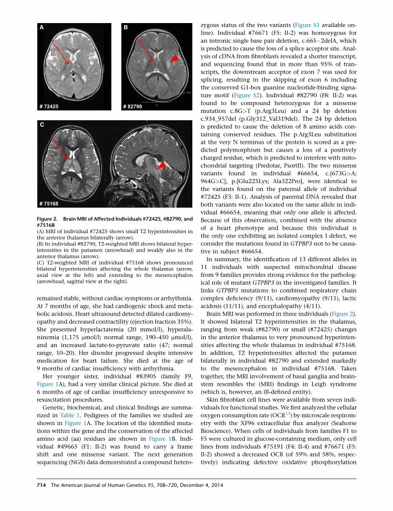
Figure 2. BrainMRI of Affected Individuals #72425, #82790, and#75168(A) MRI of individual #72425 shows small T2 hyperintensities inthe anterior thalamus bilaterally (arrow).(B) In individual #82790, T2-weighted MRI shows bilateral hyper-intensities in the putamen (arrowhead) and weakly also in theanterior thalamus (arrow).(C) T2-weighted MRI of individual #75168 shows pronouncedbilateral hyperintensities affecting the whole thalamus (arrow,axial view at the left) and extending to the mesencephalon(arrowhead, sagittal view at the right).
remained stable, without cardiac symptoms or arrhythmia.
At 7 months of age, she had cardiogenic shock and meta-
bolic acidosis. Heart ultrasound detected dilated cardiomy-
opathy and decreased contractility (ejection fraction 35%).
She presented hyperlactatemia (20 mmol/l), hyperala-
ninemia (1,175 mmol/l; normal range, 190–450 mmol/l),
and an increased lactate-to-pyruvate ratio (47; normal
range, 10–20). Her disorder progressed despite intensive
medication for heart failure. She died at the age of
9 months of cardiac insufficiency with arrhythmia.
Her younger sister, individual #83905 (family F9,
Figure 1A), had a very similar clinical picture. She died at
6 months of age of cardiac insufficiency unresponsive to
resuscitation procedures.
Genetic, biochemical, and clinical findings are summa-
rized in Table 1. Pedigrees of the families we studied are
shown in Figure 1A. The location of the identified muta-
tions within the gene and the conservation of the affected
amino acid (aa) residues are shown in Figure 1B. Indi-
vidual #49665 (F1: II-2) was found to carry a frame
shift and one missense variant. The next generation
sequencing (NGS) data demonstrated a compound hetero-
714 The American Journal of Human Genetics 95, 708–720, Decemb
zygous status of the two variants (Figure S1 available on-
line). Individual #76671 (F5: II-2) was homozygous for
an intronic single base pair deletion, c.665�2delA, which
is predicted to cause the loss of a splice acceptor site. Anal-
ysis of cDNA from fibroblasts revealed a shorter transcript,
and sequencing found that in more than 95% of tran-
scripts, the downstream acceptor of exon 7 was used for
splicing, resulting in the skipping of exon 6 including
the conserved G1-box guanine nucleotide-binding signa-
ture motif (Figure S2). Individual #82790 (F8: II-2) was
found to be compound heterozygous for a missense
mutation c.8G>T (p.Arg3Leu) and a 24 bp deletion
c.934_957del (p.Gly312_Val319del). The 24 bp deletion
is predicted to cause the deletion of 8 amino acids con-
taining conserved residues. The p.Arg3Leu substitution
at the very N terminus of the protein is scored as a pre-
dicted polymorphism but causes a loss of a positively
charged residue, which is predicted to interfere with mito-
chondrial targeting (Predotar, PsortII). The two missense
variants found in individual #66654, c.[673G>A;
964G>C], p.[Glu225Lys; Ala322Pro], were identical to
the variants found on the paternal allele of individual
#72425 (F3: II-1). Analysis of parental DNA revealed that
both variants were also located on the same allele in indi-
vidual #66654, meaning that only one allele is affected.
Because of this observation, combined with the absence
of a heart phenotype and because this individual is
the only one exhibiting an isolated complex I defect, we
consider the mutations found in GTPBP3 not to be causa-
tive in subject #66654.
In summary, the identification of 13 different alleles in
11 individuals with suspected mitochondrial disease
from 9 families provides strong evidence for the patholog-
ical role of mutant GTPBP3 in the investigated families. It
links GTPBP3 mutations to combined respiratory chain
complex deficiency (9/11), cardiomyopathy (9/11), lactic
acidosis (11/11), and encephalopathy (4/11).
Brain MRI was performed in three individuals (Figure 2).
It showed bilateral T2 hyperintensities in the thalamus,
ranging from weak (#82790) or small (#72425) changes
in the anterior thalamus to very pronounced hyperinten-
sities affecting the whole thalamus in individual #75168.
In addition, T2 hyperintensities affected the putamen
bilaterally in individual #82790 and extended markedly
to the mesencephalon in individual #75168. Taken
together, the MRI involvement of basal ganglia and brain-
stem resembles the (MRI) findings in Leigh syndrome
(which is, however, an ill-defined entity).
Skin fibroblast cell lines were available from seven indi-
viduals for functional studies. We first analyzed the cellular
oxygen consumption rate (OCR13) bymicroscale respirom-
etry with the XF96 extracellular flux analyzer (Seahorse
Bioscience). When cells of individuals from families F1 to
F5 were cultured in glucose-containing medium, only cell
lines from individuals #75191 (F4: II-4) and #76671 (F5:
II-2) showed a decreased OCR (of 59% and 58%, respec-
tively) indicating defective oxidative phosphorylation
er 4, 2014

#751
91
cont
rol
cont
rol -
T
#661
43
#661
43 -
T
#661
43 -
T
#496
65
- GTPBP3
- MRPS18B
- MRPL3
- GTPBP3(long exposure)
B
C1.
1
C1.
2
C1.
3
C1.
4
C1.
5
C2
C3
C4
C5
#496
65
#724
25
#661
43
#661
43-T
C6
C6-
T
OCR
cha
nge
from
Glc
to G
al [%
]
A
0
50
100
150
200
C1.
1
C1.
2
C1.
3
C1.
4
C1.
5
C2
C3
C4
C5
#496
65
#724
25
#661
43
#661
43-T
#751
91
#766
71
OC
R [G
lc] [
% o
f con
trol
]
*** ***
Rel
ativ
e en
zym
e ac
tivity
0
20
40
60
80
100
120
140
Complex I Complex IV
cont
rol
#839
04
#839
04-T
cont
rol
#839
04
#839
04-T
normalized to complex II
C D
**
***
160
(short exposure)
40
120
160
0
***
******
***
80
Figure 3. Analysis of Respiration Defects and GTPBP3 Protein Levels in Fibroblast Cell Lines(A) Oxygen consumption rate (OCR) of fibroblast cell lines from five affected individuals and five control subjects cultured in high-glucose (Glc) medium. Each analysis was performed in more than 15 replicates. Control one (C1) was measured five time at differentpassage numbers (C1.1–1.5, NHDFneo, Lonza). OCR was expressed as percentage relative to the average of all controls. Cells from
(legend continued on next page)
The American Journal of Human Genetics 95, 708–720, December 4, 2014 715

(Figure 3A). When cells were cultured with galactose as the
primary carbon source, rather than glucose, cells are forced
to rely on oxidative phosphorylation rather than glycolysis
in order to meet the energy demand.14,15 Accordingly, in
control cells an increase in OCR of approximately 2-fold
was observed when galactose was substituted as the pri-
mary carbon source. This increase in OCR was impaired
in fibroblasts from affected individuals #49665 (F1: II-2),
#66143 (F2: II-2), and #72425 (F3: II-1), which showed
OCR increases of only 11%, 35%, and 22%, respectively
(Figure 3B). In order to confirm that defects in GTPBP3
are the cause of this defect, we transduced three cell
lines with a wild-type copy of GTPBP3 cDNA (RefSeq
NM_32620.3) by using a lentiviral vector (pLenti 6.3/V5
TOPO, Life Technologies) as previously described.16,17
Fibroblasts from individuals #49665 and #66143 were
used for the rescue experiment, with fibroblasts from
#66654 (subject with only one affected allele) being
included as a control (C6). Unfortunately, we were unable
to recover any viable cells from subject #49665 after
the transduction procedure. Although the transduction
had no noticeable effect on the control cell line (C6-T),
transduced fibroblasts from #66143 (#66143-T) displayed
a significant improvement of OCR in galactose-containing
medium (Figure 3B). Furthermore, we detected an
isolated respiratory chain complex (RCC) I deficiency in
a fibroblast cell line from family 9. Cotransfection of indi-
vidual #83904 fibroblasts with two putative GTPBP3 iso-
forms amplified by RT-PCR, RefSeq NM_32620.3 and
NM_0128855.2 (missing 63 base pairs of exon 8), signifi-
cantly improved enzyme activities of RCC I (pIRES2-
EGFP, Clontech) (Figure 3C). Analysis of the protein levels
of GTPBP3 in five fibroblast cell lines demonstrated
reduced or undetectable amounts in individuals #49665,
#75191, #66143, #83904, and #83905, although they
showed a clear increase after transduction or transfection
(Figures S4 and 3D). In conclusion, our data demonstrate
a causal role for GTPBP3 mutations in the oxidative meta-
bolism deficiency in these individuals.
Given that homologs of GTPBP3 in other systems have
been implicated in protein synthesis, we next concen-
trated on the analysis of GTPBP3 in mitochondrial transla-
tion. The synthesis of mtDNA-encoded polypeptides,
investigated by pulse-labeling ofmitochondrial translation
products via [35S]methionine in fibroblasts of affected
individuals #75191 and #76671 showed a significant reduction of oxyand #66143 presented no defective respiration. Error bar indicates 1(B) Oxygen consumption rate of fibroblast cells cultured in galactosetrol cells cultured in galactose-containing medium compared to glu#49665, #72425, and #66143 show significant lower increase in O#66143 significantly increases the change in OCR although it has***p < 0.001.(C) Activities of respiratory chain complexes I and IV (expressed as rfected by electroporation with empty vector (pIRES2-EGFP) accordinexpression of GTPBP3 cDNAs from the same plasmid. MeasurementsSD. Activity in controls was set as 100%. **p < 0.01, **p < 0.001.(D) Levels of GTPBP3 were reduced in cells from individuals #49wtGTPBP3 cDNA. MRPS18B and MRPL3 served as mitochondrial loa
716 The American Journal of Human Genetics 95, 708–720, Decemb
individuals (for methods see Haack et al.18) was severely
and uniformly decreased to 20%–30% of control levels in
individuals #49665, #66143, and #75191 (Figures 4A and
4B). There was no detectable defect in fibroblasts from
individual #72425, which might be explained by the rela-
tively low conservation of the mutated residue in this indi-
vidual (Figure 1B). In order to exclude possible defects of
mitochondrial transcription or precursor RNA processing,
we analyzed all mitochondrially encoded rRNAs and
mRNAs in fibroblasts of individuals #49665, #66143,
#72425, and #75191 by RNA blotting and by RNA-seq in
fibroblasts of individual #49665. We found no differences
in the expression levels of the mt-RNAs between case and
control subjects. On average, the mt-RNA expression levels
were only 6% lower in individual #49665 as compared to
control individuals (data not shown). We did not observe
any appreciable reduction in steady-state levels of mature
RNAs, nor was there any accumulation of precursor RNAs
(Figure S3A). Next, we analyzed the steady-state levels of
mt-tRNAs, including those five species for which the tm5
U modification has been reported in mammals (Gln, Glu,
Lys, LeuUUR, and Trp).4 We again observed no appreciable
changes in their steady-state levels (Figure S3B). In order
to further corroborate a direct role of GTPBP3 inmitochon-
drial translation, we downregulated its expression via
RNA interference in HeLa cells (Figure 4C). Reduction of
GTPBP3 protein levels upon RNAi treatment of HeLa cells
was comparable to the reduction of its level in GTPBP3
mutant fibroblasts (Figure 4D). Downregulation ofGTPBP3
expression resulted in a general mitochondrial translation
defect, similarly to the reduction observed in subject fibro-
blasts (Figure 4D). In conclusion, the reduced translation
efficiency observed in three out of four GTPBP3 mutant
cell lines, as well as in human cells treated with GTPBP3
RNAi, confirmed an important function for GTPBP3 in effi-
cient mitochondrial protein synthesis.
In order to test the consequences of this reduced transla-
tion rate upon the protein levels of OXPHOS complexes in
mutant fibroblast cell lines, we analyzed the steady-state
levels of several nuclear-encoded subunits of the OXPHOS
system by immunoblotting. In fibroblasts from individuals
#72425, #75191, and #76671 (F3: II-1, F4: II-4, and F5:
II-2), we observed strongly reduced amounts of RCC IV.
Fibroblasts from subjects #72425, #75191, and #49665
also showed reduced levels of RCC I, whereas the levels
gen consumption whereas cells from individuals #49665, #72425,SD; ***p < 0.001.(Gal) growth medium. The average increase of OCR from five con-cose-containing medium was 107%. Cell lines from individualsCR. Lentiviral expression of wtGTPBP3 in cells from individualonly little effect in control cells (C6-T). Error bar indicates 1 SD;
atio to CII activity) are decreased in individual #83904 cells trans-g to the manufacturer’s protocol (LONZA) but are improved uponwere performed as previously described.29,30 Error bar indicates 1
665, #75191, and #66143 and elevated after transduction withding controls.
er 4, 2014

A B
C D
Figure 4. Analysis of Mitochondrial Protein Synthesis in Primary Fibroblasts and in HeLa Cells Treated with RNAi against GTPBP3(A) [35S]methioninemetabolic labeling of mitochondrial proteins in fibroblasts. Products of mitochondrial translation were labeled with[35S]methionine for 30 min, separated by a 4%–12% gradient SDS-PAGE, and visualized by autoradiography. To validate equal proteinloading, a small section of the gel was stained with Coomassie (CGS). Fibroblasts from individuals #49665, #66143, and #75191 demon-strate significant inhibition of mitochondrial protein synthesis although translation in cells from individual #72425 is not affected.(B) Quantification of radiolabelled products of mitochondrial translation. Incorporation of [35S] as in (A) was quantified by ImageQuantsoftware after exposure to a PhosphorImager screen from three independent experiments. Error bar indicates 1 SD.(C) Downregulation of GTPBP3 in HeLa cells via RNA interference. Immunoblot analysis of total HeLa cell lysate transfected with twodifferent siRNA to GTPBP3 show decreased level of GTPBP3 upon RNAi treatment for 6 days. siRNA to GFP was used as transfection con-trol. Asterisk indicates nonspecific band recognized by anti-GTPBP3 antibody in HeLa cells. b-actin serves as a loading control. Twodifferent siRNA duplexes targeting GTPBP3 were used, 1 and 2.(D) Mitochondrial translation in HeLa cells upon GTPBP3 downregulation. HeLa cells were transfected for 6 days with siRNA againstGTPBP3 and subjected to [35S]methionine metabolic labeling. Inactivation of GTPBP3 leads to the reduced efficiency of mitochondrialtranslation. Two different siRNA duplexes targeting GTPBP3 were used, 1 and 2.
of RCC II and V remained normal in all cell lines (Figure 5).
The diminished steady-state levels of respiratory chain
complexes I and IV in fibroblast cell lines are in agreement
with the impaired mitochondrial de novo translation rates
in these cells andmatch the enzymatic defects identified in
muscle biopsies of the same individuals.
Within an international cooperation between European
(Germany, UK, Italy, France, and Belgium), Israeli, and
Japanese Centers for mitochondrial disorders, we provide
statistically convincing evidence for GTPBP3 mutations
The American
leading to mitochondrial disease. To further support
collaborative studies, the global mitochondrial disease
community has established a Mitochondrial Disease
Sequence Data Resource (MSeqDR) for common genomic
data deposition and mining.
The genotype-driven analysis performed here was inde-
pendent from the clinical presentation. Nevertheless, we
identified common clinical features of the affected individ-
uals that include lactic acidosis (11/11), cardiomyopathy
(9/11), and neurological symptoms (6/11). The latter
Journal of Human Genetics 95, 708–720, December 4, 2014 717

C6
cont
rol
#496
65
#724
25
#751
91
#766
71
#661
43
#661
43-T
COXIV
SDHA
NDUFB8
ATP5A
[kDa]
15
10070
25
58
Figure 5. Immunoblot Analysis of OXPHOS Proteins inFibroblasts10 mg of detergent-solubilized total cell extract was subjectedto immunoblot analysis of OXPHOS components. Amounts ofSDHA (complex II) and ATP5A (ATPase) were unchanged in allindividuals. In cells from individuals #72425, #75191, and#76671, a reduction of COXIV (complex IV) was observed. Cellsfrom individuals #49665, #72425, and #75191 showed decreasedlevels of NDUFB8 (complex I). Antibodies used: mouse antibodiesagainst SDHA (ab14715), NDUFB8 (ab110242), ATP5A (ab14748),and rabbit antibodies against COXIV (ab16056) from Abcam andrabbit anti GTPBP3 (HPA042158) from SIGMA Aldrich.
comprised symptoms such as development delay, intellec-
tual disability, feeding difficulties, muscle hypotonia,
fatigue, visual impairment, and epileptic seizures. Severity
of the disease ranged from neonatal onset and death to
late-infantile onset and survival into the second decade of
life.Most affected individuals, however,manifested clinical
symptoms before their first birthday. This is consistentwith
the normal cellular respiration, in organello translation,
and normal levels of respiratory chain complexes reported
in individuals less severely affected and the significantly
reduced mitochondrial translation, respiration, and low
levels of complex I and IV in those severely affected.
Modifications of the tRNA ‘‘wobble-base’’ in the anti-
codon loop are required for accurate and efficient codon
recognition. The modification of position 5 (xm5) of the
U34 wobble-base of certain tRNAs is evolutionarily well
conserved, although different modified side chains have
been identified in different species. In mammals, mito-
chondria 5-taurinomethyluridine (tm5U34) is found at
the wobble-base position.19 Based upon studies in bacteria
and yeast mitochondria, GTPBP3 and MTO1 have been
proposed to generate this modification in mammalian
mitochondria. Although this prediction awaits direct
biochemical validation, the proposed functional conserva-
tion of GTPBP3 and MTO1 has been supported by the
mitochondrial localization of these proteins in human
cells and by complementation of the respiratory-deficient
phenotype in yeast by their mammalian homolog
cDNAs.20,21 Functional deficiency of homologs of GTPBP3
and MTO1 in bacteria and yeast mitochondria has been
associated with abnormal U34 modification and conse-
quently a reduced efficiency of translation.21–23 Our data
support an analogous activity of GTPBP3 in human mito-
718 The American Journal of Human Genetics 95, 708–720, Decemb
chondria since we identified a reduced efficiency of trans-
lation in three cell lines with GTPBP3 mutations and in
cells with RNAi-mediated downregulation of GTPBP3
expression. Other groups have also reported impaired
protein synthesis and reduced mitochondrial function in
GTPBP3-depleted cells.24 The defect in mitochondrial
translation was a likely cause of the combined respiratory
chain complex deficiency detected in muscle tissues of all
but one affected individual.
Like GTPBP3 mutations, MTO1 mutations are also asso-
ciated with hypertrophic cardiomyopathy (HCM), lactic
acidosis, and combined respiratory chain deficiency.
An association of MTO1 mutations with impaired mito-
chondrial translation has yet to be shown for humanmito-
chondria, but the common clinical presentation provides
support for a commonpathomechanism in theU34modifi-
cation for both diseases. So far, all individuals with MTO1
mutations presented a HCM. However, nearly all of them
have been specifically screened for MTO1 mutations based
on the clinical presentation of a HCM. Clinical and MRI
signs of brain involvement are found for both GTPBP3 and
MTO1 cases. The genotype-driven investigation presented
here identified individuals with lactic acidosis, develop-
mental delay, andMRI involvement of thalamus, putamen,
andbrainstembutwithoutHCM. It canbe expected that the
clinical spectrum associated withMTO1 deficiencywill also
broaden, with more subjects being genome-wide investi-
gated. In a very recent study, Taylor et al. indeed reported a
case subject withMTO1mutations and central neurological
features who did not have a cardiomyopathy.25
Our study highlights that defects in mitochondrial
translation, probably owing to incorrect posttranscrip-
tional modification of mt-tRNAs, are an important contrib-
utory factor to the spectrum of human mitochondrial
disease. Recent data have suggested that more than 7%
of all mt-tRNA residues undergo posttranscriptional modi-
fication, with close to 30 different modifications so far
described.4 Therefore, it is expected that future WES ana-
lyses of individuals clinically diagnosed with mitochondri-
opathy will reveal further mutations within genes coding
for mt-tRNA modifiers. Indeed, in addition to the afore-
mentioned mutations in MTO1 and TRMU, mutations in
PUS1 (MIM 608109) (which introduces pseudouridine
[J] at base positions 27, 28, and 29 in several mt-tRNAs)
have been reported in subjects affected withmitochondrial
myopathy and sideroblastic anemia (MLASA)26 (MIM
600462) and very recent studies have identified mutations
in TRIT1 (which is responsible for i6A37 modification of a
subset of mt-tRNAs) in individuals with severe combined
mitochondrial respiratory chain defects.27 Furthermore,
mtDNA mutations in mt-tRNA genes, which are a very
frequent cause of human respiratory chain deficiencies
(MITOMAP), might also affect mt-tRNA modification.
Related to the present study, it has been reported that
tm5U34 is not present in mt-tRNALeuUUR harboring the
m.3243A>G mutation (or other pathological mutations)
responsible for mitochondrial encephalopathy, lactic
er 4, 2014

acidosis, and stroke-like episodes (MELAS) (MIM 540000).
The absence of tm5U34 has been suggested to be respon-
sible for the mitochondrial translation defect in these sub-
jects.28 These results imply that deficiency of mt-tRNA
modification plays a critical role in the molecular patho-
genesis of human respiratory chain disease. Further studies
of these pathways, such as analysis of tissue-specific
regulation of mt-tRNA-modifying enzymes, might help
to explain the clinical heterogeneity observed for mito-
chondrial diseases caused by mutations in mt-tRNA genes.
In conclusion, this study shows a mitochondrial
translation disorder with a broad spectrum of clinical
presentations, which emphasizes the importance of post-
transcriptional modification of mitochondrial tRNAs for
proper mitochondrial function.
Supplemental Data
Supplemental Data include four figures and can be foundwith this
article online at http://dx.doi.org/10.1016/j.ajhg.2014.10.017.
Acknowledgments
We thankC. Terrile, M. Borzes, andC. Fischer for technical support
and F. Miyake and T. Wada for referral of sample materials. This
work was supported by the Deutsche Forschungsgemeinschaft
within the frameworkof theMunichCluster for SystemsNeurology
(EXC 1010 SyNergy), the German Bundesministerium fur Bildung
und Forschung (BMBF) through funding of the E-Rare project
GENOMIT (01GM1207 for T.M. and H.P., 2011-RARE-005-03 for
A.R. and M.D.M., J41J11000420001 for M.Z., and FWF I 920-B13
for J.A.M.), German Network for Mitochondrial Disorders (mito-
NET 01GM1113C for T.M., H.P., and P.F. and 01GM1113A for
T.K.), the German Center for Heart Research (Z76010017300 and
Z56010015300 for T.M.), European Commission 7th Framework
Program (Project N261123GEUVADIS),Medical Research Council,
UK (MC_U105697135 for T.J.N., J.R., S.F.P., C.A.P., andM.M.),Well-
come Trust Strategic Award (096919/Z/11/Z for R.W.T. and P.F.C.),
MRC Centre for Neuromuscular Diseases (G0601943), UK NHS
Highly Specialised ‘‘Rare Mitochondrial Disorders of Adults and
Children’’ Service for R.W.T. and P.F.C., Fund for Scientific Research
Belgium (FWO, contract number G.0200.10 for A.V., J.S., and
R.V.C.), Fondazione Telethon (GGP11011 and GPP10005), Italian
Ministry of Health (GR2010-2316392), CARIPLO (2011/0526),
Pierfranco and Luisa Mariani Foundation, and Italian Association
of Mitochondrial Disease Patients and Families (Mitocon) to
D.G., F.I., E.L., and M.Z., Research Program of Innovative Cell
Biology by Innovative Technology (Cell Innovation), Grant-in-
Aid for the Development of New Technology from The Promotion
and Mutual Aid Corporation for Private Schools of Japan from
MEXT for Y.O., Grants-in-Aid for the Research on Intractable
Diseases from the Ministry of Health, Labour andWelfare of Japan
for A.O., Kawano Masanori Memorial Public Interest Incorporated
Foundation for Promotion of Pediatrics for K. Murayama, Associa-
tion Francaise contre les Myopathies (AFM) for A.R. and M.D.M.,
and Fellowship from the AFM (16615 for M.D.M.).
Received: July 30, 2014
Accepted: October 29, 2014
Published: November 26, 2014
The American
Web Resources
The URLs for data presented herein are as follows:
MITOMAP, http://www.mitomap.org/MITOMAP
MSeqDR, https://mseqdr.org/
Online Mendelian Inheritance in Man (OMIM), http://www.
omim.org/
Predotar, https://urgi.versailles.inra.fr/predotar/predotar.html
PSORTII Prediction, http://psort.hgc.jp/form2.html
RefSeq, http://www.ncbi.nlm.nih.gov/RefSeq
References
1. Boczonadi, V., and Horvath, R. (2014). Mitochondria:
impaired mitochondrial translation in human disease. Int. J.
Biochem. Cell Biol. 48, 77–84.
2. Osawa, S. (1995). Evolution of the Genetic Code (Washington,
DC: ASM Press).
3. Suzuki, T. (2005). Biosynthesis and function of tRNA wobble
modifications. Top. Curr. Genet. 12, 23–69.
4. Suzuki, T., and Suzuki, T. (2014). A complete landscape of post-
transcriptional modifications in mammalian mitochondrial
tRNAs. Nucleic Acids Res. 42, 7346–7357.
5. Colby, G., Wu, M., and Tzagoloff, A. (1998). MTO1 codes
for a mitochondrial protein required for respiration in
paromomycin-resistant mutants of Saccharomyces cerevisiae.
J. Biol. Chem. 273, 27945–27952.
6. Yokoyama, S., Watanabe, T., Murao, K., Ishikura, H., Yamai-
zumi, Z., Nishimura, S., and Miyazawa, T. (1985). Molecular
mechanism of codon recognition by tRNA species with modi-
fied uridine in the first position of the anticodon. Proc. Natl.
Acad. Sci. USA 82, 4905–4909.
7. Bjork, G.R., Huang, B., Persson, O.P., and Bystrom, A.S. (2007).
A conserved modified wobble nucleoside (mcm5s2U) in lysyl-
tRNA is required for viability in yeast. RNA 13, 1245–1255.
8. Yarham, J.W., Elson, J.L., Blakely, E.L., McFarland, R., and
Taylor, R.W. (2010). Mitochondrial tRNA mutations and dis-
ease. Wiley Interdiscip. Rev. RNA 1, 304–324.
9. Zeharia, A., Shaag, A., Pappo, O., Mager-Heckel, A.-M., Saada,
A., Beinat, M., Karicheva, O., Mandel, H., Ofek, N., Segel, R.,
et al. (2009). Acute infantile liver failure due to mutations in
the TRMU gene. Am. J. Hum. Genet. 85, 401–407.
10. Ghezzi, D., Baruffini, E., Haack, T.B., Invernizzi, F., Mel-
chionda, L., Dallabona, C., Strom, T.M., Parini, R., Burlina,
A.B., Meitinger, T., et al. (2012). Mutations of the mitochon-
drial-tRNA modifier MTO1 cause hypertrophic cardiomyopa-
thy and lactic acidosis. Am. J. Hum. Genet. 90, 1079–1087.
11. Baruffini, E., Dallabona, C., Invernizzi, F., Yarham, J.W., Mel-
chionda, L., Blakely, E.L., Lamantea, E., Donnini, C., Santra,
S., Vijayaraghavan, S., et al. (2013). MTO1mutations are asso-
ciated with hypertrophic cardiomyopathy and lactic acidosis
and cause respiratory chain deficiency in humans and yeast.
Hum. Mutat. 34, 1501–1509.
12. Elstner, M., Andreoli, C., Klopstock, T., Meitinger, T., and
Prokisch, H. (2009). The mitochondrial proteome database:
MitoP2. Methods Enzymol. 457, 3–20.
13. Invernizzi, F., D’Amato, I., Jensen, P.B., Ravaglia, S., Zeviani,
M., and Tiranti, V. (2012). Microscale oxygraphy reveals
OXPHOS impairment in MRC mutant cells. Mitochondrion
12, 328–335.
14. Petrova-Benedict, R., Buncic, J.R., Wallace, D.C., and
Robinson, B.H. (1992). Selective killing of cells with oxidative
Journal of Human Genetics 95, 708–720, December 4, 2014 719

defects in galactose medium: a screening test for affected
patient fibroblasts. J. Inherit. Metab. Dis. 15, 943–944.
15. Robinson, B.H., Petrova-Benedict, R., Buncic, J.R., and
Wallace, D.C. (1992). Nonviability of cells with oxidative de-
fects in galactose medium: a screening test for affected patient
fibroblasts. Biochem. Med. Metab. Biol. 48, 122–126.
16. Danhauser, K., Iuso, A., Haack, T.B., Freisinger, P., Brockmann,
K., Mayr, J.A., Meitinger, T., and Prokisch, H. (2011). Cellular
rescue-assay aids verification of causative DNA-variants in
mitochondrial complex I deficiency. Mol. Genet. Metab.
103, 161–166.
17. Kornblum, C., Nicholls, T.J., Haack, T.B., Scholer, S., Peeva, V.,
Danhauser, K., Hallmann, K., Zsurka, G., Rorbach, J., Iuso, A.,
et al. (2013). Loss-of-function mutations in MGME1 impair
mtDNA replication and cause multisystemic mitochondrial
disease. Nat. Genet. 45, 214–219.
18. Haack, T.B., Kopajtich, R., Freisinger, P., Wieland, T., Rorbach,
J., Nicholls, T.J., Baruffini, E., Walther, A., Danhauser, K.,
Zimmermann, F.A., et al. (2013). ELAC2 mutations cause a
mitochondrial RNA processing defect associated with hyper-
trophic cardiomyopathy. Am. J. Hum. Genet. 93, 211–223.
19. Suzuki, T., Suzuki, T., Wada, T., Saigo, K., and Watanabe, K.
(2002). Taurine as a constituent of mitochondrial tRNAs:
new insights into the functions of taurine and human mito-
chondrial diseases. EMBO J. 21, 6581–6589.
20. Li, X., and Guan, M.-X. (2002). A human mitochondrial GTP
binding protein related to tRNA modification may modulate
phenotypic expression of the deafness-associated mitochon-
drial 12S rRNA mutation. Mol. Cell. Biol. 22, 7701–7711.
21. Li, X., Li, R., Lin, X., and Guan, M.-X. (2002). Isolation and
characterization of the putative nuclear modifier gene MTO1
involved in the pathogenesis of deafness-associated mito-
chondrial 12 S rRNA A1555G mutation. J. Biol. Chem. 277,
27256–27264.
22. Wang, X., Yan, Q., and Guan, M.-X. (2010). Combination of
the loss of cmnm5U34 with the lack of s2U34 modifications
of tRNALys, tRNAGlu, and tRNAGln altered mitochondrial
biogenesis and respiration. J. Mol. Biol. 395, 1038–1048.
720 The American Journal of Human Genetics 95, 708–720, Decemb
23. Murphy, F.V., 4th, Ramakrishnan, V., Malkiewicz, A., and
Agris, P.F. (2004). The role of modifications in codon discrim-
ination by tRNA(Lys)UUU. Nat. Struct. Mol. Biol. 11, 1186–
1191.
24. Villarroya, M., Prado, S., Esteve, J.M., Soriano, M.A., Aguado,
C., Perez-Martınez, D., Martınez-Ferrandis, J.I., Yim, L., Victor,
V.M., Cebolla, E., et al. (2008). Characterization of human
GTPBP3, a GTP-binding protein involved in mitochondrial
tRNA modification. Mol. Cell. Biol. 28, 7514–7531.
25. Taylor, R.W., Pyle, A., Griffin, H., Blakely, E.L., Duff, J., He, L.,
Smertenko, T., Alston, C.L., Neeve, V.C., Best, A., et al. (2014).
Use of whole-exome sequencing to determine the genetic
basis of multiple mitochondrial respiratory chain complex
deficiencies. JAMA 312, 68–77.
26. Bykhovskaya, Y., Casas, K., Mengesha, E., Inbal, A., and
Fischel-Ghodsian, N. (2004). Missense mutation in pseudour-
idine synthase 1 (PUS1) causes mitochondrial myopathy
and sideroblastic anemia (MLASA). Am. J. Hum. Genet. 74,
1303–1308.
27. Yarham, J.W., Lamichhane, T.N., Pyle, A., Mattijssen, S.,
Baruffini, E., Bruni, F., Donnini, C., Vassilev, A., He, L., Blakely,
E.L., et al. (2014). Defective i6A37 modification of mito-
chondrial and cytosolic tRNAs results from pathogenic muta-
tions in TRIT1 and its substrate tRNA. PLoS Genet. 10,
e1004424.
28. Yasukawa, T., Suzuki, T., Ueda, T., Ohta, S., and Watanabe, K.
(2000). Modification defect at anticodon wobble nucleotide
of mitochondrial tRNAs(Leu)(UUR) with pathogenic muta-
tions of mitochondrial myopathy, encephalopathy, lactic
acidosis, and stroke-like episodes. J. Biol. Chem. 275, 4251–
4257.
29. Rustin, P., Chretien, D., Bourgeron, T., Gerard, B., Rotig, A.,
Saudubray, J.M., and Munnich, A. (1994). Biochemical and
molecular investigations in respiratory chain deficiencies.
Clin. Chim. Acta 228, 35–51.
30. Rustin, P., Chretien, D., Bourgeron, T., Wucher, A., Saudubray,
J.M., Rotig, A., and Munnich, A. (1991). Assessment of the
mitochondrial respiratory chain. Lancet 338, 60.
er 4, 2014
Copyright © 2022 FDOKUMEN