The Role of Acidosis in the Pathogenesis of Severe ... - MDPI
15
biology Review The Role of Acidosis in the Pathogenesis of Severe Forms of COVID-19 Yury D. Nechipurenko 1, *, Denis A. Semyonov 2,3 , Igor A. Lavrinenko 4 , Denis A. Lagutkin 5 , Evgenii A. Generalov 6 , Anna Y. Zaitceva 7 , Olga V. Matveeva 8 and Yegor E. Yegorov 9, * Citation: Nechipurenko, Y.D.; Semyonov, D.A.; Lavrinenko, I.A.; Lagutkin, D.A.; Generalov, E.A.; Zaitceva, A.Y.; Matveeva, O.V.; Yegorov, Y.E. The Role of Acidosis in the Pathogenesis of Severe Forms of COVID-19. Biology 2021, 10, 852. https://doi.org/10.3390/biology 10090852 Academic Editors: Mohamad Goldust, Robert A. Schwartz, Dedee Murrell, Torello Lotti, Andrew Davidson and Andrés Moya Received: 29 June 2021 Accepted: 26 August 2021 Published: 31 August 2021 Publisher’s Note: MDPI stays neutral with regard to jurisdictional claims in published maps and institutional affil- iations. Copyright: © 2021 by the authors. Licensee MDPI, Basel, Switzerland. This article is an open access article distributed under the terms and conditions of the Creative Commons Attribution (CC BY) license (https:// creativecommons.org/licenses/by/ 4.0/). 1 Laboratory DNA-Protein Recognition, Engelhardt Institute of Molecular Biology, Russian Academy of Sciences, Moscow 119991, Russia 2 Institute of Molecular Medicine and Pathobiochemistry, Voyno-Yasenetsky Krasnoyarsk State Medical University, Krasnoyarsk 660022, Russia; [email protected] 3 Institute of Biophysics Siberian Branch of Russian Academy of Sciences, Krasnoyarsk 660036, Russia 4 Department of Human and Animal Physiology, Faculty of Medicine and Biology, Voronezh State University, Voronezh 394018, Russia; [email protected] 5 Department of Biological and Medical Physics, Moscow Institute of Physics and Technology, Dolgoprudny 141701, Russia; [email protected] 6 Department of Biophysics, Faculty of Physics, Lomonosov Moscow State University, Moscow 119991, Russia; [email protected] 7 Laboratory of Medical Analytical Methods and Devices, Institute for Analytical Instrumentation of the Russian Academy of Sciences, St. Petersburg 198095, Russia; [email protected] 8 Sendai Viralytics LLC, Acton, MA 117261, USA; [email protected] 9 Laboratory of Cellular Bases for the Development of Malignant Diseases, Engelhardt Institute of Molecular Biology, Russian Academy of Sciences, Moscow 119991, Russia * Correspondence: [email protected] (Y.D.N.); [email protected] (Y.E.Y.); Tel.: +7-915-268-62-14 (Y.E.Y.) Simple Summary: Recently, several studies have shown that acidosis, which is increased acidity in the blood and other body tissues, is often associated with severe COVID-19. In this article, we look at the mechanisms and consequences of acidosis that can lead to an exacerbation of COVID-19. We want to draw the attention of readers to the threshold values of such disease characteristics as hypoxia and acidosis, which are associated with a sharp deterioration in the patient’s condition. Hypoxia and acidosis mutually reinforce each other according to the principle of a vicious cycle (that is, they are involved in a system of positive feedbacks). Elevated blood lactate (lactic acid) levels are associated with poor clinical outcomes in COVID patients. As a practical recommendation, we propose to pay more attention to the prevention of acidosis, including in the early stages of the disease, when the adjustment of homeostasis requires less effort and is less risky. Abstract: COVID-19 has specific characteristics that distinguish this disease from many other infec- tions. We suggest that the pathogenesis of severe forms of COVID-19 can be associated with acidosis. This review article discusses several mechanisms potentially linking the damaging effects of COVID- 19 with acidosis and shows the existence of a vicious cycle between the development of hypoxia and acidosis in COVID-19 patients. At the early stages of the disease, inflammation, difficulty in gas exchange in the lungs and thrombosis collectively contribute to the onset of acidosis. In accordance with the Verigo-Bohr effect, a decrease in blood pH leads to a decrease in oxygen saturation, which contributes to the exacerbation of acidosis and results in a deterioration of the patient’s condition. A decrease in pH can also cause conformational changes in the S-protein of the virus and thus lead to a decrease in the affinity and avidity of protective antibodies. Hypoxia and acidosis lead to dysregu- lation of the immune system and multidirectional pro- and anti-inflammatory reactions, resulting in the development of a “cytokine storm”. In this review, we highlight the potential importance of supporting normal blood pH as an approach to COVID-19 therapy. Keywords: SARS-CoV-2; COVID-19; acidosis; hypoxia; saturation; Bohr effect; lactate; pH Biology 2021, 10, 852. https://doi.org/10.3390/biology10090852 https://www.mdpi.com/journal/biology
-
Upload
khangminh22 -
Category
Documents
-
view
0 -
download
0
Transcript of The Role of Acidosis in the Pathogenesis of Severe ... - MDPI
biology
Review
The Role of Acidosis in the Pathogenesis of Severe Formsof COVID-19
Yury D. Nechipurenko 1,*, Denis A. Semyonov 2,3, Igor A. Lavrinenko 4 , Denis A. Lagutkin 5 ,Evgenii A. Generalov 6 , Anna Y. Zaitceva 7, Olga V. Matveeva 8 and Yegor E. Yegorov 9,*
�����������������
Citation: Nechipurenko, Y.D.;
Semyonov, D.A.; Lavrinenko, I.A.;
Lagutkin, D.A.; Generalov, E.A.;
Zaitceva, A.Y.; Matveeva, O.V.;
Yegorov, Y.E. The Role of Acidosis in
the Pathogenesis of Severe Forms of
COVID-19. Biology 2021, 10, 852.
https://doi.org/10.3390/biology
10090852
Academic Editors: Mohamad
Goldust, Robert A. Schwartz,
Dedee Murrell, Torello Lotti,
Andrew Davidson and Andrés Moya
Received: 29 June 2021
Accepted: 26 August 2021
Published: 31 August 2021
Publisher’s Note: MDPI stays neutral
with regard to jurisdictional claims in
published maps and institutional affil-
iations.
Copyright: © 2021 by the authors.
Licensee MDPI, Basel, Switzerland.
This article is an open access article
distributed under the terms and
conditions of the Creative Commons
Attribution (CC BY) license (https://
creativecommons.org/licenses/by/
4.0/).
1 Laboratory DNA-Protein Recognition, Engelhardt Institute of Molecular Biology, Russian Academy ofSciences, Moscow 119991, Russia
2 Institute of Molecular Medicine and Pathobiochemistry, Voyno-Yasenetsky Krasnoyarsk State MedicalUniversity, Krasnoyarsk 660022, Russia; [email protected]
3 Institute of Biophysics Siberian Branch of Russian Academy of Sciences, Krasnoyarsk 660036, Russia4 Department of Human and Animal Physiology, Faculty of Medicine and Biology, Voronezh State University,
Voronezh 394018, Russia; [email protected] Department of Biological and Medical Physics, Moscow Institute of Physics and Technology,
Dolgoprudny 141701, Russia; [email protected] Department of Biophysics, Faculty of Physics, Lomonosov Moscow State University, Moscow 119991, Russia;
[email protected] Laboratory of Medical Analytical Methods and Devices, Institute for Analytical Instrumentation of the
Russian Academy of Sciences, St. Petersburg 198095, Russia; [email protected] Sendai Viralytics LLC, Acton, MA 117261, USA; [email protected] Laboratory of Cellular Bases for the Development of Malignant Diseases, Engelhardt Institute of Molecular
Biology, Russian Academy of Sciences, Moscow 119991, Russia* Correspondence: [email protected] (Y.D.N.); [email protected] (Y.E.Y.); Tel.: +7-915-268-62-14 (Y.E.Y.)
Simple Summary: Recently, several studies have shown that acidosis, which is increased acidity inthe blood and other body tissues, is often associated with severe COVID-19. In this article, we look atthe mechanisms and consequences of acidosis that can lead to an exacerbation of COVID-19. We wantto draw the attention of readers to the threshold values of such disease characteristics as hypoxia andacidosis, which are associated with a sharp deterioration in the patient’s condition. Hypoxia andacidosis mutually reinforce each other according to the principle of a vicious cycle (that is, they areinvolved in a system of positive feedbacks). Elevated blood lactate (lactic acid) levels are associatedwith poor clinical outcomes in COVID patients. As a practical recommendation, we propose to paymore attention to the prevention of acidosis, including in the early stages of the disease, when theadjustment of homeostasis requires less effort and is less risky.
Abstract: COVID-19 has specific characteristics that distinguish this disease from many other infec-tions. We suggest that the pathogenesis of severe forms of COVID-19 can be associated with acidosis.This review article discusses several mechanisms potentially linking the damaging effects of COVID-19 with acidosis and shows the existence of a vicious cycle between the development of hypoxiaand acidosis in COVID-19 patients. At the early stages of the disease, inflammation, difficulty in gasexchange in the lungs and thrombosis collectively contribute to the onset of acidosis. In accordancewith the Verigo-Bohr effect, a decrease in blood pH leads to a decrease in oxygen saturation, whichcontributes to the exacerbation of acidosis and results in a deterioration of the patient’s condition. Adecrease in pH can also cause conformational changes in the S-protein of the virus and thus lead to adecrease in the affinity and avidity of protective antibodies. Hypoxia and acidosis lead to dysregu-lation of the immune system and multidirectional pro- and anti-inflammatory reactions, resultingin the development of a “cytokine storm”. In this review, we highlight the potential importance ofsupporting normal blood pH as an approach to COVID-19 therapy.
Keywords: SARS-CoV-2; COVID-19; acidosis; hypoxia; saturation; Bohr effect; lactate; pH
Biology 2021, 10, 852. https://doi.org/10.3390/biology10090852 https://www.mdpi.com/journal/biology
Biology 2021, 10, 852 2 of 15
1. Introduction
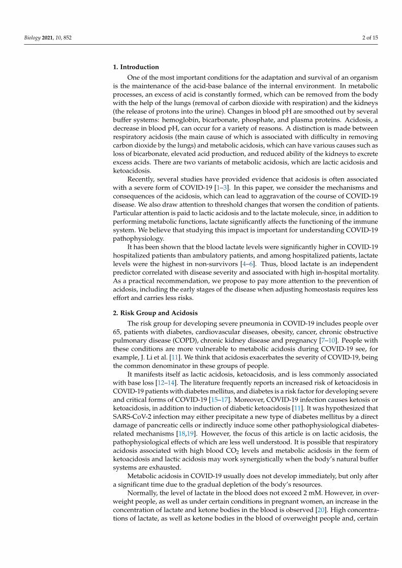
One of the most important conditions for the adaptation and survival of an organismis the maintenance of the acid-base balance of the internal environment. In metabolicprocesses, an excess of acid is constantly formed, which can be removed from the bodywith the help of the lungs (removal of carbon dioxide with respiration) and the kidneys(the release of protons into the urine). Changes in blood pH are smoothed out by severalbuffer systems: hemoglobin, bicarbonate, phosphate, and plasma proteins. Acidosis, adecrease in blood pH, can occur for a variety of reasons. A distinction is made betweenrespiratory acidosis (the main cause of which is associated with difficulty in removingcarbon dioxide by the lungs) and metabolic acidosis, which can have various causes such asloss of bicarbonate, elevated acid production, and reduced ability of the kidneys to excreteexcess acids. There are two variants of metabolic acidosis, which are lactic acidosis andketoacidosis.
Recently, several studies have provided evidence that acidosis is often associatedwith a severe form of COVID-19 [1–3]. In this paper, we consider the mechanisms andconsequences of the acidosis, which can lead to aggravation of the course of COVID-19disease. We also draw attention to threshold changes that worsen the condition of patients.Particular attention is paid to lactic acidosis and to the lactate molecule, since, in addition toperforming metabolic functions, lactate significantly affects the functioning of the immunesystem. We believe that studying this impact is important for understanding COVID-19pathophysiology.
It has been shown that the blood lactate levels were significantly higher in COVID-19hospitalized patients than ambulatory patients, and among hospitalized patients, lactatelevels were the highest in non-survivors [4–6]. Thus, blood lactate is an independentpredictor correlated with disease severity and associated with high in-hospital mortality.As a practical recommendation, we propose to pay more attention to the prevention ofacidosis, including the early stages of the disease when adjusting homeostasis requires lesseffort and carries less risks.
2. Risk Group and Acidosis
The risk group for developing severe pneumonia in COVID-19 includes people over65, patients with diabetes, cardiovascular diseases, obesity, cancer, chronic obstructivepulmonary disease (COPD), chronic kidney disease and pregnancy [7–10]. People withthese conditions are more vulnerable to metabolic acidosis during COVID-19 see, forexample, J. Li et al. [11]. We think that acidosis exacerbates the severity of COVID-19, beingthe common denominator in these groups of people.
It manifests itself as lactic acidosis, ketoacidosis, and is less commonly associatedwith base loss [12–14]. The literature frequently reports an increased risk of ketoacidosis inCOVID-19 patients with diabetes mellitus, and diabetes is a risk factor for developing severeand critical forms of COVID-19 [15–17]. Moreover, COVID-19 infection causes ketosis orketoacidosis, in addition to induction of diabetic ketoacidosis [11]. It was hypothesized thatSARS-CoV-2 infection may either precipitate a new type of diabetes mellitus by a directdamage of pancreatic cells or indirectly induce some other pathophysiological diabetes-related mechanisms [18,19]. However, the focus of this article is on lactic acidosis, thepathophysiological effects of which are less well understood. It is possible that respiratoryacidosis associated with high blood CO2 levels and metabolic acidosis in the form ofketoacidosis and lactic acidosis may work synergistically when the body’s natural buffersystems are exhausted.
Metabolic acidosis in COVID-19 usually does not develop immediately, but only aftera significant time due to the gradual depletion of the body’s resources.
Normally, the level of lactate in the blood does not exceed 2 mM. However, in over-weight people, as well as under certain conditions in pregnant women, an increase in theconcentration of lactate and ketone bodies in the blood is observed [20]. High concentra-tions of lactate, as well as ketone bodies in the blood of overweight people and, certain

Biology 2021, 10, 852 3 of 15
pregnant women are detected, including before the onset of respiratory diseases, and theoccurrence of pneumonia only aggravates the already existing acidosis.
Next, we will try to prove that metabolic acidosis is the cause and effect of manyphenomena characteristic of the severe course of COVID-19.
3. Factors Determining the Disruption of Gas Exchange in COVID-19
The pneumonia that develops with COVID-19 leads to disruption of gas exchange inthe lungs, which provokes the development of hypoxia. Hypoxia induces the transition toanaerobic metabolism and increases lactate production. The hypoxic disorder is observedin several diseases (mainly with respiratory involvement). However, this disorder isone of the main defining features of COVID-19. Severe Acute Respiratory Syndrome isabbreviated as SARS and is included in the name of the SARS-CoV-2 virus that causesCOVID-19.
There are at least four factors contributing to the disruption of gas exchange in COVID-19 (Figure 1).
Biology 2021, 10, x FOR PEER REVIEW 3 of 15
concentration of lactate and ketone bodies in the blood is observed [20]. High concentra-tions of lactate, as well as ketone bodies in the blood of overweight people and, certain pregnant women are detected, including before the onset of respiratory diseases, and the occurrence of pneumonia only aggravates the already existing acidosis.
Next, we will try to prove that metabolic acidosis is the cause and effect of many phenomena characteristic of the severe course of COVID-19.
3. Factors Determining the Disruption of Gas Exchange in COVID-19 The pneumonia that develops with COVID-19 leads to disruption of gas exchange in
the lungs, which provokes the development of hypoxia. Hypoxia induces the transition to anaerobic metabolism and increases lactate production. The hypoxic disorder is ob-served in several diseases (mainly with respiratory involvement). However, this disorder is one of the main defining features of COVID-19. Severe Acute Respiratory Syndrome is abbreviated as SARS and is included in the name of the SARS-CoV-2 virus that causes COVID-19.
There are at least four factors contributing to the disruption of gas exchange in COVID-19 (Figure 1).
Figure 1. A diagram illustrating the relationship between viral lung damage and the occurrence of hypoxia.
1. The lytic phase of the virus leads to the death of type 2 alveolocytes, which disrupts the structure of the alveoli [21]. The collapse of alveoli is facilitated by a decrease in the production of surfactant in alveolocytes, which leads to a change in surface ten-sion. As a result, the changes in air pressure in the lungs do not lead to compression and expansion of the alveolar vesicles, the dead volume of lungs increases and gas exchange decreases;
2. COVID-19 is frequently manifested by thrombosis in pulmonary capillaries, which prevents the transfer of oxygen [22];
Figure 1. A diagram illustrating the relationship between viral lung damage and the occurrence ofhypoxia.
1. The lytic phase of the virus leads to the death of type 2 alveolocytes, which disruptsthe structure of the alveoli [21]. The collapse of alveoli is facilitated by a decreasein the production of surfactant in alveolocytes, which leads to a change in surfacetension. As a result, the changes in air pressure in the lungs do not lead to compressionand expansion of the alveolar vesicles, the dead volume of lungs increases and gasexchange decreases;
2. COVID-19 is frequently manifested by thrombosis in pulmonary capillaries, whichprevents the transfer of oxygen [22];
3. Intensive production of the extracellular matrix (ECM), rich in hyaluronic acid, isdesigned to “seal” areas of extensive lung damage to prevent general lung collapse.As a side effect of this process, a part of functional alveoli is filled with ECM andswitched off from gas exchange [23];
Biology 2021, 10, 852 4 of 15
4. Another of the mechanisms limiting gas exchange in patients with COVID-19 is achange in the properties of the erythrocytes themselves. The virus can infect erythroidprecursor cells. This, in turn, leads to a greater production of immature red bloodcells, followed by their massive release into the bloodstream and a correspondingdrop in hemoglobin levels [24].
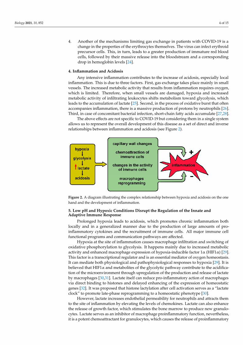
4. Inflammation and Acidosis
Any intensive inflammation contributes to the increase of acidosis, especially localinflammation. This is due to three factors. First, gas exchange takes place mainly in smallvessels. The increased metabolic activity that results from inflammation requires oxygen,which is limited. Therefore, when small vessels are damaged, hypoxia and increasedmetabolic activity of infiltrating leukocytes shifts metabolism toward glycolysis, whichleads to the accumulation of lactate [25]. Second, in the process of oxidative burst that oftenaccompanies inflammation, there is a massive production of protons by neutrophils [26].Third, in case of concomitant bacterial infection, short-chain fatty acids accumulate [27,28].
The above effects are not specific to COVID-19 but considering them in a single systemallows us to represent the overall development of this disease as a set of direct and inverserelationships between inflammation and acidosis (see Figure 2).
Biology 2021, 10, x FOR PEER REVIEW 4 of 15
3. Intensive production of the extracellular matrix (ECM), rich in hyaluronic acid, is de-signed to “seal” areas of extensive lung damage to prevent general lung collapse. As a side effect of this process, a part of functional alveoli is filled with ECM and switched off from gas exchange [23];
4. Another of the mechanisms limiting gas exchange in patients with COVID-19 is a change in the properties of the erythrocytes themselves. The virus can infect erythroid precursor cells. This, in turn, leads to a greater production of immature red blood cells, followed by their massive release into the bloodstream and a correspond-ing drop in hemoglobin levels [24].
4. Inflammation and Acidosis Any intensive inflammation contributes to the increase of acidosis, especially local
inflammation. This is due to three factors. First, gas exchange takes place mainly in small vessels. The increased metabolic activity that results from inflammation requires oxygen, which is limited. Therefore, when small vessels are damaged, hypoxia and increased met-abolic activity of infiltrating leukocytes shifts metabolism toward glycolysis, which leads to the accumulation of lactate [25]. Second, in the process of oxidative burst that often accompanies inflammation, there is a massive production of protons by neutrophils [26]. Third, in case of concomitant bacterial infection, short-chain fatty acids accumulate [27,28].
The above effects are not specific to COVID-19 but considering them in a single sys-tem allows us to represent the overall development of this disease as a set of direct and inverse relationships between inflammation and acidosis (see Figure 2).
Figure 2. A diagram illustrating the complex relationship between hypoxia and acidosis on the one hand and the development of inflammation.
5. Low pH and Hypoxic Conditions Disrupt the Regulation of the Innate and Adap-tive Immune Response
Prolonged hypoxia leads to acidosis, which promotes chronic inflammation both lo-cally and in a generalized manner due to the production of large amounts of pro-inflam-matory cytokines and the recruitment of immune cells. All major immune cell functional programs and communication pathways are affected.
Hypoxia at the site of inflammation causes macrophage infiltration and switching of oxidative phosphorylation to glycolysis. It happens mainly due to increased metabolic activity and enhanced macrophage expression of hypoxia-inducible factor 1α (HIF1α) [29]. This factor is a transcriptional regulator and is an essential mediator of oxygen ho-meostasis. It can mediate both physiological and pathophysiological responses to hypoxia [29]. It is believed that HIF1α and metabolites of the glycolytic pathway contribute to the acidification of the microenvironment through upregulation of the production and release of lactate by macrophages [30,31]. Lactate itself can reduce pro-inflammatory action of macrophages via direct binding to histones and delayed enhancing of the expression of
Figure 2. A diagram illustrating the complex relationship between hypoxia and acidosis on the onehand and the development of inflammation.
5. Low pH and Hypoxic Conditions Disrupt the Regulation of the Innate andAdaptive Immune Response
Prolonged hypoxia leads to acidosis, which promotes chronic inflammation bothlocally and in a generalized manner due to the production of large amounts of pro-inflammatory cytokines and the recruitment of immune cells. All major immune cellfunctional programs and communication pathways are affected.
Hypoxia at the site of inflammation causes macrophage infiltration and switching ofoxidative phosphorylation to glycolysis. It happens mainly due to increased metabolicactivity and enhanced macrophage expression of hypoxia-inducible factor 1α (HIF1α) [29].This factor is a transcriptional regulator and is an essential mediator of oxygen homeostasis.It can mediate both physiological and pathophysiological responses to hypoxia [29]. It isbelieved that HIF1α and metabolites of the glycolytic pathway contribute to the acidifica-tion of the microenvironment through upregulation of the production and release of lactateby macrophages [30,31]. Lactate itself can reduce pro-inflammatory action of macrophagesvia direct binding to histones and delayed enhancing of the expression of homeostaticgenes [32]. It was proposed that histone lactylation after cell activation serves as a “lactateclock” to promote late-phase reprogramming to a homeostatic phenotype [30].
However, lactate increases endothelial permeability for neutrophils and attracts themto the site of inflammation by elevating the levels of chemokines. Lactate can also enhancethe release of growth factor, which stimulates the bone marrow to produce new granulo-cytes. Lactate serves as an inhibitor of macrophage proinflammatory function, nevertheless,it is a potent chemoattractant for granulocytes, which causes the release of proinflammatory

Biology 2021, 10, 852 5 of 15
effector molecules by neutrophils [33]. Therefore, proinflammatory signals may prevailcausing hyperinflammation.
Hypoxia is likely to be an important factor contributing to the development of hyper-inflammation seen in severe COVID-19. Acidosis influences the functioning of immunecells and further stimulates the development of inflammation. Short-term hypoxia andlocal acidosis are prominent features of the inflammatory microenvironment that regulatekey transcription factors in both innate and adaptive immunity but prolonged exposureof these features on immune cells leads to dysregulation of responses to antigens. Thisdysregulation causes the transformation of physiological reactions into pathophysiologi-cal ones.
Thus, prolonged hypoxia leads to acidosis, and a combination of these factors causeshyperactivation of dendritic cells and dysregulation of cytokine response. These affectantigen internalization and presentation. Hypoxia and lactic acidosis attract macrophages,leading to the early pro-inflammatory response with delayed homeostatic reprogram-ming and potently stimulate neutrophil migration and activation. All these factors arelikely to synergistically contribute to the development and maintenance of the state ofhyperinflammation, which is characteristic of severe forms of COVID-19.
6. Bohr Effect—Basic Threshold Effect Linking Acidosis and Hypoxia
Severe pneumonia in COVID-19 is accompanied by a drop in blood oxygen saturation.At the same time, the first stage of saturation reduction proceeds relatively smoothly, it candevelop for several days. The subsequent drop in saturation and the occurrence of oxygendeficiency are already occurring rapidly and require hospitalization, oxygen support andintensive care. We believe that the threshold effect of a rapid decrease in saturation isdirectly related to acidosis and is explained by the Bohr effect, observed in the analysis ofoxygen binding by hemoglobin [34]. The curve of hemoglobin oxygen saturation in relationto oxygen partial pressure has an S shape (Figure 3). As the partial pressure of oxygen inthe blood increases, its binding to hemoglobin initially practically does not change until itreaches a certain boundary value (first inflection point), then increases sharply—and thenreaches a plateau reflecting saturation (second inflection point).
Biology 2021, 10, x FOR PEER REVIEW 6 of 15
An important regulator of oxygen transport is 2,3-diphosphoglycerate (2,3-DPG). The formation of lactate is accompanied by the accumulation of 2,3-DPG (Rappoport shunt) with the migration of the latter into erythrocytes. 2,3-DPG is incorporated into he-moglobin by forming a salt bridge between the β-subunits of the heterotetramer, which prevents oxygen re-binding. This is another mechanism that allows you to regulate the transport of oxygen, in essence, it complements the regulation with the help of the Bohr effect [39,40].
Figure 3. Different factors influencing oxygen saturation of hemoglobin. During pH change from 7.4 to 7.2, saturation decreases two times. The concentration of 2,3-diphosphoglycerate provides saturation of hemoglobin with oxygen, along with CO2 pressure. pH, negative of the base 10 loga-rithm of the activity of the H+ ion; pCO2, partial pressure of carbon dioxide gas; 2,3-DPG, 2,3-di-phosphoglycerate; p50, oxygen tension at 50% hemoglobin saturation.
7. Physiological Aspects of the Compensatory Mechanisms of Acidosis A significant contribution to the acid-base balance of the body made by the activity
of some internal organs, such as, for example, the respiratory center of the brain, which is essentially a physiological system for monitoring pH and controlling respiration [34]. When chemical stimuli, such as hypoxia and hypercapnia (elevated CO2 level in the blood), are recognized by chemoreceptors, the respiratory center increases the flow of im-pulses to the respiratory motor neurons, which results in increased ventilation. Arterial hypocapnia (low CO2 levels), on the contrary, causes a decrease in ventilation [41,42]. In the case of SARS-CoV-2 virus infection, impaired CO2 excretion through exhaled air leads to stimulation of hyperventilation to reduce CO2 concentration. If hypoventilation occurs, i.e., alveolar ventilation is insufficient to eliminate CO2 produced in the body, a hypercap-nic shift occurs, and the partial pressure of carbon dioxide gas increases [41,43,44].
Metabolic acidosis causes rapid breathing and a decrease in the concentration of car-bon dioxide in the lungs. Thus, metabolic acidosis can be compensated to an extent by respiratory alkalosis. If a decrease in pH and a drop in blood oxygen saturation has al-ready occurred, then the body’s compensatory capabilities to regulate acidosis have been exhausted [45]. Apparently, this often happens in case of SARS-CoV-2 infection since a drop in pH and blood oxygen saturation are characteristic features of the severe course of COVID-19.
The severe course of the COVID-19 disease occasionally leads to damage to the ca-rotid bodies, which are the “sensor of blood saturation” in the human body [46]. Such damage leads to the loss of the natural mechanism of restoration of oxygen saturation by increasing the respiratory rate. This sensors’ damage is another trigger, which is a conse-quence of the severe course of the disease, in which hypoxia becomes prolonged and dan-
Figure 3. Different factors influencing oxygen saturation of hemoglobin. During pH change from7.4 to 7.2, saturation decreases two times. The concentration of 2,3-diphosphoglycerate providessaturation of hemoglobin with oxygen, along with CO2 pressure. pH, negative of the base 10 log-arithm of the activity of the H+ ion; pCO2, partial pressure of carbon dioxide gas; 2,3-DPG, 2,3-diphosphoglycerate; p50, oxygen tension at 50% hemoglobin saturation.
The local oxygen concentration required to maintain normal cell metabolism in periph-eral tissues is determined by the pH in these tissues. With intensive metabolism, leadingto a decrease in pH, hemoglobin gives up O2 more easily, while binding excess protons,
Biology 2021, 10, 852 6 of 15
thereby providing efficient transport of oxygen from the lungs to tissues, and the transportof carbon dioxide (mainly in the form of bicarbonate) in the opposite direction. This systemof metabolites regulation with negative feedback is based on a cooperative pH-dependentchange in the conformation of hemoglobin and for over 100 years has been known as theVerigo-Bohr effect (hereinafter referred to as the “Bohr effect”) [35–38].
A slight deviation of blood acidity from the physiological norm can significantlychange the ability of hemoglobin to bind oxygen. It should be emphasized that the Bohreffect is a key element in the regulation of gas exchange in humans and many animals. Adecrease in blood plasma pH from 7.4 to 7.2 leads to a twofold reduction in the amountof O2 that Hb can bind at a partial pressure of oxygen in the tissue fluid of the order of20–40 mm Hg. Therefore, during oxygenation, it is critically important for the body tomaintain the optimal pH value in the blood plasma of alveolar capillaries.
An important regulator of oxygen transport is 2,3-diphosphoglycerate (2,3-DPG). Theformation of lactate is accompanied by the accumulation of 2,3-DPG (Rappoport shunt)with the migration of the latter into erythrocytes. 2,3-DPG is incorporated into hemoglobinby forming a salt bridge between the β-subunits of the heterotetramer, which preventsoxygen re-binding. This is another mechanism that allows you to regulate the transport ofoxygen, in essence, it complements the regulation with the help of the Bohr effect [39,40].
7. Physiological Aspects of the Compensatory Mechanisms of Acidosis
A significant contribution to the acid-base balance of the body made by the activity ofsome internal organs, such as, for example, the respiratory center of the brain, which isessentially a physiological system for monitoring pH and controlling respiration [34]. Whenchemical stimuli, such as hypoxia and hypercapnia (elevated CO2 level in the blood), arerecognized by chemoreceptors, the respiratory center increases the flow of impulses to therespiratory motor neurons, which results in increased ventilation. Arterial hypocapnia (lowCO2 levels), on the contrary, causes a decrease in ventilation [41,42]. In the case of SARS-CoV-2 virus infection, impaired CO2 excretion through exhaled air leads to stimulationof hyperventilation to reduce CO2 concentration. If hypoventilation occurs, i.e., alveolarventilation is insufficient to eliminate CO2 produced in the body, a hypercapnic shift occurs,and the partial pressure of carbon dioxide gas increases [41,43,44].
Metabolic acidosis causes rapid breathing and a decrease in the concentration ofcarbon dioxide in the lungs. Thus, metabolic acidosis can be compensated to an extentby respiratory alkalosis. If a decrease in pH and a drop in blood oxygen saturation hasalready occurred, then the body’s compensatory capabilities to regulate acidosis have beenexhausted [45]. Apparently, this often happens in case of SARS-CoV-2 infection since adrop in pH and blood oxygen saturation are characteristic features of the severe course ofCOVID-19.
The severe course of the COVID-19 disease occasionally leads to damage to the carotidbodies, which are the “sensor of blood saturation” in the human body [46]. Such damageleads to the loss of the natural mechanism of restoration of oxygen saturation by increasingthe respiratory rate. This sensors’ damage is another trigger, which is a consequence ofthe severe course of the disease, in which hypoxia becomes prolonged and dangerous,although it is subjectively perceived easily (the so-called state of “happy hypoxia”). Themain danger of this condition is the development of encephalopathy. We do not know towhat extent this condition generates acidosis, but the recorded oxygen saturation levels insuch patients are well below 80%.
8. COVID-19-Associated Coagulopathy May Contribute to Hypoxia and Acidosis
Disseminated intravascular hypercoagulation is a severe complication of COVID-19,exacerbating hypoxia and acidosis. The pathophysiology of this general clotting disorderis different from that of septic-related complications [47].
Biology 2021, 10, 852 7 of 15
SARS-CoV-2 affects not only alveolocytes, but indirectly damages endothelial cells.This leads to a disruption in the endothelium’s functioning, vasoconstriction, pro-inflammatory state, and induction of a procoagulant shift in the hemostatic balance [48].
Migration of monocytes to the affected endothelium induces the activation of coagula-tion cascades and platelet aggregation, resulting in disseminated intravascular coagulation(DIC) syndrome-like complications [49,50].
Acidosis May Dysregulate the Balance between Coagulation and Fibrinolysis
Hypercoagulation provokes microthrombosis in the pulmonary vessels and thencan lead to systemic thrombosis [50]. In the lungs, blood clotting disrupts gas exchangeand promotes hypoxia throughout the body. In most body tissues, blood clotting causesacute hypoxia and significantly upregulates lactate production, therefore local lactateconcentration increases. This lactate concentration can be further increased by the migrationof activated neutrophils and monocytes through the lesions in the endothelium. However,it has been shown that acidosis (pH 6.8–7.4), including the one resulting from increasedlactate concentration, causes a reversible decrease in blood clotting [51,52].
The localized decrease in pH and depletion of coagulation factors at the advancedstages of COVID-19 disease appear to lead to anticoagulant effects, thus contributing to thecyclic switch of procoagulant state to secondary hyperfibrinolysis and back compensatoryhypercoagulability [50,53]. Decompensation at one of these stages leads to a critical stateof the patient, reflected in the coagulation tests [47,53].
The body’s ability to compensate for acidosis is one of the critical factors, whichdetermine its resistance to changing phases of coagulation and fibrinolysis. Excessivelactate can break the fragile balance. For example, it has been shown that an increasedconcentration of lactate is a predictor of an extremely unfavorable course of non-COVIDDIC and pulmonary embolism [54–56].
Thus, thrombosis can lead to acidosis in two ways at once (see Figure 1): it leadsdirectly to hypoxia (the first pathway, the extreme arrow at the bottom left) and it leads toa decrease in gas exchange and, through it, to acidosis. The formation of blood clots is alsoa threshold phenomenon that leads to a sharp deterioration in the patient’s condition.
9. Diarrhea Caused by Viral Inflammation Contributes to Acidosis
Sometimes the disease is accompanied by intestinal upset, and this is associated with afurther severe course of the disease [57]. We believe that the relationship between diarrheaand severe disease is directly explained by acidosis, and we will try to demonstrate this.
It is known that the SARS-CoV-2 virus can infect the intestines, as cells with ACE2are present in large numbers there [58]. In this regard, diarrhea results in an abrupt andsignificant loss of bicarbonate [59]. After depletion of the bicarbonate buffer, the bodycannot contain the developing acidosis. The loss of bicarbonate because of diarrhea canabruptly bring the onset of acidosis closer and at the level of the whole organism.
10. The Special Role of Lactate Accumulation in COVID-19
Lactate is an important regulatory molecule; therefore, an increase in the concentrationof lactate can affect many processes in the body during the disease process.
The attitude to lactate over the past 20 years has undergone significant changes:from a “waste” metabolic product, lactate has come to be regarded as the basis of energymetabolism. Lactic acidosis corresponds to lactate concentration above 2 mM. This condi-tion is common in patients with type 2 diabetes, cardiovascular disease, and in patientswith acute inflammation. Prolonged staying in this condition depletes carbohydrate andglycogen stores in the body.
For patients with COVID-19, an increase in lactate concentration above 2 mM isassociated with an increased likelihood of fatality [6,60].
Having exhausted carbohydrate depots, the body can follow the path of synthesizingcarbohydrates from amino acids, which will lead to the need to neutralize and remove
Biology 2021, 10, 852 8 of 15
nitrogen metabolism products (ammonia and urea) and thus increase the load on thekidneys. It is necessary to pay attention to the existence of two thresholds: at a concentrationof lactate above 2 mM lipolysis is turned off, at 4 mM, the body is not able to maintain for along time the equilibrium between the formation and removal of lactate [61,62].
In 2020, an article was published in which lactate was called the “ugly duckling” ofenergy metabolism [63]. Lactate exerts its effects at different levels: through a special cellsurface receptor, or after entering (leaving) cells (both processes are associated with thetransport of a proton, therefore they depend on pH). Lactate participates in redox regulation.In the conditions of glucose depletion, lactate plays a role of a reserve source of energy.
Lactate, being a product of glycolysis, signals the body about impending hypoxiaby increasing its concentration. Recently, it has been suggested that lactate contributesto the reprogramming of macrophages into the M2 type—angiogenic, aimed at restoringdamage, and not at fighting infection—thus, the body’s resources needed to fight infectionare diverted to extraneous activities and, as a result, are depleted faster [64–67]. It isbelieved that lactate is generally an immunosuppressive agent that acts on all types ofimmune cells [68].
Despite the general anti-inflammatory nature of the action of lactate, an increase inits concentration can induce neutrophil NETosis, which leads to a noticeable increase inthe amount of extracellular DNA and increases inflammation and risk of thromboem-bolism [69].
11. Possible Approaches to Acidosis Compensation
Considering the variety of cells and organs affected, the therapy of the disease andthe prevention of the severe course of COVID-19 can be aimed at increasing the adaptivecapabilities of the body. In the case of buffer systems, to compensate for acidosis (regardlessof its metabolic or respiratory origin), appropriate drop solutions can be used to replenishthe buffer capacity of blood and to normalize blood potassium levels [70]. At the sametime, the excess content of glucose and lactate can be compensated using nicotinic acidderivatives that can be metabolized into NADP, which is a coenzyme in glycolysis. Anincrease in the concentration of NADPH leads to a decrease in the concentration of lactate,a shift in equilibrium toward accumulation of pyruvate and a reduced risk of developinglactic acidosis. Note that NADPH accelerates the reduction of oxidized glutathione, whichleads to a decrease in the aggregation of erythrocytes and platelets, which can be due tooxidized sulfhydryl groups of membrane proteins [71] (Figure 4).
Acidosis can not only be diagnosed in intensive care units (acid-base balance analysis),but it can also be detected in publicly available clinical diagnostic laboratories (CDL). Forexample, simple measurements of lactate, glucose, total protein and albumin, K+, Na+, Cl−
ions, creatinine, and urea levels are informative.The positive role of diet changes (restriction of carbohydrates, fats, spices) and ad-
herence to diet therapy in the prevention of the severe course of COVID-19 should alsobe noted. Another obvious and most important way to prevent acidosis is early oxygenintervention and maintenance of blood oxygen saturation in patients. It helps to avoid themetabolic switch to anaerobic glycolysis.
A particular way of preventing acidosis is the intake of alkaline drinks, which is usedby athletes, runners, and cyclists. Usually, sodium and potassium citrate are taken. Thesame method is used to dissolve some types of kidney stones. However, one must be awarethat direct correction of blood pH with alkaline solutions is dangerous, since it affects avariety of processes and, in general, can increase mortality among critical patients.
Determination of urine pH and blood oxygenation may indirectly indicate the stateof the bicarbonate buffer; they can be recommended for self-diagnosis during the initialperiod of the disease. Metabolic acidosis is associated with several aggravating factors,including fat oxidation inhibition and macrophage reprogramming [72–74].
Attention should be paid to drugs that provoke the development of metabolic acidosis,for example, metformin [75] and ibuprofen [76].
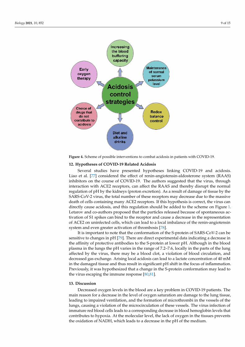
Biology 2021, 10, 852 9 of 15Biology 2021, 10, x FOR PEER REVIEW 9 of 15
Figure 4. Scheme of possible interventions to combat acidosis in patients with COVID-19.
The positive role of diet changes (restriction of carbohydrates, fats, spices) and ad-herence to diet therapy in the prevention of the severe course of COVID-19 should also be noted. Another obvious and most important way to prevent acidosis is early oxygen in-tervention and maintenance of blood oxygen saturation in patients. It helps to avoid the metabolic switch to anaerobic glycolysis.
A particular way of preventing acidosis is the intake of alkaline drinks, which is used by athletes, runners, and cyclists. Usually, sodium and potassium citrate are taken. The same method is used to dissolve some types of kidney stones. However, one must be aware that direct correction of blood pH with alkaline solutions is dangerous, since it af-fects a variety of processes and, in general, can increase mortality among critical patients.
Determination of urine pH and blood oxygenation may indirectly indicate the state of the bicarbonate buffer; they can be recommended for self-diagnosis during the initial period of the disease. Metabolic acidosis is associated with several aggravating factors, including fat oxidation inhibition and macrophage reprogramming [72–74].
Attention should be paid to drugs that provoke the development of metabolic acido-sis, for example, metformin [75] and ibuprofen [76].
12. Hypotheses of COVID-19 Related Acidosis Several studies have presented hypotheses linking COVID-19 and acidosis. Liao et
al. [77] considered the effect of renin-angiotensin-aldosterone system (RAAS) inhibitors on the course of COVID-19. The authors suggested that the virus, through interaction with ACE2 receptors, can affect the RAAS and thereby disrupt the normal regulation of pH by the kidneys (proton excretion). As a result of damage of tissue by the SARS-CoV-2 virus, the total number of these receptors may decrease due to the massive death of cells con-taining many ACE2 receptors. If this hypothesis is correct, the virus can directly cause acidosis, and this regulation should be added to the scheme on Figure 1. Letarov and co-authors proposed that the particles released because of spontaneous activation of S1 spikes can bind to the receptor and cause a decrease in the representation of ACE2 on
Figure 4. Scheme of possible interventions to combat acidosis in patients with COVID-19.
12. Hypotheses of COVID-19 Related Acidosis
Several studies have presented hypotheses linking COVID-19 and acidosis.Liao et al. [77] considered the effect of renin-angiotensin-aldosterone system (RAAS)inhibitors on the course of COVID-19. The authors suggested that the virus, throughinteraction with ACE2 receptors, can affect the RAAS and thereby disrupt the normalregulation of pH by the kidneys (proton excretion). As a result of damage of tissue by theSARS-CoV-2 virus, the total number of these receptors may decrease due to the massivedeath of cells containing many ACE2 receptors. If this hypothesis is correct, the virus candirectly cause acidosis, and this regulation should be added to the scheme on Figure 1.Letarov and co-authors proposed that the particles released because of spontaneous ac-tivation of S1 spikes can bind to the receptor and cause a decrease in the representationof ACE2 on uninfected cells, which can lead to a local imbalance of the renin-angiotensinsystem and even greater activation of thrombosis [78].
It is important to note that the conformation of the S-protein of SARS-CoV-2 can besensitive to changes in pH [79]. There are direct experimental data indicating a decrease inthe affinity of protective antibodies to the S-protein at lower pH. Although in the bloodplasma in the lungs the pH varies in the range of 7.2–7.6, locally in the parts of the lungaffected by the virus, there may be a blood clot, a violation of blood circulation, anddecreased gas exchange. Arising local acidosis can lead to a lactate concentration of 40 mMin the damaged tissue and thus result in significant pH shift in the focus of inflammation.Previously, it was hypothesized that a change in the S-protein conformation may lead tothe virus escaping the immune response [80,81].
13. Discussion
Decreased oxygen levels in the blood are a key problem in COVID-19 patients. Themain reason for a decrease in the level of oxygen saturation are damage to the lung tissue,leading to impaired ventilation, and the formation of microthrombi in the vessels of thelungs, causing a violation of the microcirculation of these vessels. The virus infection ofimmature red blood cells leads to a corresponding decrease in blood hemoglobin levels thatcontributes to hypoxia. At the molecular level, the lack of oxygen in the tissues preventsthe oxidation of NADH, which leads to a decrease in the pH of the medium.
Biology 2021, 10, 852 10 of 15
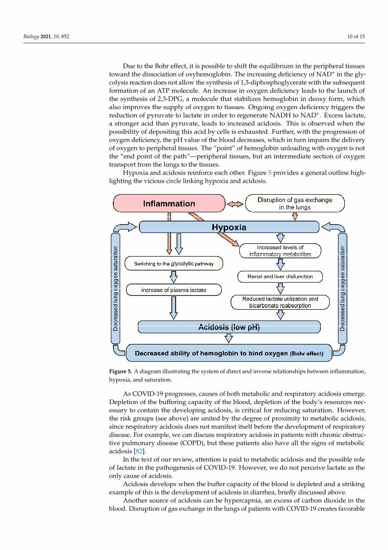
Due to the Bohr effect, it is possible to shift the equilibrium in the peripheral tissuestoward the dissociation of oxyhemoglobin. The increasing deficiency of NAD+ in the gly-colysis reaction does not allow the synthesis of 1,3-diphosphoglycerate with the subsequentformation of an ATP molecule. An increase in oxygen deficiency leads to the launch ofthe synthesis of 2,3-DPG, a molecule that stabilizes hemoglobin in deoxy form, whichalso improves the supply of oxygen to tissues. Ongoing oxygen deficiency triggers thereduction of pyruvate to lactate in order to regenerate NADH to NAD+. Excess lactate,a stronger acid than pyruvate, leads to increased acidosis. This is observed when thepossibility of depositing this acid by cells is exhausted. Further, with the progression ofoxygen deficiency, the pH value of the blood decreases, which in turn impairs the deliveryof oxygen to peripheral tissues. The “point” of hemoglobin unloading with oxygen is notthe “end point of the path”—peripheral tissues, but an intermediate section of oxygentransport from the lungs to the tissues.
Hypoxia and acidosis reinforce each other. Figure 5 provides a general outline high-lighting the vicious circle linking hypoxia and acidosis.
Biology 2021, 10, x FOR PEER REVIEW 11 of 15
Figure 5. A diagram illustrating the system of direct and inverse relationships between inflamma-tion, hypoxia, and saturation.
Respiratory and metabolic acidosis are manifested in different ways in the dynamics of the biochemical parameters of the blood, but in both cases the body is forced to spend resources on compensating for acidosis and, on protecting the lungs from the possibility of developing acidosis as well as from a drop in saturation.
14. Conclusions We have examined several ways in which hypoxia and acidosis affect the develop-
ment of severe COVID-19. This effect has been described at the levels of the organism, organs, tissues, cells, and proteins: from the compensatory regulation of acidosis by the human body to the functioning of a single hemoglobin molecule. Several mechanisms linking the damaging factors of COVID-19 with acidosis are described. These mechanisms have a trigger, step-by-step character of action with pronounced positive feedback. A drop in blood oxygen saturation due to a decrease in blood pH in accordance with Bohr effect is a characteristic feature of the severe course of the disease caused by SARS-CoV-2. This drop is a consequence of the depletion of the compensatory capabilities of the body to regulate acidosis. COVID-19 disease has a systemic damaging effect on various organs and tissues. Among them are DIC-syndrome, pneumonia, and nervous system damage. This disease leads to a variety of different complications; it seems to check the body for the presence of “weak points” and regulatory circuits that have little stability. It is quite possible that acidosis must be compensated for during the rehabilitation of these patients.
Although all the listed triggers that accelerate the onset of acidosis can be significant in other diseases, the combination of all these effects in one disease looks like a unique phenomenon. It should be emphasized that hypoxia and acidosis make it possible to bring the contribution of all these mechanisms to one “common denominator”. Ultimately, aci-dosis is a consequence of a decrease in blood oxygen saturation; however, it contributes to this decrease.
A feature of the pathogenesis of severe COVID-19 is the simultaneous presence of an infectious process and trauma (vascular damage) [86]. Treatment of these two conditions involves alternative activities of the immune system, the main regulators of which are
Figure 5. A diagram illustrating the system of direct and inverse relationships between inflammation,hypoxia, and saturation.
As COVID-19 progresses, causes of both metabolic and respiratory acidosis emerge.Depletion of the buffering capacity of the blood, depletion of the body’s resources nec-essary to contain the developing acidosis, is critical for reducing saturation. However,the risk groups (see above) are united by the degree of proximity to metabolic acidosis,since respiratory acidosis does not manifest itself before the development of respiratorydisease. For example, we can discuss respiratory acidosis in patients with chronic obstruc-tive pulmonary disease (COPD), but these patients also have all the signs of metabolicacidosis [82].
In the text of our review, attention is paid to metabolic acidosis and the possible roleof lactate in the pathogenesis of COVID-19. However, we do not perceive lactate as theonly cause of acidosis.
Acidosis develops when the buffer capacity of the blood is depleted and a strikingexample of this is the development of acidosis in diarrhea, briefly discussed above.
Another source of acidosis can be hypercapnia, an excess of carbon dioxide in theblood. Disruption of gas exchange in the lungs of patients with COVID-19 creates favorable
Biology 2021, 10, 852 11 of 15
conditions for the occurrence of hypercapnia and the development of respiratory acidosis.It is possible that the development of respiratory acidosis up to a certain point inhibits thegrowth of lactate. Experiments are described in which hypercapnic therapy (elevation ofcarbon dioxide levels in the blood) leads to a decrease in lactate [83–85].
Respiratory and metabolic acidosis are manifested in different ways in the dynamicsof the biochemical parameters of the blood, but in both cases the body is forced to spendresources on compensating for acidosis and, on protecting the lungs from the possibility ofdeveloping acidosis as well as from a drop in saturation.
14. Conclusions
We have examined several ways in which hypoxia and acidosis affect the developmentof severe COVID-19. This effect has been described at the levels of the organism, organs,tissues, cells, and proteins: from the compensatory regulation of acidosis by the humanbody to the functioning of a single hemoglobin molecule. Several mechanisms linkingthe damaging factors of COVID-19 with acidosis are described. These mechanisms havea trigger, step-by-step character of action with pronounced positive feedback. A drop inblood oxygen saturation due to a decrease in blood pH in accordance with Bohr effect is acharacteristic feature of the severe course of the disease caused by SARS-CoV-2. This dropis a consequence of the depletion of the compensatory capabilities of the body to regulateacidosis. COVID-19 disease has a systemic damaging effect on various organs and tissues.Among them are DIC-syndrome, pneumonia, and nervous system damage. This diseaseleads to a variety of different complications; it seems to check the body for the presenceof “weak points” and regulatory circuits that have little stability. It is quite possible thatacidosis must be compensated for during the rehabilitation of these patients.
Although all the listed triggers that accelerate the onset of acidosis can be significantin other diseases, the combination of all these effects in one disease looks like a uniquephenomenon. It should be emphasized that hypoxia and acidosis make it possible tobring the contribution of all these mechanisms to one “common denominator”. Ultimately,acidosis is a consequence of a decrease in blood oxygen saturation; however, it contributesto this decrease.
A feature of the pathogenesis of severe COVID-19 is the simultaneous presence of aninfectious process and trauma (vascular damage) [86]. Treatment of these two conditionsinvolves alternative activities of the immune system, the main regulators of which aremacrophages. It is possible that the problem lies precisely in the fact that the immunesystem is trying “to kill two birds with one stone”, constantly and inefficiently switchingbetween different types of responses. The disease persists; destruction and inflammationare growing.
Along with other colleagues [70], we believe that to prevent severe cases of COVID-19,increased attention should be paid to the diagnosis and possible relief of acidosis.
Author Contributions: Conceptualization, Y.D.N., D.A.S. and O.V.M.; methodology, Y.D.N., D.A.L.,A.Y.Z. and O.V.M.; validation, D.A.L. and E.A.G.; formal analysis, Y.E.Y.; investigation, Y.D.N.,E.A.G., O.V.M. and Y.E.Y.; writing—original draft preparation, D.A.S. and A.Y.Z.; writing—reviewand editing, Y.D.N., D.A.S., I.A.L. and O.V.M.; visualization, Y.D.N., I.A.L. and O.V.M.; supervision,Y.E.Y.; project administration, Y.D.N. All authors have read and agreed to the published version ofthe manuscript.
Funding: This research was funded by the Presidium of the Russian Academy of Sciences forMolecular and Cellular Biology and the Program of Fundamental Research for State Academies foryears 2013–2020, project no. 01201363818.
Institutional Review Board Statement: Not applicable.
Informed Consent Statement: Not applicable.
Data Availability Statement: All the results found are available in this manuscript.
Biology 2021, 10, 852 12 of 15
Acknowledgments: We thank Igor Prudovsky (Maine Medical Center Research Institute, USA) andDmitry Kuprash (Center for Precision Genome Editing and Genetic Technologies for Biomedicine(biomedgene.ru) for critical reading of the manuscript. The authors consider it their duty to expresstheir gratitude for the useful discussions to Andrey Rubin and Galina Riznichenko, the heads of theseminar of the Department of Biophysics of the Faculty of Biology of Moscow State University.
Conflicts of Interest: The authors declare no conflict of interest. The funders had no role in the designof the study; in the collection, analyses, or interpretation of data; in the writing of the manuscript, orin the decision to publish the results.
References1. Chhetri, S.; Khamis, F.; Pandak, N.; Al Khalili, H.; Said, E.; Petersen, E. A fatal case of COVID-19 due to metabolic acidosis
following dysregulate inflammatory response (cytokine storm). IDCases 2020, 21, e00829. [CrossRef]2. Lodyagin, A.; Batotsyrenov, B.; Shikalova, I.; Voznyuk, I. Acidosis and toxic hemolysis-goals of pathogenetic treatment of
polyorgan pathology in COVID-19. Bull. Rehabil. Med. 2020, 97, 25–30. [CrossRef]3. Shevel, E. Conditions favoring increased COVID-19 morbidity and mortality: Their common denominator and treatment. Isr.
Med. Assoc. J. IMAJ 2020, 11, 680.4. Price-Haywood, E.G.; Burton, J.; Fort, D.; Seoane, L. Hospitalization and mortality among black patients and white patients with
Covid-19. N. Engl. J. Med. 2020, 382, 2534–2543. [CrossRef] [PubMed]5. Vassiliou, A.G.; Jahaj, E.; Ilias, I.; Markaki, V.; Malachias, S.; Vrettou, C.; Ischaki, E.; Mastora, Z.; Douka, E.; Keskinidou, C.;
et al. Lactate kinetics reflect organ dysfunction and are associated with adverse outcomes in intensive care unit patients withCOVID-19 pneumonia: Preliminary results from a GREEK Single-Centre Study. Metabolites 2020, 10, 386. [CrossRef] [PubMed]
6. Velavan, T.P.; Kieu Linh, L.T.; Kreidenweiss, A.; Gabor, J.; Krishna, S.; Kremsner, P.G. Longitudinal monitoring of lactate inhospitalized and ambulatory COVID-19 patients. Am. J. Trop. Med. Hyg. 2021, 104, 1041–1044. [CrossRef]
7. Newington, J.T.; Harris, R.A.; Cumming, R.C. Reevaluating metabolism in Alzheimer’s disease from the perspective of theastrocyte-neuron lactate shuttle model. J. Neurodegener. Dis. 2013, 2013, 234572. [CrossRef]
8. Andersen, L.W.; Mackenhauer, J.; Roberts, J.C.; Berg, K.M.; Cocchi, M.N.; Donnino, M.W. Etiology and therapeutic approach toelevated lactate levels. Mayo Clin. Proc. 2013, 88, 1127–1140. [CrossRef] [PubMed]
9. Kraut, J.A.; Madias, N.E. Lactic acidosis. N. Engl. J. Med. 2014, 371, 2309–2319. [CrossRef] [PubMed]10. Ma, L.N.; Huang, X.B.; Muyayalo, K.P.; Mor, G.; Liao, A.H. Lactic acid: A novel signaling molecule in early pregnancy? Front.
Immunol. 2020, 11, 279. [CrossRef]11. Li, J.; Wang, X.; Chen, J.; Zuo, X.; Zhang, H.; Deng, A. COVID-19 infection may cause ketosis and ketoacidosis. Diabetes Obes.
Metab. 2020, 22, 1935–1941. [CrossRef]12. Kraut, J.A.; Madias, N.E. Metabolic acidosis: Pathophysiology, diagnosis and management. Nat. Rev. Nephrol. 2010, 6, 274–285.
[CrossRef]13. Chycki, J.; Kurylas, A.; Maszczyk, A.; Golas, A.; Zajac, A. Alkaline water improves exercise-induced metabolic acidosis and
enhances anaerobic exercise performance in combat sport athletes. PLoS ONE 2018, 13, e0205708. [CrossRef] [PubMed]14. Pillai, S.; Davies, G.; Lawrence, M.; Whitley, J.; Stephens, J.; Williams, P.R.; Morris, K.; Evans, P.A. The effect of diabetic ketoacidosis
(DKA) and its treatment on clot microstructure: Are they thrombogenic? Clin. Hemorheol. Microcirc. 2021, 77, 183–194. [CrossRef]15. Chee, Y.J.; Tan, S.K.; Yeoh, E. Dissecting the interaction between COVID-19 and diabetes mellitus. J. Diabetes Investig. 2020, 11,
1104–1114. [CrossRef] [PubMed]16. Orioli, L.; Hermans, M.P.; Thissen, J.P.; Maiter, D.; Vandeleene, B.; Yombi, J.C. COVID-19 in diabetic patients: Related risks and
specifics of management. Ann. Endocrinol. 2020, 81, 101–109. [CrossRef] [PubMed]17. Palermo, N.E.; Sadhu, A.R.; McDonnell, M.E. Diabetic ketoacidosis in COVID-19: Unique concerns and considerations. J. Clin.
Endocrinol. Metab. 2020, 105, 2819–2829. [CrossRef] [PubMed]18. Gentile, S.; Strollo, F.; Mambro, A.; Ceriello, A. COVID-19, ketoacidosis and new-onset diabetes: Are there possible cause and
effect relationships among them? Diabetes Obes. Metab. 2020, 22, 2507–2508. [CrossRef]19. Rubino, F.; Amiel, S.A.; Zimmet, P.; Alberti, G.; Bornstein, S.; Eckel, R.H.; Mingrone, G.; Boehm, B.; Cooper, M.E.; Chai, Z.; et al.
New-onset diabetes in Covid-19. N. Engl. J. Med. 2020, 383, 789–790. [CrossRef]20. Cecere, N.; Hubinont, C.; Kabulu Kadingi, A.; Vincent, M.F.; Van den Bergh, P.; Onnela, A.; Hantson, P. Extreme maternal
metabolic acidosis leading to fetal distress and emergency caesarean section. Case Rep. Obstet. Gynecol. 2013, 2013, 847942.[CrossRef]
21. Mason, R.J. Pathogenesis of COVID-19 from a cell biology perspective. Eur. Respir. J. 2020, 55, 2000607. [CrossRef]22. Ackermann, M.; Verleden, S.E.; Kuehnel, M.; Haverich, A.; Welte, T.; Laenger, F.; Vanstapel, A.; Werlein, C.; Stark, H.; Tzankov,
A.; et al. Pulmonary vascular endothelialitis, thrombosis, and angiogenesis in COVID-19. N. Engl. J. Med. 2020, 383, 120–128.[CrossRef]
23. Hellman, U.; Karlsson, M.G.; Engstrom-Laurent, A.; Cajander, S.; Dorofte, L.; Ahlm, C.; Laurent, C.; Blomberg, A. Presence ofhyaluronan in lung alveoli in severe Covid-19: An opening for new treatment options? J. Biol. Chem. 2020, 295, 15418–15422.[CrossRef]

Biology 2021, 10, 852 13 of 15
24. Shahbaz, S.; Xu, L.; Osman, M.; Sligl, W.; Shields, J.; Joyce, M.; Tyrrell, D.L.; Oyegbami, O.; Elahi, S. Erythroid precursorsand progenitors suppress adaptive immunity and get invaded by SARS-CoV-2. Stem Cell Rep. 2021, 16, 1165–1181. [CrossRef][PubMed]
25. Lunt, S.Y.; Vander Heiden, M.G. Aerobic glycolysis: Meeting the metabolic requirements of cell proliferation. Annu. Rev. Cell Dev.Biol. 2011, 27, 441–464. [CrossRef]
26. Borregaard, N.; Schwartz, J.H.; Tauber, A.I. Proton secretion by stimulated neutrophils. Significance of hexose monophosphateshunt activity as source of electrons and protons for the respiratory burst. J. Clin. Investig. 1984, 74, 455–459. [CrossRef]
27. Niederman, R.; Zhang, J.; Kashket, S. Short-chain carboxylic-acid-stimulated, PMN-mediated gingival inflammation. Crit. Rev.Oral Biol. Med. 1997, 8, 269–290. [CrossRef] [PubMed]
28. Erra Diaz, F.; Dantas, E.; Geffner, J. Unravelling the interplay between extracellular acidosis and immune cells. Mediat. Inflamm.2018, 2018, 1218297. [CrossRef]
29. Semenza, G.L. HIF-1: Mediator of physiological and pathophysiological responses to hypoxia. J. Appl. Physiol. 2000, 88, 1474–1480.[CrossRef]
30. Ivashkiv, L.B. The hypoxia-lactate axis tempers inflammation. Nat. Rev. Immunol. 2020, 20, 85–86. [CrossRef] [PubMed]31. Serebrovska, Z.O.; Chong, E.Y.; Serebrovska, T.V.; Tumanovska, L.V.; Xi, L. Hypoxia, HIF-1alpha, and COVID-19: From pathogenic
factors to potential therapeutic targets. Acta Pharmacol. Sin. 2020, 41, 1539–1546. [CrossRef]32. Zhang, D.; Tang, Z.; Huang, H.; Zhou, G.; Cui, C.; Weng, Y.; Liu, W.; Kim, S.; Lee, S.; Perez-Neut, M.; et al. Metabolic regulation of
gene expression by histone lactylation. Nature 2019, 574, 575–580. [CrossRef]33. Khatib-Massalha, E.; Bhattacharya, S.; Massalha, H.; Biram, A.; Golan, K.; Kollet, O.; Kumari, A.; Avemaria, F.; Petrovich-
Kopitman, E.; Gur-Cohen, S.; et al. Lactate released by inflammatory bone marrow neutrophils induces their mobilization viaendothelial GPR81 signaling. Nat. Commun. 2020, 11, 3547. [CrossRef] [PubMed]
34. Popel, A.S. Theory of oxygen transport to tissue. Crit. Rev. Biomed. Eng. 1989, 17, 257–321. [PubMed]35. Werigo, B. Zur Frage über die Wirkung des Sauerstoffs auf die Kohlensäureausscheidung in den Lungen. Arch. Gesamte Physiol.
Menschen Tiere 1892, 51, 321–361. [CrossRef]36. Bohr, C.; Hasselbalch, K.; Krogh, A. Concerning a biologically important relationship–the influence of the carbon dioxide content
of blood on its oxygen binding. Skand. Arch. Physiol. 1904, 16, 401–412.37. Gell, D.A. Structure and function of haemoglobins. Blood Cells Mol. Dis. 2018, 70, 13–42. [CrossRef] [PubMed]38. Ahmed, M.H.; Ghatge, M.S.; Safo, M.K. Hemoglobin: Structure, function and allostery. Subcell. Biochem. 2020, 94, 345–382.
[CrossRef]39. Srinivasan, A.J.; Morkane, C.; Martin, D.S.; Welsby, I.J. Should modulation of p50 be a therapeutic target in the critically ill? Expert
Rev. Hematol. 2017, 10, 449–458. [CrossRef]40. Stewart, T.; Lambourne, J.; Thorp-Jones, D.; Thomas, D.W. Implementation of early management of iron deficiency in pregnancy
during the SARS-CoV-2 pandemic. Eur. J. Obstet. Gynecol. Reprod. Biol. 2021, 258, 60–62. [CrossRef]41. Kislyakov, Y.Y.; Breslav, I.S. Respiration, Gas Dynamics and Performance in Hyperbaria; Nauka: Leningrad, Russia, 1988.42. Storz, J.F.; Moriyama, H. Mechanisms of hemoglobin adaptation to high altitude hypoxia. High Alt. Med. Biol. 2008, 9, 148–157.
[CrossRef] [PubMed]43. Kislyakov, Y.Y. O2 transport mechanisms in the microcirculation system. Physiol. J. USSR 1987, 73, 569–578.44. Tusman, G.; Bohm, S.H.; Suarez-Sipmann, F.; Scandurra, A.; Hedenstierna, G. Lung recruitment and positive end-expiratory
pressure have different effects on CO2 elimination in healthy and sick lungs. Anesth. Analg. 2010, 111, 968–977. [CrossRef]45. Zaitseva, A.Y.; Kislyakov, Y.Y.; Masing, M.S.; Davydov, V.V. Application of a non-invasive optical learning diagnostic system and
mathematical methods for analyzing multidimensional data to assess the oxygen status of human tissues. Sci. Instrum. 2020, 30,113–118.
46. Dhont, S.; Derom, E.; Van Braeckel, E.; Depuydt, P.; Lambrecht, B.N. The pathophysiology of ‘happy’ hypoxemia in COVID-19.Respir. Res. 2020, 21, 198. [CrossRef]
47. Asakura, H.; Ogawa, H. COVID-19-associated coagulopathy and disseminated intravascular coagulation. Int. J. Hematol. 2021,113, 45–57. [CrossRef]
48. Mosleh, W.; Chen, K.; Pfau, S.E.; Vashist, A. Endotheliitis and endothelial dysfunction in patients with COVID-19: Its role inthrombosis and adverse outcomes. J. Clin. Med. 2020, 9, 1862. [CrossRef] [PubMed]
49. Hottz, E.D.; Azevedo-Quintanilha, I.G.; Palhinha, L.; Teixeira, L.; Barreto, E.A.; Pao, C.R.R.; Righy, C.; Franco, S.; Souza, T.M.L.;Kurtz, P.; et al. Platelet activation and platelet-monocyte aggregate formation trigger tissue factor expression in patients withsevere COVID-19. Blood 2020, 136, 1330–1341. [CrossRef]
50. Iba, T.; Levy, J.H.; Levi, M.; Thachil, J. Coagulopathy in COVID-19. J. Thromb. Haemost. 2020, 18, 2103–2109. [CrossRef] [PubMed]51. Engstrom, M.; Schott, U.; Romner, B.; Reinstrup, P. Acidosis impairs the coagulation: A thromboelastographic study. J. Trauma Inj.
Infect. Crit. Care 2006, 61, 624–628. [CrossRef]52. Engstrom, M.; Schott, U.; Nordstrom, C.H.; Romner, B.; Reinstrup, P. Increased lactate levels impair the coagulation system–a
potential contributing factor to progressive hemorrhage after traumatic brain injury. J. Neurosurg. Anesthesiol. 2006, 18, 200–204.[CrossRef]
53. Tang, N.; Li, D.; Wang, X.; Sun, Z. Abnormal coagulation parameters are associated with poor prognosis in patients with novelcoronavirus pneumonia. J. Thromb. Haemost. 2020, 18, 844–847. [CrossRef] [PubMed]
Biology 2021, 10, 852 14 of 15
54. Kobayashi, S.; Gando, S.; Morimoto, Y.; Nanzaki, S.; Kemmotsu, O. Serial measurement of arterial lactate concentrations as aprognostic indicator in relation to the incidence of disseminated intravascular coagulation in patients with systemic inflammatoryresponse syndrome. Surg. Today 2001, 31, 853–859. [CrossRef]
55. Vanni, S.; Jimenez, D.; Nazerian, P.; Morello, F.; Parisi, M.; Daghini, E.; Pratesi, M.; Lopez, R.; Bedate, P.; Lobo, J.L.; et al. Short-termclinical outcome of normotensive patients with acute PE and high plasma lactate. Thorax 2015, 70, 333–338. [CrossRef]
56. Zabczyk, M.; Natorska, J.; Janion-Sadowska, A.; Malinowski, K.P.; Janion, M.; Undas, A. Elevated lactate levels in acute pulmonaryembolism are associated with prothrombotic fibrin clot properties: Contribution of NETs formation. J. Clin. Med. 2020, 9, 953.[CrossRef]
57. D’Amico, F.; Baumgart, D.C.; Danese, S.; Peyrin-Biroulet, L. Diarrhea during COVID-19 infection: Pathogenesis, epidemiology,prevention, and management. Clin. Gastroenterol. Hepatol. 2020, 18, 1663–1672. [CrossRef] [PubMed]
58. Xu, J.; Chu, M.; Zhong, F.; Tan, X.; Tang, G.; Mai, J.; Lai, N.; Guan, C.; Liang, Y.; Liao, G. Digestive symptoms of COVID-19 andexpression of ACE2 in digestive tract organs. Cell Death Discov. 2020, 6, 76. [CrossRef] [PubMed]
59. Gennari, F.J.; Weise, W.J. Acid-base disturbances in gastrointestinal disease. Clin. J. Am. Soc. Nephrol. 2008, 3, 1861–1868.[CrossRef] [PubMed]
60. Booth, A.L.; Abels, E.; McCaffrey, P. Development of a prognostic model for mortality in COVID-19 infection using machinelearning. Mod. Pathol. 2021, 34, 522–531. [CrossRef] [PubMed]
61. Poole, D.C.; Rossiter, H.B.; Brooks, G.A.; Gladden, L.B. The anaerobic threshold: 50+ years of controversy. J. Physiol. 2021, 599,737–767. [CrossRef]
62. Hogan, M.C. What Wasserman wrought: A celebratory review of 50 years of research arising from the concept of an ‘anaerobicthreshold’. J. Physiol. 2021, 599, 1005. [CrossRef]
63. Rabinowitz, J.D.; Enerback, S. Lactate: The ugly duckling of energy metabolism. Nat. Metab. 2020, 2, 566–571. [CrossRef]64. Dietl, K.; Renner, K.; Dettmer, K.; Timischl, B.; Eberhart, K.; Dorn, C.; Hellerbrand, C.; Kastenberger, M.; Kunz-Schughart, L.A.;
Oefner, P.J.; et al. Lactic acid and acidification inhibit TNF secretion and glycolysis of human monocytes. J. Immunol. 2010, 184,1200–1209. [CrossRef] [PubMed]
65. Suzuki, H.; Hisamatsu, T.; Chiba, S.; Mori, K.; Kitazume, M.T.; Shimamura, K.; Nakamoto, N.; Matsuoka, K.; Ebinuma, H.;Naganuma, M.; et al. Glycolytic pathway affects differentiation of human monocytes to regulatory macrophages. Immunol. Lett.2016, 176, 18–27. [CrossRef] [PubMed]
66. Cassim, S.; Pouyssegur, J. Tumor microenvironment: A metabolic player that shapes the immune response. Int. J. Mol. Sci. 2019,21, 157. [CrossRef]
67. Sun, L.; Yang, X.; Yuan, Z.; Wang, H. Metabolic reprogramming in immune response and tissue inflammation. Arterioscler. Thromb.Vasc. Biol. 2020, 40, 1990–2001. [CrossRef] [PubMed]
68. Nolt, B.; Tu, F.; Wang, X.; Ha, T.; Winter, R.; Williams, D.L.; Li, C. Lactate and immunosuppression in sepsis. Shock 2018, 49,120–125. [CrossRef]
69. Awasthi, D.; Nagarkoti, S.; Sadaf, S.; Chandra, T.; Kumar, S.; Dikshit, M. Glycolysis dependent lactate formation in neutrophils:A metabolic link between NOX-dependent and independent NETosis. Biochim. Biophys. Acta BBA—Mol. Basis Dis. 2019, 1865,165542. [CrossRef]
70. Quade, B.N.; Parker, M.D.; Occhipinti, R. The therapeutic importance of acid-base balance. Biochem. Pharmacol. 2021, 183, 114278.[CrossRef] [PubMed]
71. Coller, B.S. Leukocytosis and ischemic vascular disease morbidity and mortality: Is it time to intervene? Arterioscler. Thromb. Vasc.Biol. 2005, 25, 658–670. [CrossRef]
72. Castleberry, A.W.; Grannis, F.W., Jr. What is a reasonable cost to refute a preposterous hypothesis? Br. J. Cancer 2010, 102, 627–628.[CrossRef]
73. Corbet, C.; Pinto, A.; Martherus, R.; Santiago de Jesus, J.P.; Polet, F.; Feron, O. Acidosis drives the reprogramming of fatty acidmetabolism in cancer cells through changes in mitochondrial and histone acetylation. Cell Metab. 2016, 24, 311–323. [CrossRef]
74. Ventura, F.V.; Ruiter, J.P.; IJlst, L.; de Almeida, I.T.; Wanders, R.J. Lactic acidosis in long-chain fatty acid beta-oxidation disorders.J. Inherit. Metab. Dis. 1998, 21, 645–654. [CrossRef]
75. Cheng, X.; Liu, Y.M.; Li, H.; Zhang, X.; Lei, F.; Qin, J.J.; Chen, Z.; Deng, K.Q.; Lin, L.; Chen, M.M.; et al. Metformin is associatedwith higher incidence of acidosis, but not mortality, in individuals with COVID-19 and pre-existing type 2 diabetes. Cell Metab.2020, 32, 537–547.e3. [CrossRef]
76. Downie, A.; Ali, A.; Bell, D. Severe metabolic acidosis complicating massive ibuprofen overdose. Postgrad. Med. J. 1993, 69,575–577. [CrossRef]
77. Liao, W.H.; Yang, G.G.; Henneberg, M. The renin-angiotensin-aldosterone system inhibitors in COVID-19: From acidosis toventilation and immunity. Swiss Med. Wkly. 2020, 150, w20444. [CrossRef] [PubMed]
78. Letarov, A.V.; Babenko, V.V.; Kulikov, E.E. Free SARS-CoV-2 spike protein S1 particles may play a role in the pathogenesis ofCOVID-19 infection. Biochemistry 2021, 86, 257–261. [CrossRef]
79. Zhou, T.; Tsybovsky, Y.; Gorman, J.; Rapp, M.; Cerutti, G.; Chuang, G.Y.; Katsamba, P.S.; Sampson, J.M.; Schon, A.; Bimela, J.;et al. Cryo-EM structures of SARS-CoV-2 spike without and with ACE2 reveal a pH-dependent switch to mediate endosomalpositioning of receptor-binding domains. Cell Host Microbe 2020, 28, 867–879.E5. [CrossRef]

Biology 2021, 10, 852 15 of 15
80. Nechipurenko, Y.D.; Anashkina, A.A.; Matveeva, O.V. Change of antigenic determinants of SARS-CoV-2 virus S-protein as apossible cause of antibody-dependent enhancement of virus infection and cytokine storm. Biophysics 2020, 65, 703–709. [CrossRef][PubMed]
81. Zaichuk, T.A.; Nechipurenko, Y.D.; Adzhubey, A.A.; Onikienko, S.B.; Chereshnev, V.A.; Zainutdinov, S.S.; Kochneva, G.V.; Netesov,S.V.; Matveeva, O.V. The challenges of vaccine development against Betacoronaviruses: Antibody dependent enhancement andSendai virus as a possible vaccine vector. Mol. Biol. 2020, 54, 812–826. [CrossRef] [PubMed]
82. Bruno, C.M.; Valenti, M. Acid-base disorders in patients with chronic obstructive pulmonary disease: A pathophysiologicalreview. J. Biomed. Biotechnol. 2012, 2012, 915150. [CrossRef]
83. Engel, K.; Kildeberg, P.A.; Fine, B.P.; Winters, R.W. Effects of acute respiratory acidosis on blood lactate concentration. Scand. J.Clin. Lab. Investig. 1967, 20, 179–182. [CrossRef]
84. McLellan, T.M. The influence of a respiratory acidosis on the exercise blood lactate response. Eur. J. Appl. Physiol. Occup. Physiol.1991, 63, 6–11. [CrossRef]
85. Kato, T.; Tsukanaka, A.; Harada, T.; Kosaka, M.; Matsui, N. Effect of hypercapnia on changes in blood pH, plasma lactate andammonia due to exercise. Eur. J. Appl. Physiol. 2005, 95, 400–408. [CrossRef]
86. Lupu, L.; Palmer, A.; Huber-Lang, M. Inflammation, thrombosis, and destruction: The three-headed cerberus of trauma- andSARS-CoV-2-induced ARDS. Front. Immunol. 2020, 11, 584514. [CrossRef]