Molecular genetics of familial hypertrophic cardiomyopathy (FHC)
10
J Hum Genet (2003) 48:55–64 © Jpn Soc Hum Genet and Springer-Verlag 2003 MINIREVIEW Murali D. Bashyam · Gorinabele R. Savithri Murugapiran S. Kumar · Calambur Narasimhan Pratibha Nallari Molecular genetics of familial hypertrophic cardiomyopathy (FHC) Received: October 15, 2002 / Accepted: November 18, 2002 Introduction Familial hypertrophic cardiomyopathy (FHC) is an autoso- mal dominant disease characterized mainly by left ventricu- lar hypertrophy. The left ventricular architecture in FHC is disorganized, with hypertrophic cardiac myocytes having abnormal shapes and multiple intercellular connections and being arranged in a chaotic pattern (myocyte disarray). Myocyte loss and replacement fibrosis are prominent fea- tures (Davies 1994; Maron et al. 1987), which can result in arrhythmia and altered myocardial hemodynamics. The term “hypertrophy” denotes a thickening of tissue due to an increase in the size of the constituent cells. The term “cardi- omyopathy” means a disease of heart muscle (cardio, heart; myo, muscle; pathy, disease). Hypertrophy, in general, is recognized as a compensatory thickening of the left ven- tricle wall that occurs as a physiologic response to either excessive hemodynamic burden (such as pressure or vol- ume overload due to hypertension and aortic valve disease) or a pathologic state that compromises function of the heart muscle (Grossman et al. 1975). FHC however, is a heritable disorder that occurs in the absence of hemodynamic bur- den. FHC is the most common cause of sudden death in otherwise healthy young individuals (Maron et al. 1978, 1986). The frequency of hypertrophic cardiomyopathy is approximately 0.1%–0.2% in Europe and the United States. The genetic basis for this disorder has been shown to be mutations in the cardiac sarcomere protein-coding genes. However, it is not clear how these mutations actually induce the hypertrophic response in the heart. Efforts are underway also to establish a genotype–phenotype correla- tion for this disorder. Clinical symptoms The disease is characterized by extreme clinical and mor- phological heterogeneity, ranging from benign to severe, and is further complicated by a variable age of presentation. M.D. Bashyam (*) · M.S. Kumar Molecular Oncology, Centre for DNA Fingerprinting and Diagnostics (CDFD), Nacharam, Hyderabad 500076, India Tel. 91-40-7150008; Fax 91-40-7155610 e-mail: [email protected] G.R. Savithri Laboratory of Molecular and Cellular Biology, Centre for DNA Fingerprinting and Diagnostics (CDFD), Hyderabad, India C. Narasimhan Care Hospital, Hyderabad, India P. Nallari Department of Genetics, Osmania University, Hyderabad, India Abstract Familial hypertrophic cardiomyopathy is an auto- somal dominant disease with a wide range of clinical fea- tures from benign to severe, and is the most common cause of sudden death in otherwise healthy individuals. The two prominent clinical features are left ventricular hypertrophy and myocyte/myofibrillar disarray. The former is respon- sible for clinical symptoms such as breathlessness and an- gina, whereas the latter may lead to sudden cardiac death. The last decade has seen an enormous improvement in our understanding of the molecular genetics of this disorder. The clinical heterogeneity has been linked to genetic het- erogeneity; mutations in nine genes encoding sarcomere proteins have been shown to be the molecular basis for the disorder. However, attempts to establish a genotype–phe- notype correlation for each of the more than 100 mutations that have been identified have not been highly successful. Additional genetic loci, as well as nongenetic factors such as lifestyle, sex, and age, have also been shown to play a role in modulating the clinical presentation of the disease. How each mutation results in hypertrophy and/or myofibrillar disarray is unclear. The present review discusses the current status of the molecular genetic characterization of this im- portant disorder. Key words Hypertrophy · Cardiomyopathy · Myosin · Troponin · Tropomyosin · Actin · Myocyte disarray
-
Upload
independent -
Category
Documents
-
view
0 -
download
0
Transcript of Molecular genetics of familial hypertrophic cardiomyopathy (FHC)
B. Jochimsen et al.: Stetteria hydrogenophila 4600/55J Hum Genet (2003) 48:55–64 © Jpn Soc Hum Genet and Springer-Verlag 2003
MINIREVIEW
Murali D. Bashyam · Gorinabele R. SavithriMurugapiran S. Kumar · Calambur NarasimhanPratibha Nallari
Molecular genetics of familial hypertrophic cardiomyopathy (FHC)
Received: October 15, 2002 / Accepted: November 18, 2002
Introduction
Familial hypertrophic cardiomyopathy (FHC) is an autoso-mal dominant disease characterized mainly by left ventricu-lar hypertrophy. The left ventricular architecture in FHC isdisorganized, with hypertrophic cardiac myocytes havingabnormal shapes and multiple intercellular connections andbeing arranged in a chaotic pattern (myocyte disarray).Myocyte loss and replacement fibrosis are prominent fea-tures (Davies 1994; Maron et al. 1987), which can result inarrhythmia and altered myocardial hemodynamics. Theterm “hypertrophy” denotes a thickening of tissue due to anincrease in the size of the constituent cells. The term “cardi-omyopathy” means a disease of heart muscle (cardio, heart;myo, muscle; pathy, disease). Hypertrophy, in general, isrecognized as a compensatory thickening of the left ven-tricle wall that occurs as a physiologic response to eitherexcessive hemodynamic burden (such as pressure or vol-ume overload due to hypertension and aortic valve disease)or a pathologic state that compromises function of the heartmuscle (Grossman et al. 1975). FHC however, is a heritabledisorder that occurs in the absence of hemodynamic bur-den. FHC is the most common cause of sudden death inotherwise healthy young individuals (Maron et al. 1978,1986). The frequency of hypertrophic cardiomyopathy isapproximately 0.1%–0.2% in Europe and the UnitedStates. The genetic basis for this disorder has been shown tobe mutations in the cardiac sarcomere protein-codinggenes. However, it is not clear how these mutations actuallyinduce the hypertrophic response in the heart. Efforts areunderway also to establish a genotype–phenotype correla-tion for this disorder.
Clinical symptoms
The disease is characterized by extreme clinical and mor-phological heterogeneity, ranging from benign to severe,and is further complicated by a variable age of presentation.
M.D. Bashyam (*) · M.S. KumarMolecular Oncology, Centre for DNA Fingerprinting andDiagnostics (CDFD), Nacharam, Hyderabad 500076, IndiaTel. �91-40-7150008; Fax �91-40-7155610e-mail: [email protected]
G.R. SavithriLaboratory of Molecular and Cellular Biology, Centre for DNAFingerprinting and Diagnostics (CDFD), Hyderabad, India
C. NarasimhanCare Hospital, Hyderabad, India
P. NallariDepartment of Genetics, Osmania University, Hyderabad, India
Abstract Familial hypertrophic cardiomyopathy is an auto-somal dominant disease with a wide range of clinical fea-tures from benign to severe, and is the most common causeof sudden death in otherwise healthy individuals. The twoprominent clinical features are left ventricular hypertrophyand myocyte/myofibrillar disarray. The former is respon-sible for clinical symptoms such as breathlessness and an-gina, whereas the latter may lead to sudden cardiac death.The last decade has seen an enormous improvement in ourunderstanding of the molecular genetics of this disorder.The clinical heterogeneity has been linked to genetic het-erogeneity; mutations in nine genes encoding sarcomereproteins have been shown to be the molecular basis for thedisorder. However, attempts to establish a genotype–phe-notype correlation for each of the more than 100 mutationsthat have been identified have not been highly successful.Additional genetic loci, as well as nongenetic factors such aslifestyle, sex, and age, have also been shown to play a role inmodulating the clinical presentation of the disease. Howeach mutation results in hypertrophy and/or myofibrillardisarray is unclear. The present review discusses the currentstatus of the molecular genetic characterization of this im-portant disorder.
Key words Hypertrophy · Cardiomyopathy · Myosin ·Troponin · Tropomyosin · Actin · Myocyte disarray
Used Mac Distiller 5.0.x Job Options
This report was created automatically with help of the Adobe Acrobat Distiller addition "Distiller Secrets v1.0.5" from IMPRESSED GmbH. You can download this startup file for Distiller versions 4.0.5 and 5.0.x for free from http://www.impressed.de. GENERAL ---------------------------------------- File Options: Compatibility: PDF 1.2 Optimize For Fast Web View: Yes Embed Thumbnails: Yes Auto-Rotate Pages: No Distill From Page: 1 Distill To Page: All Pages Binding: Left Resolution: [ 600 600 ] dpi Paper Size: [ 595.3 785.2 ] Point COMPRESSION ---------------------------------------- Color Images: Downsampling: Yes Downsample Type: Bicubic Downsampling Downsample Resolution: 150 dpi Downsampling For Images Above: 225 dpi Compression: Yes Automatic Selection of Compression Type: Yes JPEG Quality: Medium Bits Per Pixel: As Original Bit Grayscale Images: Downsampling: Yes Downsample Type: Bicubic Downsampling Downsample Resolution: 150 dpi Downsampling For Images Above: 225 dpi Compression: Yes Automatic Selection of Compression Type: Yes JPEG Quality: Medium Bits Per Pixel: As Original Bit Monochrome Images: Downsampling: Yes Downsample Type: Bicubic Downsampling Downsample Resolution: 600 dpi Downsampling For Images Above: 900 dpi Compression: Yes Compression Type: CCITT CCITT Group: 4 Anti-Alias To Gray: No Compress Text and Line Art: Yes FONTS ---------------------------------------- Embed All Fonts: Yes Subset Embedded Fonts: No When Embedding Fails: Warn and Continue Embedding: Always Embed: [ ] Never Embed: [ ] COLOR ---------------------------------------- Color Management Policies: Color Conversion Strategy: Convert All Colors to sRGB Intent: Default Working Spaces: Grayscale ICC Profile: RGB ICC Profile: sRGB IEC61966-2.1 CMYK ICC Profile: U.S. Web Coated (SWOP) v2 Device-Dependent Data: Preserve Overprint Settings: Yes Preserve Under Color Removal and Black Generation: Yes Transfer Functions: Apply Preserve Halftone Information: Yes ADVANCED ---------------------------------------- Options: Use Prologue.ps and Epilogue.ps: No Allow PostScript File To Override Job Options: Yes Preserve Level 2 copypage Semantics: Yes Save Portable Job Ticket Inside PDF File: No Illustrator Overprint Mode: Yes Convert Gradients To Smooth Shades: No ASCII Format: No Document Structuring Conventions (DSC): Process DSC Comments: No OTHERS ---------------------------------------- Distiller Core Version: 5000 Use ZIP Compression: Yes Deactivate Optimization: No Image Memory: 524288 Byte Anti-Alias Color Images: No Anti-Alias Grayscale Images: No Convert Images (< 257 Colors) To Indexed Color Space: Yes sRGB ICC Profile: sRGB IEC61966-2.1 END OF REPORT ---------------------------------------- IMPRESSED GmbH Bahrenfelder Chaussee 49 22761 Hamburg, Germany Tel. +49 40 897189-0 Fax +49 40 897189-71 Email: [email protected] Web: www.impressed.de
Adobe Acrobat Distiller 5.0.x Job Option File
<< /ColorSettingsFile () /LockDistillerParams false /DetectBlends false /DoThumbnails true /AntiAliasMonoImages false /MonoImageDownsampleType /Bicubic /GrayImageDownsampleType /Bicubic /MaxSubsetPct 100 /MonoImageFilter /CCITTFaxEncode /ColorImageDownsampleThreshold 1.5 /GrayImageFilter /DCTEncode /ColorConversionStrategy /sRGB /CalGrayProfile () /ColorImageResolution 150 /UsePrologue false /MonoImageResolution 600 /ColorImageDepth -1 /sRGBProfile (sRGB IEC61966-2.1) /PreserveOverprintSettings true /CompatibilityLevel 1.2 /UCRandBGInfo /Preserve /EmitDSCWarnings false /CreateJobTicket false /DownsampleMonoImages true /DownsampleColorImages true /MonoImageDict << /K -1 >> /ColorImageDownsampleType /Bicubic /GrayImageDict << /HSamples [ 2 1 1 2 ] /VSamples [ 2 1 1 2 ] /Blend 1 /QFactor 0.9 >> /CalCMYKProfile (U.S. Web Coated (SWOP) v2) /ParseDSCComments false /PreserveEPSInfo false /MonoImageDepth -1 /AutoFilterGrayImages true /SubsetFonts false /GrayACSImageDict << /VSamples [ 2 1 1 2 ] /HSamples [ 2 1 1 2 ] /Blend 1 /QFactor 0.76 /ColorTransform 1 >> /ColorImageFilter /DCTEncode /AutoRotatePages /None /PreserveCopyPage true /EncodeMonoImages true /ASCII85EncodePages false /PreserveOPIComments false /NeverEmbed [ ] /ColorImageDict << /HSamples [ 2 1 1 2 ] /VSamples [ 2 1 1 2 ] /Blend 1 /QFactor 0.9 >> /AntiAliasGrayImages false /GrayImageDepth -1 /CannotEmbedFontPolicy /Warning /EndPage -1 /TransferFunctionInfo /Apply /CalRGBProfile (sRGB IEC61966-2.1) /EncodeColorImages true /EncodeGrayImages true /ColorACSImageDict << /VSamples [ 2 1 1 2 ] /HSamples [ 2 1 1 2 ] /Blend 1 /QFactor 0.76 /ColorTransform 1 >> /Optimize true /ParseDSCCommentsForDocInfo false /GrayImageDownsampleThreshold 1.5 /MonoImageDownsampleThreshold 1.5 /AutoPositionEPSFiles false /GrayImageResolution 150 /AutoFilterColorImages true /AlwaysEmbed [ ] /ImageMemory 524288 /OPM 1 /DefaultRenderingIntent /Default /EmbedAllFonts true /StartPage 1 /DownsampleGrayImages true /AntiAliasColorImages false /ConvertImagesToIndexed true /PreserveHalftoneInfo true /CompressPages true /Binding /Left >> setdistillerparams << /PageSize [ 576.0 792.0 ] /HWResolution [ 600 600 ] >> setpagedevice
56 N. Matsuda et al.: EGF receptor and osteoblastic differentiation
Clinically, the patients may present with some or all of thefollowing symptoms.
Dyspnea (breathlessness)
Dyspnea is usually linked to physical activity but occa-sionally may manifest even at rest and may be more pro-nounced following meals. Shortness of breath results froman elevation of left ventricular diastolic pressure due to thethickening of the ventricle wall, and this increased pres-sure is relayed to the lungs via the pulmonary capillaries,leading to pulmonary congestion characterized clinically bydyspnea.
Chest pain
Also called angina, chest pain is a common symptom. It isusually brought on by exertion and relieved by rest, but mayalso occur at rest or during sleep and may persist. Thegreatly thickened muscle demands an increased oxygensupply and chest pain results when this demand cannot bemet.
Syncope (fainting)
Patients may experience light-headedness, dizziness, and,more seriously, blackouts. Presyncope denotes dizziness,but the patient does not pass out completely. Episodesmay occur in association with exercise, with palpitations, orwithout any apparent association. Fainting episodes may bedue to an irregularity of the heartbeat, or a fall in bloodpressure leading to insufficient blood supply to the brain.
Palpitations
Patients may occasionally feel an extra beat or a skippedbeat, suggesting an irregular heart rhythm. Palpitations maystart suddenly, may appear to be very fast, and may beassociated with sweating or light-headedness.
Arrhythmias
Arrhythmias are irregularities of the heartbeat, which occurbecause of disruption of the electrical conduction system ofthe heart. Myocyte disarray may be the primary cause forthis disruption. Two types of arrhythmias, namely, ventricu-lar tachycardia (arising from the ventricles) and atrial fibril-lation (occurring in the atria) are particularly important andmay require treatment. In atrial fibrillation, the normalregular rhythm of the heartbeat is lost and is replaced by anirregular rhythm that may be episodic (paroxysmal atrialfibrillation) or persistent. The loss of normal atrial contrac-tion produces a risk of clot formation in the auricle. Some-times the heart may need to be shocked back into normalrhythm. Heart block may result if the normal electricalsignal travels down to the ventricles slowly or is completely
blocked. This is uncommon, but if this occurs, a pacemakermay be required. The importance of FHC stems from thefact that a large number of patients may have an increasedrisk of premature death (due to arrhythmia), which mayoccur with little or no warning. Ventricular fibrillationsusually precede sudden death.
Hypertrophy
The characteristic feature of FHC is the hypertrophiedheart. However, there is a high degree of variability in thelocation and kind of hypertrophy. It could be concentric(spread throughout the left ventricle wall), apical (only thebase of the left ventricle), or septal (affecting only the sep-tum). There is an additional complication associated withseptal hypertrophy. If hypertrophy occurs in the proximalregion of the septum, the inward movement of the hypertro-phied septum during systole pulls the anterior leaflet of themitral valve toward the septum (SAM, systolic anteriormotion), causing obstruction in the left ventricular outflowtract. Obstruction may also be a result of the septum thick-ening itself but is accentuated by SAM. SAM can some-times be diagnosed by listening to a “murmur” sound in theheartbeat. SAM may also result in flow of blood back intothe left atrium from the left ventricle, which is termed“mitral regurgitation.”
Diagnosis
Clinically, FHC is diagnosed based on the medical his-tory, physical examination, and electrocardiographic andechocardiographic identification of the septal and left ven-tricular hypertrophy. The electrocardiogram (ECG) usuallyshows an abnormal electrical signal due to muscle thicken-ing and disorganization of the muscle structure. In a minor-ity of patients (5%–10%), however, the ECG may benormal or show only minor changes. ECG abnormalitiesare not specific to FHC and may be encountered in otherheart conditions as well. An accurate diagnosis of FHC istherefore based on an ultrasound scan of the heart called anechocardiogram (Echo). The Echo produces a picture ofthe heart in which excessive thickness of the muscle can beeasily measured. Additional equipment called Dopplerultrasound can produce a color image of blood flow withinthe heart and measure the heart’s contraction and filling.Turbulent flow inside the heart can also be detected by thismethod.
Genetic basis of FHC
Myofibrils, which consist of repeating units known assarcomeres, make up the contractile elements in muscle. Asarcomere consists of seven major proteins and several mi-nor proteins organized into thick and thin filaments. Thickfilaments comprise myosin heavy and light chains and the
B. Jochimsen et al.: Stetteria hydrogenophila 57
cardiac troponin I (TnI3) (Kimura et al. 1997), α-tropomyo-sin (Tpm) (Watkins et al. 1995b), titin (TTN) (Satoh et al.1999), α-cardiac actin (ACTC) (Mogensen et al. 1999),and the essential and regulatory myosin light chains (Myl3/Myl2) (Poetter et al. 1996) (Table 1). Because all themutations in FHC patients so far identified are present inthe structural proteins of the muscle, the disease has beentermed “a disease of the sarcomere” (Thierfelder et al.1994).
FHC is inherited as an autosomal dominant trait.Thus, individuals with FHC must have one affectedparent and frequently have multiple affected relatives.Sporadic occurrence is relatively rare. The variable clinicalphenotypes of the disease can in part be explained by theunderlying genetic heterogeneity. The genetic heterogene-ity is accentuated by the fact that mutations in severalexons of the sarcomere genes can cause FHC. To someextent, it has been possible to correlate different mutationswith different prognoses and variable echocardiographicfindings.
�-Cardiac myosin heavy chain gene
Myosin is the major contractile protein in the heart muscleand is responsible for force generation as a result of itspulling action on actin at the expense of ATP. The firstmutation to be identified in FHC was a missense mutationin the �-cardiac myosin heavy chain gene (MYH7)(Tanigawa et al. 1990; Geisterfer-Lowrance et al. 1990).Several distinct missense mutations have since been identi-fied in this gene that result in the substitution of conservedamino acids (Watkins et al. 1992; Vikstrom and Leinwand1996). Because each of the affected amino acids is evolu-tionarily highly conserved, it has been suggested that thesemutations may compromise normal functioning of the pro-tein (Watkins et al. 1992). Several mutations are clusteredin the globular head region, and may occur in regionsthat form functionally important domains (Table 2). Asignificant fraction of mutations detected so far fall in thetail-helix that lies in the rod region of the protein (Table 2).
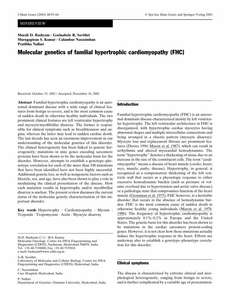
Table 1. FHC disease genes and mutation frequencies
Gene Chromosomal No. ofGene name symbol location mutations
Beta-cardiac myosin heavy chain MYH7 14q12 76Myosin-binding protein C MyBPC-3 11p11.2 33Troponin T TnT2 1q32 15Troponin I TnI3 19q13.4 8Alpha-tropomyosin Tpm 15q22.1 6Myosin essential light chain MYl3 3p21.2–3p21.3 8Myosin regulatory light chain MYl2 12q23–12q24.3 2Alpha-cardiac actin ACTC 15q11–q14 5Titin TTN 2q24.3 1
Compiled from the FHC mutation database (http://www.angis.org.au/Databases/Heart/heartbreak.html)FHC, Familial hypertrophic cardiomyopathy
myosin-binding protein C. Thin filaments consist predomi-nantly of actin and of lesser amounts of regulatory proteins,namely, tropomyosin and troponin. In the resting muscle,binding of tropomyosin to the active site of actin preventscross-bridge formation between actin and myosin, thus pre-venting muscle contraction. The process of contraction isinitiated by the release of calcium ions from the sarcoplas-mic reticulum of the heart muscle cells. The calcium ionsbind to troponin, causing the latter to move the tropomyo-sin molecule away from the cross-bridge binding sitesof actin. Subsequently, adenosine triphosphate (ATP)molecules bound to myosin are hydrolyzed by adenosinetriphosphatase (ATPase) to produce an energized myosinmolecule and the cross-bridge of the energized myosin mol-ecule binds to actin to trigger the release of the energystored in the myosin head. Energy release corresponds tothe movement of the bound cross-bridge in a rowing mo-tion, bringing the actin molecules toward the center of thesarcomere. An ATP molecule binds to the myosin moleculeand releases the cross-bridge attachment; the myosin is nowready to bind to another actin site, as long as calcium ispresent.
Recent advances in biochemistry and molecular geneticshave allowed a better understanding of the molecular basisfor FHC. By using linkage analysis and molecular geneticstudies, nine disease genes for FHC have so far been iden-tified. These include �-cardiac myosin heavy chain (MYH7)(Geisterfer-Lowrance et al. 1990; Watkins et al. 1992), car-diac myosin-binding protein C (MyBPC-3) (Watkins et al.1995a), cardiac troponin T (TnT2) (Thierfelder et al. 1994),
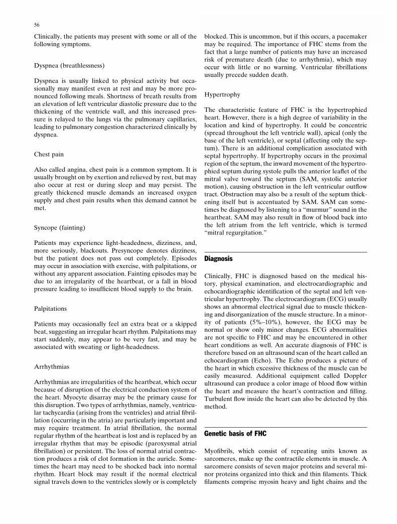
Table 2. Location of major mutations in the MYH7 gene
Exon No. of mutations Functional domain
9 5 ATPase pocket16 7 Actin binding19 6 SH3 helix20 8 Converter22 6 Tail helix/rod23 7 Tail helix/rod

58 N. Matsuda et al.: EGF receptor and osteoblastic differentiation
Recently, two families were shown to harbor mutations inthe light meromyosin domain in the rod region of the pro-tein (Blair et al. 2002). The MYH7 mutations account forabout 30% of the reported FHC cases (Table 1) (Watkinset al. 1995c). To date more than 70 different mutationshave been identified in the MYH7 gene (Table 1). Someare associated with sudden death (p.R403Q), but severalalso have a benign phenotype (p.V606M) (Epstein et al.1992; Fananapazir and Epstein 1994).
Characterization of specific MYH7 mutationsAlthough a lot of research has gone into thecharacterization of several MYH7 gene mutations,including in vitro studies and animal models, we still knowvery little about the link between specific mutations, andhypertrophy and myocyte disarray. The most wellcharacterized MYH7 mutation is p.R403Q. This is primarilybecause it is associated with sudden death in most cases.Sweeny and coworkers (1994) demonstrated that thep.R403Q mutation resulted in an 80% reduction in thefilament sliding speed in vitro, whereas Lankford andcoworkers (1996) showed reduced power output in themuscle containing the mutated protein.
Rayment and colleagues (1995) analyzed the effect of 29mutations in MYH7 that are implicated in FHC by using thecrystal structure of the chicken skeletal myosin S1 peptide.They showed that most of the mutations were clusteredaround specific sites, viz, the actin-binding interface, thenucleotide-binding pocket, or the interface of the heavychain with the essential light chain (ELC). Some of themutations in the actin-binding interface could either affectthe velocity of filament sliding, as seen in an in vitro motilityassay, or lead to a reduced actin-activated Mg-ATPase ac-tivity (Cuda et al. 1993). The mutations in the nucleotide-binding pocket can compromise the catalytic function of theprotein. Other mutations that lie in the binding site forthe ELC show the importance of the interface between theELC and the myosin heavy chain because this is requiredfor coupling ATP hydrolysis with movement. However, asmore and more mutations are being identified and charac-terized, it is becoming obvious that the mutations are actu-ally spread throughout the gene.
Sata and Ikebe (1996) expressed and purified mutatedforms of the cardiac myosin heavy chain and analyzed theirmotor and enzymatic properties in vitro. They specificallychecked four mutant proteins, all of which showed de-creased motor function in terms of ATPase activity andactin translocation. Interestingly, the degree of dysfunctionin vitro correlated with the prognosis of the diseaseresulting from the mutation. The survival values forp.V606M (actin–myosin interface), p.R249Q (ATP pocket),p.R403Q (actin–myosin interface), and p.R453C (ATPpocket) were 94%, 79%, 36%, and 34%, respectively. Thiscorrelated with the extent of decrease in myosin motoractivity. However, despite differences in prognosis, all mu-tations resulted in comparable levels of hypertrophy, indi-cating that other factors may influence cardiac hypertrophyresulting from this mutation. Roopnarine and Leinwand
(1998) analyzed the ATPase activities of three mutations(p.R249Q, p.R403Q, and p.V606M). The mutant proteinswere expressed and purified and then assayed for theirATPase activities. The p.R249Q mutant (which falls inthe base of the ATPase pocket) exhibited a 1.7-foldreduction in maximum Velocity (Vmax). A more severeaffect was observed with the p.R403Q mutation (located atthe base of a loop that appears to play an important roleduring the stereospecific actin–myosin interaction). TheVmax decreased 3.5-fold, suggesting that the actin–myosininteraction is weakened substantially. The Vmax of thep.V606M (present in the actin-binding domain) wasaffected to a lesser degree compared with the wild-typeprotein. Therefore, these results correlate with the moder-ate prognosis of the p.R249Q mutation, the poor pro-gnosis of the p.R403Q mutation, and the good prognosisof the p.V606M mutation. A study on a Korean familyrevealed that the p.G716R mutation may be associatedwith sudden and early death (Tae-Hong et al. 1998). Theseresults indicate that there may be a somewhat specificgenotype–phenotype correlation for the different MYH7mutations.
Geisterfer-Lowrance and coworkers (1996) developed amouse model for the p.R403Q mutation and showed thatthe clinical features in the mice were similar to thoseobserved in human patients. A transgenic rabbit model(harboring the p.R403Q mutation in the MYH7 gene) forhuman hypertrophic cardiomyopathy has also been devel-oped (Marian et al. 1999). The symptoms, including prema-ture death, were similar to those observed in human, thusshowing that the rabbit could be a desirable model forstudying FHC. The mouse model was also used to under-stand the basis for the highly malignant phenotype of thep.R403Q mutation (Tyska et al. 2000). The work of Tyska etal. (2000) revealed that the mutant protein actually exhibitsa 2.3-fold higher actin-activated ATPase activity, a 2.2-foldgreater average force generation, and a 1.6-fold fasteractin filament sliding in the motility assay. Therefore, anabnormal power output rather than a compensatoryresponse could lead to hypertrophic response in this mousemodel.
Richard and coworkers (1999) showed the presence ofdouble heterozygosity in a French Caribbean family suffer-ing from FHC. They analyzed 15 subjects and reported thepresence of a mutation in the MYH7 gene in exon 15(p.E483K) in four subjects, a mutation in the MyBPC3gene in exon 30 (p.E1096 termination codon) in two sub-jects, and double heterozygosity in two subjects. As ex-pected, the latter two subjects exhibited a significantlygreater left ventricular hypertrophy than did the otheraffected subjects, although the double mutation was notlethal. Compound heterozygotes for the MYH7 gene result-ing in FHC have also been described (Nishi et al. 1995;Jeschke et al. 1998). Most studies on FHC resulting frommutations in the MYH7 gene indicate that the mutatedprotein gets incorporated into the sarcomere and exertsa dominant negative effect, leading to a compensatoryhypertrophic response.

B. Jochimsen et al.: Stetteria hydrogenophila 59
Cardiac myosin-binding protein C (MyBPC3) gene
Cardiac myosin-binding protein C (MyBPC3) is the pre-dominant myosin-binding protein in the heart muscle and,unlike myosin, is not expressed in other tissues (Gautelet al. 1995). It is thought to participate in thick filamentassembly by binding myosin and titin, thus probably con-tributing to sarcomere stability (Schultheiss et al. 1990). Theprotein also has regulatory activities (Moos and Feng 1990;Schlender and Bean 1991). Unlike MYH7, mutations in theMyBPC3 appear to be in general associated with significanthypertrophy but with good prognosis. The phenotypicexpression of mutations occurs late in the life of affectedindividuals, the reasons for which are not yet clear.
The mutations identified in MyBPC3 are mostly splicesite mutations and, to a lesser extent, exon duplications(Bonne et al. 1995; Watkins et al. 1995a). In a majority ofthe cases, the mutation results in the formation of a trun-cated protein, which may lose the ability to bind to themyosin heavy chain (Bonne et al. 1995; Freiburg and Gautel1996; Okagi et al. 1993). A few missense mutations havealso been identified in this gene, including a study on aSouth-African family (Moolman-Smook et al. 1998). Yuand his colleagues (1998) analyzed two different mutationsin two Australian families. One mutation (generation of apremature stop codon instead of glutamine at position 969)resulted in a truncated protein (devoid of the C-terminal 75amino acids), which was unable to bind to cardiac myosin,and led to an accumulation of disorganized myofibrils re-sulting in a compensatory hypertrophic response. The sec-ond mutation (p.N755K) was a missense mutation locatedin a highly conserved region of the protein (linker regionbetween the phosphorylation site and the myosin/titin bind-ing region), which resulted in alteration of the function ofthe protein, leading to cardiac hypertrophy.
Animal models have been used to determine the devel-opment and progression of disease resulting from mutationsin the MyBPC3 gene. Transgenic mice expressing mutantforms of the protein exhibited a decreased shortening veloc-ity and power output and significant changes in the ultra-structure of the heart (Yang et al. 1998, 1999). The resultsalso showed that the mutant protein behaved as a poisonpolypeptide (Yang et al. 1999). The mouse model of ahomozygous mutant MyBPC3 gene was shown to resultin dilated cardiomyopathy (McConnell et al. 1999). Aknock-in mouse model of the N-terminal deleted gene wascreated recently (Witt et al. 2001). The left ventricularmuscle fibers in the transgenic mice exhibited an increasedsensitivity to Ca2�, whereas overall force production wasnot altered (Witt et al. 2001). This provides a clue as to whythe N-terminal mutant proteins lead to a hypercontractilestate of the affected muscle fibers.
Troponin T gene
The function of troponin is to move the tropomyosin away,resulting in exposure of the myosin-binding sites on actin.Missense mutations as well as splice site mutations havebeen identified in the troponin T gene (TnT2; Thierfelder
et al. 1994; Watkins et al. 1995b). Unlike MYH7 andMyBPC3, mutations in TnT2 appear to be largely associ-ated with normal ventricular thickness or mild hypertrophybut with a significantly greater frequency of sudden death(Watkins et al. 1995b). A missense mutation (p.R92Q) wastested by Oberst and colleagues (1998) in a transgenicmouse model, and the results once again showed thathypertrophic cardiomyopathy was caused by a dominant-negative effect exerted by the mutant peptide. Watkinsand coworkers (1996) studied the IVS15G�A splice sitemutation that inactivates a 5� splice donor site, leadingto either skipping of exon 15 or activation of a cryptic splicesite. Both aberrant cDNAs encode a truncated TnT2peptide lacking the conserved C terminus. The analysis ofWatkins et al. (1996) revealed that the truncated proteinwas incorporated into the sarcomere. However, the forcegenerated was less than the sarcomere containing the wild-type full-length TnT2 peptide, indicating that the truncatedpeptide exerted a dominant-negative effect.
Surprisingly, the study of the p.I91N mutation in ratTnT2 (Lin et al. 1996) showed that the mutated proteinexhibited a 50% faster thin filament movement rate. Thisresult is similar to the one obtained by Tyska and coworkers(2000) for the MYH7 p.R403Q mutation. Another studyinvolving incorporation of mutant proteins in rabbit cardiacmyofibrils indicated increased contractility of the cardiacmuscle owing either to Ca2� sensitization or potentiation ofthe maximum level of ATPase activity (Yanaga et al. 1999).Sweeney and coworkers (1998) suggested an alternativemechanism for the disease (distinct from the dominant-negative mechanism). Their work on two mutations, viz.,p.I91N and p.R92Q, revealed that these mutations mightlead to an increase in the cost of force production (less forcegenerated per ATP molecule). However, the total reduc-tion in the force generation may be minimal, thus explain-ing the moderate to nil hypertrophy seen in patients havingmutations in TnT2. Thus, even in the absence of hyper-trophy, increased cardiac output could result in energydemands that cannot be met, leading to arrhythmias andsudden death. Montgomery and coworkers (2001) haveshown that the extent of mutation-induced dysfunction de-pends not only on the nature of the TnT2 mutation, but alsoon the concentration of the mutant protein in the sarcom-ere. A study of the p.S179F mutation in exon 11 revealedthat it results in sudden death in affected individuals whenpresent in a homozygous state (Ho et al. 2000). Sehnert andcolleagues (2002) made an important observation regardingthe coexpression of different thin-filament constituents. Byusing the zebra fish model, they showed that TnT2 nullmutants resulted in a significant reduction in levels of alphatropomyosin and troponin I (Sehnert et al. 2002). Thep.R141W TnT2 mutant has been shown to cause dilatedcardiomyopathy (Li et al. 2001). Interestingly, it has beenshown that the altered thin-filament regulation caused bymutant TnT2, which causes dilated cardiomyopathy, is dif-ferent from that caused by mutants that result in hyper-trophic cardiomyopathy (Robinson et al. 2002). Recently,an interesting observation of coexistence of Friedreich’sataxia triplet repeat expansion and cardiac troponin T
60 N. Matsuda et al.: EGF receptor and osteoblastic differentiation
mutation has been reported in a 5-year-old boy (Cuda et al.2002).
Troponin I (TnI3) gene
The TnI3 is the inhibitory subunit of the troponin complex.The inhibition is released when calcium binds to the tropo-nin C subunit. Several mutations leading to FHC have beenidentified in this gene, the majority of which are missensemutations. Three mutations lie in the inhibitory region,whereas the rest are located in the C-terminal region. Someof these mutations may result in apical hypertrophy(Kimura et al. 1997). One of them, viz., p.R145G, was stud-ied in a mouse model. The results showed cardiomyocytedisarray, interstitial fibrosis, and premature death (James etal. 2000). The functional alterations that seemed to be re-sponsible for the development of cardiac disease includedincreased skinned fiber sensitivity to calcium and, at thewhole organ level, hypercontractility with diastolic dysfunc-tion (James et al. 2000). It has been shown that the p.R145Gand p.R162W mutations of the TnI gene, which have beenassociated with FHC, may cause the disease via impairedrelaxation rather than impaired contraction, as seen withsome other classes of mutants (Elliott et al. 2000).
Takahashi-Yanaga and colleagues (2001) studied the ef-fect of several TnI3 mutations. Their results demonstratedthat most of the FHC-linked cTnI mutations did affect theregulatory processes involving the TnI3 molecule, and thatat least five mutations (p.R145G, p.R145Q, p.R162W,K183del, and p.K206Q) increased the Ca2� sensitivity ofcardiac muscle contraction (Takahashi-Yanaga et al. 2001).The R145G mutation has been shown to result in severediastolic dysfunction and somewhat decreased contractilityleading to hypertrophy as a compensatory mechanism(Lang et al. 2002). The K183del is the most common muta-tion found in this gene. Kokado and colleagues (2000)showed in a study of 25 individuals from seven familiesthat this mutation resulted in variable clinical presentation(Kokado et al. 2000), once again pointing to the role ofother factors in the prognosis of the disease. Recently, thelysine 183 deletion was shown to lead to septal wall thinningand systolic dysfunction (Shimizu et al. 2002). Studies onthis mutation have also shown generation of abnormal Qwaves in patients, which could be an indication for suddencardiac death (Shimizu et al. 2002). Burton and coworkers(2002) have shown that two mutations, viz., p.R145G andp.G203S, exhibited different effects on motility and ATPaseactivity. They set up in vitro assays using recombinant mu-tant proteins expressed in Escherichia coli that revealedthat the p.G203S mutant exhibited reduced inhibition in themotility assay. However, the action of p.R145G was indis-tinguishable from that of the wild-type protein, indicatingthat this mutant protein led to hypertrophic cardiomyopa-thy without affecting its function in force production.
α-Tropomyosin (Tpm) gene
The function of tropomyosin is to cover the myosin-bindingsites on actin. Exon 5 and exon 2 of the gene are involved in
a majority of the missense mutations. A study of about 60Japanese families revealed that the clinical phenotypes ofdifferent mutants are similar, reflecting a common mecha-nism by which dysfunctional thin filaments trigger cellularhypertrophy (Yamauchi-Takihara et al. 1996). Investigatorsshowed that four different α-tropomyosin mutants resultedin an inappropriate increased force output at submaximallevels of calcium. Michele and coworkers (1999) haveshown that the severity of a direct calcium-sensitizing effectof mutations in α-tropomyosin correlated with the extent ofseverity of FHC (Michele et al. 1999). Recently, Prabhakarand colleagues (2001) studied a mouse model for theQ180G mutation that exhibited a very severe phenotypeof the disease. A novel deletion mutation in the α-tropomyosin gene has been identified in a Japanese patientwith hypertrophic cardiomyopathy (Nakajima-Taniguchi etal. 1995). Some mutant forms of α-tropomyosin may alsoresult in dilated cardiomyopathy (Olson et al. 2001).
Essential and regulatory myosin light chaingenes (Myl3/Myl2)
Poetter and colleagues (1996) hypothesized that, becausemutations in the ELC-binding region of the myosin heavychain have been implicated in FHC, it was possible thatmutations in the ELC itself could also trigger FHC. There-fore, they amplified the seven exons of the human cardiac/skeletal ELC gene separately and showed the presence of amutation (p.M149V) in 1 of the 383 families screened. Theyshowed that the rate of actin translocation in myosin iso-lated from these patients was higher compared with normal.Similar results were obtained when a mutated myosin heavychain (p.R719Q), which lies adjacent to the ELC mutation,was used in the assay. The cardiac phenotype in thesepatients was a rare subtype of FHC, which involvedmassive hypertrophy of the cardiac papillary muscles, caus-ing a midcavity obstruction. They also screened 399 unre-lated FHC cases for mutations in the adjacent myosinregulatory light chain (RLC) and found three independentmutations (p.A3T, p.E22K, p.P94R). The cardiac morphol-ogy in these patients was strikingly similar to the patientswith mutations in the ELC gene. These studies show thatmutation in those regions of ELC and RLC that may beinvolved in binding to the myosin heavy chain may lead toFHC.
α-Cardiac actin (ACTC) gene
Actin is the major constituent of thin filaments and is di-rectly involved in force generation. Mogensen and cowork-ers (1999) have identified α-cardiac actin as a novel diseasegene for FHC. The gene has a p.A295S mutation in exon 5,which results in impaired myosin binding by the protein,leading to impaired force generation and, hence, compensa-tory hypertrophy. Mutations in this gene can also causeinherited idiopathic dilated cardiomyopathy (IDC) (Takaiet al. 1999). Mogensen and coworkers (1999) have shown intheir study that mutations affecting sarcomere contraction

B. Jochimsen et al.: Stetteria hydrogenophila 61
led to FHC, whereas mutations affecting force transmissionfrom the sarcomere to the surrounding syncytium led toIDC.
Titin
The titin gene is perhaps the second-largest human geneknown to date and is an important component of the car-diac sarcomere. Satoh and coworkers (1999) identified titinas a candidate FHC gene based on analysis of 82 FHCpatients who did not harbor mutation in any of the otherknown genes. The p.R740L mutation resulted in an increasein the binding affinity of titin for alpha-actin in the yeasttwo-hybrid system. These observations suggest that the titinmutation may cause hypertrophic cardiomyopathy in thispatient via altered affinity to alpha-actin.
Evidence for involvement of multiple factors
Although FHC is predominantly recognized as a unigenedisorder, the overall clinical symptoms may be modulatedby other factors distinct from the sarcomeric mutations. Inother words, patients with identical mutations may some-times have different clinical features, indicating factors suchas modifier genes, sex, physical activity, nutrition, ethnicbackground, and other environmental factors may affectthe phenotypic expression of FHC. The most obvious factoris the extent of physical activity in the patient (Maron et al.1998). In a few cases, the disease has been observed to havea somewhat sex-specific phenotype, resulting from a varia-tion in the development of hypertrophy and progression toheart failure. However, there does not appear to be a con-trasting difference between the two sexes. Our studies onone Indian family, which harbors a mutation in the 22ndexon of the MYH7 gene, revealed clinical heterogeneityamong various affected members of the same family(M.D. Bashyam et al. 2002, unpublished observations).Several studies have revealed the involvement of other ge-netic loci in the prognosis of FHC. The most importantamong these appear to be the genes that constitute therenin-angiotensin system. The angiotensin-converting en-zyme gene (ACE) has been shown specifically to affect theprognosis in some cases (Lechin et al. 1995; Marian et al.1993). This issue has also been addressed by Ortlepp andcoworkers (2002), who showed that penetrance of a myosin-binding protein C mutation was dependent on a geneticpolymorphism associated with the renin-angiotensin sys-tem. The DD allele of the ACE I gene has been shown to beassociated with a severe form of hypertrophy and suddendeath in patients with FHC (Marian et al. 1993; Iwai et al.1994). It is possible that the extent of the contribution ofthis allele may be different in different mutations. Tessonand colleagues (1997) established an association of the AceI D allele with the MYH7 p.R403Q mutation, but not withthe MyBPC3 mutations, raising the possibility of a role foradditional genetic modifiers. Recently, Semsarian and co-workers (2001) used a mouse model to address the question
of other genetic markers that may influence phenotypicexpression of the disease.
Interesting posers
The wide spectrum of genetic heterogeneity makes itessential to use a high-throughput screening technology formutation detection. Recently, microarray technology wasadapted for this purpose with good results (Waldmuller etal. 2002). As mentioned earlier, the clinical diversity of thedisease can in some cases be explained by the underlyinggenetic heterogeneity. However, it is difficult at this time tostate with any degree of certainty that a particular mutationwill result in a specific phenotype. However, some definitepatterns have emerged over the past few years pertaining tomutations in the MYH7, TnT2, and MyBPC3 genes, prob-ably because mutations in these genes are more commonand consequently more research has been carried out onthem. For MYH7, the prognosis appears to be different fordifferent mutations. For example, the p.R403Q mutation isusually associated with a severe phenotype, leading to sud-den death in most cases, whereas the p.V606M mutation isusually more benign. Most mutations in the TnT2 gene areassociated with low penetrance and subclinical hypertrophybut a very high incidence of sudden death. Mutations in theMyBPC3 gene are usually associated with a better progno-sis and a delayed onset of symptoms. Recently, Niimura andcolleagues (2002) showed that hypertrophic cardiomyopa-thy in the elderly was caused mainly by mutations in thecardiac myosin-binding protein C, troponin I, or the alpha-cardiac myosin heavy chain genes, and was not due to genessuch as beta-cardiac myosin heavy chain, troponin T, andalpha tropomyosin.
There are several interesting aspects of the disease thatare worth investigating. For example, the molecular basisfor the hypertrophic response is not clear. The link betweenthe mutations and different kinds of hypertrophic responsealso remains to be elucidated. Numerous studies indicatethat Ca2� could be a primary signal for cardiac hypertrophy,not only because the release of calcium is the primary stepin muscle contraction, but also because hypertrophic ago-nists are known to activate calcium-dependent signaling. Ithas been shown that a calcium-regulated transcriptionfactor, nuclear factor of activated T cells (NFAT) interactswith the cardiac-specific transcription factor involvedin the hypertrophic response, viz., GATA4. This suggests apossible link between hypertrophy and regulation ofcalcium levels (Reddy 1997). Experimental induction ofcardiac hypertrophy (such as by using mechanical stress)has been shown to induce intracellular signaling pathwaysinvolving the Ras-dependent mitogen-activated proteinkinase cascade (Sadoshima and Izumo 1993). The role of c-H-ras and c-myc in the hypertrophic response has also beenstudied. c-H-ras expression is normal throughout cardiacdevelopment and after birth (Komuro et al. 1988), whereasc-myc is expressed only during embryonic stages and isundetectable after birth (Simpson 1988). Recent studies
62 N. Matsuda et al.: EGF receptor and osteoblastic differentiation
have indicated that c-H-ras expression is up-regulated dur-ing hypertrophy, and c-myc-positive patients show greatermyocyte hypertrophy than do c-myc-negative patients (Kaiet al. 1998). Therefore, the induction of these two genesmay be an important step leading to hypertrophy. Calcium-calmodulin activated phosphatase-Calcineurin has beenimplicated in playing a major role in cardiac hypertrophy(Wilkins and Molkentin 2002). Several other growth fac-tors, such as c-fos, c-jun, atrial and brain natriuretic pep-tides, and endothelin I, have been shown to be possiblyinvolved in not only pressure overload hypertrophy but alsoin hypertrophy resulting from sarcomere gene mutations(Kai et al. 1998; Derchi et al. 1992; Hasegawa et al. 1993,1996). Adult cardiomyocytes in FHC have been shown toreactivate fetal isoforms and to down-regulate adult formsof several cardiac genes, including the cardiac myosin heavychain gene (Komuro et al. 1988; Chien et al. 1993). How-ever, the cascade of events leading to this switch is yet to bedetermined.
Given the underlying genetic heterogeneity, it is possiblethat other disease genes for FHC may be identified in thefuture. This may actually further complicate attempts toestablish a genotype–phenotype correlation. Therefore, itis important to include clinical data for each mutation iden-tified so far in existing FHC mutation databases to facilitatesuch a correlation. A study of FHC in relation to its epide-miology and nongenetic factors may give a clue as to theetiology and help in better prognosis and preclinical andpresymptomatic diagnosis.
References
Blair E, Redwood C, de Jesus Oliveira M, Moolman-Smook JC, BrinkP, Corfield VA, Ostman-Smith I, Watkins H (2002) Mutations of thelight meromyosin domain of the beta-myosin heavy chain rod inhypertrophic cardiomyopathy. Circ Res 90:263–269
Bonne G, Carrier L, Bercovici J, Cruaud C, Richard P, Hainque B,Gautel M, Labeit S, James M, Beckmann J (1995) Cardiac myosinbinding protein-C gene splice acceptor site mutation isassociated with familial hypertrophic cardiomyopathy. Nat Genet11:438–440
Burton D, Abdulrazzak H, Knott A, Elliott K, Redwood C, Watkins H,Marston S, Ashley C (2002) Two mutations in troponin I that causehypertrophic cardiomyopathy have contrasting effects on cardiacmuscle contractility. Biochem J 362:443–451
Chien KR, Zhu H, Knowlton KU, Miller-Hance W, van-Bilsen M,O’Brien TX, Evans SM (1993) Transcriptional regulation duringcardiac growth and development. Annu Rev Physiol 55:77–95
Cuda G, Fananapazir L, Zhu WS, Sellers JR, Epstein ND (1993)Skeletal muscle expression and abnormal function of beta-myosinin hypertrophic cardiomyopathy. J Clin Invest 91:2861–2865
Cuda G, Mussari A, Concolino D, Costanzo FS, Strisciuglio P (2002)Co-existence of frataxin and cardiac troponin T gene mutations in achild with Friedreich Ataxia and familial hypertrophic cardiomyopa-thy. Hum Mutat 19:309–310
Davies MJ (1984) The current status of myocardial disarray in hy-ptertrophic cardiomyopathy. Br Heart J 51:361–366
Derchi G, Bellone P, Chiarella F, Randazzo M, Zino V, Vecchio C(1992) Plasma levels of atrial natriuretic peptide in hypertrophiccardiomyopathy. Am J Cardiol 70:1502–1504
Elliott K, Watkins H, Redwood CS (2000) Altered regulatory proper-ties of human cardiac troponin I mutants that cause hypertrophiccardiomyopathy. J Biol Chem 275:22069–22074
Epstein ND, Cohn GM, Cyran F, Fananapazir L (1992) Differences inclinical expression of hypertrophic cardiomyopathy associated with
two distinct mutations in the beta-myosin heavy chain gene. A908LeuVal mutation and a 403ArgGln mutation. Circulation 86:345–352
Fananapazir L, Epstein ND (1994) Genotype-phenotype correlation inhypertrophic cardiomyopathy. Insights provided by comparisons ofkindreds with distinct and identical beta-myosin heavy chain genemutations. Circulation 89:22–32
Freiburg A, Gautel M (1996) A molecular map of the interactionsbetween titin and myosin-binding protein-C. Implications forsarcomeric assembly in familial hypertrophic cardiomyopathy. EurJ Biochem 235:317–323
Gautel M, Zuffardi O, Freiburg A, Labeit S (1995) Phosphorylationswitches specific for the cardiac isoform of myosin binding protein-C:a modulator of cardiac contraction? EMBO J 14:1952–1960
Geisterfer-Lowrance A, Kass S, Tanigawa G (1990) A molecular basisfor familial hypertrophic cardiomyopathy: a beta cardiac myosinheavy chain gene missense mutation. Cell 62:999–1006
Geisterfer-Lowrance AA, Christe M, Conner DA, Ingwall JS, SchoenFJ, Seidman CE, Seidman JG (1996) A mouse model of familialhypertrophic cardiomyopathy. Science 272:731–734
Grossman W, Jones D, McLaurin LP (1975) Wall stress and patterns ofhypertrophy in the human left ventricle. J Clin Invest 56:56–64
Hasegawa K, Fujiwara H, Doyama K, Mukoyama M, Nakao K,Fujiwara T, Imura H, Kawai C (1993) Ventriclar expression of brainnatriuretic peptide in hypertrophic cardiomyopathy. Circulation88:372–380
Hasegawa K, Fujiwara H, Koshiji M, Inada T, Ohtani S, Doyama K,Tanaka M, Matsumori A, Fujiwara T, Shirakami G, Hosoda K,Nakao K, Sasayama S (1996) Endothelin-I and its receptor in hyper-trophic cardiomyopathy. Hypertension 27:259–264
Ho CY, Lever HM, DeSanctis R, Farver CF, Seidman JG, Seidman CE(2000) Homozygous mutation in cardiac troponin T: implications forhypertrophic cardiomyopathy. Circulation 102:1950–1955
Iwai N, Ohmichi N, Nakamura Y, Kinoshita M (1994) DD genotypeof the angiotensin-converting enzyme gene is a risk factor for leftventricular hypertrophy. Circulation 90:2622–2628
James J, Zhang Y, Osinska H, Sanbe A, Klevitsky R, Hewett TE,Robbins J (2000) Transgenic modeling of a cardiac troponin Imutation linked to familial hypertrophic cardiomyopathy. CircRes 87:805–811
Jeschke B, Uhl K, Weist B, Schroder D, Meitinger T, Dohlemann C,Vosberg HP (1998) A high risk phenotype of hypertrophic cardi-omyopathy associated with a compound genotype of two mutated�-myosin heavy chain genes. Hum Genet 102:299–304
Kai H, Muraishi A, Sugiu Y, Nishi H, Seki Y, Kuwahara F, KimuraA, Kato H, Imaizumi T (1998) Expression of proto-oncogenes andgene mutation of sarcomeric proteins in patients with hypertrophiccardiomyopathy. Circ Res 83:594–601
Kimura A, Harada H, Park JE, Nishi H, Satoh M, Takahashi M, HiroiS, Sasaoka T, Ohbuchi N, Nakamura T, Koyanagi T, Hwang TH,Choo JA, Chung KS, Hasegawa A, Nagai R, Okazaki O, NakamuraH, Matsuzaki M, Sakamoto T, Toshima H, Koga Y, Imaizumi T,Sasazuki T (1997) Mutations in the cardiac troponin I gene associ-ated with hypertrophic cardiomyopathy. Nat Genet 16:379–382
Kokado H, Shimizu M, Yoshio H, Ino H, Okeie K, Emoto Y,Matsuyama T, Yamaguchi M, Yasuda T, Fujino N, Ito H, Mabuchi H(2000) Clinical features of hypertrophic cardiomyopathy caused by aLys183 deletion mutation in the cardiac troponin I gene. Circulation102:6636–6639
Komuro I, Kurabayashi M, Takaku F, Yazaki Y (1988) Expression ofcellular oncogenes in the myocardium during the developmentalstage and pressure-overloaded hypertrophy of the rat heart. Circ Res62:1075–1079
Lang R, Gomes AV, Zhao J, Housmans PR, Miller T, Potter JD (2002)Functional analysis of a troponin I (R145G) mutation associatedwith familial hypertrophic cardiomyopathy. J Biol Chem 277:11670–11678
Lankford EB, Epstein ND, Fananapazir L, Sweeney HL (1996) Abnor-mal contractile properties of muscle fibers expressing beta-myosinheavy chain gene mutations in patients with hypertrophic cardiomy-opathy. J Clin Invest 95:1409–1414
Lechin M, Quinones MA, Omran A, Hill R, Yu QT, Rakowski H,Wigle D, Liew CC, Sole M, Roberts R, Marian AJ (1995)Angiotensin-I converting enzyme genotypes and left ventricularhypertrophy in patients with hypertrophic cardiomyopathy. Circula-tion 92:1808–1812

B. Jochimsen et al.: Stetteria hydrogenophila 63
Li D, Czernuszewicz GZ, Gonzalez O, Tapscott T, Karibe A, DurandJB, Brugada R, Hill R, Gregoritch JM, Anderson JL, Quinones M,Bachinski LL, Roberts R (2001) Novel cardiac troponin T mutationas a cause of familial dilated cardiomyopathy. Circulation 104:2188–2193
Lin D, Bobkova A, Homsher E, Tobachman L (1996) Altered cardiactroponin T in vitro function in the presence of a mutation implicationin familial hypertrophic cardiomyopathy. J Clin Invest 97:2842–2848
Marian AJ, Yu QT, Workman R, Greve G, Roberts R (1993)Angiotensin-converting enzyme polymorphism in hypertrophiccardiomyopathy and sudden cardiac death. Lancet 342:1085–1086
Marian AJ, Wu YU, Lim DS, McCluggage M, Youker K, Yu QT,Brugada R, DeMayo F, Quinones M, Roberts R (1999) A transgenicrabbit model for human hypertrophic cardiomyopathy. J Clin Invest104:1683–1692
Maron BJ, Roberts WC, Edwards JE, McAllister HA, Foley DD,Epstein SE (1978) Sudden death in patients with hypertrophiccardiomyopathy. Characterization of 26 patients without functionallimitation. Am J Cardiol 41:803–810
Maron BJ, Roberts WC, Epstein S (1986) Causes of sudden death incompetitive athletes. J Am Coll Cardiol 7:204–214
Maron BJ, Bonow RO, Cannon RO, Leon MB, Epstein SE (1987)Hypertrophic cardiomyopathy: interrelations of clinical manifesta-tion, pathophysiology and therapy. N Engl J Med 316:780–789
Maron BJ, Mitten MJ, Quandt EF, Zipes DP (1998) Competitiveathletes with cardiovascular disease: the case of Nicholas Knapp. NEngl J Med 339:1632–1635
McConnell BK, Jones KA, Fatkin D, Arroyo LH, Lee RT, AristizabalO, Turnbull DH, Gergakopoulos D, Kass D, Bond M, Niimura H,Schoen FJ, Conner D, Fischman DA, Seidman CE, Seidman JG,Fischman DH (1999) Dilated cardiomyopathy in homozygousmyosin-binding protein-C mutant mice. J Clin Invest 104:1235–1244
Michele DE, Albayya FP, Metzger JM (1999) Direct, convergenthypersensitivity of calcium-activated force generation producedby hypertrophic cardiomyopathy mutant α-tropomyosins in adultcardiac myocytes. Nat Med 5:1413–1417
Mogensen J, Klausen IC, Pedersen AK, Egeblad H, Bross P, KruseTA, Gregersen N, Hansen PS, Baandrup U, Borglum AD (1999)α-cardiac actin is a novel disease gene in familial hypertrophiccardiomyopathy. J Clin Invest 103:R39–R43
Montgomery DE, Tardiff JC, Chandra M (2001) Cardiac troponin Tmutations: correlation between the type of mutation and the natureof myofilament dysfunction in transgenic mice. J Physiol 15:583–592
Moolman-Smook JC, Mayosi B, Brink P, Corfield VA (1998) Identifi-cation of a new missense mutation in MyBP-C associated with hyper-trophic cardiomyopathy. J Med Genet 35:253–254
Moos C, Feng IM (1990) Effect of C-protein on actomyosin ATPase.Biochim Biophys Acta 632:141–149
Nakajima-Taniguchi C, Matsui H, Nagata S, Kishimoto T, Yamauchi-Takihara K (1995) Novel missense mutation in alpha-tropomyosingene found in Japanese patients with hypertrophic cardiomyopathy.J Mol Cell Cardiol 27:2053–2058
Niimura H, Patton KK, McKenna WJ, Soults J, Maron BJ, SeidmanJG, Seidman CE (2002) Sarcomere protein gene mutations in hyper-trophic cardiomyopathy of the elderly. Circulation 105:446–451
Nishi H, Kimura A, Harada H, Harada H, Imaizumi T, Toshima H,Sasazuki T, Kimura A (1995) A myosin missense mutation, not a nullallele, causes familial hypertrophic cardiomyopathy. Circulation91:2911–2915
Oberst L, Ahao G, Park JT, Brugada R, Michael LH, Entman ML,Roberts R, Marian AJ (1998) Dominant-negative effect of a mutantcardiac troponin T on cardiac structure and function in transgenicmice. J Clin Invest 102:1498–1505
Okagi T, Weber E, Fischman DA, Vaughan KT, Mikawas T, ReinachFC (1993) The major myosin-binding domain of skeletal muscleMyBP-C (C protein) resides in the COOH-terminal, immunoglobu-lin C2 motif. J Cell Biol 123:619–626
Olson TM, Kishimoto NY, Whitby FG, Michels VV (2001) Mutationsthat alter the surface charge of alpha-tropomyosin are associatedwith dilated cardiomyopathy. J Mol Cell Cardiol 33:723–732
Ortlepp JR, Vosberg HP, Reith S, Ohme F, Mahon NG, Schroder D,Klues HG, Hanrath P, McKenna WJ (2002) Genetic polymorphismsin the renin-angiotensin-aldosterone system associated with expres-sion of left ventricular hypertrophy in hypertrophic cardiomyopathy:a study of five polymorphic genes in a family with a disease causingmutation in the myosin binding protein C gene. Heart 87:270–275
Poetter K, Jiang H, Hassanzadeh S, Master SR, Chang A, Dalakas MC,Rayment I, Sellers JR, Fananapazir L, Epstein ND (1996) Mutationsin either the essential or regulatory light chains of myosin areassociated with a rare myopathy in human heart and skeletalmuscle. Nat Genet 13:63–69
Prabhakar R, Boivin GP, Grupp IL, Hoit B, Arteaga G, Solaro JR,Wieczorek DF (2001) A familial hypertrophic cardiomyopathyalpha-tropomyosin mutation causes severe cardiac hypertrophyand death in mice. J Mol Cell Cardiol 33:1815–1828
Rayment I, Holden HM, Sellers JR, Fananapazir L, Epstein ND (1995)Structural interpretation of the mutations in �-cardiac myosin thathave been implicated in familial hypertrophic cardiomyopathy. ProcNatl Acad Sci USA 92:3864–3868
Reddy DS (1997) Cellular and molecular biology of cardiac hypertro-phy. Curr Science 72:13–30
Richard P, Isnard R, Carrier L, Dubourg O, Donatien Y, Mathieu B,Bonne G, Gary F, Charron P, Hagege M, Komajda M, Schwartz K,Hainque B (1999) Double heterozygosity for mutations in the �-myosin heavy chain and in the cardiac myosin binding protein Cgenes in a family with hypertrophic cardiomyopathy. J Med Genet36:542–545
Robinson P, Mirza M, Knott A, Abdulrassak H, Willott R, Marston S,Watkins H, Redwood C (2002) Alterations in thin filament regula-tion induced by a human cardiac troponin T mutant that causesdilated cardiomyopathy are distinct from those induced by troponinT mutants that cause hypertrophic cardiomyopathy. J Biol Chem277:40710–40716
Roopnarine O, Leinwand LA (1998) Functional analysis of myosinmutations that cause familial hypertrophic cardiomyopathy. BiophysJ 75:3023–3030
Sadoshima J, Izumo S (1993) Mechanical stretch rapidly activatesmultiple signal transduction pathways in cardiac myocytes:potential involvement of an autocrine/paracrine mechanism. EMBOJ 12:1681–1692
Sata M, Ikebe M (1996) Functional analysis of the mutations in thehuman cardiac beta-myosin that are responsible for familial hyper-trophic cardiomyopathy. Implication for the clinical outcome. J ClinInvest 98:2866–2873
Satoh M, Takahashi M, Sakamoto T, Hirle M, Marumo F, Kimura A(1999) Structural analysis of the titin gene in HCM: identificationof a novel disease gene. Biochem Biophys Res Commun 262:411–417
Schlender KK, Bean LJ (1991) Phosphorylation of chick cardiac C-protein by calcium/calmodulin-dependent protein kinase II. J BiolChem 266:2811–2817
Schultheiss T, Lin ZX, Lu MH, Murray J, Fischman DA, Weber K,Masaki T, Imamura M, Holtzer H (1990) Differential distribution ofsubsets of myofibrillar proteins in cardiac nonstriated and striatedmyofibril. J Cell Biol 110:1159–1172
Sehnert AJ, Huq A, Weinstein BM, Walker C, Fishman M, StainierDY (2002) Cardiac troponin T is essential in sarcomere assemblyand cardiac contractility. Nat Genet 31:106–110
Semsarian C, Healey MJ, Fatkin D, Giewat M, Duffy C, Seidman CE,Seidman JG (2001) A polymorphic modifier gene alters thehypertrophic response in a murine model of familial hypertrophiccardiomyopathy. J Mol Cell Cardiol 33:2055–2060
Shimizu M, Ino H, Okeie K, Yamaguchi M, Nagata M, Hayashi K, ItohH, Iwaki T, Oe K, Konno T, Mabuchi H (2002) Septal wall thinningand systolic dysfunction in patients with hypertrophic cardiomyopa-thy caused by a cardiac troponin I gene mutation. Am Heart J143:690–695
Simpson PC (1988) Role of proto-oncogenes in myocardial hypertro-phy. Am J Cardiol 62:13G–19G
Sweeney HL, Straceski AJ, Leinwand LA, Tikunov BA, Faust L(1994) Heterologous expression of a cardiomyopathic myosinthat is defective in its actin interaction. J Biol Chem 269:1603–1605
Sweeney HL, Feng HS, Yang Z, Watkins H (1998) Functional analysesof troponin T mutations that cause hypertrophic cardiomyopathy:insights into disease pathogenesis and troponin function. Proc NatlAcad Sci USA 95:14406–14410
Tae-Hong H, Lee W-H, Kimura A, Satoh M, Nakamura T, Kim MK,Choi SK, Park JE (1998) Early expression of a malignant pheno-type of familial hypertrophic cardiomyopathy associated with aGly716Arg myosin heavy chain mutation in a Korean family. Am JCardiol 82:1509–1513
64 N. Matsuda et al.: EGF receptor and osteoblastic differentiation
Takahashi-Yanaga F, Morimoto S, Harada K, Minakami R, ShiraishiF, Ohta M, Lu QW, Sasaguri T, Ohtsuki I (2001) Functional conse-quences of the mutations in human cardiac troponin I gene found infamilial hypertrophic cardiomyopathy. J Mol Cell Cardiol 33:2095–2107
Takai E, Akita H, Shiga N, Kanazawa K, Yamada S, Terachima M,Matsuda Y, Iwai C, Kawai K, Tokota Y, Yomoyama M (1999) Mu-tational analysis of the cardiac actin gene in familial and sporadicdilated cardiomyopathy. Am J Med Genet 86:325–327
Tanigawa G, Jarcho JA, Kass S, Solomon SD, Vosberg HP, SeidmanJG, Seidman CE (1990) A molecular basis for familial hypertrophiccardiomyopathy: an alpha/beta cardiac myosin heavy chain hybridgene. Cell 62:991–998
Tesson F, Dufour C, Moolman JC (1997) The influence of theangiotensin I converting enzyme genotype in familial hypertrophiccardiomyopathy varies with the disease gene mutation. J Mol CellCardiol 29:831–838
Thierfelder L, Watkins H, MacRae C, Lamas R, McKenna W, VosbergHP, Seidman JG, Seidman CE (1994) Alpha-tropomyosin andcardiac troponin T mutations cause familial hypertrophic cardiomy-opathy: a disease of the sarcomere. Cell 77:701–712
Tyska MJ, Hayes E, Giewat M, Seidman CE, Seidman JG, WarshawDM (2000) Single-molecule mechanics of R403Q cardiac myosinisolated from the mouse model of familial hypertrophic cardiomy-opathy. Circ Res 86:737–744
Vikstrom KL, Leinwand LA (1996) Contractile protein mutations andheart disease. Curr Opinion Cell Biol 8:97–105
Waldmuller S, Freund P, Mauch S, Toder R, Vosberg HP (2002) Low-density DNA microarrays are versatile tools to screen for knownmutations in hypertrophic cardiomyopathy. Hum Mutat 19:560–569
Watkins H, Rosenzweig A, Hwang DS (1992) Characteristics andprognostic implications of myosin missense mutations in familialhypertrophic cardiomyopathy. N Engl J Med 326:1108–1114
Watkins H, Conner D, Thierfelder J, Jarcho JA, MacRae C, McKennaWJ, Maron BJ, Seidman JG, Seidman CE (1995a) Mutations in thecardiac myosin binding protein-C gene on chromosome 11 causefamilial hypertrophic cardiomyopathy. Nat Genet 11:434–437
Watkins H, McKenna WJ, Thierfelder L, Suk HJ, Anan R,O’Donoghue A, Spirito P, Matsumori A, Moravec CS, Seidman JG(1995b) Mutations in the genes for cardiac troponin T and α-tropomyosin in hypertrophic cardiomyopathy. N Engl J Med332:1058–1064
Watkins H, Seidman JG, Seidman CE (1995c) Familial hypertrophiccardiomyopathy: a genetic model of cardiac hypertrophy. Hum MolGenet 4:1721–1727
Watkins H, Seidman CE, Seidman JG, Feng HS, Sweeney HL (1996)Expression and functional assessment of a truncated cardiactroponin T that causes hypertrophic cardiomyopathy. J Clin Invest98:2456–2461
Wilkins BJ, Molkentin JD (2002) Calcineurin and cardiac hypertrophy:Where have we been? Where are we going? J Physiol 541:1–8
Witt CC, Gerull B, Davies MJ, Centneri T, Linke WA, Thierfelder L(2001) Hypercontractile properties of cardiac muscle fibers in aknock-in mouse model of cardiac myosin-binding protein-C. J BiolChem 276:5353–5359
Yamauchi-Takihara K, Nakajima-Taniguchi C, Matsui H (1996)Clinical implications of hypertrophic cardiomyopathy associatedwith mutations in the α-tropomyosin gene. Eur J Heart 76:63–65
Yanaga F, Morimoto S, Ohtsuki I (1999) Ca2� sensitization andpotentiation of the maximum level of myofibrillar ATPase activitycaused by mutations of troponin T found in familial hypertrophiccardiomyopathy. J Biol Chem 274:8806–8812
Yang Q, Sanbe A, Osinska H, Hewett TE, Klevitsky R, Robbins J(1998) A mouse model of myosin binding protein C human familialhypertrophic cardiomyopathy. J Clin Invest 102:1292–1300
Yang Q, Sanbe A, Osinska H, Hewett TE, Klevitsky R, Robbins J(1999) In vivo modeling of myosin binding protein C familial hyper-trophic cardiomyopathy. Circ Res 85:841–847
Yu B, French JA, Carrier L, Jeremy RW, McTaggart DR, NicholsonMR, Hambly B, Semsarian C, Richmond DR, Schwartz K, Trent RJ(1998) Molecular pathology of familial hypertrophic cardiomyopa-thy caused by mutations in the cardiac myosin binding protein Cgene. J Med Genet 35:205–210
![Results of Ventricular Septal Myectomy and Hypertrophic Cardiomyopathy (from Nationwide Inpatient Sample [1998–2010])](https://static.fdokumen.com/doc/165x107/632e4970f835cf7c7c0a2906/results-of-ventricular-septal-myectomy-and-hypertrophic-cardiomyopathy-from-nationwide.jpg)