PARP mediates structural alterations in diabetic cardiomyopathy
9
Original article PARP mediates structural alterations in diabetic cardiomyopathy Jane Chiu, Hana Farhangkhoee, Bing Ying Xu 1 , Shali Chen, Biju George, Subrata Chakrabarti ⁎ Department of Pathology, 4033 Dental Sciences Building, University of Western Ontario, London, Ontario, Canada abstract article info Article history: Received 28 April 2008 Received in revised form 16 June 2008 Accepted 24 June 2008 Available online 8 July 2008 Keywords: Diabetic cardiomyopathy Hyperhexosemia PARP p300 Diabetic cardiomyopathy is characterized by structural alterations such as cardiomyocyte hypertrophy, necrosis and focal fibrosis. Hyperglycemia-induced oxidative damage may play an important role in this pathogenetic process. Recent studies have shown that poly (ADP-ribose) polymerase (PARP) is activated in response to oxidative stress and cellular damage as well, plays a role in gene expression. This study investigated mechanisms of diabetes-induced, PARP-mediated development of structural alterations in the heart. Two models of diabetic complications were used to determine the role of PARP in oxidative stress, cardiac hypertrophy and fibrosis in the heart. PARP-1 knockout (PARP -/- ) mice and their respective controls were fed a 30% galactose diet while male Sprague–Dawley rats were injected with streptozotocin and subsequently treated with PARP inhibitor 3-aminobenzamide (ABA). The in vivo experiments were verified in in vitro models which utilized both neonatal cardiomyocytes and endothelial cells. Our results indicate that hyperhexosemia caused upregulation of extracellular matrix proteins in association with increased transcriptional co-activator p300 levels, cardiomyocyte hypertrophy and increased oxidative stress. These pathogenetic changes were not observed in the PARP -/- mice and diabetic rats treated with ABA. Furthermore, these changes appear to be influenced by histone deacetylases. Similar results were obtained in isolated cardiomyocytes and endothelial cells. This study has elucidated for the first time a PARP-dependent, p300-associated pathway mediating the development of structural alterations in the diabetic heart. © 2008 Elsevier Inc. All rights reserved. 1. Introduction Pathogenetic mechanisms leading to the development of diabetic cardiomyopathy may involve both endothelial cells and cardiomyo- cytes. Structurally, diabetic cardiomyopathy is characterized by capillary basement membrane thickening and focal fibrosis due to increased production of extracellular matrix (ECM) proteins and cardiomyocyte hypertrophy [1,2]. Increased oxidative stress from chronic hyperglycemia may play a significant role in this process. Oxidative stress leads to DNA breakage which renders the DNA unstable thereby activating nuclear enzyme poly (ADP-ribose) poly- merase (PARP) in an attempt to repair such damage. Activated PARP transfers ADP-ribose units from NAD + (nicotinamide adenine dinu- cleotide) to itself and other nuclear chromatin-associated proteins [3]. When PARP is overactivated, which is the case in diabetes, intracellular NAD + is depleted creating a redox imbalance further exacerbating the oxidative state in the cell. The beneficial effects of PARP inhibition has been illustrated by PARP-1 knockout (PARP -/- ) mice. These animals are protected against streptozotocin (STZ)- induced diabetes [4] and myocardial ischemia/reperfusion injury [5] among other diseases. Transcriptional co-activator and histone acetyltransferase (HAT) p300 mediates cell growth, proliferation and differentiation as well, it controls a large number of transcription factors, nuclear receptors and DNA repair enzymes including PARP [6,7]. PARP itself has been shown to facilitate p300-mediated gene transcription [8]. Previously, we have shown that diabetes upregulates the expression of p300 along with PARP activity and that inhibition of p300 prevents diabetes-induced upregulation of PARP mRNA and ECM protein fibronectin (FN). FN plays a variety of physiological roles including cell survival, migration and proliferation [9]. We have also shown that in diabetes, increased ECM protein production may be mediated through the activation of vasoactive factors such as endothelin-1 (ET-1) [10,11]. It is however not clear as to the role of PARP and its relationship with epigenetic mechanisms, such as p300 activation, in the pathogenesis of diabetic cardiomyopathy. The role of PARP activation has been demonstrated to alter the function of the heart in diabetes. However, PARP has not been studied with respect to structural changes in the diabetic heart. Hence, in this study we investigated the role of PARP in the development of structural alterations in the heart in diabetes. We determined whether these effects were mediated in association with p300 in PARP -/- animals fed a 30% galactose diet as well as chemically-induced diabetic animals. Galactose-fed animals are well studied as a model of chronic diabetic complications with both biochemical and structural changes of chronic diabetic complications having been shown in this model [12,13]. These animals also exhibit diabetes-like contractile Journal of Molecular and Cellular Cardiology 45 (2008) 385–393 ⁎ Corresponding author. Tel.: +1 519 685 8500x36350; fax: +1 519 661 3370. E-mail address: [email protected] (S. Chakrabarti). 1 Current address: Department of Forensic Science, Kunming Medical College, Kunming, Yunnan, P.R. China. 0022-2828/$ – see front matter © 2008 Elsevier Inc. All rights reserved. doi:10.1016/j.yjmcc.2008.06.009 Contents lists available at ScienceDirect Journal of Molecular and Cellular Cardiology journal homepage: www.elsevier.com/locate/yjmcc
-
Upload
independent -
Category
Documents
-
view
0 -
download
0
Transcript of PARP mediates structural alterations in diabetic cardiomyopathy
Journal of Molecular and Cellular Cardiology 45 (2008) 385–393
Contents lists available at ScienceDirect
Journal of Molecular and Cellular Cardiology
j ourna l homepage: www.e lsev ie r.com/ locate /y jmcc
Original article
PARP mediates structural alterations in diabetic cardiomyopathy
Jane Chiu, Hana Farhangkhoee, Bing Ying Xu 1, Shali Chen, Biju George, Subrata Chakrabarti ⁎Department of Pathology, 4033 Dental Sciences Building, University of Western Ontario, London, Ontario, Canada
⁎ Corresponding author. Tel.: +1 519 685 8500x36350E-mail address: [email protected]
1 Current address: Department of Forensic SciencKunming, Yunnan, P.R. China.
0022-2828/$ – see front matter © 2008 Elsevier Inc. Aldoi:10.1016/j.yjmcc.2008.06.009
a b s t r a c t
a r t i c l e i n f oArticle history:
Diabetic cardiomyopathy i Received 28 April 2008Received in revised form 16 June 2008Accepted 24 June 2008Available online 8 July 2008Keywords:Diabetic cardiomyopathyHyperhexosemiaPARPp300
s characterized by structural alterations such as cardiomyocyte hypertrophy,necrosis and focal fibrosis. Hyperglycemia-induced oxidative damage may play an important role in thispathogenetic process. Recent studies have shown that poly (ADP-ribose) polymerase (PARP) is activated inresponse to oxidative stress and cellular damage as well, plays a role in gene expression. This studyinvestigated mechanisms of diabetes-induced, PARP-mediated development of structural alterations in theheart. Two models of diabetic complications were used to determine the role of PARP in oxidative stress,cardiac hypertrophy and fibrosis in the heart. PARP-1 knockout (PARP−/−) mice and their respective controlswere fed a 30% galactose diet while male Sprague–Dawley rats were injected with streptozotocin andsubsequently treated with PARP inhibitor 3-aminobenzamide (ABA). The in vivo experiments were verified inin vitro models which utilized both neonatal cardiomyocytes and endothelial cells. Our results indicate thathyperhexosemia caused upregulation of extracellular matrix proteins in association with increasedtranscriptional co-activator p300 levels, cardiomyocyte hypertrophy and increased oxidative stress. Thesepathogenetic changes were not observed in the PARP−/− mice and diabetic rats treated with ABA.Furthermore, these changes appear to be influenced by histone deacetylases. Similar results were obtained inisolated cardiomyocytes and endothelial cells. This study has elucidated for the first time a PARP-dependent,p300-associated pathway mediating the development of structural alterations in the diabetic heart.
© 2008 Elsevier Inc. All rights reserved.
1. Introduction
Pathogenetic mechanisms leading to the development of diabeticcardiomyopathy may involve both endothelial cells and cardiomyo-cytes. Structurally, diabetic cardiomyopathy is characterized bycapillary basement membrane thickening and focal fibrosis due toincreased production of extracellular matrix (ECM) proteins andcardiomyocyte hypertrophy [1,2]. Increased oxidative stress fromchronic hyperglycemia may play a significant role in this process.Oxidative stress leads to DNA breakage which renders the DNAunstable thereby activating nuclear enzyme poly (ADP-ribose) poly-merase (PARP) in an attempt to repair such damage. Activated PARPtransfers ADP-ribose units from NAD+ (nicotinamide adenine dinu-cleotide) to itself and other nuclear chromatin-associated proteins [3].When PARP is overactivated, which is the case in diabetes,intracellular NAD+ is depleted creating a redox imbalance furtherexacerbating the oxidative state in the cell. The beneficial effects ofPARP inhibition has been illustrated by PARP-1 knockout (PARP−/−)mice. These animals are protected against streptozotocin (STZ)-induced diabetes [4] and myocardial ischemia/reperfusion injury [5]among other diseases.
; fax: +1 519 661 3370.a (S. Chakrabarti).e, Kunming Medical College,
l rights reserved.
Transcriptional co-activator and histone acetyltransferase (HAT)p300 mediates cell growth, proliferation and differentiation as well, itcontrols a large number of transcription factors, nuclear receptors andDNA repair enzymes including PARP [6,7]. PARP itself has been shownto facilitate p300-mediated gene transcription [8]. Previously, we haveshown that diabetes upregulates the expression of p300 along withPARP activity and that inhibition of p300 prevents diabetes-inducedupregulation of PARP mRNA and ECM protein fibronectin (FN). FNplays a variety of physiological roles including cell survival, migrationand proliferation [9]. We have also shown that in diabetes, increasedECM protein production may be mediated through the activation ofvasoactive factors such as endothelin-1 (ET-1) [10,11]. It is howevernot clear as to the role of PARP and its relationship with epigeneticmechanisms, such as p300 activation, in the pathogenesis of diabeticcardiomyopathy.
The role of PARP activation has been demonstrated to alter thefunction of the heart in diabetes. However, PARP has not been studiedwith respect to structural changes in the diabetic heart. Hence, in thisstudy we investigated the role of PARP in the development ofstructural alterations in the heart in diabetes. We determinedwhetherthese effects were mediated in association with p300 in PARP−/−
animals fed a 30% galactose diet as well as chemically-induceddiabetic animals. Galactose-fed animals are well studied as a model ofchronic diabetic complications with both biochemical and structuralchanges of chronic diabetic complications having been shown in thismodel [12,13]. These animals also exhibit diabetes-like contractile
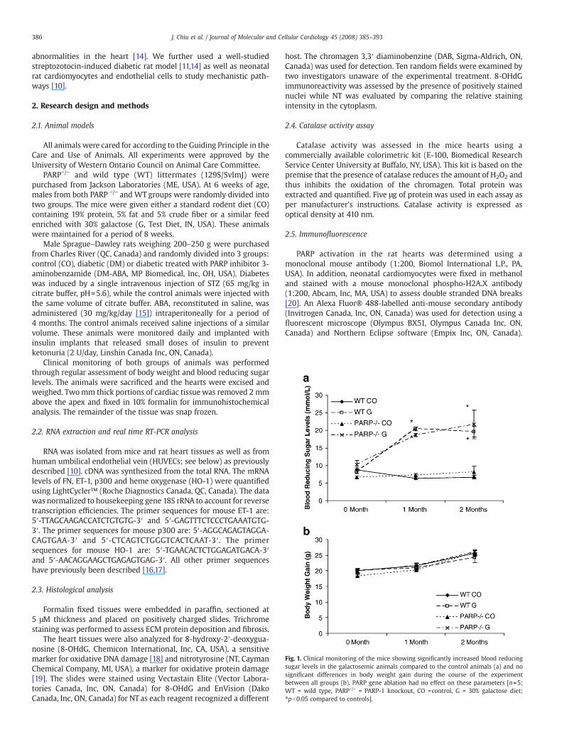
Fig. 1. Clinical monitoring of the mice showing significantly increased blood reducingsugar levels in the galactosemic animals compared to the control animals (a) and nosignificant differences in body weight gain during the course of the experimentbetween all groups (b). PARP gene ablation had no effect on these parameters [n=5;WT = wild type, PARP−/− = PARP-1 knockout, CO =control, G = 30% galactose diet;⁎pb0.05 compared to controls].
386 J. Chiu et al. / Journal of Molecular and Cellular Cardiology 45 (2008) 385–393
abnormalities in the heart [14]. We further used a well-studiedstreptozotocin-induced diabetic rat model [11,14] as well as neonatalrat cardiomyocytes and endothelial cells to study mechanistic path-ways [10].
2. Research design and methods
2.1. Animal models
All animals were cared for according to the Guiding Principle in theCare and Use of Animals. All experiments were approved by theUniversity of Western Ontario Council on Animal Care Committee.
PARP−/− and wild type (WT) littermates (129S/SvlmJ) werepurchased from Jackson Laboratories (ME, USA). At 6 weeks of age,males from both PARP −/− and WT groups were randomly divided intotwo groups. The mice were given either a standard rodent diet (CO)containing 19% protein, 5% fat and 5% crude fiber or a similar feedenriched with 30% galactose (G, Test Diet, IN, USA). These animalswere maintained for a period of 8 weeks.
Male Sprague–Dawley rats weighing 200–250 g were purchasedfrom Charles River (QC, Canada) and randomly divided into 3 groups:control (CO), diabetic (DM) or diabetic treated with PARP inhibitor 3-aminobenzamide (DM-ABA, MP Biomedical, Inc, OH, USA). Diabeteswas induced by a single intravenous injection of STZ (65 mg/kg incitrate buffer, pH=5.6), while the control animals were injected withthe same volume of citrate buffer. ABA, reconstituted in saline, wasadministered (30 mg/kg/day [15]) intraperitoneally for a period of4 months. The control animals received saline injections of a similarvolume. These animals were monitored daily and implanted withinsulin implants that released small doses of insulin to preventketonuria (2 U/day, Linshin Canada Inc, ON, Canada).
Clinical monitoring of both groups of animals was performedthrough regular assessment of body weight and blood reducing sugarlevels. The animals were sacrificed and the hearts were excised andweighed. Twomm thick portions of cardiac tissue was removed 2 mmabove the apex and fixed in 10% formalin for immunohistochemicalanalysis. The remainder of the tissue was snap frozen.
2.2. RNA extraction and real time RT-PCR analysis
RNA was isolated from mice and rat heart tissues as well as fromhuman umbilical endothelial vein (HUVECs; see below) as previouslydescribed [10]. cDNA was synthesized from the total RNA. The mRNAlevels of FN, ET-1, p300 and heme oxygenase (HO-1) were quantifiedusing LightCycler™ (Roche Diagnostics Canada, QC, Canada). The datawas normalized to housekeeping gene 18S rRNA to account for reversetranscription efficiencies. The primer sequences for mouse ET-1 are:5′-TTAGCAAGACCATCTGTGTG-3′ and 5′-GAGTTTCTCCCTGAAATGTG-3′. The primer sequences for mouse p300 are: 5′-AGGCAGAGTAGGA-CAGTGAA-3′ and 5′-CTCAGTCTGGGTCACTCAAT-3′. The primersequences for mouse HO-1 are: 5′-TGAACACTCTGGAGATGACA-3′and 5′-AACAGGAAGCTGAGAGTGAG-3′. All other primer sequenceshave previously been described [16,17].
2.3. Histological analysis
Formalin fixed tissues were embedded in paraffin, sectioned at5 μM thickness and placed on positively charged slides. Trichromestaining was performed to assess ECM protein deposition and fibrosis.
The heart tissues were also analyzed for 8-hydroxy-2′-deoxygua-nosine (8-OHdG, Chemicon International, Inc, CA, USA), a sensitivemarker for oxidative DNA damage [18] and nitrotyrosine (NT, CaymanChemical Company, MI, USA), a marker for oxidative protein damage[19]. The slides were stained using Vectastain Elite (Vector Labora-tories Canada, Inc, ON, Canada) for 8-OHdG and EnVision (DakoCanada, Inc, ON, Canada) for NT as each reagent recognized a different
host. The chromagen 3,3′ diaminobenzine (DAB, Sigma-Aldrich, ON,Canada) was used for detection. Ten random fields were examined bytwo investigators unaware of the experimental treatment. 8-OHdGimmunoreactivity was assessed by the presence of positively stainednuclei while NT was evaluated by comparing the relative stainingintensity in the cytoplasm.
2.4. Catalase activity assay
Catalase activity was assessed in the mice hearts using acommercially available colorimetric kit (E-100, Biomedical ResearchService Center University at Buffalo, NY, USA). This kit is based on thepremise that the presence of catalase reduces the amount of H2O2 andthus inhibits the oxidation of the chromagen. Total protein wasextracted and quantified. Five μg of protein was used in each assay asper manufacturer's instructions. Catalase activity is expressed asoptical density at 410 nm.
2.5. Immunofluorescence
PARP activation in the rat hearts was determined using amonoclonal mouse antibody (1:200, Biomol International L.P., PA,USA). In addition, neonatal cardiomyocytes were fixed in methanoland stained with a mouse monoclonal phospho-H2A.X antibody(1:200, Abcam, Inc, MA, USA) to assess double stranded DNA breaks[20]. An Alexa Fluor® 488-labelled anti-mouse secondary antibody(Invitrogen Canada, Inc, ON, Canada) was used for detection using afluorescent microscope (Olympus BX51, Olympus Canada Inc, ON,Canada) and Northern Eclipse software (Empix Inc, ON, Canada).
387J. Chiu et al. / Journal of Molecular and Cellular Cardiology 45 (2008) 385–393
Hoechst 33342 dye (1 μg/mL, Invitrogen Canada Inc, ON, Canada) wasused to visualize the nuclei.
2.6. Histone deacetylase (HDAC) activity assay
Nuclear protein extracts were prepared from heart tissues (50–100 mg) of both rats and mice as previously described [21]. HDACactivity from 100 μg of nuclear extract was determined using theHDAC activity assay kit (ab1432, AbCam Inc, MA, USA) as permanufacturer's instructions. HDAC activity is expressed as theabsolute amount of deacetylated lysine generated in each sample(μmol/L).
2.7. Cardiomyocyte isolation and culture
Myocytes were prepared from 1 to 2 day old neonatal Sprague–Dawley rat heart ventricles as previously described [22,23]. Non-myocytes were removed through differential attachment. The isolatedcardiomyocytes were then plated onto 6 well culture plates(Primaria™ Falcon, NJ, USA) at a density of 3.0×10⁴ cells/cm2. Thecells were maintained for 48 h in Dulbecco's Modified Eagle'sMedium/Ham's F-12 supplemented with 10% fetal bovine serum,10 µg/mL transferrin selenium, 10 µg/mL insulin, 10 ng/mL selenium,50 U/mL penicillin, 50 µg/mL streptomycin, 2 mg/mL bovine serumalbumin, 5 µg/mL linoleic acid, 3 mN pyruvic acid, 0.1 mmol/Lminimum essential medium (MEM) non-essential amino acids, 10%MEM vitamin, 0.1 mmol/L bromodeoxyuridin, 100 µm L-ascorbic acid,and 30 mmol/L HEPES (pH 7.1). The cells were serum-starvedovernight and treated with either 5 mmol/L of PARP inhibitor ABAor 16 μmol/L of 1,5-isoquinolinediol (ISO, Sigma-Aldrich, ON, Canada)for 1 h. The cardiomyocytes were then subjected to either a 5 mmol/Lglucose (low glucose, LG) or a 25 mmol/L glucose (high glucose, HG)environment for 24 h. LG was used as controls.
2.8. Endothelial cell culture
HUVECs (Clonetics, MD, USA) were cultured in endothelial cellgrowth medium (Clonetics, MD, USA). The cells were serum-starvedovernight and exposed to either 5 mmol/L of PARP inhibitor ABAdissolved in ddH20 or 0.01 μmol/L of HDAC inhibitor trichostatin A(TSA, Calbiochem, NJ, USA) dissolved in DMSO for 1 h. The cells werethen incubated with 5 mmol/L of D-glucose or 25 mmol/L of glucoseand maintained for 24 h. We have previously determined that theseconcentrations of glucose are optimal for studying glucose-inducedgene expression alterations [10,17]. Twenty five mmol/L of L-glucosewere used as controls (data not shown). The endothelial cells weresubsequently used for RNA extraction and cDNA synthesis for RT-PCRanalyses (see above). HUVECs treated with TSA are normalized toRPL13A as the mRNA level of this housekeeping gene has been shownto remain the most constant with TSA treatment [24].
2.9. Measurement of cardiomyocyte hypertrophy
Cell surface area of cardiomyocytes wasmeasured to assess cellularhypertrophy. The cells were visualized using a Leica DMIL invertedmicroscrope (Leica Wetzlar, Germany) equipped with a Polaroiddigital camera. The images were captured at 10× magnification. Cellsurface area was determined using Mocha™ Software (SPSS, IL, USA)from 50 randomly selected cells per experiment, then averaged andexpressed as pixels.
2.10. Statistical analysis
The data is expressed as mean±SEM. Statistical significance wasdetermined by ANOVA followed by Bonferroni/Dunn test. Differenceswere considered to be statistically significant at values of pb0.05.
3. Results
3.1. Hyperhexosemia-induced PARP activation and increased oxidativestress is prevented with PARP inhibition
Both mice and rats were monitored for diabetic dysmetabolismby evaluating body weight gain and blood reducing sugar levels.Galactose-fed diabetic mice showed significant elevation of bloodreducing sugar levels after one and two months of galactose feeding,however, no significant reduction of body weight was observed. Noeffect of PARP−/− was seen on blood sugar levels or body weight.These data are shown in Figs. 1a and b, respectively. The diabetic ratsshowed significantly reduced body weight (484.0±40.3 g vs. 613.5±13.8 g in controls) and hyperglycemia (18.4±6.8 mmol/L vs. 7.22±1.6 mmol/L) after 4 months of follow up. PARP inhibition had nosignificant effect on these parameters (526.1±17.9 g and 22.6±4.7 mmol/L).
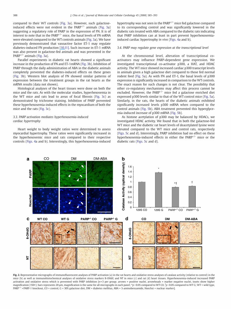
We first determined whether diabetes does in fact increase theactivation of PARP in the heart. Rat heart sections were immunofluor-escently assessed for PARP activation using a monoclonal PARPantibody. In thediabetic animals, therewas increased nuclear positivityfor PARPcompared to the control counterparts (Fig. 2a). PARP inhibitionwith ABA treatment prevented this diabetes-induced effect.
Next we assessed hyperhexosemia-induced oxidative damage inthe heart. A catalase activity assay was performed on the mice. TheWT galactose-fed mice exhibited significantly increased activitycompared to the WT control animals. It is of interest to note thatthe PARP−/− control mice had significantly lower levels of catalasecompared to the WT controls. The galactose-fed PARP−/− mice hadcatalase levels similar to the controls (Fig. 2b). These data werefurther verified with immunohistochemistry using two markers ofoxidative damage. The WT mice fed a galactose rich diet showedincreased nuclear staining of 8-OHdG, a marker of oxidative DNAdamage compared to the WT control mice (Fig. 2c). This wasparalleled in the rats, where the STZ-induced diabetic rat heartsexhibited increased nuclear staining of 8-OHdG, compared to theirage-matched controls (Fig. 2d). Interestingly, the galactose-fedPARP−/− mice and the diabetic rats treated with ABA showedcomplete normalization of the hyperhexosemia-induced effects(Figs. 2c and d).
We then tested the effect of PARP inhibition on oxidative stress byusing another sensitive oxidant injury marker. Analysis of the WTmice on the high galactose diet with NT, a marker of oxidative proteindamage, showed increased staining intensity in the cytoplasmcompared with the hearts of the WT animals on normal diet (Fig. 2c).A similar pattern was also seen in the diabetic rat hearts (Fig. 2d).Cardiac tissue from the galactose-fed PARP−/− mice and the diabeticrats with ABA treatment showed a NT staining pattern that isreminiscent of their respective control hearts; again indicating com-plete attenuation of the hyperhexosemia-induced effects (Figs. 2cand d). These results were further confirmed by the analysis of a pro-oxidant marker HO-1 [25] transcript levels. Cardiac tissues of WTgalactose-fed mice showed a significant upregulation of HO-1 (datanot shown). Inhibition of PARP also prevented this hyperhexosemiaassociated increase in the mice (data not shown). These findings arein support of Garcia Soriano et al. who found that PARP activationincreased NT positivity in the blood vessels of diabetic mice and thatpharmacological PARP inhibition can prevent this diabetes-inducedeffect [26].
3.2. Hyperhexosemia-induced ECM protein production is mediatedthrough a PARP-dependent pathway
Next, we studied the second important parameter of diabeticcardiomyopathy, ECM protein production. WT mice fed a galactoserich diet had significantly upregulated mRNA expression of FN when
388 J. Chiu et al. / Journal of Molecular and Cellular Cardiology 45 (2008) 385–393
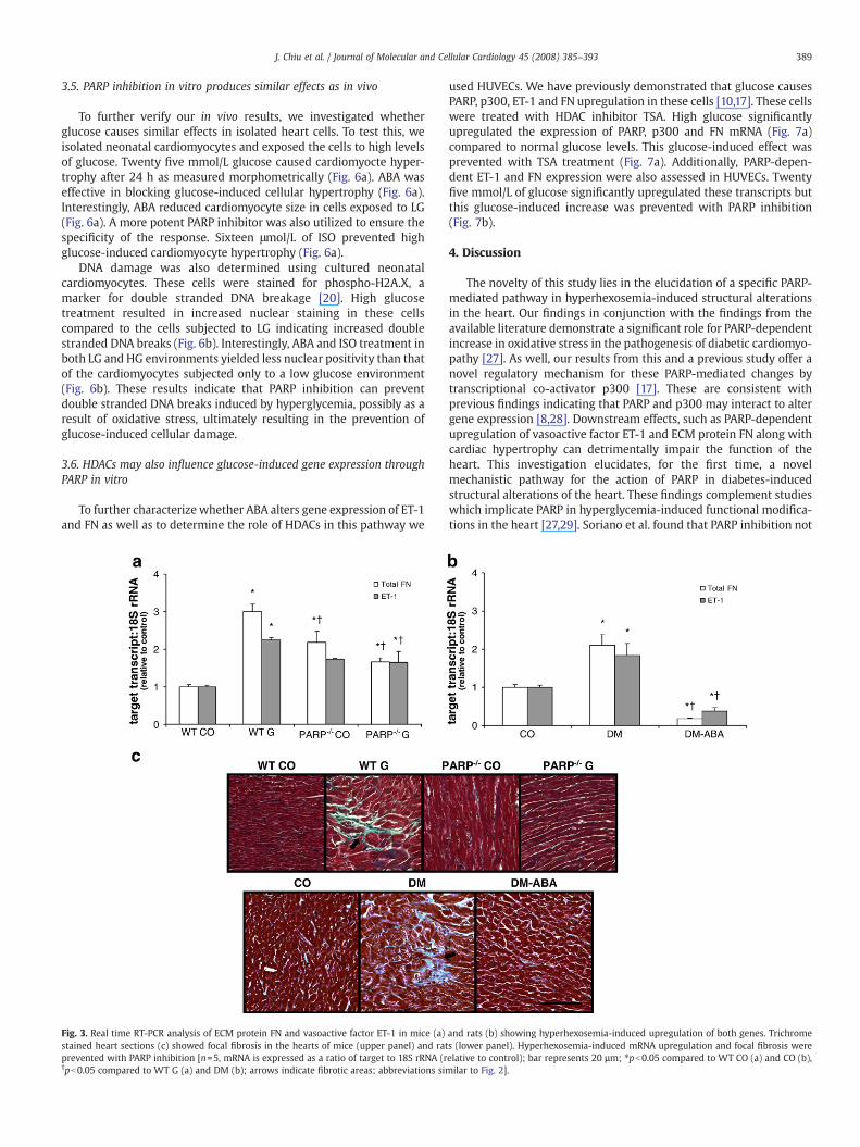
compared to their WT controls (Fig. 3a). However, such galactose-induced effects were not evident in the PARP−/− animals (Fig. 3a)suggesting a regulatory role of PARP in the expression of FN. It is ofinterest to note that in the PARP−/− mice, the basal levels of FN mRNAwere elevated compared to theWTcontrols animals (Fig. 3a). We havepreviously demonstrated that vasoactive factor ET-1 may regulatediabetes-induced FN production [10,11]. Such increase in ET-1 mRNAwas also present in galactose-fed animals and was prevented in thePARP−/− animals (Fig. 3a).
Parallel experiments in diabetic rat hearts showed a significantincrease in the production of FN and ET-1mRNA (Fig. 3b). Inhibition ofPARP through the daily administration of ABA in the diabetic animalscompletely prevented the diabetes-induced effects on these genes(Fig. 3b). Western blot analysis of FN showed similar patterns ofexpression between the treatment groups in the rat hearts as themRNA results (data not shown).
Histological analyses of the heart tissues were done on both themice and the rats. As with the molecular studies, hyperhexosemia inthe WT mice and rats lead to areas of focal fibrosis (Fig. 3c) asdemonstrated by trichrome staining. Inhibition of PARP preventedthese hyperhexosemia-induced effects in the myocardium of both themice and the rats (Fig. 3c).
3.3. PARP activation mediates hyperhexosemia-inducedcardiac hypertrophy
Heart weight to body weight ratios were determined to assessmyocardial hypertrophy. These ratios were significantly increased inthe hyperhexosemic mice and rats compared to their respectivecontrols (Figs. 4a and b). Interestingly, this hyperhexosemia-induced
Fig. 2. Representative micrographs of immunofluorescent analyses of PARP activation (a) in tmice (b) as well as immunohistochemical analyses of oxidative stress markers 8-OHdG aactivation and oxidative stress which is prevented with PARP inhibition [n=3 per groupmagnification (150×); bars represents 20 µm, magnification is the same for all micrographs inPARP−/−=PARP-1 knockout, CO = control, G = 30% galactose diet, DM = diabetes mellitus, AB
hypertrophy was not seen in the PARP−/− mice fed galactose comparedto its corresponding control and was significantly lowered in thediabetic rats treated with ABA compared to the diabetic rats indicatingthat PARP inhibition can at least in part prevent hyperhexosemia-induced cardiac hypertrophy in vivo (Figs. 4a and b).
3.4. PARP may regulate gene expression at the transcriptional level
At the chromosomal level, alteration of transcriptional co-activators may influence PARP-dependent gene expression. Weinvestigated transcriptional co-activator p300, a HAT, and HDACactivity. TheWTmice showed increased cardiac p300 transcript levelsin animals given a high galactose diet compared to those fed normalrodent feed (Fig. 5a). As with FN and ET-1, the basal levels of p300expression is significantly increased in comparison to theWTcontrols.The exact reason for such changes is not clear. The possibility thatother co-regulatory mechanisms may affect this process cannot beexcluded. However, the PARP−/− mice fed a galactose enriched dietexpressed p300 levels similar to that of the WT control mice (Fig. 5a).Similarly, in the rats, the hearts of the diabetic animals exhibitedsignificantly increased levels p300 mRNA when compared to thecontrol animals (Fig. 5b). ABA treatment prevented this hyperglyce-mia-induced increase of p300 mRNA (Fig. 5b).
As histone acetylation of p300 may be balanced by HDACs, weinvestigated HDAC activity. We found that in both the galactose-fedWT mice and the diabetic rat heart levels of deacetylated lysine wereelevated compared to the WT mice and control rats, respectively(Figs. 5c and d). Interestingly, PARP inhibition had no effect on thesehyperhexosemia-induced effects in either the PARP−/− mice or thediabetic rats (Figs. 5c and d).
he rat hearts and oxidative stress analyses of catalase activity (relative to control) in thend NT in mice (c) and rat (d) heart tissues. Hyperhexosemia-induced increased PARP; arrows = positive nuclei, arrowheads = marker negative nuclei, insets show highereach panel; ⁎pb0.05 compared toWT CO, †pb0.05 compared toWT G;WT = wild type,A = 3-aminobenzamide, Hoechst = nuclear marker].
389J. Chiu et al. / Journal of Molecular and Cellular Cardiology 45 (2008) 385–393
3.5. PARP inhibition in vitro produces similar effects as in vivo
To further verify our in vivo results, we investigated whetherglucose causes similar effects in isolated heart cells. To test this, weisolated neonatal cardiomyocytes and exposed the cells to high levelsof glucose. Twenty five mmol/L glucose caused cardiomyocte hyper-trophy after 24 h as measured morphometrically (Fig. 6a). ABA waseffective in blocking glucose-induced cellular hypertrophy (Fig. 6a).Interestingly, ABA reduced cardiomyocyte size in cells exposed to LG(Fig. 6a). A more potent PARP inhibitor was also utilized to ensure thespecificity of the response. Sixteen μmol/L of ISO prevented highglucose-induced cardiomyocyte hypertrophy (Fig. 6a).
DNA damage was also determined using cultured neonatalcardiomyocytes. These cells were stained for phospho-H2A.X, amarker for double stranded DNA breakage [20]. High glucosetreatment resulted in increased nuclear staining in these cellscompared to the cells subjected to LG indicating increased doublestranded DNA breaks (Fig. 6b). Interestingly, ABA and ISO treatment inboth LG and HG environments yielded less nuclear positivity than thatof the cardiomyocytes subjected only to a low glucose environment(Fig. 6b). These results indicate that PARP inhibition can preventdouble stranded DNA breaks induced by hyperglycemia, possibly as aresult of oxidative stress, ultimately resulting in the prevention ofglucose-induced cellular damage.
3.6. HDACs may also influence glucose-induced gene expression throughPARP in vitro
To further characterize whether ABA alters gene expression of ET-1and FN as well as to determine the role of HDACs in this pathway we
Fig. 3. Real time RT-PCR analysis of ECM protein FN and vasoactive factor ET-1 in mice (a)stained heart sections (c) showed focal fibrosis in the hearts of mice (upper panel) and ratprevented with PARP inhibition [n=5, mRNA is expressed as a ratio of target to 18S rRNA (r†pb0.05 compared to WT G (a) and DM (b); arrows indicate fibrotic areas; abbreviations sim
used HUVECs. We have previously demonstrated that glucose causesPARP, p300, ET-1 and FN upregulation in these cells [10,17]. These cellswere treated with HDAC inhibitor TSA. High glucose significantlyupregulated the expression of PARP, p300 and FN mRNA (Fig. 7a)compared to normal glucose levels. This glucose-induced effect wasprevented with TSA treatment (Fig. 7a). Additionally, PARP-depen-dent ET-1 and FN expression were also assessed in HUVECs. Twentyfive mmol/L of glucose significantly upregulated these transcripts butthis glucose-induced increase was prevented with PARP inhibition(Fig. 7b).
4. Discussion
The novelty of this study lies in the elucidation of a specific PARP-mediated pathway in hyperhexosemia-induced structural alterationsin the heart. Our findings in conjunction with the findings from theavailable literature demonstrate a significant role for PARP-dependentincrease in oxidative stress in the pathogenesis of diabetic cardiomyo-pathy [27]. As well, our results from this and a previous study offer anovel regulatory mechanism for these PARP-mediated changes bytranscriptional co-activator p300 [17]. These are consistent withprevious findings indicating that PARP and p300 may interact to altergene expression [8,28]. Downstream effects, such as PARP-dependentupregulation of vasoactive factor ET-1 and ECM protein FN along withcardiac hypertrophy can detrimentally impair the function of theheart. This investigation elucidates, for the first time, a novelmechanistic pathway for the action of PARP in diabetes-inducedstructural alterations of the heart. These findings complement studieswhich implicate PARP in hyperglycemia-induced functional modifica-tions in the heart [27,29]. Soriano et al. found that PARP inhibition not
and rats (b) showing hyperhexosemia-induced upregulation of both genes. Trichromes (lower panel). Hyperhexosemia-induced mRNA upregulation and focal fibrosis wereelative to control); bar represents 20 µm; ⁎pb0.05 compared to WT CO (a) and CO (b),ilar to Fig. 2].
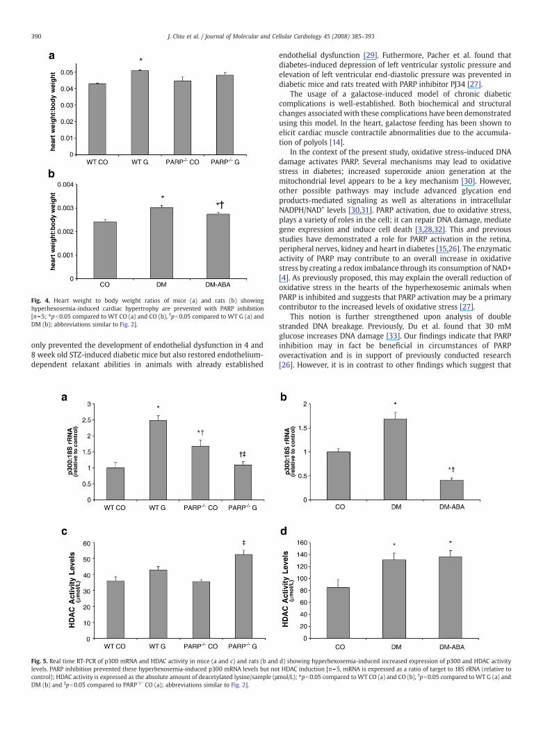
Fig. 4. Heart weight to body weight ratios of mice (a) and rats (b) showinghyperhexosemia-induced cardiac hypertrophy are prevented with PARP inhibition[n=5; ⁎pb0.05 compared to WT CO (a) and CO (b), †pb0.05 compared to WT G (a) andDM (b); abbreviations similar to Fig. 2].
390 J. Chiu et al. / Journal of Molecular and Cellular Cardiology 45 (2008) 385–393
only prevented the development of endothelial dysfunction in 4 and8 week old STZ-induced diabetic mice but also restored endothelium-dependent relaxant abilities in animals with already established
Fig. 5. Real time RT-PCR of p300 mRNA and HDAC activity in mice (a and c) and rats (b andlevels. PARP inhibition prevented these hyperhexosemia-induced p300 mRNA levels but nocontrol); HDAC activity is expressed as the absolute amount of deacetylated lysine/sample (μDM (b) and ‡pb0.05 compared to PARP−/− CO (a); abbreviations similar to Fig. 2].
endothelial dysfunction [29]. Futhermore, Pacher et al. found thatdiabetes-induced depression of left ventricular systolic pressure andelevation of left ventricular end-diastolic pressure was prevented indiabetic mice and rats treated with PARP inhibitor PJ34 [27].
The usage of a galactose-induced model of chronic diabeticcomplications is well-established. Both biochemical and structuralchanges associated with these complications have been demonstratedusing this model. In the heart, galactose feeding has been shown toelicit cardiac muscle contractile abnormalities due to the accumula-tion of polyols [14].
In the context of the present study, oxidative stress-induced DNAdamage activates PARP. Several mechanisms may lead to oxidativestress in diabetes; increased superoxide anion generation at themitochondrial level appears to be a key mechanism [30]. However,other possible pathways may include advanced glycation endproducts-mediated signaling as well as alterations in intracellularNADPH/NAD+ levels [30,31]. PARP activation, due to oxidative stress,plays a variety of roles in the cell; it can repair DNA damage, mediategene expression and induce cell death [3,28,32]. This and previousstudies have demonstrated a role for PARP activation in the retina,peripheral nerves, kidney and heart in diabetes [15,26]. The enzymaticactivity of PARP may contribute to an overall increase in oxidativestress by creating a redox imbalance through its consumption of NAD+[4]. As previously proposed, this may explain the overall reduction ofoxidative stress in the hearts of the hyperhexosemic animals whenPARP is inhibited and suggests that PARP activation may be a primarycontributor to the increased levels of oxidative stress [27].
This notion is further strengthened upon analysis of doublestranded DNA breakage. Previously, Du et al. found that 30 mMglucose increases DNA damage [33]. Our findings indicate that PARPinhibition may in fact be beneficial in circumstances of PARPoveractivation and is in support of previously conducted research[26]. However, it is in contrast to other findings which suggest that
d) showing hyperhexosemia-induced increased expression of p300 and HDAC activityt HDAC induction [n=5, mRNA is expressed as a ratio of target to 18S rRNA (relative tomol/L); ⁎pb0.05 compared toWT CO (a) and CO (b), †pb0.05 compared toWT G (a) and
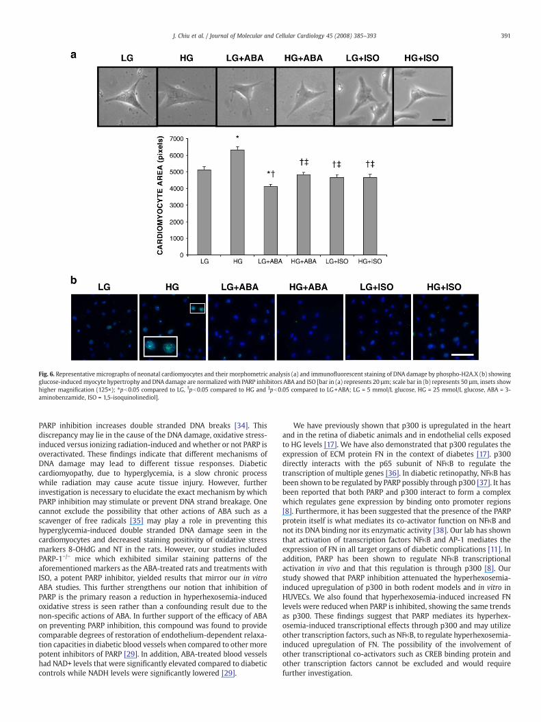
Fig. 6. Representative micrographs of neonatal cardiomyocytes and their morphometric analysis (a) and immunofluorescent staining of DNA damage by phospho-H2A.X (b) showingglucose-induced myocyte hypertrophy and DNA damage are normalized with PARP inhibitors ABA and ISO [bar in (a) represents 20 µm; scale bar in (b) represents 50 µm, insets showhigher magnification (125×); ⁎pb0.05 compared to LG, †pb0.05 compared to HG and ‡pb0.05 compared to LG+ABA; LG = 5 mmol/L glucose, HG = 25 mmol/L glucose, ABA = 3-aminobenzamide, ISO = 1,5-isoquinolinediol].
391J. Chiu et al. / Journal of Molecular and Cellular Cardiology 45 (2008) 385–393
PARP inhibition increases double stranded DNA breaks [34]. Thisdiscrepancy may lie in the cause of the DNA damage, oxidative stress-induced versus ionizing radiation-induced andwhether or not PARP isoveractivated. These findings indicate that different mechanisms ofDNA damage may lead to different tissue responses. Diabeticcardiomyopathy, due to hyperglycemia, is a slow chronic processwhile radiation may cause acute tissue injury. However, furtherinvestigation is necessary to elucidate the exact mechanism by whichPARP inhibition may stimulate or prevent DNA strand breakage. Onecannot exclude the possibility that other actions of ABA such as ascavenger of free radicals [35] may play a role in preventing thishyperglycemia-induced double stranded DNA damage seen in thecardiomyocytes and decreased staining positivity of oxidative stressmarkers 8-OHdG and NT in the rats. However, our studies includedPARP-1−/− mice which exhibited similar staining patterns of theaforementioned markers as the ABA-treated rats and treatments withISO, a potent PARP inhibitor, yielded results that mirror our in vitroABA studies. This further strengthens our notion that inhibition ofPARP is the primary reason a reduction in hyperhexosemia-inducedoxidative stress is seen rather than a confounding result due to thenon-specific actions of ABA. In further support of the efficacy of ABAon preventing PARP inhibition, this compound was found to providecomparable degrees of restoration of endothelium-dependent relaxa-tion capacities in diabetic blood vessels when compared to other morepotent inhibitors of PARP [29]. In addition, ABA-treated blood vesselshad NAD+ levels that were significantly elevated compared to diabeticcontrols while NADH levels were significantly lowered [29].
We have previously shown that p300 is upregulated in the heartand in the retina of diabetic animals and in endothelial cells exposedto HG levels [17]. We have also demonstrated that p300 regulates theexpression of ECM protein FN in the context of diabetes [17]. p300directly interacts with the p65 subunit of NFκB to regulate thetranscription of multiple genes [36]. In diabetic retinopathy, NFκB hasbeen shown to be regulated by PARP possibly through p300 [37]. It hasbeen reported that both PARP and p300 interact to form a complexwhich regulates gene expression by binding onto promoter regions[8]. Furthermore, it has been suggested that the presence of the PARPprotein itself is what mediates its co-activator function on NFκB andnot its DNA binding nor its enzymatic activity [38]. Our lab has shownthat activation of transcription factors NFκB and AP-1 mediates theexpression of FN in all target organs of diabetic complications [11]. Inaddition, PARP has been shown to regulate NFκB transcriptionalactivation in vivo and that this regulation is through p300 [8]. Ourstudy showed that PARP inhibition attenuated the hyperhexosemia-induced upregulation of p300 in both rodent models and in vitro inHUVECs. We also found that hyperhexosemia-induced increased FNlevels were reduced when PARP is inhibited, showing the same trendsas p300. These findings suggest that PARP mediates its hyperhex-osemia-induced transcriptional effects through p300 and may utilizeother transcription factors, such as NFκB, to regulate hyperhexosemia-induced upregulation of FN. The possibility of the involvement ofother transcriptional co-activators such as CREB binding protein andother transcription factors cannot be excluded and would requirefurther investigation.
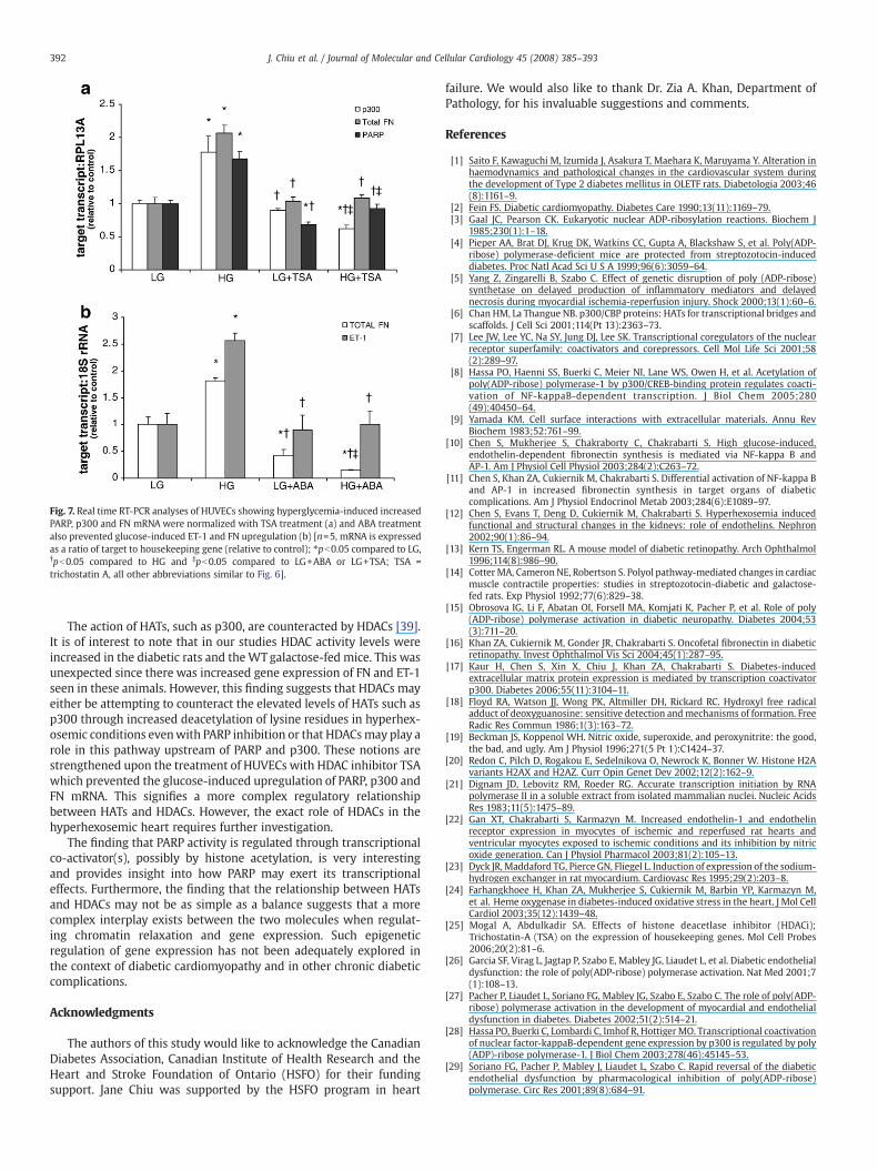
Fig. 7. Real time RT-PCR analyses of HUVECs showing hyperglycemia-induced increasedPARP, p300 and FN mRNA were normalized with TSA treatment (a) and ABA treatmentalso prevented glucose-induced ET-1 and FN upregulation (b) [n=5, mRNA is expressedas a ratio of target to housekeeping gene (relative to control); ⁎pb0.05 compared to LG,†pb0.05 compared to HG and ‡pb0.05 compared to LG+ABA or LG+TSA; TSA =trichostatin A, all other abbreviations similar to Fig. 6].
392 J. Chiu et al. / Journal of Molecular and Cellular Cardiology 45 (2008) 385–393
The action of HATs, such as p300, are counteracted by HDACs [39].It is of interest to note that in our studies HDAC activity levels wereincreased in the diabetic rats and theWT galactose-fed mice. This wasunexpected since there was increased gene expression of FN and ET-1seen in these animals. However, this finding suggests that HDACs mayeither be attempting to counteract the elevated levels of HATs such asp300 through increased deacetylation of lysine residues in hyperhex-osemic conditions evenwith PARP inhibition or that HDACsmay play arole in this pathway upstream of PARP and p300. These notions arestrengthened upon the treatment of HUVECs with HDAC inhibitor TSAwhich prevented the glucose-induced upregulation of PARP, p300 andFN mRNA. This signifies a more complex regulatory relationshipbetween HATs and HDACs. However, the exact role of HDACs in thehyperhexosemic heart requires further investigation.
The finding that PARP activity is regulated through transcriptionalco-activator(s), possibly by histone acetylation, is very interestingand provides insight into how PARP may exert its transcriptionaleffects. Furthermore, the finding that the relationship between HATsand HDACs may not be as simple as a balance suggests that a morecomplex interplay exists between the two molecules when regulat-ing chromatin relaxation and gene expression. Such epigeneticregulation of gene expression has not been adequately explored inthe context of diabetic cardiomyopathy and in other chronic diabeticcomplications.
Acknowledgments
The authors of this study would like to acknowledge the CanadianDiabetes Association, Canadian Institute of Health Research and theHeart and Stroke Foundation of Ontario (HSFO) for their fundingsupport. Jane Chiu was supported by the HSFO program in heart
failure. We would also like to thank Dr. Zia A. Khan, Department ofPathology, for his invaluable suggestions and comments.
References
[1] Saito F, Kawaguchi M, Izumida J, Asakura T, Maehara K, Maruyama Y. Alteration inhaemodynamics and pathological changes in the cardiovascular system duringthe development of Type 2 diabetes mellitus in OLETF rats. Diabetologia 2003;46(8):1161–9.
[2] Fein FS. Diabetic cardiomyopathy. Diabetes Care 1990;13(11):1169–79.[3] Gaal JC, Pearson CK. Eukaryotic nuclear ADP-ribosylation reactions. Biochem J
1985;230(1):1–18.[4] Pieper AA, Brat DJ, Krug DK, Watkins CC, Gupta A, Blackshaw S, et al. Poly(ADP-
ribose) polymerase-deficient mice are protected from streptozotocin-induceddiabetes. Proc Natl Acad Sci U S A 1999;96(6):3059–64.
[5] Yang Z, Zingarelli B, Szabo C. Effect of genetic disruption of poly (ADP-ribose)synthetase on delayed production of inflammatory mediators and delayednecrosis during myocardial ischemia-reperfusion injury. Shock 2000;13(1):60–6.
[6] Chan HM, La Thangue NB. p300/CBP proteins: HATs for transcriptional bridges andscaffolds. J Cell Sci 2001;114(Pt 13):2363–73.
[7] Lee JW, Lee YC, Na SY, Jung DJ, Lee SK. Transcriptional coregulators of the nuclearreceptor superfamily: coactivators and corepressors. Cell Mol Life Sci 2001;58(2):289–97.
[8] Hassa PO, Haenni SS, Buerki C, Meier NI, Lane WS, Owen H, et al. Acetylation ofpoly(ADP-ribose) polymerase-1 by p300/CREB-binding protein regulates coacti-vation of NF-kappaB-dependent transcription. J Biol Chem 2005;280(49):40450–64.
[9] Yamada KM. Cell surface interactions with extracellular materials. Annu RevBiochem 1983;52:761–99.
[10] Chen S, Mukherjee S, Chakraborty C, Chakrabarti S. High glucose-induced,endothelin-dependent fibronectin synthesis is mediated via NF-kappa B andAP-1. Am J Physiol Cell Physiol 2003;284(2):C263–72.
[11] Chen S, Khan ZA, Cukiernik M, Chakrabarti S. Differential activation of NF-kappa Band AP-1 in increased fibronectin synthesis in target organs of diabeticcomplications. Am J Physiol Endocrinol Metab 2003;284(6):E1089–97.
[12] Chen S, Evans T, Deng D, Cukiernik M, Chakrabarti S. Hyperhexosemia inducedfunctional and structural changes in the kidneys: role of endothelins. Nephron2002;90(1):86–94.
[13] Kern TS, Engerman RL. A mouse model of diabetic retinopathy. Arch Ophthalmol1996;114(8):986–90.
[14] CotterMA, Cameron NE, Robertson S. Polyol pathway-mediated changes in cardiacmuscle contractile properties: studies in streptozotocin-diabetic and galactose-fed rats. Exp Physiol 1992;77(6):829–38.
[15] Obrosova IG, Li F, Abatan OI, Forsell MA, Komjati K, Pacher P, et al. Role of poly(ADP-ribose) polymerase activation in diabetic neuropathy. Diabetes 2004;53(3):711–20.
[16] Khan ZA, Cukiernik M, Gonder JR, Chakrabarti S. Oncofetal fibronectin in diabeticretinopathy. Invest Ophthalmol Vis Sci 2004;45(1):287–95.
[17] Kaur H, Chen S, Xin X, Chiu J, Khan ZA, Chakrabarti S. Diabetes-inducedextracellular matrix protein expression is mediated by transcription coactivatorp300. Diabetes 2006;55(11):3104–11.
[18] Floyd RA, Watson JJ, Wong PK, Altmiller DH, Rickard RC. Hydroxyl free radicaladduct of deoxyguanosine: sensitive detection andmechanisms of formation. FreeRadic Res Commun 1986;1(3):163–72.
[19] Beckman JS, Koppenol WH. Nitric oxide, superoxide, and peroxynitrite: the good,the bad, and ugly. Am J Physiol 1996;271(5 Pt 1):C1424–37.
[20] Redon C, Pilch D, Rogakou E, Sedelnikova O, Newrock K, Bonner W. Histone H2Avariants H2AX and H2AZ. Curr Opin Genet Dev 2002;12(2):162–9.
[21] Dignam JD, Lebovitz RM, Roeder RG. Accurate transcription initiation by RNApolymerase II in a soluble extract from isolated mammalian nuclei. Nucleic AcidsRes 1983;11(5):1475–89.
[22] Gan XT, Chakrabarti S, Karmazyn M. Increased endothelin-1 and endothelinreceptor expression in myocytes of ischemic and reperfused rat hearts andventricular myocytes exposed to ischemic conditions and its inhibition by nitricoxide generation. Can J Physiol Pharmacol 2003;81(2):105–13.
[23] Dyck JR, Maddaford TG, Pierce GN, Fliegel L. Induction of expression of the sodium-hydrogen exchanger in rat myocardium. Cardiovasc Res 1995;29(2):203–8.
[24] Farhangkhoee H, Khan ZA, Mukherjee S, Cukiernik M, Barbin YP, Karmazyn M,et al. Heme oxygenase in diabetes-induced oxidative stress in the heart. J Mol CellCardiol 2003;35(12):1439–48.
[25] Mogal A, Abdulkadir SA. Effects of histone deacetlase inhibitor (HDACi);Trichostatin-A (TSA) on the expression of housekeeping genes. Mol Cell Probes2006;20(2):81–6.
[26] Garcia SF, Virag L, Jagtap P, Szabo E, Mabley JG, Liaudet L, et al. Diabetic endothelialdysfunction: the role of poly(ADP-ribose) polymerase activation. Nat Med 2001;7(1):108–13.
[27] Pacher P, Liaudet L, Soriano FG, Mabley JG, Szabo E, Szabo C. The role of poly(ADP-ribose) polymerase activation in the development of myocardial and endothelialdysfunction in diabetes. Diabetes 2002;51(2):514–21.
[28] Hassa PO, Buerki C, Lombardi C, Imhof R, Hottiger MO. Transcriptional coactivationof nuclear factor-kappaB-dependent gene expression by p300 is regulated by poly(ADP)-ribose polymerase-1. J Biol Chem 2003;278(46):45145–53.
[29] Soriano FG, Pacher P, Mabley J, Liaudet L, Szabo C. Rapid reversal of the diabeticendothelial dysfunction by pharmacological inhibition of poly(ADP-ribose)polymerase. Circ Res 2001;89(8):684–91.
393J. Chiu et al. / Journal of Molecular and Cellular Cardiology 45 (2008) 385–393
[30] Brownlee M. Biochemistry and molecular cell biology of diabetic complications.Nature 2001;414:813–20.
[31] Ceriello A. New insights on oxidative stress and diabetic complications may lead toa “causal” antioxidant therapy. Diabetes Care 2003;26:1589–96.
[32] Masson M, Rolli V, Dantzer F, Trucco C, Schreiber V, Fribourg S, et al. Poly(ADP-ribose) polymerase: structure–function relationship. Biochimie 1995;77(6):456–61.
[33] Du X, Matsumura T, Edelstein D, Rossetti L, Zsengeller Z, Szabo C, et al. Inhibitionof GAPDH activity by poly(ADP-ribose) polymerase activates three major path-ways of hyperglycemic damage in endothelial cells. J Clin Invest 2003;112(7):1049–57.
[34] Veuger SJ, Curtin NJ, Smith GC, Durkacz BW. Effects of novel inhibitors of poly(ADP-ribose) polymerase-1 and the DNA-dependent protein kinase on enzymeactivities and DNA repair. Oncogene 2004;23(44):7322–9.
[35] Wilson GL, Patton NJ, McCord JM, Mullins DW, Mossman BT. Mechanisms ofstreptozotocin- and alloxan-induced damage in rat B cells. Diabetologia 1984;27(6):587–91.
[36] Gerritsen ME, Williams AJ, Neish AS, Moore S, Shi Y, Collins T. CREB-bindingprotein/p300 are transcriptional coactivators of p65. Proc Natl Acad Sci U S A1997;94(7):2927–32.
[37] Zheng L, Szabo C, Kern TS. Poly(ADP-ribose) polymerase is involved in thedevelopment of diabetic retinopathy via regulation of nuclear factor-kappaB.Diabetes 2004;53(11):2960–7.
[38] Hassa PO, Covic M, Hasan S, Imhof R, Hottiger MO. The enzymatic and DNAbinding activity of PARP-1 are not required for NF-kappa B coactivator function.J Biol Chem 2001;276(49):45588–97.
[39] Sadoul K, Boyault C, Pabion M, Khochbin S. Regulation of protein turnover byacetyltransferases and deacetylases. Biochimie 2008;90(2):306–12.