TAB2 deficiency induces dilated cardiomyopathy by promoting ...
50
TAB2 deficiency induces dilated cardiomyopathy by promoting RIPK1-dependent apoptosis and necroptosis Haifeng Yin, … , Rachel Steinmetz, Qinghang Liu J Clin Invest. 2022. https://doi.org/10.1172/JCI152297. In-Press Preview Graphical abstract Research Cardiology Cell biology Find the latest version: https://jci.me/152297/pdf
-
Upload
khangminh22 -
Category
Documents
-
view
2 -
download
0
Transcript of TAB2 deficiency induces dilated cardiomyopathy by promoting ...
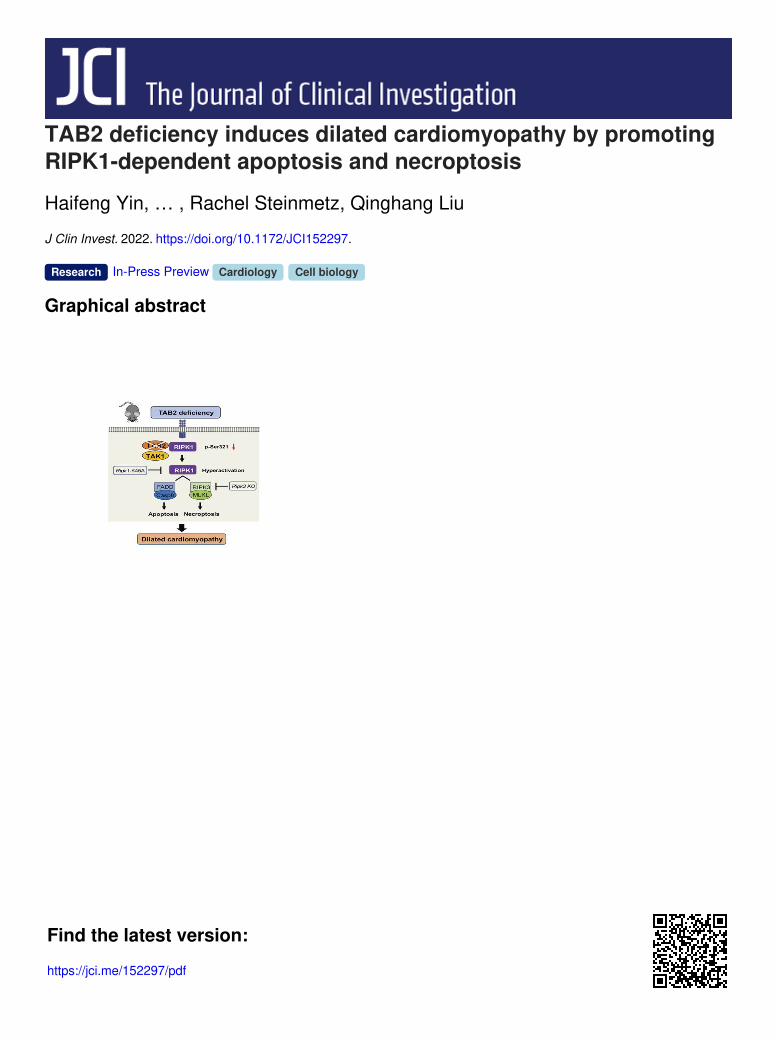
TAB2 deficiency induces dilated cardiomyopathy by promotingRIPK1-dependent apoptosis and necroptosis
Haifeng Yin, … , Rachel Steinmetz, Qinghang Liu
J Clin Invest. 2022. https://doi.org/10.1172/JCI152297.
In-Press Preview
Graphical abstract
Research Cardiology Cell biology
Find the latest version:
https://jci.me/152297/pdf

1
TAB2 deficiency induces dilated cardiomyopathy by promoting RIPK1-dependent
apoptosis and necroptosis
Haifeng Yin1*, Xiaoyun Guo1*, Yi Chen1*, Yachang Zeng1, Xiaoliang Mo1, Siqi Hong1, Hui He1,
Jing Li1, Rachel Steinmetz1, Qinghang Liu1
1Department of Physiology and Biophysics, University of Washington, Seattle, Washington, USA
*These authors contributed equally to this work
Address correspondence to:
Qinghang Liu, MD, PhD
Department of Physiology & Biophysics,
University of Washington,
1705 NE Pacific Street, HSB G424, Box 357290,
Seattle, WA 98195-7290
Tel: (206) 685-9133
Fax: (206) 685-0619
E-mail: [email protected]
The authors have declared that no conflict of interest exists.

2
Abstract
Mutations in TAB2 (transforming growth factor β activated kinase 1 binding protein 2) have been
implicated in the pathogenesis of dilated cardiomyopathy and/or congenital heart disease in
humans, but the underlying mechanisms are currently unknown. Here we identified an
indispensable role for TAB2 in regulating myocardial homeostasis and remodeling by suppressing
RIPK1 (receptor-interacting protein kinase 1) activation and RIPK1-dependent apoptosis and
necroptosis. Cardiomyocyte-specific deletion of Tab2 in mice triggered dilated cardiomyopathy
with massive apoptotic and necroptotic cell death. Moreover, Tab2-deficient mice were also
predisposed to myocardial injury and adverse remodeling following pathological stress. In
cardiomyocytes, deletion of TAB2, but not its close homologue TAB3, promoted TNFa-induced
apoptosis and necroptosis, which was rescued by forced activation of TAK1 or inhibition of RIPK1
kinase activity. Mechanistically, TAB2 critically mediates RIPK1 phosphorylation at Ser321 via a
TAK1-dependent mechanism, which prevents RIPK1 kinase activation and the formation of
RIPK1-FADD-caspase-8 apoptotic complex or RIPK1-RIPK3 necroptotic complex. Strikingly,
genetic inactivation of RIPK1 with Ripk1-K45A knock-in effectively rescued cardiac remodeling
and dysfunction in Tab2-deficient mice. Together, these data demonstrate that TAB2 is a key
regulator of myocardial homeostasis and remodeling by suppressing RIPK1-dependent apoptosis
and necroptosis. Our results also suggest that targeting RIPK1-mediated cell death signaling may
represent a promising therapeutic strategy for TAB2 deficiency-induced dilated cardiomyopathy.
Keywords: apoptosis, necroptosis, signal transduction, cardiomyopathy, cardiac remodeling
3
Introduction
Loss of cardiomyocytes by apoptotic and necrotic death is a crucial event underlying pathological
cardiac remodeling and heart failure (1,2). Similar to apoptosis, emerging evidence indicates that
necrosis can also occur in a highly regulated and genetically controlled manner, termed regulated
necrosis. A number of regulated necrosis pathways have been recently identified, including
necroptosis, ferroptosis, pyroptosis, mitochondria-mediated necrosis, and other regulated
necrotic processes (1). Necroptosis is a caspase-independent, highly regulated necrotic cell death
modality, which has recently been implicated in ischemic cardiac injury and pathological
remodeling (3-5). Necroptosis is executed through the induction of receptor-interacting protein
kinase (RIPK)1-RIPK3 necrosome, phosphorylation and oligomerization of mixed lineage kinase
domain-like (MLKL), and plasma membrane disruption (6-9).
Like apoptosis, necroptosis is induced by specific death receptors, such as tumor necrosis factor
receptor 1 (TNFR1), among other modules. Stimulation of TNFR1 by TNFa can trigger a variety
of cellular responses, including cell survival, apoptosis, and necroptosis, depending on the cellular
context. Under normal conditions, ligation of TNFR1 triggers the assembly of a plasma membrane
bound signaling complex, termed complex I, consisting of TNF receptor-associated protein with
death domain (TRADD), TNF receptor-associated protein 2 (TRAF2), cellular inhibitor of
apoptosis protein 1 and 2 (cIAP1 and cIAP2), and RIPK1 (10). Within complex I, RIPK1 is Lys63-
ubiquitinated by ubiquitin ligases TRAF2 and cIAP1/2, which then recruits and activates
transforming growth factor β-activated kinase 1 (TAK1) and the IkB kinase (IKK) complex, leading
to the activation of NFkB and transcription of prosurvival genes. NFkB-mediated induction of pro-
survival molecules is a well-defined cell death checkpoint in the TNFR1 pathway (11). Under
apoptosis-inducing conditions such as inhibition of NFkB, the TNFR1 complex internalizes and
4
converts to a cell death-inducing complex, termed complex II, consisting of TRADD, Fas-
associated protein with death domain (FADD), and caspase-8 (10). Recent studies identified
another cell death checkpoint, which is controlled by RIPK1 kinase activity. Indeed, it has been
shown that inhibition of TAK1, NEMO, IKKa/β or cIAP1/2 promotes RIPK1 activation and
sensitizes cells to RIPK1-dependent apoptosis or necroptosis independently of the NFkB pathway
(12-14). RIPK1 kinase activation induces cell death either by promoting the assembly of RIPK1-
FADD-caspase-8 apoptotic complex (13,15,16) or RIPK1-RIPK3-MLKL necroptotic complex (17).
RIPK1 kinase activity is tightly regulated by posttranslational modifications such as ubiquitination
and phosphorylation (12,16,18-20). However, the molecular signaling events that control RIPK1
kinase-dependent cell death checkpoint remain elusive.
TAB2 is an adaptor protein linking signals from the TNFR1 and other receptors to the TAK1
signaling complex by binding to Lys63-linked polyubiquitin chains (21). TAK1 has recently been
identified as a key regulator of apoptosis and necroptosis through NFkB-dependent and -
independent mechanisms (14,16). TAK1 forms a complex with TAB1 and TAB2 or TAB3, where
TAB1 functions as an activator of TAK1 by promoting TAK1 oligomerization and
autophosphorylation (22). TAB2 and its homologue TAB3 are shown to play redundant roles in
TAK1 activation and downstream signaling (23,24). Intriguingly, TAB2 also mediates TAK1
deactivation by recruiting PP6 to the TAK1 complex (25,26). Therefore, TAB2 may function as a
TAK1 activator or suppressor depending on cell types or cellular contexts. It has been
controversial as to whether TAB2 plays a role in apoptosis, necroptosis, or both. For example,
Sanjo et al. showed that deletion of TAB2 in mouse embryonic fibroblasts (MEFs) had no effects
on TNFa-induced cell death (23). In contrast, TAB2-/- dermal fibroblasts displayed increased
sensitivity to TNFa-induced necroptosis but not apoptosis (26). Moreover, TAB2-deficient
hepatocytes showed increased apoptotic cell death and caspase-3 activation after
5
lipopolysaccharide (LPS) stimulation (27). Therefore, the role of TAB2 in apoptotic and necroptotic
cell death has not been unequivocally established, and the mechanisms by which TAB2 regulates
cell death signaling remain largely unknown.
Genetic deletion of Tab2 in mice led to embryonic lethality with massive cell death in fetal liver
(23). In contrast, Tab3-/- mice showed no overt basal phenotype (28). These results suggest an
indispensable and non-redundant role for TAB2, rather than TAB3, in tissue survival and
hemostasis. Human TAB2 gene is mapped to chromosome 6q25.1. Haploinsufficiency of TAB2
caused by microdeletions of chromosome 6q25.1 in humans is associated with congenital heart
defects (CHD) and/or cardiomyopathy (29-35). CHD and cardiomyopathy are also reported in
patients with missense, nonsense, and small insertion or deletion mutations within TAB2 (32, 33,
34). Of note, some patients developed cardiomyopathy (mostly dilated cardiomyopathy) in the
absence of CHD, and cardiomyopathy can also occur later in life in those who survived CHD
(30,31,33). Most of the pathological effects are caused by loss-of-function of TAB2 allele, although
gain-of-function mutation of TAB2 has also been reported (35). The molecular mechanisms
underlying cardiac abnormalities caused by TAB2 mutations remain elusive.
Here, we identified an essential role for TAB2 in myocardial homeostasis and survival by
suppressing apoptosis and necroptosis primarily through a RIPK1-dependent mechanism. By
generating cardiomyocyte-specific Tab2 knockout mouse models, we showed that genetic
ablation of TAB2 in the adult heart promoted apoptotic and necroptotic cell death, leading to
dilated cardiomyopathy and heart failure. We also provide evidence that genetic inhibition of
RIPK1 kinase activity effectively rescued cardiac dysfunction and pathology associated with TAB2
deficiency in vivo, revealing a TAB2-mediated, RIPK1-dependent apoptotic and necroptotic
signaling pathway in regulating myocardial homeostasis and remodeling.

6
Results
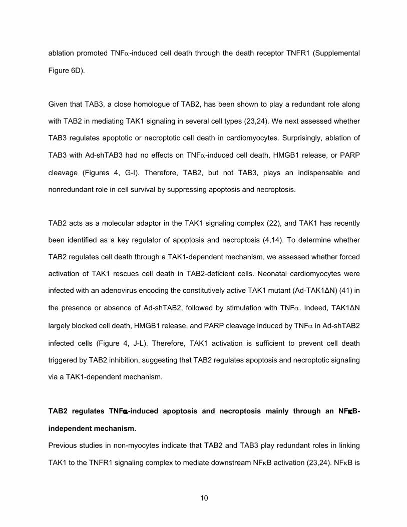
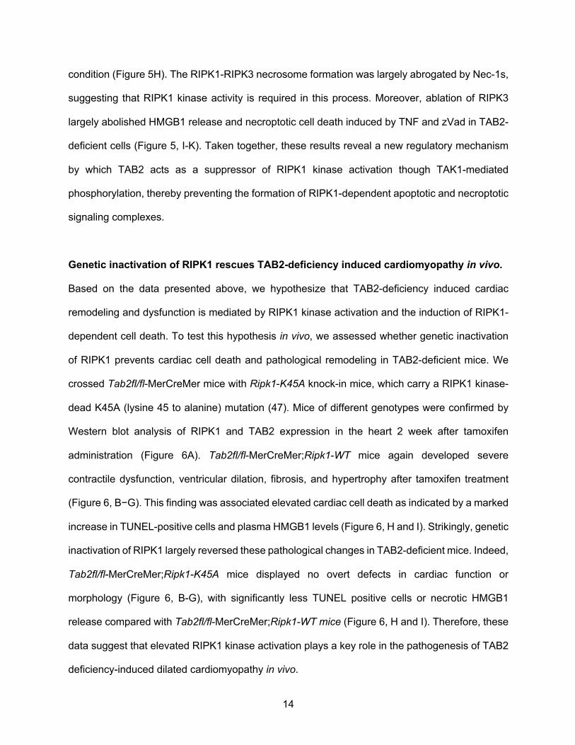
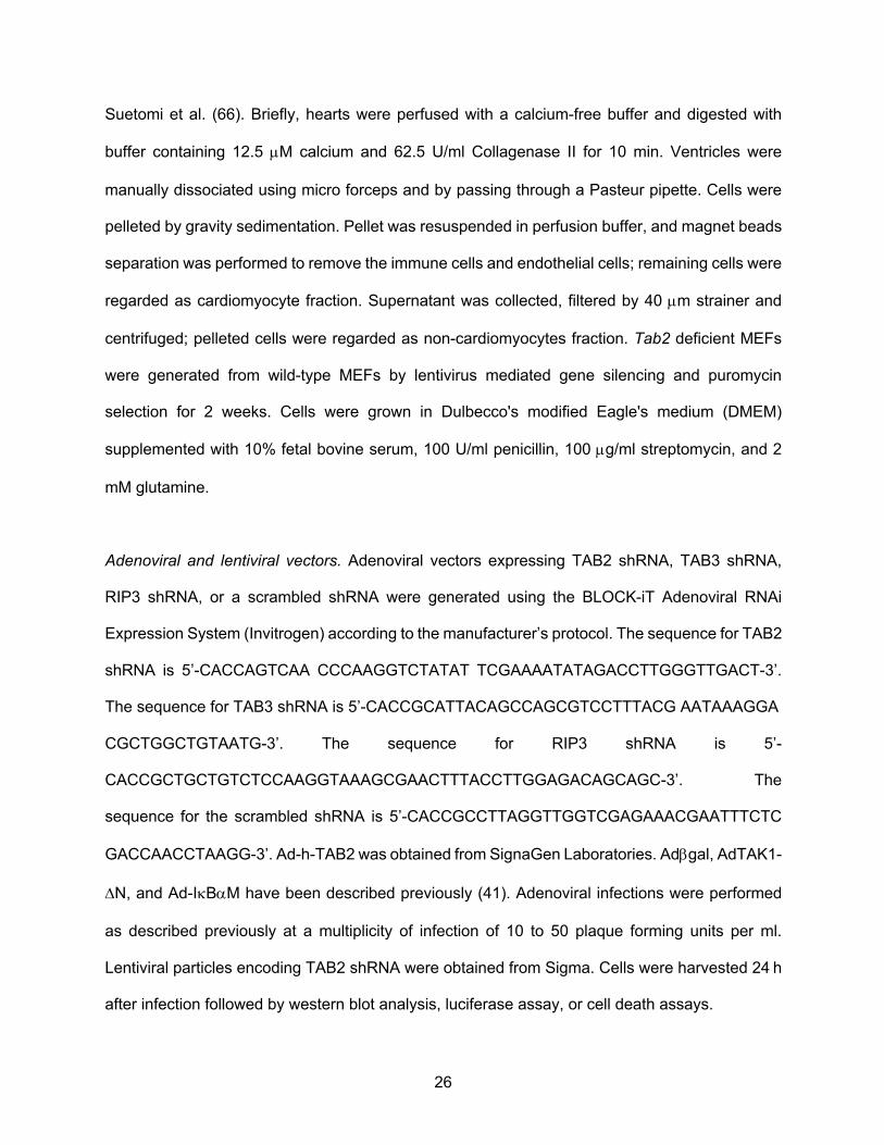
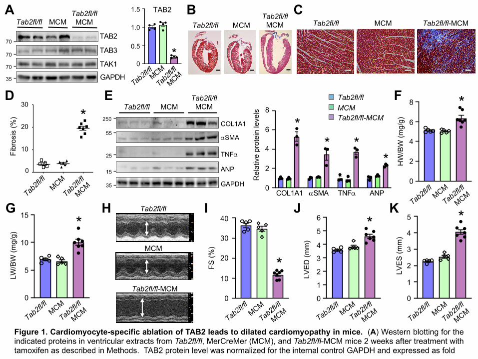
Loss of TAB2 in the adult heart induces dilated cardiomyopathy and heart failure.
To investigate the role of TAB2 in the adult heart, we used a tamoxifen-inducible Cre-mediated
recombination system for acute deletion of Tab2 in cardiomyocytes. Mice homozygous for the
Tab2-loxp targeted allele (Tab2fl/fl) (23) were crossed with aMHC-MerCreMer (MCM) transgenic
mice (36), which in the absence of tamoxifen were overtly normal with no detectable phenotype
at baseline (data not shown). Western blot analysis showed that TAB2 was efficiently deleted (>
90%) from the hearts of Tab2fl/fl-MCM mice but not Tab2fl/fl or MCM control mice after tamoxifen
treatment (Figure 1A). TAB3 or TAK1 expression was not altered (Figure 1A). Strikingly, after two
weeks of tamoxifen administration, Tab2fl/fl-MCM mice rapidly developed severe ventricular
dilation with high levels of myocardial fibrosis (Figure 1, B-D). Moreover, the protein levels of
fibrotic genes (collagen type 1 and a-smooth muscle actin), as well as atrial natriuretic peptide
(ANP) and TNFa were substantially elevated in TAB2-deficient hearts (Figure 1E). Tab2fl/fl-MCM
mice also displayed cardiac hypertrophy and pulmonary congestion as assessed by heart weight
to body weight (HW/BW) and lung weight to body weight (LW/BW) ratios (Figure 1, F and G),
suggesting that TAB2 deficiency induced pathological cardiac remodeling and congestive heart
failure. Echocardiographic analysis showed severe ventricular dilation and contractile dysfunction
in TAB2-deficient mice, as indicated by decreased fractional shortening (FS) and increased left
ventricular dimensions in end-diastole and end-systole (LVED and LEVS) (Figure 1, H-K).
Moreover, Tab2fl/fl-MCM mice showed a progressive deterioration of cardiac function, and most
of these mice died 6 weeks after tamoxifen administration (Supplemental Figure 1). Therefore,
acute deletion of Tab2 in the adult heart induced cardiac remodeling and heart failure,
recapitulating the phenotype of dilated cardiomyopathy in humans with TAB2 gene mutations (30-
33).
7
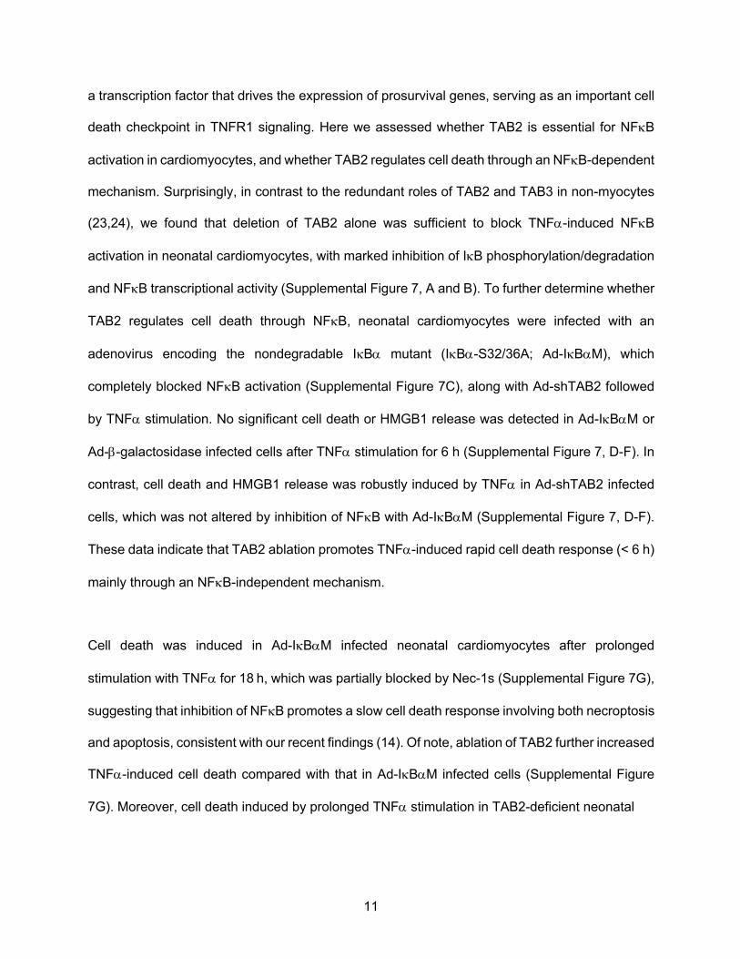
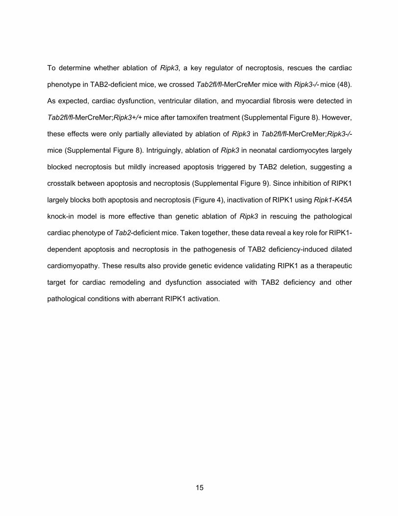
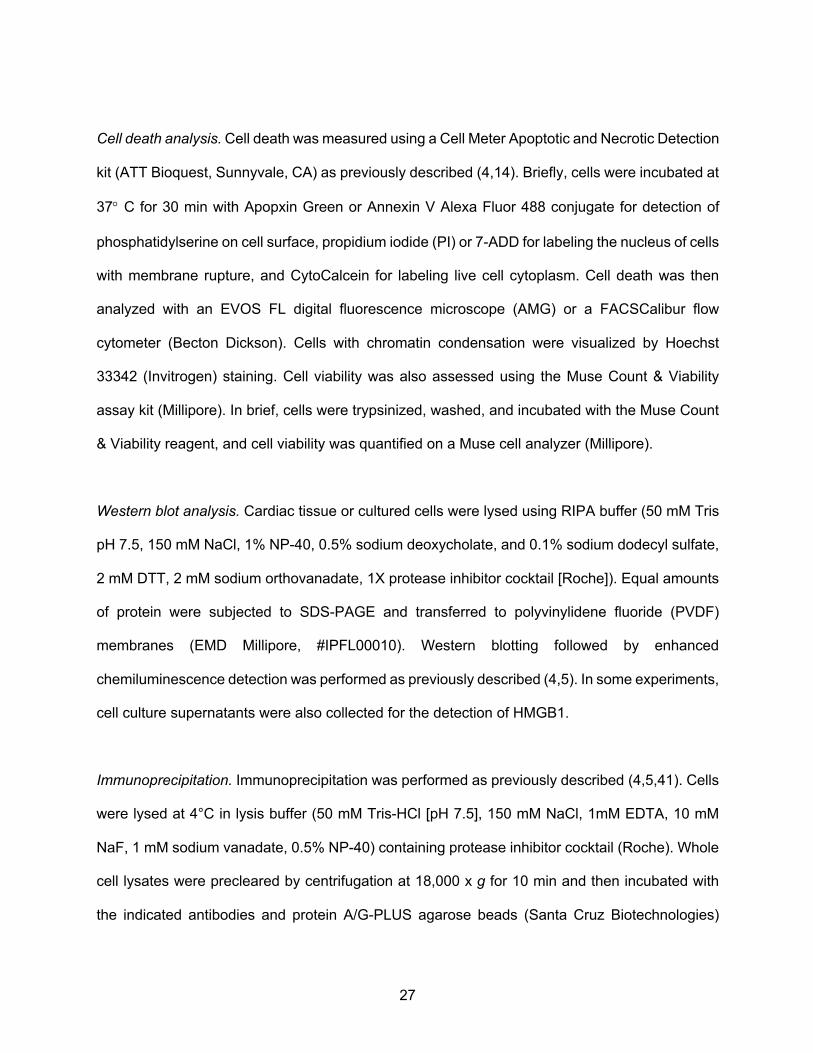
Ablation of TAB2 induces apoptotic and necroptotic cell death in the myocardium.
Next, we assessed whether apoptotic or necrotic cell death contributes to the pathological cardiac
phenotype of Tab2-deficiency. Intriguingly, protein levels of RIPK1, RIPK3, and MLKL, key
mediators of necroptosis, were significantly increased in the heart extracts of Tab2fl/fl-MCM mice
compared with littermate controls (Figure 2A). The level of phospho-RIPK1 at Ser 166, an
established marker for RIPK1 kinase activation (37), was greatly enhanced, whereas phospho-
RIPK1 at Ser321, which has recently been shown to suppress RIPK1 kinase activation (16), was
markedly decreased in TAB2-deficient hearts (Figure 2A). Moreover, a marked increase in the
plasma levels of HMGB1 (Figure 2A), a biomarker for necrotic cell death and myocardial injury
(5,38,39), was also detected in TAB2-deficient mice, along with elevated TNFa levels in the
plasma (Figure 2C). Immunofluorescent staining of cardiac sections also revealed enhanced
RIPK1 phosphorylation at Ser166 in TAB2-deficient hearts, indicating RIPK1 kinase activation
(Figure 2, D and E). Loss of nuclear HMGB1, an indicator of membrane disruption and necrosis
(5, 38,39), was also readily detectable (Figure 2, D and F). Of note, TAB2-deficient mice also
exhibited a marked increase in cleaved caspase 3 and TUNEL positive cells (Figure 2, D, G, and
H), indicating the induction of apoptosis. Together, these data indicate that loss of TAB2 promotes
apoptotic and necroptotic cell death in the myocardium, suggesting an essential role of TAB2 in
myocardial survival and homeostasis.
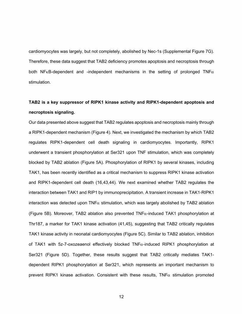
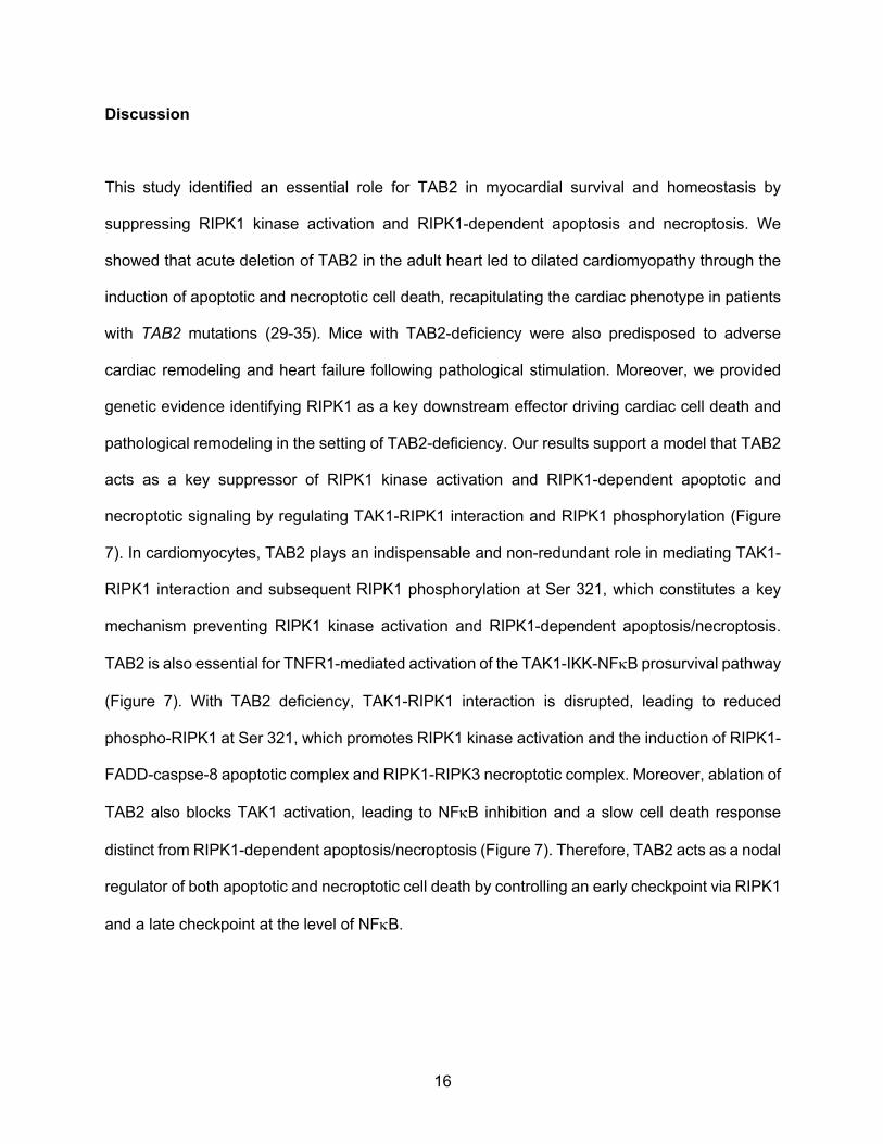
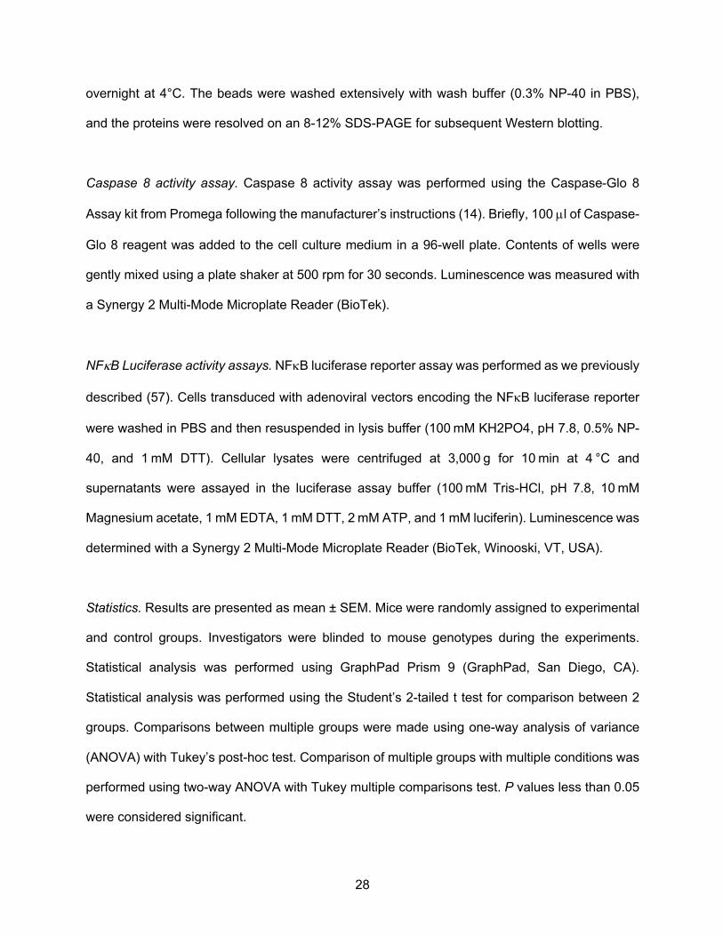
TAB2-deficiency leads to exacerbated cardiac remodeling and dysfunction following
pathological stress.
We further examined whether TAB2 plays a role in regulating myocardial remodeling and heart
failure propensity following pathological stimulation. We first measured TAB2 protein expression
in the heart subjected to transverse aortic constriction (TAC) or myocardial infarction (MI).
Myocardial TAB2 protein expression was markedly decreased following TAC and MI injury (Figure

8
3, A and B; Supplemental Figure 2, E and F), suggesting a potential role of TAB2 in cardiac
response to pathological stress. We further determined that TAB2 was markedly downregulated
in cardiomyocytes isolated from mouse heart after MI and TAC, whereas no significant change
was detected in non-cardiomyocytes (Supplemental Figure 2, A-D). Given that acute deletion of
TAB2 in the heart induced severe cardiac remodeling and failure, we generated another
cardiomyocyte-specific Tab2 knockout mouse model by crossing Tab2fl/fl mice with aMHC-Cre
mice (40). Here, TAB2 was again efficiently deleted in the heart from Tab2fl/fl-aMHC-Cre mice
(Figure 3C). Echocardiographic analysis showed that Tab2fl/fl-aMHC-Cre mice were overtly
normal at baseline within 2 months after birth (Supplemental Figure 3). However, these mice
slowly developed contractile dysfunction or ventricular dilation starting 3 months of age
(Supplemental Figure 3). In addition, inactivation of RIPK1 with Ripk1-K45A knock-in largely
rescued cardiac dysfunction and remodeling in Tab2fl/fl-aMHC-Cre mice (Supplemental Figure
4). To evaluate the role of TAB2 in TAC- or MI- induced pathological remodeling and heart failure,
here we used young Tab2fl/fl-aMHC-Cre mice (6 weeks of age), which displayed no detectable
cardiac pathology at baseline. Tab2fl/fl-aMHC-Cre mice showed exacerbated cardiac remodeling
and ventricular dilation after TAC, with a greater loss in cardiac contractile performance, greater
cardiac fibrosis, and more prominent lung edema compared with Tab2fl/fl controls (Figure 3, D-
G, and I). No significant difference in TAC-induced cardiac hypertrophy was detected between
these two groups (Figure 3H). These results indicate that TAB2-deficient mice mainly developed
dilated cardiomyopathy after TAC stimulation. The blunted hypertrophic response is possibly
caused by impaired hypertrophic signaling in the absence of TAB2 (41). Moreover, a greater
propensity for cardiac remodeling and dysfunction was also observed in Tab2fl/fl-aMHC-Cre mice
subjected to MI injury, showing increased infarct expansion, exacerbated contractile dysfunction,
enhanced ventricular dilation, increased heart size, and more severe lung edema compared with
Tab2fl/fl mice (Figure 3, J-O). Moreover, Tab2fl/fl-aMHC-Cre mice also exhibited a marked
9
increase in TUNEL positive cells as well as phospho-RIPK1 Ser166 (Supplemental Figure 5).
Taken together, loss of TAB2 promotes cardiac remodeling and dysfunction during disease
stimulation, suggesting a critical cardioprotective role for TAB2 in response to pathological stress.
TAB2, but not TAB3, is a key regulator of RIPK1-dependent apoptosis and necroptosis.
Based on the data above from our Tab2-deficient mice, we hypothesize that Tab2-deficiency
triggers adverse cardiac remodeling and heart failure by promoting apoptosis and/or necroptosis.
We first assessed whether ablation of TAB2 is sufficient to promote TNFa-induced cell death in
Tab2-deficient MEFs. Compared with wild-type MEFs, Tab2-deficient MEFs were highly sensitive
to TNFa stimulation, leading to rapid cell death with increased propidium iodide uptake (Figure
4A). This effect was abolished by cotreatment with the specific RIPK1 inhibitor necrostatin-1s
(Nec-1s) (37), but not by the pan-caspase inhibitor z-Vad-fmk (zVad), suggesting the induction of
necroptosis (Figure 4A). Moreover, HMGB1, a biomarker for necrosis, was readily detectable in
the culture supernatants of Tab2-deficient MEFs after TNFa stimulation, which was further
increased in the presence of zVad (Figure 4, B and C), consistent with the notion that caspase
inhibition promotes necroptosis (42). Intriguingly, deletion of TAB2 also enhanced TNFa-induced
PARP cleavage, which was reversed by both Nec-1s and zVad (Figures 4B). These results
indicate that deletion of TAB2 promotes RIPK1-dependent apoptosis and necroptosis. Next, we
examined whether TAB2 also regulates apoptosis and necroptosis in cardiomyocytes. Indeed,
ablation of TAB2 in neonatal cardiomyocytes with an adenoviral vector encoding TAB2 shRNA
(Ad-shTAB2) also promoted TNFa-induced apoptosis and necroptosis (Figures 4, D-F). To
exclude possible off-target effects associated with TAB2 deletion, re-constitution of TAB2-
deficient cells with an adenoviral vector expressing human TAB2 restored cellular resistance to
TNFa-induced cell death (Supplemental Figure 6, A-C). Moreover, we further showed that TAB2
10
ablation promoted TNFa-induced cell death through the death receptor TNFR1 (Supplemental
Figure 6D).
Given that TAB3, a close homologue of TAB2, has been shown to play a redundant role along
with TAB2 in mediating TAK1 signaling in several cell types (23,24). We next assessed whether
TAB3 regulates apoptotic or necroptotic cell death in cardiomyocytes. Surprisingly, ablation of
TAB3 with Ad-shTAB3 had no effects on TNFa-induced cell death, HMGB1 release, or PARP
cleavage (Figures 4, G-I). Therefore, TAB2, but not TAB3, plays an indispensable and
nonredundant role in cell survival by suppressing apoptosis and necroptosis.
TAB2 acts as a molecular adaptor in the TAK1 signaling complex (22), and TAK1 has recently
been identified as a key regulator of apoptosis and necroptosis (4,14). To determine whether
TAB2 regulates cell death through a TAK1-dependent mechanism, we assessed whether forced
activation of TAK1 rescues cell death in TAB2-deficient cells. Neonatal cardiomyocytes were
infected with an adenovirus encoding the constitutively active TAK1 mutant (Ad-TAK1ΔN) (41) in
the presence or absence of Ad-shTAB2, followed by stimulation with TNFa. Indeed, TAK1ΔN
largely blocked cell death, HMGB1 release, and PARP cleavage induced by TNFa in Ad-shTAB2
infected cells (Figure 4, J-L). Therefore, TAK1 activation is sufficient to prevent cell death
triggered by TAB2 inhibition, suggesting that TAB2 regulates apoptosis and necroptotic signaling
via a TAK1-dependent mechanism.
TAB2 regulates TNFa-induced apoptosis and necroptosis mainly through an NFkB-
independent mechanism.
Previous studies in non-myocytes indicate that TAB2 and TAB3 play redundant roles in linking
TAK1 to the TNFR1 signaling complex to mediate downstream NFkB activation (23,24). NFkB is
11
a transcription factor that drives the expression of prosurvival genes, serving as an important cell
death checkpoint in TNFR1 signaling. Here we assessed whether TAB2 is essential for NFkB
activation in cardiomyocytes, and whether TAB2 regulates cell death through an NFkB-dependent
mechanism. Surprisingly, in contrast to the redundant roles of TAB2 and TAB3 in non-myocytes
(23,24), we found that deletion of TAB2 alone was sufficient to block TNFa-induced NFkB
activation in neonatal cardiomyocytes, with marked inhibition of IkB phosphorylation/degradation
and NFkB transcriptional activity (Supplemental Figure 7, A and B). To further determine whether
TAB2 regulates cell death through NFkB, neonatal cardiomyocytes were infected with an
adenovirus encoding the nondegradable IkBa mutant (IkBa-S32/36A; Ad-IkBaM), which
completely blocked NFkB activation (Supplemental Figure 7C), along with Ad-shTAB2 followed
by TNFa stimulation. No significant cell death or HMGB1 release was detected in Ad-IkBaM or
Ad-b-galactosidase infected cells after TNFa stimulation for 6 h (Supplemental Figure 7, D-F). In
contrast, cell death and HMGB1 release was robustly induced by TNFa in Ad-shTAB2 infected
cells, which was not altered by inhibition of NFkB with Ad-IkBaM (Supplemental Figure 7, D-F).
These data indicate that TAB2 ablation promotes TNFa-induced rapid cell death response (< 6 h)
mainly through an NFkB-independent mechanism.
Cell death was induced in Ad-IkBaM infected neonatal cardiomyocytes after prolonged
stimulation with TNFa for 18 h, which was partially blocked by Nec-1s (Supplemental Figure 7G),
suggesting that inhibition of NFkB promotes a slow cell death response involving both necroptosis
and apoptosis, consistent with our recent findings (14). Of note, ablation of TAB2 further increased
TNFa-induced cell death compared with that in Ad-IkBaM infected cells (Supplemental Figure
7G). Moreover, cell death induced by prolonged TNFa stimulation in TAB2-deficient neonatal
12
cardiomyocytes was largely, but not completely, abolished by Nec-1s (Supplemental Figure 7G).
Therefore, these data suggest that TAB2 deficiency promotes apoptosis and necroptosis through
both NFkB-dependent and -independent mechanisms in the setting of prolonged TNFa
stimulation.
TAB2 is a key suppressor of RIPK1 kinase activity and RIPK1-dependent apoptosis and
necroptosis signaling.
Our data presented above suggest that TAB2 regulates apoptosis and necroptosis mainly through
a RIPK1-dependent mechanism (Figure 4). Next, we investigated the mechanism by which TAB2
regulates RIPK1-dependent cell death signaling in cardiomyocytes. Importantly, RIPK1
underwent a transient phosphorylation at Ser321 upon TNF stimulation, which was completely
blocked by TAB2 ablation (Figure 5A). Phosphorylation of RIPK1 by several kinases, including
TAK1, has been recently identified as a critical mechanism to suppress RIPK1 kinase activation
and RIPK1-dependent cell death (16,43,44). We next examined whether TAB2 regulates the
interaction between TAK1 and RIP1 by immunoprecipitation. A transient increase in TAK1-RIPK1
interaction was detected upon TNFa stimulation, which was largely abolished by TAB2 ablation
(Figure 5B). Moreover, TAB2 ablation also prevented TNFa-induced TAK1 phosphorylation at
Thr187, a marker for TAK1 kinase activation (41,45), suggesting that TAB2 critically regulates
TAK1 kinase activity in neonatal cardiomyocytes (Figure 5C). Similar to TAB2 ablation, inhibition
of TAK1 with 5z-7-oxozeaenol effectively blocked TNFa-induced RIPK1 phosphorylation at
Ser321 (Figure 5D). Together, these results suggest that TAB2 critically mediates TAK1-
dependent RIPK1 phosphorylation at Ser321, which represents an important mechanism to
prevent RIPK1 kinase activation. Consistent with these results, TNFa stimulation promoted
13
RIPK1 phosphorylation at Ser166, an indicator of RIPK1 kinase activation (37), in TAB2-deficient
cells, especially in the presence of the caspase inhibitor zVad-fmk (Figure 5E). To further
determine the mechanism by which TAB2 regulates RIPK1-dependent cell death, we performed
immunoprecipitation experiments to determine whether ablation of TAB2 promotes the assembly
of RIPK1-mediated apoptosis and necroptosis signaling complexes. Coimmunoprecipitation (Co-
IP) of both RIPK1 and caspase-8 with FADD was detected in TAB2-deficient cells upon simulation
with TNFa, indicating the induction of RIPK1-FADD-caspase-8 apoptotic complex (Figure 5F).
Similar results were obtained when co-IP was performed using an anti-RIPK1 antibody (Yin et al.,
unpublished observations). However, no interaction of FADD with either RIPK1 or caspase-8 was
detected in Ad-shScram infected neonatal cardiomyocytes treated with vehicle control, TNFa, or
TNFa plus Nec-1s (Figure 5F). Inhibition of RIPK1 with Nec-1s efficiently blocked the RIPK1-
FADD-caspase-8 interaction in TAB2-deficient cells, indicating that RIPK1 kinase activity is
required for the apoptotic complex formation (Figure 5F). Moreover, TNFa also induced a marked
increase in caspase-8 activity in TAB2-deficient cells, but not in control cells, which was blocked
by co-treatment with Nec-1s or zVad-fmk (Figure 5G). These results indicate that TAB2 negatively
regulates RIPK1 kinase activation and its interaction with FADD and caspase-8. Thus, ablation of
TAB2 promotes apoptosis signaling and caspase activation in a manner dependent on RIPK1
kinase activity.
The kinase activity of RIPK1 is also essential for the RIPK1-RIPK3 necrosome formation and
necroptotic cell death (20,46). To determine the molecular mechanism by which TAB2 regulates
necroptosis signaling, we assessed whether TAB2 ablation promotes the RIPK1-RIPK3
necrosome formation through RIPK1 kinase activation. A marked increase in RIPK1-RIPK3
interaction was detected in TAB2-deficient cardiomyocytes after stimulation with TNFa and zVad
(Figure 5H). No RIPK1-RIPK3 interaction was detected in control cardiomyocytes under the same
14
condition (Figure 5H). The RIPK1-RIPK3 necrosome formation was largely abrogated by Nec-1s,
suggesting that RIPK1 kinase activity is required in this process. Moreover, ablation of RIPK3
largely abolished HMGB1 release and necroptotic cell death induced by TNF and zVad in TAB2-
deficient cells (Figure 5, I-K). Taken together, these results reveal a new regulatory mechanism
by which TAB2 acts as a suppressor of RIPK1 kinase activation though TAK1-mediated
phosphorylation, thereby preventing the formation of RIPK1-dependent apoptotic and necroptotic
signaling complexes.
Genetic inactivation of RIPK1 rescues TAB2-deficiency induced cardiomyopathy in vivo.
Based on the data presented above, we hypothesize that TAB2-deficiency induced cardiac
remodeling and dysfunction is mediated by RIPK1 kinase activation and the induction of RIPK1-
dependent cell death. To test this hypothesis in vivo, we assessed whether genetic inactivation
of RIPK1 prevents cardiac cell death and pathological remodeling in TAB2-deficient mice. We
crossed Tab2fl/fl-MerCreMer mice with Ripk1-K45A knock-in mice, which carry a RIPK1 kinase-
dead K45A (lysine 45 to alanine) mutation (47). Mice of different genotypes were confirmed by
Western blot analysis of RIPK1 and TAB2 expression in the heart 2 week after tamoxifen
administration (Figure 6A). Tab2fl/fl-MerCreMer;Ripk1-WT mice again developed severe
contractile dysfunction, ventricular dilation, fibrosis, and hypertrophy after tamoxifen treatment
(Figure 6, B−G). This finding was associated elevated cardiac cell death as indicated by a marked
increase in TUNEL-positive cells and plasma HMGB1 levels (Figure 6, H and I). Strikingly, genetic
inactivation of RIPK1 largely reversed these pathological changes in TAB2-deficient mice. Indeed,
Tab2fl/fl-MerCreMer;Ripk1-K45A mice displayed no overt defects in cardiac function or
morphology (Figure 6, B-G), with significantly less TUNEL positive cells or necrotic HMGB1
release compared with Tab2fl/fl-MerCreMer;Ripk1-WT mice (Figure 6, H and I). Therefore, these
data suggest that elevated RIPK1 kinase activation plays a key role in the pathogenesis of TAB2
deficiency-induced dilated cardiomyopathy in vivo.
15
To determine whether ablation of Ripk3, a key regulator of necroptosis, rescues the cardiac
phenotype in TAB2-deficient mice, we crossed Tab2fl/fl-MerCreMer mice with Ripk3-/- mice (48).
As expected, cardiac dysfunction, ventricular dilation, and myocardial fibrosis were detected in
Tab2fl/fl-MerCreMer;Ripk3+/+ mice after tamoxifen treatment (Supplemental Figure 8). However,
these effects were only partially alleviated by ablation of Ripk3 in Tab2fl/fl-MerCreMer;Ripk3-/-
mice (Supplemental Figure 8). Intriguingly, ablation of Ripk3 in neonatal cardiomyocytes largely
blocked necroptosis but mildly increased apoptosis triggered by TAB2 deletion, suggesting a
crosstalk between apoptosis and necroptosis (Supplemental Figure 9). Since inhibition of RIPK1
largely blocks both apoptosis and necroptosis (Figure 4), inactivation of RIPK1 using Ripk1-K45A
knock-in model is more effective than genetic ablation of Ripk3 in rescuing the pathological
cardiac phenotype of Tab2-deficient mice. Taken together, these data reveal a key role for RIPK1-
dependent apoptosis and necroptosis in the pathogenesis of TAB2 deficiency-induced dilated
cardiomyopathy. These results also provide genetic evidence validating RIPK1 as a therapeutic
target for cardiac remodeling and dysfunction associated with TAB2 deficiency and other
pathological conditions with aberrant RIPK1 activation.
16
Discussion
This study identified an essential role for TAB2 in myocardial survival and homeostasis by
suppressing RIPK1 kinase activation and RIPK1-dependent apoptosis and necroptosis. We
showed that acute deletion of TAB2 in the adult heart led to dilated cardiomyopathy through the
induction of apoptotic and necroptotic cell death, recapitulating the cardiac phenotype in patients
with TAB2 mutations (29-35). Mice with TAB2-deficiency were also predisposed to adverse
cardiac remodeling and heart failure following pathological stimulation. Moreover, we provided
genetic evidence identifying RIPK1 as a key downstream effector driving cardiac cell death and
pathological remodeling in the setting of TAB2-deficiency. Our results support a model that TAB2
acts as a key suppressor of RIPK1 kinase activation and RIPK1-dependent apoptotic and
necroptotic signaling by regulating TAK1-RIPK1 interaction and RIPK1 phosphorylation (Figure
7). In cardiomyocytes, TAB2 plays an indispensable and non-redundant role in mediating TAK1-
RIPK1 interaction and subsequent RIPK1 phosphorylation at Ser 321, which constitutes a key
mechanism preventing RIPK1 kinase activation and RIPK1-dependent apoptosis/necroptosis.
TAB2 is also essential for TNFR1-mediated activation of the TAK1-IKK-NFkB prosurvival pathway
(Figure 7). With TAB2 deficiency, TAK1-RIPK1 interaction is disrupted, leading to reduced
phospho-RIPK1 at Ser 321, which promotes RIPK1 kinase activation and the induction of RIPK1-
FADD-caspse-8 apoptotic complex and RIPK1-RIPK3 necroptotic complex. Moreover, ablation of
TAB2 also blocks TAK1 activation, leading to NFkB inhibition and a slow cell death response
distinct from RIPK1-dependent apoptosis/necroptosis (Figure 7). Therefore, TAB2 acts as a nodal
regulator of both apoptotic and necroptotic cell death by controlling an early checkpoint via RIPK1
and a late checkpoint at the level of NFkB.

17
Human patients with TAB2 deficiency often exhibit cardiac dysfunction arising from CHD, while
some display primary cardiomyopathy without CHD. Our Tab2 knockout models developed
dilated cardiomyopathy but not CHD. Of note, here we used aMHC-Cre to drive TAB2 gene
deletion during late cardiac development in neonatal mice or MerCreMer to delete TAB2 in the
adult heart. None of these Tab2 knockout models are suitable for studying TAB2-dificiency
induced CHD. It has been shown that TAB2 is expressed in embryonic heart tissue of humans
and zebra fish, and deletion of TAB2 in zebra fish embryos led to heart abnormalities (29). It will
be important to further investigate the role of TAB2 in CHD using mouse models that ablate TAB2
during early cardiac development. It is also possible that a non-cardiomyocyte cell type may
contribute to the development of CHD. Intriguingly, inducible deletion of TAB2 in the adult heart
with MerCreMer displays more severe cardiac remodeling and dysfunction than chronic deletion
of TAB2 with aMHC-Cre. We speculate that a compensatory mechanism is induced to maintain
myocardial homeostasis in mice with chronic TAB2 deletion. However, in the acute TAB2 deletion
model, the compensatory effect does not occur when the gene is acutely deleted in the adult heart,
leading to early-onset dilated cardiomyopathy. Differential effects of chronic versus acute gene
deletion are also observed in other mouse models, such as in mice with germline versus inducible
Mcu deletion (49,50). Moreover, these two models have slightly different genetic backgrounds,
which may also account for the distinct phenotypic effects (Tab2fl/fl x MerCreMer is on C57/BL6
background whereas Tab2fl/fl x aMHC-Cre has a mixed background of C57/BL6 x 129).
We found that TAB2 protein was markedly downregulated in the heart following pathological
stress with pressure overload or myocardial infarction. The mechanism underlying TAB2
downregulation is unknown but may involve proteasome- or lysosome-mediated protein
degradation (51,52). Importantly, TAB2-deficient mice were predisposed to adverse cardiac
remodeling and heart failure following pressure overload or myocardial infarction, suggesting that
18
TAB2 is also critical for the prevention of pathological cardiac remodeling and heart failure
progression. Of note, TAK1 was also downregulated in the heart after prolonged pathological
stress (4). We found that overexpression of the active TAK1-DN mutant was able to rescue cell
death in TAB2-deficient cardiomyocytes (Figure 4K). In our previous study, we have generated
a transgenic mouse model that drives cardiac-specific expression of TAK1-DN and characterized
these mice at baseline as well as following pathological stress (53). We observed that transgenic
overexpression of TAK1-DN in the adult heart inhibited cardiac cell death and protected against
pathological remodeling after MI and TAC (53). Together, these results are consistent with the
notion that TAB2-TAK1 signaling plays a key role in regulating cardiac cell death during
pathological conditions.
Here we identified a key disease mechanism for TAB2 deficiency-induced dilated cardiomyopathy,
which involves aberrant RIPK1 activation and the induction of RIPK1-dependent apoptosis and
necroptosis. We speculate this mechanism also underlies the pathogenesis of abnormal cardiac
development associated with TAB2 gene mutation, which warrants further investigation. One of
the novel findings of this study is that TAB2 plays an indispensable and non-redundant role in
myocardial survival and homeostasis, which overturns the notion that TAB2 plays a redundant
role with TAB3 in regulating TAK1 signaling in other systems (23,24). For example, it has been
shown that simultaneous ablation of TAB2 and TAB3, but not either alone, is required to block
TAK1-mediated NFkB and MAPK activation in MEFs and Hela cells (23,24). Another new
observation is the finding that ablation of TAB2 promoted both apoptosis and necroptosis in
neonatal cardiomyocytes. In contrast, deletion of TAB3 had no effects on cell death. The role of
TAB2 in regulating cell death has been controversial, with differential effects observed in different
cell types and cellular contexts (23,26,27,54). Sanjo et al. showed that deletion of TAB2 had no
effects in TNFa-induced cell death in Tab2-/- MEFs (23). However, Tab2-/- dermal fibroblasts

19
showed increased sensitivity to TNFa-induced necroptosis but not apoptosis (26). Instead,
deletion of TAB2 promoted LPS-induced apoptosis in hepatocytes (27). Our data reveal an
indispensable role for TAB2, rather than a redundant role along with TAB3 (23,24), in both
apoptotic and necroptotic cell death signaling in cardiomyocytes.
Our data support a new model whereby TAB2 regulates cell survival and death outcomes through
two distinct checkpoints. The first checkpoint is controlled by RIPK1 kinase activity; inhibition of
RIPK1 prevents it from integrating into the apoptotic complex (RIPK1-FADD-casapase-8) or the
necrosome (RIPK1-RIPK3) (55). We found that ablation of TAB2 induces a fast cell death
response primarily by promoting RIPK1 kinase activation and the induction of RIPK1-dependent
apoptosis and necroptosis, a process independent of NFkB. The second checkpoint via NFkB
prevents cell death through upregulation of pro-survival genes such as FLIP and cIAPs (11). We
found that TAB2 ablation, through inhibition of NFkB, induces a slow cell death response involving
both apoptosis and necroptosis. Indeed, inhibition of NFkB promoted cell death after prolonged
TNFa stimulation, which was partially blocked by the RIPK1 inhibitor Nec-1s, consistent with our
previous results (14). In contrast to the severe cardiac phenotype in our Tab2-deficient mice,
cardiac-specific ablation of the major NFkB subunit RelA (NFkB-p65) showed no overt cardiac
abnormalities (56,57). Similarly, mice lacking another NFkB subunit Nfkb1 (NFkB-p50) exhibited
no detectable cardiac phenotype at baseline (58). These observations suggest that TAB2
regulates myocardial survival and homeostasis mainly through an NFkB-independent, but RIPK1-
dependent mechanism. In support of this notion, our data further showed that genetic inactivation
of RIPK1 largely rescued cardiac pathology and cell death induced by TAB2 deficiency in vivo.
Our data identified a key role for TAB2 in suppressing RIPK1 kinase activation, thus preventing
apoptotic and necroptotic cell death in the heart. The activation of RIPK1 kinase precedes and is
20
essential for the formation of apoptotic and necroptotic complexes (55). Elevated RIPK1 kinase
activity has also recently been implicated in the pathogenesis of inflammatory and degenerative
diseases in mice (55,59,60). Therefore, the kinase activity of RIPK1 requires active repression to
prevent the induction of cell death and inflammation. It has been shown that several protein
kinases, including TAK1, IKK, MK2, and TBK1, can phosphorylate RIPK1 at Ser25, Ser321, or
Thr190, constituting a critical inhibitory mechanism to suppress RIPK1 kinase activation and
subsequent cell death (16,43,44). Mechanistically, our data provide new evidence that TAB2 is
essential for TAK1-mediated phosphorylation of RIPK1 at Ser321 in neonatal cardiomyocytes,
and ablation of TAB2 blocked TAK1-RIPK1 interaction and RIPK1 phosphorylation at Se321.
Moreover, forced activation of TAK1 blocked cell death in TAB2-deficient cells, further suggesting
that TAB2 inhibits RIPK1 activation and cell death though TAK1. These results provide a key
mechanism by which TAB2 deficiency promotes RIPK1 kinase activation and subsequent
integration of RIPK1 into apoptotic or necroptotic signaling complexes. Moreover, ablation of
TAB2 also disrupted RIPK1-TAK1 interaction, which may facilitate the dissociation RIPK1 from
complex I and its integration into the cell death-inducing complexes. TAB2 functions as an adaptor
protein in the TAK1 signaling complex, which mediates TAK1 kinase activation but itself does not
bear kinase activity. We observed that overexpression of TAB2 does not promote RIPK1
phosphorylation at S321, nor did this inhibit cardiomyocyte necroptosis in vitro, in contrast to
overexpression of the constitutively active TAK1-DN mutant, which greatly induced RIPK1
phosphorylation at S321 and inhibited necroptosis (Guo et al., unpublished observations).
Collectively, these results support a model whereby deletion of TAB2 promotes
apoptosis/necroptosis by blocking TAK1 activation and subsequent RIPK1 phosphorylation at
Ser321 in cardiomyocytes. In contrast to our findings, TAK1 hyperactivation, instead of
inactivation, was observed in Tab2-/- fibroblasts, which was proposed to sensitize cells to
necroptosis (26). This discrepancy is likely caused by different cell types used but warrants further
investigation.
21
Our results provide new evidence that targeting RIPK1-dependent apoptosis and necroptosis
represents a promising therapeutic strategy for TAB2-deficiency. Indeed, genetic inactivation of
RIPK1 using a Ripk1-K45A knock-in model effectively rescued cardiac dysfunction and
remodeling associated with TAB2 deficiency. In contrast, genetic deletion of Ripk3 only partially
attenuated cardiac pathology and dysfunction in TAB2-deficient mice. Consistent with these
findings, Ripk3 deletion largely blocked necroptosis but mildly increased apoptosis in the TAB2-
deleted cardiomyocytes, whereas inhibition of RIPK1 kinase activity with Nec-1s inhibited both
apoptosis and necroptosis. Recent studies suggest that RIPK1 also has kinase-independent
scaffolding functions. The scaffolding function of RIPK1 mediates prosurvival signaling, in contrast
to the kinase activity of RIPK1, which mediates apoptosis and necroptosis. The prosurvival role
of the RIPK1 scaffold is illustrated by the neonatal lethality of RIPK1 knockout mice, showing
enhanced apoptosis and necroptosis (61,62). In contrast, mice carrying RIPK1 kinase-dead
knock-in mutations (K45A, D138N, K584R) showed no abnormalities at baseline and protected
against inflammatory and degenerative diseases in mice (59,63,64). Therefore, RIPK1 kinase
activity, but not its prosurvival scaffolding function, should be targeted to avoid cytotoxic adverse
effects. No major adverse effects would be expected for RIPK1 kinase inhibitors if the scaffolding
function or expression levels are not affected. Of note, RIPK1 inhibitors are now in clinical trials
for the treatment of several human diseases, some of which have successfully passed through
phase 1 clinical studies (46,55).
In summary, we provide in vitro and in vivo evidence that TAB2 plays an indispensable role in the
maintenance of myocardial homeostasis and the prevention of pathological remodeling by
suppressing both apoptosis and necroptosis. Mechanistically, TAB2 acts as a key suppressor of
RIPK1 kinase activity through TAK1-mediated RIPK1 phosphorylation at Ser321, thereby
preventing RIPK1 kinase activation and the formation of apoptotic and necroptotic complexes.

22
Our data also demonstrated that aberrant RIPK1 kinase activation is a key effector driving
myocardial apoptosis and necroptosis in vivo, which underlies cardiac remodeling and heart
failure induced by TAB2 deficiency. These findings strongly suggest that targeting RIPK1-
dependent apoptosis and necroptosis may represent a promising therapeutic strategy for dilated
cardiomyopathy in patients carrying TAB2 gene mutations as well as other disease conditions
with abnormal RIPK1 kinase activation.

23
Methods
Reagents. Mouse TNFa was from R&D Systems. 5z-7-oxozeaenol, and puromycin
dihydrochloride were from Sigma. zVad-FMK was from Abcam. Necrostatin-1s was from Cell
Signaling Technology. Propidium iodide and Hoechst 33342 were from Invitrogen. Lentiviral
vectors encoding TAB2 shRNA were obtained from Sigma. The following antibodies were used:
anti-TAB2 (3745), anti-TAB3 (14241), anti-RIP1 (3493), anti-phospho-RIPK1 at Ser321 (38662),
anti-phospho-RIPK1 at Ser166 (53286), anti-RIPK3 (15828), anti-TAK1 (4505), anti-phospho-
TAK1 at Thr187 (4536), anti-a-tubulin (3873), anti-HMGB1 (6893), anti-PARP (9532), anti-
caspase-8 (4790), anti-caspase-3 (9662), anti-cleaved caspase-3 (9661), anti-Bax (2772), anti-
phospho-IkBa at Ser32 (2859), anti-phospho-JNK (4668), anti-COL1A1 (91144), and anti-a-
smooth muscle actin (19245) were from Cell Signaling Biotechnology; Anti-FADD (sc-6036), anti-
RIPK3 (sc-135171), anti-IkBa (sc-371), Bcl-2 (sc-7382), and anti-glyceraldehyde-3-phosphate
dehydrogenase (GAPDH) antibodies were from Santa Cruz Biotechnology; Anti-FADD (ADI-
AAM-212-E) was from Enzo Life Sciences; Anti-MLKL (MABC604) was from Millipore Sigma;
Anti-TNFa (AF7014) as from Affinity Biosciences; Anti-TNFR1 (AF-425-PB) was from R&D
Systems; Anti-ANP (PA5-29559) was from ThermoFisher.
Mouse models. Tab2-floxed (Tab2fl/fl) mice23 were provided by Dr. Jun Ninomiya-Tsuji and
backcrossed to a C57Bl/6J background for at least 6 times. Tab2fl/fl mice were crossed with
aMHC-MerCreMer or aMHC-Cre to generate cardiomyocyte-specific Tab2-deficient mice. Ripk1-
K45A kinase-dead knock-in mice (47) were provided by Dr. Edward S. Mocarski from Emory
University and GlaxoSmithKline and were crossed with Tab2fl/fl aMHC-MerCreMer mice. Ripk3-
/- mice (48) were provided by Dr. Vishva M. Dixit from Genentech and were crossed with Tab2fl/fl
aMHC-MerCreMer mice. In some experiments, mice were treated with tamoxifen (1 mg per 20 g
24
body weight, i.p.) for 5 consecutive days. In most experiments, mice of both sexes aging from 2-
3 months were used unless otherwise stated.
Echocardiography and surgical procedures. For echocardiography, mice were anesthetized with
2% isoflurane by inhalation and scanning was performed with a VisualSonics Vevo 2100 imaging
system as we described previously (4,65). M-mode ventricular dimensions were averaged from
3-5 cycles. Fractional shortening (FS) was calculated using ventricular dimensions in end-systole
and end-diastole (LVES and LVED, respectively): FS = [(LVED - LVES)/LVED] x 100 (%).
Transverse aortic constriction (TAC) was performed to produce cardiac pressure overload in mice
using a 27-gauge needle as previously described (56). Sham-operated mice underwent the same
procedure without aortic constriction. Doppler echocardiography was performed on mice after
TAC to ensure equal pressure gradients across the aortic constriction. Pressure gradients (PG;
mm Hg) across the aortic constriction were calculated from the peak blood velocity (Vmax) (m/s)
(PG = 4 x Vmax2). The surgical procedure for myocardial infarction (MI) in the mouse with
permanent ligation of the left anterior descending artery has been described previously (53). For
both the TAC and MI surgical procedures mice were anesthetized with inhaled 2% isoflurane
(mice were intubated and respirated throughout).
Histological analysis and immunofluorescence staining. Mouse hearts were fixed in 10%
formalin/phosphate-buffered saline and dehydrated for paraffin embedding. Fibrosis was detected
with Masson's Trichrome staining on paraffin sections. Blue collagen staining was quantified using
MetaMorph 6.1 software as described previously (4). Assessment of TUNEL from paraffin
sections was performed with an ApopTag Peroxidase In Situ Apoptosis Detection Kit (Millipore,
Billerica, MA) according to the manufacturer's instructions or a TMR Red In Situ Death Detection
Kit (Roche Diagnostics, Indianapolis, IN) as described in detail previously (4,53).
25
Immunofluorescence staining was performed in frozen heart sections. Heart sections were
permeabilized for 2 min in 0.1% triton/PBS and incubated for 1 h at room temperature in blocking
buffer (PBS, 5% goat serum, 2% BSA). Primary antibody incubations were performed overnight
at 4 °C, followed by Alexa Fluor 488 or 568 conjugated secondary antibodies for 1 h at room
temperature, and subsequently for 10 min with DAPI to stain nuclei. Immunoreactivity or TUNEL
positivity was quantified from at least 20 random fields by fluorescence or light microscopy.
Measurement of plasma HMGB1 and TNFa. Plasma levels of HMGB1 in mouse were measured
using an enzyme-linked immunosorbent assay kit from Chondrex, Inc. (Redmond, WA) according
to the manufacturer’s instructions (4). Plasma levels of TNFa were measured using a TNF-alpha
Quantikine ELISA kit from R&D Systems (Minneapolis, MN) (5). Absorbance at 450 nm (sample)
and 630 nm (reference) were measured with a Synergy 2 Multi-Mode Microplate Reader (BioTek,
Winooski, VT).
Cell culture. Primary neonatal rat cardiomyocytes were prepared from hearts of 1- to 2-day-old
Sprague-Dawley rat pups as previously described (4,56). For the isolation procedure, neonatal
hearts were collected, the atria were removed, and the ventricles were cut up in HBSS prior to
enzymatic digestion. The ventricular tissue was subjected to 5 rounds of enzymatic digestion
using 0.05% pancreatin (Sigma) and 84 units/ml of collagenase (Worthington, Lakewood, NJ).
Cells were collected by centrifugation at 500×g for 5 min at 4 °C and resuspended in M199
medium. After separation from fibroblasts, enriched cardiomyocytes were plated on 1% gelatin-
coated 12-well plates for luciferase assays or on 6-cm-diameter dishes for all other experiments.
Cells were grown in M199 medium supplemented with 2% bovine growth serum (Thermo-Fisher
Scientific, # SH3054103), 100 U/ml of penicillin-streptomycin, and 2 mM L-glutamine. Adult mouse
ventricular cardiomyocyte and non-cardiomyocytes was isolated using a protocol described by
26
Suetomi et al. (66). Briefly, hearts were perfused with a calcium-free buffer and digested with
buffer containing 12.5 µM calcium and 62.5 U/ml Collagenase II for 10 min. Ventricles were
manually dissociated using micro forceps and by passing through a Pasteur pipette. Cells were
pelleted by gravity sedimentation. Pellet was resuspended in perfusion buffer, and magnet beads
separation was performed to remove the immune cells and endothelial cells; remaining cells were
regarded as cardiomyocyte fraction. Supernatant was collected, filtered by 40 µm strainer and
centrifuged; pelleted cells were regarded as non-cardiomyocytes fraction. Tab2 deficient MEFs
were generated from wild-type MEFs by lentivirus mediated gene silencing and puromycin
selection for 2 weeks. Cells were grown in Dulbecco's modified Eagle's medium (DMEM)
supplemented with 10% fetal bovine serum, 100 U/ml penicillin, 100 µg/ml streptomycin, and 2
mM glutamine.
Adenoviral and lentiviral vectors. Adenoviral vectors expressing TAB2 shRNA, TAB3 shRNA,
RIP3 shRNA, or a scrambled shRNA were generated using the BLOCK-iT Adenoviral RNAi
Expression System (Invitrogen) according to the manufacturer’s protocol. The sequence for TAB2
shRNA is 5’-CACCAGTCAA CCCAAGGTCTATAT TCGAAAATATAGACCTTGGGTTGACT-3’.
The sequence for TAB3 shRNA is 5’-CACCGCATTACAGCCAGCGTCCTTTACG AATAAAGGA
CGCTGGCTGTAATG-3’. The sequence for RIP3 shRNA is 5’-
CACCGCTGCTGTCTCCAAGGTAAAGCGAACTTTACCTTGGAGACAGCAGC-3’. The
sequence for the scrambled shRNA is 5’-CACCGCCTTAGGTTGGTCGAGAAACGAATTTCTC
GACCAACCTAAGG-3’. Ad-h-TAB2 was obtained from SignaGen Laboratories. Adbgal, AdTAK1-
DN, and Ad-IkBaM have been described previously (41). Adenoviral infections were performed
as described previously at a multiplicity of infection of 10 to 50 plaque forming units per ml.
Lentiviral particles encoding TAB2 shRNA were obtained from Sigma. Cells were harvested 24 h
after infection followed by western blot analysis, luciferase assay, or cell death assays.
27
Cell death analysis. Cell death was measured using a Cell Meter Apoptotic and Necrotic Detection
kit (ATT Bioquest, Sunnyvale, CA) as previously described (4,14). Briefly, cells were incubated at
37° C for 30 min with Apopxin Green or Annexin V Alexa Fluor 488 conjugate for detection of
phosphatidylserine on cell surface, propidium iodide (PI) or 7-ADD for labeling the nucleus of cells
with membrane rupture, and CytoCalcein for labeling live cell cytoplasm. Cell death was then
analyzed with an EVOS FL digital fluorescence microscope (AMG) or a FACSCalibur flow
cytometer (Becton Dickson). Cells with chromatin condensation were visualized by Hoechst
33342 (Invitrogen) staining. Cell viability was also assessed using the Muse Count & Viability
assay kit (Millipore). In brief, cells were trypsinized, washed, and incubated with the Muse Count
& Viability reagent, and cell viability was quantified on a Muse cell analyzer (Millipore).
Western blot analysis. Cardiac tissue or cultured cells were lysed using RIPA buffer (50 mM Tris
pH 7.5, 150 mM NaCl, 1% NP-40, 0.5% sodium deoxycholate, and 0.1% sodium dodecyl sulfate,
2 mM DTT, 2 mM sodium orthovanadate, 1X protease inhibitor cocktail [Roche]). Equal amounts
of protein were subjected to SDS-PAGE and transferred to polyvinylidene fluoride (PVDF)
membranes (EMD Millipore, #IPFL00010). Western blotting followed by enhanced
chemiluminescence detection was performed as previously described (4,5). In some experiments,
cell culture supernatants were also collected for the detection of HMGB1.
Immunoprecipitation. Immunoprecipitation was performed as previously described (4,5,41). Cells
were lysed at 4°C in lysis buffer (50 mM Tris-HCl [pH 7.5], 150 mM NaCl, 1mM EDTA, 10 mM
NaF, 1 mM sodium vanadate, 0.5% NP-40) containing protease inhibitor cocktail (Roche). Whole
cell lysates were precleared by centrifugation at 18,000 x g for 10 min and then incubated with
the indicated antibodies and protein A/G-PLUS agarose beads (Santa Cruz Biotechnologies)
28
overnight at 4°C. The beads were washed extensively with wash buffer (0.3% NP-40 in PBS),
and the proteins were resolved on an 8-12% SDS-PAGE for subsequent Western blotting.
Caspase 8 activity assay. Caspase 8 activity assay was performed using the Caspase-Glo 8
Assay kit from Promega following the manufacturer’s instructions (14). Briefly, 100 µl of Caspase-
Glo 8 reagent was added to the cell culture medium in a 96-well plate. Contents of wells were
gently mixed using a plate shaker at 500 rpm for 30 seconds. Luminescence was measured with
a Synergy 2 Multi-Mode Microplate Reader (BioTek).
NFkB Luciferase activity assays. NFkB luciferase reporter assay was performed as we previously
described (57). Cells transduced with adenoviral vectors encoding the NFkB luciferase reporter
were washed in PBS and then resuspended in lysis buffer (100 mM KH2PO4, pH 7.8, 0.5% NP-
40, and 1 mM DTT). Cellular lysates were centrifuged at 3,000 g for 10 min at 4 °C and
supernatants were assayed in the luciferase assay buffer (100 mM Tris-HCl, pH 7.8, 10 mM
Magnesium acetate, 1 mM EDTA, 1 mM DTT, 2 mM ATP, and 1 mM luciferin). Luminescence was
determined with a Synergy 2 Multi-Mode Microplate Reader (BioTek, Winooski, VT, USA).
Statistics. Results are presented as mean ± SEM. Mice were randomly assigned to experimental
and control groups. Investigators were blinded to mouse genotypes during the experiments.
Statistical analysis was performed using GraphPad Prism 9 (GraphPad, San Diego, CA).
Statistical analysis was performed using the Student’s 2-tailed t test for comparison between 2
groups. Comparisons between multiple groups were made using one-way analysis of variance
(ANOVA) with Tukey’s post-hoc test. Comparison of multiple groups with multiple conditions was
performed using two-way ANOVA with Tukey multiple comparisons test. P values less than 0.05
were considered significant.

29
Study approval. All experiments involving animals were approved by the Institutional Animal
Care and Use Committees of the University of Washington, and all studies were carried out in
accordance with the approved protocols.

30
Author contributions
QL and HY conceived and designed the study. HY, XG, YC, YZ, XM, SH, HH, JL, and RS carried
out experiments and data analysis. HY and XG performed most of the in vivo studies including
echocardiography and surgical procedures. YC and XG generated and characterized Tab2fl/fl
aMHC-MerCreMer mice and crossed them into Ripk1-K45A knock-in and Ripk3 knockout genetic
background. QL supervised the study and wrote the manuscript with input from all authors.
Acknowledgements
The authors are grateful to Jun Ninomiya-Tsuji at North Carolina State University for providing
the Tab2fl/fl mice originated at the Akira laboratory. We also thank Dr. Edward S. Mocarski at
Emory University and GlaxoSmithKline for providing Ripk1-K45A kinase-dead knock-in mice.
Ripk3-/- mice were kindly provided by Dr. Vishva M. Dixit from Genentech. This work was
supported by grants from the National Institutes of Health (R01HL155035 and R01HL116507, to
QL; T32HL007828, to HY.) and American Heart Association (19TPA34850148, to QL).
31
References
1. Del Re DP, et al. Fundamental Mechanisms of Regulated Cell Death and Implications for
Heart Disease. Physiol Rev. 2019;99(4):1765-1817.
2. Kung G, et al. Programmed necrosis, not apoptosis, in the heart. Circ Res.
2011;108(8):1017-1036.
3. Zhang T, et al. CaMKII is a RIP3 substrate mediating ischemia- and oxidative stress-
induced myocardial necroptosis. Nat Med. 2016;22(2):175-182.
4. Li L, et al. Transforming growth factor β-activated kinase 1 signaling pathway critically
regulates myocardial survival and remodeling. Circulation. 2014;130(24):2162-2172.
5. Guo X, et al. Cardioprotective Role of Tumor Necrosis Factor Receptor-Associated Factor 2
by Suppressing Apoptosis and Necroptosis. Circulation. 2017;136(8):729-742.
6. Cho YS, et al. Phosphorylation-driven assembly of the RIP1-RIP3 complex regulates
programmed necrosis and virus-induced inflammation. Cell. 2009;137:1112-1123.
7. He S, et al. Receptor interacting protein kinase-3 determines cellular necrotic response to
TNF-alpha. Cell. 2009;137(6):1100-1111.
8. Zhang DW, et al. RIP3, an energy metabolism regulator that switches TNF-induced cell
death from apoptosis to necrosis. Science. 2009;325(5938): 332-336.
9. Wang H, et al. Mixed lineage kinase domain-like protein MLKL causes necrotic membrane
disruption upon phosphorylation by RIP3. Mol Cell. 2014;54(1):133-146.
10. Micheau O, et al. Induction of TNF receptor I-mediated apoptosis via two sequential
signaling complexes. Cell. 2003;114(2):181-190.
11. O'Donnell MA, et al. RIP1 comes back to life as a cell death regulator in TNFR1 signaling.
FEBS J. 2011;278(6):877-887.
32
12. Dondelinger Y, et al. NF-κB-Independent Role of IKKα/IKKβ in Preventing RIPK1 Kinase-
Dependent Apoptotic and Necroptotic Cell Death during TNF Signaling. Mol Cell.
2015;60(1):63-76.
13. Wang L, et al. TNF-alpha induces two distinct caspase-8 activation pathways. Cell.
2008;133(4):693-703.
14. Guo X, et al. TAK1 Regulates Caspase 8 Activation and Necroptotic Signaling via Multiple
Cell Death Checkpoints. Cell Death Dis. 2016;7(9):e2381.
15. Wilson NS, et al. Death receptor signal transducers: nodes of coordination in immune
signaling networks. Nat Immunol. 2009;10(4):348-355.
16. Geng J, et al. Regulation of RIPK1 activation by TAK1-mediated phosphorylation dictates
apoptosis and necroptosis. Nat Commun. 2017;8(1):359.
17. Pasparakis M, et al. Necroptosis and its role in inflammation. Nature. 2015;517(7534):311-
320.
18. O'Donnell MA, et al. Ubiquitination of RIP1 regulates an NF-kappaB-independent cell-death
switch in TNF signaling. Curr Biol. 2007;17(5):418-424.
19. Bertrand MJ, et al. cIAP1 and cIAP2 facilitate cancer cell survival by functioning as E3
ligases that promote RIP1 ubiquitination. Mol Cell. 2008;30(6):689-700.
20. Laurien L, et al. Autophosphorylation at serine 166 regulates RIP kinase 1-mediated cell
death and inflammation. Nat Commun. 2020;11(1):1747.
21.Takaesu G, et al. TAB2, a novel adaptor protein, mediates activation of TAK1 MAPKKK by
linking TAK1 to TRAF6 in the IL-1 signal transduction pathway. Mol Cell. 2000;5(4):649-658.
22. Shibuya H, et al. TAB1: an activator of the TAK1 MAPKKK in TGF-beta signal transduction.
Science. 1996;272(5265):1179-82.
23. Sanjo H, et al. TAB2 is essential for prevention of apoptosis in fetal liver but not for
interleukin-1 signaling. Mol Cell Biol. 2003;23(4):1231-1238.
33
24. Ishitani T, et al. Role of the TAB2-related protein TAB3 in IL-1 and TNF signaling. EMBO J.
2003;22(23):6277-6288.
25. Broglie P, et al. Transforming growth factor beta-activated kinase 1 (TAK1) kinase adaptor,
TAK1-binding protein 2, plays dual roles in TAK1 signaling by recruiting both an activator
and an inhibitor of TAK1 kinase in tumor necrosis factor signaling pathway. J Biol Chem.
2010;285(4):2333-2339.
26. Morioka S, et al. TAK1 kinase switches cell fate from apoptosis to necrosis following TNF
stimulation. J Cell Biol. 2014;204(4):607-623.
27. Ikeda Y, et al. TAK1 binding protein 2 is essential for liver protection from stressors. PLoS
One. 2014;9(2):e88037.
28. Ori D, et al. Essential roles of K63-linked polyubiquitin-binding proteins TAB2 and TAB3 in B
cell activation via MAPKs. J Immunol. 2013;190(8):4037-4045.
29. Thienpont B, et al. Haploinsufficiency of TAB2 causes congenital heart defects in humans.
Am J Hum Genet. 2010;86(6):839-849.
30. Cheng A, et al. 6q25.1 (TAB2) microdeletion syndrome: Congenital heart defects and
cardiomyopathy. Am J Med Genet A. 2017;173(7):1848-1857.
31. Caulfield TR, et al. Protein molecular modeling techniques investigating novel TAB2 variant
R347X causing cardiomyopathy and congenital heart defects in multigenerational family.
Mol Genet Genomic Med. 2018;6(4):666-672.
32. Vasilescu C, et al. Genetic Basis of Severe Childhood-Onset Cardiomyopathies. J Am Coll
Cardiol. 2018;72(19):2324-2338.
33. Engwerda A, et al. TAB2 deletions and variants cause a highly recognisable syndrome with
mitral valve disease, cardiomyopathy, short stature and hypermobility. Eur J Hum Genet.
2021;29(11):1669-1676.

34
34. Chen J, et al. A novel TAB2 nonsense mutation (p.S149X) causing autosomal dominant
congenital heart defects: a case report of a Chinese family. BMC Cardiovasc Disord.
2020;20(1):27.
35. Wade EM, et al. Mutations in MAP3K7 that Alter the Activity of the TAK1 Signaling Complex
Cause Frontometaphyseal Dysplasia. Am J Hum Genet. 2016;99(2):392-406.
36. Sohal DS, et al. Temporally regulated and tissue-specific gene manipulations in the adult
and embryonic heart using a tamoxifen-inducible Cre protein. Circ Res. 2001;89(1):20-25.
37. Degterev A, et al. Identification of RIP1 kinase as a specific cellular target of necrostatins.
Nat Chem Biol. 2008;4(5):313-321.
38. Scaffidi P, et al. Release of chromatin protein HMGB1 by necrotic cells triggers
inflammation. Nature. 2002;418(6894):191-195.
39. Andrassy M, et al. HMGB1 as a predictor of infarct transmurality and functional recovery in
patients with myocardial infarction. J Intern Med. 2011;270(3):245-253.
40. Parsons SA, et al. Genetic loss of calcineurin blocks mechanical overload-induced skeletal
muscle fiber type switching but not hypertrophy. J Biol Chem. 2004;279(25):26192-261200.
41. Liu Q, et al. Interaction between TAK1-TAB1-TAB2 and RCAN1-calcineurin defines a
signalling nodal control point. Nat Cell Biol. 2009;11(2):154-161.
42. Yuan J, et al. Roles of Caspases in Necrotic Cell Death. Cell. 2016;167(7):1693-1704.
43. Dondelinger Y, et al. Serine 25 phosphorylation inhibits RIPK1 kinase-dependent cell death
in models of infection and inflammation. Nat Commun. 2019;10(1):1729.
44. Xu D, et al. TBK1 Suppresses RIPK1-Driven Apoptosis and Inflammation during
Development and in Aging. Cell. 2018;174(6):1477-1491.
45. Sakurai H, et al. Phosphorylation-dependent activation of TAK1 mitogen-activated protein
kinase kinase kinase by TAB1. FEBS Lett. 2000;474(2-3):141-145.
46. Degterev A, et al. Targeting RIPK1 for the treatment of human diseases. Proc Natl Acad Sci
USA. 2019;116(20):9714-9722.
35
47. Kaiser WJ, et al. RIP1 suppresses innate immune necrotic as well as apoptotic cell death
during mammalian parturition. Proc Natl Acad Sci USA. 2014;111(21):7753-7758.
48. Newton K, et al. Kinase RIP3 is dispensable for normal NF-kappa Bs, signaling by the B-cell
and T-cell receptors, tumor necrosis factor receptor 1, and Toll-like receptors 2 and 4. Mol
Cell Biol. 2004;24(4):1464-1469.
49. Pan X, et al. The physiological role of mitochondrial calcium revealed by mice lacking the
mitochondrial calcium uniporter. Nat Cell Biol. 2013;15(12):1464-1472.
50. Luongo TS, et al. The Mitochondrial Calcium Uniporter Matches Energetic Supply with
Cardiac Workload during Stress and Modulates Permeability Transition. Cell Rep.
2015;12(1):23-34.
51. Kim K, et al. TRIM38 regulates NF-κB activation through TAB2 degradation in osteoclast
and osteoblast differentiation. Bone. 2018;113:17-28.
52. Braun H, et al. Stabilization of the TAK1 adaptor proteins TAB2 and TAB3 is critical for
optimal NF-κB activation. FEBS J. 2020;287(15):3161-3164.
53. Li L, et al. TAK1 Regulates Myocardial Response to Pathological Stress via NFAT, NFκB,
and Bnip3 Pathways. Sci Rep. 2015;5:16626.
54. Mihaly SR, et al. Activated macrophage survival is coordinated by TAK1 binding proteins.
PLoS One. 2014;9(4):e94982.
55. Mifflin L, et al. Receptor-interacting protein kinase 1 (RIPK1) as a therapeutic target. Nat
Rev Drug Discov. 2020;19(8):553-571.
56. Liu Q, et al. Interaction between NFκB and NFAT coordinates cardiac hypertrophy and
pathological remodeling. Circ Res. 2012;110(8):1077-1086.
57. Guo X, et al. NFκB promotes oxidative stress-induced necrosis and ischemia/reperfusion
injury by inhibiting Nrf2-ARE pathway. Free Radic Biol Med. 2020;159:125-135.
58. Frantz S, et al. Absence of NF-kappaB subunit p50 improves heart failure after myocardial
infarction. FASEB J. 2006;20(11):1918-1920.
36
59. Polykratis A, et al. Cutting edge: RIPK1 Kinase inactive mice are viable and protected from
TNF-induced necroptosis in vivo. J Immunol. 2014;193(4):1539-1543.
60. Takahashi N, et al. RIPK1 ensures intestinal homeostasis by protecting the epithelium
against apoptosis. Nature. 2014;513(7516):95-99.
61. Kelliher MA, et al. The death domain kinase RIP mediates the TNF-induced NF-kappaB
signal. Immunity. 1998;8(3):297-303.
62. Dillon CP, et al. RIPK1 blocks early postnatal lethality mediated by caspase-8 and RIPK3.
Cell. 2014;157(5):1189-1202.
63. Meng H, et al. Death-domain dimerization-mediated activation of RIPK1 controls necroptosis
and RIPK1-dependent apoptosis. Proc Natl Acad Sci USA. 2018;115(9):E2001-E2009.
64. Berger SB, et al. Cutting Edge: RIP1 kinase activity is dispensable for normal development
but is a key regulator of inflammation in SHARPIN-deficient mice. J Immunol.
2014;192(12):5476-5480.
65. Li L, et al. Assessment of Cardiac Morphological and Functional Changes in Mouse Model
of Transverse Aortic Constriction by Echocardiographic Imaging. J Vis Exp.
2016;112:54101.
66. Suetomi T, et al. Inflammation and NLRP3 Inflammasome Activation Initiated in Response to
Pressure Overload by Ca2+/Calmodulin-Dependent Protein Kinase II δ Signaling in
Cardiomyocytes Are Essential for Adverse Cardiac Remodeling. Circulation.
2018;138(22):2530-2544.
A B C
HW
/BW
(mg/
g)
*
D
H JI
H
F
G
Tab2fl/fl MCMTab2fl/fl
MCM
TAB2
TAK1
TAB3
GAPDH
Tab2fl/fl MCMTab2fl/fl
MCM Tab2fl/fl MCM Tab2fl/fl-MCM
Tab2fl/fl
MCM
Tab2fl/fl
MCM
Fibr
osis
(%) *
Tab2fl/fl
Tab2fl/fl-MCM
MCM
LW/B
W (m
g/g)
LVED
(mm
)
FS (%
)
LVES
(mm
)
Tab2fl/fl
MCM
Tab2fl/fl
MCMTab2fl/fl MCM
Tab2fl/fl
MCMTab2fl/fl
MCM
Tab2fl/fl
MCM
*
* *K
*
Tab2fl/fl
MCM
Tab2fl/fl
MCM
Tab2fl/fl
MCM
Tab2fl/fl
MCM
Tab2fl/fl MCMTab2fl/fl
MCM
COL1A1
ANP
TNFa
GAPDH
aSMA
E
0
10
20
30
70
70
70
35
250
25
15
55
35
Rel
ativ
e pr
otei
n le
vels
COL1A1 aSMA TNFa ANP
** *
*0
2
4
6
8
0
5
10
15
0
10
20
30
40
0
1
2
3
4
5
6
0
1
2
3
4
5
0
2
4
6
8Tab2fl/flMCMTab2fl/fl-MCM
Figure 1. Cardiomyocyte-specific ablation of TAB2 leads to dilated cardiomyopathy in mice. (A) Western blotting for theindicated proteins in ventricular extracts from Tab2fl/fl, MerCreMer (MCM), and Tab2fl/fl-MCM mice 2 weeks after treatment withtamoxifen as described in Methods. TAB2 protein level was normalized for the internal control GAPDH and expressed as fold
*
TAB2
0.5
1.5
0
1.0
Tab2fl/flMCM
Tab2fl/fl
MCM

change. *P < 0.05 versus Tab2fl/fl or MCM. n = 4. (B and C) Masson’s trichrome-stained, paraffin-embedded cardiac sectionsfrom mice as described in A. Scale bars: 1 mm in panel B and 50 µm in panel C. (D) Myocardial fibrosis quantified withMetaMorph software. *P < 0.05 versus Tab2fl/fl or MCM. n = 5-7. (E) Western blot (left) and quantification (right) of the indicatedproteins normalized to GAPDH in cardiac extracts from mice indicated in A. (F) Heart weight to body weight ratio (HW/BW) ofmice of the indicated genotypes. *P < 0.05 versus Tab2fl/fl or MCM. (G) Lung weight to body weight ratio (LW/BW) of mice ofthe indicated genotypes. *P < 0.05 versus Tab2fl/fl or MCM. (H) Representative echocardiographic M-mode images from miceindicated in A. The vertical white arrowed lines indicate left ventricular dimension in end-diastole (LVED). (I-K)Echocardiographic assessment of fractional shortening (FS) and left ventricular dimension in end-diastole (LVED) and end-systole (LVES) in mice of the indicated genotypes. *P < 0.05 versus Tab2fl/fl or MCM. n = 5-7. Statistical analysis wasperformed using one-way ANOVA with Tukey’s post hoc test.
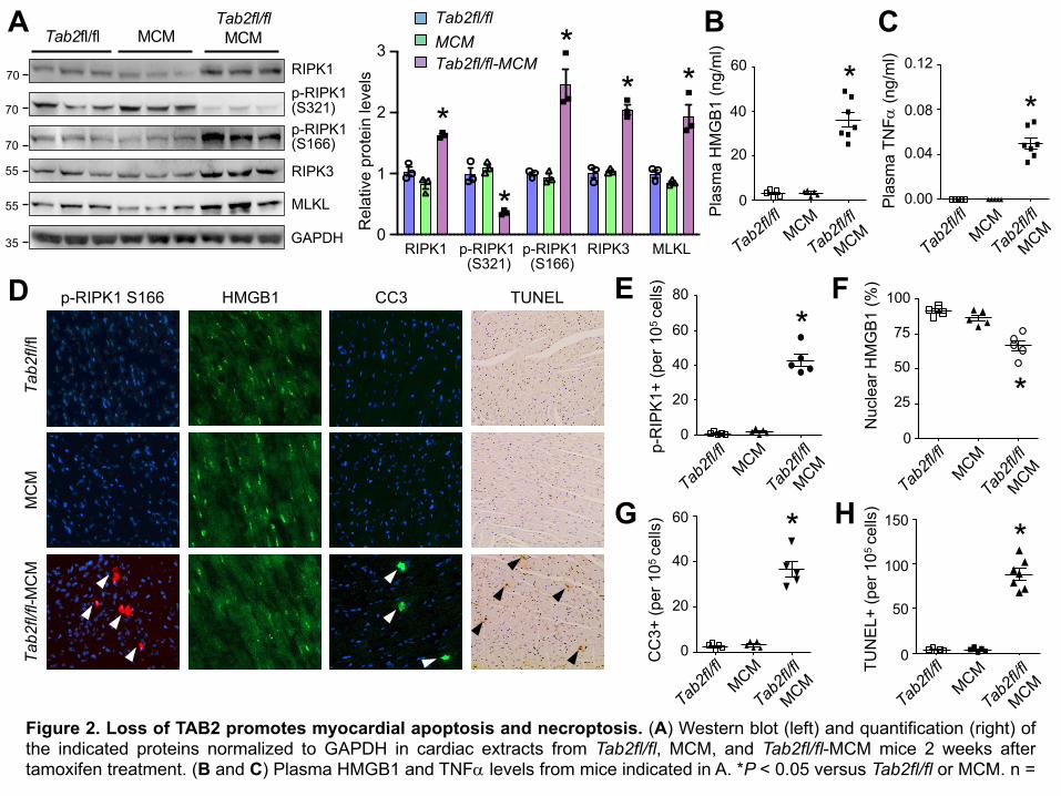
A
GAPDH
RIPK1
MLKL
RIPK3
Tab2fl/fl MCMTab2fl/fl
MCM
p-RIPK1 (S166)
p-RIPK1 (S321)
p-RIPK1 S166D
Tab2fl/
flM
CM
Tab2fl/fl-
MC
M
HMGB1 TUNEL
B
*
Plas
ma
TNFa
(ng/
ml)
0.00
0.04
0.12
0.08
Tab2fl/fl
MCM
Tab2fl/fl
MCM
Plas
ma
HM
GB1
(ng/
ml)
*
0
20
40
60
C
*
E
TUN
EL+
(per
105
cells
)
Tab2fl/fl
MCM
Tab2fl/fl
MCM
Tab2fl/fl
MCM
Tab2fl/fl
MCM
0
50
100
150
CC3
0
20
40
60
80
*
Tab2fl/fl
MCM
Tab2fl/fl
MCM
p-R
IPK1
+ (p
er 1
05 ce
lls)
*
Nuc
lear
HM
GB1
(%)
0
25
50
75
100
Tab2fl/fl
MCMTab2fl/fl
MCM
F
G H*
CC
3+ (p
er 1
05 ce
lls)
0
20
40
60
Tab2fl/fl
MCM
Tab2fl/fl
MCM
35
55
70
55
70
70
RIPK1 MLKLRIPK3p-RIPK1 (S166)
p-RIPK1 (S321)
Rel
ativ
e pr
otei
n le
vels
*
*
*
* *
Tab2fl/flMCMTab2fl/fl-MCM
0
1
2
3
Figure 2. Loss of TAB2 promotes myocardial apoptosis and necroptosis. (A) Western blot (left) and quantification (right) ofthe indicated proteins normalized to GAPDH in cardiac extracts from Tab2fl/fl, MCM, and Tab2fl/fl-MCM mice 2 weeks aftertamoxifen treatment. (B and C) Plasma HMGB1 and TNFa levels from mice indicated in A. *P < 0.05 versus Tab2fl/fl or MCM. n =
5-7. (D) Immunofluorescence staining with the indicated antibodies as well as TUNEL (Terminal deoxynucleotidyl transferasedUTP nick-end labeling) assay in cardiac sections from mice of the indicated genotypes. (E-H) Quantification of phospho-RIPK1Ser166, nuclear HMGB1, cleaved caspase-3 (CC3), and TUNEL positive cells. *P < 0.05 versus Tab2fl/fl or MCM. n = 5-7.Statistical analysis was performed using one-way ANOVA with Tukey’s post hoc test.
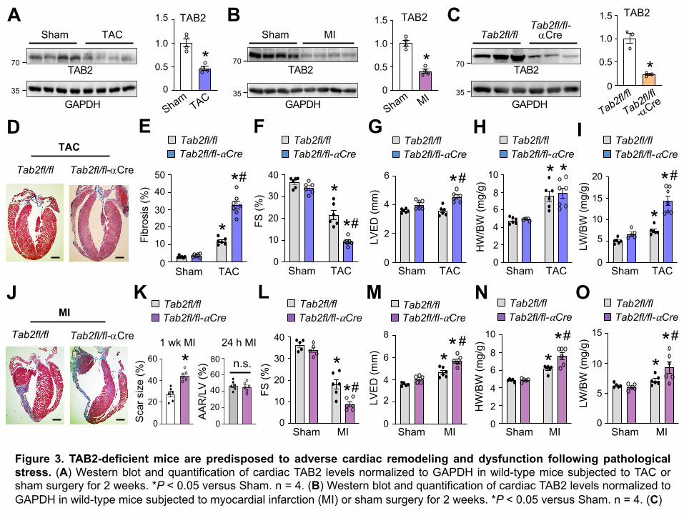
A
D
TAB2
GAPDH
Sham TAC
TAB2
GAPDH
Sham MI Tab2fl/flTab2fl/fl-aCre
TAB2
GAPDH
Sham TAC Sham TAC Sham TAC
Sham MI Sham MI Sham MI Sham MI
HW
/BW
(mg/
g)
LVED
(mm
)
FS (%
)
LW/B
W (m
g/g)
LVED
(mm
)
FS (%
)
B C
F G I
J L NM O
Tab2fl/fl Tab2fl/fl-aCre
Tab2fl/fl Tab2fl/fl-aCre
TAC
MI
Sham TAC
HW
/BW
(mg/
g)*#*
*#*
*#*
*#*
*#*
#**
#** *
LW/B
W (m
g/g)
H
70
35
70
35
70
35
ShamTAC
Sham MITab2fl/fl
Tab2fl/fl
-aCre
* * *
TAB2 TAB2 TAB2
Tab2fl/flTab2fl/fl-⍺Cre
0
2
4
68
10
0
5
15
20
10
Tab2fl/flTab2fl/fl-⍺Cre
0
10
30
20
40
0
4
6
2
Tab2fl/flTab2fl/fl-⍺Cre
Tab2fl/flTab2fl/fl-⍺Cre
0
2
4
6
8
10
0
5
15
10
Tab2fl/flTab2fl/fl-⍺Cre
Tab2fl/flTab2fl/fl-⍺Cre
0
10
30
20
40
Tab2fl/flTab2fl/fl-⍺Cre
Tab2fl/flTab2fl/fl-⍺Cre
0
4
8
2
6
0.5
1.5
0
1.0
0.5
1.5
0
1.0
0.5
1.5
0
1.0
Figure 3. TAB2-deficient mice are predisposed to adverse cardiac remodeling and dysfunction following pathologicalstress. (A) Western blot and quantification of cardiac TAB2 levels normalized to GAPDH in wild-type mice subjected to TAC orsham surgery for 2 weeks. *P < 0.05 versus Sham. n = 4. (B) Western blot and quantification of cardiac TAB2 levels normalized toGAPDH in wild-type mice subjected to myocardial infarction (MI) or sham surgery for 2 weeks. *P < 0.05 versus Sham. n = 4. (C)
Fibr
osis
(%)
0
10
3020
40
50
Tab2fl/flTab2fl/fl-⍺Cre
Sham TAC
*
#*
Scar
siz
e (%
)
0
40
20
60 *
Tab2fl/flTab2fl/fl-⍺Cre
E
K
AAR
/LV
(%)
0
40
20
60
80 n.s.
1 wk MI 24 h MI

Western blot and quantification of cardiac TAB2 levels normalized to GAPDH in Tab2fl/fl and Tab2fl/fl-aMHC-Cre mice at 2months of age. *P < 0.05 versus Tab2fl/fl. n = 3. (D) Histology images of Masson’s trichrome-stained cardiac sections from miceof the indicated genotypes one week after TAC or sham surgery. Scale bar = 1 mm. (E) Quantification of cardiac fibrosis. *P <0.05 versus Sham. #P < 0.05 versus Tab2fl/fl TAC. n = 6-7. (F and G) Echocardiographic measurement of fractional shortening(FS) and left ventricular dimension in end-diastole (LVED). *P < 0.05 versus Sham. #P < 0.05 versus Tab2fl/fl TAC. n = 6-7. (H)Heart weight to body weight ratio (HW/BW) of mice of the indicated genotypes one week after TAC or sham surgery. *P < 0.05versus Sham. (I) Pulmonary edema assessed by lung weight to body weight ratio (LW/BW) of mice of the indicated genotypes.*P < 0.05 versus Sham. #P< 0.05 versus Tab2fl/fl TAC. n = 6-7. (J) Histology images of Masson’s trichrome-stained cardiacsections from mice of the indicated genotypes 1 week after MI or sham surgery. Scale bar = 1 mm. (K) Quantification of infarctscar size of the indicated mice subjected to MI for 1 week (left). Initial area at risk (AAR) normalized to area of the left ventricle(LV) was measured 24 h after MI in the indicated mice. *P < 0.05 versus Tab2fl/fl. n = 6. (L and M) FS and LVED measured byechocardiography. *P < 0.05 versus Sham. #P < 0.05 versus Tab2fl/fl MI. n = 5-6. (N and O) HW/BW and LW/BW ratios of miceof the indicated genotypes one week after MI or sham surgery. *P < 0.05 versus Sham. #P < 0.05 versus Tab2fl/fl MI. n = 5-6.Data were analyzed by Student’s t test for comparisons between 2 groups and by 2-way ANOVA with Tukey’s multiple-comparison test for consideration of both genotypes and treatments.

% C
ell d
eath
(PI p
ositi
ve)
*
*#A
TNFCon
Tab2+/+Tab2-/-
0
20
60
40
80
zVadTNF
Nec1sTNF
TAB2
PARP
GAPDH
HMGB1 (sup.)
Tab2+/+ Tab2-/-zVadNec-1sTNF
---
--+
+-+
-++
25
100
35
70
---
--+
+-+
-++
% C
ell d
eath
(PI p
ositi
ve)
Ad-shScram Ad-shTAB2--
-+
++ TNF
zVad--
-+
++
--
-+
++
Ad-shTAB3
TAB2
PARP
GAPDH
HMGB1 (sup.)
TAB3
Ad-shScram
Ad-shTAB2
Ad-shTAB3
*
*#HG
25
100
35
70
700
20
60
40
80ConTNFTNF+zVad
**#
D
zVadTNF
Nec1sTNF
TNFCon0
20
60
40
80Ad-shScramAd-shTAB2
% C
ell d
eath
(PI p
ositi
ve)
25
100
35
70
zVadNec-1sTNF
---
--+
+-+
-++
---
--+
+-+
-++
βgal βgal
Ad-shScram Ad-shTAB2
TAK1ΔN
**§
##
Cell death (% PI positive)J
0
20
60
40
80ConTNFTNF+zVad
PARP
TAB2
HMGB1(sup.)
GAPDH
TNF--
++
-+
--
++
-+
βgal TAK1ΔNzVad
shScram shScram
TAK1
K
25
100
35
70
70
TAB2
PARP
GAPDH
HMGB1 (sup.)
TAK1ΔN
Ad-shScram Ad-shTAB2
--
++
-+
--
++
-+
βgal TAK1ΔNshTAB2shTAB2
Ad-shScram
Ad-shTAB2
Ad-shTAB3
βgal βgal
Ad-shScram Ad-shTAB2
TAK1ΔN0
TAK1ΔN
1
3
2
4ConTNFTNF+zVad
ConTNFTNF+zVad
**#
zVadTNF
Nec1sTNF
TNFCon
Ad-shScramAd-shTAB2
TNFCon0 zVadTNF
Nec1sTNF
HM
GB1
in s
uper
nata
nt
B C
E F
I
L
Tab2+/+Tab2-/-
*
*#
*
*#
**
# #
2
6
4
0
2
6
4
0
2
6
4
Figure 4. TAB2, but not TAB3, is a key regulator of apoptotic and necroptotic cell deathin cardiomyocytes. (A) Cell death assessed by propidium iodide (PI) staining of Tab2+/+ and
HM
GB1
in s
uper
nata
ntH
MG
B1 in
sup
erna
tant
HMGB1 in supernatant
Tab2-/- MEFs treated with 10 ng/ mL TNFa or vehicle control for 6 h in the presence orabsence of necrostatin-1s (Nec-1s; RIPK1 inhibitor) or zVad-fmk (zVad; pan-caspaseinhibitor). *P < 0.01 versus control; #P< 0.05 versus Tab2-/- TNF. n = 3. (B) Western blottingfor the indicated proteins in Tab2+/+ and Tab2-/- MEFs treated as in A. Sup., culturesupernatant. (C) Quantification of HMGB1 in cell culture supernatant as indicated in A. *P <0.05 versus control; #P< 0.05 versus Tab2-/- TNF. n = 3. (D) Cell death assessed by PIstaining of neonatal cardiomyocytes infected with an adenovirus encoding TAB2 shRNA(shTAB2) or a scrambled sequence (shScram), followed by stimulation with 10 ng/mL TNFa orvehicle control in the presence or absence of Nec-1 or zVad for 6 h. *P < 0.01 versus control;#P < 0.05 versus shTAB2 TNF. n = 4. (E) Western blotting for the indicated proteins in cellstreated as in D. (F) Quantification of HMGB1 in supernatant as indicated in D. *P < 0.05versus control; #P < 0.05 versus shTAB2 TNF. n = 3. (G) Cell death in neonatalcardiomyocytes infected with Ad-shTAB2, Ad-shTAB3, or Ad-shScram, followed by stimulationwith 10 ng/mL TNFa or vehicle control for 6 h, in the presence or absence of Nec-1 or zVad.*P < 0.01 versus control; #P < 0.05 versus shTAB2 TNF. n = 3. (H) Western blotting for theindicated proteins in cells treated as in G. (I) Quantification of HMGB1 in supernatant asindicated in G. *P < 0.05 versus control; #P < 0.05 versus shTAB2 TNF. n = 3. (J) Cell death inneonatal cardiomyocytes infected with Ad-shTAB2 or Ad-shScram along with adenoviralvectors expressing β-galactosidase (βgal) or the constitutively active TAK1 mutant (TAKΔN)and treated as indicated for 6 h. *P < 0.01 versus Con; #P < 0.05 versus βgal in thecorresponding group; §P < 0.05 versus Ad-shTAB2 βgal TNF. n = 3. (K) Western blotting forthe indicated proteins in cells treated as in J. (L) Quantification of HMGB1 in supernatant asindicated in J. *P < 0.05 versus Con; #P < 0.05 versus βgal in the corresponding group. n = 3.Statistical analysis was performed using 2-way ANOVA with Tukey’s multiple-comparison test.
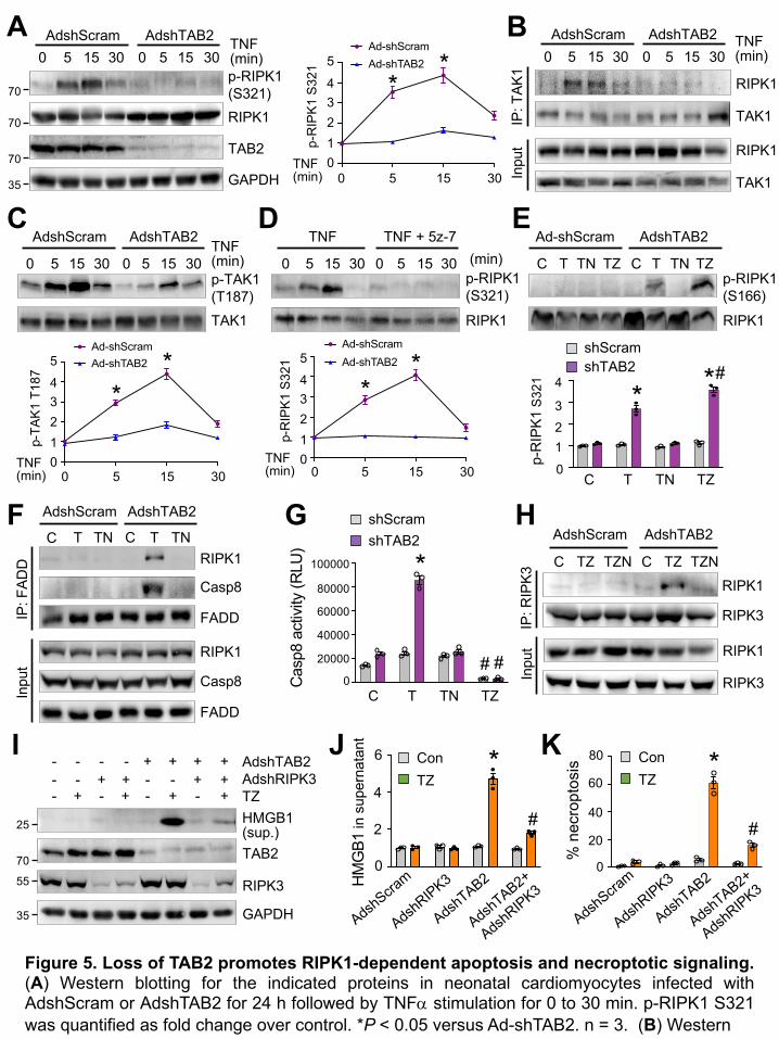
A AdshScram AdshTAB215 3050301550
p-RIPK1 (S321)
TAB2
GAPDH
RIPK170
70
70
35
p-R
IPK1
S32
1
301550TNF (min)
Ad-shScramAd-shTAB2
01234
5
**
B
IP: T
AK1
Inpu
t
RIPK1
TAK1
RIPK1
TAK1
TNF(min)
AdshScram AdshTAB215 3050301550
p-TAK1 (T187)TAK1
AdshScram AdshTAB2
15 3050301550TNF (min)
C
p-TA
K1 T
187
301550TNF (min)
Ad-shScramAd-shTAB2
012345
**
D(min)
RIPK1
TNF TNF + 5z-7
15 3050301550p-RIPK1 (S321)
p-R
IPK1
S32
1
301550TNF (min)
Ad-shScramAd-shTAB2
012345
**
TNTC TZTNTC TZ
Ad-shScram AdshTAB2
RIPK1
p-RIPK1 (S166)
E
*
shScramshTAB2 #*
p-R
IPK1
S321
123
4
0C T TN TZ
F
IP: F
ADD RIPK1
Casp8
FADD
AdshScram
TNT
AdshTAB2
TNTC
Inpu
t
RIPK1
Casp8
FADD
CG
TZTNTC
Cas
p8 a
ctiv
ity (R
LU)
*
#0
20000
60000
40000
100000
80000
shScramshTAB2
#
AdshTAB2AdshRIPK3TZ
---
--+
-+-
-++
+--
+-+
++-
+++
TAB2
RIPK3
GAPDH
HMGB1(sup.)
I
25
70
35
55
% n
ecro
ptos
is *
#
AdshTAB2
AdshR
IPK3
AdshScra
m
AdshT
AB2+
AdshR
IPK30
20
60
40
80 ConTZ
HIP
: RIP
K3 RIPK1
RIPK3
AdshScram
TZNTZC
AdshTAB2
TZNTZC
Inpu
t RIPK1
RIPK3
*
#
AdshTAB2
AdshR
IPK3
AdshScra
m
AdshT
AB2+
AdshR
IPK3
0
ConTZ
HM
GB1
in s
uper
nata
nt K4
2
6J
Figure 5. Loss of TAB2 promotes RIPK1-dependent apoptosis and necroptotic signaling.(A) Western blotting for the indicated proteins in neonatal cardiomyocytes infected withAdshScram or AdshTAB2 for 24 h followed by TNFa stimulation for 0 to 30 min. p-RIPK1 S321was quantified as fold change over control. *P < 0.05 versus Ad-shTAB2. n = 3. (B) Western
TNF(min)

blotting for the indicated proteins after immunoprecipitation (IP) with anti-TAK1 fromneonatal cardiomyocytes infected with AdshScram or AdshTAB2 followed by TNFastimulation for 0 to 30 min. For the IP data here and below, at least 3 experiments wereconducted. (C) Western blotting and quantification of phospho-TAK1 Thr187 in neonatalcardiomyocytes infected with the AdshScram or AdshTAB2 followed by TNFa stimulationfor 0 to 30 min. *P < 0.05 versus Ad-shTAB2. n = 3. (D) Western blotting and quantificationof phospho-RIPK1 Ser321 in neonatal cardiomyocytes infected with the AdshScram orAdshTAB2 followed by stimulation with TNFa in the presence or absence of the TAK1inhibitor 5z-7-oxozeaenol (5z-7). *P < 0.05 versus Ad-shTAB2. n = 3. (E) Western blottingand quantification of phospho-RIPK1 Ser166 in neonatal cardiomyocytes infected with theAdshScram or AdshTAB2 followed by treatment with vehicle control (C) or TNFa (T) in thepresence and absence of necrostatin-1s (N) or zVad-fmk (Z) for 2 h. *P < 0.05 versus Con;#P < 0.05 versus shTAB2 T. n = 3. (F) Western blotting for the indicated proteins after IPwith anti-FADD from neonatal cardiomyocytes infected with AdshScram or AdshTAB2followed by treatment with vehicle control (C) or TNFa (T) in the presence and absence ofnecrostatin-1s (N) for 2 h. (G) Caspase-8 activity in neonatal cardiomyocytes infected withAdshScram or AdshTAB2 for 24 h followed by TNFa stimulation for 6 h in the presenceand absence of necrostatin-1s (N) or zVad-fmk (Z). *P < 0.01 versus control; #P < 0.05versus shTAB2 TNF. n = 3. (H) Western blotting for the indicated proteins after IP with ananti-RIPK3 antibody from neonatal cardiomyocytes infected with AdshScram or AdshTAB2and then treated with vehicle control (C) or TNFa and zVad (TZ) in the presence orabsence of necrostatin-1s (N) for 2 h. (I) Western blotting for the indicated proteins inneonatal cardiomyocytes infected with the indicated adenoviral vectors for 24 h followed bytreatment with vehicle control or TNFa and zVad-fmk (TZ) for 6 h. (J) Quantification ofHMGB1 in supernatant as indicated in I. *P < 0.05 versus Con; #P < 0.05 versus Ad-shTAB2 TZ. n = 3. (K) Necroptosis in cells treated as indicated in I. *P < 0.01 versus Con;#P < 0.05 versus Ad-shTAB2 TZ. n = 3. Statistical analysis was performed using 2-wayANOVA with Tukey’s multiple-comparison test.

Ripk1-WT;
Ripk1-WT;
TAB2
RIPK1
GAPDH
Tab2fl/
fl
A
Tab2fl/
fl-MCM
Tab2fl/fl Tab2fl/fl-MCMRipk1-WT
Fibr
osis
(%) *
#
D
Tab2fl/fl Tab2fl/flMCM
fl/fl fl/flMCM
Ripk1-WT
*
#
*
#TU
NEL
+ (p
er 1
05 c
ells
)
Plas
ma
HM
GB1
(ng/
ml)
HW
/BW
(mg/
g)
LVED
(mm
)
FS (%
)
Tab2fl/fl Tab2fl/flMCM
Tab2fl/fl Tab2fl/flMCM
Tab2fl/fl Tab2fl/flMCM
*
# * #
*#
*
B
GF H I
C
0
50
100
150
0
20
40
60
Tab2fl/fl Tab2fl/fl-MCMRipk1-KARipk1-WT Ripk1-KA
Tab2fl/fl Tab2fl/fl-MCMRipk1-WT
Tab2fl/fl Tab2fl/fl-MCMRipk1-KARipk1-WT Ripk1-KA
E
Ripk1-KA;
Ripk1-KA;
Tab2fl/
fl
Tab2fl/
fl-MCM
Ripk1-KA
70
70
35
fl/fl fl/flMCM
fl/fl fl/flMCM
Ripk1-WT Ripk1-KA
fl/fl fl/flMCM
0
10
30
20
40
Ripk1-WTRipk1-K45A
0123456
0
2
4
6
8
0
10
30
20
Ripk1-WTRipk1-K45A
Ripk1-WTRipk1-K45A
Ripk1-WTRipk1-K45A
Figure 6. Genetic inactivation of RIPK1 rescues pathological cardiac remodeling and dysfunction in TAB2-deficientmice. (A) Western blot and quantification of cardiac TAB2 expression from mice of the indicated genotypes 2 weeks aftertamoxifen treatment. *P < 0.05 versus Tab2fl/fl. n = 3. (B and C) FS and LVED measured by echocardiography from mice of theindicated genotypes 2 weeks after tamoxifen treatment. *P < 0.01 versus Tab2fl/fl; #P < 0.05 versus Ripk1-WT;Tab2fl/fl-MCM. n= 5-7. (D and E) low and high magnification images of Masson’s trichrome-stained cardiac sections from mice indicated in A-C.
TAB2
pro
tein
0.5
1.5
0
1.0
Tab2fl/fl Tab2fl/flMCM
Ripk1-WTRipk1-K45A
* *

Scale bars: 1 mm in panel D; 50 µm in panel E. (F) Myocardial fibrosis quantified by MetaMorph software. *P < 0.01 versusTab2fl/fl; #P < 0.05 versus Ripk1-WT;Tab2fl/fl-MCM. n = 5-7. (G) Heart weight to body weight ratio (HW/BW) in mice of theindicated genotypes. *P < 0.05 versus Tab2fl/fl; #P < 0.05 versus Ripk1-WT;Tab2fl/fl-MCM. n = 5-7. (H) TUNEL-positivemyocytes in cardiac sections from mice of the indicated genotypes. *P < 0.05 versus Tab2fl/fl; #P < 0.05 versus Ripk1-WT;Tab2fl/fl-MCM. n = 5-7. (I) Plasma HMGB1 levels from mice of the indicated genotypes. *P < 0.01 versus Tab2fl/fl; #P <0.05 versus Ripk1-WT;Tab2fl/fl-MCM. n = 5-7. Statistical analysis was performed using 2-way ANOVA with Tukey’s multiple-comparison test.
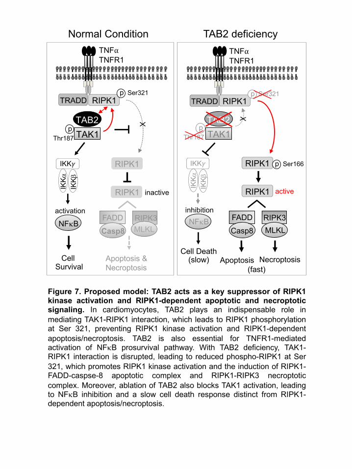
FADD
Necroptosis
RIPK3Casp8
NFkB
IKK𝛾
IKKb
IKKa
FADD
Apoptosis &Necroptosis
TNF⍺TNFR1
TAB2
RIPK3Casp8
NFkB
IKK𝛾
IKKb
IKKa
Cell Survival
Normal Condition TAB2 deficiencyTNF⍺TNFR1
TAB2
Apoptosis
MLKLMLKL
TRADD
TAK1p
Thr187p
Thr187 TAK1
p Ser321TRADD
activeinactive
activation inhibition
Cell Death(slow)
(fast)
RIPK1
RIPK1 p Ser166
RIPK1RIPK1
RIPK1
RIPK1p Ser321
Figure 7. Proposed model: TAB2 acts as a key suppressor of RIPK1kinase activation and RIPK1-dependent apoptotic and necroptoticsignaling. In cardiomyocytes, TAB2 plays an indispensable role inmediating TAK1-RIPK1 interaction, which leads to RIPK1 phosphorylationat Ser 321, preventing RIPK1 kinase activation and RIPK1-dependentapoptosis/necroptosis. TAB2 is also essential for TNFR1-mediatedactivation of NFkB prosurvival pathway. With TAB2 deficiency, TAK1-RIPK1 interaction is disrupted, leading to reduced phospho-RIPK1 at Ser321, which promotes RIPK1 kinase activation and the induction of RIPK1-FADD-caspse-8 apoptotic complex and RIPK1-RIPK3 necroptoticcomplex. Moreover, ablation of TAB2 also blocks TAK1 activation, leadingto NFkB inhibition and a slow cell death response distinct from RIPK1-dependent apoptosis/necroptosis.