Dilated cardiomyopathy is associated with reduced expression of the cardiac sodium channel Scn5a
12
Dilated cardiomyopathy is associated with reduced expression of the cardiac sodium channel Scn5a Michael Hesse a , Colleen S. Kondo b , Robert B. Clark b , Lin Su a , Frances L. Allen a , Colleen T.M. Geary-Joo a , Stanley Kunnathu b , David L. Severson c , Anders Nygren d , Wayne R. Giles b , James C. Cross a, ⁎ a Department of Comparative Biology and Experimental Medicine, Faculty of Veterinary Medicine, University of Calgary, Calgary, AB, Canada T2N 4N1 b Faculty of Kinesiology, University of Calgary, Calgary, AB, Canada T2N 4N1 c Department of Pharmacology and Therapeutics, University of Calgary, Calgary, AB, Canada T2N 4N1 d Centre for Bioengineering Research and Education, Electrical and Computer Engineering, University of Calgary, Calgary, Canada T2N 4N1 Received 22 December 2006; received in revised form 16 March 2007; accepted 16 April 2007 Time for primary review 27 days Available online 21 April 2007 Abstract Objective: Dilated cardiomyopathy (DCM) leads to dilation of the cardiac chambers and congestive heart failure. Recent reports have associated mutations in the SCN5A gene, which codes for the major cardiac sodium channel Nav1.5, with DCM. Although DCM is the most common form of cardiomyopathy, no animal studies have established this functional connection. Methods and results: We have produced transgenic mice that ectopically express the transcriptional repressor Snail in heart. These animals display severe DCM, ECG abnormalities, conduction defects, revealed by voltage-sensitive dye imaging, and significantly reduced voltage- gated sodium current as measured by patch clamping. There is a concomitant decrease in expression of the major cardiac sodium channel gene Scn5a, which we show by gene reporter assays and electrophoretic mobility shift assays is a direct target of Snail. Conclusions: Our findings indicate that a decrease in Scn5a expression and significant reduction in sodium current can result in DCM, and support the hypothesis that some mutations in the human SCN5A gene can lead to DCM. © 2007 European Society of Cardiology. Published by Elsevier B.V. All rights reserved. Keywords: Cardiomyopathy; Gene expression; Heart failure; Transgenic animal models; Na-channel This article is referred to in the Editorial by Schulze- Bahr (pages 455– 456) in this issue. 1. Introduction Dilated cardiomyopathy (DCM) is the most common form of cardiomyopathy and is the leading indication for heart transplantations [1]. The underlying cause of DCM is an initial weakening of the myocardium followed by marked adaptive dilation as a consequence of the compromised pump function. More than 20 genes are associated with DCM in humans, including structural proteins of the sarcomere that transmit the contractile force and proteins associated with Ca 2+ homeostasis [2]. Recently, mutations in ion channel genes have also been associated with DCM in humans. These include ABCC9, the gene encoding the subunit SUR2A of cardiac ATP-sensitive potassium channel [3] and SCN5A, which encodes the alpha subunit of the major Na + channel in the heart, Nav1.5. Previously, SCN5A has been implicated in conduction disturbances and other electrophysiological phenotypes [4]. Gain-of-function muta- tions in SCN5A can cause a variant of the long QT syndrome Cardiovascular Research 75 (2007) 498 – 509 www.elsevier.com/locate/cardiores ⁎ Corresponding author. Department of Comparative Biology and Experi- mental Medicine, Faculty of Veterinary Medicine, University of Calgary, HSC Room 2279, 3330 Hospital Drive, N.W., Calgary, Alberta, Canada T2N 4N1. Tel.: +1 403 220 6876; fax: +1 403 270 0737. E-mail address: [email protected] (J.C. Cross). 0008-6363/$ - see front matter © 2007 European Society of Cardiology. Published by Elsevier B.V. All rights reserved. doi:10.1016/j.cardiores.2007.04.009 at Serials Acq (Law) on August 13, 2013 http://cardiovascres.oxfordjournals.org/ Downloaded from
Transcript of Dilated cardiomyopathy is associated with reduced expression of the cardiac sodium channel Scn5a

75 (2007) 498–509www.elsevier.com/locate/cardiores
Cardiovascular Research
http://cardioD
ownloaded from
Dilated cardiomyopathy is associated with reduced expression of thecardiac sodium channel Scn5a
Michael Hessea, Colleen S. Kondob, Robert B. Clarkb, Lin Sua, Frances L. Allena,Colleen T.M. Geary-Jooa, Stanley Kunnathub, David L. Seversonc,
Anders Nygrend, Wayne R. Gilesb, James C. Crossa,⁎
a Department of Comparative Biology and Experimental Medicine, Faculty of Veterinary Medicine, University of Calgary, Calgary, AB, Canada T2N 4N1b Faculty of Kinesiology, University of Calgary, Calgary, AB, Canada T2N 4N1
c Department of Pharmacology and Therapeutics, University of Calgary, Calgary, AB, Canada T2N 4N1d Centre for Bioengineering Research and Education, Electrical and Computer Engineering, University of Calgary, Calgary, Canada T2N 4N1
Received 22 December 2006; received in revised form 16 March 2007; accepted 16 April 2007
Time for primary review 27 daysAvailable online 21 April 2007
at Serials Acq (L
aw)
vascres.oxfordjournals.org/
Abstract
Objective: Dilated cardiomyopathy (DCM) leads to dilation of the cardiac chambers and congestive heart failure. Recent reports haveassociated mutations in the SCN5A gene, which codes for the major cardiac sodium channel Nav1.5, with DCM. Although DCM is the mostcommon form of cardiomyopathy, no animal studies have established this functional connection.Methods and results: We have produced transgenic mice that ectopically express the transcriptional repressor Snail in heart. These animalsdisplay severe DCM, ECG abnormalities, conduction defects, revealed by voltage-sensitive dye imaging, and significantly reduced voltage-gated sodium current as measured by patch clamping. There is a concomitant decrease in expression of the major cardiac sodium channelgene Scn5a, which we show by gene reporter assays and electrophoretic mobility shift assays is a direct target of Snail.Conclusions: Our findings indicate that a decrease in Scn5a expression and significant reduction in sodium current can result in DCM, andsupport the hypothesis that some mutations in the human SCN5A gene can lead to DCM.© 2007 European Society of Cardiology. Published by Elsevier B.V. All rights reserved.
on Au
gust 13, 20Keywords: Cardiomyopathy; Gene expression; Heart failure; Transgenic animal models; Na-channel
13
This article is referred to in the Editorial by Schulze-Bahr (pages 455–456) in this issue.1. Introduction
Dilated cardiomyopathy (DCM) is the most commonform of cardiomyopathy and is the leading indication forheart transplantations [1]. The underlying cause of DCM is
⁎ Corresponding author. Department of Comparative Biology and Experi-mental Medicine, Faculty of Veterinary Medicine, University of Calgary, HSCRoom 2279, 3330 Hospital Drive, N.W., Calgary, Alberta, Canada T2N 4N1.Tel.: +1 403 220 6876; fax: +1 403 270 0737.
E-mail address: [email protected] (J.C. Cross).
0008-6363/$ - see front matter © 2007 European Society of Cardiology. Publishdoi:10.1016/j.cardiores.2007.04.009
an initial weakening of the myocardium followed by markedadaptive dilation as a consequence of the compromisedpump function. More than 20 genes are associated withDCM in humans, including structural proteins of thesarcomere that transmit the contractile force and proteinsassociated with Ca2+ homeostasis [2]. Recently, mutations inion channel genes have also been associated with DCM inhumans. These include ABCC9, the gene encoding thesubunit SUR2A of cardiac ATP-sensitive potassium channel[3] and SCN5A, which encodes the alpha subunit of themajor Na+ channel in the heart, Nav1.5. Previously, SCN5Ahas been implicated in conduction disturbances and otherelectrophysiological phenotypes [4]. Gain-of-function muta-tions in SCN5A can cause a variant of the long QT syndrome
ed by Elsevier B.V. All rights reserved.

499M. Hesse et al. / Cardiovascular Research 75 (2007) 498–509
at Serials Acq (L
aw) on A
ugust 13, 2013http://cardiovascres.oxfordjournals.org/
Dow
nloaded from
due to abnormal myocardial repolarization. Loss-of-functionSCN5A mutations are associated with Brugada syndrome.These patients have a high risk for developing arrhythmiaand sudden death, although their hearts are reported to bestructurally normal [5]. Some recent reports have alsoimplicated SCN5A mutations in DCM [6,7] but the data arecontroversial because DCM may be secondary to long-lasting atrial arrhythmia [8] or tachycardia-induced cardio-myopathy [9].
The Snail family of zinc finger proteins are transcriptionalrepressors that regulate genes associated with cell–celladhesion, epithelial to mesenchymal transition [10], and thetransition to endoreduplication in trophoblast cells of theplacenta, megakaryocytes and drosophila tissues [11]. Snailis expressed in the mouse embryonic heart during earlyintrauterine development [12]. In the present study, wediscovered that transgenic mice expressing Snail in thepostnatal heart show downregulation of the Scn5a Na+
channel in the heart which in turn is associated with markedDCM in the absence of any arrhythmias.
2. Experimental procedures
2.1. Mice and genotyping
All experimental protocols were approved by theUniversity of Calgary Animal Welfare Committee andfollowed the guidelines of the Canadian Council on AnimalCare and conformed with the Guide for the Care and Use ofLaboratory Animals published by the US National Institutesof Health (NIH Publication No. 85-23, revised 1996). Thegeneration of MHC-Snail mice expressing the Snail cDNAunder the control of the myosin heavy chain α promoter (TG[Myh6-Snai1]B10JCC) is described in SupplementaryMaterial and Methods. They were genotyped by PCRusing the primers 5′-CTTCCAGCCCTCTCTTTCTC-3′,5′-CGTTTTCAGTTTCCGCAGTG-3′, amplifying endoge-nous Myh6 (450 bp) and 5′-CAGGTCGTGCAGACA-CAAGG-3′, representing Snail nucleotides 549–568,amplifying an 820 bp fragment of the Snail transgene withprimer 5′-CTTCCAGCCCTCTCTTTCTC-3′. The PCR para-meters were 30 s at 94 °C, 30 s at 61 °C and 60 s at 72 °C for 30cycles. Hemi- and homozygous MHC-Snail mice weregenotyped by Southern blotting with a 1.67 kb alpha-MHCprobe to a 6 kb endogenous or 7.4 kb transgenic BamHIfragment of genomic DNA.
2.2. Histology and immunofluorescence staining
Hearts were dissected, washed in PBS and frozen in liquidnitrogen. Cryosections were fixed in acetone for 10 min at−20 °C. Sections were incubated with a 1:100 dilution ofprimary antibody against Cx43 (C-6219, Sigma) for 1 hourat RT, washed with PBS and incubated with secondaryantibody anti-rabbit Cy3-conjugated (Chemicon Internation-al) for 30 min, washed in PBS and mounted. Fibrosis in
cardiac sections was determined by Masson Trichrome stain(HT 15, Sigma).
2.3. Electron microscopy
Left ventricles were dissected from hearts, minced andfixed in 2% EM-grade glutaraldehyde (Sigma), 2% PFA in0.2 M sodium cacodylate (pH 7.4; Sigma) overnight at 4°C,and post-fixed in 1% osmium tetroxide (EM Sciences) in0.2 M sodium cacodylate (pH 7.4) for 2h at 4 °C. Tissue wasdehydrated in increasing concentrations of ethanol up to70%, treated in 2% uranyl acetate (ProSciTech) in 70%ethanol, and further dehydrated in increasing ethanolconcentrations (70–100%). Tissue was treated with propyl-ene oxide (EM Sciences) and resin embedded (EMBED 812kit, EM Sciences).
2.4. Cell cultures and transfection
P19 mouse teratocarcinoma cells were cultured in αMEMplus 10% fetal bovine serum. Transient transfections werecarried out using Lipofectamine (Invitrogen). Cells wereplated at a density of 1×10⁵ cells per well in 6 well dishes24 h prior to addition of complexes containing 5 μl oflipofectamine and 4.2 μg of DNA (200 ng of pβActin-LacZ,2 μg of P-+255, containing luciferase under control of ahuman Scn5a promoter segment from −261 to +255 [13],and the indicated amount of either pREP-mSna or pREP-mSnaΔSNAG expression vector [11]. Fresh medium wasadded 3h post-transfection, and cells were harvested after anadditional 45 h. Luminescence levels were measured with aluminometer using a kit (Promega) and were normalized toβ-galactosidase activity. Values are reported relative toempty pGL3 vector and represent means±SE for duplicatetransfected dishes in 3 experiments.
2.5. Northern blot hybridization
RNA was extracted from whole hearts using Trizol(Invitrogen). RNAs were electrophoresed in MOPS/formal-dehyde gels, capillary blotted, and UV-crosslinked ontonylon membranes (GeneScreen Plus, DuPont). Membraneswere hybridized with random primed 32P-labeled probes inhybridization buffer [14] at 60 °C overnight. A probe formouse Scn5a was amplified by PCR and consisted of bp2823 to 3253 of the Scn5a cDNA [15]. The probe forGAPDH has been described previously [15]. The membraneswere washed in 20 mM sodium phosphate buffer (pH 7.2)containing 1% sodium dodecyl sulfate at 65 °C and exposedto X-ray film.
2.6. EMSA
Electrophoretic mobility shift assay was performed asdescribed previously [11] using the oligonucleotides de-scribed in supplementary experimental procedures.
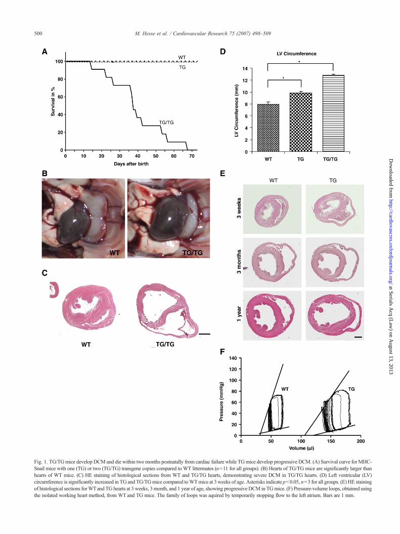
Fig. 1. TG/TGmice develop DCM and die within twomonths postnatally from cardiac failure while TGmice develop progressive DCM. (A) Survival curve forMHC-Snail mice with one (TG) or two (TG/TG) transgene copies compared to WT littermates (n=11 for all groups). (B) Hearts of TG/TG mice are significantly larger thanhearts of WT mice. (C) HE staining of histological sections from WT and TG/TG hearts, demonstrating severe DCM in TG/TG hearts. (D) Left ventricular (LV)circumference is significantly increased in TG and TG/TGmice compared toWTmice at 3 weeks of age. Asterisks indicate pb0.05, n=3 for all groups. (E) HE stainingof histological sections forWTand TG hearts at 3 weeks, 3 month, and 1 year of age, showing progressive DCM in TGmice. (F) Pressure-volume loops, obtained usingthe isolated working heart method, from WT and TG mice. The family of loops was aquired by temporarily stopping flow to the left atrium. Bars are 1 mm.
500 M. Hesse et al. / Cardiovascular Research 75 (2007) 498–509
at Serials Acq (L
aw) on A
ugust 13, 2013http://cardiovascres.oxfordjournals.org/
Dow
nloaded from

Table 1Echocardiography of 3 and 5 weeks old WT, TG and TG/TG mice
3 weeks 5 weeks
WT TG TG/TG WT TG TG/TG
IVSd (mm) 0.62±0.03 0.65±0.06 0.59±0.08 0.67±0.04 0.79±0.07 0.66±0.08IVSs (mm) 0.82±0.06 0.84±0.08 0.77±0.08 0.87±0.04 0.95±0.08 0.73±0.01LVPWd (mm) 0.71±0.04 0.74±0.06 0.67±0.04 0.83±0.04 0.76±0.05 0.69±0.13LVPWs (mm) 0.87±0.04 0.95±0.07 0.74±0.06 1.01±0.06 0.97±0.06 0.90±0.10LVIDd (mm) 3.32±0.15 3.92±0.11 pb0.05 4.19±0.38 pb0.05 3.63±0.11 4.54±0.19 pb0.05 5.32±0.22 pb0.05LVIDs (mm) 2.45±0.08 2.90±0.10 pb0.05 3.35±0.28 pb0.05 2.65±0.08 3.51±0.22 pb0.05 4.54±0.07 pb0.05LVvold 48.06±3.41 74.16±5.91 pb0.05 87.26±16.49 pb0.05 59.31±4.34 101.92±8.96 pb0.05 148.69±13.65 pb0.05LVvols 22.95±1.85 38.71±2.84 pb0.05 49.99±7.66 pb0.05 29.09±1.88 60.59±7.40 pb0.05 99.24±4.08 pb0.05FS (%) 26.18±1.41 23.52±1.97 19.72±2.66 pb0.05 25.27±1.24 21.02±2.68 15.58±2.66 pb0.05
IVSd,s: Interventricular septum thickness measured at diastole or systole, respectively; LVPWd,s: Left ventricle posterior wall thickness at diastole or systole,respectively; LVIDd,s: Left ventricle internal diameter at diastole or systole, respectively; LVvold,s: Left ventricle volume at diastole or systole, respectively; FS:fractional shortening. N=10 for WT, n=9 for TG and n=5 for TG/TG.
501M. Hesse et al. / Cardiovascular Research 75 (2007) 498–509
http://cardiovD
ownloaded from
2.7. RT-PCR
Reverse Transcriptase PCR was performed as previouslydescribed [16], using primers and conditions for Scn5a,Scn1b, and Scn2b as described in a previous paper [15].
Fig. 2. TGmice exhibit slowed ventricular conduction. (A) Representative segments (0.expanded time scale. ‘P’wave, and ‘RR’ and ‘QRS’ intervals are indicated. ‘PR’ interval iintervals from groups of 7 WT, 14 TG and 8 TG/TG mice. ⁎⁎ indicates pb0.01, and ⁎
voltage-sensitive dyemapping during spontaneous sinus rhythm for aWTand aTGheartcolored purple depolarize much later in the activation sequence. The isochrones (contouspaced considerably closer together than in the WT heart, indicating significantly slowe
2.8. Assessment of cardiac function by echocardiography
Echocardiograms (M-mode measurements) were obtainedfrom conscious mice to assess systolic function, as pre-viously described [17].
75 s) of Lead I ECG recordings fromWT, TG and TG/TGmice. (B) ECGs on ans frombeginning of Pwave to beginning of QRS complex. (C) Summarized ECG⁎⁎ indicates pb0.001, compared with WT. (D) Activation maps acquired using. Points colored red represent areas on the ventricle first to depolarize, while pointsrs) shown are drawn at 1 ms intervals. Note that isochrones in the TG heart arer conduction of the waves of cardiac depolarization in the ventricles.
at Serials Acq (L
aw) on A
ugust 13, 2013ascres.oxfordjournals.org/

Fig. 3. Intercellular coupling of myocytes is not affected in TG mice. (A) Immunofluorescence staining for Cx43 expression in the ventricle myocardium of WTand TG mice. Cx43 stained the intercalated discs of cardiomyocytes and its distribution or intensity does not differ between WT and TG hearts. Bar is 50 μm.(B) Electronmicrographs of WT and TG cardiomyocytes revealed no differences in intercalated disc morphology or structure. Bar is 500 nm. (C) Mason-Trichrome staining of histological sections from WT and TG hearts showed no significant increase in fibrosis in TG hearts. Bar is 200 μm.
502 M. Hesse et al. / Cardiovascular Research 75 (2007) 498–509
at Serials Acq (L
aw) on A
ugust 13, 2013http://cardiovascres.oxfordjournals.org/
Dow
nloaded from
2.9. Electrocardiogram (ECG) measurements
ECGswere collected from constrained, conscious 3–4weekoldWT, single gene copy (TG) and double gene copy (TG/TG)mice. Each animal was placed in a small apparatus (QRSPhenotyping Inc., Calgary, Canada) that was designed so thatits feet made contact with gel electrode pads. This allowedcollection of Lead I ECGs. Characteristic ECG waves and
intervals were identified,measured, and tabulated using a semi-automated pattern recognition program (QRS PhenotypingInc.).
2.10. Isolated, perfused working hearts
Mouse hearts were isolated, perfused and measured asdescribed previously [18].

Fig. 4. TG ventricular myocyte action potential have decreased maximum upstroke rate and reduced overshoot. (A) Action potential from WT myocyte.Stimulation rate was 1 Hz. Inset shows the upstroke phase of the action potential on an expanded time scale. Vertical and horizontal bars indicate 20 mVand 3 ms,respectively. The solid straight line indicates dV / dtmax, which was 206 V/s. (B) Action potential from TG myocyte. dV / dtmax was 90 V/s. (C) Mean (±s.e.m.)resting membrane potential (RMP) and peak of action potential, from a group of 11WTand 14 TGmyocytes. (D) Mean dVdtmax and dV / dtmin of action potentialsfrom groups of WT and TG myocytes. ⁎⁎⁎ indicates pb0.0001 compared with WT (Student's t-test). p=0.0373 for dV / dtmin.
503M. Hesse et al. / Cardiovascular Research 75 (2007) 498–509
at Serials Acq (L
aw) on A
ugust 13, 2013http://cardiovascres.oxfordjournals.org/
Dow
nloaded from
2.10.1. Voltage-sensitive dye imagingVoltage-sensitive dye imaging and data processing was
performed as described previously [19,20].
2.11. Cardiomyocyte isolation and electrophysiology
Left ventricular myocytes were isolated from 3–4 weekold WT and TG mice, using methods described previously[21]. Whole-cell patch clamp recordings of action potentialsfrom isolated WT and TG myocytes were made as describedelsewhere [21]. Voltage dependent Na+ and K+ currents wererecorded as described in supplementary methods.
2.12. Statistics
Averaged values are expressed as mean±s.e.m. Statisticallysignificant differences between mean values were determinedwith either Student's t-test or a one-way ANOVA test, asappropriate. A p value of b0.05 was considered to indicatesignificance.
3. Results
3.1. MHC-Snail transgenic mice suffer from severe DCMand die from cardiac failure
We have made transgenic mice that express the Snailtranscription factor in the heart (Supplementary materials
and methods). This expression is driven by the myosin heavychain alpha gene promoter which turns on shortly after birth[22]. Hemizygous TG mice are viable, fertile, have a normallifespan and display no obvious phenotype. However, whenTG mice were intercrossed to obtain double-transgenic mice(TG/TG), all of them died by 2 months of age and 50% weredead within 36 days (Fig. 1A). Before death, TG/TG micewere smaller than their littermates, had laboured breathingand roughened fur, and were cyanotic. At necropsy, theirhearts were enlargedwith dilation of the ventricular chambers(Fig. 1B). Ascites was observed in several cases. Histologicalanalysis of mice sacrificed at 3 weeks of age revealed dilationand thinning of the ventricular walls (Fig. 1C). Left ventriclewall thickness (LVWT), measured post mortem, was reducedin TG/TGmice (0.48±0.06 mm) compared to wildtype (WT)(1.00±0.02 mm) (p=0.0013), while the left ventriclecircumference was significantly increased (Fig. 1D).
3.2. TG and TG/TG-mice develop a progressive DCM
The strong phenotype in TG/TG mice prompted us tomore closely examine hemizygous TG mice. The leftventricle circumference was significantly increased at3 weeks of age (Fig. 1D) and this dilation became moreprominent at both 3 months and 1 year of age (Fig. 1E).Echocardiography showed that left ventricular diameter andvolumewas significantly greater in both TG and TG/TGmiceat 3 and 5 weeks, while fractional shortening, as an indicator

Fig. 5. The time- and voltage-dependent Na+ current is significantly reduced inTG hearts. (A) Example of a family of Na+ currents from a TG myocyte, in140 mM [Na+]o and 5 mM [Na+]i at 15 °C. Cell was held at −120 mV, andstepped to between−65mVand+45mV, in 10mVincrements. Cell capacitancewas 109.6 pF. (B) Current–voltage relations for WT and TG myocytes. [Na+]owas 10 mM for WT and 140 mM for TG myocytes. [Na+]i was 5 mM for bothgroups.N=8 for both groups. Continuous lines are best-fit Boltzmann functions.ForWT: Vh=−47.8 mV, Sh=5.72 mV. For TG: Vh=−32.9 mV, Sh=8.15 mV.(C) Na+ currents at −25 mV from a TG ventricular myocyte at 140 mM (solidline) and 10 mM (dashed line) [Na+]o. The decrease in current by a factor ofabout 10 in 10 mM [Na+]o was a necessary experimental manoeuvre to ensurereliable voltage clamp condition.
504 M. Hesse et al. / Cardiovascular Research 75 (2007) 498–509
at Serials Acq (L
aw) on A
ugust 13, 2013http://cardiovascres.oxfordjournals.org/
Dow
nloaded from
of cardiac performance, was unchanged in TG though wasdecreased in TG/TG mice (Table 1). The isolated workingheart preparation was used to obtain a more detailedassessment of cardiac mechanics (Fig. 1F). Left ventricularvolume increased almost two-fold in TG hearts compared toWT. Contractility, as determined by the slope of the end-systolic pressure-volume relation (ESPVR), was reduced bymore than 50% (1.5188±0.968 (n=5) vs. 3.383±0.0681(n=6), pb0.05). However, stroke volume (width of the loop)was unaffected. This was confirmed by the unchangedfractional shortening and normal lifespan in TG mice. Therightward volume shift and decreased ESPVR slope aretypical characteristics of DCM. Taken together, the data
strongly suggested that the ectopic expression of Snail in theheart lead to progressive DCM, even in hemizygous TGmice.
3.3. TG mice have slowed cardiac conduction
DCM can cause conduction delays and arrhythmias. Bothtypes of abnormalities can be detected using electrocardiogram(ECG) analysis. In these studies there was relatively littledifference in the heart rate (RR interval) amongst the three typesof mice. However the QRS complex was longer in TG and TG/TG mice compared to WT (Fig. 2A–C). In addition, the PRinterval, which is a measure of atrio-ventricular conductiontime, was also longer in transgenic animals. Notably, noarrhythmias were detected during any of the recordings of TGor TG/TG mice. This is in accordance with the normal lifespanof TG mice and the fact that TG/TG mice die from progressivecardiac failure and not sudden death.
To assess ventricular activation and conduction in greaterdetail, the pattern and velocity of conduction in the leftventricle was measured. This was done using a voltage-sensitive dye in Langendorff perfused hearts of two monthold TG mice. Conduction was slower in TG hearts comparedto WT (activation isochrones are more closely spaced)(Fig. 2D). Specifically the 10–90% sinus rhythm activationtime was more than doubled from 4.62 ms in WT (n=6) to11.55 ms in TG hearts (n=6) (p=0.02). This increase inactivation time is consistent with the broadening of the QRScomplex in the ECG. Together, these data demonstrate thatventricular conduction in TG and TG/TG hearts was signi-ficantly slowed.
3.4. The intercellular coupling of myocytes is not affectedin TG mice
The observed conduction abnormalities could have beencaused by microanatomical changes in the conduction system,compromised intercellular coupling, or an intrinsic defect inventricular myocytes. It is known that mis-expression ofconnexins can impair the cardiac conduction system, causingconduction abnormalities and arrhythmia [23]. Real-Time PCRfor Cx40, 43, and 45, the three connexins expressed in themouse heart, showed that their mRNA expression levels werenot altered in TG mice (Supplementary Table 1). Staining withan antibody against Cx43 showed no changes in expressionwithin intercalated discs or myocardium (Fig. 3A). Electronmicroscopy of myocytes from TG mice revealed no differencein intercalated disc structure (Fig. 3C). Conduction delays canalso be caused by progressive fibrosis [23]. However, theamount and distribution of collagen detected by Masson-Trichrome staining in TG mice at 3 weeks (data not shown) or1 year of age (Fig. 3B) was indistinguishable from WT.
3.5. Na+ current is significantly reduced in TG hearts
In ruling out obvious changes in the cardiac conductionsystem or intercellular coupling, it was therefore likely that the
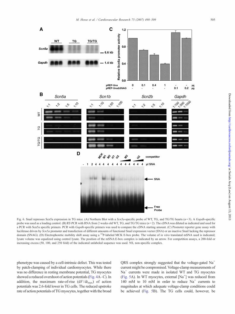
Fig. 6. Snail represses Scn5a expression in TG mice. (A) Northern Blot with a Scn5a-specific probe of WT, TG, and TG/TG hearts (n=3). A Gapdh-specificprobe was used as a loading control. (B) RT-PCR with RNA from 2 weeks old WT, TG, and TG/TGmice (n=2). The cDNAwas diluted as indicated and used fora PCR with Scn5a specific primers. PCR with Gapdh-specific primers was used to compare the cDNA starting amount. (C) Promoter reporter gene assay withluciferase driven by Scn5a promoter and transfection of different amounts of functional Snail expression vector (SNA) or an inactive Snail lacking the repressordomain (SNAG). (D) Electrophoretic mobility shift assay using a 32P-labeled MCK E-box probe. The volume of in vitro translated mSNA used is indicated;lysate volume was equalized using control lysate. The position of the mSNA:E-box complex is indicated by an arrow. For competition assays, a 200-fold orincreasing excess (50, 100, and 250 fold) of the indicated unlabeled sequence was used. NS, non-specific complex.
505M. Hesse et al. / Cardiovascular Research 75 (2007) 498–509
at Serials Acq (L
aw) on A
ugust 13, 2013http://cardiovascres.oxfordjournals.org/
Dow
nloaded from
phenotype was caused by a cell-intrinsic defect. This was testedby patch-clamping of individual cardiomyocytes. While therewas no difference in resting membrane potential, TG myocytesshowed a reduced overshoot of action potentials (Fig. 4A–C). Inaddition, the maximum rate-of-rise (dV /dtmax) of actionpotentials was 2.6-fold lower in TG cells. The reduced upstrokerate of action potentials of TGmyocytes, togetherwith the broad
QRS complex strongly suggested that the voltage-gated Na+
currentmight be compromised.Voltage-clampmeasurements ofNa+ currents were made in isolated WT and TG myocytes(Fig. 5A). In WT myocytes, external [Na+] was reduced from140 mM to 10 mM in order to reduce Na+ currents tomagnitudes at which adequate voltage-clamp conditions couldbe achieved (Fig. 5B). The TG cells could, however, be

506 M. Hesse et al. / Cardiovascular Research 75 (2007) 498–509
at Serials Acq (L
aw) on A
ugust 13, 2013http://cardiovascres.oxfordjournals.org/
Dow
nloaded from
adequately voltage-clamped in 140mMexternal [Na+] (Fig. 5A,B). Direct comparison of the Na+ current at 10 mM to that at140 mM external [Na+] in a TG myocyte showed anapproximately 10-fold reduction in magnitude of the Na+
current (Fig. 5C).
3.6. Scn5a gene transcription is strongly repressed by theSnail transcription factor
The most likely explanation for the decrease in Na+
current was a decrease of Nav1.5 expression since it is themajor Na+ channel. Indeed, microarray experiments showedthat the gene that encodes it, Scn5a, was downregulated inTG mice (Supplementary Table 2). Northern blot and semi-quantitative RT-PCR analysis confirmed reduction in Scn5amRNA in both TG and TG/TG mice compared to WT(Fig. 6A, B). The reduction in Scn5a mRNA was greater inTG/TG compared to TG mice and this correlated with themore severe phenotype. Expression of genes encoding thetwo β subunits that are known to bind to Nav1.5 [15] wasincreased in TG and TG/TG hearts (Fig. 6B), in accordancewith microarray data (Supplementary Table 2). This increasewas greater in TG/TG compared to TG mice and could be acompensatory mechanism.
The human and mouse Scn5a promoters contain severalE-box motifs [13,24], which are binding sites for Snail [11].To test this possibility directly, a luciferase reporter geneunder the control of a human Scn5a promoter fragment and aSnail expression vector were co-transfected into P19 cells.Snail caused a dose-dependent reduction in Scn5a promoteractivity, while transfection with an inactive Snail proteinlacking the repressor domain (ΔSNAG), showed no effect(Fig. 6C). Oligonucleotides containing the sequences of twoconserved E-box motifs were used in an electrophoreticmobility shift assay as competitors for binding to a knownSnail binding motif from the muscle creatine kinase gene(MCK) [11]. Competitor oligonucleotides were generated forthe mouse (M1, M2) and human (H1, H2) Scn5a promotersequence, respectively. Snail was able to bindM2 and H2, butnot M1 and H1 (Fig. 6D). These data indicate that Snail candirectly bind to and repress the activity of the Scn5apromoter.
4. Discussion
We have shown here that ectopic expression of the Snailtranscriptional repressor in the heart leads to DCM. Using acombination of approaches we have been able to associate theventricular pathology with downregulation of the major Na+
channel gene in the heart, Scn5a. While previous studies inhumans have linkedmutations in SCN5Awith DCM, our resultsare the first to establish a functional connection between loss ofNa+ channel function and severe DCM.While there are severalmouse models that develop DCM, this is the first model to bedescribed in which DCM is the primary pathology as aconsequence of reduced expression of a Na+ channel α subunit.
4.1. MHC-Snail transgenic mice are a unique modelof DCM
The Snail transgenic mouse model has several uniquefeatures that distinguish it from other animal models. First,while mutations in most DCM-associated genes in humansand mice result in other pathologies, such as musculardystrophy, hypertrophic cardiomyopathy, lipodystrophy orrestrictive cardiomyopathy, the mice described heredisplayed no additional phenotype. So far, only mutationsin metavinculin and phospholamban show no overlap withother inherited diseases [2]. Second, TG/TG mice display arapid onset of DCM as has been reported for MLP mutantmice [25]. In contrast TG mice show a slow progressiveform, as in TIMP-3 knockout mice [26]. Interestingly, noarrhythmias were detected in either TG or TG/TG mice,whereas patients with loss-of-function mutations inSCN5A display a variety of conductance abnormalities.Fibrosis is a major cause of conduction disturbances andarrhythmia and in humans, myocyte loss by apoptosis,necrosis, or other mechanisms is common in DCM [27]and leads to interstitial fibrosis. The lack of fibrosis in theTG myocardium could explain the lack of arrhythmia andmakes this TG mouse a good model for studying earlyevents in DCM.
4.2. Is DCM due to Snail-mediated downregulationof Scn5a?
In searching for the mechanism underlying DCM in theMHC-Snail transgenic mice, we have concluded that it is mostlikely due to downregulation of the Scn5a Na+ channel. Thisdownregulation is mediated by Snail binding to and repressingthe Scn5a promoter. The marked reduction in Na+ current andrelated decrease in the dV /dt of the action potential lead toslower and hence less synchronous activation of theventricular myocardium. This primary electrophysiologicaldefect could, in turn, reduce contractility. In support of thismodel, we have observed the broadening of the QRS-complex,other ECG changes, and downregulation of Scn5a expressionas early as 2 or 3 weeks of age and therefore well beforedilation of the heart chambers has developed.
We have concluded that the effect of the transgene is dueto ectopic expression of Snail per se, and not to the transgeneinsertion having a mutagenic effect based on two lines ofevidence. First, the reduction in Scn5a gene expression andNa+ channel activity is 80–90% even in single-TG mice andfurther reduced in TG/TG mice. These data are not consistentwith a simple genetic model in which the transgene insertioncompromises function of the adjacent genes. Second, in ourinitial efforts to develop the MHC-Snail transgenic mice, wehad three founders, two of which died at a few months of ageshowing signs of heart failure (Supplementary materials andmethods). Genotyping showed that one mouse had 10 andthe other 4 copies of the transgene integrated into a singlelocus, whereas the surviving founder had only two.
507M. Hesse et al. / Cardiovascular Research 75 (2007) 498–509
at Serials Acq (L
aw) on A
ugust 13, 2013http://cardiovascres.oxfordjournals.org/
Dow
nloaded from
Snail acts as a transcriptional repressor and more than 10target genes have been identified, most of them componentsof adherens and tight junctions [10]. While we cannotcompletely rule out the possibility that cell–cell junctions areaffected in the heart of MHC-Snail transgenic mice, there areno obvious morphological changes in individual cardiomyo-cytes that would indicate this. The cardiac adherens junctionprotein N-cadherin is not repressed by Snail, and E- and VE-cadherin, which are also targets of Snail, are not expressed inventricular myocytes. Also, immunoflourescence stainingfor N-cadherin as well as β-catenin in TG hearts showed nodifferences compared to WT ventricular myocytes (data notshown). Members of the tight junction protein family are notexpressed in the heart, which rules them out as possible Snailtargets. Nevertheless there might be unknown targets ofSnail, whose downregulation could cause DCM directly orindirectly. However, despite the fact that we do not haveconclusive evidence, the data presented here is consistentwith a direct downregulation of the expression of the majorNa-channel Scn5a by Snail.
4.3. Human mutations in SCN5A and heart disease
Mutations in SCN5A that cause a decrease in Na+ currentare found in patients with Brugada syndrome and Lenègredisease. Brugada syndrome is characterized by an ST segmentelevation in the right precordial ECG leads V1–V3, and a highincidence of sudden death caused by ventricular fibrillation[28]. Notably, patients with Brugada syndrome have structur-ally normal hearts. About 20% of all patients diagnosed withBrugada syndrome harbour mutations in SCN5A that cause aloss of channel function via different mechanisms [29].Lenègre disease is associated with mutations in SCN5A inwhich the Na+ current is decreased in cardiomyocytes leadingto a conduction defect that is progressive and develops alongwith age-related myocardial fibrosis.
Recently reports have suggested a connection betweenmutations in SCN5A causing conduction disease and DCM[6,7]. In one study a D1275N mutation was identified inindividuals with DCM [6]. Mutant channels were shown toactivate atmore positive voltages,which could result in reducedexcitability of the myocytes [30]. However the authors couldnot determine whether DCM is a direct consequence of SCN5Amutation or is caused by chronic arrhythmia. In a second studywith the same plus additional families, the D1275N mutationwas confirmed and 4 different additional mutations in SCN5Awere identified [7]. Of all individuals with a SCN5A mutation,38% had overt DCM and 27% had early features of DCM. Allof the mutations described in this report (D1275N, T220I,R814W,D1595H and 2550–2551 insTG) caused a reduction inNa+-current. A recent paper based on a large Finnish familyreported that the D1275N mutation is associated with cardiacconduction defects and atrial arrhythmia [31]. Dilation ofventricles was seen in only 4 out of 10 carriers. However, themean age of the patients in this study was 27.6 years while themean age at diagnosis of DCM in patients studied by McNair
and byOlsonwas 47.9 years [7], implying a late onset of DCM.In another report it was shown that carriers of Scn5amutations(R376H, R1023H, R1644C, I1968S) with structurally normalhearts and an average age of 44.8 years demonstratedcardiomyopathic changes such as myocardial cell degenerationand apoptosis [32]. This suggests a myocyte-intrinsic effect ofcertain SCN5A mutations that causes cardiomyocytes to losetheir normal contractility and/or undergo cardiomyopathicchanges, which in turn leads to dilation. In humans, it is verylikely that this process takes several years and therefore doesnot necessarilymanifest as DCMat the point of initial diagnosisof Brugada syndrome.
4.4. Functional links between Scn5a and DCM
In our mouse model, the Snail transgene is not expresseduntil birth, and therefore its effect is limited to late-developmental suppression of Scn5a expression. It thereforegives us a unique perspective on Scn5a function that isdifferent from those gained from Scn5a knockout mice.Scn5a homozygous mutant mice have morphologicalabnormalities in the ventricular chamber and die aroundembryonic day 11.5 due to cardiac standstill [33]. Hetero-zygous mutant mice are viable and have a normal lifespan.However, cardiomyocytes from Scn5a heterozygous mutantmice have a 50% reduction of their cardiac Na+ conductance,which causes impaired myocardial conduction in vitro.Aging Scn5a heterozygous mutants show progressive, age-related slowing of conduction, myocardial fibrosis, andchanges in connexin expression [34,35], to some extentresembling Lenègre disease. These results from mutant miceas well as the data from patients carrying a loss-of-functionmutation in SCN5A indicates that a 50% reduction duringdevelopment has no effect on the gross morphology of theheart, but leads to conduction slowing and arrhythmia. It ispossible that DCM is not observed in Scn5a heterozygousmutant mice, unlike some human patients with SCN5Amutations, simply because the lifespan of mice is not longenough for them to develop the pathology [33].
In our MHC-Snail transgenic model we have observed~10-fold reduction in Na+ current in juvenile and adult TGmice. The reduction is even more prominent in TG/TG mice.We were surprised that the MHC-Snail transgenic micesurvive as long as they do, given the severity of the Na+
channel defect and the fact that Scn5a null mutants areembryonic lethal. It may be that the late onset of the Snailexpression delays the reduction in Scn5a until after the criticaldevelopmental period. Alternatively, the low level of Scn5aexpression may just be enough to allow survival. Nonetheless,dramatic low-level expression of Scn5a leads to severeconduction defects and DCM in our mouse model. There isone report of compound heterozygosity for two differentSCN5A mutations in humans that reduced the Na+ current to5–10% [36]. In this case report two affected individualsinherited the W156X nonsense-mutation from the father andthe R225W missense mutation from the mother. One

508 M. Hesse et al. / Cardiovascular Research 75 (2007) 498–509
at Serials Acq (L
aw) on A
ugust 13, 2013http://cardiovascres.oxfordjournals.org/
Dow
nloaded from
individual died at 1 year of age due to intractable arrhythmiaand an autopsy revealed severeDCMwithmyocardial fibrosis.The other individual displayed tachycardia after birthand broadening of the QRS-complex but recovered after β-blockade. However, conduction slowing progressed during thelife of this individual.
Collectively, our data indicate that reductions in SCN5Afunction are associated with DCM. In addition though, it isclear that the level of the reduction of Na+ current incardiomyocytes is critical for the phenotypic outcome,varying between no changes, mild conduction delays,arrhythmia, or DCM. Also studies of these mice revealed atemporal connection between a reduction of Na+ current andthe time-course of the progressive DCM. The greater thereduction in Na+ current, the faster the onset and progressionof DCM, as seen in TG/TG mice, which develop a phenotypeas early as 2 weeks of age and die within the first two months,while TG mice with a 90% reduction in Na+ current have anormal lifespan. Extrapolated to humans, this would indicatethat a reduction of 50% of Na+ current, as seen in loss-of-function mutations in Brugada syndrome, would probablyneed several years to cause an obvious DCM. This isconsistent with the late onset of DCM (average 47.5 years) insome patients with Scn5a loss-of-function mutations [6,7].This means at least that DCM has to be taken into account as apossible long-term complication in patients with Brugadasyndrome who are initially diagnosed with conductiondefects. In addition, it is clear that Na+ channel functionand SCN5A expression should be considered as potentialunderlying mechanisms for DCM.
Acknowledgements
We thank O. Brusselers for technical assistance. We thankDM Roden for the Scn5a-promoter luciferase constructs. Thiswork was supported by grants from the Canadian Institutes ofHealth Research (CIHR) (to J.C.C. and W.R.G.) and Heart andStroke Foundation (W.R.G.). M.H. was supported by fellow-ships from the German Research Foundation (HE4477/1-1 andHE4477/1-2), and J.C.C. is an Investigator of the CIHR and aScientist of the Alberta Heritage Foundation for MedicalResearch.
Appendix A. Supplementary data
Supplementary data associatedwith this article can be found,in the online version, at doi:10.1016/j.cardiores.2007.04.009.
References
[1] MaronBJ, Towbin JA, ThieneG,AntzelevitchC,CorradoD,Arnett D, etal. Contemporary definitions and classification of the cardiomyopathies:an American Heart Association Scientific Statement from the Council onClinical Cardiology, Heart Failure and Transplantation Committee;Quality of Care and Outcomes Research and Functional Genomics andTranslational Biology InterdisciplinaryWorking Groups; and Council onEpidemiology and Prevention. Circulation 2006;113:1807–16.
[2] Osterziel KJ, Perrot A. Dilated cardiomyopathy: more genes meansmore phenotypes. Eur Heart J 2005;26:751–4.
[3] Bienengraeber M, Olson TM, Selivanov VA, Kathmann EC, O'Co-chlain F, Gao F, et al. ABCC9 mutations identified in human dilatedcardiomyopathy disrupt catalytic KATP channel gating. Nat Genet2004;36:382–7.
[4] George Jr AL. Inherited disorders of voltage-gated sodium channels.J Clin Invest 2005;115:1990–9.
[5] Brugada P,BrugadaR,AntzelevitchC, Brugada J. TheBrugada Syndrome.Arch Mal Coeur Vaiss 2005;98:115–22.
[6] McNairWP, Ku L, TaylorMR, Fain PR, DaoD,Wolfel E, et al. SCN5Amutation associated with dilated cardiomyopathy, conduction disorder,and arrhythmia. Circulation 2004;110:2163–7.
[7] Olson TM, Michels VV, Ballew JD, Reyna SP, Karst ML, Herron KJ,et al. Sodium channel mutations and susceptibility to heart failure andatrial fibrillation. JAMA 2005;293:447–54.
[8] GroenewegenWA, Wilde AA. Letter regarding article by McNair et al,“SCN5Amutation associated with dilated cardiomyopathy, conductiondisorder, and arrhythmia”. Circulation 2005;112:e9–e10.
[9] Adler E, Fuster V. SCN5A—a mechanistic link between inheritedcardiomyopathies and a predisposition to arrhythmias? JAMA 2005;293:491–3.
[10] Barrallo-Gimeno A, Nieto MA. The Snail genes as inducers of cellmovement and survival: implications in development and cancer.Development 2005;132:3151–61.
[11] Nakayama H, Scott IC, Cross JC. The transition to endoreduplicationin trophoblast giant cells is regulated by the mSNA zinc fingertranscription factor. Dev Biol 1998;199:150–63.
[12] Timmerman LA, Grego-Bessa J, Raya A, Bertran E, Perez-PomaresJM, Diez J, et al. Notch promotes epithelial-mesenchymal transitionduring cardiac development and oncogenic transformation. Genes Dev2004;18:99–115.
[13] Yang P, Kupershmidt S, Roden DM. Cloning and initial characteriza-tion of the human cardiac sodium channel (SCN5A) promoter.Cardiovasc Res 2004;61:56–65.
[14] ChurchGM,GilbertW.Genomic sequencing. ProcNatl Acad SciU SA1984;81:1991–5.
[15] Haufe V, Camacho JA, Dumaine R, Gunther B, Bollensdorff C, vonBanchet GS, et al. Expression pattern of neuronal and skeletal musclevoltage-gated Na+ channels in the developing mouse heart. J Physiol2005;564:683–96.
[16] Hemberger M, Hughes M, Cross JC. Trophoblast stem cellsdifferentiate in vitro into invasive trophoblast giant cells. Dev Biol2004;271:362–71.
[17] Semeniuk LM, Kryski AJ, Severson DL. Echocardiographic assess-ment of cardiac function in diabetic db/db and transgenic db/db-hGLUT4 mice. Am J Physiol Heart Circ Physiol 2002;283:H976–82.
[18] How OJ, Aasum E, Kunnathu S, Severson DL, Myhre ES, Larsen TS.Influence of substrate supply on cardiac efficiency, as measured bypressure-volume analysis in ex vivo mouse hearts. Am J Physiol HeartCirc Physiol 2005;288:H2979–85.
[19] Nygren A, Clark RB, Belke DD, Kondo C, Giles WR, Witkowski FX.Voltage-sensitive dye mapping of activation and conduction in adultmouse hearts. Ann Biomed Eng 2000;28:958–67.
[20] Nygren A, Kondo C, Clark RB, Giles WR. Voltage-sensitive dyemapping in Langendorff-perfused rat hearts. Am J Physiol Heart CircPhysiol 2003;284:H892–902.
[21] Brouillette J, Clark RB, Giles WR, Fiset C. Functional properties of K+currents in adult mouse ventricular myocytes. J Physiol 2004;559:777–98.
[22] Gulick J, Subramaniam A, Neumann J, Robbins J. Isolation andcharacterization of the mouse cardiac myosin heavy chain genes. J BiolChem 1991;266:9180–5.
[23] Priori SG, Napolitano C. Genetics of cardiac arrhythmias and suddencardiac death. Ann N YAcad Sci 2004;1015:96–110.
[24] Shang LL, Dudley Jr SC. Tandem promoters and developmentallyregulated 5′- and 3′-mRNA untranslated regions of the mouse Scn5acardiac sodium channel. J Biol Chem 2005;280:933–40.
509M. Hesse et al. / Cardiovascular Research 75 (2007) 498–509
Dow
nloaded fr
[25] Arber S, Hunter JJ, Ross J, Hongo M, Sansig G, Borg J, et al. MLP-deficient mice exhibit a disruption of cardiac cytoarchitectural organi-zation, dilated cardiomyopathy, and heart failure. Cell 1997;88:393–403.
[26] Fedak PW, Smookler DS, Kassiri Z, Ohno N, Leco KJ, Verma S, et al.TIMP-3 deficiency leads to dilated cardiomyopathy. Circulation 2004;110:2401–9.
[27] Kostin S, Pool L, Elsasser A, Hein S, Drexler HC, Arnon E, et al.Myocytes die by multiple mechanisms in failing human hearts. CircRes 2003;92:715–24.
[28] Brugada P, Brugada J. Right bundle branch block, persistent ST segmentelevation and sudden cardiac death: a distinct clinical and electrocardiogra-phic syndrome. A multicenter report. J Am Coll Cardiol 1992;20: 1391–6.
[29] Priori SG, Napolitano C, Gasparini M, Pappone C, Della BP, GiordanoU, et al. Natural history of Brugada syndrome: insights for riskstratification and management. Circulation 2002;105: 1342–7.
[30] GroenewegenWA, Firouzi M, Bezzina CR, Vliex S, van L. I, Sandkuijl L,et al. A cardiac sodium channel mutation cosegregates with a rareconnexin40 genotype in familial atrial standstill. Circ Res 2003;92:14–22.
[31] Laitinen-Forsblom PJ, Makynen P, Makynen H, Yli-Mayry S, Virtanen V,Kontula K, et al. SCN5A mutation associated with cardiac conductiondefect and atrial arrhythmias. J Cardiovasc Electrophysiol 2006;17:480–5.
[32] Frustaci A, Priori SG, Pieroni M, Chimenti C, Napolitano C, Rivolta I,et al. Cardiac histological substrate in patients with clinical phenotypeof Brugada syndrome. Circulation 2005;112:3680–7.
[33] Papadatos GA, Wallerstein PM, Head CE, Ratcliff R, Brady PA,Benndorf K, et al. Slowed conduction and ventricular tachycardia aftertargeted disruption of the cardiac sodium channel gene Scn5a. ProcNatl Acad Sci U S A 2002;99:6210–5.
[34] Royer A, van Veen TA, Le BS, Marionneau C, Griol-Charhbili V, LeoniAL, et al. Mouse model of SCN5A-linked hereditary Lenegre's disease:age-related conduction slowing and myocardial fibrosis. Circulation2005;111:1738–46.
[35] van Veen TA, Stein M, Royer A, Le QK, Charpentier F, Colledge WH,et al. Impaired impulse propagation in Scn5a-knockout mice:combined contribution of excitability, connexin expression, and tissuearchitecture in relation to aging. Circulation 2005;112:1927–35.
[36] Bezzina CR, Rook MB, Groenewegen WA, Herfst LJ, van der Wal AC,Lam J, et al. Compound heterozygosity for mutations (W156X andR225W) in SCN5A associatedwith severe cardiac conduction disturbancesand degenerative changes in the conduction system. Circ Res 2003;92:159–68.
om
at Serials Acq (Law
) on August 13, 2013
http://cardiovascres.oxfordjournals.org/