Effect of Dosage of Non-Chloride Accelerator versus Chloride ...
Upload
khangminh22Category
view
0download
0
UNLV Retrospective Theses & Dissertations
1-1-1998
Effects of sodium chloride and sodium gluconate concentration Effects of sodium chloride and sodium gluconate concentration
on hydration behavior and water transport in the red-spotted toad, on hydration behavior and water transport in the red-spotted toad,
Bufo punctatus Bufo punctatus
Polly Ann Sullivan University of Nevada, Las Vegas
Follow this and additional works at: https://digitalscholarship.unlv.edu/rtds
Repository Citation Repository Citation Sullivan, Polly Ann, "Effects of sodium chloride and sodium gluconate concentration on hydration behavior and water transport in the red-spotted toad, Bufo punctatus" (1998). UNLV Retrospective Theses & Dissertations. 946. http://dx.doi.org/10.25669/4p7d-wcap
This Thesis is protected by copyright and/or related rights. It has been brought to you by Digital Scholarship@UNLV with permission from the rights-holder(s). You are free to use this Thesis in any way that is permitted by the copyright and related rights legislation that applies to your use. For other uses you need to obtain permission from the rights-holder(s) directly, unless additional rights are indicated by a Creative Commons license in the record and/or on the work itself. This Thesis has been accepted for inclusion in UNLV Retrospective Theses & Dissertations by an authorized administrator of Digital Scholarship@UNLV. For more information, please contact [email protected].
INFORMATION TO USERS
This manuscript has been reproduced from the microfilm master. UMI
films the text directly from the original or copy submitted. Thus, some
thesis and dissertation copies are in typewriter face, while others may be
from any type o f computer printer.
T he quality of this reproduction is dependent upon the quality of the
copy subm itted. Broken or indistinct print, colored or poor quality
illustrations and photographs, print bleedthrough, substandard margins,
and improper alignment can adversely affect reproduction.
In the unlikely event that the author did not send UMI a complete
manuscript and there are missing pages, these will be noted. Also, if
unauthorized copyright material had to be removed, a note will indicate
the deletion.
Oversize materials (e.g., maps, drawings, charts) are reproduced by
sectioning the original, beginning at the upper left-hand comer and
continuing from left to right in equal sections with small overlaps. Each
original is also photographed in one exposure and is included in reduced
form at the back o f the book.
Photographs included in the original manuscript have been reproduced
xerographically in this copy. Higher quality 6” x 9” black and white
photographic prints are available for any photographs or illustrations
appearing in this copy for an additional charge. Contact UMI directly to
order.
UMIA Bell & Howell Information Company
300 North Zeeb Road, Ann Arbor MI 48I06-I346 USA 313/761-4700 800/521-0600
R eproduced with perm ission of the copyright owner. Further reproduction prohibited without perm ission.

Reproduced with permission of the copyright owner. Further reproduction prohibited without permission.
EFFECTS OF SODIUM CHLORIDE AND SODIUM GLUCONATE
CONCENTRATION ON HYDRATION BEHAVIOR
AND WATER TRANSPORT IN THE
RED-SPOTTED TOAD,
BVFO PUNCTATUS
by
Polly Ann Sullivan
Bachelor of Science University of Nevada, Las Vegas
1992
Master of Arts University of Nevada, Las Vegas
1995
A thesis submitted in partial fulfillment of the requirements for the degree of
M aster of Science
in
Biological Sciences
Department of Biological Sciences University of Nevada, Las Vegas
December 1998
R eproduced with perm ission of the copyright owner. Further reproduction prohibited without perm ission.
UMI Number: 1393771
UMI Microform 1393771 Copyright 1999, by UMI Company. All rights reserved.
This microfonn edition is protected against unauthorized copying under Title 17, United States Code.
UMI300 North Zeeb Road Ann Arbor, MI 48103
R eproduced with perm ission of the copyright owner. Further reproduction prohibited without perm ission.
IJNTV Thesis ApprovalThe Graduate College University of Nevada, Las Vegas
November 12 ,19 98
The Thesis prepared by
P o lly Ann S u lliv an
Entitled
E ffec ts of Sodium C hloride and Sodium Gluconate C oncentration on H ydration
Behaviour and Water T ransport in the Red-Spotted Toad,___________________
Bufo p u ncta tus
is approved in partial fulfUlment of the requirements for the degree of
Examination Committee Member
M aster o f Science in Biology
ÆExamination Commttee Chfir
Dean of the forkduate College
Exanm iVon Cbwmittee Member
fradfiate College Faculty Representative
u
R eproduced with perm ission of the copyright owner. Further reproduction prohibited without perm ission.
ABSTRACT
Effects of Sodium Chloride and Sodium Gluconate Concentration
on Hydration Behavior and Water Transport in the
Red spotted Toad, Bufo punctatus
by
Polly Ann Sullivan
Dr. Karin Hoff and Dr. Stan Hillyard,Examination Committee Co-chairs
Professors of Biology University of Nevada, Las Vegas
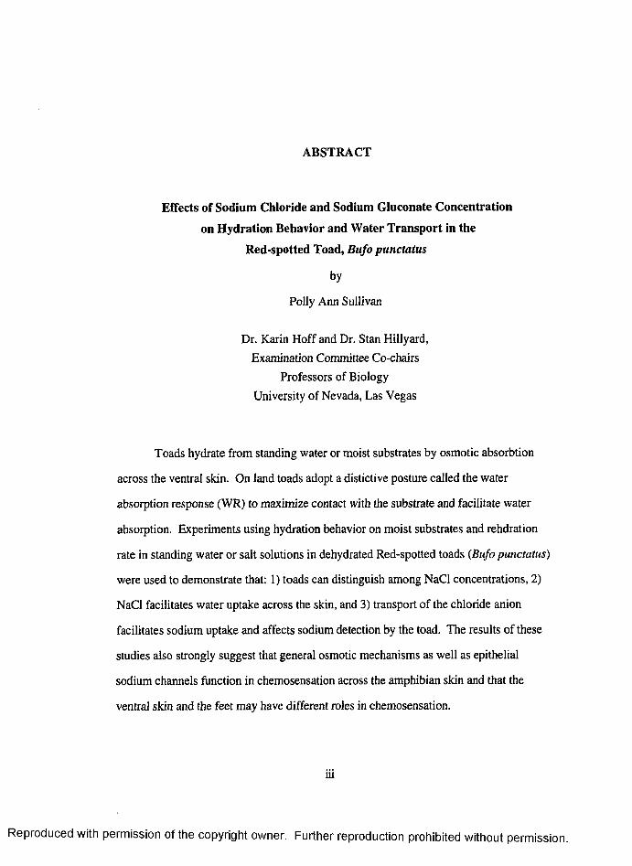
Toads hydrate from standing water or moist substrates by osmotic absorbtion
across the ventral skin. On land toads adopt a distictive posture called the water
absorption response (WR) to maximize contact with the substrate and facilitate water
absorption. Experiments using hydration behavior on moist substrates and rehdration
rate in standing water or salt solutions in dehydrated Red-spotted toads (fiufo punctatus)
were used to demonstrate that: 1) toads can distinguish among NaCl concentrations, 2)
NaCl facilitates water uptake across the skin, and 3) transport of the chloride anion
facilitates sodium uptake and affects sodium detection by the toad. The results of these
studies also strongly suggest that general osmotic mechanisms as well as epithelial
sodium channels function in chemosensation across the amphibian skin and that the
ventral skin and the feet may have different roles in chemosensation.
lU
R eproduced with perm ission of the copyright owner. Further reproduction prohibited without perm ission.
TABLE OF CONTENTS
ABSTRACT...........................................................................................................................ai
TABLE OF CONTENTS..................................................................................................... iv
LIST OF FIGURES.............................................................................................................. vi
ACKNOWLEDGEMENTS................................................................................................. vii
CHAPTER 1 INTRODUCTION AND HISTORICAL PERSPECTIVE.........................1Behavior Associated With Water U ptake............................................................... 2Seat Patch Specialization for Water Uptake.............................................................2Specialization of Amphibian Skin for Active Sodium Transport.......................... 2Sodium Transport and Salt Taste in the Mammalian Tongue................................3Sodium Transport and Salt Taste in the Amphibian S kin ..................................... 4Statement of the Problem and Hypothesis..............................................................4Behavioral and Physiological Effects of Salt Concentration ................................ 4Effects of Anions on Sodium Detection and Water Transport ..............................5
CHAPTER 2 EFFECTS OF SODIUM CHLORIDE ON HYDRATION BEHAVIORAND WATER TRANSPORT IN RED-SPOTTED TOADS, Bufo punctatus................. 7
Introduction............................................................................................................... 7Methods ...................................................................................................... 9Animals........................................................................................................9Dehydration Procedure............................................................................... 9Toad and Substrate Selection....................................................................10Data Collection and Analysis....................................................................10Hydration Behavior Experiments............................................................ 10Rehydration Experiments ........................................................................11Electrophysiology Experiments............................................................... 12
Results .................................................................................................................... 14Hydration Behavior Experiments............................................................ 14Rehydration Experiments......................................................................... 14
Discussion................................................................................................................ 16Hydration Behavior Experiments............................................................ 16Coupling of Water and Na* Transport..................................................... 18Separate Na* Pathways for Water Transport and Chemosensation... 20 A Possible Neural Transduction Pathway for Salt Discrimination ... 21
Conclusion ........................................................................................................................... 22
CHAPTER 3 EFFECTS OF SODIUM GLUCONATE CONCENTRATION ONWATER TRANSPORT AND HYDRATION BEHAVIOR INRED-SPOTTED TOADS, Bufopunctatus.........................................................................24
IV
R eproduced with perm ission of the copyright owner. Further reproduction prohibited without perm ission.
Introduction i.............................................................................................24Methods ...................................................................................................................24
Animals....................................................................................................... 27Rehydration Experiments.......................................................................... 27Hydration Behavior Experiments............................................................. 27
R esults......................................................................................................................28Rehydration Experiments.......................................................................... 30Hydration Behavior Experiments..............................................................30
Discussion............................................................................................................................. 31Anion Transport Across the Epithilium................................................................ 31Anion Transport and Chemosensation................................................................... 32
Conclusion............................................................................................................................. 33
REFERENCES......................................................................................................................35
FIGURES...............................................................................................................................38
VITA....................................................................................................................................... 47
R eproduced with perm ission of the copyright owner. Further reproduction prohibited without perm ission.
LIST OF FIGURES
Figure 1 Hydration Behavior Postures of the Toad Bufopunctatus.......................38Figure 2 Time-activity Budget for 30 Minute NaCl Hydration Behavior
E x p erim en ts ................................................................................ 39Figure 3 Duration of WR Posture for 30 Minute NaCl Hydration Behavior
E x p erim en ts ..................................................................................................40Figure 4 Weight Change for NaCl Rehydration Experiments.............................. 41Figure 5 Weight Change for NaCl/ Amiloride Rehydration Experiments.............. 42Figure 6 The Apical Cell Layer o f Toad Seat Patch Skin.................................... 43Figure 7 Integrated Neural Response of Toad Spinal Nerve #6 (normalized to the
500 mM NaCl response).......................................................................44Figure 8 Weight Change for NaCl/NaGluconate Rehydration Experiments.........45Figure 9 Duration of Hydration Behavior Posture (SPD and WR) for 15 Minute
NaCl/ NaGluconate Hydration Behavior Experiments......................... 46
VI
R eproduced with perm ission of the copyright owner. Further reproduction prohibited without perm ission.
ACKNOWLEDGEMENTS
I would like to express my sincerest thanks to Dr. Karin Hoff for her continuous
encouragement and support throughout the past several years. I have her to thank for
my success. I would like to thank Dr. Stan Hillyard for the training in electrophysiology techniques and the help given to this thesis. I want to thank Dr
Takatoshi Nagai for the time and attention given to teaching me appropriate research skills. I would like to thank Dr. Steven Carper for the excellent guidance as an advisor.I am very proud that I was a recipient of the NSF Women in Science and Engineering Research Awards and I thank Dr. Penny Amy for that honor. I would also like to thank all of the faculty and graduate students of the UNLV Department of Biology for being
there when needed.
Vll
R eproduced with perm ission of the copyright owner. Further reproduction prohibited without perm ission.
CHAPTER 1
INTRODUCTION AND HISTORICAL PERSPECTIVE
Unlike other vertebrates anuran amphibians (frogs and toads) do not drink
orally, rather they absorb water osmotically across their skin (Bentley and Yorio, 1979).
The study of water balance in terrestrial amphibians dates back as far as the 1790's
(Townson, 1795). Townson noted that water balance for terrestrial amphibians was
affected by a high rate of évapotranspiration across the skin. He also observed that in
tree frogs {Hyla arborea) and frogs {Rana tempora) the water loss was countered by
retention and reabsorption of water from the bladder and by absorption of water through
the lower abdominal skin.
Behavior Associated with Water Uptake
Field observations of toad behavior related to the absorption of water through the
skin of the lower abdomen by Stille (1952) led to a lab study of toad water uptake
behavior by Stille (1958). It was observed that dehydrated toads adopt a distinct posture
when placed on a moist surface that allows increased contact of the lower abdominal
skin with the surface. He called this posture and related behavior the water absorption
response. Compared to other areas of skin, the lower abdominal skin of many anuran
species is adapted for higher rates of water uptake and is termed the pelvic patch or seat
patch (McClanahan and Baldwin, 1969, Marrero and Hillyard, 1985).
R eproduced with perm ission of the copyright owner. Further reproduction prohibited without perm ission.
Seat Patch Specializations for Water Uptake
The toad seat patch skin has specialized morphological features. The skin has
small tubercles and channeling called epidermal sculpturing (Lilly white and Licht, 1974)
that allow increased surface area when the skin is pressed to a hydration surface
(Hillyard, 1988). There are cutaneous attachments of the gracilis minor and a
specialized cutaneous muscle, the abdominal crenator, underneath the seat patch that is
thought to facilitate expansion and contraction of the seat patch skin (Winokur and
Hillyard, 1992). The seat patch area also has higher vascularization than the pectoral
area of toad ventral skin and, in dehydrated bufonids, is associated with increase of
water. Capillaries may move the water which is absorbed across the skin into the
circulatory system. Alternatively, the large lymphatic space in the proximity of the seat
patch skin may also function in the transport of water absorbed across the skin to the
general circulation (as reviewed by Jorgensen, 1997).
Specializations of Amphibian Skin for Active Sodium Transport
In addition to water uptake, the amphibian skin is able to actively absorb Na*
and Cl from dilute pond water (Krogh, 1939) thereby allowing the frogs to maintain
ionic as well as osmotic balance. Early studies of isolated frog skin showed that there is
an electric current associated with transport of sodium ions across the skin (as reviewed
by Jorgensen, 1997). This phenomena was generally referred to as electro-osmosis.
Short circuit current measurement techniques facilitated the development of a model for
active sodium transport across amphibian epithelium (Ussing and Zerahn, 1951,
Koefeld-Johnson, Levi and Ussing, 1952). The model proposes that in frog skin,
sodium ions enter the epithelial cells down an electro-chemical gradient across the apical
membrane. The sodium entry step has been shown to be through amiloride blockable
sodium ion channels (Lindemann and Van Driessche, 1977). The Na* is then actively
R eproduced with perm ission o f the copyright owner. Further reproduction prohibited without perm ission.
transported out of the cells by a Na"" /K^ pump located in the basolateral membrane.
Removal of sodium and subsequent potassium leakage out of the cell creates a favorable
electrochemical gradient for further sodium diffusion inward and the chloride ion was
thought to diffuse passively along the electro-chemical gradient (Koefeld-Johnsen and
Ussing, 1958).
These physiological specializations for Na'̂ and Cl' absorption are present in the
toad seat patch in addition to the specializations for water uptake. Recent studies of
chloride suggest that Cl does not diffuse passively, but is actively transported by way
of a Cl'-HCOj exchange across mitochondria-rich cells located at the apical surface of
toad epithelium (Jensen, Sorensen, Larsen, and Willumsen, 1997).
The active sodium transport model was expanded to include transport of sodium
ions through all of the cell layers of the toad epithelium (stratum granulosum, stratum
spinosa and stratum basoteral) (Rick, Dorge, von Armin, Weigel and Thurau, 1981).
These authors also demonstrated that the sodium ion transport across the apical
membrane of toad skin was through amiloride blockable sodium ion channels.
Sodium Transport and Salt Taste in the Mammalian Tongue
The model for active Na* transport across amphibian skin (Ussing and Zerahn,
1951) has been applied to Na"̂ transport across canine lingual epithelium in association
with salt taste (DeSimone, Heck, Miersen and DeSimone, 1984), and in other mammals
(Stewart 1997). In lingual epithelium the inward transport of sodium is increased by the
cotransport of chloride through a paracellular pathway (Ye, Heck and DeSimone, 1991).
R eproduced with perm ission of the copyright owner. Further reproduction prohibited without perm ission.
Sodium Transport and Sait Taste in the Amphibian Skin
Recent studies by Hoff and Hillyard (1993) suggest that a portion of the Na*
chemosensory mechanism in toad skin is blockable by amiloride. That study suggests
that the mechanism of Na+ transport across amphibian skin, like the tongue, may be a
contributing factor to chemosensory transduction of this ion. However, not all Na*
avoidance is eliminated by amiloride suggesting that there is also an amiloride insensitive
component to Na' ̂chemosensation. The amiloride insensitive component may be a
paracellular route for Na* transport across the toad skin.
Studies of toad avoidance of hyperosmotic substrates has led to the investigation
of possible similar chemosensory mechanisms in toad skin (Brekke, Hillyard and
Winokur, 1991, Hoff and Hillyard, 1991). Brekke e/a/1. (1991) showed that toads
with feet covered by latex finger cots remained longer on hyperosmotic substrates than
toads with bare feet. This supports observations by Stille (1952) that suggest that toads
have a means of detecting osmotically favorable hydration sources with their feet.
Further studies are needed to substantiate these observations.
Statement of the Problem and the Hypothesis
Behavioral and Phvsiological Effects of Salt Concentration
Beyond the avoidance of hyperosmotic solutions, little is known about hydration
behavior of toads exposed to a wide range of solute concentrations that are in contact
with their skin. The first set of experiments tests the hypothesis that the hydration
behaviors of dehydrated toads, Bufo punctatus, presented with a range of hypoosmotic
and hyperosmotic NaCl concentrations will differ; that is, that toads can discern (or
taste) differences among solute concentrations.
A second set of experiments investigates whether NaCl concentration affects the
rate of water uptake by dehydrated toads. It is known that antidiuretic hormone (ADH)
R eproduced with perm ission of the copyright owner. Further reproduction prohibited without perm ission.
is released during dehydration (Nouwen and Kuhn, 1984) and that this hormone
increases both Na+ transport and water permeability of the skin (Baker and Hillyard,
1991). These experiments test the hypothesis that sodium transport is coupled with
water movement; that is, water gain will be augmented by the presence of salt in the
hydration source and that amiloride, an inhibitor of transepithelial Na+ transport will
reduce this augmentation.
From these two experimental approaches, I hope to describe the toads ability to
detect and respond to Na' ̂in its environment and also to describe a possible
physiological benefit of the changes in hydration behaviors that relates to the toads
ability to take up water.
Effects of Anions on Sodium Detection and Water Transport
Little is known about the cotransport of Na* and Cl' across the toad skin.
However, much is known about chloride transport across epithelium but the relative
amount of paracellular verses transcellular chloride transport remains controversial. Ye,
Heck and DeSimone (1991) noted that in canine lingual epithelium there is a paracellular
shunt for chloride associated with the taste receptor cells. They showed that blockage of
the movement across the tissue reduced the neural response for sodium, suggesting that
the transport of Na* across the lingual epithelium is dependent in part on the cotransport
of the chloride anion. In isolated toad epithelium, Jensen, Sorensen, Larsen, and
Willumsen (1997) showed that in dilute media the active transport of chloride is driven
by a Cl' - HCO3 exchange mechanism and is coupled to an active transport of Nan-
through the Na' ̂/K+ pump. Both transport mechanisms are located on the apical
membrane of mitochondrial rich cells. The dilute media was < 1 mM NaCl and the
active transport is thought to preserve the chloride concentration in the toads.
This study tests the hypothesis that epithelial transport of Cl affects Na*
transport, Na^ detection and water transport in toads and that this difference is
R eproduced with perm ission of the copyright owner. Further reproduction prohibited without perm ission.
discernible in toad hydration behavior and water uptake. The effects of Cl' are examined
by comparing behavioral response to equimolar concentrations of NaCl and the Na salt
of the large, impermeant gluconate anion.
R eproduced with perm ission o f the copyright owner. Further reproduction prohibited without perm ission.
CHAPTER 2
EFFECTS OF SODIUM CHLORIDE CONCENTRATION ON
HYDRATION BEHAVIOR AND WATER TRANSPORT
IN RED-SPOTTED TOADS, Bufo punctatus
Introduction
Toads in the genus Bufo have a highly permeable region in the lower ventral
skin termed a pelvic patch (McClanahan and Baldwin, 1969; Marrero and Hillyard,
1985). This highly permeable skin allows the toad to quickly recover water losses. The
Red-spotted toad, B. punctatus displays a distinct posture when hydrating that
maximizes contact of the permeable skin with the substrate. The hindlimbs are splayed
as the pelvic patch is pressed to the moist surface. This behavior is called the water
absorption response (WR) (Stille, 1958). Dehydrated individuals of this species can
gain as much as 15% of their body weight in half an hour (Hoff and Hillyard, 1993a),
while sitting in WR on a damp tissue.
Brekke et al.., { \99 \) showed that when dehydrated Bufo punctatus were placed
on a tissue saturated with hyperosmotic urea solutions they would not initiate WR. The
specialized skin of the pelvic patch of toads that enables rapid water uptake may also
contain a mechanism that allows chemosensory discrimination of hydration surfaces
(Hoff and Hillyard, 1993b). Further studies showed that dehydrated toads avoided
surfaces saturated with hyperosmotic NaCl and KCl (Hoff and Hillyard, 1993b). This
study also found that amiloride, a blocker of epithelial Na* channels, increased the
frequency of initiation of WR behavior on NaCl but not on KCl or urea solutions. The
R eproduced with perm ission of the copyright owner. Further reproduction prohibited without perm ission.
duration of the episodes of WR behavior was very brief so that the total time showing
this behavior was not significantly increased during a five-minute observation period.
These results suggest that toads initially evaluate an hydration surface with a sensory
mechanism that includes an amiloride-sensitive transport pathway that is selective for
Na* but that a commitment to maintain skin contact involves their ability to discern the
osmolality of a hydration source by an amiloride-insensitive mechanism. A possible
route of chemosensation may be through a salt sensitive mechanism in the skin of the
lower leg whereby the solute wicks up the toad skin in a capillary action (Lillywhite and
Licht, 1974) or by a sensory mechanism located in the bottom of the foot as suggested
by Stille (1952).
Little is known about the mechanism that enables toads to discern differences
among hyper and hypo -osmotic concentrations of different solutes such as NaCl, KCl
and CaCl; that are in contact with their skin. We hypothesize that toads will behave
differently to high and low concentrations of NaCl and that they will behave differently
on NaCl as compared to KCl. In this study we examine the hydration behaviors shown
by dehydrated toads, Bufo punctatus, presented with a range of NaCl concentrations
(50,100,250 and 500 mM) as rehydration sources and compare them to behaviors
shown when the toads are presented with deionized water.
A second experiment investigates whether NaCl concentration in a rehydration
source is able to influence the rate of water uptake by dehydrated toads. It is known that
antidiuretic hormone (ADH) is released during dehydration (Nouwen and Kuhn, 1984)
and that this hormone increases both Na* transport and water permeability of the skin
(Baker and Hillyard, 1991). If solute transport is coupled with water movement, we
hypothesize that water gain will be augmented by the presence of salt in the hydration
source and that inhibition of transepithelial Na* transport should reduce this
augmentation. These hypotheses were tested by recording water weight gain by
R eproduced with perm ission of the copyright owner. Further reproduction prohibited without perm ission.
dehydrated toads immersed in deionized water and NaCl solutions, in the presence and
absence of amiloride.
From these two experimental approaches, we hope to characterize the ability of
toads to detect Na* in their environment and to determine whether there is a
physiological benefit to such a behavioral mechanism in terms of their ability to regain
evaporative water loss.
Methods
Animals
Red-spotted toads, Bufo punctatus, were collected during the spring and
summer of 1995, 1996 and 1997 from springs in the Spring Mountains in Clark County
Nevada (Nevada Department of Wildlife Scientific Collection Permit # S 14965).
Experiments were conducted from May through November of the same years. Toads
ranged in size from 4 to 21 grams in weight.
Toads were kept in 75 x 30 cm terraria containing local desert sand, large
stacked rocks and pooled tap water that simulated the hydration environment of their
natural habitat. The toads were kept on a 12:12 L:D cycle at room temperature (21 -24
°C) and were fed crickets two or three times a week. Toads were allowed to acclimate to
Dehvdration Procedure
The renin-angiotensin system that regulates thirst and drinking in mammals
consists of a series of hormonal and enzymatic interactions resulting in elevated levels of
the peptide angiotensin II (All) in the plasma and cerebral fluid (reviewed by Phillips,
1987). Intraperitoneal injection of All into fully hydrated toads, Bufo punctatus,
induced water absorption response suggesting that the thirst response in toads that
induces water uptake through the skin is similar to the oral drinking response of
R eproduced with perm ission of the copyright owner. Further reproduction prohibited without perm ission.
10
mammals (Hoff and Hillyard, 1991). Therefore, in these experiments all toads were
dehydrated to insure a consistency of need for water by all specimens.
The bladder contents were emptied with a polyethylene cannula and a standard
weight (the weight of a hydrated toad with urinary bladder empty (Ruibal, 1962) was
recorded. Toads were placed in a dry 40 x 21 x 27 cm glass tank for two to four hours,
until dehydrated by approximately 10% of their standard weight {X - 9.7% , range =
3.0 % to 24.3 %). Barometric pressure and relative humidity were recorded at the start
of rehydration time and before and after each experimental trial. Changing barometric
pressure (Hoff and Hillyard 1993a) and relative humidity (Hoff and Orgeron, in prep.)
are known to affect hydration behaviors.
Toad and Substrate Selection
Toads were tested in random order but without repetition for each experimental
treatment. Each toad was presented with the different substrates in random order for the
behavioral experiments. In the water uptake experiments each animal was randomly
chosen and used only once for each solution. No toad was used in an experiment more
than once a day.
Data Collection and Analvsis
Digital timekeepers were used to record the time of onset of each behavior to
evaluate the duration of time spent in each behavior. Mean values for duration of
hydration behaviors and weight change were compared by ANOVA using the Statview
brand statistical software package (Abacus Concepts).
Hydration Behavior Experiments
The experimental process consisted of random selection of two toads, one to
serve as a control with a substrate of de-ionized water and the other placed on a
randomly selected substrate of 50,100,250 or 500 mM NaCl. Each behavioral
experiment was conducted in 40 x 21 x 27 cm glass aquaria with dark paper or plastic
R eproduced with perm ission of the copyright owner. Further reproduction prohibited without perm ission.
11
covered sides to prevent the toads from becoming frightened by the movements of the
observer. An opaque divider placed across the middle created two observational
cubicles which allowed experimental and control observations to be run simultaneously
and in close proximity. The hydration posture of the toads was observed through the
underside of the glass tank by use of an angled mirror placed beneath the tank.
Substrates were presented by saturation of a 10 cm^ Kimwipe brand tissue centered
within each observational cubicle. Three ml of de-ionized water or salt solution was
used to saturate the tissue for each trial.
The behavioral assay tracked the occurrence and duration of the hydration
behaviors of toads when they were presented with water or with NaCl solutions.
Several discrete postures and positions were defined (Figure 1, page 38):
OFF: the toad is completely off of the saturated tissue.
SPU (Seat patch up): the toad has one or more feet pressed against the tissue.
SPD (Seat patch down): the toad has at least a portion of the lower abdominal
skin, the pelvic patch area, pressed against the tissue while placing hindlimbs parallel to
the body.
WR (water absorption response): the toad has at least a portion of the lower
abdominal skin, the pelvic patch area, pressed against the tissue while placing hindlimbs
at an outward angle. This posture allows greater pelvic patch skin contact with the moist
surface than in SPD.
We recorded the duration of each posture in each experimental trial and the mean
for each substrate.
Rehvdration Experiments
The purpose of this experiment was to evaluate water uptake rates in Red-spotted
toads when forced to be in contact with an hydration source and to test whether certain
substrates are more effective as hydration sources. The dehydrated toads were placed
R eproduced with perm ission of the copyright owner. Further reproduction prohibited without perm ission.
12
in aqueous solutions of various NaCl concentrations for 20 minutes. To measure the
water uptake, each toad was weighed before and after immersion. The weight change
was assumed to be from water transport across the toad epithelium.
Each toad was placed in a straight sided widemouth glass jar 18 cm in height x 9
cm in diameter filled with 150 ml of substrate to a depth of approximately 4 cm. This
depth submerged the lower abdominal area of the toad while allowing air access for the
toad when in a comfortable sitting posture. For some very small toads the amount of
liquid was reduced by up to 40 ml.
NaCl concentrations used in the rehydration experiment were the same as for the
behavioral assays. The substrates were de-ionized water and 50 mM, 100 mM, 250
mM or 500 mlvl NaCl each without and with 10 pM amiloride. Amiloride is a specific
blocker of epithelial Na+ channels and preliminaiy experiments with isolated Bufo
punctatus skin have shown the blocker affinity (Ki) to be approximately 0.21 pM
(Hillyard, Hoff and Sullivan, 1997). Each toad was immersed in the substrate for 20
minutes. The difference between the pre-experiment weight and the post experiment
weight was the rehydration weight change. The mean value for percent weight change
in each substrate is shown in Figure 4.
Electrophvsiology Experiments
Toads that had been dehydrated for 1-2 hours were double pithed and spinal
nerve #6 was dissected from the dorsal lymphatic space. The nerve was cut near the
vertebral column and desheathed. The toad was then placed on its side so that the cut
nerve could be placed over one of a pair of silver chloride recording electrodes. The
nerve was covered with a mixture of paraffin oil and vaseline to prevent desiccation and
the second electrode was placed in soft tissue adjacent to the nerve. The recording
electrodes were connected to a Grass hi Z model HIP5 high impedance probe that also
R eproduced with perm ission of the copyright owner. Further reproduction prohibited without perm ission.
13
received an iso-ground input from a silver chloride electrode inserted into a leg muscle
of the toad.
Nerve activity was filtered within a bandwidth of 100-1,000 Hz and amplified
with a Grass model 7P511J EEG preamplifier. Output from the preamplifier was
monitored continuously on an oscilloscope and recorded on a Bio-Logic Model DTR
1205 digital audio tape recorder along with a 5 volt event marker to signal the onset and
end of application of solutions to the skin. The signal was integrated with a time
constant of 1.0 sec and the integrated signal was saved as files in a Sable Systems Data
Acquisition Release 2.0 software package with a sampling rate of 0.05 seconds. The
integrated response was calculated for specified time periods before and after the
application of test solutions.
Solutions were presented continuously to the surface of the skin by gravity feed
through silicon rubber tubing. The region of skin to be perfused was selected on the '
basis of the sensitivity of the response of nerve #6 to gentle mechanical stimulation of
the outer surface of the skin. A flow rate of approximately 0.1 ml/s was found to
minimize the level of mechanosensory stimulation due to perfusion and solution changes
were made by switching solution reservoirs so that flow was continuous and flow rates
were consistent within each experiment.
The skin was perfused with a dilute NaCl solution (0.5 or 1.0 mM) in the
control condition. Once a stable background of neural activity had been attained, a test
solution (de-ionized water, 50 mM, 100 mM, 250 mM or 500 mM NaCl) was perfused
for 25-30 seconds. This was followed by a perfusion of the control NaCl solution for
145-150 seconds followed by perfusion with the next test solution. This pattern was
followed until all of the test solutions had been evaluated.
R eproduced with perm ission of the copyright owner. Further reproduction prohibited without perm ission.
14
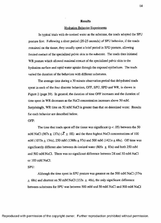
Results
Hydration Behavior Experiments
In typical trials with de-ionized water as the substrate, the toads adopted the SPU
posture first. Following a short period (20-25 seconds) of SPU behavior, if the toads
remained on the tissue, they usually spent a brief period in SPD posture, allowing
limited contact of the specialized pelvic skin to the substrate. The toads then initiated
WR posture which allowed maximal contact of the specialized pelvic skin to the
hydration surface and rapid water uptake through the exposed epithelium. The toads
varied the duration of the behaviors with different substrates.
The average time during a 30 minute observation period that dehydrated toads
spent in each of the four discrete behaviors, OFF, SPU, SPD and WR, is shown in
Figure 2 (page 39). In general, the duration of time OFF increases and the duration of
time spent in WR decreases as the NaCl concentration increases above 50 mM.
Surprisingly, WR time on 50 mM NaCl is greater than that on deionized water. Results
for each behavior are described below.
OFF:
The time that toads spent off the tissue was significant (p < .05) between the 50
mM NaCl (567s ± 137s) { X ± SE) and the three highest NaCl concentrations of 1(X)
mM (1073s ± 134s), 250 mM (1388s ± 97s) and 500 mM (1421s ± 68s). Off time was
significantly different also between de-ionized water (805s ± 83s) and both 250 mM
and 500 mM NaCl. There was no significant difference between DI and 50 mM NaCl
or 100 mM NaCl.
SPU:
Although the time spent in SPU posture was greatest on the 500 mM NaCl (374s
± 68s) and shortest on 50 mM NaCl (153s ± 46s), the only significant difference
between substrates for SPU was between 500 mM and 50 mM NaCl and 500 mM NaCl
R eproduced with perm ission of the copyright owner. Further reproduction prohibited without perm ission.
15
and de-ionized water (202s ± 38s) (both p < .05). The values for 250 mM NaCl
(277s ± 61s ) and 100 mM NaCl ( 212s + 52s) were not significantly different from
any other substrate.
SPD:
Time spent displaying SPD behavior was low and variable for all substrates and
the only significant difference was between 250 mM NaCl (124s ± 77s) and 500 mM
NaCl (3s ± 2) (p < 0.05). The values for de-ionized water (78s + 16s), 50 mM
NaCl ( 62s ± 22s) and 10 mM NaCl (53s ± 19s) were not significantly different from
any other substrate.
WR:
The duration of the WR behavior is shown in Figure 2 (page 39) and separately
in Figure 3 (page 40). De-ionized water, 50 mM and 100 mM NaCl were similarly
selected as suitable hydration substrates for dehydrated B. punctatus whereas the 250
mM and 500 mM NaCl concentrations were found unsuitable.
The mean value of time spent in WR was high for de-ionized water (714s ±
81s), 50 mM NaCl (1017s ± 145s) and on 100 mM NaCl (461s ± 137s). There
was virtually no time spent in WR on 250 mM (10s ± 4s) and 500 mM NaCl (Is ±
Is). Except for de-ionized water versus 100 mM NaCl and 250 mM versus 500 mM
NaCl the duration of WR varied significantly among substrates (p value <.05). As
noted above, toads spent more time in WR posture on 50 mM NaCl than on the more
osmotically favorable de-ionized water and this difference was significant (p < 0.05).
Rehvdration Experiments
The results of weight change for dehydrated toads immersed in de-ionized water
with or without various NaCl concentrations is shown in Figure 4 (page 41). To
account for differences in toad size and amount of dehydration, each measurement was
normalized to the toad standard weight. The highest average weight gain of standard
R eproduced with perm ission of the copyright owner. Further reproduction prohibited without perm ission.
16
weight was for toads placed for 20 minutes in 50 mM NaCl (7.5% ± 3.1%) (.^ + SE)
substrate. The average weight gain amounts for de-ionized water (4.2% ± 3.5%) and
100 mM NaCl (4.9% ± 2.6%) are significantly less than 50 mM NaCl (p < .05).
There was a small weight loss of approximately 1 % for the toads in 250 mM (-1.1% ±
2.3%) and 500 mM NaCl (-0.5% ± 2.3%) substrate, and both are significantly
different from the all other substrates (p < .05).
The results of the second rehydration experiment are shown in Figure 5. Again,
the highest average weight gain measured as percent of standard weight was for toads
placed in 50 mM NaCl (10.2% ± 1.0%) substrate, however, weight gain for toads in
50 mM NaCl with 10 |iM amiloride was significantly reduced (7.2% ± 2.6%). The
average weight gain amounts for de-ionized water (5.0% ± 2.4%) and de-ionized water
with amiloride (6.7% ± 3.2%) were significantly different but were significantly less
than those with 50 mM NaCl (p < .05). There was a small weight gain of
approximately 1 % for the toads in 250 mM NaCl (0.8% ± 3.5%) and a small weight
loss of approximately 1% for the toads in 250 mM NaCl with amiloride (-1.1% +
0.7%) substrate. Both values are significantly different from the all other substrates
(p < .05) but the 250 mM NaCl treatments with and without amiloride did not differ
significantly from each other.
Discussion
Hvdration Behavior Experiments
As shown in figure 3, the hydration behaviors indicate that de-ionized water,
100 mM NaCl and especially 50 mM NaCl are suitable hydration sources for dehydrated
B. punctatus while the 250 mM and 500 mM NaCl concentrations are unsuitable. The
results of the behavioral assays demonstrate that toads have the ability to detect and react
to differing salt concentrations in potential hydration sources. Toads also show a clear
R eproduced with perm ission of the copyright owner. Further reproduction prohibited without perm ission.
17
preference for some salt over no salt. The duration of WR on hypertonic NaCl solutions
is drastically lower compared to the duration of WR on hypotonic NaCl concentrations
(Figure 3) and reveals a threshold of tolerance. A preference by toads for some salt but
not very much salt in their hydration substrate is shown by the significant preference of
50 mM NaCl over de-ionized water and 100 mM NaCl.
WR allows maximal contact of the pelvic patch skin to the hydration surface and
rapid water uptake through the exposed epithelium. The skin of the pelvic patch has a
network o f channeling and skin thickenings (epidermal sculpturing) which increases the
area of skin that comes into contact with the hydration surface and increases potential for
water uptake (Lillywhite and Licht 1974). The epidermal sculpturing of Bufo punctatus
is especially pronounced and allows expansion of the contact area of the seat patch by
1.5 to 3.7 times (Brekke et al., 1991).
SPD posture allows limited contact of the specialized pelvic patch skin to the
substrate. The posture allows some water uptake as well as a more thorough
chemosensory evaluation of the substrate than SPU posture. The SPD is consistently of
short duration on all substrates except 500 mM NaCl, where it is not displayed at all.
This suggests that a suitable surface is readily detected by the toads. The SPD behavior
may be an information gathering posture that indicates some acceptance of the substrate.
When toads evaluate the suitability of a hydration source they first assume SPU
posture. This posture allows the toad to make a tentative evaluation of the substrate,
possibly directly through the feet (Stille, 1952; Brekke et al.., 1991). As shown in
Figure 2, SPU durations are comparable for all substrates except 500 mM NaCl where
sustained SPU behavior is noted.
A similar method was used in a study of salt taste discrimination behavior that
measured salt preference of the axolotl salamander, Ambystroma mexicanum, using a
simple behavioral assay that equated acceptance with swallowing and rejection with
R eproduced with perm ission of the copyright owner. Further reproduction prohibited without perm ission.
18
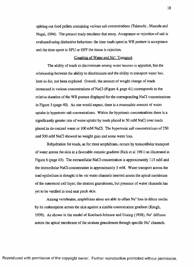
spitting out food pellets containing various salt concentrations (Takeuchi, Masuda and
Nagai, 1994). The present study emulates that assay. Acceptance or rejection of salt is
evaluated using distinctive behaviors: the time toads spent in WR posture is acceptance
and the time spent in SPU or OFF the tissue is rejection.
Coupling of Water and Na^ Transport
The ability of toads to discriminate among water sources is apparent, but the
relationship between the ability to discriminate and the ability to transport water has,
here-to-for, not been explored. Overall, the amount of weight change of toads
immersed in various concentrations of NaCl (Figure 4, page 41) corresponds to the
relative duration of the WR posture displayed for the corresponding NaCl concentrations
in Figure 3 (page 40). As one would expect, there is a reasonable amount of water
uptake in hypotonic salt concentrations. Within the hypotonic concentrations there is a
significantly greater rate of water uptake by toads placed in 50 mM NaCl over toads
placed in de-ionized water or 100 mM NaCl. The hypertonic salt concentrations of 250
and 500 mM NaCl showed no weight gain and some water loss.
Rehydration for toads, as for most amphibians, occurs by transcellular transport
of water across the skin in a favorable osmotic gradient (Rick et al 1981) as illustrated in
Figure 6 (page 43). The extracellular NaCl concentration is approximately 115 mM and
the intercellular NaCl concentration is approximately 5 mM. Water transport across the
toad epithelium is thought to be via water channels inserted across the apical membrane
of the outermost cell layer, the stratum granulosum, but presence of water channels has
yet to be verified in toad seat patch skin.
Among vertebrates, amphibians alone are able to offset Na* loss in dilute media
by its reabsorption across the skin against a sizable concentration gradient (Krogh,
1939). As shown in the model of Koefoed-Johnsen and Ussing (1958), Na* diffuses
across the apical membrane of the stratum granulosum through specific Na* channels.
R eproduced with perm ission of the copyright owner. Further reproduction prohibited without perm ission.
19
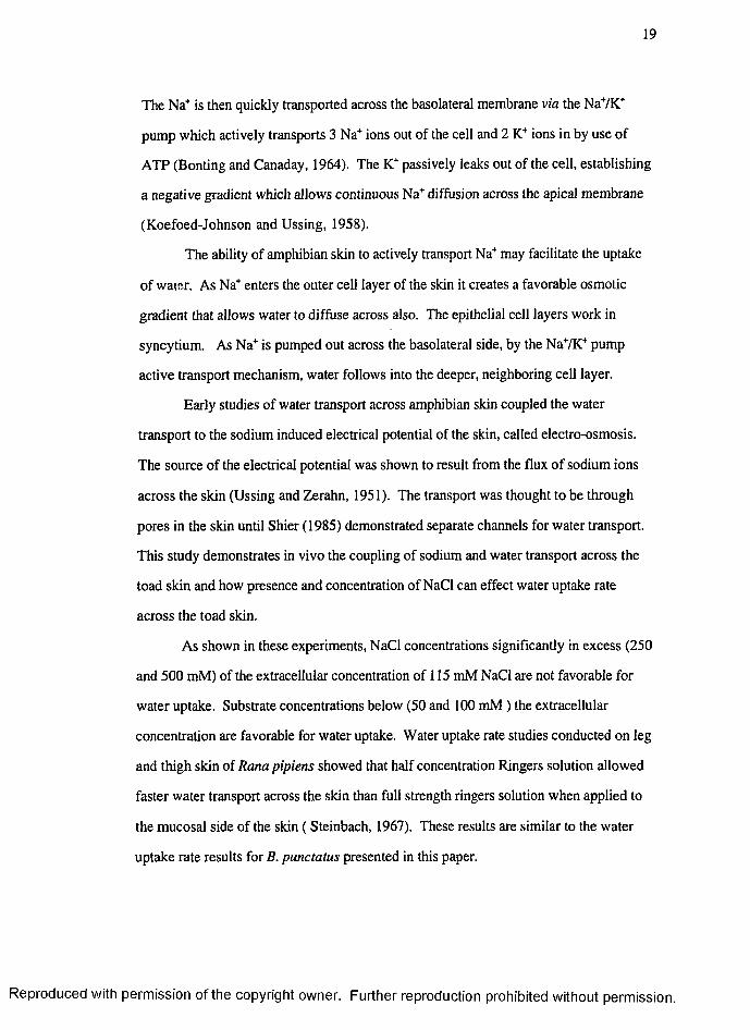
The Na* is then quickly transported across the basolateral membrane via the NaVK*
pump which actively transports 3 Na' ̂ions out of the cell and 2 ions in by use of
ATP (Bonting and Canaday, 1964). The K"" passively leaks out of the cell, establishing
a negative gradient which allows continuous Na'" diffusion across the apical membrane
(Koefoed-Johnson and Ussing, 1958).
The ability of amphibian skin to actively transport Na"" may facilitate the uptake
of water. As Na* enters the outer cell layer of the skin it creates a favorable osmotic
gradient that allows water to diffuse across also. The epithelial cell layers work in
syncytium. As Na'" is pumped out across the basolateral side, by the NaVK* pump
active transport mechanism, water follows into the deeper, neighboring cell layer.
Early studies of water transport across amphibian skin coupled the water
transport to the sodium induced electrical potential of the skin, called electro-osmosis.
The source of the electrical potential was shown to result from the flux of sodium ions
across the skin (Ussing and Zerahn, 1951). The transport was thought to be through
pores in the skin until Shier (1985) demonstrated separate channels for water transport.
This study demonstrates in vivo the coupling of sodium and water transport across the
toad skin and how presence and concentration of NaCl can effect water uptake rate
across the toad skin.
As shown in these experiments, NaCl concentrations significantly in excess (250
and 500 mM) of the extracellular concentration of 115 mM NaCl are not favorable for
water uptake. Substrate concentrations below (50 and 100 mM ) the extracellular
concentration are favorable for water uptake. Water uptake rate studies conducted on leg
and thigh skin of Rana pipiens showed that half concentration Ringers solution allowed
faster water transport across the skin than full strength ringers solution when applied to
the mucosal side of the skin ( Steinbach, 1967). These results are similar to the water
uptake rate results for B. punctatus presented in this paper.
R eproduced with perm ission o f the copyright owner. Further reproduction prohibited without perm ission.
20
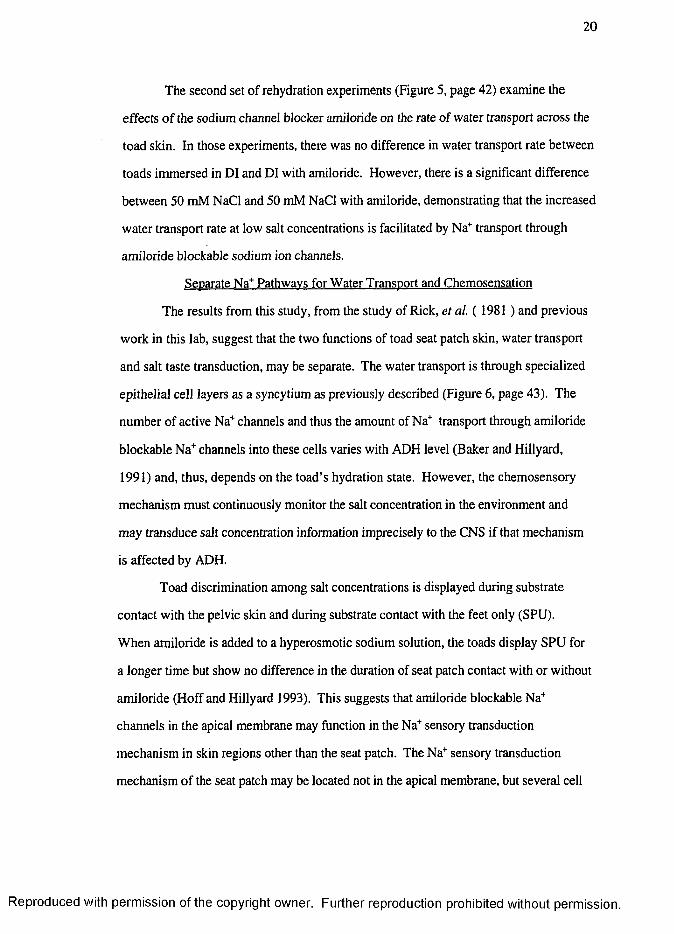
The second set of rehydration experiments (Figure 5, page 42) examine the
effects o f the sodium channel blocker amiloride on the rate of water transport across the
toad skin. In those experiments, there was no difference in water transport rate between
toads immersed in DI and DI with amiloride. However, there is a significant difference
between 50 mM NaCl and 50 mM NaCl with amiloride, demonstrating that the increased
water transport rate at low salt concentrations is facilitated by Na* transport through
amiloride blockable sodium ion channels.
Separate Na^ Pathwavs for Water Transport and Chemosensation
The results from this study, from the study of Rick, et a l (19 8 1 ) and previous
work in this lab, suggest that the two functions of toad seat patch skin, water transport
and salt taste transduction, may be separate. The water transport is through specialized
epithelial cell layers as a syncytium as previously described (Figure 6, page 43). The
number of active Na* channels and thus the amount of Na* transport through amiloride
blockable Na' ̂channels into these cells varies with ADH level (Baker and Hillyard,
1991) and, thus, depends on the toad’s hydration state. However, the chemosensory
mechanism must continuously monitor the salt concentration in the environment and
may transduce salt concentration information imprecisely to the CNS if that mechanism
is affected by ADH.
Toad discrimination among salt concentrations is displayed during substrate
contact with the pelvic skin and during substrate contact with the feet only (SPU).
When amiloride is added to a hyperosmotic sodium solution, the toads display SPU for
a longer time but show no difference in the duration of seat patch contact with or without
amiloride (Hoff and Hillyard 1993). This suggests that amiloride blockable Na"’
channels in the apical membrane may function in the Na'’ sensory transduction
mechanism in skin regions other than the seat patch. The Na* sensory transduction
mechanism of the seat patch may be located not in the apical membrane, but several cell
R eproduced with perm ission of the copyright owner. Further reproduction prohibited without perm ission.
21
layers beneath the apical membrane and the route of sodium ion transport for
chemosensation may be paracellular. Furthermore, when the sodium ion channel
blocker, amiloride, is applied to the skin of the toad Bufo marinus a several minute
wash of the blocker on the skin is required before there is any inhibition of neural
response from 250 mM NaCl (Maleek et a i, 1998). If the sensory cells are in a sub-
epithelial location the prolonged wash time may be needed to allow the large impermeant
amiloride molecule to diffuse into the epithelial cell layers beneath the apical membrane.
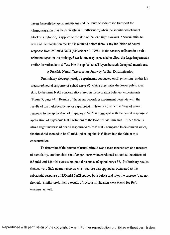
A Possible Neural Transduction Pathway for Salt Discrimination
Preliminary electrophysiolgy experiments conducted on B. punctatus in this lab
measured neural response of spinal nerve #6, which innervates the lower pelvic area
skin, to the same NaCl concentrations used in the hydration behavior experiments
(Figure 7, page 44). Results of the neural recording experiment correlate with the
results of the hydration behavior experiment. There is a distinct increase of neural
response to the application of hypertonic NaCl as compared with the neural response to
application of hypotonic NaCl solutions to the lower pelvic skin area. Since there is
also a slight increase of neural response to 50 mM NaCl compared to de-ionized water,
the threshold seemed to be 50 mM, indicating that Na’' flows into the skin at this
concentration.
To determine if the source of neural stimuli was a taste mechanism or a measure
of osmolality, another short set of experiments were conducted to look at the effects of
0.5 mM and 1.0 mM sucrose on neural response of spinal nerve #6. Preliminary results
showed very little neural response when sucrose was applied as compared to the
substantial response of 250 mM NaCl applied both before and after the sucrose (data not
shown). Similar preliminary results of sucrose application were found for Bufo
marinus as well.
R eproduced with perm ission of the copyright owner. Further reproduction prohibited without perm ission.
22
Overall there is a trend showing that as the salt concentration increases, the
amount of neural response also increases. Although inconclusive as to the specific
transduction mechanism, these results support the hypothesis that the transduction
pathway for salt discrimination in toads is at least in part, via the 6th spinal nerve.
Conclusion
Red Spotted toads, Bufo punctatus dwell in arid habitats of the southwestern
United States. They forage at night as much as 100 meters away from their canyon
springs habitat. Although they can withstand dehydration of up to 40% of body weight
(McClanahan and Baldwin, 1969; McClanahan, Ruibal and Shoemaker, 1994), when
they are faced with the mostly dry habitat of the desert, the ability to rehydrate quickly is
crucial. Their need to rehydrate is likely to be a limiting factor for forays away from
hydration sources. Therefore the ability for these toads to evaluate favorable hydration
sources is critical to their survival.
Bufo punctatus are able to detect varying concentrations of salt in a potential
hydration substrate. The display of a distinct acceptance or rejection behavior by the
toad to the various NaCl concentrations suggests a threshold exists for salt tolerance.
Results of the rehydration experiments also suggests a similar threshold for favorable
water uptake in salt solution that may correspond with the toad interstitial fluid
concentration of about 115 mM NaCl. In the Red-spotted toad, the preference or
avoidance of the various NaCl concentrations correlates with the water uptake or water
loss rate. All hyposmotic salt concentrations were found to be acceptable hydration
substrates in the behavioral experiment as well as favorable hydration sources for the
toads. Conversely, the hyperosmotic salt concentrations were avoided and were shown
to deplete much needed water from the toads. This distinct behavioral difference
between high and low salt concentrations suggests a threshold exists for salt tolerance
R eproduced with perm ission of the copyright owner. Further reproduction prohibited without perm ission.
23
in Bufo punctatus and indicates that the toads can evaluate whether a surface is
acceptable or not and act accordingly by either adopting WR on the substrate or avoiding
the substrate.
The uptake of NaCl through amiloride blockable Na* ion channels facilitates the
uptake of water and this mechanism for water uptake appears to be present in toad pelvic
skin and within the concentrations studied, is highest at the 50 mM NaCl concentration.
Within the hyposmotic substrates tested in the hydration behavior experiments, some
salt (the 50 mM concentration), was found to be preferred over deionized water as a
hydration source. This suggests that the toad has a mechanism to discriminate between
different salt concentrations and that there is a physiological reason for such a
mechanism to exist in desert dwelling anurans.
Further studies investigating salt uptake through paracellular pathways may help
to clarify the possible functional distinction between transcellular and paracellular Na*
transport. Behavioral and neural studies manipulating the cellular junctions on the apical
membrane of toad seat patch may reveal the nature of salt chemosensory and water
uptake functions of toad pelvic skin.
R eproduced with perm ission of the copyright owner. Further reproduction prohibited without perm ission.
CHAPTER 3
EFFECTS OF SODIUM GLUCONATE CONCENTRATION ON
WATER TRANSPORT AND HYDRATION BEHAVIOR IN
RED-SPOTTED TOADS, Bufo punctatus.
Introduction
Amphibians rehydrate by transporting water across the skin down a favorable
osmotic gradient (Bentley and Yorio, 1979). Many amphibian species have specialized
areas of skin that enhance water uptake. Toads in the genus Bufo have a highly
permeable region in the lower ventral skin that is specially adapted for water uptake
termed the seat patch or pelvic patch (McClanahan and Baldwin, 1969; Marrero and
Hillyard, 1985). Rehydrating toads press their pelvic patch onto the moist surface with
feet parallel to the body axis with just the central patch region touching the surface. This
is called seat patch down (SPD) behavior. In the same posture but with hindlimbs
splayed to allow maximal patch contact the behavior is called water absorption response
(WR). These behaviors together are called hydration behavior posture (Maleek et al..,
1998).
Brekke, Hillyard and Winokur (1991) showed that when dehydrated Bufo
punctatus were placed on a tissue saturated with hyperosmotic urea solutions they
would not initiate WR. Studies by Hoff and Hillyard (1993b) further showed that
dehydrated toads avoided surfaces saturated with hyperosmotic NaCl and KCl. This
24
R eproduced with perm ission o f the copyright owner. Further reproduction prohibited without perm ission.
25
study also found that amiloride, a blocker of epithelial Na* channels, increased the
frequency of initiation of WR behavior on NaCl but not on KCl or urea solutions. The
duration of the episodes of WR behavior was very brief so that the total time showing
this behavior was not increased during a five-minute observation period. These results
suggest that toads initially evaluate an hydration surface with a sensory mechanism that
includes an amiloride-sensitive transport pathway that is selective for Na'’' but that a
commitment to maintain skin contact involves their ability to discern the osmolality of an
hydration source by an amiloride-insensitive mechanism.
Further studies in this lab demonstrated that in Red-Spotted toads, Bufo
punctatus, water uptake is more rapid when the animals are immersed in 50 mM than in
water containing no salt (de-ionized water) or in 100 mM NaCl. As demonstrated in
chapter 2 , toads fail to gain weight and often lose weight when immersed in
hyperosmotic NaCl solutions of 250 and 500 mM NaCl. It was also demonstrated that
Red-spotted toads show preference for an hydration source that contains 50 mM NaCl
over de-ionized water or 100 mM NaCl and hyperosmotic salt concentrations of 250
mM and 500 mM NaCl. Preliminary electrophysiology studies suggest that the
transduction of sodium taste is at least in part through the spinal nerves that innervate the
lower abdominal skin (including the seat patch) of the toad Bufo punctatus.
The electropositive gradient established by active transport of Na’’ is neutralized
by the cotransport of Cl' across the toad skin (Koefoed-Johnsen and Ussing, 1958).
Although much is known about the transport of chloride across epithelium, but the
relative amount of paracellular versus transcellular Cl transport remains controversial.
Ye, Heck and DeSimone (1991) noted that in canine lingual epithelium there is a
paracellular shunt for chloride associated with the taste receptor cells. They showed that
blockage of the movement across the tissue reduced the neural response to sodium
suggesting that the transport of Na'’’ ions across the lingual epithelium is dependent, in
R eproduced with perm ission of the copyright owner. Further reproduction prohibited without perm ission.
26
part, on the cotransport of the chloride anion. Jensen, Sorensen, Larsen, and
Wiliumsen (1997) showed that in dilute media the active transport of chloride is driven
by a Cl - HCOj' exchange mechanism and is coupled to an active transport of Na*
through the Na7K'’ pump. Both mechanisms are located on the apical membrane of
mitochondrial rich cells in toad epithelium. Previous studies by Larsen (1991) on
principle cells of toad skin demonstrated that there was no transcellular Cl transport
across these cells. As discussed previously (see chapter 2), the chloride ion may diffuse
across the toad skin by way of an electrogradient driven paracellular route or,
alternatively, the chloride ion may transport actively across the apical membrane of the
mitchondrial rich cells in the toad skin.
Little is known about the coupling of Na* and Cl transport across the toad seat
patch skin. In this study we look at the impact of the Cl on epithelial sodium transport
by comparing the effects of the Cl and gluconate anions on the transport of Na^ across
the toad skin. Weight change of dehydrated toads placed in de-ionized water and 50
and 250 mM concentrations of both NaCl and NaGluconate was measured to determine
if the presence of the Cl', together with Na*, effects the rate of water uptake in Red-
spotted toads.
The gluconate anion is much larger than the chloride anion an d , unlike chloride,
it is impermeant to passive diffusion across the epithelium. There are no known
gluconate channels that may accommodate transcellular transport of the large anion
across the skin. The rate of gluconate penetration into the skin might thus relate to the
size of the anion and the tightness of the junctions between the epithelial cells.
An hydration behavior assay was used to investigate the ability of dehydrated
toads, Bufo punctatus, to detect salt in hydration substrates with or without the presence
of the Cl'. Time spent in hydration behavior is compared among deionized water, 50
R eproduced with perm ission of the copyright owner. Further reproduction prohibited without perm ission.
27
mM, and 250 mM concentrations of both NaCl and NaGluconate and related to water
weight gain by toads immersed in these same solutions.
Methods
Animals
Bufo punctatus were collected from several sites in the Spring Mountains in
Clark County Nevada (Nevada Department of Wildlife Scientific Collection Permit
# S 14965). Experiments were conducted from May through November of 1996 and
1997. Toads were kept in 75 x 30 cm terraria containing local desert sand, large stacked
rocks and pooled tap water that simulated the hydric conditions of their natural habitat.
The toads were kept on a 12:12 L:D cycle at room temperature (21-24 °C) and were fed
crickets twice or three times a week. Toad mass ranged from 7 to 20 grams.
For all experiments the urinary bladder contents were emptied with a
polyethylene cannula and the standard weight, the weight of a hydrated toad with empty
urinary bladder (Ruibal 1962), was recorded. The toads were placed in a dry 40 x 21 x
27 cm glass tank for two to four hours, until dehydrated by approximately 10% of the
standard weight. The actual dehydration of the toads used in the experiment ranged from
7% to 14%.
Toads were selected at random but without repetition for each experiment. Once
a toad was used in an experiment it was not used again until all other toads in the group
were used. In the behavioral experiments, the substrate was randomly selected.
Rehvdration Experiments
When in contact with a hydration surface, Bufo punctatus can take up water
rapidly. The purpose of this experiment was to measure the amount of water weight
gain when hydration behaviors do not affect contact with the substrate. Each toad was
placed in a straight sided widemouth glass jar 17 cm in height x 9 cm in diameter filled
with 150 ml of water or aqueous solutions of NaCl or NaGluconate to a depth of
R eproduced with perm ission of the copyright owner. Further reproduction prohibited without perm ission.
28
approximately 4 cm. This depth gave constant submersion of the lower abdominal area
of the toad while allowing the toad to breath air when in a comfortable sitting posture.
For the smaller toads the water level was reduced.
For this experiment the substrates used were de-ionized water, and aqueous
solutions of 50 mM, and 250 mM concentrations of both NaCl and NaGluconate. Each
toad was immersed in the substrate for 20 minutes. The difference between the standard
pre-experiment weight and the post experiment weight was the rehydration weight
change. Before each weighing, the toads were dipped in de-ionized water so that the
mass of water adhering to the skin would not bias the observed weight gain or loss.
Hydration Behavior Experiments
Each experiment was conducted in a 40 x 21 x 27 cm glass aquaria with sides
covered with black plastic or paper. Two observational cubicles were made with a
cardboard wall dividing down the middle to allow experimental and control observations
to be run in the same place and at the same time. Clear observation of the rehydration
posture displays by the toads was made through the underside of the glass tank by use
of an angled 30 cm square mirror placed beneath the tank. Presentation of the substrates
to the toads was by saturation on a 10 x 10 cm piece of laboratory tissue centered within
each observational cubicle. Three ml of substrate was used for each trial. The control
substrate was deionized water from the building tap supply.
Behavioral assays were designed to see what choices were made by dehydrated
toads when presented with 50 mM and 250 mM solutions of NaCl and NaGluconate.
Previous studies (discussed in chapter 2) have shown that Bufo punctatus shows
preference for 50 mM NaCl but avoids 250 mM NaCl. In the same study several
discrete behavioral states used in this experiment were defined and discussed in detail
(see chapter 2 for illustrations). In summary they are:
1. OFF the toad is completely off the substrate saturated tissue.
R eproduced with perm ission o f the copyright owner. Further reproduction prohibited without perm ission.
29
2. Seat patch up (SPU) the toad has one or more feet pressed against the tissue.
3. Seat patch down (SPD) the toad has a portion of the lower abdominal skin , the pelvic
patch area, pressed against the tissue while placing hindlimbs parallel to the body.
4. Water absorption response (WR) the toad has a portion of the lower abdominal skin,
the pelvic patch area, pressed against the tissue while placing hindlimbs at an
outward angle. This posture allows more pelvic patch skin contact with the moist
surface than in SPD posture.
To evaluate the impact of chloride on the decision making process as well as the
decision of whether a substrate is a favorable hydration source this study combined the
time spent in SPD and WR postures into what is termed the hydration behavior.
In typical control experiments with de-ionized water as the substrate, the toads
adopted the SPU posture first. Following a short period (20-25 seconds) of SPU
behavior, if the toads remained on the tissue, they usually spent a brief period in SPD
posture allowing limited contact of the specialized pelvic skin to the substrate. The toads
then initiated WR posture which allowed maximal contact of the specialized pelvic skin
to the hydration surface and rapid water uptake through the exposed epithelium. The
toads varied the duration of the behaviors with different substrates.
Barometric pressure and relative humidity were recorded at the time of bladder
cannulization and before and after each experimental trial. Changing barometric
pressure (Hoff and Hillyard 1993a) and relative humidity (Hoff and Orgeron, in prep.)
are known to affect hydration behaviors. The time of onset of each discrete behavioral
state was observed visually. For each experimental trial the duration in seconds for each
behavioral state was tallied. Mean values for duration of hydration behaviors and
weight change were compared by ANOVA using the Statview statistical software
package (Abacus Concepts).
R eproduced with perm ission of the copyright owner. Further reproduction prohibited without perm ission.
30
Results
Rehvdration Experiments
Results of the NaCl and NaGluconate rehydration experiment are shown as
percent weight change for toads immersed in the experimental substrates (Fig. 8, page
45). To account for differences in toad size and amount of dehydration, each
measurement of water gain was expressed as a percentage of the toad’s standard weight.
Experiments for the two salts were done over a two year time period and are normalized
to the control group (de-ionized water) done during the same time period. Figure 8
shows the weight change of toads immersed in solutions of 50 mM NaCl (2.52 ±
0.40) ( Z ± SE), 50 mM NaGluconate (0.96 ± 0.29), 250 mM NaCl (-0.64 ± 0 .3 1 )
and 250 mM NaGluconate. (-0.26 ± 0.24). All substrates are significantly different
from each other (p < 0.05) except between 250 mM NaCl and 250 mM NaGluconate.
Toads regained water significantly faster from 50 mM NaCl than from 50 mM
NaGluconate (p < 0.05). Differences between water loss in 250 mM were not
significant between NaCl and NaGluconate; however, both were significantly different
from the 50 mM solutions.
Hvdration Behavior Experiments
Duration of hydration behavior did not differ between the 50 mM salts but was
significantly longer with gluconate in the 250 mM concentrations. Figure 9 (page 46)
shows the results of hydration behavior (measured as a fraction of the associated
deionized water control group) of toads on 50 mM NaCl (1.26 ± 0.20) (.Z ± SE), 50
mM NaGluconate (1.28 ± 0.01), 250 mM NaCl (0.17 ± 0.08) and 250 mM
NaGluconate. (0.638 + 0.17).
The duration of hydration behavior for toads on 250 mM NaGluconate is
significantly longer than the duration of hydration behavior on 250 mM NaCl (p < 0.05)
R eproduced with perm ission of the copyright owner. Further reproduction prohibited without perm ission.
31
demonstrating that the toads have a higher chemosensory tolerance for 250 mM sodium
when coupled with the larger, less permeable anion, gluconate.
Discussion
Anion Transport Across the Epithelium
Past studies indicate that the Na'’' transport across the chemosensory epithelium
may be enhanced when coupled with chloride, a small ion of opposite charge (Ye et a l,
1991). Studies presented in Chapter 2 suggest two separate modes of sodium ion
transport across the skin: transcellular through amiloride blockable sodium ion channels
located on the apical membrane for water transport function, and an amiloride insensitive
component that is possibly a paracellular route for the chemosensory function of toad
seat patch skin. It is likely that an anion cotransport would be involved in both means
of Na* transport.
The effects of the anion coupling on Na* transport has been suggested to impact
the transcellular water uptake mechanism in toad epithelium (Larsen, 1991). Early
studies by Ussing and Zerahn (1951) showed that Na* is actively transported across the
toad skin by a basolaterally located NaVK* pump that maintains a favorable osmotic
gradient across the apical membrane for Na* diffusion. Rick et a/. (1981) demonstrated
that the epithelial cell layers were a syncytial Na* transport compartment. Previous
studies in this lab on the water uptake rate in toads (chapter 2) showed that some NaCl
(50 mM) increases water uptake rate over no NaCl (deionized water) in the hydration
source, demonstrating that the active transport of Na* across the toad skin facilitates
water transport. Studies for this chapter replaced the Na'’ coupled anion chloride with
gluconate. Results show that when Na* solutions are made with the impermeant anion
gluconate, the rate of water transport is reduced. This suggests that the chloride anion is
coupled to the transport of sodium across the apical membrane in toad seat patch skin.
R eproduced with perm ission of the copyright owner. Further reproduction prohibited without perm ission.
32
There are two probable pathways for the epithelial transport of chloride, passive
paracellular diffusion or active transport across the membrane of mitochondrial-rich cells
found in the toad skin.
The same water uptake experiment was done with hyperosmotic sodium solution
and results showed no difference between the two anions, chloride and gluconate, on
water uptake rate at the scale of measurement allowed by the methodology. This
indicates that osmotic water movement is the primary mechanism of water loss into
solutions having a higher external osmolality.
Anion Transport and Chemosensation
The presence of chloride may facilitate the transport of Na* across the
epithelium. If so, it could enhance the detection of salt by the toad. Previous studies in
this lab showed that toads display different and reproducible behaviors in response to
high and low NaCl concentrations. Conversely, the reduced Na* transport across the
toad epithelium may reduce the amount sodium detected by the toad chemosensory
mechanism the nature of which is unknown.
As in the studies of Ye e? a/., (1991) that demonstrated that in canine lingual
epithelium, the presence of chloride in the microenvironment of the taste cell impacts the
neural response to NaCl, the impact of chloride on the behavior of the toads may be due
to the passive diffusion of chloride into the microenvironment of the chemosensory
mechanism embedded several cell layers into the toad skin.
In this study, the higher concentration of NaGluconate shows a significant
increase in tolerance to 250 mM sodium. This may be caused by a decreased influx of
sodium across the epithelium and subsequent reduction of salt detection by
chemosensory mechanisms in the toad. The increase of seat patch exposure to the 250
mM NaGluconate is about equal between the SPD and WR postures ( SPD 107s ±
53s, WR 133s ± 57s). This may indicate that lack of chloride to facilitate Na’̂ across
R eproduced with perm ission of the copyright owner. Further reproduction prohibited without perm ission.
33
the skin makes the higher 250 mM concentration appear to be marginally potable.
Further analysis of duration of hydration behavior posture as a function of the number
of times that the behaviors are initiated (NaCl 13.2s ± 5s, NaGluconate 61s ± 19s,
p < 0.05)) shows more clearly that the toads are actually allowing a longer duration of
seat patch contact on hyperosmotic sodium concentrations when the sodium ion is
presented with gluconate. In other words, to a thirsty toad the substrate may not seem
salty enough to be avoided, but the salt is not low enough to warrant lengthy exposure
of the specialized seat patch skin when in WR posture.
Conclusion
Toads are able to detect varying concentrations of salt in a potential hydration
substrate and the acceptance or avoidance of given sodium chloride concentrations
parallells the benefit or detriment of the substrate to water uptake rate in B. punctatus.
When the sodium ion is coupled to gluconate instead of chloride in an
hyperosmotic (250 mM) sodium solution, the toads show less avoidance of the substrate
and display hydration behavior for significantly longer. This suggests that the transport
of sodium ions across the skin is coupled to the transport of the chloride anion. An
impermeant anion will slow down the rate of sodium transport across the skin thus
reducing the amount of salt detected by the toads after adopting the hydration behavior.
In the immersion studies, the water uptake rate for 50 mM NaCl is much higher
than the rate for 50 mM NaGluconate. Past studies have suggested a facilitation of
water uptake by the transport of Na^ across the skin. The epithelial water transport rate
difference between the two substrates may in part result from the presence of chloride.
The mechanism of Cl transport, whether through chloride channels associated
with the syncytial Na^ transport compartment, via a paracellular route or through
chloride channels in mitochondrial rich cells, is unknown. Future studies of water
R eproduced with perm ission of the copyright owner. Further reproduction prohibited without perm ission.
34
uptake measurement that incorporate methods to block Cl channels, may shed light on
the route(s) of sodium coupled chloride transport for the chemosensory and water
uptake mechanisms in toad seat patch skin.
R eproduced with perm ission of the copyright owner. Further reproduction prohibited without perm ission.
REFERENCES
Baker C.A. and S.D. Hillyard. 1992 Capacitance, short-circuit current and osmoticwater flow across different regions of the isolated toad skin. J. Comp. Physiol. 162:707-713.
Barker Jorgensen, C. 1997. 200 Years of Amphibian Water Economy: From Robert Townson to the Present. Biol. Rev. 72:153-237.
Bentley P.J. and T. Yorio. 1979. Do frogs drink? J. Exp. Biol. 79:41-46
Bonting, S.L. and M R. Canady 1964. Na-K activated adenosine triphosphate and sodium transport in toad bladder. Am. J. Physiol. 207:1005-1009.
Brekke, D.R., S.D. Hillyard and R.M. Winokur. 1991. Behavior associated with the water absorption response by the toad, Bufo punctatus. Copeia 1991:393-401
Christoffersen B.C., L.J. Jensen, L.J., E.H. Larsen, J.B. Sorensen and N.J.Wiliumsen. 1997. Proton pump activity of Mitochondria-rich cells. J. Exp. Physiol. 109:73-84.
DeSimone, J.A., G.L. Heck, S. Miersen and S.K. DeSimone 1984. The active ion transport properties of canine lingual epithelium in vitro. J. Gen. Physiol. 83:633-656.
Hillyard, S.D. 1988. Comparison of the surface area of pelvic and pectoral regions of toad skin using impedance analysis. Am. Zool. 28:17A.
Hillyard, S.D., K. vS. Hoff and C. Propper. 1998. The water absorption response: a behavioral assay for physiological processes in terrestrial amphibians. Phys. Zoo. 09:43:48 1-12.
Hillyard, S.D., K. Hoff and P. Sullivan. 1997. Species differences in the sensoryfunction of epithelial Na’' channels (ENaCs) in toad skin. APS Conference: The physiology and functional diversity of amiloride-sensitive Na"’' channels: a new gene superfamily. Park City Utah, October 29-November 1, 1997. The Physiologist 40: A -18.
Hoff K. vS. and S.D. Hillyard. 1991. Angiotension II stimulates cutaneous drinking in the toad, Bufo punctatus. Physiol. Zool. 64:1165-1172.
35
R eproduced with perm ission of the copyright owner. Further reproduction prohibited without perm ission.
36
Hoff K.vS. and S.D. Hillyard. 1993a. Inhibition of cutaneous water absorption indehydrated toads by saralasin is associated with changes in barometric pressure. Physiol. Zool. 66:89-98.
Hoff, K. vS. and S.D. Hillyard 1993b. Toads taste sodium with their skin: sensory function in a transporting epithelium. J. Exp. Biol. 183:347-351.
Hoff, K vS. and K. Orgeron. Effects of environmental parameters on hydration behavior of the toad, Bufo marinus. in prep.
Jensen L.J., J.N. Sorensen, E.H. Larsen and N.J. Wiliumsen. 1997. Proton pump activity o f mitochondria-rich cells: The interpretation of external proton- concentration gradients. J. Gen. Physiol. 109:73-91.
Jorgensen, C.B. 1997. 200 years of amphibian water economy: from Robert Townson to the present. Biol. Rev. 72:153-237
Koefoed-Johnsen , V., H. Levi and H.H. Ussing 1952. The mode of passage ofchloride ions through the isolated frog slun. Acta Physiol. Scand. 25:150-163.
Koefoed-Johnsen, V. and H.H. Ussing. 1958. The nature of frog skin potential. Acta Physiol. Scand. 42:298-308.
Krogh, A. 1939. Osmotic Regulation in Aquatic Animals. Cambridge: Cambridge University Press.
Larsen, E.H. 1991. Chloride transport by high-resistance heterocellular epithelia. Am. Physiol. Soc. 71:235-283
Lilliywhite, H.B. and P. Licht .1974. The movement of water over toad skin: the role of epidermal sculpturing. Copeia 1974:165-171.
Lindemann, B. and W. Van Driessche. 1977. Sodium specific membrane channels of frog skin are pores: current fluctuations reveal high turnover. Science 195:292-294.
Maleek, R., P. Sullivan, K. vS. Hoff and S.D. Hillyard. 1998. Salt sensitivity and hydration behavior of the toad Bufo marinus. submitted to Physiol. Beh.
Marrero, M B., and S.D. Hillyard. 1985. Differences in cyclic AMP levels in epithelial cells from pelvic and pectoral regions of toad skin. Comp. Biochem. Physiol. 82C:69-73.
McClanahan, L.L. Jr. and R. Baldwin. 1969. Rate of water uptake through the integument of the desert toad, Bufo punctatus. Comp. Biochem. Physiol. 28:381-389.
McClanahan L.L., Jr., R. Ruibal, and V.H. Shoemaker. 1995. Frogs and toads in deserts. Sci. Am. 274:64-70.
R eproduced with perm ission o f the copyright owner. Further reproduction prohibited without perm ission.
37
Nouwen, E.J. and E.R. Kuhn. 1984. Radioimmunoassay of arginine vasotocin and mesotocin in serum of the frog Rana ridibunda. Gen. Comp. Endochrinol. 50:242-251.
Phillips M.I. 1987. Functions of angiotension in the central nervous system. Annu. Rev. Physiol. 49:413-435.
Rick, R., A. Dorge, E. von Amim, M. Weigal and K. Thurau. 1981. Properties of the outer and inner barriers to transepithelial Na transport: an electron microprobe analysis, in. Macknight, A.D.C. and J.P. Leader eds. Epithelial Ion and Water Transport. Raven Press New York.
Ruibal, R. 1962. The adaptive value of bladder water in the toad, Bufo cognatus. Physiol. Zool. 35:218-223.
Schrier, R.W. (ed.) 1985. Vasopressin. New York: Raven Press.
Steinbach H.B.. 1967. On the ability of isolated frog skin to manufacture Ringer’s fluid. J. of Anat. Physiol. 50:2377-2389.
Stewart, R.E., J.A. DeSimone and D.L. Hill. 1997. New perspectives in gustatory physiology: transduction, development, plasticity. Am.J. Physiol. (Cell Physiol.) 41: C-1-C26.
Stille, W.T. 1952. The nocturnal amphibian fauna of the southern Lake Michigan beach. Ecology 33:149-162.
Stille, W.T. 1958. The water absorption response of an anuran. Copeia 1958:217-218.
Takeuchi, H.A., T. Masuda and T. Nagai. 1994. Electrophysiological and behavioral studies of taste discrimination in the Axolotl, Ambystoma mexicanum. Physiol. Beh. 56:121-127.
Townson, R. 1799. Tracts and Observations in Natural Historv and Phvsiologv. London: printed for the Author by J. White.
Ussing H.H. and K. Zerahn. 1951. Active transport of sodium as the source of electric current in the short-circuited isolated frog skin. Acta Physiol. Scand.23:110-127.
Winokur, R.M. and S.D. Hillyard .1992. Pelvic cutaneous musculature in toads of the genus Bufo. Copeia 1992:760-769.
Ye, Q., G.L. Heck and J.A. DeSimone 1991. The anion paradox in sodium taste reception: resolution by voltage clamp studies. Science 254:724-726.
R eproduced with perm ission of the copyright owner. Further reproduction prohibited without perm ission.
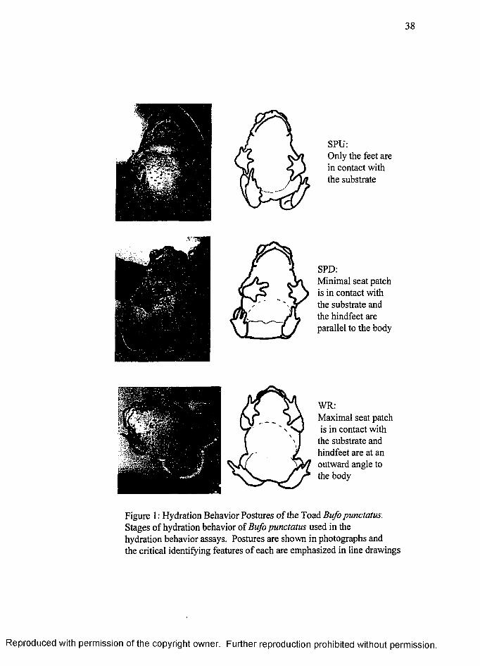
38
SPU:Only the feet are in contact with the substrate
SPD:Minimal seat patch is in contact with the substrate and the hindfeet are parallel to the body
WR:Maximal seat patch is in contact with
the substrate and hindfeet are at an outward angle to the body
Figure 1 : Hydration Behavior Postures o f the Toad Bufo punctatus. Stages of hydration behavior of Bufo punctatus used in the hydration behavior assays. Postures are shown in photographs and the critical identifying features o f each are emphasized in line drawings
R eproduced with perm ission of the copyright owner. Further reproduction prohibited without perm ission.
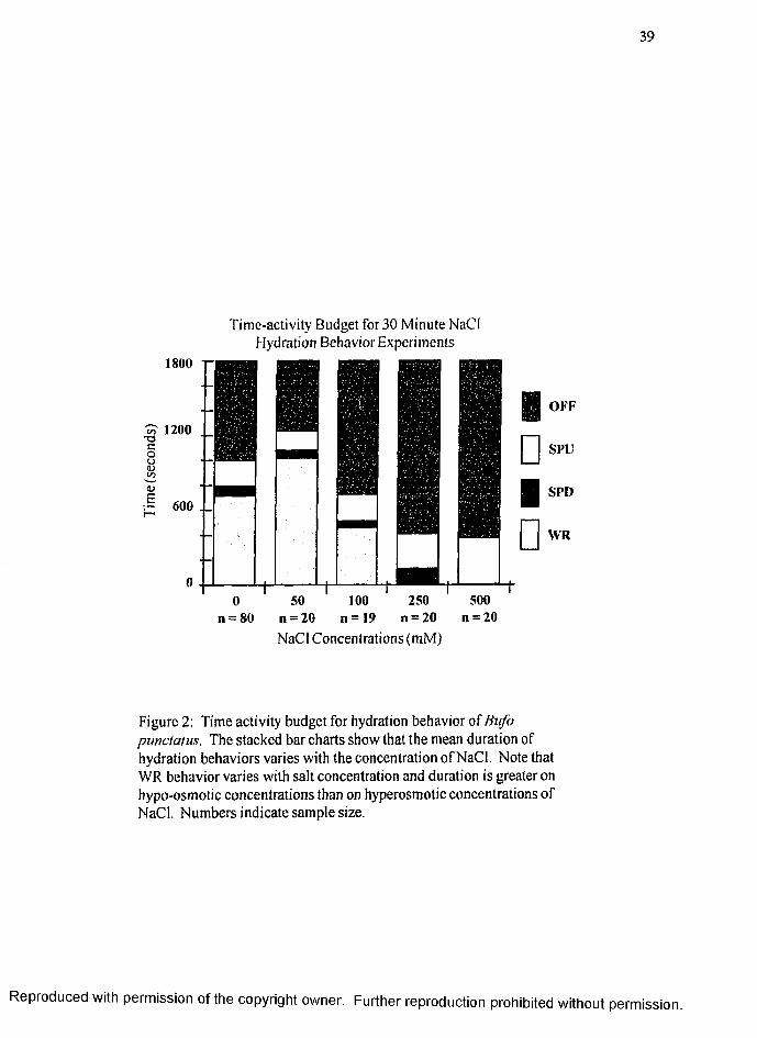
39
Time-activity Budget for 30 Minute NaCl Hydration Behavior Experiments
1800 r
1200
1)E— 600 . _
OFF
SPU
SPD
WR
50 100 250 500n = 20 n = 1 9 n = 20 n = 20
NaCl Concentrations (mM)
Figure 2: Time activity budget for hydration behavior of Bufo punctatus. The stacked bar charts show that the mean duration of hydration behaviors varies with the concentration of NaCl. Note that WR behavior varies with salt concentration and duration is greater on hypo-osmotic concentrations than on hyperosmotic concentrations of NaCl. Numbers indicate sample size.
R eproduced with perm ission of the copyright owner. Further reproduction prohibited without perm ission.
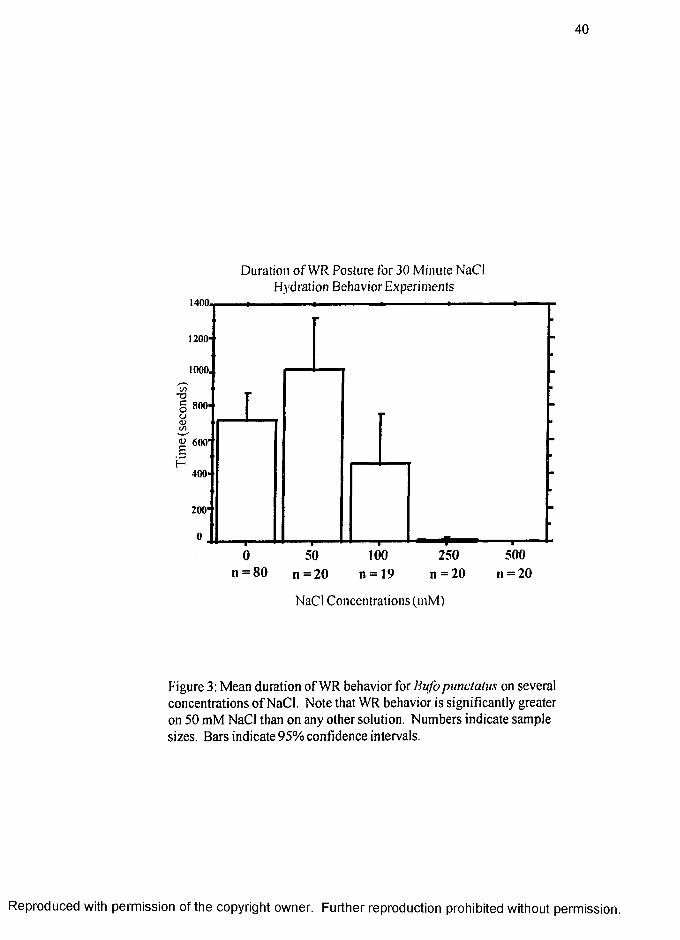
40
Duration o f WR Posture for 30 Minute NaCl Hydration Behavior Experiments
1400.
I 600
n = 20n —80 n = 20 n = 19 II = 2 0
NaCl Concentrations (mM)
Figure 3: Mean duration of WR behavior for Bufo punctatus on several concentrations o f NaCl. Note that WR behavior is significantly greater on 50 mM NaCl than on any other solution. Numbers indicate sample sizes. Bars indicate 95% confidence intervals.
R eproduced with perm ission of the copyright owner. Further reproduction prohibited without perm ission.
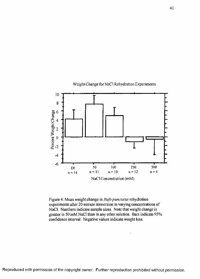
41
Weight Change forNaCi Rehydration Experiments
500 n = 4
100 1 1 = 1 0
250 n = 12n = 11n = 14
NaCl Concentration (mM)
Figure 4: Mean weight change in Bufopunctatus rehydration experiments after 20 minute immersion in varying concentrations of NaCl. Numbers indicate sample sizes. Note that weight change is greater in 50 mM NaCl than in any other solution. Bars indicate 95% confidence interval. Negative values indicate weight loss.
R eproduced with perm ission of the copyright owner. Further reproduction prohibited without perm ission.
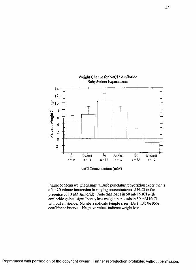
42
Weight Change for NaCl / Amiloride Rehydration Experiments
14
UJ )
I
J
12 - -
10
-2 ■ ■
_ L
d T ~
n = Ki
T
D I/A in il
n = 1 1
5 0 5 0 /A n i il
n = 11 n = 12
~ 2 d O 25()/X nU I
II = 15 n = 10
NaCl Concentration (mM)
Figure 5; Mean weight change in Bufo punctatus rehydration experiments after 20 minute immersion in varying concentrations o f NaCl in the presence o f 10 uM amiloride, Note that toads in 50 mM NaCl with amiloride gained significantly less weight than toads in 50 mM NaCl without amiloride. Numbers indicate sample sizes. Barsindicate 95% confidence interval. Negative values indicate weight loss.
R eproduced with perm ission of the copyright owner. Further reproduction prohibited without perm ission.
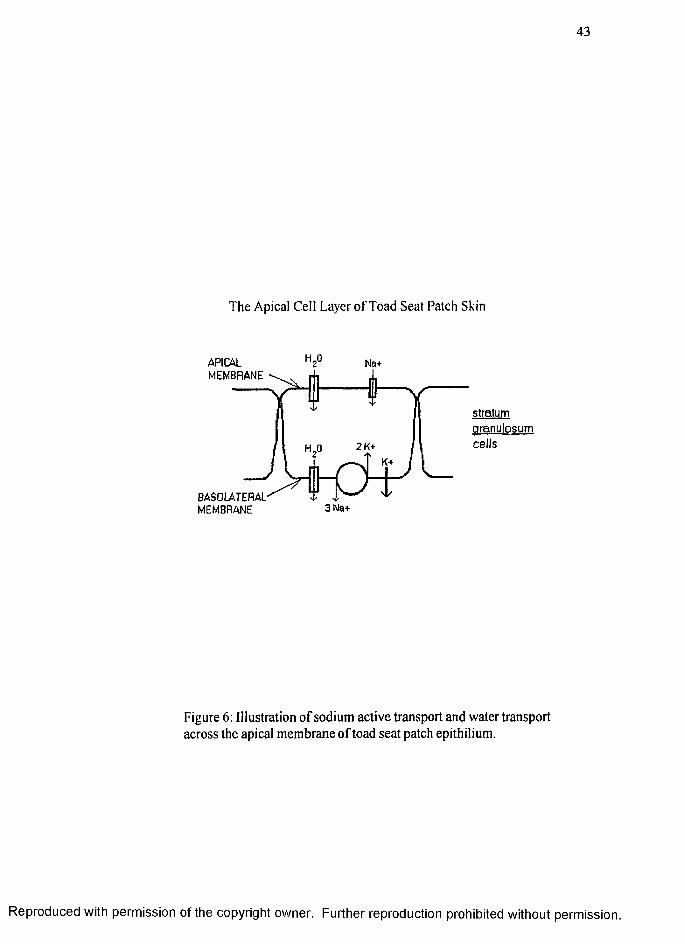
43
The Apical Ceil Layer o f Toad Seat Patch Skin
APIC4L 2̂'̂ Na+MEMBRANE
stratumgranulosumcells2 K+
3Na+
Figure 6: Illustration o f sodium active transport and water transport across the apical membrane o f toad seat patch epithilium.
R eproduced with perm ission of the copyright owner. Further reproduction prohibited without perm ission.
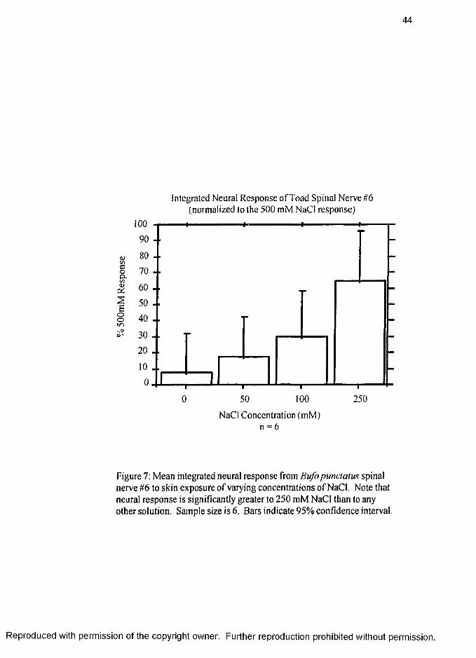
44
Integrated Neural Response o f Toad Spinal Nerve #6 (normalized to the 500 mM NaCl response)
§
IEoo
i r i
NaCl Concentration (mM) n = 6
Figure 7; Mean integrated neural response from Bufo punctatus spinal nerve #6 to skin exposure o f varying concentrations o f NaCl. Note that neural response is significantly greater to 250 mM NaCl than to any other solution. Sample size is 6. Bars indicate 95% confidence interval.
R eproduced with perm ission of the copyright owner. Further reproduction prohibited without perm ission.
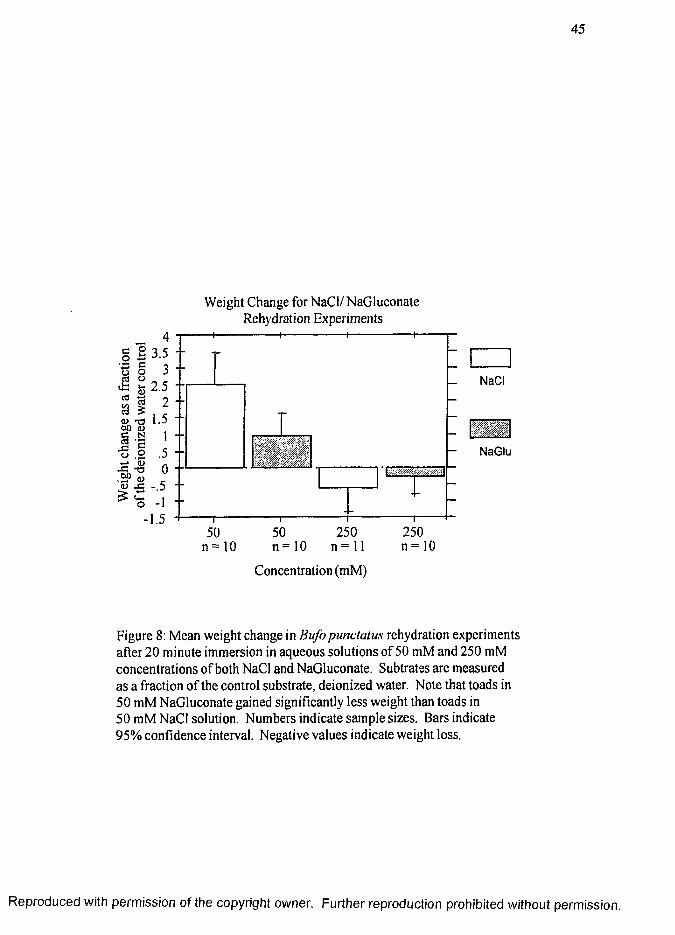
45
§ 2 3.5 oh 2.5
3 --
I ï<u t3u4-» a>
fio
21.5
1.50
-.5- 1
-1.5
Weight Change for NaCl/NaGluconate Rehydration Experiments
LNaCl
1 1 1 1-----50 50 250 250
n = 10 n = 10 n = l l n = 10
Concentration (mM)
S
— NaGlu
Figure 8: Mean weight change in Bufo punctatus rehydration experiments after 20 minute immersion in aqueous solutions of 50 mM and 250 mM concentrations of both NaCl and NaGluconate. Subtrates are measured as a fraction o f the control substrate, deionized water. Note that toads in 50 mM NaGluconate gained significantly less weight than toads in 50 mM NaCl solution. Numbers indicate sample sizes. Bars indicate 95% confidence interval. Negative values indicate weight loss.
R eproduced with perm ission o f the copyright owner. Further reproduction prohibited without perm ission.
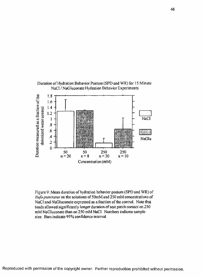
46
Duration o f Hydration Behavior Posture (SPD and WR) for 15 Minute NaCl / NaGluconate Hydration Behavior Experiments
o
p
a
l iu c
II1Q
1.81.6
1.41.2
18
.6
.4
.20
50 250 25050n = 20 n = ; n = 30
Concentration (mM)n = 10
NaCl
NaGlu
Figure 9: Mean duration of hydration behavior posture (SPD and WR) of Bufo punctatus on the solutions of 50mM and 250 mM concentrations of NaCl and NaGluconate expressed as a fraction of the control. Note that toads allowed significantly longer duration o f seat patch contact on 250 mM NaGluconate than on 250 mM NaCl. Numbers indicate sample size. Bars indicate 95% confidence interval.
R eproduced with perm ission of the copyright owner. Further reproduction prohibited without perm ission.
VTTA
Graduate College University of Nevada, Las Vegas
Polly Ann Sullivan
Local Address:3896 Commanche Avenue Las Vegas Nevada 89121
Degrees:Bachelor of Arts, Anthropology, 1993 University of Nevada Las Vegas
Master of Arts, Anthropology, 1995 University of Nevada Las Vegas,
Special Honors and Awards:NSF EPSCOR Women in Science and Engineering Research Grant;June - Aug. 1996, Sept. 1996 - May 1997, Sept. - Dec. 1997UNLV Graduate College Research Scholarship, Spring 1996UNLV Alumni Award for Academic Excellence in the Department of Anthropology1992-1993UNLV Anthropological Society Scholarship Award 1992-1993 school year UNLV Pen Pals Journal essay “Desert Treasures” 1992
Publications and Papers Presented:Maleek, R., P. A. Sullivan, K. von Seckendorff Hoff and S. D. Hillyard. ‘Salt sensitivity and hydration behavior of the toad, Bufo marinus’. (submitted to Physiology and Behavior)
Sullivan, P.A.. ‘Effects of salt concentration on hydration behavior in Red-spotted toads, Bufo punctatus’ In preparation, (plan to submit to Experimental Biology)
Hillyard,S. D., K. Hoff and P. A. Sullivan. ‘The effect of impermeant anions on Na+ chemoreception and water transport by the toad skin. ( Submitted poster to The AChemS XX meeting. April 1998
Sullivan, P.A., G. Spaulding. ‘A method of paleoenvironmental reconsruction using plant macrofossil assemblages from packrat middens’. In preparation
HilIyard,S. D., K. Hoff, K. Mace, P. A. Sullivan and K. Martin. 1997. ‘Salt appetite and avoidance in an amphibian Bufo punctatus’. The ISOT XII/AChemS XIX meeting. Park City Utah. October 1997
Hillyard, S.D., K. Hoff and P.A. Sullivan. 1997. ‘Species differences in the sensory function of epithilial Na+ channels (ENaCs) in toad skin’ (poster). The American Physiological Society Conference, San Diego, California. 5-12 July
47
R eproduced with perm ission of the copyright owner. Further reproduction prohibited without perm ission.
48
Sullivan, P.A., K.S. Hoff, S.D. Hillyard, C. Orgeron and K. Martin. 1997. ‘Effects of NaCl concentration on rehydration of the Red-Spotted Toad, Bufo punctatus' The Arizona Nevada Academy of Sciences Conference, Las Vegas, Nevada. April 19th 1997.
Hoff, K.S., S.D. Hillyard,P.A. Sullivan and K. Martin. 1996. ‘Salt appetite in a desert anuran, Bufo punctatus'. The Society for Integrative and Comparative Biology meeting, Albuquerque, New Mexico. 26-30 Dec. 1996.
Sullivan, P.A. 1992. ‘ Tactile interaction among Bottlenose dolphins (Tursiops truncatus) in captivity’. UNLV Journal of Anthropology 2 (1): 1-8
Thesis Title: Packrat middens: a tool for environmental reconstruction and human adaptation analysis
Thesis Examination Committee:Chairperson, Dr. Claude Warren, Ph.D.Committee Member. Dr. John Swetnam, Full Professor, Ph.D.Committee Member, Dr. Bernardo Arriaza, Assistant Professor, Ph.D.Committee Member, Dr. Geoffry Spaulding, Adjunct Faculty, Ph.D.Graduate Faculty Member, Dr. Charles Douglas, Full Professor, Ph.D.
R eproduced with perm ission o f the copyright owner. Further reproduction prohibited without perm ission.
IMAGE EVALUATION TEST TARGET (Q A -3 )
V /
/
%
1.0
l .l
1.25
li^iiiii 111̂ m i l 2 . 2
Il 2.0
m i l 1 . 8
I nc làâc C
1.4 1.6
150mm
/A PPLIED A IIVMGE . Inc— 1653 East Main Street
- ' Rochester. NY 14609 USA Phone: 716/482-0300 Fax: 716/288-5989
© 1993. A pplied Im age. Inc.. Ail R igh ts R ese rv ed
R eproduced with perm ission of the copyright owner. Further reproduction prohibited without perm ission.
Copyright © 2022 FDOKUMEN