Hydrophobic mismatch sorts SNARE proteins into distinct membrane domains
Upload
independentCategory
view
2download
0
5856 Biochemistry 1990, 29, 5856-5864
Evidence of Hydrophobic Domains in Human Respiratory Mucins. Effect of Sodium Chloride on Hydrophobic Binding Properties?
Viswanathan Shankar,t Bashoo Naziruddin,% Santiago Reyes de la Rocha,$ and Goverdhan P. Sachdev**i College of Pharmacy and Cystic Fibrosis Center, University of Oklahoma Health Sciences Center,
Oklahoma City, Oklahoma 73190 Received September 27, 1989; Revised Manuscript Received March 23, I990
ABSTRACT: Hydrophobic binding properties of purified human respiratory mucins were studied by the fluorescence probe technique using mansylphenylalanine (Mns-Phe) as the fluorescent probe. Mucins were purified from tracheobronchial secretions of cystic fibrosis (CF) and asthmatic patients, as well as from individuals with normal lungs, according to a protocol earlier established in our laboratory. Purified mucins were subjected to reduction-alkylation and Pronase digestion to study the effects of these treatments on the hydrophobic properties of the mucins. In addition, the effects of increased NaCl concentration on the hydrophobic properties of native and reduced-alkylated mucins were also investigated. Native mucins showed evidence of a large number of low-affinity (KD - M ) binding sites for the hydrophobic ligand Mns-Phe and had between 40 and 50 binding sites/mg of mucin. Reduction of mucin using dithiothreitol in the presence of 6 M guanidine hydrochloride and subsequent alkylation with iodoacetamide apparently caused marked conformational changes in the mucin molecules as revealed by the presence of both high-affinity (KD - 10” M) and low-affinity (KD - M ) binding sites for the probe and an increase in the number of probe binding sites. Pronase digestion of the native and reduced-alkylated mucins almost completely eliminated binding of the fluorescent probe to the mucins, showing that the binding sites are on the nonglycosylated, Pronase-sensitive portion of the mucin molecules. Increasing NaCl concentrations (0.03-1 .O M) did not appreciably alter the native mucin-induced Mns-Phe fluorescence, while that of the reduced-alkylated mucin-induced Mns-Phe fluorescence was progressively increased. The results indicate that further unfolding of reduced-alkylated mucins a t increased NaCl concentrations exposes additional hydrophobic domains. Also, the greater increase in the number of probe binding sites in the reduced-alkylated CF mucin as compared to reduced-alkylated non-CF mucins suggests possible differences in the three-dimensional structure of C F and non-CF mucins. Thus, this study provided evidence for the presence of hydrophobic domains in human respiratory mucins, and these properties, at least in part, may contribute to the polymeric structure of mucins through noncovalent interactions.
M u c i n s are high molecular weight glycoprotein components of mucus secreted by the epithelial cells lining the gastroin- testinal, tracheobronchial, and genitourinary tracts. They contribute to a large extent toward the gelation and viscoelastic properties of the mucus. Mucins are essentially composed of oligosaccharide chains densely packed into clusters and co- valently linked to the protein core (Carlstedt et al., 1985). The carbohydrate is comprised of five sugars, namely fucose, ga- lactose, N-acetylglucosamine, N-acetylgalactosamine, and sialic acid. In addition, mucins contain sulfate groups (Boat & Cheng, 1980; Chace et al., 1985). The heavily glycosylated regions are interspersed with nonglycosylated “naked” stretches of the protein core. These exposed regions of the protein core are susceptible to proteolytic attack (Carlstedt & Sheehan, 1984) and thought to be partly responsible for the interaction of mucins with many biologically important compounds such as enzymes (Ha0 et al., 1977), cations (Deman et ai., 1973; Forstner & Forstner, 1975), and proteins like serum albumin (List et a]., 1978) and ovalbumin (Creeth et al., 1979). Earlier studies have shown the presence of hydrophobic domains in canine tracheal and ovine submaxillary mucins (Sachdev et
‘This work was supported, in part, by a grant from the National Heart, Lung and Blood Institute (HL34012).
*Address correspondence to this author. f College of Pharmacy. $Cystic Fibrosis Center.
al., 1979), bovine gallbladder mucin (Smith & LaMont, 1984), and rat salivary mucin (Slomiany et al., 1988). In fact, hy- drophobic bonding has been implicated to be primarily re- sponsible for mucin-albumin interaction (List et al., 1978), resulting in the enhancement of mucus viscosity. Such in- teractions are perhaps more relevant in pathological states characterized by high albumin concentrations in mucus se- cretions (Knauff & Adams, 1968; Schachter & Dixon, 1965). However, to date, no direct evidence for the presence of hy- drophobic domains in human respiratory mucins has been reported. It is important therefore to study hydrophobic in- teractions of the mucins since they may shed light on the altered rheological properties of respiratory mucus in disease states.
Fluorescent probes are small molecules that have charac- teristic emission properties and, when coupled to biological macromolecules, can reveal subtle aspects of their structure and dynamics (Azzi, 1973). Several N-arylamino- naphthalenesulfonates have been used in the past as probes for the hydrophobic regions in proteins (Weber, 1953; McClure & Feldman, 1966; Stryer, 1968; Brand & Gohlke, 1972; Sachdev et al., 1973, 1975, 1979). These probes exhibit vir- tually no fluorescence in aqueous solutions but fluoresce in- tensely in nonpolar solvents or when bound to certain proteins and exhibit altered emission properties.
In the present study we report the hydrophobic binding properties of purified human respiratory mucins using the
0006-2960/90/0429-5856$02.50/0 0 1990 American Chemical Society

Respiratory Mucus Glycoproteins
fluorescent probe Mns-Phe and discuss the possible role of these hydrophobic domains in mucin polymeric structure. The influence of increasing NaCl concentrations on mucin hy- drophobicity was evaluated, since our recent studies (Chace et a]., 1989) have indicated that NaCl concentration exerts a marked effect on the aggregation properties of human re- spiratory mucins.
EXPERIMENTAL PROCEDURES Collection of Human Respiratory Mucus Secretions.
Tracheobronchial mucus secretions (sputum specimens) were collected from two cystic fibrosis (CF-4, CF-5) and two asthmatic (AS-1, AS-2) patients and from two individuals with normal lungs (N-3 and N-4), fitted with chronic tracheosto- mies required as respiratory support. Several specimens from the same patient were combined for the purification of mucins. All samples were collected on ice, diluted at least 5-fold with water containing gentamicin sulfate (100 pg/mL), and dia- lyzed exhaustively against a 0.02% sodium azide solution and then against deionized water. The retentate was lyophilized and stored at -20 "C (Chace et al., 1985).
[solation and Purification of Native Respiratory Mucins. A detailed protocol for the purification of the respiratory mucins has been described earlier (Chace et al., 1985). In brief, the respiratory mucus secretions were solubilized in 0.1 M Tris-HCI buffer, pH 7.5, containing 0.22 M potassium thiocyanate and 0.02% sodium azide. Following solubilization treatment, the suspension was centrifuged at 27000g for 4 h. The clear supernatant was applied to a Bio-Gel A 5-m column (5 X 90 cm) preequilibrated with 0.1 M Tris-HCI buffer, pH 7.5, containing 0.22 M potassium thiocyanate and 0.02% so- dium azide and eluted by using the same buffer. The excluded fraction containing DNA and mucus glycoprotein was treated with DNase to digest DNA molecules. Subsequently, the mucus glycoprotein was separated from the DNA fragments by rechromatography on the Bio-Gel A 5-m column. The excluded fraction containing the DNA-free mucus glycoprotein together with small amounts of low molecular weight com- ponents was further fractionated on a hydroxylapatite column (2.5 X 30 cm) equilibrated with 0.01 M phosphate buffer, pH 6.8. The column was initially eluted with the equilibrating buffer and subsequently with a discontinuous gradient of 0.15, 0.30, and 0.50 M potassium phosphate buffer, pH 6.8. The major fraction corresponding to mucus glycoprotein did not bind to the column and was eluted with the equilibrating buffer. A minor mucin component and contaminating im- purities eluted later with higher ionic strength buffer solution.
Reduction-Alkylation of Purified Respiratory Mucins. Reduction and carboxymethylation of purified mucins were performed as described by Chace et al. (1985). Mucin (10 mg) was dissolved in 0.1 M Tris-HC1 buffer, pH 8.5, con- taining 6 M guanidine hydrochloride and 0.02% sodium azide, giving a final concentration of 0.3 mg/mL. Nitrogen gas was bubbled through the solution for 30 min at 22 "C prior to addition of dithiothreitol (final concentration 25 mM). After 2 h of incubation, additional amounts of dithiothreitol were added to give a final concentration of 50 mM. The reduction was carried out for 4 h, and subsequently iodoacetamide was added to the solution to give a concentration of 0.15 M. In- cubation was done for an hour in the dark at 22 "C. The excess iodoacetamide was removed by the addition of dithio- threitol (final concentration 0.2 M). The reduced-alkylated mucins were exhaustively dialyzed against deionized water and lyophilized.
Desialylation of Purified Respiratory Mucins. Both native and reduced-alkylated mucins were enzymatically desialylated
Biochemistry, Vol. 29, No. 24, 1990 5857
by using neuraminidase (Vibrio cholerae; Calbiochem). Mucin (10 mg) was solubilized in 2 mL of 50 mM sodium acetate buffer, pH 5.5, containing 1 mM calcium chloride. An ap- propriate quantity (based on millimoles of sialic acid present in mucin) of neuraminidase was then added and the mixture incubated at 37 OC for 24 h. Following incubation samples were dialyzed exhaustively against deionized water at 4 "C and lyophilized.
Gel Electrophoresis. The purity of mucins was examined by gel electrophoresis on composite (2% acrylamide + 0.5% agarose) gels containing 0.1% sodium dodecyl sulfate (Sachdev et al., 1978; Holden et al., 1971). Duplicate gels were run for each sample, one of which was stained with the periodic acid-Schiff (PAS) reagent for carbohydrates and the other with Coomassie Brilliant Blue for proteins as described by Segrest and Jackson (1972). The gels were also examined by the silver stain method.
Pronase Digestion of Purified Native Respiratory Mucins. Proteolytic digestion of purified tracheobronchial mucin was performed as described by Smith and LaMont (1984) with slight modifications. Briefly, lyophilized mucin (2 mg/mL) was reconstituted in 0.1 M phosphate buffer, pH 6.8, con- taining 0.02% sodium azide and 100 pL of lOOX PSN anti- biotic mixture (Gibco Laboratories) per milliliter, containing 500 pg of penicillin, 500 pg of streptomycin, and 1 mg of neomycin. Protease (Streptomyces griseus, type XIV) was added in a ratio of 1:lO (w/w) to mucin at 0 and 24 h. The samples were incubated for 48 h at 37 "C, dialyzed for 72 h against deionized water at 4 OC, and then lyophilized. The treated mucins were examined by Superose 6 chromatography.
Lipid Analysis. Purified mucin (5 mg) was extracted for 30 min with 50 volumes each of chloroform/methanol (2:l v/v, twice), chloroform/methanol (1:l and 1:2 v/v), and chloroform/methanol/water (65:35:8, by volume) consecu- tively, as described by Slomiany et al. (1983). Also in a second method, samples were extracted with 4 mL of n-heptane/2- propanol (3:7 v/v) which contained 50 pg of cholesterol bu- tyrate as the internal standard (Wang et al., 1982). The neutral lipids (triglycerides, cholesterol, and cholesterol esters) were determined by gas-liquid chromatography (GLC) as described by Kuksis et al. (1975). Total fatty acids in the extracted residue were determined following saponification with ethanol-KOH mixture and subsequently analyzed by GLC as their methyl esters prepared by treatment with BF,/MeOH as described by McConathy et al. (1981). For determining the presence of covalently bound fatty acids in the purified mucins, the lyophilized, delipidated mucin was deacylated by treatment with 5 mL of 1 M hydroxylamine (pH 7.0) for 5 h at room temperature (Slomiany et al., 1983). The incubation mixture was extracted three times with hexane. The organic layers were evaporated to dryness and subjected to fatty acid analyses as described above.
Compositional Analysis of Purified Respiratory Mucins. Chemical and amino acid compositions were determined for purified native and reduced-alkylated respiratory mucins. Neutral hexose was determined by the anthrone method (Carubelli et al., 1961) using a mixture of fucose and galactose (1: 1 w/w) as the standard. Sialic acid was estimated by the resorcinol method (Svennerholm, 1957) using N-acetylneur- aminic acid as the standard. Fucose, galactose, N-acetyl- glucosamine, and N-acetylgalactosamine were determined by gas-liquid chromatography as described previously (Wang et al., 1974; Sachdev et al., 1978). In addition, after hydrolysis (4 N HCl, 4 h at 100 "C), the sugar amines were also analyzed by using an amino acid analyzer. Amino acid analysis was
5858 Biochemistry, Vol. 29, No. 24, 1990
performed by the method of Spackman (1967) using a Durrum D-500 amino acid analyzer following hydrolysis of the sample with constant-boiling HCl at 110 OC for 22 h in evacuated sealed glass tubes. Sulfate was determined by the sodium rhodizonate method (Terho & Hartiala, 1971).
Superose 6 Chromatography. A Superose 6 (10/30) FPLC column was equilibrated with 0.1 M Tris-HCl buffer, pH 7.4, containing 0.22 M potassium thiocyanate, 4 M guanidine hydrochloride, and 0.02% sodium azide. Native, reduced- alkylated, and Pronase-treated mucins were dissolved in the above buffer at a concentration of 2 mg/mL and eluted with the same buffer.
Preparation of Mucin Solution for Fluorescence Studies. Purified and lyophilized native, reduced-alkylated, and Pro- nase-treated mucins were dissolved in the appropriate buffer (2.5 mg/mL) and left on a rocker at 4 OC for 48 h. To remove dust particles, the solutions were then centrifuged at 12OOOg for 20 min, and the supernatant was used for fluorescence experiments.
Synthesis of Fluorescent Probe Mns-Phe. Mansylphenyl- alanine was essentially prepared as described earlier in detail (Sachdev et al., 1973). Aliquots from a 1.5 mM stock solution prepared by dissolving the probe in 20 mM Tris-HC1 buffer, pH 7.4, containing 0.02% sodium azide were diluted just prior to fluorescence studies to give a 200 pM solution. The com- pound and the solution were protected from light prior to their use in fluorescence experiments.
Fluorescence Studies. Fluorescence measurements were made on a Shimadzu RF-5000 fluorescence spectrophotom- eter. The excitation source was a 150-W xenon lamp. Bandwidths of 10 and 5 nm for excitation and emission spectra, respectively, were used. No corrections were introduced for changes in the sensitivity of the detection system at different wavelengths. Prior to each set of experiments, the instrument was calibrated as follows: the cuvette was filled with a standard solution of quinine sulfate in 0.1 N H$04 (1 pg/mL) and, by use of an excitation wavelength of 350 nm, the emission at 450 nm was set to read 200 arbitrary fluorescence units with the sensitivity set at “high”. All samples in these experiments had a final volume of 0.5 mL, and their fluorescence was studied in 0.5-cm2 cuvettes. Mns-Phe was prepared from stock as a 200 pM solution in 20 mM Tris-HCl buffer, pH 7.4, containing 0.02% sodium azide alone or in buffer containing appropriate additions of NaCl. Lyophilized mucin preparations were reconstituted in 20 mM Tris-HC1 buffer, pH 7.4, containing 0.02% sodium azide alone or in buffer containing appropriate additions of NaCl. All fluorescence measurements were conducted at 25 OC and spectra recorded within 30 min of preparation.
Binding of Mns-Phe by Respiratory Mucin. Respiratory mucin binding of Mns-Phe was studied by measuring mu- cin-induced alterations in the fluorescence spectra of the hy- drophobic ligand. The excitation and emission spectra of Mns-Phe (1 2.5 pM) were measured alone and in the presence of purified mucin preparation (500 pg/mL).
To evaluate the affinity of Mns-Phe binding by the re- spiratory mucin preparations, a Scatchard plot (Scatchard, 1949) was constructed in the following way: The limiting fluorescence, Fmax, was first determined by titrating a fixed amount of probe (5 pM) with increasing mucin concentrations (0.06-0.50 mg/mL) and measuring the fluorescence after each increment. A plot of 1/P versus 1/F gave a straight line, extrapolation of which to zero abscissa gave the reciprocal of Fmx. Titration of a fixed mucin concentration (0.5 mg/mL) against increasing probe concentration (4.8-66.6 pM) yielded
Shankar et al.
&
FIGURE 1 : SDS gel electrophoresis of purified native and reduced- alkylated mucins. Composite gels (2% acrylamide + 0.5% agarose) containing 0.1 % SDS were used and stained with PAS reagent. Lanes 1, 3, and 5 correspond to 60 pg each of native CF, asthmatic, and normal mucin and lanes 2, 4 and 6 correspond to 60 pg each of reduced-alkylated CF, asthmatic, and normal mucins, respectively.
values of bound and free probe concentrations. A plot of [Mns-Phe],-,, per milligram of mucin versus [ Mn~-Phe]~,,,, per milligram of mucin/ [ Mns-Phe] free gave a straight line. Extrapolation of the line to zero ordinate gave the number of binding sites (n) per milligram of mucin and the extrapolation to zero abscissa gave n/KD from which KD (dissociation con- stant) values were calculated. Ten separate fluorescence ti- trations were performed by using from 0.06 to 0.50 mg/mL of purified respiratory mucin. Mns-Phe concentration was varied from 1.5 to 12.5 pM to determine high-affinity binding (HAB) sites and from 4.8 to 66.6 pM to determine low-affinity binding (LAB) sites in each experiment. The method of least squares was used to calculate linear regression plots of the data. In all cases the correlation coefficient ( r ) value was 10.92.
Effect of NaCl Concentration on the Hydrophobic Binding Properties of Respiratory Mucins. The effect of NaCl con- centration on the hydrophobic binding properties of mucins was studied by dissolving mucin preparations (0.5 mg/mL) in. 20 mM Tris-HC1 buffer, pH 7.4, containing 0.02% sodium azide plus the desired NaCl concentration. The probe con- centration was kept constant at 12.5 pM and the relative fluorescence measured. Fluorescence titrations were also performed with the native and reduced-alkylated mucins dissolved in buffer containing different NaCl concentrations to determine the effect of NaCl on the probe binding properties of the mucins.
RESULTS Purification of Mucins. The mucins isolated from respir-
atory mucus secretions were examined for their purity by SDS composite gel electrophoresis. The CF, asthmatic, and normal mucins gave a single broad band when stained with the PAS reagent (Figure 1). No low molecular weight components were detected when the gels were stained with Coomassie
Respiratory Mucus Glycoproteins
0 0.2
8 N
C
w
I 0.1
P
Biochemistry, Vol. 29, No. 24, 1990 5859
-
-
Table 11: Amino Acid Composition of Purified CF, Asthmatic, and Normal Human Respiratory Mucins
0 ' 3 .
0 c 0 5 10 15 20 25
FRACTION NUMBER
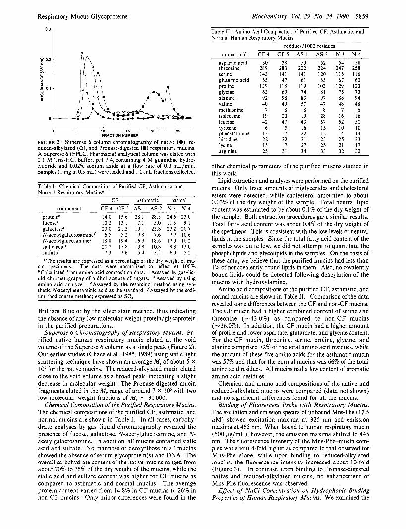
FIGURE 2: Superose 6 column chromatography of native (O) , re- duced-alkylated (O), and Pronase-digested (.) respiratory mucins. A Superose 6 (FPLC, Pharmacia) analytical column was eluted with 0.1 M Tris-HCI buffer, pH 7.4, containing 4 M guanidine hydro- chloride and 0.02% sodium azide at a flow rate of 0.3 mL/min. Samples ( I mg in 0.5 mL) were loaded and 1 .O-mL fractions collected.
Table I : Chemical Composition of Purified CF, Asthmatic, and Normal Respiratory Mucins'
CF asthmatic normal component CF-4 CF-5 AS-I AS-2 N-3 N-4
protein* 14.0 15.6 28.1 28.3 24.6 23.0 fucoseC 10.2 1 3 . 1 7.1 5.0 1 1 . 5 9.1 galactosec 23.0 21.3 19.1 23.8 23.2 20.7 N-acetylgalactosamined 6.5 5.2 9.8 7.6 7.9 10.6 N-acetylglucosamined 18.8 19.4 16.3 18.6 17.0 18.2 sialic acidC 20.2 17.8 13.8 10.8 9.3 13.0 sulfatd 7 . 3 7.6 5.4 5 .5 6 .0 5.2 'The results are expressed as a percentage of the dry weight of mu-
cin specimens. The data were normalized to reflect at 100%. bCalculated from amino acid composition data. cAssayed by gas-liq- uid chromatography of alditol acetate of sugars. dAssayed by using amino acid analyzer. CAssayed by the resorcinol method using syn- thetic N-acetylneuraminic acid as the standard. /Assayed by the sodi- um rhodizonate method; expressed as SO,.
Brilliant Blue or by the silver stain method, thus indicating the absence of any low molecular weight protein/glycoprotein in the purified preparations.
Superose 6 Chromatography of Respiratory Mucins. Pu- rified native human respiratory mucin eluted a t the void volume of the Superose 6 column as a single peak (Figure 2). Our earlier studies (Chace et al., 1985, 1989) using static light scattering technique have shown an average M, of about 5 X IO6 for the native mucins. The reduced-alkylated mucin eluted close to the void volume as a broad peak, indicating a slight decrease in molecular weight. The Pronase-digested mucin fragments eluted in the M, range of around 7 X IO5 with two low molecular weight fractions of M, - 30000.
Chemical Composition of the Purified Respiratory Mucins. The chemical compositions of the purified CF, asthmatic, and normal mucins are shown in Table I. In all cases, carbohy- drate analyses by gas-liquid chromatography revealed the presence of fucose, galactose, N-acetylglucosamine, and N- acetylgalactosamine. In addition, all mucins contained sialic acid and sulfate. No mannose or deoxyribose in all mucins showed the absence of serum glycoprotein(s) and DNA. The overall carbohydrate content of the native mucins ranged from about 70% to 75% of the dry weight of the mucins, while the sialic acid and sulfate content was higher for CF mucins as compared to asthmatic and normal mucins. The average protein content varied from 14.8% in CF mucins to 26% in non-CF mucins. Only minor differences were found in the
residues/ 1000 residues amino acid CF-4 CF-5 AS-I AS-2 N-3 N-4
aspartic acid threonine serine glutamic acid proline glycine alanine valine methionine isoleucine leucine tyrosine phenylalanine histidine lysine arginine
30 38 53 52 54 58 289 283 222 224 247 258 143 141 141 120 1 1 5 116 55 47 61 65 67 62
139 118 119 103 129 123 63 69 74 81 75 73 92 98 8 3 97 88 94 40 49 57 47 48 48
7 8 8 8 7 6 19 20 19 28 16 16 42 47 43 67 52 50
6 5 16 15 I O I O 13 7 22 12 14 14 22 22 21 23 25 23 15 17 27 25 21 17 25 31 34 33 32 32
other chemical parameters of the purified mucins studied in this work.
Lipid extraction and analyses were performed on the purified mucins. Only trace amounts of triglycerides and cholesterol esters were detected, while cholesterol amounted to about 0.03% of the dry weight of the sample. Total neutral lipid content was estimated to be about 0.1% of the dry weight of the sample. Both extraction procedures gave similar results. Total fatty acid content was about 0.4% of the dry weight of the specimens. This is consistent with the low levels of neutral lipids in the samples. Since the total fatty acid content of the samples was quite low, we did not attempt to quantitate the phospholipids and glycolipids in the samples. On the basis of these data, we believe that the purified mucins had less than 1% of noncovalently bound lipids in them. Also, no covalently bound lipids could be detected following deacylation of the mucins with hydroxylamine.
Amino acid compositions of the purified CF, asthmatic, and normal mucins are shown in Table 11. Comparison of the data revealed some differences between the CF and non-CF mucins. The C F mucin had a higher combined content of serine and threonine (-43.0%) as compared to non-CF mucins (-36.0%). In addition, the CF mucin had a higher amount of proline and lower aspartate, glutamate, and glycine content. For the CF mucin, threonine, serine, proline, glycine, and alanine comprised 72% of the total amino acid residues, while the amount of these five amino acids for the asthmatic mucin was 57% and that for the normal mucins was 66% of the total amino acid residues. All mucins had a low content of aromatic amino acid residues.
Chemical and amino acid compositions of the native and reduced-alkylated mucins were compared (data not shown) and no significant differences found for all the mucins.
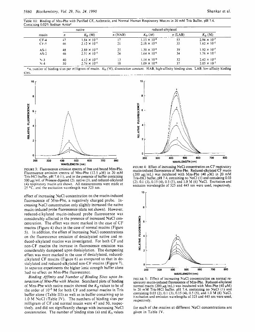
Binding of Fluorescent Probe with Respiratory Mucins. The excitation and emission spectra of unbound Mns-Phe (1 2.5 pM) showed excitation maxima a t 325 nm and emission maxima a t 465 nm. When bound to human respiratory mucin (500 pg/mL), however, the emission maxima shifted to 445 nm. The fluorescence intensity of the Mns-Phe-mucin com- plex was about 4-fold higher as compared to that observed for Mns-Phe alone, while upon binding to reduced-alkylated mucins, the fluorescence intensity increased about 10-fold (Figure 3) . In contrast, upon binding to Pronase-digested native and reduced-alkylated mucins, no enhancement of Mns-Phe fluorescence was observed.
Effect of NaCl Concentration on Hydrophobic Binding Properties of Human Respiratory Mucins. We examined the
5860 Biochemistry, Vol. 29, No. 24, 1990 Shankar et al.
Table 111: Binding of Mns-Phe with Purified CF, Asthmatic, and Normal Human Respiratory Mucins in 20 mM Tris Buffer, pH 7.4, Containing 0.02% Sodium AzideQ
w 0 z 8 6 -
native reduced-alkylated mucin n KD (MI n (HAB) K O ( M ) n (LAB) KD (MI CF-4 47 1.84 x 10⁻⁵ 17 1.13 X 10” 53 2.94 x 10⁻⁵ CF-5 46 2.12 x 10⁻⁵ 21 2.18 X 10” 35 1.62 x 10⁻⁵
AS- 1 48 2.89 x 10⁻⁵ 25 1.38 X 10” 39 1.92 x 10⁻⁵ AS-2 46 2.51 x 10⁻⁵ 26 1.64 X 10” 34 1.76 x 10⁻⁵
N-3 40 4.12 x 10⁻⁵ 13 1.18 X IO” 32 2.42 x 10⁻⁵ N-4 50 2.74 x 10⁻⁵ 18 1.09 X 10” 37 2.05 x 10⁻⁵
a n , number of binding sites per milligram of mucin. K D (M), dissociation constant. HAB, high-affinity binding sites. LAB, low-affinity binding sites.
d w 4 - e 8 K
2 -
1
0 - I
200 300 400 500 600 700 800 WAVELENGTH (nm)
FIGURE 3: Fluorescence emission spectra of free and bound Mns-Phe. Fluorescence emission spectra of Mns-Phe (12.5 pM) in 20 mM Tris-HCI buffer, pH 7.4 ( I ) , and in the presence of buffer containing 500 pg/mL of Pronase-digested (2) , native (3), and reduced-alkylated (4) respiratory mucin are shown. All measurements were made at 25 “C, and the excitation wavelength was 325 nm.
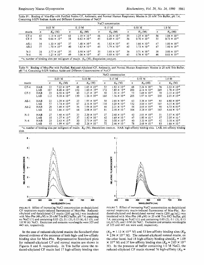
effect of increasing NaCl concentration on the mucin-induced fluorescence of Mns-Phe, a negatively charged probe. In- creasing NaCl concentration only slightly increased the native mucin-induced probe fluorescence (data not shown). However, reduced-alkylated mucin-induced probe fluorescence was considerably affected in the presence of increased NaCl con- centration. The effect was more marked in the case of C F mucins (Figure 4) than in the case of normal mucins (Figure 5). I n addition. the effect of increasing NaCl concentrations on the fluorescence emission of desialyated native and re- duced-alkylated mucins was investigated. For both C F and non-CF mucins the increase in fluorescence emission was considerably dampened upon desialylation. The dampening effect was more marked in the case of desialylated, reduced- alkylated C F mucins (Figure 6) as compared to that in de- sialylated and reduced-alkylated non-CF mucins (Figure 7). In separate experiments the higher ionic strength buffer alone had no effect on Mns-Phe fluorescence.
Binding Affinity and Number of Binding Sites upon In- teraction of Mns-Phe with Mucins. Scatchard plots of binding of Mns-Phe with native mucin showed the KD values to be of the order of M for both CF and normal mucins in Tris buffer alone (Table 111) as well as in buffer containing up to 1 .O M NaCl (Table I V ) . The numbers of binding sites per milligram of CF and normal mucin were 47 and 50, respec- tively, and did not significantly change with increasing NaCl concentration. The number of binding sites (n) and KD values
l o I 6
I 200 300 400 500 600 700 800
WAVELENGTH (I” FIGURE 4: Effect of increasing NaCl concentration on CF respiratory mucin-induced fluorescence of Mns-Phe. Reduced-alkylated CF mucin (200 pg/mL) was incubated with Mns-Phe (40 wM) in 20 mM Tris-HCI buffer, pH 7.4, containing no NaCl (1) and containing 0.03 (2), 0.1 (3), 0.15 (4), 0.5 (9, and 1.0 M (6) NaCI. Excitation and emission wavelengths of 325 and 445 nm were used, respectively.
I 200 300 400 500 600 700 800
WAVELENGTH (nm)
FIGURE 5: Effect of increasing NaCl concentration on normal re- spiratory mucin-induced fluorescence of Mns-Phe. Reduced-alkylated normal mucin (200 pg/mL) was incubated with Mns-Phe (40 pM) in 20 mM Tris-HCI buffer, pH 7.4, containing no NaCl (1) and containing 0.03 (2), 0.1 (3), 0.15 (4), 0.5 ( 5 ) , and 1.0 M (6) NaCI. Excitation and emission wavelengths of 325 and 445 nm were used, respectively.
for each of the mucins a t different NaCl concentrations are given in Table IV.
Respiratory Mucus Glycoproteins Biochemistry, VoL 29, No. 24, 1990 5861
Table IV: Binding of Mns-Phe with Purified Native CF, Asthmatic, and Normal Human Respiratory Mucins in 20 mM Tris Buffer, pH 7.4, Containing 0.02% Sodium Azide and Different Concentrations of NaCP
NaCl concentration 0.03 M 0.10 M 0.15 M 0.50 M 1.0 M
mucin n KD (MI n KD (MI n KD (MI n KD n KD (MI CF-4 42 1.10 X 43 1.19 X 48 1.26 X 55 1.25 X 56 1.04 X CF-5 36 0.66 X 38 0.62 X 45 0.49 X 56 0.70 X 55 0.73 X IO”
AS-I 34 1.30 X 37 1.48 X 41 1.82 X 45 1.66 X 47 1.80 X 10” AS-2 37 1.70 X 40 1.65 X 45 1.79 X 45 1.73 X 47 1.80 X
N-3 34 3.77 x 10⁻⁵ 35 3.88 x 10⁻⁵ 35 3.93 x 10⁻⁵ 36 3.71 x 10⁻⁵ 38 3.80 x 10⁻⁵ N-4 50 1.23 x 10⁻⁵ 49 1.04 x 10⁻⁵ 47 0.80 x 10⁻⁵ 45 0.76 x 10⁻⁵ 46 0.83 x 10⁻⁵
‘ n , number of binding sites per milligram of mucin. Kn (M), dissociation constant.
Table V: Binding of Mns-Phe with Purified, Reduced-Alkylated CF, Asthmatic, and Normal Human Respiratory Mucins in 20 mM Tris Buffer, pH 7.4, Containing 0.02% Sodium Azide and Different Concentrations of NaCP
NaCl concentration
CF-4 HAB 33 7.22 X I O ” 48 1.65 X 53 1.82 X 68 2.34 X 76 2.33 X LAB 107 8.48 X IOT5 152 1.60 X lo4 172 1.80 X 200 2.14 X I O 4 249 1.78 X IO4
CF-5 HAB 23 6.50 X I O “ 40 1.25 X 50 1.56 X lo-’ 55 1.69 X 70 2.15 X IOw5 LAB 112 9.18 X 130 1.26 X IO4 160 1.56 X 205 1.97 X 250 2.35 X IO4
AS-1 HAB 23 1.39 X IO“ 33 1.95 X IO“ 50 2.68 X IO” 63 3.38 X 10” 85 4.80 X IO” LAB 57 1.74 X 67 2.16 X 110 3.20 X 132 3.84 X IO-’ 165 5.15 X
AS-2 HAB 28 1.48 X IO“ 36 1.98 X IO” 44 2.31 X 10” 58 3.05 X IO” 90 4.73 X IO” LAB 52 1.66 X 64 2.08 X 81 2.50 X 10⁴ 3.24 X 165 4.44 X
N-3 HAB 27 1.48 X IO” 33 2.20 X 10” 37 2.60 X 10” 43 3.01 X IO” 51 3.57 X IO” LAB 35 1.27 X 37 1.42 X lo-’ 42 1.69 X 47 1.95 X 57 2.35 X
LAB 55 1.99 X 60 2.26 X IO-’ 72 2.46 X 78 2.68 X 83 1.66 X N-4 HAB 25 2.43 X 10” 30 2.75 X IO“ 36 3.00 X 10” 40 3.33 X 10“ 42 3.10 X IO“
an , number of binding sites per milligram of mucin. KD (M), dissociation constant. HAB, high-affinity binding sites. LAB, low-affinity binding sites.
6 r
6
I 200 300 400 500 600 700 800
WAVELENGTH (nm)
F I G U R E 6: Effect of increasing NaCl concentration on desialylated C F respiratory mucin-induced fluorescence of Mns-Phe. Reduced- alkylated and desialylated C F mucin (200 pg/mL) was incubated with Mns-Phe (40 pM) in 20 m M Tris-HC1 buffer, pH 7.4, containing no NaCl ( I ) and containing 0.03 (2), 0.1 (3), 0.15 (4), 0.5 ( 9 , and 1 .O M (6) NaCI. Excitation and emission wavelengths were 325 and 445 nm, respectively.
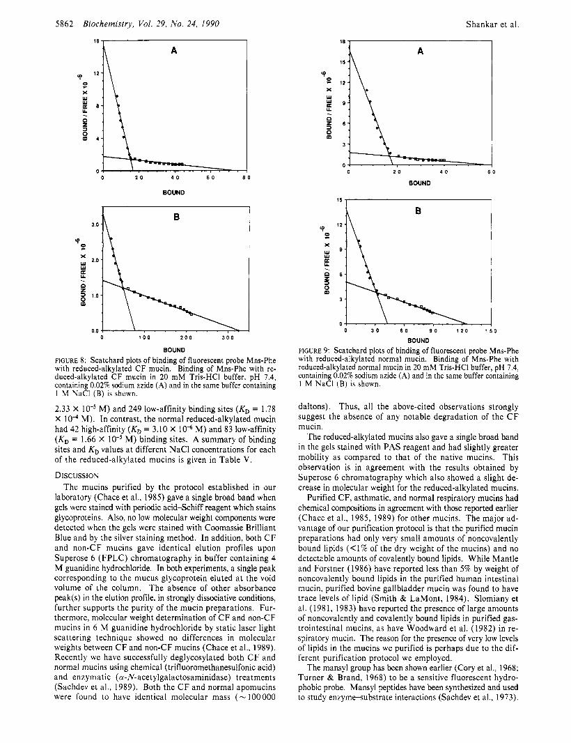
In the case of reduced-alkylated mucins the Scatchard plots showed evidence of the existence of both high- and low-affinity binding sites for Mns-Phe. Representative Scatchard plots for reduced-alkylated CF and normal mucins are shown in Figures 8 and 9, respectively. In Tris buffer alone the re- duced-alkylated C F mucin had 17 high-affinity binding sites
3 t
200 300 400 500 600 700 800 WAVELENGTH (nm)
FIGURE 7: Effect of increasing NaCl concentration on desialylated normal respiratory mucin-induced fluorescence of Mns-Phe. Re- duced-alkylated and desialylated normal mucin (200 pg/mL) was incubated with Mns-Phe (40 pM) in 20 m M Tris-HCI buffer, p H 7.4, containing no NaCl ( I ) and containing 0.03 (2), 0.1 (3), 0.15 (4), 0.5 (5), and 1 .O M (6) NaCI. Excitation and emission wavelengths of 325 and 445 nm were used, respectively.
(KD = 1.13 X 10“ M) and 5 3 low-affinity binding sites (KD = 2.94 X M). The reduced-alkylated normal mucin, on the other hand, had 18 high-affinity binding sites(K, = 1.09 X 10” M) and 37 low-affinity binding sites (KD = 2.05 X M). In the presence of buffer containing 1.0 M NaCI, the reduced-alkylated CF mucin showed 76 high-affinity (KD =
5862 Biochemistry, Vol. 29, No. 24, 1990 Shankar
18 , I
et al.
BOUND
‘4 9 X W W U U
z 3
. n
P
BOUND
FIGURE 8: Scatchard plots of binding of fluorescent probe Mns-Phe with reduced-alkylated CF mucin. Binding of Mns-Phe with re- duced-alkylated CF mucin in 20 mM Tris-HCI buffer, pH 7.4, containing 0.02% sodium azide (A) and in the same buffer containing 1 M NaCl (B) is shown.
2.33 X M) and 249 low-affinity binding sites (KD = 1.78 X M). In contrast, the normal reduced-alkylated mucin had 42 high-affinity (KD = 3.10 X 10” M) and 83 low-affinity (KD = 1.66 X M) binding sites. A summary of binding sites and KD values a t different NaCl concentrations for each of the reduced-alkylated mucins is given in Table V.
Discussio~ The mucins purified by the protocol established in our
laboratory (Chace et al., 1985) gave a single broad band when gels were stained with periodic acid-Schiff reagent which stains glycoproteins. Also, no low molecular weight components were detected when the gels were stained with Coomassie Brilliant Blue and by the silver staining method. In addition, both CF and non-CF mucins gave identical elution profiles upon Superose 6 (FPLC) chromatography in buffer containing 4 M guanidine hydrochloride. In both experiments, a single peak corresponding to the mucus glycoprotein eluted a t the void volume of the column. The absence of other absorbance peak(s) in the elution profile, in strongly dissociative conditions, further supports the purity of the mucin preparations. Fur- thermore, molecular weight determination of CF and non-CF mucins in 6 M guanidine hydrochloride by static laser light scattering technique showed no differences in molecular weights between C F and non-CF mucins (Chace et al., 1989). Recently we have successfully deglycosylated both CF and normal mucins using chemical (trifluoromethanesulfonic acid) and enzymatic (a-N-acetylgalactosaminidase) treatments (Sachdev et al., 1989). Both the C F and normal apomucins were found to have identical molecular mass (- 100 000
‘9 z X w W 0: U
z 3
. n
8
BOUND
0 3 0 6 0 9 0 1 2 0 1 5 0
BOUND
FIGURE 9: Scatchard plots of binding of fluorescent probe Mns-Phe with reduced-alkylated normal mucin. Binding of Mns-Phe with reduced-alkylated normal mucin in 20 mM Tris-HCI buffer, pH 7.4, containing 0.02% sodium azide (A) and in the same buffer containing 1 M NaCl (B) is shown.
daltons). Thus, all the above-cited observations strongly suggest the absence of any notable degradation of the C F mucin.
The reduced-alkylated mucins also gave a single broad band in the gels stained with PAS reagent and had slightly greater mobility as compared to that of the native mucins. This observation is in agreement with the results obtained by Superose 6 chromatography which also showed a slight de- crease in molecular weight for the reduced-alkylated mucins.
Purified CF, asthmatic, and normal respiratory mucins had chemical compositions in agreement with those reported earlier (Chace et al., 1985, 1989) for other mucins. The major ad- vantage of our purification protocol is that the purified mucin preparations had only very small amounts of noncovalently bound lipids (<1% of the dry weight of the mucins) and no detectable amounts of covalently bound lipids. While Mantle and Forstner (1986) have reported less than 5% by weight of noncovalently bound lipids in the purified human intestinal mucin, purified bovine gallbladder mucin was found to have trace levels of lipid (Smith & LaMont, 1984). Slomiany et al. (1 98 1, 1983) have reported the presence of large amounts of noncovalently and covalently bound lipids in purified gas- trointestinal mucins, as have Woodward et al. (1982) in re- spiratory mucin. The reason for the presence of very low levels of lipids in the mucins we purified is perhaps due to the dif- ferent purification protocol we employed.
The mansyl group has been shown earlier (Cory et al., 1968; Turner & Brand, 1968) to be a sensitive fluorescent hydro- phobic probe. Mansyl peptides have been synthesized and used to study enzymesubstrate interactions (Sachdev et al., 1973).
Respiratory Mucus Glycoproteins
Mansyl compounds have also been found to be more sensitive probes as compared to analogous dansyl compounds (Sachdev et al., 1973). Our earlier studies on the hydrophobic binding sites of canine tracheal and ovine submaxillary mucins (Sachdev et al., 1979) using the fluorescent probes mansyl- phenylalanine and sodium mansate have shown mansyl- phenylalanine to be a better probe than sodium mansate to investigate binding affinity and hydrophobic binding sites of ovine submaxillary mucin. Hence, in the present study mansylphenylalanine was used as the probe to determine the hydrophobic binding properties of the respiratory mucins.
Human respiratory mucin contained a large number of hydrophobic binding sites for Mns-Phe which appeared to be in the nonglycosylated portion of the protein core. This is supported by the observation that probe binding was almost completely eliminated after Pronase digestion of the mucins. These observations are in agreement with those obtained earlier for bovine gallbladder mucin (Smith & LaMont, 1984), bovine cervical mucin (Bhushana Rao & Masson, 1977), and rat salivary mucin (Slomiany et al., 1988). While Slomiany et al. (1 988) have suggested that associated and covalently bound lipids contribute to the hydrophobic characteristics of salivary mucins, data from our present study show that the hydrophobic properties of human respiratory mucins are primarily due to the protein core.
Our earlier studies (Chace et al., 1985) have shown that, unlike the gastric and cervical mucins, the human respiratory mucins upon reduction of disulfide bonds undergo only a slight change in molecular weight, implying that the polymeric structure of the mucin may not be based on interdisulfide bonds alone. This is further supported by the gel electro- phoretic data reported here for native and reduced mucins (Figure 1). Noncovalent interactions between mucin molecules may be partly responsible for the polymeric structure as demonstrated recently for bovine gallbladder mucin (Smith et al., 1989). We have previously shown that hydrophobic domains also exist in canine tracheal and ovine submaxillary mucins (Sachdev et al., 1979).
Reduction-alkylation of disulfide bonds in the native mucins probably causes profound changes in the three-dimensional structure of the mucin molecule, resulting in an unfolded conformation that exposes additional hydrophobic domains. The existence of both high- and low-affinity hydrophobic domains, not observed in the native mucins, lends support to this hypothesis. The unfolding is perhaps progressively in- creased with increasing NaCl concentration, thus explaining the observed increase in binding of the hydrophobic probe to the mucin molecule at higher salt concentrations. A relatively higher number of hydrophobic sites observed in the reduced- alkylated C F mucins as compared to the reduced-alkylated asthmatic or normal mucins in the presence of different NaCl concentrations (Table V) may be due to the fact that the C F mucin by virtue of being more sulfated and having a higher sialic acid content is more anionic and thus more susceptible to change in conformation mediated by the charge-shielding effects of increased NaCl concentration. To test this hy- pothesis, the effects of increasing NaCl concentrations on the fluorescence emission of desialylated native and reduced-al- kylated C F and non-CF mucins were studied. The results obtained (Figures 6 and 7) clearly indicate that the reduction in fluorescence emission upon desialylation is more marked in the case of reduced-alkylated C F mucins as compared to non-CF mucins. This shows that the greater anionic charge on the C F mucins is more responsive to the charge-shielding effects of increasing NaCl concentrations. It would be in-
Biochemistry, Vol. 29, No. 24, 1990 5863
teresting to study if removal of sulfate groups from the de- sialylated mucins further dampened the effects of increasing NaCl concentrations on fluorescence. However, in the absence of any known sulfohydrolases that can preferentially remove sulfate from the mucins, these studies are not feasible at this time.
The differences in the observed number of binding sites and the KD values obtained also suggest that the three-dimensional structures of C F and non-CF mucins may be different. The C F mucins are likely to have a more extended structure than non-CF mucins due to increased repulsion of like charges (anionic). This would account for greater access of the probe to hydrophobic binding sites upon neutralization of the charge. Interestingly, Mantle and Stewart (1989), on the basis of immunochemical studies, reported the possibility of differences in the three-dimensional configuration of C F and non-CF intestinal mucins. Our recent studies (Sachdev et al., 1989) using monoclonal antibodies raised against the C F apomucin also suggested possible differences in the three-dimensional structures of C F and non-CF mucins. Also, Rose (1988) found that meconium mucins from C F patients upon digestion with trypsin-treated TPCK yielded peptides whose hydrophobic properties differed from those obtained in the same way from mucins isolated from non-CF individuals. This was interpreted to indicate that naked peptide regions of C F mucins are dif- ferent from those of non-CF mucins.
On the basis of chemical compositional data (Table I) one may assume that an apparently lower net protein content in the case of C F mucins as compared to non-CF mucins may mean a correspondingly lower number of hydrophobic probe binding sites in the C F mucins. On the contrary, however, it must be pointed out that, rather than net protein content alone, it is the three-dimensional configuration of the protein molecule that determines the affinity of binding and acces- sibility of hydrophobic domains for the probe. The observed differences in KD values, number of binding sites, and effect of increasing NaCl concentration on the binding of the probe with the mucins strongly suggest differences in the three-di- mensional structure of C F and non-CF mucins.
To the best of our knowledge, this is the first evidence for the presence of hydrophobic domains in human respiratory mucins. At least two different types of hydrophobic regions may be present on the mucin molecule revealed upon con- formational changes induced by reduction and alkylation. On the basis of the results from this work it seems very likely that hydrophobic and other noncovalent interactions may also play a part, in addition to covalent linkages, in the total polymeric structure of the human respiratory mucins. Thus, hydrophobic interactions may play an important role in the viscoelastic and gelation properties of the respiratory mucus.
ACKNOWLEDGMENTS
during the conduct of these experiments.
REFERENCES Azzi, A. (1973) Methods Enzymol. 32, 234-246. Bhushana Rao, K. S. P., & Masson, P. L. (1977) J . Biol.
Boat, T. F., & Cheng, P. W. (1980) Fed. Proc. 39,3067-3074. Brand, L., & Gohkle, J. R. (1972) Annu. Rev. Biochem. 41,
Carlstedt, I., & Sheehan, J. K. (1984) in Mucus and Mucosa,
Carlstedt, I . , Sheehan, J. K., Corfield, A. P., & Gallagher,
We thank Dr. Kenneth V. Chace for helpful discussions
Chem. 252, 7788-7795.
843-868.
pp 157-172, Pitman, London.
J. T. (1985) Essays Biochem. 20, 40-76.
5864
Carubelli, R., Ryan, R. C., Trucco, R. E., & Caputto, R.
Chace, K. V., Flux, M., & Sachdev, G. P. (1985) Biochemistry
Chace, K. V., Naziruddin, B., Desai, V. C., Flux, M., & Sachdev, G. P. (1989) Exp. Lung Res. 15, 721-737.
Cory, R. R., Becker, R. R., Rosenbluth, R., & Isenberg, I. (1968) J . Am. Chem. SOC. 90, 1643-1647.
Creeth, J. M., Bridge, J. L., & Horton, J. R. (1979) Biochem.
Deman, J., Marcel, M., & Bruyneel, E. (1973) Biochim.
Forstner, J. F., & Forstner, G. G. (1975) Biochim. Biophys.
Hao, K., Takeuchi, T., Sato, S., & Sugimura, T. (1977) Clin.
Hill, H. D., Jr., Reynolds, J. A., & Hill, R. A. (1977) J . Biol.
Holden, K. G., Yim, N . C. F., Griggs, L. J., & Weisbach, J.
Knauff, R. E., & Adams, J . A. (1968) Clin. Chim. Acta 19,
Kuksis, A., Myher, J. J., Marai, L., & Geher, K. (1975) J .
List, S. J., Findlay, B. P., Forstner, G. G., & Forstner, J. G.
Mantle, M., & Forstner, J. F. (1 986) Biochem. Cell Biol. 64,
Mantle, M., & Stewart, G. (1989) Biochem. J . 259,243-253. McClure, W. O., & Feldman, G. M. (1966) Biochemistry 5,
McConathy, W. J., Blackett, P. R., & Kling, 0. R. (1981)
Rose, M. C. (1988) Horm. Metab. Res. 20, 601-608. Sachdev, G. P., Brownstein, A. D., & Fruton, J. S. (1973) J .
Sachdev, G. P., Brownstein, A. D., & Fruton, J . S. (1975) J .
Biochemistry, Vol. 29, No. 24, 1990
(1961) J . Biol. Chem. 236, 2381-2388.
24, 7334-7341,
J . 181, 717-724.
Biophys. Acta 297, 486-490.
Acta 386, 283-292.
Chim. Acta 79, 75-80.
Chem. 252, 3791-3798.
D. (1971) Biochemistry 10, 3105-3109.
245-248.
Chromatogr. Sci. 18, 423-430.
(1978) Biochem. J . 175, 565-571.
223-228.
1908-1 91 9.
Clin. Chim. Acta I l l , 153-162.
Biol. Chem. 248, 6292-6299.
Biol. Chem. 250, 501-507.
Shankar et al.
Sachdev, G. P., Fox, 0. F., Wen, G., Schroeder, T., Elkins, R. C., & Carubelli, R. (1978) Biochim. Biophys. Acta 536,
Sachdev, G. P., Zodrow, J. M., & Carubelli, R. (1979) Bio- chim. Biophys. Acta 580, 85-90.
Sachdev, G. P., Desai, V., Naziruddin, B., Graves, D., & Reyes de la Rocha, S. (1989) Pediatr. Pulmonol. Suppl. 4, Ab- stract 74, 130.
184-196.
Scatchard, G. (1949) Ann. N.Y. Acad. Sci. 51, 660-672. Schachter, H., & Dixon, G. H. (1965) Can. J. Biochem. 43,
Segrest, J. P., & Jackson, R. L. (1972) Methods Enzymol.
Slomiany, A., Witas, F., Aono, M., & Slomiany, B. L. (1983) J . Biol. Chem. 258, 8535-8538.
Slomiany, B. L., Galicki, N. L., Kojima, K., & Slomiany, A. (1981) Biochim. Biophys. Acta 665, 88-91.
Slomiany, B. L., Murty, V. L. N., Sarosiek, J., Piotrowski, J., & Slomiany, A. (1988) Biochem. Biophys. Res. Com- mun. 151, 1046-1053.
Smith, B. F., & LaMont, J . T. (1984) J . Biol. Chem. 259,
Smith, B. F., Peeterman, J. A., Tanaka, T., & LaMont, J. T. (1 989) Gastroenterology 97, 179-1 87.
Spackman, D. H . (1967) Methods Enzymol. 11, 3-15. Stryer, L. (1968) Science 162, 526-533. Svennerholm, L. (1957) Biochim. Biophys. Acta 24,604-61 1, Terho, T. T., & Hartiala, K. (1971) Anal. Biochem. 41,
Turner, D. C., & Brand, L. (1968) Biochemistry 7,
Wang, C. S., Burns, R. K., & Alaupovic, P. (1974) J . Bac- teriol. 120, 990-993.
Wang, C. S., Weiser, D., Alaupovic, P., & McConathy, W. J. (1982) Arch. Biochem. Biophys. 214, 26-34.
Weber, G. (1953) Adu. Protein Chem. 8, 415-459. Woodward, H., Horsey, B., Bhavanandan, V. P., & Davidson,
381-397.
28, 54-63.
1 2 1 7 0- 1 2 1 7 7.
47 1-476.
3381-3390.
E. A. (1 982) Biochemistry 21, 694-701.
Copyright © 2022 FDOKUMEN