Importance of chloride homeostasis in the operation of ...
13
HAL Id: hal-03470150 https://hal.archives-ouvertes.fr/hal-03470150 Submitted on 9 Dec 2021 HAL is a multi-disciplinary open access archive for the deposit and dissemination of sci- entific research documents, whether they are pub- lished or not. The documents may come from teaching and research institutions in France or abroad, or from public or private research centers. L’archive ouverte pluridisciplinaire HAL, est destinée au dépôt et à la diffusion de documents scientifiques de niveau recherche, publiés ou non, émanant des établissements d’enseignement et de recherche français ou étrangers, des laboratoires publics ou privés. Importance of chloride homeostasis in the operation of rhythmic motor networks Jean-Charles Viemari, Rémi Bos, Pascale Boulenguez, Cécile Brocard, Frédéric Brocard, Hélène Bras, Patrice Coulon, Sylvie Liabeuf, Edouard Pearlstein, Karina Sadlaoud, et al. To cite this version: Jean-Charles Viemari, Rémi Bos, Pascale Boulenguez, Cécile Brocard, Frédéric Brocard, et al.. Im- portance of chloride homeostasis in the operation of rhythmic motor networks. Breathe, Walk and Chew: The Neural Challenge: Part II, 188, Elsevier, pp.3-14, 2011, Progress in Brain Research, 10.1016/B978-0-444-53825-3.00006-1. hal-03470150
-
Upload
khangminh22 -
Category
Documents
-
view
1 -
download
0
Transcript of Importance of chloride homeostasis in the operation of ...
HAL Id: hal-03470150https://hal.archives-ouvertes.fr/hal-03470150
Submitted on 9 Dec 2021
HAL is a multi-disciplinary open accessarchive for the deposit and dissemination of sci-entific research documents, whether they are pub-lished or not. The documents may come fromteaching and research institutions in France orabroad, or from public or private research centers.
L’archive ouverte pluridisciplinaire HAL, estdestinée au dépôt et à la diffusion de documentsscientifiques de niveau recherche, publiés ou non,émanant des établissements d’enseignement et derecherche français ou étrangers, des laboratoirespublics ou privés.
Importance of chloride homeostasis in the operation ofrhythmic motor networks
Jean-Charles Viemari, Rémi Bos, Pascale Boulenguez, Cécile Brocard,Frédéric Brocard, Hélène Bras, Patrice Coulon, Sylvie Liabeuf, Edouard
Pearlstein, Karina Sadlaoud, et al.
To cite this version:Jean-Charles Viemari, Rémi Bos, Pascale Boulenguez, Cécile Brocard, Frédéric Brocard, et al.. Im-portance of chloride homeostasis in the operation of rhythmic motor networks. Breathe, Walk andChew: The Neural Challenge: Part II, 188, Elsevier, pp.3-14, 2011, Progress in Brain Research,�10.1016/B978-0-444-53825-3.00006-1�. �hal-03470150�
Jean-Pierre Gossard, Réjean Dubuc and Arlette Kolta (Eds.)Progress in Brain Research, Vol. 188ISSN: 0079-6123Copyright � 2011 Elsevier B.V. All rights reserved.
CHAPTER 1
Importance of chloride homeostasis in theoperation of rhythmic motor networks
Jean-Charles Viemari, Rémi Bos, Pascale Boulenguez, Cécile Brocard,Frédéric Brocard, Hélène Bras, Patrice Coulon, Sylvie Liabeuf, Edouard Pearlstein,
Karina Sadlaoud, Aurélie Stil, Sabrina Tazerart and Laurent Vinay*
Laboratoire Plasticité et Physio-Pathologie de la Motricité (UMR6196), Centre National de la Recherche Scientifique(CNRS) & Aix-Marseille Université, 31 Chemin Joseph Aiguier, Marseille Cedex 20, France
Abstract: GABA and glycine are classically called “inhibitory” amino acids, despite the fact that theiraction can rapidly switch from inhibition to excitation and vice versa. The postsynaptic action dependson the intracellular concentration of chloride ions ([Cl�]i), which is regulated by proteins in the plasmamembrane: the Kþ
–Cl� cotransporter KCC2 and the Naþ–Kþ–Cl� cotransporter NKCC1, which
extrude and intrude Cl� ions, respectively. A high [Cl�]i leads to a depolarizing (excitatory) action ofGABA and glycine, as observed in mature dorsal root ganglion neurons and in motoneurons bothearly during development and in several pathological conditions, such as following spinal cord injury.Here, we review some recent data regarding chloride homeostasis in the spinal cord and itscontribution to network operation involved in locomotion.
Keywords: inhibition; networks; KCC2; chloride; activity.
Introduction
Synaptic inhibitionmediated byGABAand glycinestrongly modulates mammalian neuronal networksfrom the early life to adulthood. The postsynapticaction of these neurotransmitters on GABAA and
*Corresponding author. Tel.: (þ33)-4-91-16-40-86;Fax.: (þ33)-4-91-77-50-84
3DOI: 10.1016/B978-0-444-53825-3.00006-1
glycine receptors depends on the intracellular con-centration of chloride ions ([Cl�]i) in the target cell.In adult healthy neurons, the activation of GABAA
and glycine receptors results in an inward flux ofCl�
and membrane potential hyperpolarization. There-fore, the inhibitory action of glycine and GABAconsists in both shunting incoming excitatorycurrents and moving the membrane potential awayfrom the action potential threshold. This “classical”
4
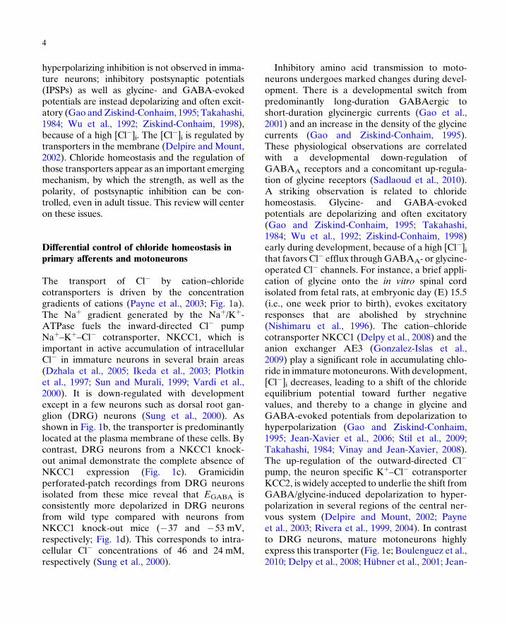
hyperpolarizing inhibition is not observed in imma-ture neurons; inhibitory postsynaptic potentials(IPSPs) as well as glycine- and GABA-evokedpotentials are instead depolarizing and often excit-atory (Gao and Ziskind-Conhaim, 1995; Takahashi,1984; Wu et al., 1992; Ziskind-Conhaim, 1998),because of a high [Cl�]i. The [Cl�]i is regulated bytransporters in the membrane (Delpire and Mount,2002). Chloride homeostasis and the regulation ofthose transporters appear as an important emergingmechanism, by which the strength, as well as thepolarity, of postsynaptic inhibition can be con-trolled, even in adult tissue. This review will centeron these issues.
Differential control of chloride homeostasis inprimary afferents and motoneurons
The transport of Cl� by cation–chloridecotransporters is driven by the concentrationgradients of cations (Payne et al., 2003; Fig. 1a).The Naþ gradient generated by the Naþ/Kþ-ATPase fuels the inward-directed Cl� pumpNaþ–Kþ
–Cl� cotransporter, NKCC1, which isimportant in active accumulation of intracellularCl� in immature neurons in several brain areas(Dzhala et al., 2005; Ikeda et al., 2003; Plotkinet al., 1997; Sun and Murali, 1999; Vardi et al.,2000). It is down-regulated with developmentexcept in a few neurons such as dorsal root gan-glion (DRG) neurons (Sung et al., 2000). Asshown in Fig. 1b, the transporter is predominantlylocated at the plasma membrane of these cells. Bycontrast, DRG neurons from a NKCC1 knock-out animal demonstrate the complete absence ofNKCC1 expression (Fig. 1c). Gramicidinperforated-patch recordings from DRG neuronsisolated from these mice reveal that EGABA isconsistently more depolarized in DRG neuronsfrom wild type compared with neurons fromNKCC1 knock-out mice (�37 and �53 mV,respectively; Fig. 1d). This corresponds to intra-cellular Cl� concentrations of 46 and 24 mM,respectively (Sung et al., 2000).
Inhibitory amino acid transmission to moto-neurons undergoes marked changes during devel-opment. There is a developmental switch frompredominantly long-duration GABAergic toshort-duration glycinergic currents (Gao et al.,2001) and an increase in the density of the glycinecurrents (Gao and Ziskind-Conhaim, 1995).These physiological observations are correlatedwith a developmental down-regulation ofGABAA receptors and a concomitant up-regula-tion of glycine receptors (Sadlaoud et al., 2010).A striking observation is related to chloridehomeostasis. Glycine- and GABA-evokedpotentials are depolarizing and often excitatory(Gao and Ziskind-Conhaim, 1995; Takahashi,1984; Wu et al., 1992; Ziskind-Conhaim, 1998)early during development, because of a high [Cl�]ithat favors Cl� efflux throughGABAA- or glycine-operated Cl� channels. For instance, a brief appli-cation of glycine onto the in vitro spinal cordisolated from fetal rats, at embryonic day (E) 15.5(i.e., one week prior to birth), evokes excitatoryresponses that are abolished by strychnine(Nishimaru et al., 1996). The cation–chloridecotransporter NKCC1 (Delpy et al., 2008) and theanion exchanger AE3 (Gonzalez-Islas et al.,2009) play a significant role in accumulating chlo-ride in immaturemotoneurons.With development,[Cl�]i decreases, leading to a shift of the chlorideequilibrium potential toward further negativevalues, and thereby to a change in glycine andGABA-evoked potentials from depolarization tohyperpolarization (Gao and Ziskind-Conhaim,1995; Jean-Xavier et al., 2006; Stil et al., 2009;Takahashi, 1984; Vinay and Jean-Xavier, 2008).The up-regulation of the outward-directed Cl�
pump, the neuron specific Kþ–Cl� cotransporter
KCC2, is widely accepted to underlie the shift fromGABA/glycine-induced depolarization to hyper-polarization in several regions of the central ner-vous system (Delpire and Mount, 2002; Payneet al., 2003; Rivera et al., 1999, 2004). In contrastto DRG neurons, mature motoneurons highlyexpress this transporter (Fig. 1e; Boulenguez et al.,2010; Delpy et al., 2008; Hübner et al., 2001; Jean-
OutNKCC KCC2
GABAAR& GlyR
GABAAR& GlyR
In
Out
In
Cl–
Depolarizingcurrent
(b)
(d) 0
–20
–40EC
1 (m
V)
–60
(f) 0
–20
–40
EC
1 (m
V)
–80
–60
∗∗∗ ∗∗
(c) (e)
(a)
Cl–K+Hyperpolarizing
current
Cl– Cl–K+ Na+
NKCC1+/+
NKCC1+/+
NKCC1–/–
NKCC1–/– KCC2+/+ KCC2–/–
3Na+
2K+
Na+/K+
ATPase
ATP
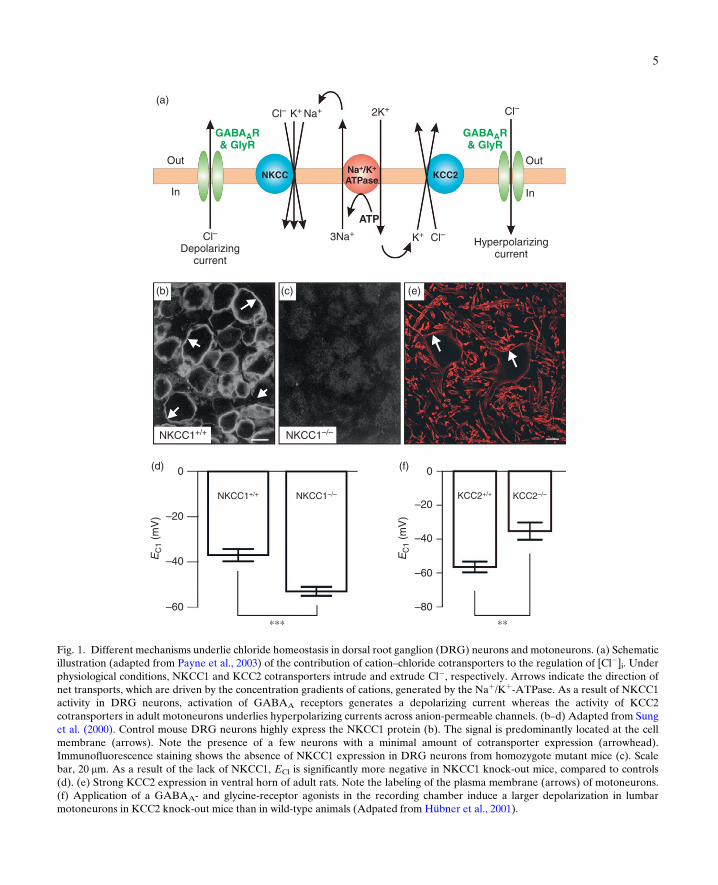
Fig. 1. Different mechanisms underlie chloride homeostasis in dorsal root ganglion (DRG) neurons and motoneurons. (a) Schematicillustration (adapted from Payne et al., 2003) of the contribution of cation–chloride cotransporters to the regulation of [Cl�]i. Underphysiological conditions, NKCC1 and KCC2 cotransporters intrude and extrude Cl�, respectively. Arrows indicate the direction ofnet transports, which are driven by the concentration gradients of cations, generated by the Naþ/Kþ-ATPase. As a result of NKCC1activity in DRG neurons, activation of GABAA receptors generates a depolarizing current whereas the activity of KCC2cotransporters in adult motoneurons underlies hyperpolarizing currents across anion-permeable channels. (b–d) Adapted from Sunget al. (2000). Control mouse DRG neurons highly express the NKCC1 protein (b). The signal is predominantly located at the cellmembrane (arrows). Note the presence of a few neurons with a minimal amount of cotransporter expression (arrowhead).Immunofluorescence staining shows the absence of NKCC1 expression in DRG neurons from homozygote mutant mice (c). Scalebar, 20 mm. As a result of the lack of NKCC1, ECl is significantly more negative in NKCC1 knock-out mice, compared to controls(d). (e) Strong KCC2 expression in ventral horn of adult rats. Note the labeling of the plasma membrane (arrows) of motoneurons.(f) Application of a GABAA- and glycine-receptor agonists in the recording chamber induce a larger depolarization in lumbarmotoneurons in KCC2 knock-out mice than in wild-type animals (Adpated from Hübner et al., 2001).
5

6
Xavier et al., 2006; Stein et al., 2004; Stil et al., 2009;Vinay and Jean-Xavier, 2008). Motoneurons aresignificantly more depolarized (>30 mV) byGABA or glycine in KCC2 knock-out mice atE18 than in wild type (�10 mV; Fig. 1f; Hübneret al., 2001). Altogether, these data demonstratethat NKCC1 and KCC2 cotransporters are impor-tant for maintaining high and low chlorideconcentrations in mature DRG neurons and mot-oneurons, respectively.
Contribution of chloride homeostasis tocell excitability
According to the classical view, postsynaptic inhi-bition induced by the activation of GABAA andglycine receptors consists in two mechanisms:shunting incoming excitatory currents and movingthe membrane potential away from the actionpotential threshold. As already mentioned,this hyperpolarization from rest is not observedin immature spinal neurons (Gao and Ziskind-Conhaim, 1995; Takahashi, 1984; Wu et al.,1992; Ziskind-Conhaim, 1998), thereby raisingthe question of the effect of subthresholddepolarizing IPSPs on neuronal excitability. It iswidely accepted that despite their depolarizingaction, these potentials are inhibitory in immaturespinal motoneurons because of the shuntingmechanism. On the basis of two reports on thecortex (Gulledge and Stuart, 2003) and the hypo-thalamus (Gao et al., 1998), we challenged theidea that depolarizing IPSPs may interact withexcitatory inputs and increase the excitability ofmotoneurons (Jean-Xavier et al., 2007). Wedemonstrated that depolarizing IPSPs have anexcitatory action by facilitating action potentialgeneration when paired with subthreshold excit-atory inputs (Fig. 2a). The effect of depolarizingIPSPs on excitability therefore depends on therelative weights of the inhibitory action of theshunting mechanisms and the excitatory actionof the depolarization. The value of ECl affectsthe strength of inhibitory connections within
spinal cord locomotor networks; the more posi-tive ECl, the lower the efficacy of inhibition. Afirst key point is the timing between inhibitoryand excitatory inputs. The inhibitory action ofshunting is exerted over a rather short periodafter IPSP onset (Fig. 2a, blockade of actionpotentials evoked by current pulses). In contrast,the time course of the depolarization is much lon-ger, because of the time constant of the mem-brane, such that the excitations occurring in thelate phase of the depolarizing IPSP sum up withthe depolarization whereas the conductance hasreturned to baseline (Fig. 2a; bottom traceshowing a subthreshold current pulse triggeringan action potential when occurring in the decayphase of the IPSP). A second key point, revealedby the use of a model (Fig. 2b), is the distancebetween inhibitory and excitatory synapses.The inhibitory action of conductance changes islocal whereas the depolarization spreads electro-tonically along the dendrites. Consequently,GABA/glycine synapses exert a strong inhibitionon proximal excitatory inputs (Fig. 2c; somaticexcitations and inhibitory synapses on the somaand on proximal dendrites) and a pure facilitation(whatever the timing) on distal excitation (Fig. 2c,bottom graph with inhibitory inputs 200 mm awayfrom excitatory synapses).
Contribution of chloride dynamics tonetwork activity
Maturation of chloride homeostasis affects net-work activity. There is a switch in the contributionof chloride-mediated conductances to spontane-ous activity from excitation to inhibition duringlate gestation. A rhythmic spontaneous activitycan be recorded in vitro very early (�E12–E14in rodents), when many lumbar motoneuronsare still migrating and extending their peripheralprojections (Hanson and Landmesser, 2003).Electrical transmission plays a significant role inthe generation of episodes since blockade of gapjunction coupling by carbenoxolone abolishes
IPSP + current-evokedaction potentials
IPSP(EIPSP= –56 mV)
Exc.
25 μm
200 μmInhibitoryinputs
–60
–70soma
25 μm
Facilitation
Inhibition
200 μm
–800 50 100 150
0 50
Time (ms)
100 150
0 50 100 150E
C1
(mV
)
–60
–70
–80
EC
1 (m
V)
–60
–70
–80E
C1
(mV
)
soma
Inhibition(a) (b)
(c)
Facilitation
IPSP + sub-thresholddepolarizing pulses
20 mV100 ms
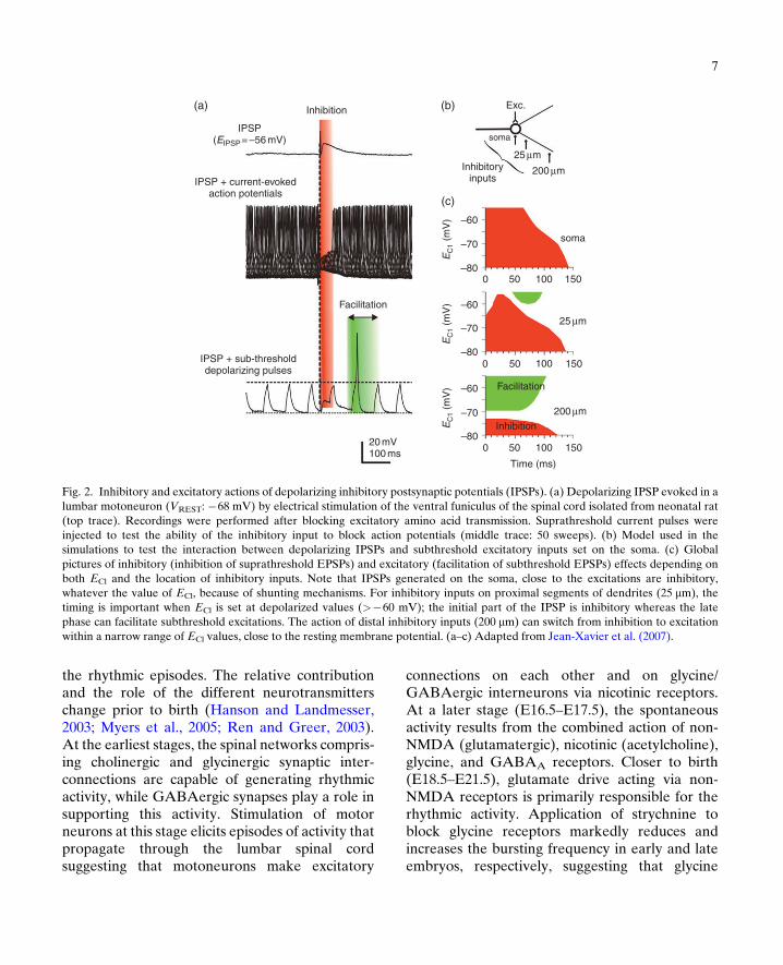
Fig. 2. Inhibitory and excitatory actions of depolarizing inhibitory postsynaptic potentials (IPSPs). (a) Depolarizing IPSP evoked in alumbar motoneuron (VREST: �68 mV) by electrical stimulation of the ventral funiculus of the spinal cord isolated from neonatal rat(top trace). Recordings were performed after blocking excitatory amino acid transmission. Suprathreshold current pulses wereinjected to test the ability of the inhibitory input to block action potentials (middle trace: 50 sweeps). (b) Model used in thesimulations to test the interaction between depolarizing IPSPs and subthreshold excitatory inputs set on the soma. (c) Globalpictures of inhibitory (inhibition of suprathreshold EPSPs) and excitatory (facilitation of subthreshold EPSPs) effects depending onboth ECl and the location of inhibitory inputs. Note that IPSPs generated on the soma, close to the excitations are inhibitory,whatever the value of ECl, because of shunting mechanisms. For inhibitory inputs on proximal segments of dendrites (25 mm), thetiming is important when ECl is set at depolarized values (>�60 mV); the initial part of the IPSP is inhibitory whereas the latephase can facilitate subthreshold excitations. The action of distal inhibitory inputs (200 mm) can switch from inhibition to excitationwithin a narrow range of ECl values, close to the resting membrane potential. (a–c) Adapted from Jean-Xavier et al. (2007).
7
the rhythmic episodes. The relative contributionand the role of the different neurotransmitterschange prior to birth (Hanson and Landmesser,2003; Myers et al., 2005; Ren and Greer, 2003).At the earliest stages, the spinal networks compris-ing cholinergic and glycinergic synaptic inter-connections are capable of generating rhythmicactivity, while GABAergic synapses play a role insupporting this activity. Stimulation of motorneurons at this stage elicits episodes of activity thatpropagate through the lumbar spinal cordsuggesting that motoneurons make excitatory
connections on each other and on glycine/GABAergic interneurons via nicotinic receptors.At a later stage (E16.5–E17.5), the spontaneousactivity results from the combined action of non-NMDA (glutamatergic), nicotinic (acetylcholine),glycine, and GABAA receptors. Closer to birth(E18.5–E21.5), glutamate drive acting via non-NMDA receptors is primarily responsible for therhythmic activity. Application of strychnine toblock glycine receptors markedly reduces andincreases the bursting frequency in early and lateembryos, respectively, suggesting that glycine

8
switches from an excitatory to an inhibitory contri-bution to spontaneous activity. This transitionoccurs at about the same time as the switch fromcholinergic to glutamatergic transmission.
The fact that the classical inhibitoryneurotransmitters can be functionally excitatorymoves the balance between excitatory and inhibi-tory drives toward excitation. This renders devel-oping networks hyperexcitable. Intracellularchloride concentration undergoes significantchanges during spontaneous activity in the chickspinal cord (Chub and O'Donovan, 2001). Afteran episode, the chloride equilibrium potentialfalls by �10 mV, corresponding to a decline ofintracellular chloride of �15 mM. This reducesthe excitatory contribution of GABA and glycineand hence network excitability. The intracellularchloride concentration is restored during theinterepisode interval, as a result presumably ofinward chloride pumping via cation–chloridecotransporters, leading to a progressive increasein network excitability. The importance of chlo-ride dynamics in the genesis of spontaneous activ-ity in the in vitro chick spinal cord between E9and E11 was confirmed by the use of a model(Marchetti et al., 2005).
Application of serotonin on the spinal cord atbirth, to activate the CPGs, evokes a fictive loco-motor pattern consisting of alternation betweenthe motor bursts on the left and right sides ofthe spinal cord, as well as alternation betweenflexor and extensor bursts on the same side. Thesame kind of experiments made five days priorto birth reveals a motor pattern with all bursts inphase (Iizuka et al., 1997; Nishimaru and Kudo,2000). The left–right alternation appears two dayslater but rhythmic bursts still occur synchronouslyin flexors and extensors. The transition from syn-chrony to alternation in the left and right ventralroots is likely due to the maturation of chloridehomeostasis.
In the respiratory network, although GABA/glycine-mediated inhibition is not essential forrhythm generation in neonatal rodent, it is wellknown that synaptic inhibition is important for
setting the different phases of the respiratoryrhythm and regulates both the excitability of glut-amatergic connections and the intrinsic mem-brane properties (Busselberg et al., 2001a,b;Ramirez et al., 1997; Shao and Feldman, 1997).As described in the case of spinal cord, the [Cl�]idetermines the functional role of inhibitoryneurotransmitters. The transition from an excit-atory to an inhibitory effect occurs earlier oninspiratory neurons than in the lumbar spinalcord (�E19; Ren and Greer, 2006). From thistime onward, GABA and glycine hyperpolarizeinspiratory neurons and suppress respiratoryfrequency.
It is important to note that KCC2cotransporters are sensitive to subtle changes inthe extracellular concentration of potassium ions([Kþ]o). A raise of [Kþ]o from 2–3 to 9–10 mMis sufficient to reverse the driving force for thenet K–Cl cotransport, thus allowing KCC2 tooperate in a reverse mode as a net influx pathway(Payne et al., 2003). As a consequence, such araise induces a 10- to 20-mV depolarizing shiftof ECl (Ren and Greer, 2006; Vinay and Jean-Xavier, 2008) in medullary slices and lumbar spi-nal cord preparations, respectively). Raising[Kþ]o is commonly used as a methodological toolto increase the excitability of in vitro preparations.This may, in addition, modify the strength of post-synaptic inhibition within networks and explainsome inconsistency among results obtained in dif-ferent experimental conditions.
The effects ofGABAon the respiratory networkhighly depend on the expression and the function-ality of cation–chloride cotransporters. During theearly life, NKCC1 is expressed at high levels andperturbations of its functionality by removing[Naþ]o or by application of bumetanide shifts ECl
to a more hyperpolarized value and reverses theexcitatory effect of GABAA receptor agonists(muscimol), whereas blockade of KCC2 by furose-mide has no effect (Ren and Greer, 2006).In contrast, in neonatal preparations, furosemidedepolarizes ECl and blocks the inhibitory effect ofmuscimol on respiratory frequency. At that time,

9
blockade of NKCC1 has no significant effect.These results are consistent with a developmentalincrease of KCC2 expression in the respiratory net-work, as in the locomotor system.
Primary afferent depolarizations and antidromicdischarges as part of the motor network
It is well established that one form of presynapticinhibition in the vertebrate spinal cord isassociated with primary afferent depolarization(PAD, Alvarez-Leefmans et al., 1998; Rudomin,1990; Rudomin et al., 1993) and that GABA,through the activation of GABAA receptors,plays a major role in the generation of PAD.Axo–axonic interactions between GABAergicterminals and primary afferents have beendemonstrated (see Alvarez, 1998, for review).PADs are reduced by GABAA receptorantagonists (Curtis and Lodge, 1982; Eccleset al., 1963; Levy, 1975; Rudomin et al., 1981).When PADs are large enough to reach firingthreshold, they trigger discharges that are anti-dromically conducted into peripheral nerves. Thiswas first demonstrated by recording the reflexresponse elicited in dorsal roots, by electricalstimulation of an adjacent dorsal root (“dorsalroot reflex,” Barron and Matthews, 1938;Toennies, 1938, see Kerkut and Bagust, 1995,for review). Antidromic discharges of primaryafferents have been observed during locomotionin the cat (fictive locomotion: Beloozerova andRossignol, 1999; Dubuc et al., 1988; Duenaset al., 1990; Gossard et al., 1991; treadmill loco-motion: Beloozerova and Rossignol, 2004; Dubucet al., 1985; Rossignol et al., 1998) and in the rat(Pilyavskii et al., 1988). A spontaneous anti-dromic activity in the dorsal roots has beendescribed in the in vitro spinal cord isolated fromadult (Bagust et al., 1989; Chen et al., 1993) oryoung hamsters (Abdul-Razzak et al., 1994; seeKerkut and Bagust, 1995, for review). Antidromicdischarges are also recorded from lumbar dorsal
roots in the neonatal rat spinal cord in vitro(Fellippa-Marques et al., 2000; Kremer andLev-Tov, 1998; Vinay and Clarac, 1999; Vinayet al., 1999). Most of these action potentials areblocked by bath application of bicuculline or pic-rotoxin, two GABAA receptor antagonists. Someactivity occurs spontaneously, consisting of rhyth-mic bursts, and is therefore likely triggered by acentrally generated rhythm. A motor activity issometimes recorded from ventral roots, occurringin phase with the dorsal root burst (Fig. 3a).Intracellular recordings from motoneurons showsubthreshold depolarizations, confirming the exis-tence of neuronal connections that coactivate themotoneurons and the primary afferent terminals.Alternatively, antidromic discharges in dorsalroots may have postsynaptic effects in lumbarmotoneurons and first-order interneurons, asshown in the trigeminal system.
Lund and coworkers indeed demonstrated theexistence of such antidromic discharges duringfictive mastication (Kolta et al., 1995; Verdieret al., 2003). They proposed the very eleganthypothesis of a functional compartmentalizationof muscle spindle afferents (Fig. 3b). In contrastto other primary afferents, the cell bodies of pri-mary afferents that innervate the spindles ofjaw-closing muscles are located in the trigeminalmesencephalic nucleus (Nvmes) and not inDRG. During fictive mastication, the firingpatterns recorded from the soma differ markedlyfrom those recorded from the caudal compart-ment of the central axon of these afferents.In 65% of cells, activation of the motor circuitsduring fictive mastication does not alter tonicorthodromic activity induced by stretch of thejaw-closing muscles and in one-third of cases only,a phasic inhibition of this activity is observed,coincident with jaw-opening phase of the cycle.In contrast, phasic inhibition is seen in the greatmajority (83%) of recordings from the caudalcompartment of the central axon. In addition,these inhibitions alternate with phasic excitationoccurring in the jaw-closing phase. The
L5 DR
(a)
(b)
L5 VR
L5 MN–69 mV
65%
35%
17 %
Central axon
83 %
JO
Soma
10 mV1 s
NV mes
NV mt
Caudalcompartment
Rostralcompartment
AMassetermusclespindle
JC
JO JC INsPAD
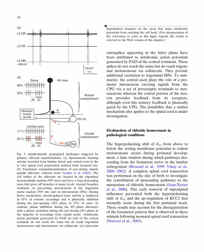
Fig. 3. Antidromically propagated discharges triggered byprimary afferent depolarizations. (a) Spontaneous burstingactivity recorded from lumbar dorsal and ventral roots in thein vitro spinal cord preparation isolated from neonatal rats.(b) Functional compartmentalization of jaw-closing musclespindle afferents, redrawn from Verdier et al. (2003). Thecell bodies of the afferents are located in the trigeminalmesencephalic nucleus (NV mes) and have a long descendingaxon that gives off branches at many levels. Axonal branchesterminate on jaw-closing motoneurons in the trigeminalmotor nucleus (NV mt) and on interneurons (INs). Duringfictive mastication, stretch-induced tonic activity is unalteredin 65% of somatic recordings and is phasically inhibitedduring the jaw-opening (JO) phase in 35% of cases. Incontrast, phasic inhibition during the JO phase alternateswith a phasic excitation during the jaw-closing (JC) phase inthe majority of recordings from caudal axons. Antidromicaction potentials generated by PAD (in red) of the centralterminals do not reach the soma but do reach trigeminalmotoneurons and interneurons via collaterals. (a) represents
hypothetical synapses on the axon that stops antidromicpotentials from reaching the cell body. (For interpretation ofthe references to color in this figure legend, the reader isreferred to the Web version of this chapter.)
b
10
extraspikes appearing in the latter phase havebeen attributed to antidromic action potentialsgenerated by PAD of the central terminals. Thesespikes do not reach the soma but do reach trigem-inal motoneurons via collaterals. They provideadditional excitation to trigeminal MNs. To sum-marize, the central axon plays the role of a pre-motor interneuron carrying signals from theCPG via a set of presynaptic terminals to mot-oneurons whereas the rostral portion of the neu-ron provides feedback from its receptors,although even this sensory feedback is phasicallygated by the CPG. The possibility that a similarmechanism also applies to the spinal cord is underinvestigation.
Dysfunction of chloride homeostasis inpathological conditions
The hyperpolarizing shift of ECl from above tobelow the resting membrane potential in rodentmotoneurons occurs during perinatal develop-ment, a time window during which pathways des-cending from the brainstem arrive in the lumbarenlargement (Brocard et al., 1999; Vinay et al.,2000, 2002). A complete spinal cord transectionwas performed on the day of birth to investigatethe contribution of descending pathways to thematuration of chloride homeostasis (Jean-Xavieret al., 2006). This early removal of supraspinalinfluences prevented both the hyperpolarizingshift of ECl and the up-regulation of KCC2 thatnormally occur during the first postnatal week.These results may account for the disorganizationof the locomotor pattern that is observed in theseanimals following neonatal spinal cord transaction(Norreel et al., 2003).

11
It is well known that several inhibitoryreflex mechanisms are decreased in patientsafter SCI (Boorman et al., 1996; Mazzocchioand Rossi, 1997; Morita et al., 2001). Themechanisms responsible for this reduced inhibi-tion were unknown until recently. Based on theabove-mentioned results showing that descendingpathways modulate chloride homeostasis, wehypothesized that a spinal cord injury in adultsmay affect the expression of KCC2 in the lumbarspinal cord and hence the strength of postsynapticinhibition. We showed that the expression ofKCC2 in the plasma membrane of motoneuronsis reduced after spinal cord injury (Boulenguezet al., 2010). We demonstrated that a blockadeof these cotransporters in intact animals rep-roduces some of the observations made in spasticparaplegic rats, suggesting that a down-regulationof KCC2 can contribute to the hyperexcitabilityof spinal networks, which is the hallmark of spas-ticity following spinal cord injury (Boulenguezet al., 2010).
Conclusion
To conclude, primary afferent terminal and mot-oneurons exhibit oppositemechanisms for chloridehomeostasis. A high [Cl�]i maintained by NKCC1cotransporters is responsible for PADs. The actionpotentials that are generated by PADs reaching fir-ing threshold have long been considered as an epi-phenomenon or an artifact due to the experimentalconditions. The ubiquity of their observation in dif-ferent motor networks underscores the need toinvestigate their role further. A low [Cl�]i ismaintained in healthy motoneurons by KCC2cotransporters; this is a requirement for a strongpostsynaptic inhibition. The excitatory effects ofGABA and glycine used to be considered only ina developmental perspective. The demonstrationthat KCC2 transporters and chloride homeostasisare affected in pathological conditions sheds newlight on these mechanisms. Restoring chloride
homeostasis, by up-regulating the functionality ofKCC2 transporters, may pave the way for newtreatments aimed at reducing spasticity followingspinal cord injury. The demonstration thatdepolarizing IPSPs can facilitate subthresholdexcitations also warrants further studies to investi-gate how these interactions contribute to synapticintegration and plasticity. That [Kþ]o and ECl arecritically dependent upon the activity raises thepossibility that the role of GABA/glycinergicinputs may reversibly switch from inhibition toexcitation, depending on the recent experience ofthe network.
Acknowledgments
Our study on the plasticity of inhibitory synaptictransmission in the spinal cord is supported bygrants (to L.V.) from the FrenchAgenceNationalepour la Recherche, the French Institut pour laRecherche sur la Moelle épinière et l'Encéphale(to L.V.) and the Christopher and DanaReeve Foundation (VB1-0502-2 and VB2-0801-2). K.S. received a grant from the AssociationFrançaise contre les Myopathies (Grant 13912).S.T. received a grant from the Fondation pour laRecherche Médicale (Grant FDT20081213783).
References
Abdul-Razzak, R., Bagust, J., & Kerkut, G. A. (1994). Postna-tal changes in the dorsal root reflex in the isolated spinalcord of the hamster Mesocricetus auratus. ComparativeBiochemistry and Physiology, 107C, 195–204.
Alvarez, F. J. (1998). Anatomical basis for presynaptic inhibi-tion of primary sensory fibers. In P. Rudomin, L. M. Romo& S. Mendell (Eds.), Presynaptic inhibition and neural con-trol (pp. 13–49). Oxford: Oxford University Press.
Alvarez-Leefmans, F. J., Nani, A., & Marquez, S. (1998).Chloride transport, osmotic balance, and presynaptic inhibi-tion. In P. Rudomin, R. Romo & L. M. Mendell (Eds.),Presynaptic inhibition and neural control (pp. 50–79).Oxford: Oxford University Press.
Bagust, J., Kerkut, G. A., & Rakkah, N. I. A. (1989). Thedorsal root reflex in isolated mammalian spinal cord. Com-parative Biochemistry and Physiology, 93A, 151–160.

12
Barron, D. H., & Matthews, B. H. C. (1938). Dorsal rootreflexes. Journal of Physiology (London), 94, 26P–27P.
Beloozerova, I., & Rossignol, S. (1999). Antidromic dischargesin dorsal roots of decerebrate cats. I: Studies at rest and dur-ing fictive locomotion. Brain Research, 846, 87–105.
Beloozerova, I. N., & Rossignol, S. (2004). Antidromic dis-charges in dorsal roots of decerebrate cats. II: Studies duringtreadmill locomotion. Brain Research, 996, 227–236.
Boorman, G. I., Lee, R. G., Becker, W. J., & Windhorst, U. R.(1996). Impaired “natural reciprocal inhibition” in patientswith spasticity due to incomplete spinal cord injury.Electroencephalography and Clinical Neurophysiology, 101,84–92.
Boulenguez, P., Liabeuf, S., Bos, R., Bras, H., Jean-Xavier, C.,Brocard, C., et al. (2010). Down-regulation of thepotassium-chloride cotransporter KCC2 contributes tospasticity after spinal cord injury. Nature Medicine, 16,302–307.
Brocard, F., Vinay, L., & Clarac, F. (1999). Gradual develop-ment of the ventral funiculus input to lumbar motoneuronsin the neonatal rat. Neuroscience, 90, 1543–1554.
Busselberg, D., Bischoff, A. M., Becker, K., Becker, C. M., &Richter, D. W. (2001). The respiratory rhythm in mutantoscillator mice. Neuroscience Letters, 316, 99–102.
Busselberg, D., Bischoff, A. M., Paton, J. F., & Richter, D. W.(2001). Reorganisation of respiratory network activityafter loss of glycinergic inhibition. Pflügers Archiv, 441,444–449.
Chen, Y., Bagust, J., Kerkut, G. A., & Tyler, A. W. (1993).Correlation between spontaneous bursts of activity recordedfrom the dorsal roots in an isolated hamster spinal cord.Experimental Physiology, 78, 811–824.
Chub, N., & O'Donovan, M. J. (2001). Post-episode depres-sion of GABAergic transmission in spinal neurons of thechick embryo. Journal of Neurophysiology, 85, 2166–2176.
Curtis, D. R., & Lodge, D. (1982). The depolarization of felineventral horn group Ia spinal afferent terminations byGABA. Experimental Brain Research, 46, 215–233.
Delpire, E., & Mount, D. B. (2002). Human and murinephenotypes associated with defects in cation–chloridecotransport. Annual Review of Physiology, 64, 803–843.
Delpy, A., Allain, A. E., Meyrand, P., & Branchereau, P.(2008). NKCC1 cotransporter inactivation underlies embry-onic development of chloride-mediated inhibition in mousespinal motoneuron. Journal of Physiology, 586, 1059–1075.
Dubuc, R., Cabelguen, J. M., & Rossignol, S. (1985). Rhyth-mic antidromic discharges of single primary afferentsrecorded in cut dorsal root filaments during locomotion inthe cat. Brain Research, 359, 375–378.
Dubuc, R., Cabelguen, J.-M., & Rossignol, S. (1988). Rhyth-mic fluctuations of dorsal root potentials and antidromic dis-charges of primary afferents during fictive locomotion in thecat. Journal of Neurophysiology, 60, 2014–2036.
Duenas, S. H., Loeb, G. E., & Marks, W. B. (1990). Monosyn-aptic and dorsal root reflexes during locomotion in normaland thalamic cats. Journal of Neurophysiology, 63,1467–1476.
Dzhala, V. I., Talos, D. M., Sdrulla, D. A., Brumback, A. C.,Mathews, G. C., Benke, T. A., et al. (2005). NKCC1 trans-porter facilitates seizures in the developing brain. NatureMedicine, 11, 1205–1213.
Eccles, J. C., Schmidt, R. F., & Willis, W. D. (1963). Pharma-cological studies on presynaptic inhibition. Journal of Physi-ology (London), 168, 500–530.
Fellippa-Marques, S., Vinay, L., & Clarac, F. (2000). Sponta-neous and locomotor-related GABAergic input onto pri-mary afferents in the neonatal rat. European Journal ofNeuroscience, 12, 155–164.
Gao, X. B., Chen, G., & van den Pol, A. N. (1998). GABA-dependent firing of glutamate-evoked action potentials atAMPA/kainate receptors in developing hypothalamicneurons. Journal of Neurophysiology, 79, 716–726.
Gao, B. X., Stricker, C., & Ziskind-Conhaim, L. (2001). Tran-sition from GABAergic to glycinergic synaptic transmissionin newly formed spinal networks. Journal of Neurophysiol-ogy, 86, 492–502.
Gao, B.-X., & Ziskind-Conhaim, L. (1995). Development ofglycine- and GABA-gated currents in rat spinal motoneurons.Journal of Neurophysiology, 74, 113–121.
Gonzalez-Islas, C. E., Chub, N. L., & Wenner, P. (2009).NKCC1 and AE3 appear to accumulate chloride in embry-onic motoneurons. Journal of Neurophysiology, 101, 507–518.
Gossard, J. P., Cabelguen, J.-M., & Rossignol, S. (1991).An intracellular study of muscle primary afferents duringfictive locomotion in the cat. Journal of Neurophysiology,65, 914–926.
Gulledge, A. T., & Stuart, G. J. (2003). Excitatory actions ofGABA in the cortex. Neuron, 37, 299–309.
Hanson, M. G., & Landmesser, L. T. (2003). Characterizationof the circuits that generate spontaneous episodes of activityin the early embryonic mouse spinal cord. Journal of Neuro-science, 23, 587–600.
Hübner, C. A., Stein, V., Hermans-Borgmeyer, I., Meyer, T.,Ballanyi, K., & Jentsch, T. J. (2001). Disruption of KCC2reveals an essential role of K–Cl cotransport already in earlysynaptic inhibition. Neuron, 30, 515–524.
Iizuka, M., Kiehn, O., & Kudo, N. (1997). Development inneonatal rats of the sensory resetting of the locomotorrhythm induced by NMDA and 5-HT. Experimental BrainResearch, 114, 193–204.
Ikeda, M., Toyoda, H., Yamada, J., Okabe, A., Sato, K.,Hotta, Y., et al. (2003). Differential development ofcation–chloride cotransporters and Cl� homeostasis con-tributes to differential GABAergic actions between devel-oping rat visual cortex and dorsal lateral geniculatenucleus. Brain Research, 984, 149–159.
13
Jean-Xavier, C., Mentis, G. Z., O'Donovan, M., Cattaert, D.,& Vinay, L. (2007). Dual personality of GABA/glycine-mediated depolarizations in the immature spinal cord.Proceedings of the National Academy of Sciences of theUnited States of America, 104, 11477–11482.
Jean-Xavier, C., Pflieger, J.-F., Liabeuf, S., & Vinay, L. (2006).Inhibitory post-synaptic potentials in lumbar motoneuronsremain depolarizing after neonatal spinal cord transectionin the rat. Journal of Neurophysiology, 96, 2274–2281.
Kerkut, G. A., & Bagust, J. (1995). The isolated mammalianspinal cord. Progress in Neurobiology, 46, 1–48.
Kolta, A., Lund, J. P., Westberg, K. G., & Clavelou, P. (1995).Do muscle-spindle afferents act as interneurons duringmastication? (letter; comment). Trends in Neurosciences,18, 441.
Kremer, E., & Lev-Tov, A. (1998). GABA-Receptor-indepen-dent dorsal root afferents depolarization in the neonatal ratspinal cord. Journal of Neurophysiology, 79, 2581–2592.
Levy, R. A. (1975). The effect of intravenously administeredgamma-aminobutyric acid on afferent fiber polarization.Brain Research, 92, 21–34.
Marchetti, C., Tabak, J., Chub, N., O'Donovan, M. J., &Rinzel, J. (2005). Modeling spontaneous activity in the devel-oping spinal cord using activity-dependent variations of intra-cellular chloride. Journal of Neuroscience, 25, 3601–3612.
Mazzocchio, R., & Rossi, A. (1997). Involvement of spinalrecurrent inhibition in spasticity. Further insight into theregulation of Renshaw cell activity. Brain, 120(Pt 6),991–1003.
Morita, H., Crone, C., Christenhuis, D., Petersen, N. T., &Nielsen, J. B. (2001). Modulation of presynaptic inhibitionand disynaptic reciprocal Ia inhibition during voluntarymovement in spasticity. Brain, 124, 826–837.
Myers, C. P., Lewcock, J. W., Hanson, M. G., Gosgnach, S.,Aimone, J. B., Gage, F. H., et al. (2005). Cholinergic inputis required during embryonic development to mediateproper assembly of spinal locomotor circuits. Neuron, 46,37–49.
Nishimaru, H., Iizuka, M., Ozaki, S., & Kudo, N. (1996). Spon-taneous motoneuronal activity mediated by glycine andGABA in the spinal cord of rat fetuses in vitro. Journal ofPhysiology, 497, 131–143.
Nishimaru, H., & Kudo, N. (2000). Formation of the centralpattern generator for locomotion in the rat and mouse.Brain Research Bulletin, 53, 661–669.
Norreel, J.-C., Pflieger, J.-F., Pearlstein, E., Simeoni-Alias, J.,Clarac, F., & Vinay, L. (2003). Reversible disorganizationof the locomotor pattern after neonatal spinal cord transec-tion in the rat. Journal of Neuroscience, 23, 1924–1932.
Payne, J. A., Rivera, C., Voipio, J., & Kaila, K. (2003).Cation–chloride co-transporters in neuronal communication,developmentand trauma.Trends inNeurosciences,26, 199–206.
Pilyavskii, A. I., Yakhnitsa, V. A., & Bulgakova, N. V. (1988).Antidromic dorsal root impulses during naturally occurringlocomotion in rats. Neurophysiology, 20, 417–422.
Plotkin, M. D., Snyder, E. Y., Hebert, S. C., & Delpire, E.(1997). Expression of the Na–K–2Cl cotransporter is devel-opmentally regulated in postnatal rat brains: A possiblemechanism underlying GABA's excitatory role in immaturebrain. Journal of Neurobiology, 33, 781–795.
Ramirez, J. M., Telgkamp, P., Elsen, F. P., Quellmalz, U. J., &Richter, D. W. (1997). Respiratory rhythm generation inmammals: Synaptic and membrane properties. RespiratoryPhysiology, 110, 71–85.
Ren, J., & Greer, J. J. (2003). Ontogeny of rhythmic motorpatterns generated in the embryonic rat spinal cord. Journalof Neurophysiology, 89, 1187–1195.
Ren, J., & Greer, J. J. (2006). Modulation of respiratoryrhythmogenesis by chloride-mediated conductances duringthe perinatal period. Journal of Neuroscience, 26,3721–3730.
Rivera, C., Voipio, J., & Kaila, K. (2004). Two developmentalswitches in GABAergic signalling: The K–Cl cotransporterKCC2, and carbonic anhydrase CAVII. Journal of Physiol-ogy, 562, 27–36.
Rivera, C., Voipio, J., Payne, J. A., Ruusuvuori, E.,Lahtinen, H., Lamsa, K., et al. (1999). The Kþ/Cl� co-trans-porter KCC2 renders GABA hyperpolarizing during neuro-nal maturation. Nature, 397, 251–255.
Rossignol, S., Beloozerova, I. N., Gossard, J. P., & Dubuc, R.(1998). Presynaptic mechanisms during locomotion. InP. Rudomin, R. Romo & L. M. Mendell (Eds.), Presynapticinhibition and neural control (pp. 385–397). Oxford: OxfordUniversity Press.
Rudomin, P. (1990). Presynaptic inhibition of muscle spindleand tendon organ afferents in the mammalian spinal cord.Trends in Neurosciences, 13, 499–505.
Rudomin, P., Engberg, I., & Jimenez, I. (1981). Mechanismsinvolved in presynaptic depolarization of group I andrubrospinal fibers in cat spinal cord. Journal of Neurophysi-ology, 46, 532–548.
Rudomin, P., Quevedo, J., & Eguibar, J. R. (1993). Presynap-tic modulation of spinal reflexes. Current Opinion in Neuro-biology, 3, 997–1004.
Sadlaoud, K., Tazerart, S., Brocard, C., Jean-Xavier, C.,Portalier, P., Brocard, F., et al. (2010). Differential plasticityof the GABAergic and glycinergic synaptic transmission torat lumbar motoneurons after spinal cord injury. Journal ofNeuroscience, 30, 3358–3369.
Shao, X. M., & Feldman, J. L. (1997). Respiratory rhythm gen-eration and synaptic inhibition of expiratory neurons in pre-Botzinger complex: Differential roles of glycinergic andGABAergic neural transmission. Journal of Neurophysiol-ogy, 77, 1853–1860.

14
Stein, V., Hermans-Borgmeyer, I., Jentsch, T. J., &Hubner, C. A. (2004). Expression of the KCl cotransporterKCC2 parallels neuronal maturation and the emergence oflow intracellular chloride. Journal of Comparative Neurol-ogy, 468, 57–64.
Stil, A., Liabeuf, S., Jean-Xavier, C., Brocard, C.,Viemari, J. C., & Vinay, L. (2009). Developmental up-regu-lation of the potassium–chloride cotransporter type 2 in therat lumbar spinal cord. Neuroscience, 164, 809–821.
Sun, D., & Murali, S. G. (1999). Naþ–Kþ–2Cl� cotransporter
in immature cortical neurons: A role in intracellular Cl� reg-ulation. Journal of Neurophysiology, 81, 1939–1948.
Sung, K. W., Kirby, M., McDonald, M. P., Lovinger, D. M., &Delpire, E. (2000). Abnormal GABAA receptor-mediatedcurrents in dorsal root ganglion neurons isolated fromNa–K–2Cl cotransporter null mice. Journal of Neuroscience,20, 7531–7538.
Takahashi, T. (1984). Inhibitory miniature synaptic potentialsin rat motoneurons. Proceedings of Royal Society of LondonB Biological Sciences, 221, 103–109.
Toennies, J. F. (1938). Reflex discharge from the spinal cordover the dorsal roots. Journal of Neurophysiology, 1,378–390.
Vardi, N., Zhang, L. L., Payne, J. A., & Sterling, P. (2000).Evidence that different cation chloride cotransporters in ret-inal neurons allow opposite responses to GABA. Journal ofNeuroscience, 20, 7657–7663.
Verdier, D., Lund, J. P., & Kolta, A. (2003). GABAergic con-trol of action potential propagation along axonal branches
of mammalian sensory neurons. Journal of Neuroscience,23, 2002–2007.
Vinay, L., Brocard, F., Clarac, F., Norreel, J. C., Pearlstein, E.,& Pflieger, J. F. (2002). Development of posture and loco-motion: An interplay of endogenously generated activitiesand neurotrophic actions by descending pathways. BrainResearch Review, 40, 118–129.
Vinay, L., Brocard, F., Fellippa-Marques, S., & Clarac, F.(1999). Antidromic discharges of dorsal root afferents inthe neonatal rat. Journal of Physiology Paris, 93, 359–367.
Vinay, L., Brocard, F., Pflieger, J. F., Simeoni-Alias, J., &Clarac, F. (2000). Perinatal development of lumbar mot-oneurons and their inputs in the rat. Brain Research Bulle-tin, 53, 635–647.
Vinay, L., & Clarac, F. (1999). Antidromic discharges of dorsalroot afferents and inhibition of the lumbar monosynapticreflex in the neonatal rat. Neuroscience, 90, 165–176.
Vinay, L., & Jean-Xavier, C. (2008). Plasticity of spinal cordlocomotor networks and contribution of cation–chloridecotransporters. Brain Research Review, 57, 103–110.
Wu, W.-L., Ziskind-Conhaim, L., & Sweet, M. A. (1992).Early development of glycine- and GABA-mediatedsynapses in rat spinal cord. Journal of Neuroscience, 12,3935–3945.
Ziskind-Conhaim, L. (1998). Physiological functions ofGABA-induced depolarizations in the developing rat spinalcord. Perspectives on Developemental Neurobiology, 5,279–287.