The Biological Clock: The Bodyguard of Temporal Homeostasis
26
CHRONOBIOLOGY INTERNATIONAL Vol. 21, No. 1, pp. 1–25, 2004 REVIEW The Biological Clock: The Bodyguard of Temporal Homeostasis Ste´ phanie Perreau-Lenz, 1,2 Paul Pe´vet, 2 Ruud M. Buijs, 1 and Andries Kalsbeek 1, * 1 Netherlands Institute for Brain Research, Amsterdam, The Netherlands 2 Neurobiologie des Rythmes, Strasbourg, France ABSTRACT In order for any organism to function properly, it is crucial that it be table to control the timing of its biological functions. An internal biological clock, located, in mammals, in the suprachiasmatic nucleus of the hypothalamus (SCN), therefore carefully guards this temporal homeostasis by delivering its message of time throughout the body. In view of the large variety of body functions (behavioral, physiological, and endocrine) as well as the large variety in their preferred time of main activity along the light:dark cycle, it seems logical to envision different means of time distribution by the SCN. In the present review, we propose that even though it presents a unimodal circadian rhythm of general electrical and metabolic activity, the SCN seems to use several sorts of output connections that are active at different times along the light: dark cycle to control the rhythmic expression of different body functions. Although the SCN is suggested to use diffusion of synchronizing factors in the rhythmic control of behavioral functions, it also needs neuronal connections for the control of endocrine functions. The distribution of the time-of-day message to neuroendo- crine systems is either directly onto endocrine neurons or via intermediate *Correspondence: Dr. Andries Kalsbeek, Netherlands Institute for Brain Research, Meibergdreef 33, 1105 AZ, Amsterdam, Netherlands; Fax: (-31) 20-6961-006; E-mail: [email protected]. 1 DOI: 10.1081/CBI-120027984 0742-0528 (Print); 1525-6073 (Online) Copyright & 2004 by Marcel Dekker, Inc. www.dekker.com
-
Upload
independent -
Category
Documents
-
view
2 -
download
0
Transcript of The Biological Clock: The Bodyguard of Temporal Homeostasis

CHRONOBIOLOGY INTERNATIONAL
Vol. 21, No. 1, pp. 1–25, 2004
REVIEW
The Biological Clock: The Bodyguard of
Temporal Homeostasis
Stephanie Perreau-Lenz,1,2
Paul Pevet,2
Ruud M. Buijs,1 and Andries Kalsbeek1,*
1Netherlands Institute for Brain Research, Amsterdam, The Netherlands2Neurobiologie des Rythmes, Strasbourg, France
ABSTRACT
In order for any organism to function properly, it is crucial that it be table to
control the timing of its biological functions. An internal biological clock,
located, in mammals, in the suprachiasmatic nucleus of the hypothalamus (SCN),
therefore carefully guards this temporal homeostasis by delivering its message of
time throughout the body. In view of the large variety of body functions
(behavioral, physiological, and endocrine) as well as the large variety in their
preferred time of main activity along the light:dark cycle, it seems logical to
envision different means of time distribution by the SCN. In the present review,
we propose that even though it presents a unimodal circadian rhythm of general
electrical and metabolic activity, the SCN seems to use several sorts of output
connections that are active at different times along the light: dark cycle to control
the rhythmic expression of different body functions. Although the SCN is
suggested to use diffusion of synchronizing factors in the rhythmic control of
behavioral functions, it also needs neuronal connections for the control of
endocrine functions. The distribution of the time-of-day message to neuroendo-
crine systems is either directly onto endocrine neurons or via intermediate
*Correspondence: Dr. Andries Kalsbeek, Netherlands Institute for Brain Research,
Meibergdreef 33, 1105 AZ, Amsterdam, Netherlands; Fax: (-31) 20-6961-006; E-mail:
1
DOI: 10.1081/CBI-120027984 0742-0528 (Print); 1525-6073 (Online)
Copyright & 2004 by Marcel Dekker, Inc. www.dekker.com

ORDER REPRINTS
neurons located in specific SCN targets. In addition, the SCN uses its connections
with the autonomic nervous system for spreading its time-of-day message, either
by setting the sensitivity of endocrine glands (i.e., thyroid, adrenal, ovary) or by
directly controlling an endocrine output (i.e., melatonin synthesis). Moreover, the
SCN seems to use different neurotransmitters released at different times along the
light: dark cycle for each of the different connection types presented. Clearly, the
temporal homeostasis of endocrine functions results from a diverse set of
biological clock outputs.
Key Words: Endocrine functions; Circadian rhythms; Suprachiasmatic nucleus.
ABBREVIATIONS
ACTH Adrenocorticotropin hormoneAVP Arginine vasopressinCRH Corticotropin-releasing hormoneDMH Dorsomedial nucleus of the hypothalamusGnRH Gonadotropin-releasing hormoneHPA-axis Hypothalamo-pituitary-adrenal axisHPG-axis Hypothalamo-pituitary-gonadal axisHPT-axis Hypothalamo-pituitary-thyroid axisLH Luteinizing hormoneMPOA Medial preoptic areaPVN Paraventricular nucleus of the hypothalamusPRL ProlactinSCG Superior cervical ganglionSCN Suprachiasmatic nucleus of the hypothalamussPVN Subparaventricular nucleus of the hypothalamusTRH Thyrotropin-releasing hormoneTSH Thyroid-stimulating hormoneVIP Vasoactive intestinal polypeptideVP Vasopressin
INTRODUCTION
One day consists of one cycle of an alternating light and dark period with aconstant duration of 24 h, and many biological functions obey the same 24 h rhythm.Thanks to experiments in rats (Richter, 1922) and in humans (Aschoff and Wever,1962), these rhythms, although influenced by the environment, are shown to becontrolled by an endogenous biological clock. Following these first experiments onbiological clock mechanisms, successive studies, mainly realized in laboratoryrodents, showed that many physiological and endocrine functions obey this rule ofan endogenous rhythm close to 24 h and present so called circadian rhythms,circadian being coined from the Latin words ‘‘circa,’’ meaning ‘‘close to’’ and ‘‘dies’’meaning ‘‘day.’’ Main behaviors such as locomotor activity, food intake, or sleep
2 Perreau-Lenz et al.

ORDER REPRINTS
present a daily rhythm that persists in constant conditions with a period close to24 h. Not only behavioral functions, but several endocrine functions as well, suchas luteinizing hormone (LH), melatonin, corticosterone, or leptin, present a clearcircadian rhythmicity (Fig. 1).
The suprachiasmatic nucleus of the hypothalamus (SCN) has been identifiedas the seat of the endogenous biological clock. A large number of studies havedemonstrated the necessity of its presence if circadian rhythms are to be sustained(Moore and Eichler, 1972; Moore and Klein, 1974; Raisman and Brown-Grant,1977; Reppert et al., 1981; Stephan and Zucker, 1972; Stoynev et al., 1982;Watanabe and Hiroshige, 1981; Van Esseveldt et al., 2000). In the past 10 years theunderstanding of the internal clock mechanism has grown extremely fast with thedevelopment of molecular biology techniques, and enormous progress has beenmade in the understanding of the molecular clockwork of this master clock. Indeed,it has been recently shown that SCN cells contain several so-called clock genes thatpresent a circadian expression and interact with each other in gene-protein-genefeedback loops (for review, Albrecht and Eichele, 2003; Reppert and Weaver, 2001).Subsequently, the same clock genes were also found to be expressed rhythmically inother structures, both in the central nervous system (Abe et al., 2002; Yamazaki et al.,2000) and in the periphery (Damiola et al., 2000; Hara et al., 2001; Yamazaki et al.,2000). But once these structures are isolated, the oscillation of their clock genes is notsustained, suggesting a necessary entrainment or synchronization of peripheraloscillators by the master clock (Balsalobre, 2002; Yamazaki et al., 2000). Despitethe discovery of the clock genes, however, some important questions still remain.For example, how do we explain the large diversity in the peak activity of differentbehaviors and endocrine functions with the unimodal activity pattern of the SCNdescribed so far with multi-unit recordings (Inouye and Kawamura, 1979; Shibataet al., 1982), and the 2-deoxy-glucose technique (Schwartz and Gainer, 1977;Schwartz et al., 1980)? Until recently the SCN was considered a ‘‘homogenous’’structure, giving one global timing signal. However, it is becoming more and moredifficult to explain how the SCN is able to control different functions with separatelytimed 24 h rhythms as diverse as shown in Fig. 1, when distributing only one uniformmessage. Although several hormones are released in a circadian manner, the time oftheir peak-release varies greatly throughout the light-dark cycle. The picturebecomes even more complicated when one realizes that in diurnal and nocturnalspecies the SCN activity functions in an identical way, i.e., despite the antiphase ofthe sleep/wake patterns, the main activity period of the SCN is found during the lightperiod in either type of species (Hofman and Swaab, 1993; Schwartz et al., 1983;Shibata and Moore, 1988). Moreover, only some endocrine rhythms are associatedwith the timing of behavioral activity, whereas others remain phase-locked to theirposition within the light-dark cycle. For example, LH and corticosterone plasmaconcentrations have their acrophase before the beginning of the activity period (i.e.,in the morning for diurnal animals, but in the evening for nocturnal animals), butthe acrophase of melatonin and leptin release is always found during the dark period,independent of the nocturnal or diurnal nature of an animal. Yet, the question thatarises is how the SCN distributes its message of time, which is built within each of itscells, to the rest of the body? Is a specific population of SCN cells responsible foreach specific output? Or, alternatively, are all SCN neurons involved in the creation
The Biological Clock 3

ORDER REPRINTS
of one uniform output signal of the biological clock that is differently interpreted bythe various clock targets?
The question of how the message of time is distributed by the SCN was firstaddressed in transplantation studies. The restoration of the circadian rhythms oflocomotor activity and drinking behavior in SCN-lesioned animals, within a weekafter having received SCN grafts (Ralph et al., 1990), even if encapsulated (Silver etal., 1996), proved that the SCN has the potential to distribute at least part of itsmessage by simple diffusion of substances in the control of behavioral functions.Indeed, recently, SCN-secreted molecules such as TGF-alpha or prokineticin 2 havebeen suggested to play a role in the control of the circadian rhythm of locomotoractivity (Cheng et al., 2002; Kramer et al., 2001). However, the above-mentionedtransplantation studies also indicated that this simple way of distribution could notbe exclusive. In contrast with behavioral functions, the rhythmicity of endocrinefunctions, such as gonadal function or daily cortisol secretion, was not restored in
Figure 1. Diurnal rhythms of release of various hormones measured in rats. However, note
the diversity in the release pattern along the light-dark cycle.
4 Perreau-Lenz et al.

ORDER REPRINTS
SCN-grafted animals (Lehman et al., 1987; Meyer-Bernstein et al., 1999). Therefore,SCN neuronal connections onto other brain targets seem to be essential to transmitthe full circadian information throughout the entire body. These first observationssupported the idea that the SCN may guard time-homeostasis within the body, usingdifferent tools of distribution. As opposed to behavioral rhythms, endocrine dailyrhythms require direct and precise regulation, which could be provided by neuronalprojections to diverse target areas. Indeed, recently De La Iglesia et al. (2003) veryelegantly demonstrated the necessity for a neural rather than a humoral SCN outputfor the daily control of neuroendocrine rhythms. SCN projections were extensivelystudied in the past by tracing and electrophysiological studies (Watts and Swanson,1987, Watts et al., 1987). Restricted to only a small part of the central nervoussystem, SCN projections are concentrated within the medial hypothalamus: medialpreoptic area (MPOA), paraventricular nucleus (PVN), subparaventricular nucleus(sPVN), and dorsomedial nucleus of the hypothalamus (DMH). In addition, outsidethe hypothalamus, the SCN only projects onto the paraventricular nucleus of thethalamus and the lateral geniculate nucleus.
In this review we will try to show the different mechanisms the SCN uses tocontrol time-dependent homeostasis of endocrine functions within the body. In thedistribution of its temporal message to the endocrine system the SCN employs ahighly specialized set of projections to three different types of neurons within themedial hypothalamus. SCN fibers directly contact both endocrine neurons andautonomic neurons. In addition, the SCN can influence endocrine rhythms via itsconnection to intermediate neurons projecting to endocrine neurons. In the presentreview we will discuss the physiological and anatomical evidence, showing that foreach specific endocrine function the SCN uses variable combinations of thesedifferent means of signal distribution. Moreover, the diversity of neurotransmitterspresent in the projecting SCN fibers indicates the possibility of a further differentialregulation.
The daily rhythms of melatonin and corticosterone release are the twoendocrine circadian rhythms that have been studied the most so far, because oftheir pronounced amplitude and the reproducible timing of their acrophase.Melatonin synthesis has the additional advantage that it is not influenced bystress. Moreover, melatonin synthesis and release precisely follow the environmentalphotoperiod, and in this way couples circadian and seasonal rhythms. Thetransformation of environmental information into a hormonal signal is essentialfor the distribution of time-of-the-year information throughout the rest of the body(Reiter, 1993). Similarly, by penetrating in all tissues of the body, corticosteronemay help the SCN to distribute its time-of-day message.
SCN Output to Endocrine Neurons
One of the means the SCN uses to control endocrine rhythms involves direct andindirect control of endocrine neurons. The SCN was shown to be able to affect,directly or indirectly, the activity of both magnocellular neurons, liberating VP oroxytocin directly within the blood stream, and of parvocellular hypophysiotropicneurons, containing releasing hormones such as corticotropin-releasing hormone
The Biological Clock 5

ORDER REPRINTS
(CRH), thyrotropin-releasing hormone (TRH), or gonadotropin-releasing hormone(GnRH). Direct functional connections between SCN and magnocellular neuronshave been demonstrated in the supraoptic nucleus of the hypothalamus (SON) (Cuiet al., 1997), as well as in the PVN (Hermes et al., 1996). In addition, also the dailyrhythm of plasma vasopressin (VP), as measured with a concentration peak duringmidday, suggests that magnocellular neurons have a diurnal activity (Terwel et al.,1992; Windle et al., 1992). Evidence for direct, but limited, connections betweenSCN and hypophysiotropic neurons containing releasing hormones has also beenpresented (Buijs et al., 1993; Kalsbeek et al., 2000a; Van Der Beek et al., 1997; Vranget al., 1995). Although both magnocellular and hypophysiotropic neurons areconnected to the clock, the influence (direct or indirect) of the SCN differs,depending on their endocrine function.
In an initial study on clock control of the hypothalamo-pituitary-adrenal (HPA)axis, we showed a strong inhibitory effect of SCN-derived VP on corticosteronerelease in the PVN/DMH area (Kalsbeek and Buijs, 1992). Both the sPVN andDMH contain g-amino-butyric acid (GABA) neurons projecting to the medialparvocellular part of the PVN (Roland and Sawchenko, 1993) and are prominenttarget areas of SCN projections. Since VP is, as reported so far, a stimulatoryneurotransmitter, and since direct contacts between SCN and hypophysiotropicneurons in the PVN are limited, we proposed that the SCN inhibits the HPA axismainly by using indirect VPergic inputs on inhibitory intermediate neurons in thesPVN and DMH. This idea was confirmed later by two successive studies. In thefirst of these two studies (Kalsbeek et al., 1992), we showed that local infusions of VPwithin the DMH immediately decreased plasma corticosterone, as well as theadrenocorticotropin hormone (ACTH) levels, in both SCN-intact and SCN-lesionedanimals. In addition, application of a VP-antagonist in the same structure caused astrong increase of plasma corticosterone levels, but only in intact animals,confirming that the VP signaling involved in the inhibition of the HPA axis derivesdirectly from the SCN. In the second study (Kalsbeek et al., 1996b), VP-antagonistapplication at different times of the light:dark cycle revealed the existence of anadditional stimulatory input from the SCN to the HPA axis. Moreover, usingelectrophysiological techniques we were able to show, in vitro, that VP increases theGABAergic inhibition of PVN neurons (Hermes et al., 2000). These results led to ahypothetical scheme, where the corticosterone circadian rhythm results fromcombined inhibitory (via VPergic release onto GABAergic interneurons) andstimulatory SCN outputs of unknown nature (Kalsbeek et al., 1996b). In contrast tothe indirect connections to the hypophysiotropic neurons, the SCN seems to have adirect influence on magnocellular neurons of the PVN. Indeed, specific VPdepletion in the SCN affects the VP content of magnocellular vs. parvocellular(i.e., hypophysiotropic neurons) PVN neurons in different ways (Gomez et al., 1997).VP content increases in magnocellular PVN neurons, whereas it decreases inparvocellular neurons, suggesting decreased release of VP in the general circulation,and an increased release of VP (and CRH) into the portal circulation. Regarding thefact that elevated plasma ACTH levels were measured after such SCN-VP depletion,these results suggest a major inhibitory action of the SCN on the HPA axis. PlasmaVP (and oxytocin) levels, however, were not measured, and a physiologicalconfirmation of the stimulatory effect of SCN-VP on the magnocellular system is
6 Perreau-Lenz et al.

ORDER REPRINTS
thus lacking. In the meantime the inhibitory effect of VP on corticosterone release asjust described has been reported by a number of other laboratories as well (Gomezet al., 1997; Wotjak et al., 1996), but still the effect is very surprising to many(endocrinologists) since the inhibitory action of VP is completely opposite to thepreviously described and better known stimulatory role of VP in the HPA stressresponse. The stimulatory action of VP on ACTH and corticosterone release wasdefinitely established in the early 1980s (Gillies et al., 1982; Rivier and Vale, 1983;Rivier et al., 1984). It turned out that VP strongly potentiated the stimulatory actionof the just-discovered true corticotrophin releasing factor, i.e., CRH (Vale et al.,1981). Indeed, shortly afterward it was shown that CRH and VP are colocalized inthe parvocellular neurons of the PVN (Kiss et al., 1984; Sawchenko et al., 1984;Tramu et al., 1993) and that many stimuli that cause an activation of the HPA-axis,such as immobilization, novelty, and hypoglycemia, go along with a depletion of theCRH as well as the VP content in the external zone of the median eminence(Bartanusz et al., 1993; Berkenbosch et al., 1989; De Goeij et al., 1992; Romero et al.,1993). The emerging view is that VP is the principal, regulated, variable that putsa situation-specific drive on the HPA-axis, whereas CRH serves mainly to impose astimulatory tone (Kovacs et al., 2000), i.e. under resting conditions the parvocellularVP neurons of the PVN do not contain detectable amounts of VP immunoreactivityor mRNA. The logical explanation for the apparently opposite actions of VP, ofcourse, is that the different effects are caused by two completely different andseparated VP systems. The inhibitory effect concerns VP-containing neurons locatedin the SCN projecting to the PVN and DMH, whereas the stimulatory effect of VPis derived from the parvocellular VP neurons located in the PVN that project to themedian eminence. In addition, comparing the responses of ACTH and cortico-sterone in more detail also reveals that the two systems employ different mechanismsto affect the HPA-axis. The circadian variation in HPA-axis activity is mainlycontrolled by changing the sensitivity of the adrenal cortex to ACTH, whereas thestress response of the HPA-axis relies heavily on the release of ACTH. A furtherdiscussion of the different VP systems and the close relation between VPergicsystems and glucocorticoids can be found in Kalsbeek et al. (2002, 2003).
The VP-mediated SCN control of the HPA axis is not exclusive; in addition,the SCN is thought to use its main peptidergic output, i.e., VP, to control thehypothalamo-pituitary-gonadal (HPG) axis as well, in a direct and indirect manner.The two essential brain areas for maintaining the LH surge are the MPOA and theSCN (Gray et al., 1978). The MPOA contains a dense concentration of estrogenreceptors and GnRH neurons, as well as a dense VP innervation originating from theSCN (De Vries et al., 1984; Hoorneman and Buijs, 1982). Once every four to fivedays, an interaction between the peripheral endocrine estrogen signal and thecircadian output leads to a strong GnRH release in the portal circulation, andconsequently to a pro-oestrus surge of LH. Direct connections between the SCNand neurons in the MPOA have been shown (De La Iglesia et al., 1995; Van DerBeek et al., 1997; Watson et al., 1995); 11–13% of GnRH neurons and up to 30% ofthe neurons containing estrogen-receptors receive direct efferent projections fromthe SCN. Since VP infusions within the MPOA are able to restore the LH surge inSCN-lesioned animals, irrespective of the time-of-day (Palm et al., 1999; 2001), andsince, in vitro, GnRH release exhibits a significant circadian rhythm in phase with
The Biological Clock 7
ORDER REPRINTS
the circadian release of VP from the SCN (Funabashi et al., 2000), we concluded thatthe SCN uses its VP-ergic output as a stimulatory signal for the control of the HPGaxis. We proposed that the SCN would stimulate the release of GnRH indirectly viaoestrogen-receptor containing neurons of the MPOA. Interestingly, VP administra-tion within the MPOA could stimulate the LH surge in intact animals only during arestricted time window, whereas in SCN-lesioned rats the LH surge could be inducedat any time. These results also suggest that apart from the main stimulatory input,the SCN also sends an inhibitory input of unknown nature during the first part ofthe day to restrain the GnRH system. Since neurons in the MPOA are also contactedby the SCN in an indirect way via the subparaventricular zone of the PVN or theretrochiasmatic area, and since direct projections from SCN neurons containingvasoactive intestinal polypeptide (VIP) onto the endocrine GnRH-containingneurons have also been demonstrated (Van Der Beek et al., 1997; 1993), furtherinvestigations will be necessary to understand fully the SCN control of the GnRHsystem. Also for a further discussion of the multiple interactions of central VPsystems and reproductive hormones see Kalsbeek et al. (2002).
Other examples of the differential SCN regulation of endocrine neurons can befound in the clock control of prolactin release and the hypothalamo-pituitary-thyroid (HPT) axis. In order to control the pro-oestrus daily surge of prolactin(PRL) the SCN also uses multiple outputs. Like the LH surge, the PRL surge isinduced by a concomitant positive estrogen feedback and SCN signaling. Wepropose that the SCN uses several ways to impose its main inhibitory input. Duringthe first part of the light period, PRL release is mainly inhibited by VP release fromSCN terminals in the MPOA (Palm et al., 2001a), probably by stimulating MPOAprojections to dopaminergic neurons in the tuberoinfundibular area (Mai et al.,1994). Moreover, two successive studies have revealed direct SCN connections,especially VIP-containing fibers, onto hypothalamic dopaminergic neuroendocrineneurons that may also have an important role in the inhibitory control of the PRLsurge (Gerhold et al., 2001; Horvath, 1997). A number of hypothalamic transmittershave been proposed to function as prolactin-releasing factors, such as VIP, serotoninor oxytocin (Arey and Freeman, 1989; Johnston and Negro-Vilar, 1988; Mai andPan, 1990; Murai et al., 1989). However, it is not yet known which one is ‘‘used’’ bythe SCN to stimulate the pro-oestrous PRL surge.
In addition, hypothalamic TRH neurons also show a daily rhythmicity in TRHmRNA (Covarrubias et al., 1988) and in peptide content (Collu et al., 1977; Martinoet al., 1985). Moreover, in vitro TRH release has also shown diurnal variations(Covarrubias et al., 1994). Recently, we showed the existence of direct contactsbetween SCN fibers and TRH neurons in the parvocellular part of the PVN as wellas their functionality in the control of thyroid hormone rhythmicity (Kalsbeek et al.,2000a). The results of this study indicate that the SCN has a stimulatory action onthyroid-stimulating hormone (TSH) and thyroid hormone secretion during thedaytime. The daytime increase of plasma TSH levels corresponds with the timeperiod of maximum VP release from the SCN, but so far we have been unable tofind effects of VP administration or withdrawal on plasma TSH concentrations(Kalsbeek et al., unpublished observations). Next to the direct and indirectconnections to endocrine neurons, the SCN also seems to use another signalingpathway to guard the temporal homeostasis of endocrine functions. Connections
8 Perreau-Lenz et al.

ORDER REPRINTS
between the SCN and pre-autonomic neurons in the PVN that are involved inthe autonomic innervation of the thyroid gland and ovary indicate that theSCN might also use the autonomic nervous system to control the circadian activityof the HPT (Kalsbeek et al., 2000a) and HPG axis (Gerendai et al., 1998, 2000),respectively.
SCN Output to Pre-autonomic Neurons
Next to its output toward endocrine neurons, the SCN utilizes a second pathwayto control the endocrine temporal homeostasis throughout the body, the autonomicnervous system. Recent evidence shows the importance of the autonomic nervoussystem in mediating the SCN control of endocrine rhythms. For example, in theHPA axis, the initial indications for a circadian neuronal control of ACTHsensitivity of the adrenal gland through the autonomic nervous system (Buijs et al.,1997; Kaneko et al., 1980; Kalsbeek et al., 1996b) have been confirmed more recentlyby further physiological and transneuronal tracing studies. Specific lesions of adrenalgland sympathetic innervation eliminate the diurnal corticosterone rhythm andsuggest that the diurnal rhythm in corticosterone secretion results in part fromneuronal inhibitory control (Jasper and Engeland, 1994). Later on, thanks totransneuronal tracing, we established the neuronal pathway connecting the SCNand the adrenal cortex, which successively relays to the PVN and the spinal cord(Buijs et al., 1999). Moreover, we showed that activation of the SCN by lightimmediately inhibits corticosterone, but not ACTH, secretion. Thus, in addition tothe endocrine control of the circadian rhythm of corticosterone via an inhibitoryaction onto endocrine neurons, the SCN also controls the daily corticosteronerhythm by setting adrenal gland ACTH sensitivity via its autonomic innervation.Besides the combined endocrine and neuronal regulation of the HPA axis, alsoother endocrine systems, such as the HPT- and HPG-axis may be controlled by theSCN in such a ‘‘dual’’ fashion. However, in these systems a clear example ofthe ‘‘sensitivity-setting’’ influence of the SCN is still lacking.
Next to the combined control of endocrine and neuronal inputs, there are alsoexamples of endocrine systems in which SCN control seems to occur mainly via itsautonomic connections. The circadian control of metabolic hormones such asinsulin, glucagon and leptin provide a good example of the control of endocrinerhythms via the autonomic nervous system. In the past five years we have provided alarge amount of evidence for such an SCN control. Using the regular schedulefeeding method we were able to prove that insulin responses, plasma glucose,plasma glucagon, and plasma leptin levels follow daily rhythms, independent ofacute effects of feeding activity (Kalsbeek and Strubbe, 1998; Kalsbeek et al., 2001,2003; La Fleur et al., 1999; Ruiter et al., 2003). Using the retrograde tracingtechnique we were able to demonstrate neuronal connections relaying informationfrom the SCN to the peripheral organs secreting these hormones, i.e., the pancreas,the liver, and the adipose tissue (Kalsbeek et al., 2000a; La Fleur et al., 2000).Moreover, additional tracing studies realized after trans-section of either thesympathetic or parasympathetic autonomic innervation of these different organs
The Biological Clock 9

ORDER REPRINTS
revealed that both divisions of the autonomic nervous system could be involved inthe circadian control of metabolic hormones (Buijs et al., 2001; Kreier et al., 2002).Very recently we were able to demonstrate that within the SCN different neurons arededicated to the control of the sympathetic and parasympathetic autonomicbranches (Buijs et al., 2003).
But the best-known example for total control of the rhythmicity solely viaautonomic connections can be found in the daily release of melatonin by the pinealgland. Many studies revealed the close relation between this endocrine function andthe biological clock. As early as the beginning of the 1940s Bargman (1943)suggested that the endocrine function of the pineal gland was regulated by light, viathe central nervous system. In 1960, Kappers revealed a clear sympatheticinnervation of the pineal gland in the rat. Soon the endocrine function of thisgland was linked to the external light-dark cycle and its influence on reproductivefunctions (Kappers, 1971). In the late 1950s, the hormone synthesized and releasedby the pineal was identified as N-acetyl-5-metoxytryptamine and named melatoninby Lerner et al. (1958). Guided by the pathways of light into the brain 15 years later,the endogenous biological clock was located in the SCN (Hendrickson et al., 1972;Moore and Eicheler, 1972; Moore and Lenn, 1972; Stephan and Zucker, 1972).Meanwhile, pineal melatonin content, with low levels during the day and high levelsduring the night, was among the first biological rhythms described and characterizedas a true circadian rhythm (Lynch, 1971; Ralph et al., 1971). In the meantime, thefunctional importance of the sympathetic innervation of the pineal gland inmelatonin release has been fully demonstrated (Drijfhout et al., 1996; Klein et al.,1971; Yuwiler et al., 1977). From the moment these milestone findings were done,tracing studies have been conducted in order to determine the existing connectionsbetween the SCN and the pineal gland. The crucial role of sympathetic innervationderived from the superior cervical ganglion has been confirmed in many species(Axelrod, 1974; Bowers et al., 1984; Kneisley et al., 1978; Li et al., 1989; Moore,1978; Reiter et al., 1982; Wurtman et al., 1967). However, it was only recently thatthe total pathway could be mapped using the retrograde trans-synaptic virus tracingtechnique (Larsen et al., 1998; Teclemariam-Mesbah et al., 1999). The SCN isconnected to the pineal gland via neurons of the PVN projecting to pre-ganglionicneurons in the intermediolateral nucleus of the thoracic spinal cord, whichsubsequently project to the noradrenergic neurons of the superior cervical ganglion(SCG). Although the total neuronal pathway was clearly defined by virtue of theviral tracing studies, the respective roles of each of the relay stations in the control ofthe melatonin synthesis rhythm still remain to be determined. Besides, the differentneurotransmitters used in each step of the pathway, as well as their specific dailypattern of release, remain to be brought to light.
The necessity of the SCN for the daily rhythmicity of melatonin synthesis wasrapidly shown in the first SCN lesions studies (Bittman et al., 1989; Moore and Klein,1974; Tessoneaud et al., 1995). VP being released by the SCN with a phase, which isthe reverse of that of melatonin release from the pineal gland (i.e., with a peak releaseduring the light period), became the first neurotransmitter proposed to have aninhibitory role in the control of melatonin rhythm. Nevertheless, the first studiesusing VP-deficient Brattleboro rats did not reveal any difference in the pinealmelatonin synthesis except for the phase of the rhythm (Reuss et al., 1990; Schroder
10 Perreau-Lenz et al.

ORDER REPRINTS
et al., 1988). In addition, local VP application within the PVN did not decreasenocturnal melatonin release (Kalsbeek et al., 1999), suggesting the involvement ofanother neurotransmitter. In the meantime, more and more evidence pointed at thepotential importance of GABA in SCN functioning. GABA was shown to beabundantly present in the SCN, even in projecting cells (Buijs et al., 1994; Hermeset al., 1996; Moore and Speh, 1993; Okamura et al., 1989; Van Den Pol and Gorcs,1986), and we decided to test the functionality of this GABAergic output. As a firststep we were able to mimic the inhibitory effect of nocturnal light exposure byadministration of the GABA-agonist Muscimol to the PVN (Kalsbeek et al., 1996a).In a follow-up study, we were able to prevent the inhibitory effect of light on nocturnalmelatonin release by infusing the GABA antagonist Bicuculline within the PVN,which demonstrated that GABA release in the PVN is necessary for the light-inducedinhibition of melatonin synthesis in rat (Kalsbeek et al., 1999). Next, we were able toshow that GABA was also involved in the circadian inhibition of melatonin synthesis,independent of its role in the direct inhibitory effect of light. Indeed, blocking GABA-ergic transmission in the PVN during (subjective) daytime increases melatoninsynthesis (Kalsbeek et al., 2000b). These results suggested that the SCN controls therhythm of melatonin synthesis by imposing an inhibitory GABA-ergic signal onto thePVN!pineal pathway during (subjective) daytime, assuming a constant stimulatoryrole for the PVN. However, recent work conducted in our group revealed that themelatonin rhythm obeys a more complex control. Indeed, the recent lesion study justmentioned, which compared the effect of lesioning either the SCN, the PVN, or theSCG onmelatonin synthesis, revealed that the SCG and the PVN are just simple relaystations of SCN outputs to the pineal gland (Perreau-Lenz, 2003). In addition, theresults of this study proved that the rhythm of melatonin synthesis is not formed by asingle circadian (daytime) inhibitory signal to the PVN but by a combination of thisinhibitory signal with a stimulatory input to the PVN, also derived from the SCN.Therefore, as it was proposed previously for the control of corticosterone by the SCN(Kalsbeek and Buijs, 1996), the SCN also seems to use multiple outputs for the controlof melatonin synthesis. Looking at the mean levels of corticosterone and melatonin inSCN-lesioned animals in comparison with the peak levels in intact animals, (Fig. 2), itis clear that the stimulatory part of the SCN output is even more important for thegeneration of the melatonin rhythm than for the generation of the corticosteronerhythm.
However, the main neuronal activity of the SCN during daytime (Bos andMirmiran, 1990; Inouye and Kawamura, 1971; Schwartz and Gainer, 1977; Shibataet al., 1982) seems to be in clear contradiction with such a pronounced stimulatoryrole during the dark period. Nevertheless, it was not until 1996 that Moore noticedthe apparent contradiction between the nocturnal silence of SCN neurons and thestimulation of melatonin synthesis. Recently, we tested this idea of a stimulatory roleof the SCN during the night in vivo (Perreau-Lenz, 2004), by measuring the acuteeffects of a temporary shutdown of the neuronal activity in the SCN on melatoninrelease. Nocturnal pineal melatonin release was measured, by microdialysis, before,during, and after a local tetrodotoxin (TTX) application of 2 h by reverse dialysiswithin the SCN. This intervention resulted in an immediate diminution of melatoninsecretion and an increased release of corticosterone. These results show that, even ifthe general neuronal activity of the SCN is rather weak at night, this nocturnal
The Biological Clock 11

ORDER REPRINTS
neuronal activity of the SCN has important physiological implications. The SCN
nocturnal neuronal activity is sufficient and, more importantly, necessary tostimulate melatonin synthesis and to inhibit corticosterone at the same time.Interestingly, unlike following the blockade of GABA-ergic transmission within thePVN (Kalsbeek et al., 2000b), TTX infusion within the SCN during daytime did not
induce any increase of melatonin levels. Apparently, silencing the total neuronalactivity of the SCN during daytime does not have the same effect on melatoninsynthesis as does selective blocking of the SCN inhibitory transmission to the PVN.Consequently, we propose that the SCN also sustains a stimulatory output to the
Figure 2. Long-term secretion pattern of melatonin (A) and corticosterone (B) in intact
(open circles) vs. SCN-lesioned (closed circles) rats measured by microdialysis in the pineal
gland. A solution of isoproterenol (10�6M) was applied through the microdialysis probe in
order to artificially stimulate the pineal gland and in this way check the capacity to measure
the release of melatonin. These graphs represent mean data (�S.E.M.) of eight SCN-intact
and eight SCN-lesioned rats. (A) Note that the relatively low melatonin levels measured in
SCN-lesioned animals increase after ISO perfusion, showing that (1) the pineal is able to
synthesize melatonin and (2) that our probes were correctly implanted in the gland. This figure
was previously published in European Journal of Neuroscience (Perreau-Lenz et al., 2003).
12 Perreau-Lenz et al.

ORDER REPRINTS
PVN during daytime, the final effect of which, in normal conditions, is overwhelmedby the simultaneous activity of the inhibitory GABA-ergic output to the PVN.Indeed, the existence of 16% of non-rhythmic cells in the SCN (Nakamura et al.,2001) would support the idea of a tonic SCN stimulatory output originating from thesame cells throughout the 24 h period. On the other hand, other recent studiesshowing that not all SCN neurons have the same phase of neuronal activity (Herzoget al., 1997; Nakamura et al., 2001; Saeb-Parsy and Dyball, 2003) suggest that theSCN could sustain several stimulatory outputs originating from different cell groups.
Our ideas on the combined inhibitory and stimulatory outputs of the SCNnicely agree with the recent studies indicating that both GABA and glutamatemay be used, respectively, as inhibitory and stimulatory SCN inputs to regions of thepreoptic area involved in the control of sleep/wake rhythm (Sun et al., 2000;Sun et al., 2001). In addition, evidence of glutamate immunoreactivity withinpresynaptic boutons in the PVN (Van Den Pol, 1991), as well as evidence of aspecific glutamate release from the SCN onto (preautonomic) PVN neurons (Csakiet al., 2000; Cui et al., 2001; Hermes et al., 1996), shores up the idea of aglutamatergic SCN input to the PVN as well. On the basis of this evidence, wedecided to test the idea that SCN-derived glutamate release within the PVN isresponsible for the nocturnal stimulation of melatonin synthesis. Indeed, blockingglutamatergic transmission within the PVN at night by bilaterally infusing theN-methyl-d-aspartate receptor-specific glutamate antagonist, MK-801, significantlydiminished melatonin levels, thus providing evidence that glutamatergic transmis-sion within the PVN is a key player in the stimulation of melatonin at night.Therefore, it is likely that the pineal melatonin rhythm can be explained by acombination of inhibitory and stimulatory SCN outputs.
SCN Output and Peripheral Oscillators
The identification of clock genes in numerous tissues other than the SCN,including neuroendocrine cells (Kriegsfeld et al., 2003) and peripheral endocrinetissues (Bittman et al., 2003; Fukuhara et al., 2000) has forced us (chronobiologists)to consider a completely new view of the role of the SCN in the control of peripheralrhythms. Instead of viewing the periphery as a passive tissue awaiting the signalsfrom the SCN in order to anticipate changes in the outside environment, it nowseems possible that peripheral tissues can oscillate independently from the SCN and,in fact, anticipate the output from the SCN. In the foregoing we proposed that theSCN controls peripheral rhythms via a dual mechanism by using its connections toboth the endocrine and pre-autonomic neurons in the hypothalamus. For instance,the daily corticosterone peak is controlled not only by inducing the release of CRHfrom the endocrine neurons in the hypothalamus, and the subsequent release ofACTH from the pituitary, but also by setting at the same time the sensitivity of theadrenal cortex to ACTH via the autonomic pathway (Buijs et al., 1999; Jasper andEngeland, 1994). In a similar way, just before the onset of a period of increasedactivity during the waking hours of the LD cycle, the SCN not only increases insulinsensitivity in order to enable the necessary increased glucose uptake by skeletal andheart muscles but also stimulates hepatic glucose production in order to prevent
The Biological Clock 13
ORDER REPRINTS
hypoglycemia due to the increased glucose uptake (La Fleur et al., 2001). Theexistence of peripheral oscillators suggests that these changes in sensitivity (forACTH or insulin) could also be due to cell-autonomous oscillations of peripheraltissues (Kita et al., 2002; Young et al., 2001) instead of SCN control. However, therhythmic capacity of peripheral tissues disappears in SCN-lesioned animals (Akhtaret al., 2002; Terazono et al., 2003), and in vitro the peripheral oscillator rhythmsdampen after two to seven days (Yamazaki et al., 2000). In addition, grafts of SCNtissue are sufficient to restore rhythms even in genetically arrhythmic mice, lackingcentral as well as peripheral oscillators (Sujino et al., 2003). Therefore, peripheraloscillators are clearly dependent on SCN inputs to maintain their rhythmicity. Atpresent we see two important functions for the peripheral oscillators: (1) local clockswill help to enhance the SCN signal and enable peripheral tissue to maintainrhythmicity during a short-term absence of SCN input (Young et al., 2001), and (2)local clocks help to synchronize the activity of an organ, and as a consequence theyalso help to enhance the feedback signal to the hypothalamus (Buijs and Kalsbeek,2001).
CONCLUSION
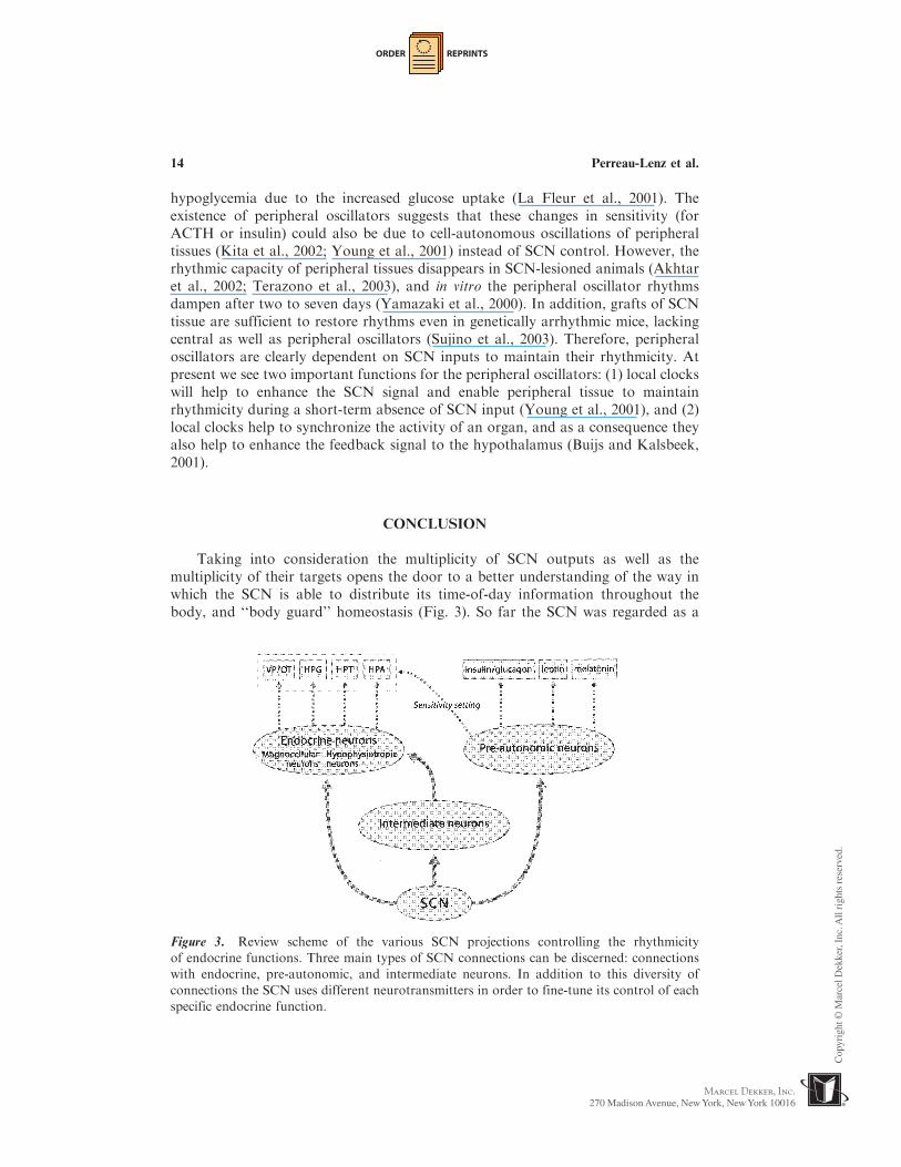
Taking into consideration the multiplicity of SCN outputs as well as themultiplicity of their targets opens the door to a better understanding of the way inwhich the SCN is able to distribute its time-of-day information throughout thebody, and ‘‘body guard’’ homeostasis (Fig. 3). So far the SCN was regarded as a
Figure 3. Review scheme of the various SCN projections controlling the rhythmicity
of endocrine functions. Three main types of SCN connections can be discerned: connections
with endocrine, pre-autonomic, and intermediate neurons. In addition to this diversity of
connections the SCN uses different neurotransmitters in order to fine-tune its control of each
specific endocrine function.
14 Perreau-Lenz et al.

ORDER REPRINTS
homogenous structure, giving off one type of global time signal. Recently progress
has been made, thanks to new techniques showing that although all SCN cells
contain a self-sustained clockwork, this molecular clockwork is expressed with a
different timing in the different subgroups of the SCN (Yan and Okamura, 2002). In
addition, although electrophysiological studies have described SCN global activity
during daytime, there is some evidence for SCN neurons with an opposite-phase of
activity (Herzog et al., 1997; Nakamura et al., 2001). Moreover, defined cell groups
in the rat SCN may show different day/night rhythms of single-unit activity in vivo
(Saeb-Parsy and Dyball, 2003), suggesting that for different subpopulations of SCN
cells the peak time of their activity differs. Together with the physiological evidence
described in the foregoing these new insights present a picture of SCN control
consisting of alternating waves of inhibitory and stimulatory inputs imposed
onto the hypothalamic endocrine and autonomic ‘‘motor neurons’’ with a
temporal distribution that is specific for each endocrine, physiological, or behavioral
system.
REFERENCES
Abe,M.,Herzog, E.D., Yamazaki, S., Straume,M., Tei, H., Sakaki, Y.,Menaker,M.,
Block, G. D. (2002). Circadian rhythms in isolated brain regions. J. Neurosci.
22:350–356.Akhtar, R. A., Reddy, A. B., Maywood, E. S., Clayton, J. D., King, V. M., Smith,
A. G., Gant, T. W., Hastings, M. H., Kyriacou, C. P. (2002). Circadian cycling
of the mouse liver transcriptome, as revealed by cDNA microarray, is driven
by the suprachiasmatic nucleus. Curr. Biol. 12:540–550.Albrecht, U., Eichele, G. (2003). The mammalian circadian clock. Curr. Opin. Genet.
Dev. 13:271–277.Arey, B. J., Freeman, M. E. (1989). Hypothalamic factors involved in the
endogenous stimulatory rhythm regulating prolactin secretion. Endocrinology
124:878–883.Aschoff, J., Wever, R. (1962). Spontanperiodik des menschen bei ausschluss aller
zeitgeber. Naturwissenschaften 49:337–342.Axelrod, J. (1974). The pineal gland: a neurochemical transducer. Science
184:1341–1348.Balsalobre, A. (2002). Clock genes in mammalian peripheral tissues. Cell Tissue Res.
309:193–199.Bargman, W. (1943). Die epiphysis cerebri. In: Von, M. W., ed. Handbuch der
Miskroskopischen Anatomie des Menschen. Berlin: Springer, 338–502.Bartanusz, V., Jezova, D., Bertini, L. T., Tilders, F. J., Aubry, J. M., Kiss, J. Z.
(1993). Stress-induced increase in vasopressin and corticotropin-releasing
factor expression in hypophysiotrophic paraventricular neurons.
Endocrinology 132:895–902.Berkenbosch, F., De Goeij, D. C., Tilders, F. J. (1989). Hypoglycemia enhances
turnover of corticotropin-releasing factor and of vasopressin in the zona
externa of the rat median eminence. Endocrinology 125:28–34.
The Biological Clock 15
ORDER REPRINTS
Bittman, E. L., Crandell, R. G., Lehman, M. N. (1989). Influences of the
paraventricular and suprachiasmatic nuclei and olfactory bulbs on melatonin
responses in the golden hamster. Biol. Reprod. 40:118–126.Bittman, E. L., Doherty, L. S., Huang, L., Paroskie, A. (2003). PeriodGene
expression in mouse endocrine tissues. Am. J. Physiol. Regul. Integr. Comp.
Physiol. 285:R561–R569.Bos, N. P., Mirmiran, M. (1990). Circadian rhythms in spontaneous neuronal
discharges of the cultured suprachiasmatic nucleus. Brain Res. 511:158–162.Bowers, C. W., Baldwin, C., Zigmond, R. E. (1984). Sympathetic reinnervation of
the pineal gland after postganglionic nerve lesion does not restore normal
pineal function. J. Neurosci. 4:2010–2015.Buijs, R. M., Markman, M., Nunes-Cardoso, B., Hou, Y. X., Shinn, S. (1993).
Projections of the suprachiasmatic nucleus to stress-related areas in the rat
hypothalamus: a light and electron microscopic study. J. Comp. Neurol.
335:42–54.Buijs, R. M., Hou, Y. X., Shinn, S., Renaud, L. P. (1994). Ultrastructural evidence
for intra- and extranuclear projections of GABAergic neurons of the
suprachiasmatic nucleus. J. Comp. Neurol. 340:381–391.Buijs, R. M., Wortel, J., Van Heerikhuize, J. J., Kalsbeek, A. (1997). Novel
environment induced inhibition of corticosterone secretion: physiological
evidence for a suprachiasmatic nucleus mediated neuronal hypothalamo-
adrenal cortex pathway. Brain Res. 758:229–236.Buijs, R. M., Wortel, J., Van Heerikhuize, J. J., Feenstra, M. G., Ter Horst, G. J.,
Romijn, H. J., Kalsbeek, A. (1999). Anatomical and functional demonstration
of a multisynaptic suprachiasmatic nucleus adrenal (cortex) pathway. Eur. J.
Neurosci. 11:1535–1544.Buijs, R. M., Chun, S. J., Niijima, A., Romijn, H. J., Nagai, K. (2001).
Parasympathetic and sympathetic control of the pancreas: a role for the
suprachiasmatic nucleus and other hypothalamic centers that are involved in
the regulation of food intake. J. Comp. Neurol. 431:405–423.Buijs, R. M., Kalsbeek, A. (2001). Hypothalamic integration of central and
peripheral clocks. Nat. Rev. Neurosci. 2:521–526.Buijs, R. M., La Fleur, S. E., Wortel, J., Van Heyningen, C., Zuiddam, L.,
Mettenleiter, T. C., Kalsbeek, A., Nagai, K., Niijima, A. (2003). The
suprachiasmatic nucleus balances sympathetic and parasympathetic output
to peripheral organs through separate preautonomic neurons. J. Comp. Neurol.
464:36–48.Cheng, M. Y., Bullock, C. M., Li, C., Lee, A. G., Bermak, J. C., Belluzzi, J.,
Weaver, D. R., Leslie, F. M., Zhou, Q. Y. (2002). Prokineticin 2 transmits the
behavioural circadian rhythm of the suprachiasmatic nucleus. Nature
417:405–410.Collu, R., Ruisseau, P. D., Tache, Y., Ducharme, J. R. (1977). Thyrotropin-releasing
hormone in rat brain: nyctohemeral variations. Endocrinology 100:1391–1393.Covarrubias, L., Uribe, R. M., Mendez, M., Charli, J. L., Joseph-Bravo, P. (1988).
Neuronal TRH synthesis: developmental and circadian TRH mRNA levels.
Biochem. Biophys. Res. Commun. 151:615–622.
16 Perreau-Lenz et al.

ORDER REPRINTS
Covarrubias, L., Redondo, J. L., Vargas, M. A., Uribe, R. M., Mendez, M.,Joseph-Bravo, P., Charli, J. L. (1994). In vitro TRH release from hypo-thalamus slices varies during the diurnal cycle. Neurochem. Res. 19:845–850.
Csaki, A., Kocsis, K., Halasz, B., Kiss, J. (2000). Localization of glutamatergic/aspartatergic neurons projecting to the hypothalamic paraventricular nucleusstudied by retrograde transport of [3H]D-aspartate autoradiography.Neuroscience 101:637–655.
Cui, L. N., Saeb-Parsy, K., Dyball, R. E. (1997). Neurones in the supraoptic nucleusof the rat are regulated by a projection from the suprachiasmatic nucleus.J. Physiol. 502:149–159.
Cui, L. N., Coderre, E., Renaud, L. P. (2001). Glutamate and GABA mediatesuprachiasmatic nucleus inputs to spinal-projecting paraventricular neurons.Am. J. Physiol. Regul. Integr. Comp. Physiol. 281:R1283–R1289.
Damiola, F., Le Minh, N., Preitner, N., Kornmann, B., Fleury-Olela, F., Schibler, U.(2000). Restricted feeding uncouples circadian oscillators in peripheral tissuesfrom the central pacemaker in the suprachiasmatic nucleus. Genes Dev.14:2950–2961.
De Goeij, D. C., Dijkstra, H., Tilders, F. J. (1992). Chronic psychosocial stressenhances vasopressin, but not corticotropin-releasing factor, in the externalzone of the median eminence of male rats: relationship to subordinate status.Endocrinology 131:847–853.
De La Iglesia, H. O., Blaustein, J. D., Bittman, E. L. (1995). The suprachiasmaticarea in the female hamster projects to neurons containing estrogen receptorsand GnRH. Neuroreport 6:1715–1722.
De La Iglesia, H. O., Meyer, J., Schwartz, W. J. (2003). Lateralization of circadianpacemaker output: activation of left- and right-sided luteinizing hormone-releasing hormone neurons involves a neural rather than a humoral pathway.J. Neurosci. 23:7412–7414.
De Vries, G. J., Buijs, R. M., Sluiter, A. A. (1984). Gonadal hormone actions on themorphology of the vasopressinergic innervation of the adult rat brain. BrainRes. 298:141–145.
Drijfhout, W. J., Van Der Linde, A. G., Kooi, S. E., Grol, C. J., Westerink, B. H.(1996). Norepinephrine release in the rat pineal gland: the input from thebiological clock measured by in vivo microdialysis. J. Neurochem. 66:748–755.
Fukuhara, C., Dirden, J. C., Tosini, G. (2000). Circadian expression of period 1,period 2, and arylalkylamine N-acetyltransferase mRNA in the rat pinealgland under different light conditions. Neurosci. Lett. 286:167–170.
Funabashi, T., Shinohara, K., Mitsushima, D., Kimura, F. (2000). Gonadotropin-releasing hormone exhibits circadian rhythm in phase with arginine-vasopressin in co-cultures of the female rat preoptic area and suprachiasmaticnucleus. J. Neuroendocrinol. 12:521–528.
Gerendai, I., Toth, I. E., Boldogkoi, Z., Medveczky, I., Halasz, B. (1998). Neuronallabeling in the rat brain and spinal cord from the ovary using viraltransneuronal tracing technique. Neuroendocrinology 68:244–256.
Gerendai, I., Toth, I. E., Boldogkoi, Z., Medveczky, I., Halasz, B. (2000). CNSstructures presumably involved in vagal control of ovarian function. J. Auton.Nerv. Syst. 80:40–45.
The Biological Clock 17
ORDER REPRINTS
Gerhold, L. M., Horvath, T. L., Freeman, M. E. (2001). Vasoactive intestinalpeptide fibers innervate neuroendocrine dopaminergic neurons. Brain Res.919:48–56.
Gillies, G. E., Linton, E. A., Lowry, P. J. (1982). Corticotropin releasing activity ofthe new CRF is potentiated several times by vasopressin. Nature 299:355–357.
Gomez, F., Chapleur, M., Fernette, B., Burlet, C., Nicolas, J. P., Burlet, A. (1997).Arginine vasopressin (AVP) depletion in neurons of the suprachiasmatic nucleiaffects the AVP content of the paraventricular neurons and stimulatesadrenocorticotrophic hormone release. J. Neurosci. Res. 50:565–574.
Gray, G. D., Soderstein, P., Tallentire, D., Davidson, J. M. (1978). Effects of lesionsin various structures of the suprachiasmatic-preoptic region on LH regulationand sexual behavior in female rats. Neuroendocrinology 25:174–191.
Hara, R., Wan, K., Wakamatsu, H., Aida, R., Moriya, T., Akiyama, M., Shibata, S.(2001). Restricted feeding entrains liver clock without participation of thesuprachiasmatic nucleus. Genes Cells 6:269–278.
Hendrickson, A. E., Wagoner, N., Cowan, W. M. (1972). An autoradiographic andelectron microscopic study of retino-hypothalamic connections. Z. Zellforsch.Mikrosk. Anat. 135:1–26.
Hermes, M. L., Coderre, E. M., Buijs, R. M., Renaud, L. P. (1996). GABA andglutamate mediate rapid neurotransmission from suprachiasmatic nucleus tohypothalamic paraventricular nucleus in rat. J. Physiol. 496:749–757.
Hermes, M. L., Ruijter, J. M., Klop, A., Buijs, R. M., Renaud, L. P. (2000).Vasopressin increases GABAergic inhibition of rat hypothalamic para-ventricular nucleus neurons in vitro. J. Neurophysiol. 83:705–711.
Herzog, E. D., Geusz, M. E., Khalsa, S. B., Straume, M., Block, G. D. (1997).Circadian rhythms in mouse suprachiasmatic nucleus explants on multimicro-electrode plates. Brain Res. 757:285–290.
Hofman, M. A., Swaab, D. F. (1993). Diurnal and seasonal rhythms of neuronalactivity in the suprachiasmatic nucleus of humans. J. Biol. Rhythms 8:283–295.
Hoorneman, E. M., Buijs, R. M. (1982). Vasopressin fiber pathways in the rat brainfollowing suprachiasmatic nucleus lesioning. Brain Res. 243:235–241.
Horvath, T. L. (1997). Suprachiasmatic efferents avoid phenestrated capillaries butinnervate neuroendocrine cells, including those producing dopamine.Endocrinology 138:1312–1320.
Inouye, S. T., Kawamura, H. (1979). Persistence of circadian rhythmicity in amammalian hypothalamic ‘‘island’’ containing the suprachiasmatic nucleus.Proc. Natl. Acad. Sci. USA 76:5962–5966.
Jasper, M. S., Engeland, W. C. (1994). Splanchnic neural activity modulatesultradian and circadian rhythms in adrenocortical secretion in awake rats.Neuroendocrinology 59:97–109.
Johnston, C. A., Negro-Vilar, A. (1988). Role of oxytocin on prolactin secretionduring proestrus and in different physiological or pharmacological paradigms.Endocrinology 122:341–350.
Kappers, J. A. (1960). The development, topographical relations and innervation ofthe epiphysis cerebri in the albino rat. Z. Zellforsch. 52:163–215.
Kappers, J. A. (1971). Regulation of the reproductive system by the pineal gland andits dependence on light. J. Neurovisc. Relat. 10:141–152.
18 Perreau-Lenz et al.

ORDER REPRINTS
Kalsbeek, A., Buijs, R. M. (1992). Peptidergic transmitters of the suprachiasmatic
nuclei and the control of circadian rhythmicity. Prog. Brain Res. 92:321–333.Kalsbeek, A., Buijs, R. M., Van Heerikhuize, J. J., Arts, M., Van Der Woude, T. P.
(1992). Vasopressin-containing neurons of the suprachiasmatic nuclei inhibit
corticosterone release. Brain Res. 580:62–67.Kalsbeek, A., Buijs, R. M. (1996). Rhythms of inhibitory and excitatory output from
the circadian timing system as revealed by in vivo microdialysis. Prog. Brain
Res. 111:273–293.Kalsbeek, A., Drijfhout, W. J., Westerink, B. H., Van Heerikhuize, J. J., Van Der
Woude, T. P., Van Der Vliet, J., Buijs, R. M. (1996a). GABA receptors in
the region of the dorsomedial hypothalamus of rats are implicated in the
control of melatonin and corticosterone release. Neuroendocrinology 63:69–78.Kalsbeek, A., Van Heerikhuize, J. J., Wortel, J., Buijs, R. M. (1996b). A diurnal
rhythm of stimulatory input to the hypothalamo-pituitary-adrenal system as
revealed by timed intrahypothalamic administration of the vasopressin V1
antagonist. J. Neurosci. 16:5555–5565.Kalsbeek, A., Strubbe, J. H. (1998). Circadian control of insulin secretion is
independent of the temporal distribution of feeding. Physiol. Behav.
63:553–558.Kalsbeek, A., Cutrera, R. A., Van Heerikhuize, J. J., Van Der Vliet, J., Buijs, R. M.
(1999). GABA release from suprachiasmatic nucleus terminals is necessary for
the light-induced inhibition of nocturnal melatonin release in the rat.
Neuroscience 91:453–461.Kalsbeek, A., Fliers, E., Franke, A. N., Wortel, J., Buijs, R. M. (2000a). Functional
connections between the suprachiasmatic nucleus and the thyroid gland as
revealed by lesioning and viral tracing techniques in the rat. Endocrinology
141:3832–3841.Kalsbeek, A., Garidou, M. L., Palm, I. F., Van Der Vliet, J., Simonneaux, V.,
Pevet, P., Buijs, R. M. (2000b). Melatonin sees the light: blocking GABA-
ergic transmission in the paraventricular nucleus induces daytime secretion
of melatonin. Eur. J. Neurosci. 12:3146–3154.Kalsbeek, A., Fliers, E., Romijn, J. A., La Fleur, S. E., Wortel, J., Bakker, O.,
Endert, E., Buijs, R. M. (2001). The suprachiasmatic nucleus generates the
diurnal changes in plasma leptin levels. Endocrinology 142:2677–2685.Kalsbeek, A., Palm, I. F., Buijs, R. M. (2002). Central vasopressin systems and
steroid hormones. Prog. Brain Res. 139:57–73.Kalsbeek, A., Ruiter, M., La Fleur, S. E., Van Heijningen, C., Buijs, R. M. (2003).
The diurnal modulation of hormonal responses in the rat varies with different
stimuli. J. Neuroendocrinol 15:1144–1155.Kaneko, M., Hiroshige, T., Shinsako, J., Dallman, M. F. (1980). Diurnal changes
in amplification of hormone rhythms in the adrenocortical system. Am. J.
Physiol. 239:R309–R316.Kiss, J. Z., Mezey, E., Skirboll, L. (1984). Corticotropin-releasing factor-
immunoreactive neurons of the paraventricular nucleus become
vasopressin positive after adrenalectomy. Proc. Natl. Acad. Sci. USA
81:1854–1858.
The Biological Clock 19

ORDER REPRINTS
Kita, Y., Shiozawa, M., Jin, W., Majewski, R. R., Besharse, J. C., Greene, A. S.,Jacob, H. J. (2002). Implications of circadian gene expression in kidney, liverand the effects of fasting on pharmacogenomic studies. Pharmacogenetics12:55–65.
Klein, D. C., Weller, J. L., Moore, R. Y. (1971). Melatonin metabolism: neuralregulation of pineal serotonin: acetyl coenzyme A N-acetyltransferase activity.Proc. Natl. Acad. Sci. U.S.A. 68:3107–3110.
Kneisley, L. W., Moskowitz, M. A., Lynch, H. G. (1978). Cervical spinal cordlesions disrupt the rhythm in human melatonin excretion. J. Neural. Transm.Suppl. 13:311–323.
Kovacs, K. J., Foldes, A., Sawchenko, P. E. (2000). Glucocorticoid negativefeedback selectively targets vasopressin transcription in parvocellular neuro-secretory neurons. J. Neurosci. 20:3843–3852.
Kramer, A., Yang, F. C., Snodgrass, P., Li, X., Scammell, T. E., Davis, F. C.,Weitz, C. J. (2001). Regulation of daily locomotor activity and sleep byhypothalamic EGF receptor signaling. Science 294:2511–2515.
Kreier, F., Fliers, E., Voshol, P. J., Van Eden, C. G., Havekes, L. M., Kalsbeek, A.,Van Heijningen, C. L., Sluiter, A. A., Mettenleiter, T. C., Romijn, J. A.,Sauerwein, H. P., Buijs, R. M. (2002). Selective parasympathetic innervation ofsubcutaneous and intra-abdominal fat—functional implications. J. Clin. Invest.110:1243–1250.
Kriegsfeld, L. J., Korets, R., Silver, R. (2003). Expression of the circadian clock genePeriod 1 in neuroendocrine cells: an investigation using mice with a Per1:GFPtransgene. Eur. J. Neurosci. 17:212–220.
La Fleur, S. E., Kalsbeek, A., Wortel, J., Buijs, R. M. (1999). A suprachiasmaticnucleus generated rhythm in basal glucose concentrations. J. Neuroendocrinol.11:643–652.
La Fleur, S. E., Kalsbeek, A., Wortel, J., Buijs, R. M. (2000). Polysynaptic neuralpathways between the hypothalamus, including the suprachiasmatic nucleus,and the liver. Brain Res. 871:50–56.
La Fleur, S. E., Kalsbeek, A., Wortel, J., Fekkes, M. L., Buijs, R. M. (2001). A dailyrhythm in glucose tolerance: a role for the suprachiasmatic nucleus. Diabetes50:1237–1243.
Larsen, P. J., Enquist, L. W., Card, J. P. (1998). Characterization of themultisynaptic neuronal control of the rat pineal gland using viral transneuronaltracing. Eur. J. Neurosci. 10:128–145.
Lehman, M. N., Silver, R., Gladstone, W.R., Kahn, R. M., Gibson, M., Bittman,E. L. (1987). Circadian rhythmicity restored by neural transplant.Immunocytochemical characterization of the graft and its integration withthe host brain. J. Neurosci. 7:1626–1638.
Lerner, A. B. T., Lee, T. H., Mori, W. (1958). Isolation of melatonin, the pinealgland factor that lightens melanocytes. J. Am. Chem. Soc. 80:2587.
Li, Y., Jiang, D. H., Wang, M. L., Jiao, D. R., Pang, S. F. (1989). Rhythms of serummelatonin in patients with spinal lesions at the cervical, thoracic or lumbarregion. Clin. Endocrinol. (Oxf ). 30:47–56.
Lynch, H. J. (1971). Diurnal oscillations in pineal melatonin content. Life Sci.10:791–795.
20 Perreau-Lenz et al.

ORDER REPRINTS
Mai, L. M., Pan, J. T. (1990). Paradoxical effects of oxytocin and vasopressin on
basal prolactin secretion and the estrogen-induced prolactin surge. Life Sci.
47:1243–1251.Mai, L. M., Shieh, K. R., Pan, J. T. (1994). Circadian changes of serum prolactin
levels and tuberoinfundibular dopaminergic neuron activities in ovariecto-
mized rats treated with or without estrogen: the role of the suprachiasmatic
nuclei. Neuroendocrinology 60:520–526.Martino, E., Bambini, G., Vaudagna, G., Breccia, M., Baschieri, L. (1985). Effects
of continuous light and dark exposure on hypothalamic thyrotropin-releasing
hormone in rats. J. Endocrinol. Invest. 8:31–33.Meyer-Bernstein, E. L., Jetton, A. E., Matsumoto, S. I., Markuns, J. F., Lehman,
M. N., Bittman, E. L. (1999). Effects of suprachiasmatic transplants on
circadian rhythms of neuroendocrine function in golden hamsters.
Endocrinology 140:207–218.Moore, R. Y., Eichler, V. B. (1972). Loss of a circadian adrenal corticosterone
rhythm following suprachiasmatic lesions in the rat. Brain Res. 42:201–206.Moore, R. Y., Klein, D. C. (1974). Visual pathways and the central neural control of
a circadian rhythm in pineal serotonin N-acetyltransferase activity. Brain Res.
71:17–33.Moore, R. Y., Lenn, N. J. (1972). A retinohypothalamic projection in the rat.
J. Comp. Neurol. 146:1–14.Moore, R. Y. (1978). Neural control of pineal function in mammals and birds.
J. Neural. Transm. Suppl. 13:47–58.Moore, R. Y., Speh, J. C. (1993). GABA is the principal neurotransmitter of the
circadian system. Neurosci. Lett. 150:112–116.Moore, R. Y. (1996). Neural control of the pineal gland. Behav. Brain Res.
73:125–130.Murai, I., Reichlin, S., Ben-Jonathan, N. (1989). The peak phase of the proestrous
prolactin surge is blocked by either posterior pituitary lobectomy or antisera
to vasoactive intestinal peptide. Endocrinology 124:1050–1055.Nakamura, W., Honma, S., Shirakawa, T., Honma, K. (2001). Regional pacemakers
composed of multiple oscillator neurons in the rat suprachiasmatic nucleus.
Eur. J. Neurosci. 14:666–674.Okamura, H., Berod, A., Julien, J. F., Geffard, M., Kitahama, K., Mallet, J.,
Bobillier, P. (1989). Demonstration of GABAergic cell bodies in the
suprachiasmatic nucleus: in situ hybridization of glutamic acid decarboxylase
(GAD) mRNA and immunocytochemistry of GAD and GABA. Neurosci.
Lett. 102:131–136.Palm, I. F., Van Der Beek, E. M., Wiegant, V. M., Buijs, R. M., Kalsbeek, A. (1999).
Vasopressin induces a luteinizing hormone surge in ovariectomized, estradiol-
treated rats with lesions of the suprachiasmatic nucleus. Neuroscience
93:659–666.Palm, I. F., Van Der Beek, E. M., Swarts, H. J., Van Der Vliet, J., Wiegant,
V. M., Buijs, R. M., Kalsbeek, A. (2001a). Control of the estradiol-
induced prolactin surge by the suprachiasmatic nucleus. Endocrinology
142:2296–2302.
The Biological Clock 21
ORDER REPRINTS
Palm, I. F., Van Der Beek, E. M., Wiegant, V. M., Buijs, R. M., Kalsbeek, A.
(2001b). The stimulatory effect of vasopressin on the luteinizing hormone surge
in ovariectomized, estradiol-treated rats is time-dependent. Brain Res.
901:109–116.Perreau-Lenz, S., Kalsbeek, A., Garidou, M. L., Wortel, J., Van Der Vliet, J.,
Van Heijningen, C., Simonneaux, V., Pevet, P., Buijs, R. M. (2003).
Suprachiasmatic control of melatonin synthesis in rats: inhibitory and
stimulatory mechanisms. Eur. J. Neurosci. 17:221–228.Perreau-Lenz, S., Kalsbeek, A., Pevet, P., Buijs, R. M. (2004). Glutamatergic clock
output stimulates melatonin synthesis at night. Eur. J. Neurosc. 19:318–324.Ralph, C. L., Mull, D., Lynch, H. J., Hedlund, L. (1971). A melatonin rhythm
persists in rat pineals in darkness. Endocrinology 89:1361–1366.Ralph, M. R., Foster, R. G., Davis, F. C., Menaker, M. (1990). Transplanted
suprachiasmatic nucleus determines circadian period. Science 247:975–978.Reppert, S. M., Perlow, M. J., Ungerleider, L. G., Mishkin, M., Tamarkin, L.,
Orloff, D. G., Hoffman, H. J., Klein, D. C. (1981). Effects of damage to the
suprachiasmatic area of the anterior hypothalamus on the daily melatonin and
cortisol rhythms in the rhesus monkey. J. Neurosci. 1:414–1425.Reppert, S. M., Weaver, D. R. (2001). Molecular analysis of mammalian circadian
rhythms. Annu. Rev. Physiol. 63:647–676.Reiter, R. J., King, T. S., Richardson, B.A., Hurlbut, E. C. (1982). Studies on pineal
melatonin levels in a diurnal species, the eastern chipmunk (Tamias striatus):
effects of light at night, propranolol administration or superior cervical
ganglionectomy. J. Neural. Transm. 54:275–284.Reiter, R. J. (1993). The melatonin rhythm: both a clock and a calendar. Experientia
49:654–664.Reuss, S., Stehle, J., Schroder, H., Vollrath, L. (1990). The role of the hypothalamic
paraventricular nuclei for the regulation of pineal melatonin synthesis: new
aspects derived from the vasopressin-deficient Brattleboro rat. Neurosci. Lett.
109:196–200.Richter, C. P. (1922). A behavioristic study of the activity of the rat. Comp. Psych.
Monogr. 1:1–55.Rivier, C., Vale, W. (1983). Interaction of corticotropin-releasing factor and arginine
vasopressin on adrenocorticotropin secretion in vivo. Endocrinology
113:939–942.Rivier, C., Rivier, J., Mormede, P., Vale, W. (1984). Studies of the nature of the
interaction between vasopressin and corticotropin-releasing factor on adreno-
corticotropin release in the rat. Endocrinology 115:882–886.Roland, B. L., Sawchenko, P. E. (1993). Local origins of some GABAergic
projections to the paraventricular and supraoptic nuclei of the hypothalamus
in the rat. J. Comp. Neurol. 332:123–143.Romero, L. M., Plotsky, P. M., Sapolsky, R. M. (1993). Patterns of adrenocortico-
tropin secretagog release with hypoglycemia, novelty, and restraint after
colchicine blockade of axonal transport. Endocrinology 132:199–204.Ruiter, M., La Fleur, S. E., Van Heijningen, C., Van der Vliet, J., Kalsbeek, A.,
Buijs, R. M. (2003). The daily rhythm in plasma glucagon concentrations in the
22 Perreau-Lenz et al.

ORDER REPRINTS
rat is modulated by the biological clock and feeding behavior. Diabetes
52:1709–1715.Saeb-Parsy, K., Dyball, R. E. (2003). Defined cell groups in the rat suprachiasmatic
nucleus have different day/night rhythms of single-unit activity in vivo. J. Biol.
Rhythms 18:26–42.Sawchenko, P. E., Swanson, L. W., Vale, W. W. (1984). Co-expression of
corticotropin-releasing factor and vasopressin immunoreactivity in parvo-
cellular neurosecretory neurons of the adrenalectomized rat. Proc. Natl. Acad.
Sci. USA 81:1883–1887.Schroder, H., Stehle, J., Henschel, M. (1988). Twenty-four-hour pineal melatonin
synthesis in the vasopressin-deficient Brattleboro rat. Brain Res. 459:328–332.Schwartz, W. J., Gainer, H. (1977). Suprachiasmatic nucleus: use of 14C-labeled
deoxyglucose uptake as a functional marker. Science 197:1089–1091.Schwartz, W. J., Davidsen, L. C., Smith, C. B. (1980). In vivo metabolic activity of a
putative circadian oscillator, the rat suprachiasmatic nucleus. J. Comp. Neurol.
189:157–167.Schwartz, W. J., Reppert, S. M., Eagan, S. M., Moore-Ede, M. C. (1983). In vivo
metabolic activity of the suprachiasmatic nuclei: a comparative study. Brain
Res. 274:184–187.Shibata, S., Oomura, Y., Kita, H., Hattori, K. (1982). Circadian rhythmic changes
of neuronal activity in the suprachiasmatic nucleus of the rat hypothalamic
slice. Brain Res. 247:154–158.Shibata, S., Moore, R. Y. (1988). Electrical and metabolic activity of suprachias-
matic nucleus neurons in hamster hypothalamic slices. Brain Res. 438:374–378.Silver, R., Lesauter, J., Tresco, P. A., Lehman, M. N. (1996). A diffusible coupling
signal from the transplanted suprachiasmatic nucleus controlling circadian
locomotor rhythms. Nature 382:810–813.Stephan, F. K., Zucker, I. (1972). Circadian rhythms in drinking behavior and
locomotor activity of rats are eliminated by hypothalamic lesions. Proc. Natl.
Acad. Sci. USA 69:1583–1586.Stoynev, A. G., Ikonomov, O. C., Usunoff, K. G. (1982). Feeding pattern and light-
dark variations in water intake and renal excretion after suprachiasmatic nuclei
lesions in rats. Physiol. Behav. 29:35–40.Sujino, M., Masumoto, K., Yamaguchi, S., Van Der Horst, G. T., Okamura, H.,
Inouye, S. I. (2003). Suprachiasmatic nucleus grafts restore circadian
behavioral rhythms of genetically arrhythmic mice. Curr. Biol. 13:664–668.Sun, X., Rusak, B., Semba, K. (2000). Electrophysiology and pharmacology of
projections from the suprachiasmatic nucleus to the ventromedial preoptic area
in rat. Neuroscience 98:715–728.Sun, X., Whitefield, S., Rusak, B., Semba, K. (2001). Electrophysiological analysis
of suprachiasmatic nucleus projections to the ventrolateral preoptic area in the
rat. Eur. J. Neurosci. 14:1257–1274.Teclemariam-Mesbah, R., Ter Horst, G. J., Postema, F., Wortel, J., Buijs, R. M.
(1999). Anatomical demonstration of the suprachiasmatic nucleus-pineal
pathway. J. Comp. Neurol. 406:171–182.
The Biological Clock 23

ORDER REPRINTS
Terazono, H., Mutoh, T., Yamaguchi, S., Kobayashi, M., Akiyama, M., Udo, R.,Ohdo, S., Okamura, H., Shibata, S. (2003). Adrenergic regulation of clock geneexpression in mouse liver. Proc. Natl. Acad. Sci. USA 100:6795–6800.
Terwel, D., Ten Haaf, J. A., Markerink, M., Jolles, J. (1992). Changes in plasmavasopressin concentration and plasma osmolality in relation to age and timeof day in the male Wistar rat. Acta. Endocrinol. (Copenh) 126:357–362.
Tessonneaud, A., Locatelli, A., Caldani, M., Viguier-Martinez, M. C. (1995).Bilateral lesions of the suprachiasmatic nuclei alter the nocturnal melatoninsecretion in sheep. J. Neuroendocrinol. 7:145–152.
Tramu, G., Croix, C., Pillez, A. (1983). Ability of the CRF immunoreactive neuronsof the paraventricular nucleus to produce a vasopressin-like material.Immunohistochemical demonstration in adrenalectomized guinea pigs andrats. Neuroendocrinology 37:467–469.
Vale, W., Spiess, J., Rivier, C., Rivier, J. (1981). Characterization of a 41-residueovine hypothalamic peptide that stimulates secretion of corticotropin andbeta-endorphin. Science 213:1394–1397.
Van Esseveldt, K. E., Lehman,M.N., Boer, G. J. (2000). The suprachiasmatic nucleusand the circadian time-keeping system revisited. Brain Res. Rev. 33:34–77.
Van Den Pol, A. N., Gorcs, T. (1986). Synaptic relationships between neuronscontaining vasopressin, gastrin-releasing peptide, vasoactive intestinal poly-peptide, and glutamate decarboxylase immunoreactivity in the suprachiasmaticnucleus: dual ultrastructural immunocytochemistry with gold-substituted silverperoxidase. J. Comp. Neurol. 252:507–521.
Van Den Pol, A. N. (1991). Glutamate and aspartate immunoreactivity inhypothalamic presynaptic axons. J. Neurosci. 11:2087–2101.
Van Der Beek, E. M., Wiegant, V. M., Van Der Donk, H. A., Van Den Hurk, R.,Buijs, R. M. (1993). Lesions of the suprachiasmatic nucleus indicate thepresence of a direct vasoactive intestinal polypeptide-containing projectionto gonadotrophin-releasing hormone neurons in the female rat. J.Neuroendocrinol. 5:137–144.
Van Der Beek, E. M., Horvath, T. L., Wiegant, V. M., Van Den Hurk, R., Buijs, R.M. (1997). Evidence for a direct neuronal pathway from the suprachiasmaticnucleus to the gonadotropin-releasing hormone system: combined tracing andlight and electron microscopic immunocytochemical studies. J. Comp. Neurol.384:569–579.
Vrang, N., Larsen, P. J., Mikkelsen, J. D. (1995). Direct projection from thesuprachiasmatic nucleus to hypophysiotrophic corticotropin-releasing factorimmunoreactive cells in the paraventricular nucleus of the hypothalamusdemonstrated by means of Phaseolus vulgaris-leucoagglutinin tract tracing.Brain Res. 684:61–69.
Watanabe, K., Hiroshige, T. (1981). Phase relation between episodic fluctuations ofspontaneous locomotor activity and plasma corticosterone in rats withsuprachiasmatic nuclei lesions. Neuroendocrinology 33:52–59.
Watson, R. E. Jr., Langub, M. C. Jr., Engle, M. G., Maley, B. E. (1995). Estrogen-receptive neurons in the anteroventral periventricular nucleus are synaptictargets of the suprachiasmatic nucleus and peri-suprachiasmatic region. BrainRes. 689:254–264.
24 Perreau-Lenz et al.
ORDER REPRINTS
Watts, A. G., Swanson, L. W., Sanchez-Watts, G. (1987a). Efferent projections ofthe suprachiasmatic nucleus: I. Studies using anterograde transport ofPhaseolus vulgaris leucoagglutinin in the rat. J. Comp. Neurol. 258:204–229.
Watts, A. G., Swanson, L. W. (1987b). Efferent projections of the suprachiasmaticnucleus: II. Studies using retrograde transport of fluorescent dyes andsimultaneous peptide immunohistochemistry in the rat. J. Comp. Neurol.258:230–252.
Windle, R. J., Forsling, M. L., Guzek, J. W. (1992). Daily rhythms in the hormonecontent of the neurohypophysial system and release of oxytocin andvasopressin in the male rat: effect of constant light. J. Endocrinol. 133:283–290.
Wotjak, C. T., Kubota, M., Liebsch, G., Montkowski, A., Holsboer, F., Neumann,I., Landgraf, R. (1996). Release of vasopressin within the rat paraventricularnucleus in response to emotional stress: a novel mechanism of regulatingadrenocorticotropic hormone secretion? J. Neurosci. 16:7725–7732.
Wurtman, R. J., Axelrod, J., Sedvall, G., Moore, R. Y. (1967). Photic and neuralcontrol of the 24-hour norepinephrine rhythm in the rat pineal gland.J. Pharmacol. Exp. Ther. 157:487–492.
Yamazaki, S., Numano, R., Abe, M., Hida, A., Takahashi, R., Ueda, M., Block,G. D., Sakaki, Y., Menaker, M., Tei, H. (2000). Resetting central andperipheral circadian oscillators in transgenic rats. Science 288:682–685.
Yan, L., Okamura, H. (2002). Gradients in the circadian expression of Per1 and Per2genes in the rat suprachiasmatic nucleus. Eur. J. Neurosci. 15:1153–1162.
Young, M. W., Razeghi, P., Cedars, A. M., Guthrie, P. H., Taegtmeyer, H. (2001).Intrinsic diurnal variations in cardiac metabolism and contractile function.Circ. Res. 89:1199–1208.
Yuwiler, A., Klein, D. C., Buda, M., Weller, J. L. (1977). Adrenergic control ofpineal N-acetyltransferase activity: developmental aspects. Am. J. Physiol.233:E141–E146.
The Biological Clock 25

Request Permission/Order Reprints
Reprints of this article can also be ordered at
http://www.dekker.com/servlet/product/DOI/101081CBI120027984
Request Permission or Order Reprints Instantly!
Interested in copying and sharing this article? In most cases, U.S. Copyright Law requires that you get permission from the article’s rightsholder before using copyrighted content.
All information and materials found in this article, including but not limited to text, trademarks, patents, logos, graphics and images (the "Materials"), are the copyrighted works and other forms of intellectual property of Marcel Dekker, Inc., or its licensors. All rights not expressly granted are reserved.
Get permission to lawfully reproduce and distribute the Materials or order reprints quickly and painlessly. Simply click on the "Request Permission/ Order Reprints" link below and follow the instructions. Visit the U.S. Copyright Office for information on Fair Use limitations of U.S. copyright law. Please refer to The Association of American Publishers’ (AAP) website for guidelines on Fair Use in the Classroom.
The Materials are for your personal use only and cannot be reformatted, reposted, resold or distributed by electronic means or otherwise without permission from Marcel Dekker, Inc. Marcel Dekker, Inc. grants you the limited right to display the Materials only on your personal computer or personal wireless device, and to copy and download single copies of such Materials provided that any copyright, trademark or other notice appearing on such Materials is also retained by, displayed, copied or downloaded as part of the Materials and is not removed or obscured, and provided you do not edit, modify, alter or enhance the Materials. Please refer to our Website User Agreement for more details.