The skin as a peripheral clock model
213
HAL Id: tel-01220534 https://tel.archives-ouvertes.fr/tel-01220534 Submitted on 26 Oct 2015 HAL is a multi-disciplinary open access archive for the deposit and dissemination of sci- entific research documents, whether they are pub- lished or not. The documents may come from teaching and research institutions in France or abroad, or from public or private research centers. L’archive ouverte pluridisciplinaire HAL, est destinée au dépôt et à la diffusion de documents scientifiques de niveau recherche, publiés ou non, émanant des établissements d’enseignement et de recherche français ou étrangers, des laboratoires publics ou privés. The skin as a peripheral clock model Taole Liu To cite this version: Taole Liu. The skin as a peripheral clock model. Molecular biology. Université de Strasbourg, 2014. English. NNT : 2014STRAJ007. tel-01220534
-
Upload
khangminh22 -
Category
Documents
-
view
1 -
download
0
Transcript of The skin as a peripheral clock model
HAL Id: tel-01220534https://tel.archives-ouvertes.fr/tel-01220534
Submitted on 26 Oct 2015
HAL is a multi-disciplinary open accessarchive for the deposit and dissemination of sci-entific research documents, whether they are pub-lished or not. The documents may come fromteaching and research institutions in France orabroad, or from public or private research centers.
L’archive ouverte pluridisciplinaire HAL, estdestinée au dépôt et à la diffusion de documentsscientifiques de niveau recherche, publiés ou non,émanant des établissements d’enseignement et derecherche français ou étrangers, des laboratoirespublics ou privés.
The skin as a peripheral clock modelTaole Liu
To cite this version:Taole Liu. The skin as a peripheral clock model. Molecular biology. Université de Strasbourg, 2014.English. �NNT : 2014STRAJ007�. �tel-01220534�
UNIVERSITÉ DE STRASBOURG
ÉCOLE DOCTORALE ED414
CNRS UPR 3212 INCI
THÈSE présentée par :
LIU Taole
soutenue le : 3 Mars 2014
pour obtenir le grade de : Docteur de l’université de Strasbourg Discipline/ Spécialité : Neurosciences
La peau, un modèle d’horloge périphérique
The skin as a peripheral clock model
Membres du jury
THÈSE dirigée par: Dr. FELDER-SCHMITTBUHL Marie-Paule Dr. PEVET Paul
RAPPORTEURS:
Dr. AUJARD Fabienne Dr. CLAUSTRAT Bruno Pr. LELIEVRE Vincent
AUTRES MEMBRES DU JURY:
Dr. FRANÇOIS-BELLAN Anne-Marie

Time is life. Life is time. To my parents
To me in the past, present and future
To Strasbourg


Acknowledgement/Remerciements
Thanks very much to the members of my thesis committee, Bruno C laustrat, Vincent
Lelievre, Fabienne Aujard, Anne-Marie François-Bellan for accepting and evaluating my
thesis work.
Firstly, I am deeply grateful for my thesis directors Marie-Paule Felder-Schmittbuhl
and Paul Pévet, who welcome me into scientific environment of biological rhythms, to
INCI, to Strasbourg for my PhD study.
Thanks Marie-Paule, sincerely for your efforts helping me into this field, with my
thesis work and final defense. I benefit quite a lot from your excellent knowledge on
circadian clock and molecular biology, as well as your unremitting work attitude and
encouragement in these three years. Your patience and kindness enlighten the way with
ups and downs forward through my PhD life. I will never forget your beautiful smile.
Thanks Paul, greatly for your help and lead in the lab. Your enthusiasm on the
science and optimism everyday set an example for scientists in my mind. Without your
support and tolerance, I cannot start and finally finish my research work. Eventually, I am
a big fan of melatonin.
Merci beaucoup à Cristina Sandu. You have helped me in almost every step in my
PhD with you brilliant and considerate teaching every day. As a nice and strict teacher,
you lead me and cover me in lots of experiments. I hope someday I could be as smart and
tough as you.
I would like to thank my group leader Etienne Challet, who continuously offers help
and constructive advices to details in my research. It was your class that the first course
on biological rhythms I attended. And thanks to other colleagues in our group, Patrick
Vuillez, Jorge Mendoza, Sylvie Raison and Daniel Clesse, for your help and
encouragement.
Acknowledge Catherine Jaeger and André Malan for your help on analysis of the
bioluminescence data.
And I am very thankful to all the people of INCI: Dominique Ciocca, Sophie Reibel
and Nicolas Lethenet, for your help in chronobiotron; Vincent-Joseph Poirel, Paul Klosen

and Marie-Pierre Laran-Chich, for your help in molecular biology work; Denis Wagner,
Christophe Kern, Sonia Benichou, Jean-George Lorentz and Dominique Streicher for
your technique support; all the teachers David Hicks, Valérie Simonneaux, Mireille
Masson-Pévet, Virginie Laurent-Gydé, for your great questions to inspire my thinking on
my work.
And many thanks to all the PhD students and Post-docs in the lab: Claire, Madah and
Ana, you friends show me around the Strasbourg when I came in the beginning; Domitille,
Ned, Cristina, Coralie, Edith, Stefanie, Ouafa, Tounès, Hanan, Amélie, Daniela, Lucas,
Laurent, Adrien, Bruce, Caroline, Julien, Vy, David…for your accompany and help.
Mostly, I want to thank my parents supporting me all the time and all the friends in
Strasbourg in France. Mama, Papa, thanks for your love. I love you. Thanksgive for all
you gift me physical and educational. It has been 10 years since I left Xi’an as a adult but
still baby to you. Your encouragement and critique is always my cherish. Great thanks to
your accompany whenever and wherever.
And great acknowledgement to CSC with financial support through my PhD. Thanks
to all chinese people.
1
TableofContents
Abbreviations ....................................................................................................................... 5 Résumé du travail de thèse .................................................................................................. 7 Thesis summary ................................................................................................................. 17
Introduction .................................................................................................................. 21
1. General features about circadian clocks .......................................................... 22
2. Molecular clock mechanisms ............................................................................ 25
2.1. The feedback loops ................................................................................ 25 2.2. Transcriptional activation of Period genes by BMAL1/CLOCK .......... 27 2.3. Post-transcriptional regulations ............................................................. 30
3. The SCN: master clock of the body and a model to understand rhythm
generation within tissues .......................................................................................... 34
3.1. The SCN clock ....................................................................................... 35 3.2. Multioscillatory nature of the SCN ........................................................ 35 3.3. Photic input: light information as a major external signal for the clock 38 3.4. Non-photic inputs: feeding and social activity ...................................... 41 3.5. Neuronal outputs from the SCN ............................................................ 42 3.6. Hormonal “outputs” from the SCN ........................................................ 45
4. The clock network and its synchronization mechanisms ............................... 48
4.1. Secondary clocks in the central nervous system .................................... 48 4.2. Peripheral circadian clocks .................................................................... 49 4.3. Difference between the SCN and peripheral clocks .............................. 50 4.4. Resetting and synchronization: external influence on phase, period and amplitude .............................................................................................................. 52 4.5. Potential synchronizers .......................................................................... 54 4.6. Circadian network throughout life ......................................................... 55 4.6.1. Postnatal development ........................................................................... 55 4.6.2. Ageing .................................................................................................... 57
5. Rhythmic properties and clock activity in the skin ........................................ 59
5.1. Skin structure and rhythmic functions ................................................... 59 5.2. The circadian rhythm of skin cell proliferation ..................................... 60 5.3. Clock gene activity in the skin ............................................................... 62 5.4. Rhythms of clock gene expression in skin cells including fibroblasts .. 63 5.5. Synchronizing signals to fibroblasts ...................................................... 65 5.6. Skin and melatonin ................................................................................ 67 5.7. Skin and glucocorticoids ........................................................................ 70 5.8. Importance of clock genes for normal functions of the skin ................. 71 5.9. Regulation of the skin temperature ........................................................ 74

2
Aim of the thesis and Methodology ...................................................................... 77
1. Aim of the thesis ................................................................................................. 78
2. Methodological strategy: real-time recording of circadian gene transcription
by bioluminescence ................................................................................................... 80
Results ............................................................................................................................. 83
Chapter 1: Characterization of the skin clock in postnatal development and ageing ............................................................................................................................................ 85
1. Abstract ................................................................................................................ 86
2. Introduction .......................................................................................................... 87
3. Materials and Methods ......................................................................................... 88
3.1. Animals .................................................................................................. 88 3.2. Tissue culture ......................................................................................... 89 3.3. Fibroblasts culture .................................................................................. 89 3.4. Data analysis and statistics ..................................................................... 90
4. Results .................................................................................................................. 92
4.1. Skin clock at different locations ............................................................. 92 4.2. Maturation and aging of the skin clock .................................................. 93 4.3. Maturation and aging in skin fibroblasts ............................................... 95 4.4. Medium change induces the oscillation of skin oscillators in the early postnatal age ......................................................................................................... 95 4.5. Comparison of the skin with other peripheral clocks ............................ 97 4.6. Temperature compensation of the skin clock ......................................... 99
5. Discussion .......................................................................................................... 101
Chapter 2: Synchronizing effects of melatonin on skin clock ................................ 105
1. Abstract .............................................................................................................. 106
2. Introduction ........................................................................................................ 107
3. Materials and Methods ....................................................................................... 109
3.1. Animals ................................................................................................ 109 3.2. Cell culture ........................................................................................... 109 3.3. Drug application ................................................................................... 110 3.4. Data analysis and statistics ................................................................... 110
4. Results ................................................................................................................ 112
4.1. Phase-dependent melatonin effect on skin fibroblasts ......................... 112 4.2. Effect of constant melatonin on the transcriptional level of skin fibroblast clock ................................................................................................... 115
5. Discussion .......................................................................................................... 115
3
Chapter 3: Construct of lentivirus of Bmal1 luciferase reporter .......................... 121
1. Abstract .............................................................................................................. 122
2. Introduction ........................................................................................................ 123
3. Materials and methods (Fig. 1) .......................................................................... 124
3.1. PCR ...................................................................................................... 124 3.2. Subcloning ........................................................................................... 125
4. Results ................................................................................................................ 126
4.1. Strategy of lentivirus construction ....................................................... 126 4.2. Construction of the pWPI-Bmal1-luciferase ....................................... 128 4.3. Bmal1-luciferase lentiviral transduction in human skin cells. ............. 130
5. Discussion .......................................................................................................... 131
General Discussion ................................................................................................... 133
1. Circadian clock in the skin .............................................................................. 135
2. Synchronization of peripheral clocks during the development and ageing 140
3. Synchronization of the multioscillatory circadian system ............................ 147
References ........................................................................................................................ 153 Annexes ............................................................................................................................ 175

4
5
Abbreviations
ACTH: adrenocorticotropic hormone
AMPK: adenosine monophosphate-activated protein kinase
ARC: arcuate nucleus
AVP: arginin vasopressin
Bmal: brain and muscle aryl hydrocarbon receptor nuclear translocator (ARNT)-like
BNST: bed nucleus of the striata terminalis
cAMP: cyclic adenosine monophosphate
CCGs: clock-controlled genes
CIRP: cold-inducible RNA-binding protein
CKI: casein kinase I
CLOCK: circadian locomoter output cycles kaput
CRE: cAMP responsive element
CREB: cAMP response element binding protein
Cry: cryptochrome
DMH: dorsomedial nucleus of the hypothalamus
DSPS: Delayed Sleep Phase Syndrome
FASPS: Familial Advanced Sleep Phase Syndrome
GABA: gamma aminobutyric acid
GHT: geniculohypothalamic tract
GRP: gastrin-releasing peptide
Gsk3β: glycogen synthase kinase 3 beta
IGL: intergeniculate leaflet
ipRGC: intrinsically photosensitive retinal ganglion cell
KO: knockout
LH: lateral hypothalamus
miRNA: micro RNA
NAc: nucleus accumbens
6
NAD: nicotinamide adenine dinucleotide
NES: nuclear export signals
NLS: nuclear localization signals
NONO: Non-POU domain-containing octamer-binding protein
NPY: neuropeptide Y
OB: olfactory bulb
PACAP: pituitary adenylate cyclase-activating polypeptide
PBS: Phosphate buffer saline
PCR: Polymerase chain reaction
Per: Period
PSF: polypyrimidine tract-binding protein-associated splicing factor
PVN: paraventricular nucleus of the hypothalamus
PVT: paraventricular nucleus of the thalamus
REV-ERB: reverse viral erythroblastis oncogene product
RHT: retinohypothalamic tract
RK: Rhodopspin Kinase
RNAPII: RNA polymerase II
ROR: retinoic acid-related orphan receptor
RORE: REV-ERB/ROR Response element
SCN: suprachiasmatic nucleus of the hypothalamus
SIN3-HDAC: SIN3 histone deacetylase
SIRT: sirtuin
SUMO: small ubiquitin-like modifier
TG: triglycerides
UTR: untranslated regions
VIP: vasoactive intestinal peptide
VMH: ventromedial nucleus of the hypothalamus
WT: wild-type
ZT: zeitgeber time
7
Résumé du travail de thèse
Les organismes vivants s'adaptent aux changements prévisibles de l'environnement
grâce à des rythmes biologiques qui sont intrinsèquement déterminés et modulés par
l'alternance des paramètres externes. Une variation environnementale majeure est
l’alternance du jour et de la nuit, au cours de laquelle la lumière et la température varient
considérablement. Les comportements cycliques et les processus physiologiques (y
compris endocriniens) rythmiques sont basés sur des horloges biologiques qui suivent les
cycles quotidiens de l’environnement et permettent aux diverses activités de se dérouler
au bon moment de la journée. Chez les mammifères, ces mécanismes sont dominés par
une horloge centrale située dans une petite structure bilatérale de l’hypothalamus, les
noyaux suprachiasmatiques (SCN), et qui génère des rythmes dont la période est
d'environ 24 heures. Le rythme de l'horloge endogène des SCN est généré de façon
continue et soutenue, même en l'absence de signaux de synchronisation de
l'environnement, et est en même temps entraîné par ces signaux ou « zeitgebers ».
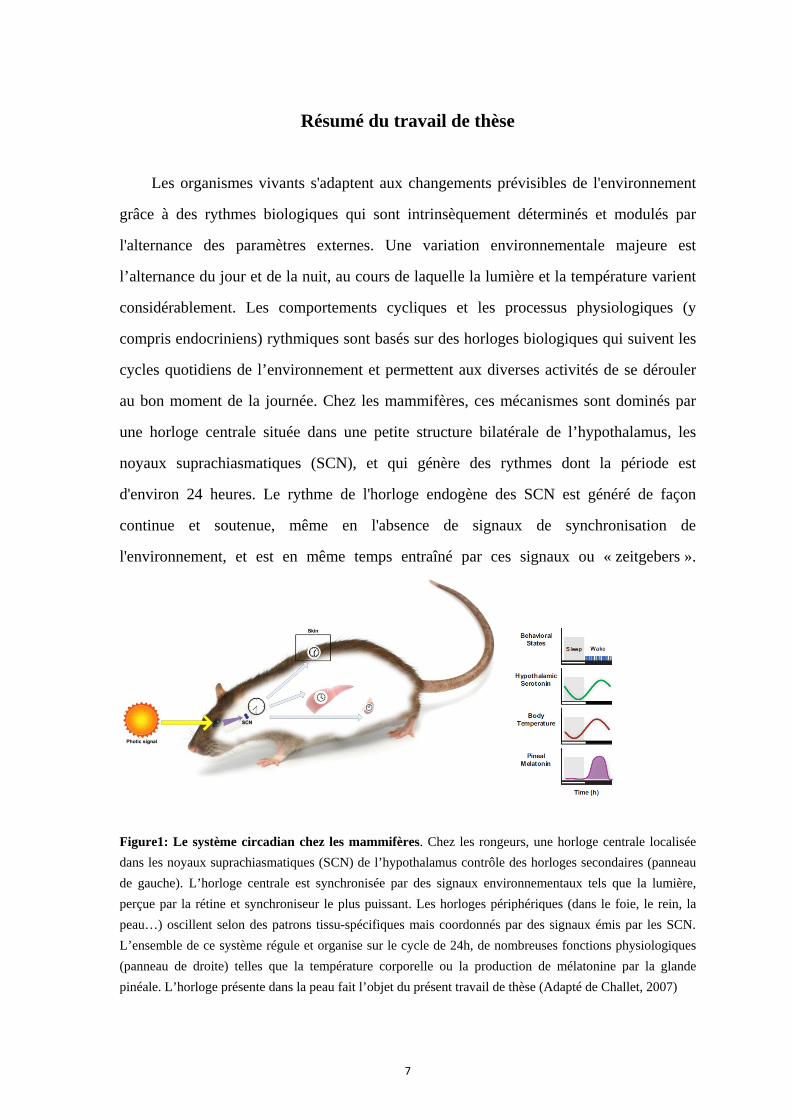
Figure1: Le système circadian chez les mammifères. Chez les rongeurs, une horloge centrale localisée
dans les noyaux suprachiasmatiques (SCN) de l’hypothalamus contrôle des horloges secondaires (panneau
de gauche). L’horloge centrale est synchronisée par des signaux environnementaux tels que la lumière,
perçue par la rétine et synchroniseur le plus puissant. Les horloges périphériques (dans le foie, le rein, la
peau…) oscillent selon des patrons tissu-spécifiques mais coordonnés par des signaux émis par les SCN.
L’ensemble de ce système régule et organise sur le cycle de 24h, de nombreuses fonctions physiologiques
(panneau de droite) telles que la température corporelle ou la production de mélatonine par la glande
pinéale. L’horloge présente dans la peau fait l’objet du présent travail de thèse (Adapté de Challet, 2007)
Figu
proc
régu
phys
beau
gén
osci
nég
Cryp
gén
Ces
dive
diff
de b
spéc
loca
exte
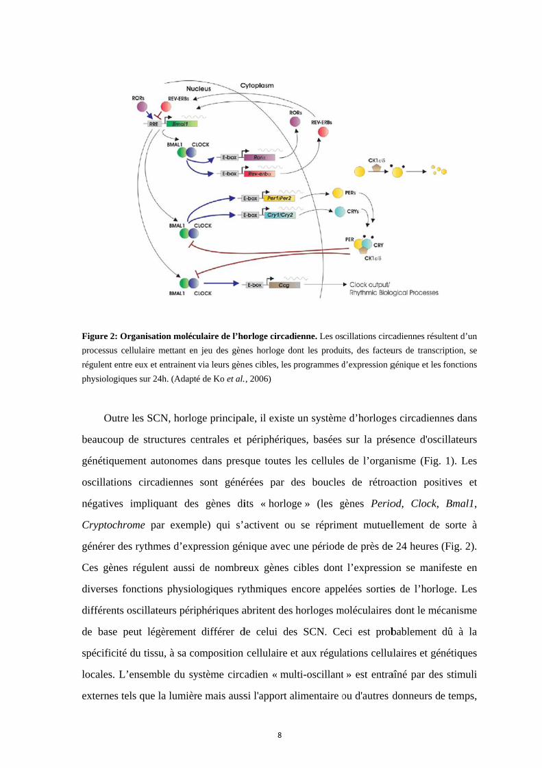
ure 2: Organi
essus cellulai
ulent entre eux
siologiques su
Outre les
ucoup de s
étiquement
illations cir
atives imp
yptochrome
érer des ryt
gènes régu
erses foncti
férents oscil
base peut
cificité du t
ales. L’ense
ernes tels qu
isation moléc
ire mettant en
x et entrainent
ur 24h. (Adapt
SCN, horlo
structures ce
autonomes
rcadiennes
liquant des
par exemp
thmes d’exp
ulent aussi
ions physio
llateurs péri
légèrement
issu, à sa co
emble du sy
ue la lumièr
ulaire de l’ho
n jeu des gèn
via leurs gèn
té de Ko et al.
oge principa
entrales et
s dans presq
sont géné
s gènes di
ple) qui s’a
pression gén
de nombre
logiques ry
phériques a
différer d
omposition
ystème circa
re mais auss
8
orloge circad
nes horloge do
nes cibles, les p
, 2006)
ale, il existe
périphériqu
que toutes
érées par d
its « horlog
activent ou
nique avec u
eux gènes c
ythmiques e
abritent des
de celui des
cellulaire e
adien « mu
si l'apport a
dienne. Les os
ont les produ
programmes d
e un système
ues, basées
les cellules
des boucles
ge » (les g
u se réprim
une période
cibles dont
encore appe
horloges m
s SCN. Ce
et aux régula
lti-oscillant
alimentaire o
cillations circ
its, des facteu
d’expression g
e d’horloge
sur la prés
s de l’organ
s de rétroa
gènes Perio
ment mutuel
e de près de
l’expressio
elées sorties
moléculaires
eci est prob
ations cellu
t » est entra
ou d'autres d
cadiennes résu
urs de transcr
génique et les
es circadienn
sence d'osc
nisme (Fig.
action posi
od, Clock,
llement de
e 24 heures
on se mani
s de l’horlo
dont le mé
bablement
ulaires et gé
aîné par des
donneurs d
ultent d’un
ription, se
fonctions
nes dans
cillateurs
1). Les
itives et
Bmal1,
sorte à
(Fig. 2).
ifeste en
oge. Les
canisme
dû à la
nétiques
s stimuli
e temps,
et s
man
horl
Figu
comp
fibro
(Ada
(pro
de l
et l
l'env
circ
prin
qu’e
Per
refle
ynchronisé
nière tissu-s
loges périph
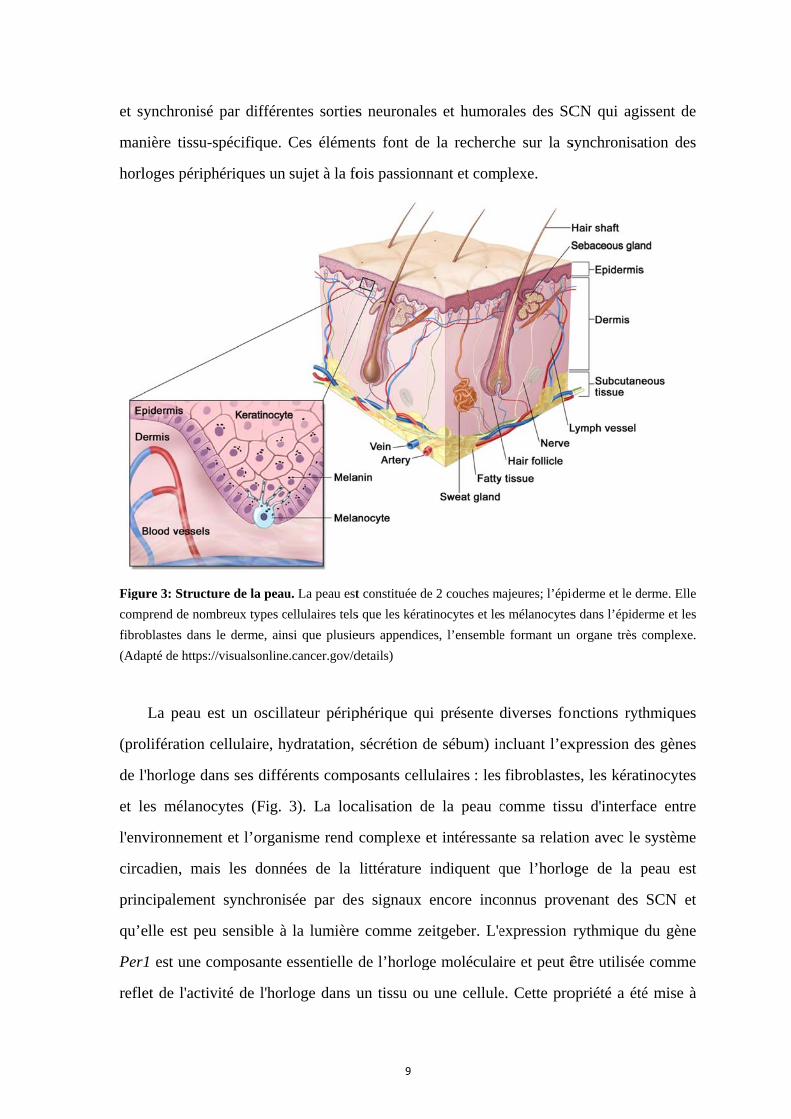
ure 3: Structu
prend de nom
oblastes dans
apté de https:/
La peau e
olifération c
l'horloge da
es mélanoc
vironnemen
cadien, mai
ncipalement
elle est peu
1 est une co
et de l'activ
par différe
spécifique.
hériques un
ure de la peau
mbreux types c
le derme, ain
//visualsonline
st un oscill
cellulaire, h
ans ses diffé
cytes (Fig.
nt et l’organ
s les donn
t synchroni
u sensible à
omposante
vité de l'hor
entes sorties
Ces élémen
sujet à la fo
u. La peau est
cellulaires tels
nsi que plusie
e.cancer.gov/d
lateur périp
ydratation,
érents comp
3). La loc
nisme rend
nées de la
sée par de
à la lumière
essentielle
rloge dans u
9
s neuronale
nts font de
ois passionn
t constituée de
s que les kérat
eurs appendice
details)
phérique qu
sécrétion d
posants cellu
calisation de
complexe e
littérature
s signaux
e comme ze
de l’horlog
un tissu ou
es et humor
e la recherc
nant et comp
e 2 couches m
tinocytes et le
es, l’ensemble
ui présente
de sébum) in
ulaires : les
e la peau c
et intéressan
indiquent q
encore inco
eitgeber. L'e
ge moléculai
une cellule
rales des SC
he sur la s
plexe.
majeures; l’épid
s mélanocytes
e formant un
diverses fon
ncluant l’ex
fibroblaste
comme tiss
nte sa relati
que l’horlo
onnus prov
expression
ire et peut ê
e. Cette pro
CN qui agi
synchronisa
derme et le de
s dans l’épide
organe très c
nctions ryth
xpression de
es, les kérat
su d'interfa
ion avec le
oge de la p
venant des
rythmique
être utilisée
opriété a été
ssent de
ation des
erme. Elle
erme et les
complexe.
hmiques
es gènes
inocytes
ce entre
système
peau est
SCN et
du gène
e comme
é mise à
prof
de l
ains
Figu
tran
Figu
trans
(Mil
(Act
biolu
fit pour dév
la luciférase
si suivre en
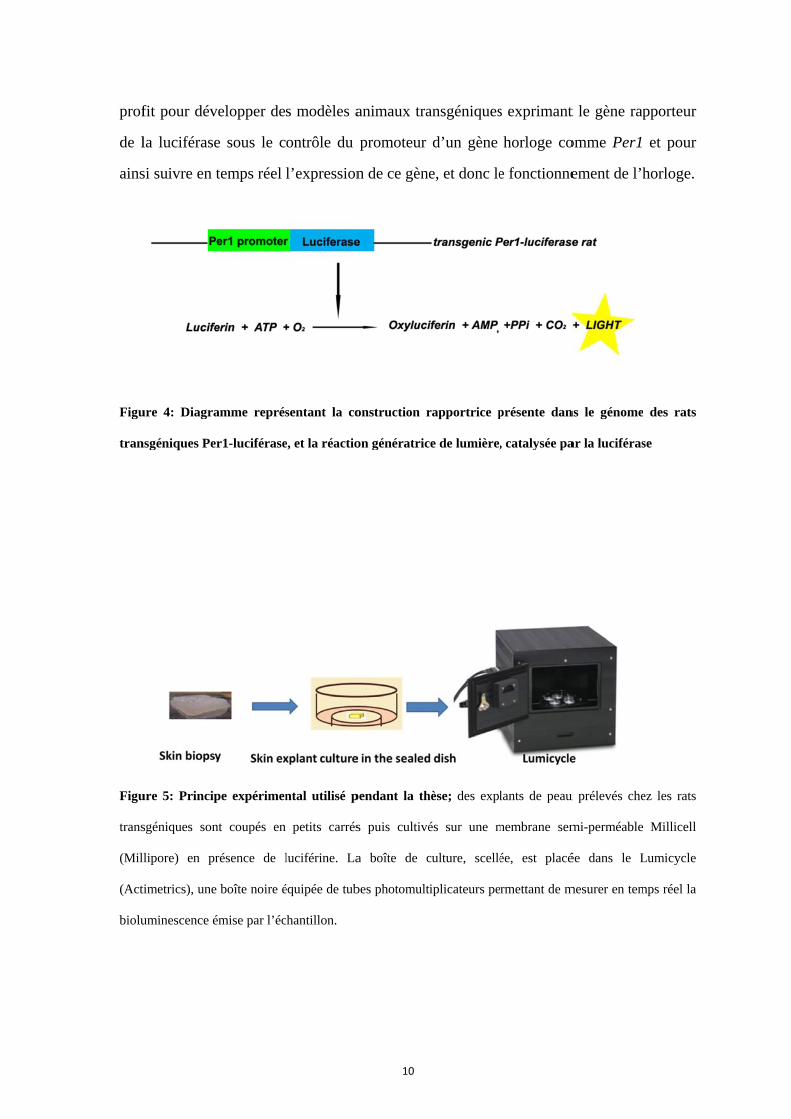
ure 4: Diagra
nsgéniques Pe
ure 5: Princip
sgéniques son
lipore) en p
timetrics), une
uminescence é
velopper de
e sous le c
temps réel
amme représ
er1-luciférase
pe expérimen
nt coupés en
résence de l
e boîte noire é
émise par l’éc
s modèles a
ontrôle du
l’expression
sentant la co
e, et la réactio
ntal utilisé p
petits carrés
luciférine. La
équipée de tub
chantillon.
10
animaux tra
promoteur
n de ce gène
onstruction r
on génératric
pendant la th
s puis cultivé
a boîte de c
bes photomult
ansgéniques
d’un gène
e, et donc le
rapportrice p
ce de lumière,
hèse; des expl
és sur une m
culture, scellé
tiplicateurs pe
s exprimant
horloge co
e fonctionne
présente dan
, catalysée pa
lants de peau
membrane sem
ée, est placé
rmettant de m
t le gène rap
omme Per1
ement de l’h
ns le génome
ar la luciféras
u prélevés che
mi-perméable
ée dans le L
mesurer en tem
pporteur
et pour
horloge.
des rats
se
ez les rats
Millicell
Lumicycle
mps réel la
11
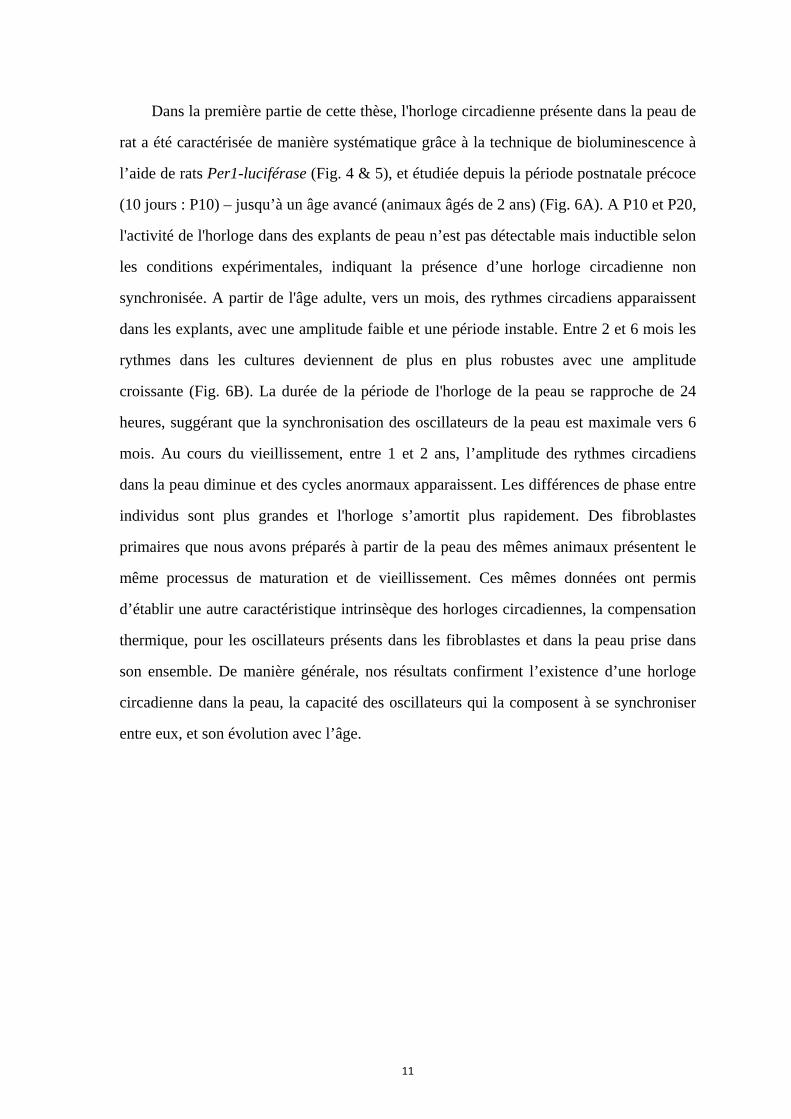
Dans la première partie de cette thèse, l'horloge circadienne présente dans la peau de
rat a été caractérisée de manière systématique grâce à la technique de bioluminescence à
l’aide de rats Per1-luciférase (Fig. 4 & 5), et étudiée depuis la période postnatale précoce
(10 jours : P10) – jusqu’à un âge avancé (animaux âgés de 2 ans) (Fig. 6A). A P10 et P20,
l'activité de l'horloge dans des explants de peau n’est pas détectable mais inductible selon
les conditions expérimentales, indiquant la présence d’une horloge circadienne non
synchronisée. A partir de l'âge adulte, vers un mois, des rythmes circadiens apparaissent
dans les explants, avec une amplitude faible et une période instable. Entre 2 et 6 mois les
rythmes dans les cultures deviennent de plus en plus robustes avec une amplitude
croissante (Fig. 6B). La durée de la période de l'horloge de la peau se rapproche de 24
heures, suggérant que la synchronisation des oscillateurs de la peau est maximale vers 6
mois. Au cours du vieillissement, entre 1 et 2 ans, l’amplitude des rythmes circadiens
dans la peau diminue et des cycles anormaux apparaissent. Les différences de phase entre
individus sont plus grandes et l'horloge s’amortit plus rapidement. Des fibroblastes
primaires que nous avons préparés à partir de la peau des mêmes animaux présentent le
même processus de maturation et de vieillissement. Ces mêmes données ont permis
d’établir une autre caractéristique intrinsèque des horloges circadiennes, la compensation
thermique, pour les oscillateurs présents dans les fibroblastes et dans la peau prise dans
son ensemble. De manière générale, nos résultats confirment l’existence d’une horloge
circadienne dans la peau, la capacité des oscillateurs qui la composent à se synchroniser
entre eux, et son évolution avec l’âge.
Figu
vieil
A) E
peau
enreg
régre
prése
prem
les g
B
A
ure 6: L’act
llissement che
Enregistremen
u prélevés sur
gistrements. L
essions sinuso
entent des R2
mier pic observ
groupes 2, 3, 6
tivité rythmi
ez les rats Per
nts représentat
des rats âgés
Les périodes,
oïdales ajustée
supérieurs à
vé. Les donné
6, 12, 16 et 24
*
ique de la
r1-luciférase.
tifs de la biol
s entre 10 jour
amplitudes et
es aux enregi
0,8, indiquan
ées montrées s
mois, respect
12
peau évolue
.
luminescence
rs et 2 ans. B
t amortissemen
strements et p
nt une bonne r
sont des valeu
tivement). * p
e dans la p
(données sou
B) Analyse des
nts (damping
pour lesquelle
rythmicité. Le
urs moyennes±
p<0.05
*
période post-
ustraites) émi
s paramètres r
rate) ont été c
es les données
es phases repr
±écart type (n=
-natale et d
ise par des ex
rythmiques dé
calculés sur la
s entre 2 moi
résentent le m
=7, 10, 9, 6, 7
durant le
xplants de
éduits des
a base des
s et 2 ans
moment du
7 et 4 pour

13
Dans la deuxième partie de cette thèse, le rôle de la mélatonine comme un
synchroniseur potentiel a été étudiée dans les fibroblastes primaires de la peau. La
mélatonine est une production hormonale de la glande pinéale, strictement synthétisée
durant la nuit sous le contrôle des SCN. Chez les mammifères à la fois diurnes et
nocturnes, la concentration de mélatonine dans le plasma présente un rythme robuste avec
un maximum de nuit et elle est donc considérée comme marqueur de phase de l’horloge
centrale. La mélatonine présente un effet d'entraînement (chronobiotique) sur l'horloge
centrale, dans une fenêtre de temps précise. Des récepteurs de haute affinité pour la
mélatonine (MT1 et MT2) sont exprimés dans différents tissus périphériques, indiquant
que la mélatonine est un candidat potentiel comme synchroniseur des horloges
périphériques, mais cet effet n’a pas été démontré. Les récepteurs MT1 et MT2 sont
exprimés dans la peau humaine et chez les rongeurs, sur les différents types cellulaires.
Dans notre étude, nous avons démontré un effet de la mélatonine sur les fibroblastes
primaires de la peau : elle induit une augmentation de l’amplitude des oscillations du gène
Per1, uniquement lorsqu’elle est appliquée à une certaine phase, mais ne modifie pas les
autres paramètres de l’horloge (Fig. 7). Il s’agit du premier travail montrant un effet de la
mélatonine sur une horloge périphérique.

A
C
D
E
14
*
A
B
Figure 7: Effe
sur les fibrob
la peau.
A) La mélato
façon signif
des oscillat
fibroblastes s
le pic de b
Effet d’un
forskoline,
l’amplitude d
Effet du tr
mélatonine
oscillations ex
du pic de réfé
en fonction
traitement (d
mélatonine s
Effet de la
l’amortisseme
moyennes±éc
* p<0.05
B
ets de la méla
blastes prima
onine augme
ficative l’am
tions lorsqu
sont traités 2h
bioluminescen
traitement
20µM, 2h
des oscillatio
raitement av
sur la phas
xprimée en fo
érence (à gauc
du mome
droite). D) Effe
sur la pério
a mélatonin
ent. V
cart‐type (
*
atonine
ires de
nte de
plitude
e les
h après
nce. B)
à la
h sur
ons. C)
vec la
se des
onction
che) ou
nt du
et de la
de. E)
e sur
Valeurs
n=7‐8).

15
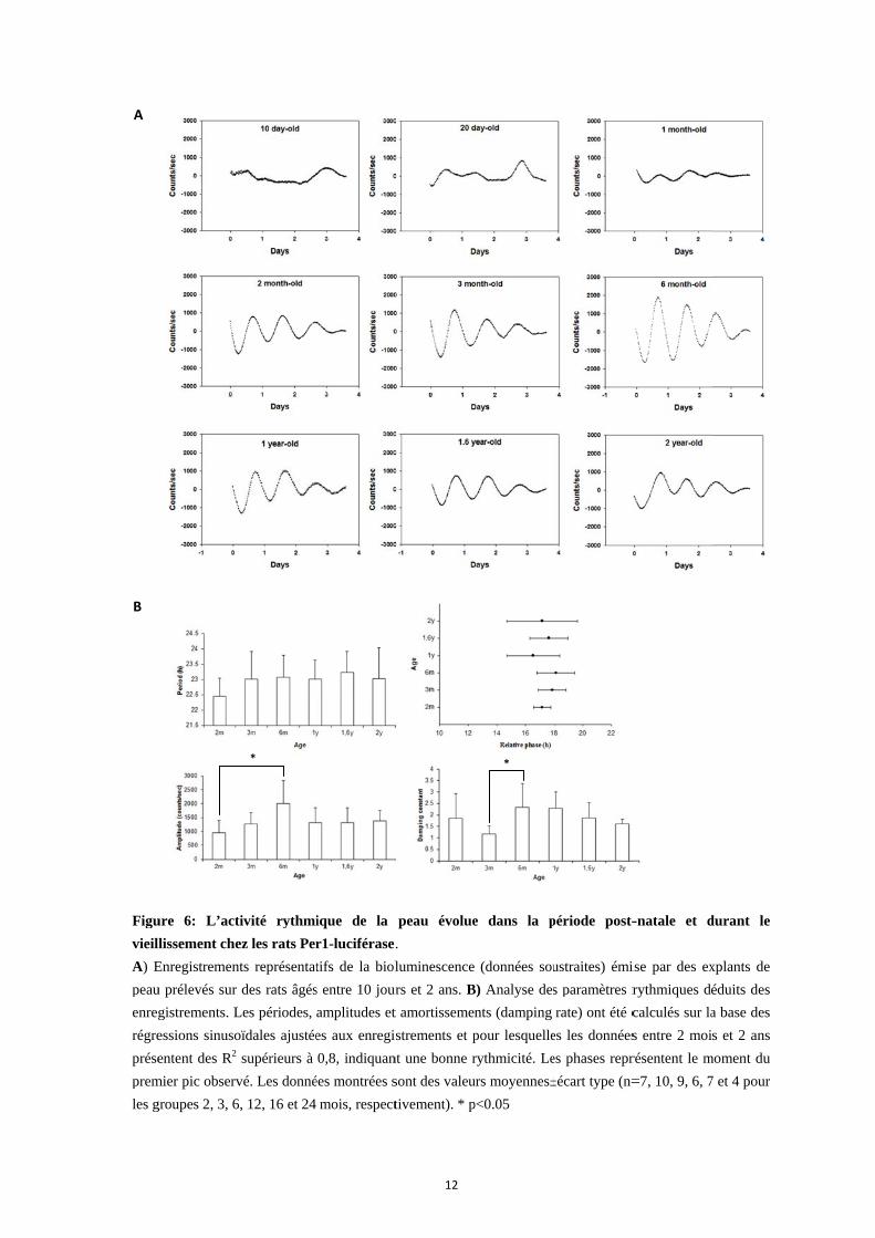
Notre étude chez le rat s’est limitée aux fibroblastes, seul type cellulaire que nous
avons pu isoler et amplifier en culture à partir de peau abdominale chez cet animal. Afin
d’étudier l’horloge de la peau dans les autres types cellulaires (kératinocytes, mélanocytes)
nous utilisons couramment des cultures préparées à partir de peau humaine obtenue après
chirurgie plastique. Afin de pouvoir également étudier ces types cellulaires avec la
technique de bioluminescence, nous avons entrepris de construire un vecteur lentiviral
exprimant le gène rapporteur luciférase sous le contrôle du promoteur du gène horloge
Bmal1 : c’est ce qui a fait l’objet de la troisième partie de mon travail de thèse (Fig. 8).
Bmal1 est un autre gène horloge requis pour l’expression de rythmes circadiens, et son
maximum présente une opposition de phase par rapport à celle de Per1. Ce vecteur ainsi
construit a été testé et des stratégies pour en améliorer le niveau d’expression et
l’amplitude de la rythmicité ont été proposées.
La présence d’oscillateurs cellulaires répandus dans tout l’organisme et
l'organisation hiérarchique du système circadien suggèrent l'importance d’une
synchronisation des horloges entre le centre et la périphérie. Notre étude démontre que la
peau est un oscillateur périphérique complexe et synchronisable, qui peut servir de
modèle pour comprendre les mécanismes moléculaires impliqués, voire pour tester
d’éventuelles stratégies thérapeutiques vis-à-vis de troubles cliniques liés à l’horloge.
Figu
sous
ure 8: Organ
s le contrôle d
isation et car
du promoteur
rte de restric
r du gène hor
16
ction partiell
rloge Bmal1
le du vecteurr lentiviral exxprimant la l
luciférase
17
Thesis summary
Animals adapt to the predictable changes in the environment through biological
rhythms which are intrinsically determined and modulated by the external alternation. A
major environmental variation is the day and night cycle, during which light and
temperature differ drastically. Virtually, all living organisms develop their circadian
rhythms by possessing biological clocks which follow daily cycles and adjust diverse
activities to the adequate time of the day. In mammals, this circadian clock mechanism is
dominated by the central clock located in a bilateral tiny region of the brain, within the
hypothalamus: the suprachiasmatic nucleus (SCN), which controls physiological
processes, including behavior and endocrine functions, with a period of approximately 24
hours. The endogenous rhythm of the SCN clock is continuously generated and sustained,
in the absence of environment timing cues, and it is at the same time entrained by these
zeitgeber (time giving) signals.
Besides the SCN as the master clock in the organisms, a system of circadian clocks
in central and peripheral structures exists on the basis of genetically autonomous
oscillators present in almost all the cells. Circadian molecular oscillations are produced
by positive and negative feedback regulatory loops involving a series of “clock” genes
(Period, Clock, Bmal1, Cryptochrome, for instance) that activate and repress each other to
finally generate rhythms with a period close to 24 hours. These rhythmic gene
expressions are transduced into many circadian outputs that drive physiological activities
and convey timing signals to other parts of the circadian system. The multioscillatory
system is entrained by external stimuli of light, food intake or other time givers and
synchronized by various neuronal and humoral outputs of the SCN which act in a tissue-
and cell-specific way. The different peripheral oscillators harbor distinct clockworks,
although they hold the same genotype as the SCN. This is possibly due to local epigenetic
regulation and systemic synchronization as well as the diversity of tissue structure and
cell composition. All these contribute to the intriguing and attractive research on the
synchronization of peripheral clocks.
18
As one special peripheral oscillator, the skin exhibits circadian rhythmic functions
correlated with the existence of rhythmic clock gene expression in its constitutive cell
types, such as keratinocytes, fibroblasts and melanocytes. These cellular oscillators
display their own clock phenotypes in vivo and in vitro. To be in concert, the
synchronization of the skin clocks is dependent on timing cues emanating from the SCN;
thus the distinct role of the skin as an interface tissue between the external and internal
environment make it both complicated and interesting among the peripheral clocks in the
circadian network. In addition, synchronization of the skin clocks provides an opportunity
to understand the mechanisms controlling circadian system of mammals. Per1 gene is a
core component of the molecular clockwork, whose circadian oscillation is required for
normal clock functions and reflects the phase of clock activity. Technically it facilitates
the study of clock properties by utilizing clock driven bioluminescence of tissue and cells
from transgenic animals carrying a luciferase reporter gene controlled by the promoter of
a clock gene, such as Per1 for instance.
Peripheral clocks undergo the processes of early development and ageing during the
lifespan. In the first part of this thesis, the skin clock was characterized systematically by
using the Per1-luciferase transgenic rat, from the early postnatal age 10 day (P10) to 2
years (old animals). At P10 and P20, the skin clock activity was arrhythmic although
some oscillations randomly showed up. The synchronization after medium refreshment
indicated that the skin clock at early postnatal age was asynchronous, which coincided
with the absence of synchronization of circadian system described during early
development. Starting at 1 month-old, circadian rhythms appeared with low amplitude
and unstable period. From 2 to 6 months, circadian oscillations developed more and more
robust with amplitude increasing significantly. The period length of skin clock got closer
to 24 hours in adult. These results suggested that the skin clock was better synchronized
till the maximum in 6 months. In aged animals, 1 to 2 year-old, circadian oscillations got
globally weaker and some abnormal cycles appeared. The phases delayed and variations
between individuals turned larger. The clock activity damped faster by aging progress. In
elderly, the skin clock was worse synchronized within itself and among individuals. We
19
also studied primary fibroblasts derived from the skin at the distinct ages and
demonstrated a similar pattern of clock activity in maturation and ageing, which was
consistent with the synchronization of the skin tissue. Another intrinsic feature of
circadian clocks, temperature compensation, was shown the first time in the skin tissue
and skin primary fibroblasts. Generally, we corroborate the existence of the skin clock
and its synchronization in the development and ageing, and characterized its clock
property within skin itself and within the circadian system.
In the second part of this thesis, the role of melatonin as a potential synchronizer was
investigated in skin primary fibroblasts. Melatonin is a hormonal output of the SCN
pacemaker with robust daily rhythms in plasma level. In both diurnal and nocturnal
mammals, melatonin is synthesized primarily in the pineal gland in the night and its
plasma rhythm is considered a reliable estimate of the SCN clock. Melatonin has shown
entrainment effect on the central clock in a restricted time window at subjective dusk
(chronobiotic effect). Since high-affinity melatonin receptors MT1 and MT2 are
expressed in various peripheral tissues besides the SCN, melatonin is a potential
candidate as a synchronizer of peripheral clocks. However, the effect of melatonin on
peripheral clocks has been poorly confirmed. MT1 and MT2 expression has been shown
in human and rodent skin tissue and cells, including fibroblasts, keratinocytes and
melanocytes. In our study, we demonstrated a phase-dependent effect of melatonin to
synchronize primary skin fibroblasts. When applied 2h after the Per1 peak, the amplitude
of oscillations was significantly increased by melatonin to the same extent as for forskolin,
a cAMP-mediated synchronizer. The relatively weak effect of melatonin indicated its
distinct role on the peripheral clocks. Long term application that induced Per1 activity
might be involved in the melatonin effect on the skin fibroblast clock. Ultimately, our
study shows the first time an effect of melatonin on synchronization of peripheral clocks.
In addition, clock gene expression is found in other skin cell-autonomous oscillators
such as keratinocytes and melanocytes, which contributes to the complexity of the
multioscillatory skin. Fusing luciferase genes to clock gene promoters has arisen as a
20
convenient method to measure circadian gene expression in tissues and cells by
bioluminescence recording. However, right now only fibroblasts can be cultured from
dermal tissues of transgenic rodents because of their serum-dependent proliferation. Other
skin cells like melanocytes and keratinocytes can be more easily derived and cultured
from human skin. Brown and his colleagues have successfully established a lentivirally
delivered luciferase reporter to measure clock genes activities in human skin cells. To
facilitate our studies on multioscillatory skin, we constructed a Bmal1-luciferase
lentivirus according to similar design, where Bmal1 is another core clock component with
peak expression almost antiphase to Per1. In the third part of thesis, construction of a
lentivirus tool delivering Bmal1-luciferase reporter was shown although it had not been
efficient enough in application. It is worth trying to improve the efficiency of this
lentiviral reporter, with which clock activities and synchronization could be elucidated
not only in fibroblasts but also in melanocytes and keratinocytes of the multioscillatory
skin.
The widespread cell-autonomous oscillators and the hierarchical organization of the
circadian system in mammals suggests the importance of synchronization of clocks in
center and periphery. The present work demonstrates the synchronization of peripheral
clocks in the skin and potential mechanism. Further investigation can enhance our
understanding of how peripheral clocks are synchronized and which roles diverse
synchronizers do play. The knowledge of these mechanisms in circadian system function
and organization might enable to improve health care and clinical treatment of
clock-related disorders.

21
Introduction
22
Rhythm, a regular recurrence or pattern in time, applies to a wide variety of cyclic
natural phenomena, with a period from microseconds to millions of years. Rhythmic units
provide an order to the world and reduce the entropy. They even describe the time per se.
For instance, the rotation of the earth, by itself and around the sun, is rhythmic enough to
determine days and seasons. Earth environment changes in the day length and
temperature. To adapt to nature, biological rhythms commonly exist in the living world,
covering periods in a wide range. For example, in seasonal cycles, including reproduction,
hibernation and migration, animals change their way of life from summer to winter,
according to the variations of temperature and food, to organize activity at the appropriate
time. Each day, the rising and setting of the sun causes daily environmental changes, and
consequently daily behaviors of animals, such as sleep-wake cycle. Hence, to adapt to and
to anticipate predictable conditions in next days, animals follow biological rhythms with a
period of approximately 24 hours, which are named circadian rhythms. Therefore,
animals (and virtually all living organisms) have developed internal systems or
endogenous abilities, called circadian clocks. The circadian clocks can drive daily
rhythms in behavior and in many physiological, including endocrine, processes. Adapting
life style to circadian rhythms is important for the health of animals. In human, life in
society can lead to dramatic conditions when resetting of the internal clocks is not done at
appropriate time. It may cause some disorders, or even diseases, in conditions like chronic
jetlag or shift work.
1. General features about circadian clocks
All biological circadian clocks have several common features: self-sustained,
entrained or synchronized by external cues, and temperature compensated. First, circadian
clocks can sustain and produce oscillations in the absence of any external cues. This
property is due to cell-autonomous persistence determined genetically and based on a
molecular clockwork which is still not completely understood yet. Second, circadian
23
clocks are capable to be entrained or synchronized by time givers, named zeitgebers, to
keep harmony with the environment. Light is the chief zeitgeber in nature. Third,
temperature compensation can make sure that endogenous periods of circadian clocks
will not be altered by changing temperature. All these properties of circadian clocks will
be discussed in detail later.
Circadian clocks have been found in virtually all organisms, from bacteria to insects,
birds and mammals. In mammals, lesions of specific brain areas confirmed that a small
structure in the brain is required for this timekeeping system (Stephan and Zucker, 1972).
This master clock is located in the suprachiasmatic nucleus (SCN), a cluster of thousands
of neurons located in the anterior hypothalamus. Animals with lesion of the SCN lost
their daily rhythms in sleep/wake activity, body temperature and production of several
hormones. It is found that many physiological processes are controlled by peripheral
oscillators (Fig.1). They form a complex circadian system that organizes an ensemble of
structures in periphery. The central clock in the SCN takes charge of this hierarchical
network to make sure that physiological functions of the distinct organs take place at the
appropriate time of the day.
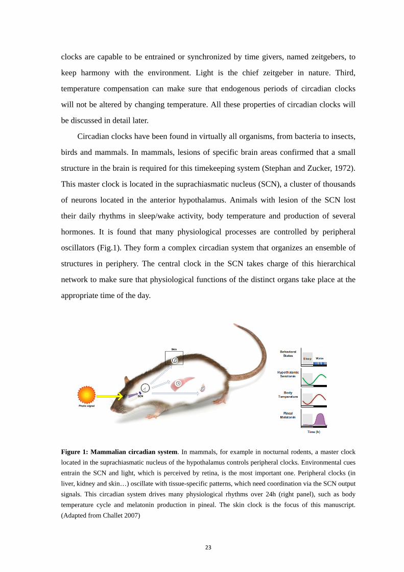
Figure 1: Mammalian circadian system. In mammals, for example in nocturnal rodents, a master clock
located in the suprachiasmatic nucleus of the hypothalamus controls peripheral clocks. Environmental cues
entrain the SCN and light, which is perceived by retina, is the most important one. Peripheral clocks (in
liver, kidney and skin…) oscillate with tissue-specific patterns, which need coordination via the SCN output
signals. This circadian system drives many physiological rhythms over 24h (right panel), such as body
temperature cycle and melatonin production in pineal. The skin clock is the focus of this manuscript.
(Adapted from Challet 2007)
24
A functional circadian system requires the central clock, with input and output
pathways. The central clock oscillates with a period close to 24 hours. On one side, inputs
derived from environment convey the timing message to entrain the central clock to
varying conditions. One of the most powerful entraining signals is the daily alternation of
light and dark, whose period is precisely 24 h. On the other side, circadian clocks are
present in almost all the cells in the organisms and outputs from the center allow the clock
system to control subsidiary targets in the brain and periphery (Fig. 1). All these
peripheral clocks also need to be synchronized within each tissue, to maintain
homeostasis required for physiological functions.
Another intrinsic feature of circadian clocks is that they are temperature
compensated. Their period lengths remain almost constant across the physiological
temperature range (Pittendrigh, 1993). This temperature compensation can be described
quantitatively by the temperature coefficient, Q10, which means the change in the rate of
a biological reaction when increasing the ambient temperature by 10 °C. This Q10 is 2-3
for most biochemical reactions but only 0.8-1.2 in circadian rhythms (Sweeney and
Hastings, 1960). It is unclear but possible that synthesis and degradation rates of core
clock components are equally affected by changing temperature. In mammals,
temperature compensation has been demonstrated in cell cultures such as fibroblasts and
SCN neurons, and tissues like SCN, lung and adrenal gland (Reyes et al., 2008; Dibner et
al., 2009; Buhr et al., 2010). This property of resilience to changes in temperature is
independent of intercellular coupling. It is crucial for phase coherence of peripheral
clocks, for which cellular transcription rates can vary dramatically in different tissues
(Dibner et al., 2010). Some interface tissues like skin, especially distal skin, confront
largely changing ambient temperatures, even in homeothermic animals. In fact, some
oscillators like fibroblasts are even overcompensated (Tsuchiya et al., 2003).

25
2. Molecular clock mechanisms
Circadian clocks are cell-autonomous processes able to generate persistent
oscillations, and which are coordinated in multicellular organisms. The unraveling of the
molecular basis of circadian clock began with the discovery of the tau mutant hamster in
the 1980s and identification of the Clock mutant mouse in the 1990s (Ralph and Menaker,
1988; Antoch et al., 1997; King et al., 1997). Then, a set of clock genes were defined as
core components whose protein products are necessary for the generation and regulation
of circadian rhythms in rest/activity cycles and, later, within individual cells throughout
the organisms. Per1 and Per2 are induced by light (Albrecht et al., 2001). The
photoinduction of the clock gene Per2 develops later than that of Per1 (Mateju et al.,
2009). Disruptions of Per1/2 or Cry1/2 cause immediate behavioral arrhythmicity when
double-knockout animals are kept in constant darkness (van der Horst et al., 1999; Zheng
et al., 2001). Deletion of Bmal1 is the only single knockout that abolishes clock function
in both the SCN and peripheral tissues (Sun et al., 2006). This chapter will focus on the
regulations underlying the molecular clockwork, with particular emphasis on Per1, whose
promoter was used as a model throughout the thesis work.
2.1. The feedback loops
The molecular mechanism of circadian clocks involves basically a
transcription-translation feedback loop, with a time constant of about 24 hours per cycle.
This dynamic loop begins with two bHLH-PAS domain transcription factors, BMAl1
(also known as aryl hydrocarbon receptor nuclear translocator-like, ARNTL) and CLOCK.
BMAL1 and CLOCK heterodimerize via PAS domains and bind to DNA via bHLH
domains. In the nucleus, BMAL1/CLOCK heterodimers bind and activate transcription of
genes containing E-box enhancer sequences in promoters, including Period (Per1 and
Per2) and Cryptochrome (Cry1 and Cry2) genes. This leads to an increase in the levels of
PERs and CRYs in the cytoplasm. PER and CRY proteins form heterodimers and, along
with
Sub
the
In th
a cr
tran
acce
elem
tran
Ror
Bma
only
cont
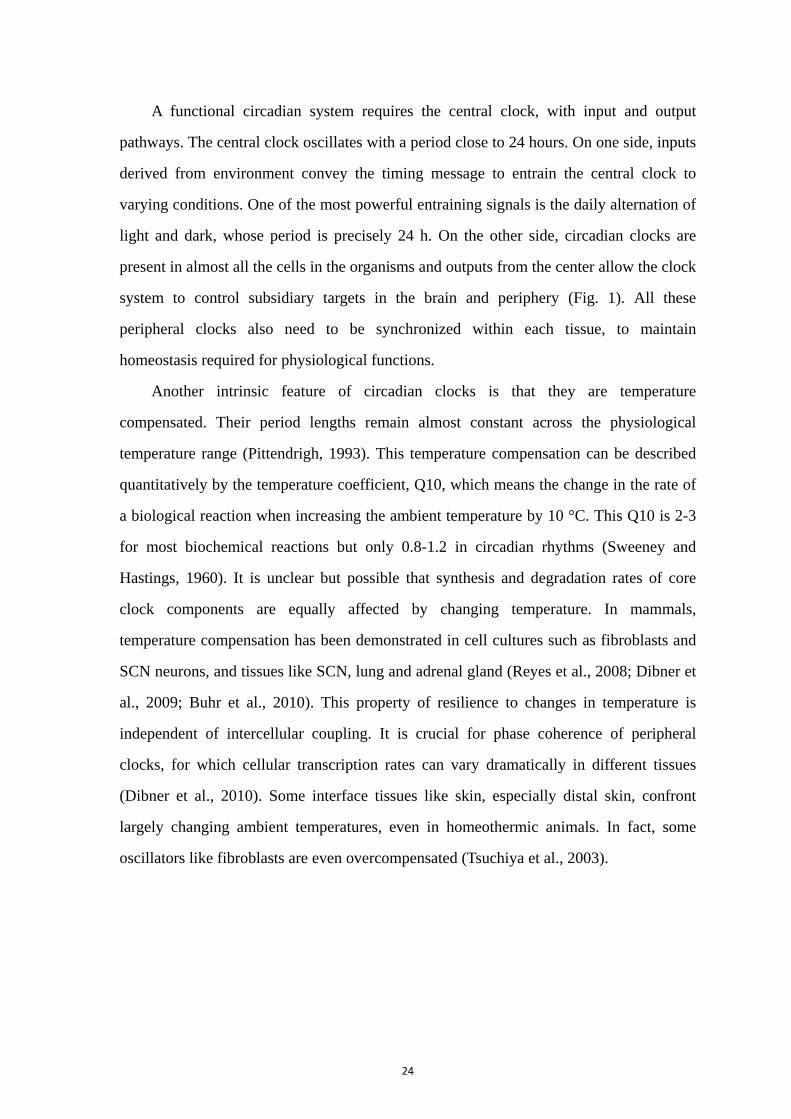
Figu
cell
trans
via th
h other pro
bsequently, n
nucleus, re
he cytoplas
ritical step
nscription: t
essory regu
ment (ROR
nscription of
rα-γ. Via co
al1, wherea
y add robus
trol and link
ure 2: The mo
generate cir
scription drive
he regulation
oteins, cons
negative fe
epressing th
sm, the PER
for relievin
this allows
ulatory loop
RE) presen
f genes enco
ompeting on
as RORs ac
stness and
k to other p
olecular clock
rcadian rhyth
es self-sustain
of their target
titute a hig
edback is a
eir own tra
R/CRY com
ng inhibition
a new cy
interconne
nt in the
oding two f
n binding to
ctivate it. Th
stability to
athways in
kwork of tran
hms involvin
ned oscillation
t genes. (Adap
26
gher order
achieved by
anscription d
mplex is deg
n of BMAL
ycle of aut
ects with the
Bmal1 pro
families of c
o the RORE
hese positiv
o the clock
the cell. (Fi
nscription-tra
ng positive a
ns in clock fact
pted from Ko
complex an
y the accum
driven by th
raded by pr
L1/CLOCK
oregulation
e core loop,
omoter. BM
cognate nuc
E, REV-ERB
ve and nega
mechanism
ig. 2)
anslation feed
and negative
tor levels and
et al., 2006)
nd transloca
mulating PER
he BMAL1
roteasomal
and initiati
n to restart.
, mediated b
MAL1/CLO
clear recepto
Bs repress t
ative regula
ms but also
dback loops.
feedback lo
, subsequently
ate to the
R/CRY com
/CLOCK c
pathways, w
ting a new
. (Fig. 2)
by the ROR
OCK activa
ors, Rev-erb
the transcri
ations of Bm
provide ad
Clock genes i
oops. Circad
y, in biologica
nucleus.
mplex in
complex.
which is
cycle of
Another
R-biding
ates the
bα-β and
iption of
mal1 not
dditional
in a single
dian gene
al rhythms
27
2.2. Transcriptional activation of Period genes by BMAL1/CLOCK
As core clock genes, Per1/2 plays an important role in the molecular machinery of
dynamic loops. The regulation of Per1/2 transcription starts from combination and
activation of cis-regulatory elements contained in their promoters. Transcription of
mPer1/2 is dependent on the levels of activation of these elements. The conserved five
E-box motifs (a consensus CANNTG DNA sequence) are found in the m (mouse) Per1
promoter. Luciferase reporter assays with deleted/point mutated mPer1 promoter
constructs demonstrate that each identified E-box contributes to transcription of Per1
activated by CLOCK/BMAL1 (Hida et al., 2000). It was described that CLOCK/BMAL1
constitutively binds to Per1 E-box sequences over the circadian cycle (Fig. 3A) (Lee et al.,
2001). Recent genomic sequence analysis of BMAL1 binding sites in the liver shows that
binding is rhythmic and identifies a tandem (E1-E2) of E-box sequences with specific
spacing, as mediating cooperative binding of CLOCK/BMAL1 and stronger
transcriptional activation (Rey et al., 2011). The circadian transcription of
CLOCK/BMAL1 target genes including Per1/2 and Cry1 is accompanied by rhythmic
chromatin remodeling and circadian binding of RNA polymerase II (RNAPII) at the
promoters of these genes (Etchegaray et al., 2003). Circadian histone H3 acetylation and
methylation create favorable environments on chromatin for RNAPII recruitment,
transcriptional initiation and elongation (Koike et al., 2012). It is the rhythmic RNAPII
recruitment at promoters, rather than a rhythmic transition from paused to productive
elongation, that executes circadian changes in genes transcription (Le Martelot et al.,
2012). The occurrence of various modifications of chromatin, occupancy of DNA by
RNAPII and presence of coactivators contribute to the narrow phase of activity for these
regulations of circadian transcription (Fig. 3B) (Koike et al., 2012).
Other cis-acting regulatory elements may play a role in shaping the kinetics of
transcription that contributes to variability or additional control of clock genes, likely
through recruitment and retention of elements of the general transcription machinery
(Suter et al., 2011). No typical TATA-box but four conserved CREs (cAMP responsive
elements) are identified in Per1 promoter region, and one CRE and two D-box sequences
28
in Per2 promoter (Hida et al., 2000; Albrecht et al., 2007). External stimuli such as
light-induced glutamate and PACAP can lead to calcium influx and elevate the level of
cAMP in the SCN. This activates several kinase pathways including Protein Kinase A
(PKA) and finally causes phosphorylation of CREB, which activates CREs contained in
the promoters of Per1 and Per2. CREs have been reported to be responsible for the
transcription of other clock-controlled genes such as arginine vasopressin and c-fos
(Robertson et al., 1995; Iwasaki et al., 1997). Per genes, activated by external stimuli
without any new proteins synthesis are considered as immediate early genes. CRE
element makes the Per2 promoter much less responsive, in contrast to similar elements in
the Per1 promoter (Travnickova-Bendova et al., 2002). In mice PER oscillation, rather
than its absolute quantity, is crucial for both cellular circadian oscillations and behavioral
rhythmicity (Chen et al., 2009).

Figu
A) S
dom
BMA
and
activ
elem
comp
CLO
co-ac
inhib
the C
Repp
ure 3: CLOCK
Structural diag
main and tande
AL1 (blue). T
179 of 626 r
vity and clock
ment in DNA
plex over the
OCK (C in ye
ctivators and
bition, the PE
CLOCK/BMA
pert & Weaver
A
K/BMAL1 he
gram of the mo
em PAS dom
This structure l
residues in B
k function. Ins
A. B) CLOCK
e circadian cy
ellow) and BM
d RNA Polym
ER/CRY comp
AL1 heterodi
r 2002)
eterodimer an
ouse CLOCK/
mains each co
lacks the C-te
BMAL1) that
set, a closer vi
K/BMAL1 he
ycle, accordin
MAL1 (B in
merase II ar
plex (P in purp
mer, and ina
29
nd its transcr
K/BMAL1 hete
ontribute to co
erminal region
lack ordered
iew of the CL
eterodimers r
ng to initial s
yellow) are b
re recruited t
ple and C in o
activate the tr
riptional com
erodimer illus
omplex forma
ns of each prot
structure but
LOCK/BMAL
remain bound
tudies in live
bound to E bo
to activate tr
orange) with o
ranscription. (
mplex
trating how th
ation between
tein (471 of 8
t are required
L1 bHLH dom
d to E box f
er. During tran
oxes in the P
ranscription.
other recruited
(Adapted from
he DNA-bindi
n CLOCK (g
855 residues in
d for CLOCK
main bound to
forming trans
anscriptional a
Per/Cry promo
During trans
ed co-represso
m Partch et a
ing bHLH
green) and
n CLOCK
K/BMAL1
an E-box
scriptional
activation,
oters, and
scriptional
ors bind to
al., 2013;
30
2.3. Post-transcriptional regulations
The general transcription factors are recruited to individual promoters of genes such
as Per1/2 and Cry1/2 upon CLOCK/BMAL1 binding, and the amount of nascent
transcripts appears to peak with a sharp phase about 8 hours after the activation start on
individual promoters (Koike et al., 2012). Microarray studies have shown that
approximately 10% of all the mammalian transcripts exhibit circadian oscillations,
varying among cell types and tissues (Lowrey and Takahashi, 2011). However, only about
30% of genes with rhythmic changes in protein levels also cycle transcriptionally (Deery
et al., 2009). Cells treated with a RNA polymerase inhibitor still provide cycling rhythms
(Dibner et al., 2009). These data indicate that the majority of rhythmic clock outputs are
possibly due to posttranscriptional regulation. Outside core clock genes,
post-transcriptional regulation likely helps to provide additional flexibility by generating
rhythms in mRNA transcripts in a tissue- or stimulus-dependent manner. Many core clock
genes and clock-controlled genes exhibit circadian oscillations in their transcript levels
involving post-transcriptional control, including miRNA and RNA binding proteins, such
as CIRP (cold-inducible RNA-binding protein) and nocturnin (Fig. 4). Transcribed from
noncoding genomic regions, miRNA interact with the 3’untranslated regions (3’UTR) of
target transcripts to repress activity of target mRNA, possibly to affect the molecular
clock (Bartel, 2009). Two miRNA, miR-219 and miR-132 have been found circadian
rhythmic in the SCN and harbor CREs or E-box in their promoters (Cheng et al., 2007). It
is proposed that miRNA-132 is involved in chromatin remodeling and translational
control in the SCN, mediating clock entrainment by regulating Per mRNA decay
(Alvarez-Saavedra et al., 2011). In Hela and NIH3T3 cells, miR-192 and miR-194 inhibit
Per genes containing corresponding target sites in their 3’UTR, which causes a shortened
circadian period (Nagel et al., 2009). In addition, several RNA-binding proteins have
been identified to regulate clock-related transcripts by distinct mechanisms. CIRP plays
an important role in the core clock posttranscriptional regulation through direct
interaction with Clock mRNA which results in mRNA stabilization in the cytoplasm
(Morf et al., 2012). CIRP transcription and translation are rhythmic under the regulation

31
of systemic signals like body temperature. A reduction of CIRP levels causes low levels
of many core circadian components including Clock, Per2 and Dbp, leading to low
amplitude of molecular circadian rhythms (Morf et al., 2012). Several RNA binding
proteins target cis-acting elements located in 3’UTR of RNAs, such as Lark which
positively regulates Per1 transcript (Kojima et al., 2007). Another RNA binding protein
Nocturnin is a deadenylase that catalyses the removal of the poly(A) tail from transcripts,
followed by mRNAs breakdown, notably of rhythmic genes (Garbarino-Pico et al., 2007).
Finally, further investigation of transcriptional and posttranscriptional molecular
machinery, as well as how locally controlled clock genes are additionally regulated by
systemically controlled genes and systemic cues, will be helpful for a complete
understanding of the circadian gene expression in each tissue and cell type.
After their mRNAs have left the nucleus, PER and CRY proteins are produced in the
cytoplasm and dimerize through their PAS domains. Then, the PER/CRY dimer is
submitted to a shuttling balance between the cytoplasm and the nucleus (Vielhaber et al.,
2001). In PER1 and PER2, the presence of nuclear localization signals (NLS) and nuclear
export signals (NES) have been characterized for nuclear import and cytoplasmic
retention (Yagita et al., 2000; Vielhaber et al., 2001). Whereas nuclear translocation of
PER1 and PER2 can occur in the absence of any CRY protein, nuclear localization of
PER/CRY proteins is a dynamic process determined by both nuclear import and nuclear
export pathways. Nucleocytoplasmic shuttling provides the opportunity for regulation of
PER/CRY protein function in the nucleus and degradation in the cytoplasm.
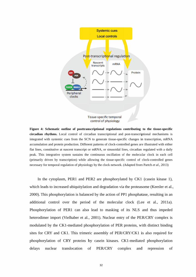
Figu
circa
integ
accu
flat l
peak
(prim
nece
whi
200
addi
Pho
hete
mod
sites
pho
dela
ure 4: Schem
adian rhythm
grated with sy
umulation and
lines, constitu
k. This integr
marily driven
essary for temp
In the cyt
ich leads to
0). This pho
itional con
osphorylatio
erodimer im
dulated by t
s for CRY
osphorylatio
ays nucle
matic outline
ms. Local co
ystemic cues
protein produ
utive at nascen
rative system
by transcrip
poral regulatio
toplasm, PE
increased u
osphorylatio
ntrol over
on of PER
mport (Vielh
the CK1-m
and CK1.
on of CRY
ear transl
of posttran
ontrol of circ
from the SCN
uction. Differe
nt transcript o
sustains the
tion) while a
on of physiolo
ER1 and PE
ubiquitylatio
on is balanc
the period
1 can also
haber et al.
mediated pho
This trimer
Y proteins b
ocation o
32
nscriptional r
cadian transcr
N to generate
ent patterns o
or mRNA, or
e continuous
allowing the
ogy by the clo
ER2 are ph
on and degr
ced by the a
d of the
o lead to m
, 2001). Nu
osphorylatio
ric assembl
by casein
of PER/C
regulations c
riptional and
e tissue-specif
f clock-contro
sinusoidal lin
oscillation of
tissue-specific
ock network. (
hosphorylate
radation via
action of PP
molecular
masking of
uclear entry
on of PER
y of PER/C
kinases. CK
CRY com
contributing
post-transcrip
fic changes in
olled genes are
nes, circadian
f the molecu
c control of
Adapted from
ed by CK1
the proteas
P1 phosphat
clock (Le
f its NLS
y of the PER
proteins, w
CRY/CK1 i
K1-mediate
mplex and
to the tissu
ptional mech
n transcriptio
re illustrated w
n regulated wi
ular clock in
clock-control
m Partch et al.
(casein ki
some (Kees
tase, resulti
ee et al.,
and thus i
ER/CRY com
with distinct
is also requ
ed phospho
d repressi
ue-specific
anisms is
n, mRNA
with either
ith a daily
each cell
led genes
, 2013)
inase 1),
ler et al.,
ing in an
2011a).
impeded
mplex is
binding
uired for
orylation
ion of
33
CLOCK/BMAL1-mediated transcriptional activities, contributing to the 24-hour kinetics
of the clockwork (Eide et al., 2005). CK1ε mutation in the tau mutant hamster leads to
shorter endogenous period. The CK1δ deficient mouse shows longer circadian periods in
free-running rhythms and liver tissue rhythms (Etchegaray et al., 2009; Lee et al., 2009).
Importantly, posttranslational modifications such as phosphorylation, SUMOylation,
deacetylation and ubiquitination can also control degradation of core clock components
and, consequently, period, as shown by the effects of proteasome inhibitors (Eide et al.,
2005). CK1 mediated phosphorylation of PER proteins promotes their ubiquitination and
following degradation via 26S proteasome (Gallego and Virshup, 2007). E3 ubiquitin
ligase FBXL3 interacts with CRY and targets it for degradation, following
phosphorylation of CRY1 by AMPK and of CRY2 by GSK3β (Harada et al., 2005;
Busino et al., 2007; Lamia et al., 2009). SUMO (small ubiquitin-like modifier) modifies
BMAL1 and promotes its proteasomal degradation (Lee et al., 2008). NAD+ dependent
histone deacetylase Sirtuin1 (SIRT1) binds to CLOCK/BMAL1 and deacetylates PER2,
promoting its degradation (Asher et al., 2008). Interestingly, SIRT1 participates in the
regulation of transcription and CLOCK/BMAL1 controls rhythmic NAD+ in turn
(Nakahata et al., 2009; Ramsey et al., 2009). This connection between circadian system
and metabolism contributes to the flexibility of the system which helps to confer
tissue-specific regulation of physiology (Asher and Schibler, 2011).
In the nucleus, the PER/CRY complex associates with CLOCK/BMAL1 and recruits
corepressors, which negatively regulate transcription activity of CLOCK/BMAL1 on core
clock genes such as Per, Cry, Rev-erb and Ror (Fig. 3B). The rhythmic binding of
PER/CRY to CLOCK/BMAL1 generates the rhythm in transcriptional activation. CRY1
can interact with CLOCK/BMAL1 independently of PER proteins, because it is found
without PER at CLOCK/BMAL1 bound sites in Dbp promoter at CT0 (Ye et al., 2011;
Koike et al., 2012). This suggests that CRY1 may act as a molecular gatekeeper to
maintain CLOCK/BMAL1 in a poised and repressed state until the proper time for
transcriptional activation. This repressed state involves epigenetic regulations. PSF
(polypyrimidine tract-binding protein-associated splicing factor) acts as a transcriptional
corepressor by rhythmically recruiting the SIN3-HDAC (SIN3 histone deacetylase)
34
complex to deacetylate histones 3 and 4 at the Per1 promoter (Duong et al., 2011). It is
reported that an RNA- and DNA-binding protein, NONO, associates with PER1/2 to
repress the transcription and gates circadian rhythm with cell cycle (Brown et al., 2005a;
Kowalska et al., 2013). By contrast, transition to the active state begins with removal of
CRY1 from CLOCK/BMAL1 complex and recruitment of coactivator proteins p300 and
WDR5 in favor of an active chromatin state, with histone acetylation and methylation
(Etchegaray et al., 2003; Brown et al., 2005a; Kiyohara et al., 2006).
Finally, because of these transcription-translation feedback loops and
posttranscriptional modulations, the expressions of clock genes show rhythmic profiles
creating molecular oscillations that can sustain themselves in the absence of external
inputs. Relative phases of clock gene expression, such as antiphase of mRNA level of Per
and Bmal1, are also a property of the molecular clockwork (Nishide et al., 2006). Due to
the complicated molecular clockwork, various clock-controlled genes (CCGs) are
expressed differently within the 24 hours with peaks occurring at different times of the
day, to regulate multiple physiological processes from cell growth to metabolism.
Elimination of one component may be compensated by others. In human, mutations
in genes of the molecular clock system are related to some circadian disorders like FASPS
(Familial Advanced Sleep Phase Syndrome) and DSPS (Delayed Sleep Phase Syndrome)
(Takahashi et al., 2008).
3. The SCN: master clock of the body and a model to understand
rhythm generation within tissues
SCN, as the central pacemaker, is capable to be entrained by environmental stimuli. It
receives diverse inputs via innervation from several regions of the brain, where three
pathways well investigated are from the retina (photic inputs), the intergeniculate leaflet
(IGL) and the raphe nuclei (non-photic signals).
35
3.1. The SCN clock
The suprachiasmatic nucleus (SCN) of the hypothalamus is a bilateral structure sited
just above the optic chiasma. The pair of nuclei are located at each side of the third
ventricle and comprise approximately 20000 neurons of small size (Moore et al., 2002).
All the circadian functions are suppressed by ablating the SCN, not any other part of the
brain. Transplantation of fetal SCN tissue into other brain regions like the third ventricle
of SCN-lesioned animals can restore circadian behavioral rhythms (Lehman et al., 1987;
Silver et al., 1996). The SCN tissue from the tau mutant hamster, which shows shorter
period in circadian rhythms, could restore rhythms in SCN-lesioned wild-type hamster
with a shortened period as in the mutant donor (Ralph et al., 1990). The SCN can
self-sustain rhythms with a period of approximately 24 hours without any inputs, for
months in slice culture (Abe et al., 2002). Metabolic activity and glucose uptake in the
SCN are dependent on time as well as gene expression (Dibner et al., 2010).
3.2. Multioscillatory nature of the SCN
In the SCN, there are mainly neurons which contribute to the pacemaker functions.
Other glial cells also exhibit circadian rhythms such as astrocytes (Prolo et al., 2005). Of
all the SCN neurons, about 10% contain vasoactive intestinal polypeptide (VIP) and 20%
contain arginine vasopressin (AVP) (Welsh et al., 2010). The GABA and GABA receptors
are present in most SCN neurons (Moore and Speh, 1993; Belenky et al., 2008).
Within the tissue, the SCN is a heterogeneous structure, with theoretical subdivisions
traditionally defined as core and shell in the ventral and dorsal parts (Fig. 5). These
regions differ by neuropeptide phenotypes, inputs and even functions. In rodents, the
ventral part cells in the SCN express VIP and GRP while the dorsal part cells express AVP
and somatostatin. However, the regional expression of neuropeptides varies substantially
amo
200
and
muc
man
spec
neur
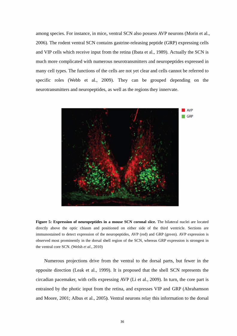
Figu
direc
imm
obse
the v
opp
circ
entr
and
ong species.
6). The rod
VIP cells w
ch more com
ny cell type
cific roles
rotransmitte
ure 5: Expres
ctly above th
munostained to
erved most pro
ventral core SC
Numerous
posite direct
cadian pacem
rained by th
Moore, 20
. For instan
dent ventral
which receiv
mplicated w
s. The funct
(Webb e
ers and neur
ssion of neur
he optic chia
o detect expre
ominently in
CN. (Welsh et
projection
tion (Leak
maker, with
he photic in
01; Albus e
ce, in mice,
SCN contai
ve input fro
with numero
tions of the
et al., 200
ropeptides,
ropeptides in
asm and pos
ession of the n
the dorsal she
t al., 2010)
s drive from
et al., 1999
h cells expre
nput from th
et al., 2005)
36
, ventral SC
ins gastrine
om the retin
ous neurotra
e cells are no
09). They
as well as t
a mouse SC
sitioned on e
neuropeptides
ell region of t
m the vent
9). It is pro
essing AVP
he retina, an
). Ventral ne
CN also poss
e-releasing p
na (Ibata et a
ansmitters a
ot yet clear
can be
the regions
CN coronal sl
either side of
s, AVP (red) a
the SCN, whe
tral to the d
oposed that
P (Li et al.,
nd expresse
eurons relay
sess AVP ne
peptide (GR
al., 1989). A
and neurope
and cells ca
grouped d
they innerv
lice. The bilat
f the third v
and GRP (gre
ereas GRP exp
dorsal parts
the shell S
2009). In tu
es VIP and
y this inform
eurons (Mo
RP) expressi
Actually the
eptides expr
annot be ref
depending
vate.
teral nuclei ar
ventricle. Sec
een). AVP exp
pression is str
s, but fewe
SCN repres
urn, the cor
GRP (Abra
mation to th
rin et al.,
ing cells
e SCN is
ressed in
ferred to
on the
re located
ctions are
pression is
rongest in
er in the
sents the
re part is
ahamson
he dorsal
37
region using VIP, GABA and GRP. In addition, expression of Per1 gene seems to start in
the dorsal SCN and to progressively spread to the ventral SCN (Yamaguchi et al., 2003).
Amplitude of rhythms in gene expression and neural activity in ventral and dorsal
neurons are relatively low and high respectively. Low amplitude rhythms may be easier to
reset to perturbations, which is supported by mathematical modeling studies (Colwell,
2011). The complexity of the SCN also exists along the other axes, both anatomically and
regarding produced peptides (Evans et al., 2011).
Another theory believes that two groups of oscillators in the SCN are identified as
activated at dawn and dusk respectively, and named morning and evening oscillators
(Daan et al., 2001). Horizontal slices of the hamster SCN show two peaks of neuronal
activity, which support this binary oscillator’s configuration (Jagota et al., 2000). Exposed
to a phase-shifting agent, these two oscillators respond differently, depending on the time
of the day. These morning and evening oscillators alter the phase under different day
lengths, helping the animals to adapt to seasonal changes. Studies at the molecular and
electrophysiological levels show that two or more types of cell populations in the SCN
can respond respectively to short and long photoperiods (Meijer et al., 2010). Recently,
different periods were observed in two regions of the SCN - a region with periods shorter
than 24 h and another with periods longer than 24 h. The short-period region is located in
the smaller medial region of the dorsal SCN, whereas the long-period region occupies the
remaining larger region(Koinuma et al., 2013). Photoperiod duration and light-dark
transitions show region-specific influence on entrainment of Per1 and Per2 expression in
subdivisions of caudal and rostral SCN (Sosniyenko et al., 2009). However, further
studies remain to be performed to trace the different oscillators to specific SCN neurons
and to clarify the mechanisms of morning and evening oscillators.
At cellular level, the SCN could be defined as an ensemble of neurons harboring cell
autonomous oscillators. Single neurons within the SCN exhibit a wide range of periods
(22 h ~ 30 h) and phases in rhythms of firing rate and gene expression (Ko et al., 2010).
This heterogeneity is also observed when dissociated SCN cells are dispersed in low
density culture (Welsh et al., 1995). Synchrony is shown when dispersed neurons are
cultured at a high density (Aton et al., 2005). To maintain synchrony within SCN

38
neuronal populations, intercellular coupling is crucial towards a robust, precise and stable
SCN rhythm. Neurotransmission and gap junctions are important in the intercellular
communication (Shirakawa et al., 2000; Rash et al., 2007; Ko et al., 2010). Interestingly,
SCN resistance to entrainment by temperature cycles, which is a powerful cue for many
species, depends on the intercellular coupling of neurons (Underwood and Calaban, 1987;
Yoshida et al., 2009; Abraham et al., 2010; Buhr et al., 2010). Even absence of cell
autonomous rhythms does not lead to loss of noise-induced oscillations in SCN explants
of Bmal1 mutant mice, which requires intercellular coupling (Ko et al., 2010). Generally,
the mechanisms and dynamics of the coupling network in the SCN still remain largely
undefined.
Synapses and gap junctions are two coupling mechanisms that get involved in the
synchronization of SCN neurons. Neuronal connections are involved in the coupling
within the SCN by neuronal firing and chemical synapses. Blocking action potentials with
TTX leads to asynchrony of SCN neurons in slice culture or high-density dispersed
culture (Honma et al., 2000; Yamaguchi et al., 2003). Gap junctions could also be
involved in SCN coupling because short-term firing synchrony can be found in SCN
slices with synaptic transmission blocked by using calcium-free medium (Bouskila and
Dudek, 1993). In SCN slices from wild-type mice, the gap junction blockers octanol or
halothane destroy circadian rhythms of neuronal firing or neuropeptide release (Shinohara
et al., 2000).
3.3. Photic input: light information as a major external signal for the clock
The retina provides the major photic input through the retinohypothalamic tract
(RHT) directly into the SCN and indirectly, via the IGL. In the retina, there exists three
main types of photoreceptor cells as rods, cones and ipRGCs (intrinsically photosensitive
retinal ganglion cells) (Fig. 6). Rods and cones are photoreceptors first discovered to be
involved in image-forming vision, whereas the ipRGCs, a subset (~1%) retina ganglion
cells, were identified recently (Berson et al., 2002). And ipRGCs contain the
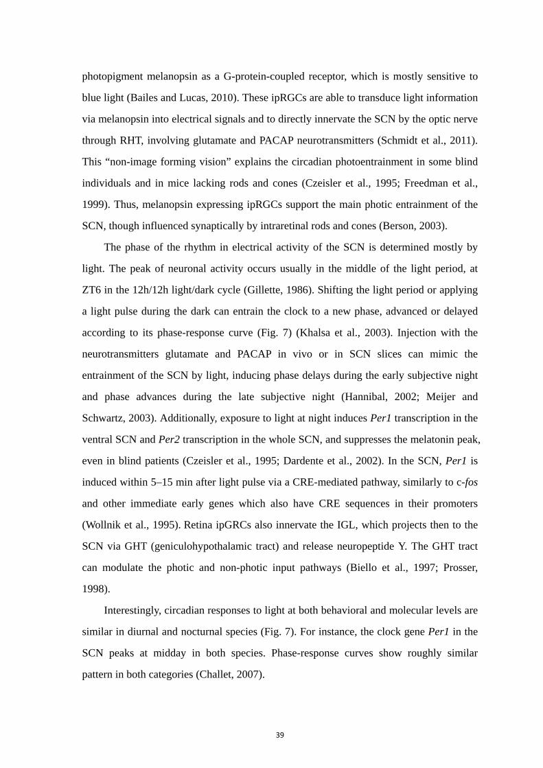
39
photopigment melanopsin as a G-protein-coupled receptor, which is mostly sensitive to
blue light (Bailes and Lucas, 2010). These ipRGCs are able to transduce light information
via melanopsin into electrical signals and to directly innervate the SCN by the optic nerve
through RHT, involving glutamate and PACAP neurotransmitters (Schmidt et al., 2011).
This “non-image forming vision” explains the circadian photoentrainment in some blind
individuals and in mice lacking rods and cones (Czeisler et al., 1995; Freedman et al.,
1999). Thus, melanopsin expressing ipRGCs support the main photic entrainment of the
SCN, though influenced synaptically by intraretinal rods and cones (Berson, 2003).
The phase of the rhythm in electrical activity of the SCN is determined mostly by
light. The peak of neuronal activity occurs usually in the middle of the light period, at
ZT6 in the 12h/12h light/dark cycle (Gillette, 1986). Shifting the light period or applying
a light pulse during the dark can entrain the clock to a new phase, advanced or delayed
according to its phase-response curve (Fig. 7) (Khalsa et al., 2003). Injection with the
neurotransmitters glutamate and PACAP in vivo or in SCN slices can mimic the
entrainment of the SCN by light, inducing phase delays during the early subjective night
and phase advances during the late subjective night (Hannibal, 2002; Meijer and
Schwartz, 2003). Additionally, exposure to light at night induces Per1 transcription in the
ventral SCN and Per2 transcription in the whole SCN, and suppresses the melatonin peak,
even in blind patients (Czeisler et al., 1995; Dardente et al., 2002). In the SCN, Per1 is
induced within 5–15 min after light pulse via a CRE-mediated pathway, similarly to c-fos
and other immediate early genes which also have CRE sequences in their promoters
(Wollnik et al., 1995). Retina ipGRCs also innervate the IGL, which projects then to the
SCN via GHT (geniculohypothalamic tract) and release neuropeptide Y. The GHT tract
can modulate the photic and non-photic input pathways (Biello et al., 1997; Prosser,
1998).
Interestingly, circadian responses to light at both behavioral and molecular levels are
similar in diurnal and nocturnal species (Fig. 7). For instance, the clock gene Per1 in the
SCN peaks at midday in both species. Phase-response curves show roughly similar
pattern in both categories (Challet, 2007).

40
Figure 6: The photic input to the SCN. The melanopsin positive ipRGC cells (red) located in the retina
and dispersed among other ganglion cells (G) deliver photic information to the SCN in the hypothalamus
via the retinohypothalamic tract (RHT). The SCN can be divided into two anatomical and functional
subdivisions: a ventral core and a dorsal shell. SCN core neurons are thought to integrate external inputs,
including light from the retina. Rods (R) and cones (C), which are located in the outer part of the retina, also
contribute to photoentrainment, via synaptic transmission. (Adapted from Cowell 2011; Reppert and
Weaver 2002)
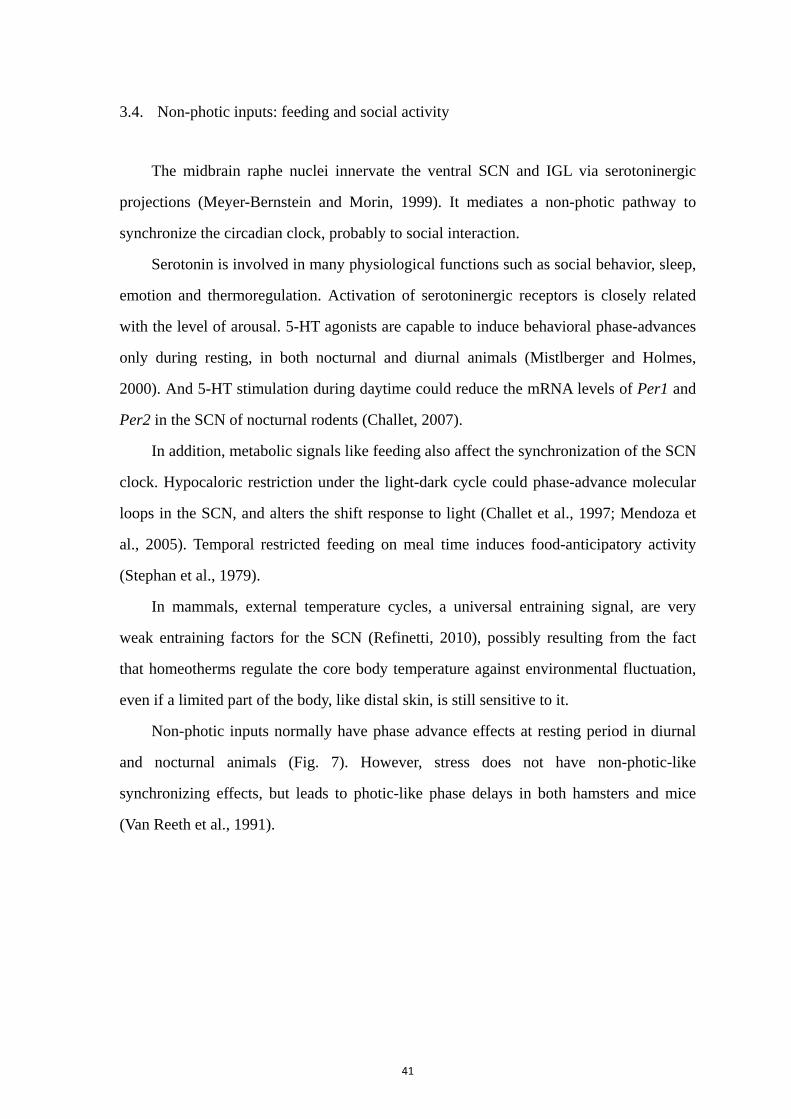
41
3.4. Non-photic inputs: feeding and social activity
The midbrain raphe nuclei innervate the ventral SCN and IGL via serotoninergic
projections (Meyer-Bernstein and Morin, 1999). It mediates a non-photic pathway to
synchronize the circadian clock, probably to social interaction.
Serotonin is involved in many physiological functions such as social behavior, sleep,
emotion and thermoregulation. Activation of serotoninergic receptors is closely related
with the level of arousal. 5-HT agonists are capable to induce behavioral phase-advances
only during resting, in both nocturnal and diurnal animals (Mistlberger and Holmes,
2000). And 5-HT stimulation during daytime could reduce the mRNA levels of Per1 and
Per2 in the SCN of nocturnal rodents (Challet, 2007).
In addition, metabolic signals like feeding also affect the synchronization of the SCN
clock. Hypocaloric restriction under the light-dark cycle could phase-advance molecular
loops in the SCN, and alters the shift response to light (Challet et al., 1997; Mendoza et
al., 2005). Temporal restricted feeding on meal time induces food-anticipatory activity
(Stephan et al., 1979).
In mammals, external temperature cycles, a universal entraining signal, are very
weak entraining factors for the SCN (Refinetti, 2010), possibly resulting from the fact
that homeotherms regulate the core body temperature against environmental fluctuation,
even if a limited part of the body, like distal skin, is still sensitive to it.
Non-photic inputs normally have phase advance effects at resting period in diurnal
and nocturnal animals (Fig. 7). However, stress does not have non-photic-like
synchronizing effects, but leads to photic-like phase delays in both hamsters and mice
(Van Reeth et al., 1991).
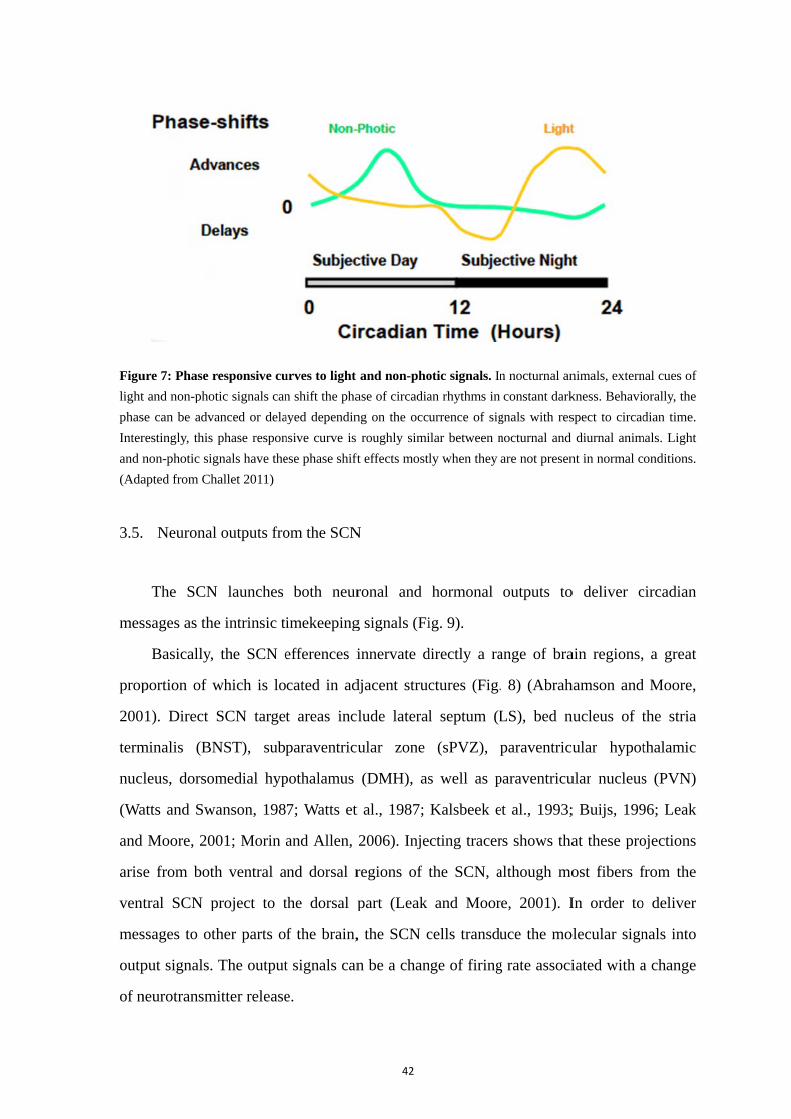
Figu
light
phas
Inter
and n
(Ada
3.5.
mes
prop
200
term
nucl
(Wa
and
aris
vent
mes
outp
of n
ure 7: Phase r
t and non-pho
se can be adv
restingly, this
non-photic sig
apted from Ch
Neuronal
The SCN
ssages as the
Basically,
portion of w
1). Direct
minalis (BN
leus, dorso
atts and Sw
Moore, 20
e from both
tral SCN p
ssages to ot
put signals.
neurotransm
responsive cu
tic signals can
anced or dela
phase respon
gnals have the
hallet 2011)
l outputs fro
N launches
e intrinsic ti
the SCN e
which is lo
SCN target
NST), subp
medial hyp
wanson, 198
001; Morin
h ventral a
project to t
ther parts o
The output
mitter release
urves to light
n shift the pha
ayed dependin
nsive curve is
ese phase shift
om the SCN
both neur
imekeeping
efferences i
cated in ad
t areas inc
paraventricu
pothalamus
7; Watts et
and Allen,
and dorsal r
the dorsal p
f the brain,
t signals can
e.
42
and non-pho
ase of circadia
ng on the occu
roughly simi
ft effects mostl
N
ronal and
g signals (Fi
innervate d
djacent struc
lude lateral
ular zone
(DMH), as
t al., 1987;
2006). Inje
regions of t
part (Leak
, the SCN c
n be a chan
otic signals. In
an rhythms in
urrence of sig
ilar between n
ly when they
hormonal
ig. 9).
directly a ra
ctures (Fig.
l septum (L
(sPVZ), p
s well as p
Kalsbeek e
cting tracer
the SCN, a
and Moor
cells transdu
nge of firing
n nocturnal an
constant dark
gnals with res
nocturnal and
are not presen
outputs to
ange of bra
. 8) (Abrah
LS), bed n
paraventricu
paraventricu
et al., 1993;
rs shows tha
although mo
re, 2001). I
uce the mo
g rate associ
nimals, extern
kness. Behavi
spect to circad
diurnal anim
nt in normal c
o deliver c
ain regions,
hamson and
nucleus of t
cular hypot
ular nucleus
; Buijs, 199
at these pro
ost fibers f
In order to
olecular sign
iated with a
nal cues of
orally, the
dian time.
mals. Light
onditions.
circadian
a great
d Moore,
the stria
thalamic
s (PVN)
96; Leak
ojections
from the
o deliver
nals into
a change

Figu
Thes
(BN
(LS)
the t
targe
spon
of e
the
Kaw
dayt
in th
con
(de
a cr
SCN
expr
nece
pota
of B
200
ure 8: Efferen
se regions co
ST), dorsome
), preoptic are
thalamus (PV
eted indirectly
Neuronal
ntaneous ac
extracellular
SCN, with
wamura, 197
time in both
he absence
sequence of
Jeu et al., 1
ritical comp
N. Predom
ressed in th
essary for
assium BK
BK channel
6). Both ph
nces from the
omprise amyg
edial hypothal
ea (POA), par
VT) and subpa
y to control ho
firing as an
ction potent
r electrodes
h high activ
79). Interes
h nocturnal
of external
f a change i
1998). Rhyt
ponent of S
minantly, th
he SCN, w
the rhythm
channels ar
l in the SCN
harmacologi
e SCN (red) to
gdala (AMY),
lamus (DMH)
aventricular n
araventricular
ormonal outpu
n SCN outpu
tial frequen
recordings
vity during
tingly, the f
and diurnal
cues (Gillet
in the mem
thmic SCN
SCN output
he fast del
with its exp
mic activity
re rhythmic
N is control
ical blockad
43
o hypothalam
, arcuate nuc
), habenula (H
nucleus of the
zone (sPVZ)
uts. (Adapted f
ut is based
ncy (Kuhlma
reveals circ
the day an
firing rate o
al animals. T
tte, 1986). T
mbrane poten
electrical a
ts. Several
layed-rectifi
pression dow
(Itri et al
cally expres
lled by the
de of BK cu
mic (yellow) a
leus (ARC),
HB), intergen
e hypothalamu
). Pineal gland
from Dibner e
on a cell au
an and McM
cadian rhyth
nd low at n
of SCN neur
The rhythms
The oscillat
ntial and inp
activity with
ion channe
fier potassiu
wn-regulate
., 2005). F
ssed in the S
intrinsic cir
urrent and g
and thalamic
bed nucleus
niculate leaflet
us (PVN), par
d (Pin) and p
et al., 2010)
utonomous c
Mahon, 200
hms in the e
night (see a
rons peaks i
s in electric
ions in the f
put resistan
h several ion
ls have bee
um channe
ed during t
Further, the
SCN, and th
rcadian cloc
enetic knoc
(green) brain
of the stria t
t (IGL), later
raventricular n
pituitary gland
circadian rh
06). In vivo
electrical ac
above) (Ino
in the midd
cal activity c
firing rate m
nce of SCN
n channels
en identifie
el is rhyth
the night, w
calcium re
he daily exp
ck (Meredit
ckout of the
n regions.
terminalis
al septum
nucleus of
d (Pit) are
hythm in
o the use
ctivity in
uye and
dle of the
continue
may be a
neurons
is likely
d in the
hmically
which is
egulated
pression
th et al.,
channel
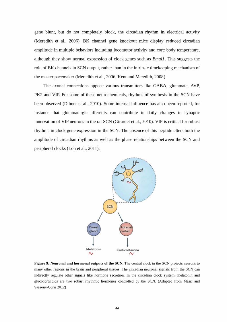
gen
(Me
amp
alth
role
the
PK2
been
inst
inne
rhyt
amp
peri
Figu
many
indir
gluco
Sass
e blunt, bu
eredith et a
plitude in m
hough they
e of BK cha
master pace
The axon
2 and VIP.
n observed
ance that
ervation of V
thms in cloc
plitude of c
ipheral cloc
ure 9: Neuron
y other region
rectly regulate
ocorticoids ar
one-Corsi 201
ut do not c
al., 2006).
multiple beh
show norm
annels in SC
emaker (Me
al connecti
For some o
(Dibner et
glutamater
VIP neuron
ck gene exp
ircadian rhy
ks (Loh et a
nal and horm
ns in the brai
e other signa
re two robus
12)
completely
BK chann
haviors inclu
mal expressio
CN output, r
eredith et al
ons oppose
of these neu
al., 2010).
gic afferen
ns in the rat
pression in
ythms as w
al., 2011).
monal outputs
n and periphe
als like horm
st rhythmic h
44
block, the
nel gene kn
uding locom
on of clock
rather than i
., 2006; Ken
e various tr
urochemical
Some inter
nts can co
SCN (Girar
the SCN. T
well as the p
s of the SCN.
eral tissues. T
mone secretion
hormones con
e circadian
nockout mi
motor activi
k genes such
in the intrin
nt and Mere
ransmitters
ls, rhythms
rnal influen
ontribute to
rdet et al., 2
The absence
phase relatio
. The central c
The circadian
n. In the circ
ntrolled by th
rhythm in
ce display
ity and core
h as Bmal1
sic timekee
edith, 2008)
like GABA
of synthesi
nce has also
o daily ch
2010). VIP i
e of this pep
onships bet
clock in the SC
neuronal sign
adian clock s
he SCN. (Ada
n electrical
reduced c
e body temp
1. This sugg
eping mecha
).
A, glutamat
is in the SC
o been repo
hanges in
is critical fo
ptide alters
tween the S
CN projects n
nals from the
system, mela
apted from M
activity
circadian
perature,
gests the
anism of
te, AVP,
CN have
rted, for
synaptic
or robust
both the
SCN and
neurons to
SCN can
tonin and
Masri and
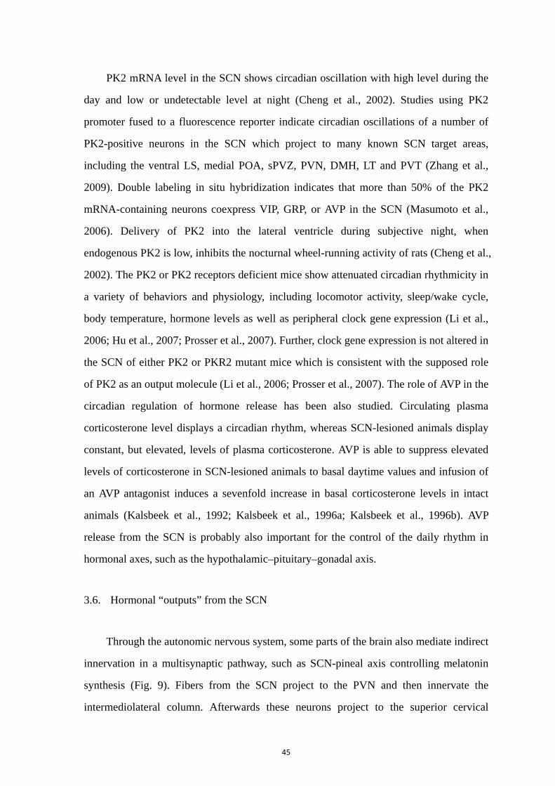
45
PK2 mRNA level in the SCN shows circadian oscillation with high level during the
day and low or undetectable level at night (Cheng et al., 2002). Studies using PK2
promoter fused to a fluorescence reporter indicate circadian oscillations of a number of
PK2-positive neurons in the SCN which project to many known SCN target areas,
including the ventral LS, medial POA, sPVZ, PVN, DMH, LT and PVT (Zhang et al.,
2009). Double labeling in situ hybridization indicates that more than 50% of the PK2
mRNA-containing neurons coexpress VIP, GRP, or AVP in the SCN (Masumoto et al.,
2006). Delivery of PK2 into the lateral ventricle during subjective night, when
endogenous PK2 is low, inhibits the nocturnal wheel-running activity of rats (Cheng et al.,
2002). The PK2 or PK2 receptors deficient mice show attenuated circadian rhythmicity in
a variety of behaviors and physiology, including locomotor activity, sleep/wake cycle,
body temperature, hormone levels as well as peripheral clock gene expression (Li et al.,
2006; Hu et al., 2007; Prosser et al., 2007). Further, clock gene expression is not altered in
the SCN of either PK2 or PKR2 mutant mice which is consistent with the supposed role
of PK2 as an output molecule (Li et al., 2006; Prosser et al., 2007). The role of AVP in the
circadian regulation of hormone release has been also studied. Circulating plasma
corticosterone level displays a circadian rhythm, whereas SCN-lesioned animals display
constant, but elevated, levels of plasma corticosterone. AVP is able to suppress elevated
levels of corticosterone in SCN-lesioned animals to basal daytime values and infusion of
an AVP antagonist induces a sevenfold increase in basal corticosterone levels in intact
animals (Kalsbeek et al., 1992; Kalsbeek et al., 1996a; Kalsbeek et al., 1996b). AVP
release from the SCN is probably also important for the control of the daily rhythm in
hormonal axes, such as the hypothalamic–pituitary–gonadal axis.
3.6. Hormonal “outputs” from the SCN
Through the autonomic nervous system, some parts of the brain also mediate indirect
innervation in a multisynaptic pathway, such as SCN-pineal axis controlling melatonin
synthesis (Fig. 9). Fibers from the SCN project to the PVN and then innervate the
intermediolateral column. Afterwards these neurons project to the superior cervical

46
ganglions that finally innervate the pineal gland, which releases the melatonin depending
on the neuronal activity of the SCN (Fig. 8) (Moore and Klein, 1974; Perreau-Lenz et al.,
2003). Another projection of the SCN, via PVN to IML, determines daily changes in
sensitivity of the adrenal gland to ACTH, while the SCN affect adrenal hormone secretion
by the classical neuroendocrine control via ACTH from the pituitary (Fig. 8) (Buijs et al.,
1999).
Moreover, the SCN transplantation can restore circadian behavioral rhythms by
implanting SCN into several brain regions (Silver et al., 1996). It has been shown that,
though fibers from the dorsal SCN project to the PVN, the PVN rhythmicity dependent
on the activity of SCN can be driven without synaptic inputs (Tousson and Meissl, 2004).
These results implicate a role of unknown paracrine factors from the SCN tissue instead
of nervous transmission. Yet, SCN grafts cannot restore the endocrine rhythms,
confirming that rhythms of hormones like melatonin and glucocorticoids are under
control of SCN neuronal outputs.
Melatonin is widely distributed in nature with functional activities in unicellular
organisms, plants, fungi and animals. In most vertebrates, melatonin is synthesized
primarily in the pineal gland at night, independently of opposite locomotor activity
pattern in nocturnal and diurnal mammals (Fig. 10). Given the fact that melatonin is not
stored in the pineal cells and immediately released into the general circulation, the plasma
level of melatonin precisely reflects its pineal synthesis. Melatonin secretion is only
influenced by the light/dark cycle, and thus could be regarded as a reliable estimate of the
SCN clock functioning. Melatonin can be also extracted from seeds and leaves of a
number of plants with concentrations tremendously higher than its night level in human
plasma. When chicks are fed melatonin-rich food, serum melatonin level increases
(Hattori et al., 1995). Circulating melatonin is metabolized mainly in the liver by
cytochrome P450. And melatonin half life is short with a first distribution of 2 min and a
second metabolic of 20 min in human (Claustrat et al., 2005). The duration of the
nocturnal peak of melatonin secretion also reflects the length of the night. Therefore,
nocturnal melatonin is an output signal of the central clock, while melatonin receptors are
expressed in many organs including SCN.
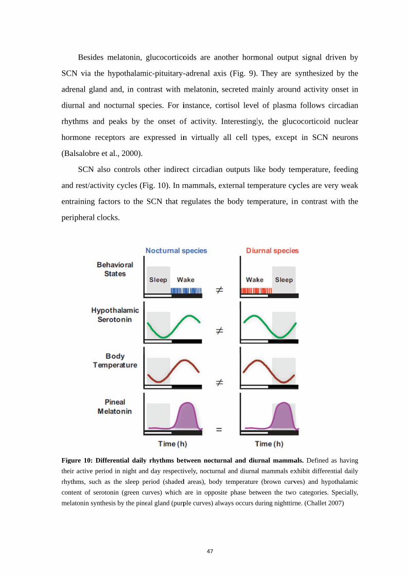
SCN
adre
diur
rhyt
horm
(Bal
and
entr
peri
Figu
their
rhyth
conte
mela
Besides m
N via the h
enal gland
rnal and no
thms and p
mone recep
lsalobre et a
SCN also
rest/activit
raining fact
ipheral cloc
ure 10: Differ
r active period
hms, such as
ent of seroton
atonin synthes
melatonin, g
hypothalami
and, in con
octurnal spe
peaks by th
ptors are e
al., 2000).
controls o
ty cycles (Fi
tors to the S
ks.
rential daily
d in night and
the sleep pe
nin (green cu
sis by the pine
glucocortico
ic-pituitary-
ntrast with m
ecies. For i
he onset of
xpressed in
other indirec
ig. 10). In m
SCN that r
rhythms bet
d day respecti
eriod (shaded
urves) which
eal gland (purp
47
oids are an
-adrenal ax
melatonin,
instance, co
f activity.
n virtually
ct circadian
mammals, e
regulates th
tween noctur
ively, nocturn
d areas), body
are in oppos
ple curves) alw
nother horm
xis (Fig. 9).
secreted m
ortisol level
Interestingl
all cell ty
n outputs li
external tem
e body tem
rnal and diu
al and diurna
y temperature
ite phase betw
ways occurs d
monal outpu
They are
mainly aroun
l of plasma
ly, the gluc
ypes, excep
ike body te
mperature cy
mperature, in
urnal mamma
l mammals ex
e (brown curv
ween the two
during nighttim
ut signal dr
synthesized
nd activity
a follows c
cocorticoid
pt in SCN
emperature,
ycles are ve
n contrast w
als. Defined
xhibit differen
ves) and hyp
o categories.
me. (Challet 2
riven by
d by the
onset in
circadian
nuclear
neurons
feeding
ery weak
with the
as having
ntial daily
othalamic
Specially,
2007)
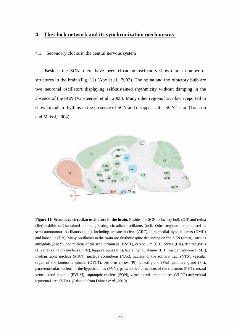
4.
4.1.
stru
two
abse
show
and
Figu
(Ret)
semi
and h
amyg
(DG
medi
organ
parav
ventr
tegm
The cloc
Secondar
Besides th
uctures in th
o neuronal
ence of the
w circadian
Meissl, 200
ure 11: Secon
) exhibit self
i-autonomous
habenula (HB
gdala (AMY)
G), dorsal raph
ian raphe nuc
an of the lam
ventricular nu
rolateral medu
mental area (V
k networ
ry clocks in
he SCN, th
he brain (Fi
oscillators
SCN (Vans
n rhythms in
04).
dary circadia
f-sustained an
oscillators (b
B). Many oscil
, bed nucleus
e nucleus (DR
cleus (MRN)
mina terminali
ucleus of the h
ulla (RVLM),
VTA). (Adapted
k and its
the central
here have
g. 11) (Abe
displaying
steensel et a
n the presen
an oscillators
nd long-lastin
blue), includin
llators in the b
of the stria te
RN), hippocam
, nucleus acc
s (OVLT), p
hypothalamus
, supraoptic n
d from Dibner
48
synchron
nervous sy
been circa
e et al., 200
self-sustain
al., 2008). M
nce of SCN
s in the brain
ng circadian o
ng arcuate nu
brain are rhyth
erminalis (BN
mpus (Hip), la
cumbens (NA
iriform cortex
s (PVN), parav
nucleus (SON
r et al., 2010)
nization m
ystem
adian oscill
02). The ret
ned rhythm
Many other
and disapp
. Besides the
oscillators (re
ucleus (ARC),
hmic quite dep
NST), cerebellu
ateral hypotha
Ac), nucleus o
x (Pi), pineal
ventricular nu
), ventrolatera
mechanism
ators show
tina and the
micity witho
regions hav
pear after SC
SCN, olfactor
ed). Other reg
dorsomedial
pending on th
um (CB), cort
alamus (LH), m
of the solitary
l gland (Pin)
ucleus of the t
al preoptic are
ms
wn in a num
e olfactory b
out damping
ve been rep
CN lesion (
ry bulb (OB)
gions are pro
l hypothalamu
he SCN (green
tex (CX), den
median emine
y tract (NTS)
), pituitary gl
thalamus (PVT
ea (VLPO) an
mber of
bulb are
g in the
ported to
Tousson
and retina
oposed as
us (DMH)
n), such as
ntate gyrus
ence (ME),
, vascular
and (Pit),
T), rostral
nd ventral
49
4.2. Peripheral circadian clocks
With the central clock in the SCN, the circadian timing system is composed of
multiple oscillators, in a hierarchical manner. Out of the brain, peripheral oscillators are
orchestrated by the SCN to appropriate phases, in order to control the numerous
physiological functions, even if they are cell-autonomous and self-sustained. Since the
finding that rat-1 immortalized fibroblasts display circadian gene expression after serum
shock, many peripheral cells and tissues have been shown to display circadian rhythms in
gene expression in mammals (Fig.12) (Balsalobre et al., 1998; Yagita et al., 2001). They
possess similar molecular clockwork to that operative in the SCN. However, the clear
rhythms quickly fade within a few days in cultures of these peripheral oscillators,
contrarily to the SCN and the retina. In addition, single cell recordings of circadian gene
expression prove that individual cells are self-sustained oscillators and able to be
synchronized (Nagoshi et al., 2004; Welsh et al., 2004). These results indicate that
peripheral oscillators harbor cell-autonomous and self-sustained clocks. Furthermore,
damped oscillations of cell cultures could be restarted by stimuli like serum shock or
medium change (Yamazaki et al., 2000). Given the fact that the peripheral clocks in mice
with SCN ablation are no longer well-coordinated and display large phase differences
among individuals, fainting of peripheral oscillations is rather due to the loss of
synchrony between cell-autonomous clocks in the absence of the SCN (Yoo et al., 2004).
This is in the opposition of SCN slices that keep synchrony in cultures. Altogether, this
suggests that the SCN-emanating signals are necessary for synchronizing the peripheral
clocks, for the phase coherence among cells and tissues.
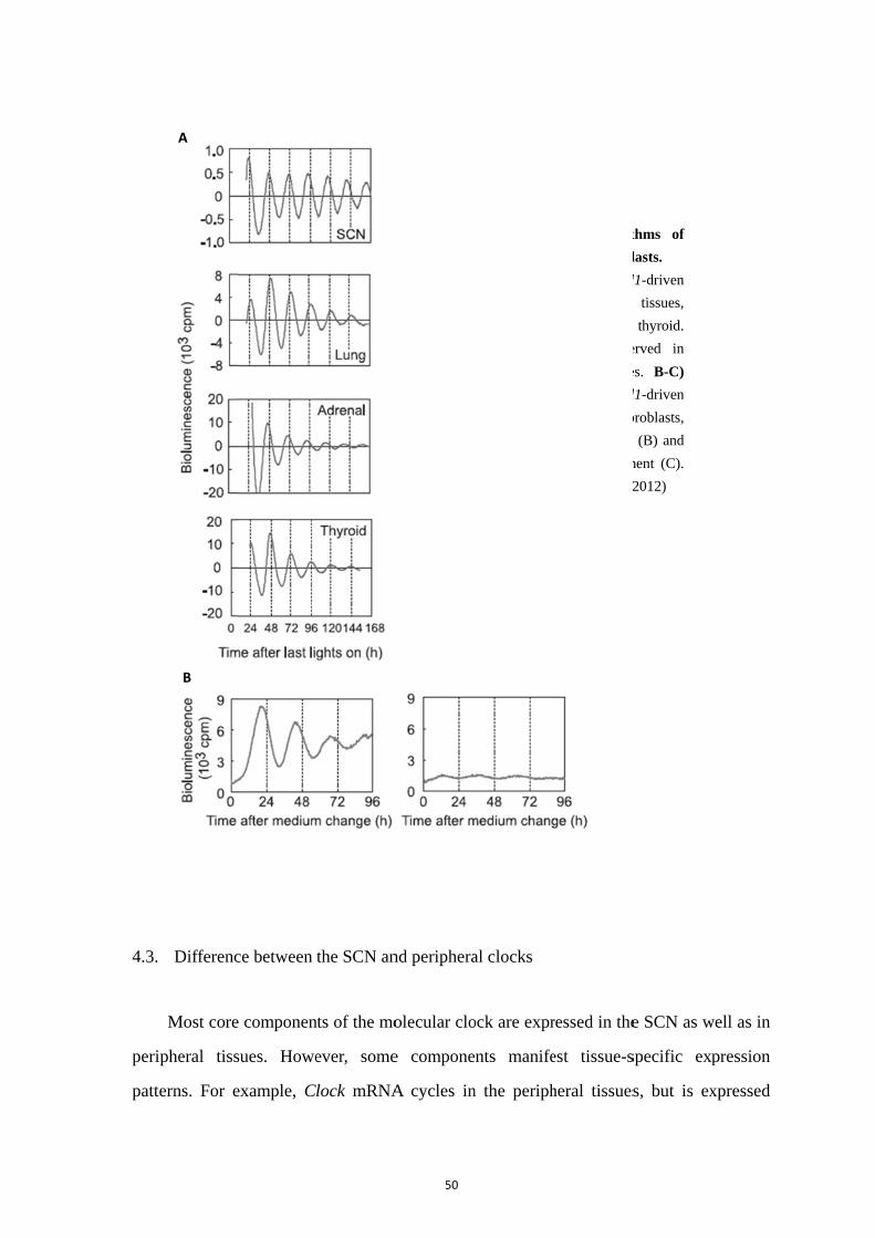
4.3.
peri
patt
Differenc
Most core
ipheral tiss
terns. For e
A
B
ce between
e componen
sues. Howe
example, C
the SCN an
nts of the mo
ever, some
lock mRNA
C
50
nd periphera
olecular clo
e compone
A cycles in
Figu
peri
A)
biolu
SCN
Circ
cent
The
biolu
with
with
(Ada
al clocks
ock are expr
ents manife
n the periph
ure 12: Cir
ipheral tissue
The recordin
uminescence
N, lung, adren
cadian rhythm
ral and peri
recordings
uminescence
h dexamethaso
hout dexameth
apted from No
ressed in the
est tissue-s
heral tissues
cadian rhyt
es and fibrobl
ngs of Bmal
of mouse
nal gland and
ms are obse
pheral tissue
of Bmal
of Rat-1 fib
one treatment
hasone treatm
oguchi et al., 2
e SCN as w
specific exp
s, but is ex
thms of
lasts.
l1-driven
tissues,
thyroid.
erved in
es. B-C)
l1-driven
broblasts,
t (B) and
ment (C).
2012)
well as in
pression
xpressed
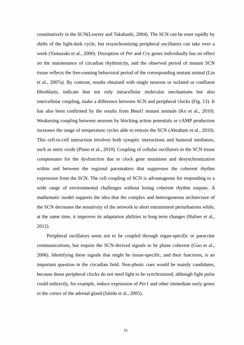
51
constitutively in the SCN(Lowrey and Takahashi, 2004). The SCN can be reset rapidly by
shifts of the light-dark cycle, but resynchronizing peripheral oscillators can take over a
week (Yamazaki et al., 2000). Disruption of Per and Cry genes individually has no effect
on the maintenance of circadian rhythmicity, and the observed period of mutant SCN
tissue reflects the free-running behavioral period of the corresponding mutant animal (Liu
et al., 2007a). By contrast, results obtained with single neurons or isolated or confluent
fibroblasts, indicate that not only intracellular molecular mechanisms but also
intercellular coupling, make a difference between SCN and peripheral clocks (Fig. 13). It
has also been confirmed by the results from Bmal1 mutant animals (Ko et al., 2010).
Weakening coupling between neurons by blocking action potentials or cAMP production
increases the range of temperature cycles able to entrain the SCN (Abraham et al., 2010).
This cell-to-cell interaction involves both synaptic interactions and humoral mediators,
such as nitric oxide (Plano et al., 2010). Coupling of cellular oscillators in the SCN tissue
compensates for the dysfunction due to clock gene mutations and desynchronization
within and between the regional pacemakers that suppresses the coherent rhythm
expression from the SCN. The cell coupling of SCN is advantageous for responding to a
wide range of environmental challenges without losing coherent rhythm outputs. A
mathematic model supports the idea that the complex and heterogeneous architecture of
the SCN decreases the sensitivity of the network to short entrainment perturbations while,
at the same time, it improves its adaptation abilities to long term changes (Hafner et al.,
2012).
Peripheral oscillators seem not to be coupled through organ-specific or paracrine
communications, but require the SCN-derived signals to be phase coherent (Guo et al.,
2006). Identifying these signals that might be tissue-specific, and their functions, is an
important question in the circadian field. Non-photic cues would be mainly candidates,
because those peripheral clocks do not need light to be synchronized, although light pulse
could indirectly, for example, induce expression of Per1 and other immediate early genes
in the cortex of the adrenal gland (Ishida et al., 2005).
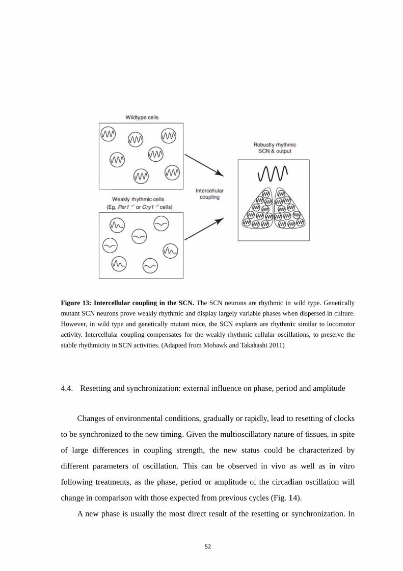
Figu
muta
How
activ
stabl
4.4.
to b
of l
diffe
follo
chan
ure 13: Interc
ant SCN neuro
wever, in wild
vity. Intercellu
le rhythmicity
Resetting
Changes o
be synchroni
large differ
ferent param
owing treat
nge in comp
A new ph
cellular coup
ons prove wea
d type and gen
ular coupling
y in SCN activ
g and synch
of environm
ized to the n
rences in c
meters of o
tments, as t
parison with
hase is usua
pling in the S
akly rhythmic
netically muta
compensates
vities. (Adapte
ronization:
mental condi
new timing
coupling str
oscillation.
the phase, p
h those expe
lly the mos
52
SCN. The SCN
c and display
ant mice, the
for the weak
ed from Moha
external inf
itions, gradu
. Given the
rength, the
This can b
period or am
ected from
st direct resu
N neurons are
largely variab
SCN explant
kly rhythmic
awk and Takah
fluence on p
ually or rap
multioscill
e new statu
be observed
mplitude of
previous cy
ult of the re
e rhythmic in
ble phases whe
s are rhythmi
cellular oscill
hashi 2011)
phase, perio
idly, lead to
atory nature
us could be
d in vivo a
f the circad
ycles (Fig. 1
esetting or s
wild type. G
en dispersed i
ic similar to l
lations, to pre
od and ampl
o resetting o
e of tissues,
e character
as well as
dian oscillat
14).
synchroniza
Genetically
in culture.
locomotor
eserve the
litude
of clocks
, in spite
rized by
in vitro
tion will
ation. In
53
the actogram of locomotor activity, this parameter is simply shown by the start of the
wheel running. In oscillations of cumulative quantities, like firing rate, mRNA level or
bioluminescence, intrinsic time of the biological oscillator can be determined by the peak,
shifting either advanced or delayed. Since the shift of the peak is due to the majority of
the population, oscillators could be classified as simultaneous or not in response to the
external cues. For instance, bimodal oscillation in resetting of SCN neurons suggests the
existence of a rapid and more efficiently synchronized subpopulation further responsible
for shifting the whole pacemaker (Rohling et al., 2011).
Another important parameter is the period of the oscillation, which defines the
circadian rhythms. Although it stays close to 24 hours in the presence of external cues in
nature, the period of circadian oscillations can significantly differ under constant
conditions or other periodical entrainment. The period could be calculated by the distance
between two neighboring peaks. Normally, the free-running periods or entrained periods
vary in the circadian range, from 20 hours to 28 hours. But it has been reported that some
extremely short or long periodic entrainments, like temperature cycles from 10 hours to
30 hours, are capable to synchronize the fibroblasts to oscillate following such
environmental periods (Saini et al., 2012).
Besides direct timing parameters such as phase and period, the strength parameter of
the amplitude reflects the phenotype of the population, in the precision and integration of
the circadian clocks. The amplitude can be determined by the distance between
consecutive trough and peak. The bigger the amplitude, the better the oscillators are
synchronized to a unique phase. At the same time, the width of the peak could also justify
the synchronized level as well as the precision and phase plasticity of intrinsic clocks. In
peripheral oscillators, the amplitudes of populations damp rapidly in a few days. Damping
rates imply the level of variation in intrinsic periods and the intercellular asynchrony,
especially among very weakly-coupled peripheral clocks.
In general, the phase and the period are prone to show the intrinsic properties of
clocks, whilst the amplitude rather reflects the synchronization of clocks.
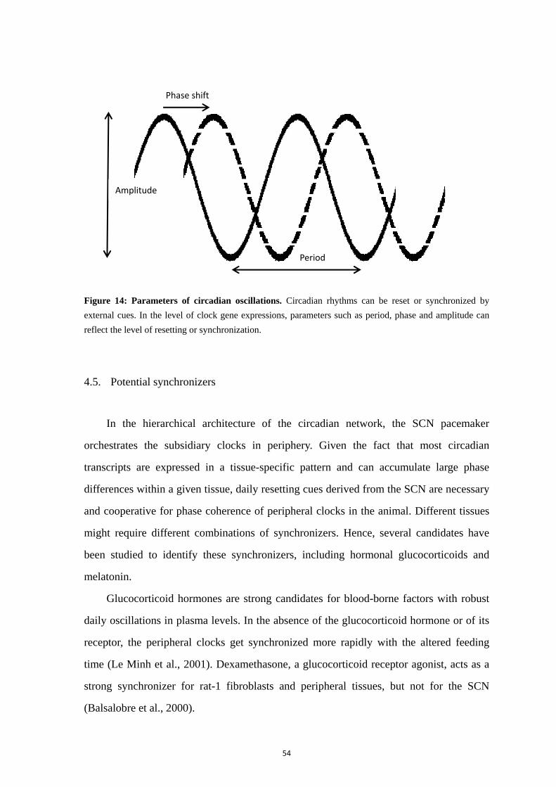
54
Figure 14: Parameters of circadian oscillations. Circadian rhythms can be reset or synchronized by
external cues. In the level of clock gene expressions, parameters such as period, phase and amplitude can
reflect the level of resetting or synchronization.
4.5. Potential synchronizers
In the hierarchical architecture of the circadian network, the SCN pacemaker
orchestrates the subsidiary clocks in periphery. Given the fact that most circadian
transcripts are expressed in a tissue-specific pattern and can accumulate large phase
differences within a given tissue, daily resetting cues derived from the SCN are necessary
and cooperative for phase coherence of peripheral clocks in the animal. Different tissues
might require different combinations of synchronizers. Hence, several candidates have
been studied to identify these synchronizers, including hormonal glucocorticoids and
melatonin.
Glucocorticoid hormones are strong candidates for blood-borne factors with robust
daily oscillations in plasma levels. In the absence of the glucocorticoid hormone or of its
receptor, the peripheral clocks get synchronized more rapidly with the altered feeding
time (Le Minh et al., 2001). Dexamethasone, a glucocorticoid receptor agonist, acts as a
strong synchronizer for rat-1 fibroblasts and peripheral tissues, but not for the SCN
(Balsalobre et al., 2000).
Amplitude
Phase shift
Period
55
Melatonin is another strong output of the SCN pacemaker with robust daily rhythms.
Exogenous melatonin can induce behavioral phase-advances when injected at subjective
dusk with pharmacological dose (Pitrosky et al., 1999; Slotten et al., 2002). Since
high-affinity melatonin receptors MT1 and MT2 are expressed in various peripheral
tissues besides the SCN, melatonin is a potential candidate as a synchronizer of peripheral
clocks.
Temporal restricted feeding with diurnal meal time in nocturnal mice proves that
feeding could be a potent synchronizer of peripheral oscillators, especially in the liver,
where even core clock genes such as Bmal1, Per1 and Per2 are not essential (Damiola et
al., 2000; Feillet et al., 2006; Pendergast et al., 2009b).
Another circadian cue regulated by the SCN is the core body temperature with a
fluctuation of 1-4 °C (Refinetti and Menaker, 1992). Simulated body temperature
fluctuations can entrain the peripheral tissues like lung and cell cultures like rat-1
fibroblasts and NIH3T3 fibroblasts (Brown et al., 2002; Buhr et al., 2010; Saini et al.,
2012). And exposure to inverted environmental temperature cycles can reverse circadian
gene expression in the liver without affecting the central clock (Brown et al., 2002). This
SCN resistance to entrainment by temperature cycles depends on the intercellular
coupling of SCN neurons (Abraham et al., 2010; Buhr et al., 2010).
4.6. Circadian network throughout life
4.6.1. Postnatal development
The clock network undergoes remarkable transformations of exogenous and
endogenous environments at perinatal periods, in altricial species like humans and rats.
Within this course, circadian oscillators may change during development to maintain
adaptive phase relationships with important aspects of the drastically changing
environments. During the short period of postnatal development, neonates are mostly
influenced by the maternal care including feeding, warmth and social contact. After birth,
56
the SCN clocks are already set to proper phase and develop intensively in the intercellular
communication by forming the synapses and cell couplings (Moore and Bernstein, 1989).
The amplitudes of the rhythms in clock gene expression in the SCN gradually increase
until the adult-like state, P10 in the rats (Sladek et al., 2004; Kovacikova et al., 2006).
The circadian activity rhythms of AANAT, the rate-limiting enzyme of melatonin
synthesis, in the pineal gland can be observed from P10 accurately reflecting rhythmic
output from the developing SCN (Deguchi, 1982; Reppert et al., 1984). Moreover,
terminals of the ipRGCs gradually spread throughout the retinal innervation zone of SCN,
though both the retina and the SCN are sensitive to the light immediately after birth
(Mateju et al., 2009). At the same time, food availability is a key environmental signal
from maternal care until weaning. Rodents feed their pups during the day while the
mother takes food during the night (Ohta et al., 2003). Restriction of maternal care by
depriving the day-time contact could cause reversed phase in the Per1 and Per2
expression in the SCN of pups. Many other results also suggest that maternal care
entrains the circadian system of pups when it becomes restricted. In this course, nocturnal
neonates are fed as the diurnal animals, and appear to be nocturnal with increasing
independence regarding maternal lactation during gradual weaning. In the peripheral
tissues, molecular clocks seem to mature at different rates and the phase gradually shifts
until the adult stage is achieved, while the SCN remains the same phase in this
developmental period (Yamazaki et al., 2009). Therefore, it is to be determined with the
role of the developing SCN in the synchronization of the peripheral clocks during the
early postnatal period, while non-photic maternal cues are important external factors.
During this period, low amplitude rhythms may be easier to reset to environmental
perturbations.
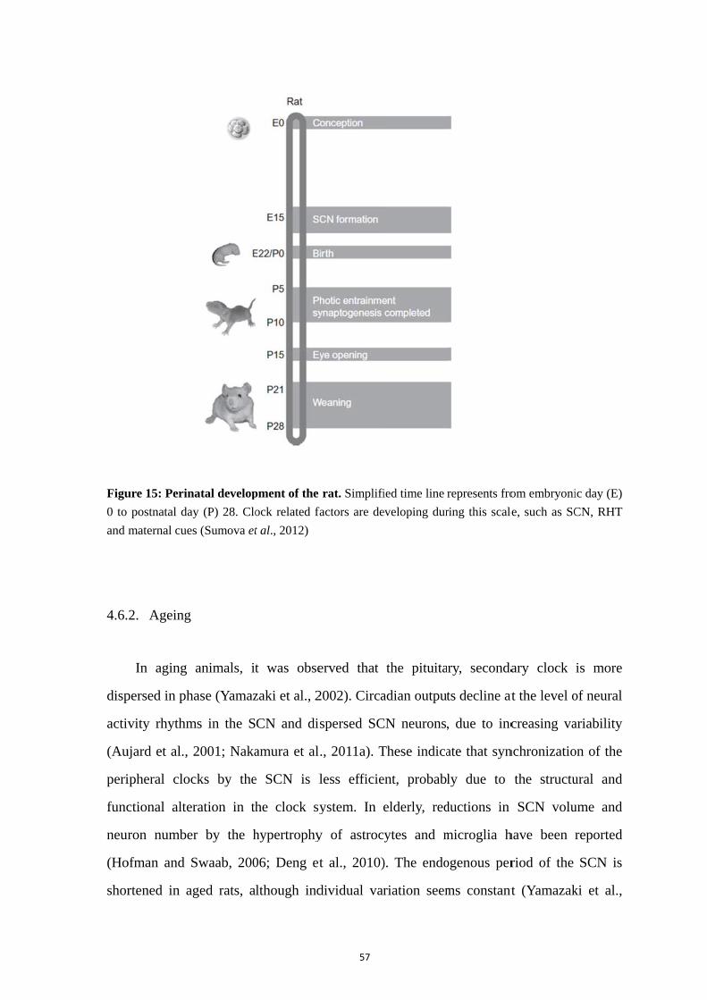
Figu
0 to
and
4.6.
disp
activ
(Au
peri
func
neur
(Ho
shor
ure 15: Perin
postnatal da
maternal cue
2. Ageing
In aging
persed in ph
vity rhythm
ujard et al.,
ipheral cloc
ctional alte
ron numbe
ofman and S
rtened in a
natal develop
ay (P) 28. Clo
es (Sumova et
animals, it
hase (Yamaz
ms in the S
2001; Naka
cks by the
eration in th
er by the h
Swaab, 200
aged rats, a
pment of the
ock related fa
t al., 2012)
t was obse
zaki et al., 2
CN and di
amura et al
e SCN is l
he clock sy
hypertrophy
06; Deng et
lthough ind
57
rat. Simplifi
factors are de
erved that
2002). Circa
spersed SC
., 2011a). T
less efficie
ystem. In
y of astroc
t al., 2010)
dividual var
ied time line
eveloping dur
the pituita
adian output
CN neurons,
These indica
ent, probab
elderly, red
cytes and m
). The endo
riation seem
represents fro
ring this scale
ary, seconda
ts decline at
, due to inc
ate that syn
ly due to
ductions in
microglia h
ogenous per
ms constant
om embryoni
e, such as SC
ary clock
at the level o
creasing va
nchronizatio
the structu
SCN volu
have been
riod of the
t (Yamazak
ic day (E)
CN, RHT
is more
of neural
ariability
on of the
ural and
ume and
reported
SCN is
ki et al.,

58
2002). Mouse lemur, a primate, exhibits a shortened free-running period in aged animals
compared to adulthood, with altered AVP and VIP rhythms (Aujard et al., 2006). In
addition, aged rodents exhibit decline of the SCN presynaptic GABAergic terminals
(Palomba et al., 2008). Decreases in neurotransmitter production of the SCN and low
amplitude of electrical activity might weaken the ability of the SCN to synchronize
peripheral oscillators.
The ageing of the SCN capacity to synchronize the circadian system has been
studied first focusing on entrainment alterations. Aged mice are less sensitive to light
entrainment than young ones (Wyse and Coogan, 2010). Ageing increases crystalline lens
light absorption especially of short wavelengths and decreases pupil area, which results in
progressive loss of retinal illumination (Turner and Mainster, 2008). Besides that, the
responsiveness of the circadian clock to some neurochemical stimuli like GABA is
attenuated(Biello, 2009). In aged hamsters, light-induced Per1 expression is markedly
reduced in the SCN with a significantly longer delay to resynchronization, although in
DD Per1 shows similar rhythms as young rat (Kolker et al., 2003).
Another factor that has been involved in aging is the physiological decline of
hormones (Goldman et al., 2011). Aging changes hormone production and corresponding
target sensitivity. Melatonin oscillates daily and decreases by aging with advanced phase,
which is a putative marker of the decline of the circadian clock (Bondy et al., 2010;
Hardeland, 2013). The aged SCN reduces responsiveness to melatonin(von Gall and
Weaver, 2008). The adrenal cortex releases less aldosterone and cortisol hormones with
aging. Circadian oscillation of cortisol damps with reduced peak levels and increased
evening levels in older people (Ferrari et al., 2001b). Interestingly, PER1 increases with
morning cortisol in elder women with low cortisol levels rather than young women with
higher cortisol levels. This PER1 response to cortisol increase seems to be sensitive at
low cortisol levels rather than at higher cortisol levels (Olbrich and Dittmar, 2012).
Corticosterone secretion is altered with advancing age due to disruption of the diurnal
rhythm of CRH (Cai and Wise, 1996a).
Another phenotype associated with aging is energy metabolism. Reduced appetite
and related change of feeding rhythms are known to be important for entraining

59
peripheral oscillators like liver. Additionally, epigenetic regulations also play a role in
ageing of circadian clock system. Different tissues like the stomach and the spleen in
older mice exhibit specific alternations of methylation and methylation frequency at the
promoters of clock genes (Zhang et al., 2013).
5. Rhythmic properties and clock activity in the skin
5.1. Skin structure and rhythmic functions
As a protective outer covering of vertebrates, skin is an interface tissue between
external conditions and the organism. Exposed to daily variations of environmental
influences like solar irradiation, humidity and temperature, the skin contributes as a
barrier, to the internal homeostasis, which itself results from multiple physiological
processes including cyclic ones, such as hormones and nutrition. The skin performs its
function at once with continuous cell proliferation and differentiation, forming the
corneum and hair, under controlled rate. Many investigations have been directed at the
circadian changes of skin functions due to their importance on health care, hair regrowth
and drug delivery (Reinberg et al., 1990). In human skin, several physiological functions
have been demonstrated to change in a circadian manner, by measuring biophysical
parameters of healthy women skin surface (Le Fur et al., 2001). Over 24 h, sebum
excretion, skin temperature, pH, capacitance (measurement of stratum corneum hydration)
and transepidermal water loss (measurement of stratum corneum barrier function), not
only show rhythmic patterns (phase, amplitude), but also differ from one site to another;
for instance, skin temperature is found rhythmic on the forearm rather than on cheeks.
These regional variations possibly can be explained by differing thickness of stratums,
vascularization and distribution of eccrine glands. Forearm skin blood flow and
temperature as well as sweat loss play a role in thermoregulation and vary along the day
during mild exercise, although the rise of heart rate is not significantly different
(Waterhouse et al., 2004). As the largest organ of mammals, skin covers a big area of
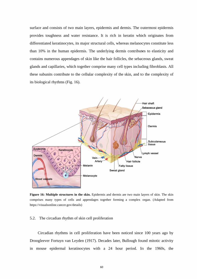
surf
prov
diffe
than
cont
glan
thes
its b
Figu
comp
https
5.2.
Dro
in
face and con
vides tough
ferentiated k
n 10% in th
tains numer
nds and cap
se subunits
biological rh
ure 16: Multi
prises many
s://visualsonlin
The circa
Circadian
oogleever Fo
mouse ep
nsists of tw
hness and w
keratinocyte
he human e
rous append
pillaries, wh
contribute t
hythms (Fig
iple structure
types of cel
ne.cancer.gov
adian rhythm
rhythms in
ortuyn van
idermal ke
wo main lay
water resis
es, its major
epidermis. T
dages of ski
hich together
to the cellu
g. 16).
es in the skin
lls and appen
v/details)
m of skin ce
n cell proli
Leyden (19
eratinocytes
60
yers, epiderm
stance. It is
r structural c
The underl
in like the h
r comprise
ular complex
n. Epidermis a
endages toget
ell proliferat
iferation ha
917). Decad
s with a
mis and der
s rich in k
cells, where
ying dermi
hair follicle
many cell t
xity of the
and dermis ar
ther forming
tion
ve been no
des later, B
24 hour
rmis. The ou
keratin whic
eas melanoc
s contribute
s, the sebac
types includ
skin, and to
re two main la
a complex o
oticed since
ullough fou
period. In
outermost ep
ch originat
cytes consti
es to elasti
ceous gland
ding fibrobl
o the compl
ayers of skin.
organ. (Adap
e 100 years
und mitotic
n the 196
pidermis
tes from
itute less
city and
ds, sweat
asts. All
lexity of
. The skin
pted from
s ago by
activity
60s, the
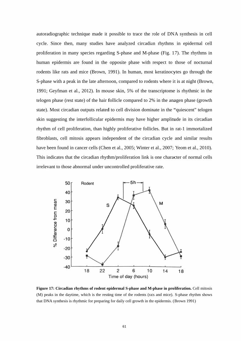
auto
cycl
prol
hum
rode
S-ph
199
telo
state
skin
rhyt
fibr
hav
This
irrel
Figu
(M)
that D
oradiograph
le. Since t
liferation in
man epiderm
ents like rat
hase with a
1; Geyfman
ogen phase (
e). Most cir
n suggesting
thm of cell
oblasts, cel
e been foun
s indicates t
levant to tho
ure 17: Circad
peaks in the
DNA synthes
hic techniqu
then, many
n many spec
mis are fou
ts and mice
peak in the
n et al., 201
(rest state) o
rcadian outp
g the interf
proliferatio
ll mitosis a
nd in cancer
that the circ
ose abnorm
dian rhythms
daytime, whic
is is rhythmic
ue made it
studies ha
cies regardi
und in the
e (Brown, 1
e late afterno
12). In mou
of the hair f
puts related
follicular ep
on, than hig
appears inde
r cells (Chen
cadian rhyth
mal under un
s of rodent ep
ch is the resti
for preparing
61
possible to
ave analyze
ing S-phase
opposite p
1991). In hu
oon, compa
use skin, 5%
follicle com
d to cell div
pidermis ma
ghly prolifer
ependent o
n et al., 200
hm/prolifera
ncontrolled p
pidermal S-p
ing time of th
g for daily cell
o trace the r
ed circadia
e and M-ph
phase with
uman, most
ared to rode
% of the tra
mpared to 2%
ision domin
ay have hig
rative follic
f the circad
05; Winter e
ation link is
proliferativ
phase and M-
he rodents (rat
l growth in the
role of DN
an rhythms
hase (Fig. 1
respect to
t keratinocy
nts where it
anscriptome
% in the ana
nate in the “
gher amplitu
cles. But in
dian cycle
et al., 2007;
s one charac
e rate.
-phase in pro
ts and mice).
e epidermis. (B
NA synthesis
in epiderm
7). The rhy
those of n
ytes go thro
t is at night
e is rhythmi
agen phase
“quiescent”
ude in its c
rat-1 immo
and similar
Yeom et al
cter of norm
oliferation. Ce
S-phase rhyth
(Brown 1991)
s in cell
mal cell
ythms in
nocturnal
ough the
(Brown,
ic in the
(growth
” telogen
circadian
ortalized
r results
., 2010).
mal cells
ell mitosis
hm shows
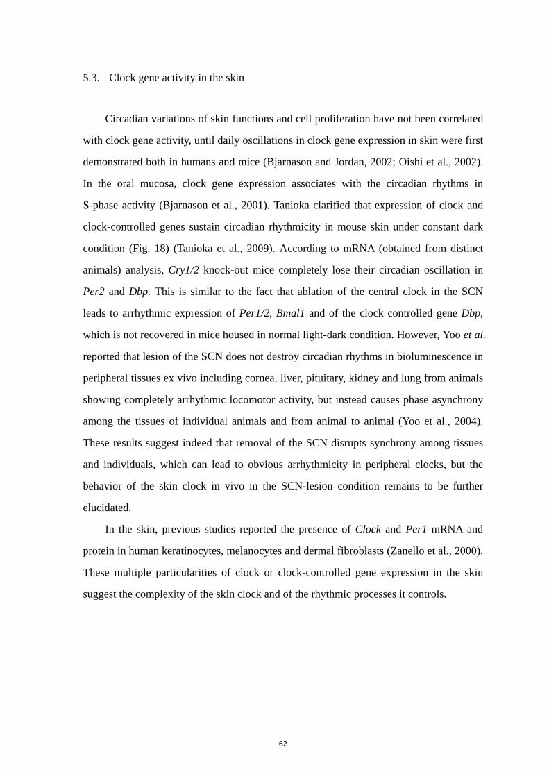
62
5.3. Clock gene activity in the skin
Circadian variations of skin functions and cell proliferation have not been correlated
with clock gene activity, until daily oscillations in clock gene expression in skin were first
demonstrated both in humans and mice (Bjarnason and Jordan, 2002; Oishi et al., 2002).
In the oral mucosa, clock gene expression associates with the circadian rhythms in
S-phase activity (Bjarnason et al., 2001). Tanioka clarified that expression of clock and
clock-controlled genes sustain circadian rhythmicity in mouse skin under constant dark
condition (Fig. 18) (Tanioka et al., 2009). According to mRNA (obtained from distinct
animals) analysis, Cry1/2 knock-out mice completely lose their circadian oscillation in
Per2 and Dbp. This is similar to the fact that ablation of the central clock in the SCN
leads to arrhythmic expression of Per1/2, Bmal1 and of the clock controlled gene Dbp,
which is not recovered in mice housed in normal light-dark condition. However, Yoo et al.
reported that lesion of the SCN does not destroy circadian rhythms in bioluminescence in
peripheral tissues ex vivo including cornea, liver, pituitary, kidney and lung from animals
showing completely arrhythmic locomotor activity, but instead causes phase asynchrony
among the tissues of individual animals and from animal to animal (Yoo et al., 2004).
These results suggest indeed that removal of the SCN disrupts synchrony among tissues
and individuals, which can lead to obvious arrhythmicity in peripheral clocks, but the
behavior of the skin clock in vivo in the SCN-lesion condition remains to be further
elucidated.
In the skin, previous studies reported the presence of Clock and Per1 mRNA and
protein in human keratinocytes, melanocytes and dermal fibroblasts (Zanello et al., 2000).
These multiple particularities of clock or clock-controlled gene expression in the skin
suggest the complexity of the skin clock and of the rhythmic processes it controls.
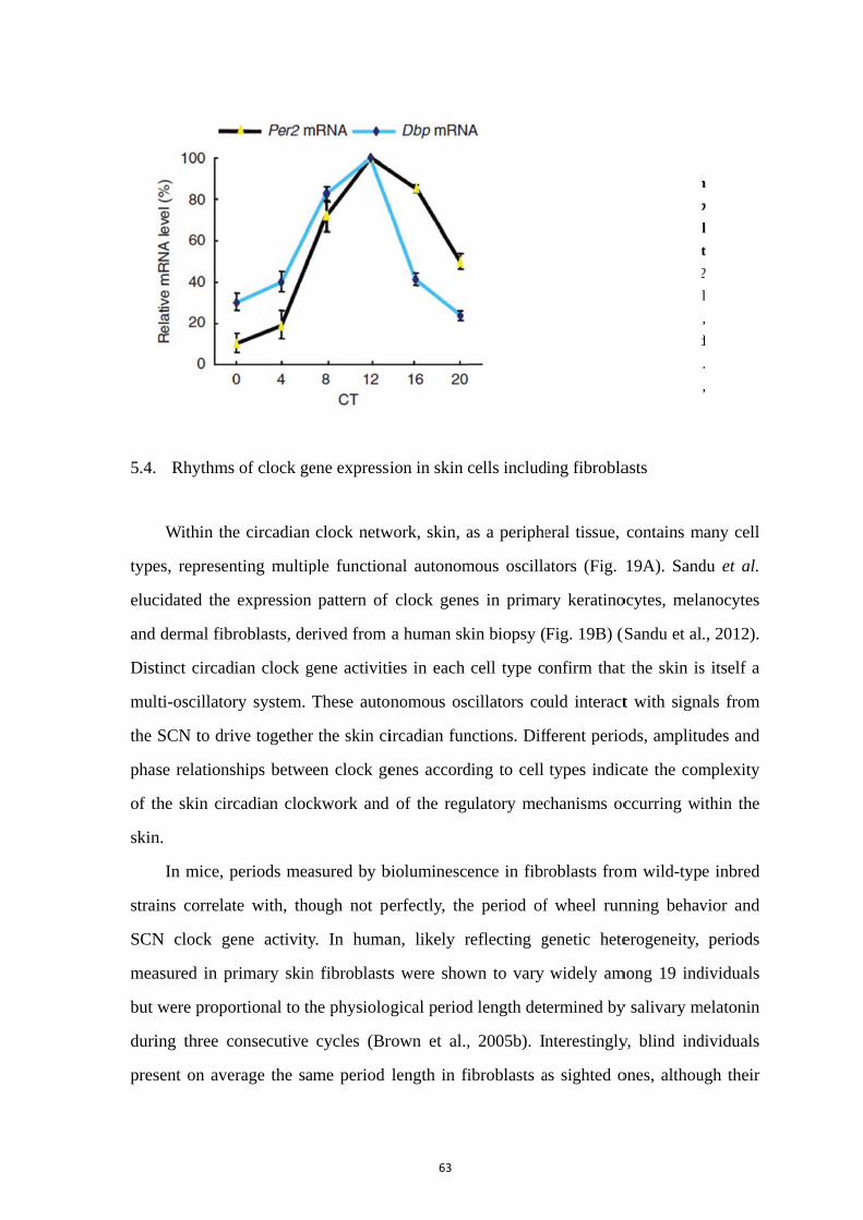
5.4.
type
eluc
and
Dist
mul
the
pha
of t
skin
strai
SCN
mea
but
duri
pres
Rhythms
Within the
es, represen
cidated the
dermal fibr
tinct circad
lti-oscillator
SCN to dri
se relations
the skin circ
n.
In mice, p
ins correlat
N clock ge
asured in pr
were propo
ing three c
sent on ave
of clock ge
e circadian
nting multip
expression
roblasts, de
dian clock g
ry system.
ive together
ships betwe
cadian cloc
periods mea
te with, tho
ene activity
rimary skin
ortional to th
onsecutive
erage the sa
ene expressi
clock netw
ple function
pattern of
rived from
gene activiti
These auton
r the skin ci
en clock ge
ckwork and
asured by b
ough not pe
y. In huma
n fibroblasts
he physiolo
cycles (Br
ame period
63
ion in skin c
work, skin, a
nal autonom
f clock gene
a human sk
ies in each
nomous os
ircadian fun
enes accord
of the regu
bioluminesce
erfectly, the
an, likely r
s were show
gical period
rown et al.,
length in fi
cells includ
as a periphe
mous oscilla
es in prima
kin biopsy (
cell type c
cillators co
nctions. Dif
ding to cell
ulatory mec
ence in fibr
e period of
reflecting g
wn to vary
d length det
, 2005b). In
fibroblasts a
Fig
exp
mR
ski
in
an
exh
con
mo
(A
20
ding fibrobla
eral tissue,
ators (Fig.
ary keratino
Fig. 19B) (S
onfirm that
uld interact
fferent perio
types indic
chanisms oc
roblasts from
f wheel run
genetic hete
widely am
termined by
nterestingly
as sighted o
gure 18: Th
pression of P
RNA level in
in of wild typ
constant da
d Dbp transcr
hibit sam
nsistent with
olecular
Adapted from T
08)
asts
contains m
19A). Sand
ocytes, mela
Sandu et al
t the skin is
t with signa
ods, amplitu
cate the com
ccurring wi
m wild-typ
nning behav
erogeneity,
mong 19 ind
y salivary m
y, blind ind
ones, althou
he circadian
Per2 and Dbp
n the dorsa
ype mice kept
arkness. Per2
riptional leve
me phase
the described
clockwork
Tanioka et al.
many cell
du et al.
anocytes
., 2012).
s itself a
als from
udes and
mplexity
ithin the
e inbred
vior and
periods
dividuals
melatonin
dividuals
ugh their
n
p
l
t
2
l
,
d
k.
,
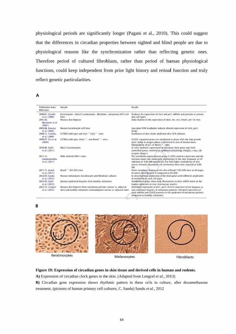
phy
that
phy
The
func
refle
Figu
A) E
B) C
treat
B
A
ysiological p
t the differe
ysiological
erefore peri
ctions, coul
ect genetic p
ure 19: Expre
Expression of c
Circadian gen
tment. (picture
periods are
ences in cir
reasons lik
iod of cult
ld keep inde
particulariti
ession of circa
circadian cloc
ne expression
es of human p
significant
cadian prop
ke the syn
tured fibrob
ependent fr
ies.
adian genes in
ck genes in the
n shows rhyth
primary cell cu
64
tly longer (
perties betw
nchronizatio
blasts, rath
rom prior li
n skin tissue
e skin. (Adapt
hmic pattern
ultures, C. San
(Pagani et a
ween sighte
on rather t
er than pe
ight history
and derived
ted from Leng
in these cel
ndu) Sandu et
al., 2010). T
d and blind
than reflect
riod of hu
and retinal
cells in huma
gyel et al., 201
lls in culture
al., 2012
This could
d people are
ting geneti
uman physi
l function a
an and roden
13)
e, after dexam
suggest
e due to
ic ones.
iological
and truly
ts.
methasone
65
As independent oscillators, primary skin cells sustain persistent but damping
oscillations of clock gene expression measured by bioluminescence for several days in
vitro (Brown et al., 2008; Johansson et al., 2011). It is reported that peripheral tissues
could oscillate in a self-sustained manner for about 7 cycles, and even over 20 cycles in
isolation without media changes (Yamazaki et al., 2000; Yoo et al., 2004). Peripheral
tissues and primary cells usually present limited cycles of oscillation when compared to
the SCN or retina, which consist of neurons mostly, and it raises the question whether the
difference is due to loss of tissue connection in primary cultures or if these cells are just
desynchronized. There is no evidence of coupling of cell-autonomous circadian
oscillators in fibroblasts but, interestingly, maturing fibroblasts cultured at high density
and with decreasing proliferative rates tend to display oscillations with higher amplitude
(O'Neill and Hastings, 2008).
5.5. Synchronizing signals to fibroblasts
Leise et al. managed to image Per2 bioluminescence for six weeks on primary
fibroblasts dissociated from mice tails and selected for 6 months in vitro (Leise et al.,
2012). These fibroblasts sustain cell-autonomous circadian oscillations in a constant
environment, at a 100 cells/mm2 density. Additionally, Noguchi et al. demonstrated that
tail fibroblasts lose clear Per2 circadian rhythmicity in low cell density (till 27 cells/mm2),
which could be rescued by increasing the density with cells that were either rhythmic
(wild type) or not, or even by adding conditioned medium from high-density fibroblast
cultures (Noguchi et al., 2013). This suggests that intercellular factors help to generate
robust circadian rhythms and that they are diffusible paracrine signals rather than direct
contact with neighboring cells. Similar effects were shown with immortalized SCN2.2
cells able to confer molecular oscillations to cocultured NIH/3T3 fibroblasts via the
secretion of unknown diffusible signals (Allen et al., 2001).
Some factors in the culture medium have been shown to affect the expression of
clock genes. Glucose down-regulates Per1/2 mRNA levels in rat-1 fibroblasts (Hirota et

66
al., 2002). Low extracellular pH (6.7) shortens periods of human primary fibroblasts from
24 h to 22.6 h, increases the amplitude and decreases the damping rate without affecting
cell viability (Lee et al., 2011b). Pharmacologically, inhibition of JNK protein kinase
(SP600125) increases the period of rat1 fibroblast rhythms, as well as of SCN, pineal
gland and lung explants from Per1- and Per2-luciferase mice (Chansard et al., 2007). As
predicted, valproic acid, enhancing JNK phosphorylation, shortens the Per1 period in
Rat1 fibroblasts. In contrary, it is reported that valproic acid has no effect on the period of
Per2 expression in primary skin fibroblasts derived from the base of the ear, while
showing significant time-dependent phase-shifting effects on the SCN and primary
fibroblasts (Johansson et al., 2011). These different effects indicate the complicated clock
machinery in cells from distinct origins. For instance, the majority of rhythmic genes in
rat-1, NIH3T3 and Rat 3Y1 fibroblasts are different, as well as those found in SCN 2.2
cells and rat SCN (Menger et al., 2007).
As one non-neuronal cell type displaying clock gene expression, immortalized rat-1
fibroblasts is a non-tumorigenic fibroblast cell line. It is derived from embryos of rats and
used frequently on studies of synchronizing signals. Isolated fibroblasts, rat-1 and
NIH3T3, exhibit cell-autonomous circadian rhythms just as SCN neurons do (Nagoshi et
al., 2004; Welsh et al., 2004). A wide variety of signals, including serum shock, forskolin,
glucocorticoids, co-cultured adrenal glands and temperature changes, can synchronize
rat-1 fibroblasts (Balsalobre et al., 1998; Balsalobre et al., 2000; Yagita and Okamura,
2000; Izumo et al., 2003; Buhr et al., 2010; Noguchi et al., 2012). However, in contrast to
SCN neurons, cultures of fibroblasts damp rapidly (Nagoshi et al., 2004). It is explained
by loss of synchrony among cells with diverse periods lacking highly efficient
communication, unlike SCN neurons (Fig. 20) (Nagoshi et al., 2004; Welsh et al., 2004).
In spite of these results, synchronization by the SCN of peripheral clocks located within
the skin remains unclear. No evidence has been found for direct inter-peripheral
coordination of circadian clock. Thus, how skin gets synchronized remains to be studied.

Figu
oscil
coor
al., 1
5.6.
of th
circ
rang
prop
com
and
Phy
high
et al
mel
199
this
ure 20: Sync
llators show a
rdinated phase
1998)
Skin and
Melatonin
he pineal gl
cadian rhyth
ge of bioact
perty, mela
mpartments.
peripheral
ysiological l
her than pla
l., 2005; Fis
latonin with
6). Regardi
molecule w
chronization
apparent arrhy
es lead to a cu
melatonin
n, one pleiot
land. In mam
hm to exert
tivities (Pev
atonin is e
Besides pin
tissues inc
levels of m
asma melato
scher et al.,
h a circadia
ing skin, me
was discove
model in c
ythmicity, even
umulative cur
tropic horm
mmals, it is
as a chemi
vet and Chal
easily distr
neal gland,
cluding the
melatonin w
onin concen
2006b). Of
an rhythm
elatonin is k
ered (Lerner
67
cell cultures.
n when rhythm
rve via the syn
mone, was fi
s secreted in
ical messen
llet, 2011).
ributed thro
melatonin
gastrointes
within tissue
ntration (Ta
f extrapinea
(Hamm an
known to lig
r and Lerne
With variab
mic at the ind
nchronizing s
irst identifie
nto the bloo
nger of ligh
Because of
oughout al
synthesis h
tinal tract a
es are quite
an et al., 199
al sites, only
nd Menaker
ghten skin p
er, 1958). C
ble phases, p
dividual level.
stimuli. (Adap
ed as a chie
d circulatio
t and darkn
f its small si
l the orga
as been fou
and the skin
specific an
99; Bubenik
y the retina
r, 1980; To
pigmentatio
Compared to
populations o
After synchr
pted from Bal
ef secretory
on with a sig
ness besides
ize and amp
anism and
und in many
n (Bubenik
nd 10 to 10
k, 2002; Ko
is found to
osini and M
on in frogs,
o the corres
of cellular
ronization,
salobre et
product
gnificant
s a wide
phiphilic
cellular
y central
k, 2002).
000-fold
obayashi
produce
Menaker,
the way
ponding
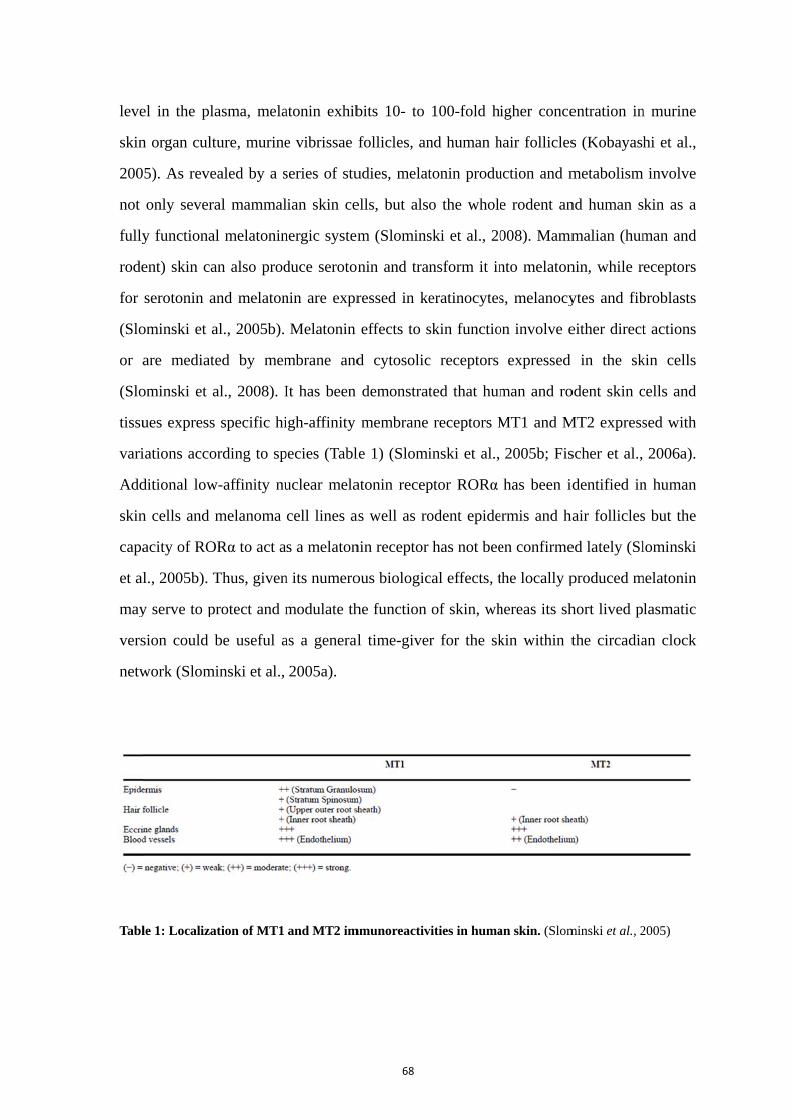
leve
skin
200
not
fully
rode
for
(Slo
or a
(Slo
tissu
vari
Add
skin
capa
et al
may
vers
netw
Tabl
el in the pl
n organ cult
5). As reve
only sever
y functiona
ent) skin ca
serotonin a
ominski et a
are mediat
ominski et a
ues express
iations acco
ditional low
n cells and
acity of RO
l., 2005b). T
y serve to p
sion could
work (Slom
le 1: Localiza
asma, mela
ture, murine
ealed by a s
al mammal
al melatonin
an also prod
and melaton
al., 2005b).
ted by mem
al., 2008). I
s specific hi
ording to sp
w-affinity nu
melanoma
ORα to act a
Thus, given
protect and m
be useful a
minski et al.,
ation of MT1
atonin exhib
e vibrissae
series of stu
lian skin ce
nergic system
duce seroton
nin are expr
Melatonin
mbrane and
It has been
igh-affinity
pecies (Tabl
uclear mela
cell lines a
s a melaton
n its numero
modulate th
as a general
2005a).
and MT2 im
68
bits 10- to
follicles, an
udies, melat
ells, but als
m (Slomins
nin and tran
ressed in ke
effects to s
d cytosolic
demonstrat
membrane
le 1) (Slomi
atonin recep
as well as ro
nin receptor
ous biologic
he function
l time-give
mmunoreactiv
100-fold hi
nd human h
tonin produ
o the whole
ski et al., 20
nsform it in
eratinocytes
skin functio
c receptors
ted that hum
receptors M
inski et al.,
ptor RORα
odent epide
has not bee
cal effects, t
of skin, wh
r for the sk
vities in huma
igher conce
hair follicles
uction and m
e rodent an
008). Mamm
nto melaton
s, melanocy
on involve e
expressed
man and ro
MT1 and M
2005b; Fis
has been id
ermis and ha
en confirme
the locally p
hereas its sh
kin within t
an skin. (Slom
entration in
s (Kobayash
metabolism
nd human s
malian (hum
nin, while r
ytes and fib
either direct
in the sk
odent skin c
MT2 express
scher et al.,
dentified in
hair follicles
ed lately (Sl
produced m
hort lived p
the circadia
minski et al., 2
n murine
hi et al.,
involve
kin as a
man and
eceptors
broblasts
t actions
kin cells
cells and
sed with
2006a).
n human
s but the
lominski
melatonin
lasmatic
an clock
2005)
69
Expression of melatonin receptors in skin cells have been found to be modified by
environmental factors, especially UVB. Effect of UVB to induce or up/down regulate
expression of MT1 and MT2 is defined by genetic background, varying between normal
and immortalized cells (Slominski et al., 2005b).
The major layers of the skin, epidermis, dermis and appendages are all targets for
melatonin regulation, including numerous effects observed in skin biology and clinical
dermatology, from pigmentation to hair regrowth (Fischer et al., 2008). Besides many
protective functions as antioxidant, damage repair, and suppressor of pathological
changes, melatonin displays some chronobiological impacts on skin, given its
photoperiod-associated changes in mammals (Fischer et al., 2006a). One of above
evidences exists in the field of hair growth. In wool industry, several kinds of goats fed
with melatonin-supplemented diet are found to increase the rate of hair growth in the
spring, the period of fur change from the winter to summer (Nixon et al., 1993; Ibraheem
et al., 1994). Mink treated with melatonin molts the summer pelage and grows the winter
pelage earlier (Rose et al., 1984). In human hair regrowth, topical melatonin application
has shown positive effect on the rate of hair found in the anagen (follicle growth) phase in
a pilot-randomized clinical study in women suffering from androgenic or diffuse alopecia
(Fischer et al., 2004). Another evidence exists in rats after pinealectomy, in which
reduction of collagen deposition induced atrophy of epidermis, dermis and hair follicle,
similar to the aging phenotype in skin thickness (Drobnik and Dabrowski, 1996).
Melatonin administration could reverse collagen fiber damage and mitochondrial swelling
in these animals.
In clinics, several skin diseases show seasonal variation in severity (Weiss et al.,
2008). Some of these diseases have been reported to exhibit abnormal plasma melatonin
level in patients. For example, plasma melatonin levels lose their circadian rhythms in
psoriasis, or display greatly reduced rhythmicity in atopic eczema (Mozzanica et al., 1988;
Schwarz et al., 1988; Rupprecht et al., 1995). Lower melatonin concentration is found in
melanoma patients compared with controls, while there exists a higher prevalence of
melanomas in pilots and aircrews with increased jetlag risks as well as office workers
exposed to fluorescent lighting (Kvaskoff and Weinstein, 2010). To many specific skin
70
diseases, skin-related pathological conditions and syndromes, melatonin shows potential
application as a skin protectant by topical or oral administration (Fischer et al., 2008).
5.7. Skin and glucocorticoids
Glucocorticoid receptors are widely expressed in skin compartments and cell types,
including keratinocytes, melanocytes and fibroblasts (Serres et al., 1996; Karstila et al.,
1994). In addition to the strong synchronizing effect of glucocorticoids, these all indicate
that glucocorticoids play an important role in synchronization of peripheral clocks in the
skin.
Interestingly, it has been further shown that the skin has the intrinsic capacity to
produce elements of HPA axis as well as corresponding receptors. CRH is mainly
produced in the PVN of the hypothalamus, although CRH gene is widely expressed in the
extracranial tissues including skin but at level much lower than the hypothalamus
(Slominski et al., 1995). However, CRH gene is only detected in human skin cells but not
rodent skin (Ermak and Slominski, 1997; Slominski et al., 1998). Hence it is proposed
that CRH is imported into skin via afferent nerves endings which targets local
CRH-dependent POMC production compartments. In the skin, POMC synthesis including
а-MSH and ACTH is regulated by multiple factors, which is, for example, stimulated by
UV and inhibited by glucocorticoids (Slominski et al., 2000). And the cortisol synthesis
has been found in human skin hair follicle, keratinocytes, melanocytes and fibroblasts (Ito
et al., 2005; Slominski et al., 2005c; Slominski et al., 2006). These findings provide the
potential of HPA axis homologue in the skin with respects to the systemic rhythmic
glucocorticoids in the synchronization of peripheral clocks, although further
investigations are needed.
71
5.8. Importance of clock genes for normal functions of the skin
Although the synchronization mechanisms of the organized skin oscillators remain
unclear, the integrity of the molecular clock in the skin has proven crucial regarding
normal skin functions and response to stressors. Bmal1 deficiency results in premature
aging phenotype in mice skin. And Clock deficiency increases the age-related dermatitis
in mice (Dubrovsky et al., 2010). Bmal1 has been proven to control the circadian
variation in the S-phase of cell cycle in keratinocyte proliferation, because
keratinocyte-specific deletion of Bmal1 demolishes the time-of-day dependent epidermal
cell division. Consequently, since cells in S-phase are sensitive to DNA damage, the
corresponding time-of-day dependent variation in epidermis susceptibility to DNA
damage induced by UVB has also been proven Bmal1-dependent in the back skin of
shaved mice (Geyfman et al., 2012). This indicates that maximal susceptibility of skin to
UVB takes place in the subjective night of nocturnal rodents, whereas it might be during
the subjective day in human skin when S-phase peaks and UV intensity is highest.
Consistently, Gaddameedhi et al. reported daily rhythmicity in mouse skin expression of a
rate-limiting subunit of the nucleotide excision repair system, the XPA protein, and
associated rhythmicity in the excision repair rate in the mouse skin following UV-induced
DNA damage (Gaddameedhi et al., 2011): this repair rate shows a minimum in the
subjective night and a maximum in the subjective day, and accordingly, mice exposed to
UV in the night display 5 fold increase in occurrence of invasive skin cancers. They have
also shown that circadian S-phase is obliterated in Cry1/2 knockout mice (Gaddameedhi
et al., 2011).
Meanwhile it was shown that Bmal1-controlled ROS levels correlate to the
expression of oxidative phosphorylation genes in the skin and fluctuate in antiphase to the
cell cycle S phase (Geyfman et al., 2012). It suggests the molecular clock system in the
skin contributes to efficient time dependent protection directed towards stressors of
intrinsic or environmental origin and which both display rhythmic levels. Nakamura et al.
have reported that the Per2 gene regulates a time-of-day-dependent variation in the
cutaneous anaphylactic reaction (Nakamura et al., 2011b). This IgE-mediated
72
immediate-type skin reaction triggered by the exposure of mast cells to specific
environmental allergens shows a diurnal rhythm. Mice with Per2 mutation lose the
circadian variation in a passive cutaneous anaphylactic reaction observed in wild type
mice, similar to those seen under adrenalectomy and in aged mice associated with loss of
rhythmic secretion of corticosterone. In addition, Per2 mutant mice exhibit decreased
sensitivity to the inhibitory effects of dexamethasone on the passive cutaneous
anaphylactic reaction and IgE-mediated mast cell degranulation (Nakamura et al., 2011b).
In delayed-type skin allergic reactions, such as contact hypersensitivity (CHS), circadian
rhythmicity might be an important regulatory factor via corticosterone cycling level.
Indeed, it was shown that Clock mutation exacerbates the immune response towards
exogenous haptens in mice, similar to the effect of adrenalectomy, likely because
corticosterone has a protective effect on CHS (Takita et al., 2013). And it was reported
that a number of antigen-presenting cells lose their circadian rhythmicity in
antigen-specific delayed-type hypersensitivity and skin inflammatory responses in
arrhythmic hamsters (Prendergast et al., 2013). These results strongly suggest that the
circadian clock drives and regulates the skin immune system and inflammatory response
under both systemic and local control.
Another intensive field of research regarding skin clocks is about hair regrowth.
Regenerative cycling of hair follicles not only has big economical perspectives in the
pharmaceutical industry, but also offers a great opportunity to explore the role of the
circadian clock in physiological tissue repair and renewal. First, Lin et al. elucidated that
the circadian clock contributes to the regulation of the non-circadian hair growth cycle by
controlling the progression of the anagen (proliferatory) phase, which may have evolved
to allow seasonal regulation of hair growth (Lin et al., 2009). It was then understood that
the clock regulates mitosis in the amplifying cells of the follicle in anagen. In the
Per2-luciferase mouse, telogen/anagen skin and vibrissae hair follicles show circadian
rhythms (Plikus et al., 2013). Some type of transient amplifying cells in anagen hair
follicles display prominent daily mitotic rhythms, and as consequence hairs grow
differently according to mitotic peak and depression. Subsequently, hair loss induced by
radiation is found more dramatic when administrated during mitotic peak. This circadian
73
radioprotective effect gets lost in Cry1/2 mutant mice, consistent with asynchronous
mitoses in their hair follicle.
Besides the importance of proliferative cells in hair growth, the hair follicle is also
quite important because it harbors epidermal stem cells. Impacts of the clock on hair
regrowth raised the question of its putative role in the crosstalk between circadian
rhythms and differentiation. Janich et al. identified two subpopulations of dormant
hair-follicle stem cells coexisting in mouse epidermis in opposite clock phases (Janich et
al., 2013). Bmal1 modulates in an oscillatory manner the expression of stem cell
regulatory genes such as those linked to Wnt and TGFβ signaling, to drive cell
populations into alternate cycles of dormancy and differentiation, responding to
repressive or activating stimuli. Disruption of the clock by deleting Bmal1 results in a
higher number of dormant stem cells and fewer proliferative cells in the hair bulge, while
Per1/2 deletion does the opposite. This looks consistent with the molecular clock
mechanisms and strengthens the key role of the circadian clock system on regulation of
stem cells differentiation. In addition, lacking Bmal1 leads to epidermis ageing and
reduction of skin tumorigenesis. This meaningful study thus demonstrates that the
circadian clock machinery controls the fate of epidermal stem cells in hair follicles, which
again links the (likely local) circadian clock to skin renewal. Recent study shows that
human keratinocyte differentiation induced by calcium shows time-dependent
predisposition (Janich et al., 2013). This predisposition time window coincides with the
transition from late-night to morning, which protects cells from radiation-induced DNA
damage. In addition, circadian arrhythmia established by overexpressing Per1/2
profoundly affects epidermal stem cell function in culture and in vivo. This might be
linked to the fact that cyclic environment-induced perturbations can contribute to ageing
and carcinogenesis in the skin (Janich et al., 2013). Accordingly, Bmal1 mutant mice
show a range of premature skin ageing phenotypes including hair regrowth defect
(Kondratov et al., 2006).
To understand the human circadian rhythms and clock gene expression, a new
approach presents a convenient, reliable and less invasive way to take use of biopsy
samples of hair follicle cells from the head or chin (Akashi et al., 2010). This technique
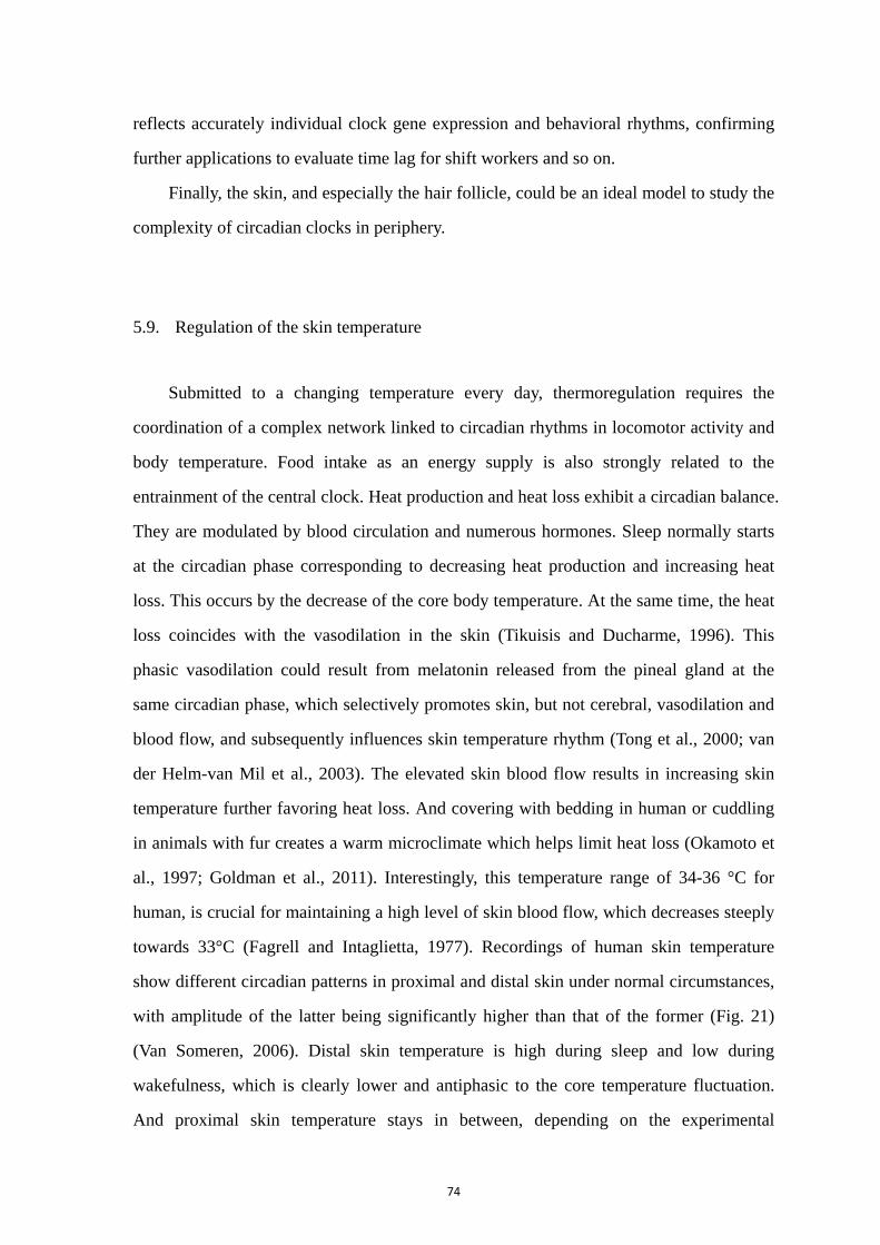
74
reflects accurately individual clock gene expression and behavioral rhythms, confirming
further applications to evaluate time lag for shift workers and so on.
Finally, the skin, and especially the hair follicle, could be an ideal model to study the
complexity of circadian clocks in periphery.
5.9. Regulation of the skin temperature
Submitted to a changing temperature every day, thermoregulation requires the
coordination of a complex network linked to circadian rhythms in locomotor activity and
body temperature. Food intake as an energy supply is also strongly related to the
entrainment of the central clock. Heat production and heat loss exhibit a circadian balance.
They are modulated by blood circulation and numerous hormones. Sleep normally starts
at the circadian phase corresponding to decreasing heat production and increasing heat
loss. This occurs by the decrease of the core body temperature. At the same time, the heat
loss coincides with the vasodilation in the skin (Tikuisis and Ducharme, 1996). This
phasic vasodilation could result from melatonin released from the pineal gland at the
same circadian phase, which selectively promotes skin, but not cerebral, vasodilation and
blood flow, and subsequently influences skin temperature rhythm (Tong et al., 2000; van
der Helm-van Mil et al., 2003). The elevated skin blood flow results in increasing skin
temperature further favoring heat loss. And covering with bedding in human or cuddling
in animals with fur creates a warm microclimate which helps limit heat loss (Okamoto et
al., 1997; Goldman et al., 2011). Interestingly, this temperature range of 34-36 °C for
human, is crucial for maintaining a high level of skin blood flow, which decreases steeply
towards 33°C (Fagrell and Intaglietta, 1977). Recordings of human skin temperature
show different circadian patterns in proximal and distal skin under normal circumstances,
with amplitude of the latter being significantly higher than that of the former (Fig. 21)
(Van Someren, 2006). Distal skin temperature is high during sleep and low during
wakefulness, which is clearly lower and antiphasic to the core temperature fluctuation.
And proximal skin temperature stays in between, depending on the experimental
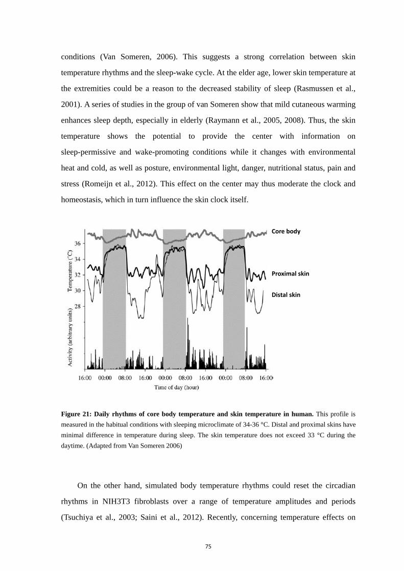
75
conditions (Van Someren, 2006). This suggests a strong correlation between skin
temperature rhythms and the sleep-wake cycle. At the elder age, lower skin temperature at
the extremities could be a reason to the decreased stability of sleep (Rasmussen et al.,
2001). A series of studies in the group of van Someren show that mild cutaneous warming
enhances sleep depth, especially in elderly (Raymann et al., 2005, 2008). Thus, the skin
temperature shows the potential to provide the center with information on
sleep-permissive and wake-promoting conditions while it changes with environmental
heat and cold, as well as posture, environmental light, danger, nutritional status, pain and
stress (Romeijn et al., 2012). This effect on the center may thus moderate the clock and
homeostasis, which in turn influence the skin clock itself.
Figure 21: Daily rhythms of core body temperature and skin temperature in human. This profile is
measured in the habitual conditions with sleeping microclimate of 34-36 °C. Distal and proximal skins have
minimal difference in temperature during sleep. The skin temperature does not exceed 33 °C during the
daytime. (Adapted from Van Someren 2006)
On the other hand, simulated body temperature rhythms could reset the circadian
rhythms in NIH3T3 fibroblasts over a range of temperature amplitudes and periods
(Tsuchiya et al., 2003; Saini et al., 2012). Recently, concerning temperature effects on
Core body
Proximal skin
Distal skin
76
peripheral clocks, heat shock response pathways have been shown to have both phase and
period influence on the circadian clock, while heat shock factors are also intimately
related to the molecular circadian clock (Buhr and Takahashi, 2013). Within the
physiological range of temperature, rat-1 and NIH3T3 fibroblasts display the property of
temperature compensation (Izumo et al., 2003; Tsuchiya et al., 2003). This characteristic
of persistence with a period ≈ 24 h, supports the idea that these oscillators are controlled
by a circadian clock (Hastings, 2001). Further studies need to confirm this property in the
skin clock per se. As one tissue facing dramatic changing temperature in the environment,
the skin can keep its stable circadian clock network in balance to hold the scheduled
homeostasis, with the advantage of temperature compensation.

77
Aim of the thesis and Methodology

78
1. Aim of the thesis
The circadian network is a complex system that involves multiple clocks located
throughout the organisms. These self-sustained clocks are at the same time driven by the
central circadian pacemaker contained within the SCN. Different cues emanating from the
SCN are essential to coordinate the peripheral clocks, while the SCN themselves get
entrained predominately by the light/dark cycle. Within each tissue, different mechanisms
deliver synchronizing signals to downstream physiological processes. To adapt to the
environment, oscillators are meant to interrelate within the circadian system. They are
independent as functional units but depend on the synchronizing messages transmitted
towards them. As discussed in the introduction, circadian clocks display the following
features: to be self-sustained, able to be entrained and temperature compensated. To study
the properties of a circadian clock in periphery and to better understand its role in the
circadian network requires characterizing the above-mentioned features and the neural or
hormonal pathway through which it gets synchronized. To get insight into these aspects
we chose as a model of peripheral oscillator the skin which characterization was the
subject of the PhD project.
The expression of circadian clock genes in the skin has been reported in mouse and
human (Tanioka et al., 2009; Sandu et al., 2012). The major skin cell types were shown to
oscillate in a circadian manner, in line with the diverse rhythmic functions they display.
Their molecular clockwork indicates that they are cell-autonomous and part of a
multioscillatory local network where synchronizing cues are unknown. In order to gain
insight into potential signals that might entrain skin clocks, we undertook a study of the
skin clock maturation during post-natal life. We extended this study to the effects of aging,
which affects the amplitude of the central clock (Nakamura et al., 2011a) and might also
affect the clock within the skin. Indeed, Bmal1 knock out results in a premature aging
phenotype in mice skin and Clock deficiency promotes age-related dermatitis in mice
(Dubrovsky et al., 2010). As an interface tissue the skin is exposed to varying
environmental temperature: thus we also analyzed its temperature compensation

79
properties as part of its global characterization.
We also investigated potential synchronizing signals by studying candidate
molecules. A wide variety of signals, including serum shock, forskolin, glucocorticoids
and temperature changes, can synchronize Rat-1 fibroblasts, a cellular model which is not
derived from the skin (Balsalobre et al., 1998; Balsalobre et al., 2000; Yagita and
Okamura, 2000; Izumo et al., 2003; Buhr et al., 2010). Another potential synchronizer is
melatonin. Melatonin is a strong output from the SCN pacemaker with robust daily
rhythms. Exogenous melatonin has been proven to affect central clocks, due to the fact
that melatonin receptors are widely expressed in the organisms including the SCN
(Pitrosky et al., 1999; Slotten et al., 2002). It has been demonstrated that human and
rodent skin harbors specific high-affinity membrane receptors for melatonin, MT1 and
MT2 (Slominski et al., 2003b; Fischer et al., 2006a). Thus, we investigated the potential
property of melatonin as a time giver to synchronize the oscillators in the skin and
focused essentially on its effect on fibroblasts.
Besides fibroblasts, there are also keratinocytes and melanocytes as main cell types
in skin. We did not yet develop proper methods to isolate these cell types from rat skin but,
since we are currently studying it in the laboratory, we turned to human skin (plasties) as
a potential source of melanocytes and keratinocytes (Sandu et al., 2012). To take
advantage of bioluminescence because it is real-time and time-saving, we designed and
constructed a lentivirus tool, following the design from a paper published by Brown et al.
(Brown et al., 2005b). In this vector, luciferase expression is controlled by the Bmal1
promoter. As reported, this construct was successfully used to assay clock properties in
human fibroblasts and keratinocytes.
In the results part afterwards, to follow the aims of the thesis, three main chapters are
organized and written as respective small papers, each including abstract, introduction,
method, result and discussion. The general discussion is to show the global view of the
thesis work. The bibliographies are listed together in the end after the general discussion.
80
2. Methodological strategy: real-time recording of circadian gene
transcription by bioluminescence
Properties of circadian clocks are usually studied by monitoring the activity level of
clock gene transcription, because of the stable intrinsic negative feedback loops in the
molecular clockwork. An important aspect here is the necessity to monitor the active
processes over a period of at least 24 hours, or several times 24 hours. Initially, this
measurement was achieved by frequent sampling over at least 24 h and analyzing the
mRNA levels over time. Although a large amount of animals and work are required,
numerous studies have managed to determine many endogenous clock properties by this
approach (Balsalobre et al., 1998; Balsalobre, 2002; Brown et al., 2002). With this
technique, several core clock genes can be checked at the same time to compare clock
activities such as relative phases (Sandu et al., 2012). However, some details in
oscillatory activity like bimodal activity can be easily missed because of unavoidable
intervals between sampling times. Another limitation would happen due to
inter-individual differences in phases which could lead to neutralization on the populated
clocks, especially in SCN-lesioned animals (Tanioka et al., 2009). Since luminescence
reporters are widely used to measure gene transcription, this technique has been adapted
to study circadian oscillations in cells, cultured tissues and whole organisms (Yamazaki et
al., 2000; Izumo et al., 2003; Vallone et al., 2004). Given that it is easy and noninvasive to
use luminescence to assay circadian genes, transgenic cells and animals have been
developed on the basis of a clock gene promoter fused to the luciferase reporter gene.
This reduces greatly the amounts of samples and inter-sample variability. Real-time
recording of circadian oscillations of gene expression can be done from the whole animal
to cultured tissue and cells (Collaco and Geusz, 2003; Yamazaki et al., 2009). As an
example, in Per1-luciferase transgenic rats, the Per1 transcription rhythm in the SCN
slices can last for 32 days continuously (Yamazaki et al., 2000). In this long-lasting
recording, properties of clocks can be observed through parameters like phase, period and
amplitude. The timing of a reference point in the cycle, normally the first peak, is
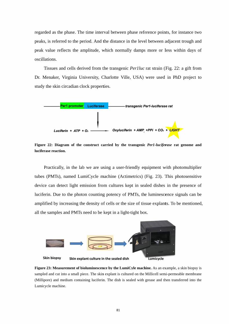
rega
peak
peak
osci
Dr.
stud
Figu
lucif
tube
devi
luci
amp
all t
Figu
samp
(Mil
Lum
arded as the
ks, is referr
k value ref
illations.
Tissues an
Menaker, V
dy the skin c
ure 22: Diag
ferase reactio
Practically
es (PMTs),
ice can det
iferin. Due
plified by in
the samples
ure 23: Measu
pled and cut in
lipore) and m
micycle machin
e phase. The
red to the pe
flects the am
nd cells der
Virginia Un
circadian cl
gram of the
on.
y, in the lab
named Lu
tect light em
to the photo
ncreasing th
and PMTs
urement of bi
nto a small pi
medium contai
ne.
e time inter
eriod. And t
mplitude, w
ived from t
niversity, C
ock propert
construct ca
b we are us
miCycle m
mission from
on counting
he density of
need to be k
ioluminescen
iece. The skin
ining luciferin
81
rval between
the distance
which norm
the transgen
Charlotte Vi
ties.
arried by th
sing a user-
machine (Ac
m cultures
g potency o
f cells or th
kept in a lig
nce by the Lu
n explant is cu
n. The dish is
n phase refe
e in the leve
mally damps
nic Per1luc
ille, USA)
he transgenic
-friendly eq
ctimetrics) (
kept in sea
of PMTs, th
he size of tis
ght-tight box
miCyle mach
ultured on the
s sealed with
erence poin
el between a
s more or l
rat strain (F
were used
c Per1-lucifer
quipment w
(Fig. 23). T
aled dishes
e luminesce
ssue explant
x.
hine. As an ex
Millicell sem
grease and th
nts, for insta
adjacent tro
less within
Fig. 22: a g
in PhD pr
rase rat gen
with photom
This photos
in the pres
ence signal
ts. To be me
xample, a skin
mi-permeable m
hen transferre
ance two
ough and
days of
gift from
roject to
nome and
multiplier
sensitive
sence of
s can be
entioned,
n biopsy is
membrane
d into the
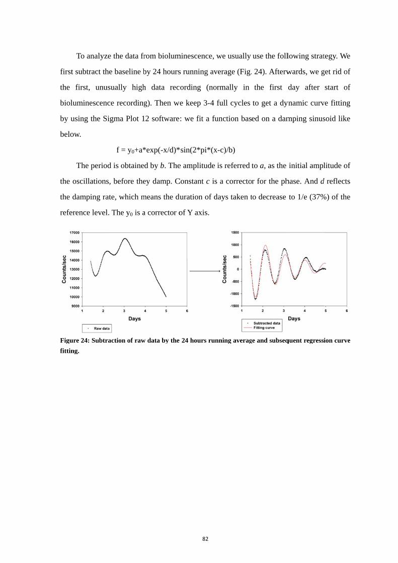
first
the
biol
by u
belo
the
the
refe
Figu
fittin
To analyz
t subtract th
first, unus
luminescenc
using the Si
ow.
The period
oscillations
damping ra
erence level
ure 24: Subtr
ng.
e the data fr
he baseline b
sually high
ce recording
igma Plot 1
f = y0+a*
d is obtaine
s, before the
ate, which m
. The y0 is a
action of raw
rom biolum
by 24 hours
h data rec
g). Then we
12 software
*exp(-x/d)*
ed by b. The
ey damp. C
means the du
a corrector o
w data by the
82
minescence,
s running av
ording (no
e keep 3-4
: we fit a fu
sin(2*pi*(x
e amplitude
Constant c is
uration of d
of Y axis.
24 hours run
we usually
verage (Fig.
ormally in
full cycles
unction bas
x-c)/b)
is referred
s a correcto
days taken t
nning averag
use the foll
24). Afterw
the first d
to get a dy
ed on a dam
to a, as the
r for the ph
to decrease
ge and subseq
lowing strat
wards, we g
day after
ynamic curv
mping sinus
initial ampl
hase. And d
to 1/e (37%
quent regress
tegy. We
get rid of
start of
ve fitting
soid like
litude of
d reflects
%) of the
ion curve

83
Results

84
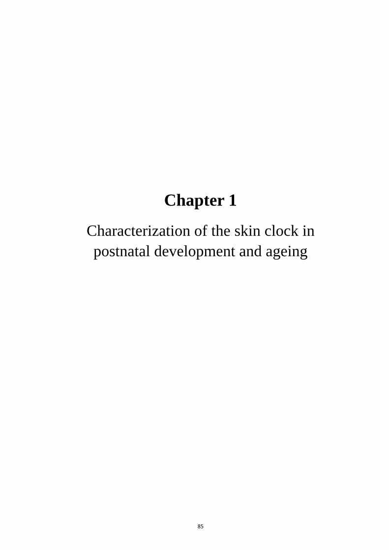
85
Chapter 1
Characterization of the skin clock in postnatal development and ageing
86
1. Abstract
In mammals, circadian rhythms are generated and controlled by a hierarchical
system including a master clock, located in the suprachiasmatic nuclei, the SCN, and
multiple oscillators present in central and peripheral tissues such as liver, retina and skin.
Due to the types of cell-autonomous oscillators that are involved, circadian clocks exhibit
tissue-specific patterns within this network. In the present study, we investigated the clock
present in the skin and characterized its properties during postnatal development and
aging.
Circadian rhythmicity was examined in abdominal skin explants from 10 day- to 2
year-old Per1-Luciferase transgenic rats, using the real-time bioluminescence recording
method. We found that the Per1 rhythmic expression shows up at the age of 1 month,
when rats get adult. Starting at 1 month, the skin clock gets more mature and
synchronized with higher amplitude oscillations, reaching the highest at 6 months. In 1 to
2 year-old rats, skin circadian rhythms show decreased amplitude with fast damping and
phases are more dispersed between individuals. Our study shows the level of
synchronization in the skin tops at the age of 6 months, whereas it is decreasing during
ageing. In addition, we characterized the skin as a temperature compensated oscillator
which might be related to its distinct role in the circadian clock network.
With this study we functionally confirm the presence of a circadian clock within the
skin and demonstrate that it is a valuable experimental model to study peripheral clock
properties and synchronization mechanisms.
87
2. Introduction
The circadian clock network is comprised of multiple oscillators that generate daily
rhythms in physiology and behavior. In this system, a central clock in the SCN can be
entrained by photic and non-photic environmental time cues. The light is the prominent
zeitgeber (time giver), which delivers time information through intrinsically
photosensitive retina ganglion cells and the retinohypothalamic tract (RHT) (Berson et al.,
2002). These external signals entrain the clock system to adapt to cyclic environment. The
SCN drive rhythmic oscillations in neuronal activities and hormonal secretions. These
outputs are essential for other oscillators in periphery to function integrally. Peripheral
oscillators harbor cell-autonomous clocks, based on an intracellular molecular mechanism.
Genetically, the molecular clockwork involves interlocking transcriptional-translational
feedback loops (Ko and Takahashi, 2006). Several transcription factors are core
components to regulate rhythmic gene expressions, with a period of about 24 hours.
Transcription of Period (Per) and Cryptochrome (Cry) genes are activated by
heterodimers of BMAL1 and CLOCK. PER and CRY proteins form a complex in the
cytoplasm and translocate to the nucleus, repressing their transcription driven by the
BMAL1/CLOCK complex. Additional regulatory loops involve Bmal1 transcriptional
regulation by REV-ERB and ROR factors. BMAL1/CLOCK activates the transcription of
Rev-erb and Ror genes, which products respectively repress and activate the transcription
of Bmal1. These negative and positive regulation loops provide original gene expression
rhythms. Posttranscriptional and posttranslational pathways also regulate the clock factors
and contribute ultimately to the mechanisms of rhythms.
Under the coordination of the SCN, peripheral clocks set appropriate phase and
period to behave in an integrated manner for many physiological rhythms. Skin is an
interesting example of peripheral clock, which displays daily rhythmic variations in
functions like sebum secretion and temperature (Le Fur et al., 2001). At the interface
between the body and external environment, skin plays a key role on the maintenance of
body homeostasis, exposed to daily variations of solar irradiation, humidity and
temperature. Previous studies reported expression of CLOCK and PER1 proteins in

88
human keratinocytes, melanocytes and dermal fibroblasts (Zanello et al., 2000). Rhythmic
expressions of clock genes have been reported in the skin in mouse and human (Tanioka
et al., 2009; Sandu et al., 2012). Several types of cells in the skin are shown to oscillate in
a circadian manner, which indicates that they are cell-autonomous. Together, these results
suggest the complexity of the skin clock and present it as a potential model to study the
peripheral clocks.
In rodents, Bmal1 deficiency results in premature aging phenotype in mice skin. And
Clock deficiency increases the age-related dermatitis in mice (Dubrovsky et al., 2010).
Old rats showed age-related shortening of the free-running period and dispersed phase in
some secondary clocks like the pituitary (Yamazaki et al., 2002). Here we characterized
the skin clock by real-time recording of skin explants from Per1-luciferase rats from the
postnatal period to 2 years. Since skin is a superficial tissue exposed to varying
environmental temperature, we studied the temperature compensation property of the skin
and of primary cells derived from it.
3. Materials and Methods
3.1. Animals
Per1-luciferase transgenic rats (Yamazaki et al. 2000) were used in this study. All
rats were raised and maintained in our animal care facilities (Animal resource and
experimentation platform, Chronobiotron, UMS 3415, France). Per1-luciferase
transgenic rats were housed under a 12:12 h light-dark cycle (lights - 300 lux - on at
07:00 A.M.). The infant rats were kept with maternal care until the beginning of
experiment. Male rats were used in all the experiments. All experiments were performed
in accordance with the rules from the French Department of Agriculture (license no
67-67-298) and the 86/609/EEC European Committee Council Directive.

89
3.2. Tissue culture
Per1-luciferase transgenic rats aged from 10 days to 2 years were used. Animals
were anesthetized and euthanized with CO2 (20% in an air tight box) (usually in the
afternoon). Right after sacrifice, abdominal fur was shaved with a single-edged razor
blade and the skin tissue was excised as much as possible from the belly. Other peripheral
tissues like eyeball and lung were removed at the same time, when necessary. Harvested
tissues were immediately placed in cold Hank’s balanced saline solution (HBSS,
Invitrogen) on ice. The skin biopsies were then dissected under the binocular lens to
remove subcutaneous tissue, mainly fat, and washed at least three times. A rectangular
piece of explant with an area of about 20 mm2 was isolated, flattened, placed (dermis side
down) on a Millicell semi-permeable membrane (PICMORG50, Millipore) and cultured
in 35 mm dishes (Nunc) with 1 ml DMEM supplemented with 10% Fetal Bovine Serum
(FBS: Biowest) and 0.1 mM luciferin (Promega). For lung and eyeball explants FBS was
replaced with 2% B27 (Invitrogen). For culture medium, 1 L of DMEM was regularly
prepared from powder (Sigma, D9202) and supplemented with 10 mM HEPES (Sigma
H0887), 25 mM D(+) glucose (Sigma G7021), 4.2 mM sodium bicarbonate (7.5%, Sigma
S8761), 2 mM L-Glutamine and antibiotics (penicillin and streptomycin) until use. Dishes
were sealed with vacuum grease (Dow Corning) and placed into the Lumicycle set at
37°C (usually by the end of the afternoon). Remaining parts of the skin biopsies were
kept temporarily in ice-cold HBSS for subsequent fibroblast culture. Light emission from
cultured tissues was measured immediately and without interruption for about 7 days:
each plate was counted for 1 min and 48 sec, every 15 min.
3.3. Fibroblasts culture
To prepare primary fibroblasts from rat skin, biopsies handled as described above
were washed in PBS and placed on a 10 cm culture dish with dermis side downwards.
Then the explant was cut into small squares with sharp surgical blades, to create edges for

90
fibroblast outgrowth. Afterwards, these skin pieces were placed in the culture dish left to
slightly dry for about 5 min until no visible PBS is left. Then warm complete DMEM
medium (20% FBS) was added to the dish to cover the skin pieces. Medium was changed
to complete DMEM (10% FBS) after fibroblasts grew out of skin pieces and cultures
were maintained for 2-3 weeks until fibroblasts reached confluence. Fibroblasts were
passaged and cultures were seeded at a density of 50,000 cells per 35 mm dish.
Bioluminescence recording of fibroblasts started when cultures reached confluence.
Condition of recordings was the same as for explants and time window for analysis
started at the first trough.
3.4. Data analysis and statistics
Acquisition of bioluminescence data reflecting Per1 activity was made with
Lumicycle Analysis software (Actimetrics). This software was also used to subtract the
baseline drift from the raw data: the 24 hours running average method was efficient in this
process. To further investigate Per1-luciferase rhythms, subtracted data were analyzed by
using Sigma Plot 12 software. A damped sinusoid function (see below) was used to fit the
subtracted curves. The unusual data, normally in the first day after bioluminescence
starting were removed. We kept 3-4 full cycles of recordings to get a dynamic fitting by
Sigma Plot 12 software. The window of analysis for skin explants started at a common
time point (slightly ahead of the first trough) corresponding to 9:30 A.M. of the second
day of culture. In the case of fibroblasts, for which cultures were started more randomly,
the window of analysis started precisely at the first trough.
f = y0+a*exp(-x/d)*sin(2*pi*(x-c)/b)
The best-fit solutions were validated based on the fact that they passed the normality
test (Shapiro-Wilk test (p = <0.0001)) and passed constant variance test. With a linear
drift of y0, the standard damped sinusoid allowed to determine the period of the
oscillations by constant b. The value of a referred to the initial amplitude when x equaled
to zero ahead of damping (therefore, the time scale was shifted for the analysis, so that 0
91
corresponded to the beginning of the studied time window). Constant c was a corrector
for the phase corresponding to the middle of trough and subsequent peak but in our
results we considered as phase value the time of the first peak. And d reflected the
damping rate, which meant the duration (days) taken to decrease to 37 % (1/e) of its
reference level. The y0 represented a Y axis corrector. Curves were considered rhythmic
when consecutive circadian cycles showed up. Samples with only one visible peak of
Per1-luc were designated as arrhythmic.
Data are presented as mean values ±SD. One-way and two-way ANOVA followed by
post hoc comparisons with the LSD Fisher test were used to compare groups. All statistics
were performed using Sigma Plot 12 software.
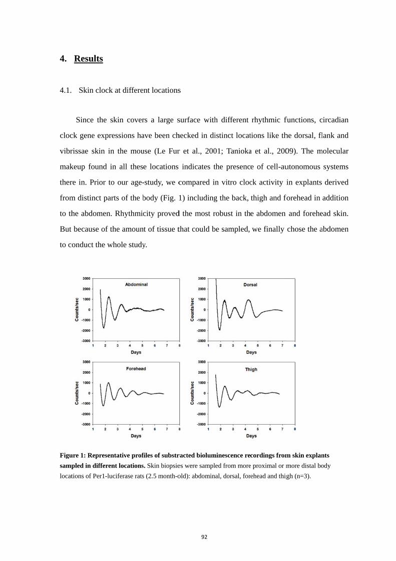
4.
4.1.
cloc
vibr
mak
ther
from
to th
But
to c
Figu
samp
locat
Results
Skin cloc
Since the
ck gene exp
rissae skin
keup found
re in. Prior
m distinct p
he abdomen
because of
onduct the w
ure 1: Repres
pled in differ
tions of Per1-
ck at differe
skin cover
pressions ha
in the mou
in all thes
to our age-
arts of the b
n. Rhythmic
f the amoun
whole study
entative prof
rent locations
luciferase rats
ent locations
rs a large s
ave been ch
use (Le Fur
e locations
-study, we c
body (Fig. 1
city proved
nt of tissue t
y.
files of substr
s. Skin biopsie
s (2.5 month-o
92
s
surface with
hecked in di
r et al., 200
indicates t
compared i
1) including
d the most r
that could b
racted biolum
es were sampl
old): abdomin
h different
istinct locat
01; Tanioka
the presenc
n vitro cloc
g the back,
robust in th
be sampled,
minescence re
led from more
nal, dorsal, for
rhythmic f
tions like th
a et al., 200
e of cell-au
ck activity i
thigh and fo
e abdomen
we finally
cordings from
e proximal or m
ehead and thig
functions, c
he dorsal, fl
09). The m
utonomous
in explants
forehead in
and forehe
chose the a
m skin explan
more distal bo
gh (n=3).
circadian
lank and
molecular
systems
derived
addition
ead skin.
abdomen
nts
ody
93
4.2. Maturation and aging of the skin clock
To understand the skin characteristics as a peripheral clock during development and
aging, we first analyzed Per1-luc expression in skin explants from rats aged between 10
days and 2 years. Three stages were found (Fig. 2A).
First stage was from 10 days to 1 month, where young rats were still kept in litter
with maternal care. No clock rhythmicity could be clearly seen at 10 and 20 days (P10
and P20). Per1 activity in skin exhibited arrhythmic pattern with random peaks and
troughs. At P10, variations in Per1 activity levels showed up only in the first day or 2-3
days later. At P20, bioluminescence showed non-circadian fluctuations with 2-3
continuous peaks but with low robustness. At 1 month, unstable circadian rhythms could
be found in skin clock activity, with low amplitude. However, the most robust peak did
not show up in the first cycle but mostly in the second one. Given the fact that the
activities from 10 days to 1month were not in the classic rhythmic way, this course was
not analyzed by curve fitting.
The second stage started at 2 months: bioluminescence recordings in adult rats
between 2 and 6 months showed circadian oscillation that appeared more and more robust
with amplitudes increasing significantly (Fig. 2B). The period at 2 months was 22.4±0.5 h
which was shorter in tendency than 23 h averagely observed at other adult ages. The
phases got progressively delayed and more variable. The amplitude of clocks in skin
damped significantly faster at 3 months. The largest amplitude combined with slowest
damping occurred at the 6 month age (amplitudes p=0.019, one-way ANOVA; damping
constant p=0.032, one-way ANOVA).
Third stage was the aging part, from 6 months to 2 years. Circadian oscillations got
globally weaker and some abnormal cycles appeared. The phase variations among
animals were much larger than in earlier adult ages. The amplitude of circadian
oscillations damped faster upon aging.
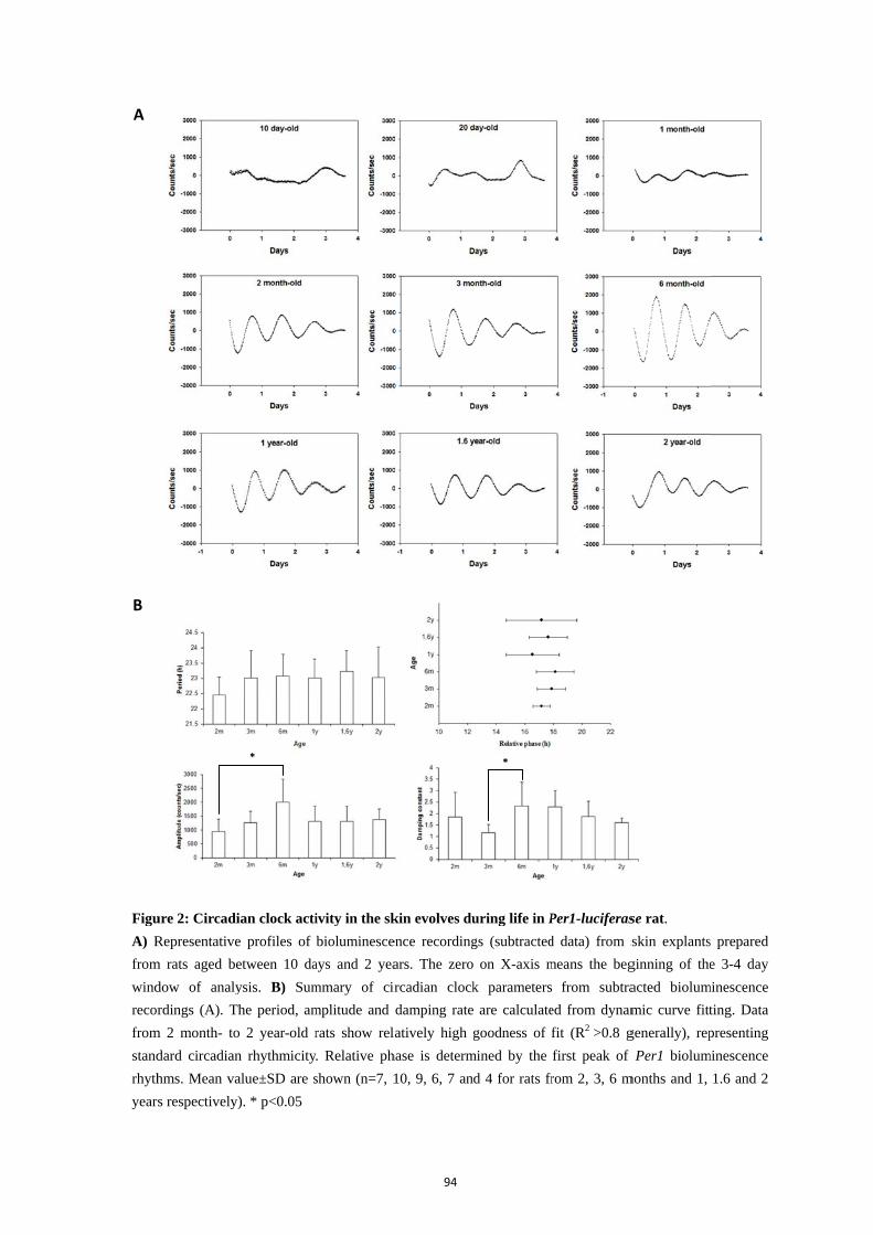
Figu
A) R
from
wind
recor
from
stand
rhyth
year
A
B
ure 2: Circadi
Representative
m rats aged be
dow of analy
rdings (A). T
m 2 month- to
dard circadian
hms. Mean va
s respectively
ian clock acti
e profiles of
etween 10 da
ysis. B) Sum
The period, am
o 2 year-old r
n rhythmicity
alue±SD are s
y). * p<0.05
*
ivity in the sk
bioluminescen
ays and 2 yea
mmary of cir
mplitude and
rats show rela
y. Relative ph
shown (n=7, 1
94
kin evolves du
ence recording
ars. The zero
rcadian clock
damping rate
atively high g
hase is determ
10, 9, 6, 7 an
uring life in P
gs (subtracted
on X-axis m
k parameters
are calculate
goodness of f
mined by the
d 4 for rats fr
*
Per1-luciferas
d data) from s
means the beg
from subtra
ed from dynam
fit (R2 >0.8 g
first peak of
rom 2, 3, 6 m
se rat.
skin explants
ginning of the
acted biolum
mic curve fitt
generally), rep
Per1 biolum
months and 1,
s prepared
e 3-4 day
minescence
ting. Data
presenting
minescence
1.6 and 2

95
4.3. Maturation and aging in skin fibroblasts
Besides the skin tissue explants, we also checked the clock activity in primary
fibroblasts from rat skin. Several ages were chosen similarly, to cover postnatal
development. Fibroblasts from donors of these ages showed distinct oscillations in
comparison with the skin explants (Fig. 3A). Fibroblasts cultured from 10 day-old donors,
showed no rhythmic clock activity, but a peak at the first day, and circadian oscillations
appeared and got more rhythmic in cell populations from the age of 2 months on. When
donors were as old as 2 years, circadian rhythms still existed in the fibroblast populations,
though some abnormal peaks and bimodal activity appeared in the activities (Fig. 3A).
Since the activities at 2 months did not conform to classical rhythmic patterns and their
goodness of fit was below 0.8, this age was not included in the curve fitting results.
During the ages between 3 months and 1.6 years, robust oscillations occurred for several
days with fast damping. The period was 26.0±1.5 h at 3 months and decreased till
22.5±0.5 h at 2 years (p<0.001 one-way ANOVA, Fig. 3B). The amplitude of fibroblast
oscillations decreased as the donors got elder (p=0.002 one-way ANOVA), while the
damping rate was slower at older age (p<0.001 one-way ANOVA), which was contrary to
the skin explants. Significant differences are determined by p < 0.05, post-hoc Fisher
LSD test.
4.4. Medium change induces the oscillation of skin oscillators in the early postnatal age
In early postnatal ages (P10 and P20), Per1 activities in the skin exhibited
arrhythmic patterns with random peaks. Half of the samples showed an unusual cycle
with high amplitude around the 4th day after sacrifice (Fig. 4A). As a synchronizing
signal reported before, medium change induced clear circadian oscillations in the skin and
in skin fibroblasts which were not rhythmic in the beginning (Fig. 4B). The induced
rhythms in fibroblasts were not as robust as in the skin. These results indicated that
autonomous oscillators are present but not yet synchronized.
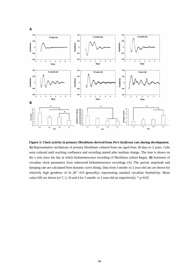
Figu
A) R
were
the x
circa
damp
relat
value
A
B
ure 3: Clock a
Representative
e cultured unt
x axis since th
adian clock p
ping rate are c
tively high go
e±SD are show
*
activity in pri
e oscillations o
til reaching co
he day at whi
parameters fro
calculated from
oodness of fi
wn (n=7, 3, 1
*
imary fibrobl
of primary fib
onfluence and
ich biolumine
om subtracted
m dynamic cu
it (R2 >0.8 g
0 and 4 for 3 m
96
lasts derived
broblasts cultu
d recording sta
escence record
d bioluminesc
urve fitting. D
generally), rep
month- to 2 y
*
from Per1-lu
ured from rats
arted after me
ding of fibrob
ence recordin
Data from 3 mo
presenting sta
ear-old rat res
uciferase rats
s aged from 10
dium change.
lasts culture b
ngs (A). The
onth- to 2 year
andard circadi
spectively). * p
s during deve
0 days to 2 ye
. The time is
began. B) Su
period, ampl
r-old rats are
dian rhythmici
p<0.05
*
lopment.
ears. Cells
shown on
mmary of
litude and
shown for
ity. Mean
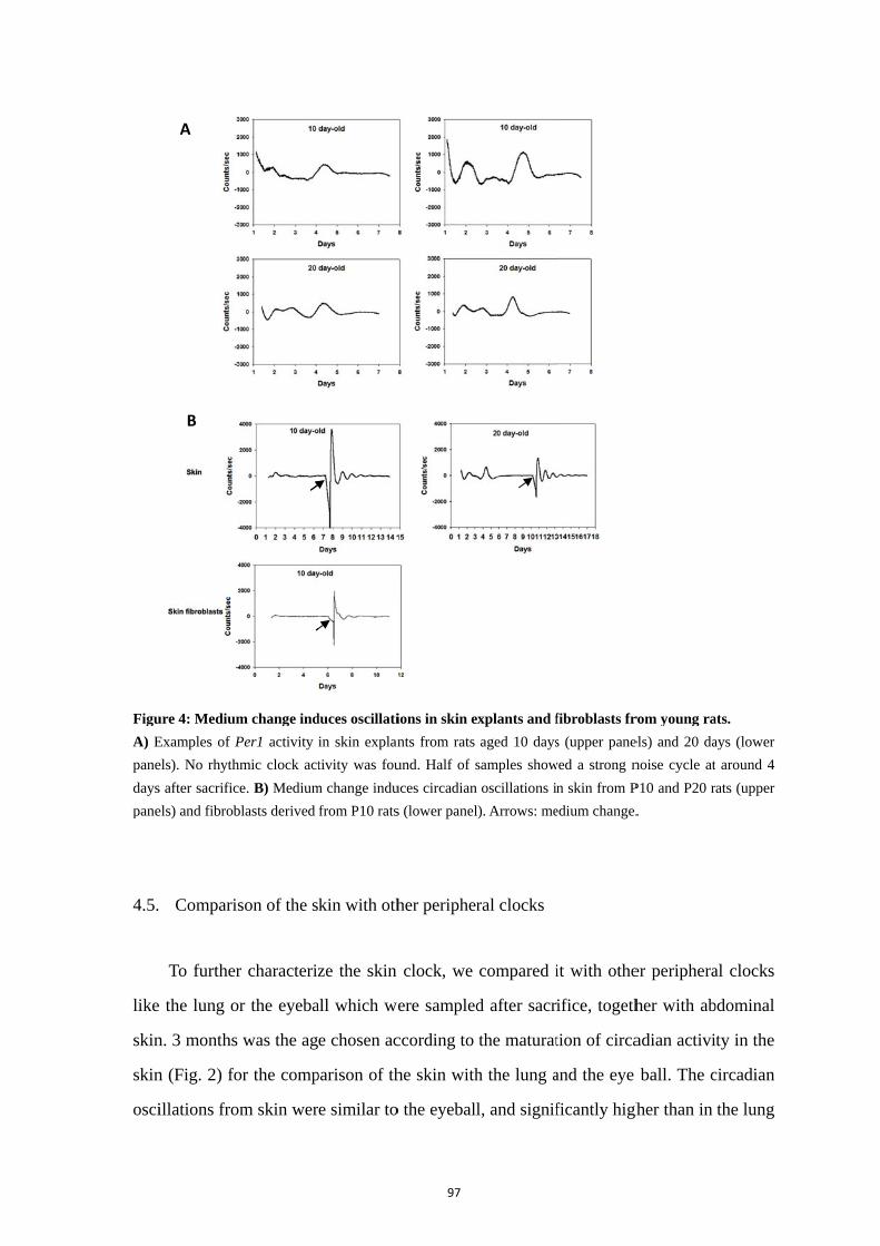
Figu
A) E
pane
days
pane
4.5.
like
skin
skin
osci
ure 4: Medium
Examples of P
els). No rhyth
s after sacrific
els) and fibrob
Comparis
To further
e the lung o
n. 3 months
n (Fig. 2) fo
illations fro
A
B
m change ind
Per1 activity
hmic clock act
ce. B) Medium
blasts derived
son of the s
r characteriz
r the eyeba
was the ag
or the comp
om skin wer
duces oscillati
in skin explan
tivity was fou
m change indu
from P10 rats
kin with oth
ze the skin
all which we
e chosen ac
parison of th
re similar to
97
ions in skin e
nts from rats
und. Half of s
uces circadian
s (lower panel
her peripher
clock, we
ere sampled
ccording to
he skin with
o the eyeball
xplants and f
aged 10 days
samples show
oscillations i
). Arrows: me
ral clocks
compared i
d after sacr
the maturat
h the lung a
l, and signif
fibroblasts fr
s (upper pane
wed a strong n
n skin from P
edium change.
it with othe
ifice, togeth
tion of circa
and the eye
ficantly high
rom young ra
els) and 20 da
noise cycle at
P10 and P20 r
.
er periphera
her with ab
adian activi
ball. The c
her than in
ats.
ays (lower
around 4
ats (upper
al clocks
bdominal
ty in the
circadian
the lung
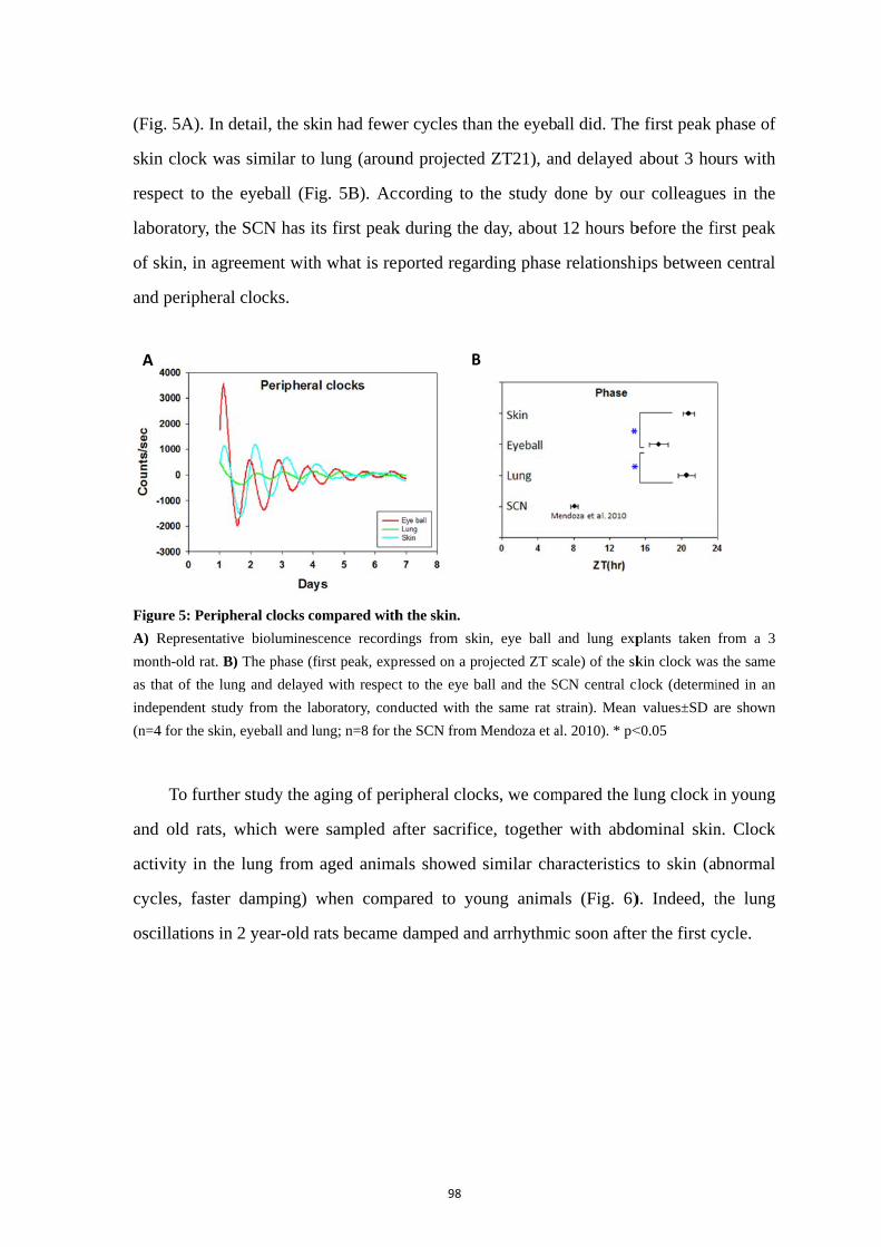
(Fig
skin
resp
labo
of s
and
Figu
A) R
mon
as th
indep
(n=4
and
activ
cycl
osci
A
g. 5A). In de
n clock was
pect to the
oratory, the
skin, in agre
peripheral
ure 5: Periphe
Representative
nth-old rat. B)
hat of the lung
pendent study
4 for the skin,
To further
old rats, w
vity in the
les, faster
illations in 2
etail, the sk
s similar to
eyeball (Fi
SCN has it
eement with
clocks.
eral clocks co
e bioluminesc
The phase (fi
g and delayed
y from the lab
eyeball and lu
r study the a
which were
lung from
damping) w
2 year-old r
kin had fewe
lung (aroun
g. 5B). Ac
ts first peak
h what is rep
ompared with
cence recordi
first peak, expr
d with respect
boratory, cond
ung; n=8 for th
aging of per
sampled a
aged anima
when comp
rats became
98
er cycles tha
nd projected
cording to
k during the
ported rega
h the skin.
ings from sk
ressed on a pr
t to the eye b
ducted with t
he SCN from
ripheral cloc
after sacrifi
als showed
pared to yo
damped an
B
an the eyeb
d ZT21), an
the study d
e day, about
arding phase
kin, eye ball
rojected ZT s
ball and the S
the same rat s
Mendoza et a
cks, we com
ce, together
similar cha
oung anima
nd arrhythm
B
all did. The
nd delayed
done by ou
12 hours b
e relationshi
and lung exp
cale) of the sk
SCN central cl
strain). Mean
al. 2010). * p<
mpared the l
r with abdo
aracteristics
als (Fig. 6)
mic soon afte
e first peak p
about 3 ho
ur colleague
before the fi
hips between
plants taken
kin clock was
lock (determi
n values±SD a
<0.05
lung clock i
ominal skin
s to skin (ab
). Indeed, t
er the first c
phase of
urs with
es in the
irst peak
n central
from a 3
s the same
ined in an
are shown
in young
n. Clock
bnormal
the lung
cycle.

Figu
the lu
4.6.
tem
mon
show
7A)
from
to te
first
degr
in fo
tem
reac
still
prop
calc
the
tem
tem
tren
wer
ure 6: Clock a
ung biolumin
Temperat
Clock act
mperatures a
nth-old rats
wed well-rh
). In contras
m 32 to 39 °
emperatures
t cycle were
ree, oscillat
following cy
mperature an
ching more
l in the circ
perty of tem
culated the Q
rhythms (
mperature by
mperatures a
nd line but n
re in agreem
activity in the
escence from
ture compen
tivity in sk
across the
and the fib
hythmic act
st, skin fibr
°C (Fig. 7B
s (p=0.013 o
e both large
tions had qu
ycles. The p
nd in fibrob
than 26 h a
cadian rang
mperature c
Q10 by rou
(Fig. 7E).
y 10 °C. Th
allowed skin
noticed that
ment with th
e lung from y
Per1-lucifera
nsation of th
kin and sk
physiologic
broblasts w
tivities at te
roblasts exh
). The ampl
one-way AN
e, while onl
uite low am
period lengt
lasts they w
at 37°C, as
ge. These re
compensatio
ghly estima
Q10 mean
he value of
n explants t
the frequen
e trend obta
99
young and ol
ase rats aged 3
he skin cloc
kin primary
cal range.
were culture
emperatures
hibited circa
litude in the
NOVA, Fig
ly the first
mplitudes in
ths of skin
were clearly
seen above
esults were
on. To furth
ating the bio
ns the cha
f Q10 was 0
to display o
ncies obtain
ained with f
ld Per1-lucife
3 months (left)
ck
y fibroblast
Skin expla
d from 3 m
s of 35 and
adian oscilla
e first cycle
. 7D). At 32
though was
the first cyc
showed at
y increasing
e (Fig. 7C a
e in consist
her characte
ochemical r
anging freq
0.77 in the
oscillations,
ned in these
fibroblasts (
erase rats. Re
) and 2 years (
ts were ch
ants were
month-old ra
37 °C, but
ations at tem
changed dr
2°C, the trou
s large at 35
cle and dam
tendency to
g (p<0.001 o
and 7D). Bu
ence with t
erize this pr
eaction rate
quency wh
skin fibrob
, we could
cases (Q10
(Fig. 7E).
epresentative p
(right) (n=4).
hecked at d
sampled fr
ats. The sk
t not at 32
mperatures
rastically ac
ugh and the
5 °C. At 37
mped like th
o increase a
one-way AN
ut the perio
the circadia
roperty in s
e from frequ
hen increas
blasts. Since
not trace th
0 calculated
profiles of
different
rom 2.5
kin clock
°C (Fig.
ranging
ccording
e peak in
7 and 39
he others
at higher
NOVA),
ods were
an clock
skin, we
uency of
sing the
e only 2
he same
as 0.78),
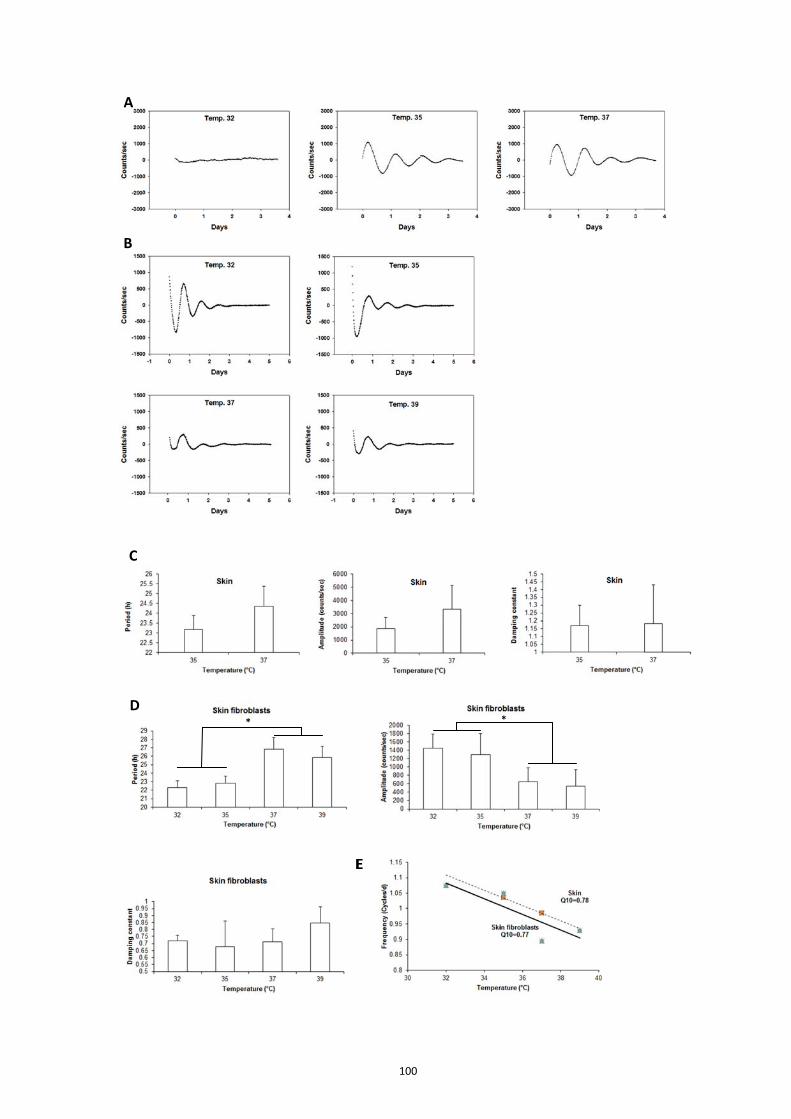
A
B
C
D *
E
100
E
*
101
Figure 7: Effects of temperature on the circadian clocks in the skin and primary fibroblasts.
A) Representative bioluminescence recordings from skin explants at 32, 35 and 37 °C. The skin biopsies
were sampled from 2.5 month-old rats. B) Representative bioluminescence recordings from skin primary
fibroblasts at 32, 35, 37 and 39 °C. Skin fibroblasts were derived from 3 month-old rats. C D) Summary of
circadian clock parameters from subtracted bioluminescence recordings in skin (C) and skin fibroblasts (D).
The period, amplitude and damping rate were calculated from dynamic curve fitting. Data are shown for
relatively high goodness of fit (R2 >0.8 generally), representing standard circadian rhythmicity. Mean
values±SD are shown (number of donors, n=3 for skin and n=4 for skin fibroblasts). * p<0.05
E) Frequency estimates of circadian rhythms by the average of period values in C and D. The Q10 is
calculated by the changing frequency when increasing the temperature by 10°C. Red: skin; blue: skin
fibroblasts
5. Discussion
The present study describes a new in vitro model for analyzing skin rhythmic
properties based on real-time bioluminescence recording in skin explants or primary
fibroblasts derived from Per1-luciferase rats. This study is the first to show that whole
skin explants are able to show high amplitude oscillations in vitro, supporting the idea
that this experimental system allows to analyze synchronization within the skin tissue as a
whole. Our work also addressed the question of the functionality of the circadian clock in
the skin and skin fibroblasts during postnatal development and ageing, an aspect of skin
rhythmic properties that has not been examined previously. The temperature study
confirmed that the clock present in skin fibroblasts conforms to the rule of temperature
compensation and that the whole skin might behave likewise.
We found clear changes in the parameters of the skin clock activity between early
life and aging. During this long life span, the internal (including circadian system) and
external environment around the skin change a lot. Involved in a clock network
coordinated by the SCN master clock, the phenotype of the skin clock might reflect the
quality of its synchronization by direct and indirect signals. Tanioka et al. showed that
circadian oscillations in the skin depend on the integrity of the SCN, although the results
of such studies conducted with several arrhythmic animals are difficult to interpret. Our
results showed that skin explants from rats over 1 month, devoid of any synchronizing
signal display high amplitude oscillations, suggesting efficient synchronization of
102
individual oscillators present in the tissue. By contrast, explants from 10-20 day rats
showed no circadian activity. The synchronizing signals from the SCN also change during
the lifetime and this potentially leads to variable clock activities in the periphery.
In the circadian system, the role of the SCN as a central pacemaker evolves with age
(Sumova et al., 2012). At P10, the amplitudes of the rhythms in clock gene expression in
the rat SCN get adult-like (Sladek et al., 2004; Kovacikova et al., 2006). The circadian
activity rhythms of AANAT, the rate-limiting enzyme of melatonin synthesis in the pineal
gland, can be observed from P10, which accurately reflects rhythmic outputs from the
developing SCN (Deguchi, 1982; Reppert et al., 1984). Peripheral clocks like liver and
lung have been found rhythmic at P11 and P20 respectively, and their phase gradually
shifts until the adult stage, while the SCN keeps the same phase during this
developmental period (Yamazaki et al., 2009). Meanwhile, the circadian clockwork in the
rat liver develops gradually and is roughly completed by 30 days after birth (Sladek et al.,
2007). We showed that in the early postnatal days, skin oscillators could be synchronized
by medium change which induced sustained oscillations in the explants (Fig. 4B). This
suggested that autonomous oscillators existed already in the P10-P20 skin. Arrhythmicity
first observed in these explants possibly reflects their desynchronisation due to the lack of
synchronizing signals. Compared with adults, neonates before weaning are exposed to
non-photic maternal cues including feeding, warmth and social contact but these might
not be efficient towards the skin clocks. Alternatively, they might require a more mature
circadian system to be efficient. Indeed, circadian activity is present early in the liver,
likely directly driven by the feeding rhythm.
Low amplitude rhythms at young age can be meaningful because it might be easier to
reset them according to environmental perturbations, an hypothesis which is supported by
mathematical modeling studies (Colwell, 2011). In the adult, we observed that periods of
skin clock activity get closer to 24 hours and that their amplitude increases. This indicates
the skin clock, until 6 months, gets more mature and better synchronized. After 1 year,
skin clock activity displays decreased amplitude and faster damping, which indicates
decreasing synchronization efficiency. Accordingly, the phases of the skin clocks get
much dispersed among individuals in aged animals. It has been reported that other
103
secondary clocks like pituitary are more dispersed in phase at old age (Yamazaki et al.,
2002). Middle-aged mice (12 to 14-months-old) have shown reduced day-night difference
in SCN neural activity (Nakamura et al., 2011a). Thus, based on the hypothesis that skin
gets entraining cues from external environment and synchronization by the SCN, our
results indicate that these processes are less efficient in the elder age but the mechanisms
involved remain to be characterized. Reversely, Bmal1 deficiency results in premature
aging phenotype in mice skin. And Clock deficiency increases the age-related dermatitis
in mice (Dubrovsky et al., 2010). These data suggest that circadian clocks are less
functional in elderly and correlated with the skin physiology. Through the development
and ageing, the changing amplitude may also result from changes in the composition of
the skin, since skin cells show cell-specific robustness in clock activities (Sandu et al.,
2012). For example, with aging, the outer skin layer (epidermis) thins, even though the
number of cell layers remains unchanged. The number of melanocytes decreases, but the
remaining melanocytes increase in size.
Fibroblasts cultures in our study showed circadian profiles similar to skin during
postnatal development. The opposite tendency of damping rate in skin fibroblasts and
skin explants suggested that whole skin oscillations not only reflect clock activity in
fibroblasts. Given the fact that the medium change in the beginning constitutes a
synchronizing signal, it may indicate that the response capacity of fibroblasts evolves
during the development and that this is likely determined endogenously. Unlike neuronal
tissues such as the SCN and the retina, apparent damping observed in the skin circadian
oscillation can be explained by a reduction of medium nutrition or luciferin–luciferase
reaction in the cells due to impoverishment of the medium conditions, although the
amount of luciferin is quite enough in the medium. In skin explants and primary
fibroblasts, refreshment of culture medium reinitiates circadian rhythms of
Per1-luciferase supporting the idea that peripheral rhythms are also self-sustained, as is
the case of the SCN circadian rhythms (Yoo et al., 2004).
Since skin is an interface tissue exposed to varying environmental temperatures, the
biological reaction underlying its clock might be influenced by these changes. At very
low temperature like 32 °C, abdominal skin lost its rhythmicity. However, in skin
104
fibroblasts, circadian rhythms still existed at 32°C, although the oscillations damped fast.
In rat-1 fibroblasts, the circadian rhythm of Per1 gene keeps its pace in low temperature
even at 28°C and Q10 in rat-1 fibroblasts is 0.85-0.88 (Izumo et al., 2003). Taken together
with those from rat-1 cells, our data indicate that the apparent desynchronization observed
at low temperature may be due to the loss of phase coherence rather than the absence of
individual rhythms. Moreover, in fibroblasts, low temperatures of 32 and 35 °C induced a
large amplitude cycle in the first day. This might be correlated with the over
compensation at these temperatures: enzymatic reactions get faster at lower temperature
and lead to the high amplitude in the Per1 gene transcription. Overall, our results indicate
that clocks in the skin are temperature compensated in the physiological range of
temperatures, which probably helps to keep its own rhythmic pace in the changing
environment. It is also one fundamental property intrinsic to circadian clocks, which is
confirmed in skin by our study.
Our data allow concluding that the skin clock undergoes maturation and ageing, with
different levels of synchronization, along the life span. Mechanisms underlying these
observations need to be further determined. We confirm that the skin is a self-sustained
oscillator, capable to be synchronized and temperature compensated. In the clock network,
skin has distinct phase and oscillation pattern as compared with other peripheral clocks. It
represents a valuable experiment model to study peripheral clock properties and
synchronization mechanisms.
105
Chapter 2
Synchronizing effects of melatonin on skin clock
106
1. Abstract
In the mammalian circadian clock system, the central pacemaker in the
suprachiasmatic nucleus (SCN) influences numerous physiological activities by launching
both neuronal and hormonal signals to deliver circadian messages as endogenous
timekeeping cues. Several SCN-controlled signals such as glucocorticoids and
temperature changes have been evidenced to have synchronizing effect on peripheral
clocks, especially fibroblasts, widely used to model peripheral synchronization.
As one significant output from the SCN, melatonin has shown entraining effect on
the central clock in a restricted time window. In our study, we demonstrated a
phase-dependent effect of melatonin to synchronize primary skin fibroblasts. Following
melatonin application at 2 h after the oscillation peak, the amplitude of Per1 activity was
significantly increased in the same range as following treatment by forskolin, another
well-studied synchronizer. Our study shows for the first time an effect of melatonin on
synchronization of peripheral clocks.

107
2. Introduction
In the circadian timing system, the SCN pacemaker orchestrates the subsidiary
clocks in periphery. Throughout the organisms, circadian oscillators locate in almost all
cells of organs and tissues. This multioscillatory network is modulated by the daily
resetting cues derived from the SCN through neuronal and humoral pathways. Among
humoral pathways, it has been reported that serum shock can synchronize rat-1 fibroblasts
and glucocorticoids can synchronize peripheral oscillators including liver, kidney, cornea
and rat-1 fibroblasts (Balsalobre et al., 1998; Balsalobre et al., 2000; Pezuk et al., 2012).
Other synchronizing candidates have been proposed including melatonin whose synthesis
is controlled by the SCN-pineal axis.
Melatonin is a strong output from the SCN pacemaker with robust daily rhythms. In
most vertebrates, melatonin is synthesized primarily in the pineal gland at night,
independently of locomotor activity pattern. Given the fact that melatonin is not stored in
the pineal cells and immediately released into the general circulation, the plasma levels of
melatonin precisely reflect its pineal synthesis. Melatonin secretion is only influenced by
the light/dark cycle, thus could be regarded as a reliable estimate of the functioning of the
SCN clock. Melatonin can induce behavioral phase-advance when injected at subjective
dusk with pharmacological doses (Pitrosky et al., 1999; Slotten et al., 2002). Since
high-affinity melatonin receptors MT1 and MT2 are expressed in various peripheral
tissues besides the SCN, melatonin is a potential candidate as a synchronizer of peripheral
clocks.
Besides pineal gland, melatonin synthesis has been found to occur in many central
and peripheral tissues including the skin (Conti et al., 2000; Bubenik, 2002; Liu et al.,
2004; Slominski et al., 2005b). Melatonin exhibits 10- to 100-fold levels in mouse skin,
vibrissae follicles, and human hair follicle with respect to plasma (Kobayashi et al., 2005).
Given the numerous known biological effects of melatonin, it may serve in the skin to
protect and modulate its function, where its short plasma half-life suggests it could be
useful as an internal time-giver for the skin in the circadian clock network.
Melatonin effects on skin function involve actions mediated by membrane receptors
108
expressed in skin cells. It has been demonstrated that human and rodent skin cells and
tissues express high-affinity membrane receptors MT1 and MT2 with variations among
species(Slominski et al., 2005b; Fischer et al., 2006a). Expression of melatonin receptors
in several skin cells including normal and malignant fibroblasts, keratinocytes and
melanocytes, indicates the potential role of melatonin as a time giver to synchronize all
the oscillators within the skin.
Expression of MT1 and MT2 receptors in skin cells has been found to be affected by
environmental factors, especially UVB, which act differentially according to cell type and
genetic background (Slominski et al., 2005b).
The major layers of the skin, epidermis, dermis and adnexa are all targets for
melatonin regulation, including numerous effects observed in skin biology and clinical
dermatology, from pigmentation to hair regrowth (Fischer et al., 2008). In rats after
pinealectomy, a reduction of collagen deposition was observed, which induces atrophy of
the epidermis, dermis and hair follicle, similar to the aging phenotype in skin thickness,
whereas melatonin administration could reverse collagen fiber damage and mitochondrial
swelling in these animals (Drobnik and Dabrowski, 1996, 1999; Esrefoglu et al., 2005).
In clinics, several skin diseases have been correlated with abnormal plasma
melatonin levels in patients. For example, plasma melatonin levels lose their circadian
rhythms in psoriasis, and their rhythms exhibit greatly reduced robustness in atopic
eczema (Mozzanica et al., 1988; Schwarz et al., 1988; Rupprecht et al., 1995). To many
specific skin diseases, skin-related pathologic conditions and syndromes, melatonin
shows potential application as a skin protectant by topical or oral administration (Fischer
et al., 2008).
To investigate the effect of melatonin on the skin clock, we performed acute
melatonin treatment at different times of the circadian cycle and measured the real-time
bioluminescence of skin primary fibroblasts derived from Per1-luciferase transgenic rats.
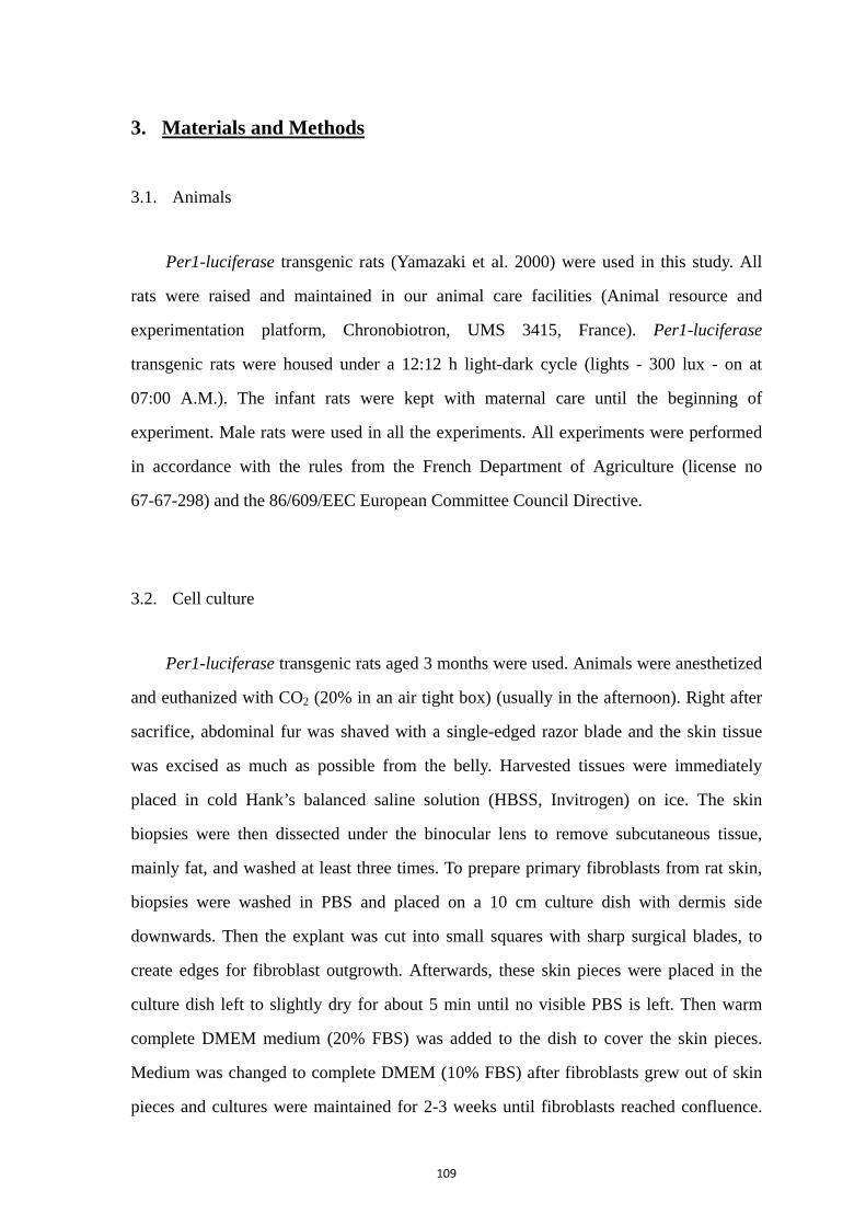
109
3. Materials and Methods
3.1. Animals
Per1-luciferase transgenic rats (Yamazaki et al. 2000) were used in this study. All
rats were raised and maintained in our animal care facilities (Animal resource and
experimentation platform, Chronobiotron, UMS 3415, France). Per1-luciferase
transgenic rats were housed under a 12:12 h light-dark cycle (lights - 300 lux - on at
07:00 A.M.). The infant rats were kept with maternal care until the beginning of
experiment. Male rats were used in all the experiments. All experiments were performed
in accordance with the rules from the French Department of Agriculture (license no
67-67-298) and the 86/609/EEC European Committee Council Directive.
3.2. Cell culture
Per1-luciferase transgenic rats aged 3 months were used. Animals were anesthetized
and euthanized with CO2 (20% in an air tight box) (usually in the afternoon). Right after
sacrifice, abdominal fur was shaved with a single-edged razor blade and the skin tissue
was excised as much as possible from the belly. Harvested tissues were immediately
placed in cold Hank’s balanced saline solution (HBSS, Invitrogen) on ice. The skin
biopsies were then dissected under the binocular lens to remove subcutaneous tissue,
mainly fat, and washed at least three times. To prepare primary fibroblasts from rat skin,
biopsies were washed in PBS and placed on a 10 cm culture dish with dermis side
downwards. Then the explant was cut into small squares with sharp surgical blades, to
create edges for fibroblast outgrowth. Afterwards, these skin pieces were placed in the
culture dish left to slightly dry for about 5 min until no visible PBS is left. Then warm
complete DMEM medium (20% FBS) was added to the dish to cover the skin pieces.
Medium was changed to complete DMEM (10% FBS) after fibroblasts grew out of skin
pieces and cultures were maintained for 2-3 weeks until fibroblasts reached confluence.
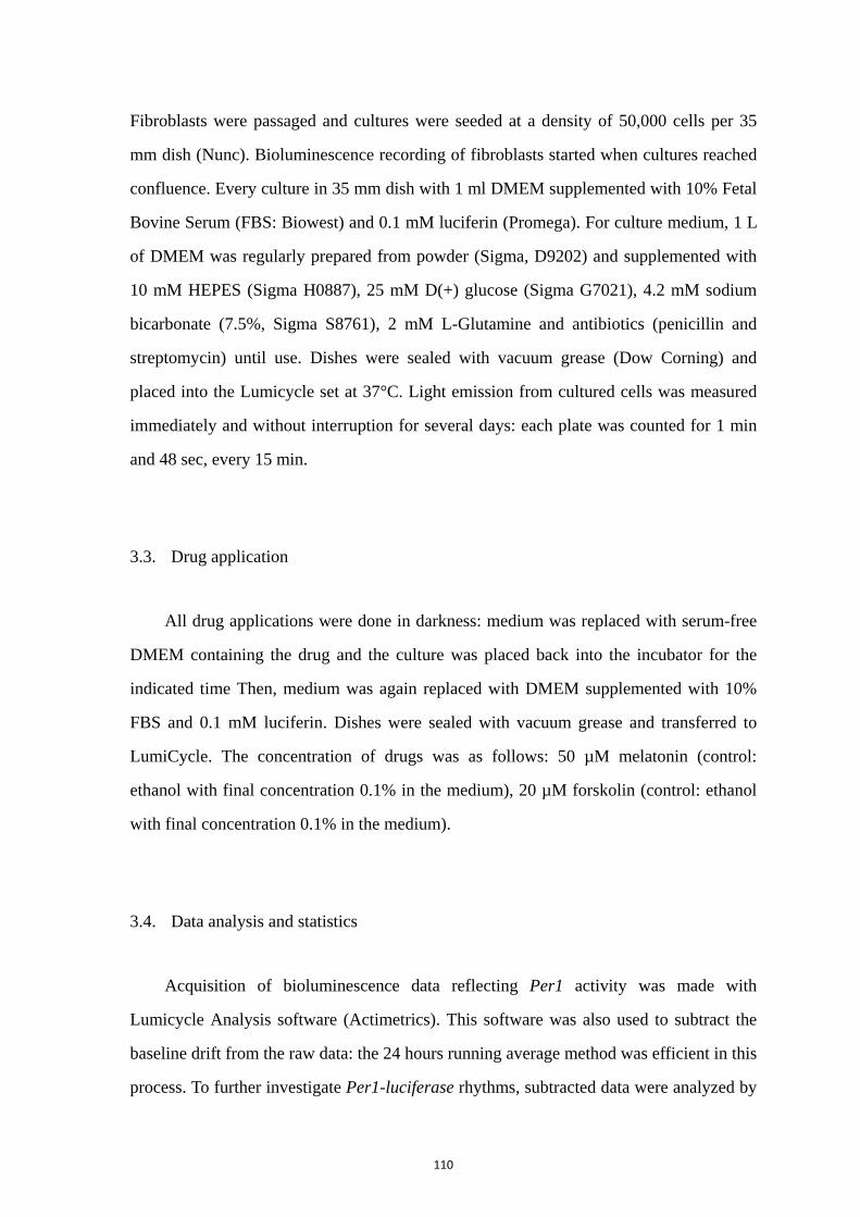
110
Fibroblasts were passaged and cultures were seeded at a density of 50,000 cells per 35
mm dish (Nunc). Bioluminescence recording of fibroblasts started when cultures reached
confluence. Every culture in 35 mm dish with 1 ml DMEM supplemented with 10% Fetal
Bovine Serum (FBS: Biowest) and 0.1 mM luciferin (Promega). For culture medium, 1 L
of DMEM was regularly prepared from powder (Sigma, D9202) and supplemented with
10 mM HEPES (Sigma H0887), 25 mM D(+) glucose (Sigma G7021), 4.2 mM sodium
bicarbonate (7.5%, Sigma S8761), 2 mM L-Glutamine and antibiotics (penicillin and
streptomycin) until use. Dishes were sealed with vacuum grease (Dow Corning) and
placed into the Lumicycle set at 37°C. Light emission from cultured cells was measured
immediately and without interruption for several days: each plate was counted for 1 min
and 48 sec, every 15 min.
3.3. Drug application
All drug applications were done in darkness: medium was replaced with serum-free
DMEM containing the drug and the culture was placed back into the incubator for the
indicated time Then, medium was again replaced with DMEM supplemented with 10%
FBS and 0.1 mM luciferin. Dishes were sealed with vacuum grease and transferred to
LumiCycle. The concentration of drugs was as follows: 50 µM melatonin (control:
ethanol with final concentration 0.1% in the medium), 20 µM forskolin (control: ethanol
with final concentration 0.1% in the medium).
3.4. Data analysis and statistics
Acquisition of bioluminescence data reflecting Per1 activity was made with
Lumicycle Analysis software (Actimetrics). This software was also used to subtract the
baseline drift from the raw data: the 24 hours running average method was efficient in this
process. To further investigate Per1-luciferase rhythms, subtracted data were analyzed by

111
using Sigma Plot 12 software. A damped sinusoid function (see below) was used to fit the
subtracted curves. The unusual data, normally in the first day after drug treatment, were
removed. We kept 3-4 full cycles of recordings to get a dynamic fitting by Sigma Plot 12
software. The window of analysis for skin fibroblasts started at a common time point
(slightly ahead of the first trough).
f = y0+a*exp(-x/d)*sin(2*pi*(x-c)/b)
The best-fit solutions were validated based on the fact that they passed the normality
test (Shapiro-Wilk test (p = <0.0001)) and passed constant variance test. With a linear
drift of y0, the standard damped sinusoid allowed to determine the period of the
oscillations by constant b. The value of a referred to the initial amplitude when x equaled
to zero ahead of damping (therefore, the time scale was shifted for the analysis, so that 0
corresponded to the beginning of the studied time window). Constant c was a corrector
for the phase corresponding to the middle of trough and subsequent peak which was used
as phase value. And d reflected the damping rate, which meant the duration (days) taken
to decrease to 37 % (1/e) of its reference level. The y0 was a corrector of Y axis. Curves
were considered rhythmic when consecutive circadian cycles showed up. Samples with
only one visible peak of Per1-luc were designated as arrhythmic and removed from the
study.
Data were presented as mean values ±SD. One-way and two-way ANOVA followed
by post hoc comparisons with the LSD Fisher test were used to compare groups. All
statistics were performed using Sigma Plot 12 software.
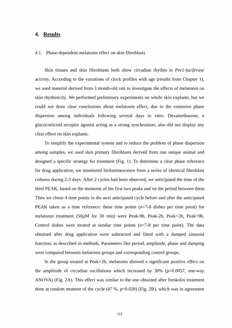
112
4. Results
4.1. Phase-dependent melatonin effect on skin fibroblasts
Skin tissues and skin fibroblasts both show circadian rhythm in Per1-luciferase
activity. According to the variations of clock profiles with age (results from Chapter 1),
we used material derived from 3 month-old rats to investigate the effects of melatonin on
skin rhythmicity. We performed preliminary experiments on whole skin explants, but we
could not draw clear conclusions about melatonin effect, due to the extensive phase
dispersion among individuals following several days in vitro. Dexamethasone, a
glucocorticoid receptor agonist acting as a strong synchronizer, also did not display any
clear effect on skin explants.
To simplify the experimental system and to reduce the problem of phase dispersion
among samples, we used skin primary fibroblasts derived from one unique animal and
designed a specific strategy for treatment (Fig. 1). To determine a clear phase reference
for drug application, we monitored bioluminescence from a series of identical fibroblast
cultures during 2-3 days. After 2 cycles had been observed, we anticipated the time of the
third PEAK, based on the moments of the first two peaks and on the period between them.
Then we chose 4 time points in the next anticipated cycle before and after the anticipated
PEAK taken as a time reference: these time points (n=7-8 dishes per time point) for
melatonin treatment (50µM for 30 min) were Peak-9h, Peak-2h, Peak+2h, Peak+9h.
Control dishes were treated at similar time points (n=7-8 per time point). The data
obtained after drug application were subtracted and fitted with a damped sinusoid
function, as described in methods. Parameters like period, amplitude, phase and damping
were compared between melatonin groups and corresponding control groups.
In the group treated at Peak+2h, melatonin showed a significant positive effect on
the amplitude of circadian oscillations which increased by 30% (p=0.0057, one-way
ANOVA) (Fig. 2A). This effect was similar to the one obtained after forskolin treatment
done at random moment of the cycle (47 %, p=0.020) (Fig. 2B), which was in agreement
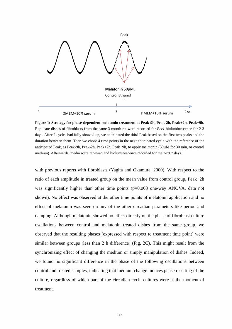
113
Figure 1: Strategy for phase-dependent melatonin treatment at Peak-9h, Peak-2h, Peak+2h, Peak+9h.
Replicate dishes of fibroblasts from the same 3 month rat were recorded for Per1 bioluminescence for 2-3
days. After 2 cycles had fully showed up, we anticipated the third Peak based on the first two peaks and the
duration between them. Then we chose 4 time points in the next anticipated cycle with the reference of the
anticipated Peak, as Peak-9h, Peak-2h, Peak+2h, Peak+9h, to apply melatonin (50µM for 30 min, or control
medium). Afterwards, media were renewed and bioluminescence recorded for the next 7 days.
with previous reports with fibroblasts (Yagita and Okamura, 2000). With respect to the
ratio of each amplitude in treated group on the mean value from control group, Peak+2h
was significantly higher than other time points (p=0.003 one-way ANOVA, data not
shown). No effect was observed at the other time points of melatonin application and no
effect of melatonin was seen on any of the other circadian parameters like period and
damping. Although melatonin showed no effect directly on the phase of fibroblast culture
oscillations between control and melatonin treated dishes from the same group, we
observed that the resulting phases (expressed with respect to treatment time point) were
similar between groups (less than 2 h difference) (Fig. 2C). This might result from the
synchronizing effect of changing the medium or simply manipulation of dishes. Indeed,
we found no significant difference in the phase of the following oscillations between
control and treated samples, indicating that medium change induces phase resetting of the
culture, regardless of which part of the circadian cycle cultures were at the moment of
treatment.
0 3 DMEM+10% serum DMEM+10% serum Days
Peak
Melatonin 50µM,
Control Ethanol
f 30 i
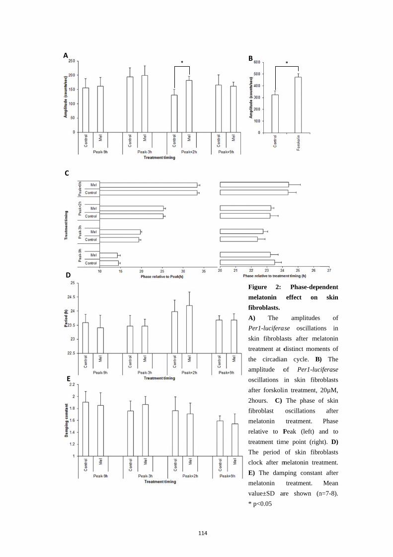
A
C
D
E
114
*
A
P
t
t
m
r
t
m
v
Figure 2:
melatonin
fibroblasts.
A) The
Per1-luciferas
skin fibrobla
treatment at d
the circadian
amplitude o
oscillations i
after forskolin
2hours. C)
fibroblast
melatonin
relative to P
treatment tim
The period
clock after m
E) The dam
melatonin
value±SD ar
* p<0.05
B
Phase-depe
effect on
amplitudes
se oscillatio
asts after me
distinct mom
n cycle. B
of Per1-luc
in skin fibr
in treatment,
The phase o
oscillations
treatment.
Peak (left) a
me point (righ
of skin fibr
melatonin trea
mping constan
treatment.
re shown (n
*
endent
skin
s of
ons in
latonin
ents of
) The
ciferase
roblasts
20µM,
of skin
after
Phase
and to
ht). D)
roblasts
atment.
nt after
Mean
n=7-8).
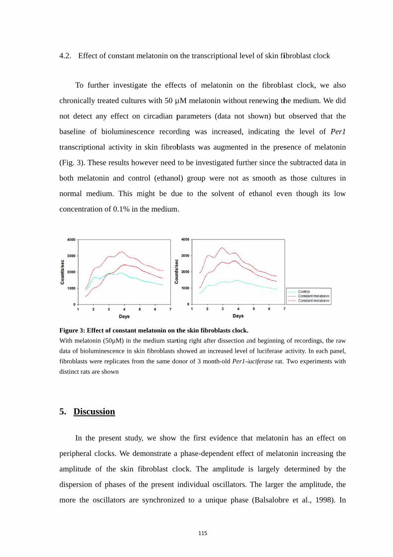
4.2.
chro
not
base
tran
(Fig
both
norm
con
Figu
With
data
fibro
distin
5.
peri
amp
disp
mor
Effect of
To further
onically trea
detect any
eline of bi
nscriptional
g. 3). These
h melatonin
mal medium
centration o
ure 3: Effect o
h melatonin (5
of biolumine
oblasts were r
nct rats are sh
Discussio
In the pre
ipheral cloc
plitude of t
persion of p
re the oscil
f constant m
r investigat
ated culture
y effect on
ioluminesce
activity in
results how
n and contr
m. This m
of 0.1% in th
of constant m
50µM) in the
escence in skin
eplicates from
hown
on
esent study,
cks. We dem
the skin fi
phases of th
llators are
melatonin on
te the effec
es with 50 µ
circadian p
ence record
skin fibrob
wever need t
rol (ethano
might be du
he medium
melatonin on t
medium start
n fibroblasts s
m the same do
, we show
monstrate a
ibroblast cl
he present i
synchronize
115
n the transcr
cts of mel
µM melaton
parameters
ding was i
blasts was
to be invest
ol) group w
ue to the s
.
the skin fibro
ting right afte
showed an inc
onor of 3 mon
the first ev
phase-depe
lock. The a
individual o
ed to a un
riptional lev
atonin on t
nin without
(data not s
increased, i
augmented
tigated furth
were not as
solvent of
oblasts clock.
er dissection a
creased level
nth-old Per1-l
vidence tha
endent effe
amplitude i
oscillators.
nique phase
vel of skin fi
the fibrobla
renewing th
shown) but
indicating
in the pres
her since the
smooth as
ethanol ev
and beginning
of luciferase
luciferase rat.
at melatonin
ct of melato
is largely d
The larger
(Balsalobr
fibroblast clo
ast clock,
he medium.
t observed
the level o
sence of m
he subtracted
s those cul
ven though
g of recording
activity. In ea
Two experim
n has an e
onin increa
determined
the amplit
re et al., 19
ock
we also
. We did
that the
of Per1
melatonin
d data in
ltures in
its low
s, the raw
ach panel,
ments with
effect on
asing the
d by the
tude, the
998). In
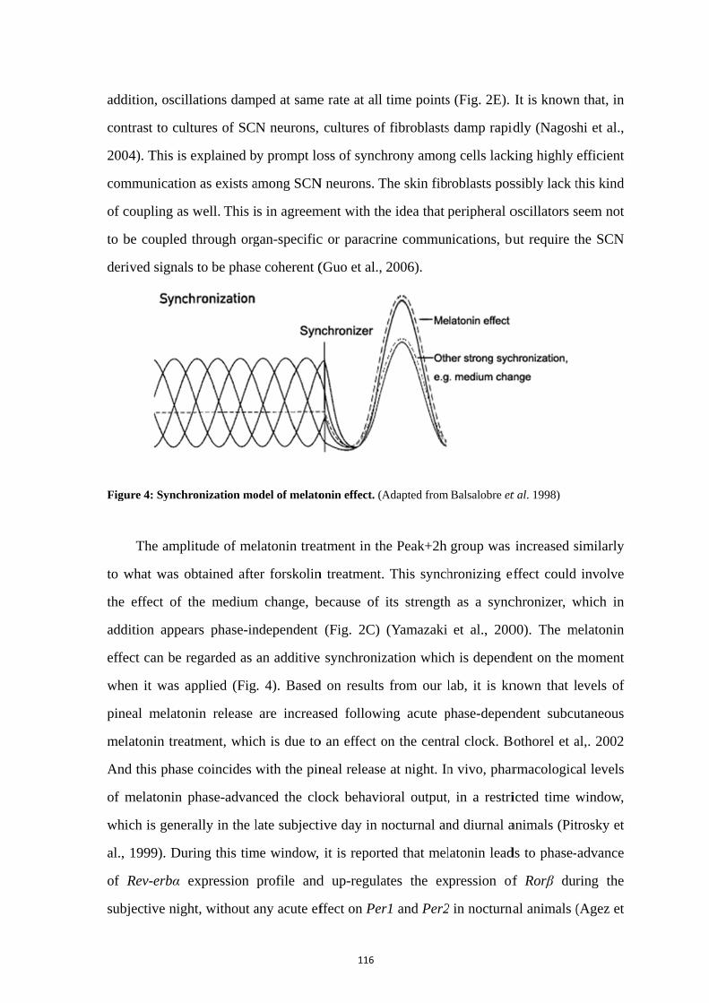
addi
cont
200
com
of c
to b
deri
Figu
to w
the
addi
effe
whe
pine
mel
And
of m
whi
al.,
of R
subj
ition, oscill
trast to cult
4). This is e
mmunication
coupling as
be coupled t
ived signals
ure 4: Synchr
The ampli
what was ob
effect of th
ition appea
ect can be re
en it was ap
eal melaton
latonin treat
d this phase
melatonin p
ich is gener
1999). Dur
Rev-erbα e
jective nigh
lations damp
tures of SCN
explained b
n as exists a
well. This i
through org
s to be phase
ronization mo
itude of me
btained afte
he medium
ars phase-in
egarded as
pplied (Fig
nin release
tment, whic
e coincides w
phase-advan
ally in the l
ring this tim
expression
ht, without a
ped at same
N neurons,
y prompt lo
among SCN
s in agreem
gan-specific
e coherent (
odel of melato
elatonin trea
er forskolin
m change, b
ndependent
an additive
. 4). Based
are increas
ch is due to
with the pin
nced the clo
late subjecti
me window,
profile and
any acute ef
116
e rate at all
cultures of
oss of synch
N neurons. T
ment with th
c or paracrin
(Guo et al.,
onin effect. (A
atment in th
n treatment.
because of
(Fig. 2C)
synchroniz
d on results
sed followi
o an effect o
neal release
ock behavio
ive day in n
it is report
d up-regula
ffect on Per
time points
f fibroblasts
hrony amon
The skin fibr
he idea that p
ne commun
2006).
Adapted from
he Peak+2h
This synch
its strength
(Yamazaki
zation which
from our l
ing acute p
on the centr
e at night. In
oral output,
nocturnal an
ted that mel
ates the ex
r1 and Per2
s (Fig. 2E).
s damp rapid
ng cells lack
roblasts pos
peripheral o
nications, bu
Balsalobre et
group was
hronizing e
h as a sync
et al., 200
h is depend
lab, it is kn
phase-depen
ral clock. B
n vivo, phar
, in a restri
nd diurnal a
latonin lead
pression of
2 in nocturna
It is known
dly (Nagos
king highly
ssibly lack t
oscillators s
ut require t
t al. 1998)
increased s
effect could
chronizer, w
00). The m
dent on the
nown that l
ndent subcu
Bothorel et a
rmacologic
icted time w
animals (Pit
ds to phase-
f Rorβ dur
nal animals (
n that, in
hi et al.,
efficient
this kind
seem not
the SCN
similarly
involve
which in
melatonin
moment
levels of
utaneous
al,. 2002
al levels
window,
trosky et
advance
ring the
(Agez et
117
al., 2007). This phase-dependent effect of melatonin is consistent with the existence of
strong rhythms in plasma melatonin levels and with the restricted window in which
melatonin affects the central clock. Whether the timing that we observed is consistent
with the nocturnal release of melatonin is to be determined. Also, since predominantly the
MT1 receptors are expressed in the skin fibroblasts, their role involved in this
phase-dependent effect needs to be determined (Slominski et al., 2003a; Fischer et al.,
2006a).
Due to the complexity of the skin, there are many types of oscillators with different
clock properties. These oscillators control numerous rhythmic physiological activities. In
the skin, various cells function in the local compartments and influence each other.
Several signal transduction receptors are coupled to classic intracellular second
messenger pathways, like cAMP mediated response. As investigated in the pituitary,
melatonin could modulate the cAMP signal pathway by inhibiting the phosphorylation of
cAMP response element binding protein (CREB) (McNulty et al., 1994). Neurons in the
SCN show oscillating activation of MAPK cascade and CREB, while efferences from the
SCN depend on the cAMP pathway and daily CREB mediated activities (Zawilska et al.,
2009). It was found that in NIH3T3 cells transfected with human MT2, high
concentration of melatonin inhibits cAMP synthesis (Reppert et al., 1995). In comparison,
forskolin elevates the cAMP level and enhances the phosphorylation and activation of
CREB. CREB has also been involved in enhancing circadian clock rhythms with the
acute induction of the Per1 mRNA in rat-1 fibroblasts (Yagita and Okamura, 2000). In
addition, melatonin has been shown to be an inhibitor of SIRT1-HDAC activity, as well
as of SIRT1 expression, indicating that melatonin plays a role in the regulation of
circadian rhythms through SIRT1 (Jung-Hynes et al., 2011). Further studies are necessary
to clarify the molecular mechanisms of melatonin effect.
The medium change effect on the circadian clock has been observed commonly, and
serum within the medium is necessary for initiation of the rhythmicity and for long term
culture (Yamazaki et al., 2000; Izumo et al., 2003; Ko and Takahashi, 2006). Serum shock
has been reported to synchronize rat-1 fibroblasts and Per1/2 plays an immediate role in
the process as being transcriptionally induced (Balsalobre et al., 1998). Serum shock

118
induces the phosphorylation of CREB in rat-1 cells as well as forskolin (Yagita and
Okamura, 2000). The melatonin effect could be masked by the strong synchronization of
medium change, which could indicate that melatonin is relatively weaker than medium
change or serum effect. To eliminate the influence of medium change from melatonin
effect, the flow-through system might be a promising method with fewer disturbances due
to manual manipulation, although it might be difficult to make the treatments in the
absence of serum in that case. It should be noted that all new circadian phases were nearly
identical irrespective of whether fibroblasts were treated with control medium or with
melatonin and, when considered with respect to the time of treatment, there were no
substantial differences. The corresponding phase response curve (PRC) conforms to type
0 PRC with large phase shifts, whereas normal photic or non-photic stimulus acts on
circadian behavior with gradual phase shifts, which is called type I PRC. The “restored”
circadian rhythms after medium change all begin with the ascending phase of circadian
cycle, which is unlikely to be due to the restart of the circadian rhythm. A switch from a
slow-resetting type I PRC to a fast-resetting type 0 PRC has been reported in circadian
clocks under increasing stimuli strength such as light intensity and drug concentration or
changing the sensitivity of the clock to the administered stimuli (Johnson, 1999).
Mammals like rodents tend to have gradually shifts and it requires large amounts of light
energy to make the switch from type I to type 0 PRC. It was found that circadian rhythms
in NIH3T3 fibroblasts reset by dexamethasone treatment demonstrate a type 0 PRC
(Nagoshi et al., 2004). Cell oscillations are instantaneously reset by a strong synchronizer
like dexamethasone or medium change in our study at whatever circadian phases they
used to be. By contrast, circadian oscillations in cultured liver tissues or in pup SCN are
phase reset depending on the timing of medium change and do not show type 0 PRC to
refreshment of medium (Nishide et al., 2006; Nishide et al., 2008). However, a type 0
PRC for medium change is found in the SCN of adult Clock mutant mice whereas the
SCN of wild-type mice does not respond to medium change at any phase (Vitaterna et al.,
2006). These results could be due to tissue architecture preserved in the liver slices and
the SCN. Cell types and cell communication may introduce variability in the response of
the circadian system to zeitgebers. In this context, instantaneous resetting of “peripheral
119
clocks” with strong synchronizers as observed in NIH3T3 cells or in our study, indicates a
possible mechanism of phase adjustment of peripheral rhythms by the SCN. The
glucocorticoid potency of dexamethasone is much higher than that of physiological
glucocorticoids like cortisol (Adrenal Cortical Steroids, Drug Facts and Comparisons, 5th
ed. St. Louis, Facts and Comparisons, Inc.:122-128, 1997). In addition, in
pharmacological treatment, concentration of dexamethasone or melatonin is quite higher
than physiological levels. SCN can be reset rapidly by shifts of light-dark cycle, but
resynchrony of peripheral oscillators can take over a week, which is regulated by
hormones like adrenal glucocorticoid (Yamazaki et al., 2000; Kiessling et al., 2010). And
it often takes more days of gradual phase advances than of phase delays. A relatively
weak synchronizer like melatonin is possibly involved in the synchronization of
peripheral clocks, which might be cell-specific, due also to local particularities in the
molecular clockwork. Given the fact that many peripheral cells are proliferative and need
to “pass” time to daughter cells, the use of a weak synchronizer that induces gradual
phase resetting might be the best compromise to maintain molecular clock phases in the
body (Nagoshi et al., 2004).
In the multioscillatory skin system, it is not easy to determine whether other
oscillators, in addition to fibroblasts, could be synchronized by melatonin The other cells
like keratinocytes are not as highly proliferative as fibroblasts, although keratinocytes
follow circadian rhythms in mitosis cycle (Brown, 1991; Geyfman et al., 2012). In cell
cultures, cell density is crucial for the synchronization among oscillators, in which
intercellular factors help to generate robust circadian rhythms (Noguchi et al., 2013).
Fibroblasts express connexins and are connected through gap junctions in high-density
cultures (Goodenough et al., 1996). However, rather than intercellular couplings,
paracrine pathways appear to play a more important role on helping synchronization
among cells (Noguchi et al., 2013). Existence of paracrine diffusible signals have been
shown in immortalized SCN2.2 cells which were able to confer molecular oscillations to
cocultured NIH/3T3 fibroblasts (Allen et al., 2001). These kinds of unknown diffusible
signals have also been found in the SCN-lesioned animals in which SCN transplantation
into distinct brain regions can restore circadian behavioral rhythms (Silver et al., 1996).

120
The cell to cell communication may play an important role in the response of peripheral
clocks to synchronizer. Other paracrine factor like fibroblasts growth factor (FGF),
epidermal growth factor (EGF), insulin-like growth factor (IGF) may also get involved.
In conclusion, our study shows for the first time evidence that melatonin has a
phase-dependent synchronizing effect on peripheral clocks. Whether this effect is
restricted to increasing molecular clock activity within cells or synchronization by
concentrating the phases, is to be elucidated further. The synchronization of other cells in
the skin could permit the opportunity to understand more about the synchronization
mechanisms in a multioscillatory system.

121
Chapter 3
Construct of lentivirus of Bmal1 luciferase reporter
122
1. Abstract
In circadian oscillatory system, cell-autonomous clocks exist in almost all the tissues
and cells through the organisms. Molecular clockwork involves clock gene activities and
drives clock-controlled outputs in single cells. Monitoring molecular clock activity leads
to further insight into circadian rhythms and their mechanisms in tissues and cells. In the
skin tissue, clock gene oscillations are found in fibroblasts, keratinocytes and
melanocytes as well as cells constituting hair follicles, including stem cells.
In the present work, we constructed a lentivirus tool delivering Bmal1-luciferase
reporter to study clock activities in human skin cells. Following the design from a paper
published by Brown and his colleagues (PLOS Biology 2005), we constructed this tool to
take advantage of bioluminescence, especially for human samples or samples from
species for which transgenic tools do not exist. Within this tool, luciferase expression was
controlled by the Bmal1 promoter which could be integrated into DNA of host cells via
lentiviral infection. This kind of lentiviral tools were successfully used to assay clock
properties in human fibroblasts and keratinocytes. We did not develop yet an appropriate
method to isolate and culture rat melanocytes and keratinocytes from skin but currently
use these cells derived from human biopsies. This lentiviral tool would help to investigate
clock activities not only in fibroblasts but also in melanocytes and keratinocytes of the
multioscillatory skin.
123
2. Introduction
The mammalian circadian system is composed of a network of circadian clocks
located throughout the organisms. A central clock residing in the suprachiasmatic nucleus
(SCN) of the brain synchronizes the other secondary oscillators present in most cells of
the body. Peripheral oscillators harbor cell-autonomous clocks, based on an intracellular
molecular mechanism. Genetically, molecular clockwork involves interlocking
transcriptional-translational feedback loops (Ko and Takahashi, 2006). Transcription of
Period (Per) and Cryptochrome (Cry) genes are activated by heterodimers of BMAL1 and
CLOCK factors. PER and CRY proteins forms a complex in the cytoplasm and
translocate to the nucleus, repressing their transcription driven by BMAL1/CLOCK.
Additional regulatory loops involve the regulation of Bmal1 transcription by REV-ERB
and ROR clock proteins. BMAL1/CLOCK activates the transcription of Rev-erb and Ror,
which products respectively repress and activate the transcription of Bmal1. These
negative and positive regulation loops provide original gene expression rhythms.
Circadian clock gene expressions in the skin have been reported in mouse and
human (Tanioka et al., 2009). Several types of skin cells, such as fibroblasts,
keratinocytes and melanocytes, display circadian oscillations in clock gene expression
indicating that the skin clocks are cell-autonomous (Sandu et al., 2012). Since transgenic
animals have been established by fusing luciferase genes to clock gene promoters, it has
become possible to measure circadian gene expression in different tissues and cells by
luminescence recording. Skin biopsies from transgenic rodents can be used for preparing
primary cell cultures but melanocytes and keratinocytes, unlike fibroblasts are difficult to
isolate and maintain in culture. Brown and his colleagues have successfully established a
lentivirally delivered luciferase reporter to measure clock gene activities in human skin
cells (Brown et al., 2005b). To facilitate our studies on multioscillatory skin, we
constructed a Bmal1-luciferase lentivirus according to Brown’s design, in order to study
circadian oscillations within human keratinocytes and melanocytes.

124
3. Materials and methods (Fig. 1)
3.1. PCR
To amplify the different fragments meant to constitute the luciferase cassette of the
vector, (SV40 late Poly Adenylation Signal (PAS), Bmal1 promoter, firefly luciferase and
Bmal1 3’UTR), we used the Polymerase Chain Reaction (PCR) technique with
corresponding primers designed specifically. Phusion polymerase (Thermo Scientific)
was used because of its high proofreading capacity allowing minor errors during
amplification. When Phusion polymerase was ineffective, Pfu polymerase (Euromedex)
was used. The PCR programs were as follows: for Phusion, initial heating at 98 °C for 2
minutes, followed by 35 cycles of denaturation at 98 °C for 10 seconds and annealing at
71 °C for 20 seconds and elongation at 72 °C for 30 seconds, final elongation at 72 °C for
5 min. For Taq polymerase and Pfu: initial heating at 95 °C for 5 minutes, followed by 35
cycles of denaturation at 95 °C for 1 min and annealing at 56 °C for 1 min and elongation
at 72 °C for 2 min, final elongation at 72 °C for 5 min. Genomic DNA was prepared from
tail biopsies of C57Bl6 mice. The fragments of SV40 late PAS and firefly luciferase were
amplified from pGL3 plasmid (Promega). SV40 late PAS, Bmal1 promoter, luciferase and
Bmal1 3’UTR fragments were purified, enzymatically digested and eventually ligated
into pBluescript SK+ vector (Stratagene). Primers were designed to contain restriction
enzyme sites for further oriented cloning into the cassette, like below: SV40 late PAS
(forward 5’GCC TCG AGC AGA CAT GAT AAG3’, reverse 5’AAG GAG CTG ACT
GGG TTG AAG3’), luciferase (forward 5’TGG AAT TCC GGT ACT GTT GG3’, reverse
5’CCG GAT CCT AGA ATT ACA CGG3’), Bmal1 promoter (forward 5’ GAT CGA TAG
GGC TAC AAC AGA ACA ACT AA C3’, reverse 5’ TCT GAA TTC CCT AAA TGT
CGC ACC AC3’), Bmal1 3’UTR (forward 5’CGG ATC CAC ACT ACA TTT GCT TTG
GC3’, reverse 5’TCT CGA GTA GGA GCA TAC CTG TAG GG3’).

125
3.2. Subcloning
Specific restriction enzyme sites at 5’ and 3’ ends of PCR fragments were introduced
by primers to facilitate further subcloning of these fragments into pBluescript SK+ which
contains multiple cloning sites and was used as an intermediate vector. The fragments of
Bmal1 promoter and Bmal1 3’UTR were amplified from the genomic DNA of mouse
with Pfu polymerase and polyA 3’ ends were added with Taq polymerase at 72 °C for 30
min. These fragments were then ligated into another intermediate vector specifically
designed for the easy cloning of A-tailed PCR fragments: PCR2.1 (Invitrogen). The
fragments of SV40 late PAS and luciferase were amplified from pGL3 plasmid, digested
with appropriate restriction enzymes and directly ligated into pBluescript SK+ vector. The
fragments of Bmal1 promoter and Bmal1 3’UTR were digested from the pCR2.1
subclones and then subcloned into pBluescript SK+ vector. Each fragment with two
cloning sites was digested by one restriction enzyme at 37 °C for 1 hour followed by the
purification by gel electrophoresis and then the other digestion was run in the appropriate
buffer. Ligation with T4 DNA ligase was performed at 14 °C for 6 h and products were
then transformed into NovaBlue competent cells (Millipore). Positive clones were
amplified and selected based on digestion with restriction enzymes. Individual positive
clones were sequenced to check for the absence of mutations in the target PCR fragment,
by LGC Genomic service.
Once the cassette comprising the 4 fragments was constituted inside the pBluescript
SK+ vector, the whole fragment was excised by XhoI digestion and cloned into the pWPI
lentiviral vector (Addgene). The pWPI vector was first linearized by Xho1 digestion (this
removed the GFP marker gene from the vector), followed by treatment with shrimp
alkaline phosphatase (SAP) at 37°C for 30 min (inactivation 10 min at 65°C) to remove
the phosphate groups on the 5' ends and prevent self-ligation of the linearized pPWI.
The final pWPI-Bmal1-luciferase construct was checked for the right orientation by
digestion with multiple appropriate restriction enzymes.
126
4. Results
4.1. Strategy of lentivirus construction
We designed the construction of lentiviral Bmal1-luciferase tool according to the
publication of Steven Brown and his colleagues in PLOS Biology 2005 (Fig.1). The
presence of the late SV40 polyadelylation signal aims at putting a stop to the
transcriptional unit located just upstream of the cassette, and which is driven by the
promoter of the EF1α gene, already present in the pWPI vector. This should also attenuate
the influence of transcription driven by the viral Long Terminal Repeat sequence located
at the 5’ end of the integrated cassette. Regarding the Bmal1 promoter fragment, we chose
to amplify the -968 / +67 region, that was meant to contain most regulatory elements and
transcription start site (based on genome annotation at the time of primer design).
Regarding 3’UTR fragment, we chose a 970 bp region starting right at the stop codon.
The fragments of SV40 late PAS and luciferase would be amplified from the plasmid
pGL3, while Bmal1 promoter and 3’UTR from mouse genomic DNA.
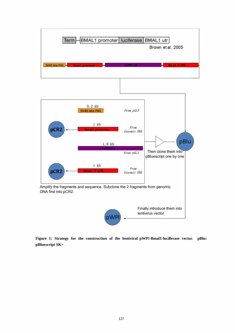
Figu
pBlu
ure 1: Strate
uescript SK+
egy for the construction
127
n of the lentiviral pWPII-Bmal1-lucifferase vector
r. pBlu:

4.2.
the
pWP
393
Figu
Construc
We cloned
right orien
PI-Bmal1-l
2 bp.
ure 2: Constru
ction of the p
d pWPI-Bm
ntation by
uciferase is
uction of the
pWPI-Bmal
mal1-lucifera
multiple e
s 13197 bp
pWPI-Bmal
128
l1-luciferas
ase as desig
enzymatic
p with the s
1-luciferase v
se
gned before
digestions
size of the
vector
(Fig. 2) an
(Fig. 3).
Bmal1-luci
nd checked i
The final
iferase inse
it was in
size of
ert being
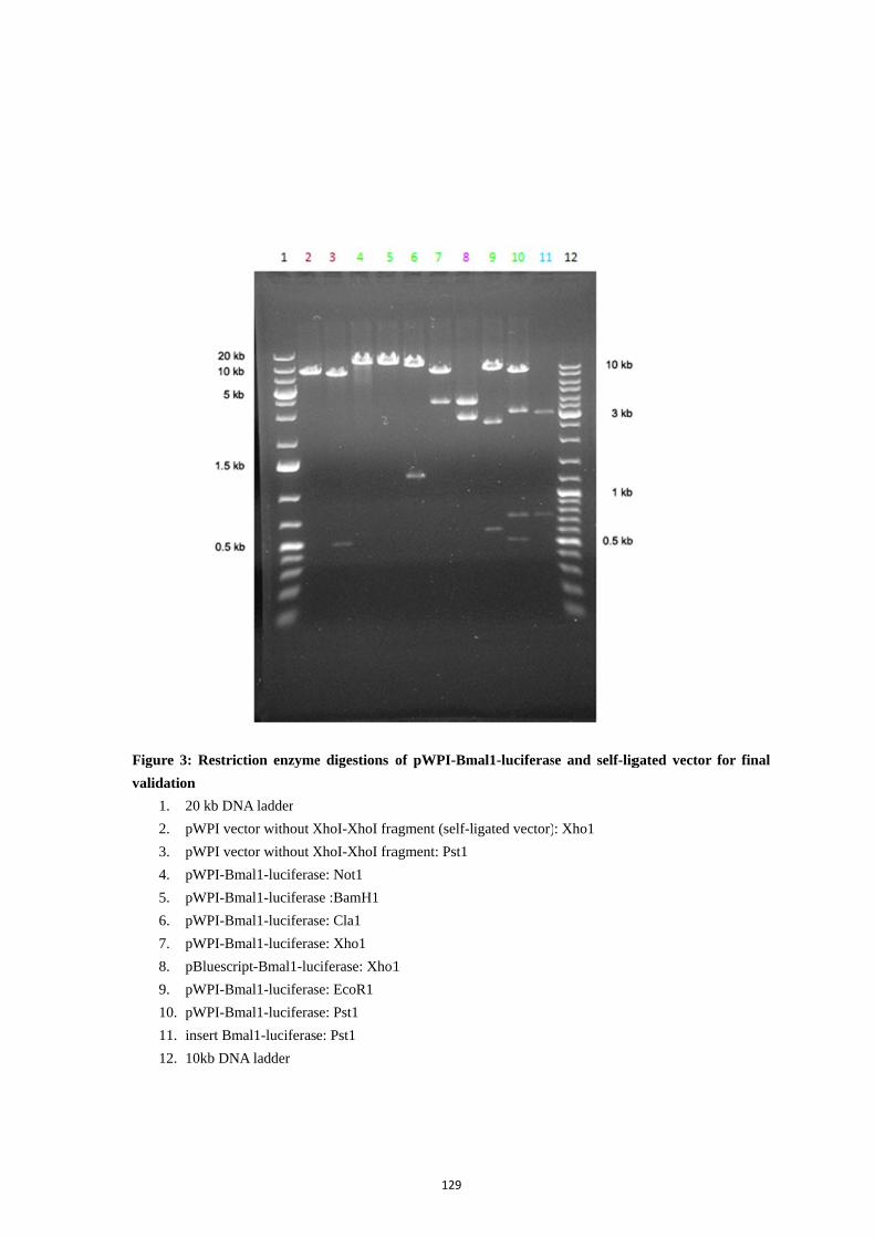
Figu
valid
ure 3: Restric
dation
1. 20 kb DN
2. pWPI ve
3. pWPI ve
4. pWPI-Bm
5. pWPI-Bm
6. pWPI-Bm
7. pWPI-Bm
8. pBluescr
9. pWPI-Bm
10. pWPI-Bm
11. insert Bm
12. 10kb DN
ction enzyme
NA ladder
ctor without X
ctor without X
mal1-luciferas
mal1-luciferas
mal1-luciferas
mal1-luciferas
ript-Bmal1-luc
mal1-luciferas
mal1-luciferas
mal1-luciferas
NA ladder
e digestions o
XhoI-XhoI fra
XhoI-XhoI fra
se: Not1
se :BamH1
se: Cla1
se: Xho1
ciferase: Xho1
se: EcoR1
se: Pst1
e: Pst1
129
of pWPI-Bm
agment (self-li
agment: Pst1
1
mal1-luciferas
igated vector)
se and self-lig
: Xho1
gated vector for final
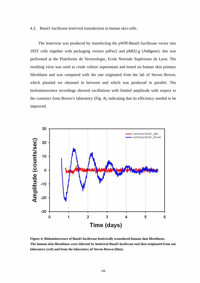
4.3.
293
perf
resu
fibr
whi
biol
the
imp
Figu
The
labo
Bmal1-lu
The lentiv
T cells tog
formed at t
ulting virus
oblasts and
ich plasmid
luminescenc
construct fr
proved.
ure 4: Biolum
human skin
oratory (red)
uciferase len
virus was pr
gether with
the Platefor
was used a
d was comp
d we obtai
ce recordin
rom Brown
minescence of
fibroblasts w
and from the
ntiviral tran
roduced by
packaging
rme de Vec
as crude cu
pared with
ined in bet
ngs showed
’s laborator
Bmal1-lucife
were infected
e laboratory o
130
nsduction in
y transfectin
vectors ps
ctorologie, E
ulture supern
the one or
tween and
oscillation
ry (Fig. 4),
erase lentivira
by lentiviral
of Steven Bro
human skin
ng the pWP
sPax2 and
Ecole Norm
natant and
riginated fro
which wa
s with limi
indicating t
ally transduc
Bmal1-lucife
own (blue).
n cells.
I-Bmal1-luc
pMD2.g (A
male Supéri
tested on h
om the lab
as produced
ted amplitu
that its effic
ced human sk
erase tool tha
ciferase vec
Addgene): t
ieure de Ly
human skin
of Steven
d in parall
ude with re
ciency need
kin fibroblast
at originated
ctor into
this was
yon. The
primary
Brown,
lel. The
espect to
ded to be
ts.
from our

131
5. Discussion
Core clock gene regulatory elements have been used separately in the context of
luciferase fusion constructs, to provide bioluminescence to measure circadian clock
activity, both in transfected cells and in explanted tissues from transgenic animals
(Yamazaki et al., 2000; Yoo et al., 2004). Many constructs of luciferase reporters driven
by clock gene promoters such as Per1 promoter, Per2 promoter and Bmal1 promoter have
been successfully used to monitor clock gene activities in a numerous types of samples
(Yamazaki et al., 2000; Nagoshi et al., 2004; Welsh et al., 2004; Yoo et al., 2004; Brown
et al., 2005b; Brown et al., 2008; Yeom et al., 2010; Noguchi et al., 2012; Saini et al.,
2012).
Besides tissues, it is convenient to measure clock-driven bioluminescence in cells
cultured from transgenic animals. In cell lines such as rat-1 fibroblasts and NIH3T3
fibroblasts, or primary cells derived from human, introduction of luciferase reporters is
required and have widely taken advantage of delivery via chemical and viral approaches.
Plasmids are easily used to transfect cell lines though exogenous DNA is not integrated
into the nuclear genome. Dilution or degradation of the introduced DNA through mitosis
following transfection can be avoided by a selection process (e.g. antibiotic resistance
encoded by the plasmid) so that a stable transfection can be accomplished with the
characteristics of good luciferase activity and good clock gene oscillations. However,
with human cells, a usually limited amount of primary cells are available and not easy to
transfect. In addition, given the small number of cells, selection is difficult to perform and
so is the stable transfection. Lentiviral infection has advantages over other transfection
methods including infection of dividing and non-dividing cells, with high copy number,
long term stable expression of a transgene and low immunogenicity. Thus, lentivirus is a
good alternative for human primary cells. In addition, since the provided titer is usually
high enough, it is quite possible to get a high percentage of transduced cells.
With the published Bmal1-luciferase construct, most reports did not explain plasmid
construction in detail. However, the exact content in Bmal1 promoter part probably plays
an important role. It is proved that the core promoter of Bmal1 gene is located upstream
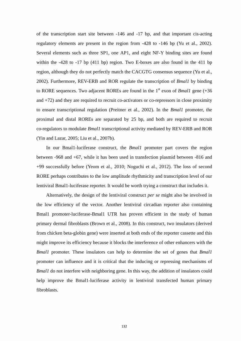
132
of the transcription start site between -146 and -17 bp, and that important cis-acting
regulatory elements are present in the region from -428 to -146 bp (Yu et al., 2002).
Several elements such as three SP1, one AP1, and eight NF-Y binding sites are found
within the -428 to -17 bp (411 bp) region. Two E-boxes are also found in the 411 bp
region, although they do not perfectly match the CACGTG consensus sequence (Yu et al.,
2002). Furthermore, REV-ERB and ROR regulate the transcription of Bmal1 by binding
to RORE sequences. Two adjacent ROREs are found in the 1st exon of Bmal1 gene (+36
and +72) and they are required to recruit co-activators or co-repressors in close proximity
to ensure transcriptional regulation (Preitner et al., 2002). In the Bmal1 promoter, the
proximal and distal ROREs are separated by 25 bp, and both are required to recruit
co-regulators to modulate Bmal1 transcriptional activity mediated by REV-ERB and ROR
(Yin and Lazar, 2005; Liu et al., 2007b).
In our Bmal1-luciferase construct, the Bmal1 promoter part covers the region
between -968 and +67, while it has been used in transfection plasmid between -816 and
+99 successfully before (Yeom et al., 2010; Noguchi et al., 2012). The loss of second
RORE perhaps contributes to the low amplitude rhythmicity and transcription level of our
lentiviral Bmal1-luciferase reporter. It would be worth trying a construct that includes it.
Alternatively, the design of the lentiviral construct per se might also be involved in
the low efficiency of the vector. Another lentiviral circadian reporter also containing
Bmal1 promoter-luciferase-Bmal1 UTR has proven efficient in the study of human
primary dermal fibroblasts (Brown et al., 2008). In this construct, two insulators (derived
from chicken beta-globin gene) were inserted at both ends of the reporter cassette and this
might improve its efficiency because it blocks the interference of other enhancers with the
Bmal1 promoter. These insulators can help to determine the set of genes that Bmal1
promoter can influence and it is critical that the inducing or repressing mechanisms of
Bmal1 do not interfere with neighboring gene. In this way, the addition of insulators could
help improve the Bmal1-luciferase activity in lentiviral transfected human primary
fibroblasts.

133
General Discussion
134
In the last decades, the circadian system has been demonstrated as a hierarchical
network that comprises numerous oscillators regulated by different zeitgebers. Within this
network, the SCN displays an indispensable function that controls many other structures,
directly or indirectly (Kalsbeek et al., 2006). These structures are regulated and
coordinated with each other and within themselves. In spite of the diversity of tissues and
cells with respect to physiological processes they are in charge of, all the paces are set at
appropriate time of the day and evolve during the life time. To keep this coherent
circadian network, many kinds of clocks in periphery possess specific patterns and
responses to various time cues. How are peripheral clocks synchronized, from the
generation of circadian molecular rhythms to the coherence among oscillators? Since the
skin expresses circadian clock genes and plays multiple roles in the organism (Le Fur et
al., 2001; Tanioka et al., 2009; Sandu et al., 2012), our goal in this thesis was to
investigate peripheral clock properties and synchronization in the skin.
As one peripheral clock, the skin displays several properties, such as being
multicellular, proliferative and multiple-influenced from endogenous and exogenous
environment, so that it constitutes an interesting model to study synchronization in the
circadian system, including through development and ageing process. Technically, the
real-time bioluminescence recordings driven by Per1 transcriptional activities constitute a
useful tool to study the clock phenotypes. The skin exhibits its distinct role in the clock
network and faces numerous zeitgebers. We showed the potential time-giver role of
melatonin which has synchronizing effects on skin clocks, at least on the skin fibroblasts,
suggesting new discussion in the field of the entrainment/synchronization of the circadian
system.
135
1. Circadian clock in the skin
Skin has its distinct structure and function in the mammalian organisms, with many
physiological activities regulated by a circadian clock. It is commonly considered that
biological circadian clocks have several features such as self-sustained, able to be
entrained or synchronized and temperature compensation. To clarify the circadian clocks
in the skin, all these properties of circadian clocks will be discussed in detail, based on
our results and those from the literature.
First, circadian clocks can sustain and produce oscillations in the absence of any
external cues. This property is due to cell-autonomous oscillators which are genetically
determined. The molecular transcriptional-translational feedback loops drive circadian
paces endogenously. In whole mouse skin, ex vivo analysis showed that expression of
core clock genes Bmal1, Cry1, Per1/2, Rev-erbα and Dbp sustain circadian rhythms under
constant dark condition (Tanioka et al., 2009). Relative phases of gene expression patterns
and that 4-8 hours phase delay between Per2 mRNA and protein accumulation, are both
consistent with the fundamental molecular feedback loops described to generate circadian
oscillations at the level of transcription (Ko and Takahashi, 2006). In human skin, our lab
has demonstrated rhythmic pattern of core clock genes transcriptions in the human
keratinocytes, melanocytes and dermal fibroblasts (Zanello et al., 2000; Sandu et al.,
2012). Thus, skin clock activity in the whole skin results from the circadian clocks
present in multiple individual cells.
These molecular-driven cell-autonomous oscillations pass through the cell division,
which may introduce a phase shift in the daughter cells because of various amounts of
core clock proteins transmitted through division (Nagoshi et al., 2004). Skin is a
self-renewing tissue in which multiple skin cells go through controlled and gated
proliferation throughout the lifespan. Several mechanisms are involved in the regulation.
For example, miRNAs regulate skin keratinocytes differentiation and other physiological
process in elderly cellular protein expression (Schneider, 2012). A clock transcriptional
regulator NONO has been reported to gate cell cycle in the skin (Kowalska et al., 2013).

136
Telomere shortening acts as a mitotic gate that prevents aberrant proliferation such as
cancer (Buckingham and Klingelhutz, 2011). Among transcripts regulated in a
daytime-dependent manner, Krüppel-like factor 9 (Klf9) was identified in human
epidermal keratinocytes as a circadian transcriptional regulator with an expression highly
sensitive to cortisol and with its protein bearing a strong anti-proliferative potential (Sporl
et al., 2012). In epidermis, putative Klf9 target genes including
proliferation/differentiation markers also show rhythmic expression in vivo (Sporl et al.,
2012). Given the fact that many skin cells keep organized growth and daily proliferation
in a controlled rate, the clock activity is quite possibly involved in this process of daily
mitosis and stem cells differentiation (Janich et al., 2011; Janich et al., 2013). Resilience
to cell division is essential in many skin physiological processes, such as terminal
differentiation of epidermal keratinocytes, activation of hair follicle. The daily mitotic
rhythmicity is one key aspect discriminating normal cells and immortalized (cancer) cells.
As proliferative cells, skin fibroblasts are modulated by the internal clocks to keep the
pace on the proliferation, which is also one factor that may cause intercellular
desynchronization in vivo and in vitro. In vitro, this fibroblasts proliferation is serum
dependent and might shift when fibroblasts are kept in the lumicycle with limited serum
for several days.
In the central clock SCN, neurons normally do not divide, whereas in the periphery
cell proliferation and differentiation is much more common for self-renew and self-repair.
And these cellular activities are often gated by local clocks. Given the fact that many cells
keep organized growth and proliferation which may introduce phase shift in divided cells,
the synchronization of clock activity is quite possibly involved in this process. Rhythmic
pattern can be passed through mitosis from mother to daughter cells (Nagoshi et al., 2004).
In skin, keratinocytes differentiation is regulated by circadian clock and arrhythmia leads
to disturbed tissue growth (Janich et al., 2011; Janich et al., 2013). In vivo, as
proliferative cells, skin fibroblasts are modulated by the synchronized internal clocks to
keep the pace on the proliferation. In vitro, loss of synchronization may cause
intercellular asynchrony in cultures. Cultured human fibroblasts lose replicative capacity
after approximately 50 population doublings, commonly termed the Hayflick limit, with a

137
half-life of only approximately 8 doublings. The relevance of either figure to survival of
cells in the body is questionable, given that stem cells in some renewing tissues
undergo >1,000 divisions in a lifetime with no morphological sign of senescence (Rubin,
2002). In another way, this could provide a easily arrhythmic model to study
synchronization, for which skin primary fibroblasts proliferation is serum dependent and
might phase shift when kept in the lumicycle with limited serum for several days.
Abnormality in the skin clock is intimately related to pathological phenotype.
Melanoma, the least common form of skin cancer, is often lethal compared with
non-melanoma skin cancers (Desotelle et al., 2012). Expression levels of clock gene
mRNAs and proteins is reduced in human skin melanoma, which significantly associates
with clinical characteristics like Breslow thickness and is likely caused by altered cellular
composition (Lengyel et al., 2013).
Understanding the molecular basis of cell-autonomous oscillations provides a good
insight into the skin circadian system. In fact, conventional methods to examine the
circadian clocks in the skin can hardly reflect continuously the functionality of
autonomous oscillators in the skin. In our study, the real-time bioluminescence recordings
of Per1 gene activity within the skin allowed the monitoring of the clock gene
rhythmicity in the global view of skin multioscillatory structure. Among different
biochemical methods used to measure circadian rhythms in vivo and in vitro, only
bioluminescence recordings employed allow long-term and continuous monitoring of
individual tissue samples, which greatly reduce the problems caused by variability among
individuals. This long lasting recording is quite meaningful for peripheral oscillators like
lung and liver which can be monitored during more cycles (Yoo et al., 2004).
Subsequently, this method provides an insight into the rhythmic pattern of constituent
cells without disturbing intercellular communication. It allowed us to study the
synchronization between living cells. In the molecular clockwork, the Per1 gene per se
reflects the general activities of circadian clocks. The Per1 deficient embryonic
fibroblasts showed rhythms with short period in the culture (Pando et al., 2002). And
Per1 and Per2 have been reported several times as immediate early regulators in the
synchronization of circadian clocks in the SCN and rat-1 fibroblasts (Balsalobre et al.,

138
1998; Farre et al., 2005). In this respect, our Bmal1-luciferase lentivirus construct will
offer an opportunity to investigate the clock of human skin in another view.
Circadian clocks are capable to be entrained or synchronized by daily external cues
to keep harmony among themselves and with the environment. Light is the most chief
zeitgeber in nature. Although it does not have direct influence on the skin clock, UVB has
been reported to alter clock gene oscillation in human keratinocytes (Kawara et al., 2002).
Not surprisingly, light exposure (3 h, broad spectrum fluorescent white light, 13000 lux)
on abdomen and chest skin shows no effect on the circadian rhythms of serum melatonin,
cortisol and thyrotropin in six subjects, which does not support the existence of
extraocular photic regulation of the circadian rhythms in humans (Lindblom et al., 2000).
The phase of the skin clock is delayed with respect to that of the SCN, as is the case for
other peripheral clocks, which might offer the duration for synchronizing signals like
neuronal or humoral to affect the skin clocks (Result Chapter 1 Fig. 5B) (Shigeyoshi et al.,
1997; Yamazaki et al., 2002). In the SCN-lesioned animals, it was not possible to assess
whether peripheral tissues did lose the circadian rhythms because of desynchronization
between individuals (Tanioka et al., 2009). This indicated that SCN controlled signals
were necessary to synchronize the skin clock between individuals. Our data (Result
Chapter 1 Fig. 4B) proved that the oscillators of the skin from 10 day-old donor can been
synchronized, which suggests that functional cell-autonomous oscillators exist and can be
entrained in the complex of the skin.
An intrinsic feature of circadian clocks is temperature compensation which ensures
that endogenous periods of circadian clocks are not altered by changing temperature
across the physiological temperature range (Pittendrigh, 1993). The temperature
coefficient Q10 is 2-3 for most biochemical reactions, but 0.8-1.2 in circadian rhythms
(Sweeney and Hastings, 1960). It is unclear but possible that synthesis and degradation
rates of core clock components are equally affected by changing temperature.
Posttranslational pathways may get involved, for instance that enzymatic activity of CK1
toward circadian substrates such as PER2 is temperature insensitive, but temperature
139
sensitive toward non-circadian substrates (Isojima et al., 2009). In mammals, temperature
compensation has been demonstrated in cell cultures like fibroblasts and SCN neurons,
and tissues like SCN, lung and adrenal gland (Reyes et al., 2008; Dibner et al., 2009;
Buhr et al., 2010). This property is crucial for phase coherence of peripheral clocks, for
which cellular transcription rates can vary dramatically according to tissue (Dibner et al.,
2010). Some interface tissues like skin, especially distal skin, are submitted to largely
changing ambient temperatures, even in homoeothermic animals. In contrary to the distal
skin, proximal skin gets close to the core body and has less difference in daily rhythms.
At very low temperature like 32 °C which does not exist in physiological condition for
the proximal skin, abdominal skin lost its rhythmicity. Even the temperature of the distal
skin is not lower than 33 °C. However, in skin fibroblasts, circadian rhythms still exist at
32 °C, although the oscillations damp rapidly. In rat-1 fibroblasts, the circadian rhythm of
Per1 gene keeps its pace at low temperature, even at 28 °C (Izumo et al., 2003). And Q10
in rat-1 fibroblasts is 0.85-0.88, indicating that fibroblasts are even overcompensated
(Tsuchiya et al., 2003). These data suggested that the loss of obvious rhythmicity at low
temperature may be due to the loss of phase coherence rather than to the absence of
individual rhythms. In fibroblasts, low temperature of 32 and 35 °C induced a large
amplitude cycle in the first day. This might be correlated with the overcompensation
reported at these temperatures. It might be that the enzymatic reactions involved in the
quite conserved transcriptional-translational loops, get faster at lower temperature and
lead to the high amplitude observed in the Per1 gene transcription. This would indicate
that skin clocks are temperature compensated in the physiological range of temperature,
which helps keep its own rhythmic pace in the environment and that cellular oscillators
are resilient to changes in temperature, independent of intercellular coupling.
140
2. Synchronization of peripheral clocks during the development and
ageing
During the life span, the functioning of circadian clocks change since the young age
to elderly. In human, babies spend a lot of time sleeping and get sleep early. It is found
that the phases of sleep/wake cycles shift delayed at adolescence, perhaps due to modern
life style and hormonal changes. Based on simple questionnaires, from adolescence
onward, human circadian clocks shift advanced upon ageing. Between the ages of 20 and
80 years, the timing of sleep shifts an average of 2 h (Roenneberg et al., 2007).
Behavioral and sleeping pattern changes are reported in subjects aged 50–60 years
(Hofman and Swaab, 2006). In addition, the total amount of slow-wave sleep decreases as
well as the period and cycles of rapid-eye-movement sleep (Espiritu, 2008). Disturbed
sleep is an explanation for advanced phase of circadian clocks in elderly. In both of center
and periphery, circadian clock gene activities and related functions have been described as
a changing process due to maturation and ageing in entrainment and synchronization.
Skin per se, during postnatal development, gets mature and aged with large
differences in its structure and physiology. Infant skin has thinner stratum corneum and
smaller keratinocytes. During this long span of time, the internal and external
environment around the skin change a lot together with the endogenous clockwork. In
elderly, the skin does not only undergo aging because of endogenous factors, but also due
to exogenous ones like direct contact with several environmental factors like UV
irradiation. UVB exposure induces the increase in dermal collagen fibril diameter and
elastogenesis (Carneiro et al., 2007). As a consequence, aged skin shows typical
characteristics including wrinkles, dryness and loss of elasticity (Makrantonaki and
Zouboulis, 2007). In addition, UVB alters clock gene oscillation in human cultured
keratinocytes in addition to its effect on the expression of melatonin receptors (Kawara et
al., 2002; Slominski et al., 2005b). Generally, “endogenous” skin aging includes decrease
in proliferative capacity, DNA repair capacity and telomeres which are common in
cellular senescence. Importantly, as a member of the multioscillatory network coordinated
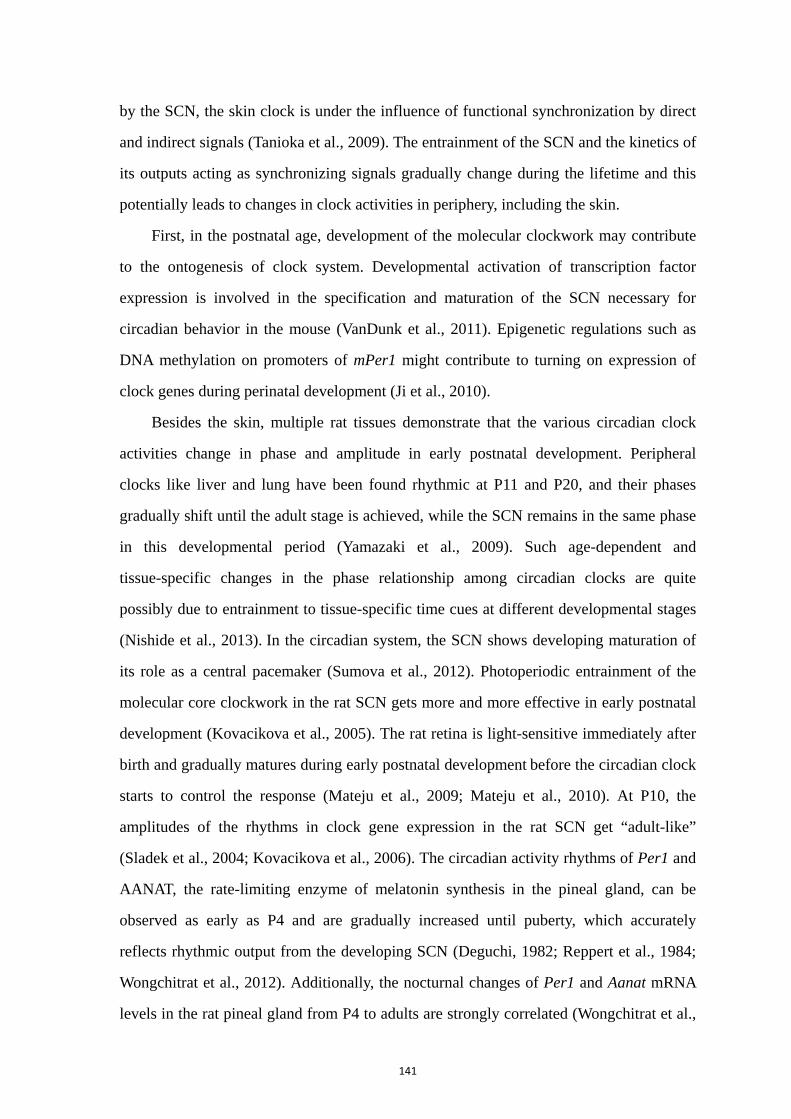
141
by the SCN, the skin clock is under the influence of functional synchronization by direct
and indirect signals (Tanioka et al., 2009). The entrainment of the SCN and the kinetics of
its outputs acting as synchronizing signals gradually change during the lifetime and this
potentially leads to changes in clock activities in periphery, including the skin.
First, in the postnatal age, development of the molecular clockwork may contribute
to the ontogenesis of clock system. Developmental activation of transcription factor
expression is involved in the specification and maturation of the SCN necessary for
circadian behavior in the mouse (VanDunk et al., 2011). Epigenetic regulations such as
DNA methylation on promoters of mPer1 might contribute to turning on expression of
clock genes during perinatal development (Ji et al., 2010).
Besides the skin, multiple rat tissues demonstrate that the various circadian clock
activities change in phase and amplitude in early postnatal development. Peripheral
clocks like liver and lung have been found rhythmic at P11 and P20, and their phases
gradually shift until the adult stage is achieved, while the SCN remains in the same phase
in this developmental period (Yamazaki et al., 2009). Such age-dependent and
tissue-specific changes in the phase relationship among circadian clocks are quite
possibly due to entrainment to tissue-specific time cues at different developmental stages
(Nishide et al., 2013). In the circadian system, the SCN shows developing maturation of
its role as a central pacemaker (Sumova et al., 2012). Photoperiodic entrainment of the
molecular core clockwork in the rat SCN gets more and more effective in early postnatal
development (Kovacikova et al., 2005). The rat retina is light-sensitive immediately after
birth and gradually matures during early postnatal development before the circadian clock
starts to control the response (Mateju et al., 2009; Mateju et al., 2010). At P10, the
amplitudes of the rhythms in clock gene expression in the rat SCN get “adult-like”
(Sladek et al., 2004; Kovacikova et al., 2006). The circadian activity rhythms of Per1 and
AANAT, the rate-limiting enzyme of melatonin synthesis in the pineal gland, can be
observed as early as P4 and are gradually increased until puberty, which accurately
reflects rhythmic output from the developing SCN (Deguchi, 1982; Reppert et al., 1984;
Wongchitrat et al., 2012). Additionally, the nocturnal changes of Per1 and Aanat mRNA
levels in the rat pineal gland from P4 to adults are strongly correlated (Wongchitrat et al.,
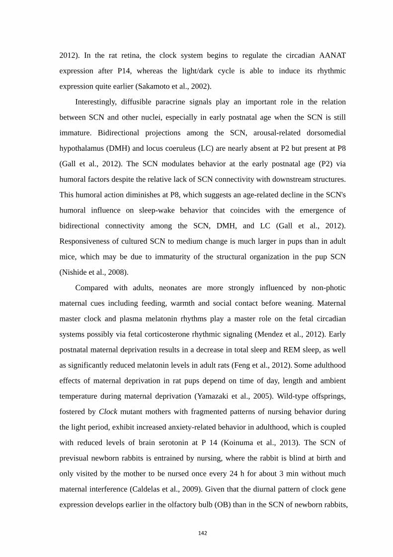
142
2012). In the rat retina, the clock system begins to regulate the circadian AANAT
expression after P14, whereas the light/dark cycle is able to induce its rhythmic
expression quite earlier (Sakamoto et al., 2002).
Interestingly, diffusible paracrine signals play an important role in the relation
between SCN and other nuclei, especially in early postnatal age when the SCN is still
immature. Bidirectional projections among the SCN, arousal-related dorsomedial
hypothalamus (DMH) and locus coeruleus (LC) are nearly absent at P2 but present at P8
(Gall et al., 2012). The SCN modulates behavior at the early postnatal age (P2) via
humoral factors despite the relative lack of SCN connectivity with downstream structures.
This humoral action diminishes at P8, which suggests an age-related decline in the SCN's
humoral influence on sleep-wake behavior that coincides with the emergence of
bidirectional connectivity among the SCN, DMH, and LC (Gall et al., 2012).
Responsiveness of cultured SCN to medium change is much larger in pups than in adult
mice, which may be due to immaturity of the structural organization in the pup SCN
(Nishide et al., 2008).
Compared with adults, neonates are more strongly influenced by non-photic
maternal cues including feeding, warmth and social contact before weaning. Maternal
master clock and plasma melatonin rhythms play a master role on the fetal circadian
systems possibly via fetal corticosterone rhythmic signaling (Mendez et al., 2012). Early
postnatal maternal deprivation results in a decrease in total sleep and REM sleep, as well
as significantly reduced melatonin levels in adult rats (Feng et al., 2012). Some adulthood
effects of maternal deprivation in rat pups depend on time of day, length and ambient
temperature during maternal deprivation (Yamazaki et al., 2005). Wild-type offsprings,
fostered by Clock mutant mothers with fragmented patterns of nursing behavior during
the light period, exhibit increased anxiety-related behavior in adulthood, which is coupled
with reduced levels of brain serotonin at P 14 (Koinuma et al., 2013). The SCN of
previsual newborn rabbits is entrained by nursing, where the rabbit is blind at birth and
only visited by the mother to be nursed once every 24 h for about 3 min without much
maternal interference (Caldelas et al., 2009). Given that the diurnal pattern of clock gene
expression develops earlier in the olfactory bulb (OB) than in the SCN of newborn rabbits,
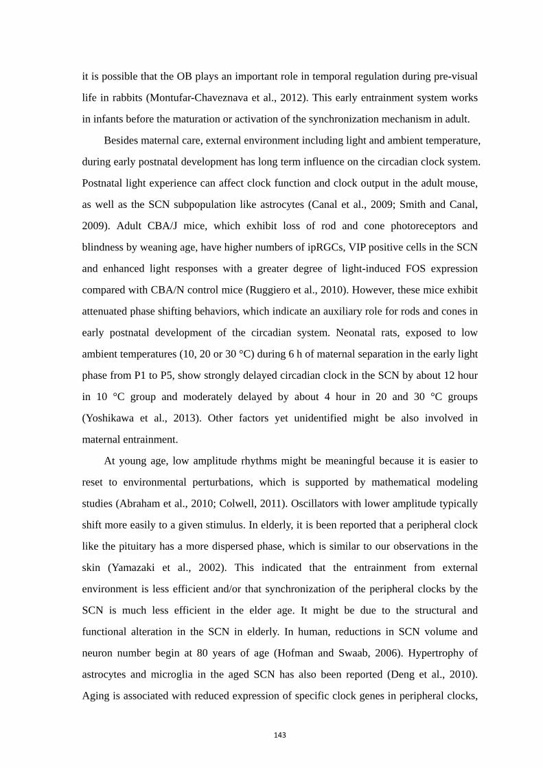
143
it is possible that the OB plays an important role in temporal regulation during pre-visual
life in rabbits (Montufar-Chaveznava et al., 2012). This early entrainment system works
in infants before the maturation or activation of the synchronization mechanism in adult.
Besides maternal care, external environment including light and ambient temperature,
during early postnatal development has long term influence on the circadian clock system.
Postnatal light experience can affect clock function and clock output in the adult mouse,
as well as the SCN subpopulation like astrocytes (Canal et al., 2009; Smith and Canal,
2009). Adult CBA/J mice, which exhibit loss of rod and cone photoreceptors and
blindness by weaning age, have higher numbers of ipRGCs, VIP positive cells in the SCN
and enhanced light responses with a greater degree of light-induced FOS expression
compared with CBA/N control mice (Ruggiero et al., 2010). However, these mice exhibit
attenuated phase shifting behaviors, which indicate an auxiliary role for rods and cones in
early postnatal development of the circadian system. Neonatal rats, exposed to low
ambient temperatures (10, 20 or 30 °C) during 6 h of maternal separation in the early light
phase from P1 to P5, show strongly delayed circadian clock in the SCN by about 12 hour
in 10 °C group and moderately delayed by about 4 hour in 20 and 30 °C groups
(Yoshikawa et al., 2013). Other factors yet unidentified might be also involved in
maternal entrainment.
At young age, low amplitude rhythms might be meaningful because it is easier to
reset to environmental perturbations, which is supported by mathematical modeling
studies (Abraham et al., 2010; Colwell, 2011). Oscillators with lower amplitude typically
shift more easily to a given stimulus. In elderly, it is been reported that a peripheral clock
like the pituitary has a more dispersed phase, which is similar to our observations in the
skin (Yamazaki et al., 2002). This indicated that the entrainment from external
environment is less efficient and/or that synchronization of the peripheral clocks by the
SCN is much less efficient in the elder age. It might be due to the structural and
functional alteration in the SCN in elderly. In human, reductions in SCN volume and
neuron number begin at 80 years of age (Hofman and Swaab, 2006). Hypertrophy of
astrocytes and microglia in the aged SCN has also been reported (Deng et al., 2010).
Aging is associated with reduced expression of specific clock genes in peripheral clocks,

144
but not systematically in the SCN (Yamazaki et al., 2002; Kolker et al., 2004). The
endogenous period of the SCN is shortened in aged rat, although individual variation
seems constant (Yamazaki et al., 2002). Mouse lemur, a primate, exhibits a shortened
free-running period in aged animals compared to adulthood, with altered AVP and VIP
rhythms (Aujard et al., 2006). In mice, circadian output declines at the level of neural
activity rhythms in the SCN by aging (Nakamura et al., 2011a). In addition, aging led to
decreased amplitude of firing-rate in dispersed SCN neurons and increased variability in
the circadian waveform (Aujard et al., 2001). It has been reported that VIP mRNA levels
in the SCN displayed a daily rhythm in young female rats which was absent in
middle-aged animals (Kawakami et al., 1997). Ageing SCN also shows alterations in the
expression AVP (Roozendaal et al., 1987). In addition, aged rodents exhibit decline of the
SCN presynaptic GABAergic terminals (Palomba et al., 2008). In elder human post
mortem SCN, the number and density of AVP/VIP-expressing neurons do not change but
the number and density of MT1-expressing neurons do (Wu et al., 2007). Decreases in
neurotransmitter production by the SCN and low amplitude of electrical activity might
weaken the ability of the SCN to synchronize peripheral oscillators. Reversely, Bmal1
deficiency results in premature aging phenotype in mice skin and Clock deficiency
increase the age-related dermatitis in mice (Dubrovsky et al., 2010).
Abnormal clock functioning in elderly could be result from worse
entrainment/synchronization, or more easily disturbed system, or both. Altered SCN
functionality affects its ability to reset peripheral oscillators and drive damped
downstream oscillators. A 6-h advance or delay in the light schedule leads to a disrupted
resynchronization of the liver of two year-old Per1-luciferase rats compared with young
rats (Davidson et al., 2008). Two year-old Per1-luciferase rats also showed altered
rhythms in peripheral tissues (Yamazaki et al., 2002). Interestingly, transplantation of a
fetal SCN into aged animals restored diurnal rhythm of corticotropin-releasing hormone
(CRH) and locomotor activity(Li and Satinoff, 1995; Cai et al., 1997). These results
confirm that the SCN plays a crucial role in aging and longevity.
Aging in the circadian system was first studied focused on alterations in the
entrainment of the SCN. Aged mice are less sensitive to light entrainment than young

145
ones (Wyse and Coogan, 2010). Although aged mice do not exhibit alteration in retinal
innervation of the SCN even with a small decrease (8-50%) in lens transmittance, they are
20 times less sensitive to the entraining light compared with young animals, which likely
occurs within the SCN and/or photoreceptors (Zhang et al., 1996; Zhang et al., 1998).
Some of the age-related changes in the response of the clock to a phase-shift stimulus can
be reversed by implanting old animals with fetal SCN tissue (Van Reeth et al., 1993; Van
Reeth et al., 1994; Turek et al., 1995). In aged rats, light-stimulation leads to decreased
response of the immediate early genes in the SCN (Sutin et al., 1993). Ageing increases
light absorption by lens crystallines especially of short wavelengths, and decreases pupil
area, which results in progressive loss of retinal illumination (Turner and Mainster, 2008).
A 10 year-old child has circadian photoreception 10-fold greater than a 95 year-old phakic
adult, while a 45 year-old adult retains only half the daily photoreception of early youth
(Turner and Mainster, 2008). Besides that, the responsiveness of the circadian clock to
some neurochemical stimuli like GABA is attenuated with ageing (Biello, 2009). In the
aged hamster, light-induced Per1 expression is markedly reduced in the SCN, with a
significantly longer delay to resynchronization, although in DD Per1 shows similar
rhythms as a young rat (Kolker et al., 2003). Still under discussion, the expression of
Per2 is impaired in the SCN of aged mice, while adverse evidence also exists (Asai et al.,
2001; Weinert et al., 2001). These findings suggest that disruption of circadian rhythms in
elderly may result, in part, from reductions in the sensitivity of the SCN to entraining
signals.
Downstream circulating factors might contribute to the aged chronotype. Indeed, it
was reported that the same skin primary fibroblasts exhibit shortened period length and
advanced phase of cellular circadian rhythms, in the presence of human serum from older
donors compared with young ones, which is caused by a thermolabile factor present in the
serum of older individuals (Pagani et al., 2011). During aging, a major factor that has
been implicated is the physiological decline of hormones (Goldman et al., 2011). Aging
changes the amounts of hormone production and the corresponding target sensitivity.
Melatonin oscillates daily and its amplitude decreases under aging with an earlier phase,
which is considered a putative marker of the decline of the circadian clock (Bondy et al.,
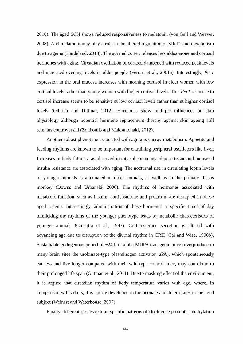
146
2010). The aged SCN shows reduced responsiveness to melatonin (von Gall and Weaver,
2008). And melatonin may play a role in the altered regulation of SIRT1 and metabolism
due to ageing (Hardeland, 2013). The adrenal cortex releases less aldosterone and cortisol
hormones with aging. Circadian oscillation of cortisol dampened with reduced peak levels
and increased evening levels in older people (Ferrari et al., 2001a). Interestingly, Per1
expression in the oral mucosa increases with morning cortisol in elder women with low
cortisol levels rather than young women with higher cortisol levels. This Per1 response to
cortisol increase seems to be sensitive at low cortisol levels rather than at higher cortisol
levels (Olbrich and Dittmar, 2012). Hormones show multiple influences on skin
physiology although potential hormone replacement therapy against skin ageing still
remains controversial (Zouboulis and Makrantonaki, 2012).
Another robust phenotype associated with aging is energy metabolism. Appetite and
feeding rhythms are known to be important for entraining peripheral oscillators like liver.
Increases in body fat mass as observed in rats subcutaneous adipose tissue and increased
insulin resistance are associated with aging. The nocturnal rise in circulating leptin levels
of younger animals is attenuated in older animals, as well as in the primate rhesus
monkey (Downs and Urbanski, 2006). The rhythms of hormones associated with
metabolic function, such as insulin, corticosterone and prolactin, are disrupted in obese
aged rodents. Interestingly, administration of these hormones at specific times of day
mimicking the rhythms of the younger phenotype leads to metabolic characteristics of
younger animals (Cincotta et al., 1993). Corticosterone secretion is altered with
advancing age due to disruption of the diurnal rhythm in CRH (Cai and Wise, 1996b).
Sustainable endogenous period of ~24 h in alpha MUPA transgenic mice (overproduce in
many brain sites the urokinase-type plasminogen activator, uPA), which spontaneously
eat less and live longer compared with their wild-type control mice, may contribute to
their prolonged life span (Gutman et al., 2011). Due to masking effect of the environment,
it is argued that circadian rhythm of body temperature varies with age, where, in
comparison with adults, it is poorly developed in the neonate and deteriorates in the aged
subject (Weinert and Waterhouse, 2007).
Finally, different tissues exhibit specific patterns of clock gene promoter methylation
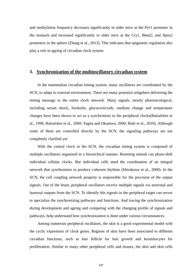
147
and methylation frequency decreases significantly in older mice at the Per1 promoter in
the stomach and increased significantly in older mice at the Cry1, Bmal2, and Npas2
promoters in the spleen (Zhang et al., 2013). This indicates that epigenetic regulation also
play a role in ageing of circadian clock system.
3. Synchronization of the multioscillatory circadian system
In the mammalian circadian timing system, many oscillators are coordinated by the
SCN, to adapt to external environment. There are many potential zeitgebers delivering the
timing message to the entire clock network. Many signals, mostly pharmacological,
including serum shock, forskolin, glucocorticoids, medium change and temperature
changes have been shown to act as a synchronizer to the peripheral clocks(Balsalobre et
al., 1998; Balsalobre et al., 2000; Yagita and Okamura, 2000; Buhr et al., 2010). Although
some of them are controlled directly by the SCN, the signaling pathways are not
completely clarified yet.
With the central clock in the SCN, the circadian timing system is composed of
multiple oscillators organized in a hierarchical manner. Resetting stimuli can phase-shift
individual cellular clocks. But individual cells need the coordination of an integral
network that synchronizes to produce coherent rhythms (Shirakawa et al., 2000). In the
SCN, the cell coupling network property is responsible for the precision of the output
signals. Out of the brain, peripheral oscillators receive multiple signals via neuronal and
humoral outputs from the SCN. To identify this signals in the peripheral target can server
to specialize the synchronizing pathways and functions. And tracing the synchronization
during development and ageing and comparing with the changing profile of signals and
pathways, help understand how synchronization is done under various circumstances.
Among numerous peripheral oscillators, the skin is a good experimental model with
the cyclic expression of clock genes. Regions of skin have been associated to different
circadian functions, such as hair follicle for hair growth and keratinocytes for
proliferation. Similar to many other peripheral cells and tissues, the skin and skin cells
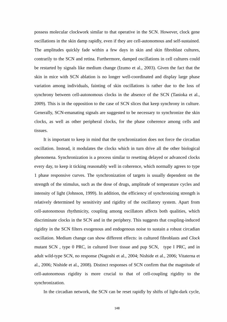
148
possess molecular clockwork similar to that operative in the SCN. However, clock gene
oscillations in the skin damp rapidly, even if they are cell-autonomous and self-sustained.
The amplitudes quickly fade within a few days in skin and skin fibroblast cultures,
contrarily to the SCN and retina. Furthermore, damped oscillations in cell cultures could
be restarted by signals like medium change (Izumo et al., 2003). Given the fact that the
skin in mice with SCN ablation is no longer well-coordinated and display large phase
variation among individuals, fainting of skin oscillations is rather due to the loss of
synchrony between cell-autonomous clocks in the absence of the SCN (Tanioka et al.,
2009). This is in the opposition to the case of SCN slices that keep synchrony in culture.
Generally, SCN-emanating signals are suggested to be necessary to synchronize the skin
clocks, as well as other peripheral clocks, for the phase coherence among cells and
tissues.
It is important to keep in mind that the synchronization does not force the circadian
oscillation. Instead, it modulates the clocks which in turn drive all the other biological
phenomena. Synchronization is a process similar to resetting delayed or advanced clocks
every day, to keep it ticking reasonably well in coherence, which normally agrees to type
1 phase responsive curves. The synchronization of targets is usually dependent on the
strength of the stimulus, such as the dose of drugs, amplitude of temperature cycles and
intensity of light (Johnson, 1999). In addition, the efficiency of synchronizing strength is
relatively determined by sensitivity and rigidity of the oscillatory system. Apart from
cell-autonomous rhythmicity, coupling among oscillators affects both qualities, which
discriminate clocks in the SCN and in the periphery. This suggests that coupling-induced
rigidity in the SCN filters exogenous and endogenous noise to sustain a robust circadian
oscillation. Medium change can show different effects: in cultured fibroblasts and Clock
mutant SCN , type 0 PRC, in cultured liver tissue and pup SCN, type I PRC, and in
adult wild-type SCN, no response (Nagoshi et al., 2004; Nishide et al., 2006; Vitaterna et
al., 2006; Nishide et al., 2008). Distinct responses of SCN confirm that the magnitude of
cell-autonomous rigidity is more crucial to that of cell-coupling rigidity to the
synchronization.
In the circadian network, the SCN can be reset rapidly by shifts of light-dark cycle,
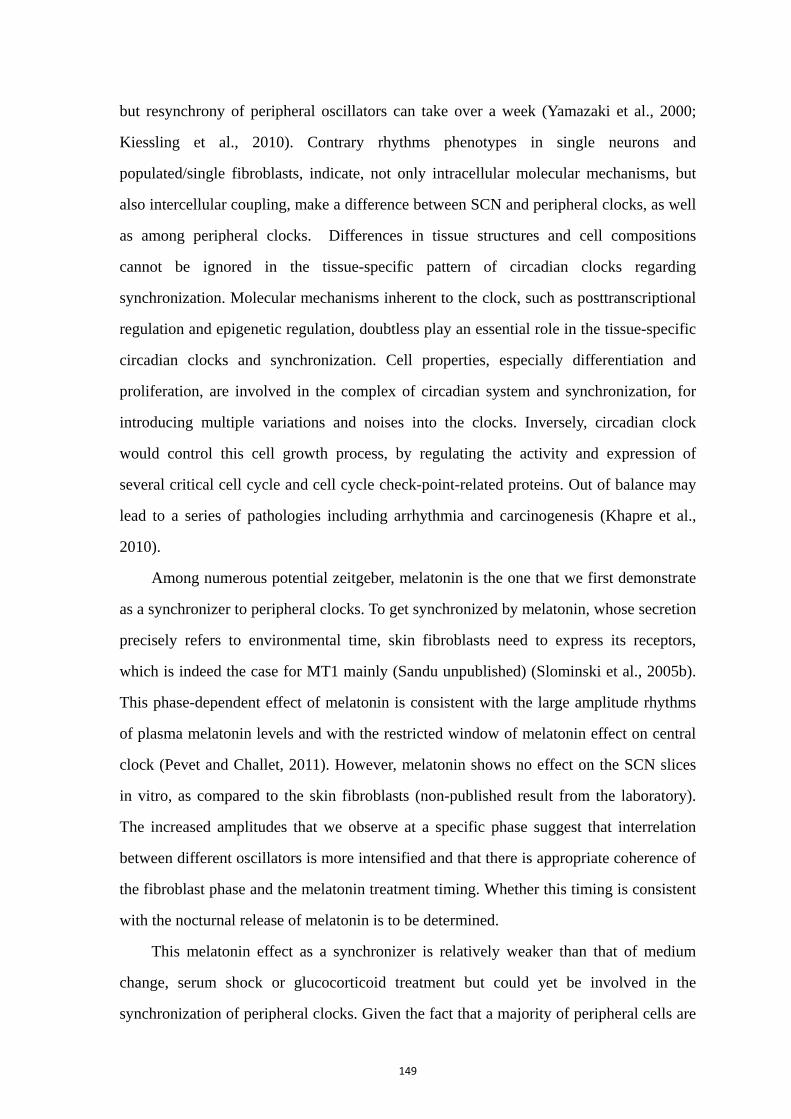
149
but resynchrony of peripheral oscillators can take over a week (Yamazaki et al., 2000;
Kiessling et al., 2010). Contrary rhythms phenotypes in single neurons and
populated/single fibroblasts, indicate, not only intracellular molecular mechanisms, but
also intercellular coupling, make a difference between SCN and peripheral clocks, as well
as among peripheral clocks. Differences in tissue structures and cell compositions
cannot be ignored in the tissue-specific pattern of circadian clocks regarding
synchronization. Molecular mechanisms inherent to the clock, such as posttranscriptional
regulation and epigenetic regulation, doubtless play an essential role in the tissue-specific
circadian clocks and synchronization. Cell properties, especially differentiation and
proliferation, are involved in the complex of circadian system and synchronization, for
introducing multiple variations and noises into the clocks. Inversely, circadian clock
would control this cell growth process, by regulating the activity and expression of
several critical cell cycle and cell cycle check-point-related proteins. Out of balance may
lead to a series of pathologies including arrhythmia and carcinogenesis (Khapre et al.,
2010).
Among numerous potential zeitgeber, melatonin is the one that we first demonstrate
as a synchronizer to peripheral clocks. To get synchronized by melatonin, whose secretion
precisely refers to environmental time, skin fibroblasts need to express its receptors,
which is indeed the case for MT1 mainly (Sandu unpublished) (Slominski et al., 2005b).
This phase-dependent effect of melatonin is consistent with the large amplitude rhythms
of plasma melatonin levels and with the restricted window of melatonin effect on central
clock (Pevet and Challet, 2011). However, melatonin shows no effect on the SCN slices
in vitro, as compared to the skin fibroblasts (non-published result from the laboratory).
The increased amplitudes that we observe at a specific phase suggest that interrelation
between different oscillators is more intensified and that there is appropriate coherence of
the fibroblast phase and the melatonin treatment timing. Whether this timing is consistent
with the nocturnal release of melatonin is to be determined.
This melatonin effect as a synchronizer is relatively weaker than that of medium
change, serum shock or glucocorticoid treatment but could yet be involved in the
synchronization of peripheral clocks. Given the fact that a majority of peripheral cells are
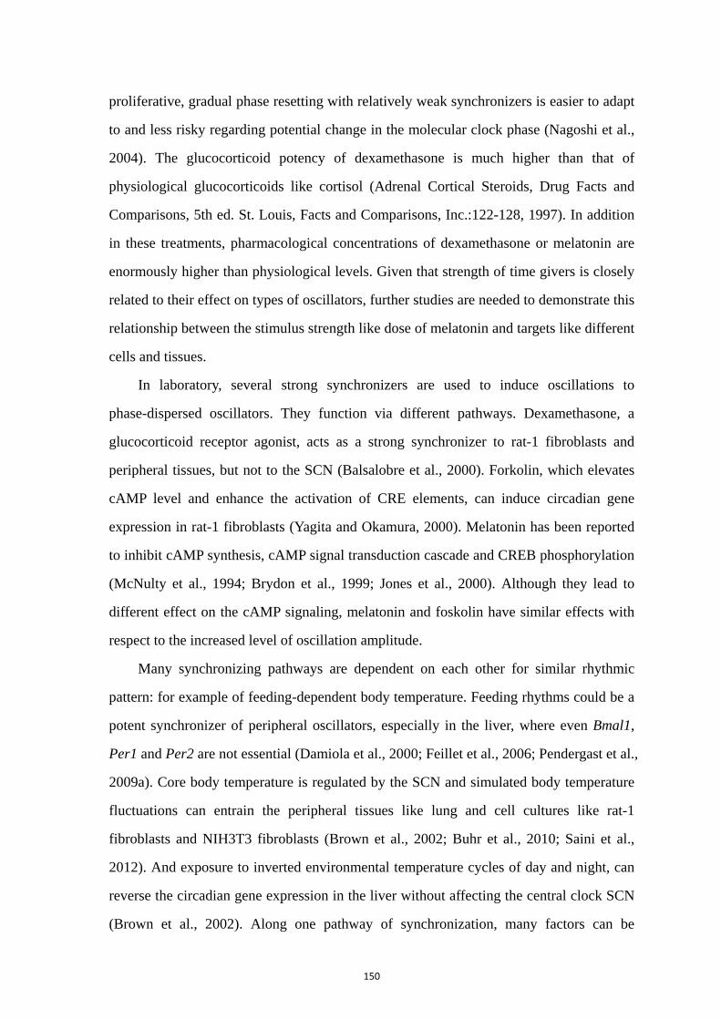
150
proliferative, gradual phase resetting with relatively weak synchronizers is easier to adapt
to and less risky regarding potential change in the molecular clock phase (Nagoshi et al.,
2004). The glucocorticoid potency of dexamethasone is much higher than that of
physiological glucocorticoids like cortisol (Adrenal Cortical Steroids, Drug Facts and
Comparisons, 5th ed. St. Louis, Facts and Comparisons, Inc.:122-128, 1997). In addition
in these treatments, pharmacological concentrations of dexamethasone or melatonin are
enormously higher than physiological levels. Given that strength of time givers is closely
related to their effect on types of oscillators, further studies are needed to demonstrate this
relationship between the stimulus strength like dose of melatonin and targets like different
cells and tissues.
In laboratory, several strong synchronizers are used to induce oscillations to
phase-dispersed oscillators. They function via different pathways. Dexamethasone, a
glucocorticoid receptor agonist, acts as a strong synchronizer to rat-1 fibroblasts and
peripheral tissues, but not to the SCN (Balsalobre et al., 2000). Forkolin, which elevates
cAMP level and enhance the activation of CRE elements, can induce circadian gene
expression in rat-1 fibroblasts (Yagita and Okamura, 2000). Melatonin has been reported
to inhibit cAMP synthesis, cAMP signal transduction cascade and CREB phosphorylation
(McNulty et al., 1994; Brydon et al., 1999; Jones et al., 2000). Although they lead to
different effect on the cAMP signaling, melatonin and foskolin have similar effects with
respect to the increased level of oscillation amplitude.
Many synchronizing pathways are dependent on each other for similar rhythmic
pattern: for example of feeding-dependent body temperature. Feeding rhythms could be a
potent synchronizer of peripheral oscillators, especially in the liver, where even Bmal1,
Per1 and Per2 are not essential (Damiola et al., 2000; Feillet et al., 2006; Pendergast et al.,
2009a). Core body temperature is regulated by the SCN and simulated body temperature
fluctuations can entrain the peripheral tissues like lung and cell cultures like rat-1
fibroblasts and NIH3T3 fibroblasts (Brown et al., 2002; Buhr et al., 2010; Saini et al.,
2012). And exposure to inverted environmental temperature cycles of day and night, can
reverse the circadian gene expression in the liver without affecting the central clock SCN
(Brown et al., 2002). Along one pathway of synchronization, many factors can be
151
involved. Meal feeding interval influences the phase of mouse peripheral circadian clocks
(Kuroda et al., 2012). The food, palatable or high-fat, has an influence on the sensitivity of
the master circadian clock to changes in motivational states related to palatability of the
food (Mendoza et al., 2008; Mendoza et al., 2010). These results suggest that the
complexity of synchronization pathways may involve not only the diversity of signals but
also intersections of transmitting routes.
As a whole circadian system, the entrainment or synchronization of the clocks to the
external environment are achieved not only by the periodicity of physical light/dark
signals but also by the periodicity of social signals involving perceptions with eyes, ears,
nose and skin (Reinberg and Touitou, 1996). Many peripheral clocks are involved in
responses to different non-photic time cues, such as odors (olfactory bulb),
feeding-fasting cycle (liver), sound (ear) and immunological cues (white blood cells)
(Amir et al., 1999; Goel, 2005; Berger, 2008; Vollmers et al., 2009). And the skin might
also be involved via temperature, touching or hormone production like vitamin D. For
example, in mice, scheduled exercise phase shifts the circadian clock in skeletal muscle
and lung but not in the SCN (Wolff and Esser, 2012). Although details of physiological
pathways that transduce environmental time information to the clock system may differ
among various cues through various tissues in various species, the essential properties of
entrainment are thought to be the same for all of them. Properties of time cues such as
strength (e.g. light intensity) and spectral composition (e.g. wavelength spectrum) and
their functional phases (e.g. morning or afternoon) have strong influences on the
entrainment.
Given the fact that circadian system develops during maturation and ageing, the
synchronizing signals, both exogenous and endogenous, may differ accordingly. For
example, the hormone level during the life has changed largely in addition to the
responsive capacities in corresponding targets. Most hormonal receptors are expressed in
a tissue-specific pattern and can lead to large differences in responsive levels and
downstream messages. Within a given individual, daily resetting cues can cooperate to
deliver combined information for phase coherence among phase differentiated oscillators.

152
Different individuals at different stages might require different combinations of
synchronizers.
In research, distinct biological models in different conditions have provided benefits
regarding the study of synchronization by specific factors. For instance, rabbit pups ingest
milk once a day and are a natural model of feeding synchronization. During nursing,
several sensory systems receive information about properties of the food including the
olfactory system, where olfactory bulb is synchronized during milk ingestion to anticipate
feeding, but during fasting its oscillations perhaps are modulated by the SCN (Nolasco et
al., 2012). Some animals live in colonies, such as bees and drosophila, with highly
developed social structures requiring temporal synchronization, where social cues may
thus be critical to the adaptive function of the circadian system (Bloch et al., 2013).
Regarding the present work, it needs to be noted that the used in vitro experimental
models have some limits as models for the skin in vivo, notably concerning its
physiological synchronization. First, the Per1 luciferase rat model may represent the
clock system in nocturnal animals but not diurnal case like human. In addition, skin
explants lose the physiological environment: both internal such as central clock and
corresponding synchronizing signals, and external like UV. Finally, the primary
fibroblasts culture, as a simplified model to study skin oscillators, undergo many
influences in vitro such as medium conditions and manipulation of medium change. Thus,
an improvement to the use of these experimental models could be to study skin clock and
its synchronization with the help of the promising flow through system in the future.

153
References
154
Abe M, Herzog ED, Yamazaki S, Straume M, Tei H, Sakaki Y, Menaker M, Block GD (2002) Circadian
rhythms in isolated brain regions. J Neurosci 22:350‐356.
Abraham U, Granada AE, Westermark PO, Heine M, Kramer A, Herzel H (2010) Coupling governs
entrainment range of circadian clocks. Mol Syst Biol 6:438.
Abrahamson EE, Moore RY (2001) Suprachiasmatic nucleus in the mouse: retinal innervation, intrinsic
organization and efferent projections. Brain Res 916:172‐191.
Agez L, Laurent V, Pevet P, Masson‐Pevet M, Gauer F (2007) Melatonin affects nuclear orphan receptors
mRNA in the rat suprachiasmatic nuclei. Neuroscience 144:522‐530.
Akashi M, Soma H, Yamamoto T, Tsugitomi A, Yamashita S, Nishida E, Yasuda A, Liao JK, Node K (2010)
Noninvasive method for assessing the human circadian clock using hair follicle cells. Proc Natl
Acad Sci U S A 107:15643‐15648.
Albrecht U, Bordon A, Schmutz I, Ripperger J (2007) The multiple facets of Per2. Cold Spring Harb Symp
Quant Biol 72:95‐104.
Albrecht U, Zheng B, Larkin D, Sun ZS, Lee CC (2001) MPer1 and mper2 are essential for normal resetting
of the circadian clock. J Biol Rhythms 16:100‐104.
Albus H, Vansteensel MJ, Michel S, Block GD, Meijer JH (2005) A GABAergic mechanism is necessary for
coupling dissociable ventral and dorsal regional oscillators within the circadian clock. Curr Biol
15:886‐893.
Allen G, Rappe J, Earnest DJ, Cassone VM (2001) Oscillating on borrowed time: diffusible signals from
immortalized suprachiasmatic nucleus cells regulate circadian rhythmicity in cultured
fibroblasts. J Neurosci 21:7937‐7943.
Alvarez‐Saavedra M, Antoun G, Yanagiya A, Oliva‐Hernandez R, Cornejo‐Palma D, Perez‐Iratxeta C,
Sonenberg N, Cheng HY (2011) miRNA‐132 orchestrates chromatin remodeling and translational
control of the circadian clock. Hum Mol Genet 20:731‐751.
Amir S, Cain S, Sullivan J, Robinson B, Stewart J (1999) Olfactory stimulation enhances light‐induced
phase shifts in free‐running activity rhythms and Fos expression in the suprachiasmatic nucleus.
Neuroscience 92:1165‐1170.
Antoch MP, Song EJ, Chang AM, Vitaterna MH, Zhao Y, Wilsbacher LD, Sangoram AM, King DP, Pinto LH,
Takahashi JS (1997) Functional identification of the mouse circadian Clock gene by transgenic
BAC rescue. Cell 89:655‐667.
Asai M, Yoshinobu Y, Kaneko S, Mori A, Nikaido T, Moriya T, Akiyama M, Shibata S (2001) Circadian
profile of Per gene mRNA expression in the suprachiasmatic nucleus, paraventricular nucleus,
and pineal body of aged rats. J Neurosci Res 66:1133‐1139.
Asher G, Schibler U (2011) Crosstalk between components of circadian and metabolic cycles in mammals.
Cell Metab 13:125‐137.
Asher G, Gatfield D, Stratmann M, Reinke H, Dibner C, Kreppel F, Mostoslavsky R, Alt FW, Schibler U
(2008) SIRT1 regulates circadian clock gene expression through PER2 deacetylation. Cell
134:317‐328.
Aton SJ, Colwell CS, Harmar AJ, Waschek J, Herzog ED (2005) Vasoactive intestinal polypeptide mediates
circadian rhythmicity and synchrony in mammalian clock neurons. Nat Neurosci 8:476‐483.
Aujard F, Herzog ED, Block GD (2001) Circadian rhythms in firing rate of individual suprachiasmatic
nucleus neurons from adult and middle‐aged mice. Neuroscience 106:255‐261.
155
Aujard F, Cayetanot F, Bentivoglio M, Perret M (2006) Age‐related effects on the biological clock and its
behavioral output in a primate. Chronobiol Int 23:451‐460.
Bailes HJ, Lucas RJ (2010) Melanopsin and inner retinal photoreception. Cell Mol Life Sci 67:99‐111.
Balsalobre A (2002) Clock genes in mammalian peripheral tissues. Cell Tissue Res 309:193‐199.
Balsalobre A, Damiola F, Schibler U (1998) A serum shock induces circadian gene expression in
mammalian tissue culture cells. Cell 93:929‐937.
Balsalobre A, Brown SA, Marcacci L, Tronche F, Kellendonk C, Reichardt HM, Schutz G, Schibler U (2000)
Resetting of circadian time in peripheral tissues by glucocorticoid signaling. Science
289:2344‐2347.
Bartel DP (2009) MicroRNAs: target recognition and regulatory functions. Cell 136:215‐233.
Belenky MA, Yarom Y, Pickard GE (2008) Heterogeneous expression of gamma‐aminobutyric acid and
gamma‐aminobutyric acid‐associated receptors and transporters in the rat suprachiasmatic
nucleus. J Comp Neurol 506:708‐732.
Berger J (2008) A two‐clock model of circadian timing in the immune system of mammals. Pathol Biol
(Paris) 56:286‐291.
Berson DM (2003) Strange vision: ganglion cells as circadian photoreceptors. Trends Neurosci
26:314‐320.
Berson DM, Dunn FA, Takao M (2002) Phototransduction by retinal ganglion cells that set the circadian
clock. Science 295:1070‐1073.
Biello SM (2009) Circadian clock resetting in the mouse changes with age. Age (Dordr) 31:293‐303.
Biello SM, Golombek DA, Harrington ME (1997) Neuropeptide Y and glutamate block each other's phase
shifts in the suprachiasmatic nucleus in vitro. Neuroscience 77:1049‐1057.
Bjarnason GA, Jordan R (2002) Rhythms in human gastrointestinal mucosa and skin. Chronobiol Int
19:129‐140.
Bjarnason GA, Jordan RC, Wood PA, Li Q, Lincoln DW, Sothern RB, Hrushesky WJ, Ben‐David Y (2001)
Circadian expression of clock genes in human oral mucosa and skin: association with specific
cell‐cycle phases. Am J Pathol 158:1793‐1801.
Bloch G, Herzog ED, Levine JD, Schwartz WJ (2013) Socially synchronized circadian oscillators. Proc Biol
Sci 280:20130035.
Bondy SC, Li H, Zhou J, Wu M, Bailey JA, Lahiri DK (2010) Melatonin alters age‐related changes in
transcription factors and kinase activation. Neurochem Res 35:2035‐2042.
Bouskila Y, Dudek FE (1993) Neuronal synchronization without calcium‐dependent synaptic transmission
in the hypothalamus. Proc Natl Acad Sci U S A 90:3207‐3210.
Brown SA, Zumbrunn G, Fleury‐Olela F, Preitner N, Schibler U (2002) Rhythms of mammalian body
temperature can sustain peripheral circadian clocks. Curr Biol 12:1574‐1583.
Brown SA, Ripperger J, Kadener S, Fleury‐Olela F, Vilbois F, Rosbash M, Schibler U (2005a)
PERIOD1‐associated proteins modulate the negative limb of the mammalian circadian oscillator.
Science 308:693‐696.
Brown SA, Kunz D, Dumas A, Westermark PO, Vanselow K, Tilmann‐Wahnschaffe A, Herzel H, Kramer A
(2008) Molecular insights into human daily behavior. Proc Natl Acad Sci U S A 105:1602‐1607.
Brown SA, Fleury‐Olela F, Nagoshi E, Hauser C, Juge C, Meier CA, Chicheportiche R, Dayer JM, Albrecht U,
Schibler U (2005b) The period length of fibroblast circadian gene expression varies widely
among human individuals. PLoS Biol 3:e338.
Brown WR (1991) A review and mathematical analysis of circadian rhythms in cell proliferation in mouse,
156
rat, and human epidermis. J Invest Dermatol 97:273‐280.
Brydon L, Petit L, de Coppet P, Barrett P, Morgan PJ, Strosberg AD, Jockers R (1999) Polymorphism and
signalling of melatonin receptors. Reprod Nutr Dev 39:315‐324.
Bubenik GA (2002) Gastrointestinal melatonin: localization, function, and clinical relevance. Dig Dis Sci
47:2336‐2348.
Buckingham EM, Klingelhutz AJ (2011) The role of telomeres in the ageing of human skin. Exp Dermatol
20:297‐302.
Buhr ED, Takahashi JS (2013) Molecular components of the Mammalian circadian clock. Handb Exp
Pharmacol:3‐27.
Buhr ED, Yoo SH, Takahashi JS (2010) Temperature as a universal resetting cue for mammalian circadian
oscillators. Science 330:379‐385.
Buijs RM (1996) The anatomical basis for the expression of circadian rhythms: the efferent projections of
the suprachiasmatic nucleus. Prog Brain Res 111:229‐240.
Buijs RM, Wortel J, Van Heerikhuize JJ, Feenstra MG, Ter Horst GJ, Romijn HJ, Kalsbeek A (1999)
Anatomical and functional demonstration of a multisynaptic suprachiasmatic nucleus adrenal
(cortex) pathway. Eur J Neurosci 11:1535‐1544.
Busino L, Bassermann F, Maiolica A, Lee C, Nolan PM, Godinho SI, Draetta GF, Pagano M (2007) SCFFbxl3
controls the oscillation of the circadian clock by directing the degradation of cryptochrome
proteins. Science 316:900‐904.
Cai A, Wise PM (1996a) Age‐related changes in the diurnal rhythm of CRH gene expression in the
paraventricular nuclei. Am J Physiol 270:E238‐243.
Cai A, Wise PM (1996b) Age‐related changes in light‐induced Jun‐B and Jun‐D expression: effects of
transplantation of fetal tissue containing the suprachiasmatic nucleus. J Biol Rhythms
11:284‐290.
Cai A, Scarbrough K, Hinkle DA, Wise PM (1997) Fetal grafts containing suprachiasmatic nuclei restore
the diurnal rhythm of CRH and POMC mRNA in aging rats. Am J Physiol 273:R1764‐1770.
Caldelas I, Gonzalez B, Montufar‐Chaveznava R, Hudson R (2009) Endogenous clock gene expression in
the suprachiasmatic nuclei of previsual newborn rabbits is entrained by nursing. Dev Neurobiol
69:47‐59.
Canal MM, Mohammed NM, Rodriguez JJ (2009) Early programming of astrocyte organization in the
mouse suprachiasmatic nuclei by light. Chronobiol Int 26:1545‐1558.
Carneiro SC, Cassia Fde F, Pascarelli BM, Souza SO, Ramos‐e‐Silva M, Filgueira AL, Japiassu MA, Takiya
CM (2007) Increase in dermal collagen fibril diameter and elastogenesis with UVB exposure: an
optical and ultrastructural study in albino Balb/c mice. Acta Dermatovenerol Croat 15:65‐71.
Challet E (2007) Minireview: Entrainment of the suprachiasmatic clockwork in diurnal and nocturnal
mammals. Endocrinology 148:5648‐5655.
Challet E, Pevet P, Vivien‐Roels B, Malan A (1997) Phase‐advanced daily rhythms of melatonin, body
temperature, and locomotor activity in food‐restricted rats fed during daytime. J Biol Rhythms
12:65‐79.
Chansard M, Molyneux P, Nomura K, Harrington ME, Fukuhara C (2007) c‐Jun N‐terminal kinase inhibitor
SP600125 modulates the period of mammalian circadian rhythms. Neuroscience 145:812‐823.
Chen R, Schirmer A, Lee Y, Lee H, Kumar V, Yoo SH, Takahashi JS, Lee C (2009) Rhythmic PER abundance
defines a critical nodal point for negative feedback within the circadian clock mechanism. Mol
Cell 36:417‐430.
157
Chen ST, Choo KB, Hou MF, Yeh KT, Kuo SJ, Chang JG (2005) Deregulated expression of the PER1, PER2
and PER3 genes in breast cancers. Carcinogenesis 26:1241‐1246.
Cheng HY, Papp JW, Varlamova O, Dziema H, Russell B, Curfman JP, Nakazawa T, Shimizu K, Okamura H,
Impey S, Obrietan K (2007) microRNA modulation of circadian‐clock period and entrainment.
Neuron 54:813‐829.
Cheng MY, Bullock CM, Li C, Lee AG, Bermak JC, Belluzzi J, Weaver DR, Leslie FM, Zhou QY (2002)
Prokineticin 2 transmits the behavioural circadian rhythm of the suprachiasmatic nucleus.
Nature 417:405‐410.
Cincotta AH, Schiller BC, Landry RJ, Herbert SJ, Miers WR, Meier AH (1993) Circadian neuroendocrine
role in age‐related changes in body fat stores and insulin sensitivity of the male Sprague‐Dawley
rat. Chronobiol Int 10:244‐258.
Claustrat B, Brun J, Chazot G (2005) The basic physiology and pathophysiology of melatonin. Sleep Med
Rev 9:11‐24.
Collaco AM, Geusz ME (2003) Monitoring immediate‐early gene expression through firefly luciferase
imaging of HRS/J hairless mice. BMC Physiol 3:8.
Colwell CS (2011) Linking neural activity and molecular oscillations in the SCN. Nat Rev Neurosci
12:553‐569.
Conti A, Conconi S, Hertens E, Skwarlo‐Sonta K, Markowska M, Maestroni JM (2000) Evidence for
melatonin synthesis in mouse and human bone marrow cells. J Pineal Res 28:193‐202.
Czeisler CA, Shanahan TL, Klerman EB, Martens H, Brotman DJ, Emens JS, Klein T, Rizzo JF, 3rd (1995)
Suppression of melatonin secretion in some blind patients by exposure to bright light. N Engl J
Med 332:6‐11.
Daan S, Albrecht U, van der Horst GT, Illnerova H, Roenneberg T, Wehr TA, Schwartz WJ (2001)
Assembling a clock for all seasons: are there M and E oscillators in the genes? J Biol Rhythms
16:105‐116.
Damiola F, Le Minh N, Preitner N, Kornmann B, Fleury‐Olela F, Schibler U (2000) Restricted feeding
uncouples circadian oscillators in peripheral tissues from the central pacemaker in the
suprachiasmatic nucleus. Genes Dev 14:2950‐2961.
Dardente H, Poirel VJ, Klosen P, Pevet P, Masson‐Pevet M (2002) Per and neuropeptide expression in the
rat suprachiasmatic nuclei: compartmentalization and differential cellular induction by light.
Brain Res 958:261‐271.
Davidson AJ, Yamazaki S, Arble DM, Menaker M, Block GD (2008) Resetting of central and peripheral
circadian oscillators in aged rats. Neurobiol Aging 29:471‐477.
de Jeu M, Hermes M, Pennartz C (1998) Circadian modulation of membrane properties in slices of rat
suprachiasmatic nucleus. Neuroreport 9:3725‐3729.
Deery MJ, Maywood ES, Chesham JE, Sladek M, Karp NA, Green EW, Charles PD, Reddy AB, Kyriacou CP,
Lilley KS, Hastings MH (2009) Proteomic analysis reveals the role of synaptic vesicle cycling in
sustaining the suprachiasmatic circadian clock. Curr Biol 19:2031‐2036.
Deguchi T (1982) Sympathetic regulation of circadian rhythm of serotonin N‐acetyltransferase activity in
pineal gland of infant rat. J Neurochem 38:797‐802.
Deng XH, Bertini G, Palomba M, Xu YZ, Bonaconsa M, Nygard M, Bentivoglio M (2010) Glial transcripts
and immune‐challenged glia in the suprachiasmatic nucleus of young and aged mice.
Chronobiol Int 27:742‐767.
Desotelle JA, Wilking MJ, Ahmad N (2012) The circadian control of skin and cutaneous photodamage.
158
Photochem Photobiol 88:1037‐1047.
Dibner C, Schibler U, Albrecht U (2010) The mammalian circadian timing system: organization and
coordination of central and peripheral clocks. Annu Rev Physiol 72:517‐549.
Dibner C, Sage D, Unser M, Bauer C, d'Eysmond T, Naef F, Schibler U (2009) Circadian gene expression is
resilient to large fluctuations in overall transcription rates. EMBO J 28:123‐134.
Downs JL, Urbanski HF (2006) Aging‐related sex‐dependent loss of the circulating leptin 24‐h rhythm in
the rhesus monkey. J Endocrinol 190:117‐127.
Drobnik J, Dabrowski R (1996) Melatonin suppresses the pinealectomy‐induced elevation of collagen
content in a wound. Cytobios 85:51‐58.
Drobnik J, Dabrowski R (1999) Pinealectomy‐induced elevation of collagen content in the intact skin is
suppressed by melatonin application. Cytobios 100:49‐55.
Dubrovsky YV, Samsa WE, Kondratov RV (2010) Deficiency of circadian protein CLOCK reduces lifespan
and increases age‐related cataract development in mice. Aging (Albany NY) 2:936‐944.
Duong HA, Robles MS, Knutti D, Weitz CJ (2011) A molecular mechanism for circadian clock negative
feedback. Science 332:1436‐1439.
Eide EJ, Kang H, Crapo S, Gallego M, Virshup DM (2005) Casein kinase I in the mammalian circadian clock.
Methods Enzymol 393:408‐418.
Ermak G, Slominski A (1997) Production of POMC, CRH‐R1, MC1, and MC2 receptor mRNA and
expression of tyrosinase gene in relation to hair cycle and dexamethasone treatment in the
C57BL/6 mouse skin. J Invest Dermatol 108:160‐165.
Espiritu JR (2008) Aging‐related sleep changes. Clin Geriatr Med 24:1‐14, v.
Esrefoglu M, Seyhan M, Gul M, Parlakpinar H, Batcioglu K, Uyumlu B (2005) Potent therapeutic effect of
melatonin on aging skin in pinealectomized rats. J Pineal Res 39:231‐237.
Etchegaray JP, Lee C, Wade PA, Reppert SM (2003) Rhythmic histone acetylation underlies transcription
in the mammalian circadian clock. Nature 421:177‐182.
Etchegaray JP, Machida KK, Noton E, Constance CM, Dallmann R, Di Napoli MN, DeBruyne JP, Lambert
CM, Yu EA, Reppert SM, Weaver DR (2009) Casein kinase 1 delta regulates the pace of the
mammalian circadian clock. Mol Cell Biol 29:3853‐3866.
Evans JA, Leise TL, Castanon‐Cervantes O, Davidson AJ (2011) Intrinsic regulation of spatiotemporal
organization within the suprachiasmatic nucleus. PLoS One 6:e15869.
Fagrell B, Intaglietta M (1977) The dynamics of skin microcirculation as a tool for the study of systemic
diseases. Bibl Anat:231‐234.
Farre EM, Harmer SL, Harmon FG, Yanovsky MJ, Kay SA (2005) Overlapping and distinct roles of PRR7
and PRR9 in the Arabidopsis circadian clock. Curr Biol 15:47‐54.
Feillet CA, Ripperger JA, Magnone MC, Dulloo A, Albrecht U, Challet E (2006) Lack of food anticipation in
Per2 mutant mice. Curr Biol 16:2016‐2022.
Feng P, Hu Y, Vurbic D, Guo Y (2012) Maternal stress induces adult reduced REM sleep and melatonin
level. Dev Neurobiol 72:677‐687.
Ferrari E, Casarotti D, Muzzoni B, Albertelli N, Cravello L, Fioravanti M, Solerte SB, Magri F (2001a)
Age‐related changes of the adrenal secretory pattern: possible role in pathological brain aging.
Brain Res Brain Res Rev 37:294‐300.
Ferrari E, Cravello L, Muzzoni B, Casarotti D, Paltro M, Solerte SB, Fioravanti M, Cuzzoni G, Pontiggia B,
Magri F (2001b) Age‐related changes of the hypothalamic‐pituitary‐adrenal axis:
pathophysiological correlates. Eur J Endocrinol 144:319‐329.

159
Fischer TW, Burmeister G, Schmidt HW, Elsner P (2004) Melatonin increases anagen hair rate in women
with androgenetic alopecia or diffuse alopecia: results of a pilot randomized controlled trial. Br
J Dermatol 150:341‐345.
Fischer TW, Slominski A, Zmijewski MA, Reiter RJ, Paus R (2008) Melatonin as a major skin protectant:
from free radical scavenging to DNA damage repair. Exp Dermatol 17:713‐730.
Fischer TW, Zmijewski MA, Zbytek B, Sweatman TW, Slominski RM, Wortsman J, Slominski A (2006a)
Oncostatic effects of the indole melatonin and expression of its cytosolic and nuclear receptors
in cultured human melanoma cell lines. Int J Oncol 29:665‐672.
Fischer TW, Zbytek B, Sayre RM, Apostolov EO, Basnakian AG, Sweatman TW, Wortsman J, Elsner P,
Slominski A (2006b) Melatonin increases survival of HaCaT keratinocytes by suppressing
UV‐induced apoptosis. J Pineal Res 40:18‐26.
Freedman MS, Lucas RJ, Soni B, von Schantz M, Munoz M, David‐Gray Z, Foster R (1999) Regulation of
mammalian circadian behavior by non‐rod, non‐cone, ocular photoreceptors. Science
284:502‐504.
Gaddameedhi S, Selby CP, Kaufmann WK, Smart RC, Sancar A (2011) Control of skin cancer by the
circadian rhythm. Proc Natl Acad Sci U S A 108:18790‐18795.
Gall AJ, Todd WD, Blumberg MS (2012) Development of SCN connectivity and the circadian control of
arousal: a diminishing role for humoral factors? PLoS One 7:e45338.
Gallego M, Virshup DM (2007) Post‐translational modifications regulate the ticking of the circadian clock.
Nat Rev Mol Cell Biol 8:139‐148.
Garbarino‐Pico E, Niu S, Rollag MD, Strayer CA, Besharse JC, Green CB (2007) Immediate early response
of the circadian polyA ribonuclease nocturnin to two extracellular stimuli. RNA 13:745‐755.
Geyfman M, Kumar V, Liu Q, Ruiz R, Gordon W, Espitia F, Cam E, Millar SE, Smyth P, Ihler A, Takahashi JS,
Andersen B (2012) Brain and muscle Arnt‐like protein‐1 (BMAL1) controls circadian cell
proliferation and susceptibility to UVB‐induced DNA damage in the epidermis. Proc Natl Acad
Sci U S A 109:11758‐11763.
Gillette MU (1986) The suprachiasmatic nuclei: circadian phase‐shifts induced at the time of
hypothalamic slice preparation are preserved in vitro. Brain Res 379:176‐181.
Girardet C, Blanchard MP, Ferracci G, Leveque C, Moreno M, Francois‐Bellan AM, Becquet D, Bosler O
(2010) Daily changes in synaptic innervation of VIP neurons in the rat suprachiasmatic nucleus:
contribution of glutamatergic afferents. Eur J Neurosci 31:359‐370.
Goel N (2005) Late‐night presentation of an auditory stimulus phase delays human circadian rhythms.
Am J Physiol Regul Integr Comp Physiol 289:R209‐216.
Goldman MB, Gomes AM, Carter CS, Lee R (2011) Divergent effects of two different doses of intranasal
oxytocin on facial affect discrimination in schizophrenic patients with and without polydipsia.
Psychopharmacology (Berl) 216:101‐110.
Goodenough DA, Goliger JA, Paul DL (1996) Connexins, connexons, and intercellular communication.
Annu Rev Biochem 65:475‐502.
Guo H, Brewer JM, Lehman MN, Bittman EL (2006) Suprachiasmatic regulation of circadian rhythms of
gene expression in hamster peripheral organs: effects of transplanting the pacemaker. J
Neurosci 26:6406‐6412.
Gutman R, Genzer Y, Chapnik N, Miskin R, Froy O (2011) Long‐lived mice exhibit 24 h locomotor circadian
rhythms at young and old age. Exp Gerontol 46:606‐609.
Hafner M, Koeppl H, Gonze D (2012) Effect of network architecture on synchronization and entrainment
160
properties of the circadian oscillations in the suprachiasmatic nucleus. PLoS Comput Biol
8:e1002419.
Hamm HE, Menaker M (1980) Retinal rhythms in chicks: circadian variation in melantonin and serotonin
N‐acetyltransferase activity. Proc Natl Acad Sci U S A 77:4998‐5002.
Hannibal J (2002) Neurotransmitters of the retino‐hypothalamic tract. Cell Tissue Res 309:73‐88.
Harada Y, Sakai M, Kurabayashi N, Hirota T, Fukada Y (2005) Ser‐557‐phosphorylated mCRY2 is degraded
upon synergistic phosphorylation by glycogen synthase kinase‐3 beta. J Biol Chem
280:31714‐31721.
Hardeland R (2013) Melatonin and the theories of aging: a critical appraisal of melatonin's role in
antiaging mechanisms. J Pineal Res 55:325‐356.
Hastings M (2001) Modeling the molecular calendar. J Biol Rhythms 16:117‐123; discussion 124.
Hattori A, Migitaka H, Iigo M, Itoh M, Yamamoto K, Ohtani‐Kaneko R, Hara M, Suzuki T, Reiter RJ (1995)
Identification of melatonin in plants and its effects on plasma melatonin levels and binding to
melatonin receptors in vertebrates. Biochem Mol Biol Int 35:627‐634.
Hida A, Koike N, Hirose M, Hattori M, Sakaki Y, Tei H (2000) The human and mouse Period1 genes: five
well‐conserved E‐boxes additively contribute to the enhancement of mPer1 transcription.
Genomics 65:224‐233.
Hirota T, Okano T, Kokame K, Shirotani‐Ikejima H, Miyata T, Fukada Y (2002) Glucose down‐regulates
Per1 and Per2 mRNA levels and induces circadian gene expression in cultured Rat‐1 fibroblasts.
J Biol Chem 277:44244‐44251.
Hofman MA, Swaab DF (2006) Living by the clock: the circadian pacemaker in older people. Ageing Res
Rev 5:33‐51.
Honma S, Shirakawa T, Nakamura W, Honma K (2000) Synaptic communication of cellular oscillations in
the rat suprachiasmatic neurons. Neurosci Lett 294:113‐116.
Hu WP, Li JD, Zhang C, Boehmer L, Siegel JM, Zhou QY (2007) Altered circadian and homeostatic sleep
regulation in prokineticin 2‐deficient mice. Sleep 30:247‐256.
Ibata Y, Takahashi Y, Okamura H, Kawakami F, Terubayashi H, Kubo T, Yanaihara N (1989) Vasoactive
intestinal peptide (VIP)‐like immunoreactive neurons located in the rat suprachiasmatic nucleus
receive a direct retinal projection. Neurosci Lett 97:1‐5.
Ibraheem M, Galbraith H, Scaife J, Ewen S (1994) Growth of secondary hair follicles of the Cashmere goat
in vitro and their response to prolactin and melatonin. J Anat 185 ( Pt 1):135‐142.
Inouye ST, Kawamura H (1979) Persistence of circadian rhythmicity in a mammalian hypothalamic
"island" containing the suprachiasmatic nucleus. Proc Natl Acad Sci U S A 76:5962‐5966.
Ishida A, Mutoh T, Ueyama T, Bando H, Masubuchi S, Nakahara D, Tsujimoto G, Okamura H (2005) Light
activates the adrenal gland: timing of gene expression and glucocorticoid release. Cell Metab
2:297‐307.
Isojima Y et al. (2009) CKIepsilon/delta‐dependent phosphorylation is a temperature‐insensitive,
period‐determining process in the mammalian circadian clock. Proc Natl Acad Sci U S A
106:15744‐15749.
Ito N, Ito T, Kromminga A, Bettermann A, Takigawa M, Kees F, Straub RH, Paus R (2005) Human hair
follicles display a functional equivalent of the hypothalamic‐pituitary‐adrenal axis and
synthesize cortisol. FASEB J 19:1332‐1334.
Itri JN, Michel S, Vansteensel MJ, Meijer JH, Colwell CS (2005) Fast delayed rectifier potassium current is
required for circadian neural activity. Nat Neurosci 8:650‐656.
161
Iwasaki Y, Oiso Y, Saito H, Majzoub JA (1997) Positive and negative regulation of the rat vasopressin gene
promoter. Endocrinology 138:5266‐5274.
Izumo M, Johnson CH, Yamazaki S (2003) Circadian gene expression in mammalian fibroblasts revealed
by real‐time luminescence reporting: temperature compensation and damping. Proc Natl Acad
Sci U S A 100:16089‐16094.
Jagota A, de la Iglesia HO, Schwartz WJ (2000) Morning and evening circadian oscillations in the
suprachiasmatic nucleus in vitro. Nat Neurosci 3:372‐376.
Janich P, Toufighi K, Solanas G, Luis NM, Minkwitz S, Serrano L, Lehner B, Benitah SA (2013) Human
Epidermal Stem Cell Function Is Regulated by Circadian Oscillations. Cell Stem Cell.
Janich P, Pascual G, Merlos‐Suarez A, Batlle E, Ripperger J, Albrecht U, Cheng HY, Obrietan K, Di Croce L,
Benitah SA (2011) The circadian molecular clock creates epidermal stem cell heterogeneity.
Nature 480:209‐214.
Ji Y, Qin Y, Shu H, Li X (2010) Methylation analyses on promoters of mPer1, mPer2, and mCry1 during
perinatal development. Biochem Biophys Res Commun 391:1742‐1747.
Johansson AS, Brask J, Owe‐Larsson B, Hetta J, Lundkvist GB (2011) Valproic acid phase shifts the
rhythmic expression of Period2::Luciferase. J Biol Rhythms 26:541‐551.
Johnson CH (1999) Forty years of PRCs‐‐what have we learned? Chronobiol Int 16:711‐743.
Jones MP, Melan MA, Witt‐Enderby PA (2000) Melatonin decreases cell proliferation and transformation
in a melatonin receptor‐dependent manner. Cancer Lett 151:133‐143.
Jung‐Hynes B, Schmit TL, Reagan‐Shaw SR, Siddiqui IA, Mukhtar H, Ahmad N (2011) Melatonin, a novel
Sirt1 inhibitor, imparts antiproliferative effects against prostate cancer in vitro in culture and in
vivo in TRAMP model. J Pineal Res 50:140‐149.
Kalsbeek A, van der Vliet J, Buijs RM (1996a) Decrease of endogenous vasopressin release necessary for
expression of the circadian rise in plasma corticosterone: a reverse microdialysis study. J
Neuroendocrinol 8:299‐307.
Kalsbeek A, Rikkers M, Vivien‐Roels B, Pevet P (1993) Vasopressin and vasoactive intestinal peptide
infused in the paraventricular nucleus of the hypothalamus elevate plasma melatonin levels. J
Pineal Res 15:46‐52.
Kalsbeek A, van Heerikhuize JJ, Wortel J, Buijs RM (1996b) A diurnal rhythm of stimulatory input to the
hypothalamo‐pituitary‐adrenal system as revealed by timed intrahypothalamic administration
of the vasopressin V1 antagonist. J Neurosci 16:5555‐5565.
Kalsbeek A, Buijs RM, van Heerikhuize JJ, Arts M, van der Woude TP (1992) Vasopressin‐containing
neurons of the suprachiasmatic nuclei inhibit corticosterone release. Brain Res 580:62‐67.
Kalsbeek A, Palm IF, La Fleur SE, Scheer FA, Perreau‐Lenz S, Ruiter M, Kreier F, Cailotto C, Buijs RM (2006)
SCN outputs and the hypothalamic balance of life. J Biol Rhythms 21:458‐469.
Karstila T, Rechardt L, Honkaniemi J, Gustafsson JA, Wikstroms AC, Karppinen A, Pelto‐Huikko M (1994)
Immunocytochemical localization of glucocorticoid receptor in rat skin. Histochemistry
102:305‐309.
Kawakami F, Okamura H, Tamada Y, Maebayashi Y, Fukui K, Ibata Y (1997) Loss of day‐night differences in
VIP mRNA levels in the suprachiasmatic nucleus of aged rats. Neurosci Lett 222:99‐102.
Kawara S, Mydlarski R, Mamelak AJ, Freed I, Wang B, Watanabe H, Shivji G, Tavadia SK, Suzuki H,
Bjarnason GA, Jordan RC, Sauder DN (2002) Low‐dose ultraviolet B rays alter the mRNA
expression of the circadian clock genes in cultured human keratinocytes. J Invest Dermatol
119:1220‐1223.
162
Keesler GA, Camacho F, Guo Y, Virshup D, Mondadori C, Yao Z (2000) Phosphorylation and
destabilization of human period I clock protein by human casein kinase I epsilon. Neuroreport
11:951‐955.
Kent J, Meredith AL (2008) BK channels regulate spontaneous action potential rhythmicity in the
suprachiasmatic nucleus. PLoS One 3:e3884.
Khalsa SB, Jewett ME, Cajochen C, Czeisler CA (2003) A phase response curve to single bright light pulses
in human subjects. J Physiol 549:945‐952.
Khapre RV, Samsa WE, Kondratov RV (2010) Circadian regulation of cell cycle: Molecular connections
between aging and the circadian clock. Ann Med 42:404‐415.
Kiessling S, Eichele G, Oster H (2010) Adrenal glucocorticoids have a key role in circadian
resynchronization in a mouse model of jet lag. J Clin Invest 120:2600‐2609.
King DP, Zhao Y, Sangoram AM, Wilsbacher LD, Tanaka M, Antoch MP, Steeves TD, Vitaterna MH,
Kornhauser JM, Lowrey PL, Turek FW, Takahashi JS (1997) Positional cloning of the mouse
circadian clock gene. Cell 89:641‐653.
Kiyohara YB, Tagao S, Tamanini F, Morita A, Sugisawa Y, Yasuda M, Yamanaka I, Ueda HR, van der Horst
GT, Kondo T, Yagita K (2006) The BMAL1 C terminus regulates the circadian transcription
feedback loop. Proc Natl Acad Sci U S A 103:10074‐10079.
Ko CH, Takahashi JS (2006) Molecular components of the mammalian circadian clock. Hum Mol Genet 15
Spec No 2:R271‐277.
Ko CH, Yamada YR, Welsh DK, Buhr ED, Liu AC, Zhang EE, Ralph MR, Kay SA, Forger DB, Takahashi JS
(2010) Emergence of noise‐induced oscillations in the central circadian pacemaker. PLoS Biol
8:e1000513.
Kobayashi H, Kromminga A, Dunlop TW, Tychsen B, Conrad F, Suzuki N, Memezawa A, Bettermann A,
Aiba S, Carlberg C, Paus R (2005) A role of melatonin in neuroectodermal‐mesodermal
interactions: the hair follicle synthesizes melatonin and expresses functional melatonin
receptors. FASEB J 19:1710‐1712.
Koike N, Yoo SH, Huang HC, Kumar V, Lee C, Kim TK, Takahashi JS (2012) Transcriptional architecture and
chromatin landscape of the core circadian clock in mammals. Science 338:349‐354.
Koinuma S, Asakawa T, Nagano M, Furukawa K, Sujino M, Masumoto KH, Nakajima Y, Hashimoto S,
Yagita K, Shigeyoshi Y (2013) Regional circadian period difference in the suprachiasmatic
nucleus of the mammalian circadian center. Eur J Neurosci 38:2832‐2841.
Kojima S, Matsumoto K, Hirose M, Shimada M, Nagano M, Shigeyoshi Y, Hoshino S, Ui‐Tei K, Saigo K,
Green CB, Sakaki Y, Tei H (2007) LARK activates posttranscriptional expression of an essential
mammalian clock protein, PERIOD1. Proc Natl Acad Sci U S A 104:1859‐1864.
Kolker DE, Vitaterna MH, Fruechte EM, Takahashi JS, Turek FW (2004) Effects of age on circadian rhythms
are similar in wild‐type and heterozygous Clock mutant mice. Neurobiol Aging 25:517‐523.
Kolker DE, Fukuyama H, Huang DS, Takahashi JS, Horton TH, Turek FW (2003) Aging alters circadian and
light‐induced expression of clock genes in golden hamsters. J Biol Rhythms 18:159‐169.
Kondratov RV, Kondratova AA, Gorbacheva VY, Vykhovanets OV, Antoch MP (2006) Early aging and
age‐related pathologies in mice deficient in BMAL1, the core componentof the circadian clock.
Genes Dev 20:1868‐1873.
Kovacikova Z, Sladek M, Bendova Z, Illnerova H, Sumova A (2006) Expression of clock and clock‐driven
genes in the rat suprachiasmatic nucleus during late fetal and early postnatal development. J
Biol Rhythms 21:140‐148.
163
Kovacikova Z, Sladek M, Laurinova K, Bendova Z, Illnerova H, Sumova A (2005) Ontogenesis of
photoperiodic entrainment of the molecular core clockwork in the rat suprachiasmatic nucleus.
Brain Res 1064:83‐89.
Kowalska E, Ripperger JA, Hoegger DC, Bruegger P, Buch T, Birchler T, Mueller A, Albrecht U, Contaldo C,
Brown SA (2013) NONO couples the circadian clock to the cell cycle. Proc Natl Acad Sci U S A
110:1592‐1599.
Kuhlman SJ, McMahon DG (2006) Encoding the ins and outs of circadian pacemaking. J Biol Rhythms
21:470‐481.
Kuroda H, Tahara Y, Saito K, Ohnishi N, Kubo Y, Seo Y, Otsuka M, Fuse Y, Ohura Y, Hirao A, Shibata S (2012)
Meal frequency patterns determine the phase of mouse peripheral circadian clocks. Sci Rep
2:711.
Kvaskoff M, Weinstein P (2010) Are some melanomas caused by artificial light? Med Hypotheses
75:305‐311.
Lamia KA, Sachdeva UM, DiTacchio L, Williams EC, Alvarez JG, Egan DF, Vasquez DS, Juguilon H, Panda S,
Shaw RJ, Thompson CB, Evans RM (2009) AMPK regulates the circadian clock by cryptochrome
phosphorylation and degradation. Science 326:437‐440.
Le Fur I, Reinberg A, Lopez S, Morizot F, Mechkouri M, Tschachler E (2001) Analysis of circadian and
ultradian rhythms of skin surface properties of face and forearm of healthy women. J Invest
Dermatol 117:718‐724.
Le Martelot G, Canella D, Symul L, Migliavacca E, Gilardi F, Liechti R, Martin O, Harshman K, Delorenzi M,
Desvergne B, Herr W, Deplancke B, Schibler U, Rougemont J, Guex N, Hernandez N, Naef F (2012)
Genome‐wide RNA polymerase II profiles and RNA accumulation reveal kinetics of transcription
and associated epigenetic changes during diurnal cycles. PLoS Biol 10:e1001442.
Le Minh N, Damiola F, Tronche F, Schutz G, Schibler U (2001) Glucocorticoid hormones inhibit
food‐induced phase‐shifting of peripheral circadian oscillators. EMBO J 20:7128‐7136.
Leak RK, Moore RY (2001) Topographic organization of suprachiasmatic nucleus projection neurons. J
Comp Neurol 433:312‐334.
Leak RK, Card JP, Moore RY (1999) Suprachiasmatic pacemaker organization analyzed by viral
transynaptic transport. Brain Res 819:23‐32.
Lee C, Etchegaray JP, Cagampang FR, Loudon AS, Reppert SM (2001) Posttranslational mechanisms
regulate the mammalian circadian clock. Cell 107:855‐867.
Lee H, Chen R, Lee Y, Yoo S, Lee C (2009) Essential roles of CKIdelta and CKIepsilon in the mammalian
circadian clock. Proc Natl Acad Sci U S A 106:21359‐21364.
Lee HM, Chen R, Kim H, Etchegaray JP, Weaver DR, Lee C (2011a) The period of the circadian oscillator is
primarily determined by the balance between casein kinase 1 and protein phosphatase 1. Proc
Natl Acad Sci U S A 108:16451‐16456.
Lee J, Lee Y, Lee MJ, Park E, Kang SH, Chung CH, Lee KH, Kim K (2008) Dual modification of BMAL1 by
SUMO2/3 and ubiquitin promotes circadian activation of the CLOCK/BMAL1 complex. Mol Cell
Biol 28:6056‐6065.
Lee SK, Achieng E, Maddox C, Chen SC, Iuvone PM, Fukuhara C (2011b) Extracellular low pH affects
circadian rhythm expression in human primary fibroblasts. Biochem Biophys Res Commun
416:337‐342.
Lehman MN, Silver R, Gladstone WR, Kahn RM, Gibson M, Bittman EL (1987) Circadian rhythmicity
restored by neural transplant. Immunocytochemical characterization of the graft and its
164
integration with the host brain. J Neurosci 7:1626‐1638.
Leise TL, Wang CW, Gitis PJ, Welsh DK (2012) Persistent cell‐autonomous circadian oscillations in
fibroblasts revealed by six‐week single‐cell imaging of PER2::LUC bioluminescence. PLoS One
7:e33334.
Lengyel Z, Lovig C, Kommedal S, Keszthelyi R, Szekeres G, Battyani Z, Csernus V, Nagy AD (2013) Altered
expression patterns of clock gene mRNAs and clock proteins in human skin tumors. Tumour Biol
34:811‐819.
Lerner AB, Lerner MR (1958) Congenital and hereditary disturbances of pigmentation. Bibl Paediatr
14:308‐313.
Li H, Satinoff E (1995) Changes in circadian rhythms of body temperature and sleep in old rats. Am J
Physiol 269:R208‐214.
Li JD, Burton KJ, Zhang C, Hu SB, Zhou QY (2009) Vasopressin receptor V1a regulates circadian rhythms of
locomotor activity and expression of clock‐controlled genes in the suprachiasmatic nuclei. Am J
Physiol Regul Integr Comp Physiol 296:R824‐830.
Li JD, Hu WP, Boehmer L, Cheng MY, Lee AG, Jilek A, Siegel JM, Zhou QY (2006) Attenuated circadian
rhythms in mice lacking the prokineticin 2 gene. J Neurosci 26:11615‐11623.
Lin KK, Kumar V, Geyfman M, Chudova D, Ihler AT, Smyth P, Paus R, Takahashi JS, Andersen B (2009)
Circadian clock genes contribute to the regulation of hair follicle cycling. PLoS Genet
5:e1000573.
Lindblom N, Heiskala H, Hatonen T, Mustanoja S, Alfthan H, Alila‐Johansson A, Laakso ML (2000) No
evidence for extraocular light induced phase shifting of human melatonin, cortisol and
thyrotropin rhythms. Neuroreport 11:713‐717.
Liu AC, Lewis WG, Kay SA (2007a) Mammalian circadian signaling networks and therapeutic targets. Nat
Chem Biol 3:630‐639.
Liu C, Fukuhara C, Wessel JH, 3rd, Iuvone PM, Tosini G (2004) Localization of Aa‐nat mRNA in the rat
retina by fluorescence in situ hybridization and laser capture microdissection. Cell Tissue Res
315:197‐201.
Liu C, Li S, Liu T, Borjigin J, Lin JD (2007b) Transcriptional coactivator PGC‐1alpha integrates the
mammalian clock and energy metabolism. Nature 447:477‐481.
Loh DH, Dragich JM, Kudo T, Schroeder AM, Nakamura TJ, Waschek JA, Block GD, Colwell CS (2011)
Effects of vasoactive intestinal peptide genotype on circadian gene expression in the
suprachiasmatic nucleus and peripheral organs. J Biol Rhythms 26:200‐209.
Lowrey PL, Takahashi JS (2004) Mammalian circadian biology: elucidating genome‐wide levels of
temporal organization. Annu Rev Genomics Hum Genet 5:407‐441.
Lowrey PL, Takahashi JS (2011) Genetics of circadian rhythms in Mammalian model organisms. Adv
Genet 74:175‐230.
Makrantonaki E, Zouboulis CC (2007) William J. Cunliffe Scientific Awards. Characteristics and
pathomechanisms of endogenously aged skin. Dermatology 214:352‐360.
Masumoto KH, Nagano M, Takashima N, Hayasaka N, Hiyama H, Matsumoto S, Inouye ST, Shigeyoshi Y
(2006) Distinct localization of prokineticin 2 and prokineticin receptor 2 mRNAs in the rat
suprachiasmatic nucleus. Eur J Neurosci 23:2959‐2970.
Mateju K, Sumova A, Bendova Z (2010) Expression and light sensitivity of clock genes Per1 and Per2 and
immediate‐early gene c‐fos within the retina of early postnatal Wistar rats. J Comp Neurol
518:3630‐3644.
165
Mateju K, Bendova Z, El‐Hennamy R, Sladek M, Sosniyenko S, Sumova A (2009) Development of the light
sensitivity of the clock genes Period1 and Period2, and immediate‐early gene c‐fos within the
rat suprachiasmatic nucleus. Eur J Neurosci 29:490‐501.
McNulty S, Ross AW, Barrett P, Hastings MH, Morgan PJ (1994) Melatonin regulates the phosphorylation
of CREB in ovine pars tuberalis. J Neuroendocrinol 6:523‐532.
Meijer JH, Schwartz WJ (2003) In search of the pathways for light‐induced pacemaker resetting in the
suprachiasmatic nucleus. J Biol Rhythms 18:235‐249.
Meijer JH, Michel S, Vanderleest HT, Rohling JH (2010) Daily and seasonal adaptation of the circadian
clock requires plasticity of the SCN neuronal network. Eur J Neurosci 32:2143‐2151.
Mendez N, Abarzua‐Catalan L, Vilches N, Galdames HA, Spichiger C, Richter HG, Valenzuela GJ,
Seron‐Ferre M, Torres‐Farfan C (2012) Timed maternal melatonin treatment reverses circadian
disruption of the fetal adrenal clock imposed by exposure to constant light. PLoS One 7:e42713.
Mendoza J, Pevet P, Challet E (2008) High‐fat feeding alters the clock synchronization to light. J Physiol
586:5901‐5910.
Mendoza J, Clesse D, Pevet P, Challet E (2010) Food‐reward signalling in the suprachiasmatic clock. J
Neurochem 112:1489‐1499.
Mendoza J, Graff C, Dardente H, Pevet P, Challet E (2005) Feeding cues alter clock gene oscillations and
photic responses in the suprachiasmatic nuclei of mice exposed to a light/dark cycle. J Neurosci
25:1514‐1522.
Menger GJ, Allen GC, Neuendorff N, Nahm SS, Thomas TL, Cassone VM, Earnest DJ (2007) Circadian
profiling of the transcriptome in NIH/3T3 fibroblasts: comparison with rhythmic gene
expression in SCN2.2 cells and the rat SCN. Physiol Genomics 29:280‐289.
Meredith AL, Wiler SW, Miller BH, Takahashi JS, Fodor AA, Ruby NF, Aldrich RW (2006) BK
calcium‐activated potassium channels regulate circadian behavioral rhythms and pacemaker
output. Nat Neurosci 9:1041‐1049.
Meyer‐Bernstein EL, Morin LP (1999) Electrical stimulation of the median or dorsal raphe nuclei reduces
light‐induced FOS protein in the suprachiasmatic nucleus and causes circadian activity rhythm
phase shifts. Neuroscience 92:267‐279.
Mistlberger RE, Holmes MM (2000) Behavioral feedback regulation of circadian rhythm phase angle in
light‐dark entrained mice. Am J Physiol Regul Integr Comp Physiol 279:R813‐821.
Montufar‐Chaveznava R, Hernandez‐Campos O, Hudson R, Caldelas I (2012) Differential maturation of
the molecular clockwork in the olfactory bulb and suprachiasmatic nucleus of the rabbit.
Neuroscience 207:198‐207.
Moore RY, Klein DC (1974) Visual pathways and the central neural control of a circadian rhythm in pineal
serotonin N‐acetyltransferase activity. Brain Res 71:17‐33.
Moore RY, Bernstein ME (1989) Synaptogenesis in the rat suprachiasmatic nucleus demonstrated by
electron microscopy and synapsin I immunoreactivity. J Neurosci 9:2151‐2162.
Moore RY, Speh JC (1993) GABA is the principal neurotransmitter of the circadian system. Neurosci Lett
150:112‐116.
Moore RY, Speh JC, Leak RK (2002) Suprachiasmatic nucleus organization. Cell Tissue Res 309:89‐98.
Morf J, Rey G, Schneider K, Stratmann M, Fujita J, Naef F, Schibler U (2012) Cold‐inducible RNA‐binding
protein modulates circadian gene expression posttranscriptionally. Science 338:379‐383.
Morin LP, Allen CN (2006) The circadian visual system, 2005. Brain Res Rev 51:1‐60.
Morin LP, Shivers KY, Blanchard JH, Muscat L (2006) Complex organization of mouse and rat
166
suprachiasmatic nucleus. Neuroscience 137:1285‐1297.
Mozzanica N, Tadini G, Radaelli A, Negri M, Pigatto P, Morelli M, Frigerio U, Finzi A, Esposti G, Rossi D, et
al. (1988) Plasma melatonin levels in psoriasis. Acta Derm Venereol 68:312‐316.
Nagel R, Clijsters L, Agami R (2009) The miRNA‐192/194 cluster regulates the Period gene family and the
circadian clock. FEBS J 276:5447‐5455.
Nagoshi E, Saini C, Bauer C, Laroche T, Naef F, Schibler U (2004) Circadian gene expression in individual
fibroblasts: cell‐autonomous and self‐sustained oscillators pass time to daughter cells. Cell
119:693‐705.
Nakahata Y, Sahar S, Astarita G, Kaluzova M, Sassone‐Corsi P (2009) Circadian control of the NAD+
salvage pathway by CLOCK‐SIRT1. Science 324:654‐657.
Nakamura TJ, Nakamura W, Yamazaki S, Kudo T, Cutler T, Colwell CS, Block GD (2011a) Age‐related
decline in circadian output. J Neurosci 31:10201‐10205.
Nakamura Y, Harama D, Shimokawa N, Hara M, Suzuki R, Tahara Y, Ishimaru K, Katoh R, Okumura K,
Ogawa H, Shibata S, Nakao A (2011b) Circadian clock gene Period2 regulates a
time‐of‐day‐dependent variation in cutaneous anaphylactic reaction. J Allergy Clin Immunol
127:1038‐1045 e1031‐1033.
Nishide SY, Honma S, Honma K (2008) The circadian pacemaker in the cultured suprachiasmatic nucleus
from pup mice is highly sensitive to external perturbation. Eur J Neurosci 27:2686‐2690.
Nishide SY, Hashimoto K, Nishio T, Honma KI, Honma S (2013) Organ specific development characterizes
circadian clock gene Per2 expression in rats. Am J Physiol Regul Integr Comp Physiol.
Nishide SY, Honma S, Nakajima Y, Ikeda M, Baba K, Ohmiya Y, Honma K (2006) New reporter system for
Per1 and Bmal1 expressions revealed self‐sustained circadian rhythms in peripheral tissues.
Genes Cells 11:1173‐1182.
Nixon AJ, Choy VJ, Parry AL, Pearson AJ (1993) Fiber growth initiation in hair follicles of goats treated
with melatonin. J Exp Zool 267:47‐56.
Noguchi T, Wang LL, Welsh DK (2013) Fibroblast PER2 circadian rhythmicity depends on cell density. J
Biol Rhythms 28:183‐192.
Noguchi T, Ikeda M, Ohmiya Y, Nakajima Y (2012) A dual‐color luciferase assay system reveals circadian
resetting of cultured fibroblasts by co‐cultured adrenal glands. PLoS One 7:e37093.
Nolasco N, Juarez C, Morgado E, Meza E, Caba M (2012) A circadian clock in the olfactory bulb
anticipates feeding during food anticipatory activity. PLoS One 7:e47779.
O'Neill JS, Hastings MH (2008) Increased coherence of circadian rhythms in mature fibroblast cultures. J
Biol Rhythms 23:483‐488.
Ohta H, Honma S, Abe H, Honma K (2003) Periodic absence of nursing mothers phase‐shifts circadian
rhythms of clock genes in the suprachiasmatic nucleus of rat pups. Eur J Neurosci 17:1628‐1634.
Oishi K, Fukui H, Sakamoto K, Miyazaki K, Kobayashi H, Ishida N (2002) Differential expressions of mPer1
and mPer2 mRNAs under a skeleton photoperiod and a complete light‐dark cycle. Brain Res Mol
Brain Res 109:11‐17.
Okamoto K, Iizuka S, Okudaira N (1997) The effects of air mattress upon sleep and bed climate. Appl
Human Sci 16:97‐102.
Olbrich D, Dittmar M (2012) The cortisol awakening response is related with PERIOD1 clock gene
expression in older women. Exp Gerontol 47:527‐533.
Pagani L, Schmitt K, Meier F, Izakovic J, Roemer K, Viola A, Cajochen C, Wirz‐Justice A, Brown SA, Eckert
A (2011) Serum factors in older individuals change cellular clock properties. Proc Natl Acad Sci U

167
S A 108:7218‐7223.
Pagani L, Semenova EA, Moriggi E, Revell VL, Hack LM, Lockley SW, Arendt J, Skene DJ, Meier F, Izakovic J,
Wirz‐Justice A, Cajochen C, Sergeeva OJ, Cheresiz SV, Danilenko KV, Eckert A, Brown SA (2010)
The physiological period length of the human circadian clock in vivo is directly proportional to
period in human fibroblasts. PLoS One 5:e13376.
Palomba M, Nygard M, Florenzano F, Bertini G, Kristensson K, Bentivoglio M (2008) Decline of the
presynaptic network, including GABAergic terminals, in the aging suprachiasmatic nucleus of
the mouse. J Biol Rhythms 23:220‐231.
Pando MP, Morse D, Cermakian N, Sassone‐Corsi P (2002) Phenotypic rescue of a peripheral clock
genetic defect via SCN hierarchical dominance. Cell 110:107‐117.
Pendergast JS, Friday RC, Yamazaki S (2009a) Endogenous rhythms in Period1 mutant suprachiasmatic
nuclei in vitro do not represent circadian behavior. J Neurosci 29:14681‐14686.
Pendergast JS, Nakamura W, Friday RC, Hatanaka F, Takumi T, Yamazaki S (2009b) Robust food
anticipatory activity in BMAL1‐deficient mice. PLoS One 4:e4860.
Perreau‐Lenz S, Kalsbeek A, Garidou ML, Wortel J, van der Vliet J, van Heijningen C, Simonneaux V, Pevet
P, Buijs RM (2003) Suprachiasmatic control of melatonin synthesis in rats: inhibitory and
stimulatory mechanisms. Eur J Neurosci 17:221‐228.
Pevet P, Challet E (2011) Melatonin: both master clock output and internal time‐giver in the circadian
clocks network. J Physiol Paris 105:170‐182.
Pezuk P, Mohawk JA, Wang LA, Menaker M (2012) Glucocorticoids as entraining signals for peripheral
circadian oscillators. Endocrinology 153:4775‐4783.
Pitrosky B, Kirsch R, Malan A, Mocaer E, Pevet P (1999) Organization of rat circadian rhythms during
daily infusion of melatonin or S20098, a melatonin agonist. Am J Physiol 277:R812‐828.
Pittendrigh CS (1993) Temporal organization: reflections of a Darwinian clock‐watcher. Annu Rev Physiol
55:16‐54.
Plano SA, Golombek DA, Chiesa JJ (2010) Circadian entrainment to light‐dark cycles involves extracellular
nitric oxide communication within the suprachiasmatic nuclei. Eur J Neurosci 31:876‐882.
Plikus MV, Vollmers C, de la Cruz D, Chaix A, Ramos R, Panda S, Chuong CM (2013) Local circadian clock
gates cell cycle progression of transient amplifying cells during regenerative hair cycling. Proc
Natl Acad Sci U S A 110:E2106‐2115.
Preitner N, Damiola F, Lopez‐Molina L, Zakany J, Duboule D, Albrecht U, Schibler U (2002) The orphan
nuclear receptor REV‐ERBalpha controls circadian transcription within the positive limb of the
mammalian circadian oscillator. Cell 110:251‐260.
Prendergast BJ, Cable EJ, Patel PN, Pyter LM, Onishi KG, Stevenson TJ, Ruby NF, Bradley SP (2013)
Impaired leukocyte trafficking and skin inflammatory responses in hamsters lacking a functional
circadian system. Brain Behav Immun 32:94‐104.
Prolo LM, Takahashi JS, Herzog ED (2005) Circadian rhythm generation and entrainment in astrocytes. J
Neurosci 25:404‐408.
Prosser HM, Bradley A, Chesham JE, Ebling FJ, Hastings MH, Maywood ES (2007) Prokineticin receptor 2
(Prokr2) is essential for the regulation of circadian behavior by the suprachiasmatic nuclei. Proc
Natl Acad Sci U S A 104:648‐653.
Prosser RA (1998) Neuropeptide Y blocks serotonergic phase shifts of the suprachiasmatic circadian
clock in vitro. Brain Res 808:31‐41.
Ralph MR, Menaker M (1988) A mutation of the circadian system in golden hamsters. Science
168
241:1225‐1227.
Ralph MR, Foster RG, Davis FC, Menaker M (1990) Transplanted suprachiasmatic nucleus determines
circadian period. Science 247:975‐978.
Ramsey KM, Yoshino J, Brace CS, Abrassart D, Kobayashi Y, Marcheva B, Hong HK, Chong JL, Buhr ED, Lee
C, Takahashi JS, Imai S, Bass J (2009) Circadian clock feedback cycle through NAMPT‐mediated
NAD+ biosynthesis. Science 324:651‐654.
Rash JE, Olson CO, Pouliot WA, Davidson KG, Yasumura T, Furman CS, Royer S, Kamasawa N, Nagy JI,
Dudek FE (2007) Connexin36 vs. connexin32, "miniature" neuronal gap junctions, and limited
electrotonic coupling in rodent suprachiasmatic nucleus. Neuroscience 149:350‐371.
Rasmussen DD, Mitton DR, Larsen SA, Yellon SM (2001) Aging‐dependent changes in the effect of daily
melatonin supplementation on rat metabolic and behavioral responses. J Pineal Res 31:89‐94.
Raymann RJ, Swaab DF, Van Someren EJ (2005) Cutaneous warming promotes sleep onset. Am J Physiol
Regul Integr Comp Physiol 288:R1589‐1597.
Raymann RJ, Swaab DF, Van Someren EJ (2008) Skin deep: enhanced sleep depth by cutaneous
temperature manipulation. Brain 131:500‐513.
Refinetti R (2010) Entrainment of circadian rhythm by ambient temperature cycles in mice. J Biol
Rhythms 25:247‐256.
Refinetti R, Menaker M (1992) The circadian rhythm of body temperature. Physiol Behav 51:613‐637.
Reinberg A, Koulbanis C, Soudant E, Nicolai A, Mechkouri M, Smolensky M (1990) Day‐night differences
in effects of cosmetic treatments on facial skin. Effects on facial skin appearance. Chronobiol Int
7:69‐79.
Reinberg AE, Touitou Y (1996) [Synchronization and dyschronism of human circadian rhythms]. Pathol
Biol (Paris) 44:487‐495.
Reppert SM, Coleman RJ, Heath HW, Swedlow JR (1984) Pineal N‐acetyltransferase activity in 10‐day‐old
rats: a paradigm for studying the developing circadian system. Endocrinology 115:918‐925.
Reppert SM, Godson C, Mahle CD, Weaver DR, Slaugenhaupt SA, Gusella JF (1995) Molecular
characterization of a second melatonin receptor expressed in human retina and brain: the
Mel1b melatonin receptor. Proc Natl Acad Sci U S A 92:8734‐8738.
Rey G, Cesbron F, Rougemont J, Reinke H, Brunner M, Naef F (2011) Genome‐wide and phase‐specific
DNA‐binding rhythms of BMAL1 control circadian output functions in mouse liver. PLoS Biol
9:e1000595.
Reyes BA, Pendergast JS, Yamazaki S (2008) Mammalian peripheral circadian oscillators are temperature
compensated. J Biol Rhythms 23:95‐98.
Robertson LM, Kerppola TK, Vendrell M, Luk D, Smeyne RJ, Bocchiaro C, Morgan JI, Curran T (1995)
Regulation of c‐fos expression in transgenic mice requires multiple interdependent transcription
control elements. Neuron 14:241‐252.
Roenneberg T, Kuehnle T, Juda M, Kantermann T, Allebrandt K, Gordijn M, Merrow M (2007)
Epidemiology of the human circadian clock. Sleep Med Rev 11:429‐438.
Rohling JH, vanderLeest HT, Michel S, Vansteensel MJ, Meijer JH (2011) Phase resetting of the
mammalian circadian clock relies on a rapid shift of a small population of pacemaker neurons.
PLoS One 6:e25437.
Romeijn N, Raymann RJ, Most E, Te Lindert B, Van Der Meijden WP, Fronczek R, Gomez‐Herrero G, Van
Someren EJ (2012) Sleep, vigilance, and thermosensitivity. Pflugers Arch 463:169‐176.
Roozendaal B, van Gool WA, Swaab DF, Hoogendijk JE, Mirmiran M (1987) Changes in vasopressin cells
169
of the rat suprachiasmatic nucleus with aging. Brain Res 409:259‐264.
Rose J, Stormshak F, Oldfield J, Adair J (1984) Induction of winter fur growth in mink (Mustela vison)
with melatonin. J Anim Sci 58:57‐61.
Rubin H (2002) The disparity between human cell senescence in vitro and lifelong replication in vivo. Nat
Biotechnol 20:675‐681.
Ruggiero L, Allen CN, Brown RL, Robinson DW (2010) Mice with early retinal degeneration show
differences in neuropeptide expression in the suprachiasmatic nucleus. Behav Brain Funct 6:36.
Rupprecht M, Hornstein OP, Schluter D, Schafers HJ, Koch HU, Beck G, Rupprecht R (1995) Cortisol,
corticotropin, and beta‐endorphin responses to corticotropin‐releasing hormone in patients
with atopic eczema. Psychoneuroendocrinology 20:543‐551.
Saini C, Morf J, Stratmann M, Gos P, Schibler U (2012) Simulated body temperature rhythms reveal the
phase‐shifting behavior and plasticity of mammalian circadian oscillators. Genes Dev
26:567‐580.
Sakamoto K, Oishi K, Ishida N (2002) Ontogeny of circadian expression of serotonin N‐acetyltransferase
mRNA in the rat retina. Neurosci Lett 317:53‐55.
Sandu C, Dumas M, Malan A, Sambakhe D, Marteau C, Nizard C, Schnebert S, Perrier E, Challet E, Pevet P,
Felder‐Schmittbuhl MP (2012) Human skin keratinocytes, melanocytes, and fibroblasts contain
distinct circadian clock machineries. Cell Mol Life Sci 69:3329‐3339.
Schmidt TM, Do MT, Dacey D, Lucas R, Hattar S, Matynia A (2011) Melanopsin‐positive intrinsically
photosensitive retinal ganglion cells: from form to function. J Neurosci 31:16094‐16101.
Schneider MR (2012) MicroRNAs as novel players in skin development, homeostasis and disease. Br J
Dermatol 166:22‐28.
Schwarz W, Birau N, Hornstein OP, Heubeck B, Schonberger A, Meyer C, Gottschalk J (1988) Alterations
of melatonin secretion in atopic eczema. Acta Derm Venereol 68:224‐229.
Serres M, Viac J, Schmitt D (1996) Glucocorticoid receptor localization in human epidermal cells. Arch
Dermatol Res 288:140‐146.
Shigeyoshi Y, Taguchi K, Yamamoto S, Takekida S, Yan L, Tei H, Moriya T, Shibata S, Loros JJ, Dunlap JC,
Okamura H (1997) Light‐induced resetting of a mammalian circadian clock is associated with
rapid induction of the mPer1 transcript. Cell 91:1043‐1053.
Shinohara K, Funabashi T, Mitushima D, Kimura F (2000) Effects of gap junction blocker on vasopressin
and vasoactive intestinal polypeptide rhythms in the rat suprachiasmatic nucleus in vitro.
Neurosci Res 38:43‐47.
Shirakawa T, Honma S, Katsuno Y, Oguchi H, Honma KI (2000) Synchronization of circadian firing rhythms
in cultured rat suprachiasmatic neurons. Eur J Neurosci 12:2833‐2838.
Silver R, LeSauter J, Tresco PA, Lehman MN (1996) A diffusible coupling signal from the transplanted
suprachiasmatic nucleus controlling circadian locomotor rhythms. Nature 382:810‐813.
Sladek M, Jindrakova Z, Bendova Z, Sumova A (2007) Postnatal ontogenesis of the circadian clock within
the rat liver. Am J Physiol Regul Integr Comp Physiol 292:R1224‐1229.
Sladek M, Sumova A, Kovacikova Z, Bendova Z, Laurinova K, Illnerova H (2004) Insight into molecular
core clock mechanism of embryonic and early postnatal rat suprachiasmatic nucleus. Proc Natl
Acad Sci U S A 101:6231‐6236.
Slominski A, Ermak G, Hwang J, Chakraborty A, Mazurkiewicz JE, Mihm M (1995) Proopiomelanocortin,
corticotropin releasing hormone and corticotropin releasing hormone receptor genes are
expressed in human skin. FEBS Lett 374:113‐116.
170
Slominski A, Ermak G, Mazurkiewicz JE, Baker J, Wortsman J (1998) Characterization of
corticotropin‐releasing hormone (CRH) in human skin. J Clin Endocrinol Metab 83:1020‐1024.
Slominski A, Wortsman J, Luger T, Paus R, Solomon S (2000) Corticotropin releasing hormone and
proopiomelanocortin involvement in the cutaneous response to stress. Physiol Rev
80:979‐1020.
Slominski A, Zbytek B, Szczesniewski A, Semak I, Kaminski J, Sweatman T, Wortsman J (2005c) CRH
stimulation of corticosteroids production in melanocytes is mediated by ACTH. Am J Physiol
Endocrinol Metab 288:E701‐706.
Slominski A, Zbytek B, Szczesniewski A, Wortsman J (2006) Cultured human dermal fibroblasts do
produce cortisol. J Invest Dermatol 126:1177‐1178.
Slominski A, Wortsman J, Tobin DJ (2005a) The cutaneous serotoninergic/melatoninergic system:
securing a place under the sun. FASEB J 19:176‐194.
Slominski A, Pisarchik A, Semak I, Sweatman T, Wortsman J (2003a) Characterization of the
serotoninergic system in the C57BL/6 mouse skin. Eur J Biochem 270:3335‐3344.
Slominski A, Tobin DJ, Zmijewski MA, Wortsman J, Paus R (2008) Melatonin in the skin: synthesis,
metabolism and functions. Trends Endocrinol Metab 19:17‐24.
Slominski A, Pisarchik A, Zbytek B, Tobin DJ, Kauser S, Wortsman J (2003b) Functional activity of
serotoninergic and melatoninergic systems expressed in the skin. J Cell Physiol 196:144‐153.
Slominski A, Fischer TW, Zmijewski MA, Wortsman J, Semak I, Zbytek B, Slominski RM, Tobin DJ (2005b)
On the role of melatonin in skin physiology and pathology. Endocrine 27:137‐148.
Slotten HA, Krekling S, Sicard B, Pevet P (2002) Daily infusion of melatonin entrains circadian activity
rhythms in the diurnal rodent Arvicanthis ansorgei. Behav Brain Res 133:11‐19.
Smith L, Canal MM (2009) Expression of circadian neuropeptides in the hypothalamus of adult mice is
affected by postnatal light experience. J Neuroendocrinol 21:946‐953.
Sosniyenko S, Hut RA, Daan S, Sumova A (2009) Influence of photoperiod duration and light‐dark
transitions on entrainment of Per1 and Per2 gene and protein expression in subdivisions of the
mouse suprachiasmatic nucleus. Eur J Neurosci 30:1802‐1814.
Sporl F, Korge S, Jurchott K, Wunderskirchner M, Schellenberg K, Heins S, Specht A, Stoll C, Klemz R,
Maier B, Wenck H, Schrader A, Kunz D, Blatt T, Kramer A (2012) Kruppel‐like factor 9 is a
circadian transcription factor in human epidermis that controls proliferation of keratinocytes.
Proc Natl Acad Sci U S A 109:10903‐10908.
Stephan FK, Zucker I (1972) Circadian rhythms in drinking behavior and locomotor activity of rats are
eliminated by hypothalamic lesions. Proc Natl Acad Sci U S A 69:1583‐1586.
Stephan FK, Swann JM, Sisk CL (1979) Anticipation of 24‐hr feeding schedules in rats with lesions of the
suprachiasmatic nucleus. Behav Neural Biol 25:346‐363.
Sumova A, Sladek M, Polidarova L, Novakova M, Houdek P (2012) Circadian system from conception till
adulthood. Prog Brain Res 199:83‐103.
Sun Y, Yang Z, Niu Z, Wang W, Peng J, Li Q, Ma MY, Zhao Y (2006) The mortality of MOP3 deficient mice
with a systemic functional failure. J Biomed Sci 13:845‐851.
Suter DM, Molina N, Gatfield D, Schneider K, Schibler U, Naef F (2011) Mammalian genes are transcribed
with widely different bursting kinetics. Science 332:472‐474.
Sutin EL, Dement WC, Heller HC, Kilduff TS (1993) Light‐induced gene expression in the suprachiasmatic
nucleus of young and aging rats. Neurobiol Aging 14:441‐446.
Sweeney BM, Hastings JW (1960) Effects of temperature upon diurnal rhythms. Cold Spring Harb Symp
171
Quant Biol 25:87‐104.
Takahashi JS, Hong HK, Ko CH, McDearmon EL (2008) The genetics of mammalian circadian order and
disorder: implications for physiology and disease. Nat Rev Genet 9:764‐775.
Takita E, Yokota S, Tahara Y, Hirao A, Aoki N, Nakamura Y, Nakao A, Shibata S (2013) Biological clock
dysfunction exacerbates contact hypersensitivity in mice. Br J Dermatol 168:39‐46.
Tan DX, Manchester LC, Reiter RJ, Qi WB, Zhang M, Weintraub ST, Cabrera J, Sainz RM, Mayo JC (1999)
Identification of highly elevated levels of melatonin in bone marrow: its origin and significance.
Biochim Biophys Acta 1472:206‐214.
Tanioka M, Yamada H, Doi M, Bando H, Yamaguchi Y, Nishigori C, Okamura H (2009) Molecular clocks in
mouse skin. J Invest Dermatol 129:1225‐1231.
Tikuisis P, Ducharme MB (1996) The effect of postural changes on body temperatures and heat balance.
Eur J Appl Physiol Occup Physiol 72:451‐459.
Tong J, Qin LQ, Wang DJ (2000) [Mechanism of pineal and suprachiasmatic regulation on circadian
rhythm of body temperature in rats]. Space Med Med Eng (Beijing) 13:101‐103.
Tosini G, Menaker M (1996) Circadian rhythms in cultured mammalian retina. Science 272:419‐421.
Tousson E, Meissl H (2004) Suprachiasmatic nuclei grafts restore the circadian rhythm in the
paraventricular nucleus of the hypothalamus. J Neurosci 24:2983‐2988.
Travnickova‐Bendova Z, Cermakian N, Reppert SM, Sassone‐Corsi P (2002) Bimodal regulation of
mPeriod promoters by CREB‐dependent signaling and CLOCK/BMAL1 activity. Proc Natl Acad Sci
U S A 99:7728‐7733.
Tsuchiya Y, Akashi M, Nishida E (2003) Temperature compensation and temperature resetting of
circadian rhythms in mammalian cultured fibroblasts. Genes Cells 8:713‐720.
Turek FW, Penev P, Zhang Y, van Reeth O, Zee P (1995) Effects of age on the circadian system. Neurosci
Biobehav Rev 19:53‐58.
Turner PL, Mainster MA (2008) Circadian photoreception: ageing and the eye's important role in
systemic health. Br J Ophthalmol 92:1439‐1444.
Underwood H, Calaban M (1987) Pineal melatonin rhythms in the lizard Anolis carolinensis: I. Response
to light and temperature cycles. J Biol Rhythms 2:179‐193.
Vallone D, Gondi SB, Whitmore D, Foulkes NS (2004) E‐box function in a period gene repressed by light.
Proc Natl Acad Sci U S A 101:4106‐4111.
van der Helm‐van Mil AH, van Someren EJ, van den Boom R, van Buchem MA, de Craen AJ, Blauw GJ
(2003) No influence of melatonin on cerebral blood flow in humans. J Clin Endocrinol Metab
88:5989‐5994.
van der Horst GT, Muijtjens M, Kobayashi K, Takano R, Kanno S, Takao M, de Wit J, Verkerk A, Eker AP,
van Leenen D, Buijs R, Bootsma D, Hoeijmakers JH, Yasui A (1999) Mammalian Cry1 and Cry2
are essential for maintenance of circadian rhythms. Nature 398:627‐630.
Van Reeth O, Hinch D, Tecco JM, Turek FW (1991) The effects of short periods of immobilization on the
hamster circadian clock. Brain Res 545:208‐214.
Van Reeth O, Zhang Y, Zee PC, Turek FW (1994) Grafting fetal suprachiasmatic nuclei in the
hypothalamus of old hamsters restores responsiveness of the circadian clock to a phase shifting
stimulus. Brain Res 643:338‐342.
Van Reeth O, Zhang Y, Reddy A, Zee P, Turek FW (1993) Aging alters the entraining effects of an
activity‐inducing stimulus on the circadian clock. Brain Res 607:286‐292.
Van Someren EJ (2006) Mechanisms and functions of coupling between sleep and temperature rhythms.
172
Prog Brain Res 153:309‐324.
VanDunk C, Hunter LA, Gray PA (2011) Development, maturation, and necessity of transcription factors
in the mouse suprachiasmatic nucleus. J Neurosci 31:6457‐6467.
Vansteensel MJ, Michel S, Meijer JH (2008) Organization of cell and tissue circadian pacemakers: a
comparison among species. Brain Res Rev 58:18‐47.
Vielhaber EL, Duricka D, Ullman KS, Virshup DM (2001) Nuclear export of mammalian PERIOD proteins. J
Biol Chem 276:45921‐45927.
Vitaterna MH, Ko CH, Chang AM, Buhr ED, Fruechte EM, Schook A, Antoch MP, Turek FW, Takahashi JS
(2006) The mouse Clock mutation reduces circadian pacemaker amplitude and enhances
efficacy of resetting stimuli and phase‐response curve amplitude. Proc Natl Acad Sci U S A
103:9327‐9332.
Vollmers C, Gill S, DiTacchio L, Pulivarthy SR, Le HD, Panda S (2009) Time of feeding and the intrinsic
circadian clock drive rhythms in hepatic gene expression. Proc Natl Acad Sci U S A
106:21453‐21458.
von Gall C, Weaver DR (2008) Loss of responsiveness to melatonin in the aging mouse suprachiasmatic
nucleus. Neurobiol Aging 29:464‐470.
Waterhouse J, Edwards B, Bedford P, Hughes A, Robinson K, Nevill A, Weinert D, Reilly T (2004)
Thermoregulation during mild exercise at different circadian times. Chronobiol Int 21:253‐275.
Watts AG, Swanson LW (1987) Efferent projections of the suprachiasmatic nucleus: II. Studies using
retrograde transport of fluorescent dyes and simultaneous peptide immunohistochemistry in
the rat. J Comp Neurol 258:230‐252.
Watts AG, Swanson LW, Sanchez‐Watts G (1987) Efferent projections of the suprachiasmatic nucleus: I.
Studies using anterograde transport of Phaseolus vulgaris leucoagglutinin in the rat. J Comp
Neurol 258:204‐229.
Webb AB, Angelo N, Huettner JE, Herzog ED (2009) Intrinsic, nondeterministic circadian rhythm
generation in identified mammalian neurons. Proc Natl Acad Sci U S A 106:16493‐16498.
Weinert D, Waterhouse J (2007) The circadian rhythm of core temperature: effects of physical activity
and aging. Physiol Behav 90:246‐256.
Weinert H, Weinert D, Schurov I, Maywood ES, Hastings MH (2001) Impaired expression of the mPer2
circadian clock gene in the suprachiasmatic nuclei of aging mice. Chronobiol Int 18:559‐565.
Weiss SC, Rowell R, Krochmal L (2008) Impact of seasonality on conducting clinical studies in
dermatology. Clin Dermatol 26:565‐569.
Welsh DK, Takahashi JS, Kay SA (2010) Suprachiasmatic nucleus: cell autonomy and network properties.
Annu Rev Physiol 72:551‐577.
Welsh DK, Logothetis DE, Meister M, Reppert SM (1995) Individual neurons dissociated from rat
suprachiasmatic nucleus express independently phased circadian firing rhythms. Neuron
14:697‐706.
Welsh DK, Yoo SH, Liu AC, Takahashi JS, Kay SA (2004) Bioluminescence imaging of individual fibroblasts
reveals persistent, independently phased circadian rhythms of clock gene expression. Curr Biol
14:2289‐2295.
Winter SL, Bosnoyan‐Collins L, Pinnaduwage D, Andrulis IL (2007) Expression of the circadian clock genes
Per1 and Per2 in sporadic and familial breast tumors. Neoplasia 9:797‐800.
Wolff G, Esser KA (2012) Scheduled exercise phase shifts the circadian clock in skeletal muscle. Med Sci
Sports Exerc 44:1663‐1670.

173
Wollnik F, Brysch W, Uhlmann E, Gillardon F, Bravo R, Zimmermann M, Schlingensiepen KH, Herdegen T
(1995) Block of c‐Fos and JunB expression by antisense oligonucleotides inhibits light‐induced
phase shifts of the mammalian circadian clock. Eur J Neurosci 7:388‐393.
Wongchitrat P, Govitrapong P, Phansuwan‐Pujito P (2012) The expression of Per1 and Aa‐nat genes in
the pineal gland of postnatal rats. J Med Assoc Thai 95 Suppl 12:S69‐75.
Wu YH, Zhou JN, Van Heerikhuize J, Jockers R, Swaab DF (2007) Decreased MT1 melatonin receptor
expression in the suprachiasmatic nucleus in aging and Alzheimer's disease. Neurobiol Aging
28:1239‐1247.
Wyse CA, Coogan AN (2010) Impact of aging on diurnal expression patterns of CLOCK and BMAL1 in the
mouse brain. Brain Res 1337:21‐31.
Yagita K, Okamura H (2000) Forskolin induces circadian gene expression of rPer1, rPer2 and dbp in
mammalian rat‐1 fibroblasts. FEBS Lett 465:79‐82.
Yagita K, Tamanini F, van Der Horst GT, Okamura H (2001) Molecular mechanisms of the biological clock
in cultured fibroblasts. Science 292:278‐281.
Yagita K, Yamaguchi S, Tamanini F, van Der Horst GT, Hoeijmakers JH, Yasui A, Loros JJ, Dunlap JC,
Okamura H (2000) Dimerization and nuclear entry of mPER proteins in mammalian cells. Genes
Dev 14:1353‐1363.
Yamaguchi S, Isejima H, Matsuo T, Okura R, Yagita K, Kobayashi M, Okamura H (2003) Synchronization of
cellular clocks in the suprachiasmatic nucleus. Science 302:1408‐1412.
Yamazaki A, Ohtsuki Y, Yoshihara T, Honma S, Honma K (2005) Maternal deprivation in neonatal rats of
different conditions affects growth rate, circadian clock, and stress responsiveness differentially.
Physiol Behav 86:136‐144.
Yamazaki S, Straume M, Tei H, Sakaki Y, Menaker M, Block GD (2002) Effects of aging on central and
peripheral mammalian clocks. Proc Natl Acad Sci U S A 99:10801‐10806.
Yamazaki S, Numano R, Abe M, Hida A, Takahashi R, Ueda M, Block GD, Sakaki Y, Menaker M, Tei H (2000)
Resetting central and peripheral circadian oscillators in transgenic rats. Science 288:682‐685.
Yamazaki S, Yoshikawa T, Biscoe EW, Numano R, Gallaspy LM, Soulsby S, Papadimas E, Pezuk P, Doyle SE,
Tei H, Sakaki Y, Block GD, Menaker M (2009) Ontogeny of circadian organization in the rat. J Biol
Rhythms 24:55‐63.
Ye R, Selby CP, Ozturk N, Annayev Y, Sancar A (2011) Biochemical analysis of the canonical model for the
mammalian circadian clock. J Biol Chem 286:25891‐25902.
Yeom M, Pendergast JS, Ohmiya Y, Yamazaki S (2010) Circadian‐independent cell mitosis in immortalized
fibroblasts. Proc Natl Acad Sci U S A 107:9665‐9670.
Yin L, Lazar MA (2005) The orphan nuclear receptor Rev‐erbalpha recruits the N‐CoR/histone
deacetylase 3 corepressor to regulate the circadian Bmal1 gene. Mol Endocrinol 19:1452‐1459.
Yoo SH, Yamazaki S, Lowrey PL, Shimomura K, Ko CH, Buhr ED, Siepka SM, Hong HK, Oh WJ, Yoo OJ,
Menaker M, Takahashi JS (2004) PERIOD2::LUCIFERASE real‐time reporting of circadian
dynamics reveals persistent circadian oscillations in mouse peripheral tissues. Proc Natl Acad
Sci U S A 101:5339‐5346.
Yoshida T, Murayama Y, Ito H, Kageyama H, Kondo T (2009) Nonparametric entrainment of the in vitro
circadian phosphorylation rhythm of cyanobacterial KaiC by temperature cycle. Proc Natl Acad
Sci U S A 106:1648‐1653.
Yoshikawa T, Matsuno A, Yamanaka Y, Nishide SY, Honma S, Honma K (2013) Daily exposure to cold
phase‐shifts the circadian clock of neonatal rats in vivo. Eur J Neurosci 37:491‐497.
174
Yu W, Nomura M, Ikeda M (2002) Interactivating feedback loops within the mammalian clock: BMAL1 is
negatively autoregulated and upregulated by CRY1, CRY2, and PER2. Biochem Biophys Res
Commun 290:933‐941.
Zanello SB, Jackson DM, Holick MF (2000) Expression of the circadian clock genes clock and period1 in
human skin. J Invest Dermatol 115:757‐760.
Zawilska JB, Skene DJ, Arendt J (2009) Physiology and pharmacology of melatonin in relation to
biological rhythms. Pharmacol Rep 61:383‐410.
Zhang C, Truong KK, Zhou QY (2009) Efferent projections of prokineticin 2 expressing neurons in the
mouse suprachiasmatic nucleus. PLoS One 4:e7151.
Zhang L, Lin QL, Lu L, Yang CC, Li YL, Sun FL, Wang DH, Cai YN, Li L (2013) Tissue‐specific modification of
clock methylation in aging mice. Eur Rev Med Pharmacol Sci 17:1874‐1880.
Zhang Y, Kornhauser JM, Zee PC, Mayo KE, Takahashi JS, Turek FW (1996) Effects of aging on
light‐induced phase‐shifting of circadian behavioral rhythms, fos expression and CREB
phosphorylation in the hamster suprachiasmatic nucleus. Neuroscience 70:951‐961.
Zhang Y, Brainard GC, Zee PC, Pinto LH, Takahashi JS, Turek FW (1998) Effects of aging on lens
transmittance and retinal input to the suprachiasmatic nucleus in golden hamsters. Neurosci
Lett 258:167‐170.
Zheng B, Albrecht U, Kaasik K, Sage M, Lu W, Vaishnav S, Li Q, Sun ZS, Eichele G, Bradley A, Lee CC (2001)
Nonredundant roles of the mPer1 and mPer2 genes in the mammalian circadian clock. Cell
105:683‐694.
Zouboulis CC, Makrantonaki E (2012) Hormonal therapy of intrinsic aging. Rejuvenation Res 15:302‐312.

175
Annexes
Article in preparation:
Characterization of circadian rhythms in rat skin and derived dermal fibroblasts
Cristina Sandu, Taole Liu, Etienne Challet, Paul Pevet and Marie-Paule Felder-Schmittbuhl.
Characterization of circadian rhythms in rat skin and
derived dermal fibroblasts
Cristina Sandu, Taole Liu, André Malan, Etienne Challet, Paul Pevet, Marie Paule Felder-
Schmittbuhl
Department of Neurobiology of Rhythms, Institute for Cellular and Integrative
Neurosciences, UPR 3212 CNRS, 5 rue Blaise Pascal, 67084, Strasbourg, France
Corresponding author: Marie-Paule Felder-Schmittbuhl, Institute for Cellular and
Integrative Neurosciences UPR 3212, ,CNRS, 5 Rue Blaise Pascal, 67084 Strasbourg, France,
Tel: +33 3 88 456644, Fax: +33 3 88 456654, email: [email protected]
Key Words: skin, dermal fibroblasts, circadian rhythms, rat, development, melatonin
Total number of words:
2
Abstract
As a peripheral tissue localised at the interface between internal and external environment,
skin performs functions which are critical for the preservation of body homeostasis, in
coordination with environmental changes. Some of these functions undergo daily variations,
such as temperature or water loss, suggesting the presence of time-keeping mechanisms.
Rhythmic functions are controlled by a network of circadian oscillators present virtually in
every cell and coordinated by the central clock located in the suprachiasmatic nuclei. At the
molecular level, circadian rhythms are generated by conserved transcriptional-translational
feedback loops involving several clock genes, among which Per1 and Per2 play a central
role.
Here we characterized the postnatal development of clock activity in skin of the transgenic
Per1-luciferase rat by real time recording of bioluminescence in explants and primary dermal
fibroblasts, and found that skin contains an internal clock undergoing age-dependant
transformation from early life to aging. By using primary dermal fibroblast cultures we also
provide evidence that melatonin treatment displays phase-dependent synchronizing effect and
that ambient temperature impacts on the oscillation pattern and the period with slight
overcompensation.
3
Introduction
In mammals, numerous physiological and behavioural processes exhibit circadian rhythms
that are controlled by an orchestra of molecular clocks in which the master clock of the
suprachiasmatic nuclei (SCN), synchronised to astronomical time, sets the phase coherence
between and within the peripheral clocks. At the molecular level, rhythmic gene expression is
controlled by interconnecting transcriptional/translational feedback loops involving clock
genes such as Bmal1, Clock, Per1-3, Cry1-2, RevErbα, Rorα and Rorβ (Ko et al., 2006).
Rhythmic clock gene expression and functions have been described in skin and derived cells
types. Generally, rodent clock transcripts (nocturnal) are in anti-phase to the human clock
transcripts (diurnal). For example, Per1 and Per2 have a peak at day-to-night transition in
mouse or at night-to-day transition in human skin and oscillate in anti-phase with Bmal1
(Tanioka et al., 2009; Sporl et al., 2012).
In 1991, Brown reported circadian rhythms in M-phase and S-phase of skin cell proliferation.
In rodents, M and S-phase have a maximum in the morning and late night, respectively, while
in human epidermis these rhythms are in opposite phase, the M-phase peaking in the early
night and S-phase after midday. A more recent study showed that the temporal fate of murine
and human epidermal stem cells is controlled by the molecular clock (Janich et al., 2011;
Janich et al., 2013). Thus, the oscillatory expression of the core clock regulatory genes is
disrupted when Bmal1 is deleted. Circadian molecular clock was shown also to be involved in
the regulation of the hair follicle cycle, a non-diurnal cyclic process (Lin et al., 2009; Plikus et
al., 2013). It was also reported that the DNA repair rate in mouse skin exhibit daily
rhythmicity, with a minimum in the morning and a maximum in the evening, indicating the
control of the UVB-induced skin cancer by the circadian rhythm (Gaddameedhi et al., 2011)
and suggesting that humans may also exhibit a daily rhythm in the susceptibility to develop
UVB-induced skin cancer Gaddameedhi et al., 2011; Geyfman et al., 2012).

4
The autonomous molecular clocks are resilient to temperature changes (Pittendrigh et al.,
1993). An interface tissue such as skin, localised between the external environment and
internal milieu, is submitted to changes in ambient temperatures. It was described that in
fibroblasts the temperature is overcompensated (Tsuchiya et al., 2003) which may be related
to the synthesis and degradation rates of core clock components.
Synchronisation of circadian rhythms in peripheral tissues as well as in primary cell cultures
(e.g. fibroblasts) was shown to be mediated via glucocorticoid signalling in a phase resetting
manner (Balsalobre et al., 2000; Dickmeis, 2009; Pezük et al., 2012). Melatonin, the night
hormone, can also act on specific receptors that are widely distributed in peripheral tissues,
indicating that melatonin may modulate a variety of physiological processes including
temporal organisation (Slominski et al., 2012). Chronobiotic effects of exogenous melatonin
were observed when it was applied before the synthesis peak of endogenous melatonin, at day
to night transition. (Slotten et al., 2002).
Molecular clocks might undergo maturation during development. SCN becomes rhythmic at
embryonic (E) day 19 to 20 and seems to keep the same phase during postnatal development
(Reppert and Schwarz, 1984, Ohta et al., 2002), while in peripheral tissues the molecular
clock shows gradual maturation rates and phases until the adult stage (Yamazaki et al. 2009).
Using the in vitro real-time bioluminescence recordings, we studied the ontogeny of
autonomous circadian rhythms in skin explants and primary dermal fibroblasts from Per1-
luciferase rats. We also investigated the effect of temperature and melatonin treatment on
dermal fibroblasts derived from young adult Per1-luciferase rats. Our data show robust
circadian oscillations in skin and dermal fibroblasts from young adult to aged animals. We
also observed a temperature effect on the period and amplitude of Per1 oscillations and a
phase specific effect of melatonin treatment on the amplitude of Per1 oscillations. This work

5
provides a functional demonstration that the skin and derived dermal fibroblasts contain an
intrinsic circadian clock able generate rhythmic activity when analysed in vitro.
6
Materials and methods
Animals
All experiments in this study were performed on Per1-luciferase transgenic rats (Yamazaki et
al., 2000) provided by Dr M. Menaker. Males from ten day to two year old, born and reared in
the Chronobiotron animal facility UMS 3415, Strasbourg, were housed in standard cages
under 12h:12h light-dark cycles (light at 300 lux) with free access to food and water. Rats
were handled according to the European Communities Council Directive of 24 November
1986 (86⁄609⁄EEC).
Skin explant cultures
Animals were euthanized with CO2 (20%) in an air-tight box. The abdominal area was
cleaned with 70% ethanol, making sure that the fur is soaked with 70% ethanol, and the fur
was shaved. An abdominal skin sample (1-4 cm2) was dissected carefully using sterile
scissors and forceps, to avoid cutting the fat layer, and immediately placed in ice cold HBSS
medium (Sigma Aldrich) supplemented with 40 µg/ml penicillin - 40 U/ml streptomycin, 4.2
mM NaHCO3 and 10 mM HEPES until processing. After sampling, the skin biopsy was
placed into a sterile 10 cm Petri dish with the epidermis side down and the hypodermis was
removed from dermis using a sharp scalpel. Subsequently, the skin biopsy was rinsed 3 times
with sterile HBSS medium. For the bioluminescence study, a 3 x 3 mm skin explants were cut
using a sharp scalpel and immediately placed with dermis side down onto a semipermeable
PTFE membrane (Millipore) into a 35 mm plastic dish (Nunclon) containing 1 ml of pre-
warmed 10% FBS (Biowest)/bioluminescence recording medium [DMEM without phenol red
(Sigma D2902) supplemented with 2 mM L-glutamine, 25 µg/ml penicillin - 25 U/ml
streptomycin, 4.2 mM NaHCO3, 10 mM HEPES, 25 mM D(+) glucose and 0.1 mM luciferin

7
(Promega)]. The remaining skin biopsy was placed into fresh ice cold HBSS medium until
further processing.
Primary fibroblast cultures
From the remaining cleaned skin biopsy, a number of 20-25 explants of 2 x 2 mm were cut
and placed with epidermis down onto a sterile 10 cm plastic dish. They were allowed 5
minutes to attach to the bottom of the dish and subsequently incubated with 4 ml of pre-
warmed DMEM medium (Gibco) supplemented with 20% FBS, 2 mM L-glutamine, 100
µg/ml penicillin - 100 U/ml streptomycin in a humidified 37°C/5%CO2 incubator. Medium
was changed every third day to allow proliferation of the fibroblasts from the explants. After
two weeks, explants were removed from the dish. The fibroblasts were eventually incubated
with 10% FBS/DMEM medium until confluence and then split. For the bioluminescence
study, 5x10⁵ cells of the obtained cell suspension were seeded into a 35 mm Nunclon plastic
dish in 10% FBS/DMEM medium and allowed to grow until the confluent cell monolayer was
formed. The remaining cell suspension was cryopreserved in DMEM medium containing 50%
FBS and 10% DMSO until further experiment.
Bioluminescence recordings
Bioluminescence recordings were performed as described by Yamazaki and Takahashi, 2005.
Bioluminescence was monitored using a Lumicycle luminometer (Actimetrics) equipped with
4 photomultiplier tubes (PMT) which allows simultaneous assessment of 32 cultures. The
photon counts were integrated every 15 minutes for 112 seconds. Skin explant dishes
prepared as above were sealed with vacuum grease (Dow Corning) immediately after
preparation of the culture, placed into the Lumicycle and bioluminescence was recorded over
several days. Confluent primary fibroblast cultures were incubated with pre-warmed 10%
FBS/bioluminescence recording medium and sealed before being placed into the Lumicycle.
8
To ensure a constant temperature during the recordings, the Lumicycle luminometer was
placed in an incubator. All recordings were performed at 37°C unless specified as in the study
of the temperature effect at 32, 35, and 39°C.
Bioluminescence data analysis
Using the Lumicycle analysis software, the 24 h running average was subtracted from the raw
bioluminescence data (3 to 4 cycles starting from the first trough which became t=0 in the
graphical representations; Figure 1A) in order to remove the baseline drift. Detrended data
were analyzed by non-linear least square regression with the help of SigmaPlot V12 (Systat
Software Inc., San Jose, CA) and software R (www.r-project.org) (Sandu et al., 2012). Data
for each recording were fitted to a cosinor derived sine wave function f = y0 + a*exp (-x/d)*
sin [2*pi*(x-c)/b] were a is the peak-to-trough amplitude (of the first oscillation), b is the
period (h), c is the phase (h) and d is the damping. Regressions were validated when all of the
following conditions were simultaneously fulfilled: non-significant Shapiro-Wilk’s test
(homogeneity of residuals) and Bartlett’s test (homogeneity of variances), significant P-value
(F-test) for the global regression, significant P-values (t-test) for each of the parameters.
Differences in bioluminescence parameters between the groups were analyzed by using one-
way analysis of variance (ANOVA) or repeated measures ANOVA (temperature study) and
post hoc comparisons with the LSD Fisher test (Sigmaplot V 12).
Results are presented as mean ± SEM.
Melatonin treatment of primary fibroblasts
Confluent fibroblast primary cultures were submitted to bioluminescence recording for
several days. After two complete cycles, the period of the 2 initial oscillations was calculated
using the Lumicycle analysis software (Actimetrics) in order to predict: 1. the peak of the
third oscillation (P) and 2. the time of four melatonin treatments during the third cycle: 9
9
hours before the putative peak (P-9h), 3 hours before peak (P-3h), 2 hours after peak (P+2h)
and 9 hours after peak (P+9h) (Figure 1B). At the predicted treatment time, fibroblast cultures
were removed from the Lumicycle and incubated at 37° with pre-warmed serum free
bioluminescence recording medium in the presence of 50 µM melatonin (Sigma Aldrich).
After 30 minutes, the medium was changed to pre-warmed 10% FBS/bioluminescence
recording medium and the sealed culture dishes were transferred back to Lumicycle for
further recording (at least 3-4 complete cycles). Control cultures (1/1000x ethanol) were
treated similarly for each melatonin treated group. Cultures removed from the Lumicycle
were carefully manipulated under dim red light.
Data obtained after melatonin treatment were subtracted (24 h running average) and analyzed
by non-linear least square regression as described above. Period, phase, amplitude, and
damping were calculated as ratios between the treated samples to the mean of the control
samples within each treatment group. Statistical significance of the differences in
bioluminescence parameters between the groups were analyzed by using one-way analysis of
variance (ANOVA) followed by post hoc comparisons with the LSD Fisher test (Sigmaplot V
12).
10
Results
Our study aimed at understanding how the characteristics of the skin clock change during
development and aging. We performed in vitro bioluminescence recordings of Per1-luciferase
on rat skin explants and primary dermal fibroblast cultures. We also studied the effect of
temperature and melatonin on Per1 expression in fibroblast cultures.
Ontogeny of Per1 circadian rhythms in rat skin explants
We established explant cultures in order to analyse the global Per1-luciferase expression at
different ages in the rat skin at 37° in vitro. Males at three different ontogeny stages were
used: 1. pups of 10 days (n=8), 20 days (n=16) and 1 month (immediately after weaning)
(n=9); 2. young adults of 2 months (n=7), 3 months (n=10) and 6 months (n=9); and 3. aged
animals at 12 months (n=6), 18 months (n=7) and 24 months (n=4).
Recordings corresponding to the first stage of development did not show consistent
oscillations. The fitted regressions at 10 days, 20 days and 1 month did not meet the analysis
conditions and could not be further analyzed (Figure 2A, first line of plots). Starting with the
age of 2 months until 24 months, sustained oscillations of Per1 were recorded during several
days (Figure 2A, second and third lines of plots).
We further determined the differences in parameters between the age groups (Figure 2B).
Calculated periods were in the circadian range, between 22.44±0.59 h and 23±0.69 h, with no
significant difference between the groups (p=0.518, one-way ANOVA). Similarly, no
significant difference was observed for the phase (maximum of the first oscillation) (p=0.370,
one-way ANOVA). A significant difference was determined by one-way ANOVA in the case
of amplitude (p=0.019, one-way ANOVA). A highest value of 2001.81±275.25 counts/sec
was obtained for the 6 months group, 2.1 fold higher than 2 month group and 1.45- 1.6 fold
higher than the other age groups.
11
Regarding the damping parameter, a significant difference (p=0.015, one-way ANOVA) was
obtained only between the 3 month (0.17±0.11 days) and 12 month (2.29±0.28 days) groups.
Per1 circadian rhythms in dermal fibroblasts
Rodent and human fibroblasts were shown to display rhythmic expression of clock genes as
they harbour a circadian oscillator (Balsalobre et al., 1998; Brown et al., 2005). We
established primary fibroblast cultures derived from rat skin explants in order to analyse the
Per1 expression during: 1. adult stage: 3 months (n=7) and 6 months (n=3); and 2. aged stage:
18 months (n=10) and 24 months (n=4). We obtained rhythmic bioluminescence recordings at
37°C for all age groups, showing robust oscillations (Figure 3A) with significantly different
circadian periods (p 0.001, one-way ANOVA) which values were between 25.99±0.55 h at 3
months to 22.5±0.27 h at 24 months, with intermediate ~ 24 h periods at 6 and 18 months
(Figure 3B).
Analysis of amplitude showed a highest level at 3 months 610.63±98.43 counts/sec (p=0.002,
one-way ANOVA), which declined progressively during aging by 2.3 fold (Figure 3B). An
opposite effect was observed for the damping parameter which significantly (p 0.001, one-
way ANOVA) increased from 0.78±0.01 days at 3 months to 2.08±0.27 days at 24 months
(Figure 3B).
Temperature effect
Fibroblasts obtained from the abdominal dermis of 3 month old rats (n=4) were submitted to
real-time bioluminescence recording at four different temperatures - 32, 35, 37 and 39°C - in
order to study the effect of environmental temperature on circadian expression of Per1-
luciferase. Dermal fibroblasts exhibited circadian Per1-luciferase oscillations at all tested
temperatures (Figure 4A). A significant variation in the means of the calculated periods was
observed between the groups by repeated measures ANOVA (p 0.001), with lower values at
12
32°C (22.33±0.40 h) and 35°C (22.85±0.41 h) and higher values at 37°C (26.81±0.70 h) and
39°C (25.84±0.67 h) (Figure 4B). A statistically significant difference (p= 0.013, repeated
measures ANOVA ) was observed in the case of the amplitude parameter; the amplitudes of
the faster cycling groups (32 and 35°C), were 2 to 2.7 fold higher than those of the slower
cycling groups (37 and 39°C) (Figure 4B). There was no statistically significant difference
between the temperature groups in the case of the damping parameter (p=0.349, repeated
measure ANOVA) (Figure 4B).
Melatonin effect on Per1 expression in primary fibroblast cultures
It was shown that melatonin can act via melatonin receptors in skin (Slominsky et al., 2005).
Generally, the synchronizing effect of pharmacological melatonin is restricted to a specific
time window corresponding to the day to night transition interval in diurnal and nocturnal
rodents (Pitrosky et al., 1999). We compared the effect on Per1-luciferase expression of
melatonin application at 4 different time points, P-9h, P-3h, P+2h and P+9h, during the third
cycle of real-time bioluminescence recording in dermal fibroblast cultures (n=7-8 in treated
and control groups). Period ratios (treated/control mean) showed values between 0.99 ± 0.007
to 1.01 ± 0.008, with no significant difference between the groups (p=0.246, one-way
ANOVA). Similar non-significant differences were obtained for phase (p=0.173, one-way
ANOVA) and damping (p=0.242, one-way ANOVA) ratios. In contrast, strong statistically
significant difference was observed in the case of amplitude ratios (p=0.003, one-way
ANOVA, the amplitude of the P+2h melatonin treated group increasing approximately 20-
34% compared with the other three treatment groups.

13
Discussion
The aim of this study was to characterize the skin clock during postnatal development and
aging in Per1-luciferase transgenic rats and to understand the effects of temperature and
melatonin treatment on dermal fibroblast cultures.
Ontogeny in skin and fibroblasts
No circadian oscillations could be observed in vitro in the skin explants from rats at 10 to 20
days. Per1 expression profiles had arrhythmic patterns, indicating that, although Per1 gene is
expressed at early ages, the molecular makeup of the clock is not fully developed or that the
synchronization of individual oscillators is not yet achieved at the tissue level. However, we
could observe a restoration of rhythmic expression of Per1 by medium change after 7-10 days
in vitro (data not shown). This might be explained by the presence of strong synchronizing
signals in the serum that are able to act on the immature molecular clock of skin explants
adapted one week in culture. Our results are in agreement with the literature: while intrinsic
rhythms may appear in the rat SCN at E19-E21 (Reppert and Schwartz, 1984) and the master
clock is fully developed at P10 (Weinert, 2005), peripheral clocks (pineal, heart, liver, lung,
adrenal, thyroid) may undergo maturation during postnatal development (Sakamoto et al.,
2002; Sladek et al., 2007; Ansari et al., 2009; Yamazaki et al., 2009; Christ et al., 2012;
Polidarova et al., 2013) being entrained mainly by non-photic maternal cues until weaning
(Seron-Ferre et al., 2007). We observed robust circadian oscillations in cultured skin explants
starting at 2 month age, with a progressive increase in amplitude until the age of 6 months.
These data reflect the sum of all functional oscillators present in the different skin cells types,
keratinocytes, fibroblasts and melanocytes (Sandu et al., 2012) that act in a coordinated
manner. Although the amplitude decreased and was eventually maintained at constant level,
we recorded also autonomous oscillations in skin explants from aged rats (12-24 months).

14
Similar bioluminescence results were previously reported in other peripheral tissues of aged
Per1-luciferase rats (Yamazaki et al., 2002). We observed also a faster damping starting at the
age of 6 months. The decline of circadian rhythms was reported in central clock in aging
mammals (Aujard et al., 2001; Nakamura et al., 2011) and, reciprocally, premature aging was
promoted in clock gene deficient mice, such as Bmal1 KO (Dubrovsky et al., 2010; for review
see Yu et al., 2011).
After analysing the global Per1 expression in skin explants, we further studied its circadian
expression in primary dermal fibroblasts in order to understand their contribution to the
temporal organization in Per1-luciferase rat skin. Our results indicate that intrinsic
rhythmicity occurs in fibroblasts in vitro at all studied ages. While in skin explants Per1
oscillated with a period of around 23 h and did not show significant differences between
groups, dissociated fibroblast cultures showed slightly different rhythmic characteristics, with
a long period of approximately 26 h in the 3 month group and shorter periods (22.5-24 h) in
the 6, 18 and 24 month animals. The 3 month group fibroblasts showed also the highest
amplitude and the fastest damping. This feature might be related to the degree of intercellular
synchronization and the robustness of the molecular clock at different ages. Recent
bioluminescence studies using Per2::Luciferase mice (Yoo et al., 2004) showed that rhythms
in cultured fibroblasts become more coherent as they mature (O’Neill and Hastings, 2008)
and that dissociated fibroblasts display periods that range between 22.5 and 27 h (Leise et al.,
2012). It is not yet clear how robust rhythms are generated or why rhythms decline during
aging. Noguchi et al., 2013 showed that diffusible paracrine signals rather than direct contact
with neighbouring cells help to restore robust circadian rhythms in low density
Per2::Luciferase tail fibroblasts cultured in the presence of conditioned medium from high
density cultures. Pagani et al., 2011 demonstrated that the absence of yet unidentified

15
thermolabile serum factors (distinct from cortisol and melatonin) rather than alteration of
molecular machinery are responsible for the desynchronization of rhythms during aging.
Effect of temperature
One important feature of circadian rhythms is the temperature compensation, the ability to
maintain a relatively stable period of oscillation over a wide range of temperatures. This
property indicates also the robustness of a circadian clock in varying environmental
conditions. We studied the effect of temperature in the dermal fibroblast cultures maintained
at 32, 35, 37 and 39°C and we observed sustained oscillations at all tested temperatures.
However, despite the fact that calculated periods were in the circadian range in all
temperature groups, we observed significantly longer periods at higher temperatures, 37 and
39°C, compared to low temperatures, 32 and 35°C, which may suggest that synthesis and
degradation rates of core clock components might be different at some temperatures and do
not behave exactly like NIH3T3 or tail fibroblasts in this respect (Izumo et al., 2003; Tsuchiya
et al., 2003, Saini et al., 2013). We also observed that, while the damping rates were similar
between the groups, the amplitude of the first oscillation at low temperatures was significantly
higher, in agreement with the overcompensation-like mechanism observed in the case of
periods. Altogether, our data indicate that dermal fibroblasts obtained from Per1-luciferase
rats show a particular response to temperature in vitro. This might reflect particular properties
regarding clock control with respect to the strong temperature fluctuations to which skin is
exposed.
Effect of melatonin
Fibroblasts were the first non-neuronal cells shown to exhibit circadian rhythms when
synchronized in vitro by a serum shock or dexamethasone (Balsalobre et al., 1999; Balsalobre
et al., 2000) or by forskolin (Izumo et al., 2003). The hormone melatonin, synthesized and
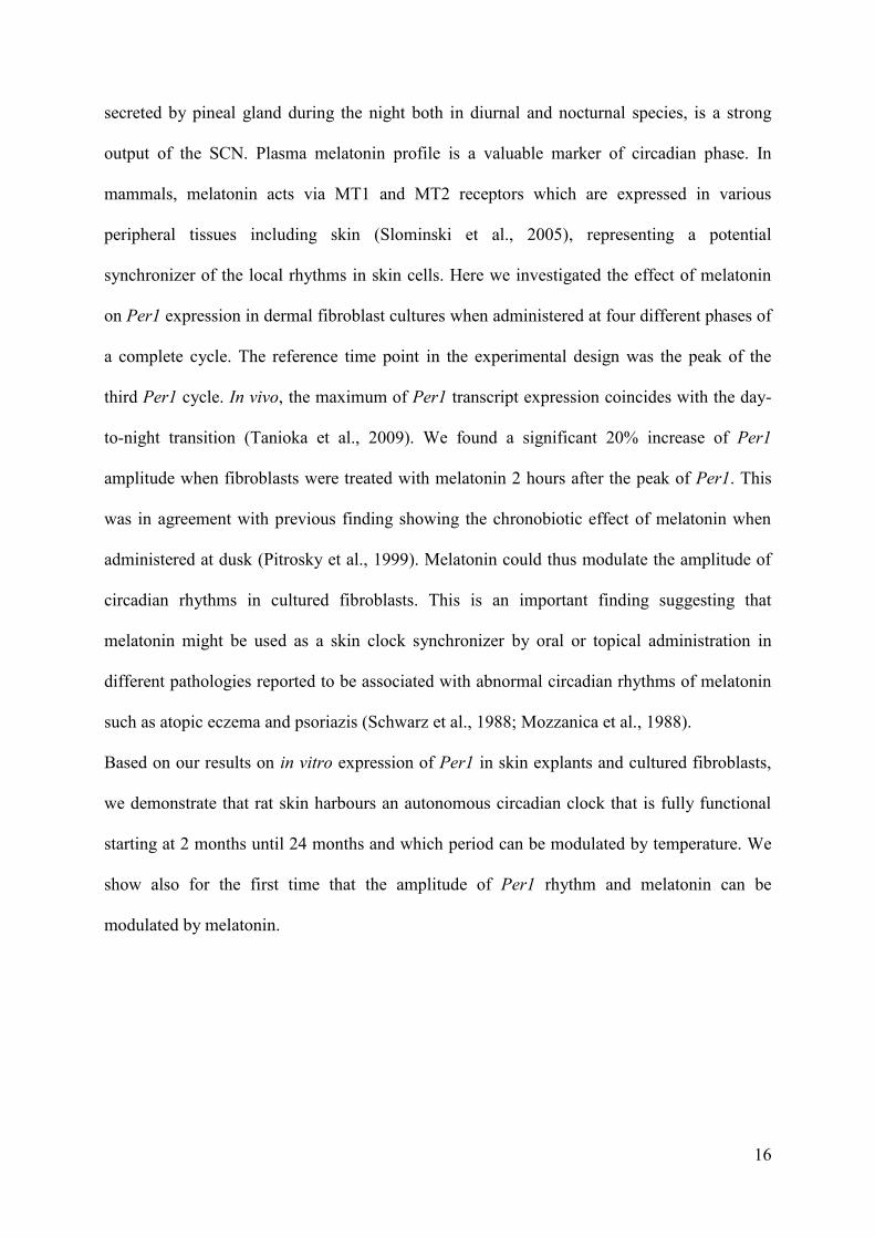
16
secreted by pineal gland during the night both in diurnal and nocturnal species, is a strong
output of the SCN. Plasma melatonin profile is a valuable marker of circadian phase. In
mammals, melatonin acts via MT1 and MT2 receptors which are expressed in various
peripheral tissues including skin (Slominski et al., 2005), representing a potential
synchronizer of the local rhythms in skin cells. Here we investigated the effect of melatonin
on Per1 expression in dermal fibroblast cultures when administered at four different phases of
a complete cycle. The reference time point in the experimental design was the peak of the
third Per1 cycle. In vivo, the maximum of Per1 transcript expression coincides with the day-
to-night transition (Tanioka et al., 2009). We found a significant 20% increase of Per1
amplitude when fibroblasts were treated with melatonin 2 hours after the peak of Per1. This
was in agreement with previous finding showing the chronobiotic effect of melatonin when
administered at dusk (Pitrosky et al., 1999). Melatonin could thus modulate the amplitude of
circadian rhythms in cultured fibroblasts. This is an important finding suggesting that
melatonin might be used as a skin clock synchronizer by oral or topical administration in
different pathologies reported to be associated with abnormal circadian rhythms of melatonin
such as atopic eczema and psoriazis (Schwarz et al., 1988; Mozzanica et al., 1988).
Based on our results on in vitro expression of Per1 in skin explants and cultured fibroblasts,
we demonstrate that rat skin harbours an autonomous circadian clock that is fully functional
starting at 2 months until 24 months and which period can be modulated by temperature. We
show also for the first time that the amplitude of Per1 rhythm and melatonin can be
modulated by melatonin.

17
Acknowledgements
We thank Sophie Reibel, Dominique Sage, Aurore Senser and Nicolas Lethenet from
Chronobiotron UMS 3415 (Strasbourg, France) for animal care.
We are grateful to Dr. Michael Menaker for kindly providing the Per1-luciferase rats.
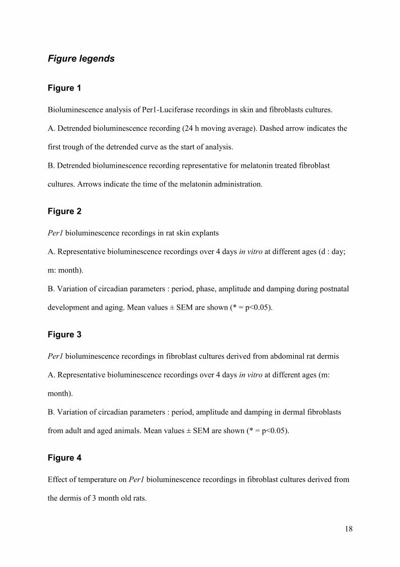
18
Figure legends
Figure 1
Bioluminescence analysis of Per1-Luciferase recordings in skin and fibroblasts cultures.
A. Detrended bioluminescence recording (24 h moving average). Dashed arrow indicates the
first trough of the detrended curve as the start of analysis.
B. Detrended bioluminescence recording representative for melatonin treated fibroblast
cultures. Arrows indicate the time of the melatonin administration.
Figure 2
Per1 bioluminescence recordings in rat skin explants
A. Representative bioluminescence recordings over 4 days in vitro at different ages (d : day;
m: month).
B. Variation of circadian parameters : period, phase, amplitude and damping during postnatal
development and aging. Mean values ± SEM are shown (* = p<0.05).
Figure 3
Per1 bioluminescence recordings in fibroblast cultures derived from abdominal rat dermis
A. Representative bioluminescence recordings over 4 days in vitro at different ages (m:
month).
B. Variation of circadian parameters : period, amplitude and damping in dermal fibroblasts
from adult and aged animals. Mean values ± SEM are shown (* = p<0.05).
Figure 4
Effect of temperature on Per1 bioluminescence recordings in fibroblast cultures derived from
the dermis of 3 month old rats.
19
A. Representative bioluminescence recordings over 4 days in vitro at 32, 35, 37 and 39°C.
B. Variation of circadian parameters : period, amplitude and damping at different
temperatures. Mean values ± SEM are shown (* = p<0.05).
Figure 5
Effect of melatonin treatment on Per1 bioluminescence recordings in fibroblast cultures
Variation of circadian parameters : period, phase, amplitude and damping represented as
treatment/control mean ratios at different phases of melatonin treatment (P : peak) Mean
values ± SEM are shown (* = p<0.05).
20
Bibliography
1. Ansari N, Agathagelidis M, Lee C, Korf HW, von Gall C. Differential maturation of
circadian rhythms in clock gene proteins in the suprachiasmatic nucleus and the pars tuberalis
during mouse ontogeny. Eur J Neurosci 2009; 29(3):477-89.
2. Aujard F, Herzog ED, Block GD. Circadian rhythms in firing rate of individual
suprachiasmatic nucleus neurons from adult and middle-aged mice. Neuroscience 2001;
106(2):255-61.
3. Balsalobre A, Brown SA, Marcacci L, Tronche F, Kellendonk C, Reichardt HM, et al.
Resetting of circadian time in peripheral tissues by glucocorticoid signaling. Science 2000;
289(5488):2344-7.
4. Balsalobre A, Damiola F, Schibler U. A serum shock induces circadian gene
expression in mammalian tissue culture cells. Cell 1998; 93(6):929-37.
5. Brown SA, Fleury-Olela F, Nagoshi E, Hauser C, Juge C, Meier CA, et al. The period
length of fibroblast circadian gene expression varies widely among human individuals. PLoS
Biol 2005; 3(10):e338.
6. Brown WR. A review and mathematical analysis of circadian rhythms in cell
proliferation in mouse, rat, and human epidermis. J Invest Dermatol 1991; 97(2):273-80.
7. Christ E, Korf HW, von Gall C. When does it start ticking? Ontogenetic development
of the mammalian circadian system. Prog Brain Res 2012; 199:105-18.
8. Dickmeis T. Glucocorticoids and the circadian clock. J Endocrinol 2009; 200(1):3-22.
21
9. Dubrovsky YV, Samsa WE, Kondratov RV. Deficiency of circadian protein CLOCK
reduces lifespan and increases age-related cataract development in mice. Aging (Albany NY)
2010; 2(12):936-44.
10. Gaddameedhi S, Selby CP, Kaufmann WK, Smart RC, Sancar A. Control of skin
cancer by the circadian rhythm. Proc Natl Acad Sci U S A 2011; 108(46):18790-5.
11. Geyfman M, Kumar V, Liu Q, Ruiz R, Gordon W, Espitia F, et al. Brain and muscle
Arnt-like protein-1 (BMAL1) controls circadian cell proliferation and susceptibility to UVB-
induced DNA damage in the epidermis. Proc Natl Acad Sci U S A 2012; 109(29):11758-63.
12. Izumo M, Johnson CH, Yamazaki S. Circadian gene expression in mammalian
fibroblasts revealed by real-time luminescence reporting: temperature compensation and
damping. Proc Natl Acad Sci U S A 2003; 100(26):16089-94.
13. Janich P, Pascual G, Merlos-Suarez A, Batlle E, Ripperger J, Albrecht U, et al. The
circadian molecular clock creates epidermal stem cell heterogeneity. Nature 2011;
480(7376):209-14.
14. Janich P, Toufighi K, Solanas G, Luis NM, Minkwitz S, Serrano L, et al. Human
epidermal stem cell function is regulated by circadian oscillations. Cell Stem Cell 2013;
13(6):745-53.
15. Ko CH, Takahashi JS. Molecular components of the mammalian circadian clock. Hum
Mol Genet 2006; 15 Spec No 2:R271-7.
16. Leise TL, Wang CW, Gitis PJ, Welsh DK. Persistent cell-autonomous circadian
oscillations in fibroblasts revealed by six-week single-cell imaging of PER2::LUC
bioluminescence. PLoS One 2012; 7(3):e33334.
22
17. Lin KK, Kumar V, Geyfman M, Chudova D, Ihler AT, Smyth P, et al. Circadian clock
genes contribute to the regulation of hair follicle cycling. PLoS Genet 2009; 5(7):e1000573.
18. Mozzanica N, Tadini G, Radaelli A, Negri M, Pigatto P, Morelli M, et al. Plasma
melatonin levels in psoriasis. Acta Derm Venereol 1988; 68(4):312-6.
19. Nakamura TJ, Nakamura W, Yamazaki S, Kudo T, Cutler T, Colwell CS, et al. Age-
related decline in circadian output. J Neurosci 2011; 31(28):10201-5.
20. Noguchi T, Wang LL, Welsh DK. Fibroblast PER2 circadian rhythmicity depends on
cell density. J Biol Rhythms 2013; 28(3):183-92.
21. Ohta H, Honma S, Abe H, Honma K. Effects of nursing mothers on rPer1 and rPer2
circadian expressions in the neonatal rat suprachiasmatic nuclei vary with developmental
stage. Eur J Neurosci 2002; 15(12):1953-60.
22. O'Neill JS, Hastings MH. Increased coherence of circadian rhythms in mature
fibroblast cultures. J Biol Rhythms 2008; 23(6):483-8.
23. Pagani L, Schmitt K, Meier F, Izakovic J, Roemer K, Viola A, et al. Serum factors in
older individuals change cellular clock properties. Proc Natl Acad Sci U S A 2011;
108(17):7218-23.
24. Pezuk P, Mohawk JA, Wang LA, Menaker M. Glucocorticoids as entraining signals
for peripheral circadian oscillators. Endocrinology 2012; 153(10):4775-83.
25. Pitrosky B, Kirsch R, Malan A, Mocaer E, Pevet P. Organization of rat circadian
rhythms during daily infusion of melatonin or S20098, a melatonin agonist. Am J Physiol
1999; 277(3 Pt 2):R812-28.
23
26. Pittendrigh CS. Temporal organization: reflections of a Darwinian clock-watcher.
Annu Rev Physiol 1993; 55:16-54.
27. Plikus MV, Vollmers C, de la Cruz D, Chaix A, Ramos R, Panda S, et al. Local
circadian clock gates cell cycle progression of transient amplifying cells during regenerative
hair cycling. Proc Natl Acad Sci U S A 2013; 110(23):E2106-15.
28. Polidarova L, Olejnikova L, Pauslyova L, Sladek M, Sotak M, Pacha J, et al.
Development and entrainment of the colonic circadian clock during ontogenesis. Am J
Physiol Gastrointest Liver Physiol 2013.
29. Reppert SM, Schwartz WJ. The suprachiasmatic nuclei of the fetal rat:
characterization of a functional circadian clock using 14C-labeled deoxyglucose. J Neurosci
1984; 4(7):1677-82.
30. Saini C, Morf J, Stratmann M, Gos P, Schibler U. Simulated body temperature
rhythms reveal the phase-shifting behavior and plasticity of mammalian circadian oscillators.
Genes Dev 2012; 26(6):567-80.
31. Sakamoto K, Oishi K, Ishida N. Ontogeny of circadian expression of serotonin N-
acetyltransferase mRNA in the rat retina. Neurosci Lett 2002; 317(1):53-5.
32. Sandu C, Dumas M, Malan A, Sambakhe D, Marteau C, Nizard C, et al. Human skin
keratinocytes, melanocytes, and fibroblasts contain distinct circadian clock machineries. Cell
Mol Life Sci 2012; 69(19):3329-39.
33. Schwarz W, Birau N, Hornstein OP, Heubeck B, Schonberger A, Meyer C, et al.
Alterations of melatonin secretion in atopic eczema. Acta Derm Venereol 1988; 68(3):224-9.
24
34. Seron-Ferre M, Valenzuela GJ, Torres-Farfan C. Circadian clocks during embryonic
and fetal development. Birth Defects Res C Embryo Today 2007; 81(3):204-14.
35. Sladek M, Jindrakova Z, Bendova Z, Sumova A. Postnatal ontogenesis of the
circadian clock within the rat liver. Am J Physiol Regul Integr Comp Physiol 2007;
292(3):R1224-9.
36. Slominski A, Wortsman J, Tobin DJ. The cutaneous serotoninergic/melatoninergic
system: securing a place under the sun. FASEB J 2005; 19(2):176-94.
37. Slominski RM, Reiter RJ, Schlabritz-Loutsevitch N, Ostrom RS, Slominski AT.
Melatonin membrane receptors in peripheral tissues: distribution and functions. Mol Cell
Endocrinol 2012; 351(2):152-66.
38. Slotten HA, Pitrosky B, Krekling S, Pevet P. Entrainment of circadian activity
rhythms in rats to melatonin administered at T cycles different from 24 hours. Neurosignals
2002; 11(2):73-80.
39. Sporl F, Korge S, Jurchott K, Wunderskirchner M, Schellenberg K, Heins S, et al.
Kruppel-like factor 9 is a circadian transcription factor in human epidermis that controls
proliferation of keratinocytes. Proc Natl Acad Sci U S A 2012; 109(27):10903-8.
40. Tanioka M, Yamada H, Doi M, Bando H, Yamaguchi Y, Nishigori C, et al. Molecular
clocks in mouse skin. J Invest Dermatol 2009; 129(5):1225-31.
41. Tsuchiya Y, Akashi M, Nishida E. Temperature compensation and temperature
resetting of circadian rhythms in mammalian cultured fibroblasts. Genes Cells 2003;
8(8):713-20.
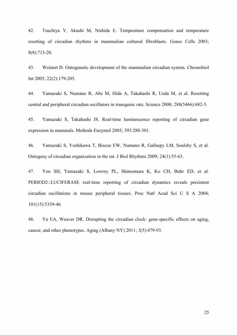
25
42. Tsuchiya Y, Akashi M, Nishida E. Temperature compensation and temperature
resetting of circadian rhythms in mammalian cultured fibroblasts. Genes Cells 2003;
8(8):713-20.
43. Weinert D. Ontogenetic development of the mammalian circadian system. Chronobiol
Int 2005; 22(2):179-205.
44. Yamazaki S, Numano R, Abe M, Hida A, Takahashi R, Ueda M, et al. Resetting
central and peripheral circadian oscillators in transgenic rats. Science 2000; 288(5466):682-5.
45. Yamazaki S, Takahashi JS. Real-time luminescence reporting of circadian gene
expression in mammals. Methods Enzymol 2005; 393:288-301.
46. Yamazaki S, Yoshikawa T, Biscoe EW, Numano R, Gallaspy LM, Soulsby S, et al.
Ontogeny of circadian organization in the rat. J Biol Rhythms 2009; 24(1):55-63.
47. Yoo SH, Yamazaki S, Lowrey PL, Shimomura K, Ko CH, Buhr ED, et al.
PERIOD2::LUCIFERASE real-time reporting of circadian dynamics reveals persistent
circadian oscillations in mouse peripheral tissues. Proc Natl Acad Sci U S A 2004;
101(15):5339-46.
48. Yu EA, Weaver DR. Disrupting the circadian clock: gene-specific effects on aging,
cancer, and other phenotypes. Aging (Albany NY) 2011; 3(5):479-93.

26
Figures
Figure 1

27
Figure 2
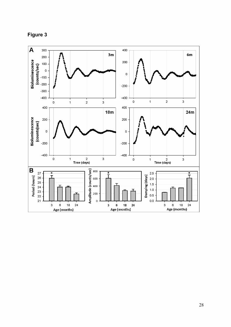
28
Figure 3

29
Figure 4
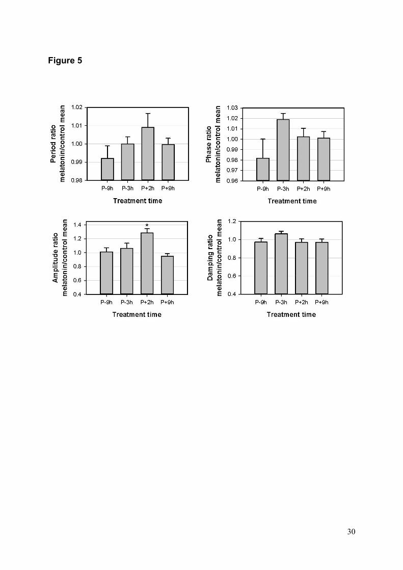
30
Figure 5


LIU Taole La peau, un modèle d’horloge
périphérique
Résumé Ce travail avait pour objet d’étudier les propriétés d’horloge et de synchronisation de la peau, un modèle potentiel d’horloge périphérique. L’activi té rythmique a été analysée par bioluminescence en temps réel, sur des explants de peau abdominale et des fibroblastes dermiques primaires, isolés à partir de rats transgéniques Per1-luciférase.
Nous avons montré que des explants de peau présentent une activité rythmique soutenue en culture, indiquant une importante synchronisation interne dans le tissu. Cette synchronisation se manifeste au cours du développement post-natal à partir de 1 mois et augmente jusqu’à 6 mois, avant de décroître, laissant place à des rythmes altérés à l’âge de 2 ans. Nous avons aussi établi que les fibroblastes dermiques présentent la propriété de compensation thermique commune à toutes les horloges circadiennes, et qu’ils sont potentiellement synchronisables par la mélatonine puisque celle-ci augmente leur amplitude en culture. Nous avons aussi préparé un vecteur lentiviral exprimant le gène rapporteur luciférase sous le contrôle du promoteur du gène horloge Bmal1, un nouvel outil pour compléter l’étude des rythmes dans les cellules de la peau.
Mots-clés: Per1; Bmal1; Horloge périphérique; Rythmes circadiens; Synchronisation; Peau; Fibroblastes; Bioluminescence; Développement postnatal; Vieillissement; Compensation thermique; Mélatonine; Lentivirus
Résumé en anglais This work aimed to investigate the skin as a potential model of peripheral clock by characterizing its rhythmic and synchronization properties. Circadian activity was examined in abdominal skin explants and fibroblasts derived from Per1-Luciferase transgenic rats by real-time recording of bioluminescence.
First, the skin clock was characterized from early postnatal to old age. Low amplitude oscillations appeared at 1 month only and their robustness increased until 6 months. In 1-2 year-old rats, skin circadian rhythms showed decreasing amplitude and abnormal cycles. Primary fibroblasts derived from the skin at the same ages demonstrated similar pattern of clock activity. Temperature compensation, an intrinsic clock feature, was shown the first time in skin and primary fibroblasts. Secondly, we demonstrated a phase-dependent effect of melatonin to increase the amplitude of oscillations in skin primary fibroblasts, indicating it displays a synchronising role in the circadian system. Finally, to facilitate our studies on the multioscillatory skin tissue, we constructed a lentivirus carrying a Bmal1-luciferase reporter, to measure clock genes activities in human skin cells.
Keywords: Per1; Bmal1; Peripheral clock; Circadian rhythms; Synchronization; Skin; Fibroblasts; Bioluminescence; Postnatal development; Ageing; Temperature compensation; Melatonin; Lentivirus