Impaired myocardial metabolic reserve and substrate selection flexibility during stress in patients...
34
Neglia et al. H-00887-2007-Accepted Version 1 Impaired Myocardial Metabolic Reserve and Substrate Selection Flexibility during Stress in Patients with Idiopathic Dilated Cardiomyopathy Danilo Neglia, 1 Alberto De Caterina, 3 Paolo Marraccini, 1 Andrea Natali, 2 Marco Ciardetti, 1 Cecilia Vecoli, 3 Amalia Gastaldelli, 1 Demetrio Ciociaro, 1 Paola Pellegrini, 1 Roberto Testa, 1 Luca Menichetti, 1 Antonio L’Abbate, 1,3 William C. Stanley, 3 Fabio A. Recchia, 3,4 1 Institute of Clinical Physiology of the National Council for Research, Pisa, Italy; 2 Department of Medicine of the University of Pisa, Pisa, Italy; 3 Scuola Superiore Sant’Anna, Pisa, Italy; 4 Department of Physiology, New York Medical College, Valhalla, NY Running Head: Abnormal Metabolism in Dilated Cardiomyopathy Corresponding Author: Danilo Neglia, MD, PhD CNR Institute of Clinical Physiology Pisa – Italy Tel +39 0503152019 Fax +39 0503152166 EMail [email protected] Page 1 of 34 Copyright Information Articles in PresS. Am J Physiol Heart Circ Physiol (October 12, 2007). doi:10.1152/ajpheart.00887.2007 Copyright © 2007 by the American Physiological Society.
-
Upload
independent -
Category
Documents
-
view
0 -
download
0
Transcript of Impaired myocardial metabolic reserve and substrate selection flexibility during stress in patients...

Neglia et al. H-00887-2007-Accepted Version
1
Impaired Myocardial Metabolic Reserve and Substrate Selection
Flexibility during Stress in Patients with Idiopathic Dilated
Cardiomyopathy
Danilo Neglia,1 Alberto De Caterina,3 Paolo Marraccini,1 Andrea Natali,2 Marco
Ciardetti,1 Cecilia Vecoli,3 Amalia Gastaldelli,1 Demetrio Ciociaro,1 Paola Pellegrini,1
Roberto Testa,1 Luca Menichetti,1 Antonio L’Abbate,1,3 William C. Stanley,3 Fabio A.
Recchia,3,4
1Institute of Clinical Physiology of the National Council for Research, Pisa, Italy; 2Department of Medicine of the University of Pisa, Pisa, Italy; 3Scuola Superiore
Sant’Anna, Pisa, Italy; 4Department of Physiology, New York Medical College, Valhalla,
NY
Running Head: Abnormal Metabolism in Dilated Cardiomyopathy
Corresponding Author: Danilo Neglia, MD, PhD CNR Institute of Clinical Physiology Pisa – Italy Tel +39 0503152019 Fax +39 0503152166 EMail [email protected]
Page 1 of 34
Copyright Information
Articles in PresS. Am J Physiol Heart Circ Physiol (October 12, 2007). doi:10.1152/ajpheart.00887.2007
Copyright © 2007 by the American Physiological Society.

Neglia et al. H-00887-2007-Accepted Version
2
ABSTRACT
Under resting conditions, the failing heart shifts fuel use towards greater glucose and lower
free fatty acids oxidation. We hypothesized that chronic metabolic abnormalities in patients
with dilated cardiomyopathy (DCM) are associated with the absence of the normal increase in
myocardial glucose uptake and maintenance of cardiac mechanical efficiency in response to
pacing stress. In 10 DCM and in 6 control subjects, we measured coronary flow by
intravascular ultrasonometry and sampled arterial and coronary sinus blood. Myocardial
metabolism was determined at baseline, during atrial pacing at 130 bpm, and at 15 minutes of
recovery, by infusing 3H-oleate and 13C-lactate and measuring transmyocardial artero-venous
differences of oxygen and metabolites. At baseline, DCM patients showed depressed coronary
flow, reduced uptake and oxidation of FFA and preferential utilization of carbohydrates.
During pacing glucose uptake increased by 106% in controls, but did not change from
baseline in DCM. Lactate release increased by 122% in DCM but not in controls. Cardiac
mechanical efficiency in DCM was not different compared to controls at baseline, but was
34% lower during stress. Fatty acid uptake and oxidation did not change with pacing in either
group. Our results show that in DCM there is preferential utilization of carbohydrates which
is associated with reduced flow and oxygen consumption at rest and an impaired ability to
increase glucose uptake during stress. These metabolic abnormalities might contribute to
progressive cardiac deterioration and represent a target for therapeutic strategies aimed at
modulating cardiac substrate utilization.
Keywords: Metabolism ; Cardiomyopathies ; Microcirculation
Page 2 of 34
Copyright Information
Neglia et al. H-00887-2007-Accepted Version
3
Introduction
Fatty acids provide 60 to 90% of the energy necessary to sustain contractile function in
the resting fasting state, with the remainder coming from glucose and lactate oxidation.
During acute cardiac stress, such as pacing or exercise, the healthy human heart rapidly
increases glucose and lactate uptake, with a relative decline in free fatty acid (FFA) uptake (1,
3-4, 9). Clinical and animal studies suggest that this response is advantageous: it increases
myocardial metabolic efficiency, since carbohydrates are a more efficient substrate than lipids,
generating more contractile power for any given rate of myocardial oxygen consumption
(MVO2) (20, 29, 31).
Dilated cardiomyopathy (DCM) is often characterized by reduced FFA uptake and
oxidation, and accelerated glycolysis and glucose oxidation under resting conditions (7, 21,
29, 31). This metabolic shift may act to optimize the limited oxidative capacity of
cardiomyopathic hearts resulting from impairment of mitochondrial function (28), and/or ATP
transfer from mitochondria to the contractile apparatus (30), or reduced myocardial perfusion
and flow reserve due to coronary microvascular dysfunction (16-17). Little is known about
myocardial metabolism in DCM patient under conditions of increased workload. Factors that
differentiate DCM from healthy conditions (altered metabolic phenotype, limited oxidative
capabilities, reduced flow availability, etc.) might limit the ability of the dysfunctional
myocardium to adapt to acute stress. The myocardial metabolic and flow response to acute
atrial pacing stress has been extensively investigated in patients with syndrome X(1, 3) and
coronary artery disease (CAD) (23-24, 39), but not in DCM patients. This information is
clinically relevant, since metabolic modulators, such as agents that partially inhibit myocardial
FFA oxidation, improve symptoms of stress-induced ischemia in angina patient (32) and
Page 3 of 34
Copyright Information
Neglia et al. H-00887-2007-Accepted Version
4
ventricular function in patients with ischemic cardiomyopathy (2, 13, 31, 37) but the rationale
for their potential use in heart failure and DCM is still under debate.
The purpose of the present investigation was to assess the relationships between
cardiac substrate metabolism, ventricular dysfunction, and coronary blood flow at rest and in
response to acute pacing stress in DCM patients and in a control group of patients with normal
coronary arteries, left ventricular mass and volume. We hypothesized that the chronic
metabolic abnormalities in DCM observed at rest are associated with the absence of the
normal response to pacing stress, namely an increase in myocardial glucose uptake and
maintenance of cardiac mechanical efficiency. Coronary hemodynamics and myocardial
substrate metabolism were measured directly by cardiac catheterization and transmyocardial
blood sampling using an infusion of 3H-oleate and 13C-lactate tracers.
MATERIALS AND METHODS
Study population
From October 2003 to May 2006, we enrolled 10 consecutive patients with DCM and
6 patients with normal LV function (controls), selected among the 329 patients admitted to the
cardiology unit of the CNR Institute of Clinical Physiology in Pisa and showing normal
coronary arteries at angiography. DCM patients were selected according to the following
inclusion criteria: age between 20 and 80 years, left ventricular ejection fraction (LVEF)
<40%, left ventricular end-diastolic diameter (LVEDD) >56 mm, angiographically normal
coronary arteries (<50% coronary diameter stenosis). Exclusion criteria were: NYHA class IV,
previous myocardial infarction, valvular heart disease, actual systemic arterial hypertension,
myocardial active inflammatory/infective diseases such as myocarditis or pericarditis, current
cigarette smoking, diabetes, presence of other systemic illnesses, such as neoplasia, renal,
Page 4 of 34
Copyright Information

Neglia et al. H-00887-2007-Accepted Version
5
hepatic or respiratory failure, atrial fibrillation. Control patients underwent coronary
angiography for history of angina-like chest pain and/or previous stress tests suspicious for
myocardial ischemia. Inclusion criteria for control patients were age between 20 and 80 years,
LVEF >50%, LVEDD <56 mm, angiographically normal coronary arteries (<50% coronary
diameter reduction) and absence of other cardiovascular or systemic diseases.
Both groups of patients underwent routine blood tests, ECG, chest X-ray and resting
2D-Echo. LV function, dimensions and mass were evaluated from 2D-Echo data according to
standardized protocols (12). Eight DCM patients were also submitted, within 30 days after
the catheterization procedure, to positron emission tomography (PET) with 13N-Ammomia to
measure specific blood flow per gram of myocardial tissue (ml/min/g) at rest. Specific flow
was computed according to standardized protocols (17) in the left anterior descending (LAD)
coronary myocardial territory (5).
The study was approved by the Local Ethical Committee and conformed to the
Declaration of Helsinki on human research, and written informed consent was obtained on the
day before cardiac catheterization after complete explanation of the protocol and potential
risks. All coronary-active drugs, including nitrates, Ca++-antagonists, !-blockers, ACE-
inhibitors or statins, were suspended at least 24 hours before catheterization.
Cardiac catheterization
In the morning, after an overnight fast, all patients were submitted to routine coronary
angiography and definitively enrolled after excluding the presence of significant coronary
stenosis.
Page 5 of 34
Copyright Information

Neglia et al. H-00887-2007-Accepted Version
6
Cardiac catheterization was performed to measure coronary perfusion pressure, flow
velocity, and cross-sectional area in the proximal third of the LAD, and the transmyocardial
artero-venous gradients across the LAD myocardial territory of labelled (L-3-13C-lactate and
[9,10-3H]-oleate) and non-labelled substrates (FFA, glucose and lactate). At the completion of
diagnostic catheterization, an i.v. bolus of 110 mg L-3-13C-lactate was administered, followed
by i.v. continuous infusion of [9,10-3H]-oleate at 84 µCi/h and L-3-13C-lactate at 130 mg/h
allowing a 30 minute equilibration period. The 13C-lactate tracer was used to the measure of
simultaneous lactate uptake and efflux from the myocardium (8, 9, 35) and the 3H-oleate
tracer to measure FFA oxidation from the myocardial production of 3H2O (21). A 6F guiding
catheter was placed in the left main coronary ostium through a 7F femoral artery introducer.
An intracoronary heparin bolus (100 U/Kg) was given and a 0.014-in Doppler flow wire
(FloWire, Volcano Corp) was advanced into the LAD. A 2.9 F, 10 MHz intravascular
ultrasound (IVUS) catheter (Eagle Eye Gold, Volcano Corp) was passed over the flow wire
and positioned in the LAD immediately distal to the first septal perforating branch. The
Doppler flow wire was positioned two centimeters distal to the tip of the IVUS catheter to
avoid artefact caused by the catheter wake. The position of the IVUS catheter and flow wire
was documented by angiography and maintained throughout the study by fluoroscopic control.
After LAD instrumentation, a 5F catheter was advanced into the coronary sinus, up to the
great cardiac vein, to withdraw venous blood from the LAD territory; then a unipolar pacing
catheter was positioned into the right atrium. Due to the complexity of the instrumentation, 5
additional patients, fulfilling the inclusion criteria but in whom the catheterization procedures
could not be completed, were excluded from the study.
Page 6 of 34
Copyright Information

Neglia et al. H-00887-2007-Accepted Version
7
After calibration, phasic and mean coronary perfusion pressure (from the guiding
catheter) and coronary flow velocity signals were continuously recorded on paper and
acquired, together with ECG (leads DI-DII-DIII), on a personal computer equipped with
dedicated software. IVUS images were obtained for !30 seconds at each step of the protocol,
with temporal synchronization with flow velocity signal, and recorded for off-line analysis.
At each step of the protocol, arterial and venous blood samples were withdrawn
simultaneously, over 30 seconds, from the femoral artery introducer and the great cardiac
vein, and immediately processed for gas analysis and measurements of metabolite
concentration.
Study protocol
The study protocol consisted of 4 steps. After the instrumentation was completed
(about 30 min), continuous monitoring of coronary hemodynamic signals was started and the
baseline artero-venous sampling was performed (”Baseline”). Heart rate was increased by
atrial pacing to 110 bpm for 3 min and to 130 bpm for 3 additional min and artero-venous
sampling was repeated (”Pacing”). Pacing was then stopped and, 15 min later, a third pair of
artero-venous samples was withdrawn (”Recovery”). After an additional 15 minutes, when all
hemodynamic parameters had returned to baseline, an i.v. adenosine infusion was given for 3
min unless it was not clinically tolerated or significant bradycardia (HR < 50 bpm) or
hypotension (systolic pressure < 90 mmHg) occurred. Coronary parameters were acquired at
the end of the third minute of adenosine administration, or just before suspension, and were
used to estimate coronary flow reserve (Step 4).
Page 7 of 34
Copyright Information

Neglia et al. H-00887-2007-Accepted Version
8
Data Analysis
Coronary flow
Coronary flow was calculated off-line by multiplying Doppler-derived mean flow
velocity by coronary lumen cross sectional area measured by IVUS as described in Data
Supplements.
Coronary flow reserve was computed as adenosine/resting CBF. An estimate of
myocardial mechanical efficiency was obtained from rate-pressure product (RPP) and MVO2
measured at each step as follows (see also Data Supplements):
(RPP/LV mass)/ [MVO2/(0.54*LV mass)] = (RPP*0.54)/ MVO2.
Metabolites
Blood samples from the femoral artery and the coronary sinus were withdrawn slowly
in order to avoid hemolysis and potential contamination with right atrial blood. The first 5 mL
from the coronary sinus catheter were discarded to avoid contamination from either saline or
blood present into the catheter. Oxygen consumption and metabolite uptake and oxidation
were measured as previously described (9-10, 26) (see data Supplement). The percent of the
MVO2 due to FFA or lactate oxidation was calculated as previously described (26). For
lactate, it was assumed that 100% of the 13C-lactate taken up by the heart is oxidized to CO2
(9).
Statistics
Data are presented as mean±SEM. Differences among patients groups for discrete
clinical variables were evaluated by "2 test. Coronary hemodynamic and metabolic data were
Page 8 of 34
Copyright Information

Neglia et al. H-00887-2007-Accepted Version
9
compared in each study condition among groups and for different study conditions in the same
group by two-ways ANOVA followed by Fisher’s exact test. An unpaired t-test was used to
compare single study conditions between groups when appropriate. P value less than 0.05 was
considered significant.
Page 9 of 34
Copyright Information

Neglia et al. H-00887-2007-Accepted Version
10
RESULTS Characteristics of the study population
Clinical, functional and biohumoral data are shown in Table 1. The two groups did not
significantly differ in age or sex. History of angina-like chest pain was more frequent in
controls while risk factors were similar in the two groups. Body mass index and the ratio of
total triglycerides over HDL cholesterol, surrogate measures of insulin resistance and the
metabolic syndrome, did not differ between the two groups. DCM patients showed reduced
LV systolic function and an enlarged LV chamber, frequent left bundle branch block, higher
BNP and NT-proBNP plasma levels. However, DCM patients did not show evidence of
advanced heart failure (8 of 10 patients in NYHA class I-II) or extensive sympathetic
activation (norepinephrine plasma levels were not significantly different between DCM and
controls). Use of primary cardioactive drugs was not significantly different between the two
groups except for digoxin and diuretics in DCM patients.
Coronary hemodynamics
In one patient in the control group the coronary sinus catheter was displaced into the
right atrium, thus metabolic measurements were not obtained. In two patients of the DCM
group pacing rate was maintained at 110 bpm since they did not tolerate higher rate; in one of
these two and in one other patient i.v. adenosine was not administered as they asked to shorten
the study.
Data on coronary hemodynamics during and after pacing stress are summarized in
Table 2. While heart rate was not significantly different between controls and DCM, coronary
Page 10 of 34
Copyright Information

Neglia et al. H-00887-2007-Accepted Version
11
mean perfusion pressure, RPP and CBF in the LAD territory were lower in DCM than in
controls in all the study conditions. From baseline to pacing, heart rate and RPP significantly
increased in both groups and were restored to baseline levels at recovery. CBF significantly
increased during pacing only in DCM group and significantly decreased at recovery in the
control group.
During intravenous adenosine infusion there was a trend for CBF to remain lower in
DCM than in controls (2.18±1.30 vs 3.29±0.89 ml/min/g, P=0.09), while coronary flow
reserve was not significantly different between the two groups (3.59±2.53 vs 3.31±1.25, ns).
Myocardial metabolism and energetics
Baseline arterial concentrations of the major cardiac substrates were not significantly
different between the two groups (Table 3) and did not significantly change during the study.
DCM patients showed a significantly lower FFA uptake and oxidation at all time points and
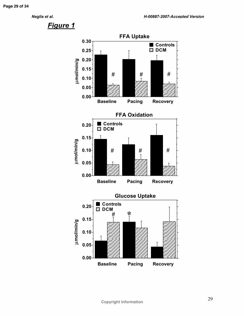
significantly higher glucose uptake than controls at baseline (Figure 1). During pacing,
glucose uptake significantly increased in controls by 106% but did not change in DCM, so
that it was no longer different between the two groups. The percent net extraction of arterial
FFA was unaffected by pacing and was similar between groups (Table 4). On the other hand,
glucose and lactate extraction were higher in DCM group than Controls. Glucose extraction
did not significantly increase during pacing stress in Controls, but decreased significantly in
the DCM compared to baseline and recovery periods (Table 4).
Tracer measured myocardial uptake of lactate was not significantly different between
controls and DCM at baseline and did not show significant changes during stress in both
groups (Figure 2). At recovery, it significantly decreased in controls. Myocardial lactate
release was significantly lower in DCM than in controls at baseline, but during pacing it
Page 11 of 34
Copyright Information
Neglia et al. H-00887-2007-Accepted Version
12
significantly increased by 122% in DCM and reached the levels similar to controls. Net uptake
tended to be higher in DCM patients but this difference was not statistically significant.
Overall, RPP and MVO2 were lower in DCM patients than in controls (Figure 3).
However, the ratio of RPP to MVO2 (an index of myocardial mechanical efficiency, i.e. work
performed for a given amount of oxygen consumed) was significantly lower in DCM only
during stress and at recovery. In fact, during pacing, RPP increased in both groups, but MVO2
increased only in DCM. At recovery, RPP returned to baseline in both groups but in controls
MVO2 declined below baseline levels. The analysis of the percent of MVO2 due to oxidation
of FFA showed that FFA contributed more to MVO2 in controls than in DCM, and this
difference was attenuated during pacing stress (Figure 4, upper panel). While the contribution
of lactate oxidation to MVO2 tended to be greater in DCM than in controls, this difference did
not reach statistical significance (Figure 4, lower panel).
Correlations between LV Remodelling, Metabolism, Energetics and Coronary Blood
Flow.
There was a strong inverse correlation between LV diameter, an index of LV
dysfunction/remodelling, and CBF, MVO2 and FFA uptake in the whole study population,
under basal conditions (Figure 5). There was a non significant trend toward a positive
correlation with glucose uptake (r=0.41, ns). Both under resting conditions and during pacing,
FFA uptake (but not glucose uptake) was directly related with MVO2 (Figure 6).
Page 12 of 34
Copyright Information

Neglia et al. H-00887-2007-Accepted Version
13
DISCUSSION
This study provides direct evidence for a chronic cardiac metabolic shift under both
resting conditions and with pacing stress in DCM patients without advanced heart failure.
This change in cardiac metabolic profile was characterized by decreased FFA uptake and
oxidation and increased carbohydrate utilization, and was associated with reduced cardiac
work, MVO2, and myocardial blood flow both at rest and in response to stress. These results
suggest the novel view that, while the switch away from FFA oxidation in the failing heart is
thought to be compensatory under resting conditions, it may be detrimental during even
moderate stress, potentially contributing to progressive deterioration of myocardial function.
Previous studies in patients with cardiomyopathy/heart failure yielded conflicting
results. Non invasive PET studies in non ischemic cardiomyopathy found reduced uptake and
oxidation of 11C-labelled palmitate (35) and/or increased uptake and utilization of 11C-
labelled glucose (7), consistent with our direct invasive measurements. However, other PET
studies, using FFA and glucose analogues in ischemic and non ischemic patients with
advanced heart failure, found opposite results, with increased lipids and decreased
carbohydrate uptake (33, 38). 22. Paolisso et al evaluated myocardial energy metabolism
in patients with heart failure by direct invasive measurement of transmyocardial substrate
gradients (22), showing increased FFA utilization. However, this study enrolled patients with
NYHA Class II-III heart failure of ischemic and non ischemic origin and no labelled
substrates were used. In the present study the patients were carefully selected to include only
those with idiopathic dilated cardiomyopathy and without advanced heart failure to avoid the
potential confounding effects of coronary disease and of systemic metabolic derangement
associated with cardiac decompensation. In severe heart failure, elevated adrenergic
stimulation may increase MVO2 and levels of FFA, and cardiac insulin resistance may
Page 13 of 34
Copyright Information

Neglia et al. H-00887-2007-Accepted Version
14
develop (19). As a consequence both fatty acid and glucose utilization may be altered and a
condition of energy imbalance may ensue as recently discussed by Neubauer (18).
The resting metabolic pattern that we found in DCM is similar to that observed in the
experimental model of pacing-induced cardiomyopathy (21, 25) and is consistent with
findings in other animal models of progressive cardiac dysfunction and heart failure (11).
This shift in myocardial substrate metabolism partially mimics the neonatal metabolic
phenotype, with glucose and lactate instead of fatty acid being the primary energy substrate
(31). It remains to be determined whether these alterations are a compensatory response of the
failing heart that optimizes cardiac energy metabolism and prevents further deterioration of
LV function, or rather is a maladaptation that contributes to progression towards
decompensation. Insight on this issue comes from mice with cardiac-specific over-expression
of the insulin-independent glucose transporter GLUT1, which have increased glucose uptake
and less contractile dysfunction and LV dilation in response to chronic pressure overload (15),
suggesting that accelerated glucose metabolism prevents hypertension-induced heart failure.
We observed an inverse correlation between cardiac FFA metabolism and the extent of
LV remodelling, which was also associated with reduced MVO2, myocardial blood flow and
cardiac work. A primary depression of myocardial function, as can be expected in DCM,
could cause reduced energy requirements and MVO2 and consequently reduced blood flow
with downregulation of FFA metabolism and possible increase in carbohydrate utilization. A
reduction of myocardial oxygen availability, due to impaired myocardial perfusion and
coronary microvascular dysfunction (16, 17) and impaired oxygen utilization consequent to
mithocondrial dysfunction (28) have been also described in DCM. These mechanisms reduce
energy transduction and further impair contractility, and could by themselves stimulate
myocardial metabolic shift from fat oxidation to preferential utilization of carbohydrates. On
Page 14 of 34
Copyright Information

Neglia et al. H-00887-2007-Accepted Version
15
the other hand, a primary metabolic abnormality could exist, with lower activity of the "-
oxidation pathway (27), lowering MVO2 and causing metabolic downregulation of coronary
flow, but also energy depletion and reduced contractile function.
Myocardial metabolic response to stress in DCM
Myocardial metabolic response to stress in DCM patients had not previously been
investigated. It is well established that in normal hearts high rate pacing induces a rapid
increase in glucose and lactate uptake coupled with a relative decline in FFA uptake (1, 3, 4,
9). Clinical and experimental studies suggest that this response is advantageous, since it
increases myocardial mechanical efficiency as carbohydrates are a more efficient substrate
than lipids and provides more energy for the generation of contractile power for any given
MVO2 (20, 29, 31). The response to pacing stress in DCM patients was very different from
the control group: glucose uptake, which at baseline was already elevated in DCM patient to
levels similar to those reached in controls during stress, did not further increase. In contrast,
FFA utilization tended to increase and the relative contribution of lipid oxidation to MVO2,
which was clearly lower in DCM than in controls in resting conditions, became similar
between the two groups during stress. Compared with controls, there was a lower increase in
myocardial mechanical efficiency during pacing in the DCM patients. Taken together, these
findings strongly indicate that there is a more limited cardiac metabolic reserve and flexibility
in DCM. One of the possible mechanisms explaining the metabolic rigidity of the failing
heart is the paradoxical downregulation of key enzymes of the carbohydrate oxidative
pathway, in spite of higher glucose oxidation, that we previously described in dogs (14). As a
consequence, the cardiomyopathic heart must face even moderate increases in cardiac work by
Page 15 of 34
Copyright Information
Neglia et al. H-00887-2007-Accepted Version
16
oxidizing less efficient substrates, such as FFA, which require higher oxygen consumption
and blood flow.
This abnormal metabolic behaviour, together with impaired mitochondrial function
(28) and limited myocardial flow and oxygen availability, possibly due to coronary
microvascular dysfunction, (16, 17) could contribute to an ischemic-like condition, (6, 36)
enhanced oxidative stress and energy depletion in the cardiomyopathic heart.
Study limitations
The small number of both DCM and control patients, due to the strict selection criteria
and the complexity of the protocol, limits the statistical power. A second limitation is the lack
of a completely normal control population. Control patients had a history of angina-like chest
pain and/or stress testing suggestive for ischemia. However, neither group of patients had the
typical clinical and ECG pattern of syndrome X nor developed chest pain or significant ST
segment depression during pacing stress. Since coronary catheterization procedures have an
intrinsic risk to life, it is unethical to study a truly normal population.
Glucose oxidation was not measured because of the significant additional adsorbed
radiation dose to the patient. It is important to know the effects of DCM and acute stress on
glucose uptake and oxidation to CO2 and release of lactate, which can only be assessed using
14C-labelled glucose. While the present study is the first to assess myocardial lactate kinetics
in DCM using 13C-lactate tracers, these studies would be greatly strengthened by dual isotopic
studies using 14C-glucose/13C-lactate tracers as previously described (9,39). Another
limitation in the metabolic measurements is the inability to measure the oxidation of
intracardiac substrate stores (e.g. glycogen and triglycerides), which likely played an
Page 16 of 34
Copyright Information

Neglia et al. H-00887-2007-Accepted Version
17
important role in supporting the increase in MVO2 in response to pacing in the DCM group
(Figure 3).
Diabetes was an exclusion criterion for enrolment of both DCM and control patients,
however it cannot be excluded that the two patient populations differ in insulin resistance, and
that this could have influenced the metabolic response to stress. Oral glucose tolerance tests
was not performed, however BMI and the triglycerides/HDL was similar between the two
groups, suggesting that whole body metabolic abnormalities were similar..
According to the study protocol, pacing moderately increased heart rate in order to
allow tolerability in DCM patients. This choice could have caused a relatively lower increase
in cardiac workload in normal hearts than in cardiomyopathic hearts where the effects of heart
rate on energy expenditure are added to those of enhanced wall stress. Due to the complexity
of the protocol we could not measure ventricular volumes to have a better estimate of actual
workload than that provided by RPP values. The somewhat artificial conditions of studying
these patients in the catheterization laboratory and the use of heparin likely resulted in
elevated FFA in both groups. Accordingly, baseline metabolic data should be extended with
caution to what one would see in the unheparized resting state. It would be best to use a more
physiological stimulus to increase MVO2, such as exercise or high dose dobutamine infusion,
however these stresses are not feasible in instrumented patients with DCM prone to
arrhythmias.
Conclusions
The present findings demonstrate that DCM patients have a shift in cardiac
metabolism away from FFA oxidation both at rest and during pacing stress, which is
associated with enlargement of the LV and reduced cardiac work, MVO2, and myocardial
Page 17 of 34
Copyright Information
Neglia et al. H-00887-2007-Accepted Version
18
blood flow. In addition, these patients show a failure to further increase glucose uptake and
possibly oxidative glycolysis in response to pacing stress. Our results suggest the novel view
that, while the switch away from FFA oxidation in DCM hearts appears to be compensatory
under resting conditions, it may be detrimental during even moderate stress, potentially
contributing to progressive deterioration of myocardial function. These findings will better
orient therapeutic strategies aimed at modulating mitochondrial substrate oxidation in order to
reduce symptoms and improve cardiac performance in DCM patients (2, 13).
Acknowledgements
This study was supported by intramural funds of the Italian National Council for Research.
F.A. Recchia is an Established Investigator of the AHA.
Disclosures
None.
Page 18 of 34
Copyright Information

Neglia et al. H-00887-2007-Accepted Version
19
References
1. Bagger JP, Thomassen A, Nielsen TT. Cardiac energy metabolism in patients with chest pain and normal coronary angiograms. Am J Cardiol. 85:315-320, 2000.
2. Belardinelli R, Purcaro A. Effects of trimetazidine on the contractile response of chronically dysfunctional myocardium to low-dose dobutamine in ischaemic cardiomyopathy. Eur Heart J. 22:2164-2170, 2002.
3. Camici P, Marraccini P, Lorenzoni R, Buzzigoli G, Pecori N, Perissinotto A, Ferrannini E, L'Abbate A, Marzilli M. Coronary hemodynamics and myocardial metabolism in patients with syndrome X: response to pacing stress. J Am Coll Cardiol.17:1461-1470, 1991.
4. Camici P, Marraccini P, Marzilli M, Lorenzoni R, Buzzigoli G, Puntoni R, Boni C, Bellina CR, Klassen GA, L'Abbate A. Coronary hemodynamics and myocardial metabolism during and after pacing stress in normal humans. Am J Physiol. 257: E309-E317, 1989.
5. Cerqueira MD, Weissman NJ, Dilsizian V, Jacobs AK, Kaul S, Laskey WK, Pennell DJ, Rumberger JA, Ryan T, Verani MS. Standardized myocardial segmentation and nomenclature for tomographic imaging of the heart. A Statement for Healthcare Professionals from the Cardiac Imaging Committee of the Council on Clinical Cardiology of the American Heart Association. Circulation. 105:539–542, 2002.
6. Chen JW, Ting CT, Chen YH, Wu TC, Hsu NW, Lin SJ, Chang MS. Differential coronary microvascular function in patients with left ventricular dysfunction of unknown cause: implication for possible mechanism of myocardial ischemia in early stage of cardiomyopathy. Int J Cardiol. 69:251–261, 1999.
7. Davila-RomanVG, Vedala G, Herrero P, de las Fuentes L, Rogers JG, Kelly DP, Gropler RJ. Altered myocardial fatty acid and glucose metabolism in idiopathic dilated cardiomyopathy. J Am Coll Cardiol. 40:271-277, 2002.
8. Gertz EW, Wisneski JA, Neese R, Bristow JD, Searle GL, Hanlon JT. Myocardial lactate metabolism: evidence of lactate release during net chemical extraction in man. Circulation. 63:1273-1279, 1981.
9. Gertz EW, Wisneski JA, Stanley WC, Neese RA. Myocardial substrate utilization during exercise in humans. Dual carbon- labeled carbohydrate isotope experiments. J Clin Invest. 82:2017-2025, 1988.
10. Hall JL, Stanley WC, Lopaschuk GD, Wisneski JA, Pizzurro RD, Hamilton CD, and McCormack JG. Impaired pyruvate oxydation but normal glucose uptake in diabetic pig heart during dobutamine-induced work. Am J Physiol Heart Circ Physiol. 271: H2320–H2329, 1996.
Page 19 of 34
Copyright Information

Neglia et al. H-00887-2007-Accepted Version
20
11. Heather LC, Cole MA, Lygate CA, Evans RD, Stuckey DJ, Murray AJ, Neubauer S, Clarke K. Fatty acid transporter levels and palmitate oxidation rate correlate with ejection fraction in the infarcted rat heart. Cardiovasc Res. 72:430-437, 2006.
12. Lang RM, Bierig M, Devereux RB, Flachskampf FA, Foster E, Pellikka PA, Picard MH, Roman MJ, Seward J, Shanewise J, Solomon S, Spencer KT, St John Sutton M, Stewart W; American Society of Echocardiography's Nomenclature and Standards Committee; Task Force on Chamber Quantification; American College of Cardiology Echocardiography Committee; American Heart Association; European Association of Echocardiography, European Society of Cardiology. Recommendations for chamber quantification. Eur J Echocardiogr. 7:79-108, 2006.
13. Lee L, Campbell R, Scheuermann-Freestone M, Taylor R, Gunaruwan P, Williams L, Ashrafian H, Horowitz J, Fraser AG, Clarke K, Frenneaux M. Metabolic modulation with perhexiline in chronic heart failure: a randomized, controlled trial of short-term use of a novel treatment. Circulation. 112:3280-3288, 2005.
14. Lei B, Lionetti V, Young ME, Chandler MP, D' Agostino C, Kang E, Altarejos M, Matsuo K, Hintze TH, Stanley WC, Recchia FA. Paradoxical downregulation of the glucose oxidation pathway despite enhanced flux in severe heart failure. J Mol Cell Cardiol. 36:567-576, 2004.
15. Liao R, Jain M, Cui L, D'Agostino J, Aiello F, Luptak I, Ngoy S, Mortensen RM, & Tian R. Cardiac-specific overexpression of GLUT1 prevents the development of heart failure attributable to pressure overload in mice. Circulation 106, 2125-2131, 2002
16. Neglia D, Nichelassi C, Trovieri MG, Sambuceti G, Giorgetti A, Pratali L, Gallopin M, Salvadori P, Sorace O, Carpeggiani C, Poddighe R, L'Abbate A, Parodi O.Prognostic role of myocardial blood flow impairment in idiopathic left ventricular dysfunction. Circulation. 105:186-193, 2002.
17. Neglia D, Parodi O, Gallopin M, Sambuceti G, Giorgetti A, Pratali L, Salvadori P, Michelassi C, Lunardi M, Pelosi G, L’Abbate A. Myocardial blood flow response to pacing tachycardia and to dipyridamole infusion in patients with dilated cardiomyopathy without overt heart failure. A quantitative assessment by positron emission tomography. Circulation. 92:796-804, 1995.
18. Neubauer S. The failing heart — an engine out of fuel. N Engl J Med. 356:1140-51, 2007.
19. Nikolaidis LA, Sturzu A, Stolarski C, Elahi D, Shen YT, Shannon RP. The development of myocardial insulin resistance in conscious dogs with advanced dilated cardiomyopathy. Cardiovasc Res. 61:297-306, 2004.
20. Opie, L. H. The Heart: Physiology and Metabolism. pp. 208-276. Raven Press, New York, 1991.
21. Osorio JC, Stanley WC, Linke A, Castellari M, Diep QN, Panchal AR, Hintze TH, Lopaschuk GD, Recchia FA. Impaired myocardial fatty acid oxidation and reduced
Page 20 of 34
Copyright Information

Neglia et al. H-00887-2007-Accepted Version
21
protein expression of retinoid X receptor-alpha in pacing-induced heart failure. Circulation. 106:606-612, 2002.
22. Paolisso G, Gambardella A, Galzerano D, D'Amore A, Rubino P, Verza M, Teasuro P, Varricchio M, D'Onofrio F. Total-body and myocardial substrate oxidation in congestive failure. Metabolism. 43:174–9, 1994.
23. Parker JO, Chiong MA, West RO, Case RB. Sequential alterations in myocardial lactate metabolism, S-T segments, and left ventricular function during angina induced by atrial pacing. Circulation. 40:113-131, 1969.
24. Parker JO, West RO, Case RB, Chiong MA. Temporal relationships of myocardial lactate metabolism, left ventricular function, and S-T segment depression during angina precipitate by exercise. Circulation. 40: 97-111, 1969.
25. Recchia FA, McConnell PI, Bernstein RD, Vogel TR, Xu X, Hintze TH. Reduced nitric oxide production and altered myocardial metabolism during the decompensation of pacing-induced heart failure in the conscious dog. Circ Res. 83:969–979, 1998.
26. Recchia FA, Osorio JC, Chandler, Xu X, Panchal RA, Lopaschuk GD, Hintze TH, Stanley WC. Reduced synthesis of NO causes marked alterations in myocardial substrate metabolism in conscious dogs Am J Physiol Endocrinol Metab. 282: E197–E206, 2002
27. Sack MN, Rader TA, Park S, Bastin J, McCune SA, Kelly DP. Fatty acid oxidation enzyme gene expression is downregulated in the failing heart. Circulation. 94:2837–2842, 1996.
28. Schulze K, Dörner A, Schultheiss HP. Mitochondrial Function in Heart Failure. Heart Failure Reviews. 4:229–244, 1999.
29. Simonsen S, Kjekshus JK. The effect of free fatty acids on myocardial oxygen consumption during atrial pacing and catecholamine infusion in man. Circulation. 58:484-491, 1978.
30. Smith CS, Bottomley PA, Schulman SP, Gerstenblith G, Weiss RG. Altered creatine kinase adenosine triphosphate kinetics in failing hypertrophied human myocardium. Circulation. 114:1151-1158, 2006.
31. Stanley WC, Recchia FA, Lopaschuk GD. Myocardial substrate metabolism in the normal and failing heart. Physiol Rev. 85:1093-1129, 2005.
32. Stanley WC. Partial fatty acid oxidation inhibitors for stable angina. Expert Opin Investig Drugs. 11:615-629, 2002.
33. Taylor M, Wallhaus TR, DeGrado TR, Russell DC, Stanko P, Nickles RJ, Stone CK.An evaluation of myocardial fatty acid and glucose uptake using PET with [18F]fluoro- 6-thia-heptadecanoic acid and [18F]FDG in patients with congestive heart failure. J Nucl Med. 42:55–62, 2001.
Page 21 of 34
Copyright Information

Neglia et al. H-00887-2007-Accepted Version
22
34. Tuunanen H, Engblom E, Naum A, Nagren K, Hesse B, Airaksinen KE, Nuutila P, Iozzo P, Ukkonen H, Opie LH, Knuuti J. Free fatty acid depletion acutely decreases cardiac work and efficiency in cardiomyopathic heart failure. Circulation. 114:2130-2137, 2006
35. Tuunanen H, Engblom E, Naum A, Scheinin M, Nagren K, Airaksinen J, Nuutila P, Iozzo P, Ukkonen H, Knuuti J. Decreased myocardial free fatty acid uptake in patients with idiopathic dilated cardiomyopathy: evidence of relationship with insulin resistance and left ventricular dysfunction. J Card Fail. 12:644-652, 2006.
36. van den Heuvel AF, van Veldhuisen DJ, van der Wall EE, Blanksma PK, Siebelink HM, Vaalburg WM, van Gilst WH, Crijns HJ. Regional myocardial blood flow reserve impairment and metabolic changes suggesting myocardial ischemia in patients with idiopathic dilated cardiomyopathy. J Am Coll Cardiol. 35:19–28, 2000.
37. Vitale C, Wajngaten M, Sposato B, Gebara O, Rossigni P, Fini M, Volterrani M, Rosano GM. Trimetazidine improves left ventricular function and quality of life in elderly patients with coronary artery disease. Eur Heart J. 25:1814-1821, 2004.
38. Wallhaus TR, Taylor M, DeGrado, Russell DC, Stanko P, Nickles RJ, Stone CK.Myocardial free fatty acid and glucose use after carvedilol treatment in patients with congestive heart failure. Circulation. 103:2441–6, 2001
39. Wisneski JA, Gertz EW, Neese RA, Gruenke LD, Craig JC. Dual carbon-labeled isotope experiments using D-[6-14C] glucose and L- [1,2,3-13C3] lactate: a new approach for investigating human myocardial metabolism during ischemia. J Am Coll Cardiol.5:1138-1146, 1985.
Page 22 of 34
Copyright Information

Neglia et al. H-00887-2007-Accepted Version
23
Table 1. Clinical, functional and biohumoral data
Controls DCM P value
N 6 10 Age 67±2 57±3 ns Males n (%) 3 (30) 7 (70) ns Angina n (%) 5 (83) 2 (20) p<.05 LBBB n (%) 2 (33) 9 (90) p<.05 LV mass (g) 164±11 287±19 p<0.01 NYHA class III n (%) 0 2 (20) ns Functional data LVEF (%) 58±1 32±1 p<.001 LVEDD (mm) 49±1 65±1 p<.001 Neurohormonal data BNP (pg/ml) 21±7 91±22 p<.05 Nt-pro-BNP (pg/ml) 123±37 593±130 p<.05 Norepinephrine(pg/ml) 210±54 412±81 ns Risk factors Body mass index (kg/m2) 28±2 29±2 ns Fasting glycemia (mg/dl) 90±4 101±9 ns Total triglycerides/HDL cholesterol 2.79±1.73 3.07±2.71 ns HDL/total cholesterol 0.23±0.02 0.24±0.02 ns Treatment Statins n (%) 3 (50) 3 (30) ns Antiplatelet n (%) 2 (33) 5 (50) ns Ca-antagonists n (%) 1 (17) 0 ns !-blockers (carvedilol) n (%) 3 (50) 6 (60) ns ACE-inhibitors/AT-1 antagonists n (%) 3 (50) 9 (90) ns Antialdosteronic n (%) 0 3 (30) ns Diuretics n (%) 1 (17) 8 (80) p<.05 Digoxin n (%) 0 5 (50) p<.05
Page 23 of 34
Copyright Information

Neglia et al. H-00887-2007-Accepted Version
24
Table 2. Hemodynamics
Controls DCM P value Heart rate (bpm) Baseline 75±5 71±4
Pacing 132±4 124±4 Recovery 77±7 71±4
ns
P value (Pacing vs Baseline and Recovery) <.001 <.001 Mean aortic pressure (mmHg) Baseline 107±2 89±3
Pacing 109±3 91±4 Recovery 105±4 90±4
<.001
P value (Pacing vs Baseline and Recovery) ns ns Rate-pressure product (mmHg * bpm) Baseline 8086±590 6320±413
Pacing 14475±642 11314±647 Recovery 8155±870 6438±440
<.001
P value (Pacing vs Baseline and Recovery) <.001 <.001 LAD CBF (ml/min/g) Baseline 1.08±.07 .66±.06
Pacing 1.19±.07 .91±.07
Recovery .84±.04* .64±.07 <.001
P value (Pacing vs Baseline and Recovery) ns <.01
*P<0.05 (Recovery vs Baseline and Pacing)
Page 24 of 34
Copyright Information

Neglia et al. H-00887-2007-Accepted Version
25
Table 3. Arterial concentration of substrates.
Controls DCM P value
FFA (µmol/ml) 1.93±.21 1.34±.18 ns
Glucose (µmol/ml) 5.45±.09 5.37±.31 ns
Lactate (µmol/ml) 0.51±.03 0.61±.08 ns
Page 25 of 34
Copyright Information

Neglia et al. H-00887-2007-Accepted Version
26
Table 4. Percent transmyocardial net extraction of arterial FFA, glucose and lactate.
Controls DCM P value#
FFA extraction (%) Baseline 15±5 18±10 ns Pacing 12±5 15±8 ns Recovery 17±6 19±12 ns Glucose extraction (%) Baseline 1±1 4±2† <0.05 Pacing 2±1 2±2†* ns Recovery 1±1 5±7† ns Lactate extraction (%) Baseline 18±14 32±11‡ ns Pacing 17±12 27±11‡ ns Recovery 9±8 29±16‡ <0.05
# DCM vs. Control under given condition. † p<0.05 DCM vs. Control for main effect. ‡ p<0.001 DCM vs. Control for main effect. * P<0.05 compared to Baseline and Recovery.
Page 26 of 34
Copyright Information
Neglia et al. H-00887-2007-Accepted Version
27
Figure Legends Figure 1. Cardiac FFA uptake and oxidation and glucose uptake in controls (black bars, n=5)
and in DCM (slashed bars, n=10) at baseline, during atrial pacing and at recovery. * P<0.05
pacing vs baseline and recovery; # P< 0.05 DCM vs controls.
Figure 2. Cardiac net lactate uptake and tracer measured lactate uptake and release in controls
(black bars, n=5) and in DCM (slashed bars, n=10) at baseline, during atrial pacing and at
recovery. *P<0.05 pacing vs baseline and recovery; † P<0.05 recovery vs baseline and pacing.
Figure 3. Rate pressure product (RPP, upper panel), an estimate of cardiac work, myocardial
oxygen consumption (MVO2, middle panel) and mechanical efficiency, estimated from the
ratio between RPP and MVO2 (lower panel), in controls (black bars, n=5) and in DCM
(slashed bars, n=10) at baseline, during atrial pacing and at recovery. All the values were
corrected for myocardial mass. *P<0.05 pacing vs baseline and recovery; # P<0.05 DCM vs
controls.
Figure 4. Percent contribution of FFA oxidation (upper panel) or lactate oxidation (lower
panel) to myocardial oxygen consumption (MVO2) in controls (black bars, n=5) and in DCM
(slashed bars, n=10) at baseline, during atrial pacing and at recovery. #P<0.05 DCM vs
controls.
(Liao et al., 2002)
Page 27 of 34
Copyright Information
Neglia et al. H-00887-2007-Accepted Version
28
Figure 5. Correlations between LV end diastolic diameter (LVEDD) and resting coronary
blood flow (CBF), myocardial oxygen consumption (MVO2) and myocardial FFA uptake
including controls (closed circles) and DCM patients (open circles).
Figure 6. Correlations between myocardial oxygen consumption (MVO2), myocardial FFA
and glucose uptake measured at rest and during pacing in controls (closed circles) and DCM
patients (open circles).
Page 28 of 34
Copyright Information
Neglia et al. H-00887-2007-Accepted Version
29
Figure 1FFA Uptake
##µm
ol/m
in/g
#µm
ol/m
in/g
FFA Oxidation
DCMControls
Glucose Uptake
µmol
/min
/g
Baseline Pacing Recovery0.00
0.05
0.10
0.15
0.20
Baseline Pacing Recovery
*
0.00
0.05
0.10
0.15
0.20
0.00
0.05
0.10
0.15
0.20
0.25
0.30
Baseline Pacing Recovery
## #
DCMControls
DCMControls
#
Page 29 of 34
Copyright Information

Neglia et al. H-00887-2007-Accepted Version
30
DCMControls
DCMControls
Figure 2
Tracer Measured Lactate Uptake
Net Lactate Uptake
µmol
/min
/g
Tracer Measured Lactate Release
µmol
/min
/g
DCMControls
µmol
/min
/g
†
0.0
0.1
0.2
0.3
0.4
Baseline Pacing Recovery
Baseline Pacing Recovery0.00
0.05
0.10
0.15
0.20
0.25
Baseline Pacing Recovery
*
#
0.00
0.05
0.10
0.15
Page 30 of 34
Copyright Information

Neglia et al. H-00887-2007-Accepted Version
31
Figure 3
RPP/LV Mass
bpm
xm
mH
g/g
*
*
##
#
DCMControls
MVO2/(0.54xLV Mass)
µmol
/min
/g
Mechanical Efficiency(RPPx0.54)/ MVO2
*
#
0
20
40
60
80
100
120
Baseline Pacing Recovery
0
2
4
6
8
10
Baseline Pacing Recovery
02468
10121416
Baseline Pacing Recovery
DCMControls
*##
*#DCMControls
Page 31 of 34
Copyright Information

Neglia et al. H-00887-2007-Accepted Version
32
Figure 4
FFA Oxidation as % of MVO2
%##
Lactate Oxidation as % of MVO2
%
DCMControls
0
20
40
60
80
100
Baseline Pacing Recovery
DCMControls
0
5
10
15
20
25
Baseline Pacing Recovery
Page 32 of 34
Copyright Information

Neglia et al. H-00887-2007-Accepted Version
33
Figure 5
MVO
2
(µm
ol/m
in/g
)
,3
,5
,7
,9
1,1
1,3
45 50 55 60 65 70 75,3
,5
,7
,9
1,1
1,3
45 50 55 60 65 70 75
CB
F
(ml/m
in/g
)
23456789
1011
45 50 55 60 65 70 7523456789
1011
45 50 55 60 65 70 75
P<.01 R=-.69
P<.001 R=-.77
LVEDD (mm)
FFA
upta
ke
(µm
ol/m
in/g
)
P<.001 R=-.81
0,05
,1,15
,2,25
,3,35
45 50 55 60 65 70 750
,05
,1,15
,2,25
,3,35
45 50 55 60 65 70 75
DCMControls
DCMControls
Page 33 of 34
Copyright Information

Neglia et al. H-00887-2007-Accepted Version
34
Figure 6
MVO2 (µmol/min/g)
P<.001 R=.71
FFA
upta
ke
( µm
ol/m
in/g
)
P=.22 R=-.24
Glu
cose
upta
ke
( µm
ol/m
in/g
)
MVO2 (µmol/min/g)
DCMControls
-,1
0
,1
,2
,3
,4
2 3 4 5 6 7 8 9 10 11-,1
0
,1
,2
,3
,4
2 3 4 5 6 7 8 9 10 11
-,1
0
,1
,2
,3
,4
2 3 4 5 6 7 8 9 10 11-,1
0
,1
,2
,3
,4
2 3 4 5 6 7 8 9 10 11
Page 34 of 34
Copyright Information