Barrett’s esophagus: management of high-grade dysplasia and cancer
Upload
johnshopkinsCategory
view
1download
0
A(
FR‡BFHFhga
a
Journal of the American College of Cardiology Vol. 53, No. 15, 2009© 2009 by the American College of Cardiology Foundation ISSN 0735-1097/09/$36.00P
Imaging in Heart Rhythm Disorders
Morphologic Variants ofFamilial Arrhythmogenic RightVentricular Dysplasia/CardiomyopathyA Genetics–Magnetic Resonance Imaging Correlation Study
Darshan Dalal, MD, MPH,* Harikrishna Tandri, MD,* Daniel P. Judge, MD,* Nuria Amat, MS,*Robson Macedo, MD,† Rahul Jain, MD,* Crystal Tichnell, MGC,* Amy Daly, MS,*Cynthia James, PHD, SCM,* Stuart D. Russell, MD,* Theodore Abraham, MD,*David A. Bluemke, MD, PHD,‡ Hugh Calkins, MD*
Baltimore and Bethesda, Maryland
Objectives The purpose of this study was to determine the extent of left ventricular (LV) involvement in individuals predis-posed to developing arrhythmogenic right ventricular dysplasia/cardiomyopathy (ARVD/C), and to investigatenovel morphologic variants of ARVD/C.
Background The discovery of desmosomal mutations associated with ARVD/C has led researchers to hypothesize equal rightventricular (RV) and LV affliction in the disease process.
Methods Thirty-eight (age 30 � 17 years; 18 males) family members of 12 desmosomal mutation-carrying ARVD/C pro-bands underwent genotyping and cardiac magnetic resonance imaging (CMR). The CMR investigators wereblinded to clinical and genetic data.
Results Twenty-five individuals had mutations in PKP2, DSP, and/or DSG2 genes. RV abnormalities were associated withthe presence of mutation(s) and with disease severity determined by criteria (minor � 1; major � 2) points forARVD/C diagnosis. The only LV abnormality detected, the presence of intramyocardial fat, was present in 4 indi-viduals. Each of these individuals was a mutation carrier, whereas 1 had no previously described ARVD/C-relatedabnormality. On detailed CMR, a focal “crinkling” of the RV outflow tract and subtricuspid regions(“accordion sign”) was observed in 60% of the mutation carriers and none of the noncarriers (p � 0.001). Thesign was present in 0%, 37%, 71%, and 75% of individuals who met 1, 2, 3, and 4� criteria points, respectively(p � 0.01).
Conclusions Despite a possible LV involvement in ARVD/C, the overall LV structure and function are well preserved. Indepen-dent LV involvement is of rare occurrence. The accordion sign is a promising tool for early diagnosis of ARVD/C.Its diagnostic utility should be confirmed in larger cohorts. (J Am Coll Cardiol 2009;53:1289–99) © 2009 bythe American College of Cardiology Foundation
ublished by Elsevier Inc. doi:10.1016/j.jacc.2008.12.045
itpd
cgmcg
rrhythmogenic right ventricular dysplasia/cardiomyopathyARVD/C) is a familial cardiomyopathy characterized clin-
rom the *Division of Cardiology, Department of Medicine, and †Department ofadiology, Johns Hopkins University School of Medicine, Baltimore, Maryland; andRadiology and Imaging Sciences, National Institutes of Health Clinical Center,ethesda, Maryland. The Johns Hopkins ARVD Program is supported by the Bogleoundation, the Campanella family, the Wilmerding Endowments, and Healingearts; this work was also supported by grants from the Donald W. Reynolds
oundation; and the W.W. Smith Charitable Trust. Dr. Abraham has receivedonoraria and research support from GE Healthcare. Dr. Tandri is supported by arant from the National Heart, Lung, and Blood Institute (HL093350). Drs. Dalalnd Tandri contributed equally to this work.
AManuscript received July 8, 2008; revised manuscript received November 5, 2008;
ccepted December 3, 2008.
cally by right ventricular (RV) dysfunction and ventricularachycardia (1–4) and histopathologically by fibrofatty re-lacement of the myocardium (5). Recent literature hasemonstrated the role of mutations in genes encoding
See page 1300
ardiac desmosomal proteins such as desmoplakin, plako-lobin, plakophilin-2, and desmoglein-2 in the develop-ent of ARVD/C (6–17). Although ARVD/C has been
onsidered primarily as a right-sided cardiomyopathy, therowing evidence supporting the desmosomal origin of
RVD/C has led investigators to hypothesize a concomitant
ieCdoo
M
PsAedkrcwRdpAvttd
ppdoan
et
caDsaaICpCdtWafse2saGsswimctWGoA(Hp221gatt
psstpdv
rv4s
1290 Dalal et al. JACC Vol. 53, No. 15, 2009Genetics and CMR in Familial ARVD/C April 14, 2009:1289–99
or independent left ventricular(LV) involvement in these patients(18). The Task Force criteria thatare widely applied to ascertain thediagnosis of ARVD/C were pub-lished in 1994 and were designedto specifically exclude LV disease(19).
The purpose of this study was2-fold. First, our study was aimedat determining the presence andextent of LV involvement in indi-viduals predisposed to developingARVD/C. The hypothesis of thestudy was that family memberscarrying mutations in genes encod-ing desmosomal proteins would beequally likely to develop right- orleft-sided cardiomyopathy. Thesecondary aim of the study was to
dentify the early morphologic variants of ARVD/C and toxamine their incremental value in the diagnosis of ARVD/C.ardiac magnetic resonance imaging (CMR), which has beeneveloped as an important tool for the noninvasive evaluationf the disease, was used to ascertain the morphologic variantsf both ventricles (20–23).
ethods
atient recruitment, evaluation, and diagnosis. Thetudy population was identified from the Johns HopkinsRVD Registry. The Johns Hopkins ARVD Registry was
stablished in 1999 with the goal of gaining insights into theiagnosis, genetic basis, and clinical course of patients withnown or suspected ARVD/C. All patients included in thisegistry provided written informed consent to participate inlinical and research genetic screening. The study protocolas approved by the Johns Hopkins Medicine Institutionaleview Board. As a routine protocol of the registry, afteriagnosis of an ARVD/C patient, all family members of theatient are invited to undergo a screening protocol forRVD/C. The screening protocol includes obtaining rele-
ant medical history, noninvasive clinical testing for ascer-ainment of the Task Force criteria, and genetic sequencingo identify possible mutations in 1 or more genes encodingesmosomal proteins.For the purpose of the present study, family members of
robands with 1 or more mutations in genes encodinglakophilin-2 (PKP2), desmoplakin (DSP), and/oresmoglein-2 (DSG2), were included. The probands werenly used for the purpose of identifying the family members,nd were excluded in all further analyses. Families in whichone of the family members underwent CMR were excluded.Patients were evaluated as described previously (4). For
ach patient, medical history and family history were ob-
Abbreviationsand Acronyms
ARVD/C � arrhythmogenicright ventriculardysplasia/cardiomyopathy
CMR � cardiac magneticresonance imaging
EDV � end-diastolic volume
EF � ejection fraction
ESV � end-systolic volume
LV � left ventricle/ventricular
ROC � receiver-operatingcharacteristic
RV � right ventricle/ventricular
RVOT � right ventricularoutflow tract
ained. Subsequently, each patient underwent noninvasive r
linical testing, including electrocardiogram, signal-veraged electrocardiogram, Holter monitoring, and CMR.iagnosis of ARVD/C was established based on the criteria
et by the Task Force of the Working Group of Myocardialnd Pericardial Disease of the European Society of Cardiologynd of the Scientific Council on Cardiomyopathies of thenternational Society and Federation of Cardiology (19).
MR. A detailed CMR was performed for the entire studyopulation irrespective of their genetic or clinical findings.MRs were performed according to standard protocols foriagnosis of ARVD/C (22). All CMR datasets were ob-ained on a 1.5-T scanner (CV/i, GE Medical Systems,
aukesha, Wisconsin) and included both fast spin-echond gradient-echo sequences. Fat- and nonfat-suppressedast spin-echo sequences were acquired in the axial andhort-axis planes with breath-hold double-inversion recov-ry blood suppression pulses. The repetition time was 1 orR-R intervals, and the time to excitation was 10 ms. The
lice thickness was 5 mm and slice gap 5 mm. The matrixnd field of view were 256 � 256 and 24 cm, respectively.radient echo sequences were acquired in the axial and
hort-axis planes using breath-hold steady-state free preces-ion imaging. The flip angle was 40°, and time to excitationas set to minimum. For steady-state free precession
maging, the slice thickness was 8 mm with a slice gap of 2m. The matrix and field of view were 256 � 160 and 36
m, respectively. A phased array cardiac coil was used for allhe studies. The datasets were transferred to an Advantage
indows workstation (GE Medical Systems) for analysis.adolinium-enhanced CMRs were reviewed for evidence
f delayed enhancement in any of the RV or LV segments.fter intravenous administration of a CMR contrast agent
0.2 mmol/kg of gadodiamide [Omniscan, Amershamealth, Princeton, New Jersey]), inversion recovery pre-
ared breath-hold cine gradient-echo images were obtained0 min after contrast agent injection. Breath-hold-dimensional imaging (7.2/3.2; inversion time optimized50 to 200 ms; flip angle, 25°; slice thickness, 8 mm; sliceap, 2 mm; number of excitations, 2; matrix, 256 � 192;nd field of view, 360 � 270 mm) scans were obtained inhe short-axis and axial planes at 10-mm intervals coveringhe entire RV and LV.
Quantitative analysis was performed using the softwarerogram MASS (Medis, Leiden, the Netherlands). End-ystolic image was defined visually as the one with themallest ventricular cavity size and end-diastolic image, ashe first image after the R-wave trigger. Quantitative CMRarameters included end-systolic volume (ESV), end-iastolic volume (EDV), and ejection fraction (EF) of bothentricles.
For qualitative assessment, the RV was divided into 4egions: 1) base; 2) mid-cavity; 3) apex; and 4) rightentricular outflow tract (RVOT). The LV was divided into
regions based on the 17-segment model (24), so thategments 1 to 6 represented the base, segments 7 to 12
epresented the midcavity, segments 13 to 16 represented
tQfl
tnrpGdp(opBiCfAppoStcNod
ppwaCpd22tCsfwccFcefcpiSpf
tCov
wFmaRdaaTcafi
sts
R
Ds1AmatPrwmcf
D
*†
1291JACC Vol. 53, No. 15, 2009 Dalal et al.April 14, 2009:1289–99 Genetics and CMR in Familial ARVD/C
he apical region, and segment 17 represented the apex.ualitative CMR parameters included globally impaired
unction, hypokinesia/akinesia, intramyocardial fat, and de-ayed enhancement of both ventricles.
Each segment of both ventricles was carefully examinedo determine the presence of typical (Online Appendix) andovel morphologic variants in both ventricles. The CMReader was blinded to other clinical and genetic data for allatients.enotype analysis. For each ARVD/C proband, genomic
eoxyribonucleic acid (DNA) was extracted from leukocytesresent in whole blood using QIAmp DNA blood maxi kitsQiagen, Inc., Valencia, California). Amplification of exonsn either side of DSP, PKP2, and DSG2 was performed asreviously described, with primer sequence (8,11–13,17,25).idirectional sequence chromatography was performed us-
ng an Applied Biosystems 3730 DNA Analyzer (Fosterity, California). Analysis of chromatograms was per-
ormed using Sequencher 4.2.2 (Gene Codes Corp., Annrbor, Michigan). Novel mutations were analyzed in aopulation of 200 individuals (400 chromosomes) from aanel of unrelated unaffected individuals. Control DNA wasbtained from the National Institute of General Medicalciences (NIGMS) Human Genetic Cell Repositoryhrough the Coriell Institute for Medical Research, and theontrols were matched to the mutation carriers by ancestry.ovel sequence variants were characterized as mutations
nly if they were absent in the unaffected controls and alsoisrupted amino acids, which are highly conserved.All consenting family members were tested for the
resence or absence of the mutation identified in the familyroband. Again, DNA was extracted from leukocytes present inhole blood and subjected to focused bidirectional sequence
nalysis using methods described in the preceding text.orrelation between CMR and genotype. Based on theresence of mutation(s) in 1 or more genes encodingesmosomal proteins, the study population was divided intodistinct groups: 1) family members with a mutation; and
) family members without a mutation. CMR characteris-ics were compared between these subgroups.
orrelation between CMR and Task Force criteria. Thetudy population was divided into subgroups based on theulfillment of the Task Force criteria. One “criteria point”as given for fulfillment of each minor criterion, and 2
riteria points were given for fulfillment of each majorriterion. Thus, ARVD/C diagnosis, according to the Taskorce definition, was made if an individual had 4 or moreriteria points. The presence of CMR characteristics wasxamined in relation to the criteria points achieved. Asamily history may provide either a major or a minorriterion regardless of any other abnormality, total criteriaoints excluding family history were also calculated for eachndividual in the study population.tatistical analyses. All continuous variables were ex-ressed as mean � SD and all categorical variables as
requency (%). Comparisons in continuous variables be-11
ween subgroups of patients were performed by t test.ategorical variables were compared by the chi-square testr Fisher exact test. Criteria points were treated as nominalariables for comparison between groups.
Receiver-operating characteristic (ROC) curve analysisas used to examine the discriminatory ability of the Taskorce criteria in identifying patients with a desmosomalutation. The presence of a desmosomal mutation was used
s a “gold standard” for this analysis. The area under theOC curve was computed using the criteria points asescribed above on a continuous scale ranging between 0nd 7. The area under the ROC curve was computed againfter incorporating the new CMR findings into the currentask Force criteria. The 2 areas under the curves were
ompared to examine the improvement in the discriminatorybility of the Task Force criteria after the addition of the newndings (26).All statistical analyses were performed using STATA
tatistical software (version 8.2, Stata Corp., College Sta-ion, Texas). A value of p � 0.05 was considered statisticallyignificant.
esults
emographics, baseline characteristic, and genotyping. Thetudy population was composed of 38 family members from2 families. The proband of each family was diagnosed withRVD/C based on the Task Force criteria and carried 1 orore mutations in the desmosomal proteins PKP2, DSP,
nd/or DSG2. Table 1 shows the details of genetic muta-ions present in the study population. Each of the enlistedKP2 mutations detected in our study population has beeneported previously (11,12,16,25,27). In addition, thereere 2 novel mutations detected in 4 individuals. A novelutation in DSG2, V56M, was detected in an individual
arrying the 2146-1G�C mutation in PKP2. Two otheramily members of this individual included in this study did
etails of Genotyping
Table 1 Details of Genotyping
Genetic CharacteristicFamilies(n � 12)
Individuals*(n � 38)
Presence of 1 or more mutations 9† 25
PKP2 only 8 21
1642delG 1 6
2146-1G�C 2 3
2509delA 1 3
2484 C�T 1‡ 3
2011delC 1 2
2489�1G�A 1 2
F424S 1 1
S140F 1 1
PKP2 (2146-1G�C) and DSG2 (V56M) 1 1
DSP (S986P) 1 3
Probands excluded are PKP2 (plakophilin 2), DSP (desmoplakin), and DSG2 (desmoglein 2).None of the family members screened from 3 families had a mutation. The probands had the
45-148delCAGA, 1613 G�A, and 2146-1 G�C mutations in the PKP2 gene. ‡Three patients fromfamily were heterozygous for 2484 C�T cryptic splice site mutation in the PKP2 gene (28).
nmcwPnwtpAltdd
antsp0wwAmgiuIi1
aap1mwRtaMCpbwpfittpic
R
twapawc
1292 Dalal et al. JACC Vol. 53, No. 15, 2009Genetics and CMR in Familial ARVD/C April 14, 2009:1289–99
ot carry the DSG2 mutation but had 2 distinct PKP2utations (2146-1G�C and S140F). The proband (ex-
luded from the study as described in the Methods section)ho is the father of these 3 individuals carried both theKP2 mutations as well as the DSG2 mutation. Anotherovel mutation detected in this study was the DSP S986P,hich was present in 3 individuals from 1 family. One of
hese individuals was diagnosed with ARVD/C (4 criteriaoints), and another individual had 3 criteria points forRVD/C diagnosis. None of the individuals from 3 fami-
ies (other than the proband) carried a desmosomal muta-ion. Overall, 25 (66%) of 38 family members had 1 or moreesmosomal mutations, and 13 (34%) had no detectableesmosomal mutation.Table 2 shows the demographics and symptoms present
t the time of screening in the study population. Althoughone of these family members had sought medical advice forhese symptoms earlier, 9 individuals had prior cardiacymptoms. Palpitations were more commonly present inatients with a mutation compared to those without (p �.05). The number of criteria points achieved by patientsith mutation(s) was greater than those achieved by patientsithout the mutation(s) (p � 0.05).RVD/C diagnosis. As shown in Table 2, 4 (11%) familyembers met an adequate set of criteria (4 criteria points or
reater) to establish ARVD/C diagnosis. All 4 of thesendividuals carried a desmosomal mutation. All 13 individ-als without a mutation achieved 2 criteria points or fewer.n fact, barring the criteria for family history (which allndividuals in this study population fulfill), only 1 of these3 individuals achieved a criteria point.
Demograhics, Prior Symptoms, Family History, aFulfillment of the Task Force Criteria (19) in the
Table 2 Demograhics, Prior Symptoms, FamFulfillment of the Task Force Criter
Characteristics
Age, yrs
Sex, male
Prior symptoms
Syncope
Palpitation*
Family history
SCD �35 yrs of age or ARVD/C by Task Force criteria
ARVD/C by histopathology evidence
Criteria points for ARVD/C diagnosis*
4�
3
2
1
Criteria points excluding family history*
2�
1
0
Values are expressed as n (%) or mean � SD. Criteria points were calcfulfilled by the individual. The presence of 4 or more criteria points
(ARVD/C) diagnosis by Task Force criteria. *p � 0.05 for comparison betweeSCD � sudden cardiac death.
Table 3 shows the electrophysiologic and structural char-cteristics, as determined by noninvasive testing, used toscertain the fulfillment of the Task Force criteria. Theresence of T-wave inversions in right precordial leads on a2-lead electrocardiogram in individuals with a desmosomalutation was significantly higher than that in individualsithout a mutation (p � 0.05). Similarly, the presence ofV wall motion abnormalities in individuals with a muta-
ion appeared to be higher than that in individuals withoutmutation (p � 0.1).orphologic variants of RV and LV by CMR. DetailedMR findings of individuals included in the study areresented in Tables 4 and 5. CMR findings were comparedetween individuals with a desmosomal mutation and thoseithout (Table 4), between individuals who met �3 criteriaoints and those with fewer criteria points (Table 5), andnally, between individuals with �1 criteria point otherhan family history and those with no criteria points otherhan family history (Table 5). Individuals with 1 criteriaoint other than family history (n � 13) represent thosendividuals with any ARVD/C-related structural or electri-al abnormality.
IGHT- VERSUS LEFT-SIDED DISEASE. As shown in Table 4,here was a significantly reduced RVEF among individualsith a desmosomal mutation compared with those withoutmutation. RVESV, EDV, and prevalence of quantitativearameters such as impaired global function, hypokinesia/kinesia, intramyocardial fat, and delayed enhancementere higher in individuals with a desmosomal mutation
ompared with those without. The lack of statistical signif-
dy Population
istory, and) in the Study Population
mily Membersith Mutation(n � 25)
Family MembersWithout Mutation
(n � 13)Total
(n � 38)
35 � 16 30 � 17 31 � 17
14 (56) 4 (31) 18 (47)
1 (4) 1 (8) 2 (5)
7 (28) 0 (0) 7 (18)
12 (48) 4 (31) 16 (42)
13 (52) 9 (69) 22 (58)
4 (16) 0 (0) 4 (11)
7 (28) 0 (0) 7 (18)
9 (36) 10 (77) 19 (50)
5 (20) 3 (23) 8 (21)
8 (32) 0 (0) 8 (21)
4 (16) 1 (8) 5 (13)
13 (52) 12 (92) 25 (66)
by adding the major (2 points each) and minor (1 point each) criteriacative of arrythmogenic right ventricular dysplasia/cardiomyopathy
ndStu
ily Hia (19
FaW
ulatedis indi
n family members with and without mutation(s).

is
rtfap
hb(A
tpe
undle
1293JACC Vol. 53, No. 15, 2009 Dalal et al.April 14, 2009:1289–99 Genetics and CMR in Familial ARVD/C
cance when comparing these parameters is likely due to themall sample size.
On comparing the RV qualitative and quantitative pa-ameters between individuals with �3 criteria points andhose with fewer criteria points (Table 5), significant dif-erences were observed in almost all qualitative parametersnd all quantitative parameters. Individuals with �3 criteriaoints had reduced EF, increased ESV and EDV, and a
Electrical and Structural Abnormalities in the St
Table 3 Electrical and Structural Abnormalit
Characteristics
Depolarization
QRS �110 ms
Epsilon wave
Late potentials
S-wave upstroke �55 ms
Repolarization
T-wave inversions beyond V2*
Arrhythmia
�1,000 PVCs on a 24-h Holter monitor
LBBB type ventricular tachycardia on a 12-leadelectrocardiogram, 24-h Holter monitor, or ETT
Structural abnormalities
Global RV dilation
RV wall motion abnormalities*†
Values expressed as n (%). *p � 0.05 for comparison between familyfamily members with and without mutation(s).
ETT � exercise tolerance testing by Bruce protocol; LBBB � left bventricular.
Global and Regional Characteristics of the RighVentricles on Magnetic Resonance Imaging by M
Table 4 Global and Regional CharacteristicsVentricles on Magnetic Resonance
Characteristics
FamilyWith M
(n �
Right ventricle
Qualitative parameters, n (%)
Impaired function 4
Hypokinesia/akinesia 4
Intramyocardial fat* 7
Delayed enhancement 5
Accordion sign† 15
Qualitative parameters, mean � SD
End-diastolic volume 153
End-systolic volume* 73
Ejection fraction‡ 53
Left ventricle
Qualitative parameters, n (%)
Impaired function 0
Hypokinesia/akinesia 0
Intramyocardial fat 4
Delayed enhancement 0
Qualitative parameters, mean � SD
End-diastolic volume 140
End-systolic volume 54
Ejection fraction 62
*p � 0.1 for comparison between family members with and without mutatiowithout mutation(s); ‡p � 0.05 for comparison between family members wit
igher prevalence of qualitative abnormalities such as glo-ally impaired function, the presence of intramyocardial fatOnline Appendix), and delayed enhancement (Onlineppendix).Finally, on comparing the RV qualitative and quanti-
ative parameters between individuals with �1 criteriaoints and those with fewer criteria points (Table 5)xcluding family history, significant differences were
opulation
n the Study Population
y MembersMutation� 25)
Family MembersWithout Mutation
(n � 13)Total
(n � 38)
2 (8) 0 (0) 2 (5)
1 (4) 0 (0) 1 (3)
23 (13) 0/12 (0) 3/35 (9)
20 (40) 2/10 (20) 10/30 (33)
7 (28) 0 (0) 7 (20)
17 (24) 0/10 (0) 4/27 (15)
17 (12) 0/10 (0) 2/27 (7)
6 (24) 1 (8) 7 (18)
6 (24) 0 (0) 6 (16)
ers with and without mutation(s); †p � 0.1 for comparison between
branch block; PVC � premature ventricular contraction; RV � right
Lefttion Carrier Status
he Right and Lefting by Mutation Carrier Status
ersn
Family MembersWithout Mutation
(n � 13)Total
(n � 38)
1 (8) 5 (13)
0 (0) 4 (11)
0 (0) 7 (18)
0 (0) 5 (13)
0 (0) 15 (39)
142 � 39 149 � 41
58 � 15 68 � 24
59 � 6 55 � 8
0 (0) 0 (0)
0 (0) 0 (0)
0 (0) 4 (11)
0 (0) 0 (0)
132 � 30 137 � 29
46 � 9 52 � 16
64 � 6 63 � 7
udy P
ies i
FamilWith
(n
3/
8/
4/
2/
memb
t anduta
of tImag
Membutatio25)
(16)
(16)
(28)
(20)
(60)
� 42
� 27
� 8
(0)
(0)
(16)
(0)
� 29
� 18
� 8
n(s); †p � 0.001 for comparison between family members with andh and without mutation(s).
othpia
tfrb�paLpactipic6Ohc
T
tc3itwOngnaW
dd(pttp(wfp
GR
*m
1294 Dalal et al. JACC Vol. 53, No. 15, 2009Genetics and CMR in Familial ARVD/C April 14, 2009:1289–99
bserved in several qualitative and quantitative parame-ers. Individuals with �1 criteria points excluding familyistory had reduced EF, increased ESV, and a higherrevalence of qualitative abnormalities such as globallympaired function, the presence of intramyocardial fat,nd delayed enhancement.
In contrast to the differences in the RV characteristics,he LV features (other than the presence of intramyocardialat in 4 individuals with a desmosomal mutation) wereemarkably similar when compared between subgroupsased on mutation-carrier status (Table 4), the presence of3 criteria points (Table 5), and the presence of �1 criteria
oint excluding family history. The only notable feature inll of these comparisons was the presence of intramyocardialV fat, which was present in 4 individuals in the studyopulation. Interestingly, each of these 4 individuals carrieddesmosomal mutation. Although no statistically signifi-
ant difference was noted when comparing between muta-ion carriers and noncarriers (p � 0.279), the presence ofntramyocardial LV fat showed a tendency to correlate withresence of �3 criteria points (p � 0.065). Three of the 4ndividuals had clinical characteristics that would qualify asriteria points: 2 were diagnosed with ARVD/C with 5 andcriteria points, respectively, and 1 had 3 criteria points.ne individual with the presence of intramyocardial LV fat
ad no abnormality that would qualify as a major or minor
lobal and Regional Characteristics of the Right and Left Ventricleesonance Imaging by the Presence of Other ARVD/C-Related Abn
Table 5 Global and Regional Characteristics of the Right and LResonance Imaging by the Presence of Other ARVD/C
All Task Force Cr
Characteristics
Family Members With<3 Criteria Points
(n � 27)
F
Right ventricle
Qualitative parameters, n (%)
Impaired function 1 (4)
Hypokinesia/akinesia 1 (4)
Intramyocardial fat 0 (0)
Delayed enhancement 1 (4)
Accordion sign 7 (26)
Qualitative parameters, mean � SD
End-diastolic volume 140 � 33
End-systolic volume 60 � 15
Ejection fraction 57 � 7
Left ventricle
Qualitative parameters, n (%)
Impaired function 0 (0)
Hypokinesia/akinesia 0 (0)
Intramyocardial fat 1 (4)
Delayed enhancement 0 (0)
Qualitative parameters, mean � SD
End-diastolic volume 133 � 27
End-systolic volume 50 � 13
Ejection fraction 62 � 7
p � 0.05 for comparison between family members with different criteria points; †p � 0.001 for coembers with different criteria points.ARVD/C � arrythmogenic right ventricular dysplasia/cardiomyopathy.
riterion. l
HE ACCORDION SIGN. While studying the cine images ofhe heart in an axial plane, a peculiar characteristic in theontraction pattern of the RV wall was observed in 15 of the8 individuals in the study population. In each of these 15ndividuals, the RVOT and/or the subtricuspid region ofhe RV free wall showed a characteristic focal “crinkling,”hich became more prominent during the systole (Fig. 1,nline Videos 1 and 2). Although this finding is not a
ormal variant of RV morphology (28), it did not affect thelobal RV function. The presence of this abnormality didot result in global RV dilation or wall motion abnormality,nd neither did it qualify as a focal RV aneurysm (20,22,29).
e have chosen to refer to this as the accordion sign.Interestingly, each of the 15 individuals with the accor-
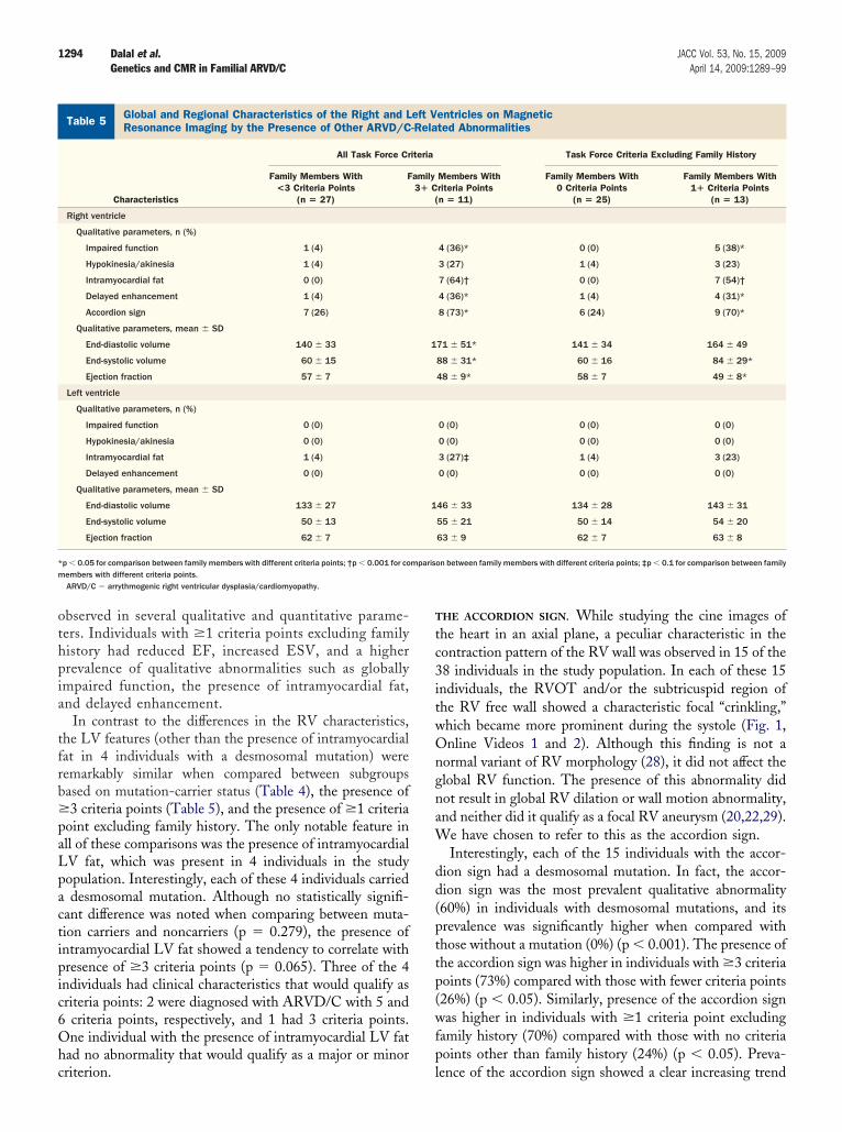
ion sign had a desmosomal mutation. In fact, the accor-ion sign was the most prevalent qualitative abnormality60%) in individuals with desmosomal mutations, and itsrevalence was significantly higher when compared withhose without a mutation (0%) (p � 0.001). The presence ofhe accordion sign was higher in individuals with �3 criteriaoints (73%) compared with those with fewer criteria points26%) (p � 0.05). Similarly, presence of the accordion signas higher in individuals with �1 criteria point excluding
amily history (70%) compared with those with no criteriaoints other than family history (24%) (p � 0.05). Preva-
Magneticlities
entricles on Magneticted Abnormalities
Task Force Criteria Excluding Family History
Members Withriteria Pointsn � 11)
Family Members With0 Criteria Points
(n � 25)
Family Members With1� Criteria Points
(n � 13)
4 (36)* 0 (0) 5 (38)*
3 (27) 1 (4) 3 (23)
7 (64)† 0 (0) 7 (54)†
4 (36)* 1 (4) 4 (31)*
8 (73)* 6 (24) 9 (70)*
71 � 51* 141 � 34 164 � 49
88 � 31* 60 � 16 84 � 29*
48 � 9* 58 � 7 49 � 8*
0 (0) 0 (0) 0 (0)
0 (0) 0 (0) 0 (0)
3 (27)‡ 1 (4) 3 (23)
0 (0) 0 (0) 0 (0)
46 � 33 134 � 28 143 � 31
55 � 21 50 � 14 54 � 20
63 � 9 62 � 7 63 � 8
on between family members with different criteria points; ‡p � 0.1 for comparison between family
s onorma
eft V-Rela
iteria
amily3� C
(
1
1
mparis
ence of the accordion sign showed a clear increasing trend

wc
muiFracTcvimaiAF(a0ac
pFasfpitraofptcRmrfi
D
Mfttsmafioct
1295JACC Vol. 53, No. 15, 2009 Dalal et al.April 14, 2009:1289–99 Genetics and CMR in Familial ARVD/C
ith disease severity, as determined by the number ofriteria points achieved (Fig. 2).
Because of the association of the accordion sign withutation status as well as disease severity, further analysis was
ndertaken to examine the utility of this novel marker indentifying early disease. Table 6 shows the ability of the Taskorce criteria to discriminate individuals with an ARVD/C-
elated mutation from those without one. The discriminatorybility is quantitatively measured by the area under the ROCurve statistic. This statistic represents the probability that theask Force criteria assign a higher value (in terms of number of
riteria points) to mutation carriers than to noncarriers. Thealue of this statistic can range between 0 and 1, with 0.5ndicating the inability of the Task Force criteria to identify
utation carriers from noncarriers and 1 indicating a perfectbility to distinguish them. Values �0.5 are indicative ofdentification of mutation carriers as noncarriers and vice versa.s shown in Table 6, the discriminatory ability of the Taskorce criteria, both including (0.68 [95% confidence interval
CI): 0.54 to 0.83] to 0.79 [95% CI: 0.73 to 0.94]; p � 0.01)nd excluding family history (0.71 [95% CI: 0.60 to 0.83] to.84 [95% CI: 0.73 to 0.94]; p � 0.01), improved significantlyfter including the presence of the accordion sign as a minor
Figure 1 The Accordion Sign
CMR image of 2 individuals demonstrating the presence of the accordionsign in systole (A) and diastole (B). Arrows indicate the accordion sign. LV � leftventricle; RV � right ventricle. For accompanying Videos 1 and 2 and their leg-ends, please see the online version of this article.
riterion under Structural Abnormalities.
A number of additional analyses (results not shown) wereerformed to ensure reliability and validity of the results.irst, all analyses for EDV, ESV, and EF were repeatedfter obtaining values adjusted for body surface area, andimilar results were noted. Second, all CMRs were reviewedor the presence of the accordion sign at 2 different timeoints, 2 weeks apart, and blinded to the initial results. Allndividuals identified as positive for the accordion sign athe first reading were detected as positive on the secondeading. No individual who was labeled as negative for theccordion sign at the first reading was detected as positiven the second reading. Third, CMRs of the probands ofamilies included in this study were evaluated for theresence of the accordion sign, and all probands were foundo be positive for the sign. In fact, in the more advancedases, the sign appeared to become more synonymous withV free wall aneurysms, suggesting that the accordion signay be a precursor of aneurysms. And finally, CMRs of 20
andomly selected disease-free individuals were evaluatedor the presence of the accordion sign, and none of thesendividuals were positive for the sign (29).
iscussion
ain findings. In a unique and an unbiased population ofamily members of ARVD/C patients undergoing prospec-ive evaluation for ARVD/C diagnosis, detailed examina-ion of RV and LV morphologies was performed using theophisticated CMR techniques. Although the RV of theutation carriers in the study population was significantly
ffected compared with noncarriers, the LV was affected lessrequently. Despite a single case with no other abnormality,ndividuals with LV involvement had other signs suggestivef ARVD/C. Moreover, the LV involvement appeared toorrelate with increasing disease severity as determined byhe presence of criteria points achieved.
0
20
40
60
80
1 point 2 points 3 points 4+ points
''
Criteria points
P=0.009
Per
cen
t w
ith
acc
ord
ion
sig
n
Figure 2 Accordion Sign and Disease Severity
Prevalence of the accordion sign presented by criteria points achieved forarrythmogenic right ventricular dysplasia/cardiomyopathy diagnosis. Minor crite-rion � 1 point; major criterion � 2 points.

piwotatdfLsaA1ipcqwfiTifumLoLssA
otebLmrt1RRL
wowC1tdi1LdST
siwbpeweLminaolsrsamod
dSocawmi
1296 Dalal et al. JACC Vol. 53, No. 15, 2009Genetics and CMR in Familial ARVD/C April 14, 2009:1289–99
Detailed CMR imaging of the RV free wall revealed aotential novel indicator of ARVD/C, and its utility indentifying individuals predisposed to developing ARVD/Cas systematically evaluated. The accordion sign was notnly the strongest qualitative indicator differentiating mu-ation carrying family members from noncarriers but waslso associated with increasing disease severity. Addition ofhis sign as a minor criterion significantly improved theiagnostic utility of the currently used Task Force criteriaor ARVD/C diagnosis.V involvement in ARVD/C. ARVD/C was first de-
cribed as a clinical entity in 1977 by Fontaine et al. (30),nd the detailed clinical characteristics in a group of 24RVD/C patients were first reported by Marcus et al. in982 (1). These pioneers of ARVD/C research and othernvestigators established ARVD/C as a cardiomyopathyredominantly affecting the RV, and left-sidedness wasonsidered as a late manifestation of the disease. Conse-uently, the Task Force criteria for ARVD/C diagnosis thatere proposed in 1994 were designed to assign a criterion
or structural disease to individuals with structural RVmpairment and “no (or only mild) LV impairment” (19).he ensuing clinical description of ARVD/C characteristics
n several large cohorts relied on inclusion of patients whoulfilled the Task Force criteria and therefore resulted in thender-representation of “left-sided arrhythmogenic cardio-yopathy,” if such an entity in fact existed (3,4,31,32).ong-term follow-up in large cohorts selected on the basisf fulfillment of the Task Force criteria has demonstratedV involvement at the advanced stages of disease progres-
ion (3,4). This finding is consistent with those demon-trated by Corrado et al. (33) in a series of hearts fromRVD/C patients examined on autopsy or transplant.More recently, ARVD/C has been shown to be a disease
f the cardiac desmosome (34). As the desmosomal struc-ure is similar on the right and the left side of the heart, thexistence of left-sided arrhythmogenic cardiomyopathy haseen hypothesized. Three studies investigating the RV andV characteristics in relation to the presence of desmosomalutations are particularly noteworthy. Bauce et al. (8)
eported their echocardiographic findings in 26 DSP muta-ion carriers among 38 individuals from 4 families. Of the4 individuals with an abnormal echocardiogram, 13 hadV involvement and 7 had LV involvement. RVEF andVEDV were similar (or slightly worse) in patients with
Ability of the Task Force Criteria to DiscriminatMembers With and Without a Desmosomal MutInclusion of the Accordion Sign as a Minor Crite
Table 6Ability of the Task Force Criteria toMembers With and Without a DesmInclusion of the Accordion Sign as a
Withou
Task Force criteria 0.6
Task Force criteria excluding family history 0.7
V involvement, and only 1 individual had an abnormal LV d
ith no abnormalities detected in the RV. In a subgroupf 22 mutation carriers among 28 genotyped individualsho underwent CMR with delayed enhancement, Sen-howdhry et al. (35) reported LV delayed enhancement in00% of the mutation carriers. In the same study, 54% ofhe mutation carriers were reported to have LV systolicysfunction. Subsequently, the same group reported LVnvolvement in 85% and predominantly left-sided disease in5% of the 39 desmosomal mutation carriers (18). SimilarV involvement was reported in a larger cohort of 200esmosomal mutation carrying and noncarrying individuals.election of the study population relied on fulfillment of theask Force and the “modified criteria.”Similar to the reports by Sen-Chowdhry et al. (35), our
tudy utilized state-of-the-art CMR protocols to character-ze myocardial tissue as well as fat and fibrosis. Consistentith their findings, the RV and LV function, as determinedy EDV and EF, was relatively preserved in our studyopulation regardless of the mutation carrier status. How-ver, the qualitative findings of our study sharply contrastith those of Sen-Chowdhry et al. (35). The frequency of
ach qualitative characteristic of ARVD/C in the RV andV was substantially lower in our study population. Theost notable was the absence of LV delayed enhancement
n any individuals in our study population as opposed toearly all individuals in their report. The only qualitative LVbnormality noted in our study population was the presencef LV intramyocardial fat in 4 mutation carriers. Theeft-sidedness of the disease demonstrated by our study isomewhat similar to that reported by Bauce et al. (8). Theesults of both studies demonstrate LV involvement in amall fraction of family members of ARVD/C patients thatre more predisposed to developing the disease (as deter-ined by mutation-carrier status). In both studies, there was
nly 1 individual with an LV abnormality on imaging whoemonstrated no other ARVD/C-related abnormality.There are several possible reasons for these observed
ifferences between our results and those reported byen-Chowdhry et al. (18) among individuals selected basedn fulfillment of Task Force criteria and the “modifiedriteria.” First, our study design was leveraged by a system-tic inclusion of family members of ARVD/C patients whoould have otherwise not sought medical attention. Thisethod of selection of the study population ensured the
nclusion of individuals who would be equally likely to
ween FamilyBefore and AfterUnder “Structural Abnormalities”
riminate Between Familyal Mutation Before and Afteror Criterion Under “Structural Abnormalities”
a Under the Curve (95% Confidence Interval)
rdion Sign With Accordion Sign p Value
–0.83) 0.79 (0.73–0.94) 0.0059
–0.83) 0.84 (0.73–0.94) 0.0046
e Betationrion
DiscosomMin
Are
t Acco
8 (0.54
1 (0.60
evelop RV and LV abnormalities. It has been previously

sto(awwgswtierwthqtitttldad
sitoLwie
mttLr(AorcatKcpwiKL
TeradsTwwtitbtooit
b(cfiAw(hcthafittoscttbSgmdMDpumvp
1297JACC Vol. 53, No. 15, 2009 Dalal et al.April 14, 2009:1289–99 Genetics and CMR in Familial ARVD/C
hown that ARVD/C is a gradually progressive disease, andhere can be a substantial lag time between the initial signsf the disease and diagnosis based on the Task Force criteria4). It is likely that individuals with a Task Force diagnosis,s was the case in the study by Sen-Chowdhry et al. (18),ould have substantially more advanced disease comparedith family members undergoing prospective clinical andenetic screening. If the LV were involved at advancedtages of the ARVD/C as proposed initially, then thereould have been an over-representation of such cases in
heir study population. Second, although the inclusion ofndividuals who fulfill the modified Task Force criteriansures inclusion of early cases of ARVD/C, it may haveesulted in a loss of specificity with inclusion of patientsith other types of cardiomyopathies. For example, under
he proposed modified criteria, an individual with a familyistory of ARVD/C with 200 ventricular ectopics wouldualify for an ARVD/C diagnosis (32). And a final poten-ial explanation for the differing results between the 2 studiess reflected in the differing frequencies of desmosomal muta-ions in the 2 study samples. While a majority of individuals inhe study by Sen-Chowdhry et al. (18) had a DSP mutation,he most common desmosomal mutation in our study popu-ation was in PKP2. The mechanism by which each of theseesmosomal proteins results in ARVD/C is largely unknown,nd it is possible that these genes affect the 2 ventriclesifferentially.The low prevalence of LV involvement observed in our
tudy population does not rule out the possibility of LVnvolvement at early stages of ARVD/C, but it underscoreshe low possibility of such occurrence. In fact, the presencef desmosomal mutation(s) in each of the 4 individuals withV involvement substantiates such a possibility. Consistentith the report by Bauce et al. (8), each of these 4
ndividuals had inferior lead T-wave inversion on a 12-leadlectrocardiogram.
Minimal LV involvement is also consistent with animalodels of ARVD/C. Basso et al. (36) studied the charac-
eristics of ARVD/C in 23 boxer dogs and found his-opathologic evidence of the disease in the LV of 11 dogs.V involvement independent of RV involvement was not
eported in any of the animals. Subsequently, Oxford et al.37) confirmed and extended these findings in a group of 12RVD/C boxer dogs. Although immunochemical analysisf intercalated disc proteins in the heart tissue of these dogsevealed a loss of the spatial organization of the molecularomponents in both RV and LV, the overall myocardialrchitecture in the LV was better preserved compared withhe RV, which showed remarkable fibrofatty replacement.aplan et al. (38,39) have proposed that disruption of
ell-to-cell coupling between the adjacent myocytes mayrecede fibro-fatty replacement. In this regard, the RV wall,hich is thinner and more susceptible to mechanical stress,
s more likely to get affected compared with the LV. Finally,irchhof et al. (40) demonstrated that the RV and not the
V was significantly affected in plakoglobin-deficient mice. phe LV size and function remained preserved despitendurance training in these mice. Studies in humans, whichely on imaging in vivo rather than detailed histopathologynd immunochemistry, are likely to detect even less evi-ence of morphologic alterations as described by thesetudies.he accordion sign. Detailed CMR study of the RV freeall revealed the presence of a new morphologic variant,hich is associated with the presence of desmosomal mu-
ations as well as disease severity. Marcus et al. (1), in theirnitial observational study of 24 patients, specifically iden-ified 3 major regions of the RV that were affected earliesty ARVD/C. These 3 areas, collectively known as theriangle of dysplasia, constituted the inferior tricuspid, theutflow tract, and the apical areas of the RV. The presencef the accordion sign in the inferior tricuspid and basal RVs consistent with these areas conventionally known to behe most affected.
The majority of the mutations reported in this study haveeen previously reported to cosegregate with ARVD/C14,27). The novel DSP mutation reported in this studyosegregated with ARVD/C among 2 individuals within aamily, and the novel DSG2 mutation was present in anndividual who already had a detrimental PKP2 mutation.lthough these mutations appear to have a low penetrancehen ARVD/C is diagnosed by the Task Force criteria
12), there is a much higher probability of mutation carriersaving ARVD/C-related abnormalities resulting in an in-omplete set of Task Force criteria (8,14). The presence ofhese mutations can therefore be used as a surrogate for theigh probability of individuals developing the disease. Theccordion sign was the single most consistent qualitativeeature that could identify mutation carriers with a sensitiv-ty of 60% and 100% specificity. The association betweenhe accordion sign and the presence of any criterion otherhan family history highlights its importance as an early signf the disease. Incorporation of the accordion sign alsoignificantly improves the discriminatory ability of theurrent Task Force criteria in identifying desmosomal mu-ation carriers. The readers of CMR studies were blinded tohe clinical and genetic data and were unlikely to be biasedy such results.tudy limitations. Studies on ARVD/C, in particular theenetic studies, are typically small in size. Our study sampleay have been underpowered to detect certain morphologic
ifferences between mutation carriers and noncarriers.oreover, the relatively low prevalence of DSG2 and theSP mutation restricted our ability to compare the mor-
hologic variants by the gene(s) involved. We were alsonable to make comparisons by severity of the desmosomalutations. Finally, our results demonstrate the incremental
alue of CMR in predicting a desmosomal mutation or theresence of the Task Force criteria and not the ARVD/C
henotype.
C
Tdarteantetd(tm
ATw
RNE
R
1
1
1
1
1
1
1
1
1
1
2
2
2
2
2
2
2
2
2
2
3
3
3
3
3
1298 Dalal et al. JACC Vol. 53, No. 15, 2009Genetics and CMR in Familial ARVD/C April 14, 2009:1289–99
onclusions
he results of our study underscore the ability of CMR toetect early morphologic changes in ARVD/C. ARVD/Cssociated with desmosomal mutations can involve both theight and the left side of the heart. Despite LV involvement,he overall structure and function is well preserved in thearly stages of the disease. Independent LV involvement is
rare occurrence and is consistent with the traditionalotion that right-sided disease precedes left-sided disease inhe majority of the cases. The accordion sign is a possiblearly morphologic variant of ARVD/C and is a promisingool for early diagnosis of the disease. Its prevalence andiagnostic utility should be confirmed in larger cohorts ofgenotyped and nongenotyped) ARVD/C patients. Objec-ive methods to assess its presence using techniques such asyocardial tagging should be developed.
cknowledgmenthe authors are grateful to the ARVD patients and familiesho have made this work possible.
eprint requests and correspondence: Dr. Darshan Dalal, 600orth Wolfe Street, Carnegie 592, Baltimore, Maryland 21209.-mail: [email protected].
EFERENCES
1. Marcus FI, Fontaine GH, Guiraudon G, et al. Right ventriculardysplasia: a report of 24 adult cases. Circulation 1982;65:384–98.
2. Marcus FI, Fontaine G. Arrhythmogenic right ventricular dysplasia/cardiomyopathy: a review. Pacing Clin Electrophysiol 1995;18:1298–314.
3. Hulot JS, Jouven X, Empana JP, et al. Natural history and riskstratification of arrhythmogenic right ventricular dysplasia/cardio-myopathy. Circulation 2004;110:1879 – 84.
4. Dalal D, Nasir K, Bomma C, et al. Arrhythmogenic right ventriculardysplasia: a United States experience. Circulation 2005;112:3823–32.
5. Thiene G, Nava A, Corrado D, et al. Right ventricular cardiomy-opathy and sudden death in young people. N Engl J Med 1988;318:129 –33.
6. Alcalai R, Metzger S, Rosenheck S, et al. A recessive mutation indesmoplakin causes arrhythmogenic right ventricular dysplasia, skindisorder, and woolly hair. J Am Coll Cardiol 2003;42:319–27.
7. Rampazzo A, Nava A, Malacrida S, et al. Mutation in humandesmoplakin domain binding to plakoglobin causes a dominant formof arrhythmogenic right ventricular cardiomyopathy. Am J HumGenet 2002;71:1200–6.
8. Bauce B, Basso C, Rampazzo A, et al. Clinical profile of four familieswith arrhythmogenic right ventricular cardiomyopathy caused bydominant desmoplakin mutations. Eur Heart J 2005;26:1666–75.
9. McKoy G, Protonotarios N, Crosby A, et al. Identification of adeletion in plakoglobin in arrhythmogenic right ventricular cardiomy-opathy with palmoplantar keratoderma and woolly hair (Naxos dis-ease). Lancet 2000;355:2119–24.
0. Protonotarios N, Tsatsopoulou A, Anastasakis A, et al. Genotype-phenotype assessment in autosomal recessive arrhythmogenic rightventricular cardiomyopathy (Naxos disease) caused by a deletion inplakoglobin. J Am Coll Cardiol 2001;38:1477–84.
1. Gerull B, Heuser A, Wichter T, et al. Mutations in the desmosomalprotein plakophilin-2 are common in arrhythmogenic right ventricularcardiomyopathy. Nat Genet 2004;36:1162–4.
2. Dalal D, Molin LH, Piccini J, et al. Clinical features of arrhythmo-
genic right ventricular dysplasia/cardiomyopathy associated with mu-tations in plakophilin-2. Circulation 2006;113:1641–9.3. Awad MM, Dalal D, Cho E, et al. DSG2 mutations contribute toarrhythmogenic right ventricular dysplasia/cardiomyopathy. Am JHum Genet 2006;79:136–42.
4. Dalal D, James C, Devanagondi R, et al. Penetrance of mutations inplakophilin-2 among families with arrhythmogenic right ventriculardysplasia/cardiomyopathy. J Am Coll Cardiol 2006;48:1416–24.
5. Syrris P, Ward D, Evans A, et al. Arrhythmogenic right ventriculardysplasia/cardiomyopathy associated with mutations in the desmo-somal gene desmocollin-2. Am J Hum Genet 2006;79:978–84.
6. van Tintelen JP, Entius MM, Bhuiyan ZA, et al. Plakophilin-2mutations are the major determinant of familial arrhythmogenic rightventricular dysplasia/cardiomyopathy. Circulation 2006;113:1650–58.
7. Pilichou K, Nava A, Basso C, et al. Mutations in desmoglein-2 geneare associated with arrhythmogenic right ventricular cardiomyopathy.Circulation 2006;113:1171–9.
8. Sen-Chowdhry S, Syrris P, Ward D, et al. Clinical and geneticcharacterization of families with arrhythmogenic right ventriculardysplasia/cardiomyopathy provides novel insights into patterns ofdisease expression. Circulation 2007;115:1710–20.
9. McKenna WJ, Thiene G, Nava A, et al. Diagnosis of arrhythmogenicright ventricular dysplasia/cardiomyopathy. Task Force of the Work-ing Group Myocardial and Pericardial Disease of the EuropeanSociety of Cardiology and of the Scientific Council on Cardiomyop-athies of the International Society and Federation of Cardiology. BrHeart J 1994;71:215–8.
0. Tandri H, Calkins H, Nasir K, et al. Magnetic resonance imagingfindings in patients meeting task force criteria for arrhythmogenicright ventricular dysplasia. J Cardiovasc Electrophysiol 2003;14:476 – 82.
1. Tandri H, Rutberg J, Bluemke DA, et al. Magnetic resonance imagingof arrhythmogenic right ventricular dysplasia. J Cardiovasc Electro-physiol 2002;13:1180.
2. Bluemke DA, Krupinski EA, Ovitt T, et al. MR imaging of arrhyth-mogenic right ventricular cardiomyopathy: morphologic findings andinterobserver reliability. Cardiology 2003;99:153–62.
3. Corrado D, Thiene G. Diagnosis of arrhythmogenic right ventricularcardiomyopathy/dysplasia: is there a single gold standard test? J Car-diovasc Electrophysiol. 2004;15:307–9.
4. Cerqueira MD, Weissman NJ, Dilsizian V, et al. Standardizedmyocardial segmentation and nomenclature for tomographic imagingof the heart: a statement for healthcare professionals from the CardiacImaging Committee of the Council on Clinical Cardiology of theAmerican Heart Association. Circulation 2002;105:539–42.
5. Syrris P, Ward D, Asimaki A, et al. Clinical expression of plakophilin-2mutations in familial arrhythmogenic right ventricular cardiomyopathy.Circulation 2006;113:356–64.
6. DeLong ER, DeLong DM, Clarke-Pearson DL. Comparing the areasunder two or more correlated receiver operating characteristic curves: anonparametric approach. Biometrics 1988;44:837–45.
7. Awad MM, Dalal D, Tichnell C, et al. Recessive arrhythmogenic rightventricular dysplasia due to novel cryptic splice mutation in PKP2.Hum Mutat 2006;27:1157.
8. Tandri H, Daya SK, Nasir K, et al. Normal reference values for theadult right ventricle by magnetic resonance imaging. Am J Cardiol2006;98:1660–64.
9. Tandri H, Rutberg J, Bluemke DA, et al. Magnetic resonance imagingof arrhythmogenic right ventricular dysplasia. J Cardiovasc Electro-physiol 2002;13:1180.
0. Fontaine G, Guiraudon G, Frank R, et al. Stimulation studies andepicardial mapping in ventricular tachycardia: study of mechanism andselection for surgery. In: Kulbertus HE, editor. Reentrant Arrhyth-mias. Lancaster, PA: MTP Publishing, 1977:334–50.
1. Nava A, Bauce B, Basso C, et al. Clinical profile and long-termfollow-up of 37 families with arrhythmogenic right ventricular cardio-myopathy. J Am Coll Cardiol 2000;36:2226–33.
2. Hamid MS, Norman M, Quraishi A, et al. Prospective evaluation ofrelatives for familial arrhythmogenic right ventricular cardiomyopathy/dysplasia reveals a need to broaden diagnostic criteria. J Am CollCardiol 2002;40:1445–50.
3. Corrado D, Basso C, Thiene G, et al. Spectrum of clinicopathologicmanifestations of arrhythmogenic right ventricular cardiomyopathy/dysplasia: a multicenter study. J Am Coll Cardiol 1997;30:1512–20.
4. Garrod DR. Desmosomes and hemidesmosomes. Curr Opin Cell Biol
1993;5:30–40.
3
3
3
3
3
4
Ki
F
1299JACC Vol. 53, No. 15, 2009 Dalal et al.April 14, 2009:1289–99 Genetics and CMR in Familial ARVD/C
5. Sen-Chowdhry S, Prasad SK, Syrris P, et al. Cardiovascular magneticresonance in arrhythmogenic right ventricular cardiomyopathy revis-ited: comparison with task force criteria and genotype. J Am CollCardiol 2006;48:2132–40.
6. Basso C, Fox PR, Meurs KM, et al. Arrhythmogenic right ventricularcardiomyopathy causing sudden cardiac death in boxer dogs: a newanimal model of human disease. Circulation 2004;109:1180–5.
7. Oxford EM, Everitt M, Coombs W, et al. Molecular composition ofthe intercalated disc in a spontaneous canine animal model ofarrhythmogenic right ventricular dysplasia/cardiomyopathy. HeartRhythm 2007;4:1196–205.
8. Kaplan SR, Gard JJ, Protonotarios N, et al. Remodeling of myocytegap junctions in arrhythmogenic right ventricular cardiomyopathy dueto a deletion in plakoglobin (Naxos disease). Heart Rhythm 2004;1:
3–11. l9. Kaplan SR, Gard JJ, Carvajal-Huerta L, et al. Structural and molecularpathology of the heart in Carvajal syndrome. Cardiovasc Pathol2004;13:26–32.
0. Kirchhof P, Fabritz L, Zwiener M, et al. Age- and training-dependentdevelopment of arrhythmogenic right ventricular cardiomyopathy inheterozygous plakoglobin-deficient mice. Circulation 2006;114:1799–806.
ey Words: cardiomyopathy y arrhythmia y magnetic resonancemaging y genetics y diagnosis.
APPENDIX
or supplementary figures and videos with
egends, please see the online version of this article.Copyright © 2022 FDOKUMEN