Bone Neoplasia in the 21st Century - Using Fibrous Dysplasia ...
Upload
khangminh22Category
view
0download
0
Bronchopulmonary Dysplasia
Citation for published version (APA):
Pierro, M. (2019). Bronchopulmonary Dysplasia: New Developments in Treatment and Prevention.https://doi.org/10.26481/dis.20190917mp
Document status and date:Published: 01/01/2019
DOI:10.26481/dis.20190917mp
Document Version:Publisher's PDF, also known as Version of record
Please check the document version of this publication:
• A submitted manuscript is the version of the article upon submission and before peer-review. There canbe important differences between the submitted version and the official published version of record.People interested in the research are advised to contact the author for the final version of the publication,or visit the DOI to the publisher's website.• The final author version and the galley proof are versions of the publication after peer review.• The final published version features the final layout of the paper including the volume, issue and pagenumbers.Link to publication
General rightsCopyright and moral rights for the publications made accessible in the public portal are retained by the authors and/or other copyrightowners and it is a condition of accessing publications that users recognise and abide by the legal requirements associated with theserights.
• Users may download and print one copy of any publication from the public portal for the purpose of private study or research.• You may not further distribute the material or use it for any profit-making activity or commercial gain• You may freely distribute the URL identifying the publication in the public portal.
If the publication is distributed under the terms of Article 25fa of the Dutch Copyright Act, indicated by the “Taverne” license above,please follow below link for the End User Agreement:
www.umlib.nl/taverne-license
Take down policyIf you believe that this document breaches copyright please contact us at:
providing details and we will investigate your claim.
Download date: 01 Feb. 2022

Bronchopulmonary Dysplasia: New Developments in Treatment
and Prevention
Dissertation
to obtain the degree of Doctor at the Maastricht University,
on the authority of the Rector Magnificus,
Prof. dr. Rianne M. Letschert
in accordance with the decision of the Board of Deans,
to be defended in public
by
Maria Pierro
born on September 3rd, 1981
in Cesena, Italy.

Supervisors:
Dr. Eduardo Villamor (ius promovendi)
Prof. dr. Bernard Thébaud (University of Ottawa)
Prof. dr. Fabio Mosca (Università degli Studi di Milano)
Co-supervisor:
Dr. Giacomo Cavallaro (Università degli Studi di Milano)
Assessment committee:
- Prof. dr. Luc JI Zimmermann - Prof. dr. Estela Rubio Gozalbo - Prof. dr. Steven H. Abman (University of Colorado) - Prof. dr. Angel Cogolludo (Universidad Complutense de Madrid)

TABLE OF CONTENTS Page
Justification, aims, and outline of the thesis 3
Chapter I 11
INTRODUCTION
Chapter II 31
SHORT-TERM, LONG-TERM AND PARACRINE EFFECT OF HUMAN UMBILICAL CORD-DERIVED STEM CELLS IN LUNG INJURY PREVENTION AND REPAIR IN EXPERIMENTAL BRONCHOPULMONARY DYSPLASIA
Chapter III 45
MESENCHYMAL STEM CELLS FOR THE PREVENTION AND TREATMENT OF BRONCHOPULMONARY DYSPLASIA IN PRETERM INFANTS
Chapter IV 71
PROBIOTIC SUPPLEMENTATION IN PRETERM INFANTS DOES NOT AFFECT THE RISK OF BRONCHOPULMONARY DYSPLASIA: A META-ANALYSIS OF RANDOMIZED CONTROLLED TRIALS
Chapter V 93
MOTHER’S OWN MILK AND BRONCHOPULMONARY DYSPLASIA: A SYSTEMATIC REVIEW AND META-ANALYSIS
Chapter VI 11
GENERAL DISCUSSION AND FUTURE PERSPECTIVES


3
Justification, aims and outline

4

5
Justification of the Thesis
Bronchopulmonary dysplasia (BPD) is a form of chronic lung disease, peculiar to the extremely preterm infant, born at the early stages of lung development (1). Up to 40% of the infants below 29 weeks’ gestation are diagnosed with BPD (2), while the composite outcome death and/or BPD occurs in 65% of the infants below 29 weeks’ gestation (2). BPD lungs consist of enlarged and simplified breathing structures, as opposed to the alveolar ducts at term, characterized by smaller, more numerous and more complex alveoli, essential to ensure adequate gas exchange (1). BPD is also characterized by an abnormal distribution of the pulmonary vessels and a reduction in the number of the small arteries, which are functionally hyper-reactive and hypertonic, culminating in pulmonary arterial hypertension (3). Impaired pulmonary vascular growth may contribute to the irreversible arrest of lung development typical of BPD (4). Early and late pulmonary hypertension, assessed by indirect echocardiographic measurements, is detected in approximately 20% to 25% of the infants suffering from BPD, worsening their outcome (3, 5, 6).
Early birth itself primarily interrupts the intricate cascade of biochemical and hormonal factors that hesitate in full and proper lung development. Consistently, low gestational age (GA) and low birth weight (BW) are the major determinants of BPD (7). However, BPD is a multifactorial disease, being the consequence of prenatal and post-natal inflammatory, oxidative, anti-angiogenic and pro-fibrotic stimuli on the developing lung (8-11). Multiple modifiable factors have been associated with an increased risk of BPD. These include, among others, maternal smoking (12, 13), placental dysfunction (14), duration of mechanical ventilation (15), cumulative supplemental oxygen (16), postnatal growth failure (17), necrotizing enterocolitis (15), and late-onset sepsis (18, 19). Other elements, such as gestational hypertension (20-22), chorioamnionitis (23, 24) and patent ductus arteriosus (22, 25) have been inconsistently linked to BPD.
Despite the continuous advances of perinatal care, BPD remains a significant burden of extreme prematurity, because it lacks a safe and effective treatment and/ or prevention strategies. The structural arrest of lung development makes traditional therapies ineffective in treating this disease (26). In the past few years, increasing insight into stem cell biology has generated excitement about the potential of stem cells to regenerate damaged organs. Among stem cells, mesenchymal stromal (stem) cells (MSCs) have attracted much attention because of their ease of isolation, multilineage potential, and immunomodulatory properties (27). Perivascular cells (PCs) from diverse human tissues give rise to adherent multilineage progenitor cells that exhibit all the features of MSCs and may embody the precursors of MSCs (28, 29). MSCs and PCs may represent a novel therapeutical option for so far untreatable diseases, including BPD. While the definitive prevention of BPD could only be obtained by avoiding preterm birth, prenatal and postnatal preventive efforts are also directed at the reduction of the other

6
stressors that may worsen the injury to the developing lung (30). Among other, human milk and probiotics have been proposed as promising options (31).
Aim and Outline of the Thesis The present thesis is a collection of studies aimed at investigating the potential use of mesenchymal stem cells and pericytes in the treatment of BPD and evaluating the possible role of probiotic supplementation and mother’s own milk in the prevention of BPD.
Chapter I is a general introduction in which we reviewed the pathophysiology and treatment of BPD with special emphasis on the role of MSCs.
In Chapter II we tested the potential of MSCs and their precursors (pericytes) in preventing and treating oxygen-induced lung injury in a murine model of BPD.
In Chapter III we performed a Cochrane meta-analysis to investigate the role of MSCs in the treatment of BPD.
In Chapter IV we performed a systematic review and meta-analysis to test the potential of probiotics in preventing BPD.
In Chapter V we performed a systematic review and meta-analysis to test the potential of mother’s own milk in preventing BPD.
Finally, in Chapter VI a general discussion of the thesis is outlined.
References
1. Kalikkot Thekkeveedu R, Guaman MC, Shivanna B. Bronchopulmonary dysplasia: Areview of pathogenesis and pathophysiology. Respiratory medicine. 2017;132:170-7.2. Stoll BJ, Hansen NI, Bell EF, Walsh MC, Carlo WA, Shankaran S, et al. Trends in CarePractices, Morbidity, and Mortality of Extremely Preterm Neonates, 1993-2012. Jama.2015;314(10):1039-51.3. Rossor T, Greenough A. Advances in paediatric pulmonary vascular disease associatedwith bronchopulmonary dysplasia. Expert review of respiratory medicine. 2015;9(1):35-43.4. Thebaud B, Abman SH. Bronchopulmonary dysplasia: where have all the vesselsgone? Roles of angiogenic growth factors in chronic lung disease. American journal ofrespiratory and critical care medicine. 2007;175(10):978-85.5. Altit G, Dancea A, Renaud C, Perreault T, Lands LC, Sant'Anna G. Pathophysiology,screening and diagnosis of pulmonary hypertension in infants with bronchopulmonarydysplasia - A review of the literature. Paediatric respiratory reviews. 2017;23:16-26.6. Mehler K, Udink Ten Cate FE, Keller T, Bangen U, Kribs A, Oberthuer A. AnEchocardiographic Screening Program Helps to Identify Pulmonary Hypertension inExtremely Low Birthweight Infants with and without Bronchopulmonary Dysplasia: A Single-Center Experience. Neonatology. 2018;113(1):81-8.
7
7. Lee JH, Noh OK, Chang YS. Neonatal Outcomes of Very Low Birth Weight Infants inKorean Neonatal Network from 2013 to 2016. Journal of Korean medical science.2019;34(5):e40.8. Pasha AB, Chen XQ, Zhou GP. Bronchopulmonary dysplasia: Pathogenesis andtreatment. Experimental and therapeutic medicine. 2018;16(6):4315-21.9. Leroy S, Caumette E, Waddington C, Hebert A, Brant R, Lavoie PM. A Time-BasedAnalysis of Inflammation in Infants at Risk of Bronchopulmonary Dysplasia. The Journal ofpediatrics. 2018;192:60-5.e1.10. Wang J, Dong W. Oxidative stress and bronchopulmonary dysplasia. Gene.2018;678:177-83.11. Valencia AM, Abrantes MA, Hasan J, Aranda JV, Beharry KD. Reactive Oxygen Species,Biomarkers of Microvascular Maturation and Alveolarization, and Antioxidants in OxidativeLung Injury. Reactive oxygen species (Apex, NC). 2018;6(18):373-88.12. Isayama T, Shah PS, Ye XY, Dunn M, Da Silva O, Alvaro R, et al. Adverse Impact ofMaternal Cigarette Smoking on Preterm Infants: A Population-Based Cohort Study. Americanjournal of perinatology. 2015;32(12):1105-11.13. Morrow LA, Wagner BD, Ingram DA, Poindexter BB, Schibler K, Cotten CM, et al.Antenatal Determinants of Bronchopulmonary Dysplasia and Late Respiratory Disease inPreterm Infants. American journal of respiratory and critical care medicine. 2017;196(3):364-74.14. Lio A, Rosati P, Pastorino R, Cota F, Tana M, Tirone C, et al. Fetal Doppler velocimetryand bronchopulmonary dysplasia risk among growth-restricted preterm infants: anobservational study. BMJ open. 2017;7(7):e015232.15. Tapia JL, Agost D, Alegria A, Standen J, Escobar M, Grandi C, et al. Bronchopulmonarydysplasia: incidence, risk factors and resource utilization in a population of South Americanvery low birth weight infants. Jornal de pediatria. 2006;82(1):15-20.16. Wai KC, Kohn MA, Ballard RA, Truog WE, Black DM, Asselin JM, et al. Early CumulativeSupplemental Oxygen Predicts Bronchopulmonary Dysplasia in High Risk Extremely LowGestational Age Newborns. The Journal of pediatrics. 2016;177:97-102.e2.17. Natarajan G, Johnson YR, Brozanski B, Farrow KN, Zaniletti I, Padula MA, et al.Postnatal weight gain in preterm infants with severe bronchopulmonary dysplasia. Americanjournal of perinatology. 2014;31(3):223-30.18. Klinger G, Levy I, Sirota L, Boyko V, Lerner-Geva L, Reichman B. Outcome of early-onset sepsis in a national cohort of very low birth weight infants. Pediatrics.2010;125(4):e736-40.19. Lahra MM, Beeby PJ, Jeffery HE. Intrauterine inflammation, neonatal sepsis, andchronic lung disease: a 13-year hospital cohort study. Pediatrics. 2009;123(5):1314-9.20. Yen TA, Yang HI, Hsieh WS, Chou HC, Chen CY, Tsou KI, et al. Preeclampsia and therisk of bronchopulmonary dysplasia in VLBW infants: a population based study. PloS one.2013;8(9):e75168.21. Rocha G, de Lima FF, Machado AP, Guimaraes H. Preeclampsia predicts higherincidence of bronchopulmonary dysplasia. Journal of perinatology : official journal of theCalifornia Perinatal Association. 2018;38(9):1165-73.22. Sung SI, Chang YS, Chun JY, Yoon SA, Yoo HS, Ahn SY, et al. Mandatory Closure VersusNonintervention for Patent Ductus Arteriosus in Very Preterm Infants. The Journal ofpediatrics. 2016;177:66-71.e1.

8
23. Torchin H, Lorthe E, Goffinet F, Kayem G, Subtil D, Truffert P, et al. HistologicChorioamnionitis and Bronchopulmonary Dysplasia in Preterm Infants: The EpidemiologicStudy on Low Gestational Ages 2 Cohort. The Journal of pediatrics. 2017;187:98-104.e3.24. Eriksson L, Haglund B, Odlind V, Altman M, Kieler H. Prenatal inflammatory riskfactors for development of bronchopulmonary dysplasia. Pediatric pulmonology.2014;49(7):665-72.25. Harkin P, Marttila R, Pokka T, Saarela T, Hallman M. Morbidities associated withpatent ductus arteriosus in preterm infants. Nationwide cohort study. The journal ofmaternal-fetal & neonatal medicine : the official journal of the European Association ofPerinatal Medicine, the Federation of Asia and Oceania Perinatal Societies, the InternationalSociety of Perinatal Obstet. 2018;31(19):2576-83.26. Jobe AH, Bancalari E. Bronchopulmonary dysplasia. American journal of respiratoryand critical care medicine. 2001;163(7):1723-9.27. Gurusamy N, Alsayari A, Rajasingh S, Rajasingh J. Adult Stem Cells for RegenerativeTherapy. Progress in molecular biology and translational science. 2018;160:1-22.28. Crisan M, Yap S, Casteilla L, Chen CW, Corselli M, Park TS, et al. A perivascular originfor mesenchymal stem cells in multiple human organs. Cell stem cell. 2008;3(3):301-13.29. Thomas H, Cowin AJ, Mills SJ. The Importance of Pericytes in Healing: Wounds andother Pathologies. International journal of molecular sciences. 2017;18(6).30. Poets CF, Lorenz L. Prevention of bronchopulmonary dysplasia in extremely lowgestational age neonates: current evidence. Archives of disease in childhood Fetal andneonatal edition. 2018;103(3):F285-f91.31. Aschner JL, Bancalari EH, McEvoy CT. Can We Prevent Bronchopulmonary Dysplasia?The Journal of pediatrics. 2017;189:26-30.

9
CHAPTER I:
INTRODUCTION*
*Based on:
Pierro M., Ciarmoli E., Thébaud B. (2016) Stem Cell Therapy for Neonatal Lung Diseases. In: Steinhoff G. (eds) Regenerative Medicine - from Protocol to Patient. Springer, Cham
Pierro M, Thébaud B. Mesenchymal stem cells in chronic lung disease: culprit or savior? Am J Physiol Lung Cell Mol Physiol. 2010 Jun;298(6):L732-4.
Pierro M, Thébaud B. MSCS in the Scenarios of Infection and Inflammation: Focus on Neonatal Diseases. Current Stem Cell Reports 2016; 2 (2): 158–167
Pierro M. Scopesi F, Ramneghi L. Update on Bronchopulmonary dysplasia: prevention and treatment. Prospettive in Pediatria 2015, 177 (45): 35-40
Pierro M, Ciarmoli E, Thébaud B. Bronchopulmonary Dysplasia and Chronic Lung Disease: Stem Cell Therapy. Clin Perinatol. 2015;42:889-910.

10
1. Definition and incidence of bronchopulmonary dysplasia
The revolutionary progress of perinatal care during the past few decades has led to a remarkable reduction of neonatal mortality (1, 2). However, this progress has created new challenges. Bronchopulmonary dysplasia (BPD), the chronic lung disease of prematurity, now occurs in extreme premature infants, disrupting normal lung growth at an early developmental stage (3). The first report of BPD dates back to the pre-surfactant era, when Northway described a form of chronic lung disease (4), defined as oxygen need for at least 28 days in conjunction with specific radiographic changes, in late preterm infants surviving from respiratory distress syndrome at birth (5). Subsequently, oxygen dependency at 36 weeks post-menstrual age (PMA) was shown to better predict long-term respiratory outcomes (6). Roughly 10 years later, in the NICHD Workshop Summary, infants below 32 weeks requiring supplemental oxygen for at least 28 days were stratified into three severity groups (mild, moderate and severe BPD), depending on the presence and the amount of supplemental oxygen and mode of respiratory support at 36 weeks PMA (7). The ‘physiologic’ definition of BPD was proposed in the attempt to compensate for the significant inter-center variability in oxygen administration (8). To be categorized as BPD patients according to the physiological definition, infants receiving less than 30% of supplemental oxygen and no respiratory support at 36 weeks PMA are challenged by reducing the fraction of administered oxygen during a standardized test. Infants who are unable to maintain saturations above 90% in room air during the test are diagnosed with BPD (9).
Although, by definition, BPD cannot be diagnosed before 28 days of life, the respiratory disease characterized by oxygen and/or ventilator-dependency from 7 to 28 days of life, represents the initial phase of the chronic process leading to BPD, thus classified as “evolving BPD” (10). Recently, the definition of BPD has been refined in order to include in the severity classification newer modes of noninvasive ventilation that were not included in the previous definitions. The severity classification has been changed from mild, moderate and severe into the new terms of grades I, II, and III. Grade III would refer to the most severe form of BPD. Early death (between 14 days of postnatal age and 36 weeks) owing to persistent parenchymal lung disease and respiratory failure that cannot be attributable to other neonatal morbidities has also been included in the definition as grade IIIa (11).
While some data suggest a trend towards a reduction of the BPD rates, most studies report a slight increase over the past two to three decades. This is likely linked to the higher survival of more immature infants at higher risk for BPD (12, 13). Moreover, incidence of BPD varies depending on the definition. The NICHD Neonatal Research Network has compared the BPD rates in extremely preterm infants (22-28 weeks gestation) according to the three definitions currently in use. The NICHD classification is associated with the highest rates (68%) due to the inclusion of infants requiring oxygen for 28 days, although breathing room air at 36 weeks PMA. Oxygen need at 36 weeks PMA and the physiological definition obtained similar results (42 and

11
40% respectively) (12, 13). It is still controversial which definition better predicts the long-term outcomes and if grading system actually improves its positive predictive value (14-16).
Figure 1. Stages of lung development Embryonic phase (3rd-7th weeks of gestation): formation of the two lung buds (primary bronchi) further branching into the secondary bronchi and into the major airways. Histologically, these structures are l ined with undifferentiated columnar epithelium (a). Mesenchymal cells (b) are crucial for epithelial-mesenchymal ‘‘cross-talk’’ cell interactions that regulate branching morphogenesis and cellular differentiation. Pseudoglandular stage (5-16th weeks of gestation): formation of the conductive airways. The epithelium starts to differentiate into non-cil iated columnar Clara cells (c), pulmonary neuroendocrine cells (d), and cil iated cells (e). Canalicular phase (16-26th weeks of gestation): formation of the respiratory bronchioles and the pulmonary acinus. Histologically, the epithelium of the distal lung begins to differentiate from cuboid type II cells (g) into squamous type I cells (h). Saccular stage (26-36th weeks of gestation): Maturation of the distal epithelium. Mature type 2 cells (g*) start producing surfactant (i), while type 1 cells form the thin layer. In the proximal epithelium cil iated (e), non-cil iated Clara (c), and neuroendocrine cells (d) increase in number. Alveolar stage (36 weeks of gestation-first few years of l ife): alveolarization and formation of the secondary crests. The microvascular maturation takes place and the capil lary bilayer (f) merge into the single-layer vascular network (f*).

12
2. Pathogenesis of BPD
Since the major pathogenetical clue of BPD is the disruption of normal lung development, a thorough insight into the normal lung development, is a prerequisite to deeply understand the pathological process leading to BPD.
Lung development is typically divided into five stages: embryonic, pseudoglandular, canalicular, saccular, and alveolar (figure 1). During the embryonic phase (third-seventh week of gestation), the human lung originates from the primitive foregut as a ventral endodermal bud, which will divide into the two lung buds (primary bronchi) and then into the secondary bronchi, further branching into the major airways. Histologically, these structures are lined with undifferentiated columnar epithelium. The pseudoglandular stage begins at the end of the fifth week of fetal life, with the branching of all conductive airways down to the terminal bronchi. The epithelium of the conducting airways starts to differentiate into non-ciliated columnar Clara cells, pulmonary neuroendocrine cells, and ciliated cells. The distal cuboidal epithelium differentiate into type II epithelial cells. Between 16 and 26 weeks, the canalicular phase takes place, leading to the formation of the respiratory bronchioles and the pulmonary acinus. During this time, the capillary bed of the distal lung remarkably increases. Histologically, the epithelium of the distal lung begins to differentiate from cuboid type II cells into squamous type I cells. During the saccular stage (26-36 weeks), the distal epithelium further mature: type 2 cells start producing surfactant, while type 1 cells form the thin layer, needed for future gas exchange. The interstitial mesenchyme becomes thinner. Saccules and ducts, the characteristic elements of this stage, consist of thick primary septae, containing a double pulmonary capillary layer. In the proximal epithelium ciliated, non-ciliated Clara, basal and neuroendocrine cells increase in number. The larger vessels of the pulmonary vasculature start to muscularize.
Starting from 36 weeks up to the first few years of life, the process of alveolarization and secondary septation ensure the formation of mature and well-organized alveoli. Equally important, the microvascular maturation takes place and the capillary bilayer merge into the single-layer vascular network, creating the efficient air-blood gas-exchange unit (17).
The form of chronic lung disease described by Northway, now referred to as "old BPD", was mainly a consequence of the aggressive ventilatory approach on a relatively mature lung, although deficient in surfactant. Histologically, "old BPD" was characterized by a diffuse injury, with significant inflammation and parenchymal fibrosis, despite numerically and structurally normal, mature and complex alveoli (4). Thanks to the improvement of neonatal care, in particular to the introduction of antenatal steroids in order to induce lung maturation, the discovery of exogenous surfactant and the use of “gentle” ventilatory techniques, nowadays infants born after the canalicular stage of lung development exceptionally suffer from chronic lung disease (7). At the same time, the increased survival of extremely premature infants has led to the appearance of a new form of chronic lung disease ("new BPD"), typical of the infants born at the early stages of lung development (22-28 weeks of gestation) (7, 18). The "new BPD" is the expression of the lung immaturity, rather than the iatrogenic damage. Histologically, the BPD lungs are characterized by less numerous, enlarged, and simplified alveoli, typical of the
13
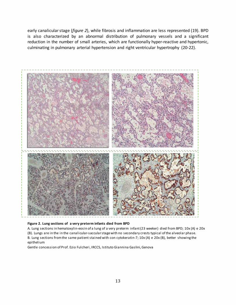
early canalicular stage (figure 2), while fibrosis and inflammation are less represented (19). BPD is also characterized by an abnormal distribution of pulmonary vessels and a significant reduction in the number of small arteries, which are functionally hyper-reactive and hypertonic, culminating in pulmonary arterial hypertension and right ventricular hypertrophy (20-22).
Figure 2. Lung sections of a very preterm infants died from BPD A. Lung sections in hematoxylin-eosin of a lung of a very preterm infant (23 weeker) died from BPD; 10x (A) e 20x(B). Lungs are in the in the canalicular-saccular stage with no secondary crests typical of the alveolar phase.B. Lung sections from the same patient stained with con cytokeratin-7; 10x (A) e 20x (B), better showing theepithelium Gentle concession of Prof. Ezio Fulcheri, IRCCS, Istituto Giannina Gaslini, Genova

14
Moreover, the imbalance between anti-oxidant defense mechanisms and increased exposure to oxygen reactive species (23, 24) exacerbates cell apoptosis and disruption of the extracellular matrix, leading to tissue remodeling. These events contribute to the development of BPD, by further perturbing the developing lung and intensifying the alveolar and vascular dysregulation, primarily caused by the premature birth (3) (figure 3).
Figure 3. Events leading to the development of BPD The "new BPD", typical of the infants born at the early stages of lung development (22-28 weeks of gestation), is the expression of the lung immaturity. Prenatal and postnatal inflammatory stimuli, such as chorioamnionitis, ventilator induced lung injury, oxygen toxicity and neonatal sepsis contribute to the development of BPD. Histologically, the BPD lungs are characterized by less numerous, enlarged, and simplified alveoli, with no secondary crests, typical of the early canalicular stage. BPD is also characterized by an abnormal distribution of pulmonary vessels and a significant reduction in the number of small arteries

15
3. Outcomes of BPD
Premature infants, with or without BPD, continue to show worse respiratory performances compared to their peers up to late childhood (25, 26). Among premature infants, BPD increases the risk for re-hospitalization during the first 2 years of life, due to acute respiratory distress and respiratory tract illness (27). Later in childhood, ex-preterm infants affected by BPD need more often respiratory medications and present more frequently with respiratory symptoms (28). Respiratory symptoms have been objectified by signs of airway obstruction at lung function tests (15). Airway obstruction in these patients seems to be caused by structural changes rather than airway hyperreactivity (29). Response to asthma medication is less pronounced and levels of exhaled nitric oxide are lower in symptomatic BPD patients as compared to asthmatic children born at term with similar grade of airway obstruction (29). In agreement with these results, structural BPD findings have been documented at the autopsy of a child diagnosed with BPD, died from asthma attack at 12 years of life (30). BPD is also associated with worse long-term developmental outcomes, including higher rates of cerebral palsy at 18–22 months’ corrected age and psychomotor delay, attentional impairments, speech and language disorders, executive deficits and behavioral problems at school age (31).
4. Current Strategies for prevention and treatment of BPD
While the definitive prevention of BPD could only be obtained by avoiding preterm birth, prenatal and postnatal preventive efforts should be directed at the reduction of the other stressors, such as postnatal infections and inflammation that may lead to the injury of the developing lung (32). Inflammatory events, such as necrotizing enterocolitis (NEC) and late-onset sepsis (LOS), are not only life-threatening for preterm infants but can likely mediate major short- and long-term adverse outcomes (33), including BPD. It is well accepted that sepsis and infections pose the premature population at increased risk of developing BPD (13, 14). Pro-inflammatory cytokines, released during NEC and LOS, may exert a direct effect on lung development or sensitize the lung to the effects of oxygen, mechanical ventilation and other stressors (34-37). On the other hand, infants suffering from NEC and LOS often require more aggressive and prolonged mechanical ventilation, which, although life-saving, is burdened by various degrees of lung injury (34-37). It has been suggested that the reduction of postnatal systemic infections has a higher impact in decreasing the inflammatory response in developing lungs, rather than avoiding invasive mechanical ventilation (37). Consistently, studies directed at evaluating the impact of quality improvement efforts to reduce LOS in preterm infants showed that a reduction in LOS is accompanied by decreased BPD rates (38, 39). Moreover, the immature immune system of the premature infants is unable to balance pro-inflammatory responses, leading to a sustained status of inflammation, that significantly contributes to several neonatal diseases, including BPD (36). Decreased number of T regulatory cells (Tregs), which constitute the anti-inflammatory T subset designed to limit and suppress excessive innate and adaptive immune responses, in the cord blood (40) and higher proportions of activated pro-inflammatory T cells in the peripheral blood during the first week of life (41) can predict the development of BPD. In experimental BPD, macrophages are polarized towards the pro-inflammatory M1 phenotype, while the anti-inflammatory M2 phenotype is inhibited (42).

16
Approaches aimed at reducing the pro-inflammatory stimuli, including “gentle“ rather than aggressive ventilatory techniques starting from the delivery room, adequate oxygen saturation targets and optimal timing for surfactant administration, seem to be partially effective in preventing BPD, by reducing inflammation to the lung. On the other hand, optimized nutrition and other strategies able to restore to pro-inflammatory/ant-inflammatory balance and the immunological status may open new therapeutical options.
In terms of medications, most of the therapies are either ineffective, inconsistently evaluated or unsafe. A meta-analysis (43) analyzed all the therapies available to prevent or treat BPD. Among the 21 drugs, 16 showed no efficacy. Out of the 5 effective drugs, only three (vitamin A, caffeine and dexamethasone) have been assessed in large or multi-centric randomized controlled trials (RCTs). However, none of them is currently recommended for prevention or treatment of BPD. The need for a long course of intramuscular injections of vitamin A may not be balanced by the modest reduction of BPD rates (44). Treatment with caffeine was associated with a significant reduction in the incidence of BPD in one large RCT that enrolled more than 2,000 patients (45). However, BPD was only a secondary measure and the results need to be evaluated in further trials. Early dexamethasone is the only drug that has been repeatedly shown to significantly reduce the incidence of BPD. However, the higher risk of cerebral palsy at 18 months of life after this treatment (especially if administered during the first week of life) have greatly limited its use. On the other hand, patients at higher risk for BPD (ie ventilator dependent after 2 weeks of life) seem to benefit from a short course of low-dose dexamethasone (46). In case they survive, these patients will be almost certainly diagnosed with BPD. Dexamethasone, by reducing the incidence of BPD, reduces its associated neurological complications as well, improving the overall outcome (47). However, dexamethasone use should be cautious and restricted to severe cases, with ventilator dependency after the first two-three weeks of life. In order to avoid the adverse consequences of dexamethasone, hydrocortisone had been increasingly used as an alternative. However a recent large RTC showed no beneficial effects of hydrocortisone as compared to placebo in reducing the composite outcome of death or BPD in preterm infants at high risk for BPD (48). In summary, no effective and safe treatment has yet been developed for BPD and novel therapies are urgently needed.

17
5. EXPERIMENTAL MODELS OF BPD
In order to test the efficacy and safety of a novel therapy, it is paramount to dispose of reliable animal models. This section briefly reviews the experimental settings adopted to investigate innovative approaches for neonatal lung diseases. Few models in different animal species have been able to mimic BPD or some of its aspects. The hyperoxia-induced lung injury (figure 4). is the most adopted model and displays several features of BPD, such as alveolar and vascular disruption, inflammation, fibrosis and oxidative stress (49, 50). Rats and mice are born at the canalicular stage of lung development, similarly to premature infants at risk for BPD. Hyperoxia-induced lung remodeling persists histologically and functionally at least up to 10 months (equivalent to 25-30 years in humans) (51). Animals exposed to hyperoxia in the neonatal period show functional impairment later in life, as documented by significantly reduced distance running on the treadmill (51). Neonatal exposure of mouse pups to hyperoxia worsens the severity of subsequent viral infections and increases mortality rates following adult influenza A inoculation (52). Few other models, although much less employed, may approximate BPD. Postnatal injection of intraperitoneal bleomycin in newborn rats from day 4 to 14 induces significant pulmonary hypertension and alveolar simplifications, with no fibrosis (53), as opposed to adult rats that suffer mostly from fibrotic lung lesions after bleomycin administration (54).Larger animals (lamb, sheep and baboon) can be delivered prematurely and undergo mechanical ventilation, reproducing the chain of events that leads to BPD. These animals show acute lung inflammation, abnormal pulmonary function, altered lung architecture, alveolar simplification, and mild fibrotic changes (55-57). However only few facilities are equipped to work with large animals and most of the data on stem cell therapy are obtained with the murine models, particularly the hyperoxia-induced lung injury.
Figure 4. Experimental BPD: the hyperoxia-induced lung injury Rat pups are naturally born during the saccular stage of lung development. Exposure of the developing rat lung to various concentration of hyperoxic gas (O2 rodent), during the alveolar stage (day 4 to day 14 of l ife) impairs
alveolarization, resulting in fewer and enlarged alveolar air spaces, resembling BPD lungs, as opposed to controls in room air (RA rodent).

18
6. REGENERATIVE MEDICINE FOR BPD: MESENCHYMAL STEM/STROMAL CELLS
Regenerative medicine appears as promising option for so far untreatable diseases. Several studies have already proven stem cell efficacy in a variety of experimental settings, including neonatal lung diseases. Stem cells, thanks to their unique possibility of restoring and regenerating damaged tissue, may structurally revert the alveolar and vascular architectural changes that make traditional therapies unsuccessful in treating BPD. Among stem cells, mesenchymal stromal cells (MSCs) have been extensively investigated, thanks to their peculiar properties that make them particularly interesting for clinical practice. MSCs are easy to isolate and display multilineage potential and compelling immunomodulatory properties (58).
6.1 Characterization and origin of MSCs
MSCs represent a broad and heterogeneous cell population, defined by three minimum criteria: (i) adhesion to plastic when cultured in a tissue culture flask under standard culturing conditions(ii) expression of specific surface markers (CD73, CD90, CD105) and lack of expression ofhematopoietic markers [CD45 (leukocytes), CD34 (hematopoietic progenitors), CD14 or CD11b(monocytes/macrophages), CD19 or CD79a (B-cells), or HLA-DR (human leukocyte antigen DRmajor histocompatibility complex type II], (iii) ability to differentiate into mesodermic(osteogenic, chondrogenic, and adipogenic) lineages upon in vitro stimulation (58). Theprevailing hypothesis, is that MSCs originate from perivascular precursors, named pericytes(figure 5) (59). Long-term cultured perivascular cells from a variety of tissues start expressingtypical markers of MSCs and become able to differentiate into osteocytes, chondrocytes, andadipocytes (59). Pericytes surround the blood vessels throughout the body and protrude intothe endothelial lumen to sense chemotactic signals and allow prompt and efficient MSCrecruitment and response to injury (figure 5) (60).
6.2 Source of MSCs
Bone marrow is the first described and best known source of MSCs (figure 6). However, bone marrow as a source of MSCs demonstrate some limitation due to the aging of the cells with the donor aging (61), the paucity of the MSCs among the other cells of the bone marrow (62) and the painful and invasive procedure needed to obtain the cells (iliac crest bone marrow aspirate).
Adipose tissue is an emerging source of MSCs (figure 6). It offers a greater number of cells and it is more accessible than the bone marrow (63). Recently, perinatal tissues have emerged as a promising source of MSCs (figure 6). The use of perinatal stem cells would be particularly suitable and practical in treating neonatal diseases. A significant advantage of perinatal tissues is their availability without invasive and painful harvesting procedures (64). Being discarded at birth, most of these tissues and fluids are entirely free from ethical concerns. Furthermore, MSCs from perinatal tissues, as compared to adult MSCs, display superior cell biological properties, such as stronger immunomodulatory and

19
immunosuppressive potential (65), improved proliferative capacity, life span (66) and stemness (67), higher trophic (68) and anti-inflammatory (66) activity as well as improved cardiac function after myocardial infarct (69).
Figure 5. Mechanism of action of MSCs. Chemokines, cytokines, and growth factors released on injury mobilize perivascular cells (pericytes), which surround the blood vessels throughout the body. Pericytes are believed to be precursors of MSCs. Activated MSCs, derived from pericytes, or exogenously delivered MSCs are recruited to the site of injury through interactions between chemokines, such as stromal cell-derived factor-1a (SDF-1a) and MSC receptors (ie, C chemokine receptor type 4, CXCR4, for SDF-1a) in a process called homing. Once homed to the damaged organ, MSCs can exert their therapeutic benefit through multiple mechanisms of action: (1) paracrine secretion of molecule to induce cell proliferation and angiogenesis and (2) interaction with immune cells.

20
Perinatal tissues are divided into extra-embryonic tissues (chorion, amniotic membranes, and umbilical cord) and placental fluids (amniotic fluid and umbilical cord blood); MSCs have been isolated from any of them, with some differences between one another. Amniotic membrane-derived MSCs cells show a pronounced inter-donor variability (70). Chorion-derived MSCs may be contaminated with maternal cells, which present lower proliferative potential as compared to cord MSCs from the same donor (71). Amniotic fluid is an interesting source of MSCs, although isolation and characterization protocols need further investigation (72). MSCs from the umbilical cord blood and umbilical tissue display high regenerative and immunosuppresive potential. However, cell presence in the cord blood is extremely rare (64). To date, among the different perinatal sources, the most practical and effective one is the cord tissue (in particular the Wharton’s Jelly), disposing of robust and reproducible techniques for harvesting and expansion (64, 73).
Figure 6. Clinically relevant sources of MSCs Clinically relevant sources of MSCs include adult sources (adipose tissue and bone marrow) and perinatal sources (chorion, amniotic membranes, amniotic fluid, and umbilical cord or cord blood).

21
6.3 Lung Resident MSCs (L-MSCs) are involved in lung development and lung homeostasis
MSCs can be isolated virtually from any tissue (74). Although most of the harvesting sites are not clinically relevant due the difficult access, the understanding of the tissue-specific MSC role in organ homeostasis and disease pathogenesis, may optimize the impact of novel therapies. MSCs from any tissue share the three minimum criteria needed to be defined as MSCs. In addition, tissue-specific MSCs, including lung resident MSCs, also display distinct functional characteristics to support their specific microenvironment (75). In particular, L-MSCs display higher gene expression of lung-specific extracellular matrix proteins, growth factors, and chemokines (76), promote proliferation of epithelial progenitor cells (77) and differentiate into epithelial cells in vitro (78). Moreover, lung mesenchyme actively guide branching of the airways and alveolar maturation during lung development, thanks to the ‘‘cross-talk’’ interactions with the epithelial cells (79). Interstitial myofibroblasts during the pseudoglandular stage and secondary crests myofibroblasts during the alveolar stage, both derived from mesenchymal precursors, have a key, yet elusive, role in lung development (80). Interestingly, MSCs from the tracheal aspirate of preterm infants undergo myofibroblastic differentiation upon TGF-beta1 stimulation, as opposed to bone marrow derived MSCs (81). L-MSCs can be isolated from fetal and adult animals (77, 82), and human lung (83, 84). The presence of donor MSCs years after a sex-mismatched lung transplant in the recipients further confirms the lung origin of L-MSCs that reside and self-renew in the adult lung (83). The existence of lung specific MSCs and the evidence of their contribution to normal lung development have provided rational support to the documented MSC therapeutic efficacy in experimental diseases, corroborating the great potential of exogenous MSC administration and/or endogenous MSC restoration in the treatment of neonatal lung disorders.
6.4. Mechanism of action of MSCs
6.4.1.Homing to site of injury and regeneration Chemotactic cytokines and signaling proteins, released by different types of injury, recruit resident and remote host MSCs in a process called “homing” (85). MSCs migrate into the local or systemic circulation through the endothelium upon interaction of their membrane receptor with chemokines (86). Once recruited into the damaged area, MSCs exert their therapeutic function (figure 7). Although MSCs have been proven to differentiate in vitro along different lineages, including lung epithelial cells and alveolar type 2 cells (87, 88), this property has not been convincingly confirmed in vivo. Moreover, independently from the ability to generate various differentiated cell lineages, it has been repeatedly shown that only few exogenously-administered cells engraft and differentiate into the damaged organs in neonatal (89) or adult lung injury models (90, 91), despite a significant beneficial effect, suggesting that MSC therapeutic benefits cannot be ascribed to cell replacement.

22
6.4.2. Paracrine effect The discrepancy between the impressive preclinical results and the low rate of engraftment has introduced the hypothesis that the few engrafted cells may secrete healing factors in a paracrine fashion to boost the local response to injury (60, 92) (figure 7). Growth factors and cytokines that can induce cell proliferation and angiogenesis, as well as anti-apoptotic, anti-oxidant, anti-fibrotic, anti-inflammatory and anti-microbial factors have been detected in the MSC media (93). These polymorphic factors are likely to explain the pleiotropic effect of the cells, making MSCs particularly appealing for the treatment of a multifactorial disease such as BPD. Recently, a new intriguing theory has been suggested. MSC may “reprogram” the injured tissue by delivering nanoparticles in the form of extracellular vesicles, subsequently incorporated by other cells (figure 7). These nanoparticles, also named exosomes, are membrane vesicles, formed through the fusion of the endosomes with the plasma membrane, and can be distinguished by their size (range in size from 30 to 100 nM) (94). Exosomes are secreted by several cell types, including stem cells and are involved in the cell-to-cell communication. They can be considered as nano-packages of bioactive molecules and non-coding microRNA (non-coding RNA involved in transcriptional regulation of gene expression) that can been transferred from one cell to another and could mediate tissue repair and remodelling (95). Exosomes can be isolated from cultured cells and delivered in vivo (96). Mitochondrial transfer from MSCs to resident lung cells via nanotubes seems to be another mechanism of action of MSCs (97) (figure 7).
6.4.3.Immunomodulatory properties The interaction with the immune system is a striking component of the MSC function and it contributes to their clinical outstanding appeal (figure 7). Undifferentiated MSCs express low levels of human leukocyte antigen (HLA) Class I and low levels of HLA Class II, enabling MSCs to avoid recognition by the immune system (98). MSCs also exert immunomodulatory effects by direct cell-to-cell contact, preventing proliferation and function of many inflammatory immune cells, including T cells, natural killer cells, B cells, monocytes, macrophages and dendritic cells (99). Although, immune rejection cannot be entirely ruled out (100), MSC are the good candidates for allogeneic therapies, thanks to their immunomodulatory potential.

23
Figure 7. MSC mechanism of action Activated MSCs, derived from pericytes, which surround the blood vessels, are recruited to the site of injury. Once homed to the damaged organ, MSCs can exert their therapeutic benefit through the interaction with immune cells, including T cells, natural killer cells, B cells, monocytes, macrophages and dendritic cells by cell-to-cell contact (2) and the paracrine secretion of anti-inflammatory, antimicrobial, anti-apoptotic and anti-scarring molecules and growth factors and other chemokines to induce cell proliferation and angiogenesis. MSC may “reprogram” the injured tissue by delivering nanoparticles in the form of extracellular vesicles (exosomes), subsequently incorporated by other cells and also through mitochondrial transfer from MSCs to resident lung cells via nanotubules (3). Other uninvestigated mechanisms may be involved in MSC mechanism of action (4).
REFERENCES
1. Gregory EC, MacDorman MF, Martin JA. Trends in fetal and perinatal mortality in theUnited States, 2006-2012. NCHS data brief. 2014(169):1-8.2. MacDorman MF, Kirmeyer SE, Wilson EC. Fetal and perinatal mortality, United States,2006. National vital statistics reports : from the Centers for Disease Control and Prevention,National Center for Health Statistics, National Vital Statistics System. 2012;60(8):1-22.3. Kalikkot Thekkeveedu R, Guaman MC, Shivanna B. Bronchopulmonary dysplasia: Areview of pathogenesis and pathophysiology. Respiratory medicine. 2017;132:170-7.4. Northway WH, Jr., Rosan RC, Porter DY. Pulmonary disease following respirator therapyof hyaline-membrane disease. Bronchopulmonary dysplasia. The New England journal ofmedicine. 1967;276(7):357-68.

24
5. Workshop on Bronchopulmonary D, National Heart L, Blood I, Division of Lung D, editors.Report of Workshop on Bronchopulmonary Dysplasia : December 4-6, 19781979; [Bethesda,Md.]: U.S. Dept. of Health, Education, and Welfare, Public Health Service, National Institutes ofHealth.6. Shennan AT, Dunn MS, Ohlsson A, Lennox K, Hoskins EM. Abnormal pulmonaryoutcomes in premature infants: prediction from oxygen requirement in the neonatal period.Pediatrics. 1988;82(4):527-32.7. Jobe AH, Bancalari E. Bronchopulmonary dysplasia. American journal of respiratory andcritical care medicine. 2001;163(7):1723-9.8. Walsh MC, Yao Q, Gettner P, Hale E, Collins M, Hensman A, et al. Impact of a physiologicdefinition on bronchopulmonary dysplasia rates. Pediatrics. 2004;114(5):1305-11.9. Walsh MC, Wilson-Costello D, Zadell A, Newman N, Fanaroff A. Safety, reliability, andvalidity of a physiologic definition of bronchopulmonary dysplasia. Journal of perinatology :official journal of the California Perinatal Association. 2003;23(6):451-6.10. Walsh MC, Szefler S, Davis J, Allen M, Van Marter L, Abman S, et al. Summaryproceedings from the bronchopulmonary dysplasia group. Pediatrics. 2006;117(3 Pt 2):S52-6.11. Higgins RD, Jobe AH, Koso-Thomas M, Bancalari E, Viscardi RM, Hartert TV, et al.Bronchopulmonary Dysplasia: Executive Summary of a Workshop. The Journal of pediatrics.2018;197:300-8.12. Stoll BJ, Hansen NI, Bell EF, Shankaran S, Laptook AR, Walsh MC, et al. Neonataloutcomes of extremely preterm infants from the NICHD Neonatal Research Network. Pediatrics.2010;126(3):443-56.13. Stoll BJ, Hansen NI, Bell EF, Walsh MC, Carlo WA, Shankaran S, et al. Trends in CarePractices, Morbidity, and Mortality of Extremely Preterm Neonates, 1993-2012. Jama.2015;314(10):1039-51.14. Ehrenkranz RA, Walsh MC, Vohr BR, Jobe AH, Wright LL, Fanaroff AA, et al. Validation ofthe National Institutes of Health consensus definition of bronchopulmonary dysplasia.Pediatrics. 2005;116(6):1353-60.15. Landry JS, Chan T, Lands L, Menzies D. Long-term impact of bronchopulmonary dysplasiaon pulmonary function. Canadian respiratory journal. 2011;18(5):265-70.16. Lefkowitz W, Rosenberg SH. Bronchopulmonary dysplasia: pathway from disease tolong-term outcome. Journal of perinatology : official journal of the California PerinatalAssociation. 2008;28(12):837-40.17. SE W. The Lung. Fetal and Neonatal Physiology, 4th Edition Saunders 2011. p. 864-75.18. Pasha AB, Chen XQ, Zhou GP. Bronchopulmonary dysplasia: Pathogenesis andtreatment. Experimental and therapeutic medicine. 2018;16(6):4315-21.19. Coalson JJ. Pathology of bronchopulmonary dysplasia. Seminars in perinatology.2006;30(4):179-84.20. Alvira CM. Aberrant Pulmonary Vascular Growth and Remodeling in BronchopulmonaryDysplasia. Frontiers in medicine. 2016;3:21.21. Mehler K, Udink Ten Cate FE, Keller T, Bangen U, Kribs A, Oberthuer A. AnEchocardiographic Screening Program Helps to Identify Pulmonary Hypertension in ExtremelyLow Birthweight Infants with and without Bronchopulmonary Dysplasia: A Single-CenterExperience. Neonatology. 2018;113(1):81-8.

25
22. Altit G, Dancea A, Renaud C, Perreault T, Lands LC, Sant'Anna G. Pathophysiology,screening and diagnosis of pulmonary hypertension in infants with bronchopulmonary dysplasia- A review of the literature. Paediatric respiratory reviews. 2017;23:16-26.23. Jankov RP, Negus A, Tanswell AK. Antioxidants as therapy in the newborn: some wordsof caution. Pediatric research. 2001;50(6):681-7.24. Valencia AM, Abrantes MA, Hasan J, Aranda JV, Beharry KD. Reactive Oxygen Species,Biomarkers of Microvascular Maturation and Alveolarization, and Antioxidants in Oxidative LungInjury. Reactive oxygen species (Apex, NC). 2018;6(18):373-88.25. Davidson LM, Berkelhamer SK. Bronchopulmonary Dysplasia: Chronic Lung Disease ofInfancy and Long-Term Pulmonary Outcomes. Journal of clinical medicine. 2017;6(1).26. Vollsaeter M, Roksund OD, Eide GE, Markestad T, Halvorsen T. Lung function afterpreterm birth: development from mid-childhood to adulthood. Thorax. 2013;68(8):767-76.27. Ralser E, Mueller W, Haberland C, Fink FM, Gutenberger KH, Strobl R, et al.Rehospitalization in the first 2 years of life in children born preterm. Acta paediatrica (Oslo,Norway : 1992). 2012;101(1):e1-5.28. Vom Hove M, Prenzel F, Uhlig HH, Robel-Tillig E. Pulmonary outcome in former preterm,very low birth weight children with bronchopulmonary dysplasia: a case-control follow-up atschool age. The Journal of pediatrics. 2014;164(1):40-5.e4.29. Baraldi E, Bonetto G, Zacchello F, Filippone M. Low exhaled nitric oxide in school-agechildren with bronchopulmonary dysplasia and airflow limitation. American journal ofrespiratory and critical care medicine. 2005;171(1):68-72.30. Cutz E, Chiasson D. Chronic lung disease after premature birth. The New England journalof medicine. 2008;358(7):743-5; author reply 5-6.31. Cheong JLY, Doyle LW. An update on pulmonary and neurodevelopmental outcomes ofbronchopulmonary dysplasia. Seminars in perinatology. 2018;42(7):478-84.32. Poets CF, Lorenz L. Prevention of bronchopulmonary dysplasia in extremely lowgestational age neonates: current evidence. Archives of disease in childhood Fetal and neonataledition. 2018;103(3):F285-f91.33. Long SS. Infection and inflammation are associated with long-term morbidities. TheJournal of pediatrics. 2017;180:1-2.34. Shahzad T, Radajewski S, Chao C-M, Bellusci S, Ehrhardt H. Pathogenesis ofbronchopulmonary dysplasia: when inflammation meets organ development. Molecular andcellular pediatrics. 2016;3(1):23.35. Speer CP. Chorioamnionitis, postnatal factors and proinflammatory response in thepathogenetic sequence of bronchopulmonary dysplasia. Neonatology. 2009;95(4):353-61.36. Pierro M, Thébaud B. MSCS in Scenarios of Infection and Inflammation: Focus onNeonatal Diseases. Current Stem Cell Reports. 2016;2(2):158-67.37. Bhandari V. Postnatal inflammation in the pathogenesis of bronchopulmonary dysplasia.Birth Defects Research Part A: Clinical and Molecular Teratology. 2014;100(3):189-201.38. Payne NR, Barry J, Berg W, Brasel DE, Hagen EA, Matthews D, et al. Sustained reductionin neonatal nosocomial infections through quality improvement efforts. Pediatrics.2012;129(1):e165-e73.

26
39. Lapcharoensap W, Kan P, Powers RJ, Shaw GM, Stevenson DK, Gould JB, et al. Therelationship of nosocomial infection reduction to changes in neonatal intensive care unit ratesof bronchopulmonary dysplasia. The Journal of pediatrics. 2017;180:105-9. e1.40. Misra RS, Shah S, Fowell DJ, Wang H, Scheible K, Misra SK, et al. Preterm cord bloodCD4+ T cells exhibit increased IL-6 production in chorioamnionitis and decreased CD4+ T cells inbronchopulmonary dysplasia. Human immunology. 2015;76(5):329-38.41. Turunen R, Vaarala O, Nupponen I, Kajantie E, Siitonen S, Lano A, et al. Activation of Tcells in preterm infants with respiratory distress syndrome. Neonatology. 2009;96(4):248-58.42. Syed MA, Bhandari V. Hyperoxia exacerbates postnatal inflammation-induced lung injuryin neonatal BRP-39 null mutant mice promoting the M1 macrophage phenotype. Mediators ofinflammation. 2013;2013.43. Beam KS, Aliaga S, Ahlfeld SK, Cohen-Wolkowiez M, Smith PB, Laughon MM. Asystematic review of randomized controlled trials for the prevention of bronchopulmonarydysplasia in infants. Journal of perinatology : official journal of the California PerinatalAssociation. 2014;34(9):705-10.44. Darlow BA, Graham PJ. Vitamin A supplementation to prevent mortality and short- andlong-term morbidity in very low birthweight infants. The Cochrane database of systematicreviews. 2011(10):Cd000501.45. Schmidt B, Roberts RS, Davis P, Doyle LW, Barrington KJ, Ohlsson A, et al. Caffeinetherapy for apnea of prematurity. The New England journal of medicine. 2006;354(20):2112-21.46. Doyle LW, Halliday HL, Ehrenkranz RA, Davis PG, Sinclair JC. An update on the impact ofpostnatal systemic corticosteroids on mortality and cerebral palsy in preterm infants: effectmodification by risk of bronchopulmonary dysplasia. The Journal of pediatrics.2014;165(6):1258-60.47. Watterberg K. Evidence-based neonatal pharmacotherapy: postnatal corticosteroids.Clinics in perinatology. 2012;39(1):47-59.48. Onland W, Cools F, Kroon A, Rademaker K, Merkus MP, Dijk PH, et al. Effect ofHydrocortisone Therapy Initiated 7 to 14 Days After Birth on Mortality or BronchopulmonaryDysplasia Among Very Preterm Infants Receiving Mechanical Ventilation: A Randomized ClinicalTrial. Jama. 2019;321(4):354-63.49. Berger J, Bhandari V. Animal models of bronchopulmonary dysplasia. The term mousemodels. American journal of physiology Lung cellular and molecular physiology.2014;307(12):L936-47.50. O'Reilly M, Thebaud B. Animal models of bronchopulmonary dysplasia. The term ratmodels. American journal of physiology Lung cellular and molecular physiology.2014;307(12):L948-58.51. Alphonse RS, Vadivel A, Fung M, Shelley WC, Critser PJ, Ionescu L, et al. Existence,functional impairment, and lung repair potential of endothelial colony-forming cells in oxygen-induced arrested alveolar growth. Circulation. 2014;129(21):2144-57.52. O'Reilly MA, Marr SH, Yee M, McGrath-Morrow SA, Lawrence BP. Neonatal hyperoxiaenhances the inflammatory response in adult mice infected with influenza A virus. Americanjournal of respiratory and critical care medicine. 2008;177(10):1103-10.53. Tourneux P, Markham N, Seedorf G, Balasubramaniam V, Abman SH. Inhaled nitric oxideimproves lung structure and pulmonary hypertension in a model of bleomycin-induced

27
bronchopulmonary dysplasia in neonatal rats. American journal of physiology Lung cellular and molecular physiology. 2009;297(6):L1103-11. 54. Chua F, Gauldie J, Laurent GJ. Pulmonary fibrosis: searching for model answers.American journal of respiratory cell and molecular biology. 2005;33(1):9-13.55. D'Angio CT, Ryan RM. Animal models of bronchopulmonary dysplasia. The preterm andterm rabbit models. American journal of physiology Lung cellular and molecular physiology.2014;307(12):L959-69.56. Albertine KH. Utility of large-animal models of BPD: chronically ventilated pretermlambs. American journal of physiology Lung cellular and molecular physiology.2015;308(10):L983-l1001.57. Yoder BA, Coalson JJ. Animal models of bronchopulmonary dysplasia. The pretermbaboon models. American journal of physiology Lung cellular and molecular physiology.2014;307(12):L970-7.58. Dominici M, Le Blanc K, Mueller I, Slaper-Cortenbach I, Marini F, Krause D, et al. Minimalcriteria for defining multipotent mesenchymal stromal cells. The International Society forCellular Therapy position statement. Cytotherapy. 2006;8(4):315-7.59. Crisan M, Yap S, Casteilla L, Chen CW, Corselli M, Park TS, et al. A perivascular origin formesenchymal stem cells in multiple human organs. Cell stem cell. 2008;3(3):301-13.60. Murphy MB, Moncivais K, Caplan AI. Mesenchymal stem cells: environmentallyresponsive therapeutics for regenerative medicine. Experimental & molecular medicine.2013;45:e54.61. Mueller SM, Glowacki J. Age-related decline in the osteogenic potential of human bonemarrow cells cultured in three-dimensional collagen sponges. Journal of cellular biochemistry.2001;82(4):583-90.62. Castro-Malaspina H, Gay RE, Resnick G, Kapoor N, Meyers P, Chiarieri D, et al.Characterization of human bone marrow fibroblast colony-forming cells (CFU-F) and theirprogeny. Blood. 1980;56(2):289-301.63. Dmitrieva RI, Minullina IR, Bilibina AA, Tarasova OV, Anisimov SV, Zaritskey AY. Bonemarrow- and subcutaneous adipose tissue-derived mesenchymal stem cells: differences andsimilarities. Cell cycle (Georgetown, Tex). 2012;11(2):377-83.64. Batsali AK, Kastrinaki MC, Papadaki HA, Pontikoglou C. Mesenchymal stem cells derivedfrom Wharton's Jelly of the umbilical cord: biological properties and emerging clinicalapplications. Current stem cell research & therapy. 2013;8(2):144-55.65. Li X, Bai J, Ji X, Li R, Xuan Y, Wang Y. Comprehensive characterization of four differentpopulations of human mesenchymal stem cells as regards their immune properties,proliferation and differentiation. International journal of molecular medicine. 2014;34(3):695-704.66. Jin HJ, Bae YK, Kim M, Kwon SJ, Jeon HB, Choi SJ, et al. Comparative analysis of humanmesenchymal stem cells from bone marrow, adipose tissue, and umbilical cord blood as sourcesof cell therapy. International journal of molecular sciences. 2013;14(9):17986-8001.67. Hsieh JY, Fu YS, Chang SJ, Tsuang YH, Wang HW. Functional module analysis revealsdifferential osteogenic and stemness potentials in human mesenchymal stem cells from bonemarrow and Wharton's jelly of umbilical cord. Stem cells and development. 2010;19(12):1895-910.

28
68. Hsieh JY, Wang HW, Chang SJ, Liao KH, Lee IH, Lin WS, et al. Mesenchymal stem cellsfrom human umbilical cord express preferentially secreted factors related to neuroprotection,neurogenesis, and angiogenesis. PloS one. 2013;8(8):e72604.69. Yannarelli G, Dayan V, Pacienza N, Lee CJ, Medin J, Keating A. Human umbilical cordperivascular cells exhibit enhanced cardiomyocyte reprogramming and cardiac function afterexperimental acute myocardial infarction. Cell transplantation. 2013;22(9):1651-66.70. Wegmeyer H, Broske AM, Leddin M, Kuentzer K, Nisslbeck AK, Hupfeld J, et al.Mesenchymal stromal cell characteristics vary depending on their origin. Stem cells anddevelopment. 2013;22(19):2606-18.71. Zhu SF, Zhong ZN, Fu XF, Peng DX, Lu GH, Li WH, et al. Comparison of cell proliferation,apoptosis, cellular morphology and ultrastructure between human umbilical cord and placenta-derived mesenchymal stem cells. Neuroscience letters. 2013;541:77-82.72. Zhu YG, Feng XM, Abbott J, Fang XH, Hao Q, Monsel A, et al. Human mesenchymal stemcell microvesicles for treatment of Escherichia coli endotoxin-induced acute lung injury in mice.Stem cells (Dayton, Ohio). 2014;32(1):116-25.73. Batsali AK, Pontikoglou C, Koutroulakis D, Pavlaki KI, Damianaki A, Mavroudi I, et al.Differential expression of cell cycle and WNT pathway-related genes accounts for differences inthe growth and differentiation potential of Wharton's jelly and bone marrow-derivedmesenchymal stem cells. Stem cell research & therapy. 2017;8(1):102.74. da Silva Meirelles L, Chagastelles PC, Nardi NB. Mesenchymal stem cells reside invirtually all post-natal organs and tissues. Journal of cell science. 2006;119(Pt 11):2204-13.75. in 't Anker PS, Noort WA, Scherjon SA, Kleijburg-van der Keur C, Kruisselbrink AB, vanBezooijen RL, et al. Mesenchymal stem cells in human second-trimester bone marrow, liver,lung, and spleen exhibit a similar immunophenotype but a heterogeneous multilineagedifferentiation potential. Haematologica. 2003;88(8):845-52.76. Hoffman AM, Paxson JA, Mazan MR, Davis AM, Tyagi S, Murthy S, et al. Lung-derivedmesenchymal stromal cell post-transplantation survival, persistence, paracrine expression, andrepair of elastase-injured lung. Stem cells and development. 2011;20(10):1779-92.77. Ingenito EP, Tsai L, Murthy S, Tyagi S, Mazan M, Hoffman A. Autologous lung-derivedmesenchymal stem cell transplantation in experimental emphysema. Cell transplantation.2012;21(1):175-89.78. Gong X, Sun Z, Cui D, Xu X, Zhu H, Wang L, et al. Isolation and characterization of lungresident mesenchymal stem cells capable of differentiating into alveolar epithelial type II cells.Cell biology international. 2014;38(4):405-11.79. McCulley D, Wienhold M, Sun X. The pulmonary mesenchyme directs lung development.Current opinion in genetics & development. 2015;32:98-105.80. Bostrom H, Willetts K, Pekny M, Leveen P, Lindahl P, Hedstrand H, et al. PDGF-Asignaling is a critical event in lung alveolar myofibroblast development and alveogenesis. Cell.1996;85(6):863-73.81. Popova AP, Bozyk PD, Goldsmith AM, Linn MJ, Lei J, Bentley JK, et al. Autocrineproduction of TGF-beta1 promotes myofibroblastic differentiation of neonatal lungmesenchymal stem cells. American journal of physiology Lung cellular and molecularphysiology. 2010;298(6):L735-43.

29
82. Summer R, Fitzsimmons K, Dwyer D, Murphy J, Fine A. Isolation of an adult mouse lungmesenchymal progenitor cell population. American journal of respiratory cell and molecularbiology. 2007;37(2):152-9.83. Lama VN, Smith L, Badri L, Flint A, Andrei AC, Murray S, et al. Evidence for tissue-resident mesenchymal stem cells in human adult lung from studies of transplanted allografts.The Journal of clinical investigation. 2007;117(4):989-96.84. Hua J, Yu H, Dong W, Yang C, Gao Z, Lei A, et al. Characterization of mesenchymal stemcells (MSCs) from human fetal lung: potential differentiation of germ cells. Tissue & cell.2009;41(6):448-55.85. Ren G, Chen X, Dong F, Li W, Ren X, Zhang Y, et al. Concise review: mesenchymal stemcells and translational medicine: emerging issues. Stem cells translational medicine. 2012;1(1):51-8. 86. Ma S, Xie N, Li W, Yuan B, Shi Y, Wang Y. Immunobiology of mesenchymal stem cells. Celldeath and differentiation. 2014;21(2):216-25.87. Krause DS, Theise ND, Collector MI, Henegariu O, Hwang S, Gardner R, et al. Multi-organ,multi-lineage engraftment by a single bone marrow-derived stem cell. Cell. 2001;105(3):369-77.88. Ma N, Gai H, Mei J, Ding FB, Bao CR, Nguyen DM, et al. Bone marrow mesenchymal stemcells can differentiate into type II alveolar epithelial cells in vitro. Cell biology international.2011;35(12):1261-6.89. van Haaften T, Byrne R, Bonnet S, Rochefort GY, Akabutu J, Bouchentouf M, et al. Airwaydelivery of mesenchymal stem cells prevents arrested alveolar growth in neonatal lung injury inrats. American journal of respiratory and critical care medicine. 2009;180(11):1131-42.90. Ding XF, Liang HY, Sun JY, Liu SH, Kan QC, Wang LX, et al. Adipose-derived mesenchymalstem cells ameliorate the inflammatory reaction in CLP-induced septic acute lung injury rats viasTNFR1. Journal of cellular physiology. 2019.91. Zhao MM, Cui JZ, Cui Y, Li R, Tian YX, Song SX, et al. Therapeutic effect of exogenousbone marrowderived mesenchymal stem cell transplantation on silicosis via paracrinemechanisms in rats. Molecular medicine reports. 2013;8(3):741-6.92. Fung ME, Thebaud B. Stem cell-based therapy for neonatal lung disease: it is in the juice.Pediatric research. 2014;75(1-1):2-7.93. Lee JW, Fang X, Krasnodembskaya A, Howard JP, Matthay MA. Concise review:Mesenchymal stem cells for acute lung injury: role of paracrine soluble factors. Stem cells(Dayton, Ohio). 2011;29(6):913-9.94. Chaput N, Thery C. Exosomes: immune properties and potential clinicalimplementations. Seminars in immunopathology. 2011;33(5):419-40.95. Kourembanas S. Exosomes: vehicles of intercellular signaling, biomarkers, and vectors ofcell therapy. Annual review of physiology. 2015;77:13-27.96. Muller L, Hong CS, Stolz DB, Watkins SC, Whiteside TL. Isolation of biologically-activeexosomes from human plasma. Journal of immunological methods. 2014;411:55-65.97. Islam MN, Das SR, Emin MT, Wei M, Sun L, Westphalen K, et al. Mitochondrial transferfrom bone-marrow-derived stromal cells to pulmonary alveoli protects against acute lunginjury. Nature medicine. 2012;18(5):759-65.

30
98. Le Blanc K, Tammik C, Rosendahl K, Zetterberg E, Ringden O. HLA expression andimmunologic properties of differentiated and undifferentiated mesenchymal stem cells.Experimental hematology. 2003;31(10):890-6.99. Gebler A, Zabel O, Seliger B. The immunomodulatory capacity of mesenchymal stemcells. Trends in molecular medicine. 2012;18(2):128-34.100. Huang L, Xu Y, Yu W, Li Y, Chu L, Dong J, et al. Effect of Robo1 on retinal pigmentepithelial cells and experimental proliferative vitreoretinopathy. Investigative ophthalmology &visual science. 2010;51(6):3193-204.

31
CHAPTER II
SHORT-TERM, LONG-TERM AND PARACRINE EFFECT OF HUMAN UMBILICAL CORD-DERIVED STEM CELLS IN LUNG INJURY
PREVENTION AND REPAIR IN EXPERIMENTAL BRONCHOPULMONARY DYSPLASIA
Pierro M, Ionescu L, Montemurro T, Vadivel A, Weissmann G, Oudit G, Emery D, Bodiga S, Eaton F, Péault B, Mosca F, Lazzari L, Thébaud B.
Thorax. 2013;68(5):475-84.

32

ORIGINAL ARTICLE
Short-term, long-term and paracrine effect of humanumbilical cord-derived stem cells in lung injuryprevention and repair in experimentalbronchopulmonary dysplasiaMaria Pierro,1,2 Lavinia Ionescu,1 Tiziana Montemurro,3 Arul Vadivel,1
Gaia Weissmann,2 Gavin Oudit,4 Derek Emery,5 Sreedhar Bodiga,4 Farah Eaton,1
Bruno Péault,6 Fabio Mosca,2 Lorenza Lazzari,3 Bernard Thébaud7
▸ An additional supplementaryfiles this published online only.To view this files please visit thejournal online (http://dx.doi.org/10.1136/thoraxjnl-2012-202323).
For numbered affiliations seeend of article
Correspondence toDr Bernard Thébaud, SprottCentre for Stem Cell Research,Ottawa Hospital ResearchInstitute, 501 Smyth Road,Ottawa, ON, Canada K1H 8L6;bthebaud@ohri
Received 21 June 2012Revised 28 September 2012Accepted 1 November 2012
To cite: Pierro M,Ionescu L, Montemurro T,et al. Thorax PublishedOnline First: 0 Month 0000doi:10.1136/thoraxjnl-2012-202323
ABSTRACTBackground Bronchopulmonary dysplasia (BPD)remains a main complication of extreme prematurity andcurrently lacks efficient treatment. Rat bone marrow-derived mesenchymal stem cells (MSC) prevent lunginjury in an oxygen-induced model of BPD. Human cordis an advantageous source of stem cells that is especiallyappealing for the treatment of neonatal diseases. Thetherapeutic benefit after established lung injury andlong-term safety of cord-derived stem cells is unknown.Methods Human cord-derived perivascular cells (PCs)or cord blood-derived MSCs were deliveredprophylactically or after established alveolar injury intothe airways of newborn rats exposed to hyperoxia, awell-established BPD model.Results Rat pups exposed to hyperoxia showed thecharacteristic arrest in alveolar growth with air spaceenlargement and loss of lung capillaries. PCs and MSCspartially prevented and rescued lung function andstructure. Despite therapeutic benefit, cell engraftmentwas low, suggesting that PCs and MSCs act via aparacrine effect. Accordingly, cell free-derivedconditioned media from PCs and MSCs also exertedtherapeutic benefit when used either prophylactically ortherapeutically. Finally, long-term (6 months) assessmentof stem cell or conditioned media therapy showed noadverse lung effects of either strategy, with persistentimprovement in exercise capacity and lung structure.Conclusions Human umbilical cord-derived PCs andMSCs exert short- and long-term therapeutic benefitwithout adverse lung effects in this experimental modeland offer new therapeutic options for lung diseasescharacterised by alveolar damage.
INTRODUCTIONLung diseases characterised by alveolar damagesuch as chronic lung disease of prematurity (orbronchopulmonary dysplasia, BPD) and emphy-sema in adults currently lack efficient treatments.A common denominator of these diseases is theabsence of injury resolution leading to distortedtissue repair resulting in arrested alveolar growth inBPD or alveolar destruction in emphysema. Despiteintense investigations, current clinical management
remains devoid of treatments specifically promotinglung repair.1
Recent insight into stem cell biology has gener-ated excitement over the potential of stem cells toregenerate damaged organs.2 Mesenchymal stemcells (MSCs) have attracted much attention becauseof their ease of isolation, multilineage developmen-tal potential and immunomodulatory properties.3
Adult rat bone marrow-derived MSCs prevent lunginjury in various experimental lung disease models2
including experimental BPD.4 5 MSCs can be iso-lated from different sources including umbilicalcord and cord blood, two neonatal cell sourceswhich show unique advantages over the adult MSCcounterpart.6
Perivascular cells (PCs) from diverse humantissues give rise to adherent multilineage progenitorcells that exhibit all the features of MSCs and mayrepresent precursors of MSCs, the native identity ofwhich has long been elusive.7 We previously showedthat PCs derived in culture from human umbilical
Key messages
What is the key question?▸ Is cord-derived cell-based therapy efficient and
safe for the prevention and/or treatment ofchronic lung disease of prematurity?
What is the bottom line?▸ Currently there is no effective treatment for the
most common complication of extremeprematurity.
Why read on?▸ Human cord-derived perivascular cells and cord
blood-derived mesenchymal stem cells partiallyprevent and restore lung structure and functionin newborn rats with experimentaloxygen-induced arrested alveolar growththrough a paracrine effect. Neither whole celltherapy nor cell-free conditioned media therapyadversely affect lung structure and function at6 months post-treatment.
Stem cell biology
33

cord vessels are candidates for lung repair due to their ability tomigrate towards an alveolar type II cell line damaged with bleo-mycin,8 but their therapeutic potential remains unknown. In thiscontext, we tested two human stem cell populations derivedfrom the perivascular compartment of the umbilical cord (PCs)and from cord blood (MSCs) in newborn rats exposed to hyper-oxia, a well-established model mimicking BPD.9 In addition, toselect the best possible approach for future clinical applications,we compared two different administration strategies—oneprophylactic and one therapeutic—after established lung injury.In order to investigate the mechanisms underlying the beneficialeffects and with the perspective of a ‘pharmaceutical’ cell-basedtherapy, we also tested the therapeutic potential of conditionedmedia (CdM) from cord-derived PCs and cord blood-derivedMSCs. Finally, we evaluated the so far unknown long-termeffects of cord-derived cell-based therapies on exercise capacityand lung structure at 6 months of age.
MATERIALS AND METHODSMore details of the methods are available in the online supple-ment. Procedures were approved by the Institutional AnimalCare and Use Committee at the University of Alberta.
PC and MSC isolation, culture and CdM generationPCs were isolated from the umbilical cords after parental consentas previously described (see online supplementary figure S1).8
CdM was obtained as previously described.5
Animal model of oxygen-arrested lung growthRat pups were exposed to normoxia (21% oxygen, controlgroup) or hyperoxia (95% oxygen, BPD group) from birth toP14 in sealed Plexiglas chambers (BioSpherix, Redfield,New York, USA) as described elsewhere.5 10
In vivo cell administrationFor prevention experiments, newborn rat pups were randomisedinto seven groups: (1) room air (RA); (2) RA+MSCs; (3) RA+PCs;(4) hyperoxia (oxygen injury model); (5) hyperoxia+human neo-natal dermal fibroblast (HNDF); (6) hyperoxia+MSCs; and (7)hyperoxia+PCs. For subsequent rescue experiments only the RA,hyperoxia, hyperoxia+MSC and hyperoxia+PC groups were ana-lysed. Cells were administered at P4 (prevention studies) or P14(regeneration studies) via an intratracheal injection (300 000/20 μland 600 000/40 μl, respectively). Lungs were harvested on P22(prevention studies) or P35 (regeneration studies). Long-term studyanimals were treated at P4 and lungs were harvested at 6 months.
In vivo CdM administrationCdM was administered daily intraperitoneally at a dose of 7 μl/gfrom P4 to P21 (prevention studies) or from P14 to P28 (regen-eration studies). Lungs were harvested on P22 (preventionstudies) or P35 (regeneration studies). Long-term study animalswere treated from P4 to P21 and lungs were harvested at6 months.
Lung function testsTests were performed on anaesthetised and paralysed animalsusing Flexivent (Scireq, Montreal, Quebec, Canada).
Lung morphometryAlveolar structures were quantified on systematically sampledformaldehyde-fixed lung sections using the mean linear inter-cept and septal counts.10 11
Barium-gelatin angiograms and vessel density countsBarium was infused in the main pulmonary artery as previouslydescribed.5 10
Right ventricular hypertrophy and pulmonary arteryremodellingThe right ventricle to left ventricle+septum ratio was used asan index of right ventricular hypertrophy.5 Pulmonary arteryremodelling was quantified by medial wall thickness.5 10
Exercise capacityRats were run on a treadmill according to a pre-establishedprotocol.
Total body CT scanAnaesthetised rats were imaged with a rodent SPECT-CT usingAmira software package (Gamma Medica, Northridge,California, USA).
Real-time PCRReal-time PCR was performed on frozen lungs from threeanimals per group harvested at various time points after injec-tion as described elsewhere.10
ImmunofluorescenceStaining was performed on non-adjacent 5 mm paraffin-embeddedlung sections using rabbit anti-human β2-microglobulin (Abcam,Cambridge, Massachusetts, USA) and appropriate secondary anti-bodies (Invitrogen, Carlsbad, California, USA).
Statistical analysisValues are expressed as means±SEM. Intergroup differenceswere assessed using analysis of variance with post hoc test(Fisher probable least significant difference test) (SPSS V.18).A p value of < 0.05 was considered statistically significant. Allinvestigators performing evaluations were blinded to the experi-mental groups.
RESULTSAirway delivery of cord-derived PCs or cord blood-derivedMSCs prevents and rescues arrested alveolar growthA total of 42 animals were used in the prevention experiments.Exposure of newborn rats to oxygen from P4 to P14, a well-established model mimicking BPD, led to distal air spaceenlargement, alveolar simplification (figure 1A–C) anddecreased lung compliance (figure 1D) compared withRA-housed animals. Prophylactic intratracheal delivery of PCsand MSCs partially preserved alveolar growth (figure 1A–C)and prevented the decrease in lung compliance (figure 1D).Conversely, HNDF used as control cells had no effect on lungfunction and structure (figure 1). PCs and MSCs had no adverseeffect on lung function and structure in RA control animals(figure 1).
A total of 24 animals were used in the rescue experiments.Administration of both PCs and MSCs at P14 as rescue therapyafter established arrested alveolar growth restored normal alveo-lar architecture (figure 2A–C).
Lung engraftment of PCs and MSCs is lowImmunofluorescent staining for human β2-microglobulin in P22lungs 18 days after administration of PCs and MSCs localisedvery few cells within the lung (figure 3A). Quantification ofhuman cells using qRT-PCR confirmed the low rate of
Pierro M, et al. Thorax 2012;0:1–10. doi:10.1136/thoraxjnl-2012-202323
Stem cell biology
34

engraftment in recipient lungs with a dramatic decrease indetected human Alu sequences from the first day after injectionto almost undetectable levels within 4 days (figure 3B). A totalof 42 animals were used (3/time point/cell type).
Therapeutic benefit of PCs and MSCs is mediatedvia a paracrine effectLow cell engraftment suggests the therapeutic benefit is unlikelyto be due to cell replacement. Evidence suggests that stem cellsact in a paracrine fashion. To verify this hypothesis, we assessedin vivo the therapeutic potential of CdM harvested from PCand MSC serum-free cultures. A total of 36 animals were usedin the prevention experiments to assess lung morphometry, lungfunction and features of pulmonary hypertension. Prophylacticdaily intraperitoneal CdM injections (7 μl/g) from P4 to P21improved alveolar architecture (figure 4A–C) and lung function(figure 4D). CdM from PCs or MSCs had no adverse effects onlung function and structure in RA control animals.
Another hallmark of BPD is rarefaction of pulmonaryvessels.12 Lung CT scans of barium-injected pulmonary arteriesshowed severe rarefaction of pulmonary vessels inoxygen-exposed animals (figure 5A). PC and MSC CdM par-tially prevented the arrest in lung angiogenesis (figure 5A).Quantification of barium gelatin-injected pulmonary vessels(a total of 24 animals were assessed for lung vessel density)
confirmed the severe decrease in pulmonary vascular density inthe hyperoxic group (figure 5B). Both PC and MSC CdM sig-nificantly attenuated the decrease in pulmonary vascular density(figure 5B), but to a lesser extent than the improvements seen inlung morphometry and function.
Pulmonary hypertension is a common complication of BPDand significantly worsens the prognosis.13 PC and MSC CdMwere effective in preventing pulmonary arterial wall remodelling(figure 6A,B) and right ventricular hypertrophy (figure 6C), twostructural features of pulmonary hypertension.
Similar to whole cell therapy, therapeutic administration ofPC and MSC CdM (from P14 to P28 assessed in 24 animals)after established lung injury improved alveolar architecture(figure 7A–C) and lung function (figure 7D).
PCs and MSCs display long-term safetySeventy-two animals were kept alive for 6 months for long-termevaluation of whole cell therapy (36 animals) and CdM therapy(36 animals).
Intrapulmonary delivery of PCs and MSCs at P4 was safe up to6 months of life. Total body CT scans did not reveal any suspi-cious images suggesting tumour formation (figure 8A). A singlesuspicious CT scan picture was ruled out as a congested vessel athistology (figure 8B). Exercise capacity, using a graduated tread-mill exercise protocol by a blinded observer, was significantly
Figure 1 Perivascular cells (PCs) and mesenchymal stem cells (MSCs) prevent hyperoxia-induced lung injury. (A) Representative H&E-stained lungsections showing larger and fewer alveoli in hyperoxia-exposed lungs compared with lungs from rats housed in room air (RA) and RA animalstreated with MSCs (RA MSCs) and PCs (RA PCs). Intratracheal administration of MSCs (O2+MSCs) and PCs (O2+PCs) in O2-exposed animals partiallypreserved alveolar growth. Human neonatal dermal fibroblast (HNDF) administration (O2 HNDF) did not show any improvement in oxygen-exposedanimals. (B,C) Quantitative confirmation is provided by the mean linear intercept (n=6/group; *p<0.001 vs O2 Ctrl and O2 HNDF. No differencesbetween O2+MSCs and O2+PCs and RA+MSC and RA+PCs) and the septal counts (n=6/group, *p<0.001 vs O2 Ctrl and O2 HNDF, †p<0.01 vsO2+MSCs and O2+PCs vs all RA groups). (D) Lung function testing shows decreased lung compliance in untreated oxygen-exposed animalscompared with RA Ctrl and RA MSC and RA PC groups. Compliance was significantly improved in oxygen-exposed animals treated with MSCs andPCs. HNDF administration (O2 HNDF) had no effect on lung compliance (n=6/group; #p<0.05 vs O2 Ctrl; *p<0.001 vs O2 Ctrl and O2 HNDF;†p<0.01 O2+MSCs and O2+PCs vs all RA groups).
Pierro M, et al. Thorax 2012;0:1–10. doi:10.1136/thoraxjnl-2012-202323 3
Stem cell biology
35

decreased in untreated oxygen-exposed rats (figure 8C). This wasassociated with persistent enlarged and simplified distal airspaces(figure 8D–F). Rats treated with whole cell therapy exhibited sig-nificantly improved exercise capacity (figure 8C) and showed
almost normal alveolar architecture (figure 9D–F) at 6 months ofage. Control RA housed animals treated with whole cell therapyshowed no adverse effect on exercise capacity (figure 8C) or lungstructure (figure 8D–F).
Figure 3 Low engraftment rate after intratracheal injection of perivascular cells (PCs) and mesenchymal stem cells. (A) Immunofluorescent stainingfor human β2-microglobulin (HB2-MG) at P22, performed in order to detect cells of human origin in the recipient lungs, showed a low rate ofengraftment of both cell types. (B) Quantitative RT-PCR for Alu sequences revealed a dramatic decrease during the first day after injection. HumanDNA became almost undetectable 4 days after injection. Values indicate 2ΔΔCT for human/rat 18s and Alu sequences. The control samples werenon-injected lungs (n=3 animals/time point).
Figure 2 Perivascular cells (PCs) and mesenchymal stem cells (MSCs) rescue hyperoxia-induced lung injury. (A) Representative H&E-stained lungsections of animals treated intratracheally with MSCs and PCs at P14 after established lung injury and harvested at P28. MSCs (O2+MSCs) and PCs(O2+PCs) in oxygen-exposed animals partially restored alveolar growth. (B,C) This is confirmed by the mean linear intercept (n=6/group; *p<0.001vs O2 Ctrl; no differences between O2+MSCs and O2+PCs and room air (RA)+MSCs and RA+PCs) and the septal counts (n=6/group; #p<0.01 vs O2Ctrl; *p<0.001 vs O2 Ctrl; †p<0.01 vs RA control).
Pierro M, et al. Thorax 2012;0:1–10. doi:10.1136/thoraxjnl-2012-202323
Stem cell biology
36

Likewise, animals evaluated 6 months after PC- andMSC-derived CdM treatment showed no suspicious CT scans(figure 9A). These animals also had significantly improved exer-cise capacity (figure 9B) and alveolar architecture (figure 9C,D).
DISCUSSIONThe major findings in this study include: (1) the efficacy ofhuman cord-derived PCs and cord blood-derived MSCs in pre-venting and rescuing oxygen-induced arrested alveolar growth;
Figure 4 Conditioned media (CdM) from perivascular cells (PCs) and mesenchymal stem cells (MSCs) prevent hyperoxia-induced lung injury.(A) Representative H&E-stained lung sections showing larger and fewer alveoli in hyperoxia-exposed lungs compared with lungs from rats housed inroom air (RA) and RA animals treated with MSC CdM and PC CdM. Daily intraperitoneal administration of MSC CdM and PC CdM inoxygen-exposed animals improved alveolar growth. (B,C) Quantitative confirmation is provided by the mean linear intercept (n=6/group; *p<0.001vs O2 Ctrl; no differences between O2+CdM MSCs and O2+PC CdM and RA+MSC CdM and RA+PC CdM) and the septal counts (n=6/group;*p<0.001 vs O2 Ctrl; †p<0.01 vs all RA groups). (D) Invasive lung function testing shows decreased lung compliance in untreated oxyten-exposedanimals compared with RA Ctrl and RA MSC CdM and RA PC CdM groups. Lung compliance was significantly improved in oxygen-exposed animalstreated with MSC CdM and PC CdM (n=6/group; *p<0.001 vs O2 Ctrl; #p<0.05 O2+MSC CdM vs RA+CdM MSC and O2+PC CdM vs RA+PC CdM).
Figure 5 Conditioned media (CdM)from perivascular cells (PCs) andmesenchymal stem cells (MSCs)improve lung angiogenesis.(A) Representative micro-CT scans ofthe pulmonary vasculature afterbarium injection into the pulmonaryartery. (B) Mean vessel densityassessed on barium-injected lungs wassignificantly decreased in the lungs ofoxygen-exposed animals. Dailyintraperitoneal administration of MSCCdM and PC CdM improved lungvessel density (n=6 animals/group;*p<0.001 vs O2 Ctrl; #p<0.05 vs O2Ctrl; †p<0.001 vs all room air Ctrl).
Pierro M, et al. Thorax 2012;0:1–10. doi:10.1136/thoraxjnl-2012-202323 5
Stem cell biology
37

(2) PCs, similar to MSCs, exert their therapeutic benefit primar-ily through a paracrine effect; and (3) the long-term efficacy andabsence of adverse lung effects of whole cell or CdM therapy at6 months.
Therapeutic potential of cord-derived cellsWe harnessed the potential of umbilical cord and cord blood asstem cell sources because of their numerous advantages, in par-ticular for neonates. Indeed, among the various sources of stemcells, umbilical cord and cord blood represent an ethically non-controversial, clinically relevant and easily accessible source ofpotent stem cells.6 14
Among stem cells, MSCs have attracted most attention andnumerous clinical trials are underway (http://clinicaltrials.gov/ct2/results?term=mesenchymal+stem+cells) to test their therapeuticpotential for regenerative purposes.3 Recently, PCs in numeroushuman organs have been characterised based on expression ofCD146, NG2 and PDGFRβ and the absence of haematopoietic,endothelial and myogenic cell markers.7 In addition to their vascu-lar functions, human PCs are multilineage progenitor cells that
natively exhibit features of MSC and give rise in culture to adher-ent cells indistinguishable from conventional MSCs, confirmingpreviously documented similarities between pericytes and MSCs.7
The capacity of human pericytes to generate skeletal muscle, bone,cartilage7 15 16 and to form vascular grafts15 has already beendocumented, but the therapeutic potential of these cells in lungdiseases has not yet been investigated.
Here we show that, similar to MSCs, cord-derived PCs dem-onstrate repair potential by preserving lung function and pre-venting oxygen-induced arrested alveolar growth in newbornrats. Our observation, combined with previous findings showingthat bone marrow- and cord-derived MSCs attenuate lunginflammation in this model,4 17 make cord-derived cell-basedtherapies appealing for the prevention of lung injury. The pre-vention approach is legitimate in BPD as one can predict whichpremature infants are at high risk for developing the disease.Furthermore, we have shown that PCs and MSCs restore lungfunction and structure after established lung injury. This is rele-vant for lung diseases characterised by currently irreversiblealveolar destruction, including emphysema.
Figure 6 Conditioned media (CdM) from perivascular cells (PCs) and mesenchymal stem cells (MSCs) prevent features of pulmonary hypertension.(A) Representative H&E sections of pulmonary arteries from the six experimental groups. (B) Hyperoxic-exposed rats had a significant increase inmedia wall thickness (MWT) compared with room air (RA) housed rat pups. MSC CdM and PC CdM significantly reduced MWT (n=5 animals/group;*p<0.001 vs O2 Ctrl; #p<0.01 vs all RA groups). (C) Fulton index, reflecting right ventricular hypertrophy, was significantly increased in untreatedoxygen-exposed rats compared with RA Ctrl, RA MSC CdM and RA PC CdM groups. MSC CdM and PC CdM significantly reduced MWT(n=5 animals/group; *p<0.001 vs O2 Ctrl; †p<0.05 vs all RA groups).
Pierro M, et al. Thorax 2012;0:1–10. doi:10.1136/thoraxjnl-2012-202323
Stem cell biology
38

Paracrine effect of cord-derived stem cellsVery few engrafted cells were detected by immunofluorescenceand analysis of human-specific Alu sequences, consistent withprevious reports suggesting that engraftment does not accountfor the therapeutic benefit.18 19 The current concept supportsthe view that MSCs act via a paracrine effect.20 Indeed, MSCsproduce and secrete a variety of cytokines, chemokines andgrowth factors that may contribute to tissue repair.21 There isevidence that pericytes exert their benefit through a similarmechanism. In the vicinity of peripheral nerves, pericytessecrete neurotrophic soluble factors facilitating axonal regener-ation in peripheral neuropathy.22 We have previously documen-ted the secretion by cultured human pericytes of diverse cytokines23
and observed that cord-derived PCs produce higher levels of kerati-nocyte growth factor (KGF)—a factor recently demonstrated tomediate the therapeutic benefit of human bone marrow-derivedMSC CdM in endotoxin-induced acute lung injury in the ex vivoperfused human lung24 and in ventilation-induced lung injury25—when co-cultured with damaged lung cells.8
In the present study we provide in vivo evidence for thetherapeutic benefit of cord-derived PC CdM. Prophylactic CdMadministration improved lung function and structure. Moreover,CdM preserved lung angiogenesis—known to contribute tonormal lung growth and to be impaired in BPD10—and
prevented pulmonary hypertension, a life-threatening complica-tion of BPD.13 We opted for daily intraperitoneal administrationof CdM, reasoning that repetitive dispensation would berequired to insure a constant release of protective factors toachieve a therapeutic benefit. Accumulating evidence, however,suggests that a single dose of CdM is enough to preventoxygen-induced lung injury in neonatal mice.4 26 This is consist-ent with recent data suggesting that MSCs act through the releaseof microparticles27 or via mitochondrial transfer.28 These observa-tions may explain why a single injection of CdM may be sufficientto obtain a therapeutic benefit. This also opens new therapeuticavenues for cell-based therapies. Indeed, the recognition of MSCrelease of microparticles acting as micropackages containing acombination of healing factors may circumvent the complex taskof identifying each of the various healing compounds and deter-mining the most potent healing combination of these factors. Thismay also be relevant for the design of clinical trials to determinethe most efficacious and safest stem cell-based approach: wholecell therapy versus cell-derived CdM versus single or multipleidentified CdM-derived compounds. Ex vivo preconditioning mayfurther enhance the efficacy and also facilitate the discovery ofMSC-derived repair molecules.29
The discrepancy between the striking improvement in lungmorphometry and lung function with CdM and a more
Figure 7 Conditioned media (CdM) from perivascular cells (PCs) and mesenchymal stem cells (MSCs) rescue hyperoxia-induced lung injury.(A) Representative H&E-stained lung sections showing larger and fewer alveoli in hyperoxia-exposed lungs compared with lungs from rats housed inroom air (RA) and RA animals treated with MSC CdM and PC CdM. Daily intraperitoneal administration of MSC CdM and PC CdM after establishedarrested alveolar growth restored almost normal lung architecture in oxygen-exposed animals. (B,C) Quantitative assessment by the mean linearintercept (n=6 animals/group; #p<0.05 vs O2 Ctrl; *p<0.001 vs O2 Ctrl; †p<0.05 vs RA Ctrl) and the septal counts (n=6 animals/group; *p<0.001vs O2 Ctrl; †p<0.05 vs RA Ctrl) confirms larger and fewer alveoli in hyperoxia-exposed lungs compared with lungs from rats housed in RA and RAanimals treated with CdM MSC and CdM PC. CdM MSC and CdM PC restored alveolar growth. (D) Invasive lung function testing shows decreasedlung compliance in untreated oxygen-exposed animals compared with RA Ctrl and RA MSC and RA PC groups. Compliance was significantlyimproved in oxygen-exposed animals treated with CdM MSC and CdM PC (n=6 animals/group; # p<0.05 vs O2 Ctrl).
Pierro M, et al. Thorax 2012;0:1–10. doi:10.1136/thoraxjnl-2012-202323
Stem cell biology
39

moderate effect on pulmonary vessel density is unexpected. Onemay speculate that secretion of epithelial growth factors byMSCs—KGF in particular—promotes preferential alveolar epi-thelial cell protection leading to improved lung histology.24 25
However, MSCs also produce many pro-angiogenic factors tostimulate vascular growth and Hansmann et al recently showedefficient restoration of the pulmonary vascular bed with intra-venous MSC CdM in neonatal mice exposed to hyperoxia.26 30
Further studies in various animal models of chronic neonatallung injury are required to clarify these observations.
Long-term effects of whole cell therapy and CdMBPD can have life-long consequences including impaired lungfunction, asthma, early onset emphysema and pulmonaryhypertension.31 Similarly, our model showed long-lasting altera-tions in lung function and structure following neonatal hyper-oxia. Animals exposed to oxygen from P4 to P14 stilldisplayed altered exercise capacity and arrested alveolar growthat 6 months of age (life span 2–3 years). More importantly, thetherapeutic benefit of both whole cell therapy and CdMadministration was sustained, showing improved exercisecapacity and lung histology 6 months after treatment.Furthermore, 6 months after injection of PCs and MSCs or
their CdM, no tumours were detectable on total body CTscans.
A limitation of the well-established neonatal rodent model tomimic BPD via hyperoxic exposure is that oxygen representsonly one among many deleterious factors contributing to BPDsuch as mechanical ventilation and pre- and postnatal inflamma-tion. Recent observations suggest that MSCs preventventilation-induced lung injury in adult rats.25 In the developinglung, human amnion epithelial cells prevent ventilation- andinflammation-induced lung injury in fetal sheep.32 33 Thesestudies add another interesting reparative cell source for cell-based therapies.
In conclusion, human umbilical cord-derived PCs, as wholecell therapy or growth factor producers, show promise as a newcell-based therapy for lung diseases characterised by arrestedalveolar growth/loss of alveoli.
Author affiliations1Department of Pediatrics, Cardiovascular Research Center and Pulmonary ResearchGroup, School of Human Development, Women and Children’s Health ResearchInstitute, University of Alberta, Edmonton, Canada2Department of Maternal and Pediatric Sciences, Fondazione IRCCS Ca’ GrandaOspedale Maggiore Policlinico, University of Milan, Milan, Italy3Cell Factory, Department of Regenerative Medicine, Fondazione IRCCS Ca’ GrandaOspedale Maggiore Policlinico, Milan, Italy
Figure 8 Long-term (6 months) safety and efficacy of stem cell therapy. (A) Representative CT scan performed at 6 months of age showed nosuspicious images in the perivascular cell (PC) group and one doubtful image in the mesenchymal stem cell (MSC) group (n=4 animals/group). Thecorresponding histology samples (B, insert) ruled out the presence of a possible tumour and indicated a congested blood vessel. (C) Oxygen-exposedanimal experienced reduced exercise capacity at 6 months of age compared with animals housed in room air (RA). Oxygen-exposed animals treatedwith MSCs and PCs had improved exercise capacity (n=6 animals/group; # p<0.05 vs O2 Ctrl; no differences between O2+MSC and RA+MSC andO2+PC and RA+PC). (D) Representative H&E-stained lung sections show persistent alveolar simplification at 6 months of age in hyperoxia-exposedanimals compared with lungs from rats housed in RA. Oxygen-exposed animals treated with MSCs and PCs presented with improved lung histology.(E,F) The mean linear intercept (n=6 animals/group; #p<0.05 vs O2 Ctrl; *p<0.001 vs O2 Ctrl; †p<0.05 O2+MSC vs RA+MSC and O2+PC vs RA+PC)and the septal counts (n=6 animals/group; *p<0.001 vs O2 Ctrl, †p<0.01 O2+MSC vs RA+MSC and O2+PC vs RA+PC) confirm arrested alveolargrowth in untreated oxygen-exposed animals and preserved alveolar structure with MSC and PC treatment.
Pierro M, et al. Thorax 2012;0:1–10. doi:10.1136/thoraxjnl-2012-202323
Stem cell biology
40

4Division of Cardiology, Department of Medicine, Mazankowski Alberta HeartInstitute, University of Alberta, Edmonton, Canada5Department of Radiology and Diagnostic Imaging, University of Alberta, Edmonton,Canada6Department of Orthopedic Surgery, Cellular and Molecular Pathology, David GeffenSchool of Medicine at UCLA, Los Angeles, California, USA7Department of Pediatrics, Children’s Hospital of Eastern Ontario, Ottawa HospitalResearch Institute, Sprott Center for Stem Cell Research, University of Ottawa,Department of Cellular and Molecular Medicine, Ottawa, Canada
Acknowledgements The authors thank Dr Lakshmi Puttagunta for expert adviceon lung pathology.
Contributors MP, LI, GO, FM and BT designed the study. MP, LI, AV, GW and FEperformed the experiments. SB carried out the western blots. DM read the CT scans.TM and LL harvested and characterised the cord-derived cells. MP, BP, LL, TM andBT drafted the manuscript and are guarantors of the paper.
Funding This work was supported by the Canadian Institutes of Health Research(CIHR MOP 84429). AV and LC were supported by a stipend from the MaternalFetal Neonatal Health Training Program (MFN) sponsored by CIHR-IHDCYH. BT isalso supported by the Alberta Heritage Foundation for Medical Research (AHFMR)/Alberta Innovates Health Solutions, Canada Foundation for Innovation (CFI), theCanada Research Chairs Program and by the Stollery Children’s Hospital Foundation.This study was also supported by grants from the 6FP EU Project—THERCORD andthe 7FP EU Project—CASCADE and REBORNE (TM and LL).
Competing interests None.
Provenance and peer review Not commissioned; externally peer reviewed.
REFERENCES1 Beers MF, Morrisey EE. The three Rs of lung health and disease: repair, remodeling,
and regeneration. J Clin Invest 2011;121:2065–73.2 Weiss DJ, Bertoncello I, Borok Z, et al. Stem cells and cell therapies in lung biology
and lung diseases. Proc Am Thorac Soc 2011;8:223–72.3 Prockop DJ, Kota DJ, Bazhanov N, et al. Evolving paradigms for repair of tissues by
adult stem/progenitor cells (MSCs). J Cell Mol Med 2010;14:2190–9.
4 Aslam M, Baveja R, Liang OD, et al. Bone marrow stromal cells attenuate lunginjury in a murine model of neonatal chronic lung disease. Am J Respir Crit CareMed 2009;180:1122–30.
5 van Haaften T, Byrne R, Bonnet S, et al. Airway delivery of mesenchymal stem cellsprevents arrested alveolar growth in neonatal lung injury in rats. Am J Respir CritCare Med 2009;180:1131–42.
6 Sullivan MJ. Banking on cord blood stem cells. Nat Rev Cancer 2008;8:555–63.7 Crisan M, Yap S, Casteilla L, et al. A perivascular origin for mesenchymal stem cells
in multiple human organs. Cell Stem Cell 2008;3:301–13.8 Montemurro T, Andriolo G, Montelatici E, et al. Differentiation and migration
properties of human fetal umbilical cord perivascular cells: potential for lung repair.J Cell Mol Med 2010;15:796–808.
9 Frank L, Bucher JR, Roberts RJ. Oxygen toxicity in neonatal and adult animals ofvarious species. J Appl Physiol 1978;45:699–704.
10 Thebaud B, Ladha F, Michelakis ED, et al. Vascular endothelial growth factor genetherapy increases survival, promotes lung angiogenesis, and prevents alveolardamage in hyperoxia-induced lung injury: evidence that angiogenesis participates inalveolarization. Circulation 2005;112:2477–86.
11 van Haaften T, Thebaud B. Adult bone marrow-derived stem cells for the lung:implications for pediatric lung diseases. Pediatr Res 2006;59:94R–9R.
12 Thebaud B, Abman SH. Bronchopulmonary dysplasia: where have all the vesselsgone? Roles of angiogenic growth factors in chronic lung disease. Am J Respir CritCare Med 2007;175:978–85.
13 Mourani PM, Sontag MK, Ivy DD, et al. Effects of long-term sildenafil treatment forpulmonary hypertension in infants with chronic lung disease. J Pediatr2009;154:379–84, 384 e1–2.
14 Majore I, Moretti P, Stahl F, et al. Growth and differentiation properties ofmesenchymal stromal cell populations derived from whole human umbilical cord.Stem Cell Rev 2011;7:17–31.
15 He W, Nieponice A, Soletti L, et al. Pericyte-based human tissue engineeredvascular grafts. Biomaterials 2010;31:8235–44.
16 Park TS, Gavina M, Chen CW, et al. Placental perivascular cells for human muscleregeneration. Stem Cells Dev 2011;20:451–63.
17 Chang YS, Choi SJ, Sung DK, et al. Intratracheal transplantation of human umbilicalcord blood derived mesenchymal stem cells dose-dependently attenuateshyperoxia-induced lung injury in neonatal rats. Cell Transplant 2011;20:1843–54.
Figure 9 Long-term (6 months) safety and efficacy of conditioned media (CdM) therapy. (A) Representative CT scan performed at 6 months of agein a CdM perivascular cell (PC) treated animal. There were no suspicious images in the treated groups (n=4 animals/group). (B) Oxygen-exposedanimals experienced reduced exercise capacity at 6 months of age compared with animals housed in room air (RA). Oxygen-exposed animals treatedwith CdM mesenchymal stem cells (MSCs) and CdM PCs had improved exercise capacity (n=6 animals/group; #p<0.05 vs O2 Ctrl; *p<0.001 vs O2Ctrl; †p<0.05 O2+MSC vs RA+MSC and O2+PC vs RA+PC). (C, D) Quantitative assessment of lung structure confirms the persistent alveolarsimplification at 6 months of age in hyperoxia-exposed animals compared with lungs from rats housed in RA. Oxygen-exposed animals treated withCdM MSC and CdM PC presented with improved lung histology (n=6 animals/group; #p<0.05 vs O2 Ctrl; *p<0.001 vs O2 Ctrl; †p<0.01 O2+MSC vsRA+MSC and O2+PC vs RA+PC).
Pierro M, et al. Thorax 2012;0:1–10. doi:10.1136/thoraxjnl-2012-202323
Stem cell biology
41

18 Chang JC, Summer R, Sun X, et al. Evidence that bone marrow cells do notcontribute to the alveolar epithelium. Am J Respir Cell Mol Biol 2005;33:335–42.
19 Kotton DN, Fabian AJ, Mulligan RC. Failure of bone marrow to reconstitute lungepithelium. Am J Respir Cell Mol Biol 2005;33:328–34.
20 Lee JW, Fang X, Krasnodembskaya A, et al. Concise review: mesenchymal stemcells for acute lung injury: role of paracrine soluble factors. Stem Cells2011;29:913–19.
21 Caplan AI, Correa D. The MSC: an injury drugstore. Cell Stem Cell 2011;9:11–15.22 Shimizu F, Sano Y, Abe MA, et al. Peripheral nerve pericytes modify the blood-nerve
barrier function and tight junctional molecules through the secretion of varioussoluble factors. J Cell Physiol 2011;226:255–66.
23 Chen CW, Montelatici E, Crisan M, et al. Perivascular multi-lineage progenitor cellsin human organs: regenerative units, cytokine sources or both? Cytokine GrowthFactor Rev 2009;20:429–34.
24 Lee JW, Fang X, Gupta N, et al. Allogeneic human mesenchymal stem cells fortreatment of E. coli endotoxin-induced acute lung injury in the ex vivo perfusedhuman lung. Proc Natl Acad Sci U S A 2009;106:16357–62.
25 Curley GF, Hayes M, Ansari B, et al. Mesenchymal stem cells enhance recoveryand repair following ventilator-induced lung injury in the rat. Thorax2012;67:496–501.
26 Hansmann G, Fernandez-Gonzalez A, Aslam M, et al. Mesenchymal stemcell-mediated reversal of bronchopulmonary dysplasia and associated pulmonaryhypertension. Pulm Circ 2012;2:170–81.
27 He J, Wang Y, Sun S, et al. Bone marrow stem cells-derived micro-vesicles protect againstrenal injury in the mouse remnant kidney model. Nephrology (Carlton) 2012;17:493–500.
28 Islam MN, Das SR, Emin MT, et al. Mitochondrial transfer from bone-marrow-derived stromal cells to pulmonary alveoli protects against acute lung injury. NatMed 2012;18:759–65.
29 Waszak P, Alphonse R, Vadivel A, et al. Preconditioning enhances the paracrineeffect of mesenchymal stem cells in preventing oxygen-induced neonatal lung injuryin rats. Stem Cells Dev 2012;21:2789–97.
30 Kinnaird T, Stabile E, Burnett MS, et al. Local delivery of marrow-derived stromal cellsaugments collateral perfusion through paracrine mechanisms. Circulation2004;109:1543–9.
31 Baraldi E, Filippone M. Chronic lung disease after premature birth. N Engl J Med2007;357:1946–55.
32 Hodges RJ, Jenkin G, Hooper SB, et al. Human amnion epithelial cells reduce ventilation-induced preterm lung injury in fetal sheep. Am J Obstet Gynecol 2012;206:448e8–15.
33 Vosdoganes P, Hodges RJ, Lim R, et al. Human amnion epithelial cells as atreatment for inflammation-induced fetal lung injury in sheep. Am J Obstet Gynecol2011;205:156e26–33.
Pierro M, et al. Thorax 2012;0:1–10. doi:10.1136/thoraxjnl-2012-202323
Stem cell biology
42

Human umbilical cord isolation and culture of pericytes. Pericytes (PCs) were isolated
from the umbilical cords after parental consent as previously described (Supplemental
Figure).1 Briefly, human umbilical cords were dissected longitudinally to expose the vein and
the two arteries and digested with 1mg/mL collagenase A (Roche Diagnostics GmbH,
Mannheim, Germany) at 37°C, for a maximum of 18 hours. The cell suspension was washed
and the cell pellet was resuspended and cultured in EGM2 medium (Lonza, Walkersville,
MD, USA) on a pre-coated gelatin layer (Sigma-Aldrich; St. Louis, MO, USA). After 1 week,
the medium was replaced with DMEM high-glucose (Invitrogen, Carlsdad, CA, USA),
supplemented with 20% fetal bovine serum (FBS; Biochrom, AG, Berlin, Germany) and 1%
penicillin/streptomycin (P/S, Sigma-Aldrich) and the cultures were maintained at 37°C in a
humidified atmosphere containing 5% CO2. Adherent PCs, 80% confluent, were passaged by
treatment with trypsin-EDTA (Gibco, Grand Island, NY, USA), and split 1:3 in uncoated
plates in the same culture conditions. Medium was changed every 3 days.
Human umbilical cord blood isolation and culture of mesenchymal stem cells
(MSCs). Human cord blood (CB) was collected from newborns after parental consent and
MSC isolation was performed within 12 hours as previously described (Supplemental
Figure).2 First, CB was centrifuged and plasma discarded. An enrichment protocol was
performed by a negative immunodepletion of CD3+, CD14+, CD38+, CD19+, glycophorin A
and CD66b+ using a commercial kit (RosetteSep Mesenchymal Stem Cell, StemCell
Technologies, Vancouver, BC, Canada), and followed by a density gradient centrifugation
(Ficoll-Paque Premium, GE Healthcare, Amersham Place, UK). After washing, cells were
cultured in Modified Eagle alpha-medium (Invitrogen) supplemented with 20% FBS
(Biochrom) and 2mM L-glutammine (Gibco). Cultures were maintained at 37°C in humidified
atmosphere containing 5% CO2. After overnight incubation, non-adherent cells were removed
Online Supplement:
Methods

and fresh medium was added; culture medium was changed every 3 days.
Generation of PCs and MSC-derived CdM. Cells were grown in 75t flask up to 90%
confluence (MSC 1.500.000 cells/ fask, PCs 1.000.000 cells/flask). Then cells were rinsed 3
times with PBS and serum free media was added. After 24 hours the supernatant was
harvested and centrifuged at 4000 RPM for 40 minutes in ultrafiltration tubes (Millipore, US)
to obtain a 25 times concentrated CdM.3 CdM was also obtained from human neonatal dermal
fibroblasts (HNDF, ATCC, Manassas, VA, USA) and cultured in Fibroblast Basal Medium
supplemented with FGM bulletkit (Lonza, Basel, Switzerland).
Animal model of O2-arrested lung growth. Rat pups were exposed to normoxia (21%
O2, control group) or hyperoxia (95% O2, BPD-group) from birth to P14 in sealed Plexiglas
chambers (BioSpherix, Redfield, NY) with continuous O2 monitoring.4,5 Dams were switched
every 48 hours between the hyperoxic and normoxic chambers to prevent damage to their
lungs and provide equal nutrition to each litter. Litter size was adjusted to 12 pups to control
for effects of litter size on nutrition and growth. Rat pups were euthanized at various time
points with intraperitoneal pentobarbital and lungs and heart were processed, according to the
performed experiments.
In Vivo Cells Administration. We performed short-term experiments using a prevention
and a rescue approach. For the prevention studies, newborn rat pups were randomized into
seven groups: (1) room air control (RA Ctrl), (2) room air+MSCs (RA MSCs), (3) room
air+PCs (RA PCs), (4) hyperoxia (O2 Ctrl, injury model), (5) hyperoxia+HNDF, (6)
hyperoxia+MSCs (O2 MSCs), and (7) hyperoxia+PCs (O2 PCs). For these prevention studies,
rat pups received 300.000 cells in 20µl at P4 via an i.t. injection and harvested at P22.
For subsequent rescue experiments, the control cell group (HNDF) was deleted because
HNDFs had no effect. For the same reason, we also deleted the room air+MSC and room
air+PC groups. Thus, for rescue studies, newborn rat pups were randomized into 4 groups: (1)
room air control (RA Ctrl), (2) hyperoxia (O2 Ctrl, injury model), (3) hyperoxia+MSCs (O2

MSCs), and (4) hyperoxia+PCs (O2 PCs). For these rescue studies, rat pups received 600.000
in 40µl at P14 and harvested at P35.
The cell dose was adjusted to animal weight and based on the literature.6
We also performed long-term studies to assess the effect of stem cell administration at 6
months. In these experiments animals were treated at P4 and harvested at 6 months. Animals
were randomized into 6 groups: (1) room air control (RA Ctrl), (2) room air+MSCs (RA
MSCs), (3) room air+PCs (RA PCs), (4) hyperoxia (O2 Ctrl, injury model), (5)
hyperoxia+MSCs (O2 MSCs), and (6) hyperoxia+PCs (O2 PCs).
In Vivo CdM Administration. We performed short-term experiments using a prevention
and a rescue approach. In the prevention studies, newborn rat pups were randomized into six
groups: (1) room air control (RA Ctrl), (2) RA+MSC CdM, (3) RA+PC CdM, (4) hyperoxia
control (O2 Ctrl), (5) O2+MSC CdM, and (6) O2+PC CdM. In these prevention studies, CdM
was administered daily IP at the dose of 7 µl/g from P4 to P21 and animals were harvested at
P22 (prevention studies).
In the rescue studies, newborn rat pups were randomized into 4 groups: (1) room air
control (RA Ctrl), (2) hyperoxia control (O2 Ctrl), (3) O2+MSC CdM, and (4) O2+PC CdM. In
these rescue studies, CdM was administered daily IP at the dose of 7 µl/g from P14 to P28 and
animals were harvested at P35. The dose of the CdM was based on Aslam et al. 7
Long-term study animals were treated from P4 to P21 and harvested at 6 months.
Lung function tests. Animals were anesthetized using ketamine (10 mg/kg i.p) and
xylazin (5 mg/kg i.p) mixture and paralyzed using a pancuronium bromide injection (1 mg/kg
i.p). Tracheostomy was performed and lung function was assessed using Flexivent (Scireq,
Montreal, QC, Canada).
Lung Morphometry. Lungs were inflated and fixed via the trachea with zinc formalin

solution at a constant pressure of 20 cm H2O.4,5 Lungs were paraffin embedded and cut into 4-
µm-thick serial sections, and lungsections were stained with hematoxylin and eosin. Alveolar
structures were quantified using the mean linear intercept as described.4,5 Six lungs/group,
three sections/lung and 100 high-power fields/section were counted.
Barium-gelatin angiograms and vessel density counts. A barium-gelatin mixture (60ºC)
was infused in the main pulmonary artery until surface filling of vessels with barium was seen
uniformly over the surface of the lung as previously described.4,5 Four to five lungs/group,
five sections/lung and ten high-power fields/section were counted. Barium-injected lung
vasculature was imaged with a rodent SPECT-CT (FLEX Pre-clinical platform) using Amira
software package (Gamma Medica, Northridge, CA).
Right ventricular hypertrophy (RVH) and pulmonary artery remodeling. The right
ventricle free wall was separated from the left ventricle and the septal wall. The tissue was
dried overnight and weighed the next day to determine the right ventricle to left
ventricle+septum ratio (RV/LV+S) as an index of RVH.5 Pulmonary artery remodeling was
quantified by the medial wall thickness (MWT).4,5 Five pups/group, three sections/lung and
ten high-power fields/section were counted.
Exercise capacity. Rats were run on a treadmill adjusting the speed according to the
following protocol: 1 min at 10 meters/min, 1 min at 11 meters/min, 1 min at 12 meters/min, 2
min at 13 meters/min, 5 min at 15 meters/min, 17 meters/min until exhaustion. Exhaustion
was defined by sitting on the shock panel longer than 5 seconds.
Total Body CT-Scan. Rats were anesthetized using inhaled isoflurane and 3 to 4 sections
with 1028 slides/section were taken with a rodent SPECT-CT (FLEX Pre-clinical platform)
using Amira software package (Gamma Medica, Northridge, CA).
Real-time PCR. Total RNA was extracted from pulverized frozen lungs using Qiagen
RNeasy kit (Qiagen, Mississauga, ON). RNA was quantified using a Nanodrop system (ND-
1000 ThermoFisher Scientific, Wilmington, DE) and cDNA was prepared from 1ung RNA

using random hexamers. PCR was performed on an ABI 7900 and using Taqman Universal
PCR master mix (Applied Biosystems), Human Alu sequence primers and values determined
from a standard curve prepared from pure pericytes and MSCs.8 All results are expressed as a
ratio of Alu sequences normalized to human 18S. Three animals/group were harvested 10 min
after injection (P4), 1 day after injection (P5), 2 days after injection (P6) and at 22 days of life.
Immunofluorescence for β2-microglobulin. Immunofluorescent staining was performed
on nonadjacent 5µm paraffin-embedded lung sections using rabbit anti-human β2-
microglobulin (Abcam, Cambridge, MA, USA) and appropriate secondary antibodies
(Invitrogen, Carlsbad, CA, USA). Nuclei were identified by DAPI staining. Five random
fields of four sections per animal were analyzed by confocal microscopy.
Statistics. Values are expressed as means ± SEM. Intergroup differences were assessed
using analysis of variance with post hoc test (Fisher’s probable least significant difference
test) (SPSS v 18). A value of P<0.05 was considered statistically significant. All investigators
performing evaluations were blinded to the experimental groups.

References
1. Montemurro T, Andriolo G, Montelatici E, et al. Differentiation and migration properties of
human fetal umbilical cord perivascular cells: potential for lung repair. J Cell Mol Med 2010;15(4):796-808.
2. Morigi M, Rota C, Montemurro T, et al. Life-sparing effect of human cord blood-mesenchymal stem cells in experimental acute kidney injury. Stem Cells;28(3):513-22.
3. Gnecchi M, Melo LG. Bone marrow-derived mesenchymal stem cells: isolation, expansion, characterization, viral transduction, and production of conditioned medium. Methods Mol Biol 2009;482:281-94.
4. Thebaud B, Ladha F, Michelakis ED, et al. Vascular endothelial growth factor gene therapy increases survival, promotes lung angiogenesis, and prevents alveolar damage in hyperoxia-induced lung injury: evidence that angiogenesis participates in alveolarization. Circulation 2005;112(16):2477-86.
5. van Haaften T, Byrne R, Bonnet S, et al. Airway Delivery of Mesenchymal Stem Cells Prevents Arrested Alveolar Growth in Neonatal Lung Injury in Rats. Am J Respir Crit Care Med 2009;180(11):1131-42.
6. Jiang M, He B, Zhang Q, et al. Randomized controlled trials on the therapeutic effects of adult progenitor cells for myocardial infarction: meta-analysis. Expert Opin Biol Ther 2010;10(5):667-80.
7. Aslam M, Baveja R, Liang OD, et al. Bone Marrow Stromal Cells Attenuate Lung Injury in a Murine Model of Neonatal Chronic Lung Disease. Am J Respir Crit Care Med 2009;180(1122-30).
8. McBride C, Gaupp D, Phinney DG. Quantifying levels of transplanted murine and human mesenchymal stem cells in vivo by real-time PCR. Cytotherapy 2003;5(1):7-18.

Supplemental Figure Legends
Supplemental Figure. A. Characterization of cord derived perivascular cells
(UCPCs). Immunofluorescence and fluorescence intensity histograms with specific antibodies
for membrane antigens (orange line) and irrelevant isotypic-matched Ab as negative control
(green line). B. Characterization of cord blood derived mesenchymal stem cells
(CBMSCs). Immunofluorescence and fluorescence intensity histograms with specific
antibodies for membrane antigens (purple line) and irrelevant isotypic-matched Ab as
negative control (green line).

43
CHAPTER III
MESENCHYMAL STEM CELLS FOR THE PREVENTION AND TREATMENT OF BRONCHOPULMONARY
DYSPLASIA IN PRETERM INFANTS
Pierro M, Thébaud B, Soll R.
Cochrane Database Syst Rev. 2017;11:CD011932.

44

Cochrane Database of Systematic Reviews
Mesenchymal stem cells for the prevention and treatment of
bronchopulmonary dysplasia in preterm infants (Review)
Pierro M, Thébaud B, Soll R
Pierro M, Thébaud B, Soll R.
Mesenchymal stem cells for the prevention and treatment of bronchopulmonary dysplasia in preterm infants.
Cochrane Database of Systematic Reviews 2017, Issue 11. Art. No.: CD011932.
DOI: 10.1002/14651858.CD011932.pub2.
www.cochranelibrary.com
Mesenchymal stem cells for the prevention and treatment of bronchopulmonary dysplasia in preterm infants (Review)
Copyright © 2017 The Cochrane Collaboration. Published by John Wiley & Sons, Ltd.
45

T A B L E O F C O N T E N T S
1HEADER . . . . . . . . . . . . . . . . . . . . . . . . . . . . . . . . . . . . . . .1ABSTRACT . . . . . . . . . . . . . . . . . . . . . . . . . . . . . . . . . . . . . .2PLAIN LANGUAGE SUMMARY . . . . . . . . . . . . . . . . . . . . . . . . . . . . . .2BACKGROUND . . . . . . . . . . . . . . . . . . . . . . . . . . . . . . . . . . . .3OBJECTIVES . . . . . . . . . . . . . . . . . . . . . . . . . . . . . . . . . . . . .4METHODS . . . . . . . . . . . . . . . . . . . . . . . . . . . . . . . . . . . . . .8RESULTS . . . . . . . . . . . . . . . . . . . . . . . . . . . . . . . . . . . . . . .
Figure 1. . . . . . . . . . . . . . . . . . . . . . . . . . . . . . . . . . . . . . 911DISCUSSION . . . . . . . . . . . . . . . . . . . . . . . . . . . . . . . . . . . . .12AUTHORS’ CONCLUSIONS . . . . . . . . . . . . . . . . . . . . . . . . . . . . . . .12REFERENCES . . . . . . . . . . . . . . . . . . . . . . . . . . . . . . . . . . . . .16CHARACTERISTICS OF STUDIES . . . . . . . . . . . . . . . . . . . . . . . . . . . . .23DATA AND ANALYSES . . . . . . . . . . . . . . . . . . . . . . . . . . . . . . . . . .23APPENDICES . . . . . . . . . . . . . . . . . . . . . . . . . . . . . . . . . . . . .23CONTRIBUTIONS OF AUTHORS . . . . . . . . . . . . . . . . . . . . . . . . . . . . .23DECLARATIONS OF INTEREST . . . . . . . . . . . . . . . . . . . . . . . . . . . . . .23DIFFERENCES BETWEEN PROTOCOL AND REVIEW . . . . . . . . . . . . . . . . . . . . .
iMesenchymal stem cells for the prevention and treatment of bronchopulmonary dysplasia in preterm infants (Review)
Copyright © 2017 The Cochrane Collaboration. Published by John Wiley & Sons, Ltd.
46

[Intervention Review]
Mesenchymal stem cells for the prevention and treatment ofbronchopulmonary dysplasia in preterm infants
Maria Pierro1,2, Bernard Thébaud3 ,4,5, Roger Soll6
1Department of Clinical Sciences and Community Health, University of Milan, Fondazione IRCCS Cà Granda Ospedale MaggiorePoliclinico, Milan, Italy. 2Neonatal Intensive Care Unit, Alessandro Manzoni Hospital, Lecco, Italy. 3Department of Pediatrics, Chil-dren’s Hospital of Eastern Ontario, Ottawa, Canada. 4Ottawa Hospital Research Institute, Sprott Center for Stem Cell Research,Ottawa, Canada. 5Department of Cellular and Molecular Medicine, University of Ottawa, Ottawa, Canada. 6Division of Neonatal-Perinatal Medicine, University of Vermont Medical Center, Burlington, Vermont, USA
Contact address: Maria Pierro, Department of Clinical Sciences and Community Health, University of Milan, Fondazione IRCCS CàGranda Ospedale Maggiore Policlinico, Milan, Italy. [email protected].
Editorial group: Cochrane Neonatal Group.Publication status and date: New, published in Issue 11, 2017.
Citation: Pierro M, Thébaud B, Soll R. Mesenchymal stem cells for the prevention and treatment of bronchopul-monary dysplasia in preterm infants. Cochrane Database of Systematic Reviews 2017, Issue 11. Art. No.: CD011932. DOI:10.1002/14651858.CD011932.pub2.
Copyright © 2017 The Cochrane Collaboration. Published by John Wiley & Sons, Ltd.
A B S T R A C T
Background
Bronchopulmonary dysplasia (BPD) remains a major complication of prematurity and currently lacks efficient treatments. Mesenchymalstem/stromal cells (MSCs) have been extensively explored as a potential therapy in several preclinical and clinical settings. Human andanimal MSCs have been shown to prevent and treat lung injury in various preclinical models of lung diseases, including experimentalBPD.
Objectives
To determine if MSCs, administered intravenously or endotracheally, are safe and effective in preventing or treating BPD, or both, inpreterm infants.
Search methods
We used the standard search strategy of the Cochrane Neonatal Review Group to search the Cochrane Central Register of ControlledTrials (CENTRAL 2016, Issue 10), MEDLINE via PubMed (1966 to 6 November 2016), Embase (1980 to 6 November 2016), andCINAHL (1982 to 6 November 2016). We also searched clinical trials databases, conference proceedings, and the reference lists ofretrieved articles for randomized controlled trials (RCTs) and quasi-RCTs.
Selection criteria
We considered RCTs and quasi-RCTs investigating prevention or treatment of BPD, or both, in preterm infants.
Data collection and analysis
Two review authors independently assessed trial quality according to prespecified criteria.
Main results
We found no RCTs or quasi-RCTs addressing the use of MSCs for prevention or treatment of BPD in premature infants. Two RCTsare currently registered and ongoing.
1Mesenchymal stem cells for the prevention and treatment of bronchopulmonary dysplasia in preterm infants (Review)
Copyright © 2017 The Cochrane Collaboration. Published by John Wiley & Sons, Ltd.
47

Authors’ conclusions
There is insufficient evidence to determine the safety and efficacy of MSCs in the treatment or prevention of BPD in premature infants.The results of the ongoing trials addressing this issue are expected in the near future.
P L A I N L A N G U A G E S U M M A R Y
Stem cells for the prevention and treatment of bronchopulmonary dysplasia in preterm infants
Background
Bronchopulmonary dysplasia (BPD) is a chronic lung disease that often complicates the course of babies who are born too early.Bronchopulmonary dysplasia can lead to serious health issues during childhood and later in life. There is currently no effective andsafe treatment for BPD. Mesenchymal stem cells (MSCs), cells that can multiply and turn into a different type of cell, can protect thedamage to newborn lungs in experimental models of BPD. Mesenchymal stem cells may bring new hope for untreatable health issuesin babies born too early, including BPD, and thus improve their survival and quality of life.
Review question
Are MSCs, administered intravenously or endotracheally, safe and effective in preventing or treating BPD, or both, in preterm infants?
Study characteristics
We found no clinical trials that addressed the use of MSCs for prevention or treatment of BPD in premature infants. However, somestudies are currently underway.
Key results
There is insufficient evidence to determine the safety and efficacy of MSCs for the treatment or prevention of BPD in prematureinfants. The results of the ongoing trials are expected in the near future.
B A C K G R O U N D
Description of the condition
Bronchopulmonary dysplasia (BPD) is considered one of the ma-jor complications of preterm birth (Farstad 2011; Jobe 2001).Bronchopulmonary dysplasia develops as a consequence of im-paired lung development, exacerbated by the imbalance betweenpro-inflammatory stimuli and anti-inflammatory defense mecha-nisms typical of the preterm infant (Jobe 2001; Speer 2006).The definition of BPD has been evolving since its first descrip-tion as 28 days of oxygen exposure with characteristic radiographicchanges (NIH 1979). Subsequently, oxygen dependency at 36weeks’ postmenstrual age was shown to better predict long-termrespiratory outcomes (Shennan 1988). The current definition ofBPD stratifies infants below 32 weeks requiring supplemental oxy-gen for at least 28 days into three severity groups (mild, mod-erate, and severe), depending on the presence and the amount
of supplemental oxygen and the mode of respiratory support at36 weeks’ postmenstrual age (Ehrenkranz 2005; Jobe 2001). The‘physiologic’ definition of BPD was proposed in an attempt toaddress the significant intercenter variability in oxygen adminis-tration (Lapcharoensap 2015). At 36 weeks’ postmenstrual age,infants receiving less than 30% supplemental oxygen are chal-lenged by reducing the fraction of administered oxygen during astandardized test. Infants who are unable to maintain saturationsabove 90% during the test are diagnosed with BPD (Walsh 2003).The incidence of BPD varies depending on the definition, com-plicating the course of up to 40% of the infants born between22 and 28 weeks’ gestation (Stoll 2010). It must be noted that,although by definition BPD cannot be diagnosed before 28 daysof life, a respiratory disease defined as oxygen and/or ventilator-dependency from 7 to 28 days of life represents the initial phaseof the chronic process leading to BPD, thus classified as “evolvingBPD” (Walsh 2006). Infants suffering from BPD are at increasedrisk of death and long-term pulmonary and neurodevelopmental
2Mesenchymal stem cells for the prevention and treatment of bronchopulmonary dysplasia in preterm infants (Review)
Copyright © 2017 The Cochrane Collaboration. Published by John Wiley & Sons, Ltd.
48

morbidities (Anderson 2006; Bhandari 2006). Several treatmentshave been used in an attempt to prevent or treat BPD. Unfortu-nately, even the most promising strategies have not been able toconfirm the initial enthusiasm in robust randomised controlledtrials (RCTs). A recent meta-analysis combining all the availablepharmacological options to prevent BPD found that only five outof the 21 drugs tested (vitamin A, caffeine citrate, dexamethasone,inositol, and clarithromycin) in RCTs may reduce the incidence ofBPD. Among these, meta-analysis could confirm the data only forvitamin A and dexamethasone, due to the lack of multiple trialsfor the other drugs (Beam 2014). Moreover, vitamin A showedonly a very modest effect (Darlow 2011), while the use of dexam-ethasone is limited in preterm infants by its well-known long- andshort-term side effects (Watterberg 2010). Despite the continuousadvance of neonatal care, BPD remains a significant burden forthe preterm population, lacking a safe and effective treatment.
Description of the intervention
Stem cells are primitive cells capable of extensive self renewal withthe potential to give rise to multiple differentiated cellular phe-notypes (Blau 2001). Among stem cells, mesenchymal stem cells(MSCs) have been largely explored as a potential regenerative ther-apy in several preclinical and clinical settings. Mesenchymal stemcells are defined mainly by three criteria:
1. adherence to plastic in standard culture conditions;2. expression and lack of specific surface markers;3. multipotent differentiation potential along the osteogenic,
chondrogenic, and adipogenic lineages (Dominici 2006;Krampera 2013).Although bone marrow is the major source of isolation, MSCs canbe obtained from any tissue. Although MSCs have been isolatedfrom any tissue (da Silva Meirelles 2006), most of the sites are notclinically relevant due to the difficulty in harvesting. Bone marrowand adipose tissue are the most utilized adult sources of MSCs(Dmitrieva 2012). Lately, MSCs obtained from perinatal tissuesand umbilical cord blood have attracted particular interest thanksto their unique advantages in terms of availability, reduced im-munogenicity, and pronounced differentiating potential (Batsali2013; Sullivan 2008).Beyond the cell source, several factors involving the manufacturingof the final stem cell product can affect the efficacy of MSC therapy.Among these, the aging of the cells with increasing passages mayhave significant consequences on their function (Bellayr 2014;Wagner 2008). A passage is the process of removing cells from aculture flask and plating them into more culture flasks. Passagingand expansion is necessary to obtain a sufficient number of cells fortransplantation. However, early-passage cells may have superiortherapeutic potential and thus improve the chances of success ofthe stem cell therapy in clinical trials.The type of transplant (autologous or allogeneic) may yield differ-ent results (Alagesan 2014). Autologous transplantation of MSCs
(from the same patient) is particularly appealing in terms of lowerrisks for infections and immune rejection. However, MSCs arecharacterized by low immunogenicity, although immune rejectionis still theoretically possible. On the other hand, allogeneic trans-plantation (from a donor) offers significant practical advantages.Human and animal bone marrow-derived and cord-derived MSCshave been shown to prevent and treat lung injury in variouspreclinical models of lung diseases, including experimental BPD(Baker 2014). Their broad-spectrum clinical potential in the res-piratory field is currently under investigation in numerous clinicaltrials (Antunes 2014). Possible side effects of MSC administra-tion are immune rejection and tumor formation, although theseare rather hypothetical possibilities. Mesenchymal stem cell safetyhas been documented in several clinical trials and confirmed in arecent meta-analysis in adult patients (Lalu 2012). In preclinicalmodels of BPD, MSC administration in the first days of life wasnot associated with long-term adverse lung effects or tumor forma-tion at the total-body computed tomography scan (Pierro 2013).A recent phase I study showed that the intratracheal transplanta-tion of human cord-derived MSCs in preterm infants at high riskfor BPD seems to be safe and feasible (Chang 2014).
How the intervention might work
Mesenchymal stem cells represent a perfect candidate for allo-geneic transplantation, thanks to their paramount immunomodu-latory properties, which allow them to reduce the risk for immunerejection (Gebler 2012). The mechanism of action of MSCs is stillunder investigation. However, MSCs seem to exert their therapeu-tic effects thanks to the paracrine secretion of anti-inflammatory,antioxidant, anti-apoptotic, trophic, and pro-angiogenic factors(Murphy 2013).Mesenchymal stem cells and their products seem to amelioratemany critical aspects of BPD pathogenesis in preclinical models,by mitigating lung inflammation, inducing vascular and alveolargrowth, and inhibiting lung fibrosis (Aslam 2009; Chang 2011;van Haaften 2009). These effects are confirmed by the significantimprovement of lung function tests and long-term exercise toler-ance in MSC-treated animals (Pierro 2013).
Why it is important to do this review
Bronchopulmonary dysplasia represents a significant burden forthe preterm population and lacks an effective treatment. Stemcells, particularly MSCs, may have the potential to regenerate lungtissue and substantially improve the outcome of this disease. Toour knowledge this is the first review addressing MSC treatmentin patients affected or at risk for BPD.
O B J E C T I V E S
3Mesenchymal stem cells for the prevention and treatment of bronchopulmonary dysplasia in preterm infants (Review)
Copyright © 2017 The Cochrane Collaboration. Published by John Wiley & Sons, Ltd.
49

To determine if MSCs, administered intravenously or endotra-cheally, are safe and effective in preventing or treating BPD, orboth, in preterm infants.
We performed the following specific comparisons.
1. Prevention: to determine if MSCs administeredintravenously or endotracheally within the first week of life canprevent BPD in extremely preterm infants less than 26 weeks’gestation.
2. Treatment of evolving BPD: to determine if MSCsadministered intravenously or endotracheally at more than oneweek of age but less than 36 weeks’ postmenstrual age canprevent BPD in preterm infants on supplemental oxygen orrespiratory support.
3. Treatment of established BPD: to determine if MSCsadministered intravenously or endotracheally at ≥ 36 weeks’postmenstrual age can reduce mortality in preterm infants onrespiratory support and/or supplemental oxygen > 30%.
We would also conduct subgroup analyses including gestationalage, source of stem cells, type of graft, route of administration,MSC dose, number of doses, passages, disease severity at time ofstudy entry, and exposure to postnatal steroids (Subgroup analysisand investigation of heterogeneity).
M E T H O D S
Criteria for considering studies for this review
Types of studies
We considered RCTs and quasi-RCTs.
Types of participants
Prevention studies
• High-risk preterm infants: extremely preterm infants (< 26weeks’ gestation) ≤ one week of age.
Treatment studies
• Preterm infants with evolving BPD: preterm infants > oneweek of age but < 36 weeks’ postmenstrual age on supplementaloxygen and invasive (conventional mechanical ventilation orhigh frequency oscillatory ventilation) or non-invasive (nasalcontinuous positive airway pressure, bilevel positive airway
pressure, non-invasive positive pressure ventilation, high flownasal cannula) respiratory support.
• Preterm infants with established BPD: preterm infants ≥
36 weeks’ postmenstrual age on invasive or non-invasiverespiratory support and/or supplemental oxygen > 30%.
Types of interventions
Mesenchymal stem cells compared to control (placebo, steroids,or no treatment), including all types of transplantation regardlessof cell source (bone marrow, cord blood versus Wharton’s jelly,placenta, adipose tissue, peripheral blood), type of graft (autolo-gous or allogeneic), route of cell administration (endotracheal orintravenous), and dose.
Types of outcome measures
Primary outcomes
1. All comparisons (prevention and treatment trials)i) Mortality prior to hospital discharge (from any cause)
2. Prevention trialsi) Neonatal mortality (mortality < 28 days of age) from
any causeii) Chronic lung disease/BPD
a) Oxygen requirement at 28 to 30 days of ageb) Oxygen requirement at 36 weeks’ postmenstrual
ageiii) Death or chronic lung disease
a) Death or oxygen requirement at 28 to 30 days ofage
b) Death or oxygen requirement at 36 weeks’postmenstrual age
3. Treatment of evolving BPDi) Oxygen requirement at 36 weeks’ postmenstrual age
ii) Death or oxygen requirement at 36 weeks’postmenstrual age
Secondary outcomes
1. Pneumothorax2. Air leak syndromes (including pulmonary interstitial
emphysema, pneumothorax, pneumomediastinum)3. Pulmonary hemorrhage4. Patent ductus arteriosus (that has been treated with cyclo-
oxygenase inhibitor or surgery)5. Culture-confirmed bacterial sepsis6. Culture-confirmed fungal sepsis7. Necrotizing enterocolitis (defined as Bell stage II or greater)
(Bell 1978)8. Periventricular leukomalacia
4Mesenchymal stem cells for the prevention and treatment of bronchopulmonary dysplasia in preterm infants (Review)
Copyright © 2017 The Cochrane Collaboration. Published by John Wiley & Sons, Ltd.
50
9. Retinopathy of prematurity in infants examined (all stagesand severe (stage 3 or greater)) (ICCROP 2005)10. Intraventricular hemorrhage (any grade and severe (grade 3to 4)) (Papile 1978)11. Pulmonary hypertension (defined by Doppler ultrasound)12. Days on assisted ventilation13. Days on supplemental oxygen14. Length of hospital stay (days)15. Cerebral palsy at 18 to 24 months’ corrected age16. Neurodevelopmental outcome at approximately two years’corrected age (acceptable range 18 months to 28 months)including: cerebral palsy, delayed neurodevelopment (BayleyScales of Infant Development Mental Developmental Index <70), legal blindness (< 20/200 visual acuity), and hearing deficit(aided or < 60 dB on audiometric testing)
i) We defined the composite outcome’neurodevelopmental impairment’ as having any one of theaforementioned deficits.17. Rehospitalization in the first two years of life18. Safety outcomes defined as tumor formation, immune-rejection, or any serious adverse event (certain, probable, orpossible according to the World Health Organization probabilityscale)We considered post hoc analyses for any unexpected adverse effectsreported by the studies.
Search methods for identification of studies
We used the criteria and standard methods of Cochrane andCochrane Neonatal (see the Cochrane Neonatal search strategyfor specialized register).
Electronic searches
We conducted a comprehensive search including the CochraneCentral Register of Controlled Trials (CENTRAL) (2016, Issue10) in the Cochrane Library, MEDLINE via PubMed (1966 to6 November 2016), Embase (1980 to 6 November 2016), andCINAHL (Cumulative Index to Nursing and Allied Health Lit-erature) (1982 to 6 November 2016) using the following searchterms: (stem cell OR Mesenchymal) AND (BPD OR bronchopul-monary dysplasia OR lung disease OR CLD OR chronic lungdisease OR respiratory distress syndrome), plus database-specificlimiters for RCTs and neonates (see Appendix 1 for the full searchstrategies for each database). We did not apply language restric-tions.We searched the bibliography cited in each publication obtainedin order to identify additional relevant articles.
Searching other resources
We handsearched the abstracts of the Society for Pediatric Researchfor the years 1985 to 1999 using the keyword ’stem cells’. We elec-tronically searched the abstracts of the Pediatric Academic Societiesfrom 2000 to 2014 through the Pediatric Academic Societies’ 2000to 2014 Archive Abstracts2View site (www.abstracts2view.com/pasall/).We checked references and cross-references from identified studies.We handsearched abstracts from the proceedings of the PediatricAcademic Societies Meetings (from January 1990 to present). Wedid not impose any language restrictions.We searched clinical trials registries (6 November 2016) for ongo-ing or recently completed trials: US National Institutes of HealthOngoing Trials Register ClinicalTrials.gov (clinicaltrials.gov), theWorld Health Organization International Clinical Trials RegistryPlatform (WHO ICTRP) (www.who.int/ictrp/en/), and the IS-RCTN registry (www.isrctn.com/).
Data collection and analysis
Selection of studies
Two review authors (MP, BT) independently searched the liter-ature as described above. We considered only RCTs and quasi-RCTs fulfilling the above criteria for inclusion in the review. Weexcluded studies published only in abstract form unless the finalresults of the trial were reported and all necessary informationcould be ascertained from the abstract or authors, or both. Tworeview authors (MP, BT) selected studies separately. Any disagree-ments were resolved by discussion involving all review authors.
Data extraction and management
Two review authors (MP, BT) independently extracted, assessed,and coded data using a form designed specifically for this review.We collected information regarding the method of randomization,blinding, drug intervention, stratification, and whether the trialwas single-center or multicenter for each included study. We notedinformation regarding trial participants including gestational agecriteria, birth weight criteria, and other inclusion or exclusion cri-teria. We extracted information on primary and secondary out-comes (Primary outcomes; Secondary outcomes). When any ques-tions arose or additional data were required, we contacted the au-thors.For each study, one review author (MP) entered data intoCochrane’s statistical software Review Manager 5 (RevMan 2014),and a second review author (BT) checked the entered data. Weplanned to replace any standard error of the mean by the corre-sponding standard deviation. We resolved any disagreements bydiscussion.
5Mesenchymal stem cells for the prevention and treatment of bronchopulmonary dysplasia in preterm infants (Review)
Copyright © 2017 The Cochrane Collaboration. Published by John Wiley & Sons, Ltd.
51

Assessment of risk of bias in included studies
We planned to assess risk of bias according to selection bias (qualityof randomization, allocation concealment/blinding of randomiza-tion), performance bias (blinding of intervention), attrition bias(completeness of follow-up), and detection bias (blinding of out-come measurement), selective reporting bias, or other bias us-ing Cochrane’s tool for assessing risk of bias (Higgins 2011). Weplanned to assess each domain as ’low risk,’ ’high risk,’ or ’unclearrisk.’ Any disagreements would be resolved by discussion involv-ing all review authors.We planned to evaluate the following issues and enter the infor-mation into a ’Risk of bias’ table (Higgins 2011).
• Sequence generation (checking for possible selection bias).◦ Was the allocation sequence adequately generated?◦ For each included study, we planned to categorize the
method used to generate the allocation sequence as:⋄ adequate (any truly random process, e.g. random
number table, computer random number generator);⋄ inadequate (any non-random process, e.g. odd or
even date of birth, hospital or clinic record number);⋄ unclear.
• Allocation concealment (checking for possible selectionbias).
◦ Was allocation adequately concealed?◦ For each included study, we planned to categorize the
method used to conceal the allocation sequence as:⋄ adequate (e.g. telephone or central
randomization, consecutively numbered, sealed, opaqueenvelopes);
⋄ inadequate (open random allocation, unsealed ornon-opaque envelopes, alternation, date of birth);
⋄ unclear.• Blinding (checking for possible performance bias).
◦ Was knowledge of the allocated interventionadequately prevented during the study? At study entry? At thetime of outcome assessment?
◦ For each included study, we planned to categorize themethods used to blind study participants and personnel fromknowledge of which intervention a participant received.
◦ We would assess blinding separately for differentoutcomes or classes of outcomes.
◦ In some situations there might have been partialblinding, for example where outcomes were self reported byunblinded participants, but they were recorded by blindedpersonnel without knowledge of group assignment. Whereneeded, we would add ’partial’ to the list of options for assessingquality of blinding.
◦ We planned to categorize the methods as:⋄ adequate, inadequate, or unclear for participants;⋄ adequate, inadequate, or unclear for personnel;⋄ adequate, inadequate, or unclear for outcome
assessors.
• Incomplete outcome data (checking for possible attritionbias through withdrawals, dropouts, protocol deviations).
◦ Were incomplete outcome data adequately addressed?◦ For each included study and for each outcome, we
would describe the completeness of data including attrition andexclusions from the analysis.
◦ We planned to note whether attrition and exclusionswere reported, the numbers included in the analysis at each stage(compared with the total randomized participants), reasons forattrition or exclusion where reported, and whether missing datawere balanced across groups or were related to outcomes.
◦ Where sufficient information was reported or suppliedby the trial authors, we would re-include missing data in theanalyses.
◦ We planned to categorize the methods as:⋄ adequate (< 20% missing data);⋄ inadequate (≥ 20% missing data);⋄ unclear.
• Selective reporting bias.◦ Were reports of the study free of the suggestion of
selective outcome reporting?◦ For each included study, we planned to describe how
we investigated the possibility of selective outcome reporting biasand what we found. We planned to assess the methods as:
⋄ adequate (where it was clear that all of the study’sprespecified outcomes and all expected outcomes of interest tothe review had been reported);
⋄ inadequate (where not all of the study’sprespecified outcomes had been reported; one or more reportedprimary outcomes were not prespecified; outcomes of interestwere reported incompletely and so could not be used; the studyfailed to include the results of a key outcome that would havebeen expected to be reported);
⋄ unclear.• Other sources of bias.
◦ Was the study apparently free of other problems thatcould put it at a high risk of bias?
◦ For each included study, we planned to describe anyimportant concerns we had about other possible sources of bias(e.g. whether there was a potential source of bias related to thespecific study design or whether the trial was stopped early owingto some data-dependent process).
◦ We planned to assess whether each study was free ofother problems that could put it at risk of bias as:
⋄ yes;⋄ no;⋄ unclear.
We planned to explore the impact of the level of bias throughundertaking sensitivity analyses if needed.
Measures of treatment effect
6Mesenchymal stem cells for the prevention and treatment of bronchopulmonary dysplasia in preterm infants (Review)
Copyright © 2017 The Cochrane Collaboration. Published by John Wiley & Sons, Ltd.
52

We planned to use risk ratios (RRs), risk differences (RDs), num-bers needed to treat for an additional beneficial outcome (NNTB)or numbers needed to treat for an additional harmful outcome(NNTH) for categorical variables, and weighted mean differences(WMDs) for continuous variables. We planned to replace anywithin-group standard error of the mean (SEM) reported in a trialby its corresponding standard deviation (SD) using the formulaSD = SEM x
√N. We planned to report 95% confidence intervals
(CIs) for each statistic.We planned to perform the statistical analyses using Review Man-ager 5 (RevMan 2014). We planned to analyze categorical data us-ing RRs and RDs. For statistically significant outcomes, we wouldcalculate NNTB or NNTH. We planned to analyze continuousdata using WMDs and standardized mean differences (SMDs).We planned to report the 95% CIs on all estimates.
Unit of analysis issues
We planned to include all RCTs and quasi-RCTs in which the unitof allocation was the individual infant.
Dealing with missing data
When any questions arose or additional data were required, wecontacted the authors.
Assessment of heterogeneity
We planned to assess the magnitude of heterogeneity of treatmenteffects using the I2 statistic. We planned to consider an I2 valueof greater than 60% as indicative of high heterogeneity. We alsoplanned to inspect each forest plot carefully for heterogeneity, asindicated by lack of overlapping CIs of individual trials.We planned to estimate the treatment effects of individual trialsand examined heterogeneity among trials by inspecting the forestplots and quantifying the impact of heterogeneity using the I2
statistic. We would grade the degree of heterogeneity as follows:• less than 25%: no heterogeneity;• 25% to 49%: low heterogeneity;• 50% to 75%: moderate heterogeneity;• greater than 75%: substantial heterogeneity.
In case of statistical heterogeneity (I2 > 50%), we would explorethe possible causes (e.g. differences in study quality, participants,intervention regimens, or outcome assessments).
Assessment of reporting biases
We planned to examine the possibility of within-study selectiveoutcome reporting for each study included in the review. Wesearched for trial protocols of included trials on electronic sourcessuch as PubMed, ClinicalTrials.gov, and the WHO ICTRP in or-der to assess whether outcome reporting seemed to be sufficiently
complete and transparent. We planned to investigate publicationbias using funnel plots if we included at least 10 clinical trials inthe review (Egger 1997; Higgins 2011).
Data synthesis
We planned to use a fixed-effect model to pool data for meta-analyses.If we identified multiple studies that we thought to be sufficientlysimilar, we planned to perform meta-analysis using Review Man-ager 5 (RevMan 2014). For categorical outcomes, we would calcu-late the typical estimates of RRs and RDs with their corresponding95% CIs; for continuous outcomes we planned to calculate MDsor a summary estimate for SMDs with their respective 95% CIs.When we judged meta-analysis to be inappropriate, we plannedto analyze and interpret individual trials separately.
Quality of evidence
We planned to use the GRADE approach, as outlined in theGRADE Handbook (Schünemann 2013), to assess the quality ofevidence for the following (clinically relevant) outcomes:
• neonatal mortality (mortality < 28 days of age) from anycause;
• chronic lung disease/BPD defined as oxygen requirement at28 to 30 days of age;
• chronic lung disease/BPD defined as oxygen requirement at36 weeks’ postmenstrual age;
• death or oxygen requirement at 36 weeks’ postmenstrualage;
• cerebral palsy at 18 to 24 months’ corrected age;• neurodevelopmental outcome at approximately two years’
corrected age (acceptable range 18 months to 28 months)including cerebral palsy, delayed neurodevelopment (BayleyScales of Infant Development Mental Developmental Index <70), legal blindness (< 20/200 visual acuity), and hearing deficit(aided or < 60 dB on audiometric testing).
Two review authors would independently assess the quality of theevidence for each of the outcomes above. We planned to considerevidence from RCTs as high quality, but downgrade the evidenceone level for serious (or two levels for very serious) limitations basedupon the following: design (risk of bias), consistency across studies,directness of the evidence, precision of estimates, and presence ofpublication bias. We planned to use the GRADEpro GuidelineDevelopment Tool to create a ‘Summary of findings’ table to reportthe quality of the evidence (GRADEpro GDT).The GRADE approach results in an assessment of the quality ofa body of evidence in one of the following four grades.
1. High: We are very confident that the true effect lies close tothat of the estimate of the effect.
7Mesenchymal stem cells for the prevention and treatment of bronchopulmonary dysplasia in preterm infants (Review)
Copyright © 2017 The Cochrane Collaboration. Published by John Wiley & Sons, Ltd.
53

2. Moderate: We are moderately confident in the effectestimate: the true effect is likely to be close to the estimate of theeffect, but there is a possibility that it was substantially different.
3. Low: Our confidence in the effect estimate is limited: thetrue effect could be substantially different from the estimate ofthe effect.
4. Very low: We have very little confidence in the effectestimate: the true effect was likely to be substantially differentfrom the estimate of the effect.
Subgroup analysis and investigation of heterogeneity
• Gestational age: < 26 weeks; 26 to 30 weeks; > 30 weeks• Source of stem cells: bone marrow; cord blood; Wharton’s
jelly; placenta; adipose tissue; peripheral blood• Type of graft: autologous; allogeneic• Route of administration: intravenous; endotracheal• MSC dose: 1 to 9 x 10⁶/kg; 1 to 9 x 10⁷/kg• Number of doses: multiple or single administration• Passage: < 3 versus 3 to 6 versus > 6• Disease severity at time of study entry
◦ Prevention: invasive respiratory support with fractionof inspired oxygen (FiO2) > 0.3 versus invasive respiratory withFiO2 < 0.3 versus non-invasive respiratory support
◦ Treatment of evolving BPD: invasive respiratorysupport with FiO2 > 0.3 versus invasive respiratory with FiO2 <0.3 versus non-invasive respiratory support
◦ Treatment of established BPD: invasive respiratorysupport with FiO2 > 0.3 versus invasive respiratory with FiO2 <0.3 versus non-invasive respiratory support versus FiO2 > 0.3 onspontaneous breathing
• Infants who had previously received postnatal steroids forprevention or treatment of chronic lung disease (dexamethasone,hydrocortisone)
Sensitivity analysis
Differences in study design of included trials might affect theresults of the systematic review. We planned to perform a sensitivityanalysis to compare the effects of stem cells in truly randomizedtrials as opposed to quasi-randomized trials.
R E S U L T S
Description of studies
Results of the search
From an initial search of 1349 citations, we extracted 2 studies forfurther examination. We excluded both studies from the review(see Characteristics of excluded studies table and PRISMA studyflow diagram Figure 1).
8Mesenchymal stem cells for the prevention and treatment of bronchopulmonary dysplasia in preterm infants (Review)
Copyright © 2017 The Cochrane Collaboration. Published by John Wiley & Sons, Ltd.
54

Figure 1. Study flow diagram.
9Mesenchymal stem cells for the prevention and treatment of bronchopulmonary dysplasia in preterm infants (Review)
Copyright © 2017 The Cochrane Collaboration. Published by John Wiley & Sons, Ltd.
55

Our search of ClinicalTrials.gov identified 10 registered studies,of which 2 protocols were randomized trials addressing the use ofMSCs for prevention or treatment of BPD in premature infants(see Characteristics of ongoing studies table). Three non-random-ized phase 1 trials are also currently registered on our targeted pa-tient group.
Included studies
No studies were identified.
Excluded studies
Two studies were identified but excluded.Chang 2014 (ClinicalTrials.gov identifier NCT01297205): this isthe first published trial on the use of MSCs for BPD in preterm in-fants with evolving BPD (5 to 14 days requiring continuous venti-latory support). It is dose-escalation trial, assessing safety and fea-sibility of a single, intratracheal administration of umbilical cordblood-derived MSCs, starting from a low dose of 1 x 10⁷ cells/kgfor the first three patients, up to the higher dose of 2 x 10⁷ cells/kg for the next six patients. Infants were enrolled between Febru-ary and September 2011. The authors reported no serious adverseeffects or dose-limiting toxicity. Levels of pro-inflammatory cy-tokines (interleukin-6, interleukin-8, matrix metalloproteinase-9,tumor necrosis factor-a, and transforming growth factor-b 1) intracheal aspirates were reduced after MSC transplantation. Theenrolled infants were also compared with historical controls (be-tween January 2009 and November 2011). Bronchopulmonarydysplasia severity appeared to be lower in the transplant recipi-ents, and rates of other adverse outcomes did not differ betweenthe comparison group and transplant recipients. We excluded thisstudy because it was a phase 1 trial and, as such, did not meet theinclusion criteria.Rudnicki 2015 (ClinicalTrials.gov identifier NCT02050971): thisis a prospective single-center study in which preterm infants bornbelow 32 weeks of gestation were randomized to either autologouscord blood transfusion or allogeneic red blood cell transfusion,in order to treat anemia of prematurity. We excluded this studybecause it was not focused on MSCs and BPD.
Ongoing studies
Our search of PubMed, ClinicalTrials.gov, and the WHO ICTRPrevealed two protocols of ongoing randomized trials on our tar-geted patient group (see Characteristics of ongoing studies table).NCT01207869: the study ’Intratracheal umbilical cord-derivedmesenchymal stem cells for severe bronchopulmonary dysplasia’is a phase 2, double-blind, randomized trial on established BPD.Infants up to six months of age with severe BPD are randomized toan intratracheal administration of either MSCs or normal saline.
The status of this Taiwanese study is unknown, having not beenverified in the past seven years. We were unable to retrieve infor-mation on this study.NCT01828957: the study ’Efficacy and safety evaluation of Pneu-mostem versus a control group for treatment of BPD in prema-ture infants’ is a double-blind, randomized trial on evolving BPD.Preterm infants less than 29 weeks of gestation are randomized toan intratracheal single dose of allogeneic cord blood-derived MSCsor normal saline, if they are still ventilated (FiO2 > 0.25) from 5to 14 days of life. The Korean study includes a registered follow-up to 60 months (NCT01897987). We contacted the researchers,who are currently analyzing the data and have also completed thefollow-up enrollment. This study constitutes the phase 2 of thepreviously published phase 1 trial (Chang 2014).Two non-randomized trials are also currently registered on ourtargeted patient group.NCT02381366: this is a US phase 1 safety study on evolvingBPD. In this dose-escalation trial, the researchers are testing twodifferent intratracheal doses (10 and 20 millions of cells) of hu-man allogeneic umbilical cord blood-derived MSCs in preterminfants born between 23 and 28 weeks’ gestation, on mechanicalventilation (FiO2 > 0.25) between 5 and 14 days of life. The cellsare frozen and administered within 24 hours from thawing. En-rollment of the study has been completed and the researchers arecurrently analyzing the data.NCT02443961: this is a Spanish phase 1 safety trial on infantswith evolving BPD. Three doses of 5 million umbilical cord-de-rived MSCs are administered to infants born below 28 weeks’ ges-tation, if they are still on mechanical ventilation (FiO2 > 0.4) at 14± 2 days of life. Cells are thawed one week before administration,expanded to passage 6 to 10, and administered intravenously inthree weekly doses.
Studies in other neonatal populations
NCT01284673: ’Characterization of the cord blood stem cell insituation of neonatal asphyxia’NCT02274428: ’Phase 1 clinical trial of PNEUMOSTEM treat-ment in premature infants with intraventricular hemorrhage’
Risk of bias in included studies
We found no studies meeting the inclusion criteria for this review.
Effects of interventions
We found no studies meeting the inclusion criteria for this review.
10Mesenchymal stem cells for the prevention and treatment of bronchopulmonary dysplasia in preterm infants (Review)
Copyright © 2017 The Cochrane Collaboration. Published by John Wiley & Sons, Ltd.
56

D I S C U S S I O N
We found no published RCTs or quasi-RCTs addressing the effectof MSC administration for prevention or treatment of BPD inpremature infants. However, cell-based therapy is a burgeoningfield, and the coming years will likely mark a turning point, sincepromising animal data have already prompted the clinical transla-tion of MSCs for numerous diseases, including BPD.
The first trial on the use of MSCs in preterm infants was publishedin 2014 (Chang 2014). This phase 1 dose-escalation trial showedthe safety and feasibility of a single, intratracheal administrationof allogeneic umbilical cord blood-derived MSCs in prematureinfants with evolving BPD (5 to 14 days of life with deterioratingrespiratory conditions). The authors reported no serious adverseeffects or dose-limiting toxicity. We excluded this study from ourreview because it is a phase 1 trial and, as such, did not meet ourinclusion criteria. The follow-up of this trial was recently pub-lished, and no significant adverse respiratory, growth, or neurode-velopmental effects were detected in the MSC-treated infants upto 24 months (Ahn 2017). This same group also has completedthe enrollment for the first RCT on the same patient population(NCT01828957). The primary outcomes in this phase 2 trial areincidence of BPD (moderate to severe) and mortality at 36 weeks’postmenstrual age. A US phase 1 safety and feasibility study testingthe effects of a single intratracheal dose of allogeneic cord blood-derived MSCs in premature infants with evolving BPD has com-pleted participant enrollment, and the results will be available inthe next few months (NCT02381366). A Spanish phase 1 trial isexpected to start enrollment in 2017. Infants with evolving BPD(≥ 14 days of life) will be treated intravenously with cord-derivedMSCs (NCT02443961).
Phase 1 and 2 trials are currently focusing on evolving BPD, whichseems at this point to be the most appropriate target population.However, evolving BPD is not a well-defined entity, and it car-ries a wide range of morbidities and mortality. In the group ofinfants enrolled in the ongoing studies, previous epidemiologicaldata have reported a mortality rate ranging from 3% to 75% anda risk of moderate to severe BPD from 39% to 100%, dependingon the severity of the disease and patient characteristics includingsex, race, and birth weight (Lughon 2011). Stratification at ran-domization, based on outcome predictors or severity of the dis-ease at study entry, would prevent inconsistency in the comparisongroups, improving the design of future phase 2 studies.
It must be noted that factors including trial design (i.e. timing androute of administration) and manufacturing procedures greatly af-fect MSC activity, requiring careful interpretation in data analysisor combining results in meta-analysis. Animal studies have shownthat early (as opposed to rescue) (Pierro 2013), intratracheal (as op-posed to intravenous) (Chang 2009), MSC administration seemsto be most efficient in the treatment of experimental BPD. As aresult of the knowledge gained from these preclinical data, all the
studies (phase 1 and 2) are currently assessing the administrationof allogeneic cord-derived or cord blood-derived MSCs to preterminfants with evolving BPD (early administration). In the Spanishstudy, umbilical cord-derived cells are thawed one week before ad-ministration, expanded to passage 6 to 10, and administered in-travenously in three weekly doses. The Korean and US studies aretesting intratracheally administered umbilical cord blood-derivedMSCs. The products are frozen and administered within 24 hoursfrom thawing, and the cells are passage 3 to 6. These two studiesare using the same pharmaceutical product. The similarities in thedesign of the ongoing studies should theoretically facilitate thecombination of study results into a meta-analysis, which would beunreliable if the study conditions were too different. In additionto the trial design, numerous and more complex variables specifi-cally related to the manufacturing of the product need to be takeninto account. The manufacturing process of MSCs includes: cellsource selection, isolation, expansion/culture conditions, cryos-torage, thawing procedure, and lot release. With regard to cellsource, perinatal tissue (fetal, placental, umbilical cord, and am-niotic) may provide MSCs with greater potency and self renewalthan older adult sources (bone marrow, adipose tissue) (Batsali2013). Among perinatal tissues, cord-derived cells can rely on themost robust protocol of harvesting. However, even when using thesame source and complying with good tissue practice (cGTP) andgood manufacturing practice (cGMP) requirements (FDA 2011;Sensebe 2011), the protocols currently in use for MSC productionare quite dissimilar (Ikebe 2014). Even small variation in the pro-cessing methods, such as starting material, plating density, devicesused for MSC culture and separation, culture media, media sup-plements or growth factors, oxygen concentration during culture,passage number, and substances added for cryopreservation, maychange the final characteristics and the functionality of the MSC-based product (Panchalingam 2015; Waszak 2012). The use ofpotency assays may improve the characterization of MSC behaviorand the prediction of their clinical efficacy, but these are currentlynot available. Since one single assay would only marginally ascer-tain the product peculiarities, the development of multiple com-plementary assays (assay matrix), may be better suited to describedifferent biological, immunological, and analytical MSC features(Galipeau 2015). Ideally, the release of an MSC lot should alwaysinclude disclosure of its ’strength’ in terms of tissue repair andimmunomodulation based on potency assays. The definition ofstandardized protocols for MSC production and the expansion ofpotency optimal requirements are to be considered a prerequisitefor scale-up of MSC therapies. Unfortunately, despite the avail-ability of several manufacturing systems, a mutual reference plat-form does not yet exist. In order to allow for eventual subgroupanalysis, all the manufacturing protocols should be detailed in themethods section. The editors and reviewers should possibly askfor comprehensive checklists, available for readers, describing inminute detail each step of MSC manufacturing.
The purity of the MSC lot is another crucial issue. After sample
11Mesenchymal stem cells for the prevention and treatment of bronchopulmonary dysplasia in preterm infants (Review)
Copyright © 2017 The Cochrane Collaboration. Published by John Wiley & Sons, Ltd.
57

collection, other cells, in particular fibroblasts, which share somephenotypic features with MSCs, should be removed. One way toobtain a pure MSC product is based on cell selection, conformedto the expression or lack of surface markers (Dominici 2006).However, the definition of MSCs and their characterization is stillevolving (Galipeau 2015; Mendicino 2014), with new markerscontinuing to be identified (Shen 2015). More advanced and spe-cific characterization protocols are needed to improve MSC qual-ity and therapeutic potential. It is possible that MSC lots consid-ered to be pure according to current knowledge will be provento express different degrees of purity, if newer or more numerousmarkers are applied. Moreover, although only circumstantial sofar, the evidence that donor-related MSC characteristics are likelyto have a role on the final product is emerging. As an example,female cord-derived MSCs show better lung protection comparedto male cord-derived MSCs in a rodent model of neonatal hyper-oxia-induced lung injury (Sammour 2016). Similarly, it is possiblethat other factors comprising, but not limited to, gestational ageat harvesting, placental diseases, chorioamnionitis, and maternalor fetal factors, may alter the efficacy of the cells. It would be idealto store a sample of each MSC lot and record all possible clini-cal details on the donor, allowing for subsequent analysis as newknowledge arises.
Safety of MSC is a pivotal issue. Few meta-analyses, either com-bining the data obtained in any clinical setting ((Lalu 2012) or in-cluding studies specific to a certain group of diseases, such as adultrespiratory health issues (Zhao 2017), showed no serious adverseeffects after MSC administration. However, the level of evidenceis low, and the safety of stem cells needs confirmation by more ro-bust and compelling data. Moreover, some cases of adverse eventslinked to MSC administration have been reported (Ning 2008;Song 2015). The US Food and Drug Administration strongly rec-ommends that scientists and pharmaceutical companies be com-mitted to the principles of adequate evidence generation whendealing with regenerative medicine, in order to ensure that theeventual adoption of this therapy has a favorable and well-definedrisk-benefit balance (Marks 2017). Mesenchymal stem cells are
thought to sense the pathological signals sent by damaged tissuesand act consequently to repair the defect. However, it cannot beexcluded that some signals could be misinterpreted by stem cells,leading to unexpected outcomes. As consequence, RCTs and meta-analysis are more likely to provide functional information whenaddressing the issue of safety in one specific disease.
In conclusion, resources should be organized in order to compriseproper long-term follow-up, possibly up to school age, for safetyand efficacy endpoints.
A U T H O R S ’ C O N C L U S I O N S
Implications for practice
There is insufficient evidence to determine the safety and efficacyof mesenchymal stem cells (MSCs) for the treatment or preventionof BPD in premature infants.
Implications for research
Continuous research and communication between basic scienceand the pharmaceutical industry are paramount for the future ofregenerative medicine. Three distinct research targets are likely tosimultaneously and independently change the face of stem celltherapy in the near future: (i) newer insight into characterizationof MSCs and standardization of potency assays; (ii) well-designedpreclinical studies to provide strong rationale for clinical trials; and(iii) rigorous clinical trials to put the current preclinical evidenceto the test and yield the required knowledge. These three separatebut interrelated levels of research should continuously influenceand redirect each other. While more knowledge is developed, on-going and future trials will need to provide for possible retrospec-tive re-evaluation (through registries of patients included in MSCtrials and storage of MSC lots) of the results based on future un-predictable discoveries.
Long-term follow-up is paramount, and all treated infants shouldbe enrolled in a registry.
R E F E R E N C E S
References to studies excluded from this review
Chang 2014 {published data only}
Chang YS, Ahn SY, Yoo HS, Sung SI, Choi SJ, Oh WI,et al. Mesenchymal stem cells for bronchopulmonary
dysplasia: phase 1 dose-escalation clinical trial. Journalof Pediatrics 2014;164(5):966–72.e6. [DOI: 10.1016/
j.jpeds.2013.12.011; PUBMED: 24508444
Rudnicki 2015 {published data only}Rudnicki J, Kawa MP, Kotowski M, Michalczyk B,
Ustianowski P, Czajka R, et al. Clinical evaluation
of the safety and feasibility of whole autologous cord
blood transplant as a source of stem and progenitor cellsfor extremely premature neonates: preliminary report.
Experimental and Clinical Transplantation 2015;13(6):563–72. [DOI: 10.6002/ect.2015.0081; PUBMED:
26643677
References to ongoing studies
NCT01207869 {published data only}NCT01207869. Intratracheal umbilical cord-derived
mesenchymal stem cells for severe bronchopulmonary
12Mesenchymal stem cells for the prevention and treatment of bronchopulmonary dysplasia in preterm infants (Review)
Copyright © 2017 The Cochrane Collaboration. Published by John Wiley & Sons, Ltd.
58

dysplasia [Intratracheal instillation of umbilical cord–derived
mesenchymal stem cells as a rescue treatment for severebronchopulmonary dysplasia]. clinicaltrials.gov/show/
NCT01207869 (first received 11 September 2010).
NCT01284673 {published data only}NCT01284673. Characterization of the cord blood stem
cell in situation of neonatal asphyxia. clinicaltrials.gov/show/NCT01284673 (first received 16 April 2010).
NCT01632475 {published data only}NCT01632475. Follow-up study of safety and efficacy of
Pneumostem in premature infants with bronchopulmonarydysplasia [Long term follow–up study of the safety and
exploratory efficacy of Pneumostem in premature infantswith bronchopulmonary dysplasia]. clinicaltrials.gov/show/
NCT01632475 (first received 27 June 2012).
NCT01828957 {published data only}NCT01828957. Efficacy and safety evaluation of
Pneumostem versus a control group for treatment ofBPD in premature infants [Randomized, double–blind,
multi–center, phase II clinical trial to evaluate the efficacyand safety of Pneumostem versus a control group for
treatment of bronchopulmonary dysplasia in prematureinfants]. clinicaltrials.gov/show/NCT01828957 (first
received 2 April 2013).
NCT01897987 {published data only}NCT01897987. Follow-up safety and efficacy
evaluation on subjects who completed PNEUMOSTEMphase-II clinical trial [Follow–up safety and efficacy
evaluation on subjects who completed the initial stage ofPNEUMOSTEM phase–II clinical trial]. clinicaltrials.gov/
show/NCT01897987 (first received 4 July 2013).
NCT02023788 {published data only}
NCT02023788. Long-term safety and efficacy follow-up study of PNEUMOSTEM in patients who completed
PNEUMOSTEM phase-I study. clinicaltrials.gov/show/NCT02023788 (first received 17 December 2013).
NCT02274428 {published data only}
NCT02274428. Phase 1 clinical trial of PNEUMOSTEMtreatment in premature infants with intraventricular
hemorrhage. clinicaltrials.gov/show/NCT02274428 (firstreceived 2 October 2014).
NCT02381366 {published data only}NCT02381366. Safety and efficacy of PNEUMOSTEM
in premature infants at high risk for bronchopulmonarydysplasia (BPD) - a US study [A phase I/II, open–label dose
escalation trial to evaluate the safety and efficacy of two doselevels of PNEUMOSTEM in premature infants at high risk
for bronchopulmonary dysplasia (BPD)]. clinicaltrials.gov/show/NCT02381366 (first received 15 February 2015).
NCT02443961 {published data only}NCT02443961. Mesenchymal stem cell therapy for
bronchopulmonary dysplasia in preterm babies [Clinicaltrial: security and feasibility of mesenchymal stem cell
therapy in treatment and prevention of bronchopulmonarydysplasia in preterm babies]. clinicaltrials.gov/show/NCT02443961 (first received 4 May 2015).
NCT02673788 {published data only}
NCT02673788. Follow-up study of safety and efficacyof Pneumostem in premature infants with intraventricular
hemorrhage. clinicaltrials.gov/show/NCT02673788 (firstreceived 26 January 2016).
Additional references
Ahn 2017Ahn SY, Chang YS, Kim JH, Sung SI, Park WS. Two-
year follow-up outcomes of premature infants enrolled inthe phase I trial of mesenchymal stem cells transplantation
for bronchopulmonary dysplasia. Journal of Pediatrics2017;185:49-54.e2. [DOI: 10.1016/j.jpeds.2017.02.061;
NCT01632475; PUBMED: 28341525
Alagesan 2014Alagesan S, Griffin MD. Autologous and allogeneic
mesenchymal stem cells in organ transplantation: what dowe know about their safety and efficacy?. Current Opinion inOrgan Transplantation 2014;19(1):65–72. [DOI: 10.1097/MOT.0000000000000043; PUBMED: 24370985
Anderson 2006
Anderson PJ, Doyle LW. Neurodevelopmentaloutcome of bronchopulmonary dysplasia. Seminarsin Perinatology 2006;30(4):227–32. [DOI: 10.1053/j.semperi.2006.05.010; PUBMED: 16860163
Antunes 2014Antunes MA, Laffey JG, Pelosi P, Rocco PR. Mesenchymal
stem cell trials for pulmonary diseases. Journal of CellularBiochemistry 2014;115(6):1023–32. [DOI: 10.1002/
jcb.24783; PUBMED: 24515922
Aslam 2009Aslam M, Baveja R, Liang OD, Fernandez-Gonzalez A,
Lee C, Mitsialis SA, et al. Bone marrow stromal cellsattenuate lung injury in a murine model of neonatal chronic
lung disease. American Journal of Respiratory and CriticalCare Medicine 2009;180(11):1122–30. [DOI: 10.1164/
rccm.200902-0242OC; PUBMED: 19713447
Baker 2014Baker CD, Alvira CM. Disrupted lung development and
bronchopulmonary dysplasia: opportunities for lung repairand regeneration. Current Opinion in Pediatrics 2014;26
(3):306–14. [DOI: 10.1097/MOP.0000000000000095;PUBMED: 24739494
Batsali 2013
Batsali AK, Kastrinaki MC, Papadaki HA. Mesenchymalstem cells derived from Wharton’s Jelly of the umbilical cord:
biological properties and emerging clinical applications.Current Stem Cell Research & Therapy 2013;8(2):144–55.
[PUBMED: 23279098]
Beam 2014Beam KS, Aliaga S, Ahlfeld SK, Cohen-Wolkowiez M,
Smith PB, Laughon MM. A systematic review of randomizedcontrolled trials for the prevention of bronchopulmonary
dysplasia in infants. Journal of Perinatology 2014;34(9):705–10. [DOI: 10.1038/jp.2014.126; PUBMED:
25010224
13Mesenchymal stem cells for the prevention and treatment of bronchopulmonary dysplasia in preterm infants (Review)
Copyright © 2017 The Cochrane Collaboration. Published by John Wiley & Sons, Ltd.
59

Bell 1978
Bell MJ, Ternberg JL, Feigin RD, Keating JP, MarshallR, Barton L, et al. Neonatal necrotizing enterocolitis.
Therapeutic decisions based upon clinical staging. Annals ofSurgery 1978;187(1):1–7. [PUBMED: 413500]
Bellayr 2014Bellayr IH, Catalano JG, Lababidi S, Yang AX, Lo Surdo JL,
Bauer SR, et al. Gene markers of cellular aging in humanmultipotent stromal cells in culture. Stem Cell Research &Therapy 2014;5(2):59. [DOI: 10.1186/scrt448; PUBMED:24780490
Bhandari 2006
Bhandari A, Panitch HB. Pulmonary outcomes inbronchopulmonary dysplasia. Seminars in Perinatology 2006;
30(4):219–26. [DOI: 10.1053/j.semperi.2006.05.009;PUBMED: 16860162
Blau 2001Blau HM, Brazelton TR, Weimann JM. The evolving
concept of a stem cell: entity or function?. Cell 2001;105(7):829–41. [PUBMED: 11439179]
Chang 2009Chang YS, Oh W, Choi SJ, Sung DK, Kim SY, Choi EY, et
al. Human umbilical cord blood-derived mesenchymal stemcells attenuate hyperoxia-induced lung injury in neonatal
rats. Cell Transplant 2009;18(8):869-86. [DOI: 10.3727/096368909X471189; PUBMED: 19500472
Chang 2011Chang YS, Choi SJ, Sung DK, Kim SY, Oh W, Yang YS, et
al. Intratracheal transplantation of human umbilical cordblood-derived mesenchymal stem cells dose-dependently
attenuates hyperoxia-induced lung injury in neonatal rats.Cell Transplantation 2011;20(11-12):1843–54. [DOI:
10.3727/096368911X565038; PUBMED: 23167961
da Silva Meirelles 2006da Silva Meirelles L, Chagastelles PC, Nardi NB.
Mesenchymal stem cells reside in virtually all post-natalorgans and tissues. Journal of Cell Science 2006;119(Pt 11):
2204-13. [DOI: 10.1242/jcs.02932; PUBMED: 16684817
Darlow 2011
Darlow BA, Graham PJ. Vitamin A supplementation toprevent mortality and short- and long-term morbidity
in very low birthweight infants. Cochrane Database ofSystematic Reviews 2011, Issue 10. [DOI: 10.1002/
14651858.CD000501.pub3
Dmitrieva 2012Dmitrieva RI, Minullina IR, Bilibina AA, Tarasova
OV, Anisimov SV, Zaritskey AY. Bone marrow- andsubcutaneous adipose tissue-derived mesenchymal stem
cells: differences and similarities. Cell Cycle 2012;11(2):377-83. [DOI: 10.4161/cc.11.2.18858; PUBMED:
22189711
Dominici 2006Dominici M, Le Blanc K, Mueller I, Slaper-Cortenbach I,
Marini F, Krause D, et al. Minimal criteria for definingmultipotent mesenchymal stromal cells. The International
Society for Cellular Therapy position statement. Cytotherapy
2006;8(4):315–7. [DOI: 10.1080/14653240600855905;
PUBMED: 16923606
Egger 1997
Egger M, Davey Smith G, Schneider M, Minder C. Biasin meta-analysis detected by a simple, graphical test. BMJ1997;315(7109):629–34. [PUBMED: 9310563]
Ehrenkranz 2005
Ehrenkranz RA, Walsh MC, Vohr BR, Jobe AH, WrightLL, Fanaroff AA, et al. National Institutes of Child Health
and Human Development Neonatal Research Network.Validation of the National Institutes of Health consensus
definition of bronchopulmonary dysplasia. Pediatrics2005;116(6):1353–60. [DOI: 10.1542/peds.2005-0249;
PUBMED: 16322158
Farstad 2011Farstad T, Bratlid D, Medbo S, Markestad T, Norwegian
Extreme Prematurity Study Group. Bronchopulmonarydysplasia - prevalence, severity and predictive factors
in a national cohort of extremely premature infants.Acta Paediatrica 2011;100(1):53–8. [DOI: 10.1111/
j.1651-2227.2010.01959.x; PUBMED: 20653607
FDA 2011
U.S. Department of Health and Human Services,Food, Drug Administration. Center for Biologics
Evaluation and Research. Guidance for industry:current good tissue practice (CGTP) and additional
requirements for manufacturers of human cells,tissues, and cellular and tissue-based products (HCT/
Ps). www.fda.gov/downloads/biologicsbloodvaccines/guidancecomplianceregulatoryinformation/guidances/
tissue/ucm285223.pdf 2011 (accessed prior to 23 October2017).
Galipeau 2015Galipeau J, Krampera M. The challenge of defining
mesenchymal stromal cell potency assays and their potentialuse as release criteria. Cytotherapy 2015;17(2):125–7.
[DOI: 10.1016/j.jcyt.2014.12.008; PUBMED: 25593076
Gebler 2012Gebler A, Zabel O, Seliger B. The immunomodulatory
capacity of mesenchymal stem cells. Trends in MolecularMedicine 2012;18(2):128–34. [DOI: 10.1016/
j.molmed.2011.10.004; PUBMED: 22118960
GRADEpro GDT [Computer program]GRADE Working Group, McMaster University.
GRADEpro GDT. Version (accessed 5 July 2017).Hamilton (ON): GRADE Working Group, McMaster
University, 2014.
Higgins 2011Higgins A, Green S, (editors). Cochrane Handbook for
Systematic Reviews of Interventions Version 5.1.0 (updatedMarch 2011). The Cochrane Collaboration, 2011.
Available from handbook.cochrane.org.
ICCROP 2005
International Committee for the Classification ofRetinopathy of Prematurity. The International
Classification of Retinopathy of Prematurity revisited.
14Mesenchymal stem cells for the prevention and treatment of bronchopulmonary dysplasia in preterm infants (Review)
Copyright © 2017 The Cochrane Collaboration. Published by John Wiley & Sons, Ltd.
60

Archives of Ophthalmology 2005;123(7):991–9. [DOI:
10.1001/archopht.123.7.991; PUBMED: 16009843
Ikebe 2014Ikebe C, Suzuki K. Mesenchymal stem cells for regenerative
therapy: optimization of cell preparation protocols. BioMedResearch International 2014;2014:951512. [DOI: 10.1155/
2014/951512; PUBMED: 24511552
Jobe 2001
Jobe AH, Bancalari E. Bronchopulmonary dysplasia.American Journal of Respiratory and Critical Care Medicine
2001; Vol. 163, issue 7:1723–9. [DOI: 10.1164/ajrccm.163.7.2011060; PUBMED: 11401896
Krampera 2013
Krampera M, Galipeau J, Shi Y, Tarte K, Sensebe L, MSCCommittee of the International Society for Cellular Therapy
(ISCT). Immunological characterization of multipotentmesenchymal stromal cells - The International Society for
Cellular Therapy (ISCT) working proposal. Cytotherapy2013;15(9):1054–61. [DOI: 10.1016/j.jcyt.2013.02.010;
PUBMED: 23602578
Lalu 2012
Lalu MM, McIntyre L, Pugliese C, Fergusson D, WinstonBW, Marshall JC, et al. Canadian Critical Care Trials
Group. Safety of cell therapy with mesenchymal stromalcells (SafeCell): a systematic review and meta-analysis of
clinical trials. PLoS ONE 2012;7(10):e47559. [DOI:10.1371/journal.pone.0047559; PUBMED: 23133515
Lapcharoensap 2015Lapcharoensap W, Gage SC, Kan P, Profit J, Shaw GM,
Gould JB, et al. Hospital variation and risk factors forbronchopulmonary dysplasia in a population-based cohort.
JAMA Pediatrics 2015;169(2):e143676. [DOI: 10.1001/jamapediatrics.2014.3676; PUBMED: 25642906
Lughon 2011Laughon MM, Langer JC, Bose CL, Smith PB,
Ambalavanan N, Kennedy KA, et al. Prediction ofbronchopulmonary dysplasia by postnatal age in extremely
premature infants. American Journal of Respiratory andCritical Care Medicine 2011;183(12):1715–22.
Marks 2017Marks PW, Witten CM, Califf RM. Clarifying stem-
cell therapy’s benefits and risks. New England Journal ofMedicine 2017;376(11):1007–9.
Mendicino 2014
Mendicino M, Bailey AM, Wonnacott K, Puri RK, BauerSR. MSC-based product characterization for clinical trials:
an FDA perspective. Cell Stem Cell 2014;14(2):141–5.[DOI: 10.1016/j.stem.2014.01.013; PUBMED: 24506881
Murphy 2013Murphy MB, Moncivais K, Caplan AI. Mesenchymal
stem cells: environmentally responsive therapeutics forregenerative medicine. Experimental & Molecular Medicine2013;45:e54. [DOI: 10.1038/emm.2013.94; PUBMED:24232253
NIH 1979
National Institutes of Health. Report of workshop onbronchopulmonary dysplasia. Washington, DC: National
Institutes of Health; 1979. NIH Publication No. 80-1660.
Ning 2008
Ning H, Yang F, Jiang M, Liangding H, Feng K, ZhangJ, et al. The correlation between cotransplantation of
mesenchymal stem cells and higher recurrence rate inhematologic malignancy patients: outcome of a pilot
clinical study. Leukemia 2008;22(3):593-9.
Panchalingam 2015Panchalingam KM, Jung S, Rosenberg L, Behie LA.
Bioprocessing strategies for the large-scale productionof human mesenchymal stem cells: a review. StemCell Research & Therapy 2015;6:225. [DOI: 10.1186/s13287-015-0228-5; PUBMED: 26597928
Papile 1978
Papile LA, Burstein J, Burstein R, Koffler H. Incidenceand evolution of subependymal and intraventricular
hemorrhage: a study of infants with birth weights lessthan 1,500 gm. Journal of Pediatrics 1978;92(4):529–34.
[PUBMED: 305471]
Pierro 2013
Pierro M, Ionescu L, Montemurro T, Vadivel A, WeissmannG, Oudit G, et al. Short-term, long-term and paracrine
effect of human umbilical cord-derived stem cells inlung injury prevention and repair in experimental
bronchopulmonary dysplasia. Thorax 2013;68(5):475–84.[DOI: 10.1136/thoraxjnl-2012-202323; PUBMED:
23212278
RevMan 2014 [Computer program]Nordic Cochrane Centre, The Cochrane Collaboration.
Review Manager 5 (RevMan 5). Version 5.3. Copenhagen:Nordic Cochrane Centre, The Cochrane Collaboration,
2014.
Sammour 2016
Sammour I, Somashekar S, Huang J, Batlahally S, BretonM, Valasaki K, et al. The effect of gender on mesenchymal
stem cell (MSC) efficacy in neonatal hyperoxia-inducedlung injury. PLoS ONE 2016;11(10):e0164269. [DOI:
10.1371/journal.pone.0164269; PUBMED: 27711256
Schünemann 2013Schünemann H, Bro ek J, Guyatt G, Oxman A, (editors).
GRADE handbook for grading quality of evidence andstrength of recommendations. The GRADE Working
Group. www.guidelinedevelopment.org/handbookUpdated October 2013 (accessed prior to 23 October
2017).
Sensebe 2011
Sensebe L, Bourin P, Tarte K. Good manufacturingpractices production of mesenchymal stem/stromal cells.
Human Gene Therapy 2011;22(1):19-26. [DOI: 10.1089/hum.2010.197; PUBMED: 21028982
Shen 2015
Shen SP, Liu WT, Lin Y, Li YT, Chang CH, Chang FW, etal. EphA2 is a biomarker of hMSCs derived from human
15Mesenchymal stem cells for the prevention and treatment of bronchopulmonary dysplasia in preterm infants (Review)
Copyright © 2017 The Cochrane Collaboration. Published by John Wiley & Sons, Ltd.
61

placenta and umbilical cord. Taiwanese Journal of Obstetrics& Gynecology 2015;54(6):749–56. [DOI: 10.1016/j.tjog.2015.10.012; PUBMED: 26700997
Shennan 1988
Shennan AT, Dunn MS, Ohlsson A, Lennox K, HoskinsEM. Abnormal pulmonary outcomes in premature infants:
prediction from oxygen requirement in the neonatal period.Pediatrics 1988;82(4):527-32. [PUBMED: 3174313]
Song 2015Song K, Li W, Li M. Acute promyelocytic leukemia
following autologous bone marrow-derived mesenchymalstem cell transplantation for traumatic brain injury: a case
report. Oncology Letters 2015;10(5):2905–8.
Speer 2006Speer CP. Pulmonary inflammation and bronchopulmonary
dysplasia. Journal of Perinatology 2006;26(Suppl 1):S57–62.[DOI: 10.1038/sj.jp.7211476; PUBMED: 16625227
Stoll 2010Stoll BJ, Hansen NI, Bell EF, Shankaran S, Laptook AR,
Walsh MC, et al. Eunice Kennedy Shriver National Instituteof Child Health and Human Development Neonatal
Research Network. Neonatal outcomes of extremelypreterm infants from the NICHD Neonatal Research
Network. Pediatrics 2010;126(3):443–56. [DOI: 10.1542/peds.2009-2959; PUBMED: 20732945
Sullivan 2008
Sullivan MJ. Banking on cord blood stem cells. Naturereviews. Cancer 2008;8(7):555–63. [DOI: 10.1038/
nrc2418; PUBMED: 18548085
van Haaften 2009
van Haaften T, Byrne R, Bonnet S, Rochefort GY, AkabutuJ, Bouchentouf M, et al. Airway delivery of mesenchymal
stem cells prevents arrested alveolar growth in neonatal lunginjury in rats. American Journal of Respiratory and CriticalCare Medicine 2009;180(11):1131–42. [DOI: 10.1164/rccm.200902-0179OC; PUBMED: 19713449
Wagner 2008
Wagner W, Horn P, Castoldi M, Diehlmann A, Bork S,Saffrich R, et al. Replicative senescence of mesenchymal
stem cells: a continuous and organized process. PLoS ONE2008;3(5):e2213. [DOI: 10.1371/journal.pone.0002213;
PUBMED: 18493317
Walsh 2003
Walsh MC, Wilson-Costello D, Zadell A, Newman N,Fanaroff A. Safety, reliability, and validity of a physiologic
definition of bronchopulmonary dysplasia. Journalof Perinatology 2003;23(6):451-6. [DOI: 10.1038/
sj.jp.7210963; PUBMED: 13679930
Walsh 2006
Walsh MC, Szefler S, Davis J, Allen M, Van MarterL, Abman S, et al. Summary proceedings from the
bronchopulmonary dysplasia group. Pediatrics 2006;117(3Pt 2):S52–6. [DOI: 10.1542/peds.2005-0620I; PUBMED:
16777823
Waszak 2012Waszak P, Alphonse R, Vadivel A, Ionescu L, Eaton F,
Thébaud B. Preconditioning enhances the paracrine effectof mesenchymal stem cells in preventing oxygen-induced
neonatal lung injury in rats. Stem Cells and Development2012;21(15):2789–97. [DOI: 10.1089/scd.2010.0566;
PUBMED: 22533467
Watterberg 2010
Watterberg KL, American Academy of Pediatrics.Committee on Fetus and Newborn. Policy statement
- postnatal corticosteroids to prevent or treatbronchopulmonary dysplasia. Pediatrics 2010;126(4):
800–8. [DOI: 10.1542/peds.2010-1534; PUBMED:20819899
Zhao 2017
Zhao R, Su Z, Wu J, Ji HL. Serious adverse events of celltherapy for respiratory diseases: a systematic review and
meta-analysis. Oncotarget 2017;8(18):30511–23.∗ Indicates the major publication for the study
16Mesenchymal stem cells for the prevention and treatment of bronchopulmonary dysplasia in preterm infants (Review)
Copyright © 2017 The Cochrane Collaboration. Published by John Wiley & Sons, Ltd.
62

C H A R A C T E R I S T I C S O F S T U D I E S
Characteristics of excluded studies [ordered by study ID]
Study Reason for exclusion
Chang 2014 Phase 1 dose-escalation trial to assess the safety and feasibility of a single, intratracheal transplantation of humanumbilical cord blood-derived mesenchymal stem cells in 9 preterm infants at high risk for bronchopulmonarydysplasiaClinicalTrials.gov identifier NCT01297205
Rudnicki 2015 Not focused on mesenchymal stem cells and bronchopulmonary dysplasiaSingle-center, prospective study of the feasibility, safety, and tolerability of autologous whole cord blood transplantin preterm infants born at less than 32 weeks of gestational age who developed anemia of prematurityClinicalTrials.gov identifier NCT02050971
Characteristics of ongoing studies [ordered by study ID]
NCT01207869
Trial name or title Intratracheal umbilical cord-derived mesenchymal stem cells for severe bronchopulmonary dysplasia
Methods Randomized double-blind controlled trialRecruitment: active, not recruiting
Participants Infants affected by severe BPD up to 6 months of life
Interventions Interventions: umbilical cord-derived MSCs (3 × 10⁶ cells per kg of the infant’s weight), instilled through a6 French end-hole catheter inserted in the endotracheal tubePlacebo comparator: normal saline (same amount of MSCs suspension) instilled through a 6 French end-holecatheter inserted in the endotracheal tube
Outcomes Primary outcome measures: the correlation between the cytokine concentrations in the bronchoalveolar fluidand pulmonary arterial pressureSecondary outcome measures: BPD severity score ranging from 0 to 6 on the serial chest radiographs
Starting date June 2010
Contact information Bai-Horng Su, MD, PhD, Chairman of Department of Pediatrics, China Medical University Hospital
Notes Recruitment: active, not recruitingStudy results: no results availableLast update: September 2010clinicaltrials.gov/show/NCT01207869Study has passed its completion date and status has not been verified in more than 2 years
17Mesenchymal stem cells for the prevention and treatment of bronchopulmonary dysplasia in preterm infants (Review)
Copyright © 2017 The Cochrane Collaboration. Published by John Wiley & Sons, Ltd.
63

NCT01284673
Trial name or title Characterization of the cord blood stem cell in situation of neonatal asphyxia
Methods Descriptive, bicentre study on 10 cord blood samples from newborn infants with neonatal asphyxia (5)according to predefined criteria, compared to healthy neonates (5). The total duration of the study will be 2years. Parents will be informed and a signed parental consent will be asked in the hours following birth beforethe in vitro study
Participants Conditions: respiratory distress syndrome
Interventions In vitro characterization of the cord blood stem cell
Outcomes Biological analysis will include elementary analyses for cell quality control, endothelial progenitor exploration,and investigation of MSC function and of their neuronal differentiation potential (on fresh and frozen samples)
Starting date Recruitment: completedStudy results: no results available
Contact information Sponsor: Assistance Publique Hopitaux De Marseille
Notes clinicaltrials.gov/show/NCT01284673Study has passed its completion date and status has not been verified in more than 2 years
NCT01632475
Trial name or title Follow-up study of safety and efficacy of Pneumostem in premature infants with bronchopulmonary dysplasia
Methods
Participants Conditions: bronchopulmonary dysplasia
Interventions Interventions: biological: Pneumostem
Outcomes
Starting date Recruitment: active, not recruitingStudy results: no results available
Contact information
Notes clinicaltrials.gov/show/NCT01632475Study has passed its completion date and status has not been verified in more than 2 years
18Mesenchymal stem cells for the prevention and treatment of bronchopulmonary dysplasia in preterm infants (Review)
Copyright © 2017 The Cochrane Collaboration. Published by John Wiley & Sons, Ltd.
64

NCT01828957
Trial name or title Efficacy and safety evaluation of Pneumostem versus a control group for treatment of BPD in prematureinfants
Methods Randomized double-blind controlled trial
Participants Preterm infants < 29 weeks’ gestation with evolving BPD (intubated and ventilated with deteriorating respi-ratory conditions) from 5 to 14 days of life
Interventions Interventions: a single intratracheal administration of allogeneic cord blood-derived MSCs. Cells are passage3 to 6 at a dose of 1 x 10⁷ cells/kg. The product is frozen and thawed overnight before administrationPlacebo comparator: normal saline
Outcomes Primary outcome measures: incidence of BPD (moderate to severe) or mortality at 36 weeks’ postmenstrualageSecondary outcome measures: complications of prematurity, days of oxygen/respiratory support, postnatalsteroids, length of stay, body growth
Starting date April 2013
Contact information Won-Soon Park, MD, PhD, Department of Pediatrics, Samsung Medical CenterAi-Rhan Kim, Department of Neonatology, Asan Medical Center
Notes Recruitment: active, not recruitingclinicaltrials.gov/show/NCT01828957Study has passed its completion date and status has not been verified in more than 2 years
NCT01897987
Trial name or title Follow-up safety and efficacy evaluation on subjects who completed PNEUMOSTEM phase-II clinical trial
Methods Recruitment: recruitingStudy results: no results available
Participants Conditions: bronchopulmonary dysplasia
Interventions Interventions: biological: PneumostemPlacebo comparator: normal saline
Outcomes
Starting date
Contact information
Notes clinicaltrials.gov/show/NCT01897987Study has passed its completion date and status has not been verified in more than 2 years
19Mesenchymal stem cells for the prevention and treatment of bronchopulmonary dysplasia in preterm infants (Review)
Copyright © 2017 The Cochrane Collaboration. Published by John Wiley & Sons, Ltd.
65

NCT02023788
Trial name or title Long-term safety and efficacy follow-up study of PNEUMOSTEM in patients who completed PNEU-MOSTEM phase-I study
Methods Recruitment: active, not recruitingStudy results: no results available
Participants Conditions: bronchopulmonary dysplasia; respiratory tract infections; premature birth of newborn
Interventions Interventions: biological: PNEUMOSTEM
Outcomes
Starting date
Contact information
Notes clinicaltrials.gov/show/NCT02023788Study has passed its completion date and status has not been verified in more than 2 years
NCT02274428
Trial name or title Phase 1 clinical trial of PNEUMOSTEM treatment in premature infants with intraventricular hemorrhage
Methods Single group assignment; not a randomized controlled trial
Participants Inclusion criteria:• 23 to 34 weeks• intraventricular hemorrhage grade 3 to 4, confirmed with brain ultrasonogram• within 7 days after intraventricular hemorrhage diagnosis
Exclusion criteria:• severe congenital anomaly• intrauterine intracranial bleeding• intracranial infection• severe congenital infection• active and uncontrolled infection, C-reactive protein > 10 mg/dL• Platelet count < 50,000/mL• severe metabolic acidosis (pH < 7.1, Base excess < -20)
Interventions Drug: Pneumostem
Outcomes Primary outcome measures: unsuspected death or anaphylactic shock within 6 hours after Pneumostemtransplantation.Secondary outcome measures: death or hydrocephalus requiring shunt operation
Starting date
Contact information
20Mesenchymal stem cells for the prevention and treatment of bronchopulmonary dysplasia in preterm infants (Review)
Copyright © 2017 The Cochrane Collaboration. Published by John Wiley & Sons, Ltd.
66
NCT02274428 (Continued)
Notes clinicaltrials.gov/ct2/show/NCT02274428Study has passed its completion date and status has not been verified in more than 2 years
NCT02381366
Trial name or title Safety and efficacy of two dose levels of PNEUMOSTEM in premature infants at high risk for bronchopul-monary dysplasia (BPD) - a US study
Methods Recruitment: recruitingStudy results: no results available
Participants Conditions: bronchopulmonary dysplasia
Interventions Interventions: biological: human umbilical cord blood-derived MSCs
Outcomes
Starting date
Contact information
Notes clinicaltrials.gov/show/NCT02381366Study has passed its completion date and status has not been verified in more than 2 years
NCT02443961
Trial name or title Mesenchymal stem cell therapy for bronchopulmonary dysplasia in preterm babies
Methods Recruitment: not yet recruitingStudy results: no results available
Participants Conditions: bronchopulmonary dysplasia
Interventions Interventions: biological: mesenchymal stem cell therapy
Outcomes
Starting date
Contact information
Notes clinicaltrials.gov/show/NCT02443961Study has passed its completion date and status has not been verified in more than 2 years
21Mesenchymal stem cells for the prevention and treatment of bronchopulmonary dysplasia in preterm infants (Review)
Copyright © 2017 The Cochrane Collaboration. Published by John Wiley & Sons, Ltd.
67

NCT02673788
Trial name or title Follow-up study of safety and efficacy of Pneumostem in premature infants with intraventricular hemorrhage
Methods
Participants
Interventions
Outcomes
Starting date
Contact information
Notes clinicaltrials.gov/show/NCT02673788Study has passed its completion date and status has not been verified in more than 2 years
BPD: bronchopulmonary dysplasiaMSC: mesenchymal stem cell
22Mesenchymal stem cells for the prevention and treatment of bronchopulmonary dysplasia in preterm infants (Review)
Copyright © 2017 The Cochrane Collaboration. Published by John Wiley & Sons, Ltd.
68

D A T A A N D A N A L Y S E S
This review has no analyses.
A P P E N D I C E S
Appendix 1. Standard search methodology
PubMed: ((randomized controlled trial [pt] OR controlled clinical trial [pt] OR randomized [tiab] OR placebo [tiab] OR drug therapy[sh] OR randomly [tiab] OR trial [tiab] OR groups [tiab]) NOT (animals [mh] NOT humans [mh]))
Cinahl: (randomized controlled trial OR controlled clinical trial OR randomized OR placebo OR clinical trials as topic OR randomlyOR trial OR PT clinical trial)
Embase: (human not animal) AND (randomized controlled trial or controlled clinical trial or randomized or placebo or clinical trialsas topic or randomly or trial or clinical trial)
Cochrane Library: No additional limiters
C O N T R I B U T I O N S O F A U T H O R S
Maria Pierro (MP), Bernard Thébaud (BT), and Roger Soll (RS) all participated in the conception and drafting of the protocol. Tworeview authors (MP, BT) independently searched the literature and assessed study eligibility as per inclusion criteria. MP, BT, and RSall participated in the writing of manuscript.
D E C L A R A T I O N S O F I N T E R E S T
Maria Pierro is a funded researcher in the field of mesenchymal stem cells.
Bernard Thébaud’s work on mesenchymal stem cells is supported by the Canadian Institute for Health Research, the Canadian StemCell Network, the Canadian Thoracic Society, the Ottawa Hospital Research Institute and the Children’s Hospital of Eastern OntarioResearch Institute, and the Ontario Institute of Regenerative Medicine.
Roger Soll is the Co-ordinating Editor of the Cochrane Neonatal Review Group, but played no part in determining if this review wasacceptable for publication.
D I F F E R E N C E S B E T W E E N P R O T O C O L A N D R E V I E W
We excluded the separate search for long-term neurodevelopmental sequelae, as we believe the initial search is comprehensive.
23Mesenchymal stem cells for the prevention and treatment of bronchopulmonary dysplasia in preterm infants (Review)
Copyright © 2017 The Cochrane Collaboration. Published by John Wiley & Sons, Ltd.
69


71
CHAPTER IV
PROBIOTIC SUPPLEMENTATION IN PRETERM INFANTS DOES NOT AFFECT THE RISK OF BRONCHOPULMONARY DYSPLASIA:
A META-ANALYSIS OF RANDOMIZED CONTROLLED TRIALS
Villamor-Martínez E, Pierro M, Cavallaro G, Mosca F, Kramer B, Villamor E
Nutrients. 2017;9(11). pii: E1197

72

nutrients
Review
Probiotic Supplementation in Preterm Infants DoesNot Affect the Risk of Bronchopulmonary Dysplasia:A Meta-Analysis of Randomized Controlled Trials
Eduardo Villamor-Martínez 1 ID , Maria Pierro 2 ID , Giacomo Cavallaro 3 ID , Fabio Mosca 3,Boris Kramer 1 and Eduardo Villamor 1,* ID
1 Department of Pediatrics, Maastricht University Medical Center (MUMC+), School for Oncology andDevelopmental Biology (GROW), 6202 AZ Maastricht, The Netherlands;[email protected] (E.V.-M.); [email protected] (B.K.)
2 Neonatal Intensive Care Unit, Alessandro Manzoni Hospital, 23900 Lecco, Italy; [email protected] Neonatal Intensive Care Unit, Department of Clinical Sciences and Community Health, Fondazione IRCCS
Cà Granda Ospedale Maggiore Policlinico, Università degli Studi di Milano, 20122 Milan, Italy;[email protected] (G.C.); [email protected] (F.M.)
* Correspondence: [email protected]; Tel.: +31-43-387-2943
Received: 28 September 2017; Accepted: 23 October 2017; Published: 31 October 2017
Abstract: Probiotic supplementation reduces the risk of necrotizing enterocolitis (NEC) and late-onsetsepsis (LOS) in preterm infants, but it remains to be determined whether this reduction translates intoa reduction of other complications. We conducted a systematic review and meta-analysis to evaluatethe possible role of probiotics in altering the risk of bronchopulmonary dysplasia (BPD). Fifteenrandomized controlled trials (4782 infants; probiotics: 2406) were included. None of the includedstudies assessed BPD as the primary outcome. Meta-analysis confirmed a significant reduction ofNEC (risk ratio (RR) 0.52, 95% confidence interval (CI) 0.33 to 0.81, p = 0.004; random effects model),and an almost significant reduction of LOS (RR 0.82, 95% CI 0.65 to 1.03, p = 0.084). In contrast,meta-analysis could not demonstrate a significant effect of probiotics on BPD, defined either asoxygen dependency at 28 days of life (RR 1.01, 95% CI 0.91 to 1.11, p = 0.900, 6 studies) or at 36 weeksof postmenstrual age (RR 1.07, 95% CI 0.96 to 1.20, p = 0.203, 12 studies). Meta-regression did notshow any significant association between the RR for NEC or LOS and the RR for BPD. In conclusion,our results suggest that NEC and LOS prevention by probiotics does not affect the risk of developingBPD in preterm infants.
Keywords: probiotics; bronchopulmonary dysplasia; sepsis; necrotizing enterocolitis
1. Introduction
Bronchopulmonary dysplasia (BPD), a chronic lung disease of prematurity, is considered oneof the major complications of premature birth [1–4]. The incidence of BPD is inversely proportionalto gestational age, with rates reaching up to 60–90% in extremely preterm infants (22–25 weeksgestation). Infants suffering from BPD are at increased risk of death and long-term pulmonary andneurodevelopmental morbidities [5–7].
The pathogenesis of BPD is initiated by the arrest in alveolar and lung vascular development,due to premature birth, and sustained by inflammatory events that play a paramount role in theprogression of BPD [3,4,8,9]. The initiation of the inflammatory response can already occur in utero,in the setting of chorioamnionitis [3,4,10,11]. Nevertheless, postnatal stimuli, such as the ex-uterohigher oxygen partial pressures, the need for oxygen administration or mechanical ventilation, andthe occurrence of postnatal infections (including late onset sepsis (LOS) and necrotizing enterocolitis
Nutrients 2017, 9, 1197; doi:10.3390/nu9111197 www.mdpi.com/journal/nutrients
73

Nutrients 2017, 9, 1197 2 of 19
(NEC)), perpetuate inflammation and lead to the establishment of BPD [12–14]. A dysregulation of theimmune system, toward a sustained status of inflammation which is characteristic of very preterminfants, completes the multifactorial pathophysiological picture [15].
Several treatments, most of which focused on anti-inflammatory or homeostasis-restoringproperties, have been attempted in order to prevent or treat BPD [16]. However, meta-analysescould confirm a reduction of BPD only for vitamin A and dexamethasone [16,17]. Moreover, vitamin Ashowed only a modest effect [17], while the use of dexamethasone is limited in preterm infants by itswell-known long- and short-term side effects [18]. Adequate timing, dose, and formulation of steroidtherapy is still under investigation in preterm infants at risk for BPD. Lately, regenerative medicine hasreceived a great deal of attention as a promising therapeutic option for complications of prematurity,including BPD [19,20]. However, the knowledge of stem cell function is still incomplete, and furtherstudies are needed to elucidate the impact of several manufacturing aspects that may determine thesuccess or failure of this therapy [19,20]. In summary, despite the continuous advances in neonatalcare, BPD remains a significant burden for the premature population, lacking a safe, effective andeasily available treatment.
Probiotics are defined as live micro-organisms which, when administered in adequate amounts,confer a health benefit on the host [21,22]. Probiotic supplementation in preterm infants is one ofthe most studied interventions in neonatal medicine [23–30]. Many randomized controlled trials(RCTs) involving the use of probiotics have been performed in the last years. Several meta-analysescombined these RCTs and demonstrated that probiotic supplementation reduces mortality, NEC, andLOS, as well as the time to achieve full enteral feeding in preterm infants [23–31]. Although until nowno study has been performed to analyze the effect of probiotics on BPD as primary outcome, a numberof RCTs included BPD as a secondary outcome. There are several hypothetical mechanisms by whichprobiotics may exert a protective effect against BPD: (1) by reducing postnatal inflammatory processessuch as NEC and LOS; (2) by modulating the immune function [32,33]; (3) by improving the nutritionalstatus and growth of the infants [30,31,34]; and (4) through the antioxidant properties of probiotics [35].Therefore, in the present systematic review we aimed to collect and analyze the current evidence onthe effects of probiotic supplementation on the risk of developing BPD in preterm infants.
2. Materials and Methods
A protocol was developed prospectively that detailed the specific objectives, criteria for studyselection, the approach to assessing study quality, clinical outcomes, and statistical methodology.The study is reported according to the PRISMA checklist [36].
2.1. Data Sources and Search Strategies
A comprehensive literature search was undertaken using PubMed, EMBASE and CENTRAL(the Cochrane Central Register of Controlled Trials, The Cochrane Library) from their inception to 1 July2017. Combinations of the following terms (including MeSH terms) were used to search for relevantpublications: (probiotic(s) OR lactobacillus OR saccharomyces OR bifidobacterium OR streptococcus)AND (“preterm infant” OR “premature infant” OR “extremely low birth weight infant” OR “very lowbirth weight infant”). Language was not restricted. Additional strategies to identify studies includedmanual review of reference lists of key articles that fulfilled our eligibility criteria, use of the “relatedarticles” feature in PubMed, use of the “cited by” tool in Web of Science and Google Scholar, and manualreview of reference lists of meta-analyses on probiotics in preterm infants [23–25,27,28,30,34,37–44].The search method used to identify all relevant articles was discussed and developed by two authors(EV-M and EV) and the final search string was approved by all authors.
2.2. Eligibility Criteria and Study Selection
The initial search was performed by two reviewers (EV-M and EV), who eliminated clearlyirrelevant articles based on the title and abstract as defined by the pre-set selection criteria. The final
74

Nutrients 2017, 9, 1197 3 of 19
selection of articles was made by mutual consideration of both authors. Studies were included if theywere RCTs involving the use of probiotics in preterm infants (gestational age, GA < 37 weeks) andreported results on BPD. BPD was defined as dependence on supplementary oxygen either at 28 daysof life (BPD28) or at a postmenstrual age (PMA) of 36 weeks (BPD36) [2]. However, the use of anotherBPD definition was not an exclusion criterion.
Studies were reviewed to ensure that study populations did not overlap by checking subjectsources and studying time-frame. Where two or more studies reported on the same population, themost recent study was preferentially used (provided it reported data on BPD) to avoid duplicate data.
2.3. Data Extraction and Assessment of Risk of Bias
Two groups of investigators (EV-M/EV and MP/GC) extracted the data independently by using adata collection form designed for this review. Data extracted included: gestational age (GA) and birthweight (BW) of participants, patient inclusion criteria, study design (age at the first day of intervention,duration of intervention, dosage, and type of probiotic), and outcomes of interest (BPD, LOS, NEC,and mortality).
Two reviewers (EV-M and EV) independently assessed risk of bias in each trial by using theCochrane “Risk of Bias Assessment Tool” [45]. For each domain (allocation sequence, allocationconcealment, blinding of participants and outcome assessors, incomplete outcome data, selectiveoutcome reporting, and other potential sources of bias) the risk of bias was assessed as low, high,or unclear. Potential discrepancies during the data extraction process and assessment of risk of biaswere resolved by discussion and consensus among all reviewers.
2.4. Statistical Analysis
Studies were combined and analyzed using comprehensive meta-analysis V3.0 software (BiostatInc., Englewood, NJ, USA). We used a random-effects model to account for anticipated heterogeneity,resulting from the differences in methodology between studies. However, analysis using a fixed-effectmodel was also carried out to ensure that the model used for the meta-analysis would not affect theresults. Effect size was expressed as Mantel–Haenszel risk ratio (RR) and 95% confidence interval (CI).Statistical heterogeneity was assessed with the Cochran’s Q statistic and by the I2 statistic, which isderived from Q and describes the proportion of total variation that is due to heterogeneity beyondchance [45]. An I2 value of 0% indicates no observed between-study heterogeneity, and large valuesshow increasing between-study heterogeneity. The risk of publication bias was assessed by visualinspection of the funnel plot and using an Egger test. To identify any study that may have exerted adisproportionate influence on the summary effect, we calculated the summary effect excluding studiesone at a time. To explore differences between studies that might be expected to influence the effect size,we performed subgroup sensitivity analysis and univariate random-effects meta-regression (methodof moments) [46,47]. A potential pitfall with meta-regression analysis is that with few trials and manypossible covariates, false positive findings and data dredging can happen [47]. We chose to prespecifyNEC, LOS, and mortality as covariates to analyze with meta-regression to protect against this issue.A probability value of less than 0.05 (0.10 for heterogeneity) was considered statistically significant.
3. Results
There was no substantial disagreement between reviewers on articles for inclusion, dataextraction, and risk of bias assessment. Based on the titles and abstracts of 1456 citations, weidentified 63 potentially relevant studies, of which 15 met the inclusion criteria [48–62] (Figure 1).The main characteristics of the studies are shown in Table 1. The 15 studies included 4782 infantsof which 2406 infants received probiotics. Twelve studies [48–51,53–58,60,62] included very preterm(GA < 32 weeks) and/or very low BW (VLBW) infants (<1500 g). One study [48] included extremelylow BW preterm infants (<1000 g). Two studies included larger preterm infants; one [52] includedinfants with GA < 34 weeks and the other [59] included infants with GA < 37 weeks. The included
75

Nutrients 2017, 9, 1197 4 of 19
studies randomized infants to different preparations, times of initiation, and duration of therapy(Table 1). Details of the risk of bias analysis are depicted in Appendix A, Table A1. None of theincluded studies reported serious adverse events potentially associated with the use of probiotics.
Nutrients 2017, 9, 1197 4 of 20
randomized infants to different preparations, times of initiation, and duration of therapy (Table 1). Details of the risk of bias analysis are depicted in Appendix A, Table A1. None of the included studies reported serious adverse events potentially associated with the use of probiotics.
Figure 1. Flow diagram of literature search process. RCTs: randomized controlled trials; BPD: bronchopulmonary dysplasia.
Records identified through database searching
(n = 3358)
Scre
enin
g In
clud
ed
Elig
ibili
ty
Iden
tific
atio
n
Additional records identified through other sources
(n = 7)
Records after duplicates removed(n = 1456)
Records screened(n = 1456)
Records excluded (n = 1393)
Reviews 335, Non-RCTs: 161, RCTs in non-preterm
population: 37 Other non-relevant studies:
800
Full-text articles assessed for eligibility
(n = 63)
Full-text articles excluded, with reasons
(n = 48): No information on BPD
Studies included in quantitative synthesis
(meta-analysis) (n = 15)
Figure 1. Flow diagram of literature search process. RCTs: randomized controlled trials; BPD:bronchopulmonary dysplasia.
76

Nutrients 2017, 9, 1197 5 of 19
Table 1. Characteristics of the included studies.
Study ParticipantsSample Size, GA (Weeks), BW (g)
Intervention Duration of Intervention Primary Outcome BPD DefinitionProbiotics Control
Akar 2017 [49]GA 32 weeks or
BW 1500 g
n = 124 n = 125 Lactobacillus reuteri vs.no probiotics From first feed until discharge Neurodevelopmental
outcome BPD36GA: 28.9 (2.1) GA: 28.6 (2.5)BW: 1138 (257) BW: 1142 (267)
Al Hosni 2012 [48] BW 501–1000 gn = 50 n = 51 Lactobacillus rhamnosus +
Bifidobacterium infantis vs.no probiotics
Once daily from the time ofinitiation of enteral feeds, until
discharge or 34 weeks PMA
% infants <10th centile at 34weeks PMA BPD36GA: 25.7 (1.4) GA: 25.7 (1.4)
BW: 778 (138) BW: 779 (126)
Costeloe 2016 [62] GA < 31 weeks
n = 650 n = 660Bifidobacterium breveBBG-001 vs. placebo
Commenced within 48 hours ofbirth, until 36 weeks PMA
or dischargeNEC � stage 2, LOS, death BPD36, Severe
BPDGA (median): 28.0
(IQR: 26.1–29.4)GA (median): 28.0
(IQR: 26.1–29.6)BW: 1039 (312) BW: 1043 (317)
Demirel 2013 [50]GA 32 weeks and
BW 1500 g
n = 135 n = 136 Saccharomyces boulardii vs.no probiotics
Once daily from the time ofinitiation of enteral feeds, until
dischargeNEC � stage 2 or death BPD28GA: 29.4 (2.3) GA: 29.2 (2.5)
BW: 1164 (261) BW: 1131 (284)
Dilli 2015 [51]GA < 32 weeks and
BW < 1500 g
n = 100 n = 100 Bifidobacterium lactis vs.placebo
From day 8 of life, once dailyuntil discharge or a maximum of
8 weeksNEC � stage 2 BPD28, BPD36GA: 28.8 (1.9) GA: 28.2 (2.2)
BW: 1236 (212) BW: 1147 (271)
Fujii 2006 [52] GA < 34 weeks
n = 11 n = 8B. breve M-16V vs. placebo From several hours after birth
until discharge
Serum cytokine levels andexpression of Transforminggrowth factor beta signaling
Smad molecules
BPD28GA: 31.3 (3.2) GA: 31.2 (2.0)
BW: 1378 (365) BW: 1496 (245)
Jacobs 2013 [53]GA < 32 weeks and
BW < 1500 g
n = 548 n = 551 B. infantis + Saccharomycesthermophilus + B. lactis
vs. placebo
From enteral feed � 6 mL/dayuntil discharge or term
corrected age.LOS BPD28, BPD36GA: 27.9 (2.0) GA: 27.8 (2.0)
BW: 1063 (259) BW: 1048 (260)
Lin 2008 [54]GA <34 weeks and
BW < 1500 g
n = 217 n = 217 Lactobacillus acidophilus +Bifidobacterium bifidum vs.
no probioticsFrom first feeding, for 6 weeks. Death or NEC � Stage 2 BPD36BW: 1029 (246) BW: 1077 (214)
Manzoni 2009 [55] BW < 1500 gn = 151 n = 168 L. rhamnosus GG +
lactoferrin vs. placeboFrom day 3 of life, for 6 weeks or
until discharge LOS BPD36GA: 29.8 (2.8) GA: 29.5 (3.2)BW: 1138 (253) BW: 1109 (269)
Saengtawesin 2014 [56] GA 34 weeks andBW 1500 g
n = 31 n = 29 L. acidophilus + B. bifidum vs.no probiotics
From first enteral feed until 6weeks of age or discharge NEC � Stage 2 BPD28GA: 31.0 (1.8) GA: 30.6 (1.8)
BW: 1250 (179) BW: 1208 (199)
77

Nutrients 2017, 9, 1197 6 of 19
Table 1. Cont.
Study ParticipantsSample Size, GA (Weeks), BW (g)
Intervention Duration of Intervention Primary Outcome BPD DefinitionProbiotics Control
Sari 2012 [57]GA <33 or BW <
1500 g
n = 86 n = 88 Lactobacillus sporogenes vs.no probiotics
From first enteral feeduntil discharge
Growth andneurodevelopment at
18–22 monthsBPD36GA: 29.7 (2.5) GA: 29.8 (2.3)
BW: 1241 (264) BW: 1278 (273)
Serce 2013 [58]GA 32 weeks and
BW 1500 g
n = 104 n = 104S. boulardii vs. placebo From first enteral feed
until dischargeNEC � Stage 2 or death
or LOS BPD36GA: 28.8 (2.2) GA: 28.7 (2.1)BW: 1126 (232) BW: 1162 (216)
Stratiki 2007 [59] GA 27–37 weeks
n = 41 n = 36
B. lactis vs. no probiotics From day 2 to discharge Intestinal permeability bythe sugar absorption test Undefined
GA (median): 31(range: 27–37)
GA (median): 30.5(range: 26–37)
BW (median): 1500(range: 900–1780)
BW (median): 1500(range: 700–1900)
Totsu 2014 [60] BW < 1500 gn = 153 n = 130
B. bifidum vs. placebo Commenced within 48 h of birthand continued until discharge
Postnatal day when enteralfeed exceeding 100
mL/kg/dayBPD28, BPD36GA: 28.6 (2.9) GA: 28.5 (3.3)
BW: 1016 (289) BW: 998 (281)
Underwood 2009(CUL) [61] 1
GA < 35 weeks andBW 750–2000 g
n = 30 n = 29 L. rhamnosus GG + inulinvs. placebo
From first feed until 28 daysor discharge Weight gain BPD36GA: 29.5 (2.6) GA: 29.3 (2.6)
BW: 1394 (356) BW: 1393 (363)
Underwood 2009(PBP) [61] 1
GA < 35 weeks andBW 750–2000 g
n = 31 n = 29 L. acidophilus +Bifidobacterium longum +B. bifidum + B. infantis +
inulin vs. placebo
From first feed until 28 days ordischarge Weight gain BPD36
GA: 30.2 (2.4) GA: 29.3 (2.6)
BW: 1461 (372) BW: 1393 (363)
1 Culturelle® (CUL) and ProBioPlus DDS® (PBP) were the names assigned by the authors to the probiotic preparations. BPD: bronchopulmonary dysplasia; BPD28: bronchopulmonarydysplasia, defined as oxygen dependence at 28 days of life; BPD36: bronchopulmonary dysplasia, defined as oxygen dependence at 36 weeks post-menstrual age; Severe BPD: defined asany baby at 36 weeks PMA still receiving mechanical ventilator support or in at least 30% oxygen or more than 0.1 L/min of low flow oxygen. BW: birth weight; GA: gestational age;IQR: interquartile range; NEC: necrotizing enterocolitis; PMA: postmenstrual age; LOS: late-onset sepsis. Data for GA and BW given in mean (standard deviation), unless noted otherwise.
78

Nutrients 2017, 9, 1197 7 of 19
BPD was not the primary outcome in any of the included studies. Six studies [53,55,58,60–62]clearly defined BPD as BPD28 and/or BPD36, whereas nine studies did not [48–52,54,56,57,59].A clarification on BPD definition was kindly provided by the authors of eight studies [48–52,54,56,57].After these clarifications, data on BPD28 were available from six studies [50–53,56,60]. We decided topool the study of Stratiki et al. [59] that did not specify a BPD definition, with studies reporting BPD28.Neither the individual studies nor the meta-analysis could detect a significant effect of probioticsupplementation on BPD28 (RR 1.01, 95% CI 0.91 to 1.11, p = 0.900, Figure 2). The use of a fixed effectmodel instead of a random effects model did not significantly affect the results of the meta-analysis (RR1.00, 95% CI 0.91 to 1.10, p = 0.999). In sensitivity analyses, excluding one study at a time, the summaryRR ranged from 0.99 (95% CI 0.89–1.10, p = 0.900), when the study of Totsu et al. [60] was excluded,to 1.04 (95% CI 0.86–1.25, p = 0.703), when the study of Jacobs et al. [53] was excluded (Appendix A,Table A2). The study of Fujii et al. [52] included larger infants than the other five studies (Table 1).However, when this study was excluded, overall results were not substantially affected (RR 1.01, 95%CI 0.91–1.11, p = 0.983). Exclusion of the study by Stratiki et al. [59], in which BPD was not clearlydefined, did not significantly affect results (RR 1.01 95% CI 0.91–1.11, p = 0.829). Further sensitivityanalysis and assessment of publication bias were not performed for BPD28 due to the low numberof studies.
Nutrients 2017, 9, x FOR PEER REVIEW 7 of 20
BPD was not the primary outcome in any of the included studies. Six studies [53,55,58,60–62] clearly defined BPD as BPD28 and/or BPD36, whereas nine studies did not [48–52,54,56,57,59]. A clarification on BPD definition was kindly provided by the authors of eight studies [48–52,54,56,57]. After these clarifications, data on BPD28 were available from six studies [50–53,56,60]. We decided to pool the study of Stratiki et al. [59] that did not specify a BPD definition, with studies reporting BPD28. Neither the individual studies nor the meta-analysis could detect a significant effect of probiotic supplementation on BPD28 (RR 1.01, 95% CI 0.91 to 1.11, p = 0.900, Figure 2). The use of a fixed effect model instead of a random effects model did not significantly affect the results of the meta-analysis (RR 1.00, 95% CI 0.91 to 1.10, p = 0.999). In sensitivity analyses, excluding one study at a time, the summary RR ranged from 0.99 (95% CI 0.89–1.10, p = 0.900), when the study of Totsu et al. [60] was excluded, to 1.04 (95% CI 0.86–1.25, p = 0.703), when the study of Jacobs et al. [53] was excluded (Appendix A, Table A2). The study of Fujii et al. [52] included larger infants than the other five studies (Table 1). However, when this study was excluded, overall results were not substantially affected (RR 1.01, 95% CI 0.91–1.11, p = 0.983). Exclusion of the study by Stratiki et al. [59], in which BPD was not clearly defined, did not significantly affect results (RR 1.01 95% CI 0.91–1.11, p = 0.829). Further sensitivity analysis and assessment of publication bias were not performed for BPD28 due to the low number of studies.
Figure 2. Random effects meta-analysis: Probiotic supplementation and risk of BPD28 (bronchopulmonary dysplasia, defined as oxygen dependence at 28 days of life). MH: Mantel–Haenszel; CI: confidence interval.
Data on BPD36 were available from 11 studies [48,49,51,53–55,57,58,60–62]. The study of Underwood et al. [61] randomized infants into three different groups: a placebo group and two treatment groups based on different probiotic preparations (Table 1). For the purposes of this analysis, the two treatment groups of the trial of Underwood et al. [61] were considered as two separate studies. The study of Lin et al. [54], showed a significant increase of the BPD36 risk in the infants receiving probiotics (RR 1.38, 95% CI 1.01 to 1.88, p = 0.043). In contrast, neither the other individual studies nor the meta-analysis could detect a significant effect of probiotic supplementation on BPD36 (RR 1.07, 95% CI 0.96 to 1.20, p = 0.203, Figure 3). Although some degree of asymmetry was observed by visual inspection of the funnel plot, Egger's test could not show any evidence of publication bias (Figure 4). The use of a fixed effect model instead of a random effects model did not significantly affect the results of the meta-analysis (RR 1.08, 95% CI 0.98 to 1.18, p = 0.123). In sensitivity analyses, excluding one study at a time, the summary RR ranged from 1.04 (95% CI 0.93–1.17, p = 0.488), when the study of Lin et al.[54] was excluded, to 1.09 (95% CI 0.97–1.23, p = 0.138), when the study of Al Hosni et al. [48] was excluded (Appendix A Table A3).
Figure 2. Random effects meta-analysis: Probiotic supplementation and risk of BPD28(bronchopulmonary dysplasia, defined as oxygen dependence at 28 days of life). MH: Mantel–Haenszel;CI: confidence interval.
Data on BPD36 were available from 11 studies [48,49,51,53–55,57,58,60–62]. The study ofUnderwood et al. [61] randomized infants into three different groups: a placebo group and twotreatment groups based on different probiotic preparations (Table 1). For the purposes of this analysis,the two treatment groups of the trial of Underwood et al. [61] were considered as two separate studies.The study of Lin et al. [54], showed a significant increase of the BPD36 risk in the infants receivingprobiotics (RR 1.38, 95% CI 1.01 to 1.88, p = 0.043). In contrast, neither the other individual studies northe meta-analysis could detect a significant effect of probiotic supplementation on BPD36 (RR 1.07,95% CI 0.96 to 1.20, p = 0.203, Figure 3). Although some degree of asymmetry was observed by visualinspection of the funnel plot, Egger's test could not show any evidence of publication bias (Figure 4).The use of a fixed effect model instead of a random effects model did not significantly affect the resultsof the meta-analysis (RR 1.08, 95% CI 0.98 to 1.18, p = 0.123). In sensitivity analyses, excluding onestudy at a time, the summary RR ranged from 1.04 (95% CI 0.93–1.17, p = 0.488), when the study of
79

Nutrients 2017, 9, 1197 8 of 19
Lin et al. [54] was excluded, to 1.09 (95% CI 0.97–1.23, p = 0.138), when the study of Al Hosni et al. [48]was excluded (Appendix A Table A3).Nutrients 2017, 9, x FOR PEER REVIEW 8 of 20
Figure 3. Random effects meta-analysis: Probiotic supplementation and risk of BPD36 (bronchopulmonary dysplasia, defined as oxygen dependence at 36 weeks post-menstrual age). MH: Mantel–Haenszel; CI: confidence interval. CUL: Culturelle preparation; PBP: ProBioPlus DDS preparation.
Figure 4. Funnel plot assessing publication bias for BPD36 (bronchopulmonary dysplasia, defined as oxygen dependence at 36 weeks post-menstrual age).
One study [62] also included, besides BPD36, the category severe BPD (defined as any baby at 36 weeks PMA still receiving mechanical ventilator support or in at least 30% oxygen or more than 0.1 L/min of low flow oxygen) (Table 2). They report that the probiotics group did not have a significantly different risk of severe BPD compared to the control group (RR 1.21, 95% CI 0.90 to 1.62, p = 0.200).
Figure 3. Random effects meta-analysis: Probiotic supplementation and risk of BPD36 (bronchopulmonarydysplasia, defined as oxygen dependence at 36 weeks post-menstrual age). MH: Mantel–Haenszel;CI: confidence interval. CUL: Culturelle preparation; PBP: ProBioPlus DDS preparation.
Nutrients 2017, 9, x FOR PEER REVIEW 8 of 20
Figure 3. Random effects meta-analysis: Probiotic supplementation and risk of BPD36 (bronchopulmonary dysplasia, defined as oxygen dependence at 36 weeks post-menstrual age). MH: Mantel–Haenszel; CI: confidence interval. CUL: Culturelle preparation; PBP: ProBioPlus DDS preparation.
Figure 4. Funnel plot assessing publication bias for BPD36 (bronchopulmonary dysplasia, defined as oxygen dependence at 36 weeks post-menstrual age).
One study [62] also included, besides BPD36, the category severe BPD (defined as any baby at 36 weeks PMA still receiving mechanical ventilator support or in at least 30% oxygen or more than 0.1 L/min of low flow oxygen) (Table 2). They report that the probiotics group did not have a significantly different risk of severe BPD compared to the control group (RR 1.21, 95% CI 0.90 to 1.62, p = 0.200).
Figure 4. Funnel plot assessing publication bias for BPD36 (bronchopulmonary dysplasia, defined asoxygen dependence at 36 weeks post-menstrual age).
One study [62] also included, besides BPD36, the category severe BPD (defined as any babyat 36 weeks PMA still receiving mechanical ventilator support or in at least 30% oxygen or morethan 0.1 L/min of low flow oxygen) (Table 2). They report that the probiotics group did not have asignificantly different risk of severe BPD compared to the control group (RR 1.21, 95% CI 0.90 to 1.62,p = 0.200).
80

Nutrients 2017, 9, 1197 9 of 19
Table 2. Subgroup analysis of probiotics and risk of BPD.
Subgroup k BPD Definition Sample Size MH RR 95% CI p
Studies where Lactobacillus was part ofthe supplementation 6 BPD36 1335 1.01 0.80–1.29 0.904
Studies where Bifidobacterium was part ofthe supplementation
5 BPD28 1601 1.00 0.90–1.11 0.9994 BPD36 2781 1.10 0.90–1.33 0.346
Single-strain supplementation 4 BPD28 773 0.97 0.79–1.18 0.7637 BPD36 2372 1.08 0.88–1.32 0.480
Multiple-strain supplementation 2 BPD28 1159 1.01 0.90–1.13 0.8295 BPD36 2012 1.06 0.87–1.29 0.574
Studies with infants mean BW < 250 g 5 BPD28 1913 1.01 0.91–1.11 0.8939 BPD36 4091 1.08 0.96–1.22 0.195
Studies with low risk of bias on randomsequence generation andallocation concealment
9 BPD36 3752 1.08 0.97–1.19 0.155
Studies with low risk of bias onincomplete outcome data 10 BPD36 3927 1.09 0.96–1.23 0.188
Studies with low risk of bias onselective reporting 9 BPD36 3493 1.06 0.94–1.18 0.344
BPD: bronchopulmonary dysplasia; BPD28: bronchopulmonary dysplasia, defined as oxygen dependence at 28 daysof life; BPD36: bronchopulmonary dysplasia, defined as oxygen dependence at 36 weeks post-menstrual age;CI: confidence interval; k: number of studies included; MH RR: Mantel–Haenszel risk ratio.
For the outcome BPD36, we conducted additional sensitivity analysis by excluding studies thathad uncertain/high risk of bias in the different domains. In addition, we carried out subgroup analysesof studies where Bifidobacterium was part of the supplementation, studies where Lactobacillus waspart of the supplementation, studies where multiple-strain supplements were used, studies wheresingle-strain supplements were used, and studies where infants had a mean BW < 1250 g. No subgroupanalysis could demonstrate a significant effect of probiotics on BPD36 (Table 2).
All the included studies reported data on NEC (Table 3) and, when pooled, we observed thatprobiotics significantly reduced the risk of developing NEC (RR 0.52, 95% CI 0.33–0.81, p = 0.004,Table 4). This significant reduction of NEC was also observed when we pooled the studies that reportedBPD28 (RR 0.40, 95% CI 0.18–0.88, p = 0.022), and when we pooled the studies that reported BPD36(RR 0.48, 95% CI 0.29–0.81, p = 0.006, Table 4). We performed meta-regression analyses (methods ofmoments) to investigate the possible correlation between the effect size for NEC and the effect sizefor BPD. As shown in Figure 5, meta-regression could not detect a statistically significant correlationbetween the reduction in NEC produced by the probiotics and the effect size for BPD36.
All the included studies reported data on LOS (Table 3), and meta-analysis demonstrated aclose to significant reduction of LOS in the probiotics group (RR 0.82, 95% CI 0.65–1.03, p = 0.084,Table 4). Similarly, the meta-analysis of studies that reported BPD28 found a close to significant effect ofprobiotics on LOS (RR 0.79, 95% CI 0.63–1.00, p = 0.054), and the meta-analysis of studies that reportedBPD36 found a close to significant reduction in LOS (RR 0.80, 95% CI 0.62–1.04, p = 0.090, Table 4).We performed meta-regression analyses (methods of moments) to investigate the possible correlationbetween the effect size for LOS and the effect size for BPD36. As shown in Figure 6, meta-regressioncould not detect a statistically significant correlation between the reduction in LOS produced by theprobiotics and the effect size for BPD36.
81

Nutrients 2017, 9, 1197 10 of 19
Table 3. NEC, LOS and mortality in the included studies.
Study NEC (Affected/Total)NEC Definition
LOS (Affected/Total)LOS Definition
Mortality (Affected/Total) Mortality DefinitionProbiotics Control Probiotics Control Probiotics Control
Akar 2017 [49] 1/124 6/125 NEC stage � 2 8/124 19/125 Culture-proven sepsis 14/200 16/200 Death before 18–24 month follow-up
Al Hosni 2012 [48] 2/50 2/51 NEC stage � 2 13/50 16/51 Culture-proven sepsis 3/50 4/51 Death before 34 weeks PMA
Costeloe 2016 [62] 61/650 66/660 NEC stage � 2 73/650 77/660 Culture-proven sepsis > 72 h 54/650 56/660 Death during primary hospitalization
Demirel 2013 [50] 6/135 7/136 NEC stage � 2 20/135 21/136 Culture-proven sepsis 5/135 5/136 Death after 7 days of life
Dilli 2015 [51] 2/100 18/100 NEC stage � 2 8/100 13/100 Culture-proven sepsis > 72 h 3/100 12/100 Not defined
Fujii 2006 [52] 0/11 0/8 Not defined 1/11 1/8 Not defined 0/11 0/8 Death during primary hospitalization
Jacobs 2013 [53] 11/548 24/551 NEC stage � 2 72/548 89/551 Culture-proven sepsis > 48 h 30/548 31/551 Death during primary hospitalization
Lin 2008 [54] 4/217 14/217 NEC stage � 2 40/217 24/217 Culture-proven > 72h 2/217 9/217 Death during intervention (6 weeks)
Manzoni 2009 [55] 0/151 10/168 NEC stage � 2 7/151 29/168 Culture-proven sepsis > 72 h 6/153 12/168 Death during primary hospitalization
Saengtawesin 2014 [56] 1/31 1/29 NEC stage � 2 2/31 1/29 Not defined 0/31 0/29 Death during primary hospitalization
Sari 2012 [57] 3/86 7/88 NEC stage � 2 24/86 19/88 Not defined 5/110 8/111 Death before 18 to 22 months of age
Serce 2013 [58] 7/104 7/104 NEC stage � 2 19/104 25/104 Culture-proven sepsis 5/104 4/104 Death during primary hospitalization
Stratiki 2007 [59] 0/41 3/36 NEC stage � 2 0/41 3/36 Culture-proven sepsis 0/41 0/36 Not defined
Totsu 2014 [60] 0/153 0/130 NEC stage � 2 6/153 10/130 Culture-proven sepsis � 1 week 2/153 0/130 Death during primary hospitalization
Underwood 2009(CUL) [61] 1 1/30 1/29 NEC stage � 2 4/30 4/29 Culture-proven sepsis > 72 h 0/30 0/29 Death during primary hospitalization
Underwood 2009(PBP) [61] 1 1/31 1/29 NEC stage � 2 2/31 4/29 Culture-proven sepsis > 72 h 0/31 0/29 Death during primary hospitalization
1 Culturelle (CUL) and ProBioPlus DDS (PBP) were the names assigned by the authors to the probiotic preparations. LOS: late-onset sepsis; NEC: necrotizing enterocolitis.
82

Nutrients 2017, 9, 1197 11 of 19Nutrients 2017, 9, x FOR PEER REVIEW 11 of 20
Figure 5. Meta-regression plot of probiotics and risk of BPD36 (bronchopulmonary dysplasia, defined as oxygen dependence at 36 weeks post-menstrual age) and probiotics and risk of necrotizing enterocolitis (NEC), CI: confidence interval.
Table 4. Random effects meta-analysis of probiotics and LOS, NEC and mortality.
Meta-Analysis k
BPD Definition
MH Risk Ratio
95% CI Z p Heterogeneity
Q p I2
Probiotics NEC
15 All 0.52 0.33 to 0.81 −2.88 0.004 22.0 0.055 40.9% 7 BPD28 0.40 0.18 to 0.88 −2.29 0.022 6.3 0.175 37.0%
12 BPD36 0.48 0.29 to 0.81 −2.73 0.006 20.4 0.025 51.1%
Probiotics LOS
15 All 0.82 0.65 to 1.03 −1.73 0.084 26.8 0.031 44.0% 7 BPD28 0.79 0.63 to 1.00 −1.93 0.054 3.6 0.72 0.0%
12 BPD36 0.80 0.62 to 1.04 −1.70 0.090 24.5 0.011 55.1%
Probiotics mortality
11 All 0.84 0.66 to 1.07 −1.38 0.169 10.4 0.410 3.4% 4 BPD28 0.78 0.37 to 1.66 −0.65 0.518 5.2 0.155 42.8%
10 BPD36 0.82 0.62 to 1.07 −1.45 0.146 10.3 0.328 12.5% BPD: bronchopulmonary dysplasia; BPD28: bronchopulmonary dysplasia, defined as oxygen dependence at 28 days of life; BPD36: bronchopulmonary dysplasia, defined as oxygen dependence at 36 weeks post-menstrual age; CI: confidence interval; k: number of studies included; LOS: late onset-sepsis; MH: Mantel–Haenszel; NEC: necrotizing enterocolitis.
Figure 5. Meta-regression plot of probiotics and risk of BPD36 (bronchopulmonary dysplasia, defined asoxygen dependence at 36 weeks post-menstrual age) and probiotics and risk of necrotizing enterocolitis(NEC), CI: confidence interval.
Table 4. Random effects meta-analysis of probiotics and LOS, NEC and mortality.
Meta-Analysis k BPDDefinition
MH RiskRatio
95% CI Z pHeterogeneity
Q p I2
Probiotics NEC15 All 0.52 0.33 to 0.81 �2.88 0.004 22.0 0.055 40.9%7 BPD28 0.40 0.18 to 0.88 �2.29 0.022 6.3 0.175 37.0%
12 BPD36 0.48 0.29 to 0.81 �2.73 0.006 20.4 0.025 51.1%
Probiotics LOS15 All 0.82 0.65 to 1.03 �1.73 0.084 26.8 0.031 44.0%7 BPD28 0.79 0.63 to 1.00 �1.93 0.054 3.6 0.72 0.0%
12 BPD36 0.80 0.62 to 1.04 �1.70 0.090 24.5 0.011 55.1%
Probioticsmortality
11 All 0.84 0.66 to 1.07 �1.38 0.169 10.4 0.410 3.4%4 BPD28 0.78 0.37 to 1.66 �0.65 0.518 5.2 0.155 42.8%
10 BPD36 0.82 0.62 to 1.07 �1.45 0.146 10.3 0.328 12.5%
BPD: bronchopulmonary dysplasia; BPD28: bronchopulmonary dysplasia, defined as oxygen dependence at28 days of life; BPD36: bronchopulmonary dysplasia, defined as oxygen dependence at 36 weeks post-menstrualage; CI: confidence interval; k: number of studies included; LOS: late onset-sepsis; MH: Mantel–Haenszel;NEC: necrotizing enterocolitis.
All the included studies reported data on mortality (Table 3), but meta-analysis could notdemonstrate a significant reduction of mortality in the probiotics group (RR 0.80, 95% CI 0.60–1.06,p = 0.114, Table 4). Moreover, the meta-analysis of studies that reported BPD28 could not find asignificant effect of probiotics on mortality (RR 0.78, 95% CI 0.37 to 1.66, p = 0.518), and neithercould the meta-analysis of studies that reported BPD36 (RR 0.77, 95% CI 0.56 to 1.05, p = 0.101).We performed meta-regression analyses (methods of moments) to investigate the possible correlationbetween the effect size for mortality and the effect size for BPD36. This meta-regression could notdetect a statistically significant correlation between the changes in mortality produced by the probioticsand the effect size for BPD36 (coefficient 0.04, 95% CI �0.13 to 0.21, p = 0.638).
83

Nutrients 2017, 9, 1197 12 of 19Nutrients 2017, 9, x FOR PEER REVIEW 12 of 20
Figure 6. Meta-regression plot of probiotics and risk of BPD36 (bronchopulmonary dysplasia, defined as oxygen dependence at 36 weeks post-menstrual age) and probiotics and risk of late-onset sepsis (LOS), CI: confidence interval.
All the included studies reported data on mortality (Table 3), but meta-analysis could not demonstrate a significant reduction of mortality in the probiotics group (RR 0.80, 95% CI 0.60–1.06, p = 0.114, Table 4). Moreover, the meta-analysis of studies that reported BPD28 could not find a significant effect of probiotics on mortality (RR 0.78, 95% CI 0.37 to 1.66, p = 0.518), and neither could the meta-analysis of studies that reported BPD36 (RR 0.77, 95% CI 0.56 to 1.05, p = 0.101). We performed meta-regression analyses (methods of moments) to investigate the possible correlation between the effect size for mortality and the effect size for BPD36. This meta-regression could not detect a statistically significant correlation between the changes in mortality produced by the probiotics and the effect size for BPD36 (coefficient 0.04, 95% CI −0.13 to 0.21, p = 0.638).
4. Discussion
Inflammatory events, such as NEC and LOS, are not only life-threatening for (very) preterm infants but also may mediate major short- and long-term adverse outcomes [63,64]. Current evidence indicates that probiotic supplementation significantly reduces NEC and LOS in preterm infants, but our data suggest that this decrease is not accompanied by a concomitant reduction in BPD. The present meta-analysis could not demonstrate any significant effect of probiotic supplementation on the risk of developing of BPD. Similarly, in a recent meta-analysis we found that probiotics did not significantly affect the risk of retinopathy of prematurity (ROP) [44]. However, our results should be interpreted with caution since the included RCTs showed relevant methodological differences in terms of enrolment criteria, timing, dose, and formulation of the probiotics used. Moreover, BPD was not the primary outcome in any of the studies and the number of RCTs of probiotics reporting on BPD as secondary outcome was relatively small. In addition, none of the included studies specifically targeted the most vulnerable population for BPD (infants < 28 weeks GA).
Inflammatory processes such as NEC and LOS may increase the risk of developing BPD through direct and indirect mechanisms. Proinflammatory cytokines may exert a direct effect on lung
Figure 6. Meta-regression plot of probiotics and risk of BPD36 (bronchopulmonary dysplasia, definedas oxygen dependence at 36 weeks post-menstrual age) and probiotics and risk of late-onset sepsis(LOS), CI: confidence interval.
4. Discussion
Inflammatory events, such as NEC and LOS, are not only life-threatening for (very) preterminfants but also may mediate major short- and long-term adverse outcomes [63,64]. Current evidenceindicates that probiotic supplementation significantly reduces NEC and LOS in preterm infants, butour data suggest that this decrease is not accompanied by a concomitant reduction in BPD. The presentmeta-analysis could not demonstrate any significant effect of probiotic supplementation on the risk ofdeveloping of BPD. Similarly, in a recent meta-analysis we found that probiotics did not significantlyaffect the risk of retinopathy of prematurity (ROP) [44]. However, our results should be interpretedwith caution since the included RCTs showed relevant methodological differences in terms of enrolmentcriteria, timing, dose, and formulation of the probiotics used. Moreover, BPD was not the primaryoutcome in any of the studies and the number of RCTs of probiotics reporting on BPD as secondaryoutcome was relatively small. In addition, none of the included studies specifically targeted the mostvulnerable population for BPD (infants < 28 weeks GA).
Inflammatory processes such as NEC and LOS may increase the risk of developing BPDthrough direct and indirect mechanisms. Proinflammatory cytokines may exert a direct effect onlung development or sensitize the lung to the effects of oxygen, mechanical ventilation, or otherstressors [8,9,15,65]. On the other hand, infants suffering from NEC and LOS often require moreaggressive and prolonged mechanical ventilation, that may lead to increased lung injury [8,9,15,65].It has been suggested that avoiding postnatal infection is more important than avoiding invasivemechanical ventilation to decrease the inflammatory response in developing lungs [65]. Studiesdirected at evaluating the impact of quality improvement efforts to reduce LOS in preterm infantsshowed that a reduction in LOS is accompanied by decreased rates of BPD [66,67]. However, BPD is amultifactorial condition in which genetic predisposition, as well as prenatal and prenatal conditionsall play a role [1–4]. In an interesting study, Lapcharoensap et al. showed a positive relationshipbetween the reduction in LOS and the reduction in BPD with a coefficient of determination (r2) of 0.08,
84

Nutrients 2017, 9, 1197 13 of 19
suggesting that only the 8% of the reduction of BPD is attributable to the reduction in nosocomialinfection rates [67].
The 15 studies included in our meta-analysis represent a subset of the larger number of RCTsincluded in the meta-analyses on probiotics for NEC and LOS prevention. Therefore, we analysedwhether the protective effects of probiotics on NEC and/or LOS were also present in the RCTs includedin our study. Pooling the 15 studies showed a significant reduction of NEC (RR 0.52, 95% CI 0.33 to0.81) and a close to significant reduction of LOS (RR 0.82 95% CI 0.65 to 1.03) in the probiotics group.We speculated that studies with higher protective effects against NEC and/or LOS would show moreeffect on the development of BPD. However, meta-regression did not show a significant correlationbetween the RR for NEC and LOS and the RR for BPD. This suggests that the reduction in postnatalinflammatory events did not translate into a reduction of BPD.
Several meta-analyses showed that probiotics reduce mortality among VLBW infants [23,25,38].It has been suggested that improved survival of VLBW infants may result in increased numbers ofpatients with BPD [68]. In the group of studies included in our meta-analysis, we could not observe asignificant effect of probiotics on mortality (RR 0.80, 95% CI 0.60 to 1.06). In addition, meta-regressioncould not show a significant correlation between the RR for mortality and the RR for BPD. Therefore,our data suggest that the effect of probiotics on mortality did not affect the rate of BPD in the RCTs.Nevertheless, a robust conclusion from meta-regression would require a larger number of includedstudies [46,47].
One important limitation inherent to any meta-analysis on BPD is the heterogeneity of thedefinition of the condition [16,69,70]. In a systematic review which included 47 RCTs of drugs forBPD, 34% did not identify the definition of BPD that was used. Of the trials that defined BPD, 22 usedoxygen dependency at 36 weeks PMA, with two trials refining that definition with a test of oxygenneed [16]. Fourteen trials provided data on oxygen requirement and four trials used both oxygensupplementation at 28 days and oxygen supplementation at 36 weeks PMA [16]. Similarly, in ourmeta-analysis only six out of 15 RCTs reported a definition of BPD. Upon request, the authors of eightstudies kindly clarified their definition. Even after clarification, there was marked heterogeneity in BPDdefinition. As pointed out by Jobe and Bancalari [69], current definitions of BPD lack precision and donot have good predictive values for later pulmonary and neurodevelopmental outcomes. There aresubstantial efforts being made to develop better diagnostic criteria for BPD [69], but it will take timebefore these improved definitions of BPD are reflected in RCTs and meta-analyses.
As mentioned above, the RCTs included in our analysis had important differences in the type,amount, and timing of probiotic supplementation. The choice of probiotic strain(s) is crucial andmeta-analyses on probiotics have been criticized because, in most of them, probiotics administered fortreatment/prevention of a specific disease or condition were all evaluated together [26,71–73]. It isnow generally accepted that different bacterial strains of the same genus and species, verified also bygenomic information, may exert completely different effects on the host [72]. Separate meta-analysesanalysing the effects of well-defined individual, single-strain or multiple-strain probiotic preparationsappear to be more appropriate, but the important heterogeneity of the RCTs makes this approach verydifficult [26,71–73]. We attempted to explore whether the studies using Lactobacillus or Bifidobacteriumspecies showed a different effect on BPD. We also performed a separate analysis for multi-strainprobiotics because recent meta-analyses suggest that the use of more than one strain has a stronger effectin the prevention of NEC [74]. None of these subgroup analyses suggested a significant preventiveeffect of probiotics on BPD. However, the number of studies included in the subgroup analysis waslow, making the results inconclusive.
Besides their effect on NEC and LOS prevention, there are some other mechanisms of actionascribed to probiotics which may directly counteract the disruption of lung development promptingto BPD [26,75]. In the first place, the immature immune system of premature infants is unable tobalance pro-inflammatory responses, leading to a sustained status of inflammation that contributessignificantly to several neonatal diseases, including BPD [15]. A decreased number of T regulatory
85

Nutrients 2017, 9, 1197 14 of 19
cells (Tregs), which constitute the anti-inflammatory lymphocytic subset, and higher proportionsof activated pro-inflammatory T cells have been related with the development of BPD [76,77].Probiotics seem to have a role in improving Treg generation, expansion and activity, while decreasingactivation/proliferation of the pro-inflammatory lymphocytic subsets. These effects may resultin the recovery of the immune homeostasis with polarization of the immune system toward ananti-inflammatory phenotype [78,79]. Secondly, it has been suggested that each additional day ofantibiotic therapy in the first 2 weeks of life in VLBW infants may be associated with an increasedBPD rate and severity [80]. This could be explained by the antibiotic-induced decrease in diversityof lung microbiota which has been linked to BPD development [81]. Probiotics are known to restoreintestinal microbiota after antibiotic therapy [82] and to produce a strong suppressive effect on airwayinflammation [83]. Lastly, poor nutrition is associated with lung underdevelopment and the occurrenceof BPD [84]. In experimental NEC, probiotic supplementation reversed the detrimental effects ofcombined hyperoxia and suboptimal nutrition on lung vascular endothelial growth factor (VEGF)levels, suggesting that this strategy may help improve lung vasculogenesis [85].
In conclusion, our study could not demonstrate any significant effect of probiotic supplementationon the risk of developing of BPD. Given the remarkable theoretical benefits of probioticssupplementation in ameliorating several aspects of BPD pathogenesis and the limitations of theanalysis, our data should be seen as a starting point rather than definitive results. The main merit ofour study was to collect, for the first time, the available data on the role of probiotic supplementationin the prevention of BPD, and to revise the possible specific mechanisms of action. Nevertheless,further experimental and clinical data are needed to draw more solid conclusions. Particularly, morestudies designed to select the optimal probiotic preparation, dosing, and duration of therapy are stillneeded [29]. These studies should compare probiotic strains that have been reported to be safe andeffective in previous trials [73] and include outcomes, such as BPD, which can be indirectly affected bythe changes in immunity and nutritional status induced by probiotic supplementation.
Acknowledgments: We thank M. Akar, M. Al-Hosni, M. Cetinkaya, G. Demirel, D. Dilli, H.-C. Lin, V. Sangtawesin,F.N. Sari, M. Underwood, and Y. Yamashiro for kindly providing additional data and clarifications on their studies.
Author Contributions: E.V.-M. carried out the search and selected studies for inclusion, collected data, contributedto statistical analysis and interpretation of results, and drafted the initial manuscript. M.P. collected data,contributed to statistical analysis and interpretation of results, and drafted the initial manuscript. G.C. collecteddata, contributed to statistical analysis and interpretation of results, and reviewed and revised the manuscript.F.M. supervised data collection, contributed to interpretation of results, and reviewed and revised the manuscript.B.K. contributed to interpretation of results and reviewed and revised the manuscript. E.V. conceptualized anddesigned the study, carried out the search and selected studies for inclusion, supervised data collection, carried outstatistical analyses, contributed to interpretation of results, and reviewed and revised the manuscript. All authorsapproved the final manuscript as submitted.
Conflicts of Interest: The authors declare no conflict of interest.
Appendix A
Table A1. Risk of bias assessment of studies included in meta-analysis.
StudyRandomSequence
Generation
AllocationConcealment
Blinding ofParticipants
and Personnel
Blinding ofOutcome
Assessment
IncompleteOutcome
Data
SelectiveReporting
OtherBias
Akar 2017 [49] LR UR LR LR UR UR UR
Al Hosni 2011 [48] UR UR LR LR LR LR LR
Costeloe 2016 [62] LR LR LR LR LR LR LR
Demirel 2013 [50] LR LR LR LR LR LR LR
Dilli 2015 [51] LR LR LR LR LR LR LR
Fujii 2006 [52] UR UR HR UR LR UR UR
Jacobs 2013 [53] LR LR LR LR LR LR LR
Lin 2008 [54] LR LR LR LR LR UR LR
86

Nutrients 2017, 9, 1197 15 of 19
Table A1. Cont.
StudyRandomSequence
Generation
AllocationConcealment
Blinding ofParticipants
and Personnel
Blinding ofOutcome
Assessment
IncompleteOutcome
Data
SelectiveReporting
OtherBias
Manzoni 2009 [55] LR UR LR LR LR LR LR
Saengtawesin 2014 [56] UR UR HR HR UR UR UR
Sari 2012 [57] LR LR LR LR LR LR LR
Serce 2013 [58] LR LR LR LR UR UR LR
Stratiki 2007 [59] UR UR LR LR UR UR LR
Totsu 2014 [60] LR UR LR LR LR LR LR
Underwood 2009 [61] LR LR LR LR LR LR LR
HR: High risk of bias; LR: Low risk of bias; UR: Unclear risk of bias.
Table A2. Sensitivity analyses for BPD28: results of random effects meta-analyses when removingone study.
Removed Study Statistics with Study Removed
MH RR Lower Limit 95% CI Upper Limit 95% CI Z-Score p
Demirel 2013 [50] 1.01 0.92 1.12 0.21 0.832Dilli 2015 [51] 1.02 0.92 1.13 0.37 0.708Fujii 2006 [52] 1.01 0.91 1.11 0.15 0.878
Jacobs 2013 [53] 1.04 0.86 1.25 0.38 0.703Saengtawesin 2014 [56] 1.00 0.90 1.10 �0.09 0.931
Totsu 2014 [60] 0.99 0.89 1.10 �0.22 0.829Stratiki 2007 [59] 1.01 0.91 1.11 0.11 0.916
BPD28: bronchopulmonary dysplasia, defined as oxygen dependence at 28 days of life; CI: confidence interval;MH RR: Mantel–Haenszel risk ratio.
Table A3. Sensitivity analyses for BPD36: results of random effects meta-analyses when removingone study.
Removed Study Statistics with Study Removed
MH RR Lower Limit 95% CI Upper Limit 95% CI Z-Score p
Akar 2017 [49] 1.09 0.97 1.21 1.45 0.146Al Hosni 2012 [48] 1.09 0.97 1.23 1.48 0.138Costeloe 2016 [62] 1.07 0.93 1.22 0.94 0.350
Dilli 2015 [51] 1.08 0.97 1.21 1.43 0.153Jacobs 2013 [53] 1.09 0.96 1.24 1.32 0.188
Lin 2008 [54] 1.04 0.93 1.17 0.69 0.488Manzoni 2009 [55] 1.08 0.96 1.20 1.32 0.187
Sari 2012 [57] 1.07 0.96 1.20 1.25 0.213Serce 2013 [58] 1.07 0.96 1.20 1.26 0.209Totsu 2014 [60] 1.05 0.93 1.17 0.76 0.448
Underwood 2009 (CUL) [61] 1 1.08 0.97 1.20 1.34 0.179Underwood 2009 (PBP) [61] 1 1.08 0.97 1.20 1.36 0.173
1 Culturelle (CUL) and ProBioPlus DDS (PBP) were the names assigned by the authors to the probiotic preparations.BPD36: bronchopulmonary dysplasia, defined as oxygen dependence at 36 weeks post-menstrual age; CI: confidenceinterval; MH RR: Mantel–Haenszel risk ratio.
References
1. Farstad, T.; Bratlid, D.; Medbø, S.; Markestad, T.; The Norwegian Extreme Prematurity Study Group.Bronchopulmonary dysplasia—Prevalence, severity and predictive factors in a national cohort of extremelypremature infants. Acta Paediatr. 2011, 100, 53–58. [CrossRef] [PubMed]
2. Jobe, A.H.; Bancalari, E. Bronchopulmonary dysplasia. Am. J. Respir. Crit. Care Med. 2001, 163, 1723–1729.[CrossRef] [PubMed]
3. Kramer, B. Antenatal inflammation and lung injury: Prenatal origin of neonatal disease. J. Perinatol. 2008, 28,S21–S27. [CrossRef] [PubMed]
4. Kramer, B.W.; Kallapur, S.; Newnham, J.; Jobe, A.H. Prenatal inflammation and lung development.Semin. Fetal Neonatal Med. 2009, 14, 2–7. [CrossRef] [PubMed]
87

Nutrients 2017, 9, 1197 16 of 19
5. Anderson, P.J.; Doyle, L.W. Neurodevelopmental outcome of bronchopulmonary dysplasia. Semin. Perinatol.2006, 30, 227–232. [CrossRef] [PubMed]
6. Bhandari, A.; Panitch, H.B. Pulmonary outcomes in bronchopulmonary dysplasia. Semin. Perinatol. 2006, 30,219–226. [CrossRef] [PubMed]
7. Stoll, B.J.; Hansen, N.I.; Bell, E.F.; Walsh, M.C.; Carlo, W.A.; Shankaran, S.; Laptook, A.R.; Sánchez, P.J.;Van Meurs, K.P.; Wyckoff, M. Trends in care practices, morbidity, and mortality of extremely pretermneonates, 1993–2012. JAMA 2015, 314, 1039–1051. [CrossRef] [PubMed]
8. Shahzad, T.; Radajewski, S.; Chao, C.-M.; Bellusci, S.; Ehrhardt, H. Pathogenesis of bronchopulmonarydysplasia: When inflammation meets organ development. Mol. Cell Pediatr. 2016, 3, 23. [CrossRef] [PubMed]
9. Speer, C.P. Chorioamnionitis, postnatal factors and proinflammatory response in the pathogenetic sequenceof bronchopulmonary dysplasia. Neonatology 2009, 95, 353–361. [CrossRef] [PubMed]
10. Thomas, W.; Speer, C.P. Chorioamnionitis is essential in the evolution of bronchopulmonary dysplasia—Thecase in favour. Paediatr. Respir. Rev. 2014, 15, 49–52. [CrossRef] [PubMed]
11. Lacaze-Masmonteil, T. That chorioamnionitis is a risk factor for bronchopulmonary dysplasia—The caseagainst. Paediatr. Respir. Rev. 2014, 15, 53–55. [CrossRef] [PubMed]
12. Van Marter, L.J.; Allred, E.N.; Pagano, M.; Sanocka, U.; Parad, R.; Moore, M.; Susser, M.; Paneth, N.;Leviton, A. Do clinical markers of barotrauma and oxygen toxicity explain interhospital variation in rates ofchronic lung disease? Pediatrics 2000, 105, 1194–1201. [CrossRef] [PubMed]
13. Shah, J.; Jefferies, A.L.; Yoon, E.W.; Lee, S.K.; Shah, P.S. Risk factors and outcomes of late-onset bacterialsepsis in preterm neonates born at <32 weeks’ gestation. Am. J. Perinatol. 2015, 32, 675–682. [PubMed]
14. Hintz, S.R.; Kendrick, D.E.; Stoll, B.J.; Vohr, B.R.; Fanaroff, A.A.; Donovan, E.F.; Poole, W.K.; Blakely, M.L.;Wright, L.; Higgins, R. Neurodevelopmental and growth outcomes of extremely low birth weight infantsafter necrotizing enterocolitis. Pediatrics 2005, 115, 696–703. [CrossRef] [PubMed]
15. Pierro, M.; Thébaud, B. Mscs in scenarios of infection and inflammation: Focus on neonatal diseases.Curr. Stem Cell Rep. 2016, 2, 158–167. [CrossRef]
16. Beam, K.S.; Aliaga, S.; Ahlfeld, S.K.; Cohen-Wolkowiez, M.; Smith, P.B.; Laughon, M.M. A systematic reviewof randomized controlled trials for the prevention of bronchopulmonary dysplasia in infants. J. Perinatol.2014, 34, 705–710. [CrossRef] [PubMed]
17. Darlow, B.A.; Graham, P. Vitamin a supplementation to prevent mortality and short-and long-term morbidityin very low birthweight infants. Cochrane Database Syst. Rev. 2011, 10, CD000501.
18. Watterberg, K.L. Policy statement—Postnatal corticosteroids to prevent or treat bronchopulmonary dysplasia.Pediatrics 2010, 126, 800–808. [CrossRef] [PubMed]
19. Pierro, M.; Ciarmoli, E.; Thébaud, B. Bronchopulmonary dysplasia and chronic lung disease: Stem celltherapy. Clin. Perinatol. 2015, 42, 889–910. [CrossRef] [PubMed]
20. Pierro, M.; Thébaud, B.; Soll, R. Mesenchymal stem cells for the prevention and treatment ofbronchopulmonary dysplasia in preterm infants. Cochrane Database Syst. Rev. 2015, CD011932. [CrossRef]
21. Hardy, H.; Harris, J.; Lyon, E.; Beal, J.; Foey, A.D. Probiotics, prebiotics and immunomodulation of gutmucosal defences: Homeostasis and immunopathology. Nutrients 2013, 5, 1869–1912. [CrossRef] [PubMed]
22. Markowiak, P.; Slizewska, K. Effects of probiotics, prebiotics, and synbiotics on human health. Nutrients2017, 9, 1021. [CrossRef] [PubMed]
23. AlFaleh, K.; Anabrees, J. Probiotics for prevention of necrotizing enterocolitis in preterm infants.Cochrane Database Syst. Rev. 2014, 4, CD005496.
24. Lau, C.S.; Chamberlain, R.S. Probiotic administration can prevent necrotizing enterocolitis in preterm infants:A meta-analysis. J. Pediatr. Surg. 2015, 50, 1405–1412. [CrossRef] [PubMed]
25. Wang, Q.; Dong, J.; Zhu, Y. Probiotic supplement reduces risk of necrotizing enterocolitis and mortalityin preterm very low-birth-weight infants: An updated meta-analysis of 20 randomized, controlled trials.J. Pediatr. Surg. 2012, 47, 241–248. [CrossRef] [PubMed]
26. Aceti, A.; Gori, D.; Barone, G.; Callegari, M.L.; Di Mauro, A.; Fantini, M.P.; Indrio, F.; Maggio, L.; Meneghin, F.;Morelli, L.; et al. Probiotics for prevention of necrotizing enterocolitis in preterm infants: Systematic reviewand meta-analysis. Ital. J. Pediatr. 2015, 41, 89. [CrossRef] [PubMed]
27. Zhang, G.Q.; Hu, H.J.; Liu, C.Y.; Shakya, S.; Li, Z.Y. Probiotics for preventing late-onset sepsis in pretermneonates: A prisma-compliant systematic review and meta-analysis of randomized controlled trials.Medicine (Baltimore) 2016, 95, e2581. [CrossRef] [PubMed]
88

Nutrients 2017, 9, 1197 17 of 19
28. Rao, S.C.; Athalye-Jape, G.K.; Deshpande, G.C.; Simmer, K.N.; Patole, S.K. Probiotic supplementation andlate-onset sepsis in preterm infants: A meta-analysis. Pediatrics 2016, 137, e20153684. [CrossRef] [PubMed]
29. AlFaleh, K.; Anabrees, J.; Bassler, D. Probiotics reduce the risk of necrotizing enterocolitis in preterm infants:A meta-analysis. Neonatology 2010, 97, 93–99. [CrossRef] [PubMed]
30. Aceti, A.; Gori, D.; Barone, G.; Callegari, M.L.; Fantini, M.P.; Indrio, F.; Maggio, L.; Meneghin, F.; Morelli, L.;Zuccotti, G. Probiotics and time to achieve full enteral feeding in human milk-fed and formula-fed preterminfants: Systematic review and meta-analysis. Nutrients 2016, 8, 471. [CrossRef] [PubMed]
31. Indrio, F.; Riezzo, G.; Tafuri, S.; Ficarella, M.; Carlucci, B.; Bisceglia, M.; Polimeno, L.; Francavilla, R. Probioticsupplementation in preterm: Feeding intolerance and hospital cost. Nutrients 2017, 9, 965. [CrossRef] [PubMed]
32. Klaenhammer, T.R.; Kleerebezem, M.; Kopp, M.V.; Rescigno, M. The impact of probiotics and prebiotics onthe immune system. Nat. Rev. Immunol. 2012, 12, 728–734. [CrossRef] [PubMed]
33. Tanaka, K.; Tsukahara, T.; Yanagi, T.; Nakahara, S.; Furukawa, O.; Tsutsui, H.; Koshida, S. Bifidobacteriumbifidum olb6378 simultaneously enhances systemic and mucosal humoral immunity in low birth weightinfants: A non-randomized study. Nutrients 2017, 9, 195. [CrossRef] [PubMed]
34. Athalye-Jape, G.; Deshpande, G.; Rao, S.; Patole, S. Benefits of probiotics on enteral nutrition in pretermneonates: A systematic review. Am. J. Clin. Nutr. 2014. [CrossRef] [PubMed]
35. Mishra, V.; Shah, C.; Mokashe, N.; Chavan, R.; Yadav, H.; Prajapati, J. Probiotics as potential antioxidants:A systematic review. J. Agric. Food Chem. 2015, 63, 3615–3626. [CrossRef] [PubMed]
36. Moher, D.; Liberati, A.; Tetzlaff, J.; Altman, D.G.; PRIAMA Group. Preferred reporting items for systematicreviews and meta-analyses: The prisma statement. BMJ 2009, 339, b2535. [CrossRef] [PubMed]
37. Agrawal, S.; Rao, S.; Patole, S. Probiotic supplementation for preventing invasive fungal infections in pretermneonates—A systematic review and meta-analysis. Mycoses 2015, 58, 642–651. [CrossRef] [PubMed]
38. Deshpande, G.; Rao, S.; Patole, S.; Bulsara, M. Updated meta-analysis of probiotics for preventing necrotizingenterocolitis in preterm neonates. Pediatrics 2010, 125, 921–930. [CrossRef] [PubMed]
39. Guthmann, F.; Kluthe, C.; Bührer, C. Probiotics for prevention of necrotising enterocolitis: An updatedmeta-analysis. Klin. Padiatr. 2010, 222, 284–290. [CrossRef] [PubMed]
40. Sawh, S.C.; Deshpande, S.; Jansen, S.; Reynaert, C.J.; Jones, P.M. Prevention of necrotizing enterocolitis withprobiotics: A systematic review and meta-analysis. PeerJ 2016, 4, e2429. [CrossRef] [PubMed]
41. Billimoria, Z.C.; Pandya, S.; Bhatt, P.; Pandya, B. Probiotics—To use, or not to use? An updated meta-analysis.Clin. Pediatr. (Phila.) 2016, 55, 1242–1244. [CrossRef] [PubMed]
42. Baucells, B.J.; Hally, M.M.; Sánchez, A.T.Á.; Aloy, J.F. Probiotic associations in the prevention of necrotisingenterocolitis and the reduction of late-onset sepsis and neonatal mortality in preterm infants under 1500 g.A systematic review. An. Pediatr. 2016, 85, 247–255. [CrossRef] [PubMed]
43. Olsen, R.; Greisen, G.; Schrøder, M.; Brok, J. Prophylactic probiotics for preterm infants: A systematic reviewand meta-analysis of observational studies. Neonatology 2016, 109, 105–112. [CrossRef] [PubMed]
44. Cavallaro, G.; Villamor-Martinez, E.; Filippi, L.; Mosca, F.; Villamor, E. Probiotic supplementation in preterminfants does not affect the risk of retinopathy of prematurity: A meta-analysis of randomized controlledtrials. Sci. Rep. 2017, 7, 13014. [CrossRef] [PubMed]
45. Higgins, J.P.; Green, S. Cochrane Handbook for Systematic Reviews of Interventions; John Wiley & Sons: Hoboken,NJ, USA, 2011; Volume 4.
46. Borenstein, M.; Hedges, L.V.; Higgins, J.; Rothstein, H.R. Meta-regression. In Introduction to Meta-Analysis;John Wiley & Sons: Chichester, UK, 2009; pp. 187–203.
47. Thompson, S.G.; Higgins, J. How should meta-regression analyses be undertaken and interpreted? Stat. Med.2002, 21, 1559–1573. [CrossRef] [PubMed]
48. Al-Hosni, M.; Duenas, M.; Hawk, M.; Stewart, L.; Borghese, R.; Cahoon, M.; Atwood, L.; Howard, D.;Ferrelli, K.; Soll, R. Probiotics-supplemented feeding in extremely low-birth-weight infants. J. Perinatol. 2012,32, 253–259. [CrossRef] [PubMed]
49. Akar, M.; Eras, Z.; Oncel, M.Y.; Arayici, S.; Guzoglu, N.; Canpolat, F.E.; Uras, N.; Oguz, S.S. Impact of oralprobiotics on neurodevelopmental outcomes in preterm infants. J. Matern. Fetal Neonatal Med. 2017, 30,411–415. [CrossRef] [PubMed]
50. Demirel, G.; Erdeve, O.; Celik, I.H.; Dilmen, U. Saccharomyces boulardii for prevention of necrotizingenterocolitis in preterm infants: A randomized, controlled study. Acta Paediatr. 2013, 102, e560–e565.[CrossRef] [PubMed]
89

Nutrients 2017, 9, 1197 18 of 19
51. Dilli, D.; Aydin, B.; Fettah, N.D.; Ozyazici, E.; Beken, S.; Zenciroglu, A.; Okumus, N.; Ozyurt, B.M.; Ipek, M.S.;Akdag, A.; et al. The propre-save study: Effects of probiotics and prebiotics alone or combined on necrotizingenterocolitis in very low birth weight infants. J. Pediatr. 2015, 166, 545.e1–551.e1. [CrossRef] [PubMed]
52. Fujii, T.; Ohtsuka, Y.; Lee, T.; Kudo, T.; Shoji, H.; Sato, H.; Nagata, S.; Shimizu, T.; Yamashiro, Y.Bifidobacterium breve enhances transforming growth factor �1 signaling by regulating smad7 expression inpreterm infants. J. Pediatr. Gastroenterol. Nutr. 2006, 43, 83–88. [CrossRef] [PubMed]
53. Jacobs, S.E.; Tobin, J.M.; Opie, G.F.; Donath, S.; Tabrizi, S.N.; Pirotta, M.; Morley, C.J.; Garland, S.M.; ProPremsStudy Group. Probiotic effects on late-onset sepsis in very preterm infants: A randomized controlled trial.Pediatrics 2013, 132, 1055–1062. [CrossRef] [PubMed]
54. Lin, H.-C.; Hsu, C.-H.; Chen, H.-L.; Chung, M.-Y.; Hsu, J.-F.; Lien, R.-I.; Tsao, L.-Y.; Chen, C.-H.; Su, B.-H.Oral probiotics prevent necrotizing enterocolitis in very low birth weight preterm infants: A multicenter,randomized, controlled trial. Pediatrics 2008, 122, 693–700. [CrossRef] [PubMed]
55. Manzoni, P.; Rinaldi, M.; Cattani, S.; Pugni, L.; Romeo, M.G.; Messner, H.; Stolfi, I.; Decembrino, L.;Laforgia, N.; Vagnarelli, F.; et al. Bovine lactoferrin supplementation for prevention of late-onset sepsis invery low-birth-weight neonates: A randomized trial. JAMA 2009, 302, 1421–1428. [CrossRef] [PubMed]
56. Saengtawesin, V.; Tangpolkaiwalsak, R.; Kanjanapattankul, W. Effect of oral probiotics supplementation inthe prevention of necrotizing enterocolitis among very low birth weight preterm infants. J. Med. Assoc. Thail.2014, 97, S20–S25.
57. Sari, F.N.; Eras, Z.; Dizdar, E.A.; Erdeve, O.; Oguz, S.S.; Uras, N.; Dilmen, U. Do oral probiotics affect growthand neurodevelopmental outcomes in very low-birth-weight preterm infants? Am. J. Perinatol. 2012, 29,579–586. [CrossRef] [PubMed]
58. Serce, O.; Benzer, D.; Gursoy, T.; Karatekin, G.; Ovali, F. Efficacy of saccharomyces boulardii on necrotizingenterocolitis or sepsis in very low birth weight infants: A randomised controlled trial. Early Hum. Dev. 2013,89, 1033–1036. [CrossRef] [PubMed]
59. Stratiki, Z.; Costalos, C.; Sevastiadou, S.; Kastanidou, O.; Skouroliakou, M.; Giakoumatou, A.; Petrohilou, V.The effect of a bifidobacter supplemented bovine milk on intestinal permeability of preterm infants. EarlyHum. Dev. 2007, 83, 575–579. [CrossRef] [PubMed]
60. Totsu, S.; Yamasaki, C.; Terahara, M.; Uchiyama, A.; Kusuda, S.; Probiotics Study Group in Japan.Bifidobacterium and enteral feeding in preterm infants: Cluster-randomized trial. Pediatr. Int. 2014,56, 714–719. [CrossRef] [PubMed]
61. Underwood, M.A.; Salzman, N.H.; Bennett, S.H.; Barman, M.; Mills, D.; Marcobal, A.; Tancredi, D.J.;Bevins, C.L.; Sherman, M.P. A randomized placebo-controlled comparison of two prebiotic/probioticcombinations in preterm infants: Impact on weight gain, intestinal microbiota, and fecal short chain fattyacids. J. Pediatr. Gastroenterol. Nutr. 2009, 48, 216. [CrossRef] [PubMed]
62. Costeloe, K.; Hardy, P.; Juszczak, E.; Wilks, M.; Millar, M.R. Bifidobacterium breve BBG-001 in very preterminfants: A randomised controlled phase 3 trial. Lancet 2016, 387, 649–660. [CrossRef]
63. Long, S.S. Infection and inflammation are associated with long-term morbidities. J. Pediatr. 2017, 180, 1–2.[CrossRef] [PubMed]
64. Been, J.V.; Dieleman, J.P.; Katgert, T.; Boelen-van der Loo, T.; van der Pal, S.M.; van Dijk, M.; Kramer, B.W.;Andriessen, P. Pain coping strategies: Neonatal intensive care unit survivors in adolescence. Early Hum. Dev.2016, 103, 27–32.
65. Bhandari, V. Postnatal inflammation in the pathogenesis of bronchopulmonary dysplasia. Birth Defects Res.Part A Clin. Mol. Teratol. 2014, 100, 189–201. [CrossRef] [PubMed]
66. Payne, N.R.; Barry, J.; Berg, W.; Brasel, D.E.; Hagen, E.A.; Matthews, D.; McCullough, K.; Sanger, K.;Steger, M.D.; Stop Transmission of Pathogens (STOP) team of the St. Paul Campus; et al. Sustained reductionin neonatal nosocomial infections through quality improvement efforts. Pediatrics 2012, 129, e165–e173.[CrossRef] [PubMed]
67. Lapcharoensap, W.; Kan, P.; Powers, R.J.; Shaw, G.M.; Stevenson, D.K.; Gould, J.B.; Wirtschafter, D.D.;Lee, H.C. The relationship of nosocomial infection reduction to changes in neonatal intensive care unit ratesof bronchopulmonary dysplasia. J. Pediatr. 2017, 180, 105.e1–109.e1. [CrossRef] [PubMed]
68. Fanaroff, A.A.; Stoll, B.J.; Wright, L.L.; Carlo, W.A.; Ehrenkranz, R.A.; Stark, A.R.; Bauer, C.R.; Donovan, E.F.;Korones, S.B.; Laptook, A.R. Trends in neonatal morbidity and mortality for very low birthweight infants.Am. J. Obstet. Gynecol. 2007, 196, 147.e1–147.e8. [CrossRef] [PubMed]
90

Nutrients 2017, 9, 1197 19 of 19
69. Jobe, A.H.; Bancalari, E.H. Controversies about the definition of bronchopulmonary dysplasia at 50 years.Acta Paediatr. 2017, 106, 692–693. [CrossRef] [PubMed]
70. Poindexter, B.B.; Feng, R.; Schmidt, B.; Aschner, J.L.; Ballard, R.A.; Hamvas, A.; Reynolds, A.M.; Shaw, P.A.;Jobe, A.H. Comparisons and limitations of current definitions of bronchopulmonary dysplasia for theprematurity and respiratory outcomes program. Ann. Am. Thorac. Soc. 2015, 12, 1822–1830. [CrossRef][PubMed]
71. Passariello, A.; Agricole, P.; Malfertheiner, P. A critical appraisal of probiotics (as drugs or food supplements)in gastrointestinal diseases. Curr. Med. Res. Opin. 2014, 30, 1055–1064. [CrossRef] [PubMed]
72. Mihatsch, W.A.; Braegger, C.P.; Decsi, T.; Kolacek, S.; Lanzinger, H.; Mayer, B.; Moreno, L.A.; Pohlandt, F.;Puntis, J.; Shamir, R. Critical systematic review of the level of evidence for routine use of probiotics forreduction of mortality and prevention of necrotizing enterocolitis and sepsis in preterm infants. Clin. Nutr.2012, 31, 6–15. [CrossRef] [PubMed]
73. Abrahamsson, T.R. Not all probiotic strains prevent necrotising enterocolitis in premature infants. Lancet2016, 387, 624–625. [CrossRef]
74. Chang, H.-Y.; Chen, J.-H.; Chang, J.-H.; Lin, H.-C.; Lin, C.-Y.; Peng, C.-C. Multiple strains probiotics appearto be the most effective probiotics in the prevention of necrotizing enterocolitis and mortality: An updatedmeta-analysis. PLoS ONE 2017, 12, e0171579. [CrossRef] [PubMed]
75. Reid, G. Probiotics: Definition, scope and mechanisms of action. Best Pract. Res. Clin. Gastroenterol. 2016, 30,17–25. [CrossRef] [PubMed]
76. Misra, R.S.; Shah, S.; Fowell, D.J.; Wang, H.; Scheible, K.; Misra, S.K.; Huyck, H.; Wyman, C.P.; Ryan, R.M.;Reynolds, A.M. Preterm cord blood CD4+ T cells exhibit increased il-6 production in chorioamnionitisand decreased cd4+ t cells in bronchopulmonary dysplasia. Hum. Immunol. 2015, 76, 329–338. [CrossRef][PubMed]
77. Turunen, R.; Vaarala, O.; Nupponen, I.; Kajantie, E.; Siitonen, S.; Lano, A.; Repo, H.; Andersson, S. Activationof T cells in preterm infants with respiratory distress syndrome. Neonatology 2009, 96, 248–258. [CrossRef][PubMed]
78. Dwivedi, M.; Kumar, P.; Laddha, N.C.; Kemp, E.H. Induction of regulatory t cells: A role for probiotics andprebiotics to suppress autoimmunity. Autoimmun. Rev. 2016, 15, 379–392. [CrossRef] [PubMed]
79. Kwon, H.-K.; Lee, C.-G.; So, J.-S.; Chae, C.-S.; Hwang, J.-S.; Sahoo, A.; Nam, J.H.; Rhee, J.H.; Hwang, K.-C.;Im, S.-H. Generation of regulatory dendritic cells and CD4+Foxp3+ T cells by probiotics administrationsuppresses immune disorders. Proc. Natl. Acad. Sci. USA 2010, 107, 2159–2164. [CrossRef] [PubMed]
80. Cantey, J.B.; Huffman, L.W.; Subramanian, A.; Marshall, A.S.; Ballard, A.R.; Lefevre, C.; Sagar, M.;Pruszynski, J.E.; Mallett, L.H. Antibiotic exposure and risk for death or bronchopulmonary dysplasiain very low birth weight infants. J. Pediatr. 2017, 181, 289.e1–293.e1. [CrossRef] [PubMed]
81. Lohmann, P.; Luna, R.A.; Hollister, E.B.; Devaraj, S.; Mistretta, T.-A.; Welty, S.E.; Versalovic, J. The airwaymicrobiome of intubated premature infants: Characteristics and changes that predict the development ofbronchopulmonary dysplasia. Pediatr. Res. 2014, 76, 294–301. [CrossRef] [PubMed]
82. Engelbrektson, A.; Korzenik, J.R.; Pittler, A.; Sanders, M.E.; Klaenhammer, T.R.; Leyer, G.; Kitts, C.L.Probiotics to minimize the disruption of faecal microbiota in healthy subjects undergoing antibiotic therapy.J. Med. Microbiol. 2009, 58, 663–670. [CrossRef] [PubMed]
83. Zhang, Q.; Ai, C.; Wang, G.; Liu, X.; Tian, F.; Zhao, J.; Zhang, H.; Chen, Y.; Chen, W. Oral application of lacticacid bacteria following treatment with antibiotics inhibits allergic airway inflammation. J. Appl. Microbiol.2015, 119, 809–817. [CrossRef] [PubMed]
84. Wemhöner, A.; Ortner, D.; Tschirch, E.; Strasak, A.; Rüdiger, M. Nutrition of preterm infants in relation tobronchopulmonary dysplasia. BMC Pulm. Med. 2011, 11, 7. [CrossRef] [PubMed]
85. Ahmad, A.; Cai, C.L.; Kumar, D.; Cai, F.; D’Souza, A.; Fordjour, L.; Ahmad, T.; Valencia, G.B.; Aranda, J.V.;Beharry, K.D. Benefits of pre-, pro-and syn-biotics for lung angiogenesis in malnutritional rats exposed tointermittent hypoxia. Am. J. Transl. Res. 2014, 6, 459–470. [PubMed]
© 2017 by the authors. Licensee MDPI, Basel, Switzerland. This article is an open accessarticle distributed under the terms and conditions of the Creative Commons Attribution(CC BY) license (http://creativecommons.org/licenses/by/4.0/).
91


93
CHAPTER V
MOTHER’S OWN MILK AND BRONCHOPULMONARY DYSPLASIA: A SYSTEMATIC REVIEW AND META-ANALYSIS
Eduardo Villamor-Martínez, Maria Pierro, Giacomo Cavallaro, Fabio Mosca and Eduardo Villamor Front Pediatr 2019; 7:224

94

SYSTEMATIC REVIEWpublished: 06 June 2019
doi: 10.3389/fped.2019.00224
Frontiers in Pediatrics | www.frontiersin.org June 2019 | Volume 7 | Article 224
Edited by:
Guido Eugenio Moro,
Italian Association of Donated Milk
Banks (AIBLUD), Italy
Reviewed by:
Hercília Guimarães,
Universidade do Porto, Portugal
Jegen Kandasamy,
University of Alabama at Birmingham,
United States
*Correspondence:
Eduardo Villamor
Specialty section:
This article was submitted to
Neonatology,
a section of the journal
Frontiers in Pediatrics
Received: 06 August 2018
Accepted: 20 May 2019
Published: 06 June 2019
Citation:
Villamor-Martínez E, Pierro M,
Cavallaro G, Mosca F and Villamor E
(2019) Mother’s Own Milk and
Bronchopulmonary Dysplasia: A
Systematic Review and
Meta-Analysis. Front. Pediatr. 7:224.
doi: 10.3389/fped.2019.00224
Mother’s Own Milk andBronchopulmonary Dysplasia: ASystematic Review andMeta-AnalysisEduardo Villamor-Martínez 1, Maria Pierro 2, Giacomo Cavallaro 3, Fabio Mosca3 and
Eduardo Villamor 1*
1 Department of Pediatrics, School for Oncology and Developmental Biology (GROW), Maastricht University Medical Center
(MUMC+), Maastricht, Netherlands, 2 UOC TIN e Neonatologia, Dipartimento Salute Mamma e Bambino, Fondazione
Poliambulanza, Brescia, Italy, 3 Neonatal Intensive Care Unit, Department of Clinical Sciences and Community Health,
Fondazione IRCCS Cà Granda Ospedale Maggiore Policlinico, Università degli Studi di Milano, Milan, Italy
Background: Bronchopulmonary dysplasia (BPD) is the most common complication of
very preterm birth and can lead to lifelong health consequences. Optimal nutrition is a
cornerstone in the prevention and treatment of BPD. In very preterm infants, mother’s
own milk (MOM) feeding is associated with lower risks of necrotizing enterocolitis,
retinopathy of prematurity, and sepsis. Although several studies have shown that MOM
may protect against BPD, a systematic analysis of the evidence has not been performed
to date.
Methods: A comprehensive literature search was conducted using PubMed/MEDLINE
and EMBASE, from their inception to 1 December 2017. Longitudinal studies comparing
the incidence of BPD in preterm infants fed with exclusive MOM, MOM supplemented
with preterm formula (PF), and/or exclusively fed with PF were selected. A random-effects
model was used to calculate the Mantel Haenszel risk ratio (RR) and 95% confidence
interval (CI).
Results: Fifteen studies met the inclusion criteria (4,984 infants, 1,416 BPD cases).
Use of exclusive MOM feedings was associated with a significant reduction in the risk
of BPD (RR 0.74, 95% CI 0.57–0.96, 5 studies). In contrast, meta-analysis could not
demonstrate a significant effect on BPD risk when infants fed with more than 50% MOM
were compared with infants fed with <50% MOM (RR 0.98, 95% CI 0.77–1.23, 10
studies) or when infants fedwithMOM supplementedwith PFwere comparedwith infants
fed with exclusive PF (RR 1.00, 95% CI 0.78–1.27, 6 studies). Meta-regression showed
that differences in gestational age were a significant confounder of the effect of MOM.
Conclusion: To our knowledge, this is the first systematic review and meta-analysis
that specifically evaluates the role of MOM on BPD. Our data indicate that MOM may
reduce the incidence of BPD when used as an exclusive diet, but this result needs to
95

Villamor-Martínez et al. Mother’s Own Milk and Bronchopulmonary Dysplasia
be interpreted with caution. We did not find the same difference in analyses with other
dosages of MOM. Further studies adequately powered to detect changes in BPD rates
and that adjust for confounders are needed to confirm the beneficial effects of MOM
on BPD.
Keywords: mother’s own milk, human milk, bronchopulmonary dysplasia, preterm formula, meta-analysis,
systematic review, meta-regression
INTRODUCTION
Mother’s own milk (MOM), fresh or frozen, is the normativestandard for preterm infant feeding and nutrition (1–4). If MOMis unavailable despite significant lactation support, pasteurizeddonor human milk (DHM) is the recommended alternativeover the use of bovine milk-based preterm formula (PF) (1–4). However, it is increasingly recognized that numerous MOMcomponents which could contribute to its protective effectsagainst adverse outcomes of prematurity are reduced or absentin DHM (5).
Bronchopulmonary dysplasia (BPD) is one of the mostcommon complications of prematurity, and it predicts multipleadverse outcomes including chronic respiratory impairment andneurodevelopmental delay (6, 7). Optimal nutritional support isconsidered a cornerstone in the treatment/prevention of BPD(8). Recently, we performed a systematic review and meta-analysis on the effects of DHM on BPD (9). Meta-analysisof randomized controlled trials (RCTs) could not demonstratethat supplementation of MOM with DHM had a significanteffect on BPD risk when compared to supplementationwith PF. However, meta-analysis of observational studiesshowed a protective effect of DHM supplementation onBPD (9). Two very recent systematic reviews confirmed thatthe protective effects of human milk (i.e., MOM and/orDHM) on BPD are only observed in meta-analysis ofobservational studies (10, 11). Using the GRADE-system (12),the authors of these meta-analyses consider the evidence tobe inconclusive.
Despite the important differences between DHM and MOM,the umbrella term “humanmilk” is frequently used to encompassboth MOM and DHM, implying that the beneficial effects ofMOM can be directly extrapolated to DHM (5, 13). Moreover,many of the studies and meta-analyses have compared PFfeedings with various combinations of PF, MOM, and DHM.A recent meta-analysis evaluated the effects of MOM onretinopathy of prematurity (ROP) (14). This analysis excludeddata on DHM and showed that the overall incidence of ROPwas reduced among infants fed MOM compared with those fedPF. To the best of our knowledge, no systematic review hasfocused on the role of MOM in the development of BPD. Theanalysis of exclusive MOM vs. PF was beyond the scope of ourprevious study (9), and Miller et al. and Huang et al. did notstudy the effect of MOM separately from that of DHM (10, 11).Therefore, we aimed to conduct a systematic review and meta-analysis on the association between MOM/PF feeding and BPDdevelopment. The present meta-analysis does not include dataon DHM.
METHODS
This study is a continuation of our previous review on DHMand BPD (9), and shares much of the same methodology. Weexpanded on the protocol of our earlier study, and specified theobjectives, criteria for study inclusion, method for evaluatingstudy quality, outcomes and statistical methodology. We reportthis study according to the guidelines of the Preferred ReportingItems for Systematic Reviews andMeta-Analysis (PRISMA) (15).The PRISMA checklist for this report can be found in theSupplementary Material.
Data Sources and Search StrategyWe modified and expanded on the search strategy of our earlierreview (9). We carried out a comprehensive literature searchusing PubMed/Medline and EMBASE, from their inceptionto March 1, 2018. The search strategy for PubMed used thefollowing terms, including Mesh terms: (breast milk OR infantfeeding OR mother’s own milk) AND (preterm infant OR verylow birth weight infant) AND (outcome OR bronchopulmonarydysplasia OR BPD) AND (observational study OR cohort studyOR case-control). We used a similar strategy in EMBASE. Weapplied no language restrictions. We translated articles whenneeded. We included cohort and case-control studies in thisreview, as well as RCTs with an observational arm. Other types ofstudies were excluded, but when considered relevant, they wereread to identify additional studies to include. We also used the“cited by” tool in Google Scholar and Web of Science to identifystudies for inclusion. Moreover, we included articles which wecame across in the elaboration of our earlier review (9).
Eligibility Criteria and Study SelectionWe included studies if they were original cohort or case-controlstudies, which examined very preterm (gestational age, GA <32weeks) or very low birth weight (VLBW, BW <1,500 g) infantsreceiving either MOM or PF, and which included at least twogroups divided according to feeding policy. Only full-lengthpublished studies were considered for inclusion. Studies wereincluded if they reported results on the incidence of BPD. Wedefined BPD as oxygen dependence at 28 days of life (BPD28)or as oxygen dependence at 36 weeks adjusted gestationalage (BPD36). Studies were excluded if the group receivingMOM or PF also received DHM. Two reviewers (EV-M, EV)independently screened the results of the searches, and includedstudies according to the inclusion criteria using EndNote(RRID:SCR_014001), using the methodology of Bramer et al.(16). Studies on which reviewers disagreed for inclusion were
Frontiers in Pediatrics | www.frontiersin.org June 2019 | Volume 7 | Article 22496

Villamor-Martínez et al. Mother’s Own Milk and Bronchopulmonary Dysplasia
identified, and discrepancies were resolved through discussion orby consulting the other authors.
Data ExtractionWe collected the following information per study: citationinformation, study design, number of patients, number ofcenters, location of study, inclusion and exclusion criteria, patientcharacteristics (GA, BW), type of feeding received (MOM,PF, combination of MOM and PF, and type of fortifier), andincidence of BPD per group. Two researchers (EV-M, EV)extracted the data using an Excel sheet designed for this review.We resolved discrepancies in data extraction through discussion,or by consulting the other authors. Another researcher (MP)independently validated the accuracy of the data extracted.
Assessment Risk of BiasTwo researchers (EV-M, MP) assessed the risk of bias in includedstudies. We used the Newcastle-Ottawa Scale (NOS) for qualityassessment of cohort and case-control studies. The NOS isused to assign a score to studies on selection (0–4 points),comparability (0–2 points), and outcome/exposure (0–3 points),for a total score of up to 9 points. Discrepancies were resolvedthrough discussion.
Statistical AnalysisWe used Comprehensive Meta-Analysis V3.0 software(RRID:SCR_012779) to combine and analyze studies. TheMantel Haenszel (MH) risk ratio (RR) for BPD with 95%confidence interval (CI) was calculated in each study. Due toanticipated heterogeneity, we used a random-effects model tocombine studies. This model accounts for heterogeneity betweenand within studies and it does not assume that “true” effect sizesare identical across studies. Subgroup analyses were conductedaccording to the mixed-effects model (17). In this model arandom-effects analysis is used to combine studies within eachsubgroup, and a fixed-effect model is used to combine subgroupsand yield the overall effect. The model does not assume study-to-study variance (tau-squared) to be the same for all subgroups.We assessed statistical heterogeneity using the Cochran’s Qstatistic, and the I2 statistic which is derived from it. We plannedto evaluate the risk of publication bias through visual inspectionof the funnel plot and with Egger’s regression test (18). Wedecided a priori to analyze the effect of GA as a confoundingfactor, by analyzing the mean difference in this covariate betweengroups, and through subgroup analysis, by removing studieswith large differences in GA from analysis. We decided to usethe group with the higher MOM intake as the reference group inall our analyses. We carried out sensitivity analyses by removingone study from analyses at a time. We used an α = 0.05 forstatistical significance (α = 0.10 for statistical heterogeneity).
RESULTS
After removing duplicates, our comprehensive search found 965articles, of which we identified 84 as potentially relevant, and15 met our inclusion criteria (19–33) after full-text review. ThePRISMA search diagram is shown in Figure 1. The characteristics
of the included studies are shown in Supplementary Table 1.Fourteen included studies were observational cohorts, of whichseven were prospective (20, 21, 23, 25, 30, 31, 33) and sevenwere retrospective (19, 22, 24, 26, 28, 29, 32), and one study wasa retrospective case-control (27). One study was excluded frommeta-analysis because it did not group by type of feeding (22).We divided studies according to the proportion of feeding thatwas MOM or PF in each group, and we made three comparisonsfor analysis: (1) Exclusive MOM vs. Any PF; (2) Mainly MOM vs.Mainly PF; (3) Any MOM vs. Exclusive PF.
Quality AssessmentThree studies scored six points on the NOS, 10 studies scoredseven points, and two studies scored the maximum of 9points. We downgraded studies in quality for not adjusting forconfounders (k = 13), for excluding infants who were lost tofollow-up (k= 2) and for not defining BPD clearly (k= 1).
Exclusive MOM vs. Any PFFive studies (19–21, 27, 30) compared infants who received adiet of exclusive MOM to infants who received MOM and anysupplementation with PF. Meta-analysis of these studies foundthat the exclusive MOM group had a reduced risk of BPD (RR0.74, 95% CI 0.57–0.96, p = 0.021, Figure 2). When we excludedthe study of Fewtrell et al. which used a different definition ofBPD (BPD28), the effect of MOM on BPD remained significant(RR 0.71, 95% CI 0.54–0.93 p = 0.014). Sensitivity analysisshowed that removing the study of Madore et al. (27) or the studyof Schanler et al. (30) made the overall association no longersignificant (Supplementary Figure 1).
When we analyzed the difference in GA between groups,meta-analysis did not find a significant difference (MDGA−0.06weeks, 95% CI−0.38–0.25, p= 0.689, Supplementary Figure 2).None of the included studies had a mean difference in GAbetween groups which was larger than 0.3 weeks.
Out of the five studies which had an exclusive MOM group,three studies (19–21) also provided data on a group of infantsreceiving exclusive PF. We carried out a subgroup analysis ofthese studies. When pooled, meta-analysis could not find asignificant difference in BPD risk between groups (RR 1.08, 95%CI 0.63–1.87, p = 0.770 Figure 3). When we excluded the studyof Fewtrell et al., which defined BPD as BPD28 instead of BPD36,the effect of MOM did not change in significance (RR 1.10,95% CI 0.58–2.09, p = 0.777). When we removed the study ofAssad et al. for having a MD in GA (of 1.5 weeks) betweengroups ≥0.5 weeks, the results did not change in significance(RR 0.88, 95% CI 0.57–1.37, p = 0.583). An analysis on thedifference in GA between groups did not find a significantdifference overall (MD −0.41 weeks, 95% CI −1.19–0.36, p =
0.295, Supplementary Figure 3).
Mainly MOM vs. Mainly PFTen studies compared infants receiving mainly MOM vs. infantsreceiving mainly PF. We included studies which had strictercriteria for comparison (i.e., exclusive MOM vs. exclusive PF) inthis analysis as well. Meta-analysis could not find a significantdifference in risk of BPD between the mainly MOM and the
Frontiers in Pediatrics | www.frontiersin.org June 2019 | Volume 7 | Article 22497

Villamor-Martínez et al. Mother’s Own Milk and Bronchopulmonary Dysplasia
FIGURE 1 | PRISMA diagram of the search.
mainly PF group (RR 0.98, 95% CI 0.77–1.23, p = 0.833,Figure 4). When we excluded the study of O’Connor et al. forusing a definition of BPD at 28 days of life instead of at 36 weeksPMA, the effect of MOM on BPD development remained non-significant (RR 0.99, 95% CI 0.75–1.31, p= 0.938). Excluding anystudy one at a time did not change the significance of the effect ofMOM on BPD (Supplementary Figure 4).
We used meta-analysis to study the differences in GAbetween the MOM and PF groups in each study. Meta-analysis found no significant MD in GA when pooling allstudies together (MD −0.31 weeks, 95% CI −0.78–0.17, p =
0.204, Supplementary Figure 5). However, individual studiesshowed significant differences in GA between groups, and theheterogeneity was very high (p < 0.001, I2 = 92.1%), whichindicated that GA could be a significant confounder. When weused subgroup analysis to exclude studies where the difference
in GA between groups was larger than 0.5 weeks, we were leftwith 6 studies, but the effect of MOM on BPD did not changesignificantly (RR 0.99, 95% CI 0.82–1.18, p= 0.890).
We used meta-regression to explore the role of GA inpotentially modifying the effect of MOM on BPD development.Meta-regression found a significant association between MD inGA and the risk of BPD in the MOM group (Coefficient: −0.59,95% CI −0.95 to −0.23, p = 0.001, R2 = 1.00, Figure 5). Thisindicates that in studies where the MOM group had a higher riskof BPD, this group was also more premature than the PF group,and in studies where the MOM group had a lower risk of BPD,this group was also more mature than the PF group.
Any MOM vs. Exclusive PFSix studies (19, 20, 23–25, 33) compared infants who received anyMOM to infants who received exclusive PF. We also included
Frontiers in Pediatrics | www.frontiersin.org June 2019 | Volume 7 | Article 22498

Villamor-Martínez et al. Mother’s Own Milk and Bronchopulmonary Dysplasia
FIGURE 2 | Meta-analysis of exclusive MOM vs. any PF and risk of BPD. MOM, mother’s own milk; PF, preterm formula; BPD, bronchopulmonary dysplasia; CI,
confidence interval; MH, Mantel-Haenszel.
FIGURE 3 | Meta-analysis of exclusive MOM vs. exclusive PF and risk of BPD. MOM, mother’s own milk; PF, preterm formula; BPD, bronchopulmonary dysplasia; CI,
confidence interval; MH, Mantel-Haenszel.
FIGURE 4 | Meta-analysis of mainly MOM vs. mainly PF and risk of BPD. MOM, mother’s own milk; PF, preterm formula; BPD, bronchopulmonary dysplasia; CI,
confidence interval; MH, Mantel-Haenszel.
Frontiers in Pediatrics | www.frontiersin.org June 2019 | Volume 7 | Article 22499

Villamor-Martínez et al. Mother’s Own Milk and Bronchopulmonary Dysplasia
FIGURE 5 | Meta-regression of MD in GA between the MOM and PF group,
and risk of BPD. CI, confidence interval; BPD, bronchopulmonary dysplasia.
studies in this comparison where the infants of the MOM groupreceived larger proportions of MOM (e.g., infants receivingmainly MOM or exclusive MOM). Meta-analysis could not find asignificant effect of any MOM on the risk of developing BPD (RR1.00, 95% CI 0.78–1.27, p = 0.975, Figure 6). When we removedthe study of Hylander et al. from the analysis, which did notclarify their definition of BPD, the effect of any MOM remainednon-significant (RR 1.07, 95% CI 0.80–1.42, p= 0.665). Removalof any one study did not affect the significance of the results(Supplementary Figure 6).
Meta-analysis found that there was a significant difference inmean GA between the any MOM group and the exclusive PFgroup, with the infants receiving any MOM being born earlier(MD−0.50 weeks, 95% CI −0.99 to−0.01, p= 0.045, Figure 7).Removing studies where the groups differed by more than 0.5weeks in GA left us with three studies (20, 24, 33), but the effectof MOM on BPD remained non-significant (RR 0.93, 95% CI0.82–1.05, p= 0.236, Supplementary Figure 7).
Publication BiasWe tested the three comparisons for publication bias(Supplementary Figure 8), but neither visual inspection ofthe funnel plot nor Egger’s regression test could find significantevidence of publication bias. A small number of studies made theanalyses on “Exclusive MOM vs. Any PF” and “Any MOM vs.Exclusive PF” inconclusive (Supplementary Figure 8).
Adjusted DataTwo studies (23, 26) reported data on BPD incidence that wasadjusted for confounders. Furman et al. (23) reported incidenceof BPD by amount of maternal milk received, and they adjustedfor several confounders (BW, sex, and ethnicity). They foundno significant difference in BPD risk for varying levels of MOMintake, compared to receiving exclusive PF. Maayan-Metzgeret al. (26) used logistic regression to adjust for confoundersincluding GA, BW and sex. They found that receiving only ormainly MOM, compared to receiving only or mainly PF, did not
significantly affect the risk of developing BPD. They found thesame result in the subgroup of infants with GA 24–28 weeks.
Other StudiesOne study (22) did not group according to MOM or PF intake.Instead they compared infants with BPD to infants without BPDand studied median intake of MOM in the first 6 weeks of life.In their study infants were given MOM, supplemented by PFwhen necessary. They found infants with BPD had a significantlylower median daily MOM intake compared to infants withoutBPD (2.3 mL/kg/d vs. 10.8). The protective effect on BPD of ahigher MOM intake at 42 days remained after adjustment forconfounding factors (RR 0.98, 95% CI 0.96–0.99, p= 0.030).
DISCUSSION
RCTs are widely regarded to provide the highest degree ofevidence (34). However, the random allocation of infants to agroup receiving PF instead of MOM is not ethical and, therefore,evidence must be based on observational studies (14, 35). To ourknowledge, this is the first systematic review and meta-analysisthat specifically evaluated the role of MOM on BPD. We foundthat MOM reduced the risk of developing BPD but only whenused as an exclusive diet. In contrast, meta-analysis could notfind significant changes in BPD risk when comparing infantsfed mainly with MOM with those fed mainly with PF, or whencomparing any MOM vs. exclusive PF.
The reduction of BPD rates whenMOM is used as an exclusivediet may have various explanations. The major pathogenetic clueof BPD is the arrest in the alveologenesis and vasculogenesisof the lung due to very preterm birth (36). Superimposedinflammatory events complete this detrimental picture (37, 38).Prenatally, in the setting of chorioamnionitis, the overwhelminginflammatory cascade may interfere with lung development(37). Postnatally, the intensive care support needed by verypreterm infants, including resuscitation, mechanical ventilation,and oxygen administration, carries a high grade of inflammationto the immature lung, leading to the establishment of BPD (38).When postnatal infections occur, the incidence of BPD sharplyincreases (39–41). Finally, inadequate nutrition can furtherworsen BPD (42).MOMmay reduce the incidence of BPD thanksto nutritional and bioactive components, counteracting oxidativestress (43), inflammation (44, 45), and nutritional flaws involvedin the BPD pathogenesis (46, 47). In addition, MOM may alsoimpact the risk of BPD indirectly by reducing the incidence ofnecrotizing enterocolitis (NEC) and late-onset sepsis (LOS).
Due to the observational character of the studies includedin the meta-analysis, the MOM and PF groups may differin a number of maternal and infant characteristics whichmay affect the development of BPD. Previous studieshave shown associations between characteristics such asethnicity, socioeconomic status, maternal education, pregnancyhypertensive disorders, smoking during pregnancy, GA, BW,infant sex, Apgar score, or respiratory distress syndrome, andrate of MOM feeding in preterm infants (32, 48–54). Weevaluated one possible major confounder: difference in GAbetween groups. GA played a role in modifying the association
Frontiers in Pediatrics | www.frontiersin.org June 2019 | Volume 7 | Article 224100

Villamor-Martínez et al. Mother’s Own Milk and Bronchopulmonary Dysplasia
FIGURE 6 | Meta-analysis of Any MOM vs. Exclusive PF and risk of BPD. MOM, mother’s own milk; PF, preterm formula; BPD, bronchopulmonary dysplasia; CI,
confidence interval; MH, Mantel-Haenszel.
FIGURE 7 | Meta-analysis of Any MOM vs. Exclusive PF, mean difference (MD) in gestational age (GA) between groups. MOM, mother’s own milk; PF, preterm formula.
between MOM and BPD, as we have shown through meta-regression and sub-group analyses. This is relevant since theincidence of all the complications of prematurity, includingNEC, LOS, and BPD, is inversely related to GA. In studies whichcompared mainly MOM vs. mainly PF, the comparison thatincluded the highest number of studies, meta-regression showeda significant correlation between difference in GA and theprotective effect of MOM on BPD (Figure 5). In other words, inthe studies where the mainly MOM group had a higher BPD risk,this group was also more premature than the mainly PF group.Interestingly, in the comparison where we found a significantpositive result (exclusive MOM vs. any PF), the differences inGA between groups were small. This suggests that the protectiveeffects of exclusive MOM are not affected by GA as confounderin this analysis.
Several studies have reported that the effects of human milkin reducing the incidence of adverse outcomes of prematurity aredose-dependent (14, 23, 31, 55–58). It has been suggested that atleast 50% of the infant’s total enteral intake should be MOM inorder to achieve a decreased incidence of NEC (13). With regards
to BPD, Patel et al. have shown a 9.5% reduction in the risk ofBPD for each 10% increase in MOM received from birth to 36weeks PMA. This may generate a reduction in BPD risk up to63% when an exclusive MOM diet is compared with an exclusivePF diet (55). Surprisingly, the present meta-analysis could notdemonstrate a different rate of BPD in infants fed exclusiveMOMwhen compared with infants fed exclusive PF. However, thisanalysis was based on only three studies (Figure 3). Moreover,in one of the studies the infants in the PF group had a markedlyhigher GA (1.5 weeks) than the infants of the MOM group. Todate, there are no exact limits set in the amount of MOM thatwould produce benefits in terms of BPD reduction (59). Thestudies that we analyzed documented a high variability of MOMamount in their study groups. Since the relation between MOMand BPD, may not be as direct as for NEC and LOS, it is possiblethat higher minimum amounts of MOMmay be needed to detectsignificant differences. In addition, the conditions of storage andthe use of fresh, refrigerated, frozen, or deep-frozen MOM mayaffect the antioxidant as well as other biological properties ofMOM (60).
Frontiers in Pediatrics | www.frontiersin.org June 2019 | Volume 7 | Article 224101

Villamor-Martínez et al. Mother’s Own Milk and Bronchopulmonary Dysplasia
CONCLUSION
Our data indicate that MOM may reduce the incidence of BPDwhen used as an exclusive diet, but this result needs to beinterpreted with caution. We did not find the same difference inanalyses with other dosages of MOM, whichmay be related to thehigh variability in the available studies and the dose-dependentbeneficial effects of MOM. It may also be due to differencesin GA between infants who receive MOM and infants whoreceive PF, which we found had modified the protective effectsof MOM against BPD. Moreover, there may be other differencesin infant and maternal characteristics that play a role and whichwe could not account for. Further studies, adequately poweredto detect changes in BPD rates, and that adjust for the differentcharacteristics of infants who receive MOM and PF are needed toconfirm the beneficial effects of MOM on BPD.
AUTHOR CONTRIBUTIONS
EV-M designed the study, performed the search, selected studiesfor inclusion, collected data, performed the statistical analyses,contributed to the interpretation of the results, and drafted the
initial manuscript. MP revised collected data, contributed tostatistical analysis and interpretation of the results, and reviewedand revised the manuscript. GC contributed to interpretationof results and reviewed and revised the manuscript. FMcontributed to interpretation of results and reviewed andrevised the manuscript. EV conceptualized and designed thestudy, performed the search, selected the studies for inclusion,supervised data collection, contributed to the statistical analysesand interpretation of the results, and reviewed and revisedthe manuscript. All authors approved the final manuscriptas submitted.
ACKNOWLEDGMENTS
We thank Paula M. Sisk for kindly providing additionalclarification on their study.
SUPPLEMENTARY MATERIAL
The Supplementary Material for this article can be foundonline at: https://www.frontiersin.org/articles/10.3389/fped.2019.00224/full#supplementary-material
REFERENCES
1. Eidelman AI, Schanler RJ, Johnston M, Landers S, Noble L, Szucs K, et al.Breastfeeding and the use of human milk. Pediatrics. (2012) 129:e827–41. doi: 10.1542/peds.2011-3552
2. World Health Organization. Guidelines on Optimal Feeding of Low Birth-Weight Infants in Low-and Middle-Income Countries. Geneva: World HealthOrganization (2011). p. 55.
3. Arslanoglu S, Corpeleijn W, Moro G, Braegger C, Campoy C,Colomb V, et al. Donor human milk for preterm infants: currentevidence and research directions. J Pediatr Gastr Nutr. (2013)57:535–42. doi: 10.1097/MPG.0b013e3182a3af0a
4. McNelis K, Fu TT, Poindexter B. Nutrition for the extremely preterminfant. Clin Perinatol. (2017) 44:395–406. doi: 10.1016/j.clp.2017.01.012
5. Meier P, Patel A, Esquerra-Zwiers A. Donor human milk update: evidence,mechanisms, and priorities for research and practice. J Pediatr. (2017) 180:15–21. doi: 10.1016/j.jpeds.2016.09.027
6. McEvoy CT, Jain L, Schmidt B, Abman S, Bancalari E, Aschner JL.Bronchopulmonary dysplasia: NHLBI Workshop on the primary preventionof Chronic Lung Diseases. Ann Am Thorac Soc. (2014) 11(Suppl 3):S146–53. doi: 10.1513/AnnalsATS.201312-424LD
7. Jensen EA, Barbara S. Epidemiology of bronchopulmonary dysplasia. BirthDefects Res A. (2014) 100:145–57. doi: 10.1002/bdra.23235
8. Wemhoner A, Ortner D, Tschirch E, Strasak A, Rudiger M. Nutrition ofpreterm infants in relation to bronchopulmonary dysplasia. BMC Pulm Med.(2011) 11:7. doi: 10.1186/1471-2466-11-7
9. Villamor-Martínez E, Pierro M, Cavallaro G, Mosca F, Kramer B,Villamor E. Donor human milk protects against bronchopulmonarydysplasia: a systematic review and meta-analysis. Nutrients. (2018)10:238. doi: 10.3390/nu10020238
10. Huang J, Zhang L, Tang J, Shi J, Qu Y, Xiong T, et al. Humanmilk as a protective factor for bronchopulmonary dysplasia: a systematicreview and meta-analysis. Arch Dis Child Fetal. (2019) 104:F128–36.doi: 10.1136/archdischild-2017-314205
11. Miller J, Tonkin E, Damarell RA, McPhee AJ, Suganuma M, SuganumaH, et al. A systematic review and meta-analysis of human milk feedingand morbidity in very low birth weight infants. Nutrients. (2018)10:E707. doi: 10.3390/nu10060707
12. Guyatt GH, Oxman AD, Vist GE, Kunz R, Falck-Ytter Y, Alonso-Coello P, et al. GRADE: an emerging consensus on rating qualityof evidence and strength of recommendations. BMJ. (2008) 336:924–6. doi: 10.1136/bmj.39489.470347.AD
13. Cacho NT, Parker LA, Neu J. Necrotizing enterocolitis andhuman milk feeding: a systematic review. Clin Perinatol. (2017)44:49–67. doi: 10.1016/j.clp.2016.11.009
14. Zhou J, Shukla VV, John D, Chen C. Human milk feeding as a protectivefactor for retinopathy of prematurity: a meta-analysis. Pediatrics. (2015)136:e1576–86. doi: 10.1542/peds.2015-2372
15. Moher D, Liberati A, Tetzlaff J, Altman DG, Group P. Preferred reportingitems for systematic reviews and meta-analyses: the PRISMA statement. BMJ.(2009) 339:b2535. doi: 10.1136/bmj.b2535
16. Bramer WM, Milic J, Mast F. Reviewing retrieved references for inclusionin systematic reviews using endnote. J Med Libr Assoc. (2017) 105:84–7. doi: 10.5195/JMLA.2017.111
17. Borenstein M, Hedges LV, Higgins J, Rothstein HR, editors. Subgroupanalyses. In: Introduction to Meta-Analysis. Hoboken, NJ: John Wiley andSons Ltd (2009). p. 149–86.
18. Egger M, Smith GD, Schneider M, Minder C. Bias in meta-analysis detected by a simple, graphical test. BMJ. (1997)315:629–34. doi: 10.1136/bmj.315.7109.629
19. Assad M, Elliott M, Abraham J. Decreased cost and improved feedingtolerance in VLBW infants fed an exclusive human milk diet. J Perinatol.(2016) 36:216–20. doi: 10.1038/jp.2015.168
20. Cortez J, Makker K, Kraemer D, Neu J, Sharma R, Hudak M. Maternal milkfeedings reduce sepsis, necrotizing enterocolitis and improve outcomes ofpremature infants. J Perinatol. (2018) 38:71–4. doi: 10.1038/jp.2017.149
21. Fewtrell MS, Morley R, Abbott RA, Singhal A, Isaacs EB, Stephenson T,et al. Double-blind, randomized trial of long-chain polyunsaturated fatty acidsupplementation in formula fed to preterm infants. Pediatrics. (2002) 110(1 Pt1):73–82. doi: 10.1542/peds.110.1.73
22. Fonseca LT, Senna DC, Silveira RC, Procianoy RS. Associationbetween breast milk and bronchopulmonary dysplasia: a single centerobservational study. Am J Perinatol. (2017) 7:264–9. doi: 10.1055/s-0036-1586503
23. Furman L, Taylor G, Minich N, Hack M. THe effect of maternal milk onneonatal morbidity of very low-birth-weight infants. Arch Pediat Adol Med.(2003) 157:66–71. doi: 10.1001/archpedi.157.1.66
Frontiers in Pediatrics | www.frontiersin.org June 2019 | Volume 7 | Article 224102

Villamor-Martínez et al. Mother’s Own Milk and Bronchopulmonary Dysplasia
24. Hylander MA, Strobino DM, Dhanireddy R. Human milk feedingsand infection among very low birth weight infants. Pediatrics. (1998)102:e38. doi: 10.1542/peds.102.3.e38
25. Jacobi-Polishook T, Collins CT, Sullivan TR, Simmer K, GillmanMW, GibsonRA, et al. Human milk intake in preterm infants and neurodevelopment at 18months corrected age. Pediatr Res. (2016) 80:486–92. doi: 10.1038/pr.2016.114
26. Maayan-Metzger A, Avivi S, Schushan-Eisen I, Kuint J. Human milk versusformula feeding among preterm infants: short-term outcomes.Am J Perinatol.(2012) 29:121–6. doi: 10.1055/s-0031-1295652
27. Madore LS, Bora S, Erdei C, Jumani T, Dengos AR, Sen S. Effects ofdonor breastmilk feeding on growth and early neurodevelopmental outcomesin preterm infants: an observational study. Clin Ther. (2017) 39:1210–20. doi: 10.1016/j.clinthera.2017.05.341
28. O’Connor DL, Jacobs J, Hall R, Adamkin D, Auestad N, Castillo M, et al.Growth and development of premature infants fed predominantly humanmilk, predominantly premature infant formula, or a combination of humanmilk and premature formula. J Pediatr Gastroenterol Nutr. (2003) 37:437–46. doi: 10.1097/00005176-200310000-00008
29. Patra K, Hamilton M, Johnson TJ, Greene M, Dabrowski E, MeierPP, et al. NICU human milk dose and 20-month neurodevelopmentaloutcome in very low birth weight infants. Neonatology. (2017) 112:330–6. doi: 10.1159/000475834
30. Schanler RJ, Lau C, Hurst NM, Smith EOB. Randomized trial of donorhuman milk versus preterm formula as substitutes for mothers’ own milkin the feeding of extremely premature infants. Pediatrics. (2005) 116:400–6. doi: 10.1542/peds.2004-1974
31. Sisk P, Lovelady C, Dillard R, Gruber K, O’shea T. Early human milk feedingis associated with a lower risk of necrotizing enterocolitis in very low birthweight infants. J Perinatol. (2007) 27:428–33. doi: 10.1038/sj.jp.7211758
32. Sisk PM, Lambeth TM, Rojas MA, Lightbourne T, Barahona M,Anthony E, et al. Necrotizing enterocolitis and growth in preterminfants fed predominantly maternal milk, pasteurized donor milk,or preterm formula: a retrospective study. Am J Perinatol. (2017)34:676–83. doi: 10.1055/s-0036-1597326
33. Vohr BR, Poindexter BB, Dusick AM, McKinley LT, Wright LL, Langer JC,et al. Beneficial effects of breast milk in the neonatal intensive care unit on thedevelopmental outcome of extremely low birth weight infants at 18 months ofage. Pediatrics. (2006) 118:e115–23. doi: 10.1542/peds.2005-2382
34. Djulbegovic B, Guyatt GH. Progress in evidence-based medicine: a quartercentury on. Lancet. (2017) 390:415–23. doi: 10.1016/S0140-6736(16)31592-6
35. Binns C, Lee MK, Kagawa M. Ethical challenges in infant feeding research.Nutrients. (2017) 9:59. doi: 10.3390/nu9010059
36. Jobe AH, Bancalari E. Bronchopulmonary dysplasia. Am J Respir Crit CareMed. (2001) 163:1723–9. doi: 10.1164/ajrccm.163.7.2011060
37. Kramer BW. Antenatal inflammation and lung injury: prenatalorigin of neonatal disease. J Perinatol. (2008) 28(Suppl 1):S21–7. doi: 10.1038/jp.2008.46
38. Shahzad T, Radajewski S, Chao CM, Bellusci S, Ehrhardt H. Pathogenesis ofbronchopulmonary dysplasia: when inflammation meets organ development.Mol Cell Pediatr. (2016) 3:23. doi: 10.1186/s40348-016-0051-9
39. Shah J, Jefferies AL, Yoon EW, Lee SK, Shah PS. Risk factors andoutcomes of late-onset bacterial sepsis in preterm neonates born at <32weeks’ gestation. Am J Perinatol. (2015) 32:675–82. doi: 10.1055/s-0034-1393936
40. Hintz SR, Kendrick DE, Stoll BJ, Vohr BR, Fanaroff AA, Donovan EF,et al. Neurodevelopmental and growth outcomes of extremely low birthweight infants after necrotizing enterocolitis. Pediatrics. (2005) 115:696–703. doi: 10.1542/peds.2004-0569
41. Brener Dik PH, Nino Gualdron YM, Galletti MF, Cribioli CM, Mariani GL.Bronchopulmonary dysplasia: incidence and risk factors. Arch Argent Pediatr.(2017) 115:476–82. doi: 10.5546/aap.2017.eng.476
42. Biniwale MA, Ehrenkranz RA. The role of nutrition in the preventionand management of bronchopulmonary dysplasia. Semin Perinatol. (2006)30:200–8. doi: 10.1053/j.semperi.2006.05.007
43. Friel JK, Martin SM, Langdon M, Herzberg GR, Buettner GR. Milkfrom mothers of both premature and full-term infants provides betterantioxidant protection than does infant formula. Pediatr Res. (2002) 51:612–8. doi: 10.1203/00006450-200205000-00012
44. Collado MC, Santaella M, Mira-Pascual L, Martinez-Arias E, Khodayar-Pardo P, Ros G, et al. Longitudinal study of cytokine expression, lipid profileand neuronal growth factors in human breast milk from term and pretermdeliveries. Nutrients. (2015) 7:8577–91. doi: 10.3390/nu7105415
45. UnderwoodMA, Gaerlan S, De LeozML, Dimapasoc L, Kalanetra KM, LemayDG, et al. Human milk oligosaccharides in premature infants: absorption,excretion, and influence on the intestinal microbiota. Pediatr Res. (2015)78:670–7. doi: 10.1038/pr.2015.162
46. Dvorak B, Fituch CC, Williams CS, Hurst NM, Schanler RJ.Concentrations of epidermal growth factor and transforminggrowth factor-alpha in preterm milk. Adv Exp Med Biol. (2004)554:407–9. doi: 10.1007/978-1-4757-4242-8_52
47. Hassiotou F, Hartmann PE. At the dawn of a new discovery: the potential ofbreast milk stem cells. Adv Nutr. (2014) 5:770–8. doi: 10.3945/an.114.006924
48. Sisk PM, Lovelady CA, Dillard RG, Gruber KJ, O’Shea TM. Maternal andinfant characteristics associated with human milk feeding in very low birthweight infants. J Hum Lact. (2009) 25:412–9. doi: 10.1177/0890334409340776
49. Jones JR, Kogan MD, Singh GK, Dee DL, Grummer-Strawn LM. factorsassociated with exclusive breastfeeding in the United States. Pediatrics. (2011)128:1117–25. doi: 10.1542/peds.2011-0841
50. Sisk PM, Lovelady CA, Dillard RG, Gruber KJ. Lactation counselingfor mothers of very low birth weight infants: effect on maternalanxiety and infant intake of human milk. Pediatrics. (2006) 117:e67–75. doi: 10.1542/peds.2005-0267
51. Furman L, Minich N, Hack M. Correlates of lactation in mothers of very lowbirth weight infants. Pediatrics. (2002) 109:e57. doi: 10.1542/peds.109.4.e57
52. Meier PP, Engstrom JL, Mingolelli SS, Miracle DJ, Kiesling S. The rushmothers’ milk club: breastfeeding interventions for mothers with very-low-birth-weight infants. J Obstet Gynecol Neonatal Nurs. (2004) 33:164–74. doi: 10.1177/0884217504263280
53. Jones JR, Kogan MD, Singh GK, Dee DL, Grummer-Strawn LM. Factorsassociated with exclusive breastfeeding in the United States. Pediatrics. (2011)128:1117–25. doi: 10.1542/peds.2011-0841d
54. Lessen R, Crivelli-Kovach A. Prediction of initiation and duration of breast-feeding for neonates admitted to the neonatal intensive care unit. J PerinatNeonatal Nurs. (2007) 21:256–66. doi: 10.1097/01.JPN.0000285817.51645.73
55. Patel AL, Johnson TJ, Robin B, Bigger HR, Buchanan A, ChristianE, et al. Influence of own mother’s milk on bronchopulmonarydysplasia and costs. Arch Dis Child Fetal Neonatal Ed. (2017)102:F256–61. doi: 10.1136/archdischild-2016-310898
56. Schanler RJ, Shulman RJ, Lau C. Feeding strategies for premature infants:beneficial outcomes of feeding fortified human milk versus preterm formula.Pediatrics. (1999) 103(6 Pt 1):1150–7. doi: 10.1542/peds.103.6.1150
57. Abrams SA, Schanler RJ, Lee ML, Rechtman DJ. Greater mortality andmorbidity in extremely preterm infants fed a diet containing cowmilk proteinproducts. Breastfeed Med. (2014) 9:281–5. doi: 10.1089/bfm.2014.0024
58. Meinzen-Derr J, Poindexter B, Wrage L, Morrow AL, Stoll B, Donovan EF.Role of human milk in extremely low birth weight infants’ risk of necrotizingenterocolitis or death. J Perinatol. (2009) 29:57–62. doi: 10.1038/jp.2008.117
59. Spiegler J, Preuss M, Gebauer C, Bendiks M, Herting E, Gopel W. Doesbreastmilk influence the development of bronchopulmonary dysplasia? JPediatr. (2016) 169:76–80 e4. doi: 10.1016/j.jpeds.2015.10.080
60. Hanna N, Ahmed K, Anwar M, Petrova A, Hiatt M, Hegyi T. Effect of storageon breast milk antioxidant activity. Arch Dis Child Fetal Neonatal Ed. (2004)89:F518–20. doi: 10.1136/adc.2004.049247
Conflict of Interest Statement: The authors declare that the research wasconducted in the absence of any commercial or financial relationships that couldbe construed as a potential conflict of interest.
Copyright © 2019 Villamor-Martínez, Pierro, Cavallaro, Mosca and Villamor. Thisis an open-access article distributed under the terms of the Creative CommonsAttribution License (CC BY). The use, distribution or reproduction in other forumsis permitted, provided the original author(s) and the copyright owner(s) are creditedand that the original publication in this journal is cited, in accordance with acceptedacademic practice. No use, distribution or reproduction is permitted which does notcomply with these terms.
Frontiers in Pediatrics | www.frontiersin.org June 2019 | Volume 7 | Article 224103

Supplementary Material
Mother’s Own Milk and Bronchopulmonary Dysplasia:
A Systematic Review and Meta-Analysis
Eduardo Villamor-Martínez, Maria Pierro, Giacomo Cavallaro, Fabio Mosca, EduardoVillamor
1 Supplementary Figures and Tables
Supplementary Figure 1. Meta-analysis of Exclusive MOM vs. Any PF, effect of removing one study each time. MOM: mother’s own milk; PF: preterm formula; MH: Mantel-Haenszel.
105

Supplementary Material
Supplementary Figure 2. Meta-analysis of Exclusive MOM vs. Any PF, mean difference (MD) in gestational age (GA) between groups. MOM: mother’s own milk; PF: preterm formula.
Supplementary Figure 3. Meta-analysis of Exclusive MOM vs. Exclusive PF, mean difference (MD) in gestational age (GA) between groups. MOM: mother’s own milk; PF: preterm formula.
106

Supplementary Figure 4. Meta-analysis of Mainly MOM vs. Mainly PF, effect of removing one study each time. MOM: mother’s own milk; PF: preterm formula.
107

Supplementary Material
Supplementary Figure 5. Meta-analysis of Meta-analysis of Mainly MOM vs. Mainly PF, mean difference (MD) in gestational age (GA) between groups. MOM: mother’s own milk; PF; preterm formula.
Supplementary Figure 6. Meta-analysis of Any MOM vs. Exclusive PF, effect of removing one study each time. MOM: mother’s own milk; PF: preterm formula.
Supplementary Figure 7. Meta-analysis of Any MOM vs. Exclusive PF and risk of BPD, only including studies where the mean difference (MD) in gestational age (GA) was <0.50 weeks. MOM: mother’s own milk; PF: preterm formula; BPD: bronchopulmonary dysplasia; CI: confidence interval.
108

Supplementary Figure 8. Funnel plot analysis of publication bias. MOM: mother’s own milk; PF: preterm formula.
109

Supplementary Table 1. Synoptic table of characteristics of all included studies.
First author, year
Location Study design Primary outcome(s)
Respiratory outcome(s)
Groups Inclusion criteria
Study duration
Fortified Patients (centers)
Comments
Assad et al. 2016
Baltimore, MD, USA
Retrospective cohort
Feeding intolerance, time to full feeds, NEC, length of stay, weight gain, cost of hospitalization
BPD36 - MOM + DHM + DHM-based fortifier;- MOM + bovine fortifier;- MOM + bovine fortifier +PF;- Exclusive PF
GA <29 weeks and/or BW ≤1500g
Until discharge
DHM fortifier in exclusive human diet, bovine fortifier in bovine groups
293 (1)
Cortez et al. 2017
Jacksonville, FL, USA
Prospective cohort
NEC, infection BPD36 - Received >95% MOM - Received >95% PF
GA <33 weeks
36h of age until discharge
Bovine fortifier added to MOM
118 (3)
Fewtrell et al. 2002
London, UK RCT (for our exposure: prospective cohort)
MDI and PDI at 18 months, Passamanick and Sherrard’s Developmental Screening Inventory at 9 months PMA
BPD28 - Exclusive MOM - Exclusive PF
BW <1750g, GA <37 weeks
Until discharge
Not specified 283 (3)
Fonseca et al. 2017
Porto Alegre, Brazil
Retrospective cohort
Amount of MOM received by BPD patients vs. non-BPD infants
BPD28 MOM + PF in varying amounts, study compares MOM-intake by BPD patients vs. non-BPD patients
GA <32 weeks and/or BW <1500g
6 weeks or discharge
Bovine fortifier added to MOM
425 (1) BPD was inversely associated with amount of MOM, even after controlling for confounders
Furman et al. 2003
Cleveland, OH, USA
Prospective cohort
Neonatal morbidity, length of hospitalization
BPD36 - Exclusive PF - 1-24 mL/kg/d MOM + PF - 25-49 mL/kg/d MOM +PF - ≥50 mL/kg/d MOM + PF
GA <33 weeks, BW <1500g
4 weeks Bovine fortifier added to MOM
119 (1) MOM-intake divided over 4 groups by volume of MOM (in mL/kg/d), 0, 1-24, 25-49 and ≥50.
Hylander et al. 1998
USA Retrospective cohort
Infection (culture proven sepsis, NEC and/or pneumonia)
BPD? - Any MOM- Exclusive PF
BW <1500g. Until discharge
Bovine fortifier added to MOM
212 (1)
Jacobi-Polishook et al. 2016
Boston, MA, USA
Prospective cohort
Neurodevelopmental outcome
BPD36 - Exclusive PF GA ≤33 weeks
40 weeks corrected age
Bovine fortifier added to MOM
611 (5)
110

First author, year
Location Study design Primary outcome(s)
Respiratory outcome(s)
Groups Inclusion criteria
Study duration
Fortified Patients (centers)
Comments
(Bayley II) at 18 months
- MOM + PF, divided into four quartiles based onMOM-intake
Maayan-Metzger et al. 2012
Tel Aviv, Israel
Retrospective cohort
Short-term neonatal outcomes
BPD28 - Only and mainly MOM- Only and mainly PF
GA ≤32 weeks
Until discharge
Bovine fortifier added to MOM
360 (1) Data taken from large prospective, randomized controlled trial, designed to assess possible benefits of supplementing formula with arachidonic and docosahexeanoic acid
Madore et al. 2017
Boston, MA, USA
Retrospective case-control
Growth, neurodevelopment
BPD36 - Exclusive MOM; - PF>50%
BW <1000g First month of life
Bovine fortifier added to MOM
81 (1)
O' Connor et al. 2003
Toronto, Canada
Retrospective cohort
Growth and development outcomes
BPD28 - >80% MOM + PF - ≥50% MOM + PF - <50% MOM +PF - MOM + >80% PF
GA <33 weeks
Until term corrected GA.
Bovine fortifier added to MOM
463 (9)
Patra et al. 2017
Chicago, IL, USA
Retrospective cohort
Neurodevelopmental outcome
BPD36 MOM + PF, split into 5 quintiles based on proportion of MOM as total intake
GA <35 weeks, BW <1500g
Until 20 months corrected GA
Bovine fortifier added to MOM
251 (1) Study uses same sample as Patel et al. 2017. We used the data from this article for meta-analysis.
Schanler et al. 2005
Houston, TX, USA
RCT (for our exposure: prospective cohort)
Late onset sepsis and/or NEC
BPD36 - Exclusive MOM - MOM + PF
GA ≤29 weeks
19 days or discharge
Bovine fortifier added to MOM
243 (1)
Sisk et al. 2007
Winston-Salem, NC, USA
Prospective cohort
NEC BPD36 - MOM >50%- PF >50%
BW 700-1500g
Until discharge
Bovine fortifier added to MOM
202 (1)
Sisk et al. 2017
Winston-Salem, NC, USA
Retrospective cohort
NEC stage ≥2 BPD36 - MOM ≥50%- DHM ≥50%- PF ≥50%
GA ≤32w and BW ≤1500g
Within 2 hours of birth until 34 weeks PMA
Bovine fortifier added to MOM and to DHM
563 (1)
Vohr et al. 2006
15 centers, USA
Prospective cohort
Neurodevelopmental outcome at 18 months
BPD36 - Any MOM + PF - Exclusive PF
BW ≤1000g Until discharge
Bovine fortifier added to MOM (varied per center)
1035 (15)
111

Supplementary M
aterial
8
BPD
28: Defined as supplem
ental oxygen after day 28 of life. BPD
36: Defined as supplem
ental oxygen at 36 weeks corrected G
A. B
PD28-
36: defined as supplemental oxygen after day 28 of life, or at 36 w
eeks corrected GA
age. BPD
?: No definition of bronchopulm
onary dysplasia given. M
OM
: Mother’s ow
n milk. PF: Preterm
formula. D
HM
: Donor hum
an milk

PRISMA 2009 Checklist
Section/topic # Checklist item Reported on page #
TITLE Title 1 Identify the report as a systematic review, meta-analysis, or both. 1 ABSTRACT Structured summary 2 Provide a structured summary including, as applicable: background; objectives; data sources; study eligibility criteria,
participants, and interventions; study appraisal and synthesis methods; results; limitations; conclusions and implications of key findings; systematic review registration number.
1
INTRODUCTION Rationale 3 Describe the rationale for the review in the context of what is already known. 2 Objectives 4 Provide an explicit statement of questions being addressed with reference to participants, interventions, comparisons,
outcomes, and study design (PICOS). 2
METHODS Protocol and registration 5 Indicate if a review protocol exists, if and where it can be accessed (e.g., Web address), and, if available, provide
registration information including registration number. 2,3
Eligibility criteria 6 Specify study characteristics (e.g., PICOS, length of follow-up) and report characteristics (e.g., years considered, language, publication status) used as criteria for eligibility, giving rationale.
3
Information sources 7 Describe all information sources (e.g., databases with dates of coverage, contact with study authors to identify additional studies) in the search and date last searched.
3
Search 8 Present full electronic search strategy for at least one database, including any limits used, such that it could be repeated.
3
Study selection 9 State the process for selecting studies (i.e., screening, eligibility, included in systematic review, and, if applicable, included in the meta-analysis).
3
Data collection process 10 Describe method of data extraction from reports (e.g., piloted forms, independently, in duplicate) and any processes for obtaining and confirming data from investigators.
3
Data items 11 List and define all variables for which data were sought (e.g., PICOS, funding sources) and any assumptions and simplifications made.
3
Risk of bias in individual studies
12 Describe methods used for assessing risk of bias of individual studies (including specification of whether this was done at the study or outcome level), and how this information is to be used in any data synthesis.
3
Summary measures 13 State the principal summary measures (e.g., risk ratio, difference in means). 4 Synthesis of results 14 Describe the methods of handling data and combining results of studies, if done, including measures of consistency
(e.g., I2) for each meta-analysis. 4
113

PRISMA 2009 Checklist
Section/topic # Checklist item Reported on page #
Risk of bias across studies 15 Specify any assessment of risk of bias that may affect the cumulative evidence (e.g., publication bias, selective reporting within studies).
4
Additional analyses 16 Describe methods of additional analyses (e.g., sensitivity or subgroup analyses, meta-regression), if done, indicating which were pre-specified.
4
RESULTS Study selection 17 Give numbers of studies screened, assessed for eligibility, and included in the review, with reasons for exclusions
at each stage, ideally with a flow diagram. 4
Study characteristics 18 For each study, present characteristics for which data were extracted (e.g., study size, PICOS, follow-up period) and provide the citations.
4, Supplementary Table 1
Risk of bias within studies 19 Present data on risk of bias of each study and, if available, any outcome level assessment (see item 12). 4 Results of individual studies 20 For all outcomes considered (benefits or harms), present, for each study: (a) simple summary data for each
intervention group (b) effect estimates and confidence intervals, ideally with a forest plot. 4-7
Synthesis of results 21 Present results of each meta-analysis done, including confidence intervals and measures of consistency. 4-7,Supplementary Figures
Risk of bias across studies 22 Present results of any assessment of risk of bias across studies (see Item 15). 7 Additional analysis 23 Give results of additional analyses, if done (e.g., sensitivity or subgroup analyses, meta-regression [see Item 16]). 4-7
DISCUSSION Summary of evidence 24 Summarize the main findings including the strength of evidence for each main outcome; consider their relevance to
key groups (e.g., healthcare providers, users, and policy makers). 7
Limitations 25 Discuss limitations at study and outcome level (e.g., risk of bias), and at review-level (e.g., incomplete retrieval of identified research, reporting bias).
8
Conclusions 26 Provide a general interpretation of the results in the context of other evidence, and implications for future research. 8
FUNDING Funding 27 Describe sources of funding for the systematic review and other support (e.g., supply of data); role of funders for
the systematic review. 9
From: Moher D, Liberati A, Tetzlaff J, Altman DG, The PRISMA Group (2009). Preferred Reporting Items for Systematic Reviews and Meta-Analyses: The PRISMA Statement. PLoS Med 6(7): e1000097. doi:10.1371/journal.pmed1000097
114


115
CHAPTER VI
GENERAL DISCUSSION AND FUTURE PERSPECTIVES

116

117
GENERAL DISCUSSION
1. Mesenchymal stem cells and perivascular cells can prevent and rescue hyperoxia-induced lung injury.
The first aim of the thesis was to investigate the possible option of a novel therapy for BPD. Stem cells, in particular mesenchymal stem cells (MSCs), may represent a promising choice for several diseases so far considered untreatable. Perivascular cells (PCs), which are considered the precursors of MSCs had never been investigated in models of neonatal diseases. We proved that systemic and endotracheal prophylactic administration of MSCs and PCs (before exposure to hyperoxia) improves survival, prevents lung injury, reduces inflammation and attenuates pulmonary hypertension in experimental BPD. Lung compliance and exercise tolerance were also significantly improved by MSC and PC administration (1). As a prove of concept, in our results PCs showed the same results as MSCs. However, MSCs have been much more deeply investigated as they have been known for decades. Chang et al, showed that MSC administration seemed to be most beneficial by endotracheal (ET), compared with intravenous (IV) or intraperitoneal administration (2). They also observed a dose-dependent effect with no benefit at 5 x 10³ (approximately 5 x 10²/g body weight) and increased benefit from 5 x 10⁴ (approximately 5 x 10³ cells/g) to 5 x 10⁵ cells (approximately 5 x 10⁴ cells/g) (3). Intrapulmonary delivery of MSCs on day 4 of life was safe up to 6 months, as assessed by lung morphometry and exercise capacity (1). In our data neonatal prophylactic endotracheal MSC treatment significantly improved exercise capacity and almost normalized alveolar architecture up to 6 months of age in rodents exposed to hyperoxia in the neonatal period. Total body computerized tomography (CT scans) showed no signs of tumor formation (1). These data need to be confirmed by systematic sampling of tissue histology.
Rescue administration of MSCs (after exposure to hyperoxia), have led to inconsistent results, with no (4) or negligible (5) improvement. However, in both of these rescue studies, the same absolute dose as the prophylactic experiments was used, without adjusting for the increase in body weight of the older animals. When we adjusted the dose per body weight, although the prophylactic treatment still elicited a superior response, the rescue administration of MSCs and PCs was able to ameliorate lung injury (1). No studies reported on the effects of rescue MSC administration on the vascular component of the lung injury. Rescue administration of MSCs shows a milder improvement in terms of inflammation and oxidation as compared to early administration (5). Taken together these data suggest a possible beneficial effect of a rescue MSC treatment, but early timing seems to be more efficient.
The evidence of low engraftment of both, MSCs and PCs, despite exciting therapeutic results, strongly confirms the stem cell paracrine activity as their major mechanism of action. This notion has paved the way to test novel cell-free derived products. Several studies have

118
documented the efficacy of MSC conditioned media (CdM) in experimental models of different diseases. In our experiments, MSC CdM could effectively prevent and treat the hyperoxia-induced lung injury. In vitro, cell-free bone marrow-derived MSC CdM prevented hyperoxia-induced alveolar epithelial cell apoptosis, accelerated wound healing of alveolar epithelial cells and preserved endothelial cord formation under hyperoxic conditions (6). In vivo, we showed that daily intraperitoneal administration of MSC and PC CdM prevents oxygen-induced alveolar simplification, improves lung function and reduces pulmonary hypertension (1). The beneficial effects in terms of lung architecture and exercise capacity of the 2 week course of CdM administration in the neonatal period were documented up to 6 months of life, with no evident side effects (1).
Although the therapeutic benefit of CdM is remarkable, the downside of this strategy is the lack of cell homing and cell adaptation to the local injurious environment. Waszak et al. exposed bone marrow derived MSCs ex vivo to hyperoxia for 24 hours. This preconditioning strategy enhanced the production of the antioxidant stanniocalcin-1 in the CdM. The beneficial effects of the CdM collected from preconditioned cells and injected into hyperoxic rats were more pronounced as compared to non-preconditioned media (7).
In the setting of hyperoxic injury, a single prophylactic injection of cell-free bone marrow-derived MSC CdM seems to be even more effective on alveolar and lung vascular growth than cell therapy itself (4), which has been documented up to 3 months (8). A single rescue dose of CdM, after exposure to hyperoxia, is also able to reverse hyperoxia-induced parenchymal fibrosis, partially reverse alveolar injury, fully reverse pulmonary hypertension and normalize lung function (9). In addition to the pleiotropic effects on lung structure, inflammation and oxidative stress, MSC CdM may also boost local repair activity such as the proliferation of resident lung progenitor cells (10). The compelling and persistent results of a single dose of CdM may be explained by the presence of microparticles and exosomes in the CdM. These particles act as therapeutic packages carrying combinations of bioactive molecules and miRNA, which have the ability to incorporate in resident cells and transfer functional RNA (11). In a variety of animal models, the use of MSC-derived exosomes, purified by ultracentrifugation and size exclusion (12), is strongly associated with improved organ function following experimental injury (13). Lee et al. demonstrated that exosomes from bone marrow-derived MSCs, in the setting of hypoxia-induced pulmonary hypertension, prevent pulmonary hypertension, right ventricular hypertrophy and hypoxia-activated inflammation, whereas exosome-depleted CdM had no effect (14). Exosomes from bone marrow-derived MSCs significantly reduce lung edema, lung protein permeability and lung inflammation in LPS-induced acute lung injury (15). In summary, evidence from animal models show that cells and cell CdM ameliorate many pathologic components leading to BPD. Early, intratracheal administration seems to be most efficient. MSCs and PCs also display long-term safety and efficacy in rodents
Although the option of a cell free product is appealing, the administration of the entire cocktail entails a risk of unanticipated side effects, due to presence of yet undetermined

119
active molecules. Technological advances are still required for the isolation, characterization, quality control, and large-scale manufacturing of exosomes. At the present, regenerative medicine seems to favour cell therapy.
2. Mesenchymal stem cells for BPD: from bench to bedside
Although there are still gaps in knowledge about MSCs, the promising animal data have prompted the clinical translation of cell therapies for yet untreatable diseases. While PCs have not been yet tested in the clinical setting, preliminary trials investigating the potential of MSCs to treat BPD have already been started.
The search for currently registered clinical trials, testing MSCs as intervention, retrieved 935 results (https://clinicaltrials.gov/ct2/results?cond=&term=mesenchymal+stem+cells&cntry=&state=&city=&dist=). Safety of MSC administration in the adult (16) and pediatric (17) population has been widely documented, while efficacy is still under investigation for several diseases.
With regards to premature infants, Chang et al showed that the endotracheal transplantation of two different doses of human cord-derived MSCs in preterm infants at high risk for BPD is feasible and apparently safe (18). The treatment was well tolerated, without dose-limiting toxicity or immediate (up to 6 hours) complications after transplantation. Six severe adverse events were recorded. However, they were typical complications of extreme prematurity and not attributed to the MSC therapy. This trial was designed to test safety and was not randomized and powered to investigate efficacy measures. The long-term follow-up study was recently published and showed no concerns (19).
To determine if MSCs, administered intravenously or endotracheally, are safe and effective in preventing and/or treating BPD, in preterm infants, we performed a Cochrane review, searching for RCTs and quasi-RCTs. We found no published studies addressing the effect of MSC administration for prevention or treatment of BPD in premature infants that fulfilled the inclusion criteria. However, nine studies are currently investigating the effects of the MSC administration in patients at risk for BPD (4 RCTs and 5 phase 1-2 single arm studies) (https://clinicaltrials.gov/ct2/results?cond=Bronchopulmonary+Dysplasia&term=mesenchymal+stem+cells&cntry=&state=&city=&dist=) and results will be available soon. It should be mentioned that pulling the data from studies on stem cells will likely be more challenging than other drugs, because the efficacy of this treatment can be hampered by several product and patient related factors. Dosage, timing, and route of administration greatly affect MSC homing, regeneration, and paracrine capacity (20). In addition to these elements, numerous and more complicated variables, altering the manufacturing of the final MSC product, need to be considered when approaching the design and the results of clinical trials assessing MSCs as intervention. The most important elements are outlined below.

120
1. Timing Treatment of established BPD requires, by definition, waiting 28 days of life for mild BPD and 36 weeks postmenstrual age (PMA) for moderate and severe BPD. Although this target may be appropriate to prove safety, confirmation of efficacy could be hampered by the delayed timing. Severely injured lungs may be too compromised to be significantly renewed. In addition, the signals, needed to recruit and modulate the effects of MSCs, change with the course of the disease, becoming minimal or absent at a chronic phase of injury (21). Prevention of BPD at birth (prophylactic) and up to 5 days (early administration) is particularly appealing because more than 50% of infants born before 26 weeks’ gestation will develop BPD (22). In addition, based on animal data, prophylactic administration seems the most effective option. However, the major limitation to this approach is the risk of overtreatment. The best therapeutic target is likely to be the evolving BPD. Although evolving BPD is not an entirely defined entity, it refers to oxygen-dependent and ventilator-dependent extremely preterm infants, between 7 and 27 days of life (23), when the respiratory disease represents the leading cause of death (24). This timeframe may represent the optimal therapeutic window for MSCs. In this population, overtreatment could be easily avoided by selecting the most compromised patients at the highest risk for death and/or severe BPD (https://neonatal.rti.org/index.cfm?fuseaction5tools.main).
2. Route of administration Efficacy, bioavailability, and functionality of any pharmacologic drug depend on the route of administration. Regarding lung diseases, the 2 most relevant routes of administration are IV and ET. Compared with traditional medications, systemic MSC infusion has the advantage of preferential migration and homing into damaged tissues (21). When considering pulmonary diseases, the localization into the lung is also facilitated by the natural biodistribution of MSCs. After IV transfusion, MSCs initially concentrate into the lung. Afterward, some cells move gradually either to the liver, spleen, kidney, or bone marrow, in non-injurious models; or to the injury sites in various experimental disease models (25, 26). Because several organs may be damaged in severely ill premature infants, MSCs may migrate toward those sites of injury, after the first passage through the lung, eventually improving the overall outcome. The IV route may also be less invasive than the ET route in spontaneously breathing patients. Despite the obvious benefits of the IV route, evidence from animal studies has shown that the ET administration provides superior therapeutic benefit at least in the experimental settings. The ET route allows delivery of MSCs at the alveolar interface and this may alter the interactions and responses of MSCs. In addition, co-administration with surfactant may facilitate the distribution of MSCs. The choice of the most convenient route of administration may be influenced by the timing of intervention

121
because survivors at 36 weeks PMA are usually supported by non-invasive ventilation, whereas patients with severe evolving BPD are still mechanically ventilated.
3. Dose The conversion of the effective preclinical doses, according to the body mass index of the premature infants (27), results in a theoretic effective dose ranging from 8 x 10⁵/kg up to 8 x 10⁶/kg (3) ET and 1.6 x 10⁶/kg (4) up to 3.2 x 10⁷/kg (2) IV. In a recent phase 1 trial, Chang and colleagues (18) did not report dose-limiting toxicity up to a dose of 2 x 10⁷/kg. The high-dose treatment seemed to be associated with better outcomes, although this study was not designed to show efficacy. These results need to be confirmed in randomized phase 2 trials that are currently on-going (NCT01828957; NCT01207869). The need for several culture passages and the volumes required to obtain adequate cell concentrations (a crucial variable for the ET route) may influence the quality of the cell product and be limiting factors to the efficacy of higher doses.
4. Source of cells The source of MSCs can greatly affect their efficacy. Although MSCs have been isolated from any tissue (28), some sources are more clinically relevant than others. Bone marrow is the first described and best-known source of MSCs. However, it may not be optimal for patient treatment due to the aging of the cell with the donor aging (29), the paucity of the MSC among the other cells present in the bone marrow aspirate (30), and the painful and invasive procedure needed to obtain the cells. Adipose tissue is an emerging source of MSCs. It offers a greater number of cells and it is more accessible than bone marrow, although aging of the cells with donor aging is still an issue (31). Human extra embryonic tissues (chorion, amniotic membranes, and umbilical cord) as well as human placental fluids (amniotic fluid and umbilical cord blood) are an appealing source of MSCs. A significant advantage of these perinatal tissues is their availability without invasive and painful harvesting procedures (32). Even more interestingly, MSCs from these neonatal tissues may be superior to MSCs derived from adult sources. Perinatal MSCs display superior cell biological properties, such as stronger immunomodulatory and immunosuppressive potential (33), improved proliferative capacity, life span (34) and stemness (32), and higher trophic (35) and anti-inflammatory (34) activity, compared with adult MSCs. To date, among the different perinatal sources, the most used and practical is the umbilical cord. In particular, robust and reproducible techniques for harvesting and expansion are available for Wharton Jelly, rather than cord blood or perivascular tissues.
5. Practical aspects of mesenchymal stromal cell manufacturing Passage number is one of the major determinants of the MSC product function. A passage is the process of removing cells from a culture flask and plating them into more culture flasks. Passaging is necessary to obtain a sufficient number of cells for transplantation. The number of passages is inversely proportional to the efficiency of homing, with freshly isolated cells

122
performing superior results to cultivated cells (36). Moreover, MSCs tend to show genotypic and phenotypic variation when cultured for extended periods of time (37), although cord-derived cells seem to be less sensitive to aging in culture (34). Early-passage cells may have superior homing and therapeutic potential and thus improve the chances of success of the stem cell therapy in clinical trials. Culture conditions Several factors, including isolation and characterization techniques, production to scale-up, cryopreservation, and banking, can affect the efficacy of MSC therapy and have been reviewed elsewhere (38). These details, however, will be important in designing and interpreting the results of clinical trials.
6. Type of transplantation Autologous transplantation of cord MSCs (from the cord of the patient) is particularly appealing in neonates (39) but has some shortcomings: (i) impracticality of the prophylactic or early administration due to lead time, (ii) need for on-site good manufacturing practice facilities to handle clinical-grade cell products, and (iii) yet to be explored possible differences between the term and preterm cord MSCs. Considering the low immunogenicity of MSCs, although immune rejection is still possible (20), allogeneic transplantation (from a donor) may be the most suitable option for preterm infants at risk for BPD (39) by providing a readily available, quality-controlled, off-the-shelf product. In conclusion, MSCs offer very promising therapeutic options for the prevention and treatment of the complications of extreme prematurity, including BPD. Although the knowledge of their function and manufacturing process is still incomplete, the documented safety of MSCs in adults and children, and the dramatic benefits observed in experimental models, has prompted the clinical translation of this promising therapy. Selection of patients, timing and route of administration, source of cells, and protocols for MSC manufacturing can determine success or failure of this therapy and indelibly influence further research. During the next 5 years, well-designed trials will indicate whether MSC therapy can become a novel breakthrough in neonatal medicine.
3. Current evidence does not support beneficial effects of probiotics in preventing BPD
While the field of regenerative medicine for the treatment of BPD will gathered the necessary information to know if MSCs may be a definitive option, some strategies could help improving the conditions of BPD patients and alter the course of the disease.
Probiotics have attracted much attention as a potential weapon to improve overall human health thanks to the alterations of the microflora of the host. The most accepted definition of probiotics of probiotics is: “live microorganisms which when administered in adequate amounts confer a health benefit on the host” (40). Probiotic bacteria can be delivered and ingested separately as medicinals or supplements, can also be mixed with or naturally exist in functional foods. Once delivered, probiotic bacteria can exert their beneficial actions

123
through different mechanisms: enhancement of the epithelial barrier, increased adhesion to intestinal mucosa, and concomitant inhibition of pathogen adhesion, competitive exclusion of pathogenic microorganisms (41). Moreover, probiotics are believed to produce active molecules that generate beneficial effects on host biological functions (41). These metabolic by products are sometimes referred to as “postbiotics” and may function biologically as immune modulators (42-44).
Considering this broad-spectrum biochemical interactions, probiotics have been tested in a variety of adult and paediatric diseases (45, 46). However, the only clinical conditions in which strains of whole live probiotics have been definitively shown to be effective so far are acute gastroenteritis and antibiotic-associated diarrhoea (45, 46) . In children, these effects are strain- and dose-dependent, more effective if started at the onset of the disease, and more evident in children living in developed countries (47, 48). However, the significance of the results of RCTs and meta-analysis testing the efficacy of probiotics are biased by a great variability administration regimens and study design. A very large number of probiotic strains have been used in clinical studies for the treatment of the same clinical condition, and the same strain of probiotics has been used to treat different diseases. Various dose ranges and types of administration (capsules, powders, tablets, drops or yogurts) have been chosen in clinical trials. Study designs of the clinical trials are also very inhomogeneous in endpoints and outcomes, limiting the validity of meta-analysis. This heterogeneity makes almost impossible to draw definitive conclusions. With regards to preterm infants, prophylactic administration of enteral probiotics has been shown to decrease the incidence of severe necrotizing enterocolitis (NEC), mortality, late-onset sepsis (LOS) and time to achieve full enteral feeding (49-51)...As a consequence, many NICUs are currently using probiotics as standard practice for prevention of NEC in preterm neonates (52-56). The beneficial effects of probiotics in diminishing inflammatory events in preterm infants, may pave the way to a possible role of probiotics in preventing BPD. However, so far, the effects of probiotics in lowering BPD rates, either as a direct effect or as a consequence of LOS and NEC reduction, have not been tested. Therefore, we performed a meta-analysis to investigate the possible link between probiotic administration and lower BPD rates. Surprisingly, despite the convincing theoretical benefits, probiotics seemed not involved in prevention of moderate and severe BPD. However, the possible role of probiotics in preventing BPD is compelling. A reduction of postnatal systemic infections may decrease the inflammatory response in developing lungs (57) and may be accompanied by decreased BPD rates (58, 59). Besides their effect on NEC and LOS prevention, there are some other mechanisms of action ascribed to probiotics which may directly counteract the disruption of lung development prompting to BPD (60). Probiotics may re-equilibrate the immature immune system of premature infants which is unable to balance pro-inflammatory responses, leading to a sustained status of inflammation, that contributes significantly to

124
several neonatal diseases, including BPD (61). In second place, probiotics may restore intestinal microbiota, whose modification given by the frequent antimicrobial therapy may predispose to BPD development (62-64). Probiotics have also been shown to produce a strong suppressive effect on airway inflammation characterized by reduced inflammatory cell infiltration and levels of Th2 cytokines in lung tissues (65). Moreover, our results should be interpreted with caution since the included RCTs, as for adult and paediatric RCTs, showed relevant methodological differences in terms of enrolment criteria, timing, dose, and formulation of probiotic used. Moreover, BPD was never the primary outcome and the number of RCTs reporting on BPD as secondary outcome is relatively small.
In conclusion, our study could not demonstrate any significant effect of probiotic supplementation on the risk of developing of BPD. Given the remarkable theoretical benefits of probiotics supplementation in ameliorating several aspects of BPD pathogenesis and the limitations of the analysis, our data should be seen as a starting point rather than definitive results. Well-designed RCTs selecting the optimal probiotic preparation, dosing, and duration of therapy and including BPD among primary outcomes are necessary.
4. Mother’s own milk diet potentially prevents BPD
Nutritional flaws are an important issue for the development of BPD (66). Choosing the optimal nutrition for preterm infants may decrease the risk of developing complications of prematurity, including BPD.
A human milk diet has been proven to provide significant health benefits to preterm infants. However, since mothers own milk is not always available, the first alternative to feed preterm infants should be pasteurized donor human milk (DHM) (67-70).
DHM was shown to reduce the incidence of NEC in several studies (71) and it was confirmed by a recent Cochrane review (72), while effects on mortality and other outcomes are not fully determined yet (72, 73). We documented in a recent meta-analysis (74) a protective effect of DHM on BPD, when compared to supplementation with formula milk in observational studies. However, pasteurization may have detrimental effects on maternal milk, causing a great reduction in vitamins (C, D and B6) and bile salt stimulated lipase (75). Using raw mother's own milk (MOM) allows avoiding human milk treatments. Besides pasteurization, refrigeration and freezing may have some consequences on the integrity of the milk. Although storing human milk at 4–6°C for up to 96 hours had no major effects on the nutritional content, enzymes and osmolality (76), it altered milk antioxidant capacities and induced lipolysis, increasing free fatty acid concentrations and decreasing pH (76, 77). Freezing human milk at −20°C and thawing it after two months has been shown to reduce vitamin C by two-thirds (77). In a previous meta-analysis, we found no differences between DHM and MOM on BPD reduction, while the meta-analysis of three observational studies showed that pasteurization of MOM seems to reduce the beneficial effects of the raw MOM (74). However, our previous meta-analysis did not investigate specifically the effects of

125
MOM on BPD and no meta-analysis to our knowledge was designed to do so. Consequently, we decided to investigate the role of MOM as a prevention strategy for BPD.
The main result of our analysis is that MOM can reduce the incidence of BPD, when used as an exclusive diet. To our knowledge this is the first meta-analysis focusing specifically on MOM and BPD. A predominantly MOM diet may reduce the incidence of BPD through different mechanisms put in place by the nutritional and bioactive components of the human milk that may counteract several mechanisms leading to BPD. These mechanisms include oxidative stress (78), inflammation (79, 80) and nutritional flaws (81, 82). In addition, MOM may also impact the risk of BPD indirectly by reducing the incidence of NEC and sepsis (72, 75).
As a secondary result, our analysis revealed that, in case any amount of infant formula is added to MOM, the beneficial effects of MOM could not be detected anymore. This could be related to the intrinsic biases of our analysis, which include the limited number of available studies, the absence of RCTs, and the heterogeneity of the design of the available observational studies. However, this result may be due to the fact that the beneficial effects of human milk are dose dependent (83-88). Most of the studies set the minimum amount of MOM to define a diet as “mainly MOM” at 50 ml/kg, based on the evidence gathered from the minimum effective dose in preventing NEC and LOS. However, since the relation between MOM and BPD, may not be as direct as for NEC and LOS, it is possible that higher minimum amounts of MOM may be needed to detect significant differences.
In conclusion our data confirm that MOM reduces the incidence of BPD when used as an exclusive diet, reinforcing the need to strongly support breastfeeding in the NICU. The donor human milk programmes should be accompanied by a more global strategy in the NICU which includes strong and specific breastfeeding support for mothers who deliver preterm and healthcare organisations should optimise how they collect, store and handle the administration of fresh mother's own milk.
5. Future perspectives
5.1 FUNCTION AND THERAPEUTIC POTENTIAL OF CORD-DERIVED MSCS AND PLACENTA-DERIVED-MSCS OBTAINED UNDER DIFFERENT PERINATAL CONDITIONS
Clinical trials investigating the therapeutic effects of exogenously administered MSCs have yield conflicting results (89). Although the cause of this inconsistency has yet to be fully established, the evidence that donor-related MSC characteristics are likely to have a role on the final product is emerging. Female cord-derived MSCs show better lung protection compared to male cord-derived MSCs in a rodent model of neonatal hyperoxia-induced lung injury (90). In-vitro studies showed that cord-derived MSC proliferation, stemness markers, telomerase, osteogenic and chondrogenic differentiation, antioxidant enzymes and gene expression for mitochondrial function were significantly lower in diabetic mothers as compared to non-diabetic mothers (91). According to this preliminary evidence, it is possible

126
that several factors comprising, but not limited to, gestational age at harvesting, placental diseases, chorioamnionitis, and maternal or foetal factors, may alter the efficacy of the cells. Moreover, the cell environment has been proven to modify the paracrine effect of MSCs in order to respond to the specific stimuli they are exposed to (7). Accordingly, the perinatal environment may “program” placenta-derived and cord-derived MSCs in order to counter each specific feto-maternal pathological condition. Studying the effects of perinatal conditions on MSC efficacy and therapeutic potential may not only help to choose the best donor in terms of efficiency of the cell product, but also target the most appropriate product for each specific foetal and neonatal condition, including the different BPD phenotypes.
Therefore, we will aim to explore MSC potency, function, stemness, aging, differentiation capacity and extracellular vesicles production under different perinatal conditions (i.e. chorioamnionitis, pregnancy induced hypertension, diabetes, IUGR) as compared to healthy pregnancy at different gestational ages.
5.2 GESTATIONAL HYPERTENSION AND BPD
It has recently been suggested that the BPD diagnosis may include a group of diseases with different pathogenesis and different phenotypes that need to be defined more specifically (92). The different pathophysiological clues leading to BPD, including altered breathing patterns, underdeveloped lung vasculature, altered alveolar surface may coexist or be the peculiar feature of each specific phenotype (92). The pathological condition leading to premature birth and its downstream signalling pathways may be a major determinant of the BPD phenotype, differently impairing the delicate balance of lung development. In most cases, premature birth is due to one of two major pathological factors: intrauterine inflammation (preterm labour, preterm membrane rupture, and cervical insufficiency) or placental vascular disorders (gestational hypertensive disorders (GHD) and foetal growth restriction) (93). Deepening the link between each mechanisms and BPD development may help the understanding of the BPD pathogenesis. A recent systematic review, examining the effects of pre-eclampsia on neonatal outcomes in premature infants, found that, although GHD did not increase the risk for BPD in the entire cohort of premature infants, it was significantly associated to BPD in infants below 29 weeks’ gestation (94). However, the study was not specifically focused on BPD and it included a limited number of articles for the BPD outcome (9 studies) (94). Therefore, we are currently conducting a more extensive meta-analysis to advance the understanding of the antecedents of BPD, in particular the vascular disorders. The analysis of antenatal conditions related to BPD, such as GHD, may confirm the hypothesis that BPD is not a univocal disease, but rather a group of different pathological entities that may benefit from tailored treatments.
5.3 CONNECTING THE DOTS BETWEEN INFANT RESPIRATORY DISTRESS SYNDROME AND BRONCHOPULMONARY DYPSLASIA

127
Despite being the most common diseases related to prematurity, both, the acute infant Respiratory Distress Syndrome (iRDS), due the lack of the tensioactive lung surfactant and the chronic form of lung disease, BPD, cannot count on well-established definitions. The lack of exact definitions generates considerable epidemiological and therapeutic uncertainty when approaching neonatal lung diseases. Although, chest X-Ray can give a clue of the underlying disease, it is neither specific nor sensitive for iRDS and BPD (95, 96).
From a practical standpoint, acute respiratory distress in premature infants is classified as iRDS in case surfactant replacement therapy is required (97), although criteria for surfactant administration are not universal (97). After surfactant therapy, half of the patients below 28 weeks’ gestation will rapidly solve the acute phase of their lung injury (98). Of these, approximately 50% will subsequently develop a chronic form of lung disease (98). On the contrary, in case surfactant does not provide a significant benefit during the acute phase, BPD develops in 70% of the patients uninterruptedly from iRDS (98). In a smaller subset of patients BPD develops after a silent acute phase (98). These patterns of lung disease are likely to be different and specific entities that need to be fully characterized.
At the present, BPD can only be diagnosed starting from 36 weeks post-menstrual age (PMA), as the most widely accepted definition for BPD is “oxygen dependency at 36 weeks PMA” (99). However, although it is not known exactly when iRDS turns into BPD, the lack of surfactant alone is inadequate to explain ventilator/oxygen-dependency after the first week of life (97, 99). Therefore, we aim to study the entire respiratory course of preterm infants in order to identify the chronic patterns as early as possible.
We will prospectively analyse and correlate antenatal determinants (maternal history, placental pathology), proteomic and metabolomic analysis of cord blood, bronchoalveolar lavage and saliva samples, the patient clinical parameters and data of serial lung ultrasound (LUS) measurements from birth to discharge. LUS has been recently introduced in neonatal care as a novel, simple and reliable, radiation free tool for the diagnosis of various neonatal lung diseases. LUS is able to detect iRDS with an extremely high specificity and sensitivity (6). Two small pilot studies found that the i-RDS pattern progress towards a BPD pattern within the first weeks of life (100). LUS can be repeated numerous times without any risk to the patient, with low cost and immediate turnaround time to obtain results, making it the perfect tool to follow-up the evolution of i-RDS towards CLD.
We aim at integrating LUS imaging, biological and clinical data though the use of big data analysis. Multi-parametric physiological data acquired by each monitor at bedside (e.g., HRC index, respiratory rate, blood oxygen saturation, perfusion index, blood pressure, body temperature matched with incubator temperature), in addition to clinical data (e.g., ventilation parameters, intravenous drugs such as xanthines, vasoactive drugs, analgesic drugs,) obtained from electronic medical records could be used to develop a model that identifies each different pulmonary phenotype. However, the huge amount of data collected from multiparameter monitoring system, electronic medical charts and

128
metabolomics/proteomic analysis are difficult to analyze and handle when using traditional database tools. Recently, the application of data-driven, machine learning techniques to medical data seemed to provide a valid alternative analytic approach in health-care settings (101, 102). Databases that include detailed patient information analyzed by big data analytic software provide researchers with an opportunity to discover novel patient subpopulations (102). Therefore, we planned to apply a Big Data-driven, machine learning approach to define and predict different neonatal pulmonary patterns. We will link the different lung patterns with in-hospital and post-discharge mortality, oxygen dependency at home, pulmonary hypertension, readmission to the hospital for respiratory reasons, episodes of wheezing up to 2 years of age.
We believe that we will be able to identify the onset of the chronic lung disease of prematurity and characterize each respiratory pattern in order to eventually target optimal and well-timed therapies.
REFERENCES
1. Pierro M, Ionescu L, Montemurro T, Vadivel A, Weissmann G, Oudit G, et al. Short-term, long-term and paracrine effect of human umbilical cord-derived stem cells in lung injury prevention and repair in experimental bronchopulmonary dysplasia. Thorax. 2013;68(5):475-84. 2. Chang YS, Oh W, Choi SJ, Sung DK, Kim SY, Choi EY, et al. Human umbilical cord blood-derived mesenchymal stem cells attenuate hyperoxia-induced lung injury in neonatal rats. Cell transplantation. 2009;18(8):869-86. 3. Chang YS, Choi SJ, Sung DK, Kim SY, Oh W, Yang YS, et al. Intratracheal transplantation of human umbilical cord blood-derived mesenchymal stem cells dose-dependently attenuates hyperoxia-induced lung injury in neonatal rats. Cell transplantation. 2011;20(11-12):1843-54. 4. Aslam M, Baveja R, Liang OD, Fernandez-Gonzalez A, Lee C, Mitsialis SA, et al. Bone marrow stromal cells attenuate lung injury in a murine model of neonatal chronic lung disease. American journal of respiratory and critical care medicine. 2009;180(11):1122-30. 5. Chang YS, Choi SJ, Ahn SY, Sung DK, Sung SI, Yoo HS, et al. Timing of umbilical cord blood derived mesenchymal stem cells transplantation determines therapeutic efficacy in the neonatal hyperoxic lung injury. PloS one. 2013;8(1):e52419. 6. van Haaften T, Byrne R, Bonnet S, Rochefort GY, Akabutu J, Bouchentouf M, et al. Airway delivery of mesenchymal stem cells prevents arrested alveolar growth in neonatal lung injury in rats. American journal of respiratory and critical care medicine. 2009;180(11):1131-42. 7. Waszak P, Alphonse R, Vadivel A, Ionescu L, Eaton F, Thebaud B. Preconditioning enhances the paracrine effect of mesenchymal stem cells in preventing oxygen-induced neonatal lung injury in rats. Stem cells and development. 2012;21(15):2789-97. 8. Sutsko RP, Young KC, Ribeiro A, Torres E, Rodriguez M, Hehre D, et al. Long-term reparative effects of mesenchymal stem cell therapy following neonatal hyperoxia-induced lung injury. Pediatric research. 2013;73(1):46-53. 9. Hansmann G, Fernandez-Gonzalez A, Aslam M, Vitali SH, Martin T, Mitsialis SA, et al. Mesenchymal stem cell-mediated reversal of bronchopulmonary dysplasia and associated pulmonary hypertension. Pulmonary circulation. 2012;2(2):170-81. 10. Tropea KA, Leder E, Aslam M, Lau AN, Raiser DM, Lee JH, et al. Bronchioalveolar stem cells increase after mesenchymal stromal cell treatment in a mouse model of bronchopulmonary

129
dysplasia. American journal of physiology Lung cellular and molecular physiology. 2012;302(9):L829-37. 11. Kourembanas S. Exosomes: vehicles of intercellular signaling, biomarkers, and vectors of cell therapy. Annual review of physiology. 2015;77:13-27. 12. Muller L, Hong CS, Stolz DB, Watkins SC, Whiteside TL. Isolation of biologically-active exosomes from human plasma. Journal of immunological methods. 2014;411:55-65. 13. Akyurekli C, Le Y, Richardson RB, Fergusson D, Tay J, Allan DS. A systematic review of preclinical studies on the therapeutic potential of mesenchymal stromal cell-derived microvesicles. Stem cell reviews. 2015;11(1):150-60. 14. Lee C, Mitsialis SA, Aslam M, Vitali SH, Vergadi E, Konstantinou G, et al. Exosomes mediate the cytoprotective action of mesenchymal stromal cells on hypoxia-induced pulmonary hypertension. Circulation. 2012;126(22):2601-11. 15. Zhu YG, Feng XM, Abbott J, Fang XH, Hao Q, Monsel A, et al. Human mesenchymal stem cell microvesicles for treatment of Escherichia coli endotoxin-induced acute lung injury in mice. Stem cells (Dayton, Ohio). 2014;32(1):116-25. 16. Lalu MM, McIntyre L, Pugliese C, Fergusson D, Winston BW, Marshall JC, et al. Safety of cell therapy with mesenchymal stromal cells (SafeCell): a systematic review and meta-analysis of clinical trials. PloS one. 2012;7(10):e47559. 17. Zheng GP, Ge MH, Shu Q, Rojas M, Xu J. Mesenchymal stem cells in the treatment of pediatric diseases. World journal of pediatrics : WJP. 2013;9(3):197-211. 18. Chang YS, Ahn SY, Yoo HS, Sung SI, Choi SJ, Oh WI, et al. Mesenchymal stem cells for bronchopulmonary dysplasia: phase 1 dose-escalation clinical trial. The Journal of pediatrics. 2014;164(5):966-72.e6. 19. Ahn SY, Chang YS, Kim JH, Sung SI, Park WS. Two-Year Follow-Up Outcomes of Premature Infants Enrolled in the Phase I Trial of Mesenchymal Stem Cells Transplantation for Bronchopulmonary Dysplasia. The Journal of pediatrics. 2017;185:49-54.e2. 20. Le Blanc K, Tammik C, Rosendahl K, Zetterberg E, Ringden O. HLA expression and immunologic properties of differentiated and undifferentiated mesenchymal stem cells. Experimental hematology. 2003;31(10):890-6. 21. Yan X, Liu Y, Han Q, Jia M, Liao L, Qi M, et al. Injured microenvironment directly guides the differentiation of engrafted Flk-1(+) mesenchymal stem cell in lung. Experimental hematology. 2007;35(9):1466-75. 22. Stoll BJ, Hansen NI, Bell EF, Walsh MC, Carlo WA, Shankaran S, et al. Trends in Care Practices, Morbidity, and Mortality of Extremely Preterm Neonates, 1993-2012. Jama. 2015;314(10):1039-51. 23. Walsh MC, Szefler S, Davis J, Allen M, Van Marter L, Abman S, et al. Summary proceedings from the bronchopulmonary dysplasia group. Pediatrics. 2006;117(3 Pt 2):S52-6. 24. Patel RM, Kandefer S, Walsh MC, Bell EF, Carlo WA, Laptook AR, et al. Causes and timing of death in extremely premature infants from 2000 through 2011. The New England journal of medicine. 2015;372(4):331-40. 25. Lee RH, Pulin AA, Seo MJ, Kota DJ, Ylostalo J, Larson BL, et al. Intravenous hMSCs improve myocardial infarction in mice because cells embolized in lung are activated to secrete the anti-inflammatory protein TSG-6. Cell stem cell. 2009;5(1):54-63. 26. Schrepfer S, Deuse T, Reichenspurner H, Fischbein MP, Robbins RC, Pelletier MP. Stem cell transplantation: the lung barrier. Transplantation proceedings. 2007;39(2):573-6. 27. Reagan-Shaw S, Nihal M, Ahmad N. Dose translation from animal to human studies revisited. FASEB journal : official publication of the Federation of American Societies for Experimental Biology. 2008;22(3):659-61. 28. da Silva Meirelles L, Chagastelles PC, Nardi NB. Mesenchymal stem cells reside in virtually all post-natal organs and tissues. Journal of cell science. 2006;119(Pt 11):2204-13. 29. Mueller SM, Glowacki J. Age-related decline in the osteogenic potential of human bone marrow cells cultured in three-dimensional collagen sponges. Journal of cellular biochemistry. 2001;82(4):583-90.

130
30. Castro-Malaspina H, Gay RE, Resnick G, Kapoor N, Meyers P, Chiarieri D, et al. Characterization of human bone marrow fibroblast colony-forming cells (CFU-F) and their progeny. Blood. 1980;56(2):289-301.31. Dmitrieva RI, Minullina IR, Bilibina AA, Tarasova OV, Anisimov SV, Zaritskey AY. Bone marrow- and subcutaneous adipose tissue-derived mesenchymal stem cells: differences and similarities. Cell cycle (Georgetown, Tex). 2012;11(2):377-83.32. Batsali AK, Kastrinaki MC, Papadaki HA, Pontikoglou C. Mesenchymal stem cells derived from Wharton's Jelly of the umbilical cord: biological properties and emerging clinical applications. Current stem cell research & therapy. 2013;8(2):144-55.33. Li X, Bai J, Ji X, Li R, Xuan Y, Wang Y. Comprehensive characterization of four different populations of human mesenchymal stem cells as regards their immune properties, proliferation and differentiation. International journal of molecular medicine. 2014;34(3):695-704.34. Jin HJ, Bae YK, Kim M, Kwon SJ, Jeon HB, Choi SJ, et al. Comparative analysis of human mesenchymal stem cells from bone marrow, adipose tissue, and umbilical cord blood as sources ofcell therapy. International journal of molecular sciences. 2013;14(9):17986-8001.35. Hsieh JY, Wang HW, Chang SJ, Liao KH, Lee IH, Lin WS, et al. Mesenchymal stem cells fromhuman umbilical cord express preferentially secreted factors related to neuroprotection, neurogenesis, and angiogenesis. PloS one. 2013;8(8):e72604.36. Rombouts WJ, Ploemacher RE. Primary murine MSC show highly efficient homing to the bonemarrow but lose homing ability following culture. Leukemia. 2003;17(1):160-70.37. Sethe S, Scutt A, Stolzing A. Aging of mesenchymal stem cells. Ageing research reviews.2006;5(1):91-116.38. Thirumala S, Goebel WS, Woods EJ. Manufacturing and banking of mesenchymal stem cells.Expert opinion on biological therapy. 2013;13(5):673-91.39. Alagesan S, Griffin MD. Autologous and allogeneic mesenchymal stem cells in organ transplantation: what do we know about their safety and efficacy? Current opinion in organ transplantation. 2014;19(1):65-72.40. Organization FaAOotUNaWH. Health and nutritional properties of probiotics in food including powder milk with live lactic acid bacteria. World Health Organization. 2001.41. Plaza-Diaz J, Ruiz-Ojeda FJ, Gil-Campos M, Gil A. Mechanisms of Action of Probiotics.Advances in nutrition (Bethesda, Md). 2019;10(suppl_1):S49-s66.42. Commane DM, Shortt CT, Silvi S, Cresci A, Hughes RM, Rowland IR. Effects of fermentationproducts of pro- and prebiotics on trans-epithelial electrical resistance in an in vitro model of the colon. Nutrition and cancer. 2005;51(1):102-9.43. Falony G, Vlachou A, Verbrugghe K, De Vuyst L. Cross-feeding between Bifidobacteriumlongum BB536 and acetate-converting, butyrate-producing colon bacteria during growth on oligofructose. Applied and environmental microbiology. 2006;72(12):7835-41.44. Bjorksten B, Naaber P, Sepp E, Mikelsaar M. The intestinal microflora in allergic Estonian and Swedish 2-year-old children. Clinical and experimental allergy : journal of the British Society for Allergy and Clinical Immunology. 1999;29(3):342-6.45. Vandenplas Y, Savino F. Probiotics and Prebiotics in Pediatrics: What Is New? Nutrients. 2019;11(2).46. Davani-Davari D, Negahdaripour M, Karimzadeh I, Seifan M, Mohkam M, Masoumi SJ, et al.Prebiotics: Definition, Types, Sources, Mechanisms, and Clinical Applications. Foods (Basel, Switzerland). 2019;8(3).47. Allen SJ, Martinez EG, Gregorio GV, Dans LF. Probiotics for treating acute infectiousdiarrhoea. The Cochrane database of systematic reviews. 2010(11):Cd003048.48. Szajewska H, Mrukowicz JZ. Probiotics in the treatment and prevention of acute infectious diarrhea in infants and children: a systematic review of published randomized, double-blind, placebo-controlled trials. Journal of pediatric gastroenterology and nutrition. 2001;33 Suppl 2:S17-25.49. Deshpande G, Rao S, Patole S, Bulsara M. Updated meta-analysis of probiotics for preventing necrotizing enterocolitis in preterm neonates. Pediatrics. 2010;125(5):921-30.

131
0 AlFaleh K, Anabrees J. Probiotics for prevention of necrotizing enterocolitis in preterm infants. Evidence-based child health : a Cochrane review journal. 2014;9(3):584-671.
AlFaleh K, Anabrees J, Bassler D. Probiotics reduce the risk of necrotizing enterocolitis in preterm infants: a meta-analysis. Neonatology. 2010;97(2):93-9.
2 Deshpande G, Rao S, Patole S. Probiotics in neonatal intensive care - back to the future. The Australian & New Zealand journal of obstetrics & gynaecology. 2015;55(3):210-7.
Hartel C, Pagel J, Rupp J, Bendiks M, Guthmann F, Rieger-Fackeldey E, et al. Prophylactic use of Lactobacillus acidophilus/Bifidobacterium infantis probiotics and outcome in very low birth weight infants. The Journal of pediatrics. 2014;165(2):285-9.e1.
Janvier A, Malo J, Barrington KJ. Cohort study of probiotics in a North American neonatal intensive care unit. The Journal of pediatrics. 2014;164(5):980-5.
Luoto R, Isolauri E, Lehtonen L. Safety of Lactobacillus GG probiotic in infants with very low birth weight: twelve years of experience. Clinical infectious diseases : an official publication of the Infectious Diseases Society of America. 2010;50(9):1327-8.
Bhandari V. Postnatal inflammation in the pathogenesis of bronchopulmonary dysplasia. Birth Defects Research Part A: Clinical and Molecular Teratology. 2014;100(3):189-201.
Payne NR, Barry J, Berg W, Brasel DE, Hagen EA, Matthews D, et al. Sustained reduction in neonatal nosocomial infections through quality improvement efforts. Pediatrics. 2012;129(1):e165-e73.
Lapcharoensap W, Kan P, Powers RJ, Shaw GM, Stevenson DK, Gould JB, et al. The relationship of nosocomial infection reduction to changes in neonatal intensive care unit rates of bronchopulmonary dysplasia. The Journal of pediatrics. 2017;180:105-9. e1.
0 Reid G. Probiotics: definition, scope and mechanisms of action. Best Practice & Research Clinical Gastroenterology. 2016;30(1):17-25.
Pierro M, Thébaud B. MSCS in Scenarios of Infection and Inflammation: Focus on Neonatal Diseases. Current Stem Cell Reports. 2016;2(2):158-67.
2 Cantey JB, Huffman LW, Subramanian A, Marshall AS, Ballard AR, Lefevre C, et al. Antibiotic exposure and risk for death or bronchopulmonary dysplasia in very low birth weight infants. The Journal of pediatrics. 2017;181:289-93. e1.
Lohmann P, Luna RA, Hollister EB, Devaraj S, Mistretta T-A, Welty SE, et al. The airway microbiome of intubated premature infants: characteristics and changes that predict the development of bronchopulmonary dysplasia. Pediatric research. 2014;76(3):294-301.
Engelbrektson A, Korzenik JR, Pittler A, Sanders ME, Klaenhammer TR, Leyer G, et al. Probiotics to minimize the disruption of faecal microbiota in healthy subjects undergoing antibiotic therapy. Journal of medical microbiology. 2009;58(5):663-70.
Zhang Q, Ai C, Wang G, Liu X, Tian F, Zhao J, et al. Oral application of lactic acid bacteria following treatment with antibiotics inhibits allergic airway inflammation. Journal of applied microbiology. 2015;119(3):809-17.
Malikiwi AI, Lee YM, Davies-Tuck M, Wong FY. Postnatal nutritional deficit is an independent predictor of bronchopulmonary dysplasia among extremely premature infants born at or less than 28weeks gestation. Early human development. 2019;131:29-35.
Breastfeeding and the use of human milk. Pediatrics. 2012;129(3):e827-41.UNICEF/WHO. Breastfeeding Advocacy Initiative. For the best start in life
http://www.unicef.org/nutrition/files/Breastfeeding_Advocacy_Strategy-2015.pdf: UNICEF and WHO Publications, February 2016; 2016 [ 69. Donor Human Milk for the High-Risk Infant: Preparation, Safety, and Usage Options in theUnited States. Pediatrics. 2017;139(1). 70. Zeilhofer UB, Frey B, Zandee J, Bernet V. The role of critical incident monitoring in detection and prevention of human breast milk confusions. European journal of pediatrics. 2009;168(10):1277-9.

132
71. Maffei D, Schanler RJ. Human milk is the feeding strategy to prevent necrotizing enterocolitis! Seminars in perinatology. 2017;41(1):36-40. 72. Quigley M, Embleton ND, McGuire W. Formula versus donor breast milk for feeding preterm or low birth weight infants. The Cochrane database of systematic reviews. 2018;6:Cd002971. 73. Miller J, Tonkin E, Damarell RA, McPhee AJ, Suganuma M, Suganuma H, et al. A Systematic Review and Meta-Analysis of Human Milk Feeding and Morbidity in Very Low Birth Weight Infants. Nutrients. 2018;10(6). 74. Villamor-Martinez E, Pierro M, Cavallaro G, Mosca F, Kramer BW, Villamor E. Donor Human Milk Protects against Bronchopulmonary Dysplasia: A Systematic Review and Meta-Analysis. Nutrients. 2018;10(2). 75. Picaud JC, Buffin R. Human Milk-Treatment and Quality of Banked Human Milk. Clinics in perinatology. 2017;44(1):95-119. 76. Slutzah M, Codipilly CN, Potak D, Clark RM, Schanler RJ. Refrigerator storage of expressed human milk in the neonatal intensive care unit. The Journal of pediatrics. 2010;156(1):26-8. 77. Buss IH, McGill F, Darlow BA, Winterbourn CC. Vitamin C is reduced in human milk after storage. Acta paediatrica (Oslo, Norway : 1992). 2001;90(7):813-5. 78. Friel JK, Martin SM, Langdon M, Herzberg GR, Buettner GR. Milk from mothers of both premature and full-term infants provides better antioxidant protection than does infant formula. Pediatric research. 2002;51(5):612-8. 79. Collado MC, Santaella M, Mira-Pascual L, Martinez-Arias E, Khodayar-Pardo P, Ros G, et al. Longitudinal Study of Cytokine Expression, Lipid Profile and Neuronal Growth Factors in Human Breast Milk from Term and Preterm Deliveries. Nutrients. 2015;7(10):8577-91. 80. Underwood MA, Gaerlan S, De Leoz ML, Dimapasoc L, Kalanetra KM, Lemay DG, et al. Human milk oligosaccharides in premature infants: absorption, excretion, and influence on the intestinal microbiota. Pediatric research. 2015;78(6):670-7. 81. Dvorak B, Fituch CC, Williams CS, Hurst NM, Schanler RJ. Concentrations of epidermal growth factor and transforming growth factor-alpha in preterm milk. Advances in experimental medicine and biology. 2004;554:407-9. 82. Hassiotou F, Hartmann PE. At the dawn of a new discovery: the potential of breast milk stem cells. Advances in nutrition (Bethesda, Md). 2014;5(6):770-8. 83. Patel AL, Johnson TJ, Robin B, Bigger HR, Buchanan A, Christian E, et al. Influence of own mother's milk on bronchopulmonary dysplasia and costs. Archives of disease in childhood Fetal and neonatal edition. 2017;102(3):F256-f61. 84. Schanler RJ, Shulman RJ, Lau C. Feeding strategies for premature infants: beneficial outcomes of feeding fortified human milk versus preterm formula. Pediatrics. 1999;103(6 Pt 1):1150-7. 85. Furman L, Taylor G, Minich N, Hack M. The effect of maternal milk on neonatal morbidity of very low-birth-weight infants. Archives of pediatrics & adolescent medicine. 2003;157(1):66-71. 86. Sisk PM, Lovelady CA, Dillard RG, Gruber KJ, O'Shea TM. Early human milk feeding is associated with a lower risk of necrotizing enterocolitis in very low birth weight infants. Journal of perinatology : official journal of the California Perinatal Association. 2007;27(7):428-33. 87. Abrams SA, Schanler RJ, Lee ML, Rechtman DJ. Greater mortality and morbidity in extremely preterm infants fed a diet containing cow milk protein products. Breastfeeding medicine : the official journal of the Academy of Breastfeeding Medicine. 2014;9(6):281-5. 88. Meinzen-Derr J, Poindexter B, Wrage L, Morrow AL, Stoll B, Donovan EF. Role of human milk in extremely low birth weight infants' risk of necrotizing enterocolitis or death. Journal of perinatology : official journal of the California Perinatal Association. 2009;29(1):57-62. 89. Zhang J, Huang X, Wang H, Liu X, Zhang T, Wang Y, et al. The challenges and promises of allogeneic mesenchymal stem cells for use as a cell-based therapy. Stem cell research & therapy. 2015;6:234. 90. Sammour I, Somashekar S, Huang J, Batlahally S, Breton M, Valasaki K, et al. The Effect of Gender on Mesenchymal Stem Cell (MSC) Efficacy in Neonatal Hyperoxia-Induced Lung Injury. PloS one. 2016;11(10):e0164269.

133
91. Kong CM, Subramanian A, Biswas A, Stunkel W, Chong YS, Bongso A, et al. Changes in Stemness Properties, Differentiation Potential, Oxidative Stress, Senescence and MitochondrialFunction in Wharton's Jelly Stem Cells of Umbilical Cords of Mothers with Gestational Diabetes Mellitus. Stem cell reviews. 2019.92. Day CL, Ryan RM. Bronchopulmonary dysplasia: new becomes old again! Pediatric research. 2017;81(1-2):210-3.93. McElrath TF, Hecht JL, Dammann O, Boggess K, Onderdonk A, Markenson G, et al. Pregnancydisorders that lead to delivery before the 28th week of gestation: an epidemiologic approach to classification. American journal of epidemiology. 2008;168(9):980-9.94. Razak A, Florendo-Chin A, Banfield L, Abdul Wahab MG, McDonald S, Shah PS, et al. Pregnancy-induced hypertension and neonatal outcomes: a systematic review and meta-analysis. Journal of perinatology : official journal of the California Perinatal Association. 2018;38(1):46-53.95. Oppenheim C, Mamou-Mani T, Sayegh N, de Blic J, Scheinmann P, Lallemand D. Bronchopulmonary dysplasia: value of CT in identifying pulmonary sequelae. AJR American journal ofroentgenology. 1994;163(1):169-72.96. Liu J, Wang Y, Fu W, Yang CS, Huang JJ. Diagnosis of neonatal transient tachypnea and itsdifferentiation from respiratory distress syndrome using lung ultrasound. Medicine.2014;93(27):e197.97. Sweet DG, Carnielli V, Greisen G, Hallman M, Ozek E, Te Pas A, et al. European ConsensusGuidelines on the Management of Respiratory Distress Syndrome - 2019 Update. Neonatology.2019;115(4):432-51.98. Laughon M, Allred EN, Bose C, O'Shea TM, Van Marter LJ, Ehrenkranz RA, et al. Patterns ofrespiratory disease during the first 2 postnatal weeks in extremely premature infants. Pediatrics. 2009;123(4):1124-31.99. Jobe AH, Steinhorn R. Can We Define Bronchopulmonary Dysplasia? The Journal ofpediatrics. 2017;188:19-23.100. Sharma D, Farahbakhsh N. Role of chest ultrasound in neonatal lung disease: a review of current evidences. The journal of maternal-fetal & neonatal medicine : the official journal of the European Association of Perinatal Medicine, the Federation of Asia and Oceania Perinatal Societies, the International Society of Perinatal Obstet. 2019;32(2):310-6.101. Martin-Sanchez F, Verspoor K. Big data in medicine is driving big changes. Yearbook ofmedical informatics. 2014;9:14-20.102. Saeed M, Villarroel M, Reisner AT, Clifford G, Lehman LW, Moody G, et al. Multiparameter Intelligent Monitoring in Intensive Care II: a public-access intensive care unit database. Critical caremedicine. 2011;39(5):952-60.
Copyright © 2022 FDOKUMEN