from developmental biology to clin - Maastricht University
165
The role of intestinal and cardiovascular maturation in the adaptation to extrauterine life : from developmental biology to clinical intervention Citation for published version (APA): Rouwet, E. V. (2002). The role of intestinal and cardiovascular maturation in the adaptation to extrauterine life : from developmental biology to clinical intervention. Universiteit Maastricht. https://doi.org/10.26481/dis.20020208er Document status and date: Published: 01/01/2002 DOI: 10.26481/dis.20020208er Document Version: Publisher's PDF, also known as Version of record Please check the document version of this publication: • A submitted manuscript is the version of the article upon submission and before peer-review. There can be important differences between the submitted version and the official published version of record. People interested in the research are advised to contact the author for the final version of the publication, or visit the DOI to the publisher's website. • The final author version and the galley proof are versions of the publication after peer review. • The final published version features the final layout of the paper including the volume, issue and page numbers. Link to publication General rights Copyright and moral rights for the publications made accessible in the public portal are retained by the authors and/or other copyright owners and it is a condition of accessing publications that users recognise and abide by the legal requirements associated with these rights. • Users may download and print one copy of any publication from the public portal for the purpose of private study or research. • You may not further distribute the material or use it for any profit-making activity or commercial gain • You may freely distribute the URL identifying the publication in the public portal. If the publication is distributed under the terms of Article 25fa of the Dutch Copyright Act, indicated by the “Taverne” license above, please follow below link for the End User Agreement: www.umlib.nl/taverne-license Take down policy If you believe that this document breaches copyright please contact us at: [email protected] providing details and we will investigate your claim. Download date: 27 Jan. 2022
-
Upload
khangminh22 -
Category
Documents
-
view
0 -
download
0
Transcript of from developmental biology to clin - Maastricht University

The role of intestinal and cardiovascular maturation inthe adaptation to extrauterine life : fromdevelopmental biology to clinical interventionCitation for published version (APA):
Rouwet, E. V. (2002). The role of intestinal and cardiovascular maturation in the adaptation to extrauterinelife : from developmental biology to clinical intervention. Universiteit Maastricht.https://doi.org/10.26481/dis.20020208er
Document status and date:Published: 01/01/2002
DOI:10.26481/dis.20020208er
Document Version:Publisher's PDF, also known as Version of record
Please check the document version of this publication:
• A submitted manuscript is the version of the article upon submission and before peer-review. There canbe important differences between the submitted version and the official published version of record.People interested in the research are advised to contact the author for the final version of the publication,or visit the DOI to the publisher's website.• The final author version and the galley proof are versions of the publication after peer review.• The final published version features the final layout of the paper including the volume, issue and pagenumbers.Link to publication
General rightsCopyright and moral rights for the publications made accessible in the public portal are retained by the authors and/or other copyrightowners and it is a condition of accessing publications that users recognise and abide by the legal requirements associated with theserights.
• Users may download and print one copy of any publication from the public portal for the purpose of private study or research.• You may not further distribute the material or use it for any profit-making activity or commercial gain• You may freely distribute the URL identifying the publication in the public portal.
If the publication is distributed under the terms of Article 25fa of the Dutch Copyright Act, indicated by the “Taverne” license above,please follow below link for the End User Agreement:
www.umlib.nl/taverne-license
Take down policyIf you believe that this document breaches copyright please contact us at:
providing details and we will investigate your claim.
Download date: 27 Jan. 2022

THE ROLE OF INTESTINAL AND CARDIOVASCULAR
MATURATION IN THE ADAPTATION
TO EXTRAUTERINE LIFE

© Ellen Valerie Rouwet, Maastricht 2002
ISBN 90-9015%6-X
Vormgevlng en druk: Stereo+C.rafia Maastricht

THE ROLE OF INTESTINAL AND CARDIOVASCULAR
MATURATION IN THE ADAPTATION
TO EXTRAUTERINE LIFE
FROM DEVELOPMENTAL BIOLOGY TO CUNICAL INTERVENTION
PROEFSCHRIFT
ter verkrijging van de graad van doctoraan de Universiteit Maastricht,
op gezag van de Rector Magnificus,Prof. dr. A.C. Nieuwenhuijzen Kruseman,
volgens het besluit van het College van Decanen,in het openbaar te verdedigen
op vrijdag 8 februari 2002 om 16.00 uur
door
Ellen Valerie Rouwet
geboren op 30 januari 1973 te Margraten

/Vo mo/oresProf. dr. G. RamsayProf. dr. D.W. Slaaf
Copromo/orDr. F.A.C. le Noble
Prof. dr. P.B. Soeters (voorzitter)Prof. dr. M J.A.P. DaemonProf. dr. C. Ince (Universiteit van Amsterdam)Prof. dr. ir. W.M.M SarisProf, dr. GJ. Tangeldcr (Vrije Universiteit Amsterdam)
Financial support for the publication of this thesis by Nutricia Nederland B.V..Kriesland Coberco Dairy Foods B.V., Bristol-Myers Squibb B.V., Byk Nederland B.V.,Artesk Bureau voor Architectuur is gratefully acknowledged.

to Aef ze / je f /wn m n </e m e w o i w zf/w zt'^iw*o*mgen zi/n
^ to ze^i-pfitozen a& rf<» oorzaa* n<rt w « /?efenp* «a»
Voor VeWto/«/<ng to wtett aruters yeneto/ dun tT(//j^i<i rfte wtfy/irfrf t/vttt«' /ra/t»iu// ^/a/wen /n e * opzic/i/ openfcaarf«/* twn zi/n
to <te roepin/? win


CONTENTS
Chapter 1General introduction 1
Chapter 2Review of the literature 9
Chapter 3
Intestinal permeability and carrier-mediated monosaccharide absorption
in preterm neonates during the early postnatal period 43
Chapter 4
Early minimal enteral feeding in very low birth weight neonates:
an effective strategy to improve mucosal functions of the immature gut 57
Chapter 5
Effect of repetitive asphyxia on leukocyte-vessel wall interactions
in the developing intestine 73
Chapter 6Development of vasomotor responses in foetal mesenteric arteries 89
Chapter 7Chronic foetal hypoxia alters haemodynamic control 107
Chapter 8
General discussion 121
Summary 141
Samenvatting HS
Dankwoord 149
Curriculum vitae 151


CHAPTER 1
General Introduction
TVetf /(/&,


Gastrointestinal complications ranging from mild feeding intolerance to fulminantnee rousing enterocolitis are major causes of morbidity and mortality in pretermneonates, particularly in those of very low birth weight (<1500 g) The incidenceof necrotising enterocolitis is 1-3 per 1000 live births. 1-8% of all admissions tothe neonatal intensive care unit, and up to 10% in very low birth weight neonates.Necrotising enterocolitis is the single most common surgical emergency in pretermneonates. With an overall mortality rate of 29%, it is expected that this diseasewill even surpass the respiratory distress syndrome as the principal cause of deathin preienn neonates. In view of the growing number of preterm births and theimproved survival rate of very low birth weight neonates, gastrointestinal complica-tions will be of increasing concern in the management of these patients (1-13).
Necrotising enterocolitis (NEC) is a syndrome which is characterised by mucosaland transmural inflammation and necrosis of a single or multiple areas of theneonatal intestine, most commonly the terminal ileum and proximal colon (4). 'ITieclinical characteristics of NEC consist of abdominal distension, rectal bleeding, highgastric residuals, and vomiting. These abdominal signs are accompanied by sys-temic signs including lethargy, frequent periods of apnea. acidosis, bacteracmia,and shock (14). Although medical treatment is the first choice therapy, surgicalresection of the necrotic part of the intestine or peritoneal drainage is indicatedin case of clinical deterioration or intestinal perforation (9,1S). Infants which sur-vive the acute stage of the disease are threatened by serious long-term sequelae. In-cluding gastrointestinal complications, such as intestinal strictures, fistulas, abscesses,and short bowel syndrome, as well as neurodevelopmental impairment (10-19).
Traditionally, three major inciting factors are implicated in the aetiology ofNEC: feeding practices, intestinal bacterial flora, and intestinal hypoxia (Figure 1).Although many demographic reports have described these variables associatedwith NEC, none of these factors in isolation can adequately explain the paihogen-esis, since no associated disease states are more common in affected neonatesas compared with age-matched controls (5-9,11,14,20-25). More than 90% of theneonates are born before term. Moreover, prematurity itself is the single consis-tently identified risk factor for this disease, the risk being highest in the youngestneonates (26). This supports the concept that NEC in preterm infants is a diseaseof multifactorial origin, being the final outcome of the interaction between animmature gastrointestinal tract and potentially injurious exogenous influences (1).
In case of preterm birth, the gastrointestinal tract is forced to adapt to thetransition from the intrauierine to the extrauterine environment ahead of time. Inneonates of very low birth weight, the intestine is exposed to the extrauterine en-vironment at 0.6-0.8 of total foetal gestation, i.e., as much as 15-8 weeks ux) early.At that moment the basic morphologic development and the differentiation of theepithelial cells of the gut have been completed. However, the development of var-ious functional aspects of the intestine occurs at a much slower pace (Chapter2). Consequently, preterm birth exposes a functionally immature intestine to thestresses of extrauterine life, requiring precocious activation of its nutritive and bar-rier functions. It is hypothesised that failure of the immature intestine to adaptadequately to the transition from the intrauterine to the extrauterine environmentpredisposes the preterm neonate to intestinal complications.
Q u r m l -GmnuLnmoDucnoN

tormuW llptd»
baclada
InmtlrM
circuMocy dtotHrbawaa
Figure I. Aetiology of neCTOlising enterocolitis

Insight into the origin of intestinal complications in prrterm neonatescomprehensive understanding of the physiology and pathophysiology of theimmature gastrointestinal tract At present there is a paucity of information on thissub|ect, since research in human foetuses and neonates is seriously confined byethical and technical restrictions. Within the framework of the present thesis wesought to enhance the insight into the intrinsic development of the gut. as wellas the interaction of the immature gut with its environment from U>ili .1 g.tMrorn-terologic and a cardiovascular point of view, using an integraiivc clinical and basicexperimental approach.
Chapter 2 summarises the current knowledge with regard to the sinumr.i! andfunctional development of the human intestine, and presents an owivicv ol thespecific nutritional management of the preterm neonate. Growth and differentia-tion of the intestine, as well as the execution of its (xistnatal nutritive and harrierfunctions are ultimately dependent on the adequacy of intestinal hlood supply. Anoverview of the complex regulation of the splanchnic circulation in the intestine isprovided in the second part of Chapter 2.
Adaptation of the neonate to extrauterine life depends on the ability of theintestine to absorb nutrients from ingested food, while preventing host invasionby luminal microorganisms. The development of the absorptive capacity and thebarrier function of the intestinal epithelium is investigated in preterm neonaiesborn between 25 and 32 weeks gestation (Chapter 3) The consequences of earlyminimal enteral feeding as a nutritional intervention to enhance the maturation ofthese functions are studied in a randomised controlled trial (Chapter 4). Researchconcerning the splanchnic circulation in preterm neonaies is limited. We thereforedesigned a novel experimental animal model for studying the microcirculalionand resistance vasculature in the developing gut. The development of leukcx-yte-vessel wall interactions in the microcirculation of the immature intestinal wall ispresented in Chapter 5. The mechanisms which regulate blood flow to the imma-ture intestine are studied in Chapters 6 and 7. The outcomes of the clinical andexperimental studies are discussed in Chapter 8. Interpretation of these data inview of the pathophysiology of intestinal complications in preterm neonates con-clude this thesis.
l -

References
1. Kliegman R M, Walker W A. and Yolken R.H. Necrotizing enterocolitis: research agenda for a disease ofunknown etiology and pathogenesis. /W»a/rÄ# 1993; 34:701-708.
2. Grosfeld J L , Cheu H, Schlatter M., West K.W., and Rescorla F.J. Changing trends in neaotizing entero-colitis./(»».fo^ 1991; 214(3): 300-307.
3. Wilson R., Kanto W.P., McCarthy B.J., Burton A.. Lewin P., and Feldman RA Age at onset of necrotizingenlerocolitis: an epldemiologic analysis. / W i a / r / t e 1982; 16:82-84.
4. Albanese C.T. and Rowe M.I Necrolizing enterocolitis. .few/« / W i o / r 5 « ^ 199S; 4(4): 200-206.5. Kosloske AM Epidemiology of necrotizing enterocolitis Hcta /feofto/r 3u/^/1994; 396:2-7.6. Stoll BJ Epidemiology of necrotizing enterocolitis. £'//» /*ri«öfo/1994; 21:205-218.7. Uaiiy R.I). Fanaroff A.A., Koroncs SB. Phillips E.A., PhillipsJ.B., and Wright LL Necrotizing enterocoli-
tis in very low birth weight infants: biodemographic and clinical correlates.//Ww/r 1991; 119:630-638.
8. Ryder R.W, Shelton J.D., and Guinan M.E. Necrotizing enterocolitis; a prospective multicenter investi-gation, ^m/tyfcfcmw/ 1980; 112:113-123.
9. Chandler J.C. and Hebra A. Necrotizing enterocolitis in infants with very low birth weight,temi» Av/M/r.S'Mfy 2000; 9:63-72.
10 Kramer M.S.. Platt R. Yang II Joseph K.S., Wen L.W., Morin L, and Usher R.H. Secular trends in pretermbirth; a hospital based cohort study./MM 1998; 280:1849-1854.
11. llolman R.C., Stoll B.J., Clarke M.J., and Glass R.I. The epidemiology of necrotizing enterocolitis infantmortality in the United States. / Im/rt /W/c/ teaM 1997; 87:2026-2031.
12. Dollfus C, Palelta M.. Siegel E., and Cross A.W. Infant mortality, /te/ia/n'cs 1990; 86:176-183.13. Snyder C.I.., (ilttes G.K., Murphy J.P., Sharp R.J., Ashcraft K.W.. and Amoury RA. Survival after necroü-
zing enterocolitis in infants weighing less than 1000 g: 25 years' experience at a single institution../AW»a/r.V«fK 1997; 32:434-437.
14. Rowe M.I., Reblock K.K., Kurkchubasche A.C., and Healey P.J. Necrotizing enterocolitis in the extremelylow birth weight infant..//Wi«/r>V«^? 1994; 29:987-991.
15. Azarow K.S., Fin S.H., Shandling B., Wesson D.. Superina R., and Filler R.M. Laparotomy or drain forperforated necrotizing enterocolitis: who gets what and why? /W»a/r itt/j» /«/1997; 12:137-139-
16. HorwilzJ.R.. lalry K.P., Cheu H.W.. Vazquez W.D., Grosfeld J.L. and Ziegler M.M. Complications aftersurgical intervention for necrotizing enterocolitis: a multicenter review./ / W w / r 5 « ^ 1995; 30:994-999.
17. Sonntag J.. Grimmer I.. Scholz T, Metze B., Wit J., and Obladen M. Growth and neurodevelopmentaloutcome of very low birthweight infants with necrotizing enterocolitis. ^cto fls«/w/r 2000; 89:528-532.
18. Vohr B.R.. Wright LL, Dusick A.M.. Mele L, VerterJ.. SteichenJ.J., Simon N.P.. Wilson DC, BroylesS.,Bauer C.R., Delaney-Black V.. Yolton KA, Fleisher B.E, Papile LA., and Kaplan M.D. Neurodevelopmen-tal and functional outcomes of extremely low birth weight infants in the National Institute of ChildHealth and Human Development Neonatal Research Network. 1993-1994. /Wio/ ras 2000; 105:1216-1226.
19. Ladd A.P., Rescorla FJ.. West K.W.. Scherer LR., Engum S.A.. and Grosfeld J.L Long-term follow-up afterbowel resection for necrotizing enterocolitis: factors affecting outcome. / /Wuilr 5i«g 1998; 33:967-972.
20. Schober I'll and Nassiri J. Risk factors and severity indices in necrotizing enterocolitis. /(rto Arefci/r4; 396:49-52.

21. Lande jL . SdimiB M. de Msauh G. Yen P. and Hxvort! M Nranattl ntcroäzing «ilfrocolitis: a
KCwpectm and roulücentnc review of 331 cases .fc& / W J w / r % ^ 19*H. 3%: "0-?3
2 1 Ein&T^WUsoniLSrean&L.ZiefierS.l^tn^iD.M.,PiKkh3fliCJ.and8bs^
n d aecreäxji^enwocdilis in premalure infants ,4*t / /Ät t 'A iMW; 14!; 16-IW
23. StolBJJCajiio«P.GlassRI.NaiwuajAJ.üKlBrjumA« Epid^tytih^ofnecroäztngenterooä&tis:
a o « control 5tud\ / A^wfr 19!» %: 4474S1.
24. IteÖBiBM,PioneC,¥tttiBo6,iBmH»G,P«la*tloiL,«KiCkEtaiarrtF Acasecontroistudyof
z m g m i m w j l f i a o c a i m i ^ c m r S y e m i ß t n e o ^ ^
14639S400.
2$. Oepiun R M Hack M Jon» F. and Fanan^T A A Epidwiiob^k >4ud\ of nrcrotiring enKfOCOÖtb
jBBi^low-birthuw^« infants, ^senceof idenüiaWe raA fafton. y/W«öfr i«»2.10fr 44*444
26. Beebs PJ and jeffrn H Risk factors for rwroüang entmicolitis the tnfluencr o( gKtationai a^r.
^ R A i to f » » ^ 1992; fö 432434.
1 - GfMCaAt INTBOOIK.TWW


CHAPTER 2
Review of the Literature
PartiIntestinal development
Part2Splanchnic circulation
77iis a/ feas/ / c/car/v 'irnou'.'« //ie> numan fco</y// (s/ine or coan;,
4m e</ m«c/i or no/ a/ aÄ
ints or /»atin^ cons»<iene^ aV>es no/ umirrs/äm/ //»cm,no«' can ^ irnotf any//t/n^ abow/ /tu/nan

10

INTESTINAL DEVELOPMENT
The pathophysiology of intestinal complications in very low birth weight neonalMappears to be closely related to the premature exposure of the intestinal tract tothe extrauterine environment. Throughout foetal gestation, the bulk of nutrientsis delivered to the foetus intravenously via the placental-umbilicaJ route. The diges-tive tract is exposed to sterile amniotic fluid, the ingestion of which contributes tofoetal gastrointestinal and somatic growth (1-3).
Birth induces an abrupt transition from the intrauterine to the extraulerineenvironment, which has important consequences for the demands placed on theintestinal tract. First, after delivery of the baby, the continuous intravenous nutrientsupply via the placental-umbilical conduit ceases and the neonate becomes solelydependent on its gastrointestinal tract tor the acquisition of sufficient nutrientsfrom orally ingested milk feeding. Second, as soon as the neonate loaves the sterileintrauterine environment and passes through the maternal birth canal, the diges-tive tract comes into contact with a large amount of antigens each day. The intes-tinal lumen is being colonised with bacteria and other microorg.inisms. .uul isexposed to dietary antigens via the oral ingestion of food. The miiMnul \\ .ill ,u is .isa barrier which prevents that potentially pathogenic microorganisms, their end»and exotoxins. and dietary antigens invade the intestinal wall and gain access tothe systemic circulation. Taken together, the switch from the intrauterine to theextrauterine environment requires the presence of the anatomical structures, aswell as the activation of the nutritive and barrier functions of the intestine in orderto support neonatal growth and survival during extrauterine life (Figure I).
ivenousnutrition
sterileamniotic flu
barrier
function
(
enteral nutrients bacteria
Figure 1. Transition from intrauterine to extrauterine environment
CHATTE* 2 - ftcvnworraLrrauTUU 11

Preterm birth is denned as birth prior to 37 weeks, i.e., prior to 0.9 of foetal gesta-tion. 'I"he preterm neonates which are at highest risk for developing intestinalcomplications are born at less than 32 weeks, i.e., prior to 0.8 of foetal gestation.As a result of this early birth, the nutritive and barrier functions of the intestine arecalled upon ahead of time. 'I"he adaptation of the intestinal tract to extrauterine lifedepends on its structural and functional development at the time of birth.
The morphological development of the primitive gut in the human foetusstarts during the fourth week of gestation. During subsequent weeks the basicstructural components of the gut, consisting of mucosa, submucosa, and musclelayers, are formed. Between 10 and 12 weeks gestation, amplification of the epithe-lial surface commences by the formation of plicae circulares, crypts, villi, and micro-villi. 'iTie structural development and the final anatomic position of the intestine arecompleted by 20 weeks gestation. The remaining period of gestation between 20and 40 weeks is mainly dedicated to length growth and functional development ofthe intestine (4,5).
The first part of this chapter provides an overview of the current knowledgeon the functional development of the intestine in the human foetus. Insight intothe time course ol the maturation of the intestinal nutritive function is an essentialprerequisite for adequate nutritional management of the preterm neonate, sincethe administration of oral feeding should obviously be adapted to the ability of theintestine to process and absorb nutrients from the intestinal lumen. In view of this,common feeding practices in the neonatal intensive care unit are discussed.

DEVELOPMENT OF INTESTINAL NUTRITIVE FUNCTION
At birth, the continuous intravenous nutrient supply from the mother via (heplacenta to the foetus ceases. The nutrient supply becomes fully dependent onintermittently administered oral milk or formula feeding. Consequently, peristalticactivity, regulated digestive enzyme production, and effective nutrient absorptionmust be present in the gastrointestinal tract at the time of birth. In this Mviinn anoverview is given on the development of these nutritive function» ul IIK Immunfoetal intestine. •
Afotor acttrtffyOral feeding after birth requires the development of efficient coordination ofsucking, swallowing, and intestinal inotility. The first sucking movements in thehuman foetus already appear by 12 weeks, and foetal swallowing of amnlotic fluidstarts by 20 weeks gestation. The coordination of sucking and swallowing, how-
ever, commences only by 34 weeks gestation (6). Intrauterine swallowing has sev-eral important functions. During the second half of gestation the foetus produces500-1000 ml of amniotic fluid daily, mainly in the form of urine and lung tluid. Sincethe foetus again ingests 300-1000 ml of this, foetal swallowing is important in theregulation of amniotic fluid volume (7,8). In addition, the absorption ot nutrientsfrom the amniotic fluid provides up to 10% of energy and nutrient requirements ofthe near term foetus and contributes to normal foetal growth (2,3,9-12).
Studies conducted in preterm infants have revealed the development of lnte.vtinal motor activity. Prior to 31 weeks gestation the small intestine displays onlyrandom contractions during fasting, with no motor response to frxid. Between 31and 34 weeks gestation clustered contractions appear during fasting, and post-prandial activity is induced. After 34 weeks gestation prolonged phasic activitywith aboral propagation of intestinal contractions occurs during fasting, which isreplaced by continuous contractile activity in response to enteral food. Migratingmotor complexes, which are responsible for the propulsion of food, appear nearterm (13,14).
tfo«The acquisition of nutrients from the intestinal lumen requires that orally admin-istered nutrients are digested into smaller components which arc subsequentlyabsorbed by the intestinal epithelium. This section summarises the current infor-mation on the appearance of digestive enzymes and the development of epithelialabsorptive mechanisms for carbohydrates, lipids, and proteins in the human intes-tine. The major part of this information is derived from studies conducted in still-born foetuses, and a minor part from studies performed in preterm neonates.
About 40-45% of the total energy content of human milk and formulaby carbohydrates (15). Carbohydrates are mainly present in the form of lactose. Thedisaccharide lactose is digested to the monosaccharides glucose and galactose bythe enzyme lactase. which is present at the epithelial brush border membrane in
CHAJra2 - RnrccworTHEUTOUTUU 13

the small intestine. Lactase appears in the human foetal gut at 9 weeks gestation.The activity of lactase increases by 14 weeks gestation, remains at this level until25 weeks, after which it rises again. By 34 weeks gestation lactase activity has in-creased to 30% of the term level, and by 38 weeks to 70%. A further increase inlactase activity occurs during the first postnatal weeks (15-19). The activity oflactase is highest in the proximal and middle jejunum, and decreases along thedistal end of the small intestine. Furthermore, laclase is predominantly located atthe tips of the intestinal villi (20-22).
In view of the nutritional importance of lactose, attention is mainly focussedon the development of lactase in the neonatal gut. However, the appearance ofother disaccharidases and polysaccharidases is relevant with respect to the prepa-ration of the gut lo the acquisition of carbohydrates from a more mixed diet duringlater postnatal life. Besides laclase, three other disaccharidases are present in thesmall intestinal brush border membrane: sucrase-isomaltase, maltase-glucoamyiase,and trehala.sc. Sucrase-isomaltase and maltase-glucoamyiase activities appear by 8weeks gestation. The levels of these enzymes steadily increase until 14 weeks gesta-tion, and rise more slowly thereafter. In contrast to lactase, the activity of theseenzymes already reaches 70% of term levels by 34 weeks gestation. Trehalase activ-ity is found in the kx-tal mucosa by 12 weeks, and reaches the adult level by 23weeks gestation. Thus, the development of these enzymes precedes that of
lactase. Furthermore, these enzymes express highest activity in the middle andlower region o/ the intestinal villi (20-22). Digestion of polysaccharides is mediatedby the enzyme amylase present in pancreatic fluid and saliva. Secretion of amylaseby the foetal pancreas is low, even at birth. In contrast, salivary amylase alreadyappears in the human foetus at 16 weeks gestation, and is therefore the most im-portant enzyme in polysaccharidc digestion in the neonate (23,24).
The digestion of carbohydrates generates the monosaccharides glucose,galactose, and fructose, which are absorbed by the enterocytes in the small intes-tine. Glucose and galactose are absorbed by active carrier-mediated transport.Transport of these monosaccharides across the brush border membrane is media-ted by the Na'/glucose carrier protein SGLT1. This transport is an energy-depend-ent process, since it is driven by a sodium gradient across the brush bordermembrane, which is maintained by the Na*/K*-ATPase at the basolateral mem-brane. Efflux of glucose and galactose at the basolateral membrane of the ente-rocytes into the blood occurs by facilitated diffusion, which is mediated by thetransporter protein GLUT2. In contrast to glucose and galactose, fructose is absorb-ed by carrier-mediated transport along its concentration gradient, which is notan energy-dependent process. Transport of this monosaccharide at the brushborder membrane is mediated by the carrier protein GLUT5, and at the basolater-al membrane by GI.IJT2. These carrier proteins are expressed in fully differentiat-ed enterocytes lining the upper third of small intestinal villi (25,26).
Active glucose transport is present in the human jejunum by 10 weeks gesta-tion, and increases between 10 and 19 weeks (27,28). Glucose transport is morepronounced in the jejunum as compared with the ileum of the human foetus,accounting for the proximal-to-distal transport gradient which is also present in themature intestine (29,30). Information with regard to the further maturation of sugar
14

transport during the second half of foetal development is lacking. Consequently,it is not known to what extent the intestine of the preterm neonate is adapted toabsorb sugars, and when glucose absorptive capacity reaches the level of the termneonate (31)-
Fat constitutes about 50% of the total energy content of milk and formula. Lipiddigestion depends on the presence of bile salts and 11 pases in the intestinal lumen.Bile salts act as detergents in the jejunum, promoting the subsequent digestion ofdietary fats by Upases. Bile salts are synthesised and secreted by the liver, and enterthe duodenum via the biliary tract in the form of bile. Most of the bile salts arereabsorbed in the terminal ileum, and return to the liver via the portal venousblood to be secreted again into the bile. Although hepatic synthesis and secretion,intestinal reabsorption, and hepatic uptake of bile salts all appear during gestation,these functions are still immature at birth, resulting in low concentrations ol iiura-luminal bile salts in neonates (26).
LJpids are digested by lipases secreted by the pancreas and stomach. Pancre-atic lipase activity is first detected in the human foetus at 21 weeks gestation, and isstill very low at birth. In contrast, gastric lipase is already secreted by 10 weeks gesta-tion, and further increases with subsequent foetal development. Therefore, gastriclipase is the most important mediator of lipid digestion in the neonate. The secretionof pancreatic lipase increases toward adult levels within t he first 6 months after birth,becoming the major mediator of fat digestion during later postnatal life (23,32).
The free fatty acids and monoglycerides generated by the hydrolysis of fat Inthe intestinal lumen are subsequently absorbed by the cnterocytcs. The diffusionof fatty acids and monoglycerides through the cell membrane, their intraccllularbinding to fatty acid-binding protein, and the formation of chylomicrons, is initi-ated between 14 and 20 weeks gestation and increases slightly after birth (33).Further insight into the development of the various stages of lipid absorption inthe human foetus remains to be established. The retention of fat from (<xxl in-creases after birth and reaches 90% of the adult level at about one month ol age(34).
Proteins contribute 5.5-8% to the total energy content of human milk and formula,and are necessary to support neonatal growth. After oral ingestion, proteins archydrolised into smaller peptides and amino acids by proteolytic enzymes, whichare subsequently absorbed by small intestinal enterocytes. Digestion of proteinsstarts in the upper gastrointestinal tract by proteases secreted in the gastric andpancreatic juices. Proteolysis in the stomach depends on two factors: the secretionof pepsin by gastric chief cells, and the production of gastric acid by parietal cells.Pepsin is already present by 16 weeks gestation (32). However, gastric acid outputin neonates is still low, and starts to increase in the course of the first year of life(35). As a result, gastric proteolytic activity in the newborn infant is only 2% of theadult level (23).
CHATTE« 2- Bcvmromnumuivu 15

Pancreatic secretion of the proteases trypsin and chymotrypsin starts at 20 weeksgestation and rapidly increases after birth in both preterm and full-term neonates(36,37). The activation of these pancreatic proteases is critically dependent onenterokinase, which is present in the brush border membrane of the proximalsmall intestine. This enzyme is first detected at 24 weeks, and increases duringgestation. At term birth the enterokinase activity is still only 25% of that in olderinfants (19).
Further digestion of proteins is mediated by various peptidases present inthe brush border membrane of the small intestine. These peptidases appear by 8weeks gestation, and their activity rises between the early foetal period and child-hcx)d (38). Taken together, the proteolytic activity at the time of birth is low at alllevels of the digestive tract, resulting in a limited capacity to digest proteins.
The small peptides and amino acids which are generated by the digestion ofproteins are subsequently absorbed by the enterocytes in the small intestine. Afterabsorption, dipeptides and tripeptides are further reduced to amino acids by sol-uble pcptklases in the cytoplasm of the enterocytes. Data on the developmentof peptide and amino acid transport, as well as of intracellular peptidases in thehuman foetus are limited. Active transport of amino acids across the enterocytebrush border membrane is first detected between 15 and 18 weeks gestation, as isthe presence of soluble peptidases (39,40). A special form of protein absorptionLs the iniaci uptake of some biologically active proteins via endocytosis (41). Thismacromolecular uptake of growth factors, hormones, and immunoglobulins fromamniotic fluid and maternal milk may contribute to the development of the diges-tive tract
It is clear from the previous paragraphs that the maturation of intestinal motoractivity, digestive enzyme secretion, and absorptive capacity mainly occurs in thecourse of the third trimester of foetal gestation. Consequently, when an infant isborn before term the development of various components of the intestinal nutri-tive function has not proceeded to the extent that the neonate can fully rely uponthe gastrointestinal tract to fulfil its nutritional demands. This obviously has impor-tant implications for the nutritional management of the preterm neonate.
16

DEVELOPMENT OF INTESTINAL BARRIER FUNCTION
The gastrointestinal tract of the foetus is sterile. However, as soon as the foetusleaves the sterile surroundings of the maternal uterus and is exposed to theextrauterine environment, microorganisms enter the intestinal lumen. A majorfunction of the gut is to protect the neonatc from invasion by these potentiallypathogenic microorganisms, as well as by their endo- and exotoxins, which Isreferred to as the intestinal barrier function.
The first micrwrganisms acquired by the neonate are those which are transferredfrom the maternal cervix, vagina, and anus during vaginal delivery, or from theenvironment in the case of caesarean secrJoa After birth, the intestinal tract Isexposed to cutaneous, oral, and environmental microorganisms from the motheror nursing staff during neonatal care, as well as to those that enter with IIMKI Alt-hough the neonatal gastrointestinal tract is rapidly exposed to a variety ot h.u tci i.iland non-bacterial species, not all these will subsequently colonise the gut. Specificbacteria become established in particular hosts during various phases of develop-ment, a process referred to as bacterial succession.
A schematic overview of the bacterial colonisation pattern in the neonatal gutis shown in Figure 2. In full-term infants, the intestinal colonisation pattern is influ-enced by the type of enteral feeding. Neonates which are fed human milk are pre-dominantly colonised by bifidobacteria and lactobacilli, whereas the intestine offormula-fed neonates contains high numbers of bactcroides and clostridia. Thesedifferences disappear after the introduction of solid food in the diet, when theintestinal microflora shifts towards a mature pattern. Mac terial succession in thepreterm neonate is different from that in the term neonate. This difference maybe related to the relatively aseptic environment of the neonatal intcasive care unit,intravenous antibiotic therapy, and delay in the initiation of enteral feeding (4H-SO).In contrast to full-term infants, the colonisation pattern in preterm neonates isnot affected by the type of enteral feeding, nor is it influenced by gcstational age,birthweight, mode of delivery, maternal antibiotic or steroid treatment, or durationof rupture of the membranes. The intestine of the preterm neonate contains relativ-ely few species, low numbers of lactobacilli and bifidobacteria, and high numbersof enterococci and staphyiococci, including 5. epitfermfefts and 5. /wcmo/y/icuJ.The numbers of lactobacilli and bifidobacteria, as well as the total bacterial countsincrease with postnatal age. The intestinal microflora of the preterm neonate startsto resemble that of the full-term neonate by the end of the first month after birth(48,51-59).
CHATTE« 2 - BKrawovraurnuTuu 17

iy* "?/'• " ' '
ckwtridia '
svnpM Aon
low number of
birth Arst wHk end of first montfi
Figure 2. Bacterial colonisation of the neonatal intestine
/tttestfno/ fearrter/wnctfowIn order to prevent luminal microorganisms and their products from invading theintestinal wall and systemic circulation, the gut is equipped with an elaborate sys-tem of non-iminunologic and immunologic defence mechanisms, collectively re-ferred to as the intestinal barrier function. In addition, the passage of dietaryantigens from the gut lumen to underlying immune cells is limited by propermucosal barrier functioning, which is essential to controlling antigenic responsesand ensuring systemic tolerance.Structural and functional components of the intestinal barrier include:• gastric acid and digestive enzyme secretion• peristalsis• mucus layer• intestinal epithelium• nonspecific host defence (including leukocytes and secretory antibacterial
peptides)• immune system:
gut associated lymphoid tissue (Peyer's patches, lamina propria lymphocytes,intraepithelial lymphocytes)secretory imtnunoglobulins (slgA, slgM)
Although immaturity of local defences has been proposed to be involved in thepathogenc-sis of NEC. information on the development of the various aforemen-tioned components of the gut barrier in humans is scarce (42,60-65).
An important part of the first-line defence to the penetration of noxious sub-stances is constituted by the intestinal epithelial lining, acting as a physical barrier
18

and actively participating in mucosal immune responses (66). The |function of the intestinal epithelium is dependent on the close interaction ofadjacent epithelial cells. To this end. epithelial cells are joined by tight functions,which form a circumferential seal at the apical pole of adjacent cells, and delineatethe distinct apical and basolateral regions of the enierocyte cell membrane. Thetight junction complex consists of various proteins, such as /.oiuila tx'dudcns pro-tein-1/2/3. occludin, and claudins. The tight junctions do not form a rigid harrierin the intercellular space, but are influenced by intracellular and external factors,including cyiokines, growth factors, synthetic peptides, and toxins. Attenuation ofthe tight junctions is associated with increased intestinal paracellular permeability,resulting in the penetration of luminal antigens (67-72).
toA role for intestinal microorganisms in the aetiology of NEC isclinical and epidemiologic observations. NEC does not occur In utero, usuallyoccurs at a time when the intestine is fully colonised, and is accompanied by bac-teraemia in 30% of cases. Moreover, cases of NEC are often clustered in time andplace, are associated with an increased frequency of gastrointestinal symptomsamong the nursery staff, and may be interrupted by inlection control measures(73-78).
The damage in NEC may be due to direct infection of the intestinal tissue by asingle pathogen. Enteropathogenic organisms which have been implicated in casesof NEC include .Vä/monißo. rotavirus, coronavirus, and enterovirus. Furthermore,the similarities between the pathologic findings in NEC and in enteric infectionscaused by Clostridial species, including pigbel and pseudoinembranous colitis,have suggested that NEC is elicited by a bacterial enterotoxin. including toxinsreleased by (7/osfrW/Mrw <fl#ic</e and <7/os/rk#um per/r»M#ms type C, or delta-toxinproduced by coagulase-negative staphylococci (79,80).
Although these observations indicate the involvement of intestinal micro-organisms in the pathogenesis of NEC, the absence of a single common pathogensuggests that NEC is not a primary infectious disease, but rather results from sec-ondary invasion of the intestine after mucosal disruption. The aberrant colonisa-tion pattern in preterm neonates combined with an immature gut barrier mayallow overgrowth and subsequent translocation by strains of common entericmicroorganisms, including £sc/ieric/ito, £nterD6oc/er; X/etafc/fci, <7/o5/rfe//um,ftpudomonas, and .SVflp/» v/ococcus species (51,58,81-83). This hypothesis Is sup-ported by the demonstration of NEC-like lesions in gnoiobioiic animal modelsinoculated with a single nonpathogenic C'/o5/rfafrum strain (84,85).
O u r m i - l m i v o r n i i n n u n M t 19

NUTRITIONAL MANAGEMENT OF THE PRETERM NEONATE
In the preterm infant, many aspects of the motor activity, digestive enzyme secre-tion, and nutrient absorption in the gastrointestinal tract are still immature. In viewof this immaturity the normal principles of entcral feeding for healthy full-terminfants cannot be applied to preterm neonates. With the emergence of necrotisingcntcrocolitis as one of the most common causes of early postnatal mortality inpreterm neonates, dietary management of these neonates has become a mainfocus of attention in neonatal intensive care.
According to the American Academy of Pediatrics, the goal of nutritional man-agement of the preterm neonate is to achieve a postnatal growth rate that approx-imates the intrautcrine growth rate of the normal foetus at the same postconcep-tional age, without imposing stress on the infant's immature intestinal, metabolic,and excretory functions (86). In order to accomplish (his goal, it is common prac-tice in most neonatal intensive care units to initiate intravenous nutrition shortlyafter birth, and to replace this with enteral nutrition as the neonate matures andits medical condition stabilises. The shift from complete parenteral to completeentcral nutrition (total caloric intake >100 kcal/kg/day) is a gradual process thatmay take several weeks, especially in very low birth weight neonates (87-94).
Intravenous nutrition is the mainstay of the nutritional management during theearly postnatal period in preterm neonates, especially those under 32 weeks gesta-tion. Intravenous nutrition consists directly after birth of dextrose solutions, inorder to maintain glucose homeostasis as well as fluid and electrolyte balance.Since nutrient stores are only limited, nutritional support by means of total par-enteral nutrition is usually initiated at the first or second day of life, and consistsof amino acids, glucose, and lipid emulsion with added electrolytes and vitamins.Analogous to the intrauterine situation, these nutrients are directly infused into thecirculation. The composition and volume of the infusate are adjusted in responseto clinical and chemical monitoring of the neonate. In many neonatal centerspreterm neonates are exclusively parenterally fed during the first one or two weeksalter birth in an attempt to spare the immature gastrointestinal tract, and preventthe development of enteral feeding-related intestinal complications (91,93,95).
Frequent complications of total parenteral nutritition are related to the inser-tion of intravenous catheters, including thrombophlebitis and sepsis. Furthermore,total parenteral nutrition is associated with metabolic complications, such ascholestatic jaundice, osteopenia. and disturbances in plasma levels of glucose,amino acids, fatty acids, and electrolytes (91).
After a period of exclusive parenteral feeding, enteral nutrition is Introduced andadvanced according to the clinical condition of the neonate until all nutrients canbe delivered via the gastrointestinal route. The absence of coordinated suckingand swallow ing (8) requires that enteral feeding is administered directly into thestomach via a nasogastric tube in neonates born prior to 34 weeks gestation. Even

then, the further propulsion of food from the stomach to the large intestine issiow. because migrating motor complexes during tasting and intestinal contrac-tions in response to iuminal food are largely absent. Because oi slow intestinal tran-sit, enteral feeding is initiated cautiously in these neonates t96»97). The optimaltiming, volume, rate of increment type, and mode of enters! nutrition for thepreterm neonate are still subject oi debate 19Ö).
There is universal agreement that human milk is the first choice enteral feeding forthe term infant. No such standard has been set for the precise nutriikm.il needsof the preterm neonate (98,99). Feeding human milk during the early postnatalperiod in preterm infants is associated with improvements in gastrointestinal func-tioning, including nutrient digestion and absorption, gastrointestinal motility, neo-natal defence against infection, and reduced incidence of necrotising enierot oliti>(99-102). Additional beneficial effects of human milk include enhance«.} brain Memmaturation, better long-term neurodevclopmental outcome and lower blood press-ure (103-105). The beneficial effects of human milk may be related to the presenceof a variety of btoactive factors, including digestive enzymes, growth factors, hor-mones, and neuropeptides, as well as antimicrobial, itnmunomodulatory, and antiinflammatory agents (64,106-110).
Nonetheless, in addition to variability in composition and availability, a.s well aslosses during storage procedures, human milk does not entirely meet the i siiin.iii-dnutritional requirements of the preterm neonate (109). Human milk Ix-yond thefirst few weeks after birth has a low energy content and provides insulin lentquantities of protein, calcium, phosphorus, and sodium. Unfortified human milkfeeding to preterm neonates may fail to support intrauterine growth rates, and mayresult in poor bone mineralisation and hyponatraemia (110). Supplementation ofexpressed breast milk with commercial fortifiers to augment its energy and nutri-tional contents improves nutritional status, weight gain, and growth in preterminfants (94,111,112).
For preterm neonates, the most appropriate substitute for human milk Upreterm formula, which is designed to meet the calculated nutritional needs ofthese patients (92). As compared with regular formula, preterm formula contain«increased amounts of energy, whey protein, medium chain triglycerides, minerals,vitamins, and trace elements, as well as a reduced lactose load. These supple-mented nutrients may account for the better ncurodevelopmental outcome inneonates fed preterm formula during the early postnatal |>eriod as compared withthose fed standard term formula (113)- Furthermore, preterm formula feeding hasbeen associated with even higher rates of weight and length gain as compared withfortified human milk feeding (102).
EanFy m/n/ma/ entenz//ee*/f n#The common feeding policy in preterm neonates discussed in the previous sec-tions has been designed to follow the natural course of events which would havetaken place during this period if the neonate had not been born prematurely.Intravenous nutrition is the sole mode of nutrient supply during the first few weeks
Qumt 2 - Imnrarniijnuiwi 21

after birth, as would be the case at this time during intrauterine life.The nutritional management of preterm neonates is adapted to the marura-
tif >nal process of the digestive tract. However, one of the major differences betweenintravenous nutrient supply to the foetus and parenteral nutrition of the preterminfant Is the absence of amniotic fluid in the lumen of the digestive tract in thelatter. During intrauterine development, the foetal gut is continuously exposed toamniotic fluid. Experimental studies suggest that the ingestion of amniotic fluidby the foetus is necessary for normal development of the gastrointestinal tract(1,114-117). This may be related to the presence of growth factors and hormonesin the amniotic fluid, such as epidermal growth factor, insulin and glucocorticoids(33,118-120). Therefore, the absence of amniotic fluid in the gut lumen may inter-fere with the development of the gastrointestinal tract in the preterm neonate.
In addition, in case of exclusive parenteral feeding, the gut of the pretermneonale is deprived of enteral nutrients. It has been shown both in neonatal ani-mals and in adult patients that enteral starvation is associated with inhibition ofintestinal growth, mucosal atrophy, a reduction of lactase and sucrase levels, de-creased absorption, and increased bacterial translocation (121-123). Hence, with-holding enteral feeding from preterm neonates, which was originally intend-ed to protect the immature gut, may actually be counterproductive to aiding thenormal maturation of the intestinal tract that started in utero with the ingestion ofamniotic fluid.
In an attempt to avoid these negative effects of enteral starvation without therisks of full-volume enteral nutrition, the concept of early minimal enteral feedingin preterm neonates has evolved. Early minimal enteral feeding is denned as theadministration of nutritionally insignificant quantities of enteral feeding (12-24ml/kg/day) soon after birth, while delivering the majority of nutrients parenterally.Various terminologies are used for these small feedings, including minimal enteralnutrition, gut priming, trophic feeding, and hypocaloric feeding (124). The effectsof early enteral feeding on short-term clinical outcome have been the subject ofinvestigation in several trials (Table 1). The mechanisms underlying the reportedbeneficial influences of early enteral feeding on gastrointestinal functioning havenot been elucidated yet. Postulated mechanisms include augmented release ofintestinal hormones (125), reduction of intestinal permeability (126), and stimu-lation of lactase activity (127) and gut motility (128-130).
22

Tririe 1. Clinical outcome in pretenn neonates after early minimal enteral feeding (88.128,131-134)
Increased weight gain and head growthLess metabolic bone diseaseReduction of physiologic and cholestau'c jaundice *Reduction in the days to full enteral feeding*Reduction in the total number of days that enteral feeding »-as withheld*Reduction in total hospital stay*Leoepoodes of sepsis <ft wo diy» of supplemental oxygenNo increase in the incidence of necrotising enterocolitis*
•mmWrntr/ demonstrated in a systematic meta-analy>u> of these studies (135)
A relationship between enteral feeding and necrotising enterocolitis is .suggested bythe epidemiologic observation that the majority of affected neonates has receivedenteral feeding prior to the onset of clinical symptoms (77). However, the diseasealso occurs in neonates which have never been fed, and delayed introduction oforal feeding does not lower the incidence of NEC (136,137).
Several mechanisms have been postulated to explain this relationship. Knteraladministration of hyperosmolar feeding or hypertonic drug solutions may causedirect mucosal damage. Moreover, specific components of human milk or for-mula, such as casein, whey protein, and Iipids, may contribute to the induction ofintestinal tissue injury either directly or via the release of inflammatory mediators(138-142). It has also been postulated that the combination of enteral feeding andbacteria in the gut lumen leads to mucosal damage, as a result of bacterial fermen-tation of unabsorbed carbohydrates and/or promotion of bacterial proliferation(81,143).
Qurrnz-•zrawornBuiflrtun 23

AIMS OF THE CLINICAL STUDIES M - ; i« ^; v ^
Adequate adaptation of the preterm neonate to the stresses of extrauterine life isin part dependent on the ability of the intestine to absorb nutrients from ingestedfood while preventing invasion by potentially harmful agents from the intestinallumen. To date, information on human functional intestinal development and theregulation thereof is scarce and are largely deduced from in vitro studies in intes-tinal tissue of stillborn human foetuses. The first aim of the clinical studies pre-sented in this thesis was to elucidate the early postnatal development of the nutri-tive and barrier functions of the intestinal epithelium in preterm neonates (Chapter3).
Prclerm birth requires a precocious activation of intestinal nutritive and barrierfunctions. Experimental evidence indicates that nutrients present in the intestinallumen are important regulators of enterocyte function. The second aim of the clini-cal studies was to assess whether the administration of small amounts of enteralfeeding soon after birth is a useful nutritional intervention to enhance the matura-tion of these intestinal epithelial functions in preterm neonates. In a randomisedcontrolled trial we investigated the effect ol early minimal enteral feeding on thedevelopment of intestinal absorptive capacity and epithelial integrity in neonatesborn between 25 and 32 weeks gestation (Chapter 4).

SPLANCHNIC CIRCULATION
The structural and functional differentiation of the gut, as weil as the maintenanceof its motor, digestive, absorptive and barrier activities are dependent on adequateintestinal blood supply. In older to deliver oxygen and substrates for the energy-dependent processes in the intestinal tissue, and u> remove absorbed nutrients,electrolytes, fluids, and metabolic waste products, an extensive vascular networkis established in the intestinal tract live perfusion of the splanchnic circulation iscontrolled by systemic as well as local factors. These factors haw Ix-en well charac-terised in the adult gut, and most of this information is derived from experimentalanimal studies. However, information regarding the control of the splanchnic cir-culation in the tbetal and neonatal gut is limited.
Adequate adaptation of the pretenn gut to extrauterine life is obviously depen-dent on the capacity of the splanchnic circulation to support its metal x>li*itt.Insight into the maturation of the regulation of intestinal blood supply duringfoetal development is prerequisite to understanding the physiology of the intestinal circulation in preierm neonates. This section presents a general overview of theregulation of blood supply in the adult and neonatal gut
The arterial supply to the small intestine is derived from branches of the superiormesenteric artery, which pass between the two layers of the mesentery towardthe mesenteric margin of the bowel. After coursing over ihe serosal surface ofthe intestine, the mesenteric arteries give rise to smaller branches which penetratethe longitudinal and circular muscle layers of the gut to enter the submucosal arte-rial plexus. From there, blood is distributed to the muscle layers, submucosa, andmucosa. Mucosal arterioles originate from the submucosal arterial plexus, ascendeach villus, and branch into capillaries in a fountain pattern at the vlllus lip. Thevillus shaft and intestinal crypts are supplied by a separate pericryptal capillarynetwork, arising from the same submucosal arteriole. The subepithclial capillaryplexus drains into a single venule that descends the villus to |oin the submucosalvenous plexus. From the submucosal veins further venous drainage of the bowelparallels its arterial supply, and ends in the portal vein (144).
o/frtIntestinal blood How, blood oxygen content, and tissue oxygen extraction deter-mine the oxygen supply at the tissue level. Blood flow through the intestinal circula-tion is dependent on the arteriovenous blood pressure gradient, and on resistanceto flow of the intestinal vascular tree. Resistance to How, in turn, is determinedby vascular topology and diameter. Arterial diameter depends on arterial smoothmuscle tone, the contractile state of the smooth muscle cells, which is controlledby both extrinsic (systemic) and intrinsic (local) mechanisms. An overview of thecomplex regulation of the intestinal blood flow is shown in Figure 3-
CHAFTHI2- RsvnworTNELrraunnu 25

/ *
arterial diameter -4 smooth muscle tone
tconstriction extrinsic regulation
sympathetic circulatingnerves factors
Figure 3. Regulation of intestinal blood How
Vasoconstrictor control of the intestinal circulation is designed to restore cardiovas-cular homeostasis under conditions of haemodynamic instability, such as hypoten-sion or hypoxia. It serves to protect the whole organism, even at the expense ofthe intestinal perfusion. Regulation of the splanchnic perfusion as one of the majorperipheral vascular beds contributes to the control of the systemic blood pressure.To this end, the intestinal arterial diameter is regulated by a number of neural andhumoral mechanisms which originate extrinsic to the gut.
Neural control is exerted by postganglionic sympathetic nerve fibers whichinnervate the mesenteric vessels and the submucosal vascular plexus of the gut,and which predominantly release norepinephrine at the terminal nerve endings.These sympathetic nerve fibers arise from neurons in the intermediolateral area ofthe luinbosacral segments of the spinal cord. The preganglionic nerve fibers jointo form the greater, lesser, and least splanchnic nerves as well as the lumbarsplanchnic nerves, which supply the coeliac, superior mesenteric, and inferiormesenteric ganglia. Neurons in these prevertebral ganglia give rise to postgan-glionic axons which form the perivascular sympathetic nerves. The sympatheticneurons in the spinal cord are controlled by descending nerve pathways from thevasomotor centre in the brain stem, which in turn receives afferent input from thebaroreceptors and chemoreceptors (145).
Humoral extrinsic control of the intestinal circulation is mainly exerted bycirculating epinephrine and norepinephrine released from the adrenal medullaCatecholamine release from the adrenal gland is controlled by preganglionic sym-

pathetic nerves, and is also directly influenced by the arterial oxygen concentration(146-148). Furthermore, other systemic vasoactive substances, such as vasopressinand angiotensin II contribute to the vasoconstrictor control of the Intestinal cir-culation (149).
Information regarding onset and maturation of theaMMfcnfUlttloaofifliafrtinal perfusion has been deducted from data on the CUdtoPMCubw ICtpome toacute hypoxia in foetal animals at different stages of development. In foetal sheepat 0.9 gestation, an acute reduction in blood oxygen content was nssocuiol wulia decrease in intestinal blood flow (150.151). This decrease in blood How 11 >uLI hoprevented by the systemic administration of an ot-adrencrgic antagonist, .is w ell .isby chemical sympathectomy. indicating the involvement of a-adrencxeptors .milsympathetic nerves in the control of intestinal blood How at this stage (152,15.i).Since no reduction in intestinal blood flow was observed in foetuses of youngergestation, it was suggested that extrinsic control of intestinal arterial diameter com-menced at the end of foetal development (154). Comparable responses were foundin the developing chick foetus (155.156).
In these studies, changes in intestinal blood flow were determine I l>\ mransof a microsphere technique or flow probe, not alterations in aruiul ill.under.Although both methods can adequately measure absolute or relative < lunges Intotal organ blood flow, the mechanisms underlying these changes cannot Ix- iden-tified by either of these techniques. As was indicated in a pa-vious paragraph,blood flow is determined by a large number of local and systemic variables. Areduction in intestinal blood flow may be secondary to active constriction of themesenteric resistance arteries (or the more distal intestinal vasculature), shuntingof blood towards other (dilated) vascular beds, or a reduction in cardiac output.Therefore, although the aforementioned observational studies provide a lirsi Indi-cation of the regulation of intestinal perfusion, they do not provide insight into thesite and mechanisms of extrinsic control of intestinal vascular tone, as well as thedevelopmental changes herein.
Intrinsic regulation of the blood flow to the intestinal tissue serves to adapt oxygendelivery to metabolic demand of the intestinal tissue, and to maintain intestinalperfusion under conditions of altered systemic haemodynamics. Two ma|or mecha-nisms are involved in the intrinsic control of diameters of intestinal resistancearteries and arterioles in the gut wall, i.e., metabolic and myogenk feedback.
Metabolic feedback regulates intestinal blood flow proportionally to tissueoxidative demand. Tissue P02 is the principal variable controlled by this mecha-nism. Increased metabolism Ls associated with tissue hypoxia and accumulation ofvasodilator metabolites, such as adenosine, CO2, H*. lactate, and histaminc (157).Dilation of precapillary arterioles by metabolic feedback increases blood flow andoxygen diffusion area in the intestinal microcirculation, thus enhancing oxygenextraction in the gut (158.159) In the adult, arterioles are more sensitive to meta-bolic feedback than resistance arteries, suggesting that metabolic regulation ofresistance arterial diameter comes into play only when the capacity of exchangevessels to maintain tissue oxygenation is insufficient (160).
CHATTE» 2- tantofiBimun« 27

Myogenic feedback modulates arterial smooth muscle tone in response to changesin perfusion pressure, and is based on the capacity of vascular smooth muscle cellsto contract in response to a stretch stimulus. Circumferential vascular wall tensionis the principal variable controlled by this mechanism. A reduction in perfusionpressure is associated with an increase in intestinal arterial diameter, while theopposite occurs in response to a rise in arterial pressure (161-163).
In addition, local vascular control is exerted by a large number of other vaso-active substances which are released within the intestinal parenchyma, includinggastrointestinal hormones (e.g., cholecystokinin, enteroglucagon, gastrin) neu-rotransmitters of the enteric nervous system (e.g., vasoactive intestinal polypep-tide, calcitonin gene-related peptide, substance P), paracrine and autocrine factors(e.g., prostaglandins, nitric oxide, endothelin, angiotensin II, serotonin), as well asabsorption of specific nutrients (164-166).
These mechanisms, either alone or in combination, reduce intestinal arterialresistance in an effort to preserve intestinal oxygenation during conditions whichcither compromise intestinal oxygen delivery or enhance tissue oxygen consump-tion. Intrinsic regulation of blood flow limits the reduction in intestinal perfusion inthe lace of moderately severe hypotension (pressure-flow autoregulation), duringcontinuous adrenergic stimulation (escape phenomenon), and during moderatehy|K>xaemia (167-169). In addition, the metabolic mechanism and other locallyreleased mediators are involved in initiating the increase in intestinal blood flow inresponse to the ingeslion of a meal (postprandial hyperaemia) (165,170,171).
Information regarding the development of the intrinsic regulation of intestinalperfusion is based on observations made in neonatal swine. It was proposed thatintrinsic regulation in the neonatal gut is predominantly exerted by metabolic feed-back mechanisms. Escape from prolonged sympathetic nerve stimulation (172,173),and postprandial hyperaemia were present in the gut of 3-day old swine (174-176).The neonatal gut also demonstrated an increase in arteriovenous oxygen contentdifference as well as an increase in capillary exchange capacity during periods ofhypotension or hypoxia (176,177). In contrast, myogenic control of arterial toneand pressure-flow autoregulation were absent in the neonatal intestinal circulation(175,178-180). As a result, moderate hypoxaemia or hypotension were accom-panied by a considerably smaller increase in intestinal blood flow in newbornswine than in older swine (158,178,181,182). These data suggest that the neonatalintestine is prone to hypoxic injury during conditions which reduce intestinal per-fusion pressure and/or arterial oxygen content, since oxygen delivery to the tissuecan only be maintained by increasing tissue oxygen extraction by opening capil-lary exchange vessels (183,184).
Based on the aforementioned studies it was proposed that the maturation ofthe intrinsic regulation of intestinal arterial tone depends on postnatal age. How-ever, several limitations preclude the interpretation of these studies with regard tothe maturation of intrinsic vascular control. These studies were conducted in neo-natal animals born at term, and may therefore not be applicable to the developinggut. In addition, many of the observations were done in isolated intestinal loops,which may not correctly represent the in vivo situation. Moreover, since total intes-tinal blood flow was determined, these studies do not provide information on the

exact site and mechanisms of intrinsic control of intestinal vascular tone, as wefi Mthe developmental changes herein.
From the previous sections it is clear that the intesunal blood supply is regulatedby two systems which affect the intestinal arterial diameter in opposite directions.Extrinsic mechanisms decrease the intestinal arterial diameter, whereas intrinsicmechanisms increase the arterial diameter. It follows that the blood supply to theintestinal tissue is ultimately determined by the balance between constriction anddilation of intestinal arteries and arter ioies. A disturbance in this balance may leadto insufficient intestinal blood supply and, consequently, intesunal hypoxia.
intestinal hypoxia causes structural and functional changes in the adult gut,such as mucosal atrophy, decreased motor activity, and increased intestinal per-meability, leading to loss of intestinal nutritive and barrier functions. Such asequence of events may also contribute to the initiation and/or progression ofintestinal complications in preterm neonates, including NEC as indicated by thepresence of coagulation necrosis in surgical specimens of affected btwel (185,186).Although the intestinal necrosis is sometimes cieariy related to insults which obvi-ously affect the oxygenation of the gut, such as inesenteric thrnmi>oe!nholism,antenatal cocaine abuse, polycythaemia, and exchange transfusion, in most casesof NEC no such trigger events have occurred.
perinatal hypoxic event
1 IL immature intrinsic I extreme extrinsic ^ B |
vasodilator capacity | vasoconstrictor capacity j
f intestinal tissue damage
P»gnre4. Common hypotheses on the role of circulatory disturbances in necrotblngenterocolHi*
CHATTE« 2 - kerav or THE UTOUTVM 29

Two major theories have been postulated regarding the origin of circulatory dis-turbances in the pathophysiology of NEC (Figure 4). According to the classic the-ory, perinatal events induce an intense constriction of the splanchnic vasculature,resulting in a severe reduction of intestinal blood flow or even complete ischaemia,eventually causing hypoxic tissue damage (77,187,188). Potential perinatal eventsimplied in the pathogenesis of NEC include birth asphyxia, respiratory distresssyndrome, persistent ductus arteriosus, and congenital heart disease (189-194).
An alternative theory poses that immaturity of intrinsic vasodilator regulationin the splanchnic circulation leads to insufficient perfusion of the developing gut(183,184). As reviewed in the previous section, metabolic and myogenic vasodilatormechanisms are only present to a limited extent in the newborn gut. In contrastto older neonates and adults, which attempt to maintain gut oxygenation by in-creasing both blood flow and tissue oxygen extraction, the newborn gut can onlyincrease oxygen extraction during periods of decreased oxygen delivery. There-fore, the immature gut is prone to compromised oxygenation under circumstancessuch as hypoxacmia and/or hypotension.
In addition to hypoxia itself, it has been proposed that subsequent restorationof oxygen supply to the intestine may produce ongoing injury to the intestinaltissue by initiating an inflammatory response (195-198). Release of reactive oxygenmetabolites and inflammatory mediators, such as platelet-activating factor andtumor necrosis factor-alpha, during hypoxia-reoxygenation has been implied inthe pathogenesis of intestinal damage in experimental animal models of NEC(199-204). Moreover, it has been shown in adult animals that accumulation of acti-vated leukocytes into the tissue during reoxygenation may aggravate intestinaldamage, via the release of cytotoxic mediators (205-207). The initiation of aninflammatory response in the neonatal gut by a wide variety of precipitating eventsmay represent a final common pathway for the development of intestinal injury(208). This is supported by the finding of elevated plasma levels of a number ofpro-inflammatory cytokines in patients with NEC, including tumor necrosis factor,platelet activating factor, interferony, and interleukin-6 and -8 (200,202,204,209-211).
In conclusion, intestinal hypoxia as a consequence of circulatory disturbancesappears to play a role in the pathophysiology of intestinal complications inprcterm neonates. However, the exact nature of such disturbances remains to beelucidated.

AIMS OF THE EXPERIMENTAL STUDIES ,*
Data regarding the circulatory physiology in the developing intestine are virtuallynonexisting. Progression in this field is hampered by the lack of an adequate ani-mal model. We therefore designed a model for studying the circulation in thedeveloping intestine of the chick foetus in vivo at consecutive stages during thesecond half of foetal development by means of intravital videomicroscopy (Chap-ter 5).
The chick foetus was selected as the experimental animal for several reasons.First, the chick foetus has been demonstrated to be a valid model for studying cir-culatory physiology (1%). With regard to the intestine, development of the humangut is more closely related to that of precocial species (pig. chick, guinea pig) ascompared with that of altricial species (rat. mouse). Second, the intestine of thechick foetus is partly located outside the abdomen in its natural omphalocele untiltwo days before hatching. This renders the intestinal circulation readily accessiblewithout elaborate invasive surgery and anaesthesia, thereby avoiding any inlluenceof anaesthetics on arterial tone. Third, foetal haemodynamic parameters an- notinfluenced by maternal factors, since the chick loetus develops independently ofits mother within the eggshell. Nutrients are provided to the foetus by the albumenand yolk-sac compartments, whereas gas exchange Ls provided by the chorioallan-toic membrane, a highly vascularised membrane which lines the inside ol the egg-shell and is regarded as the avian homologue of the mammalian placenta.
The main objective of the experimental studies was to elucidate the development of the intrinsic and extrinsic mechanisms controlling blood How to theimmature intestine. To this end, we investigated the regulation of vascular tone inmesenteric resistance arteries in response to pharmacological stimuli. In addition,we determined vasomotor responses as well as changes in heart rate and bloodpressure during acute hypoxia (Chapter 6). Subsequently, we studied the role ofoxygen in the establishment of local vasomotor and centra) haemodynamic control(Chapter 7). A detailed description of the experimental procedures is given in therespective chapters.
CHATTE« 2 - Brnrw or mr umunat 31

R e f e r e n c e s : ' ^ 5 • ;.-
1. Trahair J.T. Is fetal enteral nutrition important for normal gastrointestinal growth?: a discussion.//tonwter£«terjVtt/r1993; 17:82-85.
2. Surana R. and Puri P. Small intestinal atresia: effect on fetal nutrition. / / W w / r 5«fg 1994; 29:1250-1252.
3. Blakelock R., Upadhyay V., Kimble R., Pease P., Kolbe A., and Harding J. Is a normally functioninggastrointestinal tract necessary for normal growth in late gestation? /W<a/r 5i//g /«/1998; 13:17-20.
4. Gluckman P.D. and Heyman MA, editors. Pediatrics and Perinatology, The Scientific Basis. London:Arnold, 1996.
5. Epstein ML. and Rudolph CD. Embryology and Anatomy of the Gastrointestinal Tract. In: GluckmanP.I), and Heymann MA, editors. Pediatrics and Perinatology, the Scientific Basis. London: Arnold, 199b:597-601.
6. Herbst JJ. Development of suck and swallow./ A^ia/r G*w/ro<wAro//V«/r 1981; 2:5131-5135.7. Lebenthal E. Human Gastrointestinal Development. New York: Raven Press, 1989.8. Ross M.G. and Nijland M.J.M. Development of ingestive behavior, ,4m//>*>SK>/1998; 274: R879-R893.9. Jarocka-Cyrta E., Perin N., Keelan M., Wierzbicki E., Wierzbicki T, Clandinin M.T., and Thomson A.B.R.
Early dietary experience influences ontogeny of intestine in response to dietary lipid changes in laterlife./1m / / V » / 1 9 9 8 ; 275: G25<MJ258.
10. Pltkin K.M. and Reynolds WA Fetal ingestion and metabolism of amniotic fluid protein.f/VWto/1975; 123:356-363.
11. Ritkin R.M. and Reynolds WA Fetal ingestion and metabolism of amniotic fluid protein, J6>f«-o/1975; 123:356-363.
12. Gltlin D., Kumate J., Morales C, Noriega L, and Arevalo N. The turnover of amniotic fluid protein in thehuman conceptus. ^ro / Ofotef Gyweco/1972; 113:632-645.
13. Berseth C.L Gestational evolution of small intestinal motility in preterm and term infants./ /W/a/r1989; 115:1523-1528.
14. Berseth C.L. and Nordyke C.K. Manometry can predict feeding readiness in prelerm infants.6V«/rw»fcn)/ogy 1992; 103:1523-1528.
15. Kien C.L. Digestion, absorption, and fermentation of carbohydrates in the newborn. C/wi flpn'wato/19%; 23:211-228.
16. Mayne A., Hughes C A , Sule D., Brown G A , and McNeish AS. Development of intestinal disaccharidasesin preterm infants. £«««>/1983; 2:622-623.
17. Auricchio 8., Rubino A., and Murset G. Intestinal glycosidase activities in the human embryo, fetus, andnewborn. /WiVtfna 1965; 35:944-954.
18. Antonowicz I, Chang S.K., and Grand R.J. Development and distribution of tysosomal enzymes and dis-accharidases in human fetal intestine. 6'<t$ftw«^n)/og)' 1974; 67:51-58.
19. Antonowicz I. and Lebenthal L Developmental pattern of small intestinal enterokinase and disacchari-dase activities in the human fetus. 6'<w/rr*>«ATO&>gy 1977; 72:1299-1303.
20. Neu J. and Koldovsky O. Nutrient absorption in the preterm neonale. C/w /Vn'wato/1996; 23:229-243.21. Raul F.. Lacroix B.. and Aprahamian M. Longitudinal distribution of brush border hydrolases and mor-
phological maturation in the intestine of the preterm infant. £«r[v //«maw Dev 1986; 13:225-234.22. Auricchio S. and Sebastio G. Development of Disaccharidases. In: Lebenthal E, editor. Human Gastroin-
testinal Development. New York: Raven Press, 1989:451-470.23 Hamosh M. Digestion in the newborn C/m Anmato/1996; 23:191-209.

J i Lcbenthal Land Lee PC Development of functional responses in human exocrine pancreas./Wui/mx
1980; 166:556-560
25. Wright E.M.. Hirayann BA. Loo DDF. Turk L. and Hager K Intestinal Sugar Transport la Johnson
LR. editor Physiology of the Gastrointestinal Tract 3rd ed. New York: Raven Press, 1994:I75M772.
26. Schmitz J. Digestive and Absorptive Function In: ttalker WA, Dune PR, Hamilton JR., WalkerSmith
JA. and Watkins JB.. editors. Pediatnc Gastrointestinal Disease Philadelphia, lit. Decker Inc. 1991:
266-280.
27. KoMovsky 0. Development of Absorption of Monosaccharides. la Lebenthal K, editor Hunuui Gastroin-
testinal Development New York: Raven Press. 1989:437449.
28. Malo C Separation of two distinct Na'/T>ghicose cotransport systems in the human fetal jejunum by
ineansofthwMerentiaJspecificihfor.VO-melr^iRlucose #HKAJM/tapAysActa 1990; 1022:8-16
29- McNeish AS. Mayne A., and Ducker DA Development of D-glucose absorption in the perinatal period.
y/Wid/r Cosfownfrro/ Nutr 1983:2 (suppl): S222-S226.
30. Malo C. and Berteloot A. Proximodislal gradient of \a'-dependent D-glucose transport activity in the
brush border membrane vesicles from the human fetal small intestine F£&V£«gr 1987:220 201 20V
31. Borgstrom B., Lindquist B. and Lundh G Enzyme concentnuion and absorption of protein and glucose
in duodenum of premature infants. / ! * / £to CM/1960; 99:338-343.
32. Menard I). Monfils S.. and Tremblay E. Ontogeny of human gastric lipase and pepsin activities.
GatfrowiAro/qp- 1995.108:1650-1656.
33. Levy E, Thibault L, and Menard D. Intestinal lipids and lipoproteira in the human fetus: modulation by
epidermal Rrowth factor. /£//>»</#« 1992; 33:16071617.
34. Jarvenpaa A.L Feeding the low-birth wcighl infant. IV. Fat absorption as a function of diet and duodenal
bile adds / f e to /na 1983; 72:684489.
35. Agunod M.. Yamaguchi N.. Lopez R, Luhby A.L. and Glass G.B Correlative study of hydrochloric add,
pepsin, and intrinsic factor secretion in newborns and infants /Im / Aty />w 7969; 14:400-414
36. Kolacek S., PuntisJ.W., Lloyd DR., Brown G A, and Booth I. W. Ontogeny of pancreatic exocrine function.
/««•A üw CA»W 1990.65:178-181.
37. Boehm G, Bierbach U., DelSanto A., More G., and Minoli 1. Activities of trypsin and lipase in duodenal
aspirates of healthy preterm infants: effects of gestational and postnatal age. Mo/ Ato/tafe 1995; 67:
248-253.
38. Auricchio S., Stellato A, and De Vizia B. Development of brush border peptidase» in human and rai small
intestine during fetal and neonatal life. / W w / r t o 1981; 15:991-995.
39. Malo C. Multiple pathways for amino acid transport in brush border membrane vesicles isolated from
the human fetal small intestine, ^arfromfcro/ox)' 1991; 100:1644-1652
40. Sagawa N.. Nishimura T. Ogawa M. and lnouye A. Electrogenic absorption of sugars and amino adds
in the small intestine of the human fetus M>m&rÄiocA<w 1979, 2: 393404.
41. Walker WA Gastrointestinal defence: importance of gut dosure in control of macromokcular transport
Cdw fiwou/5>wp 1979; 70:201-216.
42. Sanderson IR. and Walker » A I ptake tod transport of macromolecules by the intestine: possible role
in clinical disorders (an update). ftuftmNkveAgy 1993; 104:622-639
43- WeamLT and Walker WA Epidermal growth factor and the developing human gut 6*i/r»«iferofcx>>
1988:94:845^47.
4 i Menard D. Growüvpromoting Pactoo and the Devtk)pinent of u^ Hunun Gut lii; l^)enthal £, edttor.
Human Gastrointestinal Development New Yort Raven Prm, 1989:123-150.
45. Jabia«onUABbsonUJuvaienP.ljndber^T, andLoiheL Humanilpha^act^
rabsofptionin earh/infancy. victa/^A/ioA- Jca«/i*^p/1989; 351:4247.
BivnworTHiLrrnuTvn 3 3

46. Roberton D.M., Paganelli R., Dinwiddie R., and Levinsky R.J. Milk antigen absorption in the preterm and
term neonate.^rcA öif CA»Vi/1982; 57:369-372.
47. Weaver IT. and Walker WA. Uptake of Macromolecules in the Neonate. In: Lebenthal E, editor. Human
Gastrointestinal Development New York: Raven Press, 1989:731-748.
48. Blakey J.L., Lubitz L, Barnes G.L., Bishop R.F, Campbell N.T., and Gillam G.L Development of gut
colonisation in preterm neonates.yAf«/;WK:n>fob/1982; 15:519-529.
49- Millar MR., MacKay P., Levene M., Langdale V., and Martin C. Enterobacteriaceae and neonatal necrofr
sing enterocolitis. nr/i />« CAi/rf 1992; 67:53-56.
50. Fryklund B., Tullus K., Berglund B., and Burman LG. Importance of the environment and the faecal
flora of infants, nursing staff and parents as sources of gram-negative bacteria colonizing newborns in
three neonatal wards, /n^c/lb« 1992; 20:253-257.
51 Dai D.W. and Walker W.A. Role of bacterial colonization in neonatal necrotizing enterocolitis and its
prevention./4cta/to«/.W» 1998;39: 357.365.
52. Gewolb I.H., Schwalbe R.S., Taciak V.L., Harrison T.S., and Panigrahi P. Stool microflora in extremely lo»
birthweighl infants. ,4 nr/i /)« CM/feto/Afeomito/fi/1999; 80: F167-F173.
53. Mackic R.I., Sghir A., and Gaskins H.R. Developmental microbial ecology of the neonatal gastrointestinal
tract, «yc/iw AWr 1999; 69 (suppl): 1O35S-1O45S.
54. Stark PL. and Lee A. The bacterial colonization of the large bowel of pre-term low birth weight neonates.
y//Vjff.tfmfe 1982; 89: 5967
55 Fryklund B. Tullus K. and Burman LG E^dejniol<wtf Mtti^JwterteiniMiMuldinto inflawioc
of procedures and patient variables.///OJI/)/«/"1991; 18:15-21.
56. Savey A., Fleurette J., and Salle B.L An analysis of the microbial flora of premature neonates. y Mw/> / « /
1992; 21:275-289.
57. Perman J.A. Gastrointestinal Flora: Developmental Aspects and Effects on Nutrients. In: Lebenthal E,
editor. Human Gastrointestinal Development. New York: Raven Press, 1989:777-786.
58. Dai D.W. and Walker WA. Protective nutrients and bacterial colonization in the immature human gut
</r- /W/rt/r 1999; 46: 553-382.
59. Sakata H., Yoshioka H., and Fujita K. Development of the intestinal flora in very low birth weight onfants
compared to normal full-term newborns. fury/toftoA-1985; 144:186-190.
60. Israel K.J. Neonatal necrotizing enterocolitis, a disease of the immature intestinal mucosal barrier.
v*rt«/to«/>rt/r.YH/>/>/1994;396: 27.32.
61. Klcirunan R.E. and Walker WA. The development of barrier function of the gastrointestinal tract
Ata ftiftrttf/r.Vtam/.S'tt/)/)/1989; 351:34-37.
62. Clancy J. and Klein R. Development of Gastrointestinal Tract Cellular Immunity. In: Lebenthal E, editor.
Human Gastrointestinal Development New York: Raven Press, 1989:699-708.
63. Insoft R.M.. S;uiderson I.R., and Walker W.A. Development of immune function in the intestine and its
role in neonatal diseases. /Ww/lrC/i» Abrtft^m 19%; 43:551-571.
64. Goldman AS Modulation of the gastrointestinal tract of infants by human milk. Interfaces and interac-
tions. An evolutionary perspective../AWr 2000; 130:426S-431S.
65. Sherman P.M., Forstner J.F.. and Forstner G.G. Mucosal Barrier and its Defense During the Perinatal
Period. In: Lebenthal E. editor Human Gastrointestinal Development. New York: Raven Press. 1989:
687-698.
66. Bloom P.D. and Boedecker E.C. Mucosal immune responses to intestinal bacterial pathogens.
fttt/ro/Hfcj/Otf 19%; 7:151-166.
67. MadaraJ L Loosening tight junctions: lessons from the intestine. /C7m /metf 1989; 83:1089-1094.
68. MadaraJ 1. Tight junctions dynamics: is paraeellular transport regulated? CW/1988; 53:497-498.

69. McKay DM and Baird A. W. Cytokine regulation of epithelial permeabUity aral k>n transport. <7«M999;44: 285-289
70. Stevenson B.R. Understanding tight junction dinical physiology at the molecular level. / cVw / « w f1999; IM: 5-4
7L MadaraJ.LRegulalKmofuSenwvementof^taacro»143-159.
72. Nusral A.. Turner JR.. and Madara J.L Molecular physiology and pathophysiology of tight junctions IVRegulation of tight junctions by extracellular stimuli: nutrients, cytokuies, and immune cells.i<«y / * x n o / 2000; 279: G8S1-G857.
73. BocdaD..Stolfi 1.LanaS.andMoroMLNosocomialnecrotising enterocoliusoutbreaks:epidemiologyand control measures. ftw-y/Ww/r 2001.160385 391
74. HenlschdJ., De Veer I.. Gastmeier P., Ruden H. and Obladen M Neonatal nosocomial infection surveil-lance: incidences by site and a duster of necrolizing enierocoliiis. /«/ir/io» 1999,27:254-2.W.
75. Gregersen N, Van Nierop W.. Von (kmberg A., l)use A., Davies V, and Cooper P Klebsiella pncuinonlwwith extended spectrum beta lactamase activity associated with a necrouzing cnterocolitis oulbrrak./WM/r/a/fctftfu/ 1999; 18:963-967.
76. Faix R.G. and Adams JT Neonatal necrotizing enterocolitis: current concepts and conlrovenies.A Ä / W w / r / n / ö u 1994; 9:1-36.
77. Kliegman R.M and Fanaroff AA Necrotizing enlerocolitis /V £»#// ,*/«/ 19H4,310:1093-1103.78. AlbanescC.T. and Rowe Ml. Necrotizing enterocolitis. .Vmin/Wi«/r.Vufx 1W>,4: 200 20679. Ahtonen P., Lehtonen O P , Kero P., Eerola E, and Hartiala K (Jostndiuni perfringciu in stool, Intrapar-
tum antibiotics and gastrointestinal signs in a neonatal intensive care unit /kto /totfro/r 1994; 83:389-390.
80. Scheifele D.W Role of bacterial toxins in neonatal necrotizing enterocolitis ./ /Ww/r 1990, 117:S44-S51.
81. Lawrence G, BalesJ., and Gaul A. Pathogenesis of neonatal necrotising enlerocolitis iUinr«/1982; I:137-139.
82. Van Acker J., De Smet F., Muyldermans G., Bougatef A., Naesscns A., and 1-auwers S Outbreak of necro^tizing enterocolitis associated with enterobacter sakazakii in powdered milk formula./ f,7i n A/kroMo/2001; 39:293-297.
83. Peter C.S.. Feuerhahn M, Bohnhorst B, Schlaud M, Ziesing S., von der Hardt II, and Poeü C.F. Necroti-sing enterocolitis: is there a relationship to specific pathogens? £«ry /te/w/r 1999,158:67-70.
84. Szyiit O. Butel M.J., and Rimbault A. An experimental model of neaolising enterocoUtis. Z«nc«f 1997;350:33-34.
85. Butel M.J., Roland N, Hibert A., Popot F, Favre A., Tessedre A.C.. Bensaada M. Kimbauh A. and Szyiit O.Clostridial pathogenidty in experimental necrotising enterocolitis in gnolobiout quails and protectiverole of bifidobacteria.y vWerfiM/crofeio/1998; 47:391-399.
86 Committee on Nutrition AA Nutritional needs for low-birth-wdght infants. /W/a /r ta 1985; 75:976^86.
87. La Gamma LF and Browne LL reeding practices for infants weighing leu than 1500 g at birth and thepathogenes» of necrotizing enterocobtis cym/Vnridto/1994; 21:271 305.
88. Grots S.J. and Sbgle TA Feeding the low birth «eight infant 67m /VnndAV 1993; 20:193-20989. Adan D., La Gamma LF. and Browne LL Nutritional management and the multisystem organ
failure / systemic inflammatory response syndrome in critically ill preterm neonates Cn/ O»nf C/»«1995;ll:7Sl-784.
90. Pereira G.R. Nutritional care of the extremely premature infanL C/in /Ifnnaio/1995; 22:61 -75.
CHAFTH2 - KBVICWorTIIELrraATVU 3 5

91. Heird WC. and Gomez MR. Parenteral nutrition. In: Tsang RC, Lucas A. Uauy R, Zlotkin S, editors.Nutritional Needs of the Preterm Infant; Sdentific Basis and Practical Guidelines. New York: CaduceusMedical Publishers, 1993:225-242.
92. Newell S.J. Enteral feeding of the micropremie. CY/n /Vnnato/ 2000; 27:221-254.93. Schanler RJ. The Low-Birth-Weight Infant. In: Walker WA, WatkinsJB, editors. Nutrition in Pediatrics;
Basic Science and Clinical Applications. 2nd ed. Hamilton: B.C. Decker Inc., 1997:392412.94. Hay W.W., Lucas A., Heird W.C., Ziegler E., Levin E., Grave G.D., Catz C.S., and Yaffe S.J. Workshop sum
mary. nutrition of the extremely low birth weight infant. / W < a / n « 1999; 104:1360-1368.95. Pinchasik D. From TPN to breast feeding • feeding the premature infant - 2000: Part I. Parenteral nutri-
tion. ^m/flpri»a/o/2001; 18:59-72.96. Berseth C.L. Neonatal small intestinal motility: motor responses to feeding in term and preterm infants.
y/W»«/r 1990; 117:777-782.97. Berseth C.L Gastrointestinal motility in the neonate. C/»« /ferwa/o/1996; 23:179-190.98. Greer F.R Feeding the premature infant in the 20th century../ AWr2001; 131:426&430S.99. Atkinson S.A. Human milk feeding of the micropremie. 6'/»» ftw'nato/ 2000; 27:235-247.
100. Lucas A. and Cole TJ. Breast milk and neonatal necrotising enterocolitis. iawce/1990; 336:1519-1523.101. Schanler R.J., Hurst N.M., and Lau C. The use of human milk and breastfeeding in premature infants,
f/i» /Vnwato/1999; 26:379-398.102. Schanler R.J., Shulman R.J., and Lau C. Feeding strategies for premature infants: beneficial outcomes of
feeding fortified human milk versus preterm formula. / W / a / n « 1999; 103:1150-1157.103. Amin S.B., Merle K.S., Orlando M.S., Dalzell L.E., and GuiUet R. Brainstem maturation in premature
Infants as a function of enleral feeding type. /Wiofrra 2000; 106:318-322.104. Lucas A., Morley R., Cole TJ.. Usier G., and Lesson-Payne C. Breast milk and subsequent intelligence
quotient in children born preterm. iam*/1992; 339:261-264.105. Stnghal A., Cole T.J., and Lucas A. Early nutrition in preterm infants and later blood pressure: two
cohorts after randomised trials, iawre/2001; 357:413-419.106. Biiescher ES. Host defense mechanisms of human milk and their relations to enteric infections and
necrotizing enterocolitis. CW» ftrwa/ö/1994; 21:247-261.107. Rodriguez-Palmero M., Koletzko B„ Kunz C, and Jensen R. Nutritional and biochemical properties of
human milk (part II): Lipids, micronutrients, and bioactive factors. 6'//'« fl?n>»ato/1999; 26:335-360.108. Garofalo R.P. and Goldman A.S. Expression of functional immunomodulatory and anti-inflammatory
factors in human milk. C//» Aw»aft>/1999:26:361-377.109. Kunz C, Rodriguez-Palmero M., Koletzko B., and Jensen R. Nutritional and biochemical properties of
human milk (part I): General aspects, proteins, and carbohydrates. C/»'» ftnaato/1999:26:307-333-110. Schanler RJ. Suitability of human milk for the low-birthweight infant. C7i« fl?n«a/ö/ 1995; 22:
207-222.111. Barrett Reis B., Hall R.T., Schanler R.J.. Berselh C.L, Chan G., Ernst JA. Lemons J., Adamkin D., Baggs
G., and O'Connor D. Enhanced growth of preterm infants fed a new powdered human milk fortifier arandomized, controlled trial. /Wia/n'cs 2000; 106:581-588.
112. Moodv G.|., Schanler R.J.. Lau C, and Shulman RJ. Feeding tolerance in premature infants fed fortifiedhuman milk../ /*w»*r£»*r ,Vu/r 2000; 30:408-412.
113. Lucas A., Morley R. and Cole TJ. Randomised trial of early diet in preterm babies and later intelligencequotient. AM/1998; 317:1481-1487.
114. Trahair J.F. and Harding R. Restitution of swallowing in the fetal sheep restores intestinal growth aftermidgestation esophageal obstruction. / /Wrafr Ga.«/nx>«*>rt>/AW>" 1995; 20:156-161.

115. TrahairJ.F «id Sangild PT Systemic and luminal influences on the perinatal development of ihr guttyw)w H«ry 1997; 24:40-50.
116. Buchmiller TL. Gregg J. Riven FA.. Diamond J.M.. and Fonkalsrud E.W Effect of esophagral ligaüonon the development of fetal rabbit intestinal lactase //Ww/r.Vufjj 1993; 28: 1475-1477
117. Muhihill S.J.. Stone MM.. Debas H.T.. and Fonkalsrud E.W. The role of amniotlc lluid in fetal nutrition.y/Ww/rtofj? 1985; 20:668472.
lift. TrahairJ.F.. Harding R.. Bocking A.D., Silver M.. and Robinson P.M The role of ingeslion in the develop-ment of the small intestine in fetal sheep. £>./£xp /'/wio/1986,71:99-104.
119. BuchmilJerTL. Fonkalsrud LW.. Kim C.S., Chopourian H L. Shaw K.S, Lam MM. and Diamond J M.Upregulation of nutrient transport in fetal rabbit intestine by iransamniotk substrate administration.
y.fcr]?/te 1992; 52:44.5447120. Chariton-Char V. and Rudolph A.M. Digestion and absorption of carbohydrates by the fetal lamb In
utero / W w / r Ate 1979; 13:1018-1025121. Castillo R.O., Pittler A, and Costa F. intestinal maturation in the rat: the role of enteral nutrients,
/ / tormferfflftrto/r 1988; 12:490495.122. Buchman A.L, Moukanel AA, Bhuu S.. Belle M, Ament M K. Eckhcrt C.I). Hollander D., Gombeln
J., Kopple J.D., and Vijayaroghavan S.R. Parenteral nutrition is associated with intestinal morphologicand functional changes in humans.y Aan?»/er£»/<r/Yn/r 1995; 19:45546Ü
123. MacFie J. Enteral versus parenteral nutrition, the significance of bacterial iranslocalion and gut barrierfunction. AWrito/» 2000; 16:606411.
124. Berseth C.L Minimal enteral feedings. C/i« /Vnrwto/1995; 22:195-205.125. Lucas A.. Bloom S.R., and Aynsley-Green A. Gut hormones and minimal enterai feeding. ^ to /b«/K4/r
Aw*/1986; 75:719-725.126. Shulman R.J, Schanler R.J., Lau C, Heitkemper M, Ou C.N., and O'Brian Smith K Early feeding, ante-
natal glucocorticoids, and human milk decrease intestinal permeability in preterm infants. /Ws«/r A"«1998; 44:519-525.
127. Shulman R.J., Schanler R.J.. Lau C, Heitkemper M, Ou C.N., and O'Brian Smith E. Early feeding, feedingintolerance, and lactase activity in preterm infants.//W/a/r 1998; 133:645-649
128. Berseth C.L Effect of early feeding on maturation of the preterm infant's small Intestine y / W w f r 1992;120:947-953.
129. Berseth C.L and Nordyke C. Enteral nutrients promote postnatal maturation of intestinal motor activityin preterm infants ^m/My«o/1993; 264: G10464I051
130. McClure R.J and Newell S.J. Randomised controlled trial of trophic feeding and gut moulity, /4rcA />aCM/feto/Afcwwto/fi/1999; 80: F54-F58.
131. rAimL.HulrnanS.WeinerJ.andKliegmuR.Bendkialen'emofearryriypocaloricent^neonatal gastrointestinal function: preHmiiary report of a randomized trial, y /W»«/r 1988; 112:622-629.
132. TrocheB,Harvey-WilkesK.,EngleW.D.NielsenH.C.,FrantzID,MitcheOMl.,andHermo»R.J.Earlyminimal enteral feedings promote growth in critically ill premature infants. Äw/ Atonofe 1995; 67:172181.
133- Meetze W H., Valentine C, McGuiganJ E. Conlon M. Sacks S . and Neu J Gastrointesünal priming priorto full enteral nutrition in very low birth weight infants. / /feftoft- 6<utawn<m>/ Au/r 1992, 15:163-170.
134 McOure R.J and Newell S J Randomised controOed study of dirucal outcome following trophic feeding.
C H A J T E « 2 - E z r a w o r n a u r a u T u u 37

135. Tyson J.E. and Kennedy K.A. Minimal enteral nutrition in parenterally fed neonates. In: Sinclair JC,Bracken MB, Soll RF, Hörbar JD, editors. Neonatal module of The Cochrane Database of SystematicReviews. Oxford, 1997.
136. Rowe M.I., Reblock K.K., Kurkchubasche A.G., and Healey P.J. Necrotizing enterocolitis in the extremelylow birth weight infant.//-W/o/r.9«^ 1994; 29:987-991.
137. La Gamma ER, Ostertag S.G., and Birenbaum H. Failure of delayed oral feedings to prevent necrotizingenterocolitis; results of study in very-low-birth-weight neonates. ,4m/flw CAM 1985; 139:385-389.
138. Willis D.M., Chabot J., Radde I.C., and Chance G.W. Unsuspected hyperosmolaljty of oral solutions con-tributing to necrotizing enterocolitis in very-low-birth-weight infants. /W/o/hcr 1977; 60:535-538.
139- Clark DA. and Miller M.J.S. Intraluminal pathogenesis of necrotizing enterocolitis./ /Wra/r 1990; 117:S64-S67.
140. Clark DA, Thompson J.E., Weiner LB., McMillan JA., Schneider A.J., and RokahrJ.E. Necrotizing ente-rocolitis: Intraluminal biochemistry in human neonates and a rabbit model. /W;«/r /tey 1985; 19:919-921.
141. Crlssinger K.D. and Tso P. The role of lipids in ischemia/reperfusion-induced changes in mucosalpermeability in developing piglets. Gtofrwwfero/qijy 1992; 102:1693-1699.
142. Klicgman R.M., Walker W.A., and Yolken R.H. Necrotizing enterocolitis: research agenda for a disease ofunknown etiology and pathogenesis. flafta/r/?es 1993; 34:701-708.
J43. Murray R.J) Effects 0/ hadmaJ fermentation end products on intestinal function- implications /orintcsUnal dysfunction.//Wrafr 1990; 117: S59-S63.
144. CxsleySniilh JR. and Gannon B.J. Intestinal Microcirculation: Spatial Organization and Fine Structure.In: Shepherd AI', Granger I)N, editors. Physiology of the Intestinal Circulation. New York: Raven Press,1984:9-31.
145. Kreulen D.L. and Keef K.D. Electrophysiologlcal and Neuromuscular Relationships in Extramural BloodVessels. In: Schultz S.G., Wood J.I)., Rauner B.B., editors. Handbook of Physiology, Section 6: TheGastrointestinal System, Volume 1, Part 2. Belhesda: American Physiological Society, 1989:1605-1634.
146. Thompson R.J., Jackson A., and Nurse C.A. Developmental loss of hypoxic chemosensitivity in ratadrenomedullary chromafh'n cells.//%y«o/1997; 489:503-510.
147. Rychkov G.Y., Adams MB., McMiUen I.C., and Roberts ML Oxygen-sensing mechanisms are present inthe chromaflin cells of the sheep adrenal medulla before birth.//>A>s/o/1998; 509:887-893.
148. Inoue M., Fujishiro N., and Imanaga I. Hypoxia and cyanide induce depolarization and catecholaminerelease in dispersed guinea-pig chromaffin cells.//%s»o/1998; 507:807-818.
149. Jacobson E.D. The Splanchnic Circulation. In: Johnson L.R., editor. Gastrointestinal Physiology. 4th ed.St.Louis: Mosby-Year Book, 1991:142.
150. Jensen A., Hohmann M., and Kuenzel W. Dynamic changes in organ blood flow and oxygen consump-tion during acute asphyxia in fetal sheep.//fcr/'Aywo/1987; 9:543-559.
151. Iwamoto U.S. Cardiovascular Effects of Acute Fetal Hypoxia and Asphyxia. In: Hanson MA., SpencerJ.A.D., Rodeck C.H., editors. Fetus and Neonate: Physiology and Clinical Applications. Volume 1: The Cir-culation. Cambridge: Cambridge University Press. 1993:197-214.
152. Reuss M.I... Parer J.T., Harris J.L, and Krueger T.R. Hemodynamic effects of alpha-adrenergic blockadeduring hypoxia in fetal sheep. ^m/OA»lef ftwco/1982; 142:410415.
153. Jensen A. and Ling I'. Foetal circulatory responses to arrest of uterine blood flow in sheep: effects ofdiemiadsympathectomy.//*T/Vr>s»o/1992; P: ^5-86.
154. Iwamoto U.S.. Kaufman T, Keil LC. and Rudolph A.M. Responses to acute hypoxemia in fetal sheep at0.6-0.7 gestation. A«/A'/rw'o/1989; 256: H613-H620.
155. Mulder ALM.. Van GoWeJ.C. Primen F.W. and Blanco CE Cardiac output distribution in response tohypoxia in the chkk embryo in the second half of the incubation time / / ' A w o / 1998; SO«: 2H1 287.
156. Van GoMe J.C. Chick embryo as a model in fetal physiology Thesis. I'nivmitv of Maastricht. 1999.157. Buckley N.M.. Diamant S.. Frasier I D . and Owusu K Histamine or adenosinr blockade .liters intestinal
blood flow autoregulation in swine Amy /*vno/1988; 254. G 1S6-G1(>1.158. Nowtcki P.T.. Miller C.E., and Haun S.E. Effects of arterial hypoxia and Lsoproterenol on in vitro postnatal
Intestinal circulation /1m//»ArKfio/1988; 2SS: H1144-HU48.159. Granger H.J. and Nyoff RA. Dynamics of intestinal oxygenation: interactions between oxygen supply
and up takes« / /%v«o /1982 ; 243: G91-G96.160. Granger D.N. Kvietys PR., and Pern MA. Role of exchange vessels in the regulation of Intestinal oorygfr
mtion.^my/'/tvf/a/1982; 242: G57(M3S74.l6l Davis M.J and Hill MA. Signaling mechanisms underlying the vascular niyogenic response /'Anno/ M r
1999; 79:387-423162. Johnson PC. Autoregulaüon of blood flow CYrr Äw1986; 59; 483-495163. Meininger GA. and Davis M.J. Cellular mechanisms involved in the vascular niyogenic rcspoiue. /4m
//*Ayrio/1992; 263: H647H6S9164. Hansen MB. Dresner L S . and Wait R.B. Profile of neurohumoral agent» on mesenleric and intestinal
Mood flow in health and disease. /%no /A"« 1998; 47:307-327.165. Matheson PJ, Wilson MA., and Garrison R.N Regulation of intestinal blood flow /.Vw>jj A « 2000; 93:
1821%.166. Nowicki P.T. Postnatal changes in gut hemodynamics: a possible role for substance P /fm // 'AVMO/ 1998;
274.G1142-G11S0.167. Chen LQ. and Shepherd A.P Role of H' and alpha2-rcccptors in escape from sympathetic vatoconstric-
tion.^my/%($ib/1991; 261: H868-H873.168. Chen LQ.. Riedel G.L., and Shepherd A.P. Norepinephrine release during autoregulatory escape: effects
of alpha2receptor blockade. ^my/'*vr»o/1991; 260:114001140«169 Shepherd A.P Local control of intestinal oxygenation and blood flow /Inn» #«• /'Ayno/1982; 44:
13-27.170. Sawmiller DR. and Chou C.C. Role of adenosine in postprandial and reactive hyperemia In canine (eju-
num.^«y/'Ä)<fw/1992; 263: G487-G493.171. Bohlen HG Integration of intestinal structure, function, and microvascular regulation, yt/icrarfrrufo-
ft«w 1998; 5: 27-37.172. Nowicki PT, Miller C.L, and Hayes JR. Effect of sustained mesenteric nerve stimulation on intestinal
oxygenation in developing swine ,4my/%wo/1991; 260: G333-G339173. Crissinger K.D., Kvietys PR., and Granger D.N. Autoregulatory escape from norepinephrine infusion:
roles of adenosine and histamineMmy/Vlpno/1988; 254: G5604565.174. Crissinger K D and Burney D L Postprandial hemodynamics and oxygenation in developing piglet
intestine. ^my^Avtw/1991; 260: G951-G957175. Nowicki PT and Miller C E. Effect of increased tissue oxygen uptake on autoregulation in postnatal
intestine Xmy/>*>sK>/1992; 263: G69<M^94176. Crissinger LD and Burney D.L Influence of luminal nutrient composition on hemüdynamia and
oxTgeualiuu in developing intestine >1 my /»*K«O/ 1992; 263: G2S4-G260177. Nowidd PT and Miller C E Regulation of capillary exchange capacity in postnatal swine intestine. >4m
/f»>wo/1993; 265: GI09&G1097178. Nowidb PT, Miller CE., and Edwards R.C. Effects of hypoxia and ischemia on autoregulation in post-
natal intestine. i4«y/%>«o/1991; 261: G152-G157.
CMArm2 - Bcvnw or n a urauTun 39

179- Crissinger K.D., Kvietys P.R., and Granger D.N. Developmental intestinal vascular responses to venouspressure elevation.^/»//%)»«>/1988; 254: G658-G663.
180. Nowicki FT and Miller C.E. Autoregulation in the developing postnatal intestinal circulalioa^»»y/Vry«o/1988; 254: G189-G193- . . - , , , . ,.
181. SzaboJ.S., Slonestreet B.S., and Oh W. Effects of hypoxemia on gastrointestinal blood flow and gastricemptying in the newborn piglet. /Wso/r /tef 1985; 19:466471.
182. Nowicki P.T., Hansen N.B., Hayes J.R., Menke JA, and Miller R.R. Intestinal blood flow and Oj uptakeduring hypoxemia in the newborn piglet / Im/ /%«o/1986; 251: G19-G24.
183. Nowicki F.T and Nankervis CA The role of the circulation in the pathogenesis of necrotizing enteroco-litis. 6'/*» /*n«ato/1994; 21:219-233.
184. Crissingcr K.I). Regulation of hemodynamics and oxygenation in developing intestine: insight into thepathogenesis of necrotizing enterocolitis. /lcta /"ao/sa/r .&/>/>/1994; 396:8-10.
185. Ballance WA., Dahms B.B., Shenker N., and Kliegman R.M. Pathology of neonatal necrotizingenterocolitis: a ten-year experience.y/W»a/r 1990; 117: S6-S13.
186. SantulliTV., Schullinger J.N., HeirdW.C., Gongaware R.D., WiggerJ., Barlow B., BlancWA., andBerdonW.K. Acute necrolizing enterocolitis in infancy: a review of 64 cases, flarta/rra 1975; 55:376-387.
187. UoydJ.R. The etiology of gastrointestinal perforations in the newborn./ fieaYa/rAwfj» 1%9; 4:77-84.188. Touloukian R.J., Posch J.N., and Spencer R. The pathogenesis of ischemic gastroenterocolitis of the
neonate: selective gut mucosal ischemia in asphyxiated neonatal piglets. / /Wutfr 5 « ^ 1972; 7:194-204.
189- Kosloske A.M. A unifying hypothesis for pathogenesis and prevention of necrotizing enterocolitis.y /W/« /r 1990; 117: S68-S74.
190. Leung M.P., Chau K.T., Hui P.W., Tain A.Y.C., Chan F.L, Lai C.L, and Yeung C.Y. Necrotizing enterocolitisin neonates with symptomatic congenital heart disease. / / W / a / r 1988; 113:1044-1046.
191. Touloukian R.J., Kadar A., and Spencer R.P. The gastrointestinal complications of neonatal umbilicalvenous exchange transfusion: a clinical and experimental study, /teaw/nc? 1973; 51:36-43.
192. Leake R.D., Thanopoulos B., and Nieberg R. Hyperviscosity syndrome associated with necrotizingenterocoliUs./lw.//te6'/i»W 1975; 129:1192-1194.
193. Bunion G.L., Durbin G.M., Mclntosh N., Shaw D.G., Taghizadeh A., Reynolds E.O.R., Rivers R.P.A., andUmian G. Necrotizing enterocolitis; controlled study of 3 years' experience in a neonatal intensive careunit.^m//>w 6'A»W 1977; 52; 772-777.
194. McElhinney D.B., Hedrick H.L, Bush D.M., Pereira GR., Stafford P.W., Gaynor J.W, Spray T.L, andWernovsky G. Necrotizing enterocolitis in neonates with congenital heart disease: risk factors and out-comes. /Wia/rra 2000; 106:1080-1087.
195. Crissinger K.D. and Granger D.N. Mucosal injury induced by ischemia and reperfusion in the pigletintestine: inlluences of age and feeding, ftw/nvw/ero/o^v» 1989; 97:920-926.
196. Crissinger K.D., Grisham M.B., and Granger D.N. Developmental biology of oxidant-producing enzymesand antioxidants in the piglet intestine. /Wia/r/tey 1989; 25:612-616.
197. MacKendrick W. and Caplan M. Necrotizing enterocolitis; new thoughts about pathogenesis and poten-tial treatments. /Wui/rtf i» AV>rtA/<m 1993; 40:10471059.
198. Clark E.S., Crissinger K.D., and Granger D.N. Oxidant-induced increases in mucosal permeability indeveloping piglets. rttfw/r Afes 1990; 28:28-37.
199 Caplan M.S.. Sun X.M., and Hsueh W. Hypoxia causes ischemic bowel necrosis in rats: the role ofplateleuctivaung factor (PAF-acether). GatfrwHfcro/cjjy 1990; 99:979-986.
200. Caplan M.S., Sun X.M.. Hsueh W.. and Hageman J.R. Role of platelet activating factor and tumor necrosisfactor-alpha in neonatal necrolizing enterocolitis.y / W w / r 1990; 116:960-964.

201 Hsueh V.. Caplan M.S.. Sun X. Tan X. MacKendrick V . and Gonzale*Oussi F Pfaudft-activattng factor,tumor necrosis factor, hypoxia and necrotizmgenlerocolitis./lrta/to^Mfr.Vi^yV 1994; 396: 11-17.
202. Ford H. deblock K.. and Row M. Role of inflammatory cyiokines and nitric oxide in the palhogenesis ofneootizmgenterocolitis / /Ww/r5 i« j j 1997; 52:275-282.
203 Muguruma K. Gray P.W., Tjoelker L.W., and Johnston J.M. The central role of PAF in necroUzingenterocolitis development. flow 1997; 3:379-382
204 Rabinowiiz S.S., Dzakpasu P. Piecuch S.. Leblanc P. Valencia C. and Kornecki K Platelet activatingfactor in infants at risk for necrotizing enterocolitis. / /Wia/r 2001,138:81 -86
205. Park P.O., Haglund U.. Bulkley G.B. and Fait K. The sequence of development of intestinal tissue in|uryafter strangulation ischemia and reperfusion. .V«fjf«T 1990; 107: S74-S8O.
206. Simpson R. Alon R. Kobzik L. Valeri C.R. Shepro I), and Hechtman H.B Neutrophil and nouneutrophil-mediated injury in intestinal ischemia-reperfusion ^»« .VW»j? 1993; 218:444-4S4.
207 Kurtel H. Tso P.. and Granger D.N. Granuloale accumulation in postischemic tntesbne role of leukocyteadhesion glycoprolein CD 11/CD18. ^ « y /'Avfw/1992.262: G87W;882
208. Caplan M.S. and MacKendrick W. Inflammatory mediators and intestinal injury, f-'/i» /tnWtfto/1994;21: 23S-24S.
209. Morecroft JA. Spitz L. Hamilton PA, and Holines SJ.K. Plasma cytokine levels in nccroUzmg cnleroco-litis. A:to AMn/w/r.Vi</y)/1994; 396:18-20.
210 Edekon MB. Bagwdl C.E.. and Rozycki H.J. Circulating pro- and counterinflammatory cytokine levelsand severity in necrolizing enterocolitis / W » « / n « 1999; 103:766-771
211. HarrisM.C.,CostarinoAT,SullivanJ.S.,DulkerianS.,McCawleyL,CorcoranL,ButlerS,andKllpatrickL Cytokine elevations in critically ill infants with sepsis and necrotizing enterocolitis / / W w / r 1994;124:105-111.
CHATTT3 2 - BCVUWOTTMELJinUTVU 4 1

42

CHAPTER 3
Intestinal Permeability and Carrier-mediated MonosaccharidcAbsorption in Preterm Neonates during the EarlyPostnatal Period
De beste mef/iexfe om de <ftn#enis /m« ver/oop /e reryo/ eM

Abstract
tf. Immaturity of intestinal epithelial barrier function and absorptive capacity may play a role in thepathophysiology of intestinal complications in preterm neonates during the early postnatal period. We deter-mined the intestinal permeability and carrier-mediated absorption of monosaccharides in preterm neonatesduring the first two weeks after birth..SWy «feflfffn. Fifty-nine preterm neonates born between 25 and 32 weeks gestation were included within 24hours of birth. Neonates received exclusively parenteral nutrition during the first 7 days after birth; enteralfeeding was initiated at day 8. An intestinal permeability-absorption test was performed at 1,4, 7, and 14days after birth. The ladulose/rhamnose ratio was determined as a marker of intestinal permeability. Urinaryexcretion percentages of l)-xylose and 3-O-methyl-D-glucose were determined as markers of passive and activecarrier-mediated monosaccharide absorption, respectively.feuto. Intestinal permeability transiently increased between day 1 and 7 in all neonates (p<0.05). Carrier-mediated monosaccharide absorption increased between day 1 and 14 in neonates of 28-30 wk (p<0.05) tothe level observed in the neonates of 30-32 wk gestation. In neonates <28 wk, intestinal permeability at day7 was higher (p<005) and carrier-mediated monosaccharide absorption at day 14 was lower (p<0.01) ascompared with the older neonates.t 'ondw/orw The barrier function of the intestinal epithelium transiently decreases during the first week«her birth In preterm neonates which are not enterally fed. Diminished barrier function and low mono-saccharide absorptive capacity, particularly in neonates <28 wk, may predispose these patients to the devel-opment of intestinal complications during the early postnatal period.

INTRODUCTION
Gastrointestinal complications, such as feeding intolerance, necrotising enterocol-itis, and gut-associated sepsis, pose a considerable problem in the c are of pretermneonates. At present, necrotising enterocoutis is the most common surgical emer-gency and a major anise of death in this patient population (1-3). These intestinalcomplications occur mostly in the fir« weeks after birth, suggesting that they resultfrom failure of the immature intestine to adapt adequately to the transition I rumintrauterine to extrauterine life.
Birth induces mo major changes in the luminal environment of the intestine.First, after delivery the neonatal gut is exposed to microorganisms and their prod-ucts. To prevent host invasion by potentially pathogenic microorganisms, a fullbarrier function of the intestinal wall is required. Second, the intravenous nutrientsupply via the placemal-umbilical circulation is interrupted after birth. Conse-quently, the neonate depends on the gastrointestinal tract for the acquisition ofnutrients through the processes of propulsion, digestion, and absorption of in-gested food.
The anatomical differentiation of the human foetal gut is already completedby 20 weeks gestation (4). However, mature levels of digestive enzyme secretionare not reached until the end of foetal gestation (5,6). Lactase activity at 34 weeksgestation is only 30% of the level in the lull-term newborn (7). Furthermore, intes-tinal motor activity in response to luminal nutrients is not present until 31 weeks,and is still immature at 40 weeks gestation (H). Thus, the development of variousfunctions of the intestine lag behind its structural development.
Little is known about the development of the barrier function as well as theabsorptive function of the human gut. intestinal barrier function is in part depend-ent on the close interaction of intact adjacent epithelial cells, which im|x-dcs thediffusion of luminal substances. This property of the epithelium can be assessedindirectly by determining the intestinal permeability to orally administered water-soluble molecules, such as lactulosc and 1.-rhainno.se (9,10). It has previously beenshown in preterm neonates that the permeability changes during the first monthafter birth, and that it may be affected by enteral nutrition (11-13)1 lowever, thesestudies did not investigate the permeability during the immediate postnatal period,and/or did not discern between preterm neonates of different gcstational ages. Theaim of the current study was therefore to investigate the intestinal permeability inpreterm neonates born at 25 to 32 weeks gestation during the first 14 days alterbirth. Next we studied the capacity of the intestinal epithelium to absorb mono-saccharides by both passive and active carrier-mediated transport mechanisms, bymeasuring the urinary excretion of orally administered I>xylose and 3-< )-methyH>glucose in these neonates. In order to investigate the influence of enteral nutrientson intestinal permeability and monosaccharide absorption, these parameters werestudied during a one-week period of exclusive parenteral nutrition as well as afterthe initiation of enteral feeding.
CHATTE» 3 - IITIMIWAI mnnuu. rowenow 4)

METHODS
The study was conducted in the neonatal intensive care unit of the University Hos-pital Maastricht from July 1997 to September 1999, and was approved by the localmedical ethics committee. Parents of eligible neonates were informed in detailabout the study, and written informed consent was obtained before enrollment.
Pretcrm neonates admitted to the intensive care unit were eligible for the studyif they were born at a gestational age of 25-32 weeks and if postnatal age was lessthan 24 hours. Criteria for exclusion included: major congenital anomalies, severeasphyxia (defined as 5-minute Apgar score ^3), persistent hypoxaemia or respira-tory acidosis, and severe hypotension. For each neonate we recorded clinicalcharacteristics at study entry, including gestational age, birth weight, sex, Apgarscore at 1 and 5 minutes, CRIB score (Clinical Risk Index for Babies) (14), pres-ence of asphyxia, mode of delivery, prolonged rupture of the membranes (>24h), chorioamnlonitis, (pre)ecIampsia/HELLP syndrome, antenatal administrationof corticosteroids, tocolysis, and multiple pregnancy. We furthermore recorded theoccurrence of sepsis, denned as clinical signs of sepsis and positive blood culture.
In order to investigate the influence of enteral nutrients on intestinal permea-bility and monosaccharide absorption, the study was implemented in two phases.Between day 1 and 7 after birth, neonates received only intravenous fluids. Intra-venous fluids consisted of 10% glucose/calcium solution for the first day, and oftotal parenteral nutrition thereafter. In general, initial fluid intake was 80 ml/kg/day and was increased by 20 ml/kg/day up to approximately 150 ml/kg/day. Thisvolume was adapted on the basis of clinical evaluation, urine production, weightmeasurement, and serum electrolyte values. From day 8 after birth onwards, neo-nates received enteral feeding in addition to parenteral nutrition. Enteral nutritionconsisted of either mothers' own milk (obtained and used within 48 hours) orlull strength (24 kcal/30 ml) preterm formula (Nenatal, Nutricia, Zoetermeer, theNetherlands), according to the choice of the parents. Enteral feeding was admin-istered as bolus feedings via a nasogastric tube at an initial volume of 12 ml/day,and increased until complete enteral nutrition (approximately 150 ml/kg/day) wasachieved. The rate of enteral feeding increment was determined by the attendingphysician and adapted as required by the infants' clinical condition. In case of feed-ing intolerance, as defined by emesis, large gastric residuals, abdominal distension,and/or ileus, enteral feedings were reduced or withheld until the problem resolved.Non-bloody gastric residuals of less than 3 ml per 2 hours were refed.
Tb allow the simultaneous evaluation of intestinal permeability and monosaccha-ride absorption, we conducted a sugar permeability-absorption test at day 1, 4, 7,and 14 after birth. The sugar solution consisted of 8.6 g lactulose (Centrafarm,Elten-Leur, the Netherlands), 140 mg L-rhamnose (Acros Organics. Pittsburgh, PA),70 mg D-xylose (Genfarma, Maarssen, the Netherlands), and 140 mg 3-O-methyi-l>glucose (Sigma. St. Louis, MO) dissolved in 100 ml demineralised water (425mOsnv'l)- Each test day, 2 ml of the sugar solution was administered to the neonate
46

via the nasogastric tube. Ail urine passed in the next 4 hours was collected in anadhesive urine bag (Urinocol Premature. Braun Biotrol. Parts, France). At d*y 14, noenteral feeding was given in the 2 hours preceding and following ingest ion of thetest solution These time points were chosen to minimise interference with routinenursery protocols of the N1CU. The complete 4-hour urine volume was measuredand a 2 ml aliquot was stored at -80 "C until analysis. The sugar absorption tost criti-cally relies on a complete urine collection. Therefore, in case urine collection failedin an infant, the sugar absorption lest was repeated the next day. If urine samplingfailed again, then measurements for the infant at this panicul.tr test day wore re-garded as missing values. Urinary concentrations of lactulose. L-rhamnose. I >-xylose,and 3-O-methyl-D-glucose were determined by gas-liquid chromatography as previ-ously described (15). This test is noninvasive, requires only minimal handling, andcan be used safely in preterm neonates (16).
The four saccharides used in this test cross the intestinal epithelium via dif-ferent pathways and are cleared by the kidneys. The percentage of the orallyadministered dose of each saccharidc which is excreted in the urine thus reflectsthe functional state of the particular intestinal permeation pathway. Lactulo.se i.s adisaccharide which crosses the intestinal epithelium by passive diffusion throughthe paracellular tight junctions. L-rhamnose is a monosaccharide which crossesthe intestinal epithelium mainly by transcellular passive diffusion through aqueouspores. The urinary excretion percentages of lactulo.se and rhaninose arc markersfor paracellular and transcellular diffusion, respectively. To correct for nonmuco.salfactors which may affect the intestinal uptake of these saccharides, including therate of gastric emptying, intestinal transit time, and renal clearance, the urinaryexcretion percentages of lactulose and rhamnose were expressed as the lactulose/rhamnose excretion ratio (L/R ratio). Since nonmucosal factors will affect urinaryexcretion of both saccharides to a similar extent, the L/R ratio provides a reliableindex of the permeability of the intestinal epithelium (9,10). D-xylosc and 3-O-methyl-D-glucose are monosaccharides which are absorbed by the intestinal cp^thelial cells via passive and active carrier-mediated transport mechanisms, respect-ively. The urinary recovery of orally administered xylose and methylglucose aremarkers for passive and active carrier-mediated monosaccharide absorption, re-spectively (17,18).
AfeosKrpttOTir o/«rfruiry D&ctote cwicen/rah'onTo investigate whether an increase in intestinal permeability to sugar probes wasassociated with an increase in the permeation of other substances present in theintestinal lumen, we measured the urinary excretion of D-lactate. This stereoisomerof L-laaate is derived from fermentation of unabsorbed carbohydrates by bacteriawithin the gut lumen, is neither produced nor metabolised by mammalian cells,and is excreted by the kidneys. Therefore, urinary D-lactate excretion is consideredto reflect bacterial production and intestinal uptake (19).
Urinary D-lactate was determined at day 1, 4, 7, and 14 after birth. Urine sam-ples were stored in 1 ml aliquots at -80 "C until measurement D-lactate was meas-ured by means of an enzymatic assay according to a method previously described(20). Briefly, thawed urine was deproteinised with perchloric acid and centrifuged.
IimSnNALEFflHEUALfVNCnON 47

The supernatant was added to a NAD*-grycine-hydrazine solution to form pyruva-tehydrazon coupled with the reduction of NAD* to NADH at a pH <90. This reac-tion was catalysed by the enzyme D-lactate dehydrogenase. NADH was measuredspectrophotometrically at 340 nm. The D-lactate concentration (in mmol/1) wasexpressed relative to the creatinine concentration (in mmol/1) in the urine sampleto yield the D-lactate/creatinine ratio.
Data are presented either as median and interquartile range (clinical parameters)or as mean ± SEM (biochemical parameters). The overall effect in time was deter-mined by a repeated measurements analysis using the GLM procedure in SPSSversion 10. However, missing data at several points in time precluded the use ofthis procedure for the assessment of differences between gestational age groups.Therefore, subsequent comparisons within groups were made using the Wilcoxonsigned ranks test. Comparisons between groups were made using the Mann-Whitney U test. Statistical significance was defined as p<0.05 (two-sided).
Table 1. Clinical characteristics of the patients
Variable
Birth weight (g)Femakmale (% female)Apgar score
1 min5min
CRIB scoreAsphyxia (%)Caesarcan delivery (%)Rupture of membranes >24 h (%)Chorioamnionitis (%)(Pre)edampsia/HELLP(%)Antenatal corticosteroids (%)Tocolysis (%)Multiple pregnancy (%)Sepsis (%)
day 1day 4day 7day 14
Group A(25-27** wk)
«-18
880(808-1006)12:6(66.7)
6(3.5-8)9(8-9)1.5(1-7)
16.7»922.216.75.6
83.372.227.8
5.65.6
16.727.8
Group B(28-29* wk)
n-24
1050(795-1240)11:13(45.8)
9(7-9)5(4«)
15(1-5)16.762.529-24.2
16.754.237.525
08.325286
Group C(30-32 wk)
«-17
1340(1105-1488)8:9(47.1)
7(5-9)9(7.5-10)1(0.5-1.5)
17.658.835.3
05.9
47.152.9353
006.7
27.3
* Values are percentage of patients except for birth weight. Apgar score, and CRIB score, which are median(.interquartile range).
48

Fifty-nine preterm neonatcs born between 25 and 32 weeks gestation were enroll-ed. Neonates were stratified lor gestational age: 25-27'<> (group A, M-18), 28-29'<"(group B, «-24), and 30-32 weeks gestation (group C n-17). Clinical characteristicsof the infants are summarised in Table 1. The sugar permeability-absorption testwas not successful in all neonates at all four tune points, due to one of the followingreasons; failure of complete urine collection, failure of chemical analysis, unstableclinical condition, or transfer to another hospital.
Repealed measurements analysis for the group of neonaies a.s a wholedemonstrated an increase in the excretion of lactulose over time (p-0.014).Subgroup analysis showed that lactulose excretion did not change significantlyduring the first 14 days after birth in group A, whereas it significantly increased ascompared with day 1 at day 4 (p-O.O33) and 7 (p-0.006) in group B. and at day 7(p-0.028) in group C (Fig. 1 A).
Repeated measurements analysis of L-rharnnose excretion demonstrated adecrease over lime for the whole group (p-0.011). The reduction In urinary L-rhamnose excretion was statistically significant in group A at day 7 (p-0.03H) and 14(p-0.050), and in group C at day 4 (p-0.046). Rhamnose excretion at day 7 and 14was significantly lower in group A as compared with the other two groups (p-0.009and p-0.008, respectively; Fig. IB).
As a result of these changes in lactulose and rhamnose excretion, the L/K ratioincreased over time for the group as a whole (p-0.001). Subgroup analysis demon-strated that the increase in L/R ratio between day 1 and 7 was significant in all threegroups (p-0.018, p-0.006, p-0.046 in group A, B, C, respectively). This jx-ak in L/Rratio at day 7 was significantly higher in group A as compared with the other twogroups (p-O.O25). The L/R ratio declined between day 7 and 14 (Fig. 1C).
The pattern of urinary D-lactate excretion closely resembled that of the L/Rratio (Fig. 2). Since no significant differences in the I)-lactate/creatininc ratio werenoted between the groups, data obtained in the three gcstational age groups we«pooled at each time point. The D-lactate/creatinine ratio was significantly higher atday 7 as compared with day 1 (102 ± 21.5 vs. 48 ± 10.3, p-0.001), and subsequentlydecreased at day 14 after birth (60± 138).
Repeated measurements analysis demonstrated an increase in the urinaryexcretion percentages of xylose and 3-O-methyl-I>glucose for the group of neo-nates as a whole (p-0.007 and p-0.028, respectively). However, subgroup analysisshowed that xylose excretion in group A did not change during the first twoweeks, and was significantly lower as compared with the other two groups at day4 (p-0.024) and 14 (p-0.002) after birth (Fig. 3A). In contrast, in group B, xyloseexcretion increased significantly at day 4 (p-0.011), 7 (p-0.005), and 14 (p-0.036)as compared with day 1. Xylose excretion was highest in group C, and remained ata constant level during the study period.
The changes in the urinary excretion percentages of methylglucose parallelledthose of xylose. In group A, methylglucose excretion did not significantly increaseduring the two weeks, and was significantly lower as compared with the othertwo groups at day 4 (p-O.O43) and 14 (p-0.003) after birth (Fig. 3B). In group B, a
CH*rrn3-bnzcnNAiErrrazuALruncnoN 49

1.2-
1.0-
£ 0.8-
0.6-
0.4-
0.2-
0.0
16-
14 -
10-
8
6 -
4 -
2 -
0
• 25-27** wk• 28-29** wk• 30-32 wk
Figure 1A
14
Figure IB
0.35 -
0.30 -
0.25 -
0.20 -
0.15-
0.10-
0.05-
nnn.
1
r
4 7 14
•
/
* / 1
*
K• * * \ ^
IFigure 1C
4 7
Postnatal day
14
Figure 1Intestinal p:ir.itTlhilar diffusion (A), transcellular diffusion (B>. and permeability ( O as detennined by theuriiian' excretion percentages of oralh' administered lactulose and L-rhamnose. and the lactulose. rhamnoseratio (L R ratio), respectively, during the first two weeks after birth. Dau are mean ± SEM; »-8,12, and 11 atday 1; M-9 . l.V and 8 at day 4 ;« - 10.15. and 7 at day 7:»-11.10. and S at day 14.*," p^O.OS, pvO.Ol, vs. day 1; *, •• pvO.05, p^O.Ol, v28 wk \-s. ^28 wk gestation

significant increase in methylglucose excretion was observed «I day 4 (p-0,041),7<p-0.0l6kand 14 (p-0.017) as compared with day I I'rinary excretion of mcthyi-ghjcose at day 1 after birth »-as highest in group C and remained at this lewdduring the first 14 days after birth.
We also evaluated the influence of the clinical variables mentioned In Table 1on the urinary- excretion percentages of each of the tour saccharides and on thelactuiose/rhamnose ratio. We compared the neonates exposed to the particularvariable with those which were not exposed. Comparisons were made for thegroup of neonates as a whole, since the numbers in the individual gestation.»! agegroups were too low to allow reliable statistical analysis. Only the L/R ratio at day1 after birth was significantly higher in neonates whose mothers received steroidsantenataliy with those who did not (0.079 ± 0.01 vs. 0.039 ± 0.01. p-O.O23). Thisdifference was not continued at later time points. No statistically significant differ-ences were detected with regard to the other factors or the type of enteral feeding.
DISCUSSION
The neonate depends on its intestine for protection against microorganisms andfor absorption of ingested nutrients. To date, little is known about the develop-ment of the barrier and nutritive functions of the intestine in preterm neonates. Inthe present study, we investigated the changes in intestinal epithelial permeabilityand carrier-mediated absorption of monosaccharides in preterm neonaie* bornbetween 25 and 32 weeks gestation. The data showed that intestinal permeabilityincreased during the first week after birth, most markedly in neonates <28 weeks,and subsequently decreased during the second postnatal week after the initiationof enteral feeding. Both passive and active carrier-mediated mono.saccharideabsorption at day 14 after birth were higher in neonates born between 28 and 32weeks as compared with those <28 weeks gestation.
Previous studies provide conflicting data with regard to neonatal intestinal per-meability. Both decreases and increases in permeability during the first month afterbirth have been reported (11-13). Interpretation of the findings of these studies isfurther hampered by discrepancies in gestational age, clinical condition, ieedingregimen, and postnatal age at study. We assessed the intestinal permeability at 1,4,7, and 14 days after birth in preterm neonates by determining the urinary lactulose/L-rhamnose excretion ratio (L/R ratio). The L/R ratio at day 1 was similar in allneonates born between 25 and 32 weeks gestation. Although there are unfor-tunately no data available on intestinal permeability in older neonates using asimilar lactulose/rhamnose test, the L/R ratio in the current study was within thenormal range previously reported for healthy infants and adults (21,18). Together,this may suggest that a mature level of intestinal epithelial barrier function Isalready attained by 25 weeks gestation
The increase in L/R ratio during the first week after birth was in part due to adecrease in rhamnose excretion in all neonates, most markedly in those born be-tween 25 and 28 weeks gestation. Since rhamnose crosses the intestinal epitheliumpredominantly via passive diffusion through small aqueous pores in the cell mem-
Qunta 3 - INTESTINAL EPITHELIAL n-ncnoN 51

I
140-
120-
100-
8 0 -
6 0 -
4 0 -
2 0 -
0
*# *
14
16-
14-
12-
10^
6 -
4 -
2 -
0 -
Postnatal day
4
I
. '
r
«h ^* *
1*
^ - _ T
M
1 1 1
14
14
Figure 2Urinary Wactate excretionduring the first two weeksafter birth in neonates of25-32 weeks gestation; D-lac-tate concentration is ex-pressed relative to thecreatinine concentration inthe urine samples. Data aremean ± SEM; «-45, 46, 47,and 38 at day 1,4, 7, and 14,respectively."*p<0.001,vs.dayl
• 25-27** wk• 28-29** wkA 30-32 wk
Figure 3A
Figure 3B
Postnatal day
Figure 3Intestinal passive (A) and active (B) carrier-mediated monosaccharide absorption as determined by theurinary excretion percentages of orally administered D-xylose and 3-0-methyl-D-glucose during the first twoweeks after birth. Data are mean ± SEM; »-10,13. and 11 at day I; »-11,13, and 8 at day 4; «-10,15. and 8at day 7;»-11,10. and 5 at day 14.• , " p<0.05, p<0.01, vs. day 1;». •• p<0.05, p<0.01, <28 wk vs. £28 wk gestation

branes of villus epithelial cells (9.10). these data indicate that transcellular diffusionacross the intestinal epithelium decreased during the first week after birth. In neo-nates 228 weeks gestation, the increase in L'R ratio also resulted from an increasein lactulose excretion. Since lactulose permeates the intestinal muctvsa via para-cellular diffusion through the tight junction complexes between adjacent epithelialcells (9.10), these data indicate that the leakiness of the tight junctions had in-creased at day 7 after birth in these neonates.
Interestingly, the L/R ratio declined after the initiation of enteral feeding duringthe second week of life. This suggests that the increase in intestinal permeabilityduring the first postnatal week was at least partly related to the absence of nutrientsin the intestinal lumen. Similar increases in permeability have been found in adultpatients for whom total parenteral nutrition is the sole nutritional source (22). Furthermore, it has been demonstrated that enteral starvation of a previously normalgut is accompanied by jejunal mucosal hypoplasia (23). A reduction of intestinalvillous height may lead to a diminished mucosal surface area for the diffusion ofrhamnose, while at the same time facilitating the diffusion of lactulose in the cry piregions (24). In addition, other factors, such as the release of cyiokinc.s and intes-tinal hypoxia during periods of intestinal hypoperfusion, may contribute to theincrease in intestinal permeability by attenuating epithelial tight junction func-tioning (25,26).
The clinical significance of an increase in intestinal permeability for sugarprobes is still a subject of investigation. Although the assumption has IK-CD madethat alterations in gut barrier function as assessed by changes in intestinal perme-ability predispose to bacterial translocation and septic complications, there is yetno evidence in humans to support this view (27,28). A direct relation Ix-tweenincreased permeability and enhanced passage of luminal factors has thus far onlybeen demonstrated for IgA immune complexes, oval bum in, and bacterial chemo-tactic peptides (29-31).
The rise in L/R ratio between day 1 and 7 was accompanied by an increasein urinary D-lactate excretion. D-lactate is derived from the fermentation of unab-so r bed carbohydrates by bacteria present in the gut lumen (19). Therefore, urinaryD-lactate excretion may increase as a consequence of enhanced production and/orincreased permeation of the substance across the intestinal epithelium. Since totalbacterial counts in the neonatal gut continue to increase during the first monthafter birth (32), it is unlikely that the transient peak at day 7 was due to increasedbacterial production. The increase in D-lactate excretion therefore most likelyresulted from increased intestinal uptake, thereby supporting the hypothesis thatincreased intestinal permeability enhances the transmucosal passage of substancesfrom the gut lumen.
In addition to the permeability of the intestinal epithelium, we also assessedthe early postnatal development of passive and active carrier-mediated monosac-charide absorption. To this end, we measured the urinary excretion percentages ofD-xylose and 3-O-methyl-D-glucose, respectively, the absorption of which mainlyoccurs in the proximal small intestine (33,34). Interestingly, in neonates born be-tween 28 and 30 weeks, absorption of these monosaccharides rapidly increasedto the levels of the older neonates at the end of the first postnatal week, even in
93

the absence of enteral nutrients. In contrast, in neonates born between 25 and 28weeks, the absorption of xylose and methylglucose remained considerably lowerthroughout the first two weeks alter birth. These changes in intestinal uptake ofxylose and methylglucose over time could not be explained by alterations in pre-or postmucosal factors, including gastric emptying, intestinal transit, or renal clear-ance, xs this would have affected the excretion of these two monosaccharides andrhamnose equally. Taken together, our data indicate that the capacity of the gutto absorb monosaccharides by passive as well as active carrier-mediated transportmechanisms is very limited in neonates born at less than 28 weeks gestationthroughout the early postnatal period.
To our knowledge, this is the first study which provides insight into the earlypostnatal development of the barrier function of the intestinal epithelium as wellas of the sugar absorptive capacity in preterm neonates. Our data indicate that pre-mature exposure of the immature intestine to the extrauterine environment in theabsence of enteral nutrients is associated with a reduction in epithelial integrity.Both a diminished barrier function and a low absorptive capacity during the earlypostnatal period, particularly in neonates born at less than 28 weeks gestation, mayunderlie the high vulnerability of these patients to intestinal complications. Giventhe finding that epithelial integrity was restored upon the initiation of enteral feed-ing, early administration of enteral nutrition may offer an effective strategy to sup-port the intestinal adaptation to extrauterine life in preterm neonates.
ACKNOWLEDGMENTS
We thank all the families for their participation in this study, and the staff of theneonatal intensive care unit, Mr. B. Panis, Mrs. A. Gerver, and the department ofClinical Chemistry for their assistance.

References
1. Intake AJ t Epidemiology of necrotiang enterocolitis ,Vto /iwtfuj/r XnyyV 1994; 396:2-7.1 Uauy R.D.. Fanaroff AA, Korones SB., Phillips LA.. PhillipsJ.B. and Wright LL Necrotiring enterocolt-
tis in very- low birth weight infants: biodemographic and clinical correlates. / /Ww/r 1991; 119;
650*38.3. AlbaneseC.T andRoweMl Necrotizingenterocolitis. A m i « / W M / T J M ^ 199S;4:200-206.4. Lebenthal L, Heitlinger LA. and Lee PC. Development of the gastrointestinal tract. / / W w / r 1983; 102:
1-9.5. Hamosh M. Digestion in the newborn tf 1» /*rr>wto/1996; 23:191 -209.6. Lebentnal E. Human Gastrointestinal Development New York: Raven Press, 1989.7. Lebenthal A. and Lebentnal E. The ontogeny of the small intestinal epithelium../ /torvrtfr A'ftfer AWr
1999; 23: S5^6.8. Berseth C.L Gastrointestinal motility in the neonate r / i* /Vmwto/1996; 23 179-1909. Bjamason I. MacPherson A, and Hollander 1) Intesünal permeability: an overview, (rttt/rcwrt/mj&gy
1995; 108:1566-1581.10. Travis S. and Menzies I Intestinal permeability; functional assessment and significance. Cf/» JW1992;
82:471-488.11. Weaver IT., Laker MR, and Nelson R Intestinal permeability in the newborn ^frA / to 6'At'A/1984; 59;
236-241.12. Beach R.C., Mend« I S , Clayden U.S., and Scopes J.W. Gastrointestinal permeability changes in the
preterm neonate.Hns* /Xs C M * 1982; 57:141-145.13. Shulman RJ, Schanler R.J., Lau C, Heitkemper M, <)u C N, andOBrian Smith K Karly feeding, antfr
natal glucocorticoids, and human milk decrease intestinal permeability in preterm infants. /W<afr / t o1998; 44:519-523.
14. The International Neonatal Network The CRIB (clinical risk index for babies) score: a tool for assessinginitial neonatal risk and comparing performance of neonatal intensive care units. Zdrxr«/1993, 542:195-198.
15. Shippee R.L., Johnson AA, Cioffi W.G.. LaskoJ., LeVoyer T.E., and Jordan B.S. Simultaneous detcrmlnation of lactulose and mannitol in urine of burn patients by gas-liquid chromatography. C//n 6/wm 1992;38:343-545.
16. Piena M, Albers M.J.I.J.. Van Haard P.M.M., Gischler S, and Tibboel I) Introduction of enteral feedingin neonates on extracorporeal membrane oxygenation after evaluation of intestinal permeabilitychanges..//Ww/rA/tf 1998; 55:50-54.
17. Ohri S.K., Somasundaram S., Koak Y, MacPherson A., Keogh BE, Taylor K.M., Menzies IS , andBjarnason I. The effect of intestinal hypoperfusion on intestinal absorption and permeability duringcardiopulmonary bypass. &u/K*n/m>/og}' 1994; 106:518-525
18. Johnston J.D., Harvey C.J., Menzies I S , and Treacher D F. Gastrointestinal permeability and absorptivecapacity in sepsis Crt £>*#« /1996 ; 24:1144-1149
19 Murray M.J., Gonze MD.. Nowak LR., and Cobb C.F. Serum DOIactate levels as an aid to diagnosingacute intestinal ischemia.^*y5«fy 1994; 167:575-578.
20. Brandt R.B.. Siegel SA, Waters MG. and Bkxh MR Spectropnotometrfc: anay lor !M->lactale Inplasma. «a/Äwctem 1980.102:39-46
21. Ford R.P.K., Menzies IS., Phillips A.D., Walker-Smith JA . and Turner M.W Intestinal sugar permea-bility; relationship to diarrhoeal disease and small bowel morphology / /Ww/ r <7<M/fwn/»n/ ,Vw/r1985; 4:568-574.
CHATTE« 3 - brnmiui OTTMEUAL rum.Tiof* 55

22. Van der Hubt R.R., van Kreel B.K., von Meyenfeldt M.F, Brummer R.J., Arends J.W., Deutz N.E., andSoeters P.B. Glulamine and the preservation of gut integrity. LÜ/K*/1993; 334:1363-1365.
23. Buchman A.L, Moukarzel AA, BhutaS., Belle M., Ament ME., EckhertCD., HollanderD., GornbeinJ., Kopple J.D., and Vijayaroghavan S.R. Parenteral nutrition is associated with intestinal morphologicand functional changes in humans./flmw/er£«fer AWr 1995; 19:453-460.
24. Hollander I). The intestinal permeability barrier. A hypothesis as to its regulation and involvement inCrohn's disease. &<»«</ G<w/>ww6ro/1992; 27:721-726.
25. Madara J.L. Loosening tight junctions; lessons from the intestine./ C/J« //iws/1989; 83:1089-1094.26. McKay D.M. and Baird AW. Cytokine regulation of epithelial permeability and ion transport. (7K/ 1999;
44:283-289.27. O'Boyle C.J., MacFle J., Dave K., Sagar P.S., Poon P., and Mitchell C.J. Alterations in intestinal barrier
function do not predispose to transiocalion of enteric bacteria in gastroenterological patients. Afc/rirto«1998; 14:358-362.
28. Kanwar S, Windsor A.C., Welsh F., Barclay GR., Guillou P.J., and Reynolds J.V. Lack of correlation be-tween failure of gut barrier function and septic complications after major upper gastrointestinal sur-gery. /<»» .?«>}? 2000; 231:88-95.
29. Davin J.C., Forget P., and Mahieu P.R. Increased intestinal permeability to 5'Cr -EDTA is correlated withIgA immune complex-plasma levels in children with IgA-associated nephropathies. i4cta / W / a / r&»»</1988; 77:118-124.
30. RamageJ.K., Stanisz A., Sdcchitano R., Hunt R.H., and Perdue M.H. Effects of immunologic reactions onrat intestinal epithelium. Correlation of increased intestinal permeability to ^'Cr -EDTA and ovalbuminduring acute inflammation and anaphylaxis. ßw/roewtero/Gigy 1988; 94:1368-1375.
31. Ferry D.M., Butt T.J., Broom M.F, Hunter J., and Chadwick VS. Bacterial chemotactic oligopepüdes andthe intestinal mucosal barrier. 6Vwftra»ten>/ogy 1989; 97:61-67.
32. Gewolb I.H., Schwalbe R.S., Taciak V.L., Harrison T.S., and Panigrahi P. Stool microflora in extremely lowbirthweight infants./(w/i /;«r.'AiWfiPto/Afeowate/iW 1999; 80: F167-F173.
33. Craig R.M. and Atkinson A.J. D-xylose testing: a review. GTw/rwwtero/qgy 1988; 95:223-231.34. Fordtran J.S., Clodi P.H., Soergel K.H., and Ingelfinger F.J. Sugar absorption tests, with special reference
to 3-O-methyl-IVglucose and D-xylose.^«» /» /m/ A/erf 1962; 57:883-891.
56

CHAPTER 4
Early Minimal Enteral Feedingin Very Low Birth Weight Neonates:an Effective Strategy to Improve Mucosal Functionsof the Immature Gut
7tene is no/iwr faves/men/ybr any tommuntty

Abstract
*. Functional immaturity of the gut poses a considerable problem in the nutritional management ofpreterm neonates, and plays a role in the pathophysiology of gastrointestinal complications in these patients.Previous reports have shown that early introduction of enteral feeding, in conjunction with parenteral nutri-tion, may be beneficial to these patients. We investigated to what extent the initiation of minimal enteral feed-ing shortly after birth, in comparison with exclusive parenteral feeding, provides an effective interventionto promote the maturation of intestinal barrier function and sugar absorptive capacity in preterm neonatesduring the early postnatal period.S/tt/ytfert/ifH. Preterm neonates (25-32 weeks gestation,«-120) stratified by gestational age were randomlyassigned to start minimal enteral feeding (12 ml/day) either at day 1 (intervention group, »-61) or at day 8(control group, H - 5 9 ) after birth. A sugar permeability-absorption test was performed at 1,4,7, and 14 daysafter birth. The lactulose/L-rhamnose ratio was determined as a marker of intestinal permeability. Urinaryexcretion percentages of D-xylose and 3-Omethyl-D-glucose were determined as markers of passive and activecarrier-mediated monosaccharide absorption, respectively.AV.sMto. Intestinal permeability during the first week after birth was significantly lower in the interventionas compared with the control group. This effect was due to a preservation of epithelial integrity in the inter-vention group. Active carrier-mediated monosaccharide absorption at the end of the second postnatal weekwas signilicuitly higher in the intervention as compared with the control group.6'o»rfftffon5. The introduction of low volumes of enteral feeding shortly after birth is an effective method toimprove the barrier function as well as to enhance the maturation of the sugar absorptive capacity of the gutIn very low birth weight neonates.
58

INTRODUCTION
Functional immaturity of the intestinal tract predisposes preterm neonates tointestinal complications, such as feeding intolerance and necrotising enterocolitis,and poses a considerable problem in the nutritional management of these patients.Due to potential complications caused by the introduction of enteral feeding,it is common practice to provide total parenteral nutrition as the sole mode ofnutritional support during the initial postnatal period. 1 lowever, studies in adultshave shown that withholding enteral feeding may rapidly lead to structural andfunctional deterioration of the gut, including a decrease in mucosal barrier func-tion (1-4). Moreover, we recently reported that the barrier function and the sugarabsorptive capacity of the intestinal epithelium are low during the early postnatalperiod in parenterally fed preterm neonates (5).
Early administration of small volumes of enteral feeding in addition to parenteral nutrition has been shown to exert beneficial effects on the premature gas-trointestinal tract, including enhanced intestinal motility. increased lacta.se activity,and greater mineral absorption (6-9). Furthermore, early minimal cntcral feedinghas been reported to improve feeding tolerance and shorten the duration of hos-pital stay (10-16).
The primary objective of this study was to identify to what extent enteral nutri-ents can be used to enhance the maturation of the epithelial barrier function andabsorptive capacity of the immature intestine. In a randomised controlled trial weevaluated intestinal permeability and carrier-mediated monosaccharide absorptionin preterm neonates receiving early minimal enteral feeding compared with thosereceiving exclusively parenteral nutrition during the initial postnatal period.
METHODS
The randomised controlled trial was conducted in the neonatal intensive care unitof the I iniversity Hospital Maastricht, and was approved by the local medical ethicscommittee. Parents of eligible neonates were informed in detail about the study,and written informed consent was obtained before enrollment.
Preterm neonates admitted to the intensive care unit were eligible for the studyif they were born at a gestational age of 25-32 weeks and if postnatal age wasless than 24 hours. Criteria for exclusion included: major congenital anomalies,severe asphyxia (denned as 5-minute Apgar score S3), persistent hypoxacmia orrespiratory acidosis, and severe hypotension. At study entry, for each nconatc werecorded gestational age, birth weight, sex, Apgar score at 1 and 5 minutes, CRIBscore (Clinical Risk Index for Babies), presence of asphyxia, mode of delivery,prolonged rupture of the membranes (>24 h), (pre)eclampsia/HELLP syndrome,chorioamnionitis, antenatal administration of corticosteroids, tocolysis, and multi-ple pregnancy.
Neonates enrolled in the study were stratified by gestationaJ age (25-27*6, 28-29*^, 30-32 weeks). A randomisation list was generated by a computer program for
CHAPTH 4 - EAKLT MINIMAL ormui. m n wo 99

each gestational age category, ensuring balance of group size by employing blocksizes of 4. At the day of birth, infants were assigned to either the early minimalenteral feeding (intervention) group or the late enteral feeding (control) group.From day 1 through 7 after birth, the intervention group received 12 ml/day ofenteral feeding, and the remaining fluids as standard parenteral nutrition solutions,whereas the control group received only parenteral nutrition. Enteral feeding wasadministered as 2-ml bolus feedings via a nasogastric tube at 4-hour intervals (9),and consisted of either human milk (obtained and used within 48 hours) or fullstrength preterm formula (Nenatal, Nutricia, Zoetermeer, the Netherlands), accord-ing to the choice of the parents. The regimen of parenteral nutrition, as well as therate of increase and total fluid intake were the same for both groups. Total fluidintake was initially 80 ml/kg/day and was increased by 20 ml/kg/day up to approxi-mately 150 ml/kg/day. Care was taken to adjust the volume of parenteral nutritionfor enteral fluid intake, in order to provide comparable fluid, energy, and nutrientintakes among intervention and control groups.
At day 8 after birth, enteral feeding was also initiated in the control group. Inboth groups the enteral feeding volume was gradually increased, while the volumeof parenteral nutrition was decreased, until complete enteral nutrition (approxi-mately 150 mlAg/day) was achieved. The rate of enteral feeding increment wasdetermined by the attending neonatologist, and adapted as required by the infants'clinical condition. In case of feeding intolerance, as denned by emesis, frequentgastric residuals of more than 3 ml, abdominal distension, and/or ileus, enteralfeedings were reduced or withheld until the problem resolved. Nonbloody gastricresiduals of less than 3 ml per 2 hours were refed.
To allow the simultaneous evaluation of intestinal permeability and monosaccha-ride absorption, we conducted a sugar permeability-absorption test at day 1, 4, 7,and 14 after birth. This test is noninvasive, requires only minimal handling, andcan be used safely in preterm neonates (17). The sugar solution was produced inthe hospital pharmacy and consisted of 8.6 g lactulose (Centrafarm, Etten-Leur, theNetherlands), 140 mg L-rhamnose (Acros Organics, Pittsburgh, PA), 70 mg D-xylose(Genfarma, Maarssen, the Netherlands), and 140 mg 3-O-methyl-D-glucose (Sigma,St. Louis, MO) dissolved in 100 ml demineralised water (425 mOsm/1). Each testday, 2 ml of the sugar solution was administered to the neonate via the nasogastrictube. All urine voided over the next 4 hours was collected in an adhesive urine bag(Urinocol Premature. Braun Biotrol, Paris, France). In the intervention group, noenteral feeding was given in the 4 hours preceding and following ingestion of thetest solution at day 1, 4, and 7. Thus, these infants received 5 x 2 ml of minimalenteral feeding and 2 ml of the sugar solution at the test days. At day 14, oral feedingwas withheld for 2 hours prior to and after the administration of the sugar solutionin both the intervention and the control groups. These time points were chosen tominimise interference with routine nursery protocols. The complete 4-hour urinevolume was measured, and a 2 ml aliquot was stored at -80 "C until analysis. Thesugar absorption test critically relies on a complete urine collection. Therefore,in case urine collection failed in an infant, the test was repeated the next day. If

urine sampling failed again, then measurements for the infant at this particular testday were regarded as missing values. Urinary concentrations of lactulose, l.-rham-nose, D-xylose, and 3-O-methyH>glucose were determined by gas-liquid chromatography as previously described (18).
The four saccharides used in thus test cross the intestinal epithelium via dif-ferent pathways and are cleared by the kidneys. The percentage of the orallyadministered dose of each saccharide which is excreted in the urine thus rellectsthe functional state of the particular intestinal permeation pathway. Laciulose is adisaccharide which crosses the intestinal epithelium by passive diffusion throughparacellular tight junctions. L-rhamnose is a monosaccharide which crosses theepithelium mainly by iranscellular passive diffusion through aqueous pores. Theurinary excretion percentages of lactulose and rhamnose are markers for paracel-lular and transcellular diffusion, respectively To correct for nommirosul factorswhich may affect intestinal uptake of these saccharides, including the rate ol gas-tric emptying, intestinal transit time, and renal clearance, the urinary excretion per-centages of lactulose and rhamnose were expressed as the lactulose/rhamnoseexcretion ratio (L/R ratio). Since nonmucosal factors will alfeci urinary excretionof both saccharides to a similar extent, the L/R ratio provides a reliable index ofthe permeability of the intestinal epithelium (19,20). l>xylose and 3-O-methyl-l)-glucose are monosaccharides which a a- absorbed by intestinal epithelial cells viapassive and active carrier-mediated transport mechanisms, respectively. The uri-nary recovery of orally administered xylose and methylglucose are markers forpassive and active carrier-mediated monosaccharide absorption, respectively(21,22).
Clinical outcome variables were evaluated at 14 and 30 days of age, or at hospitaldischarge, whichever occurred first. These include the number of days required toreach full enteral nutrition (150 ml/kg/day), the net enteral balance (enteral intake- gastric residual) at 14 and 30 days, the number of days that oral feedings werewithheld, body weight gain, the periods of sepsis (defined as clinical signs andpositive blood culture), and necrotLsing enterocolilis (NEC;). Diagnosis of NEC wasbased on the presence of clinical signs, such as abdominal distension and rectalblood loss, and the presence of intestinal pneumatosis on abdominal radiographs.In addition, in the course of the trial we determined short-term growth in a subsetof infants (n-20 in both intervention and control groups), by measuring fronto-occipital circumference, crown-rump length, and lower leg length, using methodspublished previously (23).
Several measures were taken to ensure unbiased evaluation of trial outcome. Theprincipal investigator (ER) was by no means involved in the clinical managementof the neonates. The staff of the neonatal intensive care unit, who were responsi-ble for daily care of the infants, were not involved in the trial. Urine samples werecoded, and chemical analysis was performed by the department of clinical chem-istry.
CHATTE» 4 - EAJUT MINIMAL o m u i i

Data of the sugar absorption test are presented as 95% confidence intervals (CI)for mean differences between intervention and control groups in the text, and asmean ± SKM in the graphs. Clinical variables are presented as median and inter-quartile range. Data analysis was by intention to treat, i.e., all randomised patientswere included. The overall effects in time of the indices of intestinal permeabilityand absorptive capacity were determined by a repeated measurements analysis,using the GLM procedure in SPSS version 10.
The effects of early minimal enteral feeding were analysed with generalisedleast squares (GLS) random effects models, using Stata Statistical Software version7. These models take into account the fact that each neonate had up to three fol-low-up moments (repeated measures). In addition, they handle missing valuesflexibly and provide an overall significance test of the treatment effect, thus avoid-ing the multiple testing problems that occur with multiple endpoints (potentialeffects on day 4, 7, and 14). In all these (longitudinal) analyses, the correspondingvalues at randomisation were used as a co-variable. The time points were modelledas indicator variables (day 4 as reference). In each longitudinal analysis weattempted to answer the question whether the effects of early minimal enteral feed-ing differed between lime point and gestational age (in three categories). Whenno such interactions emerged, the model was simplified to answer the questionwhether the effects of early minimal enteral feeding differed between the timepoints (treatment-time interactions). If this was not the case, a simple modelestimating the overall treatment effect was adopted. The question to what extentthe effects of early minimal enteral feeding differed between human milk andpretcrm formula was analysed using conventional least squares regression methodscontrolling for gestational age category, CRIB score, and baseline value. Clinical(dichotoinous) variables were compared using the chi-squared test or Mann-Whitney U test. To assess interactions, a cut-off level of p-0.10 was used. In all othercases statistical significance was defined as p<0.05.

RESULTS A
Recruitment of patients started in Jury 1997 and ended in September 19W.We recruited 120 preterm neonates born between 2*> and 32 weeks geatatkXLNeonates were stratified for gestational age: 2S-27*<> (H-.V7). 28-29"*> (n-48), and30-32 weeks gestation (n-35). Neonates in each of these categories were randomlyallocated to the early minimal enteral feeding (intervention) group or the late
enteral feeding (control) group within 24 hours of birth Clinical characteristics ofthe patients are summarised in Table 1. The sugar permeability absorption test wasnot successful in all neonates at all four time points, due to one of the followingreasons: failure of complete urine collection, failure of chemical analysis, unstableclinical condition, or transfer to another hospital.
Tablet. Clinical characteristics of the patients at study'entry'
VfctaMe
Birth wight (»)
fcmale:roüe<\femiie)Apgarscon
lmtai5 mil
CJUBKOR
JkMtiyxfc(%)CMMiand(il«cry(Vtupural membnnes >24 h (%)Caoriounnionlln ( \ )(Prt)eckmpstt/HELLP (%)Antenatal cortcmterokb ( \ )Tocolyw(X)MulBple prepunch*)
Neonsues 2S 2üuerveniioa
«-W
840(760-1125)4:15(21.1)
5 (a)8(74)4(1-6)1542633643165.368.4684364
7*6 wkControl»-18
880(808-1006)12:6(66.7)
6(35*)9(74)15(17)16.738922.216.75.683372.2274
NoonilP!» 2 8 .uuervemioa
**M
1140(1003-1295)8:16 (33 J)
7(4-8)9(84)1(13)2044175012.5M66.762557.5
£*»**> wkControl»-24
1050(79VI240)11:13(454)
7(54)9(84)1.5(1-5)16.76252924.216.754.237525
Seim««.«»:Intrrtrntlonn-IH
1368(I15&1493)513(274)
7.5(4 754)8(8425)1(0-2)16.76672780I I I61.1444444
UwkControl»-17
1340(11051488)8-9(471)
5(4«)9(75-10)1(0 515)17.65843S305.947152935J
" Wues are percentage of patient» except for birth weight, Apgar score, and CUB score, which are median
(interquartile range)
Qurra 4 - EABLY MINIMAL nrrtBAt rramwe.

1.2 -
1.0-
£ 0.8-
| „ e0.4 H
0.2
0.0
16-
14 -
10 -
8 -
6 -
4 -
2 -
0
0.35 -
0.30-
0.25-
0.20-
0.15 -
0.10 -
0.05-
0.00
• controlO intervention
Figure 1A
14
Figure IB
14
Figure 1C
4 7
Postnatal day
14
Figure 1Effect of carry minimal enteral feeding on intestinal paracellular diffusion (A), transcellular diffusion (B),and permeability (("). as represented by the urinary excretion percentages of orally administered lactulose,I. -rhamnose. and the lactulose rhamnose excretion ratio, during the first two weeks after birth in pretermneonates of 2S-32 weeks gestation. Data are mean ± SEM.
64

Lactulose excretion increased during the tin»! week .»her birth in ail neonates(quadratic association, p-0,001). The difference in tactulose excretion between theintervention and the control group during the period of observation was -0.024%(95% O -0.18 to 0.13%, p-0.767; Fig. 1A ).
Repeated measurements analysis of L-rhamnose excretion showed a significantdecrease during the first week after birth in all neonates (quadratic association,p-0.01). However, overall rhamnose excretion during the period of observationwas higher (2.1%, Q 0.5 lo 3-6%) in the intervention group compared with the con-trol group (p-0.009, Fig. 1B). We found no indications that this effect depended ongestational age.
As a result of these changes in lactuiose and rhamnose recovery, the L/R ratiofor all neonates (intervention and control groups) significantly increased duringthe first week after birth (quadratic association, p-O.OOOJ. The L/R ratio at day 7after birth was lower (-8.3%, Q -13-1 to -3.5%) in the intervention group as coin-pared with the control group (p-0.001, Fig. 1C). We found no indications that thiseffect depended on gestational age.
Repeated measurements analysis demonstrated significant increases in the excre-tion percentages of D-xylose and 3-O-mcthyl-I)-glucose over time lor the groupof neonates as a whole (linear associations, p-0.000 and p-0.000, rcs|K'c lively).During the period of observation, xylose recovery was not significantly differentbetween the intervention and control groups (1.5%, CI -0.7 to 3.7%; Fig. 2A). Methyl-glucose excretion at day 14 after birth was higher (4.0%, CI 1.2 to 6.9%) in theintervention as compared with the control group (p-0.007, Fig. 2B). We found noindications that this effect depended on gestational age
o/earfy m/Within the intervention group 19 infants received human milk and -12 infantsreceived preterm formula feeding during the first week after birth. The differencein l/R ratio at day 7 between human milk and formula fed neonates, adjustedfor gestational age category, CRIB score, and baseline value, was 4.1% (Cl -0.2 to9.8%, p-0.15). Similarly, the adjusted difference in urinary excretion percentagesof methylglucose at day 14 between the two groups was -23% (CI -2.8 to -7.4%,p-0.367). In addition, there were no differences between human milk (w-39) andformula fed («-41) neonates (intervention and control groups) in intestinal per-meability and monosaccharide absorption at day 14, or in clinical outcome.
CHATTEB 4 - EASLT MINIMAL DnnuLmoiNC 69

1 6 -
14 -
1 2 -
4 -
2 -
0
• controlO intervention
Figure 2A
14
Figure 2B
14Postnatal day
Figure 2Effect of earl)' minimal enteral feeding on intestinal passive (A) and active (B) carrier-mediated mono-saccharide absorption, as determined by the urinary excretion percentages of orally administered D-xyloseand 3-0-methyl-D-glucose, during the first two weeks after birth in preterm neonates of 25-32 weeksgestation. Data arc mean ± SEM.
66

C&sfcatl OBSeerae variables aie samiHarised in Table 2 In ne*Mi;ees wsmper than38 weeks nestasea. die set ef*$e?ai balance at clay 14 wax significantly higher in thetaSefweeto« as coaipaiesi wtth the control groap < pMM>2W>. A silttil;» sr i«Hk*t\ty •«:**kRämi to n«MSS6e$ bors ai 28-29**«eefe fp-O.ö63) For the sltitly fXipukttksn a»a whole» i » ^ p ^ e s i t t sMfexenees weie msed between totervYaisiii ami controlasefiates wtth repaid so the t»a»bef of days 10 l«l enierai feeding, the
tncidsriKe trf ,se|>«s.c o l i ^ wear dtagimK^xl in the ««use i>i the irtd, whkit •
[ between tfee iiötrtentkMi ans! llie « M « K 4 fäfsmp. hksrtc >iwonaies with n^rotismg estsoraiitis died
the sftsiy perfesA T«o talants swtTereJ fn«n^ aad d te l at 4ä^ 4 after hath because oi extensive
cetebrai iBSa|sieJKrli>fBal h«BK*nii^e. In three cases inortaitty wsi rdsisd toof ihese fl«>fyffi^ was eotonfaed wtth Ischerirhfa
foüowing rtsc «eteptnent of systemict l a f ^ . The other two tafsiRts died at day itt and t6 alter btath as
I ERSrobK^cr dc^cse se^iv, rt«speetiwty Aiweie Ixsa at leas than 29 w e d s gestätkm, alter tnuiti|jk-
saney, hari a ClIB seote >2» and isoefeaKl earty mtataa! enteral feeding wtth humanmilk or preterm formula. The difference in mortality rate between the interventionand control group was 8.2% (CI 1.3 to 15.1%, p-0.058).
Lower leg length gain at day 14 and day 30 was significantly greater in the inter-vention as compared with the control group as a whole (4.6 ± 0.39 vs. 3*> ± 0.37 mm,p-0.046 and 12.2 ± 1.05 vs. 9.1 ± 0.93 mm, p-O.O23). A similar difference was foundin the increase in crown-rump length at 30 days of age (36.1 ± 2.20 vs. 25.3 ± 323mm, p-0.022).
1fcble2. Clinical outcome*
Intervention Control*-!9 »-W
Intervention Control•-24 »-24
Neonairs .W .52 wkIntervention Control»-18 n-17
A«tMlcmcnlfeedln«(ikr>)Nctamlbalamxdnl)
20(17-22) 20(1*26) 1605-26) 20(16-23) I9OV27) 1403-17)
101(29-137) 26(H3) «3(^151) 137(101173)193(1271) 20302+242) 259(67321) 23*067266)0(0-1) 0(0-2) 0(0-1) 0(00)
»«tap
Dad«, no. ft)
*2(35-10ir 30(147)195049216)0(0-1) 0(0-2)
7303-10») 35(*94) 1O5(2VM3) M»(5(H9O) 155(66-24«)310(250420) 275059471) 433(370*75) 55O(34»555) 5*0(365-750) 375(2*3^9»)
»(55^) 12(50) l l(45i) 9(50) 4(235)2(105) 1(5« 2(»3) 2 (S3) 1«t) 0(0)
0(0) 2(13) 0(0) 0(0) 0(0)
* Values are either median (interquartile range) or number (%) of patients* Differs significantly from control Rroup, p<0.05
CHATTEB4 -

DISCUSSION
The aim of this randomised controlled trial was to investigate the effect of earlyminimal enteral feeding on the postnatal development of gut mucosal functionsin preterm neonates during the initial postnatal period. The data show that theintroduction of low volumes of enteral feeding starting shortly after birth in con-junction with parenteral nutrition, in comparison with exclusive parenteral feed-ing, improved the barrier function and enhanced the sugar absorptive capacity ofthe intestinal epithelium in very low birth weight neonates.
Our data confirm the role of enteral feeding in the maintenance of the struc-tural integrity of the intestinal epithelial barrier. The common practice to provideexclusively parenteral nutrition initially after birth was associated with a rise inintestinal permeability, indicating that the barrier function of the intestinal epithe-lium deteriorated in the absence of luminal nutrients (3,24). This increase was thecombined result of loosening of intercellular tight junctions, as indicated by theincrease in lactulose recovery, as well as loss of functional mucosal surface area,as indicated by the reduction in rhamnose absorption. By comparison, neonateswhich received small amounts of enteral feeding in addition to the parenteral nutri-tion from the first day after birth onward had a significantly better absorption ofrhamnose. This indicates that the initiation of minimal enteral feeding shortly afterbirth reduced the loss of functional intestinal epithelial cells, thereby preservingmucosal integrity oJ the immature gut in the early postnatal period.
Although early enteral feeding improved the barrier function of the intestinalepithelium, it did not provide evidence for a lower incidence of septic complica-tions. I lowever, the integrity of the epithelium is only one part of the intestinalbarrier. Other factors, including mucus secretion, peristalsis, gut immune function,and splanchnic blood flow are also involved in the maintenance of gut barrier func-tion.
Early minimal enteral feeding also stimulated the maturation of active carrier-mediated monosaccharide absorption in the intestine of preterm neonates, as indi-cated by the increase in the recovery of 3-O-methyl-D-glucose. Transport of thismonosaccharide is similar to dial of glucose and galactose, i.e., depends on anactive NaVK*-ATPase at the basolateral membrane of the enterocyte, and is media-ted via the same carrier proteins, NaVglucose cotransporter (SGLT1) at the brushborder membrane, and facilitated glucose transporter (GLUT2) at the basolateralmembrane (25).
The enhancement of methylglucose absorption in the early minimal feedinggroup was not accompanied by an increase in the absorption of other testsaccharides. This strongly suggests that the effect of early minimal enteral feedingwas specific for the active carrier-mediated transport mechanisms of the intestinalepithelium, and was not solely due to an increase in mucosal surface area, in-creased paracellular diffusion, or extraintestinal factors. A potential mechanism forthe enhanced uptake of methylglucose may be increased expression and activityof the epithelial carrier proteins as a result of the presence of sugars in the gutlumen (25,26). This idea is supported by findings in neonatal animal studies whichshowed that the expression of the brush-border membrane transporter proteins for
68

glucose (SGLT1) and fructose (GLUT5) is enhanced by the supply of glucose andfructose, respectively (27-29). Irrespective of the underlying mechanisms. improve-ment in active monosaccharide transport may lead to Ix-tter uptake of orallyadministered milk or formula feeding, and support the nutritional status andgrowth of the preterm infant during the early postnatal period.
In contrast to previous studies (9,30,31). the current study did not show addi-tional benefit of human milk over cow's milk-based preterm formula with regardto either intestinal epithelial barrier function and absorptive capacity or clinicaloutcome. This suggests that the beneficial effects of early enter.il feeding onpreservation of epithelial integrity' and on stimulation of carrier-mediated mon-saccharide absorption are related to the presence of nutrients in the intestinallumen (32), rather than on the presence of growth factors or other bioactive agentsfound in human milk. Alternatively, the period of human milk administration mayhave been too brief to identify measurable benefits of breast milk feeding overformula feeding on gut mucosal functions.
Short-term growth rates were significantly higher in neonates receiving earlyminimal enteral feeding compared with those receiving only parentcral feeding.These data are consistent with previous observations showing that minimal enteralfeeding is associated with greater absorption and retention ol minerals such ascalcium and phosphorus, as well as elevated levels of serum calcium and alkalinephosphatase activity, as indicators of postnatal growth (9).
Contrary to a number of previous reports, there were no differences amonggroups with regard to nutritional milestones, including the time to attain completetube-feeding, and thus duration of parenteral nutrition, or the number of daysthat feedings were withheld. However, both in the intervention and the controlgroup full enteral nutrition was already established by approximately 20 days ofage, which is considerably earlier than in previous studies in neonates of similargestational age (6,9-11,13-16). Furthermore, the overall incidence of NliC was lower(6.7%) than that reported by others (about 12%) (9,16). These improvements inclinical outcome in the current trial may be due to the overall earlier introductionand rapid progression of enteral feeding. Our feeding strategy differed fromprevious reports in that enteral feeding was considered early when initiated by 24hours after birth, and late when administered after the first week of life. In contrast,in most other reports the introduction of 'early' feeding was delayed until 3 to8 days after birth, whereas late feeding was associated with a period of entcralstarvation as long as two weeks.
The mortality rate was higher in the intervention as compared with the controlgroup. At first sight, it may be suggested that this was related to the interventionitself. However, the causes of death did not appear to be of intestinal origin. Fur-thermore, the rate of intestinal complications, such as necrotising entcrocolitis, wassimilar in both groups. Therefore, it is not very likely that the higher mortality ratein the intervention groups can be attributed to the early enteral feeding.
In conclusion, these data confirm and expand on previous observations on thebeneficial effect of early minimal enteral feeding on the maturation of intestinalfunctioning (6-9). The practice of initiating low volume enteral feedings directlyafter birth offers an effective strategy to support the intestinal adaptation to
CHATTO4 - EMUTMWW«! wimtiBWut 69

extrauterine life in preterm neonates by preventing the loss of mucosal integrityand inducing a precocious maturation of intestinal nutrient transport
ACKNOWLEDGMENTS
We thank all the families for their participation in this study, and the staff of theneonatal intensive care unit, Mr. B. Panis, Mrs. A. Gerver, and the department ofClinical Chemistry for their assistance.
70

R e f e r e n c e s
I. Van der Hülst R.R.W.J. von Meyenfeldt M.F.. van Kreel B. Brummer R.I M. ArendsJ W. Deutt N.E.P.. andSorters P.B Glutamine and the preservation of gut integrity £a*K* 1993; 354:1363-1365.
1 Maxton D G, Menzies I S . Slavin B. and Thompson R.PH Small-intestinal function during entere!feeding and starvation in man. Gfo» &i 1989:77:401-406.
3. Buchman A.L. Moukarzel AA. BhuuS.. Belle M. Ament ML. Eckhert CD.. Hollander 0.. GontbeinJ, Kopple J.D.. and Yijayaroghavan S.R. Parenteral nutrition is associated with intestinal morphologicand functional changes in humans //iwm/tr£rt&r.Vu/r 1995; 19:4SV460
4. Van der Hülst R.R.WJ, von Meyenfeldt MR. van Kreel B.K.. Thunnissen F.BJM. BTUIIUIHT RJ M,Arends J W, and Soeters P.B. Gut permeability, intestinal morphology, and nutritional depletion.AWri/fo» 1998; 14:1-6.
5. Rouwet E.V., Heineman L, Buurman WA. Ter Riet G, Ramsay G. and Blanco C.E. Early posüulaldevelopment of intestinal barrier function and absorptive capacity in preterm neonates /Ww/ricAtMfcA, in press.
6 Berseth C.L and Nordyke C. Enteral nutrienLs promote postnatal maturation of intestinal motor activityin preterm infants. ^m// 'M«o/1993; 264: GlOKvGlOSI
7. McClure RJ. and Newell S.J Randomised controlled trial of trophic feeding and gut motiliiy. i
8. ShulmanR.J..SchanlerRJ.Lau€..HeitkemperM.OuC N,andO'HrianSmithtEarivfeeding.fmlingintolerance, and lactase activity in preterm infants / /W»a/r 1998.133:645*49
9. Schanler RJ, Shulman R.J., Lau C, O'Brian Smith E. and Heitiemper MM Feeding strategic forpremature infants: randomized trial of gastrointestinal priming and tube-feeding method. / W I O / H M
1999; 103:434439.10. Troche B. Harvey Wilkes K, Engle W1). Nielsen H.C, FranU 11), Mitchell ML. and Mennos R.j Karly
minimal enteral feedings promote growth in critically ill premature infants Aio/ AtonaJp I99S; 67:172-181.
11. Dunn L, Hulman S. WeinerJ. and Kliegman R Beneficial effects of early hypocaloric enieral feeding onneonatal gastrointestinal function: preliminary report of a randomized trial / /W/a/r 1988; 112:622-629
12. Meetze W.H., Valentine C, McGuiganJ E, Conlon M. Sacks N, and Neu J Gastrointestinal priming priorto full enteral nutrition in very low birth weight infants //W/a/rGarfroenÄw/AWr 1992; 15:163-170.
13 Berseth C.L Effect of early feeding on maturation of the preterm infant's small intestine / / W w / r 1992;120:947-953.
14. Slagle TA and Gross S.J. Effect of early low-volume enteral substrate on subsequent feeding tolerance invery low birth weight infants //W»<tfr 1988; 113:526-531
15. McClure RJ. and Newell S.j. Randomised controlled study of clinical outcome following trophic feeding.^fc*DwCAiW/%<a/>«>näto/£</2000.82: F29-F33-
16. Tyson J.E. and Kennedy KA Minimal Enteral Nutrition in Parenteralh; Fed Neonates In: Sinclair JC,Bracken MB. Soil R.F, Hörbar J D . editors. Neonatal Module of The (x>chrane Database of SystematicReviews. Oxford; 1997.
17. Plena M. Albers MJIJ. Van Haard PM M. Gischfcr S., and Tibbod D Introduction of enteral feedingin neonates on extracorporeal membrane onrvymboo tflcr evaluaüon of intettinal permeabilitychanges.y/W«afr5«fj? 1998; 33.30-34.
7 ]

18. Shippee R.L.Johnson AA, Cioffi W.G., LaskoJ., LeVoyer T.E., and Jordan B.S. Simultaneous determina-tion of lactulose and mannitol in urine of burn patients by gas-liquid chromatography. C/»« C/ww 1992;38:343-345.
19. Bjarnason I., MacPherson A., and Hollander D. Intestinal permeability: an overview. Gas/rwHtero/og)'1995; 108:1566-1581.
20. Travis S. and Menzies I. Intestinal permeability: functional assessment and significance. C/»» JW 1992;82:471-488.
21. Ohri S.K., Somasundaram S., Koak Y., MacPherson A., Keogh B.E., Taylor K.M., Menzies I.S., and Bjarnason I. The effect of intestinal hypoperfusion on intestinal absorption and permeability during cardiopul-monary bypass. GÄsyroewtero/ogy 1994; 106:318-323.
22. Johnston J.D., Harvey C.J., Menzies I.S., and Treacher D.F. Gastrointestinal permeability and absorptivecapacity in sepsis. Cri/OwAfetf 1996; 24:1144-1149.
23. Gerver W.J.M., Bruin de R., and Blanco C.E. Body proportions of the infant, fi/om/1994; 6:759-766.24. Nusrat A., Turner J.R., and Madara J.L. Molecular physiology and palhophysiology of tight junctions. IV.
Regulation of tight junctions by extracellular stimuli: nutrients, cytokines, and immune cells. J 4 W //'Aynb/ 2000; 279: G851-G857.
25. Ferraris R.P. and Diamond J. Regulation of intestinal sugar transport. /%p»o//ta' 1997; 77:257-302.26. Dyer J., llosie K.B., and Shirazi-Beechy S.P. Nutrient regulation of human intestinal sugar transporter
(SGI.TI) expression. GW1997; 41: 56-59.27. Thomson A.B.R. and Wild G. Adaptation of intestinal nutrient transport in health and disease (part I).
%/W.t .W 1997; 42:453-469.28. Thomson A.B.K. and Wild G. Adaptation of intestinal nutrient transport in health and disease (part II).
O«j?/;w&il997;42:47(M88.29. Montciro I.M., Jiang I.., and Ferraris R.P. Dietary modulation of intestinal fructose transport and GLUT5
mRNA expression in hypothyroid rat pups,//W/a/r to/roster»/M/r 1999; 29:563-570.30. Catassi C, Bonucci A., Coppa G.V., Carlucci A., and Giorgi PL. Intestinal permeability changes during the
first month: effect of natural versus artificial feeding,//W/a/rGVw/roewtero/AWr 1995; 21:383-386.31. Shulman R.J., Schanler R.J., Lau C, Heitkemper M., Ou C.-N., and O'Brian Smith E. Early feeding,
antenatal glucocorticoids, and human milk decrease intestinal permeability in preterm infants, /tefta/r/tev 1998; 44: 519-523.
32. Ferraris R.P. and Carey H.V. Intestinal transport during fasting and malnutrition. /4/»«w Äw AWr 2000;20:195-219.
CHAPTER 5
Effect of Repetitive Asphyxia on Leukocyte-Vessel WallInteractions in the Developing Intestine

A b s t r a c t • • . - . * • • • . : . . . *
Information on leukocyte-vessel wall interactions (LVWl) during development of the immature intestine isscarce. We therefore designed a new experimental model for studying the microdrculation in the developingintestine of chick foetuses at days 13 (»-12), 15 (»-17), and 17 («-19) of incubation (0.6,0.7, and 0.8 ofthe incubation time, respectively) using intravital microscopy. In addition, we investigated whether episodesof asphyxia increase LVWl, and induce tissue damage in the developing intestine. Asphyxia was inducedby clamping of the chorioallantoic vein for 6 periods of 5 minutes each, with 5-minute intervals, while insham groups a sham procedure was performed. Video recordings were made before as well as 10,20, and 30minutes after the end of the asphyxia or sham protocol. Baseline number of rolling leukocytes per minutesignificantly increased (p<0.001) from 0 at 0.6 incubation to 1.5 and to 4 at 0.7 and 0.8 incubation time,respectively. At 0.6 and 0.7 incubation no adherent leukocytes were observed under baseline conditions,whereas at 0.8 incubation single leukocytes adhered to the venular wall. LVWl variably increased during thecourse of the experiments. Asphyxia neither enhanced LVWl nor induced histological damage in the intestine.These Undings indicate that 1) leukocyte-vessel wall interactions mature during foetal development, and 2)repetitive episodes of asphyxia induce neither an inflammatory response nor histological tissue injury in thedeveloping intestine from 0.6 to 0.8 incubation. We hypothesise that immaturity of leukocyte-vessel wall inter-actions, as part of the nonspecific host defence to invading bacteria, might play a role in the development ofnecrotlsing cnterocoliUs in prclerm neonates.

INTRODUCTION
Hypoxia/reoxygenation injury of the immature intestine after birth has been sug-gested to play a role in the aetiology of neonatal necrotising enterocolitis (1-3). Ithas been demonstrated that impairment of intestinal oxygen supply, as a result ofinterruption of intestinal perfusion, followed by restoration of oxygenatlon duringreperfusion results in necrosis of the intestinal mutosa (•>). This tissue injury ispartially mediated by activated leukocytes which accumulate Into the intestinalinterstitium during the reperfusion period (S.6). Intravital microscopic studieshave shown that the extravasation of leukocytes Ls preceded by rolling along andadhesion to the microvascular endothelium (7,8). Previous studies, however, wereeither conducted in adult or in full-term neonatal animals (9) Since necrotisingenterocolitis is predominantly encountered in prematurely born infants, it maybe more appropriate in this respect to investigate the effects of intestinal hypoxia/reoxygenation on induction of leukocyte-vessel wall interactions and tissue damagein a developing intestine.
Research with regard to the effects of hypoxia/reoxygenation on the microvas-culature in a developing intestine has been hampered by lack of an adequateanimal model. Therefore, the first aim of the present study was to design an ex|xrri-mental model for studying the microcirculation in a developing intestine usingintravital microscopy. The chick foetus is especially suited for this purpose, lx*causeits intestine is partly located outside the abdominal wall in an omphalocele,thereby enabling noninvase investigation of the intestinal micitxirculatlon atsuccessive stages of foetal development. Furthermore, the chick foetus Ls commc >nlyused for perinatal studies on cardiovascular (10) and immunological development(11). In the present study we evaluated the development of leukocyte-vessel wallinteractions in the intestinal microcirculation of chick foetuses at days 13. 1*>, and17 of incubation, which corresponds to 0.6,0.7, and 0.8 of the incubation time.
A major cause of hypoxia/reoxygenation of a developing intestine may berelated to episodes of perinatal asphyxia. It has been demonstrated that asphyxiaelicits a cardiovascular response, which is characterLsed by a redistribution of thecardiac output in favor of the heart and brain, at the expense of peripheral organs,including the intestine (12). A reduction in intestinal blood flow in combinationwith a diminished arterial oxygen content decreases the oxygen supply to bothendothelial and intestinal parenchyma! cells. After a period of asphyxia has ended,intestinal perfusion is restored and reoxygenation occurs. In our model we inves-tigated whether repetitive episodes of asphyxia increase leukocyte-vessel wallinteractions, and induce hLstologically detectable damage in the intestinal mucosaat 0.6,0.7, and 0.8 of chick foetal incubation time.
C H A T T O 5 - FOKTALCVT MKBOCnCULATXM« 79

MATERIALS AND METHODS
Experimental procedures were in accordance with the Dutch law on the use oflaboratory animals. Fertile Lohman selected White Leghorn eggs were incubated at37 "C and a relative air humidity of 60%, and rotated once every hour (Polyhatchincubator; Brinsea product Ltd., Sandford, England). Incubation time until hatchingis 21 days. In this study we used chick foetuses at days 13, 15, and 17 (0.6,0.7, and0.8 of the incubation time, respectively), corresponding to stages 39-43 accordingto I lamilton and I lamburger (13).
Manipulations were performed in a clinical neonatal intensive care incubator(type 7510, Drägerwerk AG, Lübeck, Germany) equipped with a dissecting micro-scope (Leica MS5, Rijswijk, the Netherlands). During preparation temperature andrelative air humidity in the incubator were maintained at 37 "C and 60%, respec-tively. The eggs were opened at the blunt side, containing the air cell. After ope-ning the egg by removing part of the eggshell and outer shell membrane, the innershell membrane was moistened with 0.9% NaCl to visualise the vessels of the under-lying chorioallantoic membrane (CAM). After penetrating the CAM in an area withsparse vascularisation the omphalocele was localised and centered at the level ofthe opening by means of two sutures (Ethicon, Prolene 6-0). The omphalocele wasopened by careful blunt dissection and the intestine was exposed. One segment ofthe umbilical loop of the ileum was placed on a 1 cm piece of cotton tape whichwas attached to the egg shell. To prevent dehydration a drop of paraffin oil wasapplied on the exposed intestinal loop after which it was covered with a thin trans-parant plastic wrapping. Only preparations which required minimal manipulationwere included, For further microscopic evaluation in vivo, the egg was transferredInto a single-egg chamber, in which temperature (37 °C) and air flow (21% O2,4 1/min) were controlled, and placed in an intravital microscope setup (Fig. 1).
monitor VCR
CCO-ounara
tamparatur* control«d *gg ctxntbar
Figure 1. Sdiematic representation of the intravital microscope setup

All in vivo observations were made with an intravital microscope (Leitz Orthoplan946627). The intestinal microvasculature was visualised with a Leitz 20x long work-ing distance objective (numerical aperture 0.32) via a Leitz Ploemopak illuminator2.1 (125x). equipped with a Leitz POL-cube (14) The intestine was epi-illuintnatedusing a 75W Xenon lamp with neutral density niters to reduce the light intensityto the lowest level that yielded good quality of the video image. Images were pro-jected onto a CCD camera with an effective scanned area of 8.3x6.3 mm (1 iam.im.it.MiPhotonics, Hamamatsu. Japan), connected to a Super-VHS Videorecorder (I'.UI.IN, >
nie, model NV-FS100HQ). and stored on videotape (Sony. Super VMS VXSlv IH0\ I).Images were displayed on a monitor screen (Sony, model PVM-122CE). Finalresolution was 1 jim.
Foetuses at predetermined incubation times were randomly divided In 2 group»asphyxia and sham groups. After an equilibration period of IS minutes, 2-minutebaseline video recordings were made of 2 separate regions of the exposed intes-tinal loop. In each region one arteriole (diameter 9-34 (im) and a parallel vcnule(17-49 >im) were selected, and these same vessels were observed throughout theexperiment. A schematic drawing of the observed region was made to facilitatelocalisation of the vessels during the course of the experiment. The microvcsselswere located in the outer layer of the intestinal wall. Selection of vessels was basedon optical clarity and, hence, the ability to visualise lcukocyte-vcsscl wall inter-actions.
Subsequently, the egg was placed in the incubator and repetitive asphyxiawas induced in the experimental groups. To this end, the chorioallantoic veinwas occluded with a small vessel clamp during 6 periods of S minutes each, withintervals of 5 minutes during which the clamp was released. Total duration of thisexperimental protocol was 60 minutes. In the sham groups, the chorioallantoicvein was manipulated but not occluded, using the same time protocol. Ten, 20, and30 minutes after the end of the asphyxia or sham protocol, microscopic imagesof the selected vessels were again recorded on videotape during 2-minute periods(Fig. 2).
10 n*i 20 mm 30mln
NBpMnWB SBpnyiM Of SnVTIprocadure (60 mln) H
Aaphyx» group* 8x6 mm ocdualon of CAM vMn wWi M n M of 9 mm
vdng(2x2mm)
Figure 2. Experimental protocol
CHATTE« 5 - FOTTAI GUT WOOOKVUIKM 77

To assess whether clamping of the chorioallantoic vein for 5 minutes indeedinduced the biochemical characteristics of asphyxia, we collected blood samplesat the end of a 5-minute period of asphyxia in a separate set of experiments at0.7 foetal incubation time (w-4) and compared the blood gas values with thoseobtained from a sham group (w-4). Blood samples (0.2 ml) were obtained fromthe omphalomesenteric artery using a heparinised syringe with a 30 G needle andanalysed at 37 "C (Radiometer ABL510, Copenhagen, Denmark).
In a separate series of foetuses at 0.8 incubation time (n-4), we investigated theeffect of topical application of increasing concentrations of N-formyl-methionyl-leucyl-phenylalanine (FMLP) on leukocyte-vessel wall interactions in intestinaland mesenteric microvessels. FMLP is a synthetic chemoattractant which inducesleukocyte adhesion in adult animal models (15). FMLP was dissolved in dimethyl-sulfoxide (1% solution) and further diluted in 0.9% NaCl to final concentrationsof 10''-10^ M. Video recordings of the vessels were made before as well as during3 minutes after topical application of 20 jil aliquots of increasing concentrations00'MO"* M) of FMLP. Solutions were prepared freshly on the day of the experi-ment.
All experiments were analysed off-line from videotape. The degree of leukocyterolling was quantified by counting the number of rolling leukocytes passing animaginary line perpendicular to the vcnular axis per minute (nRL). Leukocyteswere considered to be rolling if they could be seen moving slowly along thevenular wall. Rolling leukocytes move at least an order of magnitude slower thanfree (lowing blood cells. Since peristaltic contractions of the intestine occurredwith a frequency of approximately 3-4 waves per minute, the microvessels werenot always in focus. Rolling leukocytes were only counted between the peristalticwaves; the total counting period lasted 60 seconds per vessel. The average numberof adherent leukocytes (nAL) was determined in a 100-nm segment of the venule.Leukocytes were considered to be adherent (sticking) when they remained station-ary for more than 30 seconds. Arteriolar (Da) and venular (Dv) luminal diameterswere measured using an image-shearing device (IPM, model 908, San Diego, CA).Within the selected vessel segment, the diameter was measured three times at thesame site; the average was taken as the actual diameter. The data of all experimentalparameters obtained from the two observed regions of the intestinal loop in eachtoot us were averaged.
•S'urr/f w/ rate <««/ Ateto/ogyIn a separate series of experiments the extent of histological damage in the intes-tine was assessed after repetitive asphyxia (same protocol as for intravital micro-scopy experiments) at 0.6.0.7, and 0.8 foetal incubation time (n-10 in each group)and compared with sham foetuses of similar ages («-10 in each group). Followingcompletion of the asphyxia or sham protocol, eggs were sealed with tape andfurther incubated for 24 hours. Alter 24 hours survival rate was determined, andsurviving foetuses were sacrificed by means of decapitation. The intestine wasremoved and placed in buffered 4% formalin for at least 24 hours. Biopsy speci-
78

mens were later dehydrated and embedded in paraffin DeparafTinised 4-um sec-tions were stained with hematoxyiin-eosin stain. The degree of intestinal tissueinjury was evaluated on a grading scale from 0 to 8 according to Park et al (4X
Data are presented as median and interquartile range (i.e., spread from 25th to75th percentile). The term n refers to the number of foetuses in which observa-tions were made. In order to evaluate the influence of developmental stage on theexperimental parameters, baseline values of sham and asphyxia groups were takentogether and compared between the three incubation times. Differences from base-line values were determined by the Wilcoxon signed ranks test, while differencesbetween groups at each time point were determined by the Mann Whitney I) test.Statistical significance was defined as p<0.05.
RESULTS
Figure 3 shows a schematic picture of the intestinal microcirculation in the chickfoetus at 0.6, 0.7, and 0.8 incubation time under baseline conditions. The IIIIIDIHT
of vascular branches in the microcirculation of the outer layer of the inte.slin.il wallincreased from 0.6 to 0.8 foetal incubation time. Microscopic observations alwayswere performed in vessels with a diameter of on average 20-30 um. Since luminaldiameters of both anerioles and venules increased from 0.6 to 0.8 foetal incubationtime, we selected vessels of a higher branch order at successive stages of develop-ment
artwy
A B C
Figure 3Schematic representation of the rnkrodrculation in the outer layer of the chid foetal intestine al 0.6 CA), 0.7(B), and 0.8 (C) foetal incubation time; the site of microscopical observation is indicated by the drde.
CHATTE» 5 - FocTALtM/TMicaoencixuiTtoi« 79

Leukocyte-vessel wall interactions were only observed in venules, not in arterioles.Baseline values of the numbers of rolling and adherent leukocytes in venules ofchick foetuses at 0.6 («-12), 0.7 (n-18), and 0.8 («-18) incubation time are pre-sented in Figure 4. At 0.6 foetal incubation time a single rolling leukocyte wasobserved in venules of only 2 of the 12 foetuses. In contrast, leukocyte rolling wasobserved in all venules at 0.7 and 0.8 foetal incubation time. Leukocyte adhesionwas never observed under baseline conditions at 0.6 and 0.7 foetal incubationtime, whereas in venules of 4 of the 18 foetuses a single adherent leukocyte wasobserved at 0.8 foetal incubation lime.
I1
10-
8 -
fi -
4 -
2 -
0 -
* * *
* *
k
Figure 4A
0.6 0.7 0.8
i8
i -
o Figure 4B
0.6 0.7
Foetal incubation time
0.8
Figure 4leukocyte-vessel wall interactions in foetal intestinal venules under baseline conditions. Median numberof rolling leukoqies (nRl.) per minute (A) and number of adherent leukoqies (nAL) per 100-nm venulesegment (B) ' " chick foetuses at 0.6,0.7, and 0.8 foetal incubation time. Bars denote interquartile ranges." psO.Ol, 0.7 vs. 0.6 foetal incubation time, *** p<0.001,0.8 vs. 0.6 foetal incubation time
80

Blood gas analysis in a sham group at 0.7 incubation showed a Po of S.84 ± 0.S0kPa, Pcoj of 4.66 ± 0.29 kPa, and pH of 7.47 ± 0.01. Clamping of the chorioallantoicvein for 5 minutes resulted in a decrease of Poj (1-S7 ± 0.44 kPa). an increase in PcOj(11.25 ± 0.98 kPa). and a decrease in pH (7.07 ± 0.03); these bUxxl gas values weresignificantly different from those of the sham group (p<0.05).
The effect of repetitive asphyxia on leukocyte rolling in the three age groupsis shown in Figure 5. No significant differences in nRL Ix'tween sham aiul a>phy\i.igroups were observed at any incubation time. At 0.6 foetal incubation time (henumber of rolling leukocytes did not significantly change during the course ofthe experimental period in either the sham (n-7) or the asphyxia group (w-S).In contrast, at 0.7 foetal incubation time there was a signiticant time-dependentincrease in nRL in the sham group (n-8, p<0.0S). In the asphyxia group (it-10) at thisstage we also observed an increase in nRL, that was only significantly different I nunbaseline at 10 minutes (p<0.05) after the end of the asphyxia protocol. At 0.8 foetalincubation time nRL also increased in both the sham (M-9) and the asphyxia group(H-9) , although statistical significance was not reached. At all time-points nRL wassignificantly higher in the 0.7 and 0.8 incubation groups compared with the 0.6incubation group (p<0.01).
Figure 6 demonstrates the effect of repetitive asphyxia on leukocyte adhe-rence. No effect of asphyxia on nAL was observed at any incubation time. Exceptfor the 0.6 incubation groups, the number of adherent leukocytes (nAL) Increasedduring the course of an experiment in the sham as well xs the asphyxia groups. 1 hisincrease reached statistical significance at 10, 20, and 30 minutes after the end ofthe asphyxia or sham protocol at 0.7 incubation, and at 30 minutes in the asphyxiagroup at 0.8 foetal incubation time. The nAL after 10,20, and 30 minutes in both thesham and the asphyxia group was significantly higher at 0.7 and 0.8 incubation ascompared with 0.6 foetal incubation time (p<0.05).
Arteriolar diameters remained constant throughout the experimental periodin the sham as well as the asphyxia groups at all three incubation times. Venulardiameters also remained fairly constant during the experimental period, althoughthere was a slight decrease in venular diameter at 20 and 30 minutes in the shamgroup at 0.8 foetal incubation time (from 31 to 24 and 21 urn, respectively; p<0.05).
j9 /During 3 minutes following topical application of increasing concentrations ofFMLP onto the intestine at 0.8 foetal incubation time neither rolling nor adhesionof leukocytes was observed in any of the intestinal or mesenteric vcnules (n-4)
Survival rates 24 hours after the asphyxia or sham protocol were approximately80% in both groups at all incubation times. None of the surviving foetuses at 0.6,0.7, and 0.8 foetal incubation time from either the sham or the asphyxia groupdemonstrated any sign of histologkral damage in the intestine.
- FoerALGUTMKaocneuLATioN 8 1

25-
20 -
II""
5 -
0 -
- - --
—«e—
- ' i • ••• ' ; • - - , • : ; • , . - . -
e g i?
2 5 -
20 -
11 "
5 -
0
base
• shamO asphyxia
Figure 5A
10 20 30
O—
Figure 5B
25 -
20 -
5 -
0 -
um ) 10
(
•
f4
20 :
I ^^ J2
i Figure 5C
10 20 30
Time after asphyxia (min)
FiguresEffect of repetitive asphyxia on leukocyte rolling in intestinal venules in chick foetuses at 0.6 (A), 0.7 (B), and0.8 (('.) foetal incubation lime. Median number of rolling leukocytes (nRL) per minute in asphyxia groups andsham groups. Bars denote interquartile ranges.* p<O.OS, vs. baseline in sham group: • p<0.05, vs. baseline in asphyxia group
DISCUSSION
Our study is the first to present a mode) for in vivo evaluation of leukocyte-vesselwall interactions in the intestinal microcirculation during foetal development, inthis model we demonstrated that the level of leukocyte-wssel wall interactions be-tween 0,6 and 0.8 of chick foetal incubation time is low. At 0.6 foetal incubationtime rolling or adherent leukocytes were rarely observed. Baseline numbers ofrolling leukocytes increased at 07 and 0.8 foetal incubation time, which is incontrast to leukocyte adhesion. At these incubation times we also observed anincrease in both leukocyte rolling and adhesion during the course of an experi-ment Topical application of the chemoattfactant FMLP at 0.8 foetal incubationtime did not induce leukocyte adhesion. Repetitive episodes of asphyxia did notenhance leukocyte-vessel wall interactions. Furthermore, no hbtoiogtral damage otthe intestinal mucosa was detected 24 hours after the asphyxia protocol.
The baseline number of rolling leukocytes was low, but increased during foetaldevelopment. This increment in leukocyte rolling may be attributed to a rising levelof circulating leukocytes, or to an increase in the expression and/or function ofadhesion molecules on the leukocytes or on the endothctiai cells. Total ieuktxytecount in the chick foetus has been demonstrated to increase wich incubation time,from 0.1xl0*/l at day 15 (0.7 foetal incubation time) to 9x10**/! just before hatching(16). The low circulating leukocyte count at 0.7 foetal incubation nine may thuscontribute to the rare observation of leukocyte-vessel wall interactions in theseyoung foetuses. In our experimental setup we were not able to determitw thetype of leukocytes interacting with the venular wall. In adult animals, however,it has been shown that the majority of interacting leukocytes in the mesenteryare neutrophils (17). Because the heterophil, which is the avian equivalent of themammalian neutrophil, is the predominant leukocyte type throughout chick foetaldevelopment (16), this seems to be the most likely candidate in our experiments.
Firm adhesion of leukocytes to the venular endothelium under baseline con-ditions was almost always absent at all three incubational stages. However, at 0.7and 0.8 foetal incubation time there was an increase to an average of 2 adherentleukocytes per 100 urn during the course of an experiment (thus not under base-line conditions), indicating that all attributes necessary for leukocyte adhesion arcpresent at these stages. The discrepancy between the levels of Icukcx'yte rolling andadhesion may be related to the sequence of adhesion molecule expression duringfoetal development. Only a few studies have addressed the expression and/or func-tion of adhesion molecules involved in leuktxyte-vessel wall interactions in thefoetus. L-selectin, one of the adhesion molecules that mediates leukocyte rolling, isexpressed at adult levels on neutrophils and eosinophils in human ftK.-tu.ses as earlyas 23 weeks of gestation (18). In contrast, Anderson et al. demonstrated that induc-tion of the surface expression of Mac-1, an adhesion molecule that mediates firmleukocyte adhesion, is significantly lower in neonatal leukocytes compared withadult leukocytes (19,20).
The present study shows that leukocytes of the chick foetus do not adhere tothe venular endothelium in response to topical application of FMLP. Leukocyteadherence to the endothelium in response to FMLP is dependent on the presence
89

4 -
I -i -
1 -
0 -
"—" 0 10 20 30
»
•
*<
#
*
#
*
>
4 -
I 3.8 *
nAL
(p
er
ro
1 -
0 -
> io ;>0 30
•
ibs
*O
10 20
Time after asphyxia (min)
30
• shamO asphyxia
Figure 6A
Figure 6B
Figure 6C
Figure 6Effect of repetitive asphyxia on leukocyte adhesion in intestinal venules in chick foetuses at 0.6 (A), 0.7 (B),and 0.8 ( O foetal innibalion time Median number of adherent leukocytes (nAL) per 100-jun venule segmentin asphyxia groups and sham groups. Bars denote interquartile ranges.* p<O.OS, vs. baseline in sham group; • p<O.OS, vs. baseline in asphyxia group

of the receptor for this peptide on leukocytes, as well as subsequent espa-ssionof leukocyte adhesion molecules. Results of in vitro experiments haw shown thatFMLP induces chemotaxis of mononuclear leukocytes of adult chickens, whichsuggests that the receptor for FMLP is present in this avtan species, at least in adultanimals (21). It is not known whether this receptor is already present in foetal chickleukocytes. Because results of the current study show that letikmyje adhesioncan occur, we suggest that failure of FMLPinduced adhesion of leukocytes at 0.8foetal incubation time is related to a lack of FMLP-receplor expression. Future stu-dies addressing the development of adhesion molecule expression on leuktx"viesand endotheiial cdb in the chick foetus may define the underlying mechanisms ofthese observations.
Repetitive asphyxia enhanced neither leukocyte rolling nor leukocyte adhesionin the foetal intestinal mjerocircuiation at 0.6, 0.7, and 0.8 foetal incubation ttmecompared with sham groups at the respective ages. Although the effect of hypoxiaon adhesion moiecule expression has been investigated in adult leukocytes andendotheiial cells, no studies have addressed this subject in the »tKial period Resultsof in vitro studies have demonstrated that hypoxia indite?* t! s« m! »f udhe-sion molecules, such as P-sekrcun <22), E-selectin <25) and H.U I , . üuiar adhesk>nmolecute-1 <24)onendothelial ceils, as well as the expression of the integrin Ma« Ion adult leukocytes (25). Our finding of the absence of enhanced leukocyte-vesselwall interactions after asphyxia suggests that foetal leukocytes and/or endoihcMalcelk at 0.6 to 0.8 foetal incubation time are not yet able to express adhesion mole-cules in response to hypoxia. In this respect, the absence ot intestinal tiswc »»juryafter asphyxia in our experiments is not surprising, because leukocytes generallyare believed to be major mediators of tissue injury (6).
An alternative explanation for the absence of enhanced leukocyte-vessel wallinteractions after asphyxia may be an increase in circulating levels of varioushormones during asphyxia. It has been demonstrated in foetal sheep that acutehypoxia is associated with an increase in circulating glucocorticoids (26). Resultsof several studies in adult animal models have shown that glucocorticoids sup-press leukocyte adhesion and emigration (27,28). It has been shown that gluco-corticoids diminish the expression of E-selectin and intercellular adhesion mole-cule-1 on endotheiial cells as well as the expression of intcgrins on neutrophlls(29).
The present study may be of relevance with respect to the aetiology of necro-tising enterocolitis in preterm neonates. Our findings that asphyxia did not enhanceleukocyte-vessel wall interactions, and failed to induce tissue damage in (he devel-oping intestine does not support the hypothesis that injury in an immature intes-tine, such as in preterm neonates, can be caused by activation and infiltration ofleukocytes after episodes of asphyxia. However, in contrast to the sterile devel-oping intestine in our experiments, the immature intestine of preterm neonates isexposed to luminal bacteria and their toxins. Under these circumstances, a limitedability of leukocytes to adhere to the vessel wall and migrate into the intestinaltissue may increase the vulnerability to bacterial invasion. This is even more impor-tant considering the fact that it has been demonstrated that the gastrointestinalbarrier function is immature in preterm neonates, and that asphyxia may reduce
Ciiiii-mri rmTii niTiTTtinwnFiiiiTKifi 9$

clearance of luminal bacteria, thus facilitating bacterial translocation (30,31). Sinceleukocyte migration into sites of bacterial invasion is fundamental to the hostdefence response, we hypothesise that immaturity of leukocyte-vessel wall inter-actions in preterm neonates might play a role in the development of necrotisingenterocolitis.
86

R e f e r e n c e s
1. Capbn MS. and MacKendrick V. Necrotizing entcrocotitis: a review of pathogenetic mechanisms andimplications for prevention. / W u f r M n / 1993; 13: 357-369
1 Crissinger K.D and Granger D.S. Mucosal in|ury induced by ischaemia and rcperfuaon in the pigktintestine: influences of age and feeding. 6as/n*»tfm>&jjy 1989; 97:920-926.
3. Nowicki PT and Nankervis C A The role of the circulation in the pathogen«!* of necrotiring enteroco-litis C/i* rtww*)/1994; 21:219-233
4 Park P.O.. Haghind U., Bulkley G.B., and Fait K. The sequence of development of intestinal tissue injuryafter strangulation ischaemia and reperfusion .s'wfjjerv 1990; 10" 5"?4-580
5. Kurtd H. Tso P, and Granger D.N. Granulocyte accumulation in postischemic intestine: rok of leukocyteadhesion glycoprotein CD11/CD18. <w.//'*vrio/1992; 262: G87WM81
6 Simpson R. Alon R. Kobzik L. Valeri C.R. Shepro D. and Hechlman H.B. Neutrophll and nonneutrophil-mediated injury in intestinal ischaemia-reperfusion.M/i« ,V«fj{ 1993.218:444-454
7. BeukR.J.,HeinemanL,TangeWerG.J.,KurversHA.J.M.,BonkeHJ..andoudeEgbrinkMCA Effectsofdifferent durations of total warm ischaemia of the gut on rat mesenieric microdrculation ./Awry A"w1997; 73:14-23.
8. Granger D.N, Benoit J.N.. Suzuki M. and Grisham MB Leukocyte adherence to venular endotlirliumduring ischaemia-reperfusion ^* //'Avwo/1989; 2S7: G683-G688
9. Papparella A F. DeLuca G. Oyer C.E.. Pinar 11. and Stonestreet B.S Ischaemla-reperfusion injury in theintestines of newborn pigs. / W w / r Ate 1997; 42:180-188.
10. Golde van J. Mulder T. and Blanco C E. Changes in mean chorioallantoic artery blood flow and heartrale produced by hypoxia in the developing chick embryo /Wia/r /fe$ 1997; 42 293-298
11. Vainio 0., and Imhof BA. The immunology and developmental biology of the chicken /«/»«/««/«£>'7i*Aary 199S; 16: 365-370.
12. Jensen A., Hohmann M., and Kuenzel W. Dynamic changes in organ blood flow and oxygen cunsutuption during acute asphyxia in foetal sheep. / f l» I'*)»««/1987; 9:543-559
13 Hamburger V. and Hamilton HI. A series of normal stages in the development of the chick embryoyA/o^/»/1951; 88:49-98.
14. Slaaf D.W., Tangelder G.J., Reneman R.S., Jager K, and Bollinger A A versatile incident illuminator forintravital microscopy. /«//itficrt>o>r C/»» o p 1987; 6:391-397.
15. House S.D. and Lipowsky H.H In vivo determination of the force of leukocyte-endothelium adhesion inthe mesenteric microvasculature of the cat. 6Yrr Äte 1988; 63:658^68.
16. Romanoff A.L Handbook: The avian embryo; structural and functional development New York: TheMacmillan Company, I960.
17. Tangelder G.J.. Janssens C.J. Slaaf D.W.. oude Egbrink M.GA, and Reneman R.S. In vivo differentiationofleuk«7tesrollingmmesentericpc<t£apillaryvenuJes.^wy/'*)»io/1995; 268: H909-H915.
18. Smith JB and Tabsh KM A Foetal neutrophils and eosinophils express normal levels of Uelectinflarf/afrī 1993; 34:253-257
19 Anderson DC. Becker Freeman K.L, Heerdt B, et al. Abnormal stimulated adherence of neonatalgranulocytes: impaired induction of surface Mac-1 by chemotactic factors or secretagogues /tfocx/1987;70: 740-750.
20. Anderson DC. Rothlein R.. Martin S.D., Krater S.S., and Smith C.W. Impaired traruendoihelialmigration by neonatal neutrophils: abnormalities of Mac-1 (CDHb/CD18>dependent adherencereactions. £ W 1990; 76:2613-2621.
FOETAL GUT mcaocncuLATMm 87

21. Golemboski K.A., Bloom S.E., and Dietert R.R. Dynamics ofavian inflammatory response to cross-linkeddextran; changes in avian blood leukocyte populations. /«/7«mma/Kw 1990; 14:31-40.
22. Pinsky DJ., Naka V., Liao H., Oz M.C., Wagner D.D., Mayadas T.N., Johnson R.C., Hynes R.O., Heath M.,Lawson CA., and Stern D.M. Hypoxia-induced exocytosis of endothelial cell Weibel-Palade bodies; amechanism for rapid neutrophil recruitment after cardiac preservation. / Ö»» /«ues/ 1996; 97:495-500. . , :
23. ZuendG.,NeldonDP.,NeufeldE.J.,DzusA.L.,BischoffJ.,Mayer J.E.,andColganS.P.Hypoxiaenhancesstimulus^lependent induction of E-selectin on aortic endothelial cells, /^roc Al«//A:<w/Ä» f /ü 7996; 93:7075-7080.
24. Shreeniwas R., Koga S., Karakurum M., Pinsky D., Kaiser E., Brett J., Wolitzky B.A., Norton C, PlocinskiJ., Benjamin W., Burns D.K., Goldstein A., and Stern D. Hypoxia-mediated induction of endothelial cellinterleukin-la/C/»« /wert 1992; 90:2333-2339-
25. Montoya M.C., Luscinskas F.W., del Pozo MA., AragonesJ., and de Landazuri M.O. Reduced intracellularoxidative metabolism promotes firm adhesion of human polymorphonuclear leukocytes to vascularendothelium under How conditions. £i/ry /W/HM»O/1997; 27:1942-1951.
26. Jackson B.T., Morrison S.H., Cohn U.E., and Piasecki GJ. Adrenal secretion of glucocorticoids duringhypoxcmiain foetal sheep. £«*/ocn«o/6>gy 1989; 125:2751-2757.
27. Mizobe K., Kishihara K., El-Naggar R.E.Z., Madkour G.A., Kubo C, and Nomolo K. Restraint stress-induced elevation of endogenous glucocorticoid suppresses migration of granulocytes and macropha-ges to an inllammatory locus. /A'ewro/mmwwo/1997; 73:81-89.
28. Tailor A., Flower R.J., and Perretti M. Dexamethasone inhibits leukocyte emigration in rat mesentericpost-capillary vnuilcs: an intravital microscopy study./Z«/Aoc /fro/1997; 62:301-308.
29. Fllep J.G., Delalandrc A., Payetle Y., and Foldes-Filep E. Glucocorticoid receptor regulates expression ofL-seleclin and GDI I/CD 18 on human neutrophils. 6'mrw/a/w« 1997; 96:295-301.
30. SteinwenderG„ SchimplG., Sixl B„ Kerbler S., Ratschek M., KilzerS., Hollwarth ME.,and Wenzl H.H.Effect of early nutritional deprivation and diet on translocation of bacteria from the gastrointestinaltract in the newborn rat. / W w / r / t o 19%; 39:415-420.
31. Van Camp J.M., Tomaselli V., and Drongowski R. Bacterial translocation in the newborn rabbit: effect ofage on frequency of translocation. /Wi'a/r^tt^/«/1995; 10:134-137.
88

CHAPTER 6
Development of Vasomotor Responsesin Foetal Mesenteric Arteries
fan A/Vres*. 6* eeuw «

Abs t rac t
Changes in mesenteric arteriaJ diameters were studied using intravital microscopy in chick foetuses at days13 and 17 of incubation, corresponding to 0.6 and 0.8 foetal incubation time, both during 5 minutes ofhypoxia followed by 5 minutes of reoxygenation, and after topical administration of increasing concen-trations (KrMO"' M) of norepinephrine and acetylcholine. Baseline diameters of second order mesentericarteries increased from 56 jim at 0.6 incubation to 75 um at 0.8 incubation. Acute hypoxia induced areduction in arterial diameter to 87 ± 4.4% of baseline at 0.6 incubation and to 44 ± 6.7% at 0.8 incubation(p<0.01). During reoxygenation, mesenteric arteries dilated to 118 ± 6.5% and 121 ± 7.5% of baseline al 0.6and 0.8 foetal incubation time, respectively. Phentolamine did not affect the vasoconstriction during hypoxiaat 0.6 incubation, whereas the a-adrenergic antagonist significantly attenuated the vasoconstrictor responseat 0.8 incubation (to 93 ± 2.7% of baseline, p<0.01). Topical norepinephrine induced maximal vasoconstric-tion to 71 ± 3.0% of baseline at 0.6 incubation and to 35 ± 3.8% at 0.8 incubation (p<0.01). Maximal vasodila-tlon to topical acetylcholine was 113 ± 4.4% and 122 ± 4.8% of baseline at 0.6 and 0.8 incubation, respectively.These in vivo findings show that foetal mesenteric arteries constrict in response to acute hypoxia, and thatthe Increase In magnitude of this vasoconstrictor response from 0.6 to 0.8 of foetal development results froman increase in adrenergic constrictor capacity.
90

INTRODUCTION
Neonatal necrotising enterocolitis is a clinical condition which is characterised bynecrosis of the neonatal intestine. An imbalance between oxygen consumption ofand arterial oxygen supply to the intestine has been implicated in the pathophys-iology of this disease (1,2). Previous studies with regard to circulatory physiologyof the neonatal intestine were conducted in piglets during the first month afterbirth. However, necrotising enierocoliiis is predominantly observed in preUTinneonates, with an increased incidence with decreasing gcstational age at birth.Since arterial oxygen supply depends in part on the capability to regulate arterialdiameter, insight into the regulation of mesenteric arterial tone in the foetal devel-oping intestine may contribute to our understanding of the pat ho physiology ofthis disease.
Information regarding onset and nature of the regulation of arterial tone dur-ing foetal development may be derived from experimental studies conducted infoetal sheep and chick foetuses, which addressed the redistribution of the cardiacoutput during an acute reduction of the arterial oxygen «intent at consecutivestages of foetal gestation (3,4). it was demonstrated that acute hypoxia induceda decrease in intestinal blood flow at 0.9 of foetal gestation in both SJKH ies (4,5).Since both administration of the a-adrenergic antagonist phenoxybenzamine (6)and chemical sympathectoiny using 6-hydroxydopamine (S) blunted tht.s reduc-tion in intestinal blood flow, it was postulated that circulating catecholamines orsympathetic nerves may be involved in the control of intestinal arterial tone atthis stage of foetal gestation. However, interpretation of these observations will»respect to the regulation of intestinal arterial tone is hampered by the fact that inthese studies intestinal blood flow was measured, by means of a microsphere tech-nique or How probe, instead of actual arterial diameter. According to Poiseuille'slaw, blood flow is determined by vascular resistance and blood pressure. Hence,a reduction in intestinal blood flow during hypoxia may be caused by an increasein intestinal arterial resistance, and/or by a decrease in arterial pressure. The lattermay be due to a reduction of cardiac output or a resistance decrease in cerebral ormyocardial vascular beds (shunting). Additionally, changes in blood pressure mayalso directly influence arterial diameter through alteration of the myogenic tone ofthe vessel. In order to discern between these mechanisms responsible for a reduc-tion in intestinal blood flow, it is necessary to measure both intestinal arterial dia-meter and blood pressure during acute hypoxia.
We designed an experimental setup using intravital microscopy to investigatechanges in mesenteric arterial diameters in vivo in response to physiological andpharmacological stimuli in the developing chick foetus. The intestine of the chickfoetus is partly located outside the abdomen in the naturally occurring omphalo-cele, hence the mesenteric arteries can be studied without extensive invasive sur-gery and general anaesthesia, thereby avoiding any influence of anaesthetics onarterial tone (7). The study was conducted at days 13 and 17 of incubation, whichcorresponds to 0.6 and 0.8 foetal incubation time. It has been suggested that reg-ulation of arterial tone may be initiated in this period of foetal development (8).First, we determined the changes in mesenteric arterial diameter during an acute
CHACTU6-
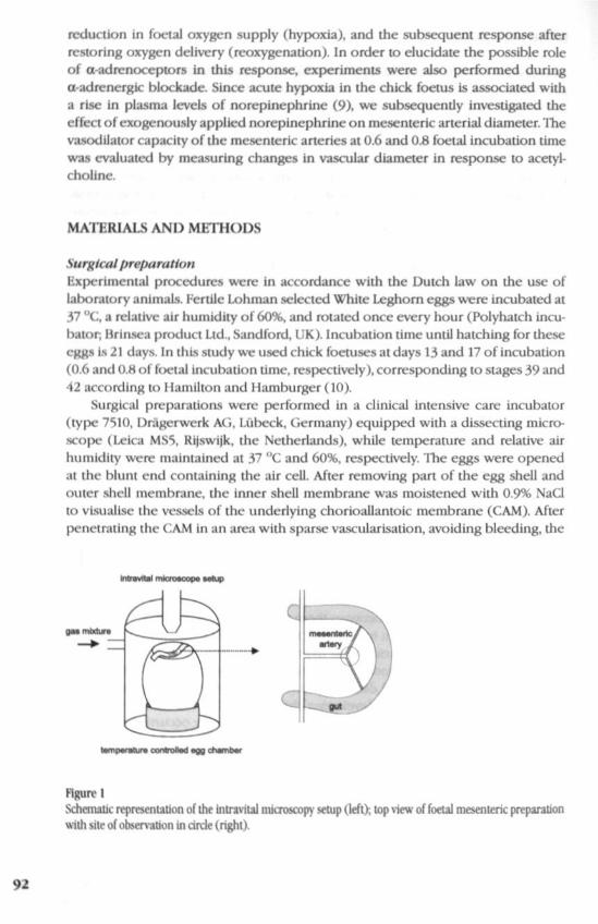
reduction in foetal oxygen supply (hypoxia), and the subsequent response afterrestoring oxygen delivery (reoxygenation). In order to elucidate the possible roleof a-adrenoceptors in this response, experiments were also performed duringa-adrenergic blockade. Since acute hypoxia in the chick foetus is associated witha rise in plasma levels of norepinephrine (9), we subsequently investigated theeffect of exogcnously applied norepinephrine on mesenteric arterial diameter. Thevasodilator capacity of the mesenteric arteries at 0.6 and 0.8 foetal incubation timewas evaluated by measuring changes in vascular diameter in response to acetyl-choline.
MATERIALS AND METHODS
lixjKTimcntal procedures were in accordance with the Dutch law on the use oflaboratory animals. Fertile Lohinan selected White Leghorn eggs were incubated at37 "C, a relative air humidity of 60%, and rotated once every hour (Polyhatch incu-bator; Brinsea product Ltd., Sandford, UK). Incubation time until hatching for theseeggs Is 21 days. In thLs study we used chick foetuses at days 13 and 17 of incubation(0.6 and 0.H of foetal incubation time, respectively), corresponding to stages 39 and42 according to Hamilton and Hamburger (10).
Surgical preparations were performed in a clinical intensive care incubator(type 7510, Drägerwerk ACi, Lübeck, Germany) equipped with a dissecting micro-scope (Leica MS5, Kijswijk, the Netherlands), while temperature and relative airhumidity were maintained at 37 "C and 60%, respectively. The eggs were openedat the blunt end containing the air cell. Alter removing part of the egg shell andouter shell membrane, the inner shell membrane was moistened with 0.9% NaClto visualise the vessels of the underlying chorioallantoic membrane (CAM). Afterpenetrating the CAM in an area with sparse vascularisation, avoiding bleeding, the
M mfcracop« M(U
k >
c
tampwturt oontrowd GQQ ensmbsr
Figure ISchematic representation of the intravital microscopy setup (left): top view of foetal mesenteric preparationwith site of observation in circle (right).
92

omphalocele was localised and centered at the level of the CAM by moons of twosutures through the connective tissue (Ethicon, Prolene 6-0). The omphalocele w«8opened by careful blunt dissection and the intestine was exposed. One segmentof the umbilical loop of the ileum with its mesentery was placed on a 1 cm pieceof cotton tape which was attached to the egg shell. Only preparations which \\x>recompleted within 15 minutes after opening the egg and which required minimalmanipulation during positioning were included in the experimental protocol. Forfurther microscopic evaluation in vivo, the egg was transferred to a single-eggchamber where temperature (37 "C) and air How (4 1/min) were controlled, andsubsequently placed in an intravital microscope setup (Fig. 1).
All in vivo observations were made with an intravital microscope (Leitz Orthoplan946627). Visualisation of the vasculature was performed with a l.eitz 2()x longworking distance objective (numerical aperture 0.32) via a Leitz Ploemopak illu-minator 2.1 (1.25x), equipped with a Leitz POL-cube (11). The intestine was epl-Uluminated using a 75W Xenon lamp. Images were protected onto a CC1) camera(Hamamatsu Photonics. IIamainat.su. Japan), connected to a Super-VMS video-recorder (Panasonic, model NV-FS100HQ), and stored on videotape (Sony, Sii|>erVHS VXSK-180V0 Images were displayed on a monitor screen (Sony, model PVM-122CE). Final resolution was 1 u.m. Arterial diameters were analysed off-line usingan image-shearing device (IPM, model 908, San Diego, CA). All experiments wereperformed on anatomically similar locations of second order branches of theomphalomesenteric artery.
Drw#s <znrf so/wtfonsNorepinephrine (1-artenolol-bitrate, NE), acetylcholine (ACh), sodium nitroprus-side, adenosine, papaverine, and phentolamine (PHENT) were obtained fromSigma Chemical Co. All drugs were dissolved in HF.PF.S buffered Krebs with thefollowing composition (in mM): NaCl 1433, KCl 4.7, MgSO« 1.2, KH^PO« 1.2, CaCl,2.5, glucose 5.6, HEPES 15. The pH of the buffer was adjusted to 7.4. Final concen-trations of the solutions ranged from 10* to 10"* M. All drugs were freshly pre-pared on the day of the experiment; temperature of the solutions at the time ofadministration was 37 °C.
o/ocu/eEvaluation of the effect of acute hypoxia on second order mesenteric arterialdiameters was performed at 0.6 («-8) and 0.8 (w-7) foetal incubation time. Afteran equilibration period of 15 minutes following surgical preparation, a 2-minutebaseline recording was made. Subsequently, hypoxia was induced by replacingthe ambient air (21% Oj) in the egg chamber by 100% Nj (4 l/min) for 5 minute»,according to a method described previously (4). After completion of the hypoxicperiod, reoxygenation was achieved by replacing the N^ with ambient air. Duringthis period one mesenteric artery was continuously kept in focus and recorded.
CHAPTEX6 - POCrALVASOMOTOIUaPOm» 93

To verify whether this protocol actually reduced blood oxygen content, i.e., resul-ted in hypoxaemia, blood samples were collected at the end of the 5-minute periodof hypoxia in a separate series of chick foetuses at 0.6 (n-6) and 0.8 («-7) foetalincubation time and bicxxl gas values were compared with control groups undernormoxic conditions. Samples (0.2 ml) were obtained by withdrawing bloodfrom the chorioaliantoic vein using a 1 ml heparinised syringe attached to a 21Gneedle and analysed at 37 "C using a blood gas analyser (Radiometer ABL510,Copenhagen, Denmark). The chorioaliantoic vein, being the avian equivalentof the mammalian umbilical vein, transports blood from the chorioaliantoicmembrane (where gas exchange takes place) to the foetus. Therefore, changes inchorioaliantoic vein blood gas values reflect changes in foetal arterial blood gasvalues.
/4/orx/pressure* measurementsIn an additional series of experiments at 0.6 (n-5) and 0.8 (n-6) foetal incubationtime, mean arterial pressure and heart rate were determined under normoxicconditions xs well as during a 5-minute period of hypoxia. The technique for meas-uring NCXKJ pressure in the chick foetus was adapted from a method previouslydcscrllxrd by Tazawa et al., and has been demonstrated to provide reliable bloodpressure measurements while maintaining foetal gas exchange (12). Briefly, afteropening of the egg in the clinical incubator, one of the two branches of thechorioallantoic artery was cannulated with a 10-cm-long nylon catheter (internaldiameter 0.5 mm) which was tilled with 0.9% NaCl. The catheter was inserted intothe artery with its tip pointing upstream. The free end of the catheter was con-nected to a pressure transducer (Baxter Uniilow, Baxter B.V., Uden, the Netherlands)which was placed al the same height as the egg. Pressure signals were recordedon a computer using a data acquisition system and the heart rate was calculated.
/jfTiff q/7op»ca//y app/KYThe effect of a-adrenergic blockade on vasomotor responses to acute hypoxia wasassessed in chick foetuses at 0.6 (n-8) and 0.8 (n-8) foetal incubation time. Afterequilibration and baseline recording a 20 ul aliquot of a 10-' M phentolamine solu-tion (PMENT), corresponding to a single dose of 6.35 ng, was applied to the mesen-teric arteries. During the 5 minutes after application of phentolamine, a recordingwas made to assess the effect of a-adrenergic blcxrkade on baseline arterial diame-ter. Subsequently, hypoxia was induced for 5 minutes followed by 5 minutes ofreoxygenation. During this period the same mesenteric artery was continuouslykept in focus and recorded.
In a separate series of chick foetuses at 0.6 (n-8) and 0.8 (n-10) foetal incubationtime, we assessed constrictor responses of the mesenteric arteries to norepineph-rine (NE). After equilibration and baseline recording a cumulative log molar con-centration-response curve was constructed by applying 20 iü aliquots of increasingconcentrations (Kr*-UH M) of NE to the mesenteric arteries, corresponding tosingle closes of 6.38 ng- 63-8 |ig. After application of each dose a 2-minute recordingwas made.

To verify the efficacy of the a-adrenergic blockade we also constructed a con-centration-response curve for NK S minutes after topical administration of a 20 nlaliquot of a 10'* M phentolamine solution to the mesenteric arteries in a separateseries of chick foetuses at 0.6 (n-6) and 0.8 (w-6) foetal incubation time.
To assess the vasodilator capacity of second order mesenteric arteries both underbaseline conditions and after induction of arterial tone using 10"* M topicallyapplied NE, we measured changes in mesenteric arterial diameters in response totopically applied acetylcholine (ACh). Dilator responses under baseline conditionswere assessed in chick foetuses at 0.6 (n-6) and 0.8 (n-7) foetal incubation time.After equilibration and baseline recording a cumulative log molar concentration-response curve was constructed by applying 20 pJ aliquots of increasing concen-trations (10 -10"' M) of ACh to the mesenteric arteries, corres|x>nding to singledoses of 3.63 ng-36.3 Mg- After each dose a 2 minute recording was made Dilatorresponses in NE-constricted arteries were assessed in a similar way by topicalapplication of 20 nl aliquots of ACh 10*-10"* M after administration of lO'' M NK tothe mesenteric arteries.
In a separate series of chick foetuses at 0.8 foetal Incubation lime (n-S),we investigated whether topical application of a 20 (il aliquot of 10' M ACh indu-ced maximal vasodilation. To this end, the vasodilator effect of JO''' M ACh wascompared to a cocktail of 10-* M each of sodium nitroprusside, adenosine, andpapaverine. No additional increment in arterial diameter was observed after appli-cation of this cocktail on top of ACh (data not shown), indicating that a 20 |il aliquotof 10^ M ACh induced maximal vasodilalion.
The effect of repetitive application of the buffer solution was assessed in aseparate series of chick foetuses at 0.8 foetal incubation time (w-5). TO this end,20 til aliquots of HEPES buffered Krebs were applied to the mesenteric arteries10 times, with 2-minute intervals. No significant changes in arterial diameters wereobserved during 10 successive applications of 20 pi of HEPES buffered Krebswithin a 30-minute period (data not shown).
Lununal diameter of a second order mesenteric artery was measured at about 50|im from the bifurcation of the first order mesenteric artery, a site with clear dis-tinction of the inner margins of the vessel wall (13). In the course of an experi-ment, all measurements were performed at this site of the second order mesentericartery. Changes in diameter are presented as percentage of baseline diameter, withbaseline being 100%.
Data are expressed as mean ± SEM. The term w refers to the number of foetuses; oneartery per foetus was selected. Statistical comparisons between groups were madeusing the Mann-Whitney U test Statistical comparisons within groups were madeusing the Wilcoxon signed ranks test Statistical significance was denned as a p<0.05.
CHATTn6-FOCIALWOMOTMIBMrONMi 95

100-
80
60 -
40 -
ao-
-100 0 1,100 200 300Time (s)
400 500 600
Figure 2Diameter tracing of theeffect of hypoxia andreoxygenation on asecond order mesentericartery at 0.8 foetal incu-bation time. Arterial dia-meter was measuredevery 15 seconds, underbaseline conditions(-120-0 s), during hypo-xia (0-300 s), and duringreoxygenation (300-600s); TN indicates timepointof maximal vasoconstric-tion.
hypoxia reoxygenation
140 -• 0.6• 0.8
5' 10'
hypoxia reoxygenation
Figure 3Effect of 5 minutesofhypo-xia followed by 5 min-utes of reoxygenation onmesenteric arterial dia-meter at 0.6 and 0.8 foetalincubation time. Acutehypoxia induced a sig-nificant reduction in ar-terial diameter at both0.6 and 0.8 foetal incuba-tion time. The magnitudeof the vasoconstrictionwas significantly largerin the older foetuses.During reoxygenation asimilar level of vasodila-tion above baseline wasobsened at both stages offoetal incubation.»« p<0.01, 0.6 vs. 0.8foetal incubation time
96

RESULTS
Figure 2 illustrates a diameter tracing of the effect of acute hypotrift aorder mesenteric artery at 0.8 foetal incubation time. The artery was recorded con-tinuously and arterial diameter was measured every IS seconds. The response tohypoxia was characterised by an initial transient constriction with ;t peak afterabout 100 seconds, followed by a relaxation to baseline level that continued untilthe end of the 5-minute hypoxic period. Reoxygenation resulted in a vasodilaiionabove baseline, which was maintained until the end of the "»-minute period ofreoxygenation.
In subsequent experiments, mesenteric arterial diameters were measured atfour time points: before hypoxia (base), at maximal vasoconstriction during hypoxiaOV). at the end of the hypoxic period (*>'), and at the end of reoxygenaUon(10'). Baseline diameters increased from 56 ± 2 9 um at 0.6 incubation to 7S ± 2.4jim at 0.8 foetal incubation time. During hypoxia arterial diameters significantlydecreased to 87 ± 4.4% of baseline (from 56 to 49 urn, p<0.05) at 0.6 foetal incuba-tion time and to 44 ± 6.7% <from 64 to 28 urn, p<0.05) at 0.8 foetal incubation time(Fig. 3). The magnitude of this vasoconstrictor response was fourfold larger at 0.8as compared with 0.6 foetal incubation time (p<0.01). Maximal vasoconstrictionduring the 5-minute period of hypoxia was observed at 106 (51-207) seconds and105 (79-137) seconds from the start of hypoxia at 0.6 and 0.8 foetal incubation time,respectively, and lasted for approximately 30 seconds. During reoxygenation asignificant vasodilation above baseline was observed in both groups (to 118 ± 6.5%and 121 ± 7.5% of baseline, p<0.05, at 0.6 and 0.8 foetal incubation lime, respect-ively).
Blood gas analysis demonstrated that 5 minutes of hypoxia induced a signifi-cant decrease of arterial Po^ from 11.7 to 30 kPa at 0.6 foetal incubation time, anda reduction of arterial Poj from 7.2 to 1.6 kPa at 0.8 foetal incubation time Theabsolute level of the arterial Po^ at the end of the 5-minute period of hy|x>xiawas higher at 0.6 as compared with 0.8 foetal incubation time. However, whenexpressed as a percentage of control (normoxic) oxygen tension, arterial Po^ wasreduced to a similar extent at 0.6 and 0.8 foetal incubation time (74% and 78%,respectively, Table 1).
Table I. Blood gas values at 0.6 and 0.8 foetal incubation time at the end of the 5- minute period of hypoxia(100% Nj) as compared with control conditions (21% 0j)
0.6 foetal Incubation time 0.8 foetal Incubation time
control (n-5) hypoxia (n-5) control (n-7) hypoxia (n-7)
11.65 ±0.63 2.98±0.26* 7.18±0.59 1.57±0.18"
Pcoj(kPa) 1.98 ±0.07 1.43 ±0.04* 4.57 ±0.41 4.45 ±0.25
pH 7.57 ±0.02 7.63 ±0-03 7.56 ±0.03 7.53 ±003
*. ** p<0.05, p<0.01, at the end of 5 minute» of hypoxia vs. control
CHATTE« 6 - ForrALVASOMoroBBHFofoes 97

baselinehypoxia
Figure 4A
Figure 4B
Figure-»Effect of 5 minutes of hypoxia on mean arterial pressure (A) and heart rate (B) at 0.6 and 0.8 foetal incuba-tion time.* p<O.OS, vs. baseline; *• p<0.01,0.6 vs. 0.8 foetal incubation time
Mean arterial pressure under baseline conditions significantly increased from 11± 0.4 mm Hg at 0.6 foetal incubation time to 21 ± 0.6 mm Hg at 0.8 foetal incu-bation time (p<0.01). During acute hypoxia mean arterial pressure significantlydecreased to 4.0 ± 1.1 mm Hg (p<0.05) at 0.6 and 11.8 ± 2.8 mm Hg (p<0.05) at0.8 foetal incubation lime (Fig. 4A). Heart rate under baseline conditions was notsignificantly different between 0.6 and 0.8 foetal incubation time (216 ± 3 beats/min and 210 ± 14 beats/min. respectively). During hypoxia heart rate significantlydecreased to 68 ± 31 beats/'min (p<0.05) at 0.6 and 132 ± 20 beats/min (p<0.05) at0.8 foetal incubation time (Fig. 4B).
98

Topical application of PHENT alone did no« significantly alter baseline meMMOriCanerial diameters at 0.6 and 0.8 foetal incubation time (97 ± 2S% and 98± 5.0%,respectively). At 0.6 foetal incubation time, neither the hypoxia-:issociated vaao-constriction (to 89 ± 4.0% of baseline) nor the vascxJUation during reoxygenatk>n(to 116 ± 3-2% of baseline) were significantly affected by a-adrenergic blockade.In contrast, at 0.8 foetal incubation time the vasoconstrictor response during hypoxia was significantly attenuated by a-adrenergic blockade (p<0.01. Fig. S). In thepresence of PHENT acute hypoxia induced only a slight decrease in mesentertcarterial diameter (to 93 ± 2.7% of baseline, p-0.05). Subsequently, diameters In-creased to 117 ± 33% above baseline(p<0.05). and further increased to 122± 2.7%(p<0.05) during reoxyge nation.
140
120-
g 100 -
I 80-
o «H
40-
20 -
• hypoxiaD hypoxia+PHENT
5" 10'
hypoxia reoxygenabon
Rgure5Effect of topically applied phentolamine on vasomotor responses during acute hypoxia and reoxygenationat 0.8 foetal incubation time. Alpha-adrenergic blockade significantly reduced the hypoxia-assodated vaso-constriction.»• p<0.01, hypoxia in the presence of phentolamine vs. hypoxia alone
O u r m 6 - FOETAL VASOMOTO« 99

140 -
120-
100 -
80 -
6 0 -
4 0 -
2 0 -
0 -
• 0.6• 0.8
1 1 1
\
« •
1 1 1
base -3 -2
Figure 6Effect of topically appliednorepinephrine on mesente-ric arterial diameter at 0.6and 0.8 foetal incubation time.Maximal constriction to NEwas significantly greater at 0.8as compared with 0.6 incuba-tion.##, ### p<0.01, p<0.001, 0.6vs. 0.8 foetal incubation time
140-
• NED PHENT + NE
##
base -6 -5
Log
-3 -2
Figure 7Efficacy of a-adrenergic bloc-kade with phentolamine. Thevasoconstrictor response to NEwas significantly reducedby phentolamine at 0.8 foetalincubation time.#, ## p<0.05, p<0.01, NEin thepresence of phentolamine vs.NE alone
100

Cumulative application of increasing concentrations of NE caused a decreasein arterial diameter in both age groups (Fig. 6). Maximal vasoconstriction wasobserved after application of 10** M (0.6 incubation) or 10' M (0.8 incubation) NE.At these applied concentrations, arterial diameters significantly III-CIV.IM-II to "1 t3.0% (p<0.05) and 35 ± 3.8% (p<0.01) of baseline at 0.6 and 0 8 I.KI.II HU uh.inontime, respectively. The magnitude of maximal constriction to NF. was approximatelytwofold larger at 0.8 as compared with 0.6 foetal incubation time (p*0.01).
Topical administration of a 20 uJ aliquot of 10' M phentolamine to themesenteric arteries completely prevented the reduction in arterial diameter bynorepinephrine at 0.6 foetal incubation time. At 0.8 foetal incubation time the con-centration-response curve for norepinephrine shifted to the right in the presenceof phentolamine, indicating a reduction in norepinephrine sensitivity (Fig. 7).
Subsequent application of increasing concentrations of ACh to the same arteriesresulted in a gradual vasodilation at both 0.6 and 0.8 foetal incubation time.Maximal arterial diameters were observed after application of 10 ^ M ACh, and weresignificantly different between 0.6 and 0.8 foetal incubation time (63 ± 3.5 Jim vs. 91± 2.3 um, p<0.001). However, expressed as percentage change from baseline, maxi-mal levels of vasodilation were not significantly different between the two stages offoetal development (to 113 ± 4.4% and 122 ± 4.8% of baseline at 0.6 and 0.8 foetalincubation time, respectively; Fig. 8A).
Topical application of increasing doses of ACh under baseline conditions, i.e.,without prior constriction with NE, also caused a concentration-dependent dila-tion of the mesenteric arteries at 0.6 and 0.8 foetal incubation time. Maximal levelsof vasodilation in both groups were not significantly different (to 125 ± 2.6% and133 ± 5.4% of baseline at 0.6 and 0.8 foetal incubation time, respectively; Fig. 8B).
At both stages of foetal development, maximal levels of vasodilation afterapplication of 10"' M ACh were similar in NE-constricted arteries and in arteriesunder baseline conditions. Furthermore, maximal diameters in response to topicalapplication of ACh were not significantly different from maximal diametersobserved during reoxygenation after 5 minutes of hypoxia.
CHATTE! 6 - FOBDU. VA3OMQTO« «ESTO!« 1 0 1

140-
120-
•5 «0-
40 -
20-
0
140 -
120-
I 100-
.8 80-
•9 6 0 -
40 -
2 0 -
0
••*
NE10* -6 -5 -4
Log [
base -5
0.60.8
Figure 8A
Figure 8B
Figure 8Effect of topicalh' applied acetylcholine on mesenteric arterial diameter at 0.6 and 0.8 foetal incubation time.ACh induced a significant increase in diameter above baseline level both in arteries subjected to NE-inducedconstriction (A) and in arteries under baseline conditions (B). Note that maximal vasodilation to ACh wassimilar at 0.6 and 0.8 foetal incubation time.#, ##, ##« pvO.OS, pvO.01, pvO.001,0.6 vs. 0.8 foetal incubation time

DISCUSSION
In order to obtain information with regard to the regulation of vascular tone inmesenteric arteries during foetal development, we designed a novel cx|X*rimentalsetup for measurements of mesenieric arterial diameter in the intact chick loeiusat different stages of foetal development. Between 0.6 and 0.8 of chick foetalincubation time, baseline luminal diameters of second order mesenteru .UUMK-S
increased from 56 (xm to 75 jim. Since topical administration of the vasodilatorsubstance acetylcholine induced an increase in arterial diameter of approximately20% above baseline at both 0.6 and 0.8 foetal incubation time, it may be concludedthat the mesenteric arteries already exhibit a degree of vascular tone under KIM-line conditions. Topical administration of the non-selective a-adrenergic ai u .)>•< > 111 siphentolamine did not affect baseline arterial diameter This suggests that during tin-observed period of foetal development a-adrenergic mechanisms are not involvedIn the establishment of baseline arterial tone at this level of the arterial tree.
The mesenteric arteries constricted in response to topically applied norepi-nephrine at both 0.6 and 0.8 foetal incubation time. Furthermore, this constrictionwas significantly attenuated in the presence of phentolamine. This indicates thatfunctional a-adrenergic pharmacomechanicai coupling is present in the mesen-teric arteries as early as 0.6 foetal inclination time. The maximal constrictorresponse to norepinephrine increased about twofold between 0.6 and O.H foetalincubation time. This may be due to developmental changes in the efficacy ofthe a-adrenergic signal transduction pathway, amount of adrenoceptors as well asmaturation of the vascular smooth muscle contractile apparatus. Interestingly, themean arterial pressure increased about twofold during thus period of development,from 11 mm Hg at 0.6 to 21 mm Hg at 0.8 foetal incubation time. Assuming thatthe perfusion pressure at the level of the second order mesenteric arteries alsoincreased, the arteries at 0.8 foetal incubation time were subjected to a highertransmural pressure as compared with 0.6 foetal incubation time. Hence, in orderto obtain the observed decreases in luminal diameter, the contractile force gener-ated by the mesenteric arteries at 0.8 foetal incubation time must be substantiallylarger as compared with 0.6 foetal incubation time.
A 5-minute period of hypoxia induced a transient decrease in mesenteric arte-rial diameter at both stages of foetal development The magnitude of the hypoxia-associated vasoconstrictor response increased from a 13% reduction of arterialdiameter at 0.6 incubation to a reduction of 56% at 0.8 foetal incubation lime. At0.6 foetal incubation time application of phentolamine did not significantly affectthe vasoconstriction. This suggests that the constrictor response is not mediated bya-adrenergic receptors at this stage of foetal development. In contrast, at 0.8 foetalincubation time the vasoconstriction during hypoxia was substantially attenuatedin the presence of phentolamine. This suggests that it is at least in part mediated bya-adrenoceptors at this stage of foetal development
At 0.8 foetal incubation time the concentration-response curve for norepi-nephrine shifted to the right in the presence of phentolamine. This indicates areduction in norepinephrine sensitivity, which is typical of a competitive antago-nist such as phentolamine. Based on these results it cannot be ruled out that the
CHAPTER 6 - FOCTAJ. VASOMOTOB «espoNses 1 0 3

140-
120-
100 -
80 -
60-
40-
20-
reoxygenattonACh
AChreoxygenation
baseline
hypoxia
Figure9 UVasomotor responses offoetal mesenteric arteriesat 0.6 and 0.8 foetal incuba-tion time. Vasoconstrictorresponses to acute hypo-xia and to topically appliedNE increased during foetaldevelopment, while vaso-dilator responses duringreoxygenation and to topi-cally applied ACh weresimilar at 0.6 and 0.8 foetalincubation lime.
0.6I
0.8
Foetal incubation time
7% constriction which remained during acute hypoxia in the presence of phentola-mlne is related to incomplete a-adrenergic blockade at 0.8 foetal incubation time.
Central haemodynamic measurements showed that during acute hypoxia bothheart rate and blood pressure decreased considerably at 0.6 as well as at 0.8 foetalincubation time. Assuming a reduction of perfusion pressure at the level of theincsenterlc arteries, luminal diameter may passively decrease due to elastic recoil ofthe arteries subjected to a lower perfusion pressure. This may alternatively explainthe observed small diameter reduction in the presence of phentolamine.
Finally, it may be argued that the difference in the vasoconstrictor responseduring acute hypoxia between 0.6 and 0.8 foetal incubation time is due to a dif-ference in the level of hypoxia. Indeed, arterial P02 during acute hypoxia is lowerat 0.8 as compared with 0.6 foetal incubation time. However, arterial P02 undernormoxic conditions is also proportionally lower at 0.8 as compared with 0.6foetal incubation time, thus the relative decrease in arterial Poj is comparable. Themechanisms underlying the developmental change in arterial P02 under normoxicconditions are beyond the scope of this article but are discussed in detail byTazawa et al (14). Briefly, it has been demonstrated that during the third trimesterof chick foetal incubation under normoxic conditions there is a gradual decreasein arterial 1*0 and an increase in Pco, which is in part metabolically compensated.According to these authors the changes in blood gas levels during late foetal devel-opment are related to a rise in metabolic rate of the foetus during this period ofincubation. Oxygen concentration of the arterial blood, however, is maintained byconcomitant increases in haematoerit and oxygen affinity of haemoglobin. Since ithas been postulated that oxygen sensitive cells sense the oxygen concentration ofthe blood (.9), it seems unlikely that the difference in the vasoconstrictor response
104

during acute hypoxia between 0.6 and 0.8 foetal incubation time is due to the dif-ference in arterial Po_,
Thus, the current study shows that foetal mesenteric arteries constrict in re-sponse to acute hypoxia. and indicates that the increase in magnitude of this vaso-constrictor response during 0.6O.8 of foetal development results from an increase inadrenergic constrictor capacity.
Interestingly, during the reoxygenation period the mesenteric arterial diame-ters increased above baseline levels, suggesting a hyperaemlc response at both 0.6and 0.8 foetal incubation time. The maximal arterial diameters obtained during thereoxygenation period were comparable to the levels of vasodilation obtained withtopically applied 10^ M acetylcholine, a concentration sufficient to obtain maxi-mal vasodilation. The vasodilator response actually started during the hypoxicphase and progressed during the reoxygenation phase. Maximal vasoconstrlciionduring the S-minute period of hypoxia was observed at approximately 100 secondsfrom the start of hypoxia. and lasted for about 30 seconds. By the end of the5-minute hypoxic period arterial diameters returned almost to baseline level. Incontrast, the vasoconstriction observed after topical application of norepinephrineunder normoxic conditions lasted as long as 30 minutes (data not shown). Sinceacute hypoxia in the chick foetus is associated with a rise in plasma levels ofnorepinephrine (IS), it is interesting to observe a vasodilator res|jonse alreadyduring the hypoxic phase. This suggests that secondary to the initial const riet ion Inresponse to hypoxia a potent vasodilator substance is released locally.
In conclusion, the chick foetal mesenteric vasculature is a readily accessiblevascular bed for studying the development of vasomotor control in small resist-ance-type arteries. In the current study we showed that a-adrenergic pharmacome-chanical coupling is already present in chick foetal mesenteric arteries as early as0.6 foetal incubation time. The ability of mesenteric arteries to constrict in responseto both physiological and pharmacological stimuli increases between 0.6 and 0.8foetal incubation time. In contrast, vasodilator responses of the mesenteric arteriesremain constant during this period of foetal development. This may suggestthat the maturation of vasodilator mechanisms precedes that of vasoconstrictormechanisms during foetal development
CHATTE« 6 - FOETAL VASOMOTCM

R e f e r e n c e s
1. Crissinger K.D. Regulation of hemodynamics and oxygenation in developing intestine insight into thepalhogenesis of necrotizingenterocolitis. A:to ftf«//o//\S'«/>/>/1994; 396:8-10.
2. Nowicki P.T. and Nankervis C A The role of the circulation in the pathogenesis of necrotizing enteroco-litis. Gfo» /feri/wto/1994; 21:219-233.
3. Jensen A. and Berger R. Foetal circulalory responses to oxygen lack./ Zter P/r)sw/1991; 16:181-207.4. Mulder A.L.M., van Golde J.C., Prinzen F.W., and Blanco C.L. Cardiac output distribution in response to
hypoxia in the chick embryo in the second half of the incubation time.//ttys/o/1998; 508:281-287.5. Jensen A. and l.ang IJ. Foetal circulatory responses to arrest of uterine blood flow in sheep: effects of
chemical sympathectomy./AWA)*«)/1992; 17:75-86.6. Reuss M.I.., Parer J.T., HarrisJ.L, and Krueger T.R. Hemodynamic effects of alpha-adrenergic blockade
during hypoxia in foetal sheep. /4m/Ofo'te/6)>»eeo/1982; 142:410-415.7. Le NobleJ.L, Struyker-Boudier H.A., and SmitsJ.F. Differential effects of general anesthetics on regional
vasoconstrictor responses in the rat. /1n:/i /«//'/wnmwroä!}'« TA«-1987; 289:82-92.8. Iwamoto HS, Kaufman T, Keil I X , and Rudolph A.M. Responses to acute hypoxemia in foetal sheep at
O.frO.7 gestaUon.Hmy/'V/o/1989; 256: H613-H620.9. Semenza d.i.. Perspectives on oxygen sensing tW/1999; 98: 281-284.
10 Hamburger V and Hamilton IIL A series of normal stages in the development of the chick embryo.y-MwpAo/1951; 88:49^8.
11. Slaaf D.W., Tangclder G.J., Reneman R.S., Jager K, and Bollinger A. A versatile incident illuminator forintravital microscopy. / « / / A/icrocw 6'/»« £*p1987; 6:391-397.
12. Höchel J., Akiyania R., Masuko T., PearsonJ.I, Nichelmann M., and Tazawa H. Development of heart rateirregularities in chick embryos.Hm//'Ays»o/1999; 275: H527-H533.
13. Le Noble J.L., Tangelder G.J., Slaaf D.W., van Essen H, Reneman R.S., and Struyker-Boudier HA Afunctional morphometric study of the cremaster muscle microcirculation in young spontaneouslyhypertensive rats..///)/*>r/«w 1990; 8:741-748.
14. Tazawa II., Visschedijk A.H.J., WiltniannJ., and Piiper J. Gas exchange, blood gases and acid-base statusin the chick before, during and after hatching. /fef/>fW%Mb/1983; 53:173-185.
15. Witlmann J. and Prechtl J. Respirator)' function of catecholamines during the late period of aviandevelopment. /to/>/W'Ay.$io/1991; 83:375-386.
106
CHAPTER 7
Chronic Foetal Hypoxia Alters Haemodynamic Control
7ru/y, fejrf/i marts HO/ <a fagton/ng feu/ a s<a#e /n /(/e's

108

INTRODUCTION
During embryonic development, blood vessels arise through the procMMtof va*culogenesis. angiogenesis. and aneriogenesis. The primitive vends develop iMOarteries and veins after recruitment of vascular smooth muscle cells and com-mencement of perfusion. Molecular mechanisms controlling the morphogenictransition of primitive blood vessels into a mature vascular network are rapidlybeing unraveled (1). However, little is known on the acquisition of vasomotorproperties. It is still unknown when these newly formed arteries attain the abilityto regulate local vascular tone in response to the oxygen demand of the individualorgans, when central nervous coordination over the different vascular Iwls isinstituted in order to regulate blood pressure, and which signals guide the matu-ration of this complex regulation.
According to the foetal programming hypothesis, aberrant development ofthe mechanisms which regulate vascular tone lies at the origin of c ardiovasculardiseases in later life, for example essential hypertension. It is speculated that dc|>rivalion of oxygen availability during foetid development, which is associated withtntrauterine growth retardation, induces permanent alterations in the functionalbehaviour of arteries, thereby increasing peripheral vascular resistance and pro-gramming for hypertension. This hypothesis is based on epidcmiologk relation-ships between low birth weight and development of cardiovascular disease Inadulthood (2).
It is well established that hypoxia triggers the production and release of vas-cular endothelial growth factor (VEGF) and adenosine, which stimulate angiogen-esis and arteriogenesis, respectively (1,3,4). Both factors also exert vasodilatoreffects (5). These observations support the idea that chronic hypoxia may alterarterial network design and local regulation favouring a reduction in peripheralvascular resistance, rather than the hypothesised rise in resistance. Such an uncom-pensated decrease in vascular resistance might result in a drop in central bloodpressure. In order to retain blood pressure, it may be postulated that foetalhypoxia interferes with central autonomic nervous control of the vasculature.Recent experimental evidence indeed suggests that hypoxia or angiogcnic factorsrelated to hypoxia may exert neurotrophic effects or modulate nervous function(63).
It is clear that oxygen availability is a critical determinant of the long-termdesign of the vascular system. The objective of this study was to investigate /«MI« the role of oxygen on the long-term establishment of local vascular tone aswell as on the regulation of central haemodynamics. To this end, we studied theeffects of prolonged exposure to a physiologically relevant level of hypoxia onot-adrenoceptor mediated vasoconstriction and nitric oxide-dependent dilation, aswell as the functional maturation of the sympathetic nervous system, in combi-nation with measurements of arterial pressure and heart rate in near-term chickfoetuses.
CHATTE« 7 - Cmomc FOETAL HYPOHA 109

MATERIALS AND METHODS
Experimental procedures were in accordance with the Dutch law on the use oflaboratory animals. Fertile Lohman selected White Leghorn eggs were incubatedat 37 "C and a relative air humidity of 60%. Experiments were conducted in chickfoetuses at day 19 of the 21-day incubation period, corresponding to 0.9 of totalfoetal incubation time. From the first day of incubation onward, foetuses in thenormoxic (N) group were incubated at 21% O2, whereas foetuses in the chronichypoxic (CH) group were incubated at 15% O2. Survival rate over the 19-day incu-bation period in the CH group was lower as compared with the N group (51.4% vs.72.5%).
To assess the effect of chronic hypoxia on foetal arterial oxygen levels and ery-thropoiesls, blood gas values and haematocrk were determined under baseline(21 % Oj) conditions in a separate series of normoxic and chronic hypoxic foetuses,as previously described (9). Body weights were measured to determine the effectof chronic hypoxia on foetal growth.
Swrjj /ra/ /we/rarwf/onAll surgk al preparations were performed in a clinical infant incubator (21% O2), asdescribed in detail elsewhere (9)- In short, the egg was opened at the blunt endcom.lining the air cell, and alter penetrating the chorioallantoic membrane (CAM),the foetus was stabilised by means of two sutures through connective tissue ofthe abdominal wall. For assessment of vasomotor responses, the omphalocele wascentered at the level of the CAM, opened by careful blunt dissection, and the intes-tine was exposed. A segment of the ileum with its mesentery was placed on a 1 cmpiece of cotton tape which was attached to the egg shell. Subsequently, the egg wastransferred to a single-egg chamber where temperature (37 "C) and air flow (21%Oj, 4 l/min) were controlled, and placed in an intravital microscope setup (9).
The intestine was epi-illuminated to iivo with a 75W Xenon lamp. Second ordermesenteric resistance arteries were visualised using an intravital microscope,equipped with a 20x long working distance objective, and a Ploemopak illuminator2.1 (1.25x) with a polarizer-analyzer cube (Leitz), resulting in a 25x magnificationat the camera plane. Images were projected onto a CCD camera (HamamatsuPhotonics, llain.unat.su, Japan), displayed on a monitor screen, and stored onvideotape. Final resolution was 1 (im.
l.ummal arterial diameters were analysed off-line using an image-shearingdevice (1PM, model 908, San Diego, CA). In the course of an experiment, all meas-urements were performed at the same site of the mesenteric resistance artery.Changes in diameter are presented as percentage of baseline diameter, with base-line being 100%.
&Y/x»rf »it»«/«/ profoco/To investigate the effect of chronic hypoxia on the establishment of a-adrenoceptormediated vasoconstriction and dilation, we measured the changes in mesenteric arte-
110

rial diameter in response to topically applied norepinephrine (NE) and additionalacetylcholine (ACh) in normoxic and chronic hypoxk" foetuses. Alter an equi-libration period of 15 minutes following surgical preparation, a 2-minute baselinerecording was made. A cumulative log molar concentration-response curve wasconstructed by sequential application of 20 nl aliquots of increasing concentrations(10*-10 •* M)of NE to the mesentery, and recording of the mesenteric artery linage for2 minutes following each application. The vasodilator capacity of t he N Ii-constrictedarteries was subsequently determined in a similar way by measuring the changesIn arterial diameters in response to application of 20|*1 aliquots of H)"-10 •* M ACh.
The effect of chronic hypoxia on the functional maturation of perivascuiarsympathetic nerves was assessed by measuring changes in mesenteric arterialdiameter in response to topically applied ryramine (Tyr, 10MO'' M). Tyraminereleases norepinephrine fn>m perivascuiar sympathetic nerve endings, and doesnot affect presynaptic reuptake (10,11).
In addition, changes in mesenteric arterial diameter as well as in centralhaemodynamic parameters were determined in response to a short period ofsevere hypoxia. After equilibration and baseline recording, acute hypoxia wasinduced by replacing the ambient air (21% O^) in the egg chamber by 100% N^{4 l/min) for 5 minutes. Reoxygenation was achieved by replacing the N^ withambient air. During this period one mesenteric artery was continuously kept infocus and recorded.
Mean arterial pressure and heart rate under baseline conditions as well as duringacute hypoxia were measured according to a previously described method (9). Inshort, a 10-cm-long nylon catheter (internal diameter 0.5 mm) which was filled with0.9% NaCl was inserted into one of the two branches of the chorioallantoic arterywith its tip pointing upstream. The free end of the catheter was connected to apressure transducer (Baxter Uniflow, Baxter B.V., Uden, the Netherlands) whichwas placed at the same height as the egg. Pressure signals were recorded on acomputer using a data acquisition system and the heart rate was calculated.
Norepinephrine (1-artenolol-bitrate, NE), acetylcholine (ACh) and tyramine (Tyr)were obtained from Sigma Chemical Co (St. Louis, MO). Drugs were dissolved inHEPES buffered Krebs with the following composition (in mM): NaCl 1433, KC14.7,MgSO« 1.2, KH2PO41.2, CaClj 2.5, glucose 5.6, HEPES 15. The pH of the buffer wasadjusted to 7.4. Temperature at the time of administration was 37 "C.
Dafcj analysisData are expressed as mean ± SEM. The term n refers to the number of foetuses;observations were made in one artery per foetus. Statistical comparisons betweengroups were made using the Mann-Whitney U test Statistical comparisons withingroups were made using the Wilcoxon signed ranks test. Statistical significance wasdenned as p<0.05.
CHAPrEB7-Onofncpon'ALHYPaoaA H I

RESULTS
The effecLs of chronic hypoxia (15% Oj) on foetal characteristics at 0.9 incubationtime, as measured under baseline (21% O2) conditions, are shown in Table 1.Chronic hypoxic foetuses had a significantly lower body weight (21.9 ± 0.49 vs. 24.7± 0.40 g, p<0.001), and higher haematocrit (0.34 ± 0.014 vs. 0.30 ± 0.010, p<0.05) ascompared with normoxic foetuses. The reduced survival rate, lower body weight,and higher hacmntocrit in chronic hypoxic foetuses are indicative of prolongedreduction in oxygen availability to the growing foetus, which justifies the experi-mental protocol used. Arterial POj levels under baseline (normoxic) conditionswere similar in both groups (6.3 ± 0.96 and 5.8 ± 0.46 kPa), whereas PCO2 was lowerin the CH group (3.9 ± 0.26 vs. 4.8 ± 0.34 kPa, p<0.05).
Table I. Effects of dironic hypoxia on baseline foetal characteristics at 0.9 foetal incubation time
Variable
Body wrlght (g)
Haemaiocrit
Blood gas values
Poj(kPa)
Pco,(kPa)
PH
Nonnoxk(n)
24.7 ±0.40 (21)
0.3010.01(17)(7)5.78 ±0.464.83 ±0.347.48 ±0.04
Chronic hypoxic (»)
21.9 ±0.49*'* (22)0.34 ±0.014* (7)(7)6.30 + 0.963.93 ±0.26*7.47 ±0.03
' p<O.OS, p<0.001, chronic hypoxic vs. normoxic foetuses
Absolute baseline diameters were similar in the CH and N group (89 ± 5.1 and 98± 5.0 urn, «-7 and w-9). Cumulative application of increasing concentrations ofnorepinephrine (NE) caused a decrease in arterial diameter in the CH and N group(Fig. 1A). Maximal constrictor response, obtained at a concentration of 10'* M NE,was similar in Ixith groups (to 34 ± 36% and 29 ± 2.0% of baseline).
Subsequent application of increasing concentrations of acetylcholine (ACh) tothe NE-constricted arteries resulted in a gradual dilation in the CH and N group.Mesenteric arteries in the CH group dilated to a significantly larger extent inresponse to 10"* M ACh as compared with the N group (to 88 ± 12.0% vs. 67 ± 9.1%of baseline, p<0.05), indicating a shift to the left of the dose-response curve forACh (Fig. IB). Maximal dilator response, attained at a concentration of 10•* M ACh,was also significantly greater in the CH group as compared with the N group (to118 ± -».9% vs. 105 ± 2.4% of baseline, p<0.05; Fig. 2). However, absolute maximaldiameters were similar in both groups (104 ± 5.0 and 104 ± 4.9 Jim).
112

120-
100-
80
6 0 -
4 0 -
2 0 -
* NA CH
Figure IA
base •« -5 -4 -3 -2
120-
100-
dia
me
bas
el
80 -
6 0 -
40 -
2 0 -
Figure IB
base -5
Figure 1Effect of topically applied norepinephrine (A) and acetylcholine (B) on mesenteric arterial diameter innormoxic (N) and chronic hypoxic (CH) foetuses at 0.9 foetal incubation time. Maximal constriction to NEwas similar in both groups. Subsequently, acetylcholine was applied on the NE-preconstricted mesentericarteries. Sensitivity to ACh was higher in CH as compared with N foetuses, as indicated by the left-shift of thedose-response curve. Data are expressed relative to baseline diameter (100%).*P<0.05,CH vs. N group
CHAPTER 7 - CHBONIC POCTAL HYPOXIA 113

25-
20 -
15 -
10-
5 -
Figure 2Maximal vasodilation to ACh10-2 M was greater in CH ascompared with N foetuses. Dataare expressed as percentageincrease of baseline diameter.*p<0.05,CH vs. N group
CH
topical .klmmiMr.iiion of 20-fil aliquoLs of increasing concentrations of tyramineCl'yr) induced a reduction in inesenteric arterial diameter in both the CH and N(both n-8 , data not shown). Maximal constrictor response, attained at a concen-tration of 1 0 ' M Tyr, was significantly greater in the CH compared with the N group(37 ± 3 6 % vs. 19 ± 5.2% decrease of baseline, p<0.01; Fig. 3).
Figure 3Effect of topically applied tyra-mine on mesenteric arterialdiameter at 0.9 foetal incuba-tion time. Maximal constrictionto Tyr 10-3 M was larger in CHas compared with N foetuses.Data are expressed as percen-tage decrease of baseline dia-meter."p<0.01,CH vs. N group
CH
114

Absoiute baseline diameters were similar in the CH and N group {94 ± 4.8 and 95± 4.5 ftm, «-9 and n-10). The vasomotor responses of the mesenteric resistancearteries to acute hypoxia (0% Oj) and subsequent reoxygenation (21% O^) weresimilar in the CH and X group, in magnitude as well as in time course (Fig. 4).In both groups, a significant and sustained reduction in arterial diameter wasobserved during 5 minutes of acute hypoxia (to 42 ± 2.5% and 46 ± 5.3% of baseline,p<0.01). which returned to baseline during reoxygenation.
Mean arterial pressure under baseline conditions was similar in the CH and Ngroup (27 ± 2.2 and 25 ± 1.4 mm Hg; n-8 and n-9; Fig. 5A). However, during acutehypoxia. arterial pressure significantly decreased in the CH group (to 22 ± 1.8 mmHg. p<0,05), whereas it remained constant in the N group (25 ± 0.9 mm Hg). Duringreoxygenation arterial pressure significantly increased above baseline in the CHand N group (to 31 ± 2.2 and 35 ± 1.7 mm Hg, p<0.05). This iiutv.i-.i- u t s greaterin the N group (p<0.01). Baseline heart rate was also .similar in the CH and Ngroup (205 ± 6 and 199 ± 9 beats/min; Fig. 5B). Although acute hypoxia induced asignificant fall in heart rate in both groups (p<0.01), heart remained at a higher levelin the CH group as compared with the N group (132 ± 3 vs. 117 ± 6 beats/'min,p<0.05). During reoxygenation heart rate returned to baseline in the N group (185± 8 beats/min), but only partially recovered in the CH group (172 ± 3 beats/min,p<0.05).
120-
100 -
I| 80 H
ra 60 H.o
4 0 -
2 0 -
A NA CH
0 100 200 300 400 500 600
Time (s)
1 I
Figure 4Effect of 5 minutes ofsevere hypoxia (0% O2)followed by S minutes ofreoxygenation (21% O2)on mesenteric arterialdiameter at 0.9 foetalincubation time. Timecourse and magnitudeof the arterial response(sustained constrictionduring acute hypoxia,dilation to baselineduring reoxygenation)was similar in CH andN foetuses. Data are ex-pressed relative to base-line diameter (100%).
hypoxia reoxygenation
CHAPTEB 7 - CHBONIC FOETAL HYPOXIA 115

I1
240 -
220 -
200 -
180 -
160-
140 -
120 -
100 -
80 -
6 0 -
40 -
2 0 -
NCH
Figure 5A
10'
Figure 5B
base
I10'
hypoxia reoxygenation
Figure 5Effect of S nünutes of severe hypoxia on mean arterial pressure (A) and heart rate (B) at 0.9 foetal incubationtime. Baseline blood pressure and heart rate was similar in CH and N foetuses. Acute hypoxia induced a fallin pressure and heart rate in CH foetuses. Although heart rate decreased to a similar degree, blood pressurewas maintained in N foetuses. The increase in arterial pressure during reoxygenation was lower in CH ascompared with N foetuses.
116
DISCUSSION
This <n H I « study conducted in intact, unanaesthetised near-term foetuses was ini-tiated by the concept that reduced oxygen availability during foetal developmentmay alter the functional behaviour of foetal arteries, and may influence blotxlpressure regulation. We demonstrated that chronic hypoxia results in enhancedcontractile responsiveness to direct stimulation of perivascular sympathetic nervesin the mesenteric vascular bed of near-term foetuses. Since maximal respon-siveness and sensitivity to norepinephrine were not altered, these data indicatethat chronic hypoxia induces sympathetic hyperinnorvation, which may lead toan increase in arterial tone. Chronic hypoxia was, however, not accompaniedby an elevated systemic blood pressure level. These findings support the ideathat hypoxia influences central autonomic nervous control over the individualvascular beds in order to maintain cardiovascular homeostasis.
Chronic hypoxia induced a twofold increase in the contractile resjx>nse totyramine at 0.9 foetal incubation time. /« «tfix> measurement of arterial diameterreduction using tyramine is a sophisticated method enabling direct sympatheticnerve stimulation. The contractile response to tyramine hits been demoastratcdto reflect the total norepinephrine content of the perivascular sympathetic nerveendings (11). Since the a-adrenergic sensitivity of the mesenteric arteries in ourexperiments was unaffected by chronic hypoxia, the greater constrictor res|x>nseto tyramine of mesenteric arteries in chronic hypoxic as compared with nonnoxlcfoetuses is indicative of a higher norepinephrine content in the perivascularsympathetic nerves of chronic hypoxic foetuses. These data suggest that chronichypoxia induces functional sympathetic hyperinnervation of peripheral resistancearteries.
Increased arterial tone after chronic hypoxia was suggested by the responseto acetylcholine. Compared to baseline levels, the maximal vasodilator response toacetylcholine was larger in chronic hypoxic as compared with normoxic foetuses.However, in absolute values, arterial diameters during maximal dilation were sim-ilar in both groups. Since we have previously shown that acetylcholine at a con-centration of 10"* M increases arterial diameter to a maximal extent (9), these dataindicate that chronic hypoxia increases mesenteric arterial tone under restingconditions.
The increase in arterial tone at 0.9 foetal incubation time was not reflected incentral haemodynamics, since mean arterial pressure and heart rate were similarin chronic hypoxic and normoxic foetuses. One explanation for this discrepancymay be that the total vascular resistance had not changed in chronic hypoxic foe-tuses. Two important determinants of vascular resistance are vascular tone andarterial branching pattern. Chronic hypoxia has previously been shown to stimulateangiogenesis in chick foetuses. Perfusion experiments showed that chick foetusesincubated at an oxygen level of 15% had a reduced whole body peripheral vas-cular resistance during maximal vasodilation, indicating that a hypoxia associatedchange in architecture of the vascular network was accompanied by a decrease instructural vascular resistance (12,13). It may thus be suggested that the observedincrease in arterial tone is compensated by a change in vascular network structure.
CHAPTEB 7 - CHBONIC FOETAL HYPOXIA 117

Acute hypoxia is a stimulus for increasing efferent sympathetic nervous activity,and release of catecholamines from the adrenal gland (14). Our data showthat in response to acute hypoxia, blood pressure in chronic hypoxic foetusesdropped, whereas blood pressure in normoxic foetuses remained at a constant leveLHowever, the level of mesenteric arterial constriction was similar in both groups.Assuming that constriction in the splanchnic circulation reflects a generalisedrise in peripheral vascular resistance (15), the blood pressure decrease in chronichypoxic foetuses may have been caused by a reduction in cardiac output. AlthoughIt is known that acute hypoxia exerts a negative inotropic effect on the heart,studies demonstrating that chronic hypoxia potentiates this effect are lacking. Toaddress this question further, detailed cardiac output measurements are necessary,but are currently beyond the possibilities of our foetal model.
At the cellular level, hypoxia increases the activity of the transcription factorshypoxia-induciblc factor-1 (HIF-1) and HIF-2 (or F.PAS-1). These transcription fac-tors trigger the expression of genes which are held responsible for hypoxia asso-ciated angiogenesis, including VEGF (16). Furthermore, HIF-1 and VEGF inducethe expression of nitric oxide synthascs in endothelial cells, promoting the pro-duction of nitric oxide (17-19). ThLs may explain the enhanced sensitivity of theprcconstricted mesenteric arteries for the muscarinic agonist acetylcholine, aknown stimulator of endothelial nitric oxide production, which we observed inchronic hypoxic foetuses. VEGF also stimulates axonal outgrowth and cell survivalin the peripheral nervous system (7), and via its coreceptor neuropilin-1, is impli-cated in sympathetic axon guidance (6). Knock-out mice lacking the hypoxia-rcsponse element in the VEGF promotor showed motor-neuron defects (8).However, it is yet not known whether sympathetic neurons are also affected.
Based on colocalisation of the expression of HIF-2 with the expression oftyroslne-hydroxylase, the rate limiting enzyme in the synthesis of norepinephrine,HIF-2 has been postulated to upregulate norepinephrine production (20). Micelacking HIF-2 die at midgestation due to profoundly reduced catecholamine levels(21). The current study shows that chronic hypoxia increases the arterial respon-siveness to tyramine. This is the first direct evidence obtained IM w'ro of enhancednorepinephrine production and secretion by perivascular sympathetic nerves afterprolonged exposure to hypoxia. The increase in arterial tone after chronic hypoxiais the functional effect of this increase in sympathetic innervation.
Although hypoxia has been reported to alter the expression of a-adrenergicreceptor subtypes (22), the current study did not show any change in the sensitiv-ity or maximal contractile responsiveness of the mesenteric arteries to norepi-nephrine after chronic hypoxia.
In conclusion, the present study indicates that reduced oxygen availabilityto the developing foetus results in sympathetic hyperinnervation, an increase inmesenteric arterial tone, but no change in blood pressure and heart rate in near-term foetuses. We postulate that oxygen plays a critical role in the long-termorchestration of the haemodynamic regulation in pace with the structural designof the cardiovascular system.
118

References
L Cvmeliei P Mechanisms of autogenesis and arteriogenesis Voter* .Wurf 2000:6:W9-.W.
1 Bark» D.J.P., Osmond C, Golding j . Kuh D, and Wadsworth M.EJ. Growth in utero. Wood pressure in
cbUdbood and adult life, and mortality from cardiovascular disease ÄW 1989;29& 564-567.
S CM« MB, Tamuzzer R W. Caballero S., Oieck M.J, Daw Ml. Spoerri P.E., ftoktistov I.. Biaggioni I,,
Shryod J.C, and BeiardineBi L Adenosine receptor activation induces vascular endothelial growth
bctor in human retinal endothelial cdls. C.»r Äte 1999; 85: 699^06
t Adair T.H., Montani J.P, Srick DM, and Guyton AC Vascular development in chick embryos; a possible
role for adenosine .^« /«r j sw/1989; 2%: H240-H246.
5. ZacharyI. and Gliki G Signa! transduetion mechanisms mediating biological actions of the vascular
endothelial growth factor family C W K * « S T / t e 2001; 49: S6S-S81
6. Chen H., He Z., Bagri A. and Tessier-Lavigne M Semaphorin-neuropilin imeractioi» underlying
sympathetic axon responses to class III semaphorins .Vlpwron 1998:21 1283 1290.
7. Sondell M. Lundborg G.. and Kanje M Vascular endoihelial growth factor has neurotrophic activity and
stimulates axonal outgrowth, enhancing cell survtval and Seh wann cell proliferation in the peripheral
nen'oussystem/A'a/fiMn 1999,19:5731-5740.
8. Oosthuyse B., Moons L, Slorkebaum E., Beck H., Nuyens D. Brusselmans K., Van Oorpe J. Hellings P.,
Gorselink M., Heymans S., Theilmeier G. Dewerchin M, Laudcnbadi V, \ermylen P.. Raal II, Acker T.,
Vkminda ¥., Van den Bosch L, (.ashman V. Fujisawa H, Drosl MR, Sciol K, Bruviilnckx F, Hicklln
O.J., Ince C, et ai. Deletion of the hypoxia-response element in the vascular endothelial growth factor
promotor causes motor neuron degeneration. yVa/G^wtf 2001; 28:131-138.
9. Rouwet E.V., De Mey J.G.R., Slaaf D.W., Heineman E, Ramsay G., and Le Noble F.A.C. Development of
vasomotor responses in fetal mesenteric arteries. /4m / /*/r>««'o/ //«art Ci>r / % « o / 2000; 279:
H1097-H1105.
10. Le Noble F.A.C., Ruijtenbeek K., Gommers S., De Mey J.G., and Blanco (I.E. Contractile and relaxing
reactivity in carotid and femoral arteries of chicken embryos. m / / % « o / //eart Cm: /tyy.Mo/ 2000;
278:H126l-H1268.
11. Takauchi Y., Yamazaki T, and Akiyama T. Tyramine-induced endogenous noradrenaline efflux from in
situ cardiac sympathetic nerve endings in cats. i4cto / ^ w / Ä r a w / 2000; 168:287-293.
12. Strick D.M., Waycaster R.L., Montani J.P., Gay W.J., and Adair T.H. Morphometric measurements of
chorioallantoic membrane vascularity: effects of hypoxia and hyperoxia. /4m/ />A)»»o/ 1991; 260:
H1385-H1389.
13. Adair T.H., Guyton A.C., Montani J.P., Lindsay H.L., and Stanek K.A. Whole body structural vascular
adaptation to prolonged hypoxia in chick embryos. i4«//%wo/1987; 252: H1228-H1234.
14. Inoue M., Fujishiro N., and Imanaga 1. Hypoxia and cyanide induce depolarization and catecholamine
release in dispersed guinea-pig chromaffin cells.y/%s/o/1998; 507:807-818.
15. Jensen A. and Berger R. Regional distribution of cardiac output. In: Hanson MA., Spencer J.A.D., Rodeck
C.H., editors. Fetus and Neonale: Physiology and Clinical Applications. Volume 1: The Circulation.
Cambridge: Cambridge University Press, 1993:23-74.
16. Semenza G.L Hypoxia-inducible factor 1: master regulator of O2 homeostasis. Cwrr 0/HH 6'ewe/ /Jw
1998:8:588-594.
17. Kroll J. and Waltenberger J. VEGF-A induces expression of eNOS and iNOS in endothelial cells via VEGF
receptor-2 (KDR). ßwcAm 5/o/>*yi /?« O>wm«w 1998; 252:743-746.
CHAPTER 7 - CHWMOC FOETAL HYPOXIA 119

18. Kroll J. and Waltenberger J A novel function of VEGF receptor-2 (KDR): rapid release of nitric oxide inresponse to VEGF A stimulation in endothelial cells ßiocAm A/op/tys / t o Comm«« 1999; 265:636439.
19- Palmer LA., Semenza G.L, Stoler M.H., and Johns RA Hypoxia induces type IINOS gene expression inpulmonary artery endothelial cells via H1F-1. jtmy/'Apno/1998; 274: L212-L219.
20. Favier J., Kempf H., Corvol P., and Gase J.M. Cloning and expression pattern of EPASI in the chickenembryo. /ZAVleff 1999; 462:19-24.
21. Tian H, Hammer R.E., Matsumoto A.M., Russell D.W., and Mcknight S.L The hypoxia-responsive tran-scription factor EPASI is essential for catecholamine homeostasis and protection against heart failureduring embryonic development, ft«« /J«> 1998; 12:3320-3324.
22. Eckhart A.D., Zhu /.., Arendshorst W.J., and Faber J.E. Oxygen modulates alphalB-adrenergic receptorgene expression by arterial but not venous vascular smooth muscle. ./4m / /'/r>sw/ 1996; 271:H1S99-H16O8.

CHAPTER 8
General Discussion
Afeu/gwte,

122

During foetal life an orderly sequence of developmental changes instructure and function prepares the gut for the demands of extrautertae life.Although the morphological differentiation of the intestinal epithelium is alreadycompleted halfway through foetid gestation, the functional development of the gutoccurs at a much slower pace (Chapter 2). When an infant is born before term thetransition from the intrauterine to the extrauterine environment is forced tooccur ahead of time, which requires a precocious activation of gut functions.Within the framework of this thesis the intrinsic development of the gut as well asthe interaction of the immature gut with its environment were investigated fromboth a gastroenterologic and a cardiovascular point of view. The obtained insightsinto developmental biology may enhance our understanding of the complex mul-tifactorial origin of intestinal complications in preterm neonates (Figure 1).
—^r-^MATUV* RATION
splanchniccirculation r eptthollal function»
Intrinsic f pwmMbUHy
M H H M IschumJa
banter function | «tnorpffva capaelly
bacterial Imntiocatfon nutrimt raaMteerptton
Figure 1Novel insights into the multifactorial origin of intestinal complications in preterm neonates.
CHAPTER 8 - GENERAL DISCUSSION 123

INTESTINAL MATURATION
Our observations in preterm neonates provide evidence for the concept that thefunctional development of the human intestinal tract is partly governed by aninternal biologic clock and partly by external triggers (1-5). The appearance of var-ious components of the barrier and nutritive functions of the gut was controlledby an ontogenetic timing mechanism, modulated by dietary factors. Whereas theepithelial barrier function had progressed close to a mature level already by 25weeks gestation, the integrity of the epithelium deteriorated in the absence ofluminal nutrients. Passive and active carrier-mediated monosaccharide absorptionstarted to increase by 30 weeks postconceptional age, independently of the pres-ence of luminal nutrients (Chapter 3). However, the development of the intestinalabsorptive capacity for glucose was enhanced by the administration of enteralnutrition shortly after birth (Chapter 4).
This Is in line with previous findings in transplantation models. When humansmall and large intestine were xenografted to mice, the foetal intestine underwentnormal morphological differentiation in the absence of exogenous control byluminal factors and systemic hormones (6). Similarly, in rodent models the appear-ance of sucra.se, as well as the induction of fructose absorption and the expressionof its apical transporter protein GLUT5 were independent of external signals (7,8).A precocious expression of nutrient transporters such as GLUT5 could be inducedby early dietary manipulation (7,9-11).
The time of appearance of a wide range of gut functions is of eminent importancewhen an infant is born before its intrauterine development has been completed.Premature exposure of the gut to the extrauterine environment in the absence ofeffective nutritive and barrier functions is a central component to the pathophys-iology of intestinal and systemic complications in preterm neonates (Figure 1).
In contrast to the foetus, which receives most of the nutrients directly intothe circulation, nutrients arc introduced to the neonate via oral feeding that needsdigestion and subsequent absorption by the intestinal epithelium before reachingthe circulation. Approximately 40% of the energy content of human milk and pre-term infant formula is provided by carbohydrates (8 g/dl), mainly in the form oflactose (12,13). The digestion of lactose has been shown to be incomplete priorto 37 weeks gestation due to low lactase activity (14). In addition, we demonstra-ted that the capacity for the carrier-mediated absorption of the products of thedigestive process, glucose and galactose, is limited during the first few weeks afterbirth in preterm neonates, particularly in neonates born at less than 28 weeksgestation (Chapter 3). Consequently, introduction of oral feeding to a pretermneonate in excess of the digestive and absorptive capacity of the gut may lead to thesequelae of carbohydrate malabsorption.
Fermentation of ingested carbohydrates by bacteria in the lumen of the distalsmall intestine and colon generates organic acids (e.g., lactate, acetate, butyrate,propionate) and gases (e.g., Hj, CO.., methane) (12,15-17). Although short-chainfatty acids are a major fuel source for colonocytes, excessively high levels of organicacids, especially butyrate. decrease the luminal pH and cause mucosal damage
134

(18). Furthermore, butyrate has been shown to stimulate cytoktne release fromintestinal epithelial cells (19). In addition, gaseous distension of the intestinallumen may reduce blood flow in the intestinal wall resulting in compromisedtissue oxygenation. and finally hypoxic injury (20). A role for carbohydrate malab-sorption and fermentation in the pathophysiology of NEC is supported by thefinding that removal of dietary lactose in a gnotobiotic lactase-defkient animalmodel in quails reduced the production of organic acids and hydrogen and sup-pressed NEC-lÜce caecal lesions (21). Furthermore, hydrogen gas has been foundin intestinal specimens, and breath hydrogen excretion is increased in patientswith NEC (22,23). Low sugar absorptive capacity may thus, as a consequence ofcarbohydrate malabsorpuon, predispose the preterm neonate to mucosal injury.
In addition to nutrients, the neonatal gut is exposed to microorganisms. In con-trast to the foetus, which develops within the sterile amniotk iluid, the newlxirninfant must be prepared to deal with bacterial colonisation of the gut and withthe ingestion of dietary antigens. To prevent the penetration of potentially harmfulsubstances, the intestinal tract is equipped with an elaborate system of defencemechanisms (24). An important line of delence is »instituted by the epithelialmonolayer, composed of cells and the intercellular tight junctions (2S). We showedthat the premature transition from intrauterine to extrauterine life is associatedwith a temporary reduction in the integrity of the epithelial barrier. Hits apjx-ars tobe caused by a disruption of the cellular elements and by modulation of the tightjunctions, resulting in an increase in intestinal permeability (Chapter 3).
In our study, the intestinal permeability increased during a period of enteralstarvation in preterm neonates, and was restored or prevented by enteral nutrition(Chapter 4). This points to a role for enteral nutrition in the origin of these earlypostnatal changes. Our observations are in line with the changes in intestinal mor-phology and function that occur in adult patients for whom parenteral nutritionis the sole mode of feeding, including mucosal hypoplasia and increased perme-ability (26,27). It may be speculated that other factors, including exposure to bac-teria, circulatory disturbances and intestinal hypoxia, contribute to the loss ofepithelial integrity during the neonatal period (28).
In addition to the physical properties of the epithelial barrier, several keycomponents of the intestinal mucosal immune system are immature in the pretermneonate. These include the germinal centers in Peyer's patches, IgA producingplasma cells in the lamina propria, and inlraepithelial lymphocytes, which do notappear until the end of foetal gestation (29). Furthermore, recent studies showedthat the level of enteric defensin expression, an antibacterial peptide produced byPaneth cells, is low in the developing gut (30,31 )• Consequently, the gut of the verylow birth weight infant is precociously colonised by an atypical bacterial flora andexposed to dietary antigens in the presence of a vulnerable mucosal barrier.
What may be the consequence of a disrupted intestinal barrier for the pretermneonate? According to the gut origin hypothesis, failure of intestinal barrier func-tion may permit bacterial and endotoxin translocation. This may subsequentlytrigger splanchnic and systemic cytokine generation and elicit a systemic inflam-matory response, leading to sepsis and multiple organ failure (32). Likewise,
CHAPTEK 8 - GENEKAJ. DISCUSSION 125

translocation of bacteria and proinflammatory dietary proteins, such as casein, hasbeen suggested in the initiation of intestinal complications and sepsis in pretermneonates (20,33,34). In adult patients increased intestinal permeability, endotox-aemia and/or bacterial translocation have been observed following burn injury,major trauma, sepsis, acute pancreatitis, and major surgery (35-42). Furthermore,bacterial translocation has been associated with an increased incidence of infec-tive complications, at least in immunocompromised patients (40,43). Althoughthese descriptive studies do not provide absolute proof of a causality betweenintestinal permeability, bacterial translocation, and septic morbidity (39,44), they dosupport the hypothesis that translocation of bacteria and their products is animportant early step in the promotion of sepsis and multiple organ failure in theadult. ()ur finding of enhanced systemic recovery of bacterial D-lactate in the pres-ence of Increased intestinal permeability (Chapter 3) is the first indication that dis-rupted barrier function is accompanied by intrusion of bacterial products fromthe gut lumen, thereby supporting the gut origin hypothesis in preterm neonates.
It is becoming clear that the intestinal epithelium does not only constitute aphysical barrier, but also participates in the mucosal immune response by active-ly interacting with luminal microorganims through microbial-epithelial crosstalk(45-47). It has been shown that cnterocytes are able to mount an innate immuneresponse to bacterial components, such as lipopolysaccharide and peptidoglycan,resulting In the release of inflammatory cytokines. This immune response is medi-ated by Toll like receptors present on intestinal epithelial cells (48). In recent inves-tigations these receptors have also been identified on the basolateral surface ofhuman foetal small intestinal crypt enterocytes (49). Moreover, exposure of foetalenterocytes to lipopolysaccharide and interleukin-1 increased the transcription ofToll-like receptors, and generated a greater release of inflammatory cytokines ascompared with mature enterocytes (50). Epithelial cytokine production, in turn,can diminish intercellular tight junction integrity and alter intestinal permeability,which further compromises the intestinal barrier (51). It is speculated that expo-sure of immature intestinal epithelial cells to bacteria leads to an imbalance be-tween release of pro- and anti-inflammatory cytokines from the intestinal mucosa.resulting in an excessive local and systemic inflammatory response (50,52-54).Attenuation of the physical properties and immaturity of the innate immune func-tion of the intestinal epithelium, in conjunction with microbial colonisation of theintestinal tract, may underlie the development of local intestinal and systemic com-plications in preterm neonates.
From the previous section it is clear that due to low absorptive capacity and vulne-rable epithelial barrier function during the early postnatal period (Chapter 3). pre-term neonates may fail to adapt to the extrauterine environment. Fortunately, theluminal surroundings of the intestinal epithelium are directly amenable to dietarymanipulation. This provides the opportunity for the design of clinically applicableinterventions to improve the barrier and nutritive functions of the immature gut.Indeed, our studies suggest that enteral nutrients aid in the maintenance of theepithelial integrity, and even induce a precocious enhancement of nutrient trans-porters (Chapter 4).
126

These insights into postnatal events that affect gut development contribute tothe growing body of data available on gastrointestinal ontogeny, and .dlowan increasingly rational approach to the nutritional management of very lowbirth weight infants. Nowadays, protocols for nutritional care vary considerablybetween neonatal intensive care units due to a scarcity of evidence. It is the fear offeeding related complications by care givers, particularly necrotising enterocolitis.which governs when feedings are started, how rapidly they are advanced, whattype of feeding is used, and when they are interrupted. As a consequence, it is stillcommon practice in many neonatal units to provide only total parenteral nutritioninitially after birth. The limited absorptive capacity of the intestinal epitheliumindeed precludes the administration of large volumes of and rapid advances inenteral feeding. However, complete enteral starvation is accompanied by a delete-rious effect on the integrity of the mucosal barrier (Chapter 3). Based on currentclinical evidence in preterm neonates, the early introduction of small volumes ofhuman milk or preterm formula shortly after birth supplemented by parenteralfeeding to fulfil nutritional and energy requirements is a safe method to supjx>rtthe maturation of intestinal functions which started in utcro with the tngestion ofamniotic fluid.
In addition to nourishment, enteral feeding may thus also be considered as aform of nutritional therapy to enhance the adaptation of the immature gut to thetransition from the intrauterine to the extrauterine environment. Karly provision ofunmodified human milk or preterm formula is a first step in this process. In recentyears, attention in the management of critically ill adult patients has focussed onthe use of nutritional support. The optimal route of nutrient delivery in criticallyill adults remains controversial (44,55-60). Potentially beneficial effects of supple-mented enteral feeding on gut barrier and immune function and reduction ininfectious complications have been demonstrated for a wide range of nutrients,including L-glutamine, L-arginine, omega-3 essential fatty acids, and nucleotides(61-63). In preterm neonates enteral glutamine supplementation is safe, and isassociated with a reduction in septic events and feeding intolerance (64-66).
Another approach to promote the adaptation of the gut is to manipulate thedevelopment and composition of the intestinal microflora in the preterm neonate.One way of influencing the composition of the intestinal microbiota is the oraladministration of viable preparations of harmless bacteria, referred to as probi-otics. It is purported that the probiotic species occupies the same site as the poten-tial pathogen, and thereby outcompetes the pathogenic strain by colonisationresistance (67,68). Protective effects of probiotic supplementation with bifidobac-teria have been demonstrated in animal models of NEC (69-72). Enteral probioticsupplementation with lactobacilli and bifidobacteria may be beneficial in pretermneonates by modulating their colonisation patterns (73-76).
Together, a nutritional program consisting of the early administration of lowvolumes of enteral nutritional regimens enriched with specific nutrients, such asglutamine and arginine, prebiotics (77), such as fructo-oligosaccharides, and pro-biotics, such as lactobacilli and bifidobacteria (68,74-76), could be a promisingapproach to promote the adaptation of the gut, thereby preventing intestinal com-plications and improving the health status of the neonate during early postnatallife.
CHAPTU 8 - GcmatAL DISCUSSION 127

In addition to short-term benefits of nutritional management, nutrition during crit-ical periods of neonatal development may profoundly influence long-term out-comes, a concept known as nutritional programming (78-81). Prospective ran-domised trials in preterm neonates showed that even a brief period of breast milkconsumption is associated with better neurodevelopment, lower blood pressure,and enhanced bone mineralisation during childhood and adolescence (82-85). Onthe other hand, prolonged breast feeding for more than four months is associatedwith a reduction in arterial distensibility, as an early marker of cardiovascular dis-ease (86). Likewise, based on our data it may be speculated that dietary manipu-lation in early life affects the development of gut functioning. This is an additionalreason to pay close attention to the nutritional management of preterm neonates.
128

VASCULAR MATURATION
The experimental studies in the chick foetus provide unique insights into thedevelopment of the regulation of the perrusion in the immature gut. Hie balancebetween intrinsic and extrinsic regulation of intestinal arterial snuxtth muscletone shifts from predominant intrinsic control towards extrinsic control. Or, statedotherwise, the level of vasomotor regulation shifts from predominantly localcontrol in young foetuses to central control at the end of foetal development.
Extrapolation of these conclusions to the human situation provides a newconcept regarding the role of circulatory disturbances and hypoxia in intestinalcomplications in preterm neonates. We hypothesise that consequent to the switchfrom local to central vasomotor control, the origin of intestinal tissue damage in thecourse of intestinal development shifts from a central to a local problem in oxygendistribution.
Regulation of arterial smooth muscle tone and thereby arterial diameter is adynamic process, which is determined by the balance between dilator and con-strictor mechanisms. We obtained insight into the dynamics of vasomotor controlby investigating the mesenteric arterial response to acute hypoxia under physio-logic conditions in the intact foetus (Figure 2). Mesenteric arteries between 0.6and 0.9 of foetal development exhibited a constrictor response during acutehypoxia. The vasoconstrictton was rapidly taken over by a dilator response (Chap-ter 6), and was only maintained in near-term foetuses (Chapter 7). This indicatesthat the delicate balance of vasomotor regulation shifts from local dilator control tocentral constrictor control during foetal development.
140
120
I 100-
| 80
CO
2 60 -
40 -
20 -
Figure 2Change in peripheralarterial diameterduring acute hypoxiaat 0.6-0.9 of foetaldevelopment.
basei
5'
hypoxia reoxygenabon
CHAPTER 8 - GENEKAL DISCUSSION 129

140-
120 -
100-
80 -
60-
4 0 -
20 -
ac8tytctK>«nereoxygenaüonbaseline
hypoxia
norepinephrme
Figure 3
0.6 0.8
Foetal incubation time
0.9
B«Ml vascular tone in the mesenteric arteries was established by 0.6 of foetaldevelopment (Chapter 6). Potential candidates for the establishment of vasculartone identified in the neonatal intestinal circulation include a myogenic response,catecholamines, and endothelin-1 (87). Towards the end of foetal developmentmesenteric arterial diameter was already maximal under resting conditions(Chapter 7). Thus, in spite of an increase in extrinsic vasoconstrictor control,mesenteric arterial tone under resting conditions decreased towards the end offoetal development (Figure 3). Our findings are in line with previous observationsin neonatal piglets, which showed that blood flow is near maximal to meet the rel-atively high oxygen demand of the growing gut under normal physiologic cir-cumstances (88-90).
Due to the establishment of vascular tone in the foetal mesenteric arteries at0.6 gestation, the capacity to dilate was present from this early stage onward(Chapter 6). Moreover, the maximal vasodilator capacity of 2096 is similar to thatpreviously reported for the same vessels in adult animals (91). At least some ofthe vasodilator control in the foetal arteries was conducted via receptor mediatedmechanisms, including the adenosine receptor and the muscarinergic receptor-nitric oxide system (Chapter 6). This confirms the importance of nitric oxide in thelocal regulation of blood flow in the foetal gut, as it is in the neonatal gut (92-95).
Consequent to the early presence of vasodilator control, the perfusion of the gutwas preserved during acute hypoxia despite a serious reduction in blood pressure inthe young foetus (Chapter 6). Several potential mechanisms may be responsible forthe hy|K)xia-associated vasodilation, including the local release of a vasodilatormetalxilite. a direct effect of hypoxia on the vascular wall, and/or a myogenicresponse. Evidence In support of the latter is provided by the relation betweenchanges in mesenteric arterial diameter and in systemic blood pressure in foetuses
130
of different gestational ages (Chapters 6 and 7). During the hypoadc periodvasodilauon occurred when acute hypoxia was accompanied by a fall in hkxxtpressure, but did not occur when systemic blood pressure remained constant.Furthermore, the increase in arterial diameter generally started during the secondminute of hypoxia when arterial pressure reached its lowest level This Indicatesthat a myogenic response is already established at 0.6 of foetal development, andis an important participant in the intrinsic regulation of the circulation in thedeveloping gut.
Intrinsic vasodilator control of the splanchnic circulation serves to maintainthe local perfusion in order to optimise tissue oxygonation necessary for the struc-tural and functional maturation of the developing gut. By the end of foetal devel-opment a hierarchical regulation of the circulation is installed, which serves tomaintain the oxygen supply to those organs which are essential to the survival ofthe whole organism, i.e., the heart and brain To this end, extrinsic neumlnimor.ilconstrictor control becomes established during the second half of foetal develop-ment in less vital vascular beds, including the splanchnic circulation.
Vasoconstriction mediated by a-adrenoceptors was already inducible in themesenteric resistance arteries by 0.6 gestation, but was not yet involved in the reg-ulation of intestinal arterial tone during hypoxia at this stage (Chapter 6) Theadrenergic constrictor capacity of the mesenteric resistance arteries increasedconsiderably between 0.6 and 0.9 of foetal development (Chapter 7). The mostimportant mediators of the humoral control of the foetid mesenteric resistancearteries are a-adrenoceptors, as indicated by the finding that the hypoxia-assoclat-ed constriction was prevented for 95% by the a-adrenoceptor antagonist phento-lamine (Chapter 6). This implicates that the proposed contribution of other con-strictor agents, such as endothelin, vasopressin, and angiotensin II (96-100), as wellas an effect of hypoxia itself on vascular smooth muscle (101,102), is negligible invivo. As a consequence of enhanced adrenergic constrictor capacity, the magni-tude of the constrictor response to acute hypoxia increased during the second halfof foetal gestation (Chapter 6).
Our findings also shed light on the role of the sympathetic nervous system inthe vascular response to hypoxia in the foetus. Functional neural control of theintestinal circulation by perivascular sympathetic nerves was only attained at 0.9of foetal gestation (Chapter 7). Considering this fact, sympathetic neural control isnot involved in the a-adrenoceptor mediated constrictor response to hypoxia priorto this stage. It is therefore highly likely that after the appearance of functionala-adrenergic receptors on the vascular smooth muscle cells, adrenergic control isinitially exerted by circulating catecholamines released from the adrenal gland as adirect consequence of a reduction in blood oxygen concentration (103-105).
This sequence of the maturation process has obvious consequences for the regu-lation of the perfusion of the developing gut. Due to the relatively limited a-adren-ergic pharmacomechanical coupling in the presence of already well establishedintrinsic vasodilator properties, intestinal blood flow during acute hypoxia waspreserved prior to 0.9 of foetal development (Chapter 6). Vasoconstriction startedto predominate following the establishment of neural control of the intestinal cir-culation, leading to a fall in gut perfusion during acute hypoxia (Chapter 7).
CHAPTER 8 - GENERAL DISCUSSION 1 3 1

30-
Figure4Change in arterialpressure during acutehypoxia at 0.&O.9 offoetal development.
base 5'
I
i
10*
hypoxia reoxygenatton
Kvcn morc important are the implications of local vasomotor control in individualvascular beds lor the integrated regulation of systemic haemodynamics (Figure 4).Acute hypoxia was accompanied by a strong decrease in heart rate in all foetusesbetween 0.6 and 0.9 of development (Chapter 7). Since heart rate is a major deter-minant of the cardiac output, bradycardia is accompanied by a drop in cardiacoutput. The latter, in turn, leads to a fall in blood pressure, unless it is compensatedby a simultaneous rise in peripheral vascular resistance. Assuming that the splanch-nic circulation is representative of other major peripheral vascular beds, includingthe renal tract and musculoskeletal system (106), predominance of local vasodila-tor control prior to 0.9 of foetal development precluded a compensatory increasein peripheral vascular resistance, resulting in a decrease in arterial pressure duringacute hypoxia (Cliapter 6). Dominance of vasoconstriction in the peripheral vas-culature in foetuses near term resulted in maintenance of the systemic bloodpressure during acute hy|x«ia (Chapter 7), which is vital to preserve the perfusionand oxygenation of the heart and brain in order to protect the survival of the wholeorganism (107). In conclusion, the maturation process of vasomotor control hasimportant consequences for the distribution of oxygen when oxygen supply to thedeveloping organism becomes compromised. Consequent to the shift from localto central control of vasomotor tone, the level of oxygen distribution shifts at theexpense* of the individual organs in favour of the survival of the organism.
132

Extrapolation of these conclusions provides a new concept regarding the roleof circulatory disturbances and hypoxia in intestinal complications in preterinneonates (Figure 1). In previous reports it was postulated that the intestinebecomes ischaemic during a perinatal hypoxic event, due to exaggerated vaso-constriction and/or insufficient autoregulation of the intestinal circulation.Subsequent restoration of the oxygen supply to the gut was supposed to Inducean inflammatory response, causing further damage to the gut tissue, ultimatelyresulting in necrotising enterocolitis (Chapter 2). However, these reports ilul nottake into account the aspect of development, i.e.. that the preterin neonate is IK>I 11in the course of the second half of foetal gestation. Our experimental data suggestthat the immature gut is actually well protected from ischaemia-reperfusion injury.First, perfusion of the developing gut was preserved during hypoxic periods, dueto the early establishment of local vasodilator control (Chapter (>). Second, thedeveloping gut was protected from reoxygenation damage following episode«. <>fhypoxia, due to immaturity of leukocyte-vessel wall interactions, as part ol a localinflammatory response (Chapter S). In view of the switch from local to centralvasomotor control with increasing gestational age, a role for local gut ischaeinia inthe initiation of tissue damage, if any. is only likely in (near) term neonates.
These findings are consistent with the epidcmiologic evidence. Perinatalhypoxic events are more common in near- and full-term neonates afflicted withNEC as compared with healthy gestational age-matched controls. Moreover, inthese neonates NEC usually develops within several days alter such an event (108-111). In preterm neonates, however, such a consistent causal or temporal associa-tion between perinatal hypoxic events and NEC has not been demonstrated (112-115).
Temporary reductions in arterial oxygen content due to respiratory and car-diac distress are common in preterm neonates (116). As we showed, the ability tomaintain cardiovascular homeostasis under conditions of compromised oxygensupply depends on the level vasomotor control. We hypothesise that prematureexposure to the extrauterine environment, especially at less than 32 weeks (lessthan 0.8 gestation), vasomotor control may still be inadequate to anticipateepisodes of hypoxia. A generalised failure of systemic haemodynamic control maycontribute to intestinal complications in preterm neonates (Figure 1).
CHAPTER 8 - GENnuLMKUMioif 1 3 3

CONCLUDING REMARKS ;
The number of preterm births, especially very low birth weight infants, has steadi-ly risen during the last two decades. This appears largely attributable to theincreased use of earlier obstetric interventions for pathologic pregnancies, suchas those complicated by severe pre-eclampsia, foetal growth retardation, andprolonged rupture of membranes, the increase in the rate of multiple births(caused largely by treatment for infertility), and the increase in the proportion ofdeliveries to women aged 35 years or older (117-119). In addition, specific perinataland neonatal therapeutic interventions, such as antenatal steroids and exogenoussurfactants, have contributed to continued improvement in the survival of very lowbirth weight neonates (120-122).
Ciiving premature birth to an infant is traumatic for parents, and the provisionof intensive care to these patients is expensive. Preterm birth is the most impor-tant child health problem in developed societies, as it is the leading cause of infantmortality and is associated with major short-term as well as long-term respiratory,intestinal, ophihalmologic, and neurodevelopmental morbidity (123,124). There isa growing appreciation that external triggers, including nutrition and oxygenationstatus, during critical windows of development may profoundly influence long-term outcomes. This adds a new dimension to the care of the preterm neonate andprovides a potential opportunity to prevent such long-term sequelae. The growingnumber of very low birth weight neonates and their specific pathologies under-lines the need for further basic experimental and clinical research in advancingour understanding of the maturation processes involved in the adaptation toexirauterine life.
134

R e f e r e n c e s •
1. Sanderson I.R. The physicochemiad environment of the neonatal intestine. Ha» / Or* AWr 1999:69(suppl): 1028S-1034S.
2. Sanderson I.R. Dietary regulation of genes expressed in the developing intestinal epithelium. .4« / CW*AWr 1998:68:999-1005.
3. Montgomery R.K., Mulberg A.E., and Grand RJ Development of the human gastrointestinal tracttwenty yean of progress. GarftwwJnD/eyjy 1999:116:702-731.
4. Thiesen A.. Wild G. Keelan M. Clandinin M.T.. McBumey M. Van Aerde |., and Thomson A.B.R.Ontogeny of intestinal nutrient transport 6'dn //'Awio//'/wrw<j<W 2000; 78: 513-527
5. Pacha J. Development of intestinal transport function in mammals /'Merto/ # « ' 2000; 80:1633-1667.6. Savidge T.C.. Morey A.L, Ferguson DJ.. Fleming K.A . Shmakov A.N.. and Phillips A.D Human intestinal
development in a severe-combined unmunodehcieni xenograft model M ^ W W / M / I O » 1995; 58:361-371.
7. Shu R.. David ES., and Ferraris R.P Dietary fructose enhances intestinal fructose transport and GLUTSexpression in weaning rats ,4m./ /'Avno/1997; 272: G44WJ453.
8. Rede! CA., Shulman R.J.. and Tivev DR. Determinants of lactc«e digestion in the miniature pig % / to&r 1997:42:137-144.
9. Shu R, David ES. and Ferraris R.P Luminal fructose modulates fructose transport and GLUT-Sapression in small intestine of weaning rats, ^m / f/ryfio/1998; 274: G232-G239.
10. Monteiro IM.. Jiang L, and Ferraris R.P Dietary modulation of intestinal fructose transport and GLUTSmRNA expression in hvpothyroid rat pups //Wirt/rf/V«/m<'«/«7t>/Au/r IW9. 29: 56.V57O.
11. Trahair J.F. and Sangild PT Systemic and luminal influences on the perinatal development of the guL£ p « w Htf /1997; 24:40-50
12. Kien C.L Digestion, absorption, and fermentation of carbohydrates in the newborn. C/m /*n'»a/ö/1996; 23:211-228.
13- Kunz C, Rodriguez-Palmero M., Koletzko B., and Jensen R. Nutritional and biochemical properties ofhuman milk (part I): General aspects, proteins, and carbohydrates. 6'/»« /'mwato/1999; 26:307-333-
14. Shulman R.J., Feste A., and Ou C. Absorption of lactose, glucose polymers, or combination in prematureinfants.//Ww/r 1995; 127:626-631-
15. Kien C.L. Colonic fermentation of carbohydrate in the premature infant: possible relevance tonecrotizingenterocolitis.y/W»«/r 1990; 117: S52-S57.
16. Olesen M., Gudmand-Hoyer E„ Holst J.J., and Jorgensen S. Importance of colonic bacterial fermentationin short bowel patients; small intestinal malabsorption of easily digestible carbohydrate. W» Ms .Vc/'1999; 44:1914-1923.
17. Macfarlane G.T. and Macfarlane S. Human colonic microbiota: ecology, physiology and metabolicpotential of intestinal bacteria. &a»</<;<w/>w«fen>/&</>/>/1997; 222:3-9
18. Murray R.D. Effects of bacterial fermentation end products on intestinal function: implications forintestinal dysfunction.//W/a/r 1990; 117: S59-S63.
19. Fusunyan R.D., Quinn J J., and Ohno Y. Butyrate enhances IL-8 secretion from intestinal epithelial cellsin response to IL-1 beta and lipopolysaccharide. /W/a/rÄes 1998; 43:84-90.
20. Clark DA and Miller M.J.S. Intraluminal pathogenesis of necrotizing enterocolitis.//W/a/r 1990; 117:S64-S67.
21. Szybt O., Butel M.J., and Rimbault A. An experimental model of necrotising enterocolitis.350:33-34.
CHAPTER 8-GewEBALDBCDSMOf* 1 3 5

22. Cheu H.W, Brown DR., and Rowe M.I. Breath hydrogen excretion as a screening test for the earlydiagnosis of neaotizingenterocoütis.^«/ötf £ M / 1 9 8 9 ; 143:156-159.
23. Engel R.R., Virning N.L, Hunt CL, and Leviu M.D. Origin of mural gas in necrotizing enlerocolitis.
24. Sanderson I.R. and Walker WA Mucosal Barrier: an Overview. l a Ogra PL, Mestecky J., Lamm ME,Strober W., Bienenstock J., and McGheeJ.R., editors. Mucosal Immunology. 2nd ed. San Diego: AcademicPress, 1999:5-17.
25. Nusrat A., Turner JR., and MadaraJ.L Molecular physiology and pathophysiology of tight junctions. IV.Regulation of tight junctions by extracellular stimuli: nutrients, cytokines, and immune cells, / lmy/%*»»/ 2000; 279: G851-C857.
26 Maxton O.G., Menzies I S , Slavin B, and Thompson R.P.H. Small-intestinal function during enteralfeeding and starvation in man. 6'/»» ,fc/1989; 77:401406.
27. Buchman A.L, Moukarzel A.A., Bhula S., Belle M., Ament ML, Eckhert CD, Hollander D., GornbeinJ., Kupple J.D., and Vijayaroghavan S.R. Parcntcral nutrition is associated with intestinal morphologicand functional changes in humans../ /'an?«A?r£«terAWr 1995; 19:455460.
28. Xu D.Z., Lu y , Kubicka R., and Deitch LA. The effect of hypoxia/reoxygenalion on the cellular functionof intestinal epithelial cells../ 7 w « » w 1999; 46:280-285.
29. Insofl KM, Sanderson IK. and Walker WA Development of immune function in the intestine and itsrole in neonatal diseases. /Ww/rCVi» /VortA-4m 1996; 43:551-571.
30. Mallow KB, Harris A., Salzman N.. Russell J P., DeBerardinis R.J., Ruchelli L, and Bevins CL Humanenteric defensins; gene structure and developmental expression../ flio/ 6/wm 1996; 271:40384045.
31. Salzman N., Polin R.A., Harris MC, Ruchelli L, Hebra A., Zirin-Butler S.,Jawad A, Porter LA., andBevins CL Enteric defensin expression in neaotizing enlerocolitis. / W w / r Ates 1998; 44:20-26.
32. Swank G.M. and Deitch LA. Role of the gut in multiple organ failure: bacterial translocation andpermeability changes. &brW./.yu>g 1996; 20:411417.
33. Deitch EA Role of bacterial translocation in necrotizing enlerocolitis. /Icto /fc«/iVtfr &</>/>/1994; 396:33-36.
34. Clark D.A., Thompson J.L, Weiner LB., McMillan JA, Schneider A.J., and Rokahr J.L Necrotizingenterocolilis: inlraluminal biochemistry in human neonates and a rabbit model. /Wntfr Äes 1985; 19:919-921.
35. LeVoyer T., Cioffi W.G.J., and Pratt L. Alterations in intestinal permeability after thermal injury. ^rcAtotf 1992; 127:26-29.
36. Pape H.C., Dwenger A., and Regel G. Increased gut permeability after multiple trauma. S r / 5 « / g 1994;81:850-852.
37. Johnston J.D., Harvey C.J., Menzies I.S., and Treacher D.F. Gastrointestinal permeability and absorptivecapacity in sepsis. C'rr/ 6'an? Mtf 19%; 24:1144-1149.
38. Juvonen P.O., Alhava E.M., and TakalaJ.A. Gut permeability in patients with acute pancreatitis. & « « < /GVw/nwifcro/2000; 12:1314-1318.
39. K;um-.ir S., Windsor A.C. Welsh F., Barclay G.R., Guillou P.J., and Reynolds J.V. Lack of correlationbetween failure of gut barrier function and septic complications after major upper gastrointestinalsurgery. -4»» i«»jj 2000; 231:88^5.
40. O'Boyle C.J., MacFieJ., Mitchell C.J.Johnston J.D., Sagar P.M., and Sedman P.C. Microbiology of bacterialiranslocalion in humans. 6'«/1998; 42:29-35.
41. Riddington D.W., Venkatesh B., Boivin CM., Bonser R.S., Elliott T.S.J., Marshall T, Mountford PJ., andBion J.F. Intestinal permeability, gastric intramucosal pH. and systemic endotoxemia in patients under-going cardiopulmonary bypass.^UW 19%; 275; 1007-1012.
136

42. Windiurch RA. Thupari J.N.. and Munster A.M. Endotaxemia in burn patients. levels of circulatingendotonns are related to bum s i » äwgrrv 198": 102. S08-8I2.
43. Macfie J.. Oloyte C, MitcheD CJ.. Buckley P.M.. Johnstone D. and Sudworth P. Gut origin of sepsis: aprospective study investigating associations between bacterial traralocation. gastric nucrutlora. ;uidseptic morbidity CM/1999:4S: 225-228.
44. MacFie J. Enteral versus parenteral nutrition: the significance of bacterial trarakxation and gut-barrierfunction. AWrifto* 2000; 16:606411
45. Bloom P.D. and Boedecfcer LC Mucosal immune responses to intestinal bacterial pathogens V m «Catfrom/ftr/Ow 19%; 7:151-166.
46. Medzhitov R andJanewayC. Innate immunity ,V f u g / / M A / 2 0 0 0 : 545: 558-544.47. McCormick BA. Gerwirtz AT. and Madara 11. Epithelial crosstalk with bacteria and immune cells.
CiirrCjpwi C d d r o c n W 1998; 14:49249748. Äderen A. and I'leviich R.J Toll-like receptors in the induction of the innate immune response. Ab&nr
2000; 406:782-787.49. Fusunyan R.D., Nanthakumar N.N.. Raldeon ME., and Walker W.A. Evidence for an Innate Immune
response in the immature human intestine: loll-like receptors on fetal enterocytes. /W/«i/r Afar 2001,49:589-593.
50. Nanlhakumar N.N., Fusunyan R.D.. Sanderson 1. and Walker W.A Inflammation In the developinghuman intestine: a possible pathophysioiogic contribution to necrotizingcnterocoliUs. rtw Aad ,4c«/&> KM 2000-, 97:60434048.
51. McKay DM and Baird AW Cytokine regulation of epithelial permeability and ion transport. <7w/1999;44: 285-289.
52. Shah U. and Walker WA Adverse host responses to bacterial toxins In human infants /AWr2000; 150;42OS425S
53. Nadler ER, Dickinson E., Knisely A., Zhang X.R., Boyle P., Beer-Stolz D„ Watkins S.C., and Ford H.R.Expression of inducible nitric oxide synthase and interleukin-12 in experimental necroUzing enterocoli-tis./5Kr#/to 2000; 92: 71-77.
54. Edelson M.B., Bagwell C.E., and Rozycki HJ. Circulating pro- and rounterinflarnmatory cytokine levelsand severity in necrotizing enterocolitis. /terf»a/n« 1999; 103:766-771.
55. ReynoldsJ.V., Kanwar S., Welsh F.K.S., Windsor A.C.J., Murchan P., Barclay GR., and Gulllou PJ. Does theroute of feeding modify gut barrier function and clinical outcome in patients after ma|or upper gastro-intestinal surgery?/ /tore«/er£«fer AWr 1997; 21:196-201.
56. Braga M., Gianotti L, Gentilini 0., Parisi V., Salis C, and Di Carlo V. Early postoperative enteral nutritionimproves gut oxygenation and reduces costs compared with total parenteral nutrition. 6'ri/ Care Afe/2001; 29:242-248.
57. Woodcock N.P., Zeigler D., Palmer M.D., Buckley P., Mitchell CJ, and MacFie J. Enteral versus parenteralnutrition: a pragmatic study. AWn/iow 2001; 17:1-2.
58. Moore FA, Feliciano D.V., Andrassy R.J., McArdle A.H, Booth F.V., Morganstein-Wagner T.B., KellumJ.M.,Welling R.E., and Moore E.E. Early enteral feeding, compared with parenteral reduces postoperativeseptic complications - the results of a meta-analysis.^«« &/r# 1992; 216:172-183.
59. Minard G. and Kudsk KA Nutritional support and infection: does the route matter? HforW/ J'wrg 1998;22:213-219.
60. Moore FA. Common mucosal immunity: a novel hypothesis. y4«w .farg 2000; 231:9-10.61. Heys S.D., Walker LG, Smith I, and Eremin 0. Enteral nutritional supplementation with key nutrients
in patients with critical illness and cancer, .d»« &<fg 1999; 229:467-477.
CHAPTER 8 - GBHMMALPMCUMTOH 1 3 7

62. Rowlands B.J. and Gardiner K.R. Nutritional modulation of gut inflammation, / t a r AWr 5oc 1998; 57:395401.
63. Bower R.H., Cerra F.B., Bershadsky B., Licari J.J., Hoyt D.B., Jensen G.L, Van Buren CT., Rothkopf MM,Daly J.M., and Adekberg B.R. Early enleral administration of a formula (Impact) supplemented witharginine, nucleotides, and fish oil in intensive care unit patients: results of a multicenter, prospective,randomized, clinical trial. 6'nV tans Ate/1995; 23:436449-
64. Dallas M.J., Bowling D, Roig J.-C., Auestad N., and Neu J. Enleral glutamine supplementation for very-low-birth-weight infants decreases hospital costs.//frnew/w£«/«• AWr 1998; 22:352-356.
65. Neu J, DeMarco V., and Weiss M. Glutamine supplementation in low-birth-weight infants: mechanismsof acÜon.//fc«>«&T£«£r AWr 1999; 23: S49-S51.
66. Neu J., Roig J C, and Meetze W.H. Enteral glutamine supplementation for very low birth weight infantsdecreases morbidity.//tefta/r 1997; 131:691-699
67. Mackie R.I., Sghir A., and Gaskins H.R. Developmental microbial ecology of the neonatal gastrointestinaltract. ,4m/r;/i>i AWr 1999; 69 (suppl): 1O35S-1O45S.
68. Duffy LC. Interactions mediating bacterial translocation in the immature intestine./ AWr 2000; 130:432S436S.
69 (apian M.S.. Miller-Catchpole R., Kaup S., Russell T., Lickerman M., Amer M., Xiao Y, and Thomson R.Bilidobactcrial supplementation reduces the incidence of necrouzing enterocolitis in a neonatal ratmodel r;<tt/fWHÄw/GKy I999; 117:577-583.
70. Catala I., Hulcl M.J.. hcnsaada M., Popot F., Tessedre A.C., Rimbault A., and Szylit 0. Oligofructosecontributes to the protective role of biiidobacleria in experimental necrotising enterocolitis in quails./Afa/iWrrotoo/1999; 48:89-94.
71. Butcl M.J., Roland N., Hibert A., Popot F, Favrc A.. Tessedre A.C., Bensaada M, Rimbault A., and Szylit O.Closlrldial palhogenidty in experimental necrotising enterocolitis in gnotobiotic quails and protectiverole of bifidobacterta./A/«/Aftcrofoo/1998; 47:391-399.
72. Butel MJ. and Szylit 0. Bitidobacteria in necrotizing enterocolitis. Gas/roe«ten>/ogy 2000; 118:1280-1281.
73. Caplan M.S. andJilllng T. Neonatal necrotizing enterocolitis: possible role of probiotic supplementation./ /Wi '« / r 6V*rfFW»ten>/ AWr 2000; 30: S18-S22.
74. Millar MR., Bacon C, Smith S.L., Walker V., and Hall M.A. Enteral feeding of premature infants withLactobacillus GG. <<n:/i /J« 6Ä/W1993; 69:483487.
75. Kilajima H., Sumida Y., Tanaka R., Yuki N., Takayama H., and Fujimura M. Early administration ofß«/Wofe«fterr«w foww to preterm infants: randomised controlled trial. ^rcA Z>w CA»/rf 1997; 76:F101-F107.
76. Gronlund M.M., Lehtonen O.P.. Kero P., Saxelin M., and Salminen S. iactoia«//«« C(? supplementationdoes not reduce faecal colonization of AtetoW/a arytoca in preterm infants. >4cto /faftfai/r 1997; 86:785-786.
77. Gibson G.R. and Roberfroid M.B. Dietary modulation of the human colonic microflora: introducing theconcept of prcbiotics./AWr 1995; 125:1401-1412.
78. Lucas A.. Morley R., and Cole T.J. Randomised trial of early diet in prelerm babies and later intelligencequotient At//1998; 317:1481-1487.
79. Morley R. and Lucas A. Influence of early diet on outcome in preterm infants. A * J / W « t f r 5i</>/)/1994:405:123126.
80. Lucas A. Programming by early nutrition: an experimental approach./ AWr 1998; 128:401S406S.81. Lucas A. Role of nutritional programming in determining adult morbidity. i4ncA D« 6V»W 1994; 71:
288-290.
138

82. Lucas A-. Moriey R.. Cole T.J.. Lister G.. and Lesson-Payne C. Breast milk and subsequent intelligencequotient in children bom preterm. i amrt 1992; 339:261-264.
83. Singhal A^ Cote T.J.. and Lucas A Early nutrition in preterm infants and later blood pressure: twocohorts after randomised trials. £ a * « f 2001; 357:415-419
8 4 Btabop N.J., Dahlenburg S.L. Fewtrell M.S.. Moriey R. and Lucas A. Earh diet of prelerm infants andbone mineralisation ai age 5 years. Art» ftWutfr .<*<*«</19%; 85; 230-236.
85. Lucas A., Moriey R.. Cole T.J.. and Gore S.M. A randomised multicenter study of human milk versusformula and later developnient in preterm infant"; ,4rrA fliv t'Aiitf 1994; 70: K141F146.
86. Leeson C.P., Kattenhom M. Deanfield IE. and Lucas A. Duration of breast feeding and arterialdistensibility in earh adult life: population based study ÄW/ 2001; 322:643-647
87. Nankervis CA.. Dunaway DJ., and Nowicki PT Determinants of terminal mesenlerk artery resistanceduring the first postnatal month ^ w / / 'MMO/ 2001; 280: G67&G6B6.
88. Nowicki P.T. Neonatal intestinal circulation. In: Handbook of Physiology. Belhesda: American Physio-logical Society, 1989:1597-1603.
89. Nowicki PT.. Miller C.E.. and Edwards R.C. Effects of hypoxia and ischemia on autoregulation In post-natal intestine. Xm//>/rKM>/1991; 261: GI52-G157
90. Nowicki PT and Miller C.E. Effect of increased tissue oxygen uptake on auloregulaiion in postnatalIntestine ^wy/'/wno/1992; 263: G69(M&94
91. Mian R and Marshall J.M. The role of adenosine in mediating vasodilatation in mesenlertc circulationof the rat in acute and chronic hypoxia. / /Vrv»b/1995; 489:225-234.
92. Nowicki P.T and Nankervis CA. The role of the circulation in the palhogenesb of necroUzing enteroco-litis. f/»»flpn»«ito/1994; 21: 219-233-
93. Nowicki P.T. The effects of ischemia reperfusion on endothelial cell function in postnatal intestine./Ww/r/?w 1996; 393:267-274
94. Nowicki P.T. Postnatal changes in gut hemodynamics: a possible role for substance PMm//%.s»o/1998;274:G1142-G115O.
95. Reber K.M., Mager G.M., Miller C.E., and Nowicki P.T. Relationship between flow rate and NO productionin postnatal mesenteric arteries. 4 m / P A ^ / o / 2001; 280: G43-G50.
96. Green L.R., McGarrigle H.H.G., Bennet L, and Hanson MA. Angiotensin II and cardiovascular chemo-reflex responses to acute hypoxia in late gestation fetal sheep.//%.$(o/1998; 507:857-867.
97. Wadsworth R.M. Vasoconstrictor and vasodilator effects of hypoxia. 7Mtf 1994; 15:47-53.98. Raff H. and Wood C.E. Effect of age and blood pressure on the heart rate, vasopressin, and rerun response
to hypoxia in fetal sheep. 4«/ /%ynb/1992; 263: R880-R884.99- Perez R., Espinoza M., Riquelme R., Parer J.T., and Uanos A.J. Arginine vasopressin mediates cardio-
vascular responses to hypoxemia in fetal sheep. /4W/Z'ÄV««)/ 1989; 256: R1011-R1018.100. Rose C.E., Godine R.L., Rose K.Y., Anderson R.J., and Carey R.M. Role of arginine vasopressin and
angiotensin II in cardiovascular responses to combined acute hypoxemia and hypercapnic acidosls inconscious dogs./ C/»n /««*$/1984; 74:321-331.
101. Aalkjaer C. and Lombard J.H. Effect of hypoxia on force, intracellular pll and Ca ' concentration in ratcerebral and mesenteric small arteries.//%f»o/1995; 482:409-419.
102. Nankervis CA. and Miller C.E. Developmental differences in response of mesenteric artery to acutehypoxia in vitro.>4m//'/rv«o/1998; 274: G694-G699
103. Thompson R.J., Jackson A., and Nurse CA. Developmental loss of hypoxic chemosensitivity in ratadrenomedullary chromaffin cells.//'*v«o/1997; 489:503-510.
104. Rychkov G.Y., Adams M.B., McMillen I.C., and Roberts ML. Oxygen-sensing mechanisms are present inthe chromaffin cells of the sheep adrenal medulla before birth.//'/rj««o/1998; 509:887-893.
CHAPTER 8 - G E N E I A L D H C U M I O I V 1 3 9

105. Inoue M, Pujishiro N., and Imanaga I. Hypoxia and cyanide induce depolarization and catecholaminerelease in dispersed guinea-pig chromaffin celb.//>A)(no/1998; 507:807-818.
106. Mulder A.L.M., Van GoMe J.C, Prinzen F W., and Blanco C.E. Cardiac output distribution in response tohypoxia in the chick embryo in the second half of the incubation time./ />*)»«/1998; 508:281-287.
107 Jensen A, Hohmann M., and Kuenzel W. Dynamic changes in organ blood flow and oxygen consump-tion during acute asphyxia in fetal s h e e p / ß w /%«o/1987; 9:543-559-
108. Andrews DA, Sawin R.S., Ledbetter DJ., Schaller R.T., and Hatch E.I. Necrotizing enterocolitis in termneonales /fm/.farj? 1990; 159:507-509.
109 Wilson R., Kanto W.P., McCarthy B.J., Burton A., Lewin P., and Feldman RA Age at onset of necrotizingenterocoliüs: an epidemiologic analysis. /W/a/r/?«1982; 16:82-84.
110. Wiswell T.E., Robertson C.F., Jones T.A., and Tuttle DJ. Necrotizing enlerocolitis in full-term neonates, acase-control study /Im/flwCMrf 1988; 142:532-535.
111. De Gamarra F.., Helardot P, Moriette G, Murat I., and Relier J.P. Necrotizing enterocoliüs in full-termnewborns. flm//V<w/afe 1983; 44:185-192.
112. Beeby P.J. and Jeffery H. Risk factors for necrotizing enterocoliüs: the influence of gestaüonal age. ^rcA/ to fAdtf 1992; 67:432-434.
113. Kanto W.P., Wilson R., Breart G.L, Zierler S., Purohit D.M., Peckham G.J., and Ellison C. Perinatal eventsand necroüzlng enterocoliüs in premature infants, m / Dw C/»/rf 1987; 141:167-169.
114 Stoll BJ Epidemiology of necrotizing enterocoliüs. tf/w flerirato/1994; 21: 205-218.115. Kliegman KM. Hack M , Jones P., and Fanaroff A.A. Epidemiologic study of necrotizing enterocoliüs
among low birth weight infants; absence of idenUfiaWe risk factors./ /W»a/r 1982; 100:440444.116 Adams JA., Zabaleta I.A.. and Sackner MA. Hypoxcmic events in spontaneously breathing premature
infants: eüologlc basis. f W u l r A s 1997; 42:463-471.117. Kramer M.S.. Platt R. Yang H., Joseph K.S., Wen L.W., Morin L, and Usher R.H. Secular trends in preterm
birth; a hospital-based cohort study./AW 1998; 280:1849-1854.118. Joseph K.S., Kramer M.S., and Marcoux S. Determinants of secular trends in preterm birth in Canada,
tf A I J ? / / A / « / 1998; 339:1434-1439-119. Den Ouden A.L. Toenamc van het aantal vroeggeboorten in Nederland: vergelijking van 1983 en 1993-
AW7l!/rf.«rArGVwraWl998; 142:127-131.120. Kliegman R.M. Neonatal technology, perinatal survival, social consequences, and the perinatal paradox.
^«.//'i/W(C //«»//A 1995; 85:909-913.121. Hack M, Hörbar J.D.. and Malloy M.H. Very low birth weight outcomes of the NICHD neonatal network.
/ W w / n w 1991; 87:587-597.122. Hack M.. Friedman H., and Fanaroff A.A. Outcomes of extremely low-birth weight infants, fla/ra/r»«
19%; 98:931-937.123. Vohr B.R., Wright L.L., Dusick A.M., Mele L, Verier J., SteichenJ.J., Simon N.P., Wilson D.C.. Broyies S.,
Bauer C.R.. Delaney-Black V.. Yolton KA, Fleisher BE.. Papile LA., and Kaplan M.D. Neurodevelop-menta) and functional outcomes of extremely low birth weight infants in the National Institute ofChild Health and Human Development Neonatal Research Network, 1993-1994. /Widfrro 2000; 105:1216-1226.
124. Dollfus C, Paletta M, Siegel E., and Cross AW. Infant mortality. A*/»a/rics 1990; 86:176-183.
140
SUMMARY
Intestinal complications such as necrotising enterocolitis aix- at present main causesof morbidity and mortality among preterm neonates, in particular very low birthweight neonates. This topic is introduced in Chapter I Several factors are impliedin the pathogenesis of intestinal complications, including enteral feeding, intestinalbacterial flora, and dismrbances in intestinal perfusion and oxygenatkm I lowever,the lack of a comprehensive understanding of the underlying pathophysiologlcmechanisms has precluded the development of effective preventive measures.Since the risk of intestinal complications is related to the degree of pa-maturity,it appears that intestinal complications are the final outcome of the interactionbetween an immature gastrointestinal tract and potentially injurious exogenousinfluences. The preterm neonate Ls exposed to the stresses of extramerine lileahead of time, requiring precocious adaptation of its intestinal tract and cardio-vascular system. It is hypothesised that a failure to adapt to ihe premature- transitionfrom the intrauterine to the extrauterine environment predisposes the pretennneonate to intestinal complications.
In the first part of Chapter 2 an overview is given of the current knowledge regard-ing the development of the human gut. Attention is addressed to the intestinalnutritive and barrier functions, i.e.. the ability to absorb nutrients from Ingestedfood, and to pa-vent invasion of potentially harmful luminal factors. In addition,the specific nutritional management of these patients Ls summarised. The main-tenance of the structural and functional differentiation of the developing gutcritically depends on adequate blood supply to meet the metabolic demand of theintestinal tissue. The second part of Chapter 2 presents a review of current litera-ture concerning the regulation of intestinal perfusion.
To obtain insight into the functional maturation of the human intestinal epitheliumand the influence of nutrition during the early postnatal period on this process,we conducted a randomised controlled trial in preterm neonates. In Chapter 3the nutritive and barrier functions of the intestinal epithelium were investigatedin preterm neonates of 25-32 weeks gestation. By means of a sugar absorption-permeability test we determined passive (D-xylose) and active (3-O-methyl-D-glucose) carrier-mediated absorption of monosaccharides as well as epithelial per-meability (lactulose/L-rhamnose ratio) at 1, 4, 7, and 14 days after birth. Neonatesreceived total parenteral nutrition during the first postnatal week, and enteral nutri-tion was initiated thereafter.
With respect to the development of intestinal nutritive function our datashowed that the ability of the intestinal epithelium to absorb monosaccharides bypassive and active carrier-mediated transport is established by 25 weeks gestationand increases around the 30th week of gestation, independent of the presenceof enteral nutrients. Regarding the development of intestinal barrier function wedemonstrated that epithelial barrier function is attained by 25 weeks gestation.However, the intestinal permeability increased in the course of the first postnatalweek, and restored upon the initiation of enteral feeding. This suggests that the
SUMMAIY 1 4 1

preservation of the integrity of the epithelial barrier is dependent on the presenceof luminal nutrients.
In Chapter 4 we evaluated the effects of early administration of small volumesof enteral nutrition on the maturation of the intestinal sugar absorptive capacityand barrier function in preterm neonates. To this end, neonates were randomlyassigned to one of two feeding protocols. Neonates in the intervention groupreceived 12 ml/day of human milk or preterm formula in addition to parenteralnutrition during the first postnatal week. The control group only received par-enteral nutrition during the first week after birth, and enteral feeding was initiatedthereafter.
Neonates in the intervention group had higher rates of active carrier-mediatedmono-sacrharidc absorption at the end of the second week as compared with thecontrol group. Furthermore, intestinal permeability during the first postnatal weekwas lower in neonates receiving early enteral feeding. These data indicate that theadministration of small amounts of enteral feeding shortly after birth is an effec-tive nutritional intervention to accelerate the maturation of the sugar absorptivecapacity of the intestine as well as to preserve the epithelial barrier function in verylow birth weight neonates during the early postnatal period.
In the first part of Chapter 5 a novel foetal animal model is described, which wasdesigned to study the microcirculation of the immature gut, as well as to investigatethe maturation of vasomotor responses of the mesenteric resistance arteries andsystemic haemodynamics in vivo during the second half of foetal development.
In the second part of Chapter 5 the response of the intestinal wall microcircu-lation to repetitive periods of asphyxia and reoxygenation was investigated at con-secutive stages of foetal development. A temporary impairment of the oxygensupply followed by reoxygenation was not accompanied by an increase in leuko-cyte-vessel wall interactions, did not lead to leukocyte accumulation in the intes-tinal tissue, and did not induce histologic damage of the intestinal tissue. Thesefindings contradict common theories regarding the role of ischaemia-reperfusioninjury in the origin of necrotising enterocolitis. In contrast to the adult gut, leuko-cyte sequestration into the intestinal tissue after episodes of hypoxia-reoxygena-tion is not a major contributor to the initiation of injury in the immature gut.
In Chapter 6 the establishment of vasomotor regulation in the intestine during thesecond half of foetal development was investigated. To this end, we studied in vivodiameter changes in mesenteric resistance arteries, as well as changes in heart rateand blood pressure in response to pharmacologic stimuli and to acute hypoxia inchick foetuses between 0.6 and 0.8 of development
With respect to the development of vasodilator control we demonstrated thata maximal vasodilator response to a variety of agents was already present at 0.6gestation. This indicates that the vasodilator capacity of the mesenteric resistancearteries is already established at an early stage of foetal development. Regardingthe maturation of vasoconstrictor control we showed that the adrenergic regula-tion of the intestinal circulation is initiated at approximately 0.6 gestation, is predo-

minantly mediated by a-adrenoceptors, and strongly increases towards the end offoetal gestation. Acute hypoxia was associated with a fall in heart rate and meanarterial pressure, and induced a transient constriction of the mesenteric arterieswhich was principally mediated by a-adrenoceptors. The magnitude of the hypoxia-associated constriction in the intestinal circulation considerably increased duringthe second half of foetal development, due to the enhancement of the a-adrenergicconstrictor capacity. The predominance of local \~as<xlilaior properties preservedgut blood How despite a reduction in perfusion pressuav
In Chapter 7 we investigated the role of oxygen on the long-term establishment oflocal vasomotor and central haeinodynamic control. To this end. we studied theeffects of prolonged exposure to hypoxia on dilator and constrictor properties ofmesenteric resistance arteries, as well as the structural and functional maturationof perivascular sympathetic nerves, in combination with measurements of bloodpressure and heart rate in near-term chick foetuses. Chronic hyp« >\u \\ is .I»M H i.iu-dwith functional sympathetic hyperinncrvation, an increase in IIICM-IIU-I u .nu tultone, but no change in blood pressure and heart rate at 0.9 of foetal devel« >j)imiit.
Together, the experimental data indicate that in the young foetus vasomotorcontrol is established at the level of the individual organs, in ordci u > pt«-M-i \ <• tissueoxygenation necessary for the structural and functional maturation ol ilu devel-oping organs. At the end of foetal development, central vasomotor control takesover, resulting in a hierarchical regulation of the circulation, whu li M-IWS to main-tain oxygen supply to those organs which are essential to the survival ol the wholeorganism. Oxygen availability to the developing foetus appears to play a criticalrole in the long-term orchestration of the haeinodynamic regulation in pace withthe structural design of the cardiovascular system.
In Chapter 8 the outcomes of the clinical and experimental studies are discussedin light of the role of intestinal and cardiovascular maturation in the precociousadaptation to extrauterine life, as well as the implications for the pathogenesis andpotential prevention of intestinal complications in preterm neonates.
SUMMABY 143

144
SAMENVATTING
Darmcompücaties vormen in toeneinende mate een belangrijk probleem bij dezorg voor te vroeg geboren baby's (premature neonaten). Een deel van deze kindc-ren heeft milde darmproblemen, zoals moeiie met het verdragen van borst- of lles-voeding in de ecrste weken na de geboorte. De meest ernstige darmaandoening bi|premature neonaten is necrotiserende enterocolitis (N H > HK-IIMI ivn «.pr.ikc v.meen ontsteking van een of meerdere gedeeltes vandedumu.' en ot dikkr il.uin. dieuiteindelijk leidt tot onherstelbare beschadiging van het darmwivKi-l M r kointbi| 1-3 per 1000 pasgeborenen voor, en zelfs bij 6-10% van de baby's geboun voor32 weken zwangerschapsduur Een op de drie getroffen neonaten overlijt.li ,un «.litziektebeeld. De behandeling bestaat in het merendeel van de gevallen nit chirurgi-sche verwijdering van het afgestorven darmsegmeni. Ondankseen Mi-ikc MIIHIO-
ring van de klinische zorg voor te vroeg geborenen, en met name \i >oi AVI viocggeborenen, is NEC momenteel een van de belangrijkste dood.M>ot/.ikc.n hij dezepatienten. Ook bij de neonaten die de eerste moeilijke periode owrleven. heeft ditziektebeeld vaak ingrijpende complicaties op de längere termijn. IX- uitgobrvideoperatieve ingrepen op jonge leeftijd leiden niet alleen tot levcnslange lx-|>eikin-gen van de darmfunctie, maar kunnen tevens ernstige ontwikkelingsstoornis.sentot gevolg hebben.
Ondanks uitgebreid onderzoek is de oorzaak van darmcomplicatk-s bij tevroeg geborenen nog onbekend. Er wordt verondersteld dat drle factoren betrok-ken zijn bij het ontstaan van NEC: voeding, bacterien, en stoornlssen In do blot-d-en zuurstofvoorziening van de darm. Echter, geen van deze factoren alzoiulcrlijkkan het ontstaan van dit ziektebeeld volledig verklaren. Het is gebleken dat dekans op het ontwikkelen van darmcomplicaties nauw samenhangt met de matevan vroeggeboorte. Dit wijst erop dat immaturiteit of "onrijpheid" van de dann eencentrale rol speelt in deze problematiek.
Voor de geboorte is de darm een relatief inactief orgaan, gevuld met sterielvruchtwater. Immers, de foetus krijgt zijn voedingsstoffen via de navelstrcng directin het bloed toegediend. Na de geboorte verändert deze situatie op twee fronten.Ten eerste is de baby afhankelijk van de darm voor de opname van voedingsstof-fen uit melk. Bovendien komt de dann in contact met bacterien, via de voeding, demoeder en de omgeving. Hierdoor worden twee belangrijke darmfuncties aange-sproken, waarbij de darmcellen (het darmepitheel) een essentiele taak vervullen.Enerzijds is het darmepitheel verantwoordelijk voor de opname van voedingsstof-fen (de nutritieve functie). Anderzijds biedt de nauwe interactie tussen intactedarmcellen bescherming tegen de invasie van schadelijke componenten uit dedarm, zoals bacterien en hun producten (de barridre functie).
Tijdens de foetale ontwikkeling wordt de darm op deze functies voorbereid.De structurele aanleg van het maagdarmkanaal vindt plaats tijdens de eerste helftvan de zwangerschap en is reeds compleet rond de 20e week. De functioneleontwikkeling van de darm blijkt daarentegen een stuk langzamer te verlopen envindt grotendeels plaats in de laatste paar maanden van de zwangerschap. Dit heeftbelangrijke consequenties in geval van vroeggeboorte. Immers, in tegenstelling totde normale zwangerschapsduur van 40 weken komt de te vroeg geborene in een
SAMENVATTING 145

eerder stadium van de ontwikkeling ter wereld. Als gevolg hiervan wordt een func-tioneel immature darm aan de omgeving blootgesteld en worden de nutritieve enbarriere functies van de darm voortijdig vereist Het is de vraag in hoeverre dedarm hierop is voorbereid. Een onvoidoende adaputie van de premature neonaataan de overgang van het intrauteriene naar het extrauteriene milieu kan predispo-nvren tot intcsiinale complicaties.
In het eerste gedeelte van dit proefschrift werd bestudeerd in hoeverre de darmvan te vroeg geboren baby's in staat is om suikers zoals glucose op te nemen (deabsorpticcapaciteit) en tcgclijkertijd de passage van mogelijk schadelijke compo-nentcn te voorknmen (de barridre functie). Immers, een van de belangrijkste ener-glebronnen voor de baby in borst- en flesvoeding is (melk)suiker. Bovendien werdondcrzocht of het mogelijk is om de ontwikkeling van deze darmfuncties te stimu-leren d(x>r tocdiening van kleine hoeveelheden borst- of flesvoeding kort na degeboorte, /.ogenaamde stimulatic-voeding.
Het onderzock werd uitgevoerd bi| 120 premature neonaten geboren tussen25 en 32 weken /wangerschapsduur die waren opgenomen in de neonatale inten-sive care unit. Door iniddcl van loling werd bepaald of de neonaten gedurendedc eerste week na de geboorte uitsluitend voeding via een infuus kregen (late voe-ding gniep) of infuusvocding aangevuld met 12 ml/dag borst- of flesvoeding toe-gediend via ecu inaagsonde (vrocge voeding groep). De absorptiecapaciteit en dedoorlaalbaarhcid, ;ds maal voor de barriere functie, van de darm werden gemetenmot Ix-hulp van een siiikerabsorpiielest op dag 1, 4, 7 en 14 na de geboorte.
De tapacileit van de darin om glucose op le nemen was laag bij neonatenlonger dan 28 weken, en steeg rond de 30e week. Vroege borst- of flesvoeding ver-lioogde de absorptiecupaciteit. De barriere functie van de darm was reeds ontwik-keld rond de 25e week, lichter, bij de kinderen die alleen infuusvoeding kregen,nam de barriere funciie van de dann tijdens de eerste week na de geboorte af. Dezealnainc werd vcx>rkomen door vroege borst- of flesvoeding.
Deze resultaten tonen aan dat vroegtijdige toediening van kleine hoeveelhe-den borst- of flesvoeding een eenvoudige methode is om de maturatie van denutritieve functie van de darm te stimuleren en de barriere functie van de darm tebevorderen bij premature neonaten.
In het tweede gedeelte van dit proefschrift werd de bloed- en zuurstofvoorzieningvan de zieh ontwikkelende dann onderzocht. Immers, de ontwikkeling en instand-houding van de dann zijn afhankclijk van een adequate toevoer van bloed enzuurstof aan het darmweefsel. Stoornissen in de bloedvoorziening van de imma-ture darm zijn mogelijk betrokken bij het ontstaan van darmcomplicaties bij prema-ture neonaten. Hr wordt verondersteld dal tijdens een kortdurend zuurstofgebrekvan de baby, bijvoorbeeld gedurende de geboorte, de doorbloeding van de imma-ture dann tijdelijk vermindert. Het tekort aan zuurstoftoevoer naar de darm en hetopt reden van een ontstekingsreactie na het herstel van de doorbloeding zoudenkunnen leiden tot een onhersielbare beschadiging van het darmweefsel.
De doorbloeding van hei darmvaatbed wordt hoofdzakelijk door twee facto-ren bepaald: de mate waarin de aanvoerende bloedvaien openstaan (de arteriele
146

diameter) en de bloeddruk. I'it onderzoek bty vohvassen dieren blijki dat de arte-riele diameter door een groot aantal factoren wordt beinvloed. Doze factoren ver-oorzaken ofwel een loename in de diameter (dilatatie) ofwel een ainame in dediameter (constrictie), en leiden daardoor respectievelijk tot een toe- of ainame vande darmdoorbloeding. Deze factoren kunnen grofweg in twee groepen wordenverdeeld. Factoren die lokaal in de darin worden geproduceerd, bljvoorbeeld inde vaarwand en in het darmweefsel, reguleren de arteriele diameter zodanlg datde bloedvxxjrziening wordt aangepast aan de behoefte van «.U- il.irm /elf (Ynn.tlefactoren daarentegen coördineren de verdeling van hot Hocdvoluinc mvon «.U- \ i-r-schillende organen en zijn belangrijk bij de handhaving van de hlooddruk. I liorux'behoren adrenaline, dat in het bloed vrijkomt vanuit de bijnier, en (sympathische)zenuwen rond de bloedvaten.
In tegenstelling tot de volwassen dann is het niet bekcnd hoe de doorbloedingvan de immature dann wordt gereguleerd. IX* mogelijkhoden »in deze onderwor-pen te bestuderen in de menselijke foetus en de te vroeg geborene zijn vanwegeethische en technische redenen zeer beperkt IXrhalve hebben wij een nieuwexperimenteel diermodel opgezet, waarbij het vaatlx-d van de darmwand en deaanvoerende bloedvaten van de dann werden onderzocht bij kippenembryo's tij-dens de tweede helft van de foetale ontwikkeling.
Ten eerste werd in dit model onderzcx'ht of een kondurende onderbroking vande zuursioftoevoer aan het embryo aanlciding geeft tot een ontsteking en Ixvscha-diging van de dann. Als maat hiervoor werd de interactie tussen de witte blood-cellen (leukocyten) in het bloed en de vaatwand bepaakl in de kleine bloedvaten(microcirculatie) van de darmwand. Deze experimenten tonen aan dat herhaaldeperiodes van kortdurend zuurstofgebrek geen toename in leukocyt-vaatwandinter-acües en geen beschadiging van de darm veroorzaken. Echter, behalve als onder-deel van een ontstekingsproces, vormen leukocyt-vaatwandinteracties ook eenbelangrijke component van de aspeeifieke afweer tegen bacterien. De beperkteaanwezigheid van dergelijke interacties in de microcirculatie draagt derhalve bij toteen verminderde barriere funetie van de immature darm.
Ten tweede werd in het kippenembryo model de regulatie van de doorbloe-ding van de darm bestudeerd op verschillende tijdstippen van de foetale ontwikke-ling. Hiertoe werd met behulp van een microscoop de reactie van de aanvoerendebloedvaten van de darm gemeten op lokaal toegediende Stoffen en op een kortdu-rende onderbreking van de zuurstoftoevoer aan het embryo. Deze experimententonen aan dat in het jonge embryo het vermögen tot constrictie van de bloedva-ten in de darm slechts beperkt is, terwijl het vermögen tot dilatatie in dit stadiumwel al volledig is ontwikkeld. Naarmate de ontwikkeling van het embryo vordert,neemt het vermögen tot constrictie toe. Bovendien blijkt dat de doorbloedingvan de darm in eerste instantie wordt gereguleerd door lokale factoren en dat cen-trale regulatie pas tegen het einde van de ontwikkeling gaat overheersen. Dezebalansverschuiving heeft belangrijke consequenties wanneer de zuurstoftoevoeraan het embryo tijdelijk wordt verminderd. Het jonge embryo kan onder dergelijkeomstandigheden wel de bloedvoorziening van de darm, maar niet de bloeddrukhandhaven. Dit kan leiden tot verminderde doorbloeding van de organen die vitaalzijn voor de overleving van het individu, zoals het hart en de hersenen, en derhalve
SAMENVATTMG 147

tot de dood. Daarentegen is het oude embryo goed in Staat om zijn bloeddruk instand te houden, ofschoon dit ten koste gaat van de darmdoorbloeding.
Deze experimentele gegevens bieden nieuw inzicht in de rol van stoornissenin de bloed- en zuurstofvoorziening in het ontstaan van darmcomplicaties bij tevroeg geborenen. Wij denken nu dat darmschade bij premature neonaten (verge-lijk het jonge embryo) niet primair wordt veroorzaakt door een lokale stoornis inde bloed- en zuurstofvoorziening van de darm, maar dat darmcomplicaties bij dezekinderen een lokaal gevolg zijn van een algeheel onvermogen tot adequate regula-tie van de bloeddruk.
Met het oog op het groeiende aantal zeer vroeg geborenen en de toenemendeoverlevingskansen van deze kinderen zullen darmcomplicaties in de toekomst naarverwachting een steeds groter probleem worden op de neonatale intensive careunit. De ernst van deze aandoeningen onderstreept de noodzaak tot fundamenteelwetenschappeli|k en klinisch onderzock naar de verdere onrwikkeling van preven-tieve maatregelen en nieuwe behandelingsinethoden.
148

Dankwoord
Graag maak ik van deze gelegenhcid gebruik om iedereen te bedanken die aan detotstandkoming van dit proefschrift heeft bijgedragen.
Een bijzonder woord van dank gaat uit naar
Prof. dr. G. RamsayProf. dr. D.W SlaafDr. FA.C. le NobleProf. dr. E. HeinemanProf. dr. WA BuurmanProf. dr. CE. BlancoDr. G. ter Riet
Algemene (ChirurgieKindergeneesku nde/Neonatologie
m.n. alle verpleegkundigen van de NICU. drs. B. Panls en Mw. A. GcrverFysiologie
m.n. het laboratorium voor MicrocirculatleAnatomie/EmbryologieFarmacologieKlinische Chemieen alle ondersteunende diensten
Martijn BroedersXander WehrensEsther LutgensTim LeinerRoland BeukMarc DaemenBart StubenitskyFrancois van DielenMisha LuyerJolanda van GoldeRuben DammersTryfon Vainas
En natuurlijk mama, papa en Wim.
149

150

CURRICULUM VTTAE
Name:Date of birth:Place of birth;
Ellen Valerie RouwetJanuary 30,1973Margraten, the Netherlands
1985-1991 Gymnasium-S, Jeanne d'Arc College Maastricht, tin- Wtlurlands1991-1992 Propaedeu.se Medicine, University of Maastricht, ilu Netherlands1992-1995 Doctoral phase Medicine, University of Maastricht.
the Netherlands fc«m /flirte >1995-2001 Internships Medicine, I Iniversity of Maastricht, the NetherlandsJanuary 2002 Start residency in General Surgery, University Hospital Maastricht.
the Netherlands
1993-1995 Student researcher, department of General Surgery,University Hospital Maastricht, the Netherlands
1996 Research fellow, department ot Physiology, University ofMaastricht, the Netherlands
1997-2002 PhD student, department of General Surgery, University HospitalMaastricht, and department of Physiology, CardiovascularResearch Institute Maastricht, the Netherlandsunder the guidance of Prof. dr. G. Ramsay, Prof. dr. D.W. Slaaf,Dr. FA.C. le Noble
Student Research Prize 1995, Faculty of Medicine, University of Maastricht,the Netherlands
Dutch Hippokrates Award 1995, University of Leiden, the Netherlands
Young Investigator Award, World Congress of Pediatric Gastroenterology,Hepatology and Nutrition, August 2000, Boston, USA
Prize for Best Presentation, 13'^ SEOHS symposium for experimental surgicalresearch, November 2000, Utrecht, the Netherlands
151

Kurvers H., Stassen F., Jacobs M., Daemen M., Rouwet E.. Van den Wildenberg F.,KiLslaar P., Slaaf D., Reneman R., and De Mey J. Microcirculatory abnormalities inreflex sympathetic dystrophy and an animal model of neuropathic pain; hypersen-sitivity to catecholamines consequent to autonomic denervation? jWBP-pywfeMÜfe-rtn 1994; 2: 2-4.
Spincemaillc G., Barendse G., Rouwet F.., Kurvers H., Adang E., Van Kleef M., Dinge-mans W., and Van den Wildenberg F. Spinal cord stimulation and reflex sympathe-tic dystrophy; review of the literature and proposal for a protocol. 77»e ftefo C/inft;1995; 8: 155-160.
Daemen M., Kurvers H., Kitslaar P., Van den Wildenberg F., Beuk R., Rouwet E.,Jacobs M., and Reneman R. Microcirculatoire aspecten van een neuropathisch pijn-syndroom: de rol van het sympathische zenuwstelsel. Afafertondls 7yd!sc>in//
1995; 8: 9-1.1
Rouwet K., Spincemaillc G., Barendse G., Van Kleef M., Adang E., Kurvers H.,Daemen M., and Van den Wildenberg F. Spinal cord stimulation in de behandelingvan reflex sympathische dysirofie. /WÖ/'-/rf/«feKi/crt« 1996; 16:8-11.
Tromp S., Slaaf I")., Van Breda E., Tangelder G.J., Reneman R., Rouwet E., and OudeKgbrink M. Arc mast cells involved in regulation of leukocyte rolling in mesentericvenules of anesthetized rabbits? In: Proceedings of the Sixth World Congressfor Microcirculation, edited by K. Messmcr and W.M. Kubier. Monduzzi Editore,Bologna, Italia. 1996: p. 649-653.
Kurvers H., Tangelder G.J., De Mey J., Slaaf D., Van den Wildenberg F., Kitslaar P..Reneman R., Rouwet E., and Jacobs M. Skin blood flow disturbances in the con-tralateral limb in a peripheral mononeuropathy in the rat. AfeMrascfence 1996; 74:935-943.
Kurvers 11., Tangelder G.J., De Mey J., Reneman R., Slaaf D., Rouwet E., Van den Wil-denlx-rg I'., Kitslaar P., and Jacobs M. The influence of partial nerve injury in the raton efferent function of sympathetic and antidromically acting sensory nerve fibers.
19%; 41: 981-988.
Rouwet E., Beuk R., Heineman E., Slaaf D., and Oude Egbrink M. Effect of repetitiveasphyxia on leukocyte-vessel wall interactions in the developing intestine.//Wttfrr,Vw»s 2000; 35: 49-55.(Chapter 5, this thesis)
152

Rouwet E., De Mey J., Slaaf D., Heineman E.. Ramsay G.. and Le Noble F. Devel-opment of vasomotor responses in fetal mesenteric arteries. /4w / /¥tvsio/ //?orfC»>r /% vno/ 2000; 279: H1097-H1105.(Chapter 6, this thesis)
Wehrens X.. Rouwet E., Oude Egbrink M.. Slaaf D., and Ramsay G. Effects ofexperimental lower limb ischaemia-reperfusion injury on the mesenteric microcirculation. flry^Mfg 2002 ; 89:185-191.
Rouwet E., Heineman E., Ter Riet G., Ramsay G., Buurman W., and Blanco C. Intesti-nal permeability and carrier-mediated moiiosaccharide absorption in preterm neo-nates during the early postnatal period. /V<ftafr/tes 2002; 51:64-70.(Chapter 3, this thesis)
Rouwet E., Blanco C, Ter Riet G., Ramsay G., Buurman W., and Ileineiuan li. Earlyminimal enteral feeding in very low birth weight neonates: an effective strategy toimprove mucosal functions of the immature gut. A'ufcmtoprf(Chapter 4, this thesis)
Rouwet E., Tintu A., Schellings M., Van BUsen M.. Lutgens E., Hofsua I... SlaafD., Ramsay G., and Le Noble F. Embryonic hyjxwia induces aortic hyfK'rtrophlcgrowth, left ventricular dysfunction, and sympathetic hypcrinncrvation of periph-eral arteries in the avian fetus. .(Chapter 7, this thesis)
153

STERE« GRAFIA