
HPV-related head and neck cancer - Maastricht University
184
HPV-related head and neck cancer Citation for published version (APA): Straetmans, J. (2020). HPV-related head and neck cancer: clinical features and implications for tumor staging and therapeutic strategies. [Doctoral Thesis, Maastricht University]. Maastricht University. https://doi.org/10.26481/dis.20201110js Document status and date: Published: 01/01/2020 DOI: 10.26481/dis.20201110js Document Version: Publisher's PDF, also known as Version of record Please check the document version of this publication: • A submitted manuscript is the version of the article upon submission and before peer-review. There can be important differences between the submitted version and the official published version of record. People interested in the research are advised to contact the author for the final version of the publication, or visit the DOI to the publisher's website. • The final author version and the galley proof are versions of the publication after peer review. • The final published version features the final layout of the paper including the volume, issue and page numbers. Link to publication General rights Copyright and moral rights for the publications made accessible in the public portal are retained by the authors and/or other copyright owners and it is a condition of accessing publications that users recognise and abide by the legal requirements associated with these rights. • Users may download and print one copy of any publication from the public portal for the purpose of private study or research. • You may not further distribute the material or use it for any profit-making activity or commercial gain • You may freely distribute the URL identifying the publication in the public portal. If the publication is distributed under the terms of Article 25fa of the Dutch Copyright Act, indicated by the “Taverne” license above, please follow below link for the End User Agreement: www.umlib.nl/taverne-license Take down policy If you believe that this document breaches copyright please contact us at: [email protected] providing details and we will investigate your claim. Download date: 04 Aug. 2022
-
Upload
khangminh22 -
Category
Documents
-
view
0 -
download
0
Transcript of HPV-related head and neck cancer - Maastricht University

HPV-related head and neck cancer
Citation for published version (APA):
Straetmans, J. (2020). HPV-related head and neck cancer: clinical features and implications for tumorstaging and therapeutic strategies. [Doctoral Thesis, Maastricht University]. Maastricht University.https://doi.org/10.26481/dis.20201110js
Document status and date:Published: 01/01/2020
DOI:10.26481/dis.20201110js
Document Version:Publisher's PDF, also known as Version of record
Please check the document version of this publication:
• A submitted manuscript is the version of the article upon submission and before peer-review. There canbe important differences between the submitted version and the official published version of record.People interested in the research are advised to contact the author for the final version of the publication,or visit the DOI to the publisher's website.• The final author version and the galley proof are versions of the publication after peer review.• The final published version features the final layout of the paper including the volume, issue and pagenumbers.Link to publication
General rightsCopyright and moral rights for the publications made accessible in the public portal are retained by the authors and/or other copyrightowners and it is a condition of accessing publications that users recognise and abide by the legal requirements associated with theserights.
• Users may download and print one copy of any publication from the public portal for the purpose of private study or research.• You may not further distribute the material or use it for any profit-making activity or commercial gain• You may freely distribute the URL identifying the publication in the public portal.
If the publication is distributed under the terms of Article 25fa of the Dutch Copyright Act, indicated by the “Taverne” license above,please follow below link for the End User Agreement:
www.umlib.nl/taverne-license
Take down policyIf you believe that this document breaches copyright please contact us at:
providing details and we will investigate your claim.
Download date: 04 Aug. 2022

Jos_Omslag_v1.indd 2-3Jos_Omslag_v1.indd 2-3 21/09/2020 10:36:1221/09/2020 10:36:12


HPV-related head and neck cancer:clinical features and implications for tumor
staging and therapeutic strategies10 novermber 2020
Jos.M.J.A.A. Straetmans, MD
Jos_Binnenwerk_v3.indd 1Jos_Binnenwerk_v3.indd 1 24/09/2020 17:04:0724/09/2020 17:04:07

ColofonHPV-related head and neck cancer: clinical features and implications for tumor staging and therapeutic strategiesJos M.J.A.A. Straetmans, MD
ISBN: 978-94-6416-197-7
Copyright © Jos M.J.A.A. Straetmans, MDAll rights reserved. No part of this thesis may be reproduced, stored or transmitted in any way or by any means without the prior permission of the author, or when applicable, of the publishers of the scientific papers.
Cover design by Sanne LambermonLayout and design by Stijn Eikenaar | persoonlijkproefschrift.nlPrinting: Ridderprint | www.ridderprint.nl
Jos_Binnenwerk_v3.indd 2Jos_Binnenwerk_v3.indd 2 24/09/2020 17:04:0724/09/2020 17:04:07

HPV-related head and neck cancer: clinical features and implications for tumor staging and
therapeutic strategies
Proefschrift
Ter verkrijging van de graad Doctor aan de Universiteit Maastricht, op gezag van de Rector
Magnificus, prof. dr. Rianne M. Letschert, volgens het besluit van het College van Decanen, in het
openbaar te verdedigen op dinsdag 10 november 2020 om 16:00.
Door
Jos M.J.A.A. Straetmans, MD
Geboren 17 augustus 1981 te Luik, België
Jos_Binnenwerk_v3.indd 3Jos_Binnenwerk_v3.indd 3 24/09/2020 17:04:0724/09/2020 17:04:07

Promotores:
Prof. dr. B. Kremer
Prof. dr. E.J.M. Speel
Beoordelingscommissie:
Prof .dr. A. zur Hausen (Voorzitter),
Prof. dr. M.W.M van den Brekel (Universiteit van Amsterdam/Antoni van Leeuwenhoek Ziekenhuis
Amsterdam),
Prof. dr. J.A. Langendijk, Universitair Medisch Centrum Groningen,
Prof. dr. P.A.W.H. Kessler,
Dr. A. Hoeben.
2
Doctoral advisors - Promotoren:
Prof. dr. B. Kremer
Prof. dr. E.J.M. Speel
Dissertation committee - Beoordelingscommissie:
Prof .dr. A. zur Hausen, Hoogleraar Pathologie, UM (Voorzitter),
Prof. dr. M.W.M van den Brekel, Hoogleraar Oncologie Gerelateerde Stem- en Spraakstoornissen,
Universiteit van Amsterdam/Antoni van Leeuwenhoek Ziekenhuis Amsterdam,
Prof. dr. J.A. Langendijk, Hoogleraar Radiotherapie, Universitair Medisch Centrum Groningen,
Prof. dr. P.A.W.H. Kessler, Hoogleraar Mondziekten en Kaakchirurgie, UM,
Dr. A. Hoeben, Oncologie Centrum, Maastricht UMC+.
Jos_Binnenwerk_v3.indd 4Jos_Binnenwerk_v3.indd 4 24/09/2020 17:04:0724/09/2020 17:04:07

CONTENTSChapter 1 General introduction 6
Chapter 2 Human papillomavirus reduces the prognostic value of nodal involvement in tonsillar squamous cell carcinomas.
24
Chapter 3 P16INK4A immunostaining is a strong indicator for high-risk-HPV-associated oropharyngeal carcinomas and dysplasias, but is unreliable to predict low-risk-HPV-infection in head and neck papillomas and laryngeal dysplasias.
42
Chapter 4 Value of human papillomavirus testing in the diagnostic workup of lymph node metastases from an unknown primary tumor to the neck.
62
Chapter 5 Management of neck metastases of unknown primary origin united in two European centers.
66
Chapter 6 Tumor control of cervical lymph node metastases of unknown primary origin: the impact of the radiotherapy target volume.
88
Chapter 7 Additional parameters to improve the prognostic value of the 8th edition of the UICC classification for HPV-related oropharyngeal tumors
106
Chapter 8 Next-generation treatment strategies for human papillomavirus-related head and neck squamous cell carcinoma: where do we go?
138
Chapter 9 General discussion and valorisation 170
Chapter 10 Summary / Samenvatting 200
Appendix List of publications 204
Acknowledgments (Dankwoord)
Curriculum vitae
Jos_Binnenwerk_v3.indd 5Jos_Binnenwerk_v3.indd 5 24/09/2020 17:04:0724/09/2020 17:04:07

Jos_Binnenwerk_v3.indd 6Jos_Binnenwerk_v3.indd 6 24/09/2020 17:04:0724/09/2020 17:04:07

1General introduction
Jos_Binnenwerk_v3.indd 7Jos_Binnenwerk_v3.indd 7 24/09/2020 17:04:0724/09/2020 17:04:07

8
Chapter 1
1.1. HUMAN PAPILLOMAVIRUS (HPV) AND HEAD AND NECK SQUAMOUS CELL CARCINOMAS (HNSCCS)Head and neck squamous cell carcinomas (HNSCCs) affect approximately 600000 patients per year and are associated with a mortality of 40-50%.1 Abuse of tobacco and alcohol are well-established risk factors for the development of HNSCCs. The incidence of head and neck cancer as a consequence of tobacco and alcohol has decreased during the past decades.2 However, the incidence of oropharyngeal squamous cell carcinomas (OPSCCs) has not decreased implying the role of another agent in its carcinogenesis, id est human papillomavirus (HPV). In fact, epidemiological evidence has revealed a rapid increase in the prevalence rates of HPV-associated OPSCCs. 3, 4 In the USA and Northern Europe more than 70% of OPSCCs are estimated to be HPV-associated, as compared with only 17% in Southern Europe.3-6
Regarding the role of HPV in human lesions, mucocutaneous warts already were described in the classic Greek era. The viral etiology of HPV causing warts in humans was demonstrated more than a century ago in 1907 by Cuffio et al.7 In 1983, the first evidence was published for HPV involvement in benign and malignant squamous cell tumors of the oral mucosa by Syrjänen et al., and it was demonstrated that HPV may be the etiological agent of a subgroup of oral squamous cell carcinomas.8 It was not until 1989 that the first report on the presence of HPV16 DNA in tonsillar squamous cell carcinomas was published (TSCCs).9
Nowadays, HPV - and HPV16 as the predominant type - has been well established as additional risk factor for OPSCCs, as has been demonstrated in several meta-analyses.10 The oropharyngeal subsites of the palatine tonsils and base of the tongue (BOT) are particularly involved.11
1.2. ANATOMY OF THE OROPHARYNX
The oropharynx is situated posterior of the oral cavity and cranially and caudally connected to the nasopharynx and the larynx/hypopharynx, respectively.The oropharynx contains: 1) BOT, 2) the palatine tonsils and tonsillar pillars, 3) the soft palate, and 4) the pharyngeal wall (Figure 1).Anteriorly, the border of the oropharynx is demarcated by the circumvallate papillae indicating the beginning of the BOT. The BOT is mainly composed of lymphoid tissue and extends laterally to the glossopalatine sulcus. The BOT ends inferiorly in the vallecula. The inferior border of the oropharynx is situated at this point, at the top of the hyoid bone.
Jos_Binnenwerk_v3.indd 8Jos_Binnenwerk_v3.indd 8 24/09/2020 17:04:0724/09/2020 17:04:07

9
General introduction
Figure 1. Anatomy of the oropharynx
(For the National Cancer Institute © (2016) Terese Winslow LLC, U.S. Govt. has certain rights)
The palatine tonsils are located on the lateral wall of the oropharynx and are -just as the BOT- mainly composed of lymphoid tissue in a fibrous capsule. They are situated posterior to the retromolar trigone and are surrounded by the anterior and posterior tonsillar pillars. The anterior tonsillar pillar is formed by the palatoglossal muscle, whereas the posterior pillar is formed by the palatopharyngeal muscle.Together with the adenoid located in the nasopharynx, the lymphoid tissue of the tonsils and the BOT form the ring of Waldeyer.The soft palate forms the cranial border of the oropharynx and caudal border of the nasopharynx. It connects anteriorly with the hard palate demarcating the border with the oral cavity.Posterior to the tonsillar pillars, the oropharynx is surrounded by pharyngeal wall, which is composed of the pharyngeal constrictor muscles, and forms as such the posterior wall and lateral walls of the oropharynx.
1
Jos_Binnenwerk_v3.indd 9Jos_Binnenwerk_v3.indd 9 24/09/2020 17:04:0824/09/2020 17:04:08

10
Chapter 1
1.3. HPV: CARCINOGENESIS AND DEREGULATION OF THE CELL CYCLEThe tonsillar crypts may serve as the probable site for HPV to enter the tonsillar mucosa as it is considered to function as a storage location for HPV during and/or after an HPV-infection. The crypt also lacks the highly structured barrier function of the squamous epithelium and has a high number of epithelial reserve cells/stem cells.12, 13 The possible occurrence of microlesions in the tonsillar squamous epithelium subsequently allows the virus to penetrate the mucosa and infect the basal cell layer of stratified epithelium. It has to be noted that primary premalignant lesions that can be attributed to the presence of high-risk HPV (HR-HPV) are only rarely observed.14 Therefore, studies on uterine cervical carcinogenesis provide most current knowledge on the initiation of HPV-associated mucosal disease and the dysregulation of the cell cycle by HR-HPV.6, 15
After infection by HR-HPV, early HPV genes E1 and E2 are expressed and the viral DNA replicates from episomal DNA. The infected cells replicate and move into the parabasal layers. Then E6 and E7 are expressed, which result in suppression of differentiation and re-entering of the cell cycle. Infected cells move forward to the superficial layers in the epithelium, where the viral genome is replicated and late genes L1 and L2 and E4 are expressed. L1 and L2 proteins enable encapsulation of the viral genomes in the nucleus to form progeny virions (infectious virus particles), which are shed from the squamous epithelium to initiate new infections. Known uterine cervical lesions in which these non-neoplastic productive HPV infections occur are cervical intraepithelial neoplasia 1 (CIN1) or low-grade intra-epithelial lesions (LSIL). Regression of these lesions by an adequate immune response is still possible. 6, 15
Approximately 5% of HPV infections are persistent, leading to local immune suppression, accumulation of chromosomal changes in the infected host cells, deregulated expression of HPV early genes, and reduced viral production. Approximately 0.3-1.2% of initial infections progress to invasive cancer. 12 In this transition, viral DNA often integrates into the host genome, leading to disruption or loss of E2 and upregulation of E6 and E7 oncogene expression. However, expression of viral oncoproteins E6 and E7 can also occur independently of HPV DNA integration in the host genome.12, 16 Although much research have been carried out to unravel the physical status and copy number of HPV in HNSCCs, the involvement of these parameters in deregulating human gene expression and their value in predicting prognosis has not been sufficiently clarified yet12,17
The ubiquitous expression of viral oncoproteins E6 and E7 is essential for tumor development and results in inactivation of p53 and the retinoblastoma protein (pRb), respectively. This leads amongst others to cell cycle deregulation and increased cellular proliferation, and inhibition of apoptosis. As a consequence, cyclin-dependent kinase (CDK) inhibitors, including p16Ink4a and p21Cip1/Waf1, and the Mdm2 inhibitor p14Arf, are upregulated, which subsequently leads to CDK4/6 inhibition and downregulation of Cyclin D1.18-21 Zhang et al. recently identified two possible HPV-positive OPSCC subgroups based on molecular expression profiles: HPV-KRT (HPV-keratinocyte differentiation and
Jos_Binnenwerk_v3.indd 10Jos_Binnenwerk_v3.indd 10 24/09/2020 17:04:0824/09/2020 17:04:08

11
General introduction
oxidative reduction process) or HPV-IMU (HPV-immune response and mesenchymal cell differentiation) tumors (Figure 2).22 Further study is required to confirm these findings.23
In contrast, HPV-negative carcinomas, induced by smoking and alcohol consumption, are generally characterized by near universal loss of function TP53 mutations and CDKN2A inactivation (p16Ink4a loss of function) or CCND1 amplification (overexpression Cyclin D1), also resulting in deregulation of the cell cycle and apoptosis.14, 23 Within HPV-negative HNSCCs, two distinct subgroups have been identified based on the presence of numerous or absent copy number alterations (CNA), resp. CNA-high and CNA-silent.23, 24
Figure 2. Genomic carcinogenesis models of head and neck squamous cell carcinoma.
A schematic overview of squamous cell carcinogenesis in the head and neck is shown. The main distinction among head and neck squamous cell carcinomas (HNSCCs) is the presence of three genetic subgroups: tumors that contain transcriptionally active human papillomavirus (HPV+ve), tumors that are HPV-negative (HPV–ve) and have numerous copy number alterations (CNA-high), and tumors that are HPV–ve but CNA-silent.HPV infection in oral squamous epithelium mostly leads to productive infections, while infection particularly in specific oropharyngeal crypt cells (light blue) might lead to an oncogenic event resulting in either HPV-KRT (HPV-keratinocyte differentiation and oxidative reduction process) or HPV-IMU (HPV-immune response and mesenchymal cell differentiation)22 tumors. These subgroups have been identified by expression profiling but have not been definitively verified and are still under investigation.
1
Jos_Binnenwerk_v3.indd 11Jos_Binnenwerk_v3.indd 11 24/09/2020 17:04:0924/09/2020 17:04:09

12
Chapter 1
The P53 and RB pathways that play a key role in cell cycle control are frequently abrogated in HPV−ve tumors, except they seem to remain active in CNA-silent tumors. In addition, the etiology of this latter subgroup remains unclear, and ageing is hypothesized to be the risk factor. Many cancer genes and pathways seem to be involved in the progression of the HPV–ve, CNA-high tumors, but FAT1 and NOTCH1, which might act in the WNT–β-catenin pathway, are worth mentioning, and smoking is a known risk factor. At least three subgroups of tumors can be identified based on expression profiling, indicated as classical, basal and mesenchymal, but more may exist. The classical HPV–ve, CNA-high subgroup is characterized by nuclear factor erythroid 2-related factor 2 (NFE2L2) pathway mutations. HPV–ve tumors typically develop from mucosal precursor changes that can present as leukoplakias. Cells in these ‘fields’ progress to cancer by an accumulation of mutations. The current lack of data on precursor changes hampers the precise timing of events, but it is likely that the accumulation of events is the most important factor. Specific details and references are indicated in the main text. CASP8, caspase 8; CDKN2A, cyclin-dependent kinase inhibitor 2A; CUL3, cullin 3; KEAP1, kelch-like ECH-associated protein 1.(source: Leemans CR, et al. The molecular landscape of head and neck cancer. Nat Rev Cancer. 2018;18:269‐282. Approval for reproduction of the figure was obtained by Nature Cancer Reviews)23
1.4. PREVALENCE OF HPV: INFLUENCE OF DETECTION METHODIn contrast to HNSCCs caused by tobacco and alcohol, the incidence of HPV in TSCCs has increased over the past decades and ranges from 20% to 90% in different studies.4 Incidence numbers of HPV involvement in HNSCCs depend on the detection approach used and also a wide geographical variation in HPV distribution has been described.5,
18, 25-27 Furthermore, the presence and detection of HPV-DNA in the tumor as only criterion for HPV-positivity might overestimate the true role of HPV in head and neck carcinogenesis, as it may reflect, for example, a transient infection unrelated to the carcinogenic process occurring for example after oral sexual intercourse, or DNA contamination during tissue processing.28, 29 Currently there is no single test that is considered the ‘gold standard’ for HPV-positivity in formalin fixed, paraffin-embedded (FFPE) HNSCC tissue. In order to detect a true biological HPV infection, it therefore has been proposed to combine HPV DNA detection by PCR with a second test that demonstrates viral activity. One option is reverse transcriptase PCR for HPV E6/E7 transcripts, which is easy to carry out on fresh frozen tissue, but may be cumbersome on FFPE tissue.29 As an alternative, p16Ink4a overexpression has been reported as the most reliable surrogate marker for the presence of HR-HPV in OPSCC.18, 30-32 Many studies have shown, that the combination of p16Ink4aA immunohistochemistry (p16Ink4a-IHC) followed by detection of HR-HPV DNA by PCR is a reliable, and accurate algorithm to distinguish HPV-positive from -negative HNSCCs.18, 19, 33-36 Nevertheless, false-positive rates for p16Ink4a-IHC has been reported (up to 7%).32 Results on HPV-prevalence and consequently all results of clinical data associated with HPV in OPSCCs, thus, should be taken with care and with special regard to the detection methods used.
Jos_Binnenwerk_v3.indd 12Jos_Binnenwerk_v3.indd 12 24/09/2020 17:04:0924/09/2020 17:04:09

13
General introduction
1.5. FEATURES DISTINGUISHING HPV-POSITIVE FROM -NEGATIVE HNSCCS: ARE THEY STRAIGHTFORWARD?There is increasing evidence that the pathogenesis of HPV-positive tumors is different from their HPV-negative counterparts, which is confirmed by molecular and clinical differences between the two subgroups.11 But is it really that simple?Weinberger et al. (2006) proposed a model for the development of HNSCCs, in which three classes are differentiated, divided into two arms. In the first arm, tumorigenesis is induced by alcohol consumption and/or smoking tobacco (Class I). However, in this group HPV-superinfection of the tumor site may occur, with biological features that do not differ from alcohol/tobacco-related tumors (Class II). In contrast, in the second arm, the inducer of tumorigenesis is HPV (Class III), which is proposed to be independent of smoking tobacco and alcohol consumption.37 In general, patients with HPV-associated HNSCCs tend to be younger, smoke less tobacco and consume less alcohol, and have a more favorable prognosis when compared to patients with HPV-negative carcinomas. The true etiological role of HPV in HNSCCs, however, is challenged by factors such as concomitant tobacco and alcohol use together with the mentioned limitations caused by detection approaches used. This leads to questions like: Is the presence of HPV 16 DNA in the oropharyngeal tumor of a tobacco-smoking patient indicative of a HPV- or smoking-driven carcinogenesis? And why is the prevalence of OPSCCs increased in smoking patients, independent of the presence of HPV in their tumors?38
From a molecular point of view, literature has provided evidence that there are also molecular differences within HPV-positive and HPV-negative HNSCCs (Zhang et al (2016), Leemans et al (2018), (TGCA: 2015), indicating the presence of different subgroups even within the three “Weinberger”-classes: CNA-high and -silent HPV-negative HNSCCs, and the subgroups HPV-KRT and HPV-IMU within the HPV-positive carcinomas. Verification of these subgroups and their association with clinical characteristics remains to be studied.22-24 On top of this, Wichmann et al. highlighted the possible importance of variants within the HPV 16-virusses worldwide, and their possible associated differences in clinical behavior and outcome in head and neck cancer, as is observed in uterine cervical cancer.39, 40
Clinical characteristics and demographics associated with HPV-positive tumors are also not always unequivocal. The numerous studies published are more than once contradictory and are once again often biased by definition and detection of HPV.28,
41-44 Identifying the risk profile prone to HPV-induced HNSCC development seems to be complicated by the interaction of the different clinical variables as tobacco and alcohol use, age, gender, and sexual behavior. Moreover these variables are also affected by culture differences worldwide and even within continents (e.g. southern versus northern Europe).41
1
Jos_Binnenwerk_v3.indd 13Jos_Binnenwerk_v3.indd 13 24/09/2020 17:04:0924/09/2020 17:04:09

14
Chapter 1
Therefore, the identification of the ‘typical’ HPV-positive HNSCC patient may be less straightforward than expected. Therefore, research should be directed to improve the identification of the HPV-positive “persona”, because it may help in the selection of true-HPV-positive tumors as well as in identifying subgroups of HPV-positive tumors associated with different risk levels regarding disease progression and survival.
1.6. PROGNOSIS OF HPV-POSITIVE OPSCC: THE INFLUENCE OF ADEQUATE STAGING AND THERAPEUTIC MANAGEMENT1.6.1. Lymphatic drainage of the oropharynxTo describe the role of TNM classification in OPSCCs and how classification is affected by HPV, the first part of this section is dedicated to the description of the lymphatic drainage of the oropharynx. This is important for the understanding of possible differences in drainage dependent on the oropharyngeal subsites.Lymph node drainage from the BOT and the palatine tonsils is typically to levels II to IV and the lateral retropharyngeal nodes.45 The most common site of lymph node metastasis is to the jugulodigastric nodes in region II. Occasionally, levels Ib and V become involved. Level Ib is at increased risk of involvement in case of significant invasion of the oral tongue. Midline tumors, such as BOT tumors, are at higher risk for bilateral lymph node metastasis. Small lateralized TSCCs without midline extension have less risk of contralateral lymphadenopathy. Sood et al. confirmed the differences in lymphatic drainage between TSCCs and carcinomas of the BOT.46 The first group is found to have more ipsilateral node involvement, whereas bilateral node involvement occurs almost only when the BOT is involved.
1.6.2. Influence of HPV on the UICC staging system: consequences for therapeutic decision-making and prognosisAt the start of our research on HPV in OPSCCs, the most important prognostic marker for all head and neck carcinomas including carcinomas originating from the oropharynx was lymph node involvement (N-status). 47-49 Smoking and alcohol consumption were associated with a worse survival. HPV-presence in OPSCCs was reported to be associated with a better survival when compared to HPV-negative tumors. TNM-classification (7th edition UICC staging system) was leading in therapeutic decision making.30
The standard treatment of OPSCCs in the Netherlands has been dependent on tumor location and TNM stage (UICC), independent of HPV-status. Smaller primary tumors can be treated surgically or with radiotherapy, although currently tumors are predominantly treated with radiotherapy. Larger tumors (T3 and T4) are preferably treated with radiochemotherapy or dependent on the overall condition and age of the patient with radiotherapy alone. In residual/recurrent cases, salvage surgery may be performed.50
Jos_Binnenwerk_v3.indd 14Jos_Binnenwerk_v3.indd 14 24/09/2020 17:04:0924/09/2020 17:04:09

15
General introduction
Literature on the prognostic importance of tumor spread to the cervical lymph nodes in TSCCs was not consistent when our research started. A higher N status was not always reported to result in a worse prognosis. It was reported that HPV-associated tumors show another clinical behavior than HPV-negative tumors. They often present with advanced lymph nodes in early primary tumor stages, resulting in advanced staged OPSCCs despite small (T1-T2) primary tumors.30, 51, 52 Moreover, HPV-positive OPSCCs respond significantly better to therapy than HPV-negative tumors.53 Therefore, the presence of HPV in OPSCCs alters the consequences of staging for determining patients’ prognosis and choice of treatment.37, 54-57 As a consequence in the course of the present PhD research the TNM classification for OPSCC has been adapted. In the newly introduced 8th edition UICC staging system, HPV-classification based on P16Ink4a-IHC testing alters the staging of lymph node involvement of OPSCCs which is comparable to the staging of nasopharyngeal carcinomas as demonstrated in the ICON-S-study.58
1.6.3. Role of HPV in cervical metastases of unknown primary tumorsKnowledge on HPV-positive OPSCCs and its related pattern of lymph node metastases suggests a role for HPV detection in the diagnostic work-up of cervical metastases of unknown primary tumors (CUP). The additional value of HPV-testing in the diagnostic work-up in CUP patients was advocated in order to find the primary tumor, help improve outcomes and investigate de-intensification of treatment protocols in this group of patients because patients are often treated extensively with neck dissection, bilaterally applied radiotherapy and radiotherapy of the pharyngeal axis.59, 60 However, studies on the prevalence of HPV in lymph node metastases of which the primary tumor could not be detected after diagnostic workup, so-called “true” CUPs, are scarce and contradictory, and HPV prevalence rates range from 0% to 100% and were tested in relatively small sample numbers (range 1-25).60-65
1.6.4. De-escalation of therapy in HPV-positive TSCCs: primum non nocereHPV-related OPSCC has emerged as a separate entity in terms of etiology, biology and clinical behavior; importantly, it has a more favorable prognosis and may require less intensive therapy. The ultimate goal in the treatment of patients with OPSCCs is to improve the efficacy and minimize the toxic effects of treatment. There are increasing indications that de-escalation could be possible. Different de-escalation trials have been recently published and/or are going on, however, with no clear evidence for adjusting current treatment protocols.66-68 Furthermore, it has to be kept in mind, that despite advances and innovations in multimodality treatment and a better understanding of head and neck carcinogenesis, the survival rates of a subgroup of locally advanced OPSCCs have not improved substantially and that the prognosis for patients with recurrent disease and/or distant metastases remains very poor even when tumors are HPV-positive.69
1
Jos_Binnenwerk_v3.indd 15Jos_Binnenwerk_v3.indd 15 24/09/2020 17:04:0924/09/2020 17:04:09

16
Chapter 1
Current de-escalation trials therefore focus on low-risk HPV-positive OPSCC subgroups. To better identify which patient groups are favorable for less intense therapy, adequate understanding of risk-profiles within HPV-positive OPSCCs is necessary.
1.7. AIMS OF THIS THESIS
Within the above mentioned context a couple of questions arose, which are given below. This thesis aims to provide the necessary answers to these questions and to attribute to a better understanding of the role of HPV in the clinical presentation of OPSCCs and CUPs, and its implications for tumor staging and therapeutic strategies.
1.7.1. To what extent are lymph nodes affected in HPV-positive carcinomas and what is the value of N-status in HPV-positive carcinomas?For this purpose, the prognostic value of N status in TSCCs was examined in a population of 81 patients, while also taking into consideration the HPV status, clinicopathological features (age, gender, TNM classification, tumor differentiation grade, smoking tobacco, and alcohol consumption), and treatment of the tumor.
1.7.2. Is p16Ink4a immunohistochemistry (p16Ink4a-IHC) a reliable surrogate marker for the presence of high-risk- and low-risk-HPV in benign and malignant head and neck lesions?To address this question, p16Ink4A-IHC immunohistochemistry was performed on a series of benign and dysplastic head and neck lesions and more specifically on paraffin-embedded tissue sections of 162 oropharyngeal squamous cell carcinomas (OPSCC), 14 tonsillar and 23 laryngeal dysplasias, and 20 tonsillar and 27 laryngeal papillomas. PCR, enzyme-immunoassay and FISH analysis were used to assess HPV-presence and type.
1.7.3. Is there an additional value for HPV-testing in the diagnostic work-up of CUP to identify a possible missed primary tumor?29 true-CUP patients were analyzed for the presence of p16Ink4a and HPV 16 DNA in the histologic specimens of the resected cervical lymph node metastases. Patients with CUP underwent a comprehensive diagnostic-work-up and were considered as true-CUP patients if a primary tumor was not detected through staging examination.
Jos_Binnenwerk_v3.indd 16Jos_Binnenwerk_v3.indd 16 24/09/2020 17:04:0924/09/2020 17:04:09

17
General introduction
1.7.4. If HPV-positive tumors present more often with metastases to the regional lymph nodes, what is the frequency of HPV in cervical metastases of carcinomas with unknown primary tumor (CUP) in the head and neck area? And independent of HPV-status, is de-escalation of therapy an option?
For this purpose, data of patients with ‘true’-CUP were evaluated for the presence of HPV by detection of p16Ink4a and HPV DNA, and how this affects outcome. First, the presence of HPV in neck metastases of CUP was analyzed. The data of a Dutch and a German academic cancer center (Maastricht and Cologne) were merged and a retrospective analysis was carried out. Second, the different therapeutic strategies and survival rates of both centers were analyzed. This gave us the possibility to study the additional value of irradiating the pharyngeal axis, as well as that of bilateral versus ipsilateral postsurgical radiation, with or without concomitant chemotherapy.
1.7.5. Does de-escalation of therapy in CUP results in different outcome and is it safe?To critically evaluate the results of the above mentioned bi-national study, the outcome of patients with cervical CUP (n=124) in relation to the applied treatment in two Dutch academic head and neck clinics was examined. Both centers had a congruent history of de-escalation of therapy over time. Results of unilateral versus bilateral post-operative irradiation and radiotherapy of the pharyngeal axis were compared in terms of disease-free survival (DFS), regional recurrence rate (RCR) and overall survival (OS). In the same series the relation of HPV-positivity (assessed by detection of p16Ink4a and HPV DNA) of affected lymph nodes with outcome was investigated retrospectively.
1.7.6. What is the prognostic value of lymph node metastases in HPV-positive TSCCs and to what extend do the 7th and the 8th edition of the UICC classification system take this into account? Which parameters besides TNM enhance reliability in predicting prognosis and should be considered for risk stratification in therapeutic decision-making in the future?
The aim of this study was to contribute to a better tumor classification system for HPV-associated TSCCs. We focused on a large series of 368 TSCCs, which were subjected to HPV-analysis using p16-overexpression, HPV-specific PCR and/or FISH. All cases were staged with the 7th and the 8th edition of OPSCC and the prognostic value of T-, N- and M-status was then examined. Then additional parameters (age, smoking behavior, alcohol consumption, tumor differentiation grade, and treatment) were included in the analysis. It was shown that the addition of some of those parameters could improve the prognostic value of the latest (8th) TNM staging system for TSCCs.
1
Jos_Binnenwerk_v3.indd 17Jos_Binnenwerk_v3.indd 17 24/09/2020 17:04:0924/09/2020 17:04:09

18
Chapter 1
1.7.7. Does HPV-related tumor biology in head and neck offer new targets for therapy?In a review clinical and molecular characteristics of HPV-positive and – negative HNSCC were studied and possible ways to target specifically the HPV-infected cells were explored to elucidate possible new therapeutic strategies for these tumors.
Jos_Binnenwerk_v3.indd 18Jos_Binnenwerk_v3.indd 18 24/09/2020 17:04:0924/09/2020 17:04:09

19
General introduction
REFERENCES
1. Ferlay J, Soerjomataram I, Dikshit R, et al. Cancer incidence and mortality worldwide: sources, methods and major patterns in GLOBOCAN 2012. Int J Cancer 2015;136:E359-E386.
2. Schantz SP, Yu GP. Head and neck cancer incidence trends in young Americans, 1973–1997, with a special analysis for tongue cancer. Arch Otolaryngol Head Neck Surg 2002;128:268–274.
3. Chaturvedi AK, Engels EA, Pfeiffer RM et al. Human papillomavirus and rising oropharyngeal cancer incidence in the United States. J Clin Oncol 2011;29:4294–4301.
4. Nasman A, Attner P, Hammarstedt L et al. Incidence of human papillomavirus (HPV) positive tonsillar carcinoma in Stockholm, Sweden: an epidemic of viral-induced carcinoma? Int J Cancer 2009;125:362–366.
5. Castellsagué X, Mena M, Alemany L. Epidemiology of HPV-Positive Tumors in Europe and in the World. Recent Results Cancer Res. 2017;206:27–35.
6. de Martel C, Plummer M, Vignat J, Franceschi S. Worldwide burden of cancer attributable to HPV by site, country and HPV type. Int J Cancer 2017;141:664-670.
7. Ciuffo G. Innesto postiveo con filtrado di verrucae volgare. Gior Ital D Mal Ven 1907;48:12-1.8. Syrjänen K, Syrjänen S, Lamberg M, Pyrhönen S, Nuutinen J. Morphological and
immunohistochemical evidence suggesting human papillomavirus (HPV) involvement in oral squamous cell carcinogenesis. Int J Oral Surg 1983; 12:418-424.
9. Brandsma JL, Abramson AL (1989) Association of papillomavirus with cancers of the head and neck. Arch Otolaryngol Head Neck Surg 1989;115:621-625.
10. Ndiaye C, Mena M, Alemany L, et al. HPV DNA, E6/E7 mRNA, and p16INK4a detection in head and neck cancers: a systematic review and meta-analysis [published correction appears in Lancet Oncol. 2015 Jun;16(6):e262]. Lancet Oncol. 2014;15:1319-1331.
11. Hafkamp HC, Speel EJM, Haesevoets A, et al. A subset of head and neck squamous cell carcinomas exhibits integration of HPV 16:18 DNA and overexpression of 16INK4A and p53 in the absence of mutations in p53 exons 5–8. Int J Cancer 2003;107:394-400.
12. Speel EJ. HPV Integration in Head and Neck Squamous Cell Carcinomas: Cause and Consequence. Recent Results Cancer Res. 2017;206:57-72.
13. Egawa N, Egawa K, Griffin H, Doorbar J. Human papillomaviruses; epithelial tropisms and the development of neoplasia. Viruses 2015;7:3863–3890.
14. Mooren JJ, Gultekin SE, Straetmans JM, et al. P16 (INK4A) immunostaining is a strong indicator for high-risk-HPV-associated oropharyngeal carcinomas and dysplasias, but is unreliable to predict low-risk-HPV-infection in head and neck papillomas and laryngeal dysplasias. Int J Cancer 2014;134:2108–2117.
15. Woodman CBJ, Collins SI, Young LS. The natural history of cervical HPV infection. Nat Rev Cancer 2007;7:11–22.
16. Huebbers CU, Preuss SF, Kolligs J, et al. Integration of HPV6 and downregulation of AKR1C3 expression mark malignant transformation in a patient with juvenile-onset laryngeal papillomatosis. PLoS ONE 2013;8:e57207.
17. Olthof NC, Speel EJM, Kolligs J, et al. Comprehensive analysis of HPV 16 integration in OSCC reveals no significant impact of physical status on viral oncogene and virally disrupted human gene expression. PLoS ONE 2014;9:e88718.
1
Jos_Binnenwerk_v3.indd 19Jos_Binnenwerk_v3.indd 19 24/09/2020 17:04:0924/09/2020 17:04:09

20
Chapter 1
18. Klussmann JP, Gültekin E, Weissenborn SJ, et al. Expression of p16 protein identifies a distinct entity of tonsillar carcinomas associated with human papillomavirus. Am J Pathol 2003;162:747-53.
19. Hafkamp HC, Mooren JJ, Claessen SM, et al. P21 Cip1/WAF1 expression is strongly associated with HPV-positive tonsillar carcinoma and a favorable prognosis. Mod Pathol 2009;22:686–98.
20. Li W, Thompson CH, Cossart YE, et al. The expression of key cell cycle markers and presence of human papillomavirus in squamous cell carcinoma of the tonsil. Head Neck 2004;26:1–9.
21. Wiest T, Schwarz E, Enders C, et al. Involvement of intact HPV16 E6/E7 gene expression in head and neck cancers with unaltered p53 status and perturbed pRb cell cycle control. Oncogene 2002; 21:1510–7.
22. Zhang Y, Koneva LA, Virani S, et al. Subtypes of HPV-Positive Head and Neck Cancers Are Associated with HPV Characteristics, Copy Number Alterations, PIK3CA Mutation, and Pathway Signatures. Clin Cancer Res. 2016;22(18):4735-4745.
23. Leemans CR, Snijders PJF, Brakenhoff RH. The molecular landscape of head and neck cancer [published correction appears in Nat Rev Cancer. 2018;18:662]. Nat Rev Cancer. 2018;18:269-282.
24. Cancer Genome Atlas Network. Comprehensive genomic characterization of head and neck squamous cell carcinomas. Nature 2015;517:576-582.
25. Klozar J, Kratochvil V, Salakova M, et al. HPV status and regional metastasis in the prognosis of oral and oropha- ryngeal cancer. Eur Arch Otorhinolaryngol 2008;265: S75-S82.
26. Ragin CCR, Taioli E. Survival of squamous cell carcinoma of the head and neck in relation to human papillomavirus infection: review and meta-analysis. Int J Cancer 2007; 121:1813–1820.
27. Fakhry C, Gillisson ML. Clinical implications of human papillomavirus in head and neck cancers. J Clin Oncol 2006;24:2606–2611.
28. Castellsagué X, Alemany L, Quer M et al. HPV involvement in head and neck cancers: comprehensive assessment of biomarkers in 3680 patients. J Natl Cancer Inst 2016;108:djv403.
29. van Houten VM, Snijders PJ, van den Brekel MW, et al. Biological evidence that human papillomaviruses are etiologically involved in a subgroup of head and neck squamous cell carcinomas. Int J Cancer 2001;93:232-235.
30. Hafkamp HC, Manni JJ, Haesevoets A, et al. Marked differences in survival rate between smokers and nonsmokers with HPV 16associated tonsillar carcinomas. Int J Cancer 2008;122:2656-64.
31. Rietbergen MM, Leemans CR, Bloemena E, et al. Increasing prevalence rates of HPV attributable oropharyngeal squamous cell carcinomas in the Netherlands as assessed by a validated test algorithm. Int J Cancer 2013;132:1565-71.
32. Jordan RC, Lingen MW, Perez-Ordonez B, et al. Validation of methods for oropharyngeal cancer HPV status determination in US cooperative group trials. Am J Surg Pathol 2012;36:945-54.
33. Fregonesi PAG, Teresa DB, Duarte RA, et al. p16(INK4A) immunohistochemical overexpression in premalignant and malignant oral lesions infected with human papillomavirus. J Histochem Cytochem 2003;51:1291-7.
34. Klaes R, Friedrich T, Spitkovsky D, et al. Overexpression of p16(INK4A) as a specific marker for dysplastic and neoplastic epithelial cells of the cervix uteri. Int J Cancer 2001;92:276-84.
Jos_Binnenwerk_v3.indd 20Jos_Binnenwerk_v3.indd 20 24/09/2020 17:04:0924/09/2020 17:04:09

21
General introduction
35. Smeets SJ, Hesselink AT, Speel EJ, Haesevoets A, Snijders PJ, Pawlita M et al. A novel algorithm for reliable detection of human papillomavirus in paraffin embedded head and neck cancer specimen. Int J Cancer 2017;121:2465-2472.
36. Rietbergen MM, Brakenhoff RH, Bloemena E, Witte BI, Snijders PJ, Heideman DA, et al. Human papillomavirus detection and comorbidity: critical issues in selection of patients with oropharyngeal cancer for treatment De-escalation trials. Ann Oncol 2013;24:2740-2745.
37. Weinberger PM, Yu Z, Haffty BG, et al. Molecular classification identifies a subset of human papillomavirus-associated oropharyngeal cancer with favourable prognosis. J Clin Oncol 2006; 24:736-47.
38. Anantharaman D, Muller DC, Lagiou P, et al. Combined effects of smoking and HPV16 in oropharyngeal cancer. Int J Epidemiol 2016;45:752-761.
39. Wichmann G. Variation of HPV Subtypes with Focus on HPV-Infection and Cancer in the Head and Neck Region. Recent Results Cancer Res. 2017;206:113-122.
40. Hoffmann M, Lohrey C, Hunziker A, Kahn T, Schwarz E. Human papillomavirus type 16 E6 and E7 genotypes in -neck carcinomas. Oral Oncol 2004;40:520–524.
41. Golusinski P. Risk Factors for Oral Infection with Human Papillomavirus. Recent Results Cancer Res. 2017;206:73-85.
42. Gillison ML, Broutian T, Pickard RK et al. Prevalence of oral HPV infection in the United States, 2009-2010. JAMA 2012;307:693-703.
43. Chaturvedi AK, Graubard BI, Broutian T et al. NHANES 2009–2012 findings: association of sexual behaviors with higher prevalence of oral oncogenic human papillomavirus infections in U.S. men. Cancer Res 2015;75:2468-77.
44. Kreimer AR, Pierce Campbell CM, Lin HY et al (2013) Incidence and clearance of oral human papillomavirus infection in men: the HIM cohort study. Lancet 2013;382:877-87.
45. Romesser PB, Riaz N, Ho AL, Wong RJ, Lee NY. 68. Cancer of the head and neck. Abeloff’s Clinical Oncology (Fifth Edition) 2014:1037-70.
46. Sood AJ, McIlwain W, O’Connell B, Nguyen S, Houlton JJ, Day T. The association between T-stage and clinical nodal metastasis in HPV-positive oropharyngeal cancer. Am J Otolaryngol 2014;35:463-8.
47. Pfıster DG, Spencer S, Brizel DM, et al. Head and neck cancers, version 2.2014. Clinical practice guidelines in oncology. J Natl Compr Canc Netw.2014;12:1454-1487.
48. Mukherji SK, Armao D, Joshi VM. Cervical nodal metastasis in squamous cell carcinoma of the head and neck: what to expect. Head Neck 2001;23:995–1005.
49. Ferlito A, Rinaldo A, Silver CE, et al. Neck dissection: then and now. Auris Nasus Larynx 2006;33:365–374.
50. Takes RP. Staging of the neck in patients with head and neck squamous cell cancer: imaging techniques and biomarkers. Oral Oncol 2004;40:656–667.
51. Friesland S, Fernberg JO, Lundell G, Munck-Wikland E, Strander H, Lewensohn R. Prognostic impact of complete remission after preoperative irradiation of tonsillar carci- noma: a retrospective analysis of the radiumhemmet data, 1980–1995. Int J Radiat Oncol Biol Phys 1999;45: 1259–1266.
52. Al-Abdulwahed S, Kudryk W, Al-Rajhi N, et al. Carcinoma of the tonsil: prognostic factors. J Otolaryngol 1997;26: 296–299.
53. Ang KK, Harris J, Wheeler R, Weber R, Rosenthal DI, Nguyen-Tân PF, et al. Human papillomavirus and survival of patients with oropharyngeal cancer. N Engl J Med 2010; 363:24-35.
1
Jos_Binnenwerk_v3.indd 21Jos_Binnenwerk_v3.indd 21 24/09/2020 17:04:0924/09/2020 17:04:09

22
Chapter 1
54. Hafkamp HC, Manni JJ, Speel EJM. Role of human papillomavirus in the development of head and neck squamous cell carcinoma. Acta Otolaryngol 2004;124:520–526.
55. Furniss CS, McClean MD, Smith JF, et al. Human papillo- mavirus 16 and head and neck squamous cell carcinoma. Int J Cancer 2007;120:2386–2392.
56. Klusmann JP, Weissenborn SJ, Wieland U, et al. Prevalence, distribution, and viral load of human papillomavirus 16 DNA in tonsillar carcinoma. Cancer 2001;92:2875–2884.
57. Ragin CCR, Taioli E. Survival of squamous cell carcinoma of the head and neck in relation to human papillomavirus infection: review and meta-analysis. Int J Cancer 2007; 121:1813–1820.
58. O’Sullivan B, Huang SH, Su J, et al. Development and validation of a staging system for HPV-related oropharyngeal cancer by the International Collaboration on Oroharyngeal cancer Network for Staging (ICON-S): a multicenter cohort study. Lancet Oncol 2016; 17:440-451.
59. Strojan P, Ferlito A, Medina JE, et al. Contemporary management of lymph node metastases from an unknown primary to the neck: I. A review of diagnostic approaches. Head Neck 2011;Epub ahead of print.
60. Goldenberg D, Begum S, Westra WH, et al. Cystic lymph node metastasis in patients with head and neck cancer: an HPV-associated phenomenon. Head Neck 2008;30:898–903.
61. Weiss D, Koopmann M, Rudack C. Prevalence and impact on clinicopathological characteristics of human papillomavirus-16 DNA in cervical lymph node metastases of head and neck squamous cell carcinoma. Head Neck 2011;33:856–862.
62. Armas GL, Su CY, Huang CC, Fang FM, Chen CM, Chien CY. The impact of virus in N3 node dissection for head and neck cancer. Eur Arch Otorhinolaryngol 2008;265:1379–1384.
63. Barwad A, Sood S, Gupta N, Rajwanshi A, Panda N, Srinivasan R. Human papilloma virus associated head and neck cancer: a PCR based study. Diagn Cytopathol 2011;Epub ahead of print.Hoffmann M, Gottschlich S, Georeogh T, et al. Human papillomaviruses in lymph node neck metastases of head and neck cancers. Acta Otolaryngol 2005;125:415–421.
64. Desai PC, Jaglal MV, Gopal P, et al. Human papillomavirus in metastatic squamous carcinoma from unknown primaries in the head and neck: a retrospective 7 year study. Exp Mol Pathol 2009;87:94–98.
65. Compton AM, Moore-Medlin T, Herman-Ferdinandez L, et al. Human papillomavirus in metastatic lymph nodes from unknown primary head and neck squamous cell carcinoma. Otolaryngol Head Neck Surg 2011; 145:51–57.
66. Cmelak A. Symprom reduction from IMRT dose deintensification: Resulkts from ECOG 1308 using the Vanderbilt Head and Neck Symptom Survey version 2 (VHNSS V2). J Clin Oncol 2015;33 (suppl. Abstr 6021).
67. Vermorken JB, Stöhlmacher-Williams J, Davidenko I et al (2013) Cisplatin and fluorouracil with or without panitumumab in patients with recurrent or metastatoc squamous-cell carcinoma of the head and neck (SPECTRUM): an open-label phase 3 randomised trial. Lancet Oncol 14:697–710.
68. Vermorken JB, Psyrri A, Mesia R et al. Impact of tumor HPV status on outcome in patients with recurrent and/or metastatic squamous cell carcinoma of the head and neck receiving chemotherapy with or without cetuximab: retrospective analysis of the phase III EXTREME trial. Ann Oncol 2014;25:801–807.
69. Baxi S, Fury M, Ganly I, et al. Ten years of progress in head and neck cancers. J Natl Compr Canc Netw 2012;10:806-810.
Jos_Binnenwerk_v3.indd 22Jos_Binnenwerk_v3.indd 22 24/09/2020 17:04:0924/09/2020 17:04:09

23
General introduction
1
Jos_Binnenwerk_v3.indd 23Jos_Binnenwerk_v3.indd 23 24/09/2020 17:04:0924/09/2020 17:04:09

Jos_Binnenwerk_v3.indd 24Jos_Binnenwerk_v3.indd 24 24/09/2020 17:04:0924/09/2020 17:04:09

2Human papillomavirus reduces the prognostic value of nodal involvement in tonsillar squamous cell carcinomas.
Published: Straetmans JM†, Olthof N†, Mooren JJ, de Jong J, Speel EJ*, Kremer B*. Human papillomavirus reduces the prognostic value of nodal involvement in tonsillar squamous cell carcinomas. Laryngoscope 2009;119:1951-1957.†,* Contributed equally
Jos_Binnenwerk_v3.indd 25Jos_Binnenwerk_v3.indd 25 24/09/2020 17:04:0924/09/2020 17:04:09

26
Chapter 2
2.1. ABSTRACT
Objective/Hypothesis: Assessment of the prognostic value of nodal status in relation to human papillomavirus (HPV) status and the various treatment modalities in tonsillar squamous cell carcinomas (TSCC).Study design: Retrospective 5-year survival-analysisMethods: A 5-year follow-up of disease-free, disease-specific and overall survival in a group of 81 patients with TSCC was conducted. The nodal status and integration of HPV-DNA in the genome (detected with fluorescence in situ hybridization) as prognostic indicators was examined while correcting for other clinical parameters (smoking habits, alcohol consumption, treatment modality, differentiation, TNM-classification).Results: Of TSCCs, 41% were positive for HPV type 16. In these TSCCs, the primary tumor was significantly smaller when compared to HVP-negative TSCCs (p=0.04), whereas the percentage of cases with cervical metastases was identical. In the total tumor population, it was not nodal involvement , but rather HPV manifestation, which was related to patient prognosis. Within the treatment modalities (surgery combined with radiotherapy and radiotherapy alone), neither nodal status nor HPV were prognostic indicators.Conclusion: Since a substantial percentage of TSCCs is HPV-positive and metastasizes to cervical lymph nodes in less advanced primary tumors, the N-status is an unreliable prognostic indicator in TSCCs. HPV is only prognostically relevant in the total tumor population, but loses its value within patient groups receiving a single treatment modality. The value of HPV for prognosis of patients with TSCC requires further study.
Key words: Human papillomavirus, tonsil, carcinoma, nodal involvement, treatment, survival
Jos_Binnenwerk_v3.indd 26Jos_Binnenwerk_v3.indd 26 24/09/2020 17:04:0924/09/2020 17:04:09

27
HPV and the value of N-status in TSCC
2.2. INTRODUCTION
During the past decades numerous investigations have shown an etiologic relationship between oncogenic or high-risk human papillomavirus (HPV) and squamous cell carcinomas of the tonsil (TSCC).1–4 The incidence of HPV in TSCC is increasing, possibly related to changes in sexual behavior, and ranges from 20% to 80% in different studies depending on the detection approach used.5–8 There is increasing evidence that the pathogenesis of HPV-positive tumors is different from their HPV-negative counterparts, which is confirmed by molecular and clinical differences between the two subgroups. Therefore, a different clinical approach may be advisable for both TSCC groups.2–4,7,9
Weinberger et al. proposed a model for the development of HPV-positive carcinomas, in which three classes are differentiated and divided into two arms.9 In the first arm, tumorigenesis is induced by alcohol consumption and/or smoking tobacco (class I). In this group, HPV superinfection of the tumor site may occur, resulting in an HPV-positive tumor with biological features that resemble alcohol/tobacco-related tumors (class II). In the second arm, the inducer of tumorigenesis is HPV (class III), independent of smoking tobacco and alcohol consumption. Many studies suggest a better prognosis for patients with HPV-associated head and neck carcinomas,7 as a consequence of an improved response to treatment when compared to HPV-negative tumors.10,11 Therefore HPV testing has recently been proposed to be included in the standard diagnostic work-up for oropharyngeal carcinomas.8,10
Another factor influencing prognosis is tumor spread to cervical lymph nodes. For head and neck tumors in general, a positive N-status is considered the most reliable prognostic marker for an unfavorable prognosis.12-14 However, TSCC literature is not consistent in confirming the prognostic value of N-status in TSCC .15-17 The present study aims to examine the prognostic value of N-status in a series of 81 TSCC, while also taking into consideration the HPV-status, clinicopathological features (age, gender, TNM-classification, tumor differentiation grade, smoking tobacco and alcohol consumption), as well as treatment of the tumor.
2.3. MATERIALS AND METHODS
2.3.1. Tumor material and patient dataThe study population consists of 81 TSCC patients (mean age 58,9 yrs; range 39-87 yrs; 73% male)2, diagnosed between 1992 and 2001 at the Maastricht university Medical Centre. The formaldehyde-fixed, paraffin-embedded archival biopsy and resection materials of these patients had been classified by histopathology at the Department of Pathology, University Hospital Maastricht, The Netherlands and analyzed for the presence of oncogenic HPV16 DNA by means of polyùerase chain reaction (PCR), fluorescence in situ hybridisation (FISH) as well as p16 immunostaining.17 Data on age, gender, TNM classification, tumor differentiation grade, smoking habits, amount of
2
Jos_Binnenwerk_v3.indd 27Jos_Binnenwerk_v3.indd 27 24/09/2020 17:04:0924/09/2020 17:04:09

28
Chapter 2
alcohol consumption, treatment modality, and follow-up were collected from the head and neck tumor database of our institute and from reviewing clinical, pathological, radiological and surgical reports. Tumors of patients treated before 1997 were re-classified according to the UICC classification. Data on tumor differentiation grade were unavailable for three patients.Patients were classified as smokers (> 1 cigarette, pipe, and/or cigar per day) (n=69) or non-smokers (n=12). The latter group consisted of patients who had never smoked (n=10) and former smokers (n=2), who had stopped smoking more than 10 years before diagnosis of TSCC. Patients were also classified as drinkers (consumption of > 2 whiskey equivalents (~10g alcohol) per day) or non-drinkers (0-2 whiskey equivalents per day). All patients were treated irrespective of their HPV status by multimodal regimens including local resection (LR), combined resection (CR): neck dissection plus local resection, i.e. tonsillectomy or combined mandibular operation (command-procedure), radiotherapy (RT), local resection plus radiotherapy (LR/RT), combined resection plus radiotherapy (CR/RT), chemotherapy (ChT) or chemotherapy in combination with previous other treatment modalities.Patients with tumors feasible for resection without unacceptable compromising organ functionality were treated with radical resection and elective neck dissection and postoperative radiotherapy if indicated. In patients who where primarily treated with radiotherapy the neck was also treated with radiotherapy.During multidisciplinary counseling, treatment plans were designed dependent on tumor size, neck staging, presence of distant metastases, feasibility of surgery, histopathology of resection specimens, clinical condition and comorbidities. With the exception of microcarcinomas, elective treatment of the neck was performed routinely, including in the N0 neck, because of the high incidence of occult metastases in TSCC.14, 18-20
For patients treated with radiotherapy alone, histopathological data from surgical neck dissection were consequently unavailable. Therefore clinical staging (including panendoscopy, magnetic resonance imaging of the neck, ultrasound with fine-needle aspiration, and X-thorax) was used in this study. In the CR/RT group, there was one patient with a N0 neck according to diagnostic work-up, where a positive lymph node was observed in histopathological analysis after ipsilateral elective neck dissection. This patient was considered as N0 in this study.Follow-up data were collected with a minimum of 5 years after treatment for all patients.The investigation was conducted in accordance with the declaration with the 18th meeting of the World Medical Association in Helsinki 1964 and the subsequent revisions. The study protocol was approved by the institutional ethical committee. Written consent was obtained from all the patients included in this study.
2.3.2. Statistical analysisThe association of N-status and HPV status with other factors associated with prognosis, including age at time of diagnosis, gender, TNM-status, tumor differentiation grade,
Jos_Binnenwerk_v3.indd 28Jos_Binnenwerk_v3.indd 28 24/09/2020 17:04:0924/09/2020 17:04:09

29
HPV and the value of N-status in TSCC
smoking habits and amount of alcohol consumption, were analyzed by cross-tabulations using the 2-tailed χ²-test or 2-tailed Fisher exact test adhering to a significance level of p< 0.05.Survival curves for disease-free survival (DSF), disease-specific survival (DSS) and overall survival (OS), were calculated using the Kaplan-Meier method.21 Five-year survival was calculated from the date of diagnosis until death or until discharge from follow-up. DFS was calculated from the date of diagnosis until the date of recurrence (local, regional or distant). Patients without recurrence were censored at the date of the last follow-up or the date of death. Differences between survival times was determined by the log rank test in univariate analyses (significance level of p < 0.05).21 Four patients initially presented with distant metastases were excluded from the survival analysis.Multivariate analyses were performed using the Cox proportional hazards model. Variables included were HPV, smoking tobacco, alcohol consumption and T-classification. The inclusion of treatment modality as a variable depended on the number of patients within each treatment group. Variables remained in the model if their p-values were below 0.10.2SPSS Base System version 12.0 was used for the statistical analysis.
2.4. RESULTS
2.4.1. N-status and HPV-status in relation to patient characteristics.A positive N-status, as determined in the diagnostic work-up by the clinical investigation and radiology, was more frequently observed in non- and former-smokers (p=0.048). Other clinico-pathological factors (age, alcohol consumption, TNM-stage and tumor differentiation grade) and HPV-status did not appear to be associated with N-status (Table 1).HPV-status, however, correlated with a poor tumor differentiation grade (p=0.015), as did less or no smoking of tobacco (p=0.011) and alcohol consumption (p=0.003) (Table 1). Primary tumor size at time of presentation was found to be significantly smaller in the HPV-positive group than in the HPV-negative group (p = 0.041) in spite of comparable frequencies of nodal involvement in both groups. Moreover, in case of nodal involvement, a swelling in the neck was the main reason for visiting the ENT-outpatient department for 15 of the 24 patients with HPV-positive TSCC, compared to only 6 of the 30 patients with HPV-negative TSCC (p=0.001).
2.4.2 N-status and HPV-status in relation to patient treatment.The different treatment modalities for TSCC are listed in Table 2. The two largest patient groups receiving a single treatment modality included thirty patients treated with combined surgery and radiotherapy (CR/RT) and 30 patients received radiotherapy (RT). The other treatment modalities were not taken into consideration in this analysis because of the limited number of patients in these groups. Functional resectable tumors
2
Jos_Binnenwerk_v3.indd 29Jos_Binnenwerk_v3.indd 29 24/09/2020 17:04:0924/09/2020 17:04:09

30
Chapter 2
were preferably treated with CR/RT, whereas patients with functional irresectable tumors or other contra-indications for surgery were treated with RT alone.T-status was significantly lower in the CR/RT group than in the RT-group (p=0.001), whereas nodal involvement did not differ. HPV-positive TSCC were more often treated with CR/RT (p=0.035), whereas HPV-negative TSCC were more often treated with RT (p=0.048). In the CR/RT group, all HPV-positive patients had a positive N-status whereas in HPV-negative patients significantly less nodal involvement was present (p=0.003).Patients treated with CR/RT (resectable tumors) smoked significantly less than patients treated with RT (p=0.001). Smoking habits observed in both treatment modality groups were HPV-dependent. Nine of the 16 HPV-positive patients who were treated with CR/RT were nonsmokers, whereas the eight HPV-positive patients treated with RT (irresectable tumors) all smoked (p=0.007). In patients with HPV-negative tumors, no significant differences in smoking habits were found between the treatment modality groups. Alcohol consumption did not differ between treatment modality groups. Patients treated with CR/RT had a far more favorable 5-year overall, disease-specific and disease-free survival compared to patients treated with RT (log rank, p<0.001) (Figure 3: A-C). This outcome based on patient treatment dod not differ between the HPV-positive and HPV-negative patients with TSCC (OS, DSS, log rank p<0.001; DFS, log rank p=0.001). Moreover, in both treatment modality groups, there were no differences notied for the development of local and regional recurrence or for the development of distant metastases between HPV-positive and HPV-negative patients with TSS (Fisher exact test).
2.4.3. N-status and HPV-status in relation to outcome.Nodal involvement did not correlate with survival in either the entire group of TSCC (Fig. 1A–C) or in the treatment subgroups CR/RT and RT. Remarkably, a trend for a favorable 5-year DFS rate was observed in patients with a lymph node metastasis (P=0.067) (Fig. 1C).A statistically significant difference between the prognosis of HPV-positive and HPV-negative TSCCs was not found in the univariate analysis. However, there was a trend for a better DSS in the HPV-positive group (log rank, p=0.094) (Figure 2: A-C). Also within the treatment sub-groups CR/RT and RT, HPV-status proved to be an unreliable prognostic indicator.The influence of N-status on prognosis was also analyzed in patients with HPV-positive and HPV-negative TSCC. In the HPV-negative group, the presence of nodal involvement seemed to be a related to an unfavorable 5-year OS, DSS and DFS (Fig. 3). In the HPV-positive group, however, the presence of nodal involvement seemed to be related with a better OS, DSS and DFS.In multivariate analysis neither N-status, nor gender, age and tumor differentiation grade, appeared to have a statistically significant influence on survival. For HPV-negative TSCCs a 2 times higher risk of cancer death was found (95% confidence interval (CI) = 0.9-4.2) compared to patients with HPV-positive tumors. Patients with a tumor staged T3
Jos_Binnenwerk_v3.indd 30Jos_Binnenwerk_v3.indd 30 24/09/2020 17:04:0924/09/2020 17:04:09

31
HPV and the value of N-status in TSCC
or T4 had a 2.6 times increased risk of cancer death (95% CI = 1.4-4.9) compared to patients with tumor staged T1 or T2. However, the strongest prognostic factor was smoking: smokers had a 5.5 fold higher risk (95% CI = 1.3-23.6) of dying from cancer when compared to non-smokers.17
Multivariate analysis within treatment modality groups CR/RT and RT were not performed, due to the limited number of patients in each of these groups (n=30).
2.5. DISCUSSION
In head and neck squamous cell carcinoma (HNSCC), N-status is known to be an important prognostic factor.22-27 Nodal involvement reduces survival by more than 50% in patients with HNSCCs.22-27 However in recent years, the prognostic value of nodal involvement in TSCC is becoming increasingly controversial.15-17 A possible explanation for this finding is the heterogeneity in etiological factors underlying tumorigenesis in different head and neck mucosa areas. For example, HPV appears to play a much more prominent etiological role in TSCC than in other head and neck tumors.28,29 Moreover, the incidence of HPV in TSCC has increased substantially in the past few decades.5-8 Therefore, investigation into whether the presence of HPV underlies the decreased prognostic value of nodal involvement is warranted.In our study we noticed that the only clinical parameter associated with a positive neck status in TSCCs was absence of tobacco smoking. This parameter, however, was found to correlate much stronger with the presence of HPV in the tumor (41% of cases). This observation has also been described in other studies.7-9 Additional parameters correlating to HPV included less or no alcohol consumption, a poor tumor differentiation grade and a smaller T-stage. The latter finding suggests a different tumor biology for HPV-positive TSCCs with regional spreading occurring at smaller primary tumor sizes.Possibly, HPV-positive tumors were detected in smaller T-stages because of an earlier clinical presentation of a lymph node metastasis in this group. We observed a relationship between a swelling in the neck as presenting symptom and the presence of HPV in the primary tumor. This has also been described in literature.17 This pattern of regional tumor spreading in smaller primary tumor sizes may contribute to an earlier detection of a so far “unknown” primary tumor at a smaller size. According to this hypothesis, the tumor biology of HPV-positive tumors would result in the detection of tumors with smaller T-stages.In this study patients with tumors feasible for resection with respect to organ functionality were treated with CR/RT, and (functionally) inoperable tumors were treated with RT. As a consequence, the CR/RT group showed a significantly better outcome. Because of their smaller primary tumor sizes, HPV-positive TSCCs were more often feasible for resection and subsequently more often treated with CR/RT. We would like to stress that conclusions with respect to the efficacy of these treatment modalities should not be extracted from the data presented here.
2
Jos_Binnenwerk_v3.indd 31Jos_Binnenwerk_v3.indd 31 24/09/2020 17:04:0924/09/2020 17:04:09

32
Chapter 2
To what extent patient-dependent factors as life-style and co-morbidities influence clinical choice and treatment outcome remains to be studied. A favorable performance status appears to be related to HPV-positive tumors.11 The inverse relation between tobacco smoking, alcohol consumption and HPV-status, suggests also that the difference in life-style may result in a decreased prevalence of co-morbidities in the HPV-positive tumor population.N-status was not found to be of prognostic value in TSCC. Patients with HPV-negative TSCC were found to have a 2 times greater risk of cancer death. Separate analysis of the CR/RT- and RT did not indicate that these two parameters had an effect on prognosis. HPV-positive tumors, thus, show no significant improvement of response to therapy within the different modality groups. Although this could be caused by the relatively limited number of patients in the different treatment groups, a recent study also showed no significantly favorable prognosis of HPV-positive oropharyngeal tumors when treated with combined radio/chemotherapy.11 Multiple hypotheses for a better outcome of HPV-positive tumors have been put forward. They are all based on factors related to therapy-outcome: absence of field cancerization as a consequence of the inverse relation of HPV and tobacco smoking and alcohol consumption, an intact apoptotic tumor response to radiation and an immune surveillance to viral-specific tumor antigens.30-33 However, in most cases multimodal treatment modalities have been used.In our study, the presence of nodal involvement in HPV-negative TSCC, seemed to expose a negative influence on the prognosis. However in HPV-positive TSCC, nodal involvement even appeared to ameliorate outcome. This suggests that the tumor biology of HPV-positive TSCC is not only different from the HPV-negative TSCC but also has a great influences on the clinical presenation and outcome. As mentioned, nodal involvement in HPV-positive TSCCs is often the presenting symptom and seems to indicate the presence of a smaller primary tumor in HPV-positive TSCCs (squealer node in unknow primary tumors). Subsequently, these HPV-positive TSCCs are more feasible for a radical therapeutic approach. This indicates that the outcome of HPV-positive tumors is not only dependent on a better response to different (multimodal) treatment modalities, but more importantly, the presence of HPV in TSCCs seems to determine the choice of treatment as a result of its biology. As a consequence of the controversial prognostic value of nodal involvement, we advise the implementation of testing HPV diagnostically to stratify in TSCC tumor staging.
2.6. CONCLUSION
HPV-positive tumors, which are associated with less smoking and alcohol, have a different tumor biology. They have smaller primary tumor sizes while regional lymph node involvement is comparable to HPV-negative tumors. Our data indicate that the relatively favorable prognosis of HPV-related TSCC is determined by the choice
Jos_Binnenwerk_v3.indd 32Jos_Binnenwerk_v3.indd 32 24/09/2020 17:04:0924/09/2020 17:04:09

33
HPV and the value of N-status in TSCC
of treatment as a result of its biology. The prognostic value of nodal involvement is reduced by the presence of HPV. HPV-testing in the diagnostic work-up is therefore advised in TSCC tumor staging.
2
Jos_Binnenwerk_v3.indd 33Jos_Binnenwerk_v3.indd 33 24/09/2020 17:04:1024/09/2020 17:04:10

34
Chapter 2
REFERENCES
1. Gillison ML, Koch WM, Capone RB, et al. Evidence for a causal association between human papillomavirus and a subset of head and neck cancers. J Nat Cancer Inst 2000; 92:709-20.
2. Hafkamp HC, Manni JJ, Speel EJM. Role of human papillomavirus in the development of head and neck squamous cell carcinoma. Acta Otolaryngol 2004; 124:520-6.
3. Furniss CS, McClean MD, Smith JF, et al. Human papillomavirus 16 and head and neck squamous cell carcinoma. Int J Cancer 2007; 120:2386-92.
4. Klusmann JP, Weissenborn SJ, Wieland U, et al. Prevalence, distribution, and viral load of human papillomavirus 16 DNA in tonsillar carcinoma. Cancer 2001; 92:2875-84.
5. Hafkamp HC, Speel EJM, Haesevoets A, et al. A subset of head and neck squamous cell carcinomas exhibits integration of HPV 16:18 DNA and overexpression of 16INK4A and p53 in the absence of mutations in p53 exons 5-8. Int J Cancer 2003; 107:394-400.
6. Klozar J, Kratochvil V, Salakova M, et al. HPV status and regional metastasis in the prognosis of oral and oropharyngeal cancer. Eur Arch otorhinolaryngol 2008; 265:S75-82.
7. Ragin CCR, Taioli E. Survival of squamous cell carcinoma of the head and neck in relation to human papillomavirus infection: review and meta-analysis. Int J Cancer 2007; 121:1831-20.
8. Fakhry C, Gillisson ML. Clinical implications of human papillomavirus in head and neck cancers. J Clin Oncol 2006; 24:2606-11.
9. Weinberger PM, Yu Z, Haffty BG, et al. Molecular classification identifies a subset of human papillomavirus-associated oropharyngeal cancer with favourable prognosis. J Clin Oncol 2006; 24:736-47.
10. National Comprehensive Cancer Network®. NCCN Clinical practice guidelines in oncology – Head and Neck Cancers v.2.2008. Cancer of the oropharynx. ORPH-1.
11. Fakhry C, Westra WH, Li S, et al. Improved survival of patients with human papillomavirus-positive head and neck squamous cell carcinoma in a prospective trial. J Natl Cancer Inst 2008; 100:261-9.
12. Mukherji SK, Armao D, Joshi VM. Cervical nodal metastasis in squamous cell carcinoma of the head and neck: what to expect. Head Neck 2001; 23:995-1005.
13. Ferlito A, Rinaldo A, Silver CE, et al. Neck dissection: then and now. Auris Nasus Larynx 2006; 33:365-74.
14. Takes RP. Staging of the neck in patients with head and neck squamous cell cancer: imaging techniques and biomarkers. Oral Oncol 2004; 40:656-67.
15. Friesland S, Fernberg JO, Lundell G, Munck-Wikland E, Strander H, Lewensohn R. Prognostic impact of complete remission after preoperative irradiation of tonsillar carcinoma: a retrospective analysis of the radiumhemmet data, 1980-1995. Int J Radiat Oncol Biol Phys 1999; 45:1259-66.
16. Al-Abdulwahed S, Kudryk W, Al-Rajhi N, et al. Carcinoma of the tonsil: prognostic factors. J Otolaryngol 1997; 26:296-9.
17. Hafkamp HC, Manni JJ, Haesevoets A, et al. Marked differences in survival rate between smokers and nonsmokers with HPV 16-associated tonsillar carcinomas. Int J Cancer 2008; 122:2656-64.
18. Werning JW, Heard C, Pagano C, Khuder S. Elective management of the clinically negative neck by otolaryngologists in patient with oral tongue cancer. Arch Otolaryngol Head Neck Surg 2003; 129:83-8.
Jos_Binnenwerk_v3.indd 34Jos_Binnenwerk_v3.indd 34 24/09/2020 17:04:1024/09/2020 17:04:10

35
HPV and the value of N-status in TSCC
19. Weiss MH, Harrsion LB, Isaacs RS. Use of decision analysis in planning and management strategy for the stage N0 neck. Arch Otolaryngol Head Neck Surg 1994; 120:699-702.
20. Baatenburg de Jong RJ, Knegt P, Verwoerd CD. Reduction of the number of neck treatments in patients with head and neck cancer. Cancer 1993; 71:2312-8.
21. Kaplan EL, Meier P. Nonparametric estimation from incomplete observations. J Am Stat Assoc 1958; 53:457-81.
22. Kalnins IK, Leonard AG, Sako K, Razack MS, Shedd DP. Correlation between prognosis and degree of lymph node involvement in carcinoma of the oral cavity. Am J Surg 1977; 134:450-4
23. Ono I, Ebihara S, Saito H, Yoshizumi T. Correlation between prognosis and degree of lymph node involvement in carcinoma of the head and neck. Auris Nasus Larynx 1985; 12:S85-9.
24. Jakobsen J, Hansen O, Jorgensen KE, Bastholt L. Lymph node metastases from laryngeal and pharyngeal carcinomas – calculation of burden of metastasis and its impact on prognosis. Acta Oncol 1998; 37:499-93
25. Kehrl W, Wenzel S, Niendorf A. Effect of various forms of metastatic lymph node involvement on prognosis of squamous epithelial carcinomas of the upper aerodigestive tract. Laryngorhinootologie 1998; 77:569-75
26. Ganzer U, Meyer-Breiting E, Ebbers J, Vosteen KH. Effect of tumor size on lymph node metastasis and type of treatment on the prognosis of hypopharyngeal cancer. Laryngol Rhinol Otol 1982; 61:622-8
27. Leemans CR, Tiwari RM, van der Waal I, Karim AB, Nauta JJ, Snow GB. Neck lymph node dissection in squamous cell carcinoma originating in the head and neck area: the significance for the prognosis. Ned Tijdschr Geneeskd 1992; 136:221-5.
28. Layland MK, Sessions DG, Lenox J. The influence of lymph node metastasis in the treatment of squamous cell carcinoma of the oral cavity, oropharynx, larynx, and hypopharynx: N0 versus N+. Laryngoscope 2005; 115:629-39.
29. Sundaram K, Schwartz J, Har-El G, Lucente F. Carcinoma of the oropharynx: factors affecting outcome. Laryngoscope 2005; 115:1536-42.
30. Mellin H, Friesland S, Lewensohn R, Dalianis T, Munck-Wikland E. Human papillomavirus (HPV) DNA in tonsillar cancer: clinical correlates, risk of relapse, and survival. Int J Cancer 2000; 89:300-4.
31. Ferris RL, Martinez I, Sirianni N, et al. Human papillomavirus-16 associated squamous cell carcinoma of the head and neck (SCCHN): a natural disease model provides insights into viral carcinogenesis. Eur J Cancer 2005; 41:807-15.
32. Lindel K, Beer KT, Laissue J, Greiner RH, Aebersold DM. Human papillomavirus positive squamous cell carcinoma of the oropharynx: a radiosensitive subgroup of head and neck carcinoma. Cancer 2001; 92:805-13.
33. DeWeese TL, Walsh JC, Dillehay LE, et al. Human papillomavirus E6 and E7 oncoproteins alter cell cycle progression but not radiosensitivity of carcinoma cells treated with low-dose-rate radiation. Int J Radiat Oncol Biol Phys 1997; 37:145-54.
2
Jos_Binnenwerk_v3.indd 35Jos_Binnenwerk_v3.indd 35 24/09/2020 17:04:1024/09/2020 17:04:10

36
Chapter 2
Figure 1. A) N-status-dependent 5-year Overall Survival, B) N-status-dependent 5-year Disease Specific Survival, C) N-status-dependent 5-year Disease Free Survival.
Jos_Binnenwerk_v3.indd 36Jos_Binnenwerk_v3.indd 36 24/09/2020 17:04:1124/09/2020 17:04:11

37
HPV and the value of N-status in TSCC
Figure 2. A) HPV-dependent 5-year Overall Survival, B) HPV-dependent 5-year Disease Specific Survival, C) HPV-dependent 5-year Disease Free Survival.
2
Jos_Binnenwerk_v3.indd 37Jos_Binnenwerk_v3.indd 37 24/09/2020 17:04:1124/09/2020 17:04:11

38
Chapter 2
Figure 3. A) N-status-dependent 5-year Disease Specific Survival in patients with HPV-positive TSCCs and B) N-status-dependent 5-year Disease Specific Survival in patients with HPV-negative TSCCs.
Jos_Binnenwerk_v3.indd 38Jos_Binnenwerk_v3.indd 38 24/09/2020 17:04:1224/09/2020 17:04:12
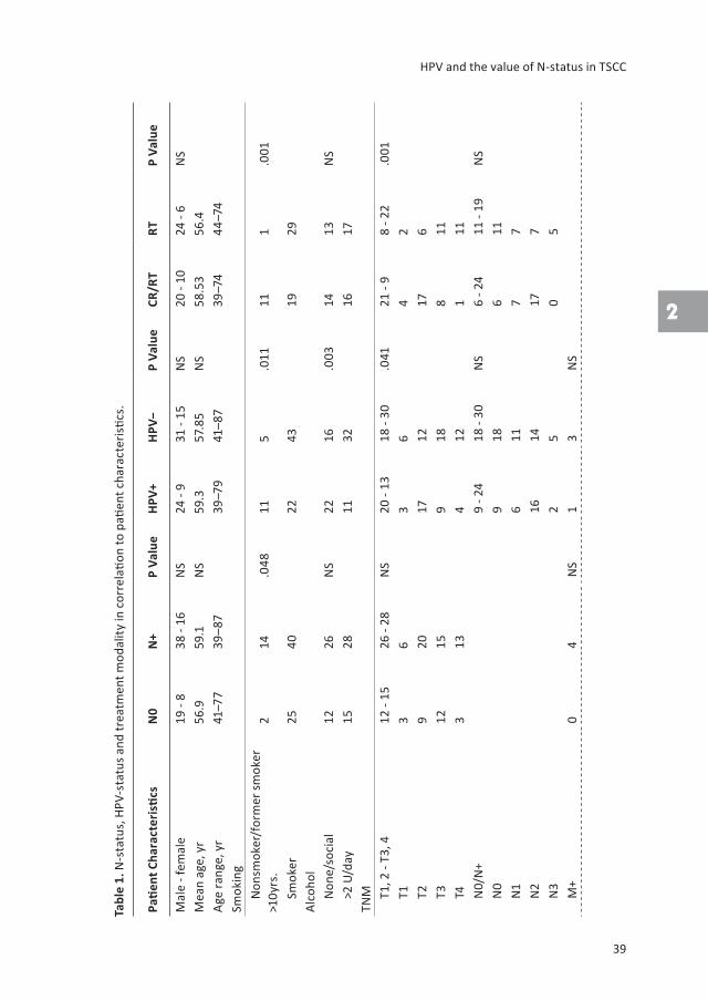
39
HPV and the value of N-status in TSCC
Tabl
e 1.
N-s
tatu
s, H
PV-s
tatu
s and
trea
tmen
t mod
ality
in c
orre
latio
n to
pati
ent c
hara
cter
istic
s.
Patie
nt C
hara
cter
istic
sN
0N
+P
Valu
eH
PV+
HPV
−P
Valu
eCR
/RT
RTP
Valu
e
Mal
e - f
emal
e19
- 8
38 -
16N
S24
- 9
31 -
15N
S20
- 10
24 -
6N
SM
ean
age,
yr
56.9
59.1
NS
59.3
57.8
5N
S58
.53
56.4
Ag
e ra
nge,
yr
41–7
739
–87
39
–79
41–8
7
39–7
444
–74
Sm
okin
g
N
onsm
oker
/for
mer
smok
er
>10y
rs.
214
.048
115
.011
111
.001
Sm
oker
2540
22
43
1929
Al
coho
l
Non
e/so
cial
1226
NS
2216
.003
1413
NS
>2
U/d
ay15
28
1132
16
17
TNM
T1
, 2 -
T3, 4
12 -
1526
- 28
NS
20 -
1318
- 30
.041
21 -
98
- 22
.001
T1
36
3
6
42
T29
20
1712
17
6
T3
1215
9
18
811
T43
13
412
1
11
N
0/N
+
9 - 2
418
- 30
NS
6 - 2
411
- 19
NS
N
0
918
6
11
N1
6
11
77
N2
16
14
177
N3
2
5
05
M+
04
NS
13
NS
2
Jos_Binnenwerk_v3.indd 39Jos_Binnenwerk_v3.indd 39 24/09/2020 17:04:1224/09/2020 17:04:12

40
Chapter 2
Tabl
e 1.
Con
tinue
d.
Patie
nt C
hara
cter
istic
sN
0N
+P
Valu
eH
PV+
HPV
−P
Valu
eCR
/RT
RTP
Valu
e
Tum
or d
iffer
entia
tion
grad
e
Poor
722
NS
1712
.015
1110
NS
M
oder
ate/
wel
l20
29
1534
18
20
HPV1
6 st
atus
Po
sitiv
e9
24N
S
168
.035
N
egati
ve18
30
14
22
Loca
l rec
urre
nce
82
,002
*2
8N
S0
60,
024*
Regi
onal
recu
rren
ce3
2N
S1
4N
S2
2N
SDi
stan
t met
asta
sis1
3N
S2
2N
S1
3N
STo
tal:
81 p
atien
ts27
54
3348
30
30
P-va
lues
wer
e ob
tain
ed u
sing
2-ta
iled
χ²-t
est
*ast
erisk
s ind
icat
e th
e us
e of
Fish
er e
xact
test
Abbr
evia
tions
. CR/
RT: p
atien
ts tr
eate
d w
ith tu
mor
surg
ery
and
neck
diss
ectio
n in
com
bina
tion
with
radi
othe
rapy
; RT:
pati
ents
trea
ted
with
radi
othe
rapy
; N
S: n
o si
gnifi
canc
e de
tect
ed (p
-val
ue >
0.0
5).
Jos_Binnenwerk_v3.indd 40Jos_Binnenwerk_v3.indd 40 24/09/2020 17:04:1224/09/2020 17:04:12

41
HPV and the value of N-status in TSCC
Table 2. HPV in correlation with treatment.
Treatment HPV+ HPV− TotalLocal resection (LR) 0 1 1Combined resection (CR) 3 3 6LR/RT 3 2 5CR/RT 16 14 30Radiotherapy (RT) 8 22 30Chemotherapy (ChT) 2 1 3CR/RT/ChT 1 0 1RT/ChT 0 3 3None 0 2 2Total 33 48 81
2
Jos_Binnenwerk_v3.indd 41Jos_Binnenwerk_v3.indd 41 24/09/2020 17:04:1224/09/2020 17:04:12

Jos_Binnenwerk_v3.indd 42Jos_Binnenwerk_v3.indd 42 24/09/2020 17:04:1224/09/2020 17:04:12

3P16INK4A immunostaining is a strong indicator for high-risk-HPV-associated oropharyngeal carcinomas and dysplasias, but is unreliable to predict low-risk-HPV-infection in head and neck papillomas and laryngeal dysplasias.
Published: Mooren JJ, Gültekin SE, Straetmans JM, Haesevoets A, Peutz-Kootstra CJ, Huebbers CU, Dienes HP, Wieland U, Ramaekers FC, Kremer B, Speel EJ, Klussmann JP. P16(INK4A) immunostaining is a strong indicator for high-risk-HPV-associated oropharyngeal carcinomas and dysplasias, but is unreliable to predict low-risk-HPV-infection in head and neck papillomas and laryngeal dysplasias. Int J Cancer 2014;134:2108-2117.
Jos_Binnenwerk_v3.indd 43Jos_Binnenwerk_v3.indd 43 24/09/2020 17:04:1224/09/2020 17:04:12

44
Chapter 3
3.1. ABSTRACT
Human papillomavirus (HPV) is a risk factor for the development of benign and malignant mucosal head and neck lesions. P16INK4A is often used as a surrogate marker for HPV-infection, although there is still controversy with respect its reliability. Our aim was to determine if p16INK4A overexpression can accurately predict both high-risk and low-risk-HPV-presence in (pre)malignant and benign head and neck lesions. P16INK4A immunohistochemistry was performed on paraffin-embedded tissue sections of 162 oropharyngeal squamous cell carcinomas (OPSCC), 14 tonsillar and 23 laryngeal dysplasias, and 20 tonsillar and 27 laryngeal papillomas. PCR, enzyme-immunoassay and FISH analysis were used to assess HPV-presence and type. Of the 162 OPSCC and 14 tonsillar dysplasias, 51 (31%) and 10 (71%) were HPV16-positive, respectively. All tonsillar papillomas were HPV-negative and four laryngeal dysplasias and 26 laryngeal papillomas were positive for HPV6 or −11. P16INK4A immunohistochemistry revealed a strong nuclear and cytoplasmic staining in 50 out of 51 HPV16-positive and 5 out of 111 HPV-negative OPSCC (p < 0.0001) and in all HPV16-positive tonsillar dysplasias, whereas highly variable staining patterns were detected in the papillomas and laryngeal dysplasias, irrespective of the HPV-status. In addition, the latter lesions generally showed a higher nuclear than cytoplasmic p16INK4A immunostaining intensity. In conclusion, our data show that strong nuclear and cytoplasmic p16INK4A overexpression is a reliable surrogate indicator for HPV16 in OPSCC and (adjacent) dysplasias. For HPV6 or −11-positive and HPV-negative benign and premalignant lesions of the tonsil and larynx, however, p16INK4A immunostaining is highly variable and cannot be recommended to predict HPV-presence.
Keywords: human papillomavirus, immunohistochemistry, FISH, PCR
Jos_Binnenwerk_v3.indd 44Jos_Binnenwerk_v3.indd 44 24/09/2020 17:04:1224/09/2020 17:04:12

45
P16INK4A and HPV in head and neck lesions
3.2. INTRODUCTION
Human papillomavirus (HPV)-infections may lead to the development of head and neck mucosal lesions. High-risk (HR)-HPV16 is involved in the carcinogenesis of oropharyngeal squamous cell carcinomas (OPSCC) and is an indicator of favorable prognosis, independent of the applied treatment modality. The reported incidence of HPV16 in OPSCC ranges from 25% to 93% in different studies, and appears to have increased during the last decade. Also the incidence of OPSCC and its proportion within the total head and neck squamous cell carcinoma (HNSCC) population are increasing.1, 2 Low-risk (LR)-HPV6 and −11 especially play a role in the development of benign laryngeal lesions. HPV-positivity may help in these lesions to predict a higher risk for recurrence and a lower risk for malignant progression in relation to HPV-negative, smoking-induced laryngeal lesions.3
In general, two methods are used for the detection of HPV-DNA in clinical diagnosis and biomedical research, i.e ., polymerase chain reaction (PCR)-based methods and fluorescence in situ hybridization (FISH). PCR is a highly sensitive method, but may not be able to distinguish biologically and clinically relevant HPV-containing lesions from cases with an extracellular virus contamination. Although FISH is a less sensitive method, it is highly specific and has the advantage to visualise the virus-DNA in situ and allows the distinction between an episomal and integrated status. Because both techniques are relatively costly, technically demanding and requiring sophisticated laboratory facilities as well as experienced personnel,4 practical alternatives or complementary procedures for HPV-testing have been explored, including the immunohistochemical detection of p16INK4A.5
HPV-positive carcinomas are characterized by overexpression of viral oncoproteins E6 and E7 leading to inactivation of p53 and the retinoblastoma protein (pRb), respectively, thereby inducing cell cycle deregulation and inhibition of apoptosis. As a result, cyclin-dependent kinase (CDK) inhibitors, including p16INK4A and p21Cip1/WAF1, and the MDM2 inhibitor p14ARF are generally upregulated, which subsequently leads to downregulation of cyclin D1 and inhibition of its complex formation with CDK4.6-9 Of these HPV-associated changes in protein expression, p16INK4A overexpression has been reported to be the most reliable surrogate marker for the presence of HR-HPV in OPSCC.6, 10-12 In contrast, HPV-negative carcinomas, induced by smoking and alcohol consumption, are generally characterized by inactivation of the p16INK4A gene through loss of 9p21. Therefore, the choice of p16INK4A immunostaining to distinguish HPV-positive and -negative malignancies is most obvious.6, 7, 13, 14
P16INK4A immunostaining has long been identified as an objective biomarker for HR-HPV-positive (pre)malignancies of the uterine cervix, allowing the unambiguous identification of truly dysplastic cells in biopsies, thereby reducing interobserver disagreement and improving diagnostic specificity.6, 15, 16 In contrast, normal cervical epithelium and inflammatory or metaplastic lesions are usually p16INK4A negative, while genital lesions containing LR-HPV6 or −11 often show only weak p16INK4A immunostaining.14,
3
Jos_Binnenwerk_v3.indd 45Jos_Binnenwerk_v3.indd 45 24/09/2020 17:04:1224/09/2020 17:04:12

46
Chapter 3
17-19 Moreover, several studies reported a strong correlation between p16INK4A immunostaining intensity and frequency of positive cells with increasing severity of the cervical lesion.18, 20-22 The College of American Pathologists and the American Society for Colposcopy and Cervical Pathology has recently published their recommendations on using p16INK4A immunostaining as a biomarker in lower anogenital squamous lesions, introducing the concept of “block-positivity,” defined as continuous strong nuclear or nuclear plus cytoplasmic p16INK4A staining of the basal cell layer with upward extension. The authors state that this scoring method allows a categorization of precancerous disease when the differential diagnosis is between a premalignancy [-intraepithelial neoplasia (IN) 2 or -IN 3] and a mimic of a premalignancy known to be not related to neoplastic risk. Furthermore, it can be used when -IN 2 is in the differential diagnosis to distinguish between a premalignant disease and a low-grade lesion/a non-HPV-associated pathology. As such, the method is used as an adjudication tool for cases in which there is a professional disagreement in histologic specimen interpretation.23
Despite the fact that p16INK4A immunohistochemistry is a low-cost test that can easily be implemented in daily diagnostic practice, its reliability as a surrogate marker for the presence of HR-HPV has been subject of debate.18, 24 HPV-negative but p16INK4A positive cases, although small in number, have been described in many series. It has been suggested that this p16INK4A positivity results from pRb inactivation by means of mechanisms independent of HPV-infection.4-9, 25, 26 Also the lack of standardization of p16INK4A immunohistochemistry and its evaluation criteria may be a source of misinterpretations. The foregoing arguments have been a reason for hesitation to introduce the p16INK4A test into the clinic.15
Although numerous studies analyzed the expression of p16INK4A in HR- and LR-HPV-associated cervical premalignancies, these data are scarce for premalignant and benign head and neck lesions, and therefore the aim of the present study was to determine the usefulness of this biomarker in neoplasms of the oropharynx and larynx. For this purpose we analysed a series of 162 OPSCC as well as a range of tonsillar and laryngeal dysplasias and papillomas, part of which was expected to harbor HR- and LR-HPV-types.
3.3. MATERIAL AND METHODS
3.3.1. Tumor material and patient dataBiopsy and resection material was obtained from 227 patients resulting in 246 samples, taken at different moments in time. Since our main goal was to evaluate the potential of p16INK4A immunostaining patterns as a surrogate marker for HPV in daily routine diagnostic practice, i.e ., in every single biopsy sample a pathologist receives from the clinic, we present the analyses in the total number of samples in the main text (Table 1 and Figs. 1a and 1b). For comparison we have included the analyses per patient in Supporting Information Table 1 and Supporting Information Figs. 1a and 1b.
Jos_Binnenwerk_v3.indd 46Jos_Binnenwerk_v3.indd 46 24/09/2020 17:04:1224/09/2020 17:04:12

47
P16INK4A and HPV in head and neck lesions
Formaldehyde-fixed and paraffin-embedded tissues were selected from the archives of the Departments of Pathology of the University of Cologne, Germany and the Maastricht University Medical Centre, The Netherlands. Patients were diagnosed between 1986 and 2007.Table 1 summarizes the clinicopathological features of the cases used in this study.Patient age at first diagnosis ranged from 39–87 years (mean 60) in the OPSCC group with no significant difference between the HPV-positive and the HPV-negative cases. In the papilloma group, the age ranged from 1 to 72 years (mean 43) and in the dysplasia group from 36 to 82 years (mean 62). The specimens included 162 OPSCC (from 162 patients) and 84 benign and premalignant samples (from 65 patients), i.e ., 14 tonsillar dysplasias from 12 patients, 13 of which were adjacent to an invasive carcinoma, 20 tonsillar papillomas (from 20 patients), 23 laryngeal dysplasias (from 19 patients) and 27 laryngeal papillomas (from 14 patients).A correlation of the HPV16 status with the disease-free survival (DFS) data with a maximum follow-up time up to 180 months and a mean of 36 months revealed that the after 5 years was 55% for patients with an HPV-negative carcinoma and 74% for patients with an HPV-positive carcinoma (Hazard ratio = 0.4; 95% Confidence Interval = 0.2–0.8), indicating that we used a representative OPSCC patient group.The OPSCC and dysplasias were histologically classified according to the criteria of the World Health Organization (C.J.P.-K. and H.P.D.).27 Eleven out of 14 tonsillar dysplasias were graded as severe and three as moderate. From the laryngeal dysplasias six were graded as mild, eight as moderate and nine cases as severe (see Table 1).
3.3.2. P16INK4A immunohistochemistryImmunostaining of p16INK4A was performed on 4-µm-thick formaldehyde fixed, paraffin-embedded tissue sections, which were deparrafinized using xylol. As a primary p16INK4a antibody clone E6H4 (Roche, Almere, The Netherlands) was used and detected using Powervision (DAKO A/S, Glostrup, Denmark) and peroxidase-DAB visualisation, as previously described.7
Three independent observers (J.J.M., S.E.G., E.-J.S.) performed evaluations of the immunostained samples and consensus was acquired about the scores.P16INK4A immunostaining patterns were first scored on a semiquantative scale, using the following evaluation criteria: Score 0 indicates that the majority of the cells are negative and that only up to 5% of the cells show nuclear immunoreactivity with or without cytoplasmic staining; Score 1 indicates that 5–10% of the cells show a nuclear reactivity with or without cytoplasmic positivity; Score 2 indicates a focal staining pattern with 10–25% of the cells showing nuclear reactivity with or without cytoplasmic positivity; Score 3 indicates that >25% cells show a strong nuclear staining reaction with or without cytoplasmic positivity.14
The proportion of positive cells is the primary parameter, with the intensity as a secondary scoring parameter. All cases scored as 2 and 3 showed a strong immunostaining intensity, while few cases scored as 1 stained less intensely positive.
3
Jos_Binnenwerk_v3.indd 47Jos_Binnenwerk_v3.indd 47 24/09/2020 17:04:1324/09/2020 17:04:13

48
Chapter 3
In addition, we scored the p16INK4A staining patterns according to the “block-type” immunopositivity approach, defined as p16INK4A only being block positive if continuous (>70%) strong nuclear with or without cytoplasmic staining is present (in all head and neck lesions) and staining is observed in the basal cell layer with extension upwards (in the benign and premalignant lesions).23 Controls included sections of uterine cervical IN grade 2 and 3 (positive controls) and sections from the same cases on which the p16INK4A antibody was substituted by a monoclonal mouse IgG2a antibody to Aspergillus Niger or by buffer without a primary antibody (negative controls).
3.3.3. DNA isolation and HPV typing by PCRFor DNA isolation, tissues were processed with the QIAamp Tissue Kit (Qiagen, Hilden, Germany) according to the manufacturers’ instructions. Total cellular DNA was eluted with 250 μl of the AE-buffer (Qiagen) and 5 μl were used in each of the PCR analyses.To test the quantity and quality of the DNA samples and to demonstrate that the samples were free from inhibitory substances, PCR was performed for the ß-Globin gene, resulting in a 268 bp PCO4/GH20 PCR product.28 As negative controls water and as positive controls human placental DNA were included in each PCR run.HPV sequences were detected by highly sensitive group-specific nested PCR protocols with degenerate primers A5/A10 and A6/A8 for HPV as previously described.29 PCR products (5 µl) were separated on 2% agarose gels and visualized by ethidium bromide staining. For HPV typing internal biotinylated A6/A8-PCR products (270 bp) were hybridized with 37 type-specific digoxigenin-labeled oligonucleotide probes in an enzyme-immunoassay as described earlier.30, 31
3.3.4. Detection of HPV6-, 11-, 16- and 18 by FISHFISH was performed on 4-µm thick formaldehyde fixed and paraffin-embedded tissue sections tissue as described previously.32-34 Briefly, sections were deparaffinized, pre-treated with 85% formic acid/0.3% H2O2, 1 M NaSCN and 4 mg/ml pepsin, post-fixed in 1% formaldehyde in PBS, dehydrated in an ethanol series and hybridized with digoxigenin-labeled HPV6-, 11-, 16- and 18-specific probes (PanPath, Budel, The Netherlands), respectively, depending on PCR results, according to the manufacturer’s instructions. After hybridization the preparations were washed stringently in 50% formamide, 2× SSC at 42°C (two times 5 min). The probes were detected by subsequent application of mouse anti-digoxigenin (Sigma, St. Louis, MO), peroxidase-conjugated rabbit anti-mouse and peroxidase-conjugated swine anti-rabbit (both DAKO), and visualized by a peroxidase based amplification reaction using rhodamine-labeled tyramide.35, 36 Preparations were mounted in Vectashield (Vector Laboratories, Burlingame, CA) containing 4,6-diamidino-2-phenyl indole (DAPI; Sigma: 0.2 g/ml). Microscope images were recorded with the Metasystems Image Pro System (black and white CCD camera; Sandhausen, Germany) mounted on top of a Leica DM-RE fluorescence microscope equipped with DAPI and rhodamine filters.
Jos_Binnenwerk_v3.indd 48Jos_Binnenwerk_v3.indd 48 24/09/2020 17:04:1324/09/2020 17:04:13

49
P16INK4A and HPV in head and neck lesions
Evaluation of nuclear hybridization signals was performed by two investigators (J.J.M., E.-J.M.S.) according to the criteria described by Mooren et al .,37 i.e ., nuclear punctate signals were considered to indicate integrated HPV-DNA and diffuse signals to indicate episomal HPV-DNA. Controls included hybridizations on HPV6-, −11-, −16- and 18-positive formaldehyde-fixed, paraffin-embedded tissues of known human uterine cervical and head and neck (pre)malignancies. Negative controls consisted of HPV-negative tissue sections as determined by PCR and hybridizations omitting the viral probe.
3.3.5. Statistical analysisHPV-status and p16INK4A immunostaining were correlated using cross-tabulations and the two-tailed Fisher exact test and/or Chi-square test. We regarded a p -value ≤ 0.05 as level of significance. DFS in the OPSCC group was calculated from the date of diagnosis until the date of recurrence (local, regional or distant, whichever occurred first). Patients without recurrence were censored at the date of the last follow-up or the date of death. The statistical significance of differences between survival times was determined by the log rank test in univariate analysis. All statistical analyses were performed by PASW Statistics version 18.0.
3.4. RESULTS
3.4.1. HPV-status of the head and neck lesions examined in this studyThe HPV-status of the OPSCC, the premalignant and the benign head and neck lesions is presented in Table 1. Only when the presence of HPV was detected by both PCR and FISH the sample was judged to be HPV-positive.Of the 162 OPSCC 51 (31%) were found to be positive for HPV16. All 51 cases showed integrated HPV16, as concluded from the FISH-analyses. No other HPV-subtypes were detected in these lesions.Forty of the 84 benign and premalignant lesions (48%) were HPV-positive: 10 (71%) of the 14 tonsillar dysplasias harbored HPV16; 4 (16%) of the 23 laryngeal dysplasias harbored HPV6, of which two also contained HPV11; of the 27 laryngeal papillomas 17 (63%) were positive for HPV6, and nine (33%) for HPV11. The frequency of HPV-infections in these lesions is in accordance with previous studies.38-41 Remarkably, all 20 samples of tonsillar papillomas were HPV-negative.In all HPV16-positive tonsillar dysplasias the virus was integrated (Fig. 2b ), whereas in one sample additionally an episomal FISH pattern was detected in the cell nuclei surrounding tonsillar crypts (data not shown). In all, except one sample, of the HPV6- or 11-positive laryngeal lesions the virus was episomal (Fig. 2h ).In the Supporting Information Table 1, the HPV-positivity per patient is presented, for comparison, showing only a slightly difference in the frequency of HPV-positivity in the
3
Jos_Binnenwerk_v3.indd 49Jos_Binnenwerk_v3.indd 49 24/09/2020 17:04:1324/09/2020 17:04:13

50
Chapter 3
tonsillar dysplasias (83% per patient vs . 71% per sample) and the laryngeal papillomas (93% vs . 96%).
3.4.2. Strong correlation between p16INK4A overexpression and HR-HPV16 in OPSCC and tonsillar dysplasiasThe p16INK4A scores in the HPV-positive and -negative lesions, using the two different scoring methods, are shown per subgroup in Figs. 1a and 1b . The analyses per patient are presented in Supporting Information Figures 1a and 1b.The presence of HR-HPV16 in the OPSCC was significantly associated (p < 0.0001) with a strong p16INK4A immunostaining reaction (staining Score 3) and with a block positive p16INK4A immunostaining: 50 of 51 HPV16-positive OPSCC showed strong nuclear and cytoplasmic p16INK4A immunopositivity in more than 70% of the tumor cells, whereas only five of the 111 HPV16-negative tumors were p16INK4A positive.Also all 10 HPV16-positive tonsillar dysplasia samples showed strong (Score 3) p16INK4A expression (Fig. 2a). In eight of these an equally strong staining intensity in the nuclei and cytoplasm could be observed in more than 70% of the dysplastic cells and these were scored as block positive. The remaining two HPV16-positive samples showed a strong (Score 3) p16INK4A immunostaining reaction in 40–50% of the lesion, being block negative.Of the 4 HPV-negative tonsillar dysplasia samples, two showed an intermediate p16INK4A staining pattern (Score 2) and two samples from one patient were negative (Score 0), implying that these were all block negative.
3.4.3. Highly variable p16INK4A immunostaining patterns in the tonsillar papillomas and laryngeal lesionsIn the 20 HPV-negative tonsillar papillomas, the p16INK4A immunostaining patterns were highly variable and ranged from a negative score to a strong staining intensity in 30–80% of the cells (Figs. 2d –2f and Fig. 1a). Only one sample was p16INK4A block positive (Fig. 1b). In 12 (86%) out of the 14 p16INK4A positive tonsillar papillomas, the immunostaining intensity of the nucleus was stronger than that of the cytoplasm.Of the 23 laryngeal dysplasias, four samples (two from one patient) showed a strong, four a low to intermediate and 15 (two from one patient) no p16INK4A expression (Fig. 1a ). Three of the four HPV6-positive lesions, of which two samples from the same patient harbored HPV11-DNA, were p16INK4A negative. The remaining HPV6-positive dysplasia sample showed a strong p16INK4A immunostaining (Score 3) and was also block positive (Figs. 2c and 2g and Figs. 1a and 1b ). There was no association between p16INK4A score and the degree of dysplasia. Although all five mild dysplasias were also p16INK4A negative, the moderate and severe dysplasias showed negative to strong p16INK4A staining patterns.P16INK4A staining patterns in the HPV6 and-11-positive laryngeal papillomas varied from negative to strongly positive. Five out of the 26 HPV-positive laryngeal papillomas were p16INK4A block positive, three of these being from the same patient. Similar to most
Jos_Binnenwerk_v3.indd 50Jos_Binnenwerk_v3.indd 50 24/09/2020 17:04:1324/09/2020 17:04:13

51
P16INK4A and HPV in head and neck lesions
tonsillar papillomas, all of the p16INK4A positive laryngeal lesions showed a stronger nuclear than cytoplasmic staining intensity.In the Supporting Information Figures 1a and 1b , the p16INK4A-positivity per patient is presented, showing only minor differences in the frequencies of p16INK4A-immunostaining scores, compared to those presented in Figures 1a and 1b (analyses per sample).
3.5. DISCUSSION
Overexpression of p16INK4A has been put forward as a specific surrogate biomarker for the presence of HR-HPV, in particular in (pre)malignancies of the uterine cervix, as well as those in the head and neck region.6, 7, 13, 14 However, there still exists controversy on the reliability of p16INK4A expression as indicator for the presence of HPV, particularly in the latter group. In our study, we analysed the relationship between HR- and LR-HPV-status and p16INK4A immunostaining patterns in 246 samples of benign, premalignant and malignant head and neck lesions. A strong (Score 3) and block positive nuclear and cytoplasmic p16INK4A staining pattern was predominantly found to correlate with HPV16-containing OPSCC and tonsillar dysplasias. In contrast, the tonsillar and laryngeal papillomas, as well as the laryngeal dysplasias showed a highly variable p16INK4A staining pattern, independent of the HPV-status. By using the block-type scoring approach, however, most of these lesions were interpreted as being p16INK4A block negative.It is now well established that HPV-associated HNSCC represent a separate entity, which is clinically and molecularly distinct from its tobacco- and/or alcohol-induced counterpart.2, 7, 37 The upregulation of p16INK4A results from inactivation of pRb by the HPV-E7 oncoprotein, or alternatively through E7-mediated epigenetic induction of KDM6B, subsequently leading to activation of the p16INK4A gene.6, 16
Although several studies have reported a high interobserver conformity in the evaluation of p16INK4A immunostaining patterns,4, 14, 18, 19, 42 others have indicated limitations in this procedure. The immunohistochemical p16INK4A staining procedure is subject to variations in the protocol and to difficulties in the interpretation of the staining patterns as a result of different scoring criteria.17 There is an emerging consensus that only samples showing strong nuclear and cytoplasmic p16INK4A immunostaining, observed in more than 25% of tumor cells, should be considered HPV-positive.5, 13, 14, 18, 20-22 Recent studies even suggest to use 70% immunopositivity of the carcinoma tissue as a cut-off point.4, 11, 12 In our study, we indeed observed the strong and block positive p16INK4A immunostaining in all except one HPV16-positive OPSCC. Despite the fact that a small fraction of the HPV-negative OPSCC did show p16INK4A positivity, the correlation of this surrogate marker with HPV16-presence is highly significant.Also in all HPV16-positive tonsillar dysplasias of the present study a strong, and in most cases block positive, p16INK4A immunostaining pattern could be detected, which is in accordance with the results in cervical dysplasias.14, 19 Similar to OPSCC, also in these
3
Jos_Binnenwerk_v3.indd 51Jos_Binnenwerk_v3.indd 51 24/09/2020 17:04:1324/09/2020 17:04:13

52
Chapter 3
lesions we identified p16INK4A positive staining in two HPV-negative samples, albeit with an intermediate score and being block negative.The use of p16INK4A immunostaining as surrogate marker for HPV-presence in laryngeal dysplasias and head and neck papillomas, however, is unreliable. In the HPV6/11-positive laryngeal dysplasia samples the majority of cases was p16INK4A negative, while 1/3 of the HPV-negative samples did show p16INK4A positivity. LR-HPV-integration seldomly occurs and the only laryngeal papilloma sample harboring nuclear punctate HPV6-FISH signals only showed weak p16INK4A immunostaining. In a recent study on juvenile onset laryngeal papillomatosis progressing to carcinoma in one patient, the papilloma with episomal and the carcinoma with integrated HPV6 were all p16INK4A negative.43
All our tonsillar papilloma samples were HPV-negative, whereas 70% of the samples showed variable p16INK4A positivity, indicating that p16INK4A immunostaining appears of no use for predicting HPV-presence in these lesions. This is in accordance with a previous study on tumor-free palatine tonsils, all proven to be negative for HPV, but showing p16INK4A overexpression in a quarter of these normal cases.25 By using the block-type scoring system, however, most tonsillar and laryngeal papillomas and laryngeal dysplasias were interpreted as being p16INK4A block negative. This fits with the fact that these head and neck lesions are usually HPV-negative or contain LR-HPV types. Block-positivity is used to identify HR-HPV-containing precancerous anogenital lesions, which is in accordance with the block-positivity observed in our study in OPSCC and tonsillar dysplasias, being associated with HR-HPV.23
Also at a subcellular level variability in p16INK4A immunostaining patterns could be observed: in the HPV16-positive OPSCC and tonsillar dysplasias we predominantly found an equally strong p16INK4A staining intensity in nuclei and cytoplasm, whereas in the other head and neck lesions the nuclear intensity was generally stronger than that of the cytoplasm.The fact that studies on oral papillomas showed an HPV-positivity in 13–60%, predominantly HPV6 or −11,44-46 makes the absence of HPV in the tonsillar papillomas in our series remarkable. The lack of HPV in these samples might be explained by elimination of the virus by the immune system with persistence of the lesions, or by the fact that other viruses, yet to be discovered, play an etiological role in the development of the tonsillar papillomas. Also the mechanisms underlying the predilection of different HPV-types for different anatomical head and neck lesions remain to be further explored.The highly variable p16INK4A immunostaining patterns in the benign and premalignant head and neck lesions may be explained by several other factors. It is well described in the literature that in uterine cervical lesions LR-HPV6 results in a less intense p16INK4A immunostaining pattern as compared to HPV16, which is caused by the fact that the affinity of the HPV6 E7 protein for cellular pRb is 10-fold lower than that of HPV16 E7.19, 47,
48 Therefore, it is surprising that our HPV6/11-containing benign and premalignant head and neck samples were often strongly positive, or completely negative for p16INK4A.
Jos_Binnenwerk_v3.indd 52Jos_Binnenwerk_v3.indd 52 24/09/2020 17:04:1324/09/2020 17:04:13

53
P16INK4A and HPV in head and neck lesions
Since a subgroup of HPV-negative lesions also shows overexpression of p16INK4A, several factors have been proposed to explain this observation, which remain to be studied. These include infection with other viruses (i.e ., cytomegalovirus and adenovirus), which functionally inactivate pRb in a similar fashion as the HPV-oncogene E7,49, 50 physiological stress, oncogene-driven senescence by functional overactivation of (proto)oncogenes including Ras, Raf, MEK and E2F or replicative senescence due to DNA-damage or oxidative stress.51-54
In summary, our results indicate that a strong nuclear and cytoplasmic p16INK4A immunostaining pattern can accurately predict the presence of HR-HPV16 in OPSCC and tonsillar dysplasias. Our data underscore the proposed cut-off level of 70% p16INK4A positive cells, corresponding to block positive p16INK4A immunopositivity, in these lesions as indicator for HR-HPV16-presence. In the other premalignant and benign head and neck lesions, however, caution is recommended when using this surrogate marker for HPV-infection.
3
Jos_Binnenwerk_v3.indd 53Jos_Binnenwerk_v3.indd 53 24/09/2020 17:04:1324/09/2020 17:04:13

54
Chapter 3
REFERENCES
1. Shaw R, Robinson M. The increasing clinical relevance of human papillomavirus type 16 (HPV-16) infection in oropharyngeal cancer. Br J Oral Maxillofac Surg 2011; 49: 423– 9.
2. Olthof NC, Straetmans JMJAA, Snoeck R, et al. Next-generation treatment strategies for human papillomavirus-related head and neck squamous cell carcinoma: where do we go? Rev Med Virol 2012; 22: 88– 105.
3. Goon P, Sonnex C, Jani P, et al. Recurrent respiratory papillomatosis: an overview of current thinking and treatment. Eur Arch Oto-Rhino-Laryngol 2007; 265: 147– 51.
4. Thavaraj S, Stokes A, Guerra E, et al. Evaluation of human papillomavirus testing for squamous cell carcinoma of the tonsil in clinical practice. J Clin Pathol 2011; 64: 308– 12.
5. El-Naggar AK, Westra WH. p16 expression as a surrogate marker for HPV-related oropharyngeal carcinoma: a guide for interpretative relevance and consistency. Head Neck 2011; 34: 459– 61.
6. Klussmann JP, Gültekin E, Weissenborn SJ, et al. Expression of p16 protein identifies a distinct entity of tonsillar carcinomas associated with human papillomavirus. Am J Pathol 2003; 162: 747– 53.
7. Hafkamp HC, Mooren JJ, Claessen SM, et al. P21 Cip1/WAF1 expression is strongly associated with HPV-positive tonsillar carcinoma and a favorable prognosis. Mod Pathol 2009; 22: 686– 98.
8. Li W, Thompson CH, Cossart YE, et al. The expression of key cell cycle markers and presence of human papillomavirus in squamous cell carcinoma of the tonsil. Head Neck 2004; 26: 1– 9.
9. Wiest T, Schwarz E, Enders C, et al. Involvement of intact HPV16 E6/E7 gene expression in head and neck cancers with unaltered p53 status and perturbed pRb cell cycle control. Oncogene 2002; 21: 1510– 7.
10. Hafkamp HC, Manni JJ, Haesevoets A, et al. Marked differences in survival rate between smokers and nonsmokers with HPV 16-associated tonsillar carcinomas. Int J Cancer 2008; 122: 2656– 64.
11. Rietbergen MM, Leemans CR, Bloemena E, et al. Increasing prevalence rates of HPV attributable oropharyngeal squamous cell carcinomas in the Netherlands as assessed by a validated test algorithm. Int J Cancer 2013; 132: 1565– 71.
12. Jordan RC, Lingen MW, Perez-Ordonez B, et al. Validation of methods for oropharyngeal cancer HPV status determination in US cooperative group trials. Am J Surg Pathol 2012; 36: 945– 54.
13. Fregonesi PAG, Teresa DB, Duarte RA, et al. p16(INK4A) immunohistochemical overexpression in premalignant and malignant oral lesions infected with human papillomavirus. J Histochem Cytochem 2003; 51: 1291– 7.
14. Klaes R, Friedrich T, Spitkovsky D, et al. Overexpression of p16(INK4A) as a specific marker for dysplastic and neoplastic epithelial cells of the cervix uteri. Int J Cancer 2001; 92: 276– 84.
15. Klaes R, Benner A, Friedrich T, et al. p16INK4a immunohistochemistry improves interobserver agreement in the diagnosis of cervical intraepithelial neoplasia. Am J Surg Pathol 2002; 26: 1389– 99.
16. McLaughlin-Drubin ME, Crum CP, et al. Human papillomavirus E7 oncoprotein induces KDM6A and KDM6B histone demethylase expression and causes epigenetic reprogramming. Proc Natl Acad Sci USA 2011; 108: 2130– 5.
Jos_Binnenwerk_v3.indd 54Jos_Binnenwerk_v3.indd 54 24/09/2020 17:04:1324/09/2020 17:04:13

55
P16INK4A and HPV in head and neck lesions
17. van Bogaert L-J. P16(INK4a) immunocytochemistry/immunohistochemistry: need for scoring uniformization to be clinically useful in gynecological pathology. Ann Diagn Pathol 2012; 16: 422– 6.
18. Yildiz IZ, Usubutun A, Firat P, et al. Efficiency of immunohistochemical p16 expression and HPV typing in cervical squamous intraepithelial lesion grading and review of the p16 literature. Pathol Res Pract 2007; 203: 445– 9.
19. Sano T, Oyama T, Kashiwabara K, et al. Expression status of p16 protein is associated with human papillomavirus oncogenic potential in cervical and genital lesions. Am J Pathol 1998; 153: 1741– 8.
20. Lambert APF, Anschau F, Schmitt VM. p16INK4A expression in cervical premalignant and malignant lesions. Exp Mol Pathol 2006; 80: 192– 6.
21. Murphy N, Ring M, Killalea AG, et al. p16INK4A as a marker for cervical dyskaryosis: CIN and cGIN in cervical biopsies and ThinPrep smears. J Clin Pathol 2003; 56: 56– 63.
22. Missaoui N, Trabelsi A, Hmissa S, et al. p16INK4A overexpression in precancerous and cancerous lesions of the uterine cervix in Tunisian women. Pathol Res Practice 2010; 206: 550– 5.
23. Darragh TM, Colgan TJ, Thomas Cox J, et al. The Lower Anogenital Squamous Terminology Standardization project for HPV-associated lesions: background and consensus recommendations from the College of American Pathologists and the American Society for Colposcopy and Cervical Pathology. Int J Gynecol Pathol 2013; 32: 76– 115.
24. Holzinger D, Schmitt M, Dyckhoff G, et al. Viral RNA patterns and high viral load reliably define oropharynx carcinomas with active HPV16 involvement. Cancer Res 2012; 72: 4993– 5003.
25. Klingenberg B, Hafkamp HC, Haesevoets A, et al. p16 INK4A overexpression is frequently detected in tumour-free tonsil tissue without association with HPV. Histopathology 2010; 56: 957– 67.
26. Harris SL, Thorne LB, Seaman WT, et al. Association of p16(INK4a) overexpression with improved outcomes in young patients with squamous cell cancers of the oral tongue. Head Neck 2011; 33: 1622– 7.
27. Shanmugaratnam S. Histologic typing of tumors of the upper respiratory tract and ear. Geneva (Switzerland): World Health Organization, 1991.
28. Bauer HM, Ting Y, Greer CE, et al. Genital human papillomavirus infection in female university students as determined by a PCR-based method. JAMA 1991; 265: 472– 7.
29. Wieland U, Jurk S, Weissenborn S, et al. Erythroplasia of queyrat: coinfection with cutaneous carcinogenic human papillomavirus type 8 and genital papillomaviruses in a carcinoma in situ. J Invest Dermatol 2000; 115: 396– 401.
30. Jacobs MV, Snijders PJ, van den Brule AJ, et al. A general primer GP5+/GP6(+)-mediated PCR-enzyme immunoassay method for rapid detection of 14 high-risk and 6 low-risk human papillomavirus genotypes in cervical scrapings. J Clin Microbiol 1997; 35: 791– 5.
31. van den Brule AJ, Pol R, Fransen-Daalmeijer N, et al. GP5+/6+ PCR followed by reverse line blot analysis enables rapid and high-throughput identification of human papillomavirus genotypes. J Clin Microbiol 2002; 40: 779– 87.
32. Hafkamp HC, Speel EJM, Haesevoets A, et al. A subset of head and neck squamous cell carcinomas exhibits integration of HPV 16/18 DNA and overexpression of p16INK4A and p53 in the absence of mutations in p53 exons 5–8. Int J Cancer 2003; 107: 394– 400.
33. Hopman AH, Kamps MA, Speel EJ, et al. Identification of chromosome 9 alterations and p53 accumulation in isolated carcinoma in situ of the urinary bladder versus carcinoma in situ associated with carcinoma. Am J Pathol 2002; 161: 1119– 25.
3
Jos_Binnenwerk_v3.indd 55Jos_Binnenwerk_v3.indd 55 24/09/2020 17:04:1324/09/2020 17:04:13

56
Chapter 3
34. Hopman AHN, Smedts F, Dignef W, et al. Transition of high-grade cervical intraepithelial neoplasia to micro-invasive carcinoma is characterized by integration of HPV 16/18 and numerical chromosome abnormalities. J Pathol 2004; 202: 23– 33.
35. Speel EJ, Ramaekers FC, Hopman AH. Sensitive multicolor fluorescence in situ hybridization using catalyzed reporter deposition (CARD) amplification. J Histochem Cytochem 1997; 45: 1439– 46.
36. Hopman AH, Ramaekers FC, Speel EJ. Rapid synthesis of biotin-, digoxigenin-, trinitrophenyl-, and fluorochrome-labeled tyramides and their application for In situ hybridization using CARD amplification. J Histochem Cytochem 1998; 46: 771– 7.
37. Mooren JJ, Kremer B, Claessen SMH, et al. Chromosome stability in tonsillar squamous cell carcinoma is associated with HPV16 integration and indicates a favorable prognosis. Int J Cancer 2013; 132: 1781– 9.
38. Lindeberg H, Krogdahl A. Laryngeal dysplasia and the human papillomavirus. Clin Otolaryngol Allied Sci 1997; 22: 382– 6.
39. Brito H, Vassallo J, Altemani A. Detection of human papillomavirus in laryngeal squamous dysplasia and carcinoma. An in situ hybridization and signal amplification study. Acta Otolaryngol 2000; 120: 540– 4.
40. Gallo A, Degener AM, Pagliuca G, et al. Detection of human papillomavirus and adenovirus in benign and malignant lesions of the larynx. YMHN 2009; 141: 276– 81.
41. Laco J, Slaninka I, Jirásek M, et al. High-risk human papillomavirus infection and p16INK4a protein expression in laryngeal lesions. Pathol Res Practice 2008; 204: 545– 52.
42. Schlecht NF, Brandwein-Gensler M, Nuovo GJ, et al. A comparison of clinically utilized human papillomavirus detection methods in head and neck cancer. Mod Pathol 2011; 24: 1295– 305.
43. Huebbers CU, Preuss SF, Kolligs J, et al. Integration of HPV6 and downregulation of AKR1C3 Expression mark malignant transformation in a patient with Juvenile-Onset laryngeal papillomatosis. PLoS ONE 2013; 8: e57207.
44. Young SK, Min KW. In situ DNA hybridization analysis of oral papillomas, leukoplakias, and carcinomas for human papillomavirus. Oral Surg Oral Med Oral Pathol 1991; 71: 726– 9.
45. Zeuss MS, Miller CS, White DK. In situ hybridization analysis of human papillomavirus DNA in oral mucosal lesions. Oral Surg Oral Med Oral Pathol 1991; 71: 714– 20.
46. Syrjanen SM, Syrjanen KJ, Lamberg MA. Detection of human papillomavirus DNA in oral mucosal lesions using in situ DNA-hybridization applied on paraffin sections. Oral Surg Oral Med Oral Pathol 1986; 62: 660– 7.
47. Gage JR, Meyers C, Wettstein FO. The E7 proteins of the nononcogenic human papillomavirus type 6b (HPV-6b) and of the oncogenic HPV-16 differ in retinoblastoma protein binding and other properties. J Virol 1990; 64: 723– 30.
48. Xiong Y, Kuppuswamy D, Li Y, et al. Alteration of cell cycle kinase complexes in human papillomavirus E6- and E7-expressing fibroblasts precedes neoplastic transformation. J Virol 1996; 70: 999– 1008.
49. Helt AM, Galloway DA. Mechanisms by which DNA tumor virus oncoproteins target the Rb family of pocket proteins. Carcinogenesis 2003; 24: 159– 69.
50. Castillo JP, Kowalik TF. Human cytomegalovirus immediate early proteins and cell growth control. Gene 2002; 290: 19– 34.
51. Cánepa ET, Scassa ME, Ceruti JM, et al. INK4 proteins, a family of mammalian CDK inhibitors with novel biological functions. IUBMB Life 2007; 59: 419– 26.
Jos_Binnenwerk_v3.indd 56Jos_Binnenwerk_v3.indd 56 24/09/2020 17:04:1324/09/2020 17:04:13

57
P16INK4A and HPV in head and neck lesions
52. Krishnamurthy J, Torrice C, Ramsey MR, et al. Ink4a/Arf expression is a biomarker of aging. J Clin Invest 2004; 114: 1299– 307.
53. Ben-Porath I, Weinberg RA. The signals and pathways activating cellular senescence. Int J Biochem Cell Biol 2005; 37: 961– 76.
54. Kim Sh S-H, Kaminker P, Campisi J. Telomeres, aging and cancer: in search of a happy ending. Oncogene 2002; 21: 503– 11.
3
Jos_Binnenwerk_v3.indd 57Jos_Binnenwerk_v3.indd 57 24/09/2020 17:04:1324/09/2020 17:04:13

58
Chapter 3
Table 1. HPV-status and clinicopathological features of all head and neck lesion studied
OPSCC Tonsillar dysplasia
Tonsillar papilloma
Laryngeal dysplasia
Laryngeal papilloma
(n = 162) (n = 14) (n = 20) (n = 23) (n = 27)Number of different patients
162 12 20 19 14
GenderMale 117 (72%) 9 (75%) 12 (60%) 18 (95%) 12 (86%)Female 45 (28%) 3 (25%) 8 (40%) 1 (5%) 2 (14%)Mean age at first diagnosis (range)
60 (39–87) 60 (36–79) 42 (5–66) 63 (43–82) 44 (1–72)
TNM statusT1 23 (14%)T2 54 (33%)T3 46 (29%)T4 39 (24%)N0 51 (31%)N1 29 (18%)N2 64 (39%)N3 15 (9%)Missing 3 (3%)M+ 4 (3%)Histology grade (all samples)Mild 0 (0%) 6 (26%)Moderate 3 (21%) 8 (35%)Severe 11 (79%) 9 (39%)Number of HPV-positive samples
51 (31%) 10 (71%) 0 (0%) 4 (17%) 26 (96%)
HPV–PCR (all samples)HPV 6 4a 17HPV 11 2 9HPV 16 51 10HPV–FISH (all samples)Integrated 51 9 1bEpisomal 1 4 26
a Two out of these four HPV6-positive samples were also positive for HPV11.b This sample showed both episomal and integrated HPV6.
Jos_Binnenwerk_v3.indd 58Jos_Binnenwerk_v3.indd 58 24/09/2020 17:04:1324/09/2020 17:04:13

59
P16INK4A and HPV in head and neck lesions
Figure 1
(a) P16INK4A immunostaining scores in all (pre)malignant and benign head and neck lesions. Scoring criteria: Score 0 negative; Score 1: 5–10% of the cells show a nuclear reactivity with or without cytoplasmic positivity; Score 2: focal staining pattern with 10–25% of the cells showing nuclear reactivity with or without cytoplasmic positivity; Score 3: >25% cells show a strong nuclear staining reaction with or without cytoplasmic positivity.14 (b) Immunostaining results for p16INK4A in all (pre)malignant and benign head and neck lesions based on the block-type scoring approach, defined as p16INK4A only being interpreted as positive if continuous (>70%) strong nuclear with or without cytoplasmic staining is present (in all head and neck lesions) and is observed in the basal cell layer with extension upwards (in the benign and premalignant lesions).23
3
Jos_Binnenwerk_v3.indd 59Jos_Binnenwerk_v3.indd 59 24/09/2020 17:04:1324/09/2020 17:04:13

60
Chapter 3
Figure 2
Representative examples of p16INK4A immunohistochemistry (a , c–g) and HPV-specific FISH analysis (b , h) in head and neck lesions. (a ) Strong nuclear and cytoplasmic p16INK4A immunostaing (Score 3) and also block-positive p16INK4A staining pattern in a tonsillar dysplasia; (b) Punctate signal (red) per nucleus (blue DAPI) indicating integrated HPV16 in a tonsillar dysplasia; (c) Strong nuclear and cytoplasmic p16INK4A immunostaining (Score 3) and also block-positive p16INK4A staining pattern in a laryngeal dysplasia sample; (d) Tonsillar papilloma sample showing strong nuclear and weaker cytoplasmic p16INK4A immunostaining (Score 3), but scored as being block-negative. (e , f) Tonsillar papilloma samples showing a focal staining pattern with 10–25% of the cells showing nuclear and cytoplasmic positivity (Score 2; e), and showing 5–10% of the cells with a nuclear and cytoplasmic positivity (Score 1; f). (g) Laryngeal dysplasia sample showing 0–5% of the cells showing nuclear and cytoplasmic p16INK4A immunopositivity (Score 0); (h) Diffusely (red) stained nuclei (blue DAPI) indicating episomal presence of HPV6 in a laryngeal papilloma.
Jos_Binnenwerk_v3.indd 60Jos_Binnenwerk_v3.indd 60 24/09/2020 17:04:1324/09/2020 17:04:13

61
P16INK4A and HPV in head and neck lesions
3
Jos_Binnenwerk_v3.indd 61Jos_Binnenwerk_v3.indd 61 24/09/2020 17:04:1324/09/2020 17:04:13

Jos_Binnenwerk_v3.indd 62Jos_Binnenwerk_v3.indd 62 24/09/2020 17:04:1324/09/2020 17:04:13

4Value of human papillomavirus testing in the diagnostic workup of lymph node metastases from an unknown primary tumor to the neck.
Published: Straetmans JM, Speel EJ, Kremer B. Value of human papillomavirus testing in the diagnostic workup of lymph node metastases from an unknown primary tumor to the neck. Head Neck 2012;34:1819-1821.
Jos_Binnenwerk_v3.indd 63Jos_Binnenwerk_v3.indd 63 24/09/2020 17:04:1324/09/2020 17:04:13

64
Chapter 4
In their article, Strojan et al1 promote human papillomavirus (HPV) testing in patients with metastases of unknown primary tumors to the neck (CUP). This advice is merely based on retrospective studies on patients with CUP in which the HPV status of the metastasis was compared with that of the primary tumor identified during diagnostic workup, suggesting that HPV positivity is indicating an origin in the oropharynx.2–6 If the authors are right, HPV testing could improve the sensitivity of current extensive diagnostic workup protocols applied to patients with true CUP by identifying “missed” (or formerly undetected) oropharyngeal primary tumors.However, studies on the prevalence of HPV in lymph node metastases of which the primary tumor could not be detected after diagnostic workup so-called “true” CUPs are scarce and contradictory. HPV prevalence rates range from 0% to 100% and were tested in very small sample numbers: Weiss et al7 0/1 (0%), Armas et al8 0/4 (0% in N3 necks; 4/4 Epstein Barr virus–positive), Begum et al5 3/10 (30%), Barwad et al9 9/17 (53%), Hofmann et al10 2/3 (66%), Desai et al11 4/6 (66%), and Goldenberg et al3 2/2 (100%). Compton et al12 investigated HPV16 presence by DNA in situ hybridization in a cohort of resected, nonirradiated true CUPs: of 11/25 p16-positive, there were 7/25 HPV-positive (28%) tumors. However, a relationship with survival was not found. So far, no studies have been reported in which additional HPV testing has led to the discovery of the primary tumor in case the regular diagnostic workup revealed a true CUP.In our institute, 29 true CUPs (all squamous cell carcinomas) were analyzed with no prior history of head and neck carcinoma. Diagnostic workup consisted of MRI or CT of the head and neck, CT of the thorax, and ultrasonography with fine-needle aspiration cytologic study in all patients. In 18 patients, positron emission tomography–CT of the whole body was executed. Panendoscopy was routinely performed with biopsy of the base of the tongue, epipharynx, and ipsilateral tonsillectomy. After treatment with ipsilateral neck dissection (29/29) and ipsilateral radiotherapy of the neck (27/29), without radiation of pharyngeal axis and contralateral neck (21/27), 5-year follow-up revealed no primary tumors. HPV was tested on all (nonirradiated) surgical specimens (paraffin-embedded): 5/29 were p16-positive without association with survival. However, in none of the specimens was HPV-DNA detected by fluorescence in situ hybridization and polymerase chain reaction (HPV16-DNA), or GP5 /6 PCR (0%). Therefore, in our cohort, HPV testing did not add value to the described diagnostic workup and did not influence therapeutic decision-making, for example, additional postsurgical radiotherapy of the oropharynx.Currently, the prevalence of HPV in true CUPs seems insignificant despite the relationship of oropharyngeal viral carcinogenesis and its pattern of lymph node metastasis. The prognostic and therapeutic implications of HPV in true CUPs are therefore limited until now. Probably, most HPV-positive primary tumors of the oropharynx are detected by tonsillectomy or “blind biopsies” of the oropharyngeal region, resulting in low percentages of HPV- positive true CUPs. A prospective study is warranted to investigate the value of HPV presence in lymph node metastases of true CUPs regarding the detection of the primary oropharygneal tumor and to explore whether de-escalation of therapy in HPV-positive true CUPs can be considered.
Jos_Binnenwerk_v3.indd 64Jos_Binnenwerk_v3.indd 64 24/09/2020 17:04:1424/09/2020 17:04:14

65
Value of HPV in the diagnostic work-up of CUP
REFERENCES
1. Strojan P, Ferlito A, Medina JE, et al. Contemporary management of lymph node metastases from an unknown primary to the neck: I. A review of diagnostic approaches. Head Neck 2011;Epub ahead of print.
2. Zhang MQ, El-Mofty SK, D’avila RM. Detection of human papillomavirus-related squamous cell carcinoma cytologically and by in situ hybridization in fine-needle aspiration biopsies of cervical metastasis: a tool for identifying the site of an occult head and neck primary. Cancer 2008; 114:118–123.
3. Goldenberg D, Begum S, Westra WH, et al. Cystic lymph node metastasis in patients with head and neck cancer: an HPV-associated phenomenon. Head Neck 2008;30:898–903.
4. El-Mofty SK, Zhang MQ, Davila RM. Histologic identification of human papillomavirus (HPV)-related squamous cell carcinoma in cervical lymph nodes: a reliable predictor of the site of an occult head and neck primary carcinoma. Head Neck Pathol 2008;2:163–168.
5. Begum S, Gillison ML, Nicol TL, Westra WH. Detection of human pap- illomavirus-16 in fine-needle aspirates to determine tumor origin in patients with metastatic squamous cell carcinoma of the head and neck. Clin Cancer Res 2007;13:1186–1191.
6. Begum S, Gillison ML, Ansari-Lari MA, Shah K, Westra WH. Detection of human papillomavirus in cervical lymph nodes: a highly effective strategy for localizing site of tumor origin. Clin Cancer Res 2003;9: 6469–6475.
7. Weiss D, Koopmann M, Rudack C. Prevalence and impact on clinicopathological characteristics of human papillomavirus-16 DNA in cervical lymph node metastases of head and neck squamous cell carcinoma. Head Neck 2011;33:856–862.
8. Armas GL, Su CY, Huang CC, Fang FM, Chen CM, Chien CY. The impact of virus in N3 node dissection for head and neck cancer. Eur Arch Otorhinolaryngol 2008;265:1379–1384.
9. Barwad A, Sood S, Gupta N, Rajwanshi A, Panda N, Srinivasan R. Human papilloma virus associated head and neck cancer: a PCR based study. Diagn Cytopathol 2011;Epub ahead of print.Hoffmann M, Gottschlich S, Georeogh T, et al. Human papillomaviruses in lymph node neck metastases of head and neck cancers. Acta Otolaryngol 2005;125:415–421.
10. Desai PC, Jaglal MV, Gopal P, et al. Human papillomavirus in metastatic squamous carcinoma from unknown primaries in the head and neck: a retrospective 7 year study. Exp Mol Pathol 2009;87:94–98.
11. Compton AM, Moore-Medlin T, Herman-Ferdinandez L, et al. Human papillomavirus in metastatic lymph nodes from unknown primary head and neck squamous cell carcinoma. Otolaryngol Head Neck Surg 2011; 145:51–57.
4
Jos_Binnenwerk_v3.indd 65Jos_Binnenwerk_v3.indd 65 24/09/2020 17:04:1424/09/2020 17:04:14

Jos_Binnenwerk_v3.indd 66Jos_Binnenwerk_v3.indd 66 24/09/2020 17:04:1424/09/2020 17:04:14

5Management of neck metastases of unknown primary origin united in two European centers.
Published: Straetmans J†, Vent J†, Lacko M, Speel EJM, Huebbers C, Semrau R, Hoebers F, Mujagic Z, Klussmann JP, Preus SF*, Kremer B*. Management of neck metastases of unknown primary origin united in two European centers. Eur Arch Otorhinolaryngol 2015;272:195-205.†,* Contributed equally
Jos_Binnenwerk_v3.indd 67Jos_Binnenwerk_v3.indd 67 24/09/2020 17:04:1424/09/2020 17:04:14

68
Chapter 5
5.1. ABSTRACT
Combined analysis of diagnostic and therapeutic management of neck metastases of carcinoma of unknown primary origin (‘true CUP’) in two European tertiary referral centers (University Medical centers of Maastricht, NL and cologne, D) to contribute to the ongoing discussion on management in CUP. Retrospective analysis of 29 (Maastricht) and 22 (cologne) true cervical CUP syndrome patients (squamous cell carcinoma). The diagnostic and therapeutic approaches were correlated with clinical follow-up data and HPV status. In total, 48 out of 51 true CUP patients received postsurgical adjuvant radio- therapy. In eight patients from cologne, this was combined with concomitant platin-based chemotherapy. neither in cologne nor in Maastricht, radiotherapy of the pharyngeal mucosa was commonly performed (n=6, 12.5%). The percentage of patients who were irradiated ipsilaterally or bilaterally did not differ between both institutes (N= 21/27 in Maastricht vs. 11/21 in Cologne), nor did the 5-year overall survival differ significantly. Oncogenic HPV was only found in 4 out of 51 CUPs (7, 8 %). Therefore, no relation with overall and recurrence-free survival could be detected. no occult primary tumors were revealed during follow-up despite de-escalation of therapy by abandoning irradiation of the pharyngeal mucosa in both institutes. There were no significant differences between ipsilateral and bilaterally irradiated patients regarding overall and recurrence-free survival. The occurrence of distant metastases was more often noticed in ipsilaterally treated patients as compared to bilaterally radiated patients (8 vs. 2, p=0.099). Those patients all had been classified N2b or higher. International guidelines still are not unified and there is an urgent need for a consented therapeutic regimen. Comparison of two international strategies on the management of CUP patients is presented and further research is recommended regarding the role of radiotherapy of the pharyngeal axis, the value of unilateral and bilateral radiotherapy and the role of concomitant or induction chemotherapy in CUP patients, particularly in N2b or higher-staged neck disease. The prevalence and role of HPV in true CUP after thorough diagnostic work-up seem limited in our case series, particularly when compared to the role in oropharyngeal carcinomas.
Keywords cervical carcinoma of unknown primary (CUP) · neck · Diagnosis · Treatment · Outcome
Jos_Binnenwerk_v3.indd 68Jos_Binnenwerk_v3.indd 68 24/09/2020 17:04:1424/09/2020 17:04:14

69
Management of CUP in 2 European centers
5.2. INTRODUCTION
Carcinoma of unknown primary cancer (CUP) is defined as a metastatic disease with its origin remaining unknown despite extensive clinical, laboratory and imaging examinations.1 Up to 10 % of all cervical lymph node metastases present without a known primary site.1 The histopathology is most commonly squamous cell carcinoma, but CUP lymph nodes can also consist of adenocarcinoma, as reported in a review of 223 patients by Lee et al.2
A unified strategy for CUP management is desirable, not only to optimize diagnostics and therapy, but also to minimize side effects and finally to improve the outcome and survival of those patients. However, for CUP syndrome, no joined diagnostic or treatment strategies have yet been determined. In multiple countries, national consensus guidelines (e.g., German Oncologic Society3, Dutch national Guidelines4) recommend diagnosis and treatment strategies of cervical CUP. However, they vary,5–7 and no unified regimen has yet been established.1, 8, 9
Rodel et al.10 already in 2009 called for a unified treatment strategy in cervical CUP syndrome, and outlined in a heterogeneously treated group of 58 patients that the treatment needs to be unified and optimized.In Germany, ultrasonography of the neck, in some centers including fine needle aspiration cytology (USgFNAC) of the suspected lesion, is generally recommended.11 Fluoro-deoxy-glucose positron emission tomography (FDG-PET) may be helpful to identify a primary cancer due to its enhanced glucose metabolism.12, 13 However, in Germany, it is not universally conducted – be it for financial and insurance aspects (high costs which are not always covered by insurances) or for availability (not every hospital has a positron tomogram). Thus, variations in diagnostic and therapeutic approaches to patients presenting with CUP syndrome may lead to differences in prognosis and outcome within one country and even between centers.In the Netherlands, general diagnostic approaches in patients with suspected cervical CUP syndrome include ultrasonography of the neck with guided fine needle aspiration cytology, CT and/or MRI of the head and neck, as well as chest X-ray.11, 14, 15 In the last decade, PET-CT scanning has been introduced and has established an important role in detecting occult tumor sites.14, 16 Therefore, a combined FGD-PET-CT of the whole body is currently recommended as stated in the Dutch national guidelines.8, 14, 15, 17–22
In case of squamous cell carcinoma (SCC), there is international consensus that the above-mentioned diagnostic approaches should be completed with panendoscopy (pharyngeal and laryngeal endoscopy, combined with cervical esophagoscopy and a bronchoscopy under general anesthesia), to search for a potential primary tumor in the mucosal alignment of the larynx, pharynx, trachea and upper esophagus.16 Systematic biopsies of suspected regions, biopsies of the nasopharynx and base of the tongue, as well as an ipsilateral tonsillectomy are routinely performed to detect occult primary tumors. If a tonsillectomy was performed in the past, biopsies of the tonsillar fossa should be obtained.
5
Jos_Binnenwerk_v3.indd 69Jos_Binnenwerk_v3.indd 69 24/09/2020 17:04:1424/09/2020 17:04:14

70
Chapter 5
Retrospective studies on patients with CUP syndrome, in which the HPV status of the neck lymph node metastasis was compared to that of the eventually identified primary tumor, showed that HPV positivity is an indicator for a primary cancer originating in the oropharynx.23–26 HPV testing could, therefore, improve the sensitivity of current extensive diagnostic work-up protocols applied to true CUP patients by indicating an occult oropharyngeal primary tumor in HPV-positive metastasis. Thus, the addition of HPV testing to the diagnostic work-up in patients with CUP is recommended by some authors23–27 and the recommendations particularly concern the investigation of HPV status in true CUP patients to detect potential HPV-positive primary cancers which were missed during the regular diagnostic work-up in CUP.28, 29 However, it is questionable whether HPV-positive primary tumors of the tonsil or base of tongue are not already detected by tonsillectomy, biopsies of the base of tongue and/or biopsies during panendoscopy.24, 25, 28–32
After the diagnosis of a ‘true CUP syndrome’ with exclusion of a primary cancer, an appropriate treatment modality should be established. To achieve loco-regional control, a combination of surgery (tonsillectomy as a diagnostic tool to exclude a primary cancer and ipsilateral neck dissection) and adjuvant radiotherapy are most commonly performed in CUP syndrome.14, 18, 33 Singular treatment modalities, such as neck dissection or radiotherapy alone, may be appropriate for patients with more favorable clinical N-stages.14, 18, 34 Also, adjuvant chemoradiation (with or without a neck dissection) can be applied in patients with CUP, but may be associated with an increased toxicity.35, 36
There is a current debate on most efficient therapies regarding oncologic results and associated toxicity of more aggressive treatment protocols. The extent of radiotherapy, in particular whether administered ipsilaterally or bilaterally in the neck and with or without additional radiation of the pharyngeal mucosa, is subject of controversies; and a general consensus is yet to be established.37
In the literature, depending on the used treatment modalities and patient characteristics, 5-year loco-regional disease-free (LR-DF) survival rates vary from 44.0 to 66.0 %.20, 33, 38,
39 Five-year overall survival rates vary from 27.0 to 66.0 %.14, 16, 18–20, 33, 36, 39, 40
5.2.1. Hypothesis and aimThis present study aimed at analyzing the current therapeutic regimen conducted in two European oncologic centers. The two collaborating head and neck cancer centers, both tertiary referral hospitals in University medical centers (Maastricht, Netherlands, 121,050 inhabitants with a referral area containing about 1,500,000 inhabitants, and Cologne, Germany, 1,000,000 inhabitants) united in this retrospective analysis of patient records to analyze and compare their data.Patient data were evaluated regarding the role for HPV testing and the influence of HPV prevalence on outcome was explored, as well as the value of irradiating the pharyngeal mucosa to treat occult primary tumor sites. Lastly, regional control rates were described
Jos_Binnenwerk_v3.indd 70Jos_Binnenwerk_v3.indd 70 24/09/2020 17:04:1424/09/2020 17:04:14

71
Management of CUP in 2 European centers
and compared in patients treated with bilateral versus ipsilateral postsurgical radiation, with or without concomitant chemotherapy.
5.3. MATERIALS AND METHODS
5.3.1. Patient populationThe data of patients presenting with CUP syndrome to the Department of Otorhinolaryngology, Head and neck Surgery of the University of Cologne and Maastricht Medical Centre, from 1997 to 2010 were retrospectively assessed. Patients were included if no primary tumor was detected during thorough staging examinations (‘true CUP’) and if histopathological material was available (Cologne: n = 22; Maastricht: n = 29). Thus, patients who only received radiation without surgical therapy were excluded. This retrospective investigation was approved by the ethics committees of the University of Cologne as well as Maastricht Medical Centre.
5.3.2. Diagnostic work-up in both centersIn Cologne, all patients underwent ultrasound of the neck with ultrasound guided fine needle aspiration cytology (USgFNAC) of the suspected mass, computed tomography (CT) or magnetic resonance imaging (MRI) of the head and neck, CT of the abdomen and thorax, abdominal ultrasonography, skeletal scintigraphy and positron emission tomography (FDG-PET, since 2008).In Maastricht, patients underwent ultrasonography of the neck and USgFNAC of the suspected lesion, MRI or/CT of head and neck, chest X-ray or CT-thorax and combined PET/CT scanning (whole body, since 2002).In both institutes, a panendoscopy was performed: In Cologne, patients underwent panendoscopy of the upper aerodigestive tract (pharyngeal and laryngeal endoscopy combined with rigid esophago-(gastroduodeno-)scopy and a rigid bronchoscopy under general anesthesia. If no primary tumor was found in these staging examinations, bilateral tonsillectomy and biopsies of the base of tongue as well as curettage of the nasopharynx for histopathological evaluation were executed.In Maastricht, patients underwent a panendoscopy similarly, during which biopsies of base of tongue and nasopharynx and an ipsilateral tonsillectomy were performed. The contralateral tonsil was not removed.
5.3.3. Patient collectiveIn Maastricht, true CUP Syndrome is classified as no known primary cancer found—when a primary cancer can be identified, patients are registered by tumor location. It was, therefore, not possible to investigate retrospectively how many patients initially presented with a neck mass suspicious of CUP. In Cologne, similarly, patients presenting with a neck mass are treated with intensive staging, however, we were able to retrospectively show in how many patients a primary cancer could be found in this
5
Jos_Binnenwerk_v3.indd 71Jos_Binnenwerk_v3.indd 71 24/09/2020 17:04:1424/09/2020 17:04:14

72
Chapter 5
patient collective (in 20 out of 47 patients, 30 %).41. Only the 27 true CUP patients were analyzed in the presented study.Of all neck dissections in Cologne, the resection margins were classified by the pathologist as resected radically (R0). In Maastricht, the width of resection margins was specified in mm (Table 3).
5.3.4. Histopathology: additional HPV testing in the diagnostic work-upIn both institutes, HPV testing was assessed as follows: the lymph node metastases obtained via neck dissection were investigated retrospectively for HPV-DNA using PCR, and HPV typing was subsequently performed, as well as p16-immunohistochemistry. Since our two cancer centers collaborate, the HPV-investigations, including HPV-analysis, were performed at the same laboratories to exclude inter-investigator variations. Tissue biopsies from lymph node metastases were analyzed according to the following protocols. HPV-16-FISH (fluorescence in situ hybridization) analyses were performed retrospectively in 2011, as previously described by Hafkamp et al.42, 43
5.3.5. Treatment protocols in both centers
CologneIn Cologne, patients underwent a neck dissection of the ipsilateral neck: lymph nodes of levels I to V, as classified by Robbins,44, 45 were dissected with intended preservation of the jugular vein, the sternocleidomastoid muscle and the accessory nerve. The tissue from each level was placed in a separate pathology jar, submersed in 4 % formaldehyde and then submitted for histopathological investigation, where the lymph nodes were dissected, embedded and sectioned as described by Schroeder et al.46 Regarding adjuvant radio- and chemotherapy, no standard protocol was used for determining the choice between ipsi- and bilateral radiotherapy whether or not combined with chemotherapy. Adjuvant radiotherapy was performed with linear accelerators (6 MeV) after three-dimensional treatment planning. The clinical target volume included the involved lymph node area, ipsi- or contralateral cervical and supraclavicular lymph nodes. The pharyngeal mucosa was not irradiated as such. Total dose to this volume was in the mean 50.4 Gy. Macroscopically involved lymph nodes were boosted to 59.4–63 Gy. Chemotherapy consisted of carboplatin administration (20 mg/m2 body surface daily in week one and five, n = 8 in Cologne). no standard protocol was used for the choice of administering concomitant chemotherapy. In general, the decision for adjuvant therapy was made in consensus with the patient: after surgery, in an interdisciplinary informative discussion with the patient, all advantages and risks and side effects of chemoradiation were explained. The patient could then decide on the adjuvant therapy he desired, and in case of lymph nodes larger than N1, the general recommendation for adjuvant chemoradiation was given.
Jos_Binnenwerk_v3.indd 72Jos_Binnenwerk_v3.indd 72 24/09/2020 17:04:1424/09/2020 17:04:14

73
Management of CUP in 2 European centers
MaastrichtRegarding the treatment protocol in Maastricht, a modified radical neck dissection was performed (levels I to V), in which the jugular vein and sternocleidomastoid muscle routinely were resected.9, 47 The accessory nerve was preserved if feasible. Patients treated before 2002 in general underwent an ipsilateral neck dissection and post-operative radiotherapy of the pharyngeal mucosa and the bilateral neck. The elective radiation dose to the uninvolved neck regions and the pharyngeal mucosa was 46–50 Gy. The regions of the involved nodes were treated up to 60–66 Gy. After 2002, the treatment protocol was changed with abandoning radiation treatment of the pharyngeal mucosa and the contralateral neck, only performing post-operative ipsilateral radiation of the neck following ipsilateral neck dissection.Also, after 2002, in the N1 (single metastatic lymph node <3 cm) without radiographic signs of extracapsular growth in the staging examinations, single treatment modalities, e.g. ipsilateral surgery or radiotherapy alone, were occasionally performed in Maastricht (in 25 % of CUP patients in Maastricht in the decade). In patients with neck dissection, radiological findings were verified by histopathology. Patients not undergoing surgical therapy were thus excluded because their lymph node state was not pathologically confirmed and HPV status could not be examined. Concurrent chemotherapy during radiation was not used in the Maastricht cohort.
5.3.6. Statistical analysisStatistical analysis was performed using SPSS® (Statistical Package for the Social Sciences, USA, Versions 18.0 and 19.0). Disease-free survival (DFS) and overall survival (OS) rates were estimated using the Kaplan–Meier algorithm for incomplete observations. The overall survival time was defined as the interval between the date of diagnosis and the last date when the patient was known to be alive (censored) or date of death for any reason (uncensored). The disease-free survival rate was measured as the period of time between the date of diagnosis and the date of the last follow-up examination in which the patient was disease free (censored), or the date of first recurrence independently if it was a local, regional, or distant recurrence (uncensored). The log-rank test was used to test for differences between subgroups. Statistical correlation was performed with the Chi-square test. A p value <0.05 was considered statistically significant in two-sided tests.
5.4. RESULTS
5.4.1. Demography: comparison of patient groups between Maastricht and CologneThere were no significant differences concerning age, tobacco smoking behavior, alcohol consumption and tumor differentiation grade between the patients of Maastricht and Cologne. Regarding gender, the Maastricht-group consisted of 22 men and 7
5
Jos_Binnenwerk_v3.indd 73Jos_Binnenwerk_v3.indd 73 24/09/2020 17:04:1424/09/2020 17:04:14

74
Chapter 5
women whereas in the Cologne-group, there were only men (n = 22, p < 0.001). No differences were found in both institutions comparing the clinical N-status determined after radiology with the pathological N-status determined after neck dissection. Both clinical and pathological N-status were distributed equally in both study groups (Table 1). In the Maastricht-group, however, nine pN3-neck lymph node metastases were included, compared to three pN3-metastases in Cologne [31 % (9/29) and 13.63 % (3/22), p = 0.147].
None of the patients presented with contemporary distant metastases at the time of diagnosis.Prior to panendoscopy and neck dissection, two patients from Maastricht underwent a diagnostic nodal excision at the neck at other hospitals as compared to seven patients from Cologne (p = 0.021). The average time from first presentation of disease until neck dissection at the University Medical Centers was 40 days in Maastricht and 26 days in Cologne (p = 0.005). In histopathological analysis, extracapsular growth of the metastases was found in 22 patients from Maastricht (75.9 %) as compared to 18 patients from Cologne (81.8 %, p value: NS).
5.4.2. Treatment of CUP patientsOut of the 51 included patients, 48 patients received adjuvant radiotherapy. Concomitant chemotherapy was administered to eight patients in Cologne (carboplatin 20 mg/m2 body surface daily in week 1 and 5). Three patients were not irradiated: two patients refused further treatment and in the third patient, a distant metastasis was discovered at the planning PET-CT of the whole body, as it has become routine work-up in Holland in recent years.Two patients received adjuvant radiotherapy elsewhere and the records were unclear whether treatment was administered ipsi- or bilaterally. Those patients were excluded from the analysis when comparing the extension of adjuvant radiotherapy.Thirty-two patients received postsurgical radiotherapy in the ipsilateral neck and 14 patients bilaterally. Eight patients received concomitant chemotherapy (ipsilateral RT: n = 2; bilateral RT: n = 6) and another six patients were treated with additional radiotherapy of the pharyngeal mucosa (bilateral RT: n = 6).No differences were noted between adjuvantly treated groups regarding age, gender, institute of inclusion in Maastricht or Cologne, smoking and alcohol behavior. Patients treated with adjuvant bilateral radiotherapy were all but two (pN2a) staged with pN2b or higher involved lymph nodes.
5.4.3. Five-year overall survival and disease-free survival rates related to adjuvant radiation strategyThe 5-year overall survival (5-year OS) was 54.9 % in our patient population (n = 51). For patients treated with ipsilateral adjuvant radiotherapy, the 5-year OS was 67.6 % as compared to 35.7 % for the bilateral RT-group (log rank, p < 0.001, Fig. 1). This
Jos_Binnenwerk_v3.indd 74Jos_Binnenwerk_v3.indd 74 24/09/2020 17:04:1424/09/2020 17:04:14

75
Management of CUP in 2 European centers
difference is caused by the difference in pN-status between both groups, as patients treated with neck dissection and adjuvant bilateral radiotherapy were all —but two (pN2a)— staged with pN2b or higher involved lymph nodes. The 5-year OS for patients with a pathologically staged neck pN1-N2a was 81.8 % as compared to 46.2 % for pN2b or higher. In the latter group, however, 5-year OS also did not differ significantly between both adjuvant radiotherapy-groups.
5.4.4. Local control: the value of radiotherapy of the pharyngeal mucosa in treating possibly missed primary tumorsOnly six patients in our patient population of n = 51 received radiotherapy of the pharyngeal mucosa. There was only one oropharyngeal tumor found during follow-up presenting 52 months after therapy. This tumor was treated as a second primary cancer considering the period of more than 4 years after the initial CUP syndrome (Table 2; Fig. 1). Table 2 shows tumor characteristics and clinical follow-up data.
5.4.5. Regional control: the value of adjuvant ipsilateral versus bilateral radiotherapyNine patients experienced regional recurrence during follow-up and in one patient, there was residual disease after treatment. All recurrences were seen in patients with a maximum nodal size of >3 cm or a conglomerate of lymph nodes.Regarding regional recurrence rates, there was no significant difference between the ipsilaterally or bilaterally adjuvant RT-group (6 vs. 2 recurrences in resp. 32 and 14 patients). There was also no difference in sidedness of the recurrence between both groups: 50 % of recurrences in both groups were found in the contralateral neck. However, in two patients with recurrent disease in the contralateral neck, remission of disease could be established after dissection of the contralateral lymph nodes.In one patient, staged pN3, PET/CT scanning after radiotherapy revealed incomplete response to therapy after R2 resection and also a contralateral recurrence and a newly diagnosed distant metastasis to the lung.
5.4.6. Occurrence of distant metastasesIn total, nine patients developed distant metastases. They were all initially treated for pN2b or higher-staged lymph node involvement.There was no significant difference in the occurrence of distant metastases between patients treated with adjuvant ipsilateral radiotherapy (8/32) and patients treated with bilateral radiotherapy (1/14) or with concomitant chemotherapy (1/8). There were no further patient characteristics associated with the occurrence of distant metastases.
5.4.7. Disease-free survivalIn total, 13 patients presented with recurrence of disease. All recurrences or distant metastases were found within 2 years after therapy. For ipsilateral radiotherapy, the 2-year disease-free survival was 68.8 % (n = 32). When comparing pN1-N2a with pN2b
5
Jos_Binnenwerk_v3.indd 75Jos_Binnenwerk_v3.indd 75 24/09/2020 17:04:1424/09/2020 17:04:14

76
Chapter 5
or higher-staged neck, the 2 year-DFS was 88.9 % compared to 60.9 %. For bilateral radiotherapy, the 2-year disease-free survival was 78.6 % only (n = 14: all but two pN2a necks were pN2b or higher).One patient experienced residual disease after adjuvant radiotherapy together with a contralateral recurrence and distant metastasis (mainly to the lung, but also bone and liver). Interestingly, six patients experienced recurrent regional disease within 6 months after radiotherapy, five of which simultaneously showed distant metastases.Table 3 shows the different diagnostic and treatment strategies of CUP patients in Maastricht and Cologne.
5.4.8. The role of HPV testing in CUP syndromeIn none of the 29 tested samples of Maastricht, HPV could be found when tested for p16-/HPV-PCR and HPV-FISH. In the 22 tested neck dissection samples of Cologne, four samples were identified as HPV-positive (18.2 %). In one of the four HPV-positive patients, an oropharyngeal tumor originating from the base of tongue was found after 52 months of follow-up. In Maastricht, no HPV-positive tumor was found.In this total investigated population, the prevalence of HPV was 7.8 %. The relationship of HPV association with local control, regional control and the 5-year OS was absent as a consequence of the low prevalence. In Maastricht, no HPV-positive tumor was found. Table 2 shows the results of HPV testing in Cologne, related with tumor characteristics and clinical follow-up.In comparison to oropharyngeal cancers, in which percentages of HPV infection are reported to be as high as 30– 70 %, this appears to be a different tumor entity.41, 48
5.5. DISCUSSION
The diagnostic and therapeutic approach of cervical metastases of unknown primary tumors is debated in the current literature.16, 17, 19
International guidelines still are scarce and diverse and there is an urgent need for a unified regime. This current description of treatment strategies in two European tertiary medical centers highlights the international diversity in therapeutic approaches, despite the high medical and scientific standards. Most international guidelines focus on CUP syndrome in general (also inguinal and para-aortal, published by hemato-oncological societies) and are not specific for the management of cervical CUP (e.g. http://www.NICE.org.uk, http://www.dgho-onkopedia.de).Comprehensive guidelines for the management of cervical CUP syndrome have been published by the national comprehensive cancer network (NCCN, http://www.NCC.org guidelines) and also by the Dutch head and neck society (NWHHT). These guidelines are similar, but allow multiple treatment modalities for the different N-status. For N1-status, monotherapy is suggested (neck dissection or radiotherapy). N2-N3-staged neck statuses are preferably treated with neck dissection of levels I to V. Postsurgical
Jos_Binnenwerk_v3.indd 76Jos_Binnenwerk_v3.indd 76 24/09/2020 17:04:1424/09/2020 17:04:14

77
Management of CUP in 2 European centers
treatment consists of ipsilateral radiotherapy and its extension is dependent on the size and number of affected lymph nodes, the presence of extracapsular spread and the resection status (R0/R1). The possibility to treat the neck bilaterally is mentioned, but not explicitly recommended.Also, the addition of concomitant chemoradiation or induction chemotherapy is indicated as optional in guidelines, and needs consideration in the presence of extracapsular spread. Discussion on the role of chemotherapy in the guidelines is based upon findings in studies on patients with neck metastases in known primary head and neck carcinomas rather than upon results in studies on CUP patients.Regarding the role of radiotherapy of the pharyngeal mucosa, guidelines include this in the discussion for consideration, and mention a possible restriction of radiotherapy only to the oropharyngeal and nasopharyngeal mucosa when HPV or EGFR is detected.HPV-associated oropharyngeal carcinomas are present with smaller primary tumors and often bulky neck disease.49 The HPV prevalence in ‘true CUP syndromes’ can, therefore, be related with occult and missed primary tumors of the oropharynx. Moreover, in the Dutch guideline, one can consider primary(chemo-)radiation instead of surgery as a consequence of probable relation of CUP-disease with a primary tumor of Waldeyer’s ring.50, 51 A positive HPV association could support this change of treatment protocol in the affected CUP patients.This study combines the treatment strategies in two European tertiary referral centers (University Medical centers of Cologne and Maastricht). To attribute to the voids in the described guidelines, the prevalence of HPV and the postsurgical management of CUP patients in both institutes were retrospectively assessed comparing ipsilateral and bilateral radiotherapy, radiotherapy of the pharyngeal axis and the addition of chemotherapy. Therefore, the presented data included only true CUP patients who were primarily treated surgically and excluded all cases in which a primary cancer was found. In both institutes, patients routinely underwent neck dissection levels I to V and all but three patients were treated with postsurgical radiotherapy. From all patients, histopathologic samples were available for analysis of the prevalence of HPV.The pitfalls of this retrospective analysis are the low case number and the diverse approaches to cervical CUP syndrome in diagnostics and therapies — the broad variety prohibits any statistical evaluation or conclusion. However, it does indicate the need for a unified regimen and calls for clear guidelines on the background of the ongoing discussions.The diagnostic work-up in both institutes was similar, however, in Cologne, a bilateral tonsillectomy was performed as compared to a unilateral tonsillectomy in Maastricht. This might have led to selection bias as possible contralateral occult primary tumors can be missed. Nevertheless, no occult primary tumor revealed during follow-up or that HPV prevalence was 0 % in the Maastricht population.As described, neck dissections were performed through levels I to V in both institutions, however, in contrast to Maastricht, the sternocleidomastoid muscle was routinely preserved in Cologne.
5
Jos_Binnenwerk_v3.indd 77Jos_Binnenwerk_v3.indd 77 24/09/2020 17:04:1424/09/2020 17:04:14

78
Chapter 5
5.5.1. Role of additional HPV testing in CUP patientsStudies on the prevalence of HPV in lymph node metastases, of which the primary tumor could not be detected after diagnostic work-up — so-called true CUPs — are scarce and contradicting. HPV prevalence rates range from 0 until 100 % tested in very small samples, ranging from n = 2 to n = 25.23–25, 27, 52 In none of these studies, a relation between HPV status and survival was found. Moreover, in the current literature, no study reports of HPV testing during diagnostic work-up leading to the discovery of the primary tumor, in case the regular diagnostic work-up revealed no primary cancer, i.e., a true CUP syndrome. In this study, no HPV was detected in 29 true CUPs of Maastricht. In Cologne, only four HPV-positive CUP patients were found. Therefore, HPV positivity in those patients did not result in different outcome, although it has to be acknowledged that the number of patients is very limited for implications of HPV in true CUPs in this group of patients. This stands in great contrast to oropharyngeal tumors, in which HPV prevalence rates range from 20 to 90 % 48 and they appear to be a different tumor entity.41, 48 This seems important, as some institutes treat CUP syndrome akin to oropharyngeal tumors. Probably, most HPV-positive occult primary tumors of the oropharynx are detected by tonsillectomy or biopsies of the base of tongue resulting in low percentages of HPV-positive ‘true CUPs’. However, in the diagnostic work-up of patients referred with the clinical diagnosis ‘CUP syndrome’, HPV detection in USgFNA could show an important role in helping to identify a primary tumor in the oropharynx.41
A prospective study is suggested, in which the removed tonsils and oropharyngeal biopsies are thoroughly inspected for the presence of small (microscopic) primary tumors in case of HPV presence detected during the diagnostic work-up of CUP. It seems, however, that after a thorough diagnostic work-up, the prevalence, which in this study is only 7.8 %, and consequently the role of HPV in true CUP is very limited.
5.5.2. Therapeutic decision making in CUPAs in recent years, multiple institutes down-regulated the treatment of CUP patients from adjuvant bilateral radiotherapy including the pharyngeal mucosa towards adjuvant ipsilateral radiotherapy, studies offering a clear comparison are lacking in literature. Both collaborating institutes have undergone a similar down regulation of therapy, as it was observed that morbidity in the first group was high and the occurrence of recurrent, mostly contralateral, disease most often was curable and did not affect the mortality rates. This resulted in a comparison between patients adjuvantly treated with ipsilateral radiotherapy (n = 32) and contralateral radiotherapy (n = 14). There was, however, a prolonged time from initial diagnosis until treatment in Maastricht as compared to Cologne (40 vs. 26 days). Whether this difference affected outcome remains unclear. Moreover, there was no significant difference in distribution between pN1-2a and pN2b-3 staged necks in both institutes. The latter group of patients more frequently developed recurrent disease and distant metastasis during follow-up.
Jos_Binnenwerk_v3.indd 78Jos_Binnenwerk_v3.indd 78 24/09/2020 17:04:1424/09/2020 17:04:14

79
Management of CUP in 2 European centers
5.5.3. Adjuvant radiotherapy of the pharyngeal mucosaDuring follow-up, only one oropharyngeal tumor manifested after 52 months, located in the base of the tongue. Regarding the period after first presentation, this tumor was considered as a second primary cancer. One possible explanation for the fact that no primary cancer manifested during follow-up after therapy might be that small primary tumors of the base of the tongue or other pharyngeal subsides could be combated by the patient’s immuno-system.53 Another reason could be that unintended radiation dose to the mucosa, especially when not applying conformal radiotherapy, may result in successful eradication of a microscopic, initially undetected primary tumor in the pharyngeal region, although no radiotherapy of the pharyngeal mucosa is given. A third reason could be that histopathologically undetected micro carcinomas have been resected during diagnostic tonsillectomy and biopsies.
5.5.4. Overall and disease-free survival: postsurgical ipsilateral versus bilateral radiotherapy of the neck with or without chemotherapyPatients with pN2b or higher-staged necks experience a worse overall and disease-free survival. Bilateral radiotherapy was administered in only pN2b or higher-staged necks. The overall survival rate of bilaterally treated patients was worse as compared to ipsilaterally treated patients. This difference disappeared after stratification for lymph nodes staged pN2b or higher.Neither were there differences between patients treated with ipsilateral nor bilateral radiotherapy regarding a recurrence-free survival rate, nor regarding the occurrence of distant metastases between patients treated with ipsilateral or bilateral radiotherapy with or/without chemotherapy.54
Regarding total disease-free survival, it was noted that all recurred during the first 2 years after treatment. The disease-free survival for ipsilaterally treated patients was almost 90 % in lymph nodes staged lower than pN2b. When comparing lymph nodes staged pN2b or higher, the DFS was just over 60 % for ipsilaterally treated patients compared to almost 80 % for bilaterally treated patients. This was, however, not significant.Despite the similar recurrence rates in both ipsilaterally and bilaterally treated patients, for higher-staged necks (or maximum nodal size more than 3 cm), additional treatment could be considered. Bilateral radiotherapy showed to be associated with a slightly better disease-specific survival (p = NS), but on the other hand increased radiotherapy-associated morbidity. Moreover, in case of contralateral recurrences, initial ipsilateral treatment offered the potential of salvage surgery in combination with radiotherapy. We, therefore, suggest a large prospective multicenter study in which subgroups can be expanded since in the current literature these data are lacking but are crucial.
5
Jos_Binnenwerk_v3.indd 79Jos_Binnenwerk_v3.indd 79 24/09/2020 17:04:1424/09/2020 17:04:14

80
Chapter 5
5.6. CONCLUSION
This study united patient data from two European institutes to compare the therapeutic strategy for cervical metastasis of unknown primary squamous cell carcinoma. The presented data support the ongoing discussion in a call for a consensus strategy for the management in diagnostics and therapy of cervical CUP syndrome, particularly regarding the role of omission of radiotherapy of the pharyngeal axis, ipsilateral as compared to bilateral postsurgical radiotherapy and the role of concomitant chemotherapy and the prevalence of HPV in true CUPs. To increase evidence and to unify the treatment strategies for patients with CUP syndrome, a systematic (international) multicenter comparative study is needed.Regarding HPV prevalence in true CUPs, in only four patients presenting with CUP, association with HPV was found (7.8 %) and data, therefore, did not provide insight in the influence of HPV presence on outcome after therapy in this patient population. The value of HPV testing in neck metastases of unknown primary tumors seems, in the presented patient collective, to be of limited value particularly when compared with prevalence rates in oropharyngeal carcinomas. Further investigation is advised.
Acknowledgments We thank the Jean-Uhrmacher-Foundation for financial support.
Jos_Binnenwerk_v3.indd 80Jos_Binnenwerk_v3.indd 80 24/09/2020 17:04:1424/09/2020 17:04:14

81
Management of CUP in 2 European centers
REFERENCES
1. Weber A, Schmoz S, Bootz F (2001) CUP (carcinoma of unknown primary) syndrome in head and neck: clinic, diagnostic, and therapy. Onkologie 24(1):38–43
2. Lee NK, Byers RM, Abbruzzese JL, Wolf P (1991) Metastatic adenocarcinoma to the neck from an unknown primary source. Am J Surg 162(4):306–309
3. Löffler H, Neben K, Krämer A (2014) CUP-Syndrom. Der Radiologe 54(2):107–1114. Hüebner G, Bokemeyer C (2005) CUP-Syndrome. German Society of Hemato-Oncology. http://
www.dghoonkopedia.de/de/onkopedia/archiv/cup-syndrom/cup-syndrom-stand-dez5. St Pigortsch, Zimmermann F (2009) Halslymphknotenmetastasen bei unbekanntem
Primärtumor. In: Mast G (ed) Manual Kopf-Hals-Malignome, 4th edn. W. Zuckerschwerdt, Munich, pp 308–3110
6. Calabrese L, Jereczek-Fossa BA, Jassem J et al (2005) Diagnosis and management of neck metastases from an unknown primary. Acta Otorhinolaryngol Ital 25(1):2–12
7. Hossfeld DK, Wittekind Ch (2005) Metastasen bei unbekanntem Primärtumor—Das CUP-Syndrom. Deutsches Ärzteblatt 102(13):904–907
8. Haas I, Hoffmann TK, Engers R, Ganzer U (2002) Diagnostic strategies in cervical carcinoma of an unknown primary (CUP). Eur Arch Otorhinolaryngol 259(6):325–333
9. Balm AJ, van Velthuysen ML, Hoebers FJ, Vogel WV, van den Brekel MW (2010) Diagnosis and treatment of a neck node swelling suspicious for a malignancy: an algorithmic approach. Int J Surg Oncol 2010:581540
10. Rodel RM, Matthias C, Blomeyer BD, Wolff HA, Jung K, Christiansen H (2009) Impact of distant metastasis in patients with cervical lymph node metastases from cancer of an unknown primary site. Ann Otol Rhinol Laryngol 118(9):662–669
11. Park JJ, Emmerling O, Westhofen M (2012) Role of neck ultrasound during follow-up care of head and neck squamous cell carcinomas. Acta Otolaryngol 132(2):218–224
12. Menzel , Berner U, Grünwald F (2012) Positronen-Emissions-Tomographie in der Onkologie (PET in Oncology). Hessisch Ärzteblatt 2002(07):405–411
13. Rades D, Kuhnel G, Wildfang I, Borner AR, Knapp W, Karstens JH (2001) The value of positron emission tomography (PET) in the treatment of patients with cancer of unknown primary (CUP). Strahlenther Onkol 177(10):525–529
14. Koivunen P, Laranne J, Virtaniemi J et al (2002) Cervical metastasis of unknown origin: a series of 72 patients. Acta Otolaryngol 122(5):569–574
15. Cianchetti M, Mancuso AA, Amdur RJ et al (2009) Diagnostic evaluation of squamous cell carcinoma metastatic to cervical lymph nodes from an unknown head and neck primary site. Laryngoscope 119(12):2348–2354
16. Boscolo-Rizzo P, Da Mosto MC, Gava A, Marchiori C (2006) Cervical lymph node metastases from occult squamous cell carcinoma: analysis of 82 cases. ORL J Otorhinolaryngol Relat Spec 68(4):189–194
17. Donta TS, Smoker WR (2007) Head and neck cancer: carcinoma of unknown primary. Top Magn Reson Imaging 18(4):281–292
18. Jereczek-Fossa BA, Jassem J, Orecchia R (2004) Cervical lymph node metastases of squamous cell carcinoma from an unknown primary. Cancer Treat Rev 30(2):153–164
19. Boscolo-Rizzo P, Gava A, Da Mosto MC (2007) Carcinoma metastatic to cervical lymph nodes from an occult primary tumor: the outcome after combined-modality therapy. Ann Surg Oncol 14(5):1575–1582
5
Jos_Binnenwerk_v3.indd 81Jos_Binnenwerk_v3.indd 81 24/09/2020 17:04:1424/09/2020 17:04:14

82
Chapter 5
20. Hauswald H, Lindel K, Rochet N, Debus J, Harms W (2008) Surgery with complete resection improves survival in radiooncologically treated patients with cervical lymph node metastases from cancer of unknown primary. Strahlenther Onkol 184(3):150–156
21. Kothari P, Randhawa PS, Farrell R (2008) Role of tonsillectomy in the search for a squamous cell carcinoma from an unknown primary in the head and neck. Br J Oral Maxillofac Surg 46(4):283–287
22. Randall DA, Johnstone PA, Foss RD, Martin PJ (2000) Tonsillectomy in diagnosis of the unknown primary tumor of the head and neck. Otolaryngol Head neck Surg 122(1):52–55
23. Begum S, Gillison ML, Ansari-Lari MA, Shah K, Westra WH (2003) Detection of human papillomavirus in cervical lymph nodes: a highly effective strategy for localizing site of tumor origin. Clin Cancer Res 9(17):6469–6475
24. Begum S, Gillison ML, Nicol TL, Westra WH (2007) Detection of human papillomavirus-16 in fine-needle aspirates to determine tumor origin in patients with metastatic squamous cell carcinoma of the head and neck. Clin Cancer Res 13(4):1186–1191
25. Desai PC, Jaglal MV, Gopal P et al (2009) Human papillomavirus in metastatic squamous carcinoma from unknown primaries in the head and neck: a retrospective 7 year study. Exp Mol Pathol 87(2):94–98
26. Hoffmann M, Gottschlich S, Gorogh T et al (2005) Human papillomaviruses in lymph node neck metastases of head and neck cancers. Acta Otolaryngol 125(4):415–421
27. El-Mofty SK, Zhang MQ, Davila RM (2008) Histologic identification of human papillomavirus (HPV)-related squamous cell carcinoma in cervical lymph nodes: a reliable predictor of the site of an occult head and neck primary carcinoma. Head Neck Pathol 2(3):163–168
28. Weiss D, Koopmann M, Rudack C (2011) Prevalence and impact on clinicopathological characteristics of human papillomavirus-16 DNA in cervical lymph node metastases of head and neck squamous cell carcinoma. Head Neck 33(6):856–862
29. Barwad A, Sood S, Gupta N, Rajwanshi A, Panda N, Srinivasan R (2012) Human papilloma virus associated head and neck cancer: a PCR based study. Diagn Cytopathol 40(10):893–897. doi:10.1002/dc.21667
30. Armas GL, Su CY, Huang CC, Fang FM, Chen CM, Chien CY (2008) The impact of virus in N3 node dissection for head and neck cancer. Eur Arch Otorhinolaryngol 265(11):1379–1384
31. Compton AM, Moore-Medlin T, Herman-Ferdinandez L et al (2011) Human papillomavirus in metastatic lymph nodes from unknown primary head and neck squamous cell carcinoma. Otolaryngol Head Neck Surg 145(1):51–57
32. Goldenberg D, Begum S, Westra WH et al (2008) Cystic lymph node metastasis in patients with head and neck cancer: an HPV- associated phenomenon. Head Neck 30(7):898–903
33. Klop WM, Balm AJ, Keus RB, Hilgers FJ, Tan IB (2000) Diagnosis and treatment of 39 patients with cervical lymph node metastases of squamous cell carcinoma of unknown primary origin, referred to Netherlands Cancer Institute/Antoni van Leeuwenhoek Hospital, 1979-98. Ned Tijdschr Geneeskd 144(28):1355–1360
34. Erkal HS, Mendenhall WM, Amdur RJ, Villaret DB, Stringer SP (2001) Squamous cell carcinomas metastatic to cervical lymph nodes from an unknown head-and-neck mucosal site treated with radiation therapy alone or in combination with neck dissection. Int J Radiat Oncol Biol Phys 50(1):55–63
35. Christiansen H, Hermann RM, Martin A, Nitsche M, Schmid-Berger H, Pradier O (2005) Neck lymph node metastases from an unknown primary tumor retrospective study and review of literature. Strahlenther Onkol 181(6):355–362
Jos_Binnenwerk_v3.indd 82Jos_Binnenwerk_v3.indd 82 24/09/2020 17:04:1424/09/2020 17:04:14

83
Management of CUP in 2 European centers
36. Patel RS, Clark J, Wyten R, Gao K, O’Brien CJ (2007) Squamous cell carcinoma from an unknown head and neck primary site: a “selective treatment” approach. Arch Otolaryngol Head Neck Surg 133(12):1282–1287
37. Ligey A, Gentil J, Crehange G et al (2009) Impact of target volumes and radiation technique on loco-regional control and survival for patients with unilateral cervical lymph node metastases from an unknown primary. Radiother Oncol 93(3):483–487
38. Grau C, Johansen LV, Jakobsen J, Geertsen P, Andersen E, Jensen BB (2000) Cervical lymph node metastases from unknown primary tumours. Results from a national survey by the Danish society for head and neck oncology. Radiother Oncol 55(2):121–129
39. Grau C, Johansen LV, Jakobsen J, Geertsen PF, Andersen EV, Jensen BB (2001) Cervical lymphatic metastases from occult primary tumor. A nation-wide 20-year study from the Danish society of head and neck oncology. Ugeskr Laeger 163(10):1432–1436
40. Erkal HS, Mendenhall WM, Amdur RJ, Villaret DB, Stringer SP (2001) Squamous cell carcinomas metastatic to cervical lymph nodes from an unknown head and neck mucosal site treated with radiation therapy with palliative intent. Radiother Oncol 59(3):319–321
41. Vent J, Haidle B, Wedemeyer I et al (2013) P16 expression in carcinoma of unknown primary: diagnostic indicator and prognostic marker. Head Neck 35(11):1521–1526
42. Hafkamp HC, Speel EJ, Haesevoets A et al (2003) A subset of head and neck squamous cell carcinomas exhibits integration of HPV 16/18 DNA and overexpression of p16INK4A and p53 in the absence of mutations in p53 exons 5-8. Int J Cancer 107(3):394–400
43. Hafkamp HC, Manni JJ, Speel EJ (2004) Role of human papillomavirus in the development of head and neck squamous cell carcinomas. Acta Otolaryngol 124(4):520–526
44. Robbins KT (1998) Classification of neck dissection: current concepts and future considerations. Otolaryngol Clin N Am 31(4):639–655
45. Robbins KT, Shaha AR, Medina JE et al (2008) Consensus statement on the classification and terminology of neck dissection. Arch Otolaryngol Head Neck Surg 134(5):536–538
46. Schroeder U, Dietlein M, Wittekindt C et al (2008) Is there a need for positron emission tomography imaging to stage the N0 neck in T1-T2 squamous cell carcinoma of the oral cavity or oropharynx? Ann Otol Rhinol Laryngol 117(11):854–863
47. Balm AJ, Brown DH, De Vries WA, Snow GB (1990) Blindness: a potential complication of bilateral neck dissection. J Laryngol Otol 104(2):154–156
48. Marur S, D’Souza G, Westra WH, Forastiere AA (2010) HPV- associated head and neck cancer: a virus-related cancer epidemic. Lancet Oncol 11(8):781–789
49. Straetmans JM, Olthof N, Mooren JJ, de Jong J, Speel EJ, Kremer B (2009) Human papillomavirus reduces the prognostic value of nodal involvement in tonsillar squamous cell carcinomas. Laryngoscope 119(10):1951–1957
50. Forastiere A, Koch W, Trotti A, Sidransky D (2001) Head and neck cancer. N Engl J Med 345(26):1890–1900
51. Forastiere A, Weber R, Ang K (2008) Treatment of head and neck cancer. N Engl J Med 358(10):1076–1078
52. Hoffmann M, Gottschlich S, Gorogh T et al (2005) Human papillomaviruses in lymph node neck metastases of head and neck cancers. Acta Otolaryngol 125(4):415–421
53. Varadhachary GR (2007) carcinoma of unknown primary origin. Gastrointest Cancer Res 1(6):229–235
54. Fakhrian K, Thamm R, Knapp S et al (2012) Radio (chemo) therapy in the management of squamous cell carcinoma of cervical lymph nodes from an unknown primary site. a retrospective analysis. Strahlenther Onkol 188(1):56–61
5
Jos_Binnenwerk_v3.indd 83Jos_Binnenwerk_v3.indd 83 24/09/2020 17:04:1424/09/2020 17:04:14

84
Chapter 5
Figure 1. Five-year disease-free survival in patients treated with adju- vant ipsilateral (a) and bilateral (only n2b or higher) (b) radiotherapy
Jos_Binnenwerk_v3.indd 84Jos_Binnenwerk_v3.indd 84 24/09/2020 17:04:1524/09/2020 17:04:15

85
Management of CUP in 2 European centers
Table 1. Comparison of N-status in the Maastricht- and in the Cologne-group
Maastricht Cologne p value n = 29 n = 22 pN statusa N1 2 2 N2a 3 4 N2b 15 13 N3 9 3 0.147
a Diagnostic work-up did not reveal any cN2c-staged neck. Therefore, no bilateral neck dissections were performed in both institutes
Table 2. Results of HPV testing in Cologne, related with tumor characteristics and clinical follow-up
HPV-positive CUPs (n = 4) HPV-negative CUPs (n = 18) p valuePrimary tumor 1 (base of tongue: 52
month)0 NS
N-status pN1 1 1 pN2a 0 4 pN2ba 2 11 pN3 1 2 NSRegional recurrence 0 2 NSDistant metastases 0 2 NSFive-year overall survival
3 (75 %) 12 (66.7 %) NS
NS (log rank)
a n = 1: 24 months
5
Jos_Binnenwerk_v3.indd 85Jos_Binnenwerk_v3.indd 85 24/09/2020 17:04:1524/09/2020 17:04:15

86
Chapter 5
Table 3. Treatment of CUP patients in Maastricht and Cologne
Maastricht Cologne p value (n = 29) (n = 22) Nodus extirpation 2 7 0.021Neck dissection 29 22 NSExtracapsular growth 22 18 NSRadical resection (R0) 18 Resection margin < 1 mm 18 Unknown Resection margin 1–5 mm 4 Unknown Resection margins ≤ 5 mm 7 Unknown Angio-invasive growth 9 Unknown Perineural growth 4 Unknown Adjuvant RT 27 21 NSUnilateral 21 11 NSBilateral 6 8 NSPharyngeal axis 6 0 NSUnknown 0 2 NSPlus chemotherapy 0 8 0.000Time until OR (days) Average 40 26 0.005
Jos_Binnenwerk_v3.indd 86Jos_Binnenwerk_v3.indd 86 24/09/2020 17:04:1524/09/2020 17:04:15

87
Management of CUP in 2 European centers
5
Jos_Binnenwerk_v3.indd 87Jos_Binnenwerk_v3.indd 87 24/09/2020 17:04:1524/09/2020 17:04:15

Jos_Binnenwerk_v3.indd 88Jos_Binnenwerk_v3.indd 88 24/09/2020 17:04:1524/09/2020 17:04:15

6Tumor control of cervical lymph node metastases of unknown primary origin: the impact of the radiotherapy target volume.
Published: Straetmans JMJAA, Stuut M, Wagemakers S, et al. Tumor control of cervical lymph node metastases of unknown primary origin: the impact of the radiotherapy target volume. Eur Arch Otorhinolaryngol 2020;277:1753-1761.
Jos_Binnenwerk_v3.indd 89Jos_Binnenwerk_v3.indd 89 24/09/2020 17:04:1524/09/2020 17:04:15

90
Chapter 6
6.1. ABSTRACT
Purpose: Debate on the extent of treatment of neck metastasis of cancer of unknown primary tumors (CUPs) is still ongoing. In two Dutch tertiary referral centers, the post-surgical radiation target volume changed from the bilateral neck including the pharyngeal axis to the unilateral neck only, in the course of the last decade. This study aims to investigate the outcome of patients with CUP before and after de-escalation of post-surgical radiotherapy.Methods: Data of two Dutch tertiary referral centers were merged. Disease-free survival (DFS), overall survival (OS), and regional control rate (RCR) of 80 patients diagnosed with CUP (squamous cell and undifferentiated carcinomas) between 1990 and 2009 were retrospectively analyzed.Results: Thirty patients received bilateral neck and pharyngeal axis radiotherapy and 42 patients ipsilateral radiotherapy only. In another eight patients, the postsurgical radiation target volume was expanded to the contralateral neck or to the pharyngeal axis, due to suspicious lesions on imaging. The 5-year DFS, OS and RCR were 60%, 51.2%, and 80% respectively, in the total patient population. RCR did not differ in patients treated with ipsilateral as compared to bilateral radiotherapy, nor did 5-year OS and DFS. No tumors occurred in the pharyngeal axis.Conclusion: In this study, omitting elective treatment of the contralateral neck and pharyngeal axis did not lead to a decrease in locoregional control or survival rates when treating patients with CUP.
Keywords: [Lymph node, *pathology], [Neoplasms, Unknown primary], [Neck dissection], [Radiotherapy], [Survival Rate]
Jos_Binnenwerk_v3.indd 90Jos_Binnenwerk_v3.indd 90 24/09/2020 17:04:1524/09/2020 17:04:15

91
Impact of RT target volume on tumor control in CUP
6.2. INTRODUCTION
Cervical lymph node metastases of carcinomas of unknown primary origin (CUP) represent 2-5% of all malignancies in the head and neck region [1]. Diagnostic approaches in patients with CUP are comprehensive, including a full history, a physical examination, ultrasonography (US) of the neck combined with fine needle aspiration cytology (FNAC), magnetic resonance imaging (MRI) of the head and neck region and/or computed tomography (CT) with (whole body) FDG-PET scan. The latter has been introduced in the last decades. Next, a panendoscopy is performed together with systematic biopsies of suspect regions and blind biopsies of the nasopharynx and base of the tongue, as well as an ipsilateral or bilateral tonsillectomy.1, 2
Human papillomavirus (HPV)-positivity in the lymph node metastasis is an indicator for a primary cancer originating in the oropharynx.3 Sensitivity of diagnostic work-up may therefore be improved by testing on p16INK4A-immunohistochemistry and HPV-16 DNA polymerase chain reaction (PCR).4 Correspondingly, detection of Epstein-Barr virus (EBV) nucleic acids in lymph node metastases of unknown origin suggests a nasopharyngeal primary tumor.5 Current TNM-classification (Eight Edition) has adopted special staging systems for HPV and EBV-associated lymph node metastases in CUP in which these entities have been classified as oropharyngeal or nasopharyngeal carcinoma respectively (T-stage as T0).6
In literature, 5-year loco-regional disease-free survival rates and 5-year overall survival (OS) rates vary from 17-85 and 22-79%, respectively, dependent on the treatment modalities applied and patient characteristics.2, 7-10 Generally, treatment consists of primary surgery (with or without postoperative radiotherapy) or primary radiotherapy. Radiotherapy comprises uni- or bilateral neck radiation, with or without radiation of the pharyngeal axis. In the last years, chemoradiotherapy has also been applied as a treatment option for selected cases.11 However, the optimal treatment for CUP and which tumor- and patient characteristics should steer treatment decision-making, is still a matter of debate.10 A particular issue is whether radiotherapy should include the bilateral neck and pharyngeal mucosa or only the unilateral neck. Extensive radiotherapy may prevent recurrence in the contralateral neck and outgrowth of the occult primary tumor at the mucosal site but this is at the cost of significant increase of acute and late morbidity.7, 12-15 Some studies suggest that there is no difference in OS between patients treated with unilateral or bilateral radiotherapy and that patients can be spared the morbidity of bilateral treatment.12,13 Recently, two reviews on treatment modalities in CUP also concluded that more evidence is needed regarding the extent of radiotherapy.9,10
The aim of this study is to determine the outcome of patients with cervical CUP in relation to the applied treatment in two Dutch head and neck clinics. Results of post-surgical unilateral versus bilateral post-operative irradiation and radiotherapy of the pharyngeal axis are compared in terms of disease-free survival (DFS), regional
6
Jos_Binnenwerk_v3.indd 91Jos_Binnenwerk_v3.indd 91 24/09/2020 17:04:1524/09/2020 17:04:15

92
Chapter 6
recurrence rate (RCR) and OS. Also, the relation of HPV-detection in affected lymph nodes with outcome was investigated retrospectively.
6.3. MATERIAL AND METHODS
6.3.1. PatientsData of patients presenting with cervical CUP at the departments of Otorhinolaryngology, Head and Neck Surgery of the Maastricht University Medical Centre (Maastricht UMC: Center 1) (n=60) and the Radboud University Medical Center Nijmegen (Radboud UMC: Center 2) (n=64), the Netherlands, from 1990 until 2009 were retrospectively assessed. In- and exclusion of patients with CUP are described in Figure 1. Approval by the ethics committee of both institutes was obtained.All patients with CUP were discussed in the centers’ multidisciplinary head and neck tumor boards: the most actual edition of the TNM-classification of the International Union Against Cancer was used to determine treatment plans. In the included era, HPV and EBV were not routinely tested.
Diagnostic work‐upThe diagnostic work-up included a full history, a physical examination, ultrasonography (US) of the neck combined with fine needle aspiration cytology (FNAC), magnetic resonance imaging (MRI) of the head and neck region and/or computed tomography (CT) with (whole body) FDG-PET scan, introduced in the last decade. Also, a panendoscopy was performed together with systematic biopsies of suspect regions and blind biopsies of the nasopharynx and base of the tongue, as well as an ipsilateral or bilateral tonsillectomy. The contralateral tonsil was not removed routinely. If the patient had undergone tonsillectomy in the past, only biopsies of the tonsillar fossa were obtained.
6.3.2. TreatmentIn both centers, the protocol for treatment of CUP syndrome is based on the Dutch national guideline: “Primary tumor unknown”.16 Initially, treatment of CUP involved an ipsilateral neck dissection with adjuvant bilateral radiotherapy including radiotherapy of the pharyngeal axis. In the course of the last decade, the radiation target volume was reduced. The practice of adjuvant radiotherapy of the contralateral N0-neck, and radiotherapy of the pharyngeal axis were abandoned, and changed to post-operative ipsilateral radiation only.Radiotherapy in Maastricht UMC: The elective radiation dose to the uninvolved neck regions and the pharyngeal axis was 46-50 Gy. The regions of the involved nodes were treated up to 66 Gy. Radiotherapy in Radboudumc: The median elective radiation dose to the uninvolved neck regions and pharyngeal axis was 50 Gy and the median dose delivered to the pathologically involved node level(s), was 64 Gy, range 56-70 Gy,
Jos_Binnenwerk_v3.indd 92Jos_Binnenwerk_v3.indd 92 24/09/2020 17:04:1524/09/2020 17:04:15

93
Impact of RT target volume on tumor control in CUP
depending on histopathological criteria (higher dose if extranodal growth or close or positive resection margins).Radiation therapy was administered using techniques that were available in those periods: in the beginning of the study, patients, undergoing bilateral neck irradiation including the mucosal axis, were treated with 2-D radiation with parallel opposing beams for the upper neck and a matching anterior lower neck field. Unilateral radiation for patients receiving ipsilateral neck radiation only was given by oblique wedge-pair beams. These techniques evolved into 3-D radiation and ultimately Intensity Modulated Radiation Therapy (IMRT).Chemoradiation as part of the treatment protocol of CUP, was not used in both centers during the period of inclusion of this study. None of the included patients were therefore treated with adjuvant and/or concomitant chemotherapy.
6.3.3. Follow-upFollow-up consisted of a periodic history and physical examination during five years in all patients and was scheduled every 2 months in the first year and extended to every 6 months during the fifth year after treatment of CUP. In case of suspect local, regional and/or distant failure, additional imaging tests and/or panendoscopy were performed, when considered necessary and when further treatment options were still present.
6.3.4. StatisticsStatistical analysis of the data was performed using SPSS software (v17.0). When comparing groups, the Pearson Chi-square test was used. Survival rates and data on disease-specific control in patients were calculated from the date of the first pathological confirmation of disease. There were no patients lost to follow-up. DFS, OS, RCR were computed with Kaplan Meier survival analysis.DFS was defined as the survival until recurrence of disease locally, regionally and/or distantly. In order to report on the value of ipsilateral versus bilateral radiotherapy of the neck, the RCR is used. OS was defined as the survival until death. The log-rank test was used for univariate comparisons of the survival functions. Nominal two-sided p-values are reported, the significance level was set at p ≤.05.
6.3.5. HPV-statusThe presence of HPV was retrospectively determined according to the algorithm described by Smeets et al.17 Thirty-two tumor samples in the center 1 and 40 samples in the center 2 were available in which immunohistochemical (IHC) detection of p16INK4A, and/or HPV16 DNA polymerase chain reaction (PCR) were performed.
6
Jos_Binnenwerk_v3.indd 93Jos_Binnenwerk_v3.indd 93 24/09/2020 17:04:1524/09/2020 17:04:15

94
Chapter 6
6.4. RESULTS
6.4.1. Study populationNo differences were noted regarding the diagnostic work-up in the two centers. In total, 80 patients with CUP were analyzed in this study (Table 1). They all underwent a neck dissection followed by post-operative radiotherapy with curative intent (Table 2). Bilateral radiotherapy combined with irradiation of the pharyngeal axis was performed in 30 patients (38%) and ipsilateral radiotherapy without irradiation of the pharyngeal axis in 42 (52%).Due to suspicious lesions found on imaging - but not histologically or cytologically confirmed, postoperative ipsilateral radiotherapy was combined with treatment of the pharyngeal axis in 2 patients, and another 5 patients received radiotherapy of the bilateral neck without irradiation of the pharyngeal axis. Finally, one patient with limited pN1-disease did not receive additional radiotherapy of the ipsilateral neck, but instead the pharyngeal axis was irradiated because of a suspected lesion found by imaging (Table 2).
6.4.2. OutcomeFive-year DFS, OS and RCR were 60%, 51.2% and 80%, respectively (Table 3). There were no significant differences in survival between the group of patients irradiated ipsilaterally and those treated bilaterally. Also, the 5-year regional control rates did not differ between both groups, resp. 77.3% and 82.9% (p=.54) (Figure 2), nor did the 5-year contralateral recurrence rate (p=.23).In both groups, no primary tumors occurred in the pharyngeal axis during follow-up. Two primary tumors, both located in the floor of the mouth outside the pharyngeal axis, emerged during follow-up in the total population (in both patients 31 months after initial treatment). The first patient was initially treated with bilateral neck irradiation without the pharyngeal axis. The second patient was treated with radiotherapy of the bilateral neck and pharyngeal axis.Twenty-three patients developed distant metastases during follow-up. This was not related to the extent of radiotherapy used.
6.4.3. HPV-statusIn total, 4 out of 72 histopathological samples of cervical metastases tested positive for both p16INK4A-expression and HPV DNA (5.7%), 3 patients of which were treated with radiotherapy of the pharyngeal axis, including the oropharyngeal mucosa. Five-year DFS, OS and RCR in the four HPV-positive patients were all 100%.
Jos_Binnenwerk_v3.indd 94Jos_Binnenwerk_v3.indd 94 24/09/2020 17:04:1524/09/2020 17:04:15

95
Impact of RT target volume on tumor control in CUP
6.5. DISCUSSION
The objective of this study was to investigate the outcome of patients with CUP before and after de-escalation of post-surgical radiotherapy applied in two Dutch tertiary referral centers. In this study, no differences were found regarding survival and regional control rate in patients with CUP treated with neck dissection and post-operative bilateral radiotherapy including radiation of the pharyngeal axis (n=30) compared to patients with CUP treated with neck dissection and post-operative ipsilateral radiotherapy solely (n=42). Eight patients received additional therapy of the contralateral neck or the pharyngeal axis as a consequence of radiological suspicion of disease although this was not pathologically confirmed. In addition, no primary tumors occurred in the pharyngeal axis, even though radiotherapy to the pharyngeal axis was abandoned in 47 out of 80 patients.
6.5.1. Unilateral versus bilateral radiotherapyIn our study, no differences in survival rates and moreover no differences in regional control rates were found between the group of CUP-patients treated ipsilaterally and those treated with bilateral radiotherapy of the neck.These results correspond with previous research by our group in which 29 patients of our cohort were compared with 22 patients with CUP in a German tertiary head and neck cancer referral center.18 In that study significant more contralateral recurrences were seen in the ipsilateral radiated patients compared to the bilateral radiated patients. In the current study, the above mentioned cohort of 29 patients was expanded to 80 patients which were homogeneously treated; the regional recurrence rate concerning contralateral relapses did not significantly differ.In an early review of literature by Nieder et al. (2001), a median nodal relapse of 19% (rang 8-45%) after comprehensive radiotherapy compared to 51.5% (range 31-63%) after ipsilateral radiotherapy was described.13 However, regarding 5-year overall survival rates no differences were noted between both groups (resp. 50%, range 34-63%, compared to 36.5%, range 22-41%). In a more recent meta-analysis by Liu et al (2016) of 16 studies that report outcome between bilateral versus ipsilateral radiotherapy,9 a significant reduced relative risk of 0.61 was described for nodal recurrence in patients treated with more comprehensive radiotherapy, however no differences between both groups were found for 5-year OS and DFS. This lack of difference in overall survival between both treatment groups was confirmed in a recent review of Müller von der Grün et al (2017).10 Our study also shows no significant differences regarding 5-year DFS and OS. Moreover, no differences were found regarding (ipsilateral and contralateral) regional control rates between both groups. In our study, the inclusion of the contralateral neck in 5 patients in which radiologically suspected lesions were found during radiation treatment planning without histologically or cytologically confirmation, may have contributed to the lack of differences in 5-year RCR between both groups. The strict evaluation criteria of CUP and uniform treatment (all patients underwent unilateral
6
Jos_Binnenwerk_v3.indd 95Jos_Binnenwerk_v3.indd 95 24/09/2020 17:04:1524/09/2020 17:04:15

96
Chapter 6
neck dissection combined with post-operative radiotherapy) may have contributed to this favorable RCR when compared to other studies. Altogether, in our study metastatic disease (n=23) was a more common reason of disease failure than locoregional failure (n=18). The radiotherapy target volume was not related to the occurrence of distant metastases. This supports the current findings in literature that the possible benefit of extended volume radiotherapy on a slightly improved locoregional control, if present, cannot be translated into improved overall survival rates. 10,19
6.5.2. Radiotherapy of the pharyngeal axisIn our study, no primary tumors occurred in the pharyngeal axis regardless of inclusion of the pharyngeal mucosa in the radiation target volume. Two out of 80 patients developed a primary tumor during follow-up, both located in the oral cavity, which is generally not included in the target volume of pharyngeal axis irradiation. In a review of literature by Reddy et al (2001), a higher local (mucosal) failure is reported in patients who received treatment to the neck alone (44%) compared to those who received radiotherapy to the pharyngeal axis (8%).14 Also, lower primary tumor emergence rates were described for patients treated with bilateral radiotherapy when compared to patients treated with ipsilateral radiotherapy.7,14 A recent meta-analysis also reported a significantly lower 5-year primary tumor emergence rate (12%; RR=0.44) and a lower 5-year DFS rate when comprehensive radiation volumes were used.9 Again, the 5-year overall survival did not differ significantly for ipsilateral and comprehensive radiated patients, whereas acute severe toxicity and xerostomia were significantly increased in the latter group.Contributing factors to the current reported low primary emergence rate might be the strict evaluation criteria of CUP in which only true-CUP-patients are selected, the comprehensive diagnostic work-up used in both centers in this study and the addition of radiotherapy of the pharyngeal axis in 3 patients in which lesions were suspected on imaging studies. The importance of a comprehensive diagnostic work-up is illustrated by the significant decrease of primary tumor emergence rates in literature after the introduction of PET-CT in the radiation treatment planning in CUP-patients.7,10,13Nevertheless, the additional value of PET remains hard to quantify next to panendoscopy with blind biopsies of the base of tongue and tonsillectomy.20
Furthermore in literature, the importance of selecting of irradiation volumes regarding the treatment of the primary site is emphasized with the introduction of IMRT, which allows preserving organs at risk (salivary tissue in particular), but on the other hand can miss the primary tumor that coincidently would have been treated with older less sophisticated techniques.
6.5.3. HPV and CUPIn the 8th edition of the UICC’s TNM-classification for CUP-patients, HPV- and EBV-associated lymph node involvement is staged separately corresponding to N-staging in HPV-associated oropharyngeal and EBV-associated nasopharyngeal carcinomas.
Jos_Binnenwerk_v3.indd 96Jos_Binnenwerk_v3.indd 96 24/09/2020 17:04:1524/09/2020 17:04:15

97
Impact of RT target volume on tumor control in CUP
Inclusion of HPV- and EBV-testing in CUP may support prediction of the prognosis and the location of the primary tumor in the oropharynx and/or nasopharynx respectively.21 The prevalence of HPV-associated head and neck carcinomas still rises and the prognosis remains more favorable as in non-HPV-associated carcinomas. As patients treated with radiotherapy of the ipsilateral neck only were more often included in the second half of this study, it was interesting to investigate the possible presence of an HPV-endemic in this group and its influence on the presented outcome. Patients in our study cohort were treated before the introduction of HPV- and EBV-assessment to the routine diagnostic work-up and a retrospective analysis for HPV-presence could be performed in 72 out of 80 patients. Nevertheless only in 4 patients HPV/p16INK4a-presence. Possibly, most HPV-positive primary tumor of the oropharynx were detected by tonsillectomy or blind biopsies of the oropharyngeal region resulting in low percentages of HPV-positive true CUPs.22
6.5.4. Study designIn previous research we compared 29 patients of the currently presented cohort with 22 patients from a cohort in Germany.22 Due to a relative heterogenous treatment strategy in both centers, this study aimed to collect a larger patient group with a more homogenous therapeutic approach. Therefore, data was merged from two Dutch tertiary referral centers in which the diagnostic and therapeutic development evolved almost identically, in the period of inclusion of more than two decades. This retrospective study still encountered possible limitations to investigate the impact of radiotherapy target volume of the neck and pharyngeal axis on tumor control in CUP. The number of included patients is rather small (n=80). However the selected study group was derived from a collection of 2 cohorts which were homogenously treated in 2 Dutch tertiary referral centers, and was the result of a meticulous exclusion of non-true-CUP. The inclusion period was extended over 2 decades: in the first decade of inclusion, radiotherapy of the bilateral neck and the pharyngeal axis was predominantly performed, whereas post-surgical ipsilateral radiotherapy was more often applied in the second decade of inclusion. This latter group, experienced a higher availability of IMRT compared to 2DRT in the first group, and was also more prone to have PET-CT included in the diagnostic work-up. The chance to coincidently radiate a missed primary tumor with IMRT is nevertheless smaller then with older less sophisticated techniques. Regarding the role of PET, de Bree et al (2010) already mentioned the inability to quantify the additional value of PET next to a comprehensive diagnostic work-up including panendoscopy, tonsillectomy and blind biopsies of the base of tongue.19 The influence of a possible HPV-endemic in the latter half of the study, was also minimalized as only 4 (out of 72) patients turned out to have HPV-associated disease. Again, the role of a comprehensive diagnostic work-up, particularly of the oropharyngeal region, may have led to this low prevalence of HPV in CUP.In order to create the ideal world to compare the outcome of ipsilateral radiotherapy only with a comprehensive radiotherapeutic regime in CUP-patients, Nieder et al
6
Jos_Binnenwerk_v3.indd 97Jos_Binnenwerk_v3.indd 97 24/09/2020 17:04:1624/09/2020 17:04:16

98
Chapter 6
recommended in 2001 a randomized controlled trial.13 It is reported that a similar trial which started in 2002, was never accomplished (EORTC-24001-22005) as a consequence of very limited patient enrollment.10 The low prevalence of patients with CUP and the heterogeneous treatment strategies as a consequence of the lack of well-designed studies, are important limiting factors to create a study design with an acceptable methodological work-up regarding this subject. A prospective multicenter approach in which homogenous therapeutic strategies are applied, are considered to be feasible.
6.6. CONCLUSION
In this study, omitting irradiation of the pharyngeal axis in patients with cervical lymph node metastases of unknown primary origin after performing a comprehensive diagnostic work-up, including PET-CT and pandendoscopy with tonsillectomy and blind biopsies of the base of tongue, did not result in the emergence of a primary tumor in the pharyngeal axis during five years of follow-up. This can avoid acute and late toxicity of comprehensive radiotherapy of the pharyngeal mucosa with significant improvement of long-term quality of lifer of these patients. Also, the absence of post-surgical radiotherapy of the contralateral neck in CUP did not lead to a decrease of regional control rates nor of survival rates. The occurrence of distant metastases was the most important reason for failure of disease-free survival in this study. The true impact of radiotherapy target volume in CUP-patients still needs further investigation and at least requires a prospective multicenter approach.
Jos_Binnenwerk_v3.indd 98Jos_Binnenwerk_v3.indd 98 24/09/2020 17:04:1624/09/2020 17:04:16

99
Impact of RT target volume on tumor control in CUP
REFERENCES
1. Strojan P, Ferlito A, Medina JE, Woolgar JA, Rinaldo A, Robbins KT, et al (2013) Contemporary management of lymph node metastases from an unknown primary to the neck: I. A review of diagnostic approaches. Head Neck 35:123-32
2. Hauswald H, Lindel K, Rochet N, Debus J, Harms W (2008) Surgery with complete resection improves survival in radiooncologically treated patients with cervical lymph node metastases from cancer of unknown primary. Strahlenther Onkol 184:150-6
3. Begum S, Gillison ML, Nicol TL, Westra WH (2007) Detection of human papillomavirus-16 in fine-needle aspirates to determine tumor origin in patients with metastatic squamous cell carcinoma of the head and neck. Clin Cancer Res 13:1186-1191
4. Takes RP, Kaanders JH, van Herpen CM, Merkx MH, Slootweg PJ, Melchers WJ (2016) Human papillomavirus detection in fine needle aspiration cytology of lymph node metastasis of head and neck squamous cell cancer. J Clin Virol 85:22-26
5. Yap Y, Hassan S, Chan M, Choo P, Ravichandran M (2007) Epstein-Barr virus DNA detection in the diagnosis of nasopharyngeal carcinoma. Otolaryngol Head neck Surg 136:986-991
6. Huang SH, O’Suillivan B (2007) Overview of the 8th Edition TNM Classsification for Head and Neck Cancer. Curr Treat Options Oncol 18:40
7. Grau C, Johansen LV, Jakobsen J, Geertsen P, Andersen E, Jensen BB (2000) Cervical lymph node metastases from unknown primary tumours. Results from a national survey by the Danish Society for Head and Neck Oncology. Radiother Oncol 55:121-9
8. Klop WM, Balm AJ, Keus RB, Hilgers FJ, Tan IB (2000) Diagnosis and treatment of 39 patients with cervical lymph node metastases of squamous cell carcinoma of unknown primary origin, referred to Netherlands Cancer Institute/Antoni van Leeuwenhoek Hospital, 1979-98. Ned Tijdschr Geneeskd 144:1355-60
9. Liu X, Li D, Zhu X (2016) Optimization of radiotherapy for neck carcinoma metastasis from unknown primary sites: ameta-analysis. Oncotarget 7:78736-46
10. Muller von der Grün, Tahtali A, Ghanaati S, Rödel C, Balermpas P (2017) Diagnostic and treatment modalities for patients with cervical lymph node metastases of unknown primary site – current status and challenges. Radiation Oncology 12:82
11. Huo M, Panizza B, Bernard A, Porceddu SV (2018) Head and neck squamous cell carcinoma of unknown primary: Outcomes of a pre-defined institutional treatment policy in a region with a high prevalence of skin cancer. Oral Oncol 77:43-48
12. Ligey A, Gentil J, Crehange G, et al (2009) Impact of target volumes and radiation technique on loco-regional control and survival for patients with unilateral cervical lymph node metastases from an unknown primary. Radiother Oncol 93:483-7
13. Nieder C, Gregoire V, Ang KK (2001) Cervical lymph node metastases from occult squamous cell carcinoma: cut down a tree to get an apple? Int J Radiat Oncol Biol Phys 50:727-33
14. Reddy SP, Marks JE (1997) Metastatic carcinoma in the cervical lymph nodes from an unknown primary site: results of bilateral neck plus mucosal irradiation vs. ipsilateral neck irradiation. Int J Radiat Oncol Biol Phys 37:797-802
15. Fakhrian K, Thamm R, Knapp S, Molls M, Pigorsch S, Haller B, Geinitz H (2012) Radio(chemo)therapy in the management of squamous cell carcinoma of cervical lymph nodes from an unknown primary site: a retrospective analysis. Strahlentherapie und Onkologie 138:656-61
16. Nederlandse Vereniging voor Pathologie (2012) IKNL-richtlijn primaire tumor onbekend. Oncoline. https//www.oncoline.nl/primaire-tumor-onbekend. Accessed 31 October 2019
6
Jos_Binnenwerk_v3.indd 99Jos_Binnenwerk_v3.indd 99 24/09/2020 17:04:1624/09/2020 17:04:16

100
Chapter 6
17. Smeets SJ, Hesselink AT, Speel EJ, et al (2007) A novel algorithm for reliable detection of human papillomavirus in paraffin embedded head and neck cancer specimen. Int J Cancer 121:2465-7
18. Straetmans J, Vent J, Lacko M et al (2015) Management of neck metastases of unknown primary origin united in two European centers. Eur Arch Otorhinolaryngol 272:195
19. Gani C, Eckert F, Müller AC, Mauz PS, Thiericke J, Bamberg M, et al (2013) Cervical squamous cell lymph node metastases from an unknown primary site: survival and patterns of recurrence after radiotherapy. Clin Med Insights Oncol 7:173-80
20. de Bree R (2010) The real additional value of FDG-PET in detecting the occult primary tumour in patients with cervical lymph node metastases of unknown primary tumour. Eur Arch Otorhinolaryngol 267:1653-5
21. Ragin C, Taoioli E (2007) Survival of squamous cell carcinoma of the head and neck in relation to Human papillomavirus infection: review and meta-analysis. Int J Cancer 121:1813-1820
22. Straetmans JM, Speel EJ, Kremer B (2012) Value of human papillomavirus testing in the diagnostic workup of lymph node metastases from an unknown primary tumor to the neck. Head Neck 34:1819-20
Jos_Binnenwerk_v3.indd 100Jos_Binnenwerk_v3.indd 100 24/09/2020 17:04:1624/09/2020 17:04:16

101
Impact of RT target volume on tumor control in CUP
Figure 1. In- and exclusion criteria used for selecting patients with CUP in both participating centers.
6
Jos_Binnenwerk_v3.indd 101Jos_Binnenwerk_v3.indd 101 24/09/2020 17:04:1624/09/2020 17:04:16

102
Chapter 6
Figure 2. Regional control rate comparing patients treated with post-operative ipsilateral (n=44) versus bilateral radiotherapy of the neck (n=35)
Log-rank: p-value = NS
Jos_Binnenwerk_v3.indd 102Jos_Binnenwerk_v3.indd 102 24/09/2020 17:04:1624/09/2020 17:04:16

103
Impact of RT target volume on tumor control in CUP
Table 1. Demographic data
Total patient population
Center 1 Center 2 p-value
n= 80 n= 43 n= 37 Follow-up time (months) mean 43.1 45.3 40.6 NS range 3-200 4-200 3-116 Age (years) mean 63.1 62,9 63.2 NS range 41-86 41-83 46-86 NSMale/female 59/21 35/8 24/13 NSNon-smoker n (%) 1 (1.25) 1 (2,3) 0 (0) NSalcohol consumption ≤2 U/day
n (%) 36 (45) 18 (41,86) 16 (43.2) NS
Histopathological data squamous cell carcinoma
71 36 35
undifferentiated carcinoma
9 7 2
pN-status pN1 3 3 0 NS(UICC version 7) pN2a 17 8 9 pN2b 40 20 20 pN3 20 12 8
Abbreviations: NS: not significant.
6
Jos_Binnenwerk_v3.indd 103Jos_Binnenwerk_v3.indd 103 24/09/2020 17:04:1624/09/2020 17:04:16

104
Chapter 6
Table 2. Type of treatment based on pN-status
number of patients
% pN1 pN2a pN2b pN3 p-value
Type of neck dissection
Radical neck dissection
43 54 1 8 22 12
Modified radical neck dissection
22 28 1 6 12 3
Extended radical neck dissection
10 12 0 1 4 5
Selective neck dissection (regions I/II/III)
5 6 1 2 2 0 NS
Type of postoperative radiotherapy
Ipsilateral radiotherapy of the neck
44 55 2 11 21 10 NS
Bilateral radiotherapy of the neck
35 44 0 6 19 10 NS
Radiotherapy of the pharyngeal axis
33 41 2 6 16 9 NS
Type of combined therapeutical strategy
ND + RT-ipsi 42 52 1 11 20 10
ND + RT-bilat + RT-PA
30 38 0 6 15 9
ND + RT-ipsi + RT-PA
2 3 1 0 1 0
ND + RT-bilat 5 6 0 0 4 1
ND + RT-PA 1 1 1 0 0 0 NS Total patient population
80 100 3 17 40 20
Abbreviations: NS: not significant; ND: neck dissection; RT-bilat: radiotherapy of the bilateral neck; RT-PA: radiotherapy of the pharyngeal axis.
Jos_Binnenwerk_v3.indd 104Jos_Binnenwerk_v3.indd 104 24/09/2020 17:04:1624/09/2020 17:04:16

105
Impact of RT target volume on tumor control in CUP
Table 3. Five-year overall survival, disease-free survival and regional control rate in the total patient population and in relation wih pN-status and therapy
Disease-free survival
Overall survival
Regional control rate
N % % %Total patient population
80 60 51.2 80
pN-status pN1 3 100 66.7 100 pN2a 17 88.2 76.5 88.2 pN2b 40 45 45 80 pN3 20 60 40 70 p-value .014 NS NSType of neck dissection Radical neck dissection 43 53.5 41.9 76.7 Modified radical neck
dissection22 72.7 63.6 86.4
Extended radical neck dissection
10 40 50 70
Selective neck dissection (regions I/II/III)
5 100 80 100
p-value .047 NS NSPost-operative ispilateral
Post-operative ipsilateral RT
44 61.4 47.7 77.3
versus bilateral radiotherapy
Post-operative bilateral RT
35 57.1 54.3 82.9
p-value NS NS NSPost-operative RT of Included 33 60.6 54.5 81.8the pharyngeal axis Not included 47 59.6 48.9 78.7 p-value NS NS NSTherapy strategy ND + RT-ipsi* 42 61.9 50 78.6 ND + RT-bilat-PA** 30 60 56.7 83.3 p-value NS NS NS
*ND + RT-ipsi: neck dissection and post-operative ipsilateral radiotherapy without radiation of the pharyngeal axis **ND + RT-bilat-PA: neck dissection with post-operative radiotherapy including radiation of the pharyngeal axis.
6
Jos_Binnenwerk_v3.indd 105Jos_Binnenwerk_v3.indd 105 24/09/2020 17:04:1624/09/2020 17:04:16

Jos_Binnenwerk_v3.indd 106Jos_Binnenwerk_v3.indd 106 24/09/2020 17:04:1624/09/2020 17:04:16

7Additional parameters to improve the prognostic value of the 8th edition of the UICC classification for HPV-related oropharyngeal tumors
Submitted. Straetmans J.M.J.A.A., Stuut M., Lacko M, Hoebers F, Speel E.M.J., Kremer B. Additional parameters to improve the prognostic value of the 8th edition of the UICC classification for HPV-related oropharyngeal tumors.
Jos_Binnenwerk_v3.indd 107Jos_Binnenwerk_v3.indd 107 24/09/2020 17:04:1624/09/2020 17:04:16
EMBARGOED

8Next-generation treatment strategies for human papillomavirus-related head and neck squamous cell carcinoma: where do we go?
Published: Olthof NC†, Straetmans JM†, Snoeck R, Ramaekers FC, Kremer B*, Speel EJ*. Next-generation treatment strategies for human papillomavirus-related head and neck squamous cell carcinoma: where do we go? Rev Med Virol 2012;22:88-105.†,* Contributed equally
Jos_Binnenwerk_v3.indd 139Jos_Binnenwerk_v3.indd 139 24/09/2020 17:04:2224/09/2020 17:04:22

140
Chapter 8
8.1. SUMMARY
Oncogenic human papillomavirus (HPV) is currently recognised as a major risk factor for the development of head and neck squamous cell carcinomas (HNSCC). HPV is mostly detected in tumours arising from the oropharynx and more specifically from the tonsil. HPV-related tumours display clinical and molecular characteristics that are distinct from HPV-unrelated tumours, which are generally induced by alcohol and tobacco abuse. Detection of biologically active HPV in HNSCC has prognostic relevance, which warrants the separate classification of HPV-induced tumours and is a prerequisite for further optimisation of treatment protocols for this distinct group. Current guidelines for the treatment of oropharyngeal squamous cell carcinoma (OPSCC) have not incorporated specific treatment modalities for HPV-related tumours. The development of such treatment options is still in a preclinical phase or in early clinical trials. Recent data on treatment response of OPSCC have been obtained by retrospectively analysing HPV-status and indicate that patients with HPV-related tumours show a favourable prognosis, independent of the type of treatment. These patients may benefit from de-intensified treatment, which should be assessed in prospective clinical trials. The development and future use of new antiviral and immunomodulatory therapeutics may be instrumental in this approach to improve survival rates and decrease disease-and-treatment-related morbidity. In this review we will focus on present therapeutic HPV-targeting strategies and discuss future directions for de-intensified treatment of HPV-positive HNSCC.
Jos_Binnenwerk_v3.indd 140Jos_Binnenwerk_v3.indd 140 24/09/2020 17:04:2224/09/2020 17:04:22

141
Therapies for HPV-related head and neck cancer
8.2.INTRODUCTION
Head and neck cancer is a serious health care problem in many parts of the world.1 The vast majority of head and neck cancers are squamous cell carcinomas originating from the mucosal epithelium lining the oral cavity, nasal cavity, pharynx and larynx.2 In 2008, head and neck squamous cell carcinomas (HNSCC) were estimated to cause 480 000 new cancer cases and 273 000 cancer deaths worldwide.1 Despite the fact that advances have been made in diagnosis and treatment, mortality rates have only marginally decreased over the last decades and the 5-year survival rate currently ranges between 40%–60%.3 Approximately 80%–90% of HNSCC develop in patients with a history of alcohol and tobacco abuse, including tobacco and betel quid chewing and snuff dipping.4 These factors are also responsible for the process of ‘field cancerisation’ in the entire head and neck region,5 leading to multiple primary tumours in up to 40% of patients.6 Patients without exposure to these risk factors account for 10%–20% of HNSCC. These tumours are predominantly associated with viral carcinogenesis, including infection with EBV in nasopharyngeal carcinomas 7 and, to a greater extent, infection with oncogenic human papillomavirus (HPV) in the oropharynx, in particular in the lingual and palatine tonsils. In the last decade, the incidence of HPV-related oropharyngeal squamous cell carcinoma (OPSCC) has increased relative to the total group of HNSCC.4, 8 Infection rates in OPSCC range from 20% to more than 90% in different studies, depending on geographical factors and the detection method used.9-12
In this review, we will present the clinical and molecular features of HPV-positive HNSCC. Subsequently we will focus on the current knowledge of potential anti-HPV strategies and discuss the most promising modalities for the treatment of HPV-positive HNSCC.
8.3. METHODS
Besides relevant articles selected from the general literature concerning HPV-related carcinogenesis and references therein, specific literature on treatment options for HPV-related HNSCC was obtained by a bibliographical search in PubMed, Medline and Embase, from inception to May 2011, using the search term (HPV OR papillomavirus OR papilloma) AND (HNSCC OR ‘head and neck cancer’ OR oropharyngeal OR oropharynx OR oral OR pharyngeal OR pharynx OR buccal OR base of tongue OR tongue OR tonsillar OR tonsil OR floor of mouth OR mouth OR vallecula) AND (treatment OR antiviral OR therapy) AND (cancer OR carcinoma OR tumour OR tumour OR neoplasm). This search yielded 1246 results in PubMed, 137 in Medline and 309 in Embase. Based on inspection of the title and/or abstract of these publications, 63 relevant papers on treatment options and some references therein were included in this review. Moreover, ongoing clinical trials concerning new therapeutic options for HPV-related HNSCC were identified from the Cochrane Controlled Trial Register and from the US National Institute of Health Clinical Trials (www.clinicaltrials.gov), yielding five relevant results.
8
Jos_Binnenwerk_v3.indd 141Jos_Binnenwerk_v3.indd 141 24/09/2020 17:04:2224/09/2020 17:04:22

142
Chapter 8
8.4. HUMAN PAPILLOMAVIRUS AND TUMOURIGENESIS
8.4.1. Human papillomavirusHuman papillomaviruses are non-enveloped viruses, containing circular double-stranded DNA of approximately 8 kb, that are highly epitheliotropic and known to infect both mucosal and cutaneous epithelia.13 Papillomaviruses are species-specific and the human papillomavirus family can be classified into five genera and subdivided into 31 species and 120 types.14 A subgroup of 15 HPV types is linked to the development of malignant lesions of mucosal and cutaneous epithelia, and is considered to comprise high-risk (HR) HPVs.15 All HR-HPVs belong to the alpha-genus, including HPV-16 and HPV-18, which are found in ~50% and ~20% of cervical malignancies, respectively.16 Differences in the capacity to deregulate cellular protein function by viral oncogenes E6 and E7 account for the carcinogenic properties of HR-HPV in comparison with low risk (LR) HPVs.17, 18 LR-HPV types, such as HPV-6 and HPV-11, are often found in benign mucosal lesions and are only sporadically associated with carcinomas. Human lesions in which HPV types of the alpha-genus appear to be involved are summarised in Table 1.
8.4.2. Human papillomavirus replication and integrationThe HPV life cycle is linked to the differentiation of the infected epithelial cell. HPV infection is initiated by binding of the virion L1 protein to heparan sulphate proteoglycans (HSPG) on segments of the basement membrane, which are exposed at sites of (micro)injury. This induces conformational changes and L2 cleavage finally resulting in binding of the L1 capsid protein to a so far undetermined cell surface receptor.19 The cell adhesion receptor α6-integrin has been implicated to be this receptor,20 but does not seem to be essential for HPV infection. However, α6-integrin might be a matrix component closely associated with HSPG.21 The circular HPV DNA comprises 8 genes, coding for six early (E) and two late (L) proteins (Figure 1). The E-proteins regulate and facilitate virus-replication and are expressed early after infection.22 Oncoproteins E6 and E7 have a direct effect on several essential cellular processes, such as cell cycle and apoptosis regulation. E6 promotes degradation of p53 through interaction with E6-associated protein (E6AP), an E3 ubiquitin ligase, and subsequent ubiquitination and proteasomal degradation. Amongst others, this alters transcription of p53 target genes and activates human telomerase reverse transcriptase (hTERT), resulting in cell survival and ultimately in genetic instability.23, 24 The oncoprotein E7 binds to the unphosphorylated retinoblastoma tumour suppressor protein (pRb), which promotes the release of transcription factor E2F, leading to activation of the cell cycle and transition through the G1/S-phase, needed for DNA-replication.25-27 As a consequence, p16INK4A is upregulated but is unable to properly inhibit the cell cycle. Expression of oncoproteins E6 and E7 is tightly regulated by E2, the main regulator of viral gene transcription.28 Molecular studies have shown that integration of HPV often leads to a
Jos_Binnenwerk_v3.indd 142Jos_Binnenwerk_v3.indd 142 24/09/2020 17:04:2224/09/2020 17:04:22

143
Therapies for HPV-related head and neck cancer
disruption in the E1/E2 open reading frame and concurrent loss of the E4 and E5 and parts of the E2 and L2 genes.29 E2 function can moreover be abrogated by epigenetic alterations of the viral genome such as methylation of the E2 binding site in the long control region.30 Absence of E2 function results in upregulation of the expression of oncoproteins E6 and E7, which in turn leads to uncontrolled cell cycle progression (see Figure 1).The major structural protein L1 of the HPV capsid is sufficient for self-assembly into a capsid, but entry of the virus into the cell is co-dependent on L2, the minor structural protein.19, 31
Under normal circumstances, HPV maintains an episomal state, and infection with HPV is transient. In a recent prospective cohort study, the reported average duration of active episomal infection in the uterine cervix appears to be approximately 8 months.32 Although uterine cervical HPV infection prevalence decreases with increasing age,33 it is unclear whether age affects the duration of infection. Persistent infection, however, might lead to integration of the virus.34, 35 Numerous investigations have shown an etiological relationship between infection with HR-HPV infection and the development of uterine cervical squamous cell carcinomas (UCSCC) and other anogenital squamous cell carcinomas.36, 37 More than 90% of UCSCC contain and express HR-HPV sequences, which are predominantly present in an integrated form.38, 39 HPV-16 is the most common HPV type and is detected in more than 50% of UCSCC, followed by HPV-18, HPV-33 and HPV-45 (Table 1).37, 40
The precise relationship between HR-HPV integration and head and neck carcinogenesis is less clear, partly because primary premalignant lesions of the oropharynx are seldom detected. Although controversial data have been reported,41-44 integration of HR-HPV in OPSCC is a prevailing finding.
8.4.3. High-risk human papillomavirus in head and neck squamous cell carcinomaPatients with a history of HPV-related anogenital carcinomas, patients seropositive for HPV-16, and husbands of patients diagnosed with uterine cervical dysplasia or carcinoma in situ all show increased risk rates for developing OPSCC.45-47
The involvement of HPV in head and neck tumourigenesis was first proposed by Syrjänen et al.,48 who showed histopathological features of HPV infections in 40% of patients, and HPV-positive nuclei in 20% of patients using immunohistochemistry. Since then many studies have provided evidence that infection with HR-HPV is a significant independent risk factor for HNSCC and is associated with high-risk sexual behaviour.9, 10, 49-51 HR-HPV positive tumours are most frequently found in the oropharynx and are associated with HPV-16 in >90% of cases.9, 49, 52, 53 Because patients with OPSCC often present with metastatic disease at first diagnosis, information on the persistence of oropharyngeal HPV infections and premalignant lesions in this region is scarce.54-56 HPV prevalences of less than 1% have been found in tumour-negative tonsillar tissue samples, screened for HPV with PCR.51, 57, 58
8
Jos_Binnenwerk_v3.indd 143Jos_Binnenwerk_v3.indd 143 24/09/2020 17:04:2224/09/2020 17:04:22

144
Chapter 8
8.4.4. HR-HPV detection and tumour characteristicsThe reported overall incidence of HPV in OPSCC ranges from less than 20% to more than 90% in different studies. This variation depends on several factors, including geographical features, sample preparation and detection methods used but also the amount and manner of tobacco consumption depending on geographical location.9, 12, 54, 59-61 It has been shown that not all tumours tested positive for HPV DNA can be regarded as etiologically HPV-related.50, 62 A clinically relevant infection, that is, a transcriptionally active infection should be present, which can be demonstrated by detectable expression of the viral oncogenes E6 and E7.63 This correlates strongly with overexpression of the CDK inhibitor p16INK4A, which is considered a reliable surrogate marker for HR-HPV infection in most cases.41, 64 A reliable algorithm for HPV detection should thus start with p16INK4A detection, followed by in situ hybridisation (ISH) and/or RT-PCR analysis of E6/E7 transcripts after HPV typing,41 as suggested by two recent reports.59, 65 A representative example of these analyses is shown in Figure 2.The HPV-associated OPSCC are now considered to comprise a separate entity with typical clinical and molecular features. Table 2 summarises the major differences between HPV-positive and HPV-negative OPSCC.The HPV-positive OPSCC are characterised by overexpression of oncoproteins E6 and E7 leading to degradation of p53 and pRb, thereby inducing cell cycle and apoptosis deregulation. As a result, CDK inhibitors including p16INK4A, p14ARF, p18INK4C and p21Cip1/WAF1 are upregulated, which subsequently leads to downregulation of cyclin D1 and inhibition of complex formation with CDK4.22, 62, 66, 67 In HPV-negative tumours, cell cycle deregulation is established by p53 and pRb gene mutations, or alternatively by inactivation of p16INK4A and p14ARF gene expression through mutation, promoter hypermethylation or homozygous deletion,22 or activation of cyclin D1 expression via 11q13 amplification 68. High expression of EGFR by transcriptional upregulation is generally present in this OPSCC subgroup.22, 56, 62, 66-68 Upregulation of EGFR expression is usually not seen in HPV-positive OPSCC.56, 67, 69-71
In addition, global genome and protein scanning approaches have been and are being used to unravel DNA, mRNA, microRNA, and protein signatures specific for HPV-positive and HPV-negative OPSCC. So far, these studies revealed that HPV-positive tumours exhibit a relatively stable genome with 11q and 16q loss,72-74 and upregulate transcriptional activity of cell cycle regulators (as mentioned previously), transcription factors (e.g. TFDP2, ZNF238, TAF7L and RPA2) and DNA repair proteins (e.g. RFC4 and RFC5). Also, HPV-positive tumours show decreased expression of genes involved in immune responses (e.g. IFIT1, IFITM1-3, IFI6-16, IFI44L, OAS2 and IFN-κ).68, 75-79 In addition, these tumours differentially express microRNAs, and for example upregulate miR-363 (belonging to the oncogenic miR-106a-363 cluster) and downregulate miR-218. A recent proteome analysis comparing HPV-positive and HPV-negative oral squamous cell carcinomas (OSCC) reported upregulation of thioredoxin and epidermal-fatty acid binding protein.80 Thioredoxin is an important redox-mediator that stimulates cell growth and inhibits apoptosis under adverse conditions, apparently including HPV
Jos_Binnenwerk_v3.indd 144Jos_Binnenwerk_v3.indd 144 24/09/2020 17:04:2224/09/2020 17:04:22

145
Therapies for HPV-related head and neck cancer
infections, as also seen in cervical carcinomas. Epidermal fatty-acid binding protein, although, mainly involved in fatty acid uptake, transport and metabolism, also functions in cellular signalling, affecting differentiation, growth regulation and gene expression.80 Although expression of 3q-specific genes has been reported as being specific for HPV-positive OPSCC, this finding remains to be confirmed, because extra copies of 3q-genes have been found in both HPV-positive and HPV-negative tumours.68, 77
8.5. CURRENT TREATMENT OF OPSCC AND EFFECT OF HR-HPV STATUS ON TREATMENT RESPONSE8.5.1. Current treatment modalitiesCurrent international clinical guidelines for HNSCC treatment mention HPV as a risk factor for OPSCC. The American National Comprehensive Cancer Network has suggested to include HPV detection in the diagnostic work-up of these tumours.81 However, the treatment guidelines do not offer therapeutic modalities specific for HPV-related tumours. Current therapeutic options include surgery, radiotherapy, chemotherapy (CT), immunomodulatory therapies or combinations of the foregoing. Surgery as primary treatment avoids toxicity caused by radiotherapy and CT but causes loss of function, particularly in patients with larger tumours. The development of laser surgery and transoral robotic surgery (TORS) for OPSCC reduces functional morbidity as a consequence of three-dimensional visualisation and the ability to manipulate and perform reconstruction of the oropharynx without the need for an open surgical approach.82, 83 Regional infiltration of critical structures and unacceptable loss of function after surgery can classify a tumour as functionally or technically unresectable. In those cases, radiotherapy and/or CT is the treatment of choice84 when aiming at restoring function, however with the disadvantage of therapy-related local and systemic side effects.85 The final choice of treatment is based upon clinical variables such as tumour type, localisation and stage,86 age of the patient, general medical and psychomedical condition 81 and individual preferences of the patient.
8.5.2. Effect of HR-HPV status on outcomeIn retrospective studies, HR-HPV- and/or p16INK4a positive tumours have been found to respond better to multimodal therapies as compared to HPV-negative tumours, thereby favouring patient survival.41, 42, 53, 64, 87, 88 More recent retrospective studies have shown that this favourable outcome is independent of treatment modalities.89-95 However, the heterogeneity of the HNSCC patient populations and consequent variability with regard to the HPV and/or p16INK4A status, as well as applied treatment protocols, has most probably negatively influenced the association between HR-HPV status and outcome in these studies. It can be anticipated that the actual difference in clinical outcome
8
Jos_Binnenwerk_v3.indd 145Jos_Binnenwerk_v3.indd 145 24/09/2020 17:04:2324/09/2020 17:04:23

146
Chapter 8
between HPV-positive and HPV-negative cases will become even more pronounced when comparing a homogeneous population of OPSCC and application of reliable detection methods for clinically relevant HPV-infections. Prospective clinical trials are required to further validate HR-HPV presence as predictive factor for therapy outcome and to determine whether treatment de-intensification might improve quality of life while preserving the favourable clinical outcome in HPV-positive OPSCC patients.96, 97
An explanation for the favourable response may lie in the fact that, although the pRb-pathway and p53-pathway are compromised in HPV-positive tumours, they retain some function, such that under the pressure of radiotherapy and/or CT, p53-mediated apoptotic pathways may still function. The presence of wild-type p53 in combination with low levels of Bcl-2/Bcl-xL and EGFR, which are features of HPV-positive tumours in non-smokers, may enhance this treatment advantage.9, 53, 63, 67, 69, 71 Moreover, limited tobacco and/or alcohol use reduces field cancerisation and the chance of developing a second primary tumour or distant metastasis in HPV-positive tumours,41, 53, 93 which underscores the need to investigate the effect of tobacco and alcohol exposure on the biological behaviour of HPV-positive OPSCC, as recently proposed.41, 94
Besides the overall better survival of patients with HPV-positive tumours, their treatment may be further improved by the implementation of strategies that either 1) promote the immune response to eradicate the virus, 2) inhibit viral DNA replication, 3) specifically target viral oncoproteins or 4) have an effect on deregulated signal transduction pathways specific for HPV-positive tumour cells. In the following section, we review these strategies, their mode of action and possible benefits for patients with HPV-induced HNSCC.
8.6. PROPHYLACTIC AND THERAPEUTIC ALTERNATIVES FOR HPV-POSITIVE OPSCC
8.6.1. Immunomodulating therapies
VaccinationTwo prophylactic vaccines, containing recombinant virus-like particles (VLP) composed of the L1 proteins of the respective HPV types31 have been marketed recently, which are Cervarix® (GlaxoSmithKline, Brentford, Middlesex, TW8 9GS, United Kingdom) and Gardasil® (Merck, NJ, USA). Both vaccines have been FDA-approved for use in girls and young women 98, 99 and Gardasil® has also been approved for use in men.100
Cervarix® is a bivalent vaccine that protects against infection with HPV-16 and HPV-18, whereas the quadrivalent vaccine Gardasil® provides protection against HPV-6, HPV-11, HPV-16 and HPV-18. Reports indicate that Cervarix® also offers cross-protection against HPV-31, HPV-45 and HPV-52,101, 102 and Gardasil® possibly against HPV-31.103 More robust cross-protection may be induced by adding L2 minor capsid proteins to the vaccine.104
Jos_Binnenwerk_v3.indd 146Jos_Binnenwerk_v3.indd 146 24/09/2020 17:04:2324/09/2020 17:04:23

147
Therapies for HPV-related head and neck cancer
Both vaccines induce high antibody titers and seem to be well-tolerated and safe and provide >90% protection in HPV-16 and HPV-18 in naïve females when given in three doses within six months.102, 105 Currently, only young HPV-naïve females are vaccinated because vaccination of women actively expressing HPV-16 or HPV-18 at study entry did not result in decreased development of cervical intraepithelial neoplasia (CIN) lesions.102, 105 However, vaccination with Gardasil® also provided >90% protection in women with evidence of past infection (seropositive and HPV DNA negative) with one or more of the HPV-types against which the vaccine is directed.106, 107 Long-term benefits of vaccination are not yet known, but it is hypothesised that vaccination could also strongly reduce the number of HPV-related OPSCC. This would indicate that HPV-naïve boys and young men should be vaccinated as well, because HPV-related OPSCC is diagnosed in males more often than in females.61, 69 However, seeing that patients usually present with HPV-related head and neck tumours from the fifth decennium of life onwards, the efficiency of vaccination in these patients will only become evident within a few decades.Patients with HPV-related disease may benefit from the development of therapeutic vaccines. These vaccines are designed to induce cell-mediated immunity against the overexpressed foreign viral oncoproteins, particularly E6 and E7. There are four classes of therapeutic vaccines: 1) live-vector based; 2) peptide/protein based; 3) nucleic acid based; and 4) whole cell vaccines (for a comprehensive review see 108). In anogenital and uterine cervical lesions therapeutic vaccination has been shown to generate specific immunological and clinical responses, including complete regression of the lesion in 22% of patients with CIN III lesions as reported in a study using a fusion protein-based vaccine.109-111 A preclinical study using a DNA-based vaccine demonstrated that such an approach for therapeutic vaccination was efficacious in a mouse model of HPV-related HNSCC.112 Clinical trials to evaluate the effectiveness of therapeutic vaccination in HPV-related HNSCC are ongoing.113, 114
InterferonInterferons are cytokines that are produced by many cell types in response to infection with bacteria, viruses and parasites.115 Two classes of IFNs can be distinguished: Class I consists of IFN-α and IFN-β, and Class II consists of IFNγ. Class I IFNs are secreted from infected cells and bind to the ubiquitously expressed heterodimeric interferon receptor. Binding of IFNα/β to the interferon receptor induces the transcription of several host cell proteins that inhibit viral replication in the infected epithelial cell, and leads to activation and the production of IFNγ in dendritic cells. IFNγ can also be produced by activated Th1 cells. Both classes of IFNs possess antiviral and antiproliferative properties. IFNγ can also activate macrophages and natural killer lymphocytes, and induce translocation of the major histocompatibility complex Classes I and II to the cell membrane.115 The interferon response, however, is suppressed upon HPV infection because several HPV proteins (E1, E6 and E7) interfere with the IFN signal transduction cascade by binding to, for example, Tyk2 kinase, IRF-1 and IRF-3, p48 and p56 leading to downregulation of
8
Jos_Binnenwerk_v3.indd 147Jos_Binnenwerk_v3.indd 147 24/09/2020 17:04:2324/09/2020 17:04:23
148
Chapter 8
the levels of IFN-inducible genes, such as TNSFS10, IFIT1 and IFI54.78, 116, 117
Despite this, a successful immune response to HPV is generally seen in healthy individuals, as for example reported in the studies of van der Burg and co-workers,118, 119 showing high frequencies of circulating CD4+ T-helper cells reacting with HPV16 E2 and E6, indicating a cell-mediated Th1 immune response. In persisting lesions, application of IFN therapy may restore antiviral defence mechanisms, thereby supporting effective treatment of HPV-infected lesions. IFN therapy proved to be beneficial in HPV infections such as condylomata acuminate,119, 120 whereas the use of IFN therapy in HPV-associated anogenital intraepithelial neoplasia has been assessed in several studies with contradicting results. Improved outcome for IFN-treated patients was shown in some studies,121, 122 whereas others reported no change in response rates between treated patients and controls.123, 124 This might be attributed to the fact that local application seems to achieve better responses than systemic application.120 In addition, it seems that IFN therapy can eradicate episomal HPV infection but leads to growth advantage for cells containing integrated HPV.125, 126 IFN-induced upregulation of p56, which blocks HPV replication by binding to the E1 protein and inhibits its helicase activity, may explain this effect on episomal infection.127, 128 Loss of (parts of) E1 and E2 by viral integration, resulting in upregulation of E6 and E7 as stated previously could explain the lack of effect of IFN treatment in these cells and their selective growth advantage. On the contrary, IFN was also shown to increase viral early gene transcription in a cell model.129 In recurrent respiratory papillomatosis (RRP) a long-term response to IFN-α therapy was seen in patients with HPV-6-related papillomas, but patients with HPV-11-related papillomas were much less responsive to IFN therapy.130 In conclusion, the beneficial effects of IFN therapy seem to be limited to episomal infections, which limits the applicability of this therapy in HPV-positive carcinomas.
8.6.2. Antiviral therapy
CidofovirCidofovir [(S)-1-(3-hydroxy-2-phosphonylmethoxypropyl)cytosine] (HPMPC) is a nucleoside analogue of deoxycitidine monophosphate with a remarkably broad spectrum of antiviral activities directed against DNA viruses, including HPV and polyoma.131 After intracellular double phosphorylation, the structure resembles dCTP and can act as a competitive substrate. After removal of the diphosphate group cidofovir can be incorporated into viral DNA during replication, resulting in selective antiviral activity for those viruses encoding their own DNA polymerase. Viral DNA polymerases, for instance cytomegalovirus, display greater affinity for cidofovir than human cellular DNA polymerases. Although HPVs do not produce viral DNA polymerases, cells infected with HPV show enhanced susceptibility to cidofovir-induced apoptosis as compared to non-infected cells for a yet unknown reason.132, 133
In 1998, it was shown by Andrei et al . that acyclic nucleoside phosphonate (ANP) analogues, such as cidofovir, show a selective antiproliferative effect in HPV-bearing
Jos_Binnenwerk_v3.indd 148Jos_Binnenwerk_v3.indd 148 24/09/2020 17:04:2324/09/2020 17:04:23

149
Therapies for HPV-related head and neck cancer
tumour cell lines CK-1, SiHa, CaSki and HeLa.134 This effect is partly induced by its non-selective toxicity to rapidly dividing cells.134, 135 Apoptosis might also be induced by accumulation of the tumour suppressor proteins p53 and p21Cip1/WAF1,132, 133 although an increase in p53 expression was not found in the HNSCC cell line UPCI:SCC090.136 However, by combining cidofovir with radiotherapy, the radiosensitivity of UPCI:SCC090 and other HPV-containing cell lines could be enhanced in vitro, 132, 136 as well as in vivo in nude mouse xenografts.132 CT combined with cidofovir also yielded a synergistic effect in an HNSCC cell line model.137 One study expressed concern about using cidofovir for the treatment of RRP,138 as it demonstrated high malignancy transformation rates in rats and cell lines. In humans, this effect has not been reported, and cidofovir is already applied as an effective adjuvant therapy for HPV-induced RRP in humans.139
For the treatment of various HPV-related lesions the route of administration may be an important factor. Cidofovir can be applied systemically or topically or injected intralesionally. Although concern was raised about possible nephrotoxicity in systemic use, this side-effect can be greatly diminished by administration of probenecid and prehydration with saline solution.140
In a clinical setting, it was shown that local therapy with cidofovir gel resulted in complete or partial regression of uterine CIN II and III lesions 141, 142 as well as vulvar and other intraepithelial neoplasms.143, 144 On the other hand, intralesional treatment with cidofovir of one patient with an invasive carcinoma in the respiratory tract and a history of RRP only lead to minor clinical effects, limited to the superficial portion of the tumour.145
Clinical trials using cidofovir as an adjuvant therapy in cervical cancer have started,146 but trials for its application in HNSCC have to be initiated.
Interfering RNAsThe RNA interference (RNAi) can be used to inactivate gene expression and so far encouraging results have been reported for the treatment of HPV-related carcinomas in vitro as well as in vivo . Chen and co-workers, for example, reported a 50% reduction of E7 mRNA expression in HPV-6b/11 E7-expressing mouse tumour models.147 RNAi against HPV-16 E6 and/or E7 has been shown to degrade these mRNAs leading to decreased expression of the gene products in both cervical as well as HNSCC cell line models. This resulted in restoration of pRb function and upregulation of p53 and p21Cip1/WAF1, leading to substantial apoptotic cell death.148-150 RNAi against HPV-18 E6 and E7 has also seemed to possess antitumour activity by retarding the growth of HeLa-cell induced tumours in NOD-SCID mice 151 and to enhance the chemotherapeutic effect of cisplatin in HeLa cells in vitro.152
8.6.3. Molecular therapy based on cellular targetsBecause inactivated tumour suppressor gene products such as p53 and p16INK4A are difficult to restore by molecular therapy, many studies have focussed on the identification of oncogenes and deregulated cell signalling pathways in HNSCC. Key
8
Jos_Binnenwerk_v3.indd 149Jos_Binnenwerk_v3.indd 149 24/09/2020 17:04:2324/09/2020 17:04:23

150
Chapter 8
pathways involved in HNSCC include EGFR, PI3K-PTEN-AKT, TGFβ and NF-κB signalling for which inhibitors are available, for example, the anti-EGFR antibody cetuximab 153, or being tested in several clinical trials (for reviews, see 72, 73).In cervical cancer EGFR overexpression has been shown to negatively affect overall survival in patients treated with radiotherapy.154 Anti-EGFR therapy using cetuximab lead to a therapeutic response in 12.5% of patients with uterine cervical SCC.155 Also in HNSCC, including HPV-positive OPSCC, overexpression of EGFR correlates with poor prognosis,67, 70, 71 although only a small subgroup of HPV-positive OPSCC exhibit EGFR protein accumulation.67, 71 Large prospective trials with anti-EGFR therapy in HPV-positive HNSCC have been initiated,97 although its efficacy is most probably limited to the small subgroup of EGFR-expressing tumours.Alternatively, the PI3K-PTEN-AKT pathway might be an efficient target because HPV-positive OPSCC show extra copies of chromosome 3q in up to two-thirds of cases,68 including the 3q26 locus, harbouring the PI3K gene.Tumour angiogenesis and metastases are correlated to upregulation of the TGF-β and NFκB pathways.156, 157 HPV positive OPSCC have been shown to metastasise in an earlier stage compared to HPV negative OPSCC,158 indicating earlier endothelial-to-mesenchymal transition (EMT), which is characterised by the expression of vimentin, downregulation of E-cadherin and upregulation of β-catenin.159 This suggests that EMT might be related to upregulation of these pathways and that particularly HPV-positive OPSCC might be a potentially interesting group for NFκB-inhibitors, for which a clinical trial has recently started.160
Increased degradation of cell cycle regulatory proteins p53 and pRb by the oncoproteins E6 and E7, can be inhibited by targeting the proteasomal pathway. Ritonavir, a protease inhibitor (PI) that is used in HIV-infected patients, inhibits the chemotryptic activity of the human cellular 20S proteasome while increasing the tryptic activity,161 resulting in reduced protein degradation. It was shown to enhance antitumour activity when combined with radiotherapy both in vitro and in vivo in a Hep-2 head and neck carcinoma model,161 later however shown to be contaminated with HeLa cells. The PI Lopinavir was shown to restore p53 expression and to induce apoptosis in SiHa cells.162 Although several clinical trials have evaluated the effectiveness of PIs in the treatment of HIV, clinical trials in the treatment of HPV-related disease have not been initiated.Finally, replication of the HPV virus can be targeted. In episomal HPV infection, replication is initiated by binding of E2 to its origin of replication.54 In human transcription factors, the most commonly found DNA binding motifs are zinc fingers. Recently, artificial zinc fingers (AZF) have been developed as a potent new inhibitor of HPV.163 When linked to a cell-penetrating peptide (CPP) these AZF were shown to inhibit HPV-18 for 97%.164 However, because the CPP-AZF is designed to prevent E2 from binding to its origin of replication, they are only effective in episomal HV infection.
Jos_Binnenwerk_v3.indd 150Jos_Binnenwerk_v3.indd 150 24/09/2020 17:04:2324/09/2020 17:04:23

151
Therapies for HPV-related head and neck cancer
8.7. DISCUSSION
The past decade has provided evidence for a biological association between oncogenic HPV and OPSCC. HPV-induced OPSCC show molecular and clinical features that are clearly different from tobacco-and-alcohol-induced tumours and these differences seem to underlie prognostic differences between both tumour subgroups. Independent of treatment modality, patients with HPV-positive tumours demonstrate up to 30% better survival rates. In the past decennia, intensification of treatment was the most important strategy to improve survival of patients with HNSCC. However intensification of treatment, combined with increased side effects, is finally reaching the maximum tolerance of the patient and this limits the intensity of treatment. Until now, no differentiation of therapeutic strategies has been made between the HPV-positive and HPV-negative subgroups in international guidelines on OPSCC treatment.81 Because of the clinical and molecular differences between both groups, the question arises whether HPV-positive tumours need equally intensive treatment protocols as their HPV-negative counterparts. Moreover, additional antiviral therapeutic strategies can possibly improve survival without increasing therapy-related morbidity in HPV-positive tumours. In current and future studies. we should, therefore, aim at improving the quality of life in patients by de-intensification regimens in selected cases. Next-generation treatment strategies for HPV-associated cancers should focus on decreasing adjuvant radiotherapy and chemotherapy, whether or not combined with therapeutic options specifically targeting HPV. Assessing which therapy is most effective will finally lead to a more personalised approach for individual patients.Immunomodulating therapies, such as IFN therapy, may have beneficial effects, but this seems to be limited to episomal infections. This conveys the need to reliably establish the integration status of HPV infection. That this criterion has not yet been met becomes apparent when observing the reported integration frequencies, which range from 0% to 100%, depending on the population studied and methods used.41, 42
Tumour-specific host responses could also be enhanced by depletion of CD4+/CD25+ regulatory T-cells (Tregs). Increased expression of Tregs was shown in patients with CIN and cervical cancer.165, 166 It is hypothesised that the enlarged population of Tregs suppresses HPV-specific immunity and inhibits tumour-specific T-cell responses. Upregulated Tregs have already been depleted using an anti-CD25 antibody, such as PC61.167
Other immunomodulating therapies such as imiquimod, a topical immune response modifier that has successfully been used in the treatment of anogenital lesions with episomal HPV infections,168 are thought to be unsuitable for application in HPV-related HNSCC and RRP. Application to cutaneous epithelia is known to induce local inflammatory responses and pain, which will be enhanced in mucosal epithelia. Moreover, the substance cannot be controlled to reach all tumour parts when topically applied, and, like IFN-therapy, will at best lead to eradication of only episomal HPV infections, whereas a large proportion of HPV-positive HNSCC show viral integration.169
8
Jos_Binnenwerk_v3.indd 151Jos_Binnenwerk_v3.indd 151 24/09/2020 17:04:2324/09/2020 17:04:23

152
Chapter 8
Antiviral therapies such as cidofovir and RNAi have already shown promising results and are expected to have progressive impact on the treatment of HPV-associated lesions. Cidofovir has been tested in cervical cancers and RRP, where it has been applied topically or intralesionally in most studies. It has been shown that combining IFN therapy with cidofovir could enhance the antiviral and antiproliferative effects of either substance alone, and it is postulated that adding IFN therapy could further improve the auspicious effects of cidofovir combined with CT and/or radiotherapy.170 Furthermore, it is recommended to assess the effects of cidofovir as adjuvant therapy in the treatment of HPV-associated HNSCC in a larger, prospective clinical trial.The RNAi treatment, although tested in mouse models, has not yet been evaluated for use in human HPV-associated HNSCC. Such studies can, however, be expected in the near future, judging from patents referring to the use of oligonucleotides in the treatment of HPV infections (see for example 171).Therapeutic approaches based on the molecular profile of the tumours are emerging in an adjuvant setting. However, one of the major drawbacks of such an approach is that the applicability should be assessed for each individual patient. For example, cetuximab can only be applied in a small subgroup of patients with HNSCC because HPV-positive tumours tend to show a low EGFR expression. In the current practice, Cetuximab is already used for larger non-resectable head and neck tumours, irrespective of HPV status. With regard to PIs and AZFs, no clinical studies have yet tested the applicability of these therapeutic options in the treatment of HPV-related carcinomas.Because therapeutic vaccination is expected to have minimal side effects it can be combined with other therapeutic approaches, such as radiotherapy and/or CT, to obtain synergistic effects. However, therapeutic vaccines are still in a developmental stage.Although a significant reduction in the burden of HPV-related diseases can be anticipated if prophylactic vaccination will live up to its promises, only HPV-naïve females are currently vaccinated. We firmly believe that young HPV-naïve boys should also be vaccinated in order to achieve optimal protection, although it needs to be validated whether vaccination is cost-effective.In conclusion, we can state that although it has become evident that HPV-positive HNSCC have a better prognosis that their HPV-negative counterparts, the choice of therapy for these two subgroups of HNSCC will strongly depend on the outcome of ongoing clinical trials, including de-intensification protocols and implementation of treatment options based on new insights into the molecular biology of HPV-infection.
Conflict of interestThe authors have no competing interest.
Jos_Binnenwerk_v3.indd 152Jos_Binnenwerk_v3.indd 152 24/09/2020 17:04:2324/09/2020 17:04:23

153
Therapies for HPV-related head and neck cancer
REFERENCES
1. Ferlay J, Shin HR, Bray F, et al. Estimates of worldwide burden of cancer in 2008: GLOBOCAN 2008. International Journal of Cancer 2010; 127: 2893– 2917. DOI: 10.1002/ijc.25516
2. Johnson N, Franceschi S, Ferlay J, et al. Oral Cavity and Oropharynx. In: Pathology and Genetics Head and Neck Tumours, L Barnes, J Eveson, P Reichart, D Sidransky (ed). IARC Press: Lyon, 2005; 163– 208.
3. Jemal A, Siegel R, Ward E, et al. Cancer statistics, 2008. CA: a Cancer Journal for Clinicians 2008; 58: 71– 96.
4. Sturgis EM, Cinciripini PM. Trends in head and neck cancer incidence in relation to smoking prevalence: an emerging epidemic of human papillomavirus-associated cancers? Cancer 2007; 110: 1429– 1435. DOI: 10.1002/cncr.22963
5. Braakhuis BJ, Tabor MP, Kummer JA, et al. A genetic explanation of Slaughter’s concept of field cancerization: evidence and clinical implications. Cancer Research 2003; 63: 1727– 1730.
6. Haughey BH, Gates GA, Arfken CL, et al. Meta-analysis of second malignant tumors in head and neck cancer: the case for an endoscopic screening protocol. Annals of Otology, Rhinology and Laryngology 1992; 101: 105– 112.
7. Pattle SB, Farrell PJ. The role of Epstein-Barr virus in cancer. Expert Opinion on Biological Therapy 2006; 6: 1193– 1205. DOI: 10.1517/14712598.6.11.1193
8. Carvalho AL, Nishimoto IN, Califano JA, et al. Trends in incidence and prognosis for head and neck cancer in the United States: a site-specific analysis of the SEER database. International Journal of Cancer 2005; 114: 806– 816. DOI: 10.1002/ijc.20740
9. Hafkamp HC, Speel EJ, Haesevoets A, et al. A subset of head and neck squamous cell carcinomas exhibits integration of HPV 16/18 DNA and overexpression of p16INK4A and p53 in the absence of mutations in p53 exons 5–8. International Journal of Cancer 2003; 107: 394– 400. DOI: 10.1002/ijc.11389
10. Ragin CC, Taioli E. Survival of squamous cell carcinoma of the head and neck in relation to human papillomavirus infection: review and meta-analysis. International Journal of Cancer 2007; 121: 1813– 1820. DOI: 10.1002/ijc.22851
11. Fakhry C, Gillison ML. Clinical implications of human papillomavirus in head and neck cancers. Journal of Clinical Oncology 2006; 24: 2606– 2611. DOI: 10.1200/JCO.2006.06.1291
12. Nasman A, Attner P, Hammarstedt L, et al. Incidence of human papillomavirus (HPV) positive tonsillar carcinoma in Stockholm, Sweden: an epidemic of viral-induced carcinoma? International Journal of Cancer 2009; 125: 362– 366. DOI: 10.1002/ijc.24339
13. de V i l l ier s EM, Fauquet C , Broker T R , et a l . C lass ific ation of papillomaviruses. Virology 2004; 324: 17– 27. DOI: 10.1016/j.virol.2004.03.033
14. Bernard HU, Burk RD, Chen Z, et al. Classification of papillomaviruses (PVs) based on 189 PV types and proposal of taxonomic amendments. Virology 2010; 401: 70– 79. DOI: 10.1016/j.virol.2010.02.002
15. World Health Organization. Human papillomaviruses: IARC Monographs on the evaluation of carcinogenic risks to humans. International Agency for Research on Cancer: Lyon, 2007
8
Jos_Binnenwerk_v3.indd 153Jos_Binnenwerk_v3.indd 153 24/09/2020 17:04:2324/09/2020 17:04:23

154
Chapter 8
16. Munoz N, Bosch FX, de Sanjose S, et al. Epidemiologic classification of human papillomavirus types associated with cervical cancer. The New England Journal of Medicine 2003; 348: 518– 527.
17. Hiller T, Poppelreuther S, Stubenrauch F, et al. Comparative analysis of 19 genital human papillomavirus types with regard to p53 degradation, immortalization, phylogeny, and epidemiologic risk classification. Cancer Epidemiology, Biomarkers & Prevention 2006; 15: 1262– 1267. DOI: 10.1158/1055-9965.EPI-05-0778
18. Pim D, Banks L. Interaction of viral oncoproteins with cellular target molecules: infection with high-risk vs low-risk human papillomaviruses. APMIS 2010; 118: 471– 493. DOI: 10.1111/j.1600-0463.2010.02618.x
19. Schiller JT, Day PM, Kines RC. Current understanding of the mechanism of HPV infection. Gynecologic Oncology 2010; 118: S12- 17. DOI: 10.1016/j.ygyno.2010.04.004
20. Yoon CS, Kim KD, Park SN, et al. alpha(6) Integrin is the main receptor of human papillomavirus type 16 VLP. Biochemical and Biophysical Research Communications 2001; 283: 668– 673. DOI: 10.1006/bbrc.2001.4838
21. Horvath CA, Boulet GA, Renoux VM, et al. Mechanisms of cell entry by human papillomaviruses: an overview. Virology Journal 2010; 7: 11. DOI: 10.1186/1743-422X-7-11
22. zur Hausen H. Papillomaviruses and cancer: from basic studies to clinical application. Nature Reviews. Cancer 2002; 2: 342– 350. DOI: 10.1038/nrc798
23. McMurray HR, McCance DJ. Human papillomavirus type 16 E6 activates TERT gene transcription through induction of c-Myc and release of USF-mediated repression. Journal of Virology 2003; 77: 9852– 9861. DOI: 10.1128/JVI.77.18.9852-9861.2003
24. Liu X, Dakic A, Zhang Y, et al. HPV E6 protein interacts physically and functionally with the cellular telomerase complex. Proceedings of the National Academy of Sciences of the United States of America 2009; 106: 18780– 18785. DOI: 10.1073/pnas.0906357106
25. Funk JO, Waga S, Harry JB, et al. Inhibition of CDK activity and PCNA-dependent DNA replication by p21 is blocked by interaction with the HPV-16 E7 oncoprotein. Genes & Development 1997; 11: 2090– 2100. DOI: 10.1101/gad.11.16.2090
26. Jones DL, Alani RM, Munger K. The human papillomavirus E7 oncoprotein can uncouple cellular differentiation and proliferation in human keratinocytes by abrogating p21Cip1-mediated inhibition of CDK2. Genes & Development 1997; 11: 2101– 2111. DOI: 10.1101/gad.11.16.2101
27. Zerfass-Thome K, Zwerschke W, Mannhardt B, et al. Inactivation of the cdk inhibitor p27KIP1 by the human papillomavirus type 16 E7 oncoprotein. Oncogene 1996; 13: 2323– 2330.
28. Dowhanick JJ, McBride AA, Howley PM. Suppression of cellular proliferation by the papillomavirus E2 protein. Journal of Virology 1995; 69: 7791– 7799.
29. Ziegert C, Wentzensen N, Vinokurova S, et al. A comprehensive analysis of HPV integration loci in anogenital lesions combining transcript and genome-based amplification techniques. Oncogene 2003; 22: 3977– 3984. DOI: 10.1038/sj.onc.1206629
30. Park IS, Chang X, Loyo M, et al. Characterization of the methylation patterns in human papillomavirus type 16 viral DNA in head and neck cancers. Cancer Prevention Research (Philadelphia, Pa.) 2011; 4: 207– 217. DOI: 10.1158/1940-6207.CAPR-10-0147
31. Stanley MA . Human papil lomavirus vaccines. Reviews in Medical Virology 2006; 16: 139– 149. DOI: 10.1002/rmv.498
Jos_Binnenwerk_v3.indd 154Jos_Binnenwerk_v3.indd 154 24/09/2020 17:04:2324/09/2020 17:04:23

155
Therapies for HPV-related head and neck cancer
32. Goodman MT, Shvetsov YB, McDuffie K, et al. Prevalence, acquisition, and clearance of cervical human papillomavirus infection among women with normal cytology: Hawaii human papillomavirus cohort study. Cancer Research 2008; 68: 8813– 8824. DOI: 10.1158/0008-5472.CAN-08-1380
33. Bosch FX, Lorincz A, Munoz N, et al. The causal relation between human papillomavirus and cervical cancer. Journal of Clinical Pathology 2002; 55: 244– 265.
34. Hopman AH, Smedts F, Dignef W, et al. Transition of high-grade cervical intraepithelial neoplasia to micro-invasive carcinoma is characterized by integration of HPV 16/18 and numerical chromosome abnormalities. The Journal of Pathology 2004; 202: 23– 33. DOI: 10.1002/path.1490
35. Vinokurova S, Wentzensen N, Kraus I, et al. Type-dependent integration frequency of human papillomavirus genomes in cervical lesions. Cancer Research 2008; 68: 307– 313. DOI: 10.1158/0008-5472.CAN-07-2754
36. De Vuyst H, Clifford GM, Nascimento MC, et al. Prevalence and type distribution of human papillomavirus in carcinoma and intraepithelial neoplasia of the vulva, vagina and anus: a meta-analysis. International Journal of Cancer 2009; 124: 1626– 1636. DOI: 10.1002/ijc.24116
37. Smith JS, Lindsay L, Hoots B, et al. Human papillomavirus type distribution in invasive cervical cancer and high-grade cervical lesions: a meta-analysis update. International Journal of Cancer 2007; 121: 621– 632. DOI: 10.1002/ijc.22527
38. Cricca M, Morselli-Labate AM, Venturoli S, et al. Viral DNA load, physical status and E2/E6 ratio as markers to grade HPV16 positive women for high-grade cervical lesions. Gynecologic Oncology 2007; 106: 549– 557. DOI:10.1016/j.ygyno.2007.05.004
39. Guo M, Sneige N, Silva EG, et al. Distribution and viral load of eight oncogenic types of human papillomavirus (HPV) and HPV 16 integration status in cervical intraepithelial neoplasia and carcinoma. Modern Pathology 2007; 20: 256– 266. DOI:10.1038/modpathol.3800737
40. Sigurdsson K, Taddeo FJ, Benediktsdottir KR, et al. HPV genotypes in CIN 2–3 lesions and cervical cancer: a population-based study. International Journal of Cancer 2007; 121: 2682– 2687. DOI: 10.1002/ijc.23034
41. Hafkamp HC, Manni JJ, Haesevoets A, et al. Marked differences in survival rate between smokers and nonsmokers with HPV 16-associated tonsillar carcinomas. International Journal of Cancer 2008; 122: 2656– 2664. DOI: 10.1002/ijc.23458
42. Mellin H, Dahlgren L, Munck-Wikland E, et al. Human papillomavirus type 16 is episomal and a high viral load may be correlated to better prognosis in tonsillar cancer. International Journal of Cancer 2002; 102: 152– 158. DOI: 10.1002/ijc.10669
43. Koskinen WJ, Chen RW, Leivo I, et al. Prevalence and physical status of human papillomavirus in squamous cell carcinomas of the head and neck. International Journal of Cancer 2003; 107: 401– 406. DOI: 10.1002/ijc.11381
44. Ukpo OC, Moore EJ, Smith DI. Human papillomavirus and oropharyngeal cancer. The New England Journal of Medicine 2007; 357: 1156– 1157; author reply 1157–1158.
45. Frisch M, Biggar RJ. Aetiological parallel between tonsillar and anogenital squamous-cell carcinomas. Lancet 1999; 354: 1442– 1443. DOI:10.1016/S0140-6736(99)92824-6
46. Mork J, Lie AK, Glattre E, et al. Human papillomavirus infection as a risk factor for squamous-cell carcinoma of the head and neck. The New England Journal of Medicine 2001; 344: 1125– 1131.
8
Jos_Binnenwerk_v3.indd 155Jos_Binnenwerk_v3.indd 155 24/09/2020 17:04:2324/09/2020 17:04:23

156
Chapter 8
47. Hemminki K, Dong C, Frisch M. Tonsillar and other upper aerodigestive tract cancers among cervical cancer patients and their husbands. European Journal of Cancer Prevention 2000; 9: 433– 437.
48. Syrjanen K, Syrjanen S, Lamberg M, et al. Morphological and immunohistochemical evidence suggesting human papillomavirus (HPV) involvement in oral squamous cell carcinogenesis. International Journal of Oral Surgery 1983; 12: 418– 424. DOI: 10.1016/S0300-9785(83)80033-7
49. Klussmann JP, Weissenborn SJ, Wieland U, et al. Prevalence, distribution, and viral load of human papillomavirus 16 DNA in tonsillar carcinomas. Cancer 2001; 92: 2875– 2884.
50. van Houten VM, Snijders PJ, van den Brekel MW, et al. Biological evidence that human papillomaviruses are etiologically involved in a subgroup of head and neck squamous cell carcinomas. International Journal of Cancer 2001; 93: 232– 235. DOI: 10.1002/ijc.1313
51. D’Souza G, Agrawal Y, Halpern J, et al. Oral sexual behaviors associated with prevalent oral human papillomavirus infection. Journal of Infectious Diseases 2009; 199: 1263– 1269. DOI: 10.1086/597755
52. Kreimer AR, Clifford GM, Boyle P, et al. Human papillomavirus types in head and neck squamous cell carcinomas worldwide: a systematic review. Cancer Epidemiology, Biomarkers & Prevention 2005; 14: 467– 475.
53. Gillison ML, Koch WM, Capone RB, et al. Evidence for a causal association between human papillomavirus and a subset of head and neck cancers. Journal of the National Cancer Institute 2000; 92: 709– 720.
54. McKaig RG, Baric RS, Olshan AF. Human papillomavirus and head and neck cancer: epidemiology and molecular biology. Head & Neck 1998; 20: 250– 265. DOI: 10.1002/(SICI)1097-0347(199805)20:3 < 250::AID-HED11 > 3.0.CO;2-O
55. Chen R, Aaltonen LM, Vaheri A. Human papillomavirus type 16 in head and neck carcinogenesis. Reviews in Medical Virology 2005; 15: 351– 363. DOI: 10.1002/rmv.471
56. Mooren JJ, Hopman AHN, Ramaekers FCS, et al. Chromosome stability in tonsillar squamous cell carcinoma is associated with HPV and a favorable prognosis (O191). Oral Oncology Supplement 2009; 3: 119– 120. DOI: 10.1016/j.oos.2009.06.276
57. Klingenberg B, Hafkamp HC, Haesevoets A, et al. p16 INK4A overexpression is frequently detected in tumour-free tonsil tissue without association with HPV. Histopathology 2010; 56: 957– 967. DOI: 10.1111/j.1365-2559.2010.03576.x
58. Herrero R, Castellsague X, Pawlita M, et al. Human papillomavirus and oral cancer: the International Agency for Research on Cancer multicenter study. Journal of the National Cancer Institute 2003; 95: 1772– 1783. DOI: 10.1093/jnci/djg107
59. Smeets SJ, Hesselink AT, Speel EJ, et al. A novel algorithm for reliable detection of human papillomavirus in paraffin embedded head and neck cancer specimen. International Journal of Cancer 2007; 121: 2465– 2472. DOI: 10.1002/ijc.22980
60. Lajer CB, von Buchwald C. The role of human papillomavirus in head and neck cancer. APMIS 2010; 118: 510– 519. DOI: 10.1111/j.1600-0463.2010.02624.x
61. Marur S, D’Souza G, Westra WH, et al. HPV-associated head and neck cancer: a virus-related cancer epidemic. The Lancet Oncology 2010; 11: 781– 789. DOI: 10.1016/S1470-2045(10)70017-6
62. Wiest T, Schwarz E, Enders C, et al. Involvement of intact HPV16 E6/E7 gene expression in head and neck cancers with unaltered p53 status and perturbed pRb cell cycle control. Oncogene 2002; 21: 1510– 1517. DOI: 10.1038/sj/onc/1205214
Jos_Binnenwerk_v3.indd 156Jos_Binnenwerk_v3.indd 156 24/09/2020 17:04:2324/09/2020 17:04:23

157
Therapies for HPV-related head and neck cancer
63. Weinberger PM, Yu Z, Haffty BG, et al. Molecular classification identifies a subset of human papillomavirus–associated oropharyngeal cancers with favorable prognosis. Journal of Clinical Oncology 2006; 24: 736– 747. DOI: 10.1200/JCO.2004.00.3335
64. Klussmann JP, Gultekin E, Weissenborn SJ, et al. Expression of p16 protein identifies a distinct entity of tonsillar carcinomas associated with human papillomavirus. American Journal of Pathology 2003; 162: 747– 753. DOI:10.1016/S0002-9440(10)63871-0
65. Shi W, Kato H, Perez-Ordonez B, et al. Comparative prognostic value of HPV16 E6 mRNA compared with in situ hybridization for human oropharyngeal squamous carcinoma. Journal of Clinical Oncology 2009; 27: 6213– 6221. DOI: 10.1200/JCO.2009.23.1670
66. Hafkamp HC, Mooren JJ, Claessen SM, et al. P21 Cip1/WAF1 expression is strongly associated with HPV-positive tonsillar carcinoma and a favorable prognosis. Modern Pathology 2009; 22: 686– 698. DOI:10.1038/modpathol.2009.23
67. Reimers N, Kasper HU, Weissenborn SJ, et al. Combined analysis of HPV-DNA, p16 and EGFR expression to predict prognosis in oropharyngeal cancer. International Journal of Cancer 2007; 120: 1731– 1738. DOI: 10.1002/ijc.22355
68. Klussmann JP, Mooren JJ, Lehnen M, et al. Genetic signatures of HPV-related and unrelated oropharyngeal carcinoma and their prognostic implications. Clinical Cancer Research 2009; 15: 1779– 1786. DOI: 10.1158/1078-0432.CCR-08-1463
69. Kumar B, Cordell KG, Lee JS, et al. EGFR, p16, HPV Titer, Bcl-xL and p53, sex, and smoking as indicators of response to therapy and survival in oropharyngeal cancer. Journal of Clinical Oncology 2008; 26: 3128– 3137. DOI: 10.1200/JCO.2007.12.7662
70. Kim SH, Koo BS, Kang S, et al. HPV integration begins in the tonsillar crypt and leads to the alteration of p16, EGFR and c-myc during tumor formation. International Journal of Cancer 2007; 120: 1418– 1425. DOI: 10.1002/ijc.22464
71. Kong CS, Narasimhan B, Cao H, et al. The relationship between human papillomavirus status and other molecular prognostic markers in head and neck squamous cell carcinomas. International Journal of Radiation Oncology, Biology, and Physics 2009; 74: 553– 561. DOI: 10.1016/j.ijrobp.2009.02.015
72. Leemans CR, Braakhuis BJ, Brakenhoff RH. The molecular biology of head and neck cancer. Nature Reviews. Cancer 2011; 11: 9– 22. DOI: 10.1038/nrc2982
73. Grimminger CM, Danenberg PV. Update of prognostic and predictive biomarkers in oropharyngeal squamous cell carcinoma: a review. European Archives of Oto-Rhino-Laryngology 2011; 268: 5– 16. DOI: 10.1007/s00405-010-1369-x
74. Jung AC, Briolat J, Millon R, et al. Biological and clinical relevance of transcriptionally active human papillomavirus (HPV) infection in oropharynx squamous cell carcinoma. International Journal of Cancer 2010; 126: 1882– 1894. DOI: 10.1002/ijc.24911
75. Rincon-Orozco B, Halec G, Rosenberger S, et al. Epigenetic silencing of interferon-kappa in human papillomavirus type 16-positive cells. Cancer Research 2009; 69: 8718– 8725. DOI: 10.1158/0008-5472.CAN-09-0550
76. Martinez I, Wang J, Hobson KF, et al. Identification of differentially expressed genes in HPV-positive and HPV-negative oropharyngeal squamous cell carcinomas. European Journal of Cancer 2007; 43: 415– 432. DOI: 10.1016/j.ejca.2006.09.001
77. Slebos RJ, Yi Y, Ely K, et al. Gene expression differences associated with human papillomavirus status in head and neck squamous cell carcinoma. Clinical Cancer Research 2006; 12: 701– 709. DOI: 10.1158/1078-0432.CCR-05-2017
78. Schlecht NF, Burk RD, Adrien L, et al. Gene expression profiles in HPV-infected head and neck cancer. The Journal of Pathology 2007; 213: 283– 293. DOI: 10.1002/path.2227
8
Jos_Binnenwerk_v3.indd 157Jos_Binnenwerk_v3.indd 157 24/09/2020 17:04:2324/09/2020 17:04:23

158
Chapter 8
79. Wald AI, Hoskins EE, Wells SI, et al. Alteration of microRNA profiles in squamous cell carcinoma of the head and neck cell lines by human papillomavirus. Head & Neck 2010; 33: 504– 512. DOI: 10.1002/hed.21475
80. Melle C, Ernst G, Winkler R, et al. Proteomic analysis of human papillomavirus-related oral squamous cell carcinoma: identification of thioredoxin and epidermal-fatty acid binding protein as upregulated protein markers in microdissected tumor tissue. Proteomics 2009; 9: 2193– 2201. DOI: 10.1002/pmic.200800882
81. Clinical practice guidelines in oncology - Head and Neck Cancers. http://www.nccn.org. [18-01-2011]
82. Moore EJ, Henstrom DK, Olsen KD, et al. Transoral resection of tonsillar squamous cell carcinoma. Laryngoscope 2009; 119: 508– 515. DOI: 10.1002/lary.20124
83. Park YM, Lee JG, Lee WS, et al. Feasibility of transoral lateral oropharyngectomy using a robotic surgical system for tonsillar cancer. Oral Oncology 2009; 45: e62- 66. DOI:10.1016/j.oraloncology.2009.02.012
84. Haigentz M, Jr., Silver CE, Corry J, et al. Current trends in initial management of oropharyngeal cancer: the declining use of open surgery. European Archives of Oto-Rhino-Laryngology 2009; 266: 1845– 1855. DOI: 10.1007/s00405-009-1109-2
85. Corvo R. Evidence-based radiation oncology in head and neck squamous cell carcinoma. Radiotherapy and Oncology 2007; 85: 156– 170. DOI: 10.1016/j.radonc.2007.04.002
86. Sobin L, Gosposarowics M, Wittekind C. Head and neck. In: TNM Classification of malignant Tumors, B O’Sullivan (ed). Wiley-Blackwell: West-Sussex, 2010; 17– 50.
87. Lindel K, Beer KT, Laissue J, et al. Human papillomavirus positive squamous cell carcinoma of the oropharynx: a radiosensitive subgroup of head and neck carcinoma. Cancer 2001; 92: 805– 813.
88. Ringstrom E, Peters E, Hasegawa M, et al. Human papillomavirus type 16 and squamous cell carcinoma of the head and neck. Clinical Cancer Research 2002; 8: 3187– 3192.
89. Fischer CA, Zlobec I, Green E, et al. Is the improved prognosis of p16 positive oropharyngeal squamous cell carcinoma dependent of the treatment modality? International Journal of Cancer 2010; 126: 1256– 1262. DOI: 10.1002/ijc.24842
90. Fallai C, Perrone F, Licitra L, et al. Oropharyngeal squamous cell carcinoma treated with radiotherapy or radiochemotherapy: prognostic role of TP53 and HPV status. International Journal of Radiation Oncology, Biology, and Physics 2009; 75: 1053– 1059. DOI: 10.1016/j.ijrobp.2008.12.088
91. Lassen P, Eriksen JG, Hamilton-Dutoit S, et al. Effect of HPV-associated p16INK4A expression on response to radiotherapy and survival in squamous cell carcinoma of the head and neck. Journal of Clinical Oncology 2009; 27: 1992– 1998. DOI: 10.1200/JCO.2008.20.2853
92. Fakhry C, Westra WH, Li S, et al. Improved survival of patients with human papillomavirus-positive head and neck squamous cell carcinoma in a prospective clinical trial. Journal of the National Cancer Institute 2008; 100: 261– 269. DOI: 10.1093/jnci/djn011
93. Maxwell JH, Kumar B, Feng FY, et al. Tobacco use in human papillomavirus-positive advanced oropharynx cancer patients related to increased risk of distant metastases and tumor recurrence. Clinical Cancer Research 2010; 16: 1226– 1235. DOI: 10.1158/1078-0432.CCR-09-2350
94. Ang KK, Harris J, Wheeler R, et al. Human papillomavirus and survival of patients with oropharyngeal cancer. The New England Journal of Medicine 2010; 363: 24– 35.
Jos_Binnenwerk_v3.indd 158Jos_Binnenwerk_v3.indd 158 24/09/2020 17:04:2324/09/2020 17:04:23

159
Therapies for HPV-related head and neck cancer
95. Lassen P. The role of human papillomavirus in head and neck cancer and the impact on radiotherapy outcome. Radiotherapy and Oncology 2010; 95: 371– 380. DOI: 10.1016/j.radonc.2010.04.022
96. Psyrri A, Gouveris P, Vermorken JB. Human papillomavirus-related head and neck tumors: clinical and research implication. Current Opinion in Oncology 2009; 21: 201– 205. DOI: 10.1097/CCO.0b013e328329ab64
97. Marur S. Paclitaxel, cisplatin, and cetuximab followed by cetuximab and intensity-modulated radiation therapy in treating patients with HPV-associated stage III or stage IV cancer of the oropharynx that can be removed by surgery. NCT01084083, 2010. http://clinicaltrials.gov
98. Centres for disease control and prevention (CDC). FDA licensure of bivalent human papillomavirus vaccine (HPV2, Cervarix) for use in females and updated HPV vaccination recommendations from the Advisory Committee on Immunization Practices (ACIP). MMWR Morbidity and Mortal Weekly Report 2010; 59: 626– 629.
99. Markowitz LE, Dunne EF, Saraiya M, et al. Quadrivalent human papillomavirus vaccine: recommendations of the advisory committee on immunization practices (ACIP). MMWR Recommendations and Reports 2007; 56: 1– 24.
100. Centres for disease control and prevention (CDC). FDA licensure of quadrivalent human papillomavirus vaccine (HPV4, Gardasil) for use in males and guidance from the Advisory Committee on Immunization Practices (ACIP). MMWR Morbidity and Mortality Weekly Report 2010; 59: 630– 632.
101. Jenkins D. A review of cross-protection against oncogenic HPV by an HPV-16/18 AS04-adjuvanted cervical cancer vaccine: importance of virological and clinical endpoints and implications for mass vaccination in cervical cancer prevention. Gynecologic Oncology 2008; 110: S18- 25. DOI:10.1016/j.ygyno.2008.06.027
102. Paavonen J, Naud P, Salmeron J, et al. Efficacy of human papillomavirus (HPV)-16/18 AS04-adjuvanted vaccine against cervical infection and precancer caused by oncogenic HPV types (PATRICIA): final analysis of a double-blind, randomised study in young women. Lancet 2009; 374: 301– 314. DOI:10.1016/S0140-6736(09)61248-4
103. Brown DR, Kjaer SK, Sigurdsson K, et al. The impact of quadrivalent human papillomavirus (HPV; types 6, 11, 16, and 18) L1 virus-like particle vaccine on infection and disease due to oncogenic nonvaccine HPV types in generally HPV-naive women aged 16–26 years. Journal of Infectious Diseases 2009; 199: 926– 935. DOI: 10.1086/597307
104. Jagu S, Kwak K, Garcea RL, et al. Vaccination with multimeric L2 fusion protein and L1 VLP or capsomeres to broaden protection against HPV infection. Vaccine 2010; 28: 4478– 4486. DOI: 10.1016/j.vaccine.2010.04.039
105. Joura EA, Kjaer SK, Wheeler CM, et al. HPV antibody levels and clinical efficacy following administration of a prophylactic quadrivalent HPV vaccine. Vaccine 2008; 26: 6844– 6851. DOI: 10.1016/j.vaccine.2008.09.073
106. Olsson SE, Kjaer SK, Sigurdsson K, et al. Evaluation of quadrivalent HPV 6/11/16/18 vaccine efficacy against cervical and anogenital disease in subjects with serological evidence of prior vaccine type HPV infection. Human Vaccines 2009; 5: 696– 704.
107. Munoz N, Manalastas R, Jr., Pitisuttithum P, et al. Safety, immunogenicity, and efficacy of quadrivalent human papillomavirus (types 6, 11, 16, 18) recombinant vaccine in women aged 24–45 years: a randomised, double-blind trial. Lancet 2009; 373: 1949– 1957. DOI: 10.1016/S0140-6736(09)60691-7
8
Jos_Binnenwerk_v3.indd 159Jos_Binnenwerk_v3.indd 159 24/09/2020 17:04:2324/09/2020 17:04:23

160
Chapter 8
108. Hung CF, Ma B, Monie A, et al. Therapeutic human papillomavirus vaccines: current clinical trials and future directions. Expert Opinion on Biological Therapy 2008; 8: 421– 439. DOI:10.1517/14712598.8.4.421
109. Einstein MH, Kadish AS, Burk RD, et al. Heat shock fusion protein-based immunotherapy for treatment of cervical intraepithelial neoplasia III. Gynecologic Oncology 2007; 106: 453– 460. DOI:10.1016/j.ygyno.2007.04.038
110. Santin AD, Bellone S, Palmieri M, et al. Human papillomavirus type 16 and 18 E7-pulsed dendritic cell vaccination of stage IB or IIA cervical cancer patients: a phase I escalating-dose trial. Journal of Virology 2008; 82: 1968– 1979. DOI:10.1128/JVI.02343-07
111. Trimble CL, Frazer IH. Development of therapeutic HPV vaccines. The Lancet Oncology 2009; 10: 975– 980. DOI: 10.1016/S1470-2045(09)70227-X
112. Wu A, Zeng Q, Kang TH, et al. Innovative DNA vaccine for human papillomavirus (HPV)-associated head and neck cancer. Gene Therapy 2011; 18: 304– 312. DOI:10.1038/gt.2010.151
113. Edelman M. MAGE-A3/HPV 16 vaccine for squamous cell carcinoma of the head and neck. NCT00257738, 2005. http://clinicaltrials.gov
114. Edelman M. Four doses of MAGE vaccine for patients with squamous cell carcinoma of the head and neck. NCT00704041, 2009. http://clinicaltrials.gov
115. Mak T, Saunders M. Cytokines and cytokine receptors. In: The immune response - basic and clinical principles, T Picknett, V Lebedeva (ed). Elsevier Academic Press: Oxford, 2006; 464– 517.
116. Beglin M, Melar-New M, Laimins L. Human papillomaviruses and the interferon response. Journal of Interferon & Cytokine Research 2009; 29: 629– 635. DOI:10.1089/jir.2009.0075
117. Nees M, Geoghegan JM, Hyman T, et al. Papillomavirus type 16 oncogenes downregulate expression of interferon-responsive genes and upregulate proliferation-associated and NF-kappaB-responsive genes in cervical keratinocytes. Journal of Virology 2001; 75: 4283– 4296. DOI: 10.1128/JVI.75.9.4283-4296.2001
118. de Jong A, van der Burg SH, Kwappenberg KM, et al. Frequent detection of human papillomavirus 16 E2-specific T-helper immunity in healthy subjects. Cancer Research 2002; 62: 472– 479.
119. Welters MJ, de Jong A, van den Eeden SJ, et al. Frequent display of human papillomavirus type 16 E6-specific memory T-helper cells in the healthy population as witness of previous viral encounter. Cancer Research 2003; 63: 636– 641.
120. Yang J, Pu YG, Zeng ZM, et al. Interferon for the treatment of genital warts: a systematic review. BMC Infectious Diseases 2009; 9: 156. DOI:10.1186/1471-2334-9-156
121. Gonzalez-Sanchez JL, Martinez-Chequer JC, Hernandez-Celaya ME, et al. Randomized placebo-controlled evaluation of intramuscular interferon beta treatment of recurrent human papillomavirus. Obstetrics and Gynecology 2001; 97: 621– 624.
122. Syed TA, Ahmadpour OA. Human leukocyte derived interferon-alpha in a hydrophilic gel for the treatment of intravaginal warts in women: a placebo-controlled, double-blind study. International Journal of STD and AIDS 1998; 9: 769– 772.
123. Bornstein J, Pascal B, Zarfati D, et al. Recombinant human interferon-beta for condylomata acuminata: a randomized, double-blind, placebo-controlled study of intralesional therapy. International Journal of STD and AIDS 1997; 8: 614– 621.
Jos_Binnenwerk_v3.indd 160Jos_Binnenwerk_v3.indd 160 24/09/2020 17:04:2324/09/2020 17:04:23

161
Therapies for HPV-related head and neck cancer
124. Yliskoski M, Syrjanen K, Syrjanen S, et al. Systemic alpha-interferon (Wellferon) treatment of genital human papillomavirus (HPV) type 6, 11, 16, and 18 infections: double-blind, placebo-controlled trial. Gynecologic Oncology 1991; 43: 55– 60.
125. Chang YE, Pena L, Sen GC, et al. Long-term effect of interferon on keratinocytes that maintain human papillomavirus type 31. Journal of Virology 2002; 76: 8864– 8874. DOI: 10.1128/JVI.76.17.8864-8874.2002
126. Herdman MT, Pett MR, Roberts I, et al. Interferon-beta treatment of cervical keratinocytes naturally infected with human papillomavirus 16 episomes promotes rapid reduction in episome numbers and emergence of latent integrants. Carcinogenesis 2006; 27: 2341– 2353. DOI: 10.1093/carcin/bgl172
127. Saikia P, Fensterl V, Sen GC. The inhibitory action of P56 on select functions of E1 mediates interferon’s effect on human papillomavirus DNA replication. Journal of Virology 2010; 84: 13036– 13039. DOI: 10.1128/JVI.01194-10
128. Terenzi F, Saikia P, Sen GC. Interferon-inducible protein, P56, inhibits HPV DNA replication by binding to the viral protein E1. EMBO Journal 2008; 27: 3311– 3321. DOI: 10.1038/emboj.2008.241
129. Lace MJ, Anson JR, Klingelhutz AJ, et al. Interferon-beta treatment increases human papillomavirus early gene transcription and viral plasmid genome replication by activating interferon regulatory factor (IRF)-1. Carcinogenesis 2009; 30: 1336– 1344.
130. Gerein V, Rastorguev E, Gerein J, et al. Use of interferon-alpha in recurrent respiratory papillomatosis: 20-year follow-up. Annals of Otology, Rhinology and Laryngology 2005; 114: 463– 471.
131. De Clercq E. Antiviral drug discovery and development: where chemistry meets with biomedicine. Antiviral Research 2005; 67: 56– 75. DOI: 10.1016/j.antiviral.2005.05.001
132. Abdulkarim B, Sabri S, Deutsch E, et al. Antiviral agent Cidofovir restores p53 function and enhances the radiosensitivity in HPV-associated cancers. Oncogene 2002; 21: 2334– 2346. DOI: 10.1038/sj/onc/1205006
133. Andrei G, Snoeck R, Schols D, et al. Induction of apoptosis by cidofovir in human papillomavirus (HPV)-positive cells. Oncology Research 2000; 12: 397– 408.
134. Andrei G, Snoeck R, Piette J, et al. Antiproliferative effects of acyclic nucleoside phosphonates on human papillomavirus (HPV)-harboring cell lines compared with HPV-negative cell lines. Oncology Research 1998; 10: 523– 531.
135. Spanos WC, El-Deiry M, Lee JH. Cidofovir incorporation into human keratinocytes with episomal HPV 16 results in nonselective cytotoxicity. Annals of Otology, Rhinology and Laryngology 2005; 114: 840– 846.
136. Sirianni N, Wang J, Ferris RL. Antiviral activity of Cidofovir on a naturally human papillomavirus-16 infected squamous cell carcinoma of the head and neck (SCCHN) cell line improves radiation sensitivity. Oral Oncology 2005; 41: 423– 428. DOI: 10.1016/j.oraloncology.2004.11.003
137. Park J, Kommareddi P, Nair T, et al. Cidofovir modulates HPV E6 and E7 as well as p53, Rb and MDM2 in HPV16 naturally transformed head and neck squamous cell carcinoma and enhances chemosensitivity to cisplatin. Proceedings of the American Association for Cancer Research . Orlando, Fl, USA. 2011.
138. Donne AJ, Hampson L, He XT, et al. Potential risk factors associated with the use of cidofovir to treat benign human papillomavirus-related disease. Antiviral Therapy 2009; 14: 939– 952.
8
Jos_Binnenwerk_v3.indd 161Jos_Binnenwerk_v3.indd 161 24/09/2020 17:04:2324/09/2020 17:04:23

162
Chapter 8
139. Donne AJ, Rothera MP, Homer JJ. Scientific and clinical aspects of the use of cidofovir in recurrent respiratory papillomatosis. International Journal of Pediatric Otorhinolaryngology 2008; 72: 939– 944. DOI: 10.1016/j.ijporl.2008.04.003
140. Wolf DL, Rodriguez CA, Mucci M, et al. Pharmacokinetics and renal effects of cidofovir with a reduced dose of probenecid in HIV-infected patients with cytomegalovirus retinitis. Journal of Clinical Pharmacology 2003; 43: 43– 51. DOI: 10.1177/0091270002239705
141. Snoeck R, Noel JC, Muller C, et al. Cidofovir, a new approach for the treatment of cervix intraepithelial neoplasia grade III (CIN III). Journal of Medical Virology 2000; 60: 205– 209. DOI: 10.1002/(SICI)1096-9071(200002)60:2 < 205::AID-JMV16 > 3.0.CO;2–8
142. Van Pachterbeke C, Bucella D, Rozenberg S, et al. Topical treatment of CIN 2+ by cidofovir: results of a phase II, double-blind, prospective, placebo-controlled study. Gynecologic Oncology 2009; 115: 69– 74. DOI: 10.1016/j.ygyno.2009.06.042
143. Snoeck R, Bossens M, Parent D, et al. Phase II double-blind, placebo-controlled study of the safety and efficacy of cidofovir topical gel for the treatment of patients with human papillomavirus infection. Clinical Infectious Diseases 2001; 33: 597– 602. DOI: 10.1086/322593
144. Tristram A, Fiander A. Clinical responses to Cidofovir applied topically to women with high grade vulval intraepithelial neoplasia. Gynecologic Oncology 2005; 99: 652– 655. DOI:10.1016/j.ygyno.2005.07.127
145. Petersen BL, Buchwald C, Gerstoft J, et al. An aggressive and invasive growth of juvenile papillomas involving the total respiratory tract. Journal of Laryngology and Otology 1998; 112: 1101– 1104.
146. Deutsch E. Cidofovir in treating patients with stage IB, stage II, stage III, or stage IVA cervical cancer who are receiving chemotherapy and radiation therapy. NCT00811408, 2008. http://clinicaltrials.gov
147. Chen XZ, Zhu KJ, Xu Y, et al. RNA interference silences the human papillomavirus 6b/11 early gene E7 in vitro and in vivo. Clinical and Experimental Dermatology 2010; 35: 509– 515. DOI: 10.1111/j.1365-2230.2009.03624.x
148. Butz K, Ristriani T, Hengstermann A, et al. siRNA targeting of the viral E6 oncogene efficiently kills human papillomavirus-positive cancer cells. Oncogene 2003; 22: 5938– 5945. DOI: 10.1038/sj.onc.1206894
149. Rampias T, Sasaki C, Weinberger P, et al. E6 and E7 gene silencing and transformed phenotype of human papillomavirus 16-positive oropharyngeal cancer cells. Journal of the National Cancer Institute 2009; 101: 412– 423. DOI: 10.1093/jnci/djp017
150. Sima N, Wang W, Kong D, et al. RNA interference against HPV16 E7 oncogene leads to viral E6 and E7 suppression in cervical cancer cells and apoptosis via upregulation of Rb and p53. Apoptosis 2008; 13: 273– 281. DOI: 10.1007/s10495-007-0163-8
151. Yamato K, Yamada T, Kizaki M, et al. New highly potent and specific E6 and E7 siRNAs for treatment of HPV16 positive cervical cancer. Cancer Gene Therapy 2008; 15: 140– 153. DOI:10.1038/sj.cgt.7701118
152. Putral LN, Bywater MJ, Gu W, et al. RNA interference against human papillomavirus oncogenes in cervical cancer cells results in increased sensitivity to cisplatin. Molecular Pharmacology 2005; 68: 1311– 1319. DOI: 10.1124/mol.105.014191
153. Merlano M, Russi E, Benasso M, et al. Cisplatin-based chemoradiation plus cetuximab in locally advanced head and neck cancer: a phase II clinical study. Annals of Oncology 2011; 22: 712– 717. DOI: 10.1093/annonc/mdq412
Jos_Binnenwerk_v3.indd 162Jos_Binnenwerk_v3.indd 162 24/09/2020 17:04:2324/09/2020 17:04:23

163
Therapies for HPV-related head and neck cancer
154. Gaffney DK, Haslam D, Tsodikov A, et al. Epidermal growth factor receptor (EGFR) and vascular endothelial growth factor (VEGF) negatively affect overall survival in carcinoma of the cervix treated with radiotherapy. International Journal of Radiation Oncology, Biology, and Physics 2003; 56: 922– 928. DOI:10.1016/S0360-3016(03)00209-8
155. Kummel S, Heidecke H, Brock B, et al. Imatinib--a possible therapeutic option for cervical carcinoma: results of a preclinical phase I study. Gynäkologisch-Geburtshilfliche Rundschau 2008; 48: 94– 100. DOI: 10.1159/000119032
156. Karin M, Greten FR. NF-kappaB: linking inflammation and immunity to cancer development and progression. Nature Reviews Immunology 2005; 5: 749– 759. DOI: 10.1038/nri1703
157. Molinolo AA, Amornphimoltham P, Squarize CH, et al. Dysregulated molecular networks in head and neck carcinogenesis. Oral Oncology 2009; 45: 324– 334. DOI: 10.1016/j.oraloncology.2008.07.011
158. Straetmans JM, Olthof N, Mooren JJ, et al. Human papillomavirus reduces the prognostic value of nodal involvement in tonsillar squamous cell carcinomas. Laryngoscope 2009; 119: 1951– 1957. DOI: 10.1002/lary.20593
159. Stenner M, Yosef B, Huebbers CU, et al. Nuclear translocation of beta-catenin and decreased expression of epithelial cadherin in human papillomavirus-positive tonsillar cancer: an early event in human papillomavirus-related tumour progression? Histopathology 2011; 10.1111/j.1365-2559.2011.03805.x
160. Soo K. Study of post-op adjuvant concurrent chemo-RT with or without nimotuzumab for head & neck cancer. NCT00957086, 2009. http://clinicaltrials.gov
161. Schmidtke G, Holzhutter HG, Bogyo M, et al. How an inhibitor of the HIV-I protease modulates proteasome activity. Journal of Biological Chemistry 1999; 274: 35734– 35740. DOI: 10.1074/jbc.274.50.35734
162. Hampson L, Kitchener HC, Hampson IN. Specific HIV protease inhibitors inhibit the ability of HPV16 E6 to degrade p53 and selectively kill E6-dependent cervical carcinoma cells in vitro. Antiviral Therapy 2006; 11: 813– 825.
163. Mino T, Hatono T, Matsumoto N, et al. Inhibition of DNA replication of human papillomavirus by artificial zinc finger proteins. Journal of Virology 2006; 80: 5405– 5412. DOI:10.1128/JVI.01795-05
164. Mino T, Mori T, Aoyama Y, et al. Cell-permeable artificial zinc-finger proteins as potent antiviral drugs for human papillomaviruses. Archives of Virology 2008; 153: 1291– 1298. DOI: 10.1007/s00705-008-0125-7
165. Lepique AP, Daghastanli KR, Cuccovia IM, et al. HPV16 tumor associated macrophages suppress antitumor T cell responses. Clinical Cancer Research 2009; 15: 4391– 4400. DOI: 10.1158/1078-0432.CCR-09-0489
166. Visser J, Nijman HW, Hoogenboom BN, et al. Frequencies and role of regulatory T cells in patients with (pre)malignant cervical neoplasia. Clinical and Experimental Immunology 2007; 150: 199– 209. DOI: 10.1111/j.1365-2249.2007.03468.x.
167. Chuang CM, Hoory T, Monie A, et al. Enhancing therapeutic HPV DNA vaccine potency through depletion of CD4 + CD25+ T regulatory cells. Vaccine 2009; 27: 684– 689. DOI: 10.1016/j.vaccine.2008.11.042
168. Mahto M, Nathan M, O’Mahony C. More than a decade on: review of the use of imiquimod in lower anogenital intraepithelial neoplasia. International Journal of STD and AIDS 2010; 21: 8– 16. DOI:10.1258/ijsa.2009.009309
8
Jos_Binnenwerk_v3.indd 163Jos_Binnenwerk_v3.indd 163 24/09/2020 17:04:2424/09/2020 17:04:24

164
Chapter 8
169. Lucker GP, Speel EJ, Creytens DH, et al. Differences in imiquimod treatment outcome in two patients with bowenoid papulosis containing either episomal or integrated human papillomavirus 16. The Journal of Investigative Dermatology 2007; 127: 727– 729. DOI:10.1038/sj.jid.5700578
170. Johnson JA, Gangemi JD. Alpha interferon augments cidofovir’s antiviral and antiproliferative activities. Antimicrobial Agents and Chemotherapy 2003; 47: 2022– 2026. DOI: 10.1128/AAC.47.6.2022-2026.2003
171. Benson J, Bramlage B, Fitzgerald K, et al. dsRNA compositions and methods for treating HPV infection. Alnylam Pharmaceuticals Inc . 2011. http://www.freepatentsonline.com
172. Doorbar J. Molecular biology of human papillomavirus infection and cervical cancer. Clinical Science (London, England) 2006; 110: 525– 541. DOI:10.1042/CS20050369
173. Ganguly N, Parihar SP. Human papillomavirus E6 and E7 oncoproteins as risk factors for tumorigenesis. Journal of Biosciences 2009; 34: 113– 123.
174. Hafkamp HC, Manni JJ, Speel EJ. Role of human papillomavirus in the development of head and neck squamous cell carcinomas. Acta Oto-Laryngologica 2004; 124: 520– 526. DOI 10.1080/00016480310016893
175. Hamid NA, Brown C, Gaston K. The regulation of cell proliferation by the papillomavirus early proteins. Cellular and Molecular Life Sciences 2009; 66: 1700– 1717. DOI:10.1042/CS20050369
176. Beaudenon S, Praetorius F, Kremsdorf D, et al. A new type of human papillomavirus associated with oral focal epithelial hyperplasia. The Journal of Investigative Dermatology 1987; 88: 130– 135.
177. Cuberos V, Perez J, Lopez CJ, et al. Molecular and serological evidence of the epidemiological association of HPV 13 with focal epithelial hyperplasia: a case-control study. Journal of Clinical Virology 2006; 37: 21– 26. DOI:10.1016/j.jcv.2006.04.003
178. Altavilla G, Staffieri A, Busatto G, et al. Expression of p53, p16INK4A, pRb, p21WAF1/CIP1, p27KIP1, cyclin D1, Ki-67 and HPV DNA in sinonasal endophytic Schneiderian (inverted) papilloma. Acta Oto-Laryngologica 2009; 129: 1242– 1249.
179. Syrjanen KJ. HPV infections in benign and malignant sinonasal lesions. Journal of Clinical Pathology 2003; 56: 174– 181. DOI: 10.1136/jcp.56.3.174
180. Goon P, Sonnex C, Jani P, et al. Recurrent respiratory papillomatosis: an overview of current thinking and treatment. European Archives of Oto-Rhino-Laryngology 2008; 265: 147– 151. DOI: 10.1007/s00405-007-0546-z
181. Stamataki S, Nikolopoulos TP, Korres S, et al. Juvenile recurrent respiratory papillomatosis: still a mystery disease with difficult management. Head & Neck 2007; 29: 155– 162. DOI: 10.1002/hed.20491
182. Campisi G, Giovannelli L, Arico P, et al. HPV DNA in clinically different variants of oral leukoplakia and lichen planus. Oral Surgery, Oral Medicine, Oral Pathology, Oral Radiology, and Endodontics 2004; 98: 705– 711. DOI:10.1016/j.tripleo.2004.04.012
183. Yang SW, Lee YS, Chen TA, et al. Human papillomavirus in oral leukoplakia is no prognostic indicator of malignant transformation. Cancer Epidemiology 2009; 33: 118– 122. DOI: 10.1016/j.canep.2009.05.003
184. Barzon L, Militello V, Pagni S, et al. Distribution of human papillomavirus type in the anogenital tract of females and males. Journal of Medical Virology 2010; 82: 1424- 1430. DOI: 10.1002/jmv.21733
Jos_Binnenwerk_v3.indd 164Jos_Binnenwerk_v3.indd 164 24/09/2020 17:04:2424/09/2020 17:04:24

165
Therapies for HPV-related head and neck cancer
185. Bulk S, Berkhof J, Bulkmans NW, et al. Preferential risk of HPV16 for squamous cell carcinoma and of HPV18 for adenocarcinoma of the cervix compared to women with normal cytology in The Netherlands. British Journal of Cancer 2006; 94: 171– 175. DOI: 10.1038/sj.bjc.6602915
186. Castellsague X, Diaz M, de Sanjose S, et al. Worldwide human papillomavirus etiology of cervical adenocarcinoma and its cofactors: implications for screening and prevention. Journal of the National Cancer Institute 2006; 98: 303– 315. DOI: 10.1093/jnci/djj067
187. Severson J, Evans TY, Lee P, et al. Human papillomavirus infections: epidemiology, pathogenesis, and therapy. Journal of Cutaneous Medicine and Surgery 2001; 5: 43– 60.
188. Ashinoff R, Li JJ, Jacobson M, et al. Detection of human papillomavirus DNA in squamous cell carcinoma of the nail bed and finger determined by polymerase chain reaction. Archives of Dermatology 1991; 127: 1813– 1818.
189. Kreuter A, Gambichler T, Pfister H, et al. Diversity of human papillomavirus types in periungual squamous cell carcinoma. British Journal of Dermatology 2009; 161: 1262– 1269. DOI: 10.1111/j.1365-2133.2009.09343.x
190. Li W, Thompson CH, O’Brien CJ, et al. Human papillomavirus positivity predicts favourable outcome for squamous carcinoma of the tonsil. International Journal of Cancer 2003; 106: 553– 558. DOI: 10.1002/ijc.11261
191. Mao L, Hong WK, Papadimitrakopoulou VA. Focus on head and neck cancer. Cancer Cell 2004; 5: 311– 316. DOI: 10.1016/S1535-6108(04)00090-X
192. Ritchie JM, Smith EM, Summersgill KF, et al. Human papillomavirus infection as a prognostic factor in carcinomas of the oral cavity and oropharynx. International Journal of Cancer 2003; 104: 336– 344. DOI: 10.1002/ijc.10960
8
Jos_Binnenwerk_v3.indd 165Jos_Binnenwerk_v3.indd 165 24/09/2020 17:04:2424/09/2020 17:04:24

166
Chapter 8
Figure 1
Structure of circular episomal and linear integrated HPV DNA. The HPV genome is usually present in many episomal copies in the nucleus of infected cells. In the transition to cancer, viral DNA often integrates in 1 or more copies into the host genomic DNA. During this process, the ring structure of the HPV-DNA molecule is most often opened within the E2 reading frame, frequently leading to deletion of E4 and E5 and part of E2 and L2. The subsequent upregulation of E6 and E7 oncoproteins results in deregulation of cell signaling pathways, which, amongst others, leads to increased cellular proliferation and inhibition of apoptosis.172-175
Figure 2
Representative examples of strong nuclear and cytoplasmic p16INK4A immunostaining (A) and punctate nuclear HPV-16 FISH signals indicating viral integration (B) shown on paraffin embedded, formalin fixed tissue sections of oropharyngeal squamous cell carcinoma. An example of E6-specific HPV-16 RT-PCR products on a 1% agarose gel, on RNA extracted from cell lines and fresh-frozen oropharyngeal squamous cell carcinoma tumour tissues, are shown in (C)
Jos_Binnenwerk_v3.indd 166Jos_Binnenwerk_v3.indd 166 24/09/2020 17:04:2424/09/2020 17:04:24

167
Therapies for HPV-related head and neck cancer
Table 1. Involvement of human papillomavirus types of the alpha-genus in benign and malignant human lesions. The major human papillomavirus types for the different lesions are indicated in bold
Lesion HPV types found ReferencesHead and neck benign
Focal Epithelial Hyperplasia 13, 32 176, 177
Sinonasal papilloma 6, 11, 18 178, 179
Laryngeal papilloma and dysplasia 6 1), 11 , 16, 18 180, 181
Oral leukoplakia and lichen planus 6, 16, 18, 31, 33 182, 183
Head and neck malignant
Oropharyngeal squamous cell carcinoma
6, 11, 16 , 18, 31, 33, 35 52, this review
Oral squamous cell carcinoma 16 , 18 52, 60
Laryngeal squamous cell carcinoma 6, 11, 16 , 30 52, 60
Sinonasal carcinoma 16 , 18 179
Anogenital
Anogenital2) benign lesions3) 6, 11, 16 , 18, 31, 33, 53, 56, 58, 66, 83 36, 184
Anogenital§ (squamous cell) carcinoma
6, 11, 16 , 18, 31, 33, 45 36
Cervical intraepithelial neoplasia and uterine cervical squamous cell carcinoma
6, 11, 16, 18 , 31, 33, 35, 39, 45, 51, 52, 56, 58, 59, 66, 68, 70, 73, 82
16, 37
Adenocarcinoma in situ and uterine cervical adenocarcinoma
16 , 59 ,58 ,51 ,45 ,35 ,33 , 18 185, 186
Cutaneous
Common skin warts 2, 3, 7, 10, 27, 28 15, 187
Periungual squamous cell carcinoma
16, 26, 33, 51, 56, 73 188, 189
1) The major HPV types for the different lesions are indicated in bold;2) including anal, vulvar, vaginal and penile lesions;3) including warts, lichen sclerosis, squamous cell carcinoma in situ , adenocarcinoma in situ and intraepithelial neoplasia.
8
Jos_Binnenwerk_v3.indd 167Jos_Binnenwerk_v3.indd 167 24/09/2020 17:04:2424/09/2020 17:04:24

168
Chapter 8
Table 2. Clinical and molecular differences between OPSCC with or without HR-HPV *
HPV-positive HPV-negativeClinical characteristics Preferred location oropharynx all sitesDegree of differentiation poorly
differentiatedmoderately to well differentiated
Baseloid appearance more often less oftenT-stage at diagnosis T1-2 T3-4Disease stage (TNM) more advanced less advancedAverage age slightly younger
than 60 yearsslightly older than 60 years
Tobacco (ab)use low highAlcohol (ab)use low high5-year disease free survival 70-90% 30-60%Second primary tumours within 5 years 0-10% 10-15%Local recurrences within 5 years 10-20% 25-55%Molecular characteristics E6/E7 expression + -p53 downregulation + -pRb downregulation + -p16INK4A overexpression + - **p14ARF overexpression + - **p18INK4C overexpression + -p21Cip1/WAF1 overexpression + -Cyclin D1 overexpression - + ***EGFR overexpression - + ***
* Summarised from 10, 22, 41, 56, 59, 62, 65-68, 92, 94, 158, 174, 190-192.** Inactivated by gene mutation, hypermethylation or homozygous deletion.*** Mainly induced by gene amplification or transcriptional upregulation.
Jos_Binnenwerk_v3.indd 168Jos_Binnenwerk_v3.indd 168 24/09/2020 17:04:2424/09/2020 17:04:24

169
Therapies for HPV-related head and neck cancer
8
Jos_Binnenwerk_v3.indd 169Jos_Binnenwerk_v3.indd 169 24/09/2020 17:04:2424/09/2020 17:04:24

Jos_Binnenwerk_v3.indd 170Jos_Binnenwerk_v3.indd 170 24/09/2020 17:04:2424/09/2020 17:04:24

9General discussion and valorization
Jos_Binnenwerk_v3.indd 171Jos_Binnenwerk_v3.indd 171 24/09/2020 17:04:2424/09/2020 17:04:24

172
Chapter 9
9.1. IMPLICATIONS OF HPV-RELATED TUMOR BIOLOGY ON TUMOR STAGING AND PROGNOSISIn the past decade it has become clear that a biological association is present between oncogenic HPV and a subgroup of OPSCCs. HPV-induced OPSCCs show distinct molecular and clinical features that are different from tobacco-and- alcohol-induced tumors and these differences seem to underlie prognostic differences between both tumor subgroups.1,2 In chapter 2 we showed that HPV-positive tumors, which are associated with less smoking and alcohol consumption, have a different tumor biology. They have smaller primary tumor sizes, although regional lymph node involvement is comparable to HPV-negative tumors. In our study, this resulted in a different choice of treatment, but independent of treatment modality, HPV-positive tumors had a better prognosis.3
The prognostic value of nodal involvement is reduced by the presence of HPV. It was not until 2007 that the World Health Organization officially recognized HPV as causative agent in the development of head and neck cancer.4 Our study described in Chapter 2 was the first to address that lymph node involvement and extent of nodal disease do not have a reliable predictive value when the seventh edition UICC staging system for OPSCCs is used. We therefore advised an HPV-dependent staging system for the diagnostic work-up in oropharyngeal tumor staging. Similar results to our findings were found by the groups of Klozar et al. and Fritsch et al.5-6 When reviewing literature reporting on the prognostic value of N-status in tonsillar squamous cell carcinomas (TSCCs), 10 studies were published prior to 1990, which all reported that N-status was of prognostic importance (although only 2 studies provided results based on statistical analysis). In contrast, from 1990 onwards, only 4 out of 12 studies showed N-status to be of prognostic relevance.7-28 This indicated that the prognostic shift of nodal status in TSCCs in literature runs parallel with the increase in HPV-prevalence in HNSCCs. In 2013, Dahlstrom et al. described in a US population of 3891 patients with OPSCCs collected between 1955 and 2004, a similar shift in prognostic reliability of TNM-status over time: compared to the previous study period, patients after 1995 were more often of younger age, had more tongue base or tonsillar tumors, were more often never smokers or former smokers and died only half as often.29 The TNM-classification predicted the survival of patients treated prior to 1995 accurately, but lost its predictive value for patients treated between 1995 and 2004. They also noted an unusually favorable outcome for stage III and IV disease in the most recent decade. In 2015, data of the Surveillance, Epidemiology, and End Results (SEER) database for OPSCC – also without knowledge of HPV status - described similar changes in prognostic significance.30 Similar to our results in chapter 2, the SEER-study noted a reduction of hazard ratio for survival for all N2 subcategories in the 7th edition UICC staging system compared with N0 disease.
Jos_Binnenwerk_v3.indd 172Jos_Binnenwerk_v3.indd 172 24/09/2020 17:04:2424/09/2020 17:04:24

173
General discussion and valorization
In this period, it was recognized that in OPSCCs HPV-positivity devalued the prognostic value of N-status described in the7th edition UICC staging system.29 Therefore, a new staging system for HPV-positive OPSCCs was the next step. First, Ang et al. presented a risk-model to predict survival for stage III and IV OPSCCs treated with concomitant chemoradiotherapy, in which smoking status was combined with N stages “N0-2a” and “N2b-3”.31 In the HPV-positive group, smoking status (more or less than 10 pack years) discriminated between mild and moderate risk. Within the HPV-positive smoking group, difference in outcome was based on nodal status “N0-2a” and “N2b-3”, respectively, resulting in mild and moderate risk-associated groups. Spector et al. subsequently, subdivided N-status on the basis of diameter and number of nodes in 3 risk groups: a single node <6cm ipsilaterally or contralaterally as “HPV+ N1”, a single node ≥ 6cm or ≥ 2 nodes ipsilaterally/contralaterally or ≥3 nodes bilaterally as “HPV+ N2”. “HPV+ N3” was defined as neck node status with matted nodes.32 In 2015, Huang et al. studied a large group of 810 patients with OPSCCs and noted prognostic discrimination by using the 7th edition UICC staging system in HPV-positive tumors.33 Compared to the HPV-negative tumors showing outcomes worsening from stage I to IV, their HPV-positive group showed no significantly different survival rates from stage I to IV. Two additional classifications were proposed using the existing T and N categories of the 7th edition UICC staging system for the HPV+ cohort. The first model, which was based on recursive partitioning (RPA), showed a better predictive value for prognosis (stage I: T1–3, N0–N2b; stage II: T1–3, N2c; stage III: T4 or N3, stage IV: M1 as stage IV). Interestingly, 56% of patients classified as stage III or IV according to 7th edition UICC staging system, changed to stage I when using the new model. The second model proposed by Huang et al. was based on adjusted hazard ratios (AHR): 4 prognostic groups in HPV-associated OPSCCs without hematogenous metastases were discriminated, based on N status (N0-2c vs. N3), T status (T1-3 vs. T4), smoking behavior (fewer vs. more than 20 pack-years history) and age (younger vs. older than 70 years).33 Dahlstrom et al. (2016) were not able to validate Huang’s results; consequently, they proposed an own HPV-associated system, in which N status was staged corresponding to nasopharyngeal carcinomas.34 Finally, the classification system suggested by O’Sullivan et al. (ICON-S; 2016) was used for the clinical TNM-classification in the 8th edition of the UICC staging system for HPV-positive carcinomas.35 In this classification, N-status is based on the sidedness and maximum diameter of the nodes rather than on the number of nodes. It enhances the UICC staging system into more valid groups compared with the 7th edition to facilitate patient counseling, cancer surveillance, and translational research, and furthermore to optimize clinical trials design and outcome reporting.36 For this multi-institutional analysis, also the data on HPV-positive OPSCCs of our study group were included. Besides the clinical staging system, a pathological staging system has been introduced o establish prognosis and guide adjuvant therapy decision in surgically-managed HPV-positive OPSCCs.37 In contrast to clinical staging, the number of nodes (with a cut-off point of 4) determines N status (ranging from N0 to N2 without differentiation between N2a, N2b and N2c) for pathological staging.
9
Jos_Binnenwerk_v3.indd 173Jos_Binnenwerk_v3.indd 173 24/09/2020 17:04:2424/09/2020 17:04:24

174
Chapter 9
This newly introduced 8th edition UICC staging system for HPV-positive OPSCCs was validated by others.38-42 We also compared this 8th edition with the previous 7th edition UICC staging system for HPV-positive OPSCCs in this thesis. In our study only TSCCs were included, a very strict definition of HPV-positivity was used, and the added prognostic value of additional non-anatomical parameters was tested, as described in chapter 7 and further on discussed in this chapter in 7.6.
9.2. ADEQUATE DETECTION OF BIOLOGICALLY RELEVANT HPV-POSITIVITY IN OPSCCSIn chapter 2 we showed that the impact of HPV on tumor biology, prognosis, and choice of treatment is large. Therefore, detection of biologically relevant HPV-positivity is of increasing importance. Literature has addressed the issue of more consensus on the exact definition of HPV-associated OPSCCs.44 HPV infection alone is not sufficient to classify an OPSCC as HPV-related, because the presence of HPV-DNA may only reflect a transient infection. In chapter 3, the reliability of p16INK4A immunohistochemistry (p16Ink4a-IHC) as surrogate marker for HPV was tested. We scored the p16Ink4a-IHC staining patterns according to the “block-type” immunopositivity approach, defined as p16Ink4a -IHC only being block positive if continuous (>70%) strong nuclear with or without cytoplasmic staining is present (in all head and neck lesions) and staining is observed in the basal cell layer with extension upwards (in benign and premalignant lesions).45 Our results indicate that a strong nuclear and cytoplasmic p16Ink4a-IHC pattern can accurately predict the presence of HR-HPV16 in OPSCC and tonsillar dysplasias.46 In low-risk-HPV6/11-positive benign and premalignant tonsillar and laryngeal lesions, however, the predictive value of p16Ink4a-IHC was lower and therefore caution is recommended when using this surrogate marker for HPV-infection. More recent studies have shown that in tumors outside the oropharynx, p16Ink4a-IHC has also less predictive value for HPV-presence.46-48
Despite the good correlation between P16Ink4a and the presence of HR-HPV16 in OPSCC, the decision to use p16Ink4a-IHC as sole diagnosticum to identify a HPV-positive OPSCC in the 8th edition UICC staging system may result in false-HPV-positive tumors. This was also recently emphasized by Bussu et al.49 Moreover, differences in geographical patterns of HPV-prevalence implicate, that the positive predictive value of p16Ink4a-IHC as solitary diagnostic tool will drop if the a priori probability of having a HPV-positive OPSCC is lowered by 30 to 40%, as is the case in Europe compared to the US.50 Throughout this thesis, therefore, p16Ink4a-IHC was combined with HPV-DNA PCR and/or Fluorescence In Situ Hybridization (FISH). We found that 14 out of 124 p16Ink4a-positive patients were not HPV-positive (11.2%). Taberna et al. recently investigated the treatment outcome of HPV-positive OPSCCs depending on the definition of HPV-positivity.51 They confirmed that definitions of HPV-positivity have
Jos_Binnenwerk_v3.indd 174Jos_Binnenwerk_v3.indd 174 24/09/2020 17:04:2424/09/2020 17:04:24

175
General discussion and valorization
impact on TNM-classification and patients’ prognosis and adequate testing with at least PCR for detecting HPV DNA next to p16Ink4a-IHC is emphasized.52, 53
Nevertheless, p16Ink4a-IHC is a widely available, low-cost test in the general pathology laboratory in contrast to more expensive HPV DNA PCR and HPV-in situ hybridization analyses, which require a specialized laboratory and expertise. Therefore, it has been adopted as surrogate marker for HPV in the since 2018 used 8th edition UICC staging system for OPSCCs.54
Prigge et al. suggested in their meta-analysis the most desirable technique to identify HPV-positive OPSCCs: the detection of HPV E6 and/or E7 oncogene transcripts of all HR-HPV types (a), in the form of all splice transcript variants (b), from fresh-frozen tumor tissue (c), and performed on isolated tumor cells (d), e.g. by means of tumor microdissection.55 This “ideal” detection strategy is considerably laborious and, consequently, not realizable in the routine diagnostic setting.55 Therefore, the combined p16Ink4a-IHC and HPV-DNA PCR assay significantly enhances specificity while maintaining high sensitivity. This diagnostic test combination thus represents an attractive testing strategy for the reliable diagnosis of HPV-positive OPSCCs in the clinical setting and may constitute an inclusion criterion for future therapeutic trials.47
9.3. IS THERE ADDITIONAL VALUE FOR HPV-TESTING IN CUP-SYNDROME?Because HPV-positive OPSCCs often spread to the lymph nodes already at low T-stages, a clear role for HPV-detection in the diagnostic work-up of cervical metastases of unknown primary tumors (CUP), to identify primary tumors in the oropharynx, has been advocated.56 However, studies on the prevalence of HPV in lymph node metastases of which the primary tumor could not be detected after a comprehensive diagnostic workup, so-called “true” CUPs, are scarce and contradictory. In these studies, HPV prevalence rates range from 0% to 100% and were tested in very small sample numbers (range 1-25).56-62 In chapter 4, we collected 29 true-CUP patients, of which the primary tumor was not present within 6 months of follow-up after treatment. Treatment consisted of a neck dissection and/or (chemo)radiotherapy. In this patient group 5/29 neck metastases were p16Ink4a-positive but in none of the specimen HPV DNA was detected by FISH and PCR (0%). No association between p16Ink4a-positivity and survival was found. All specimens were therefore regarded HPV-negative.63 This indicates that the additional value of HPV-testing, next to a thoroughly performed diagnostic work-up, including panendoscopy, blind biopsies of tongue base and nasopharynx, bilateral tonsillectomy, and additional imaging, including PET-CT scanning, is limited.
9
Jos_Binnenwerk_v3.indd 175Jos_Binnenwerk_v3.indd 175 24/09/2020 17:04:2424/09/2020 17:04:24

176
Chapter 9
9.4. SHOULD PATIENTS WITH CUP SYNDROME WITH OR WITHOUT HPV ALWAYS BE TREATED EXTENSIVELY, OR IS DE-ESCALATION AN OPTION?
More than a decade ago in the Netherlands, patients with CUP were treated extensively with postoperative radiotherapy of the bilateral neck and radiotherapy of the pharyngeal axis, in some cases combined with concurrent chemotherapy. The question arose whether de-escalation of therapeutic regimes in patients with CUP with our without HPV is possible. To enlarge our CUP patient group (see 7.3), we united the patient collectives of two European University medical centers (Cologne, Germany and Maastricht, the Netherlands; n = 51) and compared their data (chapter 5). Only true CUP patients (no primary tumor found within 6 month of follow-up) whose neck metastases were primarily treated surgically were included. The prevalence of HPV in this Maastricht-Cologne cohort of true CUP-patients was 7.8% (4/51 HPV-positive true CUPs).64 No statistically significant difference of HPV-positivity between Maastricht and Cologne was found. Because of the low percentage of HPV-positivity, the influence of HPV positivity on outcome after therapy could not be assessed. Comparable to chapter 4, the added value of HPV testing in neck metastases of CUPs was found to be limited in chapter 5, especially when compared with OPSCCs in which the prevalence of HPV is much higher.56
In the same study data about the therapeutic strategy for cervical metastasis of CUPs were retrospectively compared. All patients underwent a neck dissection, but the postsurgical management differed between patients and institutions. This enabled us to compare ipsilateral and bilateral radiotherapy, with or without radiation of the pharyngeal axis and with or without addition of chemotherapy. No significant differences in disease specific and overall survival between all subgroups was found, indicating that de-escalation might be a safe option in CUP-patients. Advanced nodal disease was, however, associated with a worse outcome, independent of treatment modality. The same was the case for the occurrence of regional recurrences after therapy: in almost half of the patients with regional recurrences during follow-up, distant metastases occurred (n = 6/13). Recently, also Sprave et al. found a high incidence of metachronous distant metastases in patients with regional recurrences.65 In that study, the combined radiochemotherapy of the pharyngeal axis and bilateral cervical lymph nodes led to good results in case of specific risk factors (extra nodal spread and residual tumors), 65 which were not included in our study.The presented data of the Maastricht-Cologne study contribute evidence to the ongoing discussion, in which the need for uniform international guidelines to treat patients with cervical CUP syndromes is claimed.66 Our data indicate that the omission of radiotherapy of the pharyngeal axis and the contralateral neck after neck dissection,
Jos_Binnenwerk_v3.indd 176Jos_Binnenwerk_v3.indd 176 24/09/2020 17:04:2424/09/2020 17:04:24

177
General discussion and valorization
as well as the omission of concomitant chemotherapy, might be safe under certain conditions. However, the variety of therapeutic strategies in both institutes in this complex population of patients with cervical CUP syndrome, prohibited any statistical evaluation or definitive conclusion in our study. Therefore, further research in a more homogeneous patient population was needed, addressed in chapter 6.
9.5. FURTHER EVIDENCE FOR SAFE DE-ESCALATION OF THERAPY FOR CUP PATIENT: NEED FOR TRIALS?In order to homogenize treatment modalities of the neck and to exclude regional influences on the prevalence of HPV-positive CUPs, we merged our Maastricht database with a patient population collected at the Radboud University Medical center, Nijmegen, the Netherlands (n=80) (chapter 6). The protocols for diagnostics and treatment of CUP patients were similar in Nijmegen and Maastricht, and de-escalation of therapy in CUP patients was performed simultaneously in both institutes, starting in 2002. Again, in only 4 out of 72 neck dissection specimen presence of HPV DNA in tumor cells was found (6%) confirming previous results.67 Therefore, also in this merged patient cohort HPV testing did not add value to the described diagnostic workup and did not influence therapeutic decision-making, for example, additional postsurgical radiotherapy of the oropharynx. This might be unexpected taking into consideration that small (T1-2 stage) HPV-positive OPSCC frequently are spreading to the lymph nodes and thus in principle could account for a substantial number of CUP. Probably, most HPV-positive primary tumors of the oropharynx are detected by tonsillectomy or “blind biopsies” of the oropharyngeal region, resulting in low percentages of HPV-positive true CUPs. Altogether, our studies in chapters 4-6 revealed that HPV-status was of insignificant importance to be used as in stratifying patients with CUP. Because of the low prevalence of HPV-positive CUPs, HPV-diagnostics improved neither the diagnostic and therapeutic work-up nor the outcome. As a result, the current treatment of patients with HPV-related CUP does not differ from the standard of care treatment.66 Nevertheless, de-intensification of therapy remains an interesting option to be examined in the (small) group of HPV-positive CUP-patients.56
Furthermore, in chapter 6, we confirmed our previous observation that omitting irradiation of the pharyngeal axis in patients with cervical true CUP syndrome does not result in the emergence of a primary tumor in the pharyngeal axis during five years of follow-up. This can avoid acute and late toxicity of comprehensive radiotherapy of the pharyngeal mucosa with significant improvement of long-term quality of life for these patients. Also, the absence of post-surgical radiotherapy of the contralateral neck in CUP did not lead to a decrease of regional control rates nor of survival rates.
9
Jos_Binnenwerk_v3.indd 177Jos_Binnenwerk_v3.indd 177 24/09/2020 17:04:2424/09/2020 17:04:24

178
Chapter 9
In order to compare the outcome of ipsilateral radiotherapy solely with a comprehensive radiotherapeutic regime in CUP-patients, Nieder et al. recommended a randomized controlled trial already in 2001.68 Only one randomized controlled trial was started since 2002, but was never completed (EORTC-24001-22005) as a consequence of limited patient enrollment.69 Still, a prospective multicenter approach to analyze the true impact of radiotherapy target volume in CUP-patients is needed. However, the low prevalence of patients with CUP and the heterogeneous treatment strategies in different countries and regions are important limiting factors to design a large international study providing sufficient evidence for an international guideline.For that reason, the American Society of Clinical Oncology convened an Expert Panel of medical oncology, surgery, radiation oncology, radiology, pathology, and advocacy experts to conduct a literature search including 100 relevant studies published from 2008 through 2019 including our presented results in this thesis.70 Available evidence and informal consensus was used to develop evidence-based guideline recommendations about appropriate pre-operative evaluation, appropriate surgical diagnostic and therapeutic procedures, treatment considerations for surgical management, radiotherapy and chemotherapy in patients with CUP. Regarding radiotherapy of the occult primary tumor site, it was stated that solely radiotherapy of the ipsilateral oropharynx (i.e. ipsilateral tonsillar bed, ipsilateral soft palate and the mucosa of the entire tongue base) is recommended in patients with CUP treated with primary radiotherapy for one or more unilaterally located lymph nodes, not greater than 6 cm (AJCC 8th N1 HPV+ve and AJCC 8th N1-N2b HPV-ve), and in case of available PET-CT scan and performed contralateral tonsillectomy. Patients with CUP presenting with bilateral located lymph nodes not greater than 6cm (AJCC 8th N2 HPV+ve and AJCC 8th N2c HPV-ve), require bilateral treatment of the oropharyngeal mucosa. Patients with CUP presenting with nodes in the lower cervical stations (III and IV) should be considered for treatment of the larynx and hypopharynx, given the marginally higher risk of spread to stations III and IV from these organs.70
The following argumentation was offered regarding radiotherapy of the unilateral versus bilateral neck: as bilateral neck irradiation for CUP has been considered standard of care historically, this approach is accompanied with considerable toxicity, including increasing dose to salivary glands, larynx, pharyngeal constrictors, mandible, hypopharynx, and esophagus. Following high-resolution imaging, ipsilateral only radiotherapy has been demonstrated to results in very acceptable rates of contralateral failure and reduced doses to the above-named structures. Ipsilateral neck irradiation is recommended in patients with CUP with a unilateral single node without extranodal extension and preferably in lymph node level II. In all other patients with CUP (multiple nodes, nodes, node(s) greater than 6 cm, level III or IV nodes, and/or clinical or radiologic ENE), bilateral neck treatment is recommended as in these patients higher rates of contralateral involvement are noted and prognosis is worse.70
These above cited recommendations are in line with our presented results. However, the outcomes of interest in the above cited ASCO guidelines were survival, local and
Jos_Binnenwerk_v3.indd 178Jos_Binnenwerk_v3.indd 178 24/09/2020 17:04:2424/09/2020 17:04:24

179
General discussion and valorization
regional disease control, and quality of life.70 In our presented results in chapters 5 and 6, the occurrence of distant metastases was the most important limiting factor of survival. Again, this affected the more advanced staged necks, i.e. more than one lymph node or lymph node size more than 6cm (AJCC 7th N2b or higher). This underscores the importance of including distant disease control as primary endpoint in the evaluation of therapeutic strategies in CUP patients, as recently also was emphasized by Sprave et al.65
9.6. THE PROGNOSTIC VALUE OF LYMPH NODE METASTASES IN HPV-POSITIVE TSCCS AND HOW IT INFLUENCED THE 7TH AND THE 8TH EDITION OF THE UICC CLASSIFICATION SYSTEM.Chapter 7 describes that the predictive value of the 7th edition UICC tumor staging system for OPSCCs, and N-status in particular, has shifted over time as a consequence of the epidemic of HPV-associated HNSCCs. Since our study in 2009 (chapter 2), which was the first to report the direct correlation of HPV-status and the diminished prognostic value of N-status, different larger studies followed confirming our data.43 This finally resulted in the new 8th edition UICC tumor staging system, in which a separate classification system for HPV-positive tumors has been introduced.54 In chapter 7 we first summarized the changes of the 8th compared to the 7th edition of the UICC staging system: nodal stages underwent a transformation, clinical neck stages N1 until N2b were re-classified as cN1 including all ipsilateral neck metastases no greater than 6 centimeters, bilateral neck nodes were classified as cN2, and nodes greater than 6 cm as cN3. Along with the clinical staging, a pathological staging system was introduced based on the number of positive nodes identified by histopathological examination after neck surgery. These changes in tumor staging were then evaluated by testing the prognostic value of the different 7th- as well as 8th UICC tumor stages in HPV-positive OSCCs. The examination also included the separate T-, N-, and M-stages. An unselected population of 368 patients with SCCs of the tonsil, which is the site associated with the highest prevalence of HPV, was included in the study.71 Next to T-, N-, and M-stages the influence of patient-associated clinical variables including tumor differentiation grade, age, smoking behavior, alcohol consumption and treatment were taken into account.In total, 110 tumors were tested HPV-positive with p16Ink4a-IHC, HPV16-DNA PCR and/or FISH. Advanced stage HPV-positive tumors staged with the 7th edition UICC tumor staging system had a favorable prognosis. These tumors, however, had despite more advanced N-status (resulting in a higher overall stage) smaller primary tumors. When applying the 8th edition staging system we noted that 54% of all tumors were
9
Jos_Binnenwerk_v3.indd 179Jos_Binnenwerk_v3.indd 179 24/09/2020 17:04:2424/09/2020 17:04:24
180
Chapter 9
classified as stage I compared to 5% of patients in the 7th edition. At the same time in the 7th edition 56% of the TSCCs were classified as stage IVa compared to only 3% of patients that were classified as stage IV in the 8th edition. We found that the 8th edition UICC tumor staging system, therefore, did better separate the different staged tumors in survival analysis. Our results were in line with other studies confirming that the introduction of the 8th HPV-associated tumor staging system is a step forward in staging HPV-associated OPSCCs.43 However, when testing T-, c/pN- and M-status separately we found only cN3- and M1-status to be “anatomical” variables that significantly influence prognosis negatively. As a consequence of a favorable prognosis in HPV-positive T4-tumors, T-status in general was not associated with survival in our study. There was also no difference in survival dependent on pN-status. However, pN-status could be classified in only 38 HPV-positive TSCCs, because in those patients a neck dissection was performed.Cramer et al. recently validated the 8th edition UICC staging system in a population of more than 15,000 patients (USA) and demonstrated an improved prediction of prognosis for HPV-positive OPSCC patients compard to the previous 7th edition UICC staging sytem.72 Also for T-, cN- and pN-status, the prognostic value could be validated in the HPV-positive population. In contrast to the validation study by Cramer et al., our study focused exclusively on SCCs of the tonsil, which is the subsite with the highest percentage of HPV-positive tumors. HPV-positivity was tested using HPV16-DNA PCR and/or FISH in addition to p16-IHC, which was the only test used in the study by Cramer et al. On the other hand, our study contained a much smaller patient population, although 110 unselected HPC-positive TSCCs were included. Despite these differences, the overall outcome of both studies were not similar. In our study population, the 8th edition of the UICC tumor staging system was also associated with a better prognostic value for tumor stage. However, in our study no significant prognostic value of N-stages cN0 to cN2 and pN0 to pN2 was found. Only cN3-status and M1-status were significantly associated with unfavorable prognosis. The cut-off point for a favorable prognostic value in our study of TSCCs was N3, however, only very few bilaterally involved (and thus cN2) neck stages were diagnosed. The study of MacKenzie et al. confirmed this observation and reported that only lymph nodes larger than 6cm (cN3) were associated with a worse survival.73
A clinically negative (cN0) neck status was not associated with a better prognosis compared to N1 in our HPV-positive TSCC patient population, and showed even a worse prognosis than the N1 neck. In the validation study by Cramer et al., also no significant differences in survival were found between cN stages cN0 versus cN1 and cN2.72 Moreover, although the validation of the prognostic value of the clinical N-status (8th edition) was successfully performed, cN1-status was associated with a significantly better survival than N0 and also the bilateral involved neck (cN2-status) was not associated with a significantly worse survival than the clinically negative neck after adjustment for age, sex and race. Only cN3-status was significantly associated with a worse survival.72
Jos_Binnenwerk_v3.indd 180Jos_Binnenwerk_v3.indd 180 24/09/2020 17:04:2424/09/2020 17:04:24

181
General discussion and valorization
In previous research, it was noticed that patients with HPV-positive carcinomas more often had a lymph node as presenting symptom when compared to their HPV-negative counter parts. In HPV-positive carcinomas such a finding thus may guide the subsequent discovery of the primary tumor, and futhermore it led to the hypothesis that oncogenic HPV infection may play a substantial role in CUP-syndrome.3 As a consequence, patients presenting with these “alarming” nodes are expected to have a better prognosis upon treatment, which we did find in our study. Fritsch et al. and Ang et al. also described that patients with an HPV-positive single neck node between 3 and 6cm (N2a, 7th edition) had a better outcome than patients without lymph node metastases.6, 74 Fritsch et al. compared outcome based on N-status between HPV-dominant (tonsillar fossa and base of tongue) and non-HPV-dominant oropharyngeal subsites in more than 15,000 OPSCCs.6 In the HPV-dominant population, cN2a (7th ed.) was associated with a better survival than cN0/1 and in the total population no differences in outcome were noted as long as lymph node metastases were unilateral (<cN2c). In our study only patients with TSCCs were included. In this strongly HPV dominant subgroup similar results were found and a clinically negative neck (cN0) in HPV-positive TSCCs was not associated with a better survival than necks with lymph nodes smaller than 6 centimeter in diameter (i.e. cN1- and cN2-status). Possibly, unknown factors next to HPV-driven carcinogenesis play a role in the outcome of the clinical negative neck in HPV-positive OPSCCs, taken also into account the fact that the presentation of OPSCCs without involved neck nodes is atypical for HPV-associated tumoral behavior.A point of discussion in the literature is that the influence of N-status on prognosis in HPV-positive tumors is often analyzed for all oropharyngeal subsites, without even discriminating between HPV-dominant or non-dominant subsites. In chapters 2 and 7, patients with TSCCs, the most HPV-dominant oropharyngeal subsite, were selected. Sood et al. described that a bilaterally involved neck status predominantly is seen in base of tongue tumors, indicating that lymph node dissemination patterns even differ within the HPV-dominant oropharyngeal sites.76 This may explain the low number of bilaterally involved necks in TSCCs and the lack of significance of the cN2-stage classifying for bilateral neck involvement in the 8th edition which we found in our study. The location of the tumor in the different subsites of the oropharynx therefore likely plays a prominent role in the development of advanced (N-) tumor stages in HPV-positive OPSCCs.All in all, the cN status classification assessed for HPV-positive OPSCCs according to the 8th edition UICC tumor staging system still turns out to be a suboptimal predictor of survival. In our study only cN3 and M1 were associated with a worse survival. Therefore, other, “non-anatomical” variables have been investigated to improve risk assessment of HPV-positive tumors.
9
Jos_Binnenwerk_v3.indd 181Jos_Binnenwerk_v3.indd 181 24/09/2020 17:04:2424/09/2020 17:04:24

182
Chapter 9
9.7. ADDITIONAL NON-ANATOMICAL FACTORS IMPROVING PROGNOSIS IN HPV-RELATED OROPHARYNGEAL TUMOR STAGING.The goal of implementing the 8th UICC staging system for HPV-positive OPSCCs was to more accurately represent the superior survival outcomes seen in these tumors and thereby to improve the prognostic value of the system and possibilities to guide treatment decisions.77
As mentioned earlier, our results described in chapter 7 were in line with other studies confirming that the introduction of the 8th HPV-associated tumor staging system is a step forward in staging HPV-associated OPSCCs. However, in our study the survival of HPV-positive TSCCs was not predominantly dependent on TNM-status even when using the 8th edition. The most significant prognostic factors in HPV-positive TSCCs were smoking, age, N3-status, and the presence of distant metastases. Our study indicated that the prognostic value of the 8th edition UICC staging system can be improved by including smoking history and age with a cut-off point of 65 years as additional prognostic factors. Therefore, we have proposed a risk model for HPV-positive TSCCs based on smoking history, age, nodal size of >6 cm and presence of distant metastases resulting in 4 groups. The first group consisting of non- or former smokers (patients who quitted smoking more than 10 years prior to the diagnosis of TSCC independent of the number of previously smoked pack years) was associated with a 5-yr OS of 95.1% even in advanced tumor stages. In groups 2 and 3, all patients smoked daily. Group 2 included smokers aged 65 or younger with an associated overall survival rate of 75.6%. In group 3 patients older than 65 who smoked had a 5-yr OS of 46.2%. Group 4 consisted of patients with N3- and M1-status (5-yr OS: 0%). Interestingly, two patients survived with a N3-staged neck, they were both non-smokers. Within the different groups, T- and/or N-status did not further differentiate between survival rate.The issue of re-staging HPV-associated OPSCCs using N status alone or in combination with other clinical parameters has been addressed from various perspectives in the literature. Results of our research group on a prognostic model for OPSCCs was previously presented by Rios et al.78 These results were validated by Rietbergen et al. in a larger cohort, showing the large impact of performance status on outcome in the whole patient group.79 However, within the HPV-positive subgroup no further differentiation in risk profiles was provided.In the recursive partitioning analysis of the radiation therapy oncology group by Ang et al. (2010), non- anatomic parameters such as age and tobacco smoking were included for the first time and an important prognostic value was found for tobacco smoking.75 A predictive model for the prognosis of stage III and IV OPSCCs treated with concomitant chemoradiotherapy was presented, in which smoking status was combined with N stages “N0-2a” and “N2b-3”. In the HPV-positive groups, smoking status discriminated between mild and moderate risk. These results were validated by others.80, 81 Huang
Jos_Binnenwerk_v3.indd 182Jos_Binnenwerk_v3.indd 182 24/09/2020 17:04:2524/09/2020 17:04:25

183
General discussion and valorization
et al. (2015) discriminated 4 prognostic groups in HPV-associated OPSCCs without hematogenous metastases based on N status (N0-2c vs. N3), T status (T1-3 vs. T4), smoking behavior (fewer vs. more than 20 pack-years history) and age (younger vs. older than 70 years), with associated5-year overall survival rates of respectively 89%, 64%, 57% and 40%.82 Regarding smoking, Marur et al. noticed that treatment failures in a de-escalating regime of combining cetuximab with radiotherapy were seen in smokers (>10 packyears).83 However, Haigentz et al. emphasized that including smoking in a predictive model goes along with great limitations as a consequence of the lack of validated, prospective data and the subjectivity of the data collection on tobacco use.84 Further study by Rietbergen et al. showed no differences in outcome regarding smokers versus non-smokers.79 In that study, smoking status was defined based on the number of pack years and no separate classification was performed for former-smoker status, which might have influenced the results for the smoking group.79 Moreover, Broughman et al. postulated to leave the 10 packyear rule, proposed by Ang et al. as stratifier in HPV-positive OPSCCs, because of the favorable prognosis of former-smoking status independent of the number of pack years in their recent study. 75, 85 These findings correspond with our results which show favorable outcomes in former-smokers with HPV-positive OPSCCs as presented in chapter 7. This stresses the importance of adequate history taking regarding smoking status in the work-up in a patient population with HPV-positive OPSCCs.86
In conclusion, in our study the outcome of HPV-positive TSCCs was not predominantly dependent on TNM status even when using the 8th edition. The most significant prognostic factors in HPV-positive TSCCs were smoking, age, and also N3-status and the presence of distant metastases. In our predictive model, a prognostic role for age with a cut-off point of 65 years was observed. Non- or former smokers had a very favorable prognosis of more than 95% 5-year OS even in more advanced tumor stages and in former smokers who quitted smoking longer than 10 years ago the number of pack years had no influence on prognosis. We think that this model could provide a simple additional tool for predicting the prognosis of HPV-positive TSCCs in the clinical setting.
9.8. HPV-POSITIVE HNSCC BIOLOGY AND DIRECTIONS FOR THERAPY.The previous chapters of this thesis pointed out that detection of biologically active HPV in HNSCCs has prognostic relevance and, therefore, a separate classification of HPV-induced tumors has been introduced.3, 54 Further optimization of treatment protocols for this distinct group of HNSCCs is the next step. Data on treatment response of OPSCCs indicate that large low-risk subgroups of patients with HPV-positive tumors show up to 30% better survival rates than patients with HPV-negative tumors, independent of the type of treatment, as a consequence of their different tumor biology (Chapters 2 and
9
Jos_Binnenwerk_v3.indd 183Jos_Binnenwerk_v3.indd 183 24/09/2020 17:04:2524/09/2020 17:04:25

184
Chapter 9
7).3, 71, 75 Due to these advances in insights in the clinical and molecular behavior of HPV-positive OPSCCs, the National Comprehensive Cancer Network (NccN) Guidelines have made a distinction between treatment pathways for P16Ink4a-positive and -negative OPSCCs. However, the incorporated treatment strategies for both groups in the current guidelines remain almost identical and are mainly based on surgery, radiotherapy and chemotherapy.87
To improve the efficacy of treatment while preventing increased side effects in patients with of HPV-positive OPSCCs, the question arose whether treatment de-intensification and/or new HPV-targeted therapeutic options are possible. This question was addressed in chapter 8, in a large review on (future) therapeutic options for HPV-positive tumors.88 Literature describes two main strategies for specific treatment of HPV-positive tumors. The first strategy focuses on the unique clinical behavior of HPV-associated HNSCCs and selects the patients based on risk-profiles to modulate and possibly de-intensify treatment. Do HPV-positive tumors need treatment protocols as intensive as their HPV-negative counterparts? Does the high chemo- and radiosensitivity of HPV-positive HNSCCs offer possibilities for de-escalation of therapy leading to reduced therapy-induced toxicities? In chapter 8, future directions for de-intensified treatment of HPV-positive HNSCCs were discussed.88 Until now, mainly improved techniques associated with less treatment-related morbidity like IMRT/IMPT or TORS have been implemented in clinical practice.89-92 Since the publication of our review, several de-escalation therapy trials for HPV-positive OSCCs have been performed, which can be summarized in three categories. In the first category, there are four large phase II studies, in which induction chemotherapy followed by reduced-dose RT led to less toxicities in p16Ink4a-positive and/or HPV-positive patients treated with the reduction dose RT: different low-risk profiles (‘less than 20 pack years’: Quarterback-trial, ‘< T4, < N2c, and ≤ 10 pack-year smoking history’: ECOG E1308-study) and ‘T1-T3, N0-N2b, and <10 pack-years’: Optima II -trial) were associated with favorable outcomes in the reduction dose RT groups. These data support phase 3 clinical trials of radiation dose reduction after induction chemotherapy to be tested as a treatment strategy in HPV-positive OPSCCs and to quantify survival gain compared to standard of care.93-96
A second category is dose reduction of concurrent chemoradiotherapy. Chera et al. reported results of two performed phase II trials, in which patients with T0-T3, N0-N2c, M0, p16Ink4a-positive disease and a minimal smoking history were treated with 60 GY (16% less than standard dose) of intensity-modulated radiotherapy with concurrent weekly intravenous cisplatin (30 mg/m2; 40% less than standard dose). A good preservation of quality of life and an excellent 3-year tumor control and survival was found.97 These first promising results have been reported in the second half of the last decade, and a phase III trial have to be awaited for.In a third category, outcome was evaluated when concurrent cetuximab-based chemotherapy instead of cisplatin-based chemotherapy was administered. Phase III-trials by Gillison et al. and Mehanna et al. both noticed a higher locoregional control in the cetuximab-arm, however, overall survival was superior in the cisplatin-arm.98-99 Jones
Jos_Binnenwerk_v3.indd 184Jos_Binnenwerk_v3.indd 184 24/09/2020 17:04:2524/09/2020 17:04:25

185
General discussion and valorization
et al. recently updated the results of this large phase III trial and concluded that the standard regimen of concurrent chemoradiation therapy being cisplatin-radiotherapy should not be replaced in HPV-positive OPSCCs.100
On the basis of the above-mentioned conflicting results, there are no phase III trials to provide sufficient evidence that systemic therapy or radiotherapy may be de-intensified in HPV-positive HNSCC. Dose reduction of RT after a clinically good response to induction chemotherapy seems promising, particularly in well-defined low-risk subgroups of HPV-positive OPSCCs.
A second strategy of altering the therapeutic approach for HPV-positive HNSCC is to target HPV-specific molecular characteristics and search for new therapeutic options, in other words to provide HPV-positive tumors with another treatment protocol than their HPV-negative counterparts? In chapter 8, we, therefore, focused next on present therapeutic HPV-targeting strategies.Different prophylactic and therapeutic alternatives for the current treatments of HPV-positive OPSCCs were discussed: 1) immunomodulating therapies including prophylactic and therapeutic vaccines. 2) antiviral therapies including interfering RNA and cidofovir; and 3) molecular therapy based on cellular targets including, protease inhitors ritonavir and lopinavir, artificial zinc fingers, NFκB and anti-EGFR therapy.88
Currently, these above mentioned targeted therapies for HPV-positive HNSCC seem promising, but still have not proven their efficacy in HPV-positive OPSCCs, i.e. data on vaccine efficacy in OPSCCs is lacking,101 or data is currently too preliminary.102, 103 Interfering RNA has also not been tested in vivo in human HNSCCs up to now,104 and cidofovir was recently tested in human HPV-positive HNSCC cell lines where it induced S- and G2/M phase arrest, resulting in mitotic catastrophe but not in apoptosis.105 Protease inhibitors ritonavir and lopinavir have proven their efficacy in the treatment of HPV and although their therapeutic effect seems promising in cervical high-grade squamous intraepithelial lesions, clinical trials in HPV-positive OPSCCs are needed.106 Regarding EGFR-targeted therapy, finally, the inferiority of cetuximab to cisplatinum in concurrent radiotherapeutic regimes in overall survival in the general HPV-positive populations was described above.98, 99 A second EGFR-targeted therapeutic agent, panitumumab, did also not show better outcome in HPV-positive OPSCCs in the SPECTRUM and PARTNER trials.107, 108 The lack of overexpression of EGFR in a large group of HPV-positive OPSCCs may contribute to the failure of improving outcome based on EGFR-targeted therapies. Notwithstanding, narrowcasting EGFR-targeted therapy to the right selection of HNSCC patients still seems a serious option to explore.A recent perspective regarding alternative therapies for HPV-positive HNSCC since the publication of our review is immunotherapy. Kim et al. recently classified OPSCCs immunologically into immune-rich (IR), mesenchymal (MS) and xenobiotic (XB) subtypes based on RNA-sequencing data.109 All IR type tumors were HPV-positive, most XB types were HPV negative, and MS types showed both HPV-positive and -negative tumors. The IR type was associated with a favorable response signature during anti-PD-1/PD-L1
9
Jos_Binnenwerk_v3.indd 185Jos_Binnenwerk_v3.indd 185 24/09/2020 17:04:2524/09/2020 17:04:25

186
Chapter 9
therapy, which seems a promising target in this HPV-positive OPSCC subgroup.110 The programmed death-1/programmed death ligand-1 (PD-1/PD-L1) pathway (referred to as the PD pathway) induces immunosuppression in the tumor microenvironment: tumor cells and other cells in tumor microenvironment can express high levels of PD-L1, which results in suppressed immunity upon interaction with PD-1. PD-L1–expressing cells use multiple mechanisms to suppress tumor immunity, e.g. PD-L1 on tumor cells can act as a receptor, and the signal delivered from PD-1 on T cells can protect tumor cells from cytotoxic lysis.110 Normal human tissues seldom express PD-L1 protein on their cell surface, with the exception of tonsillar tissue a.o.110 Based on durable objective response rates and a favorable safety profile (according to the results of the CheckMate 141 and KEYNOTE-048 trials), PD-L1-checkpoint inhibitors Nivolumab and Pembrolizumab (i.e. antibodies targeting the PD-1 receptor of lymphocytes) have been approved by the US Food and drug administration (FDA) for the treatment in HNSCCs.111,112 So far, however, no phase 3 trials were conducted regarding outcome after immunotherapy in HPV-positive OPSCCs. he implementation of immune checkpoint inhibitors revealed a new research field in cancer therapeutics which is evolving quickly. Besides anti-PD-1 and anti-PD-L1 therapy, other therapeutics which interfere with immune checkpoint are currently subject of ongoing studies in HNSCCs. Table 1 gives an overview of the ongoing studies related to anti-PD-1, anti-PD-L1, anti-CTLA4, anti-NKG2A and anti-PI3K therapeutics in HNSCCs. Overall, the development of new antiviral and immunomodulatory treatments may be instrumental in the future therapy management of HPV-positive HNSCCs to improve survival rates and decrease disease-and-treatment-related morbidity. There is rapidly increasing evidence for molecular and immunologic subgroups within the HPV-positive OPSCCs and also within HNSCCs in general, including the HPV-positive tumors. These subgroups most likely will show a different tumoral behavior and response to therapy. For example, Zhang et al. described different subgroups of HPV-positive HNSCCs that can be identified molecularly, i.e. HPV-KRT (upregulation of keratinization and oxidation-reduction process) and HPV-IMU (upregulation of mesenchymal and immune-response genes).113 They emphasizes that further research is needed for a better understanding of these HPV-positive subgroups. The HPV-KRT group for example proved to be associated with the occurrence of PIK3CA mutations in the tumor. Recently, Beaty et al. performed two phase II trials in which the clinical significance of PIK3CA mutations in 77 HPV-associated OPSCC patients was studied. In these studies de-intensified CRT (60 Gy intensity-modulated radiotherapy with concurrent weekly cisplatin) was given.114 PIK3CA mutation was the only variable which was significantly associated with a worse disease-free survival (multivariate analysis: HR 5.71). Furthermore, recent research by Locati et al. reclassified these two molecularly defined subgroups (HPV-KRT en HPV-IMU) into three clusters.115 Cluster 1 is an immune-related subgroup characterized by high IFNγ signaling, associated with a good prognosis and probably the subgroup with a good response to immunotherapy. The HPV-KRT subgroup was further reclassified into two subgroups: Cluster 2 is an epithelial-mesenchymal transition-related subgroup (EMT),
Jos_Binnenwerk_v3.indd 186Jos_Binnenwerk_v3.indd 186 24/09/2020 17:04:2524/09/2020 17:04:25

187
General discussion and valorization
characterized by fibroblast infiltration, increase in hypoxia and EMT-upregulation, and Cluster 3 is a proliferation-related subgroup, characterized by upregulation of E2F (G1 checkpoint transcription factor) and G2M checkpoint genes. HPV-positive tumors in Cluster 3 are associated with an intermediate risk profile and tumors in Cluster 2 with a high rate of integration of HPV DNA into the host genome and an unfavorable prognosis. HPV-positive tumors in this latter Cluster 2 subgroup are potential candidates for treatment intensification according to Locati et al.115
Further research must help to gain insight and develop tools for identification of risk-profiles based on clinical (smoking, age, cN3-status a.o.) and molecular characteristics (PIK3CA-mutation a.o.), which will play a key role in the stratification of patients for therapeutic decision making.
9.9. CONCLUSION: CHOOSING THE RIGHT THERAPY FOR THE RIGHT PATIENTSHPV-positive OPSCCs have a different tumor biology and clinical behavior compared with their HPV-negative counterparts. The reported favorable prognosis despite frequent spread of HPV-associated tumors to the cervical lymph nodes have strongly influenced the discussion of adequate tumor staging in HPV-positive OPSCCs. This led to the implementation of a separate staging system for HPV-positive OPSCCs in the 8th UICC tumor staging edition. The associated favorable prognosis make HPV-related tumors more eligible for de-escalation of therapy to reduce treatment-related toxicities. However, HPV-positive tumors also prove to be a heterogeneous group of tumors and additional parameters are needed in stratifying risk groups, e.g. non-and former smoking status and age. Moreover, the rapidly increasing evidence of molecular and immunologic subgroups within the HPV-positive OPSCCs and within HNSCCs in general indicate that also other factors are influencing tumoral behavior and response to therapy. Therefore, when developing new HPV-targeted therapeutic strategies, these different molecular and biological characteristics must be taken into account.The expected role of HPV infection in cervical lymph nodes of unknown primary origin was refuted in this thesis. Nevertheless, de-escalation of therapy in CUP still remains a serious option, independent of HPV, because it proved to be safe in a comprehensive, treated patient population described in this thesis.Future trials on next generation treatment strategies for HPV-associated cancers should focus on reducing adjuvant radiotherapy and chemotherapy, whether or not in combination with therapeutic options specifically targeting HPV, HPV-related molecular biomarkers, and HPV-related subgroups defined by immunological and biological characteristics. This will enable the selection of superior treatment strategies for high-risk tumors and possible de-escalation therapies for low-risk groups to reduce toxic side-effects and minimalize compromised functional outcome.
9
Jos_Binnenwerk_v3.indd 187Jos_Binnenwerk_v3.indd 187 24/09/2020 17:04:2524/09/2020 17:04:25

188
Chapter 9
REFERENCES
1. Hafkamp HC, Manni JJ, Haesevoets A, et al. Marked differences in survival rate between smokers and nonsmokers with HPV 16-associated tonsillar carcinomas. Int J Cancer. 2008;122:2656–2664.
2. Hafkamp HC, Manni JJ, Speel EJ. Role of human papillomavirus in the development of head and neck squamous cell carcinomas. Acta Otolaryngol. 2004;124(4):520–526.
3. Straetmans JM, Olthof N, Mooren JJ, de Jong J, Speel EJ, Kremer B. Human papillomavirus reduces the prognostic value of nodal involvement in tonsillar squamous cell carcinomas. Laryngoscope 2009;119:1951-7.
4. World Health Organization. IARC Monographs on the Evaluation of Carcinogenic Risks to Humans - Monograph 90. Lyon, France: Interna onal Agency for Research on Cancer; 2007.
5. Klozar J, Koslabova E, Kratochvil V, Salakova M, Tachezy R. Nodal status is not a prognostic factor in patients with HPV-positive oral/oropharyngeal tumors. J Surg Oncol 2013; 107:625-33.
6. Fritsch VA, Sharma AK, Shirai K, Day TA. Improved survival for N2A oropharynx squamous cell carcinoma vs. N0/N1: Is our staging system still accurate? A population based analysis of 15,588 cases. Oral Oncology 2013; 49S1:S12-S13.
7. Edström S, Jeppson PH, Lindström J. Carcinoma of the tonsillar region. Aspects on treatment modalities with reference to a study on patients treated by irradiation. Laryngoscope 1978; 88:1019-23.
8. Mantravadi RVP, Liebner EJ, Ginde JV. An analysis of factors in the successful management of cancer of tonsillar region. Cancer 1978; 41:1054-58.
9. Petrovich Z, Kuisk H, Jose L, Barton R, Rice D. Advanced carcinoma of the tonsil. Acta Radiol Oncol 1980; 19:425-31.
10. Tong D, Laramore GE, Griffin TW, Russell AH, Tesh DW, Taylor WJ, et al. Carcinoma of the tonsillar region. Cancer 1982; 49:2009-14.
11. Dubois JB, Broquerie JL, Delard R, Pourquier H. Analysis of the results of irradiation in the treatment of tonsillar region carcinomas. Int J Radiation Oncology Biol Phys 1983; 9:1195-1203.
12. Orregia F, De Stefani E, Deneo-Pellegrini H, Olivera L. Carcinoma of the tonsil. A retrospective analysis of prognostic factors 1983; 109:305-9.
13. Amornmarn R, Prempree T, Jaiwatana J, Wizenberg MJ. Radiation management of carcinoma of the tonsillar region. Cancer 1984; 54:1293-99.
14. Mizono GS, Diaz RF, Fu KK, Boles R. Carcinoma of the tonsillar region. Laryngoscope 1986; 96:240-4.
15. Vallis MP, Cleeland J, Bradley PJ, Morgan DAL. Radiation therapy of squamous carcinoma of the tonsil: an analysis of prognostic factors and of treatment failures. Brit J Radiol 1986; 59:251-6.
16. Lusinchi A, Wibault P, Marandas P, Kunkler I, Eschwege F. Exclusive radiation therapy: the treatment of early tonsillar tumors. Int J Radiation Oncology Biol Phys 1989; 17:273-7.
17. Di Marco A, Rizzotti A, Grandinetti A, Campostrini F, Palazzi M, Garusi G. External radiotherapy in the treatment of tonsillar carcinomas. Analysis of 183 cases. Tumori 1990; 76:244-9.
Jos_Binnenwerk_v3.indd 188Jos_Binnenwerk_v3.indd 188 24/09/2020 17:04:2524/09/2020 17:04:25

189
General discussion and valorization
18. Mak-Kregar S, Hilgers FJM, Baris G, Schouwenburg PF, Hart GAM. Carcinoma of the tonsillar region: comparison of the two staging systems and analysis of prognostic factors. Laryngoscope 1990; 100:634-8.
19. Al-Abdulwahed S, Kudryk W, Al-Rajhi N, Hanson J, Jenkins H, Gaedke H, et al. Carcinoma of the tonsil: prognostic factors. J Otolaryngol 1997; 26:296-9.
20. Perez CA, Patel MM, Chao KSC, Simpson JR, Sessions D, Spector GJ. Cancer of the tonsillar fossa: prognostic factors and long-term therapy outcome. Int J Radiation oncology Biol Phys 1998; 42:1077-84.
21. Friesland S, Fernberg JO, Lundell G, Munck-Wikland E, Strander H, Lewensohn R. Prognostic impact of complete remission after preoperative irradiatin of tonsillar carcinoma: a retrospective analysis of the radiumhemmet data, 1980-1995. Int J Radiation Oncology Biol Phys 1999; 45:1259-66.
22. Mellin H, Friesland S, Lewelsohn R, Dalianis T, Munck-Wikland E. Human papillomavirus (HPV) DNA in tonsillar cancer: clinical correlates, risk of relapse, and survival. Int J Cancer 2000; 89:300-4.
23. Charbonneau N, Gelinas M, Del Vecchio P, Guertin L, Larochelle D, Tabet JC, et al. Primary radiotherapy for tonsillar carcinoma: a good alternative to a surgical approach. J Otolaryngol 2006; 35:227-34.
24. Pitkin L, Luangdilok S, Corbishley C, Wilson PO, Dalton P, Bray D, et al. Expression of CC chemokine receptor 7 in tonsillar cancer predicts cervical nodal metastasis, systemic relapse and survival. British Journal of Cancer 2007; 97:670-7.
25. Chien CY, Su CY, Fang FM, Huang HY, Chuang HC, Chen CM, et al. Lower prevalence but favorable survival for human papillomavirus-related squamous cell carcinoma of tonsil in Taiwan. Oral oncology 2008; 44:174-9.
26. Hafkamp HC, Manni JJ, Haesevoets A, Voogd AC, Schepers M, Bot FJ, et al. Marked differences in survival rate between smokers and nonsmokers with HPV 16-associated tonsillar carcinomas. Int J Cancer 2008; 122:2656-64.
27. Aziz L, Nyman J, Edström S. T but not N stage predicts survival for patients with tonsillar carcinoma treated with external radiotherapy and brachytherapy. Acta Oncol 2010; 49:821-5.
28. Bachar GY, Goh C, Goldstein DP, O’Sullivan B, Irish JC. Long-term outcome analysis after surgical salvage for recurrent tonsil carcinoma following radical radiotherapy. Eur Arch Otorhinolaryngol 2010 ;267:295-301.
29. Dahlstrom KR, Calzada G, Hanby JD, et al. An evolu on in demographics, treatment, and outcomes of oropharyngeal cancer at a major cancer center: a staging system in need of repair. Cancer. 2013;119:81-89.
30. Keane FK, Chen YH, Neville BA, et al. Changing prognos c signifi cance of tumor stage and nodal stage in patients with squamous cell carcinoma of the oropharynx in the human papillomavirus era. Cancer. 2015;121:2594-2602.
31. Ang KK, Harris J, Wheeler R, Weber R, Rosenthal DI, Nguyen-Tân PF, et al. Human papillomavirus and survival of patients with oropharyngeal cancer. N Engl J Med 2010; 363:24-35.
32. Spector ME, Gallagher KK, Bellile E, Chinn SB, Ibrahim M, Byrd S, et al. Patterns of nodal metastasis and prognosis in human papillomavirus positive oropharyngeal squamous cell carcinoma. Head Neck 2014; 36:1233-40.
9
Jos_Binnenwerk_v3.indd 189Jos_Binnenwerk_v3.indd 189 24/09/2020 17:04:2524/09/2020 17:04:25

190
Chapter 9
33. Huang SH, XU W, Waldron J, et al. Refining American Joint Commite on Cancer/Union for International Cancer Control TNM Stage and prognostic groups for human papillomavirus-related oropharyngeal carcinomas. J Clin Oncol 2015;33:836-45.
34. Dahlstrom KR, Garden AS, William WN Jr, Lim MY, Sturgis EM. Proposed staging system for patients with HPV-related oropharyngeal cancer based on nasopharyngeal cancer N categories. J Clin Oncol 2016; 34:1848-54.
35. O’Sullivan B, Huang SH, Su J, et al. Development and validation of a staging system for HPV-related oropharyngeal cancer by the International Collaboration on Oroharyngeal cancer Network for Staging (ICON-S): a multicenter cohort study. Lancet Oncol 2016; 17:440-451.
36. Lydiatt W, O’Sullivan B, Patel S. Major Changes in Head and Neck Staging for 2018. Am Soc Clin Oncol Educ Book 2018;38:505-514.
37. Haughey BH, Sinha P, Kallogjeri D, et al. Pathology-based tagging for HV-positive squamous carcinoma of the oropharynx. Oral Oncol 2016;62:11-19.
38. Husain ZA, Chen T, Corso CD, et al. A comparison of prognostic ability of staging systems for human papillomavirus-related oropharyngeal squamous cell carcinoma. JAMA Oncol. 2017;3:358-365.
39. Malm IJ, Fan CJ, Yin LX, et al. Evaluation of proposed staging systems for human papillomavirus-related oropharyngeal squamous cell carcinoma. Cancer. 2017;123:1768-1777.
40. Mizumachi T, Homma A, Sakashita T, et al. Confirmation of the eighth edition of the AJCC/UICC TNM staging system for HPV-mediated oropharyngeal cancer in Japan. Int J Clin Oncol. 2017;22:682-689.
41. Porceddu SV, Milne R, Brown E, et al. Validation of the ICON-S staging for HPV-associated oropharyngeal carcinoma using a pre-defined treatment policy. Oral Oncol. 2017;66:81-86.
42. Wurdemann N, Wagner S, Sharma SJ, et al. Prognostic impact of AJCC/UICC 8th Edition new staging rules in oropharyngeal squamous cell carcinoma. Front Oncol. 2017;7:129.
43. Nauta IH, Rietbergen MM, van Bokhoven AAJD, et al. Evaluation of the eighth TNM classification on p16-positive oropharyngeal squamous cell carcinomas in the Netherlands and the importance of additional HPV DNA testing. Ann Oncol. 2018 May 1;29(5):1273-1279.
44. Darragh TM, Colgan TJ, Thomas Cox J, et al. The Lower Anogenital Squamous Terminology Standardization project for HPV-associated lesions: background and consensus recommendations from the College of American Pathologists and the American Society for Colposcopy and Cervical Pathology. Int J Gynecol Pathol 2013;32: 76–115.
45. Mooren JJ, Gültekin SE, Straetmans JM, et al. P16(INK4A) immunostaining is a strong indicator for high-risk-HPV-associated oropharyngeal carcinomas and dysplasias, but is unreliable to predict low-risk-HPV-infection in head and neck papillomas and laryngeal dysplasias. Int J Cancer. 2014;134:2108–2117.
46. Castellsagué X, Mena M, Alemany L. Epidemiology of HPV-Positive Tumors in Europe and in the World. Recent Results Cancer Res. 2017;206:27–35
47. Robinson M. HPV Testing of Head and Neck Cancer in Clinical Practice. Recent Results Cancer Res. 2017;206:101-111.
48. Huebbers CU, Akgül B. HPV and cancer of the oral cavity. Virulence 2015;6:244-8.49. Bussu F, Ragin C, Boscolo-Rizzo P, et al. HPV as a marker for molecular characterization
in head and neck oncology: Looking for a standardization of clinical use and of detection method(s) in clinical practice. Head Neck. 2019;41(4):1104–1111.
Jos_Binnenwerk_v3.indd 190Jos_Binnenwerk_v3.indd 190 24/09/2020 17:04:2524/09/2020 17:04:25

191
General discussion and valorization
50. Boscolo-Rizzo P, Dietz A. The AJCC/UICC eighth edition for staging head and neck cancers: Is it wise to de-escalate treatment regimens in p16-positive oropharyngeal cancer patients? Int J Cancer 2017;141:1490-1491.
51. Taberna M, Mena M, Tous S, et al. HPV-relatedness definitions for classifying HPV-related oropharyngeal cancer patient do impact on TNM classification and patients’ survival. PLoS One. 2018;13(4):e0194107.
52. Smeets SJ, Hesselink AT, Speel EJ, Haesevoets A, Snijders PJ, Pawlita M et al (2007) A novel algorithm for reliable detection of human papillomavirus in paraffin embedded head and neck cancer specimen. Int J Cancer 2007;121:2465–2472.
53. Rietbergen MM, Leemans CR, Bloemena E, et al. Increasing prevalence rates of HPV attributable oropharyngeal squamous cell carcinomas in the Netherlands as assessed by a validated test algorithm. Int J Cancer 2013;132:1565–1571.
54. O’Sullivan B, Huang SH, Su J, et al. Development and validation of a staging system for HPV-related oropharyngeal cancer by the International Collaboration on Oroharyngeal cancer Network for Staging (ICON-S): a multicenter cohort study. Lancet Oncol 2016; 17:440-451.
55. Prigge ES, Arbyn M, von Knebel Doeberitz M, Reuschenbach M. Diagnostic accuracy of p16INK4a immunohistochemistry in oropharyngeal squamous cell carcinomas: A systematic review and meta-analysis. Int J Cancer 2017;140:1186-1198.
56. Strojan P, Ferlito A, Medina JE, et al. Contemporary management of lymph node metastases from an unknown primary to the neck: I. A review of diagnostic approaches. Head Neck 2011;Epub ahead of print.
57. Goldenberg D, Begum S, Westra WH, et al. Cystic lymph node metastasis in patients with head and neck cancer: an HPV-associated phenomenon. Head Neck 2008;30:898–903.
58. Weiss D, Koopmann M, Rudack C. Prevalence and impact on clinicopathological characteristics of human papillomavirus-16 DNA in cervical lymph node metastases of head and neck squamous cell carcinoma. Head Neck 2011;33:856–862.
59. Armas GL, Su CY, Huang CC, Fang FM, Chen CM, Chien CY. The impact of virus in N3 node dissection for head and neck cancer. Eur Arch Otorhinolaryngol 2008;265:1379–1384.
60. Barwad A, Sood S, Gupta N, Rajwanshi A, Panda N, Srinivasan R. Human papilloma virus associated head and neck cancer: a PCR based study. Diagn Cytopathol 2011;Epub ahead of print.Hoffmann M, Gottschlich S, Georeogh T, et al. Human papillomaviruses in lymph node neck metastases of head and neck cancers. Acta Otolaryngol 2005;125:415–421.
61. Desai PC, Jaglal MV, Gopal P, et al. Human papillomavirus in metastatic squamous carcinoma from unknown primaries in the head and neck: a retrospective 7 year study. Exp Mol Pathol 2009;87:94–98.
62. Compton AM, Moore-Medlin T, Herman-Ferdinandez L, et al. Human papillomavirus in metastatic lymph nodes from unknown primary head and neck squamous cell carcinoma. Otolaryngol Head Neck Surg 2011; 145:51–57.
63. Straetmans JM, Speel EJ, Kremer B. Value of human papillomavirus testing in the diagnostic workup of lymph node metastases from an unknown primary tumor to the neck. Head Neck. 2012;34:1819–1821.
64. Straetmans J, Vent J, Lacko M, et al. Management of neck metastases of unknown primary origin united in two European centers. Eur Arch Otorhinolaryngol. 2015;272(1):195–205.
65. Sprave T, Rühle A, Hees K, et al. Radiotherapeutic management of cervical lymph node metastases from an unknown primary site - experiences from a large cohort treated with modern radiation techniques. Radiat Oncol. 2020;15:80.
9
Jos_Binnenwerk_v3.indd 191Jos_Binnenwerk_v3.indd 191 24/09/2020 17:04:2524/09/2020 17:04:25

192
Chapter 9
66. Rassy E, Nicolai P, Pavlidis N. Comprehensive management of HPV-related squamous cell carcinoma of the head and neck of unknown primary. Head Neck. 2019;41:3700–3711.
67. Straetmans JMJAA, Stuut M, Wagemakers S, et al. Tumor control of cervical lymph node metastases of unknown primary origin: the impact of the radiotherapy target volume [published online ahead of print, 2020 Feb 25]. Eur Arch Otorhinolaryngol. 2020;10.1007/s00405-020-05867-2.
68. Nieder C, Gregoire V, Ang KK. Cervical lymph node metastases from occult squamous cell carcinoma: cut down a tree to get an apple? Int J Radiat Oncol Biol Phys 2001;50(3):727-33.
69. Muller von der Grün, Tahtali A, Ghanaati S, Rödel C, Balermpas P. Diagnostic and treatment modalities for patients with cervical lymph node metastases of unknown primary site – current status and challenges. Radiation Oncology 2017;12:82.
70. Maghami E, Ismaila N, Alvarez A, et al. Diagnosis and Management of Squamous Cell Carcinoma of Unknown Primary in the Head and Neck: ASCO Guideline [published online ahead of print, 2020 Apr 23]. J Clin Oncol. 2020;JCO2000275.
71. Straetmans J, Stuut M, Lacko M. et al. Additional parameters to improve the prognostic value of the 8th edition of the UICC classification for HPV-related oropharyngeal tumors. (submitted).
72. Cramer JD, Hicks KE, Rademaker AW, Patel UA, Samant S. Validation of the eighth edition American Joint Committee on Cancer staging system for human papillomavirus-associated oropharyngeal cancer. Head Neck 2018;40:457-466.
73. Mackenzie P, Pryor D, Burmeister E, et al. T-category remains an important prognostic factor for oropharyngeal carcinoma in the era of human papillomavirus. Clin Oncol (R Coll Radiol). 2014;26:643-7.
74. Fritsch VA, Sharma AK, Shirai K, Day TA. Improved survival for N2A oropharynx squamous cell carcinoma vs. N0/N1: Is our staging system still accurate? A population based analysis of 15,588 cases. Oral Oncology 2013; 49S1:S12-S13.
75. Ang KK, Harris J, Wheeler R, Weber R, Rosenthal DI, Nguyen-Tân PF, et al. Human papillomavirus and survival of patients with oropharyngeal cancer. N Engl J Med 2010; 363:24-35.
76. Sood AJ, McIlwain W, O’Connell B, Nguyen S, Houlton JJ, Day T. The association between T-stage and clinical nodal metastasis in HPV-positive oropharyngeal cancer. Am J Otolaryngol 2014;35:463-8.
77. van Gysen K, Stevens M, Guo L, et al. Validation of the 8th edition UICC/AJCC TNM staging system for HPV associated oropharyngeal cancer patients managed with contemporary chemo-radiotherapy. BMC Cancer. 2019;19(1):674.
78. Rios Velazquez E, Hoebers F, Aerts HJ, et al. Externally validated HPV-based prognostic nomogram for oropharyngeal carcinoma patients yields more accurate predictions than TNM staging [published correction appears in Radiother Oncol. 2017 Aug;124(2):337-338]. Radiother Oncol. 2014;113(3):324–330.
79. Rietbergen MM, Witte BI, Velazquez ER, et al. Different prognostic models for different patient populations: validation of a new prognostic model for patients with oropharyngeal cancer in Western Europe. Br J Cancer. 2015;112(11):1733–1736.
80. Granata R, Miceli R, Orlandi E, et al. Tumor stage, human papillomavirus and smoking status affect the survival of patients with oropharyngeal cancer: an Italian validation study. Ann Oncol 2012;23:1832-1837.
Jos_Binnenwerk_v3.indd 192Jos_Binnenwerk_v3.indd 192 24/09/2020 17:04:2524/09/2020 17:04:25

193
General discussion and valorization
81. Vainshtein JM, Spector ME, McHugh JB, et al. Refining risk stratification for locoregional failure after chemoradiotherapy in human papillomavirus-associated oropharyngeal cancer. Oral Oncol 2014;50:513-9.
82. Huang SH, XU W, Waldron J, et al. Refining American Joint Commite on Cancer/Union for International Cancer Control TNM Stage and prognostic groups for human papillomavirus-related oropharyngeal carcinomas. J Clin Oncol 2015;33:836-45.
83. Marur S, Li S, Cmelak AJ, Gillison ML, et al. E1308: Phase II Trial of Induction Chemotherapy Followed by Reduced-Dose Radiation and Weekly Cetuximab in Patients With HPV-Associated Resectable Squamous Cell Carcinoma of the Oropharynx- ECOG-ACRIN Cancer Research Group. J Clin Oncol 2017 10;35:490-497.
84. Haigentz M Jr, Suarez C, Strojan P, et al. Understanding Interactions of Smoking on Prognosis of HPV-Associated Oropharyngeal Cancers. Adv Ther. 2018;35:255-260.
85. Broughman JR, Xiong DD, Moeller BJ, et al. Rethinking the 10-pack-year rule for favorable human papillomavirus-associated oropharynx carcinoma: A multi-institution analysis [published online ahead of print, 2020 Mar 13]. Cancer. 2020;10.1002/cncr.32849.
86. Anantharaman D, Muller DC, Lagiou P, et al. Combined effects of smoking and HPV16 in oropharyngeal cancer. Int J Epidemiol. 2016;45(3):752–761.
87. NCCN Practice Guidelines in Oncology. Head and Neck Cancers. Version 3/2019. Available online: https://www.nccn.org/professionals/physician_ gls/pdf/head-and-neck.pdf
88. Olthof NC, Straetmans JM, Snoeck R, Ramaekers FC, Kremer B, Speel EJ. Next-generation treatment strategies for human papillomavirus-related head and neck squamous cell carcinoma: where do we go? Rev Med Virol 2012;22:88-105.
89. Andreassen CN, Eriksen JG, Jensen K, et al. IMRT - Biomarkers for dose escalation, dose de-escalation and personalized medicine in radiotherapy for head and neck cancer. Oral Oncol 2018;86:91-99.
90. Scholfield DW, Gujral DM, Awad Z. Transoral Robotic Surgery for Oropharyngeal Squamous Cell Carcinoma: Improving Function While Maintaining Oncologic Outcome. Otolaryngol Head Neck Surg 2020;162:267-268.
91. Weinstein GS, O’Malley BW Jr., Snyder W, Sherman E, Quon H (2007) Transoral robotic surgery: radical tonsillectomy. Arch Otolaryngol–Head Neck Surg 133:1220–1226.
92. Weinstein GS, O’Malley BW Jr, Rinaldo A, Silver CE, Werner JA, Ferlito A (2015) Understanding contraindications for transoral robotic surgery (TORS) for oropharyngeal cancer. Eur Arch oto-rhino-laryngol (official journal of the European Federation of Oto-Rhino-Laryngological Societies (EUFOS): affiliated with the German Society for Oto-Rhino-Laryngology—Head and Neck Surgery) 272:1551–1552.
93. Marur S, Li S, Cmelak AJ, Gillison ML, Zhao WJ, Ferris RL, et al. E1308: Phase II trial of induction chemotherapy followed by reduced-dose radiation and weekly cetuximab in patients with HPV-associated resectable squamous cell carcinoma of the oropharynx-ECOG-ACRIN Cancer Research Group. J Clin Oncol 2017;35:490–7.
94. Chen AM, Felix C, Wang PC, et al. Reduced-dose radiotherapy for human papillomavirus-associated squamous-cell carcinoma of the oropharynx: a single-arm, phase 2 study. Lancet Oncol 2017;18:803–11.
95. Seiwert TY, Foster CC, Blair EA, et al. OPTIMA: a phase II dose and volume de-escalation trial for human papillomavirus positive oropharyngeal cancer. Ann Oncol 2019;30:297–302.
96. Misiukiewicz K, Gupta V, Miles BA, et al. Standard of care vs reduced-dose chemoradiation after induction chemotherapy in HPV+ oropharyngeal carcinoma patients: The Quarterbacktrial. Oral Oncol 2019;95:170–7.
9
Jos_Binnenwerk_v3.indd 193Jos_Binnenwerk_v3.indd 193 24/09/2020 17:04:2524/09/2020 17:04:25

194
Chapter 9
97. Chera BS, Amdur RJ,Tepper JE, et al. Mature results of a prospective study of deintensified chemoradiotherapy for low-risk human papillomavirus-associated oropharyngeal squamous cell carcinoma. Cancer 2018;124:2347–54.
98. Gillison, M.L., Trotti, A.M., Harris, J. et al. Radiotherapy plus cetuximab or cisplatin in humanpapillomavirus-positive oropharyngeal cancer (NRG Oncology RTOG 1016): A randomised, multicentre, non-inferiority trial. Lancet 2019, 393, 40–50.
99. Mehanna, H. Robinson, M., Hartley, A., et al. Radiotherapy plus cisplatin or cetuximab in low-risk human papillomavirus-positive oropharyngeal cancer (De-ESCALaTE HPV): An open-label randomized controlled phase 3 trial. Lancet 2019, 393, 51–60.
100. Jones, D.A., Mistry, P., Dalby, M., et al. Concurrent cisplatin or cetuximab with radiotherapy for HPV-positive oropharyngeal cancer: Medical resource use, costs, and quality-adjusted survival from the De-ESCALaTE HPV trial. Eur. J. Cancer 2020, 124, 178–185.
101. Whang SN, Filippova M, Duerksen-Hughes P. Recent Progress in Therapeutic Treatments and Screening Strategies for the Prevention and Treatment of HPV-Associated Head and Neck Cancer. Viruses. 2015;7:5040-5065.
102. Zandberg DP, Rollins S, Goloubeva O, et al. A phase I dose escalation trial of MAGE-A3- and HPV16-specific peptide immunomodulatory vaccines in patients with recurrent/metastatic (RM) squamous cell carcinoma of the head and neck (SCCHN). Cancer Immunol Immunother 2015;64:367–379.
103. Reuschenbach M, Pauligk C, Karbach J, Rafiyan MR, Kloor M, Prigge ES, Sauer M, Al-Batran SE, Kaufmann AM, Schneider A, Jäger E, von Knebel Doeberitz M. A phase 1/2a study to test the safety and immunogenicity of a p 16INK4a peptide vaccine in patients with advanced human papillomavirus-associated cancers. Cancer 2016;112:1425–1433.
104. Shaikh MH, Clarke DTW, Johnson NW, McMillan NAJ. Can gene editing and silencing technologies play a role in the treatment of head and neck cancer? Oral Oncol. 2017;68:9–19.
105. Verhees F, Legemaate D, Demers I, et al. The Antiviral Agent Cidofovir Induces DNA Damage and Mitotic Catastrophe in HPV-Positive and -Negative Head and Neck Squamous Cell Carcinomas In Vitro. Cancers (Basel) 2019;11:919.
106. Hampson L, Maranga IO, Masinde MS, et al. A Single-Arm, Proof-Of-Concept Trial of Lopimune (Lopinavir/Ritonavir) as a Treatment for HPV-Related Pre-Invasive Cervical Disease. PLoS One. 2016;11:e0147917.
107. Vermorken JB, Stöhlmacher-Williams J, Davidenko I et al. Cisplatin and fluorouracil with or without panitumumab in patients with recurrent or metastatoc squamous-cell carcinoma of the head and neck (SPECTRUM): an open-label phase 3 randomised trial. Lancet Oncol 2013;14:697–710.
108. Wirth LJ, Dakhil S, Kornek G, et al. PARTNER: An open-label, randomized, phase 2 study of docetaxel/cisplatin chemotherapy with or without panitumumab as first-line treatment for recurrent or metastatic squamous cell carcinoma of the head and neck. Oral Oncol 2016;61:31-40.
109. Kim MH, Kim JH, Lee JM, et al. Molecular subtypes of oropharyngeal cancer show distinct immune microenvironment related with immune checkpoint blockade response [published online ahead of print, 2020 Apr 1]. Br J Cancer 2020;10.1038/s41416-020-0796-8.
110. Chen L, Han X. Anti-PD-1/PD-L1 therapy of human cancer: past, present, and future. J Clin Invest 2015;125:3384-3391.
111. https://www.fda.gov/drugs/resources-information-approved-drugs/hematologyoncology-cancer-approvals-safety-notifications
Jos_Binnenwerk_v3.indd 194Jos_Binnenwerk_v3.indd 194 24/09/2020 17:04:2524/09/2020 17:04:25

195
General discussion and valorization
112. Fazer C, Price KA. Management of Immune-Related Dermatitis and Mucositis Associated With Pembrolizumab in Metastatic Human Papillomavirus-Associated Squamous Cell Carcinoma of the Oropharynx. JCO Oncol Pract 2020;16(2_suppl):20s-24s.
113. Zhang Y, Koneva LA, Virani S, et al. Subtypes of HPV-Positive Head and Neck Cancers Are Associated with HPV Characteristics, Copy Number Alterations, PIK3CA Mutation, and Pathway Signatures. Clin Cancer Res 2016;22(18):4735-4745.
114. Beaty BT, Moon DH, Shen CJ, et al. PIK3CA mutation in HPV-associated OPSCC patients receiving deintensified chemoradiation [published online ahead of print, 2019 Nov 20]. J Natl Cancer Inst. 2019;djz224.
115. Locati LD, Serafini MS, Iannò MF, et al. Mining of Self-Organizing Map Gene-Expression Portraits Reveals Prognostic Stratification of HPV-Positive Head and Neck Squamous Cell Carcinoma. Cancers (Basel) 2019;11:1057.
9
Jos_Binnenwerk_v3.indd 195Jos_Binnenwerk_v3.indd 195 24/09/2020 17:04:2524/09/2020 17:04:25

196
Chapter 9Ta
ble
1. O
ngoi
ng c
linic
al tr
ials
invo
lvin
g im
mun
omod
ulati
on in
HN
SCC
Dru
gsD
rug
type
sTr
ial n
umbe
rna
me
phas
est
atus
Des
crip
tion
cem
pilim
ab +
ISA1
01B
anti
PD-1
age
nt
+ th
erap
eutic
va
ccin
e
NCT
0366
9718
II
recr
uitin
gBl
inde
d, p
lace
bo-c
ontr
olle
d, ra
ndom
ized,
ph
ase
2 st
udy
cem
pilim
ab +
ISA1
01B
anti
PD-1
age
nt
+ th
erap
eutic
va
ccin
e
NCT
0439
8524
II
not y
et
recr
uitin
gPh
ase
II st
udy
in R
ecur
rent
/Met
asta
tic
HPV1
6 Po
sitiv
e O
PSCC
with
dis
ease
pr
ogre
ssio
n aft
er p
rior a
nti-P
D-1
ther
apy
avel
imab
+ T
G40
01an
ti PD
-L1
agen
t +
ther
apeu
tic
vacc
ine
NCT
0326
0023
Ib
/II
recr
uitin
gPh
ase
Ib/I
I Tria
l in
HPV-
16 p
ositi
ve
Recu
rren
t or M
etas
tatic
Mal
igna
ncie
s (in
cl. O
PSCC
)du
rval
umab
+ M
EDI0
457
anti
PD-L
1 ag
ent
+ th
erap
eutic
va
ccin
e
NCT
0400
1413
II
recr
uitin
gPh
ase
2 tr
ials
in H
PV-p
ositi
ve H
NSC
C.
pem
brol
izum
ab +
PDS
0101
anti
PD-L
1 ag
ent
+ th
erap
eutic
va
ccin
e
NCT
0426
0126
Vers
atile
-002
IIno
t yet
re
crui
ting
Phas
e 2,
ope
n-la
bel,
mul
ticen
ter s
tudy
in
the
first
line
trea
tmen
t of H
PV16
and
PD
-L1
posi
tive
recu
rren
t or m
etas
tatic
HN
SCC:
com
parin
g re
sults
with
KE
YNO
TE-0
48 s
tudy
.pe
mbr
olizu
mab
+ C
UE-
101
anti
PD-L
1 ag
ent
+ a
E7-p
HLA-
IL2-
Fc F
usio
n Pr
otei
n
NCT
0397
8689
KEYN
OTE
KN
-A78
Ire
crui
ting
Phas
e1 s
tudy
of C
UE
101
in se
cond
line
or
CU
E 10
1 w
ith p
embr
olizu
mab
in fi
rst
line
HPV+
R/M
HN
SCC.
CU
E-10
1 is
a no
vel f
usio
n pr
otei
nstim
ulati
ng tu
mor
sp
ecifi
c T
cells
to e
radi
cate
HPV
-driv
en
mal
igna
ncie
s.
Jos_Binnenwerk_v3.indd 196Jos_Binnenwerk_v3.indd 196 24/09/2020 17:04:2624/09/2020 17:04:26

197
General discussion and valorization
Tabl
e 1.
Con
tinue
d.
Dru
gsD
rug
type
sTr
ial n
umbe
rna
me
phas
est
atus
Des
crip
tion
pem
brol
izum
ab +
G
SK33
5960
9an
ti PD
-L1
+ an
ti-IC
OS
agen
t N
CT04
1286
96IN
DUCE
-3II
recr
uitin
gRa
ndom
ized,
dou
ble-
blin
d, a
dapti
ve
Phas
e II/
III s
tudy
com
parin
g G
SK33
5960
9 in
duci
ble
T ce
ll co
-stim
ulat
ory
rece
ptor
(IC
OS)
ago
nist
and
pem
brol
izum
ab
to p
embr
olizu
mab
plu
s pla
cebo
in
parti
cipa
nts w
ith p
rogr
amm
ed d
eath
re
cept
or 1
-liga
nd 1
(PD
-L1)
com
bine
d po
sitiv
e sc
ore
(CPS
) >=1
R/M
HN
SCC.
nivo
lum
ab +
ipili
mum
ab
+ RT
anti
PD-1
age
nt
+ an
ti-CT
LA4
agen
t+ R
T
NCT
0379
9445
II
recr
uitin
gPh
ase
2 St
udy
in L
AHPV
-pos
itive
OPS
CCs.
nivo
lum
ab +
ipili
mum
aban
ti PD
-1 a
gent
+
anti-
CTLA
4 ag
ent
NCT
0300
3637
IMCI
SIO
NIb
/II
recr
uitin
gPh
ase
IB/I
I tria
l in
adva
nced
sta
ge H
NSC
C
durv
alum
ab +
tr
emel
imum
ab +
SBR
Tan
ti PD
-L1
agen
t +
anti
CTLA
4 ag
ent +
SBR
T
NCT
0361
8134
Ib
/II
recr
uitin
gPh
ase
Ib/I
I tria
l in
HPV-
posi
tive
OPS
CCs.
mon
alizu
mab
+ c
etux
imab
anti
NKG
2a a
gent
+
anti
EGFR
-ag
ent
NCT
0264
3550
Inte
rlink
-1Ib
/II
recr
uitin
gPh
ase
1b/2
tria
l in
HPV+
and
HPV
- Re
curr
ent o
r Met
asta
tic H
NSC
C.
9
Jos_Binnenwerk_v3.indd 197Jos_Binnenwerk_v3.indd 197 24/09/2020 17:04:2624/09/2020 17:04:26

198
Chapter 9
Tabl
e 1.
Con
tinue
d.
Dru
gsD
rug
type
sTr
ial n
umbe
rna
me
phas
est
atus
Des
crip
tion
mon
alizu
mab
+ d
urva
lum
ab
(am
ong
othe
r coh
orts
)an
ti N
KG2a
age
nt
+ an
ti PD
-L1
agen
t
NCT
0308
8059
UPS
TREA
MII
recr
uitin
gBi
omar
ker-
driv
en tr
ial i
n re
curr
ent o
r m
etas
tatic
HN
SCC
prog
ress
ing
after
fir
st-li
ne p
latin
um-b
ased
che
mot
hera
py.
Base
d on
pot
entia
l bio
mar
kers
and
m
olec
ular
alte
ratio
ns id
entifi
ed in
th
e bi
opsy
from
the
cent
ral p
latfo
rm,
patie
nts w
ill b
e al
loca
ted
in d
iffer
ent
biom
arke
r-po
sitiv
e an
d im
mun
othe
rapy
co
hort
s.bu
parli
sib (B
KM12
0) +
ci
spla
tin +
XRT
PI3K
inhi
bito
r +
chem
othe
rapy
+
radi
othe
rapy
NCT
0211
3878
Ib
/II
activ
e, n
ot
recr
uitin
gPh
ase
Ib s
tudy
is c
ombi
ning
sta
ndar
d ch
emor
adio
ther
apy
with
wee
kly
cisp
latin
an
d BK
M12
0 in
LAH
NSC
C.
alpe
lisib
(BYL
719)
PI3K
inhi
bito
rN
CT03
6015
07
IIcu
rren
tly
susp
ende
d (IR
B co
nsen
t fo
rms)
Phas
e II
tria
l stu
dies
of a
lpel
isib
in H
PV-
asso
ciat
ed s
tage
I-IV
A HN
SCC
that
can
be
rem
oved
by
surg
ery.
(Sta
tus w
as c
heck
ed o
n w
ww
.clin
ical
tria
ls.g
ov, A
ugus
t 202
0)
Jos_Binnenwerk_v3.indd 198Jos_Binnenwerk_v3.indd 198 24/09/2020 17:04:2624/09/2020 17:04:26

199
General discussion and valorization
9
Jos_Binnenwerk_v3.indd 199Jos_Binnenwerk_v3.indd 199 24/09/2020 17:04:2624/09/2020 17:04:26

Jos_Binnenwerk_v3.indd 200Jos_Binnenwerk_v3.indd 200 24/09/2020 17:04:2624/09/2020 17:04:26

10Summary / Samenvatting
Jos_Binnenwerk_v3.indd 201Jos_Binnenwerk_v3.indd 201 24/09/2020 17:04:2624/09/2020 17:04:26

202
Chapter 10
In this thesis the clinical features of HPV-related head and neck cancers and their implications for tumor staging and therapeutic strategies were studied. A couple of important, clinically relevant findings were made. Compared to HPV-negative tumors, HPV-positive tumors have different clinical and biological characteristics. The prognostic value of the severity of neck node involvement is significantly reduced in HPV-positive tumors, which we published as the first group worldwide. [Chapter 2]. A series of other and larger studies followed, which confirmed our observation and finally contributed to an adapted TNM classification for HPV-positive oropharyngeal squamous cell carcinomas (8th edition UICC tumor staging system OPSCCs), which is partly based on our data. Next, we showed that p16Ink4a-immunostaining is a strong surrogate marker for high risk-HPV16 in OPSCCs and tonsillar dysplasias [Chapter 3]. Definitions of HPV-positivity have impact on TNM-classification and patients’ prognosis: adequate testing with at least PCR for detecting HPV DNA next to P16Ink4a-immunostaining is emphasized.During our research the question arose whether the presence of HPV in neck metastases of cancers of unknown primary (CUP) could be used to identify the primary tumor location and to guide treatment. However, only a very low rate of HPV-positive metastases was found. [Chapter 4]. On the other hand we were able to show that de-intensified therapeutic regimens without postoperative radiation of the pharyngeal axis and the contralateral neck do not worsen prognosis, first in a smaller and more heterogeneous [Chapter 5] and subsequently in a larger and more homogeneous population [Chapter 6]. Results recently contributed to the publication of the first guideline on diagnosis and management of squamous cell carcinoma of unknown primary in the head and neck by the American Society of Clinical Oncology (ASCO).In the recently introduced 8th edition UICC tumor staging system for HPV-positive tumors, still only anatomical parameters have been used to stage OPSCC, although in literature there were indications that non-anatomical parameters also are of significance to predict prognosis. We therefore tested the prognostic value of the 8th edition UICC tumor staging system for HPV-positive OPSCCs in a large series of exclusively tonsillar squamous cell carcinomas (TSCCs) and tested the prognostic value of other, non-anatomical factors [Chapter 7]. We have shown that the correlation between N-status and prognosis is still limited and that non-anatomical factors have a highly significant influence on prognosis (particularly non- and former smoking status and age). Based on this finding we proposed a further improved staging system for OPSCC.The possibilities to improve existing and to develop new therapeutic options based on the different clinical and biological characteristics of HPV-positive compared to HPV-negative OPSCCs have been addressed in a review on the most promising approaches [Chapter 8]. Present therapeutic HPV-targeting strategies and future directions for de-intensified treatment of HPV-positive HNSCC were updated and are further debated in the general discussion [Chapter 9, point 9.8.In dit proefschrift werden de klinische kenmerken van HPV-gerelateerde hoofdhalstumoren en hun implicaties voor tumorstadiëring en therapeutische
Jos_Binnenwerk_v3.indd 202Jos_Binnenwerk_v3.indd 202 24/09/2020 17:04:2624/09/2020 17:04:26

203
Summary / Samenvatting
strategieën bestudeerd. Er werden een aantal belangrijke en klinisch relevante bevindingen gedaan. Vergeleken met HPV-negatieve tumoren hebben HPV-positieve tumoren andere klinische en biologische kenmerken. De prognostische waarde van de ernst van de betrokkenheid van lymfklieren is bij HPV-positieve tumoren significant verminderd, hetgeen wij als eerste groep wereldwijd publiceerden. [Hoofdstuk 2]. Een reeks andere en grotere studies volgden die onze observatie bevestigden, en uiteindelijk hebben geleid tot een aangepaste TNM classificatie voor HPV-positieve orofarynxcarcinomen (OPSCC’s) (8e editie UICC tumor staging system), mede gebaseerd op onze data. Vervolgens toonden we dat p16Ink4a-immunostaining een sterke surrogaatmarker is voor hoog-risico-HPV16 in OPSCC’s en tonsillaire dysplasieën [Hoofdstuk 3]. Definities van HPV-positiviteit hebben echter invloed op de TNM-classificatie en de prognose van patiënten: adequate tests met ten minste PCR voor het opsporen van HPV-DNA naast P16Ink4a-immunostaining wordt benadrukt.Tijdens ons onderzoek kwam de vraag naar voren of de aanwezigheid van HPV in nekmetastasen van onbekende primaire tumoren gebruikt kan worden om de primaire tumorlocatie te identificeren en de behandeling te begeleiden. Er werd echter slechts een zeer laag percentage HPV-positieve metastasen gevonden. [Hoofdstuk 4]. Aan de andere kant konden we aantonen dat de prognose niet verslechterd wordt door minder intensieve therapeutische regimes zonder postoperatieve bestraling van de pharyngeale as en de contralaterale hals, eerst in een kleinere en meer heterogene populatie [Hoofdstuk 5] en later in een grotere en meer homogene populatie [Hoofdstuk 6]. De resultaten hebben recent bijgedragen aan de publicatie van de eerste richtlijn over de diagnose en behandeling van plaveiselcelcarcinoom van onbekende primaire tumoren in het hoofd en de nek door de American Society of Clinical Oncology (ASCO).In de recent geïntroduceerde 8e editie UICC tumor stadiëring voor HPV-positieve tumoren worden vooralsnog enkel anatomische parameters gebruikt om OPSCC’s te classificeren, hoewel er in de literatuur aanwijzingen zijn dat ook niet-anatomische parameters van significante invloed zijn op de prognose. Daarom hebben we de prognostische waarde van de 8e editie TNM classificatie van OPSCC’s getest in een grote reeks van uitsluitend tonsilcarcinomen en hebben we de prognostische waarde van andere, niet-anatomische factoren [Hoofdstuk 6] getest. We hebben aangetoond dat de correlatie tussen N-status en prognose nog steeds beperkt is en dat niet-anatomische factoren een zeer significante invloed hebben op de prognose (m.n. niet-roken en gestopt met roken, en leeftijd). Op basis hiervan hebben we een verbeterde stadiëring voor OPSCC’s voorgesteld.De mogelijkheden om bestaande en nieuwe therapeutische opties te verbeteren op basis van de verschillende klinische en biologische kenmerken van HPV-positieve in vergelijking met HPV-negatieve OPSCC’s zijn onderzocht in een review van de hiervoor meest veelbelovende benaderingen [Hoofdstuk 7]. De huidige therapeutische strategieën gericht op HPV en toekomstige richtingen voor minder intensieve behandeling van HPV-positieve hoofd-halstumoren werden geüpdate en verder besproken in de algemene discussie [Hoofdstuk 9, punt 9.8.].
10
Jos_Binnenwerk_v3.indd 203Jos_Binnenwerk_v3.indd 203 24/09/2020 17:04:2624/09/2020 17:04:26

Jos_Binnenwerk_v3.indd 204Jos_Binnenwerk_v3.indd 204 24/09/2020 17:04:2624/09/2020 17:04:26

AAppendix
List of publicationAcknowledgments (Dankwoord)Curriculum Vitae
Jos_Binnenwerk_v3.indd 205Jos_Binnenwerk_v3.indd 205 24/09/2020 17:04:2624/09/2020 17:04:26

206
LIST OF PUBLICATIONS
W1‐publications1. Straetmans JMJAA, Stuut M, Wagemakers S, Hoebers F, Kaanders JHAM,
Speel EJM, Melchers WJG, Slootweg P, Kremer B, Lacko M, Takes RP. Tumor control of cervical lymph node metastases of unknown primary origin: the impact of the radiotherapy target volume. Eur Arch Otorhinolaryngol. 2020;277(6):1753-1761.
2. Ragin C, Liu JC, Jones G, Shoyele O, Sowunmi B, Kennett R, Groen HJ, Gibbs D, Blackman E, Esan M, Brandwein MS, Devarajan K, Bussu F, Chernock R, Chien CY, Cohen MA, Samir EM, Mikio S, D’Souza G, Funchain P, Eng C, Gollin SM, Hong A, Jung YS, Krüger M, Lewis J Jr, Morbini P, Landolfo S, Rittà M, Straetmans J, Szarka K, Tachezy R, Worden FP, Nelson D, Gathere S, Taioli E.. Prevalence of HPV Infection in Racial-Ethnic Subgroups of Head and Neck Cancer Patients. Carcinogenesis. 2017;38(2):218-229.
3. Stuut M, van Zwieten G, Straetmans JM, Lacko M, Stumpel CT. The inflatable thymus herniation of the normal mediastinal thymus: A case report and review of the literature. Int J Pediatr Otorhinolaryngol. 2016;83:74-77.
4. Rios Velazquez E, Hoebers F, Aerts HJ, Rietbergen MM, Brakenhoff RH, Leemans RC, Speel EJ, Straetmans J, Kremer B, Lambin P. Externally validated HPV-based prognostic nomogram for oropharyngeal carcinoma patients yields more accurate predictions than TNM staging [published correction appears in Radiother Oncol. 2017 Aug;124(2):337-338]. Radiother Oncol. 2014;113(3):324-330.
5. Straetmans J, Vent J, Lacko M, Speel EJ, Huebbers C, Semrau R, Hoebers F, Mujagic Z, Klussmann JP, Preuss SF, Kremer B. Management of neck metastases of unknown primary origin united in two European centers. Eur Arch Otorhinolaryngol. 2015;272(1):195-205.
6. Mooren JJ, Gültekin SE, Straetmans JM, Haesevoets A, Peutz-Kootstra CJ, Huebbers CU, Dienes HP, Wieland U, Ramaekers FC, Kremer B, Speel EJ, Klussmann JP. P16(INK4A) immunostaining is a strong indicator for high-risk-HPV-associated oropharyngeal carcinomas and dysplasias, but is unreliable to predict low-risk-HPV-infection in head and neck papillomas and laryngeal dysplasias. Int J Cancer. 2014;134(9):2108-2117.
7. Straetmans JM, Speel EJ, Kremer B. Value of human papillomavirus testing in the diagnostic workup of lymph node metastases from an unknown primary tumor to the neck. Head Neck. 2012;34(12):1819-1821.
8. Olthof NC, Straetmans JM, Snoeck R, Ramaekers FC, Kremer B, Speel EJ. Next-generation treatment strategies for human papillomavirus-related head and neck squamous cell carcinoma: where do we go?. Rev Med Virol. 2012;22(2):88-105. (contributed equally as first author)
Jos_Binnenwerk_v3.indd 206Jos_Binnenwerk_v3.indd 206 24/09/2020 17:04:2624/09/2020 17:04:26

207
Appendix
9. Straetmans J, Schlöndorff G, Herzhoff G, Windfuhr JP, Kremer B. Complications of midline-open tracheotomy in adults. Laryngoscope. 2010;120(1):84-92.
10. Bittermann AJ, Straetmans JM, Huysentruyt CJ, Kross KW. Pathology quiz case 1. Nasopharyngeal tuberculosis. Arch Otolaryngol Head Neck Surg. 2010;136(7):744-747.
11. Straetmans JM, Olthof N, Mooren JJ, de Jong J, Speel EJ, Kremer B. Human papillomavirus reduces the prognostic value of nodal involvement in tonsillar squamous cell carcinomas. Laryngoscope. 2009;119(10):1951-1957.
12. Klussmann JP, Mooren JJ, Lehnen M, Claessen SM, Stenner M, Huebbers CU, Weissenborn SJ, Wedemeyer I, Preuss SF, Straetmans JM, Manni JJ, Hopman AH, Speel EJ. Genetic signatures of HPV-related and unrelated oropharyngeal carcinoma and their prognostic implications. Clin Cancer Res. 2009;15(5):1779-1786.
13. Straetmans J, Stokroos R. Extramedullary plasmacytomas in the head and neck region. Eur Arch Otorhinolaryngol. 2008;265(11):1417-1423.
14. Straetmans JM, van Schrojenstein Lantman-de Valk HM, Schellevis FG, Dinant GJ. Health problems of people with intellectual disabilities: the impact for general practice. Br J Gen Pract. 2007;57(534):64-66.
15. Kremer B, Straetmans JM, Henquet CJ, Frank J. Erythema nodosum as an early sign of Crohn’s disease. Int J Dermatol. 2007;46 Suppl 3:27-29.
16. Straetmans J, Lok W, Stokroos R. Horner’s syndrome as a complication of acute otitis media. B-ENT. 2006;2(4):181-184.
Submitted papers1. Straetmans JMJAA, Stuut M, Lacko M, Hoebers F, Speel EMJ, Kremer B.
Additional parameters to improve the prognostic value of the 8th edition of the UICC classification for HPV-related oropharyngeal tumors.
Wn‐publications1. Decuypere FAG, Theunissen P, Straetmans J. Pilomatrixoma (calcifying
epithelioma of Malherbe). Eur. J. Pediat. Dermatol.2014; 24:82-5.2. Straetmans JMJAA, Alles M, Schuil PJ. ‘Pit’falls bij nasale dermoïdsinuscysten
bij volwassenen Nederlands Tijdschrift voor Keel-Neus-Oorheelkunde 2009;15(4):169-172.
3. Van Schrojenstein Lantman-de Valk HJM, Straetmans JMJAA, Schellevis FG, Dinant GJ. Gezondheisproblemen van mensen met verstandelijke beperkingen in de huisartsenpraktijk. Huisarts Wet 2008;51(2):62-5.
4. Kox JJHFM, Straetmans JMJAA, Anten MHME, Postma AA. Uw diagnose? (over metastasering van een parotistumor langs de nervus trigeminus in de sinus cavernosus). Tijdschr Neurol Neurochir 2013;114:88-89.
A
Jos_Binnenwerk_v3.indd 207Jos_Binnenwerk_v3.indd 207 24/09/2020 17:04:2624/09/2020 17:04:26

208
ACKNOWLEDGMENTS (DANKWOORD)
Als laatste hoofdstuk van dit proefschrift is dit zeker niet het minst leuke om te schrijven. De weg van het onderzoek kent immers meerdere vertragingen, van files tot doodlopende zijstraten. Een vlotte passage wordt nog minder vanzelfsprekend wanneer een promotietraject wordt gecombineerd met een opleiding, bouw van een huis en een gezinsleven. Het vergt doorzettingsvermogen van de onderzoeker en niettemin van de promotoren, maar de voldoening bij het afronden is zeker niet minder groot. Graag wil ik iedereen bedanken die dit onderzoek mogelijk heeft gemaakt.Als eerste mijn promotoren, prof. dr. B. Kremer en prof. dr. E.J.M. Speel.Bernd, als opleider KNO heb je me de mogelijkheid geboden mijn droom te kunnen verwezenlijken om KNO-arts en vervolgens hoofd-halschirurg te worden, waarvoor dank. Ik bewonder de wijze waarop je leiding geeft en de gunstige invloed van jouw people management op het opleidingsklimaat in het MUMC+. Ik heb veel van je geleerd en raak telkens weer geïnspireerd tijdens onze ontmoetingen omtrent de voortgang van dit proefschrift. Ik hoop dat we elkaar na het afronden van dit promotietraject nog vaak mogen ontmoeten om de samenwerking tussen onze vakgroepen in de regio Zuid-Limburg verder uit te diepen, al was het maar voor de voortreffelijke kazen en wijnen die je telkens bij deze ontmoetingen presenteert.Ernst-Jan, dank voor al jouw hulp bij dit proefschrift. Vanuit jouw moleculaire celbiologische achtergrond, bracht je telkenmale interessante invalshoeken naar voren omtrent de gepresenteerde klinische resultaten. Dit voegde doorheen het proefschrift een erg boeiende dimensie toe die ik als zeer verrijkend heb ervaren. Daarnaast heb ik je ook tijdens de buitenlandse congressen meer en meer als mens mogen leren kennen en waarderen.De collega’s die bijdroegen tot de totstandkoming van de diverse artikels in dit proefschrift. Met name Frank Hoebers, Jos de Jong, Jeroen Mooren, Nadine Olthof, Julia Vent en Sanne Wagemakers.Nadine en Jeroen, dank voor de fijne samenwerking bij de publicaties die we samen schreven. Mijn jaloezie ging meer dan eens uit naar jullie voortreffelijke HPV-gerelateerde moleculaire kennis . Ik hoop dat ik net zoals jullie met veel verve mijn proefschrift kan verdedigen.Julia, thanks for the enthusiastic cooperation. I really enjoyed our pizzas-meetings in Cologne.Sanne, dank voor je samenwerking en met name je nauwgezette dataverzameling die een van de laatste hoofdstukken mee hielp afronden. Ik wens je een erg mooie carrière toe.Frank en Jos, als leden van de vakgroep radiotherapie aan de Maastro heb ik veel van jullie geleerd. De dynamiek die in jullie kliniek heerst werkt inspirerend en straalt veel ambitie uit.
Jos_Binnenwerk_v3.indd 208Jos_Binnenwerk_v3.indd 208 24/09/2020 17:04:2624/09/2020 17:04:26

209
Appendix
De stafleden, de fellows en de AIOS MUMC+ tijdens mijn assistenten- en fellow-jaren aldaar. Dank voor de samenwerking, en gezelligheid tijdens die periode. De assistententrips zijn onuitwisbaar uit het geheugen.Dank aan de B-opleiders die me hebben begeleid tijdens de perifere stages gedurende de opleiding KNO. Frank, Nies en Maarten, nog steeds koester ik de ontzettend gezellige tijd bij jullie in het Catharien. Paul, Martijn en Harm, dank voor de fijne tijd als allereerste Helmondse opleideling.Prof. Manni, met veel droefheid vernamen we uw heengaan dit voorjaar. Ik ben u dankbaar voor uw niet aflatende interesse in de voortgang en de begeleiding van eenieder die zich interesseerde in het KNO- en hoofd-halschirurgische vak.Prof. Tan, beste Bing, als opleider dank ik u voor de fijne samenwerking samen met alle collega’s van het hoofd-halsteam in het NKI-AVL, stafleden, assistenten en onderzoekers. Ik beschouw mezelf als bevoorrecht om als fellow hoofd-hals deel te hebben mogen uitmaken van jullie prachtige multidisciplinaire organisatie.De vakgroepleden KNO Zuyderland MC. Beste collega’s, met veel liefde richt ik me tot jullie om mijn dank te uiten voor de fijne weg die we de afgelopen jaren hebben afgelegd. De fusie van onze huizen heeft ons onbezorgd welbevinden op de proef gesteld en ons tegelijk gesterkt als vakgroep. De gezelligheid is er, de expertise ook, en de ambitie om beide te laten excelleren en balanceren. Ik ben trots om deel uit maken van ons team. Op de vele toasts die mogen volgen!De oud-leden vakgroep Atrium MC. Beste Henk, Gert-Jan en Tammo, dank voor de fundamenten van de maatschap KNO die jullie ons achterlieten. Ik ben trots dat we collegialiteit, gezamenlijkheid, en gelijke inzet hoog in ons vaandel houden. We blijven gelukkig in nauw contact, (nazorg aan oud-leden wordt immers contractueel geborgd).De medewerkers op de poli KNO en OK in het Zuyderland medisch centrum. Allen dank voor jullie gezellige aanwezigheid op de werkvloer. Jullie inzet en toewijding getuigen van een intrinsieke motivatie die mij nauw aan het hart ligt. Het doet me elke dag plezier jullie betrokkenheid te mogen ervaren. Ik gun mezelf een loopbaan in een dergelijke sfeer.Paranimfen Job Postelmans en Lennaert Hoep.Beste Job, als KNO-opleidingsmaatje bouwden we een band op die ook vandaag nog steeds overeind blijft. Weinig woorden zijn nodig om te begrijpen hoe de vlag erbij hangt. Leuk dat je wilt paranimfen, hopelijk mag je een stelling voorlezen.Beste Lennaert, de afgelopen jaren heb ik je leren kennen als een integere en fijne collega. Ik heb erg genoten van de tijd in jullie praktijk en kijk met plezier terug op hoe de lijntjes in Roermond zo lekker kort en efficiënt bleken. De samenwerking op jullie poli en koffieleuten met de dames zijn mooie herinneringen. Fijn dat je wilt paranimfen.Mijn familie en vrienden. Sanne dank voor je mooie cover en je vriendschap. Dank aan alle vrienden en (schoon)familie voor de humor van elke dag en het samenzijn op de momenten die ons zijn gegund.
A
Jos_Binnenwerk_v3.indd 209Jos_Binnenwerk_v3.indd 209 24/09/2020 17:04:2624/09/2020 17:04:26

210
Zussen Greta, Ann en Danielle, dank voor jullie geduld en liefde. Ik zie jullie en jullie families erg graag.Lieve mama en papa, opvoeding en omgeving zijn zo belangrijke bouwstenen voor ons als mens. Dank voor de zorgeloze kindertijd, de liefde waarmee jullie ons leerden naar het leven te kijken, te relativeren en de belangrijke momenten naar waarde te schatten. Dank ook voor de kansen die we allen volop kregen, en voor de wilskracht en volhardendheid om ze te benutten.Aan mijn vrouw Ilse. Gelukkig zeggen we elkaar voldoende vaak hoe graag we elkaar zien. Leven met jou geeft alle momenten the extra touch. Dank voor Camille en Julien, onze twee schatten mogen zich gelukkig prijzen met jou als hun moeder. Die liefde is oneindig!
Jos_Binnenwerk_v3.indd 210Jos_Binnenwerk_v3.indd 210 24/09/2020 17:04:2624/09/2020 17:04:26

211
Appendix
CURRICULUM VITAE
Jos Marie Jan Arthur Alfons Straetmans werd op 17 augustus 1981 geboren in Luik (Rocourt), België. Hij doorliep zijn middelbare onderwijs op het Onze-Lieve-Vrouwecollege in Tongeren waar hij in 1999 zijn humaniora afrondde in de afstudeerrichting Latijn-Wetenschappen. Aansluitend begon hij zijn studie geneeskunde aan de Universiteit van Maastricht die hij afrondde in 2005. In 2006 startte hij de opleiding tot KNO-arts aan het MUMC+ (opleiders: prof. dr. Bernd Kremer en prof. dr. Robert Stokroos) waarbij twee perifere stages werden doorlopen in resp. het Elkerliek Ziekenhuis Helmond (opleider: dr. Paul Schuil) en Catharina Ziekenhuis Eindhoven (opleider: dr. Frank Adriaansen). Aansluitend volgde hij een opleiding tot KNO-arts/Hoofd-halschirurg tijdens een fellowship Hoofd-halsoncologie dat hij doorliep in het MUMC+ (opleider: prof. dr. Bernd Kremer) en het NKI-AVL te Amsterdam (opleider: prof. dr. Bing Tan). Tijdens deze gehele opleidingsperiode werd de basis gelegd voor dit proefschrift. Hij bleef hierna iets meer dan een jaar verbonden aan het MUMC+ en startte parallel als chef-de-clinique in zowel het Laurentius Ziekenhuis in Roermond als het Atrium Medisch Centrum in Heerlen. Op 1 december 2014 werd hij voltijds maatschapslid van de vakgroep KNO in het Atrium MC te Heerlen, die opging in de vakgroep KNO Zuyderland Medisch Centrum, als gevolg van de fusie van het Atrium MC met het Orbis Medische Centrum Sittard. Vanaf 2017 was hij tevens bestuurslid bij het Medisch Specialistisch Bedrijf Zuyderland MC tot aan de oprichting van de huidige Bestuursraad medio 2019. Sinds 1 juni 2019 vervult hij de rol van voorzitter van de vakgroep KNO. Grensoverschrijdend Zuid-Limburg is zijn thuis waar hij woont met zijn vrouw Ilse en twee kinderen Camille en Julien. Samen genieten ze er van de mooie omgeving, lekker eten, en reizen naar het zuiden.
A
Jos_Binnenwerk_v3.indd 211Jos_Binnenwerk_v3.indd 211 24/09/2020 17:04:2624/09/2020 17:04:26

Jos_Omslag_v1.indd 2-3Jos_Omslag_v1.indd 2-3 21/09/2020 10:36:1221/09/2020 10:36:12