Oral HPV infection and persistence in patients with head and neck cancer
11
Oral HPV infection and persistence in patients with head and neck cancer Patrizia Morbini, MD, PhD, a,b Barbara Dal Bello, MD, PhD, b Paola Alberizzi, MLS, b Laura Mannarini, MD, PhD, c Niccolò Mevio, MD, d Matteo Garotta, MD, d Federica Mura, MD, d Carmine Tinelli, MD, e Giulia Bertino, MD, d and Marco Benazzo, MD d,f University of Pavia, Pavia, Italy; IRCCS Policlinico S. Matteo Foundation, Pavia, Italy; and Azienda Ospedaliera Carlo Poma, Mantova, Italy Objective. To investigate the presence and persistence of human papillomavirus (HPV) infection in the oral mucosa of patients with head and neck squamous cell carcinoma (HNSCC), and its correlation with prognosis. Study design. HPV infection was characterized in tumors and pre and posttreatment oral scrapings in 51 patients with HNSCC and matched controls using the SPF10 LiPA Extra assay. p16INK4A immunostain and in situ hybridization for high-risk HPV genotypes recognized transcriptionally active infection in tumor samples. The risk of infection was compared in patients and controls. The association of pretreatment HPV status with recurrence and survival and with posttreatment HPV persistence was assessed. Results. Oral HPV infection risk was significantly higher in patients with HNSCC than in controls (P < .001). Oral HPV infection was associated with infection in the first posttreatment scrapings (P ¼ .015), but did not affect recurrence or prognosis. Conclusion. Oral HPV infection is frequent in patients with HNSCC and has no prognostic implications, suggesting that posttreatment polymerase chain reaction monitoring on oral cells is not effective to monitor patient recurrence risk. (Oral Surg Oral Med Oral Pathol Oral Radiol 2013;116:474-484) The human papillomavirus (HPV) is involved in the genesis of tumors of the upper digestive tract, particu- larly of squamous cell carcinomas (SCCs) arising in the tonsils and the oropharyngeal region. 1,2 Despite changes in behavioral exposure to traditional risk factors for head and neck SCC (HNSCC), 3 the world- wide incidence of oropharyngeal SCC has steadily increased over recent years, which has been attributed largely to the increasing number of HPV-associated tumors. 4 The identification of HPV in oropharyngeal carcinomas has prognostic significance, with longer survival and higher rate of response to therapy in cases positive for HPV. 5-7 The natural history of HPV infection has been extensively detailed in the uterine cervix, 8,9 whereas less data is available on the different phases of HPV infection and oncogenesis in the head and neck. In the female genital tract, HPV persistence in the cervical mucosa is the strongest risk factor for high-grade intraepithelial and invasive SCC, 10,11 and increases the risk of tumor recurrence. 12,13 HPV infection and persistence is easily monitored on cervical exfoliated cells, which can be used for the molecular identification and typing of HPV. 14,15 It has been suggested that oral exfoliated cells could be similarly used to monitor HPV infection in the oral cavity and the risk of oropharyn- geal SCC. 16 Several authors 17-20 reported that HPV infection is more common in oral mucosa cells of patients with HNSCC as compared with controls, although scarce information is available on the persis- tence of viral infection after tumor treatment 21 and on its correlation with tumor recurrence. 22 Defining the role of HPV infection in head and neck cancerogenesis is further complicated by a wide range of analytical methods available for virus detec- tion, each with different sensitivity and specificity and different targets. In particular, when compared with the gold standard of viral oncogene transcription, 23 widely The research was partially supported by grants RC08017800/12 and RC08053903/12 from the Italian Health Ministry to the IRCCS Policlinico San Matteo Foundation, Pavia. a Department of Molecular Medicine, Unit of Pathology, University of Pavia. b Department of Pathology IRCCS Policlinico S. Matteo Foundation. c Direzione Sanitaria, Azienda Ospedaliera Carlo Poma. d Department of Otolaryngology IRCCS Policlinico S. Matteo Foundation. e Clinical Epidemiology and Biometric Unit, IRCCS Policlinico S. Matteo Foundation. f Department of Surgical, Diagnostic and Pediatric Science, Unit of Otolaryngology, University of Pavia. Received for publication Feb 27, 2013; returned for revision Jun 4, 2013; accepted for publication Jun 14, 2013. Ó 2013 Elsevier Inc. All rights reserved. 2212-4403/$ - see front matter http://dx.doi.org/10.1016/j.oooo.2013.06.019 Statement of Clinical Relevance The study sustains the hypothesis that incidental oral human papillomavirus infection and persistence are frequently observed in head and neck carcinoma and bear no prognostic implications, whereas oncogenic infection is rare and limited to a subset of oropha- ryngeal cancers. 474 Vol. 116 No. 4 October 2013
-
Upload
independent -
Category
Documents
-
view
0 -
download
0
Transcript of Oral HPV infection and persistence in patients with head and neck cancer

Vol. 116 No. 4 October 2013
Oral HPV infection and persistence in patients with head andneck cancerPatrizia Morbini, MD, PhD,a,b Barbara Dal Bello, MD, PhD,b Paola Alberizzi, MLS,b Laura Mannarini, MD, PhD,c
Niccolò Mevio, MD,d Matteo Garotta, MD,d Federica Mura, MD,d Carmine Tinelli, MD,e Giulia Bertino, MD,d andMarco Benazzo, MDd,f
University of Pavia, Pavia, Italy; IRCCS Policlinico S. Matteo Foundation, Pavia, Italy; and Azienda Ospedaliera Carlo Poma, Mantova, Italy
Objective. To investigate the presence and persistence of human papillomavirus (HPV) infection in the oral mucosa of
patients with head and neck squamous cell carcinoma (HNSCC), and its correlation with prognosis.
Study design. HPV infection was characterized in tumors and pre and posttreatment oral scrapings in 51 patients with HNSCC
and matched controls using the SPF10 LiPA Extra assay. p16INK4A immunostain and in situ hybridization for high-risk HPV
genotypes recognized transcriptionally active infection in tumor samples. The risk of infection was compared in patients and
controls. The association of pretreatment HPV status with recurrence and survival and with posttreatment HPV persistence
was assessed.
Results. Oral HPV infection risk was significantly higher in patients with HNSCC than in controls (P < .001). Oral HPV
infection was associated with infection in the first posttreatment scrapings (P ¼ .015), but did not affect recurrence
or prognosis.
Conclusion. Oral HPV infection is frequent in patients with HNSCC and has no prognostic implications, suggesting that
posttreatment polymerase chain reaction monitoring on oral cells is not effective to monitor patient recurrence risk.
(Oral Surg Oral Med Oral Pathol Oral Radiol 2013;116:474-484)
The human papillomavirus (HPV) is involved in thegenesis of tumors of the upper digestive tract, particu-larly of squamous cell carcinomas (SCCs) arising inthe tonsils and the oropharyngeal region.1,2 Despitechanges in behavioral exposure to traditional riskfactors for head and neck SCC (HNSCC),3 the world-wide incidence of oropharyngeal SCC has steadilyincreased over recent years, which has been attributedlargely to the increasing number of HPV-associatedtumors.4 The identification of HPV in oropharyngealcarcinomas has prognostic significance, with longersurvival and higher rate of response to therapy in casespositive for HPV.5-7
The natural history of HPV infection has beenextensively detailed in the uterine cervix,8,9 whereasless data is available on the different phases of HPV
The research was partially supported by grants RC08017800/12 andRC08053903/12 from the Italian Health Ministry to the IRCCSPoliclinico San Matteo Foundation, Pavia.aDepartment of Molecular Medicine, Unit of Pathology, Universityof Pavia.bDepartment of Pathology IRCCS Policlinico S. Matteo Foundation.cDirezione Sanitaria, Azienda Ospedaliera Carlo Poma.dDepartment of Otolaryngology IRCCS Policlinico S. MatteoFoundation.eClinical Epidemiology and Biometric Unit, IRCCS Policlinico S.Matteo Foundation.fDepartment of Surgical, Diagnostic and Pediatric Science, Unit ofOtolaryngology, University of Pavia.Received for publication Feb 27, 2013; returned for revision Jun 4,2013; accepted for publication Jun 14, 2013.� 2013 Elsevier Inc. All rights reserved.2212-4403/$ - see front matterhttp://dx.doi.org/10.1016/j.oooo.2013.06.019
474
infection and oncogenesis in the head and neck. In thefemale genital tract, HPV persistence in the cervicalmucosa is the strongest risk factor for high-gradeintraepithelial and invasive SCC,10,11 and increases therisk of tumor recurrence.12,13 HPV infection andpersistence is easily monitored on cervical exfoliatedcells, which can be used for the molecular identificationand typing of HPV.14,15 It has been suggested that oralexfoliated cells could be similarly used to monitor HPVinfection in the oral cavity and the risk of oropharyn-geal SCC.16 Several authors17-20 reported that HPVinfection is more common in oral mucosa cells ofpatients with HNSCC as compared with controls,although scarce information is available on the persis-tence of viral infection after tumor treatment21 and onits correlation with tumor recurrence.22
Defining the role of HPV infection in head andneck cancerogenesis is further complicated by a widerange of analytical methods available for virus detec-tion, each with different sensitivity and specificity anddifferent targets. In particular, when compared with thegold standard of viral oncogene transcription,23 widely
Statement of Clinical Relevance
The study sustains the hypothesis that incidental oralhuman papillomavirus infection and persistence arefrequently observed in head and neck carcinoma andbear no prognostic implications, whereas oncogenicinfection is rare and limited to a subset of oropha-ryngeal cancers.

OOOO ORIGINAL ARTICLE
Volume 116, Number 4 Morbini et al. 475
used consensus polymerase chain reaction (PCR)-basedmethods are not sufficiently specific, while p16INK4A(p16) immunostain and in situ hybridization (ISH) havelow sensitivity.23 Determination of p16 expression andISH are nonetheless currently the methods of choice inmost laboratories to identify HPV-associated oropha-ryngeal SCC,20,21,23 but cannot easily be applied to oralcytology monitoring, where HPV DNA amplificationis the most convenient method to assess the presence ofthe virus.20,21
We have recently demonstrated with a highly sensitivecommercial PCR and reverse hybridization-based assaythe presence of several low-risk (LR) and high-risk (HR)HPV genotypes in over 90% of paired HNSCC biopsiesand oral mucosa samples, with an excellent agreementfor HPV infection and genotype characterizationbetween cancer and cytologic samples.24 In the presentstudy, we investigated the persistence of HPV infectionsin the oral cavity of patients with HPV-positive andHPV-negative HNSCC after cancer treatment by meansof repeated oral mucosa scrapings during patient follow-up, to shed light on the natural history of HPV infectionin the upper digestive tract and on its correlation withdisease recurrence and survival.
MATERIALS AND METHODSPatients and samplingThe study series consists of 51 consecutive patientspresenting to our center between 2008 and 2011, whounderwent an endoscopic biopsy for SCC of the mouth,tongue, oropharynx, larynx, or hypopharynx. Onlypatients with biopsy and cytologic samples adequate forthe study were included.24 The mean age of patientswas 62.2 (standard deviation, SD 11.5) years and themale to female ratio was 43:8 (Table I). Inclusioncriteria were the first presentation of a previouslyuntreated invasive SCC, and intention to referring toour center for further therapy and follow-up. Afterbiopsy, 23 (45%) patients were treated with surgeryalone, 5 (9.8%) only with radiotherapy, 4 (7.8%),4 (7.8%), and 1 (1.9%), respectively, with chemo andradiotherapy, surgery and radiotherapy, and surgery andchemotherapy, and 14 (27.4%) patients with surgeryfollowed by chemo and radiotherapy (Table I). Biopsysamples were fixed in formalin and processed routinelyfor histopathologic study. The first cytologic sample(t0) was obtained from each patient 3-8 days afterendoscopy via a gentle scraping of the mucosa of thecheeks with a plastic spatula. Follow-up cytologicsamples were obtained in the same way during plannedcontrol visits to the outpatient clinic at 6 and 12 monthsafter hospital discharge, and at any subsequent controlvisit until June 2012. Posttreatment scrapings wereclassified as t1-t5 according to the number of scrapingsobtained from each patient. The scraped cells were
suspended immediately in ThinPrep-Preserv-Cyt solu-tion (Cytec Corporation, Marlborough, MA) and storedat 4 �C. All follow-up biopsies performed in the headand neck region and metastatic sites from the enrolledpatients were included in the study. The negativecontrol group consisted of patients who were seen at theotolaryngology clinic for benign conditions and whoagreed to a buccal scrape. For each enrolled patient, 1control was chosen after matching by age (within 5-year categories) and sex among the control group. Themean age of the control subjects was 61.6 (SD 11.1)years. The protocol was reviewed and approved by theInstitutional Ethical Review Board and is in compliancewith the Helsinki Declaration. Each subject enrolled inthe project signed a detailed informed consent form.
HPV DNA detection and typingDNA extraction from buccal scrapes was performedwithin 1 week of sampling by lysis and digestion withproteinase K. Briefly, pelleted cells from 1.5 mL ofPreserv-Cyt solution were washed in phosphate buff-ered saline (PBS) and resuspended in 100 mL of lysissolution (KCL 50 mM, TriseHCl 10 mM (pH 8.3),MgCl 2 2.5 mM, Tween 20 0.45%, NP40 0.45%,proteinase K 500 mg/mL) at 56 �C for 1 h. Followingheat inactivation of proteinase K, 10 mL of the solutionwas used for PCR amplification of the HPV sequencesfrom the L1 region using SPF10 primers in a finalreaction volume of 50 mL for 40 cycles.
For DNA isolation from the formalin-fixed paraffin-embedded biopsies, 3-5 10-mm-thick sections wereincubated in 200 mL of lysis solution (1 mg/mLproteinase K in Tris 50 mM, pH 8.0, ethyl-enediaminetetraacetic acid (EDTA) 1 mM, Tween 200.45%, and octylphenoxypolyethoxyethanol (IGEPAL)CA-630 0.45%) for 16-24 h at 56 �C. Proteinase K wasinactivated with heat and the lysates were centrifuged toeliminate wax, purified with spin columns (Quiagen,Crawley, UK), and resuspended in 100 mL of TRIS-EDTA 0.1%. Ten microliters of the solution were usedfor the reaction. Positive and negative controls wereintroduced in each set of 12 reactions; these includedDNA from SiHa and HeLa cell lines at a specifiednumber of HPV copies and blank reagents throughoutall steps of the procedure. Concurrent amplification ofhuman HLA-DPB1 gene was included in the assay as aninternal control for DNA adequacy. HPV type-specificsequences were detected by the line probe INNO-LiPAHPV genotyping assay version Extra (InnogeneticsNV, Ghent, Belgium) according to the manufacturer’sinstructions. The Extra version of the assay allows thesimultaneous and separate detection of 18 HR (16, 18,26, 31, 33, 35, 39, 45, 51, 52, 53, 56, 58, 59, 66, 68, 73,and 82), 7 LR (6, 11, 40, 43, 44, 54, and 70), and
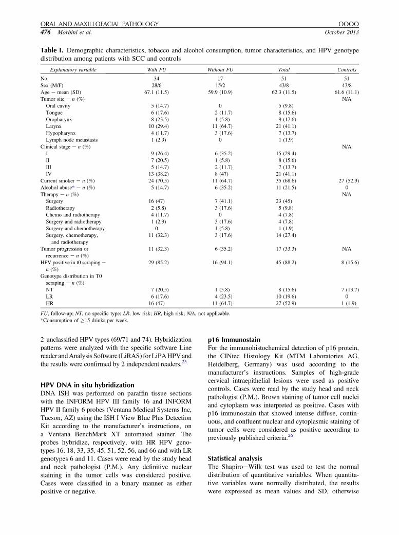
Table I. Demographic characteristics, tobacco and alcohol consumption, tumor characteristics, and HPV genotypedistribution among patients with SCC and controls
Explanatory variable With FU Without FU Total Controls
No. 34 17 51 51Sex (M/F) 28/6 15/2 43/8 43/8Age e mean (SD) 67.1 (11.5) 59.9 (10.9) 62.3 (11.5) 61.6 (11.1)Tumor site e n (%) N/A
Oral cavity 5 (14.7) 0 5 (9.8)Tongue 6 (17.6) 2 (11.7) 8 (15.6)Oropharynx 8 (23.5) 1 (5.8) 9 (17.6)Larynx 10 (29.4) 11 (64.7) 21 (41.1)Hypopharynx 4 (11.7) 3 (17.6) 7 (13.7)Lymph node metastasis 1 (2.9) 0 1 (1.9)
Clinical stage e n (%) N/AI 9 (26.4) 6 (35.2) 15 (29.4)II 7 (20.5) 1 (5.8) 8 (15.6)III 5 (14.7) 2 (11.7) 7 (13.7)IV 13 (38.2) 8 (47) 21 (41.1)
Current smoker e n (%) 24 (70.5) 11 (64.7) 35 (68.6) 27 (52.9)Alcohol abuse* e n (%) 5 (14.7) 6 (35.2) 11 (21.5) 0Therapy e n (%) N/A
Surgery 16 (47) 7 (41.1) 23 (45)Radiotherapy 2 (5.8) 3 (17.6) 5 (9.8)Chemo and radiotherapy 4 (11.7) 0 4 (7.8)Surgery and radiotherapy 1 (2.9) 3 (17.6) 4 (7.8)Surgery and chemotherapy 0 1 (5.8) 1 (1.9)Surgery, chemotherapy,and radiotherapy
11 (32.3) 3 (17.6) 14 (27.4)
Tumor progression orrecurrence e n (%)
11 (32.3) 6 (35.2) 17 (33.3) N/A
HPV positive in t0 scraping en (%)
29 (85.2) 16 (94.1) 45 (88.2) 8 (15.6)
Genotype distribution in T0scraping e n (%)NT 7 (20.5) 1 (5.8) 8 (15.6) 7 (13.7)LR 6 (17.6) 4 (23.5) 10 (19.6) 0HR 16 (47) 11 (64.7) 27 (52.9) 1 (1.9)
FU, follow-up; NT, no specific type; LR, low risk; HR, high risk; N/A, not applicable.*Consumption of �15 drinks per week.
ORAL AND MAXILLOFACIAL PATHOLOGY OOOO
476 Morbini et al. October 2013
2 unclassified HPV types (69/71 and 74). Hybridizationpatterns were analyzed with the specific software Linereader andAnalysis Software (LiRAS) forLiPAHPVandthe results were confirmed by 2 independent readers.25
HPV DNA in situ hybridizationDNA ISH was performed on paraffin tissue sectionswith the INFORM HPV III family 16 and INFORMHPV II family 6 probes (Ventana Medical Systems Inc,Tucson, AZ) using the ISH I View Blue Plus DetectionKit according to the manufacturer’s instructions, ona Ventana BenchMark XT automated stainer. Theprobes hybridize, respectively, with HR HPV geno-types 16, 18, 33, 35, 45, 51, 52, 56, and 66 and with LRgenotypes 6 and 11. Cases were read by the study headand neck pathologist (P.M.). Any definitive nuclearstaining in the tumor cells was considered positive.Cases were classified in a binary manner as eitherpositive or negative.
p16 ImmunostainFor the immunohistochemical detection of p16 protein,the CINtec Histology Kit (MTM Laboratories AG,Heidelberg, Germany) was used according to themanufacturer’s instructions. Samples of high-gradecervical intraepithelial lesions were used as positivecontrols. Cases were read by the study head and neckpathologist (P.M.). Brown staining of tumor cell nucleiand cytoplasm was interpreted as positive. Cases withp16 immunostain that showed intense diffuse, contin-uous, and confluent nuclear and cytoplasmic staining oftumor cells were considered as positive according topreviously published criteria.26
Statistical analysisThe ShapiroeWilk test was used to test the normaldistribution of quantitative variables. When quantita-tive variables were normally distributed, the resultswere expressed as mean values and SD, otherwise
OOOO ORIGINAL ARTICLE
Volume 116, Number 4 Morbini et al. 477
median and interquartile range (IQR; 25th-75thpercentile) were reported; comparisons betweengroups were performed with the appropriate parametricor nonparametric tests. Qualitative variables weresummarized as counts and percentages, and patientgroups were compared using c2 test or Fisher’s exacttest, as appropriate.
Logistic regression models were used to find theodds of HPV infection among controls and patients.Results were expressed as odds ratio (OR, crude oradjusted for age, sex, smoke, and alcohol) with 95%confidence interval (CI). Disease-specific survival(DSS) and disease-free survival (DFS) were derivedwith the use of the KaplaneMeier product-limit methodand the log-rank test was used to analyze comparisonsamong groups. Univariate and multivariate Cox pro-portional hazard models were fitted to investigate theassociations between DSS and DFS and covariates.Results were expressed as hazard ratios (HRs) with95% CI. All tests were 2 sided. Data were analyzedwith the STATA statistical package (release 10.0, 2009,Stata Corporation, College Station, TX).
RESULTSt0 Cytologic samplesHPV DNA was present in 45 cytologic samples(88.2%). Twenty-seven (52.9%) of them hosted 1 ormore HR HPV genotypes (range 1-5), 10 (17.8%)hosted only LR genotypes, and in 8 (15.6%) no specificviral type could be identified. In the control population,1 (1.9%) sample was positive for HR HPV, and in 7(13.7%) no specific viral type could be identified. Caseand control series significantly differed in the propor-tion of cases positive for HPV and HR HPV, and in thedistribution of LR, HR, and nonetype-specific infec-tions (P < .001) (Table I).
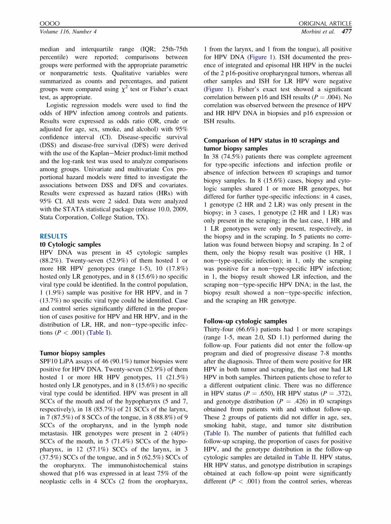
Tumor biopsy samplesSPF10 LiPA assays of 46 (90.1%) tumor biopsies werepositive for HPV DNA. Twenty-seven (52.9%) of themhosted 1 or more HR HPV genotypes, 11 (21.5%)hosted only LR genotypes, and in 8 (15.6%) no specificviral type could be identified. HPV was present in allSCCs of the mouth and of the hypopharynx (5 and 7,respectively), in 18 (85.7%) of 21 SCCs of the larynx,in 7 (87.5%) of 8 SCCs of the tongue, in 8 (88.8%) of 9SCCs of the oropharynx, and in the lymph nodemetastasis. HR genotypes were present in 2 (40%)SCCs of the mouth, in 5 (71.4%) SCCs of the hypo-pharynx, in 12 (57.1%) SCCs of the larynx, in 3(37.5%) SCCs of the tongue, and in 5 (62.5%) SCCs ofthe oropharynx. The immunohistochemical stainsshowed that p16 was expressed in at least 75% of theneoplastic cells in 4 SCCs (2 from the oropharynx,
1 from the larynx, and 1 from the tongue), all positivefor HPV DNA (Figure 1). ISH documented the pres-ence of integrated and episomal HR HPV in the nucleiof the 2 p16-positive oropharyngeal tumors, whereas allother samples and ISH for LR HPV were negative(Figure 1). Fisher’s exact test showed a significantcorrelation between p16 and ISH results (P ¼ .004). Nocorrelation was observed between the presence of HPVand HR HPV DNA in biopsies and p16 expression orISH results.
Comparison of HPV status in t0 scrapings andtumor biopsy samplesIn 38 (74.5%) patients there was complete agreementfor type-specific infections and infection profile orabsence of infection between t0 scrapings and tumorbiopsy samples. In 8 (15.6%) cases, biopsy and cyto-logic samples shared 1 or more HR genotypes, butdiffered for further type-specific infections: in 4 cases,1 genotype (2 HR and 2 LR) was only present in thebiopsy; in 3 cases, 1 genotype (2 HR and 1 LR) wasonly present in the scraping; in the last case, 1 HR and1 LR genotypes were only present, respectively, inthe biopsy and in the scraping. In 5 patients no corre-lation was found between biopsy and scraping. In 2 ofthem, only the biopsy result was positive (1 HR, 1nonetype-specific infection); in 1, only the scrapingwas positive for a nonetype-specific HPV infection;in 1, the biopsy result showed LR infection, and thescraping nonetype-specific HPV DNA; in the last, thebiopsy result showed a nonetype-specific infection,and the scraping an HR genotype.
Follow-up cytologic samplesThirty-four (66.6%) patients had 1 or more scrapings(range 1-5, mean 2.0, SD 1.1) performed during thefollow-up. Four patients did not enter the follow-upprogram and died of progressive disease 7-8 monthsafter the diagnosis. Three of them were positive for HRHPV in both tumor and scraping, the last one had LRHPV in both samples. Thirteen patients chose to refer toa different outpatient clinic. There was no differencein HPV status (P ¼ .650), HR HPV status (P ¼ .372),and genotype distribution (P ¼ .426) in t0 scrapingsobtained from patients with and without follow-up.These 2 groups of patients did not differ in age, sex,smoking habit, stage, and tumor site distribution(Table I). The number of patients that fulfilled eachfollow-up scraping, the proportion of cases for positiveHPV, and the genotype distribution in the follow-upcytologic samples are detailed in Table II. HPV status,HR HPV status, and genotype distribution in scrapingsobtained at each follow-up point were significantlydifferent (P < .001) from the control series, whereas
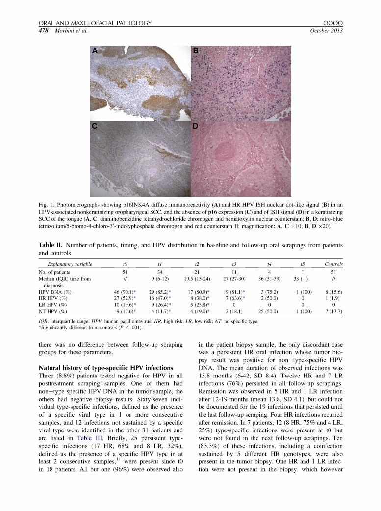
Fig. 1. Photomicrographs showing p16INK4A diffuse immunoreactivity (A) and HR HPV ISH nuclear dot-like signal (B) in anHPV-associated nonkeratinizing oropharyngeal SCC, and the absence of p16 expression (C) and of ISH signal (D) in a keratinizingSCC of the tongue (A, C: diaminobenzidine tetrahydrochloride chromogen and hematoxylin nuclear counterstain; B, D: nitro-bluetetrazolium/5-bromo-4-chloro-30-indolyphosphate chromogen and red counterstain II; magnification: A, C �10; B, D �20).
Table II. Number of patients, timing, and HPV distribution in baseline and follow-up oral scrapings from patientsand controls
Explanatory variable t0 t1 t2 t3 t4 t5 Controls
No. of patients 51 34 21 11 4 1 51Median (IQR) time from
diagnosis// 9 (6-12) 19.5 (15-24) 27 (27-30) 36 (31-39) 33 (e) //
HPV DNA (%) 46 (90.1)* 29 (85.2)* 17 (80.9)* 9 (81.1)* 3 (75.0) 1 (100) 8 (15.6)HR HPV (%) 27 (52.9)* 16 (47.0)* 8 (38.0)* 7 (63.6)* 2 (50.0) 0 1 (1.9)LR HPV (%) 10 (19.6)* 9 (26.4)* 5 (23.8)* 0 0 0 0NT HPV (%) 9 (17.6)* 4 (11.7)* 4 (19.0)* 2 (18.1) 25 (50.0) 1 (100) 7 (13.7)
IQR, interquartile range; HPV, human papillomavirus; HR, high risk; LR, low risk; NT, no specific type.*Significantly different from controls (P < .001).
ORAL AND MAXILLOFACIAL PATHOLOGY OOOO
478 Morbini et al. October 2013
there was no difference between follow-up scrapinggroups for these parameters.
Natural history of type-specific HPV infectionsThree (8.8%) patients tested negative for HPV in allposttreatment scraping samples. One of them hadnonetype-specific HPV DNA in the tumor sample, theothers had negative biopsy results. Sixty-seven indi-vidual type-specific infections, defined as the presenceof a specific viral type in 1 or more consecutivesamples, and 12 infections not sustained by a specificviral type were identified in the other 31 patients andare listed in Table III. Briefly, 25 persistent type-specific infections (17 HR, 68% and 8 LR, 32%),defined as the presence of a specific HPV type in atleast 2 consecutive samples,11 were present since t0in 18 patients. All but one (96%) were observed also
in the patient biopsy sample; the only discordant casewas a persistent HR oral infection whose tumor bio-psy result was positive for nonetype-specific HPVDNA. The mean duration of observed infections was15.8 months (6-42, SD 8.4). Twelve HR and 7 LRinfections (76%) persisted in all follow-up scrapings.Remission was observed in 5 HR and 1 LR infectionafter 12-19 months (mean 13.8, SD 4.1), but could notbe documented for the 19 infections that persisted untilthe last follow-up scraping. Four HR infections recurredafter remission. In 7 patients, 12 (8 HR, 75% and 4 LR,25%) type-specific infections were present at t0 butwere not found in the next follow-up scrapings. Ten(83.3%) of these infections, including a coinfectionsustained by 5 different HR genotypes, were alsopresent in the tumor biopsy. One HR and 1 LR infec-tion were not present in the biopsy, which however
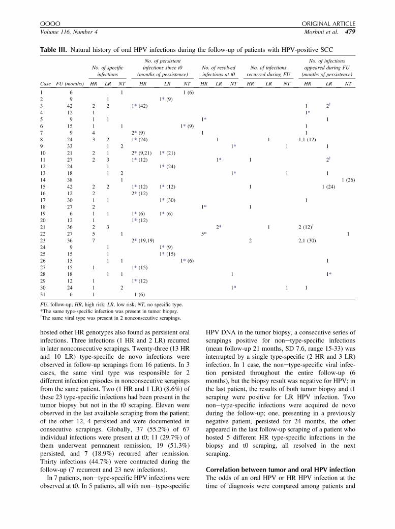
Table III. Natural history of oral HPV infections during the follow-up of patients with HPV-positive SCC
Case FU (months)
No. of specificinfections
No. of persistentinfections since t0
(months of persistence)No. of resolvedinfections at t0
No. of infectionsrecurred during FU
No. of infectionsappeared during FU(months of persistence)
HR LR NT HR LR NT HR LR NT HR LR NT HR LR NT
1 6 1 1 (6)2 9 1 1* (9)3 42 2 2 1* (42) 1 2y
4 12 1 1*5 9 1 1 1* 16 15 1 1 1* (9) 17 9 4 2* (9) 1 18 24 3 2 1* (24) 1 1 1,1 (12)9 33 1 2 1* 1 110 21 2 1 2* (9,21) 1* (21)11 27 2 3 1* (12) 1* 1 2y
12 24 1 1* (24)13 18 1 2 1* 1 114 38 1 1 (26)15 42 2 2 1* (12) 1* (12) 1 1 (24)16 12 2 2* (12)17 30 1 1 1* (30) 118 27 2 1* 119 6 1 1 1* (6) 1* (6)20 12 1 1* (12)21 36 2 3 2* 1 2 (12)y
22 27 5 1 5* 123 36 7 2* (19,19) 2 2,1 (30)24 9 1 1* (9)25 15 1 1* (15)26 15 1 1 1* (6) 127 15 1 1* (15)28 18 1 1 1 1*29 12 1 1* (12)30 24 1 2 1* 1 131 6 1 1 (6)
FU, follow-up; HR, high risk; LR, low risk; NT, no specific type.*The same type-specific infection was present in tumor biopsy.yThe same viral type was present in 2 nonconsecutive scrapings.
OOOO ORIGINAL ARTICLE
Volume 116, Number 4 Morbini et al. 479
hosted other HR genotypes also found as persistent oralinfections. Three infections (1 HR and 2 LR) recurredin later nonconsecutive scrapings. Twenty-three (13 HRand 10 LR) type-specific de novo infections wereobserved in follow-up scrapings from 16 patients. In 3cases, the same viral type was responsible for 2different infection episodes in nonconsecutive scrapingsfrom the same patient. Two (1 HR and 1 LR) (8.6%) ofthese 23 type-specific infections had been present in thetumor biopsy but not in the t0 scraping. Eleven wereobserved in the last available scraping from the patient;of the other 12, 4 persisted and were documented inconsecutive scrapings. Globally, 37 (55.2%) of 67individual infections were present at t0; 11 (29.7%) ofthem underwent permanent remission, 19 (51.3%)persisted, and 7 (18.9%) recurred after remission.Thirty infections (44.7%) were contracted during thefollow-up (7 recurrent and 23 new infections).
In 7 patients, nonetype-specific HPV infections wereobserved at t0. In 5 patients, all with nonetype-specific
HPV DNA in the tumor biopsy, a consecutive series ofscrapings positive for nonetype-specific infections(mean follow-up 21 months, SD 7.6, range 15-33) wasinterrupted by a single type-specific (2 HR and 3 LR)infection. In 1 case, the nonetype-specific viral infec-tion persisted throughout the entire follow-up (6months), but the biopsy result was negative for HPV; inthe last patient, the results of both tumor biopsy and t1scraping were positive for LR HPV infection. Twononetype-specific infections were acquired de novoduring the follow-up; one, presenting in a previouslynegative patient, persisted for 24 months, the otherappeared in the last follow-up scraping of a patient whohosted 5 different HR type-specific infections in thebiopsy and t0 scraping, all resolved in the nextscraping.
Correlation between tumor and oral HPV infectionThe odds of an oral HPV or HR HPV infection at thetime of diagnosis were compared among patients and
ORAL AND MAXILLOFACIAL PATHOLOGY OOOO
480 Morbini et al. October 2013
controls. Patients were significantly more likely thancontrols to have any oral HPV infection (OR adjustedfor sex, age, smoke, alcohol 87.38, 95% CI 15.81-483.02, P < .001) or HR HPV infection (adjusted OR79.54, 95% CI 8.44-749.60, P < .001). Fisher’s exacttest showed a significant association of HPV statusbetween tumors and t0 scrapings; no association wasobserved with scrapings obtained at t1, t2, and t3.Tumor HR HPV status was significantly associatedwith t0 and t1 scrapings. A significant association wasobserved for HPV and HR HPV status also between t0and t1 but not t2 or t3, and between t1 and t2 but not t3scrapings (Table IV).
HPV infection in follow-up biopsy samplesSixteen posttreatment biopsies were obtained from 15patients, all with previous HPV-positive cytologicsamples. Biopsies included 7 local recurrences, 1 secondtumor, 2 lymph nodes, and 1 pulmonary metastasis, and5 non-neoplastic tissue samples from the site of thetreated primary tumor. The mean time from the initialdiagnosis was 10.2 months (SD 13.8, range 1-33) for thenon-neoplastic samples, 3 of which were obtained aftersurgical treatment and 1 after radiotherapy, 20.8 months(SD 12.1, range 9-38) for local recurrences, whichoccurred in 3 cases after surgery and chemo-radiotherapy, in 2 after chemoradiotherapy, and in 2after surgery alone, 11 months for the second tumor,presenting after chemoradiotherapy, 16 and 24 monthsfor the lymph node metastases, both occurring aftersurgery, and 26 months for the pulmonary metastasis,which presented in a patient treated with surgery andchemoradiotherapy. HPV DNA was found in all biop-sies, independently of their site, time interval, type oftreatment, and diagnosis (negative, tumor, or metas-tasis). The same type-specific infections observed in thetemporally closest scraping were present in all samples;in 4 of them, 1 further viral type not observed in thescraping was present in the biopsy, and in 1 case 1 ofthe viral types present in the scraping was absent in thebiopsy. None of the posttreatment biopsies were positiveby p16 or ISH.
Clinical follow-upMedian follow-up time was 28 months (IQR 19-42,range 6-51). Thirteen patients had 1 or more localrecurrences (n ¼ 7), a second SCC in the upper aero-digestive tract (n ¼ 1), metastasis to the laterocervicallymph nodes (n ¼ 3), and lung metastasis (n ¼ 2), ata median time of 17 months (IQR 9-23, range 3-36)after the initial diagnosis and response to therapy. At12, 24, and 36 months the rate of patients free fromdisease was 87.0% (95% CI 73.3-93.2), 76.6% (95% CI60.6-86.7), 65.6% (95% CI 47.0-79.1), respectively;
DFS ranged from 3 to 51 months (median 26, IQR15-40). Eleven (19.6%) patients died. Four of them didnot respond to treatment or died of surgical complica-tions: 2 patients of 52 and 79 years with stage IVtumors suffered disease progression despite surgery andadjuvant chemo and radiotherapy, 1 (73 years old, withstage IV tumor) died of surgery-related complications,and 1 (77 years old, stage II) did not respond toradiotherapy, while surgery and chemotherapy hadbeen excluded for severe concomitant diseases. Sixother patients died of their disease, including 4 withdisease recurrence, 1 with lung metastasis, and 1 withlaterocervical metastasis. One patient died of myocar-dial infarction with no evidence of tumor. One patientwith a second tumor, 1 with recurrent tumor, 1 withlung metastasis, and 1 with laterocervical metastasiswere alive with disease at the time of the last follow-up;2 local recurrences and 1 laterocervical metastasis weresuccessfully treated with surgery. At 12, 24, and 36months the rate of patients who were alive was 92.0%(95% CI 80.1-96.9), 85.2% (95% CI 71.3-92.7), 73.1%(95% CI 56.0-84.4), respectively. None of the controls,including the 1 with HR HPV infection, were diagnosedwith HNSCC at our center after enrollment in thecontrol group.
Correlation of HPV infection with outcomeand survivalHPV DNA was present in the biopsies and in oralscrapings of 15 (88.2%) of 17 patients who suffereddisease progression, recurrences, second tumors, ormetastases, and in 30 (88.2%) of 34 patients who didnot. Eleven (64.7%) and 16 (47%) infections, respec-tively, were sustained by HR HPV in the biopsies; 10(58.8%) and 14 (41.1%) in the cytologic samples. HPVwas present in the tumors and in oral scrapings of all 11patients with unfavorable outcome (8 HR in both typesof sample) and of all 4 patients who were alive withdisease (3 HR in the biopsies and 2 in the scrapings); ofthe 36 patients with favorable outcome, 31 (86.1%) hadHPV-positive tumors and 32 (88.8%) had HPV-positivescrapings; HR genotypes were present in 16 (44.4%)tumors and in 17 (47.2%) cytologic samples. Twenty-eight patients were treated with protocols includingchemo and/or radiotherapy: HPV and HR HPV infec-tions were present, respectively, in 12 (92.6%) and 9(63.2%) tumors of the 13 patients of this group thatexperienced disease recurrence or progression, and in13 (86.6%) and 6 (40%) tumors of the 15 patients thatdid not (P > .05). Oral HPV infection was present in 11(84.6%) patients with recurrence or progression and in12 (80%) patients without; HR genotypes were presentin 8 (66.6%) and 7 (46.6%) cases, respectively. In these28 patients, HPV was present in the tumors and in theoral cavity of all 7 patients with unfavorable outcome

Table IV. Concordance (%) and significance levels (P) of Fisher’s exact tests for HPV and HR HPV infection of eachsample series compared with the following series
HPV biopsy HPV t0 HPV t1 HPV t2 HR HPV biopsy HR HPV t0 HR HPV t1 HR HPV t2
t0 91.2% (<0.001) 96.1% (<0.001)t1 85.3% (0.094) 88.2% (0.015) 70.6% (0.037) 70.6% (0.037)t2 85.7% (0.080) 85.7% (0.080) 90.5% (0.012) 66.7% (0.203) 61.9% (0.038) 76.2% (0.032)t3 90.9% (0.182) 100% (0.182) 72.7% (0.491) 72.7% (0.491) 81.8% (0.088) 81.8% (0.088) 72.7% (0.242) 63.6% (0.545)
HPV, human papillomavirus; HR, high risk.
OOOO ORIGINAL ARTICLE
Volume 116, Number 4 Morbini et al. 481
(5 HR in both types of sample); of the 4 patients whowere alive with disease, 4 had HPV-positive tumors (allHR) and 3 had HPV-positive oral scrapings (2 HR); ofthe 17 patients with favorable outcome, 14 (82.3%) hadHPV-positive tumors and 13 (76.4%) HPV-positivescrapings; HR genotypes were present, respectively, in7 (41.1%) and in 8 (47%) samples. Fisher’s exact testdid not show a significant association of HPV and HRHPV infection and outcome in all evaluated groups(P > .05).
The Cox proportional hazard analysis did not showany correlation between DFS or DSS and the presenceof HPV or HR HPV infection in tumor or oral mucosacells at t0. Only clinical stage IV was significantlycorrelated with shorter DSS and DFS (Figure 2). Inpatients with cytologic follow-up, Fisher’s exact testdid not show any association between HPV or HR HPVinfection at t1, t2, and t3 and tumor recurrence.
DISCUSSIONThe present study reports that oral HPV infections atthe time of tumor diagnosis and their persistence duringthe first months of follow-up are significantly morefrequent in subjects with HPV-positive SCC than withHPV-negative SCC, and that the risk of hosting HPV inthe oral mucosa is significantly higher in subjects withhead and neck carcinomas than in controls. The highprevalence of oral HPV infection that we observed inHNSCC as compared with the general populationsuggests that the presence of the tumor might favor thelocalization and persistence of the virus in the tumorcells27,28 and in adjacent mucosa,17,20 possibly medi-ated by immune suppressive mechanisms. It hasrecently been shown that SCCs activate several strate-gies in order to evade the host immune response,29
which could favor viral infection and persistence inboth tumor and oral mucosa. An increased risk of HPVoral infection because of immunologic dysfunction hasindeed been documented in patients positive forHIV,30,31 and in non-HIV tobacco smokers.32,33
Detecting HR HPV DNA in tumor tissue, while sup-porting an association between virus and cancer, does notdistinguish whether HPV is transcriptionally active andthus able to promote carcinogenesis (the so-called driverinfection), or inactive, probably representing incidental or
passenger infection. To determine the oncogenic activityof HPV, direct or indirect methods that assess viralintegration or viral oncogene transcription are neces-sary.23 In the present study, an oncogenic HR HPVinfection was demonstrated by means of validated p16immunostain and ISH in only 2 oropharyngeal SCCs,which is the site where HPV carcinogenesis has beenmost consistently documented,1,2 whereas most of theinfections recorded in tumors by PCR did not showevidence of viral integration or transcription. The mark-edly different rates of HPV positivity that we obtainedwith PCR and ISH in our tumor series likely depend onthe different analytical sensitivities of the 2 tests: SPF10LiPA has the highest sensitivity among HPV typingassays, because of the short amplified DNA fragment (65base pairs).34 SPF10 LiPA sensitivity differs for differentHPV genotypes, but is at least 1-10 copies per 103 cells,35
whereas the ISH estimated sensitivity threshold is 1-2viral copies per cell.36 We assume that, in PCR-positiveand ISH-negative tissue samples from the present series,the viral load was lower than this threshold, as observedby Chaturvedi et al.37 who documented with quantitative(q)PCR that 22.5% and 50%, respectively, of SPF10LiPA HPV16- and HPV none16-positive tumors hadless than 1 viral genome per cell. The fact that HPVtranscriptional activity has been associated with a viralload of at least 0.5 copies per cell38,39 supports thehypothesis that PCR-positive p16- and/or ISH-negativeinfections observed in the present study carry no onco-genic activity. However, on the basis of previous litera-ture data40-43 we cannot definitely exclude oncogenetranscription in these infections. A conclusive clarifica-tion of this aspect would require the amplification of HPVE6/E7 messenger RNA, which could not be performed inour series because of the lack of frozen tumor samples.Indirect evidence from prognostic and survival associa-tions suggests that most infections identified in thepresent study are passenger infections, as opposed to thestrong prognostic correlations recently reported fororopharyngeal SCCs associated with HR HPV infec-tion,5-7 we did not find any differences in the rates ofdisease progression or recurrence, nor in outcome orsurvival of patients positive for HPV or HR HPV DNA.No correlation was found even when we considered onlythose patients who had been treated with protocols

Fig. 2. KaplaneMeier survival curves showing a significant association between clinical stage IV and DSS; the presence of HPVand HR HPV infections in tumor biopsies and in oral scrapings did not influence patient survival.
ORAL AND MAXILLOFACIAL PATHOLOGY OOOO
482 Morbini et al. October 2013
including chemo and/or radiotherapy, which tend to bemore effective in HPV-associated tumors.7
Very few studies have so far addressed the issue ofHPV persistence in the oral mucosa after tumor treat-ment. Recently, Agrawal et al.21 demonstrated thatposttreatment oral HPV infections were significantlymore frequent among individuals diagnosed withHPV16-positive HNSCC than with HPV16-negativeHNSCC, and that not only HPV16 but also other HRand LR HPV types were more common in the oralmucosa of patients with positive tumors. These obser-vations are in agreement with ours regarding HPVpersistence in the oral cavity after tumor treatment.We also documented that over two-thirds of the type-specific oral infections present at diagnosis persistedin the follow-up for at least 6 months, with a maximumof 42 months. Moreover, several infections appearingposttreatment were sustained by viral types responsiblefor previously resolved infections, suggesting thereactivation of a latent infection or re-infection froma site of persistence. Interestingly, the strength ofassociation between tumor and oral positivity for HPVand HR HPV progressively reduced with increasingfollow-up time. When we compared cytologic sample
groups, the strongest correlation was found between theclosest scraping series (t0 and t1, t1 and t2), suggestingthat the infection profile changes over time, with bothresolutions and new infections.
We observed several persistent infections in whicha specific viral type could not be identified. In most ofthese patients, a specific genotype was characterizedin a single follow-up sample. It is possible that thenonetype-specific positive results were because of thepresence of a number of viral genomes lower thanthe threshold of detection of the linear array, and thatthe genotype was recognized only when the viral loadincreased above this threshold (as a consequence ofactive replication or of sampling issues). As an alter-native hypothesis, it is recognized that consensusprimers amplifying the HPV L1 region (includingSPF10) may fail to identify fully integrated viruses,yielding false-negative44 or nonetype-specific results(Morbini et al., personal communication). This phe-nomenon fully explains our observation of a persistentHR HPV oral infection in a patient with nonetype-specific viral DNA in the biopsy, and could be true forthe persistent nonetype-specific infections positive forHR HPV in a single scraping. It seems, however,
OOOO ORIGINAL ARTICLE
Volume 116, Number 4 Morbini et al. 483
unlikely for the LR infections with a similar behavior,given that integration is not commonly observed in LRHPV genotypes. Finally, SPF10 LiPA amplifies butdoes not recognize cutaneous HPV genotypes, whichhave been previously found in the oral mucosa.45 Theeventual presence of a genital type-specific infection inthese patients renders this possibility unlikely. Quanti-tative, genotype-specific PCR on cytologic samples willhelp to clarify this aspect.
As far as prognosis is concerned, we could not findany association between HPV persistence in the oralmucosa and tumor recurrence, as previously reported.22
On the contrary, patients with and without oral HPVinfection or persistence showed similar recurrence rates,and all posttreatment biopsies including normal mucosafrom sites of previous tumors hosted the virus. Theprognostic relevance of oral infections, especiallypersistent ones, is probably correlated with their onco-genic potential. No direct assumption could be drawn onthe nature (passenger versus driver) of the oral infectionsrecorded with scrapings, since viral transcriptionalactivity was not determined in oral cytologic samples.However, all persistent infections documented in post-treatment biopsies did not show transcriptional activity.Literature data offer indirect evidence that oral infectionsare probably passenger: qPCR studies reported a medianviral load of 13.0 HPV genomes per 103 oral exfoliatedcells in patients with oropharyngeal cancers,20 whichis lower than the loads associated, respectively, withoncogene transcription,38,39 and with replicative infec-tions in the cervical mucosa (50-100 copies per cell).9
The lack of prognostic implications for the presenceand persistence of HPV DNA in the oral mucosasuggests that consensus PCR on oral scraping is noteffective for posttreatment monitoring, whereas itsresults may bear severe psychosocial implications forpatients and families. As in tissue samples used forcancer diagnosis, we need to establish parameters thatmore accurately discriminate between passenger andpotentially oncogenic infections in the oral mucosa, tomake oral cytology an effective instrument for patientscreening and monitoring.46
REFERENCES1. Adelstein DJ, Ridge JA, Gillison ML, et al. Head and neck squa-
mous cell cancer and the human papillomavirus: summary of aNational Cancer Institute State of the Science Meeting, November9-10, 2008, Washington, DC. Head Neck. 2009;31:1393-1422.
2. El-Mofty SK, Patil S. Human papillomavirus (HPV)-relatedoropharyngeal nonkeratinizing squamous cell carcinoma: char-acterization of a distinct phenotype. Oral Surg Oral Med OralPathol Oral Radiol Endod. 2006;101:339-345.
3. Sturgis EM, Cinciripini PM. Trends in head and neck cancerincidence in relation to smoking prevalence: an emergingepidemic of human papillomavirus-associated cancers? Cancer.2007;110:1429-1435.
4. Marur S, D’Souza G, Westra WH, Forastiere A. HPV-associatedhead and neck cancer: a virus-related cancer epidemic. LancetOncol. 2010;11:781-789.
5. Fakhry C, Westra WH, Li S, et al. Improved survival of patientswith human papillomavirus-positive head and neck squamous cellcarcinoma in a prospective clinical trial. J Natl Cancer Inst.2008;100:261-269.
6. Lassen P, Eriksen JG, Hamilton-Dutoit S, Tramm T, Alsner J,Overgaard J. Effect of HPV-associated p16INK4A expressionon response to radiotherapy and survival in squamous cellcarcinoma of the head and neck. J Clin Oncol. 2009;27:1992-1998.
7. Ang KK, Harris J, Wheeler R, et al. Human papillomavirus andsurvival of patients with oropharyngeal cancer. N Engl J Med.2010;363:24-35.
8. Doorbar J, Quint W, Banks L, et al. The biology and life cycle ofhuman papillomaviruses. Vaccine. 2012;30(suppl 5):F55-F70.
9. Stubenrauch F, Laimins LA. Human papillomavirus life cycle:active and latent phases. Semin Cancer Biol. 1999;9:379-386.
10. Bodily J, Laimins LA. Persistence of human papillomavirusinfection: keys to malignant progression. Trends Microbiol.2011;19:33-39.
11. Rositch AF, Koshiol J, Hudgens MG, et al. Patterns of persistentgenital human papillomavirus infection among women world-wide: a literature review and meta-analysis. Int J Cancer.2013;133:1271-1285.
12. Venturoli S, Ambretti S, Cricca M, et al. Correlation of high-riskhuman papillomavirus genotypes persistence and risk of residualor recurrent cervical disease after surgical treatment. J Med Virol.2008;80:1434-1440.
13. Ramos MC, Pizarro De Lorenzo BH, Michelin MA, Murta EF.High-grade cervical intraepithelial neoplasia, human papilloma-virus and factors connected with recurrence following surgicaltreatment. Clin Exp Obstet Gynecol. 2008;35:242-247.
14. Gravitt PE, Coutlee F, Iftner T, Sellors JW, Quint WG,Wheeler CM. New technologies in cervical cancer screening.Vaccine. 2008;26(suppl 10):K42-K52.
15. Dal Bello B, Spinillo A, Alberizzi P, et al. Cervical infections bymultiple human papillomavirus (HPV) genotypes: prevalence andimpact on the risk of precancerous epithelial lesions. J Med Virol.2009;81:703-712.
16. Fakhry C, Rosenthal BT, Clark DP, Gillison ML. Associationbetween oral HPV16 infection and cytopathology: evaluation ofan oropharyngeal “pap-test equivalent” in high-risk populations.Cancer Prev Res. 2011;4:1378-1384.
17. Giovannelli L, Campisi G, Lama A, et al. Human papillomavirusDNA in oral mucosal lesions. J Infect Dis. 2002;185:833-836.
18. Smith EM, Hoffman HT, Summersgill KF, Kirchner HL,Turek LP, Haugen TH. Human papillomavirus and risk of oralcancer. Laryngoscope. 1998;108:1098-1103.
19. Smith EM, Ritchie JM, Summersgill KF, et al. Human papillo-mavirus in oral exfoliated cells and risk of head and neck cancer.J Natl Cancer Inst. 2004;96:449-455.
20. D’Souza G, Kreimer AR, Viscidi R, et al. Caseecontrol study ofhuman papillomavirus and oropharyngeal cancer. N Engl J Med.2007;356:1944-1956.
21. Agrawal Y, Koch WM, Xiao W, et al. Oral human papilloma-virus infection before and after treatment for human papilloma-virus 16-positive and human papillomavirus 16-negative headand neck squamous cell carcinoma. Clin Cancer Res. 2008;14:7143-7150.
22. Chuang AY, Chuang TC, Chang S, et al. Presence of HPV DNAin convalescent salivary rinses is an adverse prognostic marker inhead and neck squamous cell carcinoma. Oral Oncol. 2008;44:915-919.

ORAL AND MAXILLOFACIAL PATHOLOGY OOOO
484 Morbini et al. October 2013
23. Westra WH. Detection of human papillomavirus in clinicalsamples. Otolaryngol Clin North Am. 2012;45:765-777.
24. Morbini P, Dal Bello B, Alberizzi P, et al. Exfoliated cells of theoral mucosa for HPV typing by SPF10 in head and neck cancer.J Virol Methods. 2012;186:999-1103.
25. Dal Bello B, Spinillo A, Alberizzi P, Cesari S, Gardella B,Silini EM. Validation of the LiPA human papillomavirus typingassay using formalin-fixed paraffin-embedded cervical biopsysamples. J Clin Microbiol. 2009;47:2175-2180.
26. Thomas J, Primeaux T. Is p16 immunohistochemistry a morecost-effective method for identification of human papilloma virus-associated head and neck squamous cell carcinoma? Ann DiagnPathol. 2012;16:91-99.
27. Andrews E, Seaman WT, Webster-Cyriaque J. Oropharyngealcarcinoma in non-smokers and non-drinkers: a role for HPV. OralOncology. 2009;45:486-491.
28. da Silva CE, da Silva ID, Cerri A, Weckx LL. Prevalence of humanpapillomavirus in squamous cell carcinoma of the tongue. Oral SurgOral Med Oral Pathol Oral Radiol Endod. 2007;104:497-500.
29. Duray A, Demoulin S, Hubert P, Delvenne P, Saussez S. Immunesuppression in head and neck cancers: a review. Clin DevImmunol; 2010. 2010:701657.
30. Fatahzadeh M, Schlecht NF, Chen Z, et al. Oral human papillo-mavirus detection in older adults who have human immunodefi-ciency virus infection. Oral Surg Oral Med Oral Pathol OralRadiol. 2013;115:505-514.
31. Beachler DC, Weber KM, Margolick JB, et al. Risk factors fororal HPV infection among a high prevalence population of HIV-positive and at-risk HIV-negative adults. Cancer EpidemiolBiomarkers Prev. 2012;21:122-123.
32. Gillison ML, Broutian T, Pickard RKL, et al. Prevalence of oralHPV infection in the United States, 2009-2010. JAMA. 2012;307:693-703.
33. Kreimer AR, Villa A, Nyitray AG, et al. The epidemiology of oralHPV infection among a multinational sample of healthy men.Cancer Epidemiol Biomarkers Prev. 2011;20:172-182.
34. Kleter B, van Doorn LJ, ter Schegget J, et al. Novel short-fragmentPCR assay for highly sensitive broad-spectrum detection of ano-genital human papillomaviruses. Am J Pathol. 1998;153:1731-1739.
35. Moreau F, Fetouchi R, Micalessi IM, et al. Detection and geno-typing of human papillomavirus by real-time PCR assay. J ClinVirol. 2013;56:244-249.
36. Lizard G, Démares-Poulet MJ, Roignot P, Gambert P. In situhybridization detection of single-copy human papillomavirus onisolated cells, using a catalyzed signal amplification system:GenPoint. Diagn Cytopathol. 2001;24:112-116.
37. Chaturvedi AK, Engels EA, Pfeiffer RM, et al. Human papillo-mavirus and rising oropharyngeal cancer incidence in the UnitedStates. J Clin Oncol. 2011;29:4294-4301.
38. Smeets SJ, Hesselink AT, Speel EM, et al. A novel algorithm forreliable detection of human papillomavirus in paraffin embeddedhead and neck cancer specimen. Int J Cancer. 2007;121:2465-2472.
39. Jung AC, Briolat J, Millon R, et al. Biological and clinical rele-vance of transcriptionally active human papillomavirus (HPV)infection in oropharynx squamous cell carcinoma. Int J Cancer.2010;126:1882-1894.
40. Schache AG, Liloglou T, Risk JM, et al. Evaluation of humanpapilloma virus diagnostic testing in oropharyngeal squamous cellcarcinoma: sensitivity, specificity and prognostic discrimination.Clin Cancer Res. 2011;17:6262-6271.
41. Ukpo OC, Flanagan JJ, Ma X, Luo Y, Thorstad WL, Lewis JS.High-risk human papillomavirus E6/E7 mRNA detection bya novel in situ hybridization assay strongly correlates with p16expression and patient outcomes in oropharyngeal squamous cellcarcinoma. Am J Surg Pathol. 2011;35:1343-1350.
42. Shi W, Kato H, Perez-Ordonez B, et al. Comparative prognosticvalue of HPV16 E6 mRNA compared with in situ hybridizationfor human oropharyngeal squamous cell carcinoma. J Clin Oncol.2009;27:6213-6221.
43. Hoffmann M, Tribius S, Quabius ES, et al. HPV DNA E6*I-mRNA expression and p16INK4A immunohistochemistry inhead and neck cancer e how valid is p16INK4A as surrogatemarker? Cancer Lett. 2012;323:88-96.
44. Walboomers JM, Jacobs MV, Manos MM, et al. Human papil-lomavirus is a necessary cause of invasive cervical cancerworldwide. J Pathol. 1999;189:12-19.
45. Kurose K, Terai M, Soedarsono N, et al. Low prevalence of HPVinfection and its natural history in normal oral mucosa amongvolunteers on Miyako Island, Japan. Surg Oral Med Oral PatholOral Radiol Endod. 2004;98:91-96.
46. Lingen MW. Can saliva-based HPV tests establish cancer risk andguide patient management? Oral Surg Oral Med Oral PatholOral Radiol Endod. 2010;110:273-274.
Reprint requests:
Patrizia Morbini, MD, PhDDepartment of Molecular MedicineUnit of Pathology, Via Forlanini 1627100 Pavia, [email protected]