In situ human papillomavirus (HPV) genotyping of cervical intraepithelial neoplasia in South African...
7
J Clin Pathol 1991;44:400-405 In situ human papillomavirus (HPV) genotyping of cervical intraepithelial neoplasia in South African and British patients: Evidence for putative HPV integration in vivo K Cooper, C S Herrington, A K Graham, M F Evans, J O'D McGee Abstract In South Africa asymptomatic wart virus infection diagnosed by morpho- logical criteria occurs in 16-20% of all ethnic groups; the incidence in black women is 66%. To identify human papillomavirus (HPV) types the pre- valence of HPV in cervical intraepi- thelial neoplasia (CIN) in South African women (n = 72) with age matched British women (n = 73) was compared by non-isotopic in situ hybridisation (NISH) using digoxigenin labelled probes for HPV 6, 11, 16, 18, 31, 33 and 35 on archival biopsy specimens. A higher proportion of British biopsy specimens (68%) contained HPV than those from South Africa (50%) in CIN 2 and 3; this difference was due to HPV 16. Thirty six per cent of the positive biopsy specimens from South African women also con- tained HPV 33/35 compared with 16% in the United Kingdom. There was no dif- ference in HPV detection with age in either group. These data indicate that HPV types vary geographically, with "minor" HPV types being more common in South Africa. Three qualitatively dis- tinct NISH signals were observed; a dif- fuse (type 1) signal in superficial cells, mainly koilocytes; a punctate signal (type 2) in basal/"undifferentiated" cells in CIN 3; and combined type 1 and 2 signals in CIN with wart virus infection (type 3). The punctate signal may represent HPV integration. University of Oxford, NuffieldDepartmentof Pathology and Bacteriology, John Radcliffe Hospital, Oxford OX3 9DU K Cooper C S Herrington A K Graham M F Evans J O'D McGee Correspondence to: Professor J O'D McGee Accepted for publication 6 December 1990 Over 60 types of human papillomavirus (HPV) have been described over the past decade, with HPV 6, 11, 42, 43, 44 associated predominantly with genital condylomata and low grade cervical dysplasia (cervical intraepithelial neoplasia (CIN 1-2)), and HPV 16, 18-and to a lesser extent, 31, 33, 35, 45, 51, 52, 56, associated with high grade cervical dysplasia (CIN 3).' 2 Munoz et al summarised all hybridisation studies in women with CIN and cervical cancer (1983-1987) in whom the prevalence of HPV types 6, 11, 16 or 18 had been reported.3 There was wide variation in prevalence within the same and among dif- ferent countries. There are no data on the prevalence of HPV types in CIN in South Africa where cervical cancer is becoming increasingly common; nor have HPV 31, 33, and 35 been analysed in all grades of CIN in South Africa or the United Kingdom. It has been contended that morphological criteria alone predict HPV 16 infection in severe grades of CIN associated with wart virus infection,45 specifically koilocytosis, which has been used as an absolute com- parator for assessing sensitivity of hybridisa- tion assays for HPV typing.6 This paper addresses two questions: (i) the prevalence of HPV types in CIN 2 and 3, with and without evidence of wart virus infection, in South African and British patients; and (ii) do morphological criteria alone predict HPV infection or the infecting HPV type? Methods Archival, formalin fixed, paraffin wax embed- ded colposcopic cervical punch biopsy specimens of CIN (n = 72) were obtained at random for the year 1988 from the routine surgical files of the King Edward VIII Hospi- tal, Durban, South Africa. An age matched group of biopsy specimens (n = 73) from the same year were similarly retrieved from the Oxford surgical files. All were colposcopic referrals for abnormal cervical smears. As control groups, 19 normal cervices from hys- terectomy specimens removed for fibroids were obtained from the United Kingdom group, as well as 15 normal colposcopic cervical biopsy specimens from South African patients referred for inflammatory atypia. Slides stained with haematoxylin and eosin from each of the biopsy specimens were re- viewed and classified into three morphological groups according to the grade of CIN and the presence or absence of morphological evidence of wart virus infection by standard criteria.7 In particular, the CIN 3 + wart virus infection group was defined as fulfilling the criteria for CIN 3 and wart virus infection within the same segment of epithelium. There was no case of CIN 2 without wart virus infection. Biopsy specimens with CIN 1 or wart virus infection alone were not considered for the study; the morphological criteria for the differentiation of these two groups is con- troversial. NISH All chemicals were obtained from Sigma (UK) or BDH (UK) unless otherwise stated. 400 group.bmj.com on July 23, 2016 - Published by http://jcp.bmj.com/ Downloaded from
-
Upload
independent -
Category
Documents
-
view
2 -
download
0
Transcript of In situ human papillomavirus (HPV) genotyping of cervical intraepithelial neoplasia in South African...
J Clin Pathol 1991;44:400-405
In situ human papillomavirus (HPV) genotypingof cervical intraepithelial neoplasia in SouthAfrican and British patients: Evidence for putativeHPV integration in vivo
K Cooper, C S Herrington, A K Graham, M F Evans, J O'D McGee
AbstractIn South Africa asymptomatic wartvirus infection diagnosed by morpho-logical criteria occurs in 16-20% of allethnic groups; the incidence in blackwomen is 66%. To identify humanpapillomavirus (HPV) types the pre-
valence of HPV in cervical intraepi-thelial neoplasia (CIN) in South Africanwomen (n = 72) with age matchedBritish women (n = 73) was comparedby non-isotopic in situ hybridisation(NISH) using digoxigenin labelled probesfor HPV 6, 11, 16, 18, 31, 33 and 35 on
archival biopsy specimens. A higherproportion of British biopsy specimens(68%) contained HPV than those fromSouth Africa (50%) in CIN 2 and 3; thisdifference was due to HPV 16. Thirty sixper cent of the positive biopsy specimensfrom South African women also con-
tained HPV 33/35 compared with 16% inthe United Kingdom. There was no dif-ference in HPV detection with age ineither group. These data indicate thatHPV types vary geographically, with"minor" HPV types being more commonin South Africa. Three qualitatively dis-tinct NISH signals were observed; a dif-fuse (type 1) signal in superficial cells,mainly koilocytes; a punctate signal(type 2) in basal/"undifferentiated" cellsin CIN 3; and combined type 1 and 2signals in CIN with wart virus infection(type 3). The punctate signal may
represent HPV integration.
University of Oxford,NuffieldDepartmentofPathology andBacteriology, JohnRadcliffe Hospital,Oxford OX3 9DUK CooperC S HerringtonA K GrahamM F EvansJ O'D McGeeCorrespondence to:Professor J O'D McGee
Accepted for publication6 December 1990
Over 60 types of human papillomavirus(HPV) have been described over the pastdecade, with HPV 6, 11, 42, 43, 44 associatedpredominantly with genital condylomata andlow grade cervical dysplasia (cervicalintraepithelial neoplasia (CIN 1-2)), and HPV16, 18-and to a lesser extent, 31, 33, 35, 45,51, 52, 56, associated with high grade cervicaldysplasia (CIN 3).' 2 Munoz et al summarisedall hybridisation studies in women with CINand cervical cancer (1983-1987) in whom the
prevalence of HPV types 6, 11, 16 or 18 hadbeen reported.3 There was wide variation in
prevalence within the same and among dif-ferent countries. There are no data on the
prevalence of HPV types in CIN in SouthAfrica where cervical cancer is becoming
increasingly common; nor have HPV 31, 33,and 35 been analysed in all grades of CIN inSouth Africa or the United Kingdom.
It has been contended that morphologicalcriteria alone predict HPV 16 infection insevere grades of CIN associated with wartvirus infection,45 specifically koilocytosis,which has been used as an absolute com-parator for assessing sensitivity of hybridisa-tion assays for HPV typing.6
This paper addresses two questions: (i) theprevalence ofHPV types in CIN 2 and 3, withand without evidence of wart virus infection,in South African and British patients; and(ii) do morphological criteria alone predictHPV infection or the infecting HPV type?
MethodsArchival, formalin fixed, paraffin wax embed-ded colposcopic cervical punch biopsyspecimens of CIN (n = 72) were obtained atrandom for the year 1988 from the routinesurgical files of the King Edward VIII Hospi-tal, Durban, South Africa. An age matchedgroup of biopsy specimens (n = 73) from thesame year were similarly retrieved from theOxford surgical files. All were colposcopicreferrals for abnormal cervical smears. Ascontrol groups, 19 normal cervices from hys-terectomy specimens removed for fibroidswere obtained from the United Kingdomgroup, as well as 15 normal colposcopiccervical biopsy specimens from South Africanpatients referred for inflammatory atypia.
Slides stained with haematoxylin and eosinfrom each of the biopsy specimens were re-viewed and classified into three morphologicalgroups according to the grade of CIN and thepresence or absence of morphologicalevidence of wart virus infection by standardcriteria.7 In particular, the CIN 3 + wartvirus infection group was defined as fulfillingthe criteria for CIN 3 and wart virus infectionwithin the same segment of epithelium. Therewas no case of CIN 2 without wart virusinfection. Biopsy specimens with CIN 1 orwart virus infection alone were not consideredfor the study; the morphological criteria forthe differentiation of these two groups is con-troversial.
NISHAll chemicals were obtained from Sigma(UK) or BDH (UK) unless otherwise stated.
400
group.bmj.com on July 23, 2016 - Published by http://jcp.bmj.com/Downloaded from

In situ HP Vgenotyping in CIN
Table 1 HPV typing in CIN in South African and British patients
HPV types
Cases examined Negative 16 18 33 35 Total
Biopsy South South South South South South Southdiagnosis African British African British African British African British African British African British African British x2 test
CIN 2 +wart virusinfection 17 24 8 10 1 10 1 1 5 1 2 2 9(53%) 14(58%) p > 0.05
CIN 3 +wart virusinfection 32 30 19 5 8- 17 1 5 1 3 3 0 13(41%) 25(83%) p < 0-01
CIN3only 23 19 9 8 7 7 5 2 2 0 0 2 14(61%) 11(58%) p > 0-10Total 72 73 36 23 16 34 7 8 8 4 5 4 36(50%) 50(68%) p < 0 05
ProbesThe HPV 6 probe consisted of the amp2fragment of the viral genome cloned intopBR322,8 the HPV 11 probe, the whole viralgenome in pBR322,9 the HPV 16 probe, thewhole viral genome in pAT153,'0 the HPV 18probe, the whole viral genome in pBR322,"the HPV 31 probe, the whole viral genome inpT713,'2 the HPV 33 probe, the whole viralgenome in pBR322"3 and the HPV 35 probe,the whole viral genome in pT713.'4
Digoxigenin was incorporated into plasmidDNAs by nick translation as describedpreviously.'5 The nick translated fragmentsranged from 50-1000 base pairs with a mediansize of 200-400 base pairs. 16
Unmasking of nucleic acids in biopsy specimensParaffin wax sections were prepared as
described previously'7 and incubated inproteinase K (Boehringer, Germany) at a con-
centration of 500 Mug/ml in 10 mM phosphate,100 mM NaCl, pH 7-2 (PBS) for 15 minutes,washed thoroughly in distilled water followedby PBS and dried at 75'C.
HybridisationAliquots of hybridisation mix (8 ,ul) contain-ing 2 ng/gl of digoxigenin labelled HPV 6, 1 1,16, 18, 31, 33 or 35 were added to each well onmultispot slides which were covered with a
14 mm glass coverslip (Chance, UK) and theslides placed in a moist Terasaki plate. The
307
25-
20-
(a
15-0
0
z10-
5-
O-
i Oxford patients|- South African patients
I I II1. I
Age (years)Figure I Comparative age distribution between SouthAfrican and British groups.
hybridisation mix and hybridisation con-ditions were as described previously.'5
Detection of hybridisation signalDetection was performed using a three stepmethod as previously described.'7 Briefly,slides were washed in three changes of4 x SSC at 22°C (five minutes each change),soaked in blocking agent TBT (0-05 mol/lTRIS-HCI, 0 10 mol/l sodium chloride(pH 7 2) containing 3% (w/v) bovine serumalbumin and 0 05% Triton-X 100 (v/v)) at22°C for 10 minutes. Sections were incubatedin monoclonal antidigoxin (Sigma UK)diluted 1 in 10 000 in TBT for 30 minutes at22°C. A second incubation was in biotinylatedrabbit anti-mouse (F(ab')2 fragment) (Dako,UK) diluted 1 in 200 in TBT and the third inavidin-peroxidase (Dako, UK) diluted 1 in 75in TBT containing 5% (w/v) non-fat milk.Signal was developed using aminoethylcar-bazole (AEC) and hydrogen peroxide (Zymed,USA). After probe detection slides were airdried at 42°C, counterstained progressively inhaematoxylin, and mounted in glycerol jelly.
Distributions were compared with a twotailed x2 test, using Yates' correction whereapplicable. The age distributions were com-pared using the Mann-Witney U test.
ResultsNo signal was obtained with any of the HPVprobes used in this study in the normal cervicesfrom British patients (n = 19) or the SouthAfrican group (n = 15).
DISTRIBUTION OF HPV TYPESThe overall HPV DNA detection rate in theSouth African (50%) and British (68%) groups(table 1) was significantly different (p < 0-05).
Table 2 Comparative relation between age andHPVtypes
Age
<30y >30y
South SouthHPV types* African British African British
16 5 14 1 1 2018 3 3 4 533 6 3 2 135 2 1 3 3Negative 12 7 24 16
*HPV 6, 11, 31 were not present in any of the biopsyspecimens.
I . . .
401
group.bmj.com on July 23, 2016 - Published by http://jcp.bmj.com/Downloaded from
Cooper, Herrington, Graham, Evans, McGee
0 41
if
A-'
My,Vw 11
.:6
Figure 2 Type I pattern of red NISH signal confined to the upper haepithelium.
In the cases positive forpredominated, with a prevalenc44% in the British and South .respectively. This contrasted wiHPV 33/35 detecting rate of 360/3 in the positive cases in theseries and 16% in the British greno difference in the prevalencbetween the two groups. HPVwere not present in any of the ca
CORRELATION OF MORPHOLOGY WOF HPV TYPE (table 1)Taking all HPV types together,difference between the South Afiish groups in biopsy specimens s.
with morphological evidence i
infection and CIN 3 without wation. In contrast, a higher propo:lesions with wart virus infectHPV sequences in the Britishcompared with the South A(41%); this was significant (p <
due to the presence of HPV 16British cases. Similarly, HPV 16higher proportion of CIN 2 lesivirus infection in the Britishcompared with the South Africabut there was a higher HPV 3
.1
a~~~~I
Figure 3 Diffuse red intranuclear signal present predominantly in celmorphological evidence of wart virus infection.
rate of these lesions in the South African group(41 %) compared with the British group (13%).The prevalence of HPV 16 sequences
increased in both the South African and BritishA& groups, with increasing severity of CIN irre-
* s^ spective of the presence or absence of mor-4W,, phological evidence of wart virus infection
(British group with CIN 2, 42%; CIN 3,49%:0 * South African group with CIN 2, 6%; CIN 3,
27%). A similar trend was also present forHPV 18 (British group with CIN 2, 4%;CIN 3,14%: South African group with CIN 2,6%; CIN 3, 1 1 %). The number ofcases in eachof these groups was not sufficiently large forstatistical analysis.Combining both groups, the rate of HPV
If of the squamous positivity did not increase when there wasmorphological evidence ofwart virus infection;CIN 2 and 3 with evidence of wart virus
4PV HPV 16 infection was 59-2%; CIN 3 without evidence-e of 68% and of wart virus infection was 59-5%. Retrospec-African series, tive review of the morphological features ofith a combined parallel haematoxylin and eosin stained sec-
in CIN 2 and tions of the NISH positive biopsy specimenslouin riN2and did not show any distinctive features that couldSouth African be used to predict the HPV type in any of the
ce of HPV 18 three morphological groups.6, 11, and 31
CORRELATION BETWEEN AGE AND HPV DNA INses.
CIN
IITH PRESENCE A comparison of the South African and Britishgroups by age is shown in fig 1. There was no
there was no difference in the distribution as assessed by theMann-Witney U test (p = 0 24).
howing GIN 2 Separation of the two groups according toof wart virus age (those under 30 and those aged 30 or more)ofwrtvirus vinfc did not show any statistical difference in thertviousinofeI 3 detection rate of HPV DNA (under 30,ion contained p > 01; 30 and over, p > 0-05) (table 2).:ion contained Using the same age group divisions, no dif-gfrican group ference was noted within either nationality infria)androuap terms of NISH positivity (South Africanin 57% of the group, p > 0 1: British group, p > 0 1). The
combined overall detection rate of HPV 16was found in ains w dithwart sequences was not significantly different be-
group (42%/) tween these two age groups (p > 0 1).m group (6%);3/35 infection PATTERN OF NISH SIGNAL
Although the location of HPV DNA in epi-thelia varied, two distinct patterns of signalwere evident. In CIN 2 and 3 with morpho-
'=' logical evidence of wart virus infection (eitheri* ^ adjacent to or coincident with the CIN area) the
signal was located predominantly in the super-ficial and intermediate layers of the squamousepithelium irrespective of the viral type (fig 2).This intranuclear signal was diffuse and gran-
* t ular: this is referred to as a type 1 signal (fig 3).Lesions with CIN 3 with no morphologicalevidence of wart virus infection were notassociated with any specific location of signalwithin the epithelia. Rather, the signal was
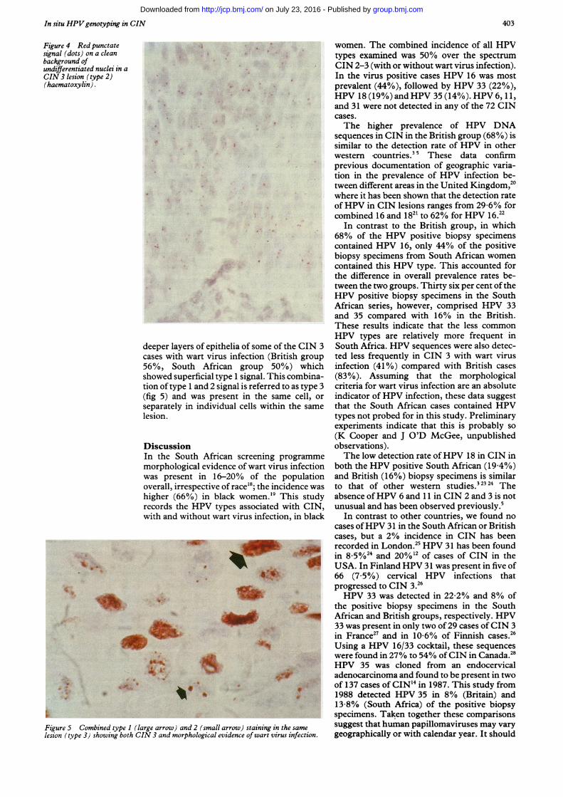
V.` random in distribution and was present in basaland parabasal cells. This took the form of adistinct red punctate signal (dots) on a cleanbackground of haematoxylin stained nuclei:this is referred to as type 2 signal (fig 4) and issimilar to that observed earlier for CaSki cells
is showing which contain integrated HPV 16.'" Interest-ingly, this type of signal was also present in the
402
group.bmj.com on July 23, 2016 - Published by http://jcp.bmj.com/Downloaded from
In situ HPVgenotyping in CIN
Figure 4 Red punctatesignal (dots) on a cleanbackground ofundifferentiated nuclei in aCIN 3 lesion (type 2)(haematoxylin).
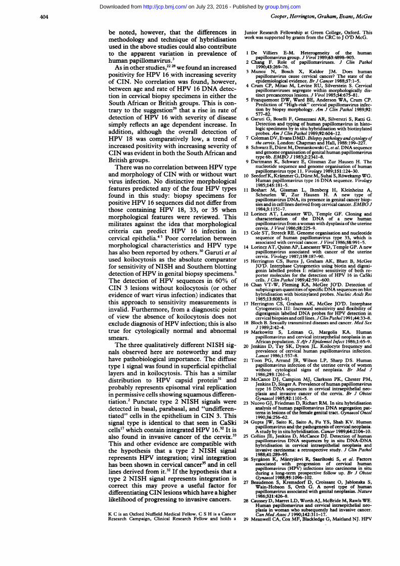
deeper layers of epithelia of some of ticases with wart virus infection (Briti56%, South African group 50%showed superficial type 1 signal. This etion oftype 1 and 2 signal is referred to(fig 5) and was present in the sameseparately in individual cells withinlesion.
DiscussionIn the South African screening pr
morphological evidence of wart viruswas present in 16-20% of the pi
overall, irrespective of race'8; the incichigher (66%) in black women.'9 TIrecords the HPV types associated wwith and without wart virus infection
6.
4l
..1A-
_ .
I.
. 1
I.., 4i
f~~ ~ ~~~~
ao-4
Figure 5 Combined type 1 (large arrow) and 2 (small arrow) staining in thilesion (type 3) showing both CIN 3 and morphological evidence of wart virus
women. The combined incidence of all HPVtypes examined was 50% over the spectrumCIN 2-3 (with or without wart virus infection).In the virus positive cases HPV 16 was mostprevalent (44%), followed by HPV 33 (22%),HPV 18 (19%) and HPV 35 (14%). HPV 6, 11,and 31 were not detected in any of the 72 CINcases.The higher prevalence of HPV DNA
sequences in CIN in the British group (68%) issimilar to the detection rate of HPV in otherwestern -countries."5 These data confirmprevious documentation of geographic varia-tion in the prevalence of HPV infection be-tween different areas in the United Kingdom,20where it has been shown that the detection rateof HPV in CIN lesions ranges from 29-6% forcombined 16 and 1821 to 62% for HPV 16.22
In contrast to the British group, in which68% of the HPV positive biopsy specimenscontained HPV 16, only 44% of the positivebiopsy specimens from South African womencontained this HPV type. This accounted forthe difference in overall prevalence rates be-tween the two groups. Thirty six per cent of theHPV positive biopsy specimens in the SouthAfrican series, however, comprised HPV 33and 35 compared with 16% in the British.These results indicate that the less commonHPV types are relatively more frequent in
he CIN 3 South Africa. HPV sequences were also detec-ish group ted less frequently in CIN 3 with wart virus,) which infection (41%) compared with British casescombina- (83%). Assuming that the morphologicalas type 3 criteria for wart virus infection are an absolute
e cell, or indicator of HPV infection, these data suggestthe same that the South African cases contained HPV
types not probed for in this study. Preliminaryexperiments indicate that this is probably so(K Cooper and J O'D McGee, unpublishedobservations).
ogramme The low detection rate ofHPV 18 in CIN ininfection both the HPV positive South African (19 4%)opulation and British (16%) biopsy specimens is similarlence was to that of other western studies.3 23 24 Thehis study absence ofHPV -6 and 11 in CIN 2 and 3 is not,ith CIN, unusual and has been observed previously.5., in black In contrast to other countries, we found no
cases ofHPV 31 in the South African or Britishcases, but a 2% incidence in CIN has beenrecorded in London.25 HPV 31 has been foundin 8.5%24 and 20%12 of cases of CIN in theUSA. In Finland HPV 31 was present in five of66 (7 5%) cervical HPV infections thatprogressed to CIN 3.26HPV 33 was detected in 22-2% and 8% of
the positive biopsy specimens in the South_ African and British groups, respectively. HPV
33 was present in only two of 29 cases ofCIN 3in France27 and in 10-6% of Finnish cases.26Using a HPV 16/33 cocktail, these sequenceswere found in 27% to 54% ofCIN in Canada.28HPV 35 was cloned from an endocervical
10 adenocarcinoma and found to be present in twoof 137 cases of CIN'4 in 1987. This study from
* 1988 detected HPV 35 in 8% (Britain) and13-8% (South Africa) of the positive biopsyspecimens. Taken together these comparisons
e same suggest that human papillomaviruses may varyinfection. geographically or with calendar year. It should
403
group.bmj.com on July 23, 2016 - Published by http://jcp.bmj.com/Downloaded from
Cooper, Herrington, Graham, Evans, McGee
be noted, however, that the differences inmethodology and technique of hybridisationused in the above studies could also contributeto the apparent variation in prevalence ofhuman papillomavirus.3As in other studies,22 28 we found an increased
positivity for HPV 16 with increasing severityof CIN. No correlation was found, however,between age and rate of HPV 16 DNA detec-tion in cervical biopsy specimens in either theSouth African or British groups. This is con-trary to the suggestion29 that a rise in rate ofdetection of HPV 16 with severity of diseasesimply reflects an age dependent increase. Inaddition, although the overall detection ofHPV 18 was comparatively low, a trend ofincreased positivity with increasing severity ofCIN was evident in both the South African andBritish groups.There was no correlation between HPV type
and morphology of CIN with or without wartvirus infection. No distinctive morphologicalfeatures predicted any of the four HPV typesfound in this study: biopsy specimens forpositive HPV 16 sequences did not differ fromthose containing HPV 18, 33, or 35 whenmorphological features were reviewed. Thismilitates against the idea that morphologicalcriteria can predict HPV 16 infection incervical epithelia.45 Poor correlation betweenmorphological characteristics and HPV typehas also been reported by others.0 Garuti et alused koilocytosis as the absolute comparatorfor sensitivity of NISH and Southern blottingdetection of HPV in genital biopsy specimens.6The detection of HPV sequences in 60% ofCIN 3 lesions without koilocytosis (or otherevidence of wart virus infection) indicates thatthis approach to sensitivity measurements isinvalid. Furthermore, from a diagnostic pointof view the absence of koilocytosis does notexclude diagnosis of HPV infection; this is alsotrue for cytologically normal and abnormalsmears.The three qualitatively different NISH sig-
nals observed here are noteworthy and mayhave pathobiological importance. The diffusetype 1 signal was found in superficial epitheliallayers and in koilocytosis. This has a similardistribution to HPV capsid protein31 andprobably represents episomal viral replicationin permissive cells showing squamous differen-tiation.2 Punctate type 2 NISH signals weredetected in basal, parabasal, and "undifferen-tiated" cells in the epithelium in CIN 3. Thissignal type is identical to that seen in CaSkicells'7 which contain integrated HPV 16.32 It isalso found in invasive cancer of the cervix.33This and other evidence are compatible withthe hypothesis that a type 2 NISH signalrepresents HPV integration; viral integrationhas been shown in cervical cancer35 and in celllines derived from it.32 If the hypothesis that atype 2 NISH signal represents integration iscorrect this may prove a useful factor fordifferentiatingCIN lesions which have a higherlikelihood of progressing to invasive cancers.
K C is an Oxford Nuffield Medical Fellow. C S H is a CancerResearch Campaign, Clinical Research Fellow and holds a
Junior Research Fellowship at Green College, Oxford. Thiswork was supported by grants from the CRC to J O'D McG.
1 De Villiers E-M. Heterogeneity of the humanpapillomavirus group. J Virol 1989;63:4898-903.
2 Chang F. Role of papillomaviruses. J Clin Pathol1990;43:269-76.
3 Munoz N, Bosch X, Kaldor JM. Does humanpapillomavirus cause cervical cancer? The state of theepidemiological evidence. Br J Cancer 1988;57:1-5.
4 Crum CP, Mitao M, Levine RU, Silverstein S. Cervicalpapillomaviruses segregate within morphologically dis-tinct precancerous lesions. J Virol 1985;54:675-81.
5 Franquemont DW, Ward BE, Anderson WA, Crum CP.Prediction of "High-risk" cervical papillomavirus infec-tion by biopsy morphology. Am J Clin Pathol 1989;92:577-82.
6 Garuti G, Boselli F, Genazzani AR, Silverstri S, Ratti G.Detection and typing of human papillomavirus in histo-logic specimens by in situ hybridisation with biotinylatedprobes. Am J Clin Pathol 1989;92:604-12.
7 Coleman DV, Evans DMD. Bilopsy pathology and cytology ofthe cervix. London: Chapman and Hall, 1988:199-227.
8 Schwarz E, Durst M, Demankowski C, et al. DNA sequenceand genome organisation of genital human papillomavirustype 6b. EMBO J 1983;2:2341-8.
9 Dartmann K, Schwarz E, Gissman Zur Hausen H. Thenucleotide sequence and genome organisation of humanpapillomavirus type I 1. Virology 1989;151:124-30.
10 SeedorfK, Krimmer G, Durst M, Suhai S, Rowekamp WG.Human papillomavirus type 16 DNA sequence. Virology1985;145:181-5.
11 Boshart M, Gissman L, Ikenberg H, Kleinheinz A,Scheurlen W, Zur Hausen H. A new type ofpapillomavirus DNA, its presence in genital cancer biop-sies and in cell lines derived from cervical cancer. EMBO J1984;3:1 151-7.
12 Lorincz AT, Lancaster WD, Temple GF. Cloning andcharacterisation of the DNA of a new humanpapillomavirus from a woman with dysplasia of the uterinecervix. J Virol 1986;58:225-9.
13 Cole ST, Streeck RE. Genome organisation and nucleotidesequence of human papillomavirus type 33, which isassociated with cervical cancer. J Virol 1986;58:991-5.
14 Lorincz AT, Quinn AP, Lancaster WD, Temple GP. A newpapillomavirus associated with cancer of the uterinecervix. Virology 1987;159:187-90.
15 Herrington CS, Bums J, Graham AK, Bhatt B, McGeeJO'D. Interphase Cytogenetics using biotin and digoxi-genin labelled probes I: relative sensitivity of both re-porter molecules for the detection of HPV 16 in CaSkicells. J Clin Pathol 1989;42:591-600.
16 Chan VT-W, Fleming KA, McGee JO'D. Detection ofsubpicogram quantities of specific DNA sequences on blothybridisation with biotinylated probes. Nucleic Acids Res1985;13:8083-91.
17 Herrington CS, Graham AK, McGee JO'D. InterphaseCytogenetics III: Increased sensitivity and flexibility ofdigoxigenin labelled DNA probes for HPV detection incervical biopsies and cell lines. JClin Pathol 1991;44:33-8.
18 Bloch B. Sexually transmitted diseases and cancer. Med SexJ 1989;2:42-4.
19 Markowitz S, Leiman G, Margolis KA. Humanpapillomavirus and cervical intraepthelial neoplasia in anAfrican population. S Afr J Epidemiol Infect 1986;1:65-9.
20 Jenkins D, Tay SK, Dyson JL. Koilocyte frequency andprevalence of cervical human papillomavirus infection.Lancet 1986;1:557-8.
21 Toon PG, Arrand JR, Wilson LP, Sharp DS. Humanpapillomavirus infection of the uterine cervix of womenwithout cytological signs of neoplasia. Br Med J1986;293: 1261-4.
22 McCance DJ, Campion MJ, Clarkson PK, Chester PM,Jenkins D, Singer A. Prevalence ofhuman papillomavirustype 16 DNA sequences in cervical intraepithelial neo-plasia and invasive cancer of the cervix. Br J ObstetGynaecol 1985;92:1101-5.
23 Nuovo GJ, Friedman D, Richart RM. In situ hybridisationanalysis of human papillomavirus DNA segregation pat-terns in lesions of the female genital tract. Gynaecol Oncol1990;36:256-62.
24 Gupta JW, Saito K, Saito A, Fu YS, Shah KV. Humanpapillomavirus and the pathogenesis of cervical neoplasia.A study by in situ hybridisation. Cancer 1989;64:2104-10.
25 Collins JE, Jenkins D, McCance DJ. Detection of humanpapillomavirus DNA sequences by in situ DNA-DNAhybridisation in cervical intraepithelial neoplasia andinvasive carcinoma: a retrospective study. J Clin Pathol1988;41:289-95.
26 Syrginen K, Mintyjarvi R, Saarikoski S, et al. Factorsassociated with progression of cervical humanpapillomavirus (HPV) infections into carcinoma in situduring a long-term prospective follow up. Br J ObstetGynaecol 1988;95:1096-102.
27 Beaudenon S, Kremsdorf D, Croissant 0, Jablonska S,Wain-Hobson S, Orth G. A novel type of humanpapillomavirus associated with genital neoplasias. Nature1986;321:426-8.
28 Caussey D, Marret LD, Worth AJ, McBride M, Rawls WE.Human papillomavirus and cervical intraepithelial neo-plasia in woman who subsequently had invasive cancer.Can Med Assoc J 1990;142:31 1-17.
29 Meanwell CA, Cox MF, Blackledge G, Maitland NJ. HPV
404
group.bmj.com on July 23, 2016 - Published by http://jcp.bmj.com/Downloaded from

In situ HPVgenotyping in CIN
16 DNA in normal and malignant cervical epithelium:Implications for the aetiology and behaviour of cervicalneoplasia. Lancet 1987;i:703-7.
30 Kadish AS, Burk RD, Kress Y, Calderin S, Romney SL.Human papillomavirus of different types in precancerouslesions of the uterine cervix: histologic, immunocyto-chemical and ultrastructural studies. Hum Pathol1986;17:384-92.
31 Graham AK, Herrington CS, McGee JO'D. Sensitivity andspecificity of monoclonal antibodies to humanpapillomavirus type 16 capsid protein: Comparison withsimultaneous viral detection by non isotopic hybridisa-
tion. J Clin Pathol 1991;44:96-101.32 Mincheva A, Gissman L, Zur Hausen H. Chromosomal
integration sites of human papillomavirus DNA in threecervical cancer cell lines mapped by in situ hybridisation.Med Microbiol Immunol 1987;176:245-56.
33 Cooper K, Herrington CS, Evans MF, Graham AK, McGeeJO'D. In situ evidence for HPV 16, 18, 33 integration incervical squamous cell cancer in Britain and South Africa.J Clin Pathol 1991:43:1406-90.
34 Durst M, Kleinheinz A, Holtz M, Gissman L. The physicalstate of HPV type 16 DNA in benign and malignantgenital tumours. J Gen Virol 1985;66:1515-22.
405
group.bmj.com on July 23, 2016 - Published by http://jcp.bmj.com/Downloaded from

integration in vivo.patients: evidence for putative HPV neoplasia in South African and Britishgenotyping of cervical intraepithelial In situ human papillomavirus (HPV)
K Cooper, C S Herrington, A K Graham, M F Evans and J O McGee
doi: 10.1136/jcp.44.5.4001991 44: 400-405 J Clin Pathol
http://jcp.bmj.com/content/44/5/400Updated information and services can be found at:
These include:
serviceEmail alerting
box at the top right corner of the online article. Receive free email alerts when new articles cite this article. Sign up in the
Notes
http://group.bmj.com/group/rights-licensing/permissionsTo request permissions go to:
http://journals.bmj.com/cgi/reprintformTo order reprints go to:
http://group.bmj.com/subscribe/To subscribe to BMJ go to:
group.bmj.com on July 23, 2016 - Published by http://jcp.bmj.com/Downloaded from