
Bcl-2 proto-oncogene expression in low- and high-grade prostatic intraepithelial neoplasia

Research ArticleAn Intelligent Clinical Decision Support System forPatient-Specific Predictions to Improve Cervical IntraepithelialNeoplasia Detection
Panagiotis Bountris,1 Maria Haritou,2 Abraham Pouliakis,3 Niki Margari,3
Maria Kyrgiou,4,5 Aris Spathis,3 Asimakis Pappas,6 Ioannis Panayiotides,7
Evangelos A. Paraskevaidis,8 Petros Karakitsos,3 and Dimitrios-Dionyssios Koutsouris1
1 Biomedical Engineering Laboratory, School of Electrical and Computer Engineering, National Technical University of Athens,Iroon Politechniou 9, 15773 Zografou Campus, Athens, Greece
2 Institute of Communication and Computer Systems, National Technical University of Athens, Iroon Politechniou 9,15773 Zografou Campus, Athens, Greece
3 Department of Cytopathology, School of Medicine, University General Hospital “ATTIKON”, University of Athens, Rimini 1,12462 Athens, Greece
4West LondonGynaecological Cancer Center, Queen Charlotte’s and Chelsea, Hammersmith Hospital, Imperial Healthcare NHS Trust,London W12 0HS, UK
5Division of Surgery and Cancer, Faculty of Medicine, Imperial College, London W12 0NN, UK6 3rd Department of Obstetrics and Gynecology, University General Hospital “ATTIKON”, School of Medicine, University of Athens,Rimini 1, 12462 Athens, Greece
7 2nd Department of Pathology, University General Hospital “ATTIKON”, School of Medicine, University of Athens, Rimini 1,12462 Athens, Greece
8Department of Obstetrics and Gynecology, University Hospital of Ioannina, St. Niarchou Str, 45500 Ioannina, Greece
Correspondence should be addressed to Panagiotis Bountris; [email protected]
Received 9 October 2013; Revised 10 February 2014; Accepted 16 March 2014; Published 9 April 2014
Academic Editor: Nasimul Noman
Copyright © 2014 Panagiotis Bountris et al. This is an open access article distributed under the Creative Commons AttributionLicense, which permits unrestricted use, distribution, and reproduction in any medium, provided the original work is properlycited.
Nowadays, there are molecular biology techniques providing information related to cervical cancer and its cause: the humanPapillomavirus (HPV), including DNA microarrays identifying HPV subtypes, mRNA techniques such as nucleic acid basedamplification or flow cytometry identifying E6/E7 oncogenes, and immunocytochemistry techniques such as overexpressionof p16. Each one of these techniques has its own performance, limitations and advantages, thus a combinatorial approach viacomputational intelligence methods could exploit the benefits of each method and produce more accurate results. In this articlewe propose a clinical decision support system (CDSS), composed by artificial neural networks, intelligently combining the resultsof classic and ancillary techniques for diagnostic accuracy improvement. We evaluated this method on 740 cases with completeseries of cytological assessment, molecular tests, and colposcopy examination. The CDSS demonstrated high sensitivity (89.4%),high specificity (97.1%), high positive predictive value (89.4%), and high negative predictive value (97.1%), for detecting cervicalintraepithelial neoplasia grade 2 or worse (CIN2+). In comparison to the tests involved in this study and their combinations, theCDSS produced the most balanced results in terms of sensitivity, specificity, PPV, and NPV. The proposed system may reduce thereferral rate for colposcopy and guide personalised management and therapeutic interventions.
1. Introduction
Cervical cancer is the third most common cancer and thefourth leading cause of cancer death in females worldwide
[1]. Cervical cancer is known to be caused almost always byhuman papillomavirus (HPV) infection which is the com-monest sexually transmitted infection worldwide. However,the presence of HPV does not always lead to disease [2].
Hindawi Publishing CorporationBioMed Research InternationalVolume 2014, Article ID 341483, 20 pageshttp://dx.doi.org/10.1155/2014/341483

2 BioMed Research International
About 100 types of HPV virus have been identified thatcan infect humans. Among them, at least 15 are oncogenicand thus can cause cancer of the cervix [3, 4]. Improvedunderstanding of HPV infection and the natural history ofcervical neoplasia have resulted in the addition of the HPVDNA test along with the Pap test.
From the meta-analysis of the most authoritative pub-lished studies [5–8] it can be concluded that the sensitivityof Pap test combined with the HPV DNA test is higher thanthe sensitivity of each individual method. This observationsuggests that the two methods complement each othereffectively. In contrast, the specificity of the Pap test combinedwith the HPVDNA test was lower than the ratings of the twomethods separately as they differ in sensitivity and specificity[9, 10]. Regarding the positive predictive value (PPV) thefindings are equivocal: some studies report that the values ofPPV were similar for each method separately and for theircombination, while others report smaller values of PPV fortheir combination. As expected, the negative predictive value(NPV) of HPVDNA test in conjunction with the Pap test washigh and some studies report values of almost 100%.
In the recent years, new technologies for cervical cancerdetection have been promoted to physicians and the public.Some studies proposed the shift from DNA detection tomRNA identification of the viral E6/E7 oncogenes that arelinked to oncogenic activation. Among them, mRNA typingwith nucleic acid sequence based amplification (NASBA) [11–13] and flow cytometry (mRNA-Flow-FISH) techniques forE6/E7 HPV mRNA detection have been enrolled in cancerand precancerous lesions’ detection with promising resultsin increasing PPV and reducing unnecessary recalls andreferrals to colposcopy [14–18]. At the same time, it seems thatthe immunocytochemical detection of genetic effects such asoverexpression of p16 is a methodology which can increasethe diagnostic accuracy of the Pap test [19, 20].
Several published studies in the literature are attemptingto clarify the role of each technique as a unique test tosubstitute or replace the Pap test [5–8, 11, 14–24]. By thedetailed analysis of the published studies it can be con-cluded that the performance of the methods under controldiffer significantly, affected by the disease incidence andthe prevalence of HPV infection in the population studygroup, resulting in that the individual application of onemethod, even if it offers a level of protection, does not reliablydetermine the risk of each individual woman.
Advances in the areas of computer science and artificialintelligence allow the development of computer systems thatsupport clinical diagnosis or therapeutic and treatment deci-sions based on individualised patient data [25, 26]. Clinicaldecision support systems (CDSSs) aim to codify and strate-gically manage biomedical knowledge to handle challengesin clinical practice using mathematical modelling tools,medical data processing techniques, and artificial intelligencemethods. CDSSs cover a wide range of applications, fromdiagnosis’ support tomodelling the probability of occurrenceof various diseases or the efficiency of alternative therapeuticschemes. To do so, they are using not only individual patientdata but also data on risk factors and efficiency of availabletherapeutic schemes stored in databases. CDSSs are based
on statistical analysis methods, such as regression analysis,or artificial intelligence techniques, such as artificial neuralnetworks (ANNs) and pattern recognition techniques [27].These can be used in order to extract hidden informationwithessential clinical value from large datasets. Based on complexalgorithms, CDSSs may combine in a nonlinear complexway a number of characteristics, for example, data relatedto the patient (epidemiologic data, medical history, etc.),data related to the disease (examinations’ results, biomarkers,course of the disease, etc.), or data related to the treatment(drug selection, drug doses, etc.). In this way, CDSSs provideclinicians with patient-specific assessments or recommenda-tions to aid clinical decisionmaking, or, evenmore, to providepredictions of diagnostic or prognostic outcomes.
Regarding cervical cancer, an intelligent decision makingsystem may support physicians to improve the selection ofprotocols for monitoring, diagnosing, and treating womenwith intraepithelial lesions or cervical cancer or even supportthe rational selection and the patient-specific follow-updecision making for women who have been treated for high-grade lesions. The majority of published studies, regardingintelligent systems for cervical cancer support, are concernedabout computer aided diagnosis systems based on eithercytology or colposcopy image analysis [28–31]. On the otherhand, various papers have been published in the past fewyears concerning bioinformatics’ CDSSs based on ANNsfor cancer improved detection, treatment, and follow-upsupport [32–37]. To the best of our knowledge, however,a similar bioinformatics intelligent CDSS for supportingand improving cervical cancer detection and triage, like theproposed system, has not been reported in the literature.
This study aims to investigate the potential role of anovel intelligent bioinformatics CDSS which intelligentlycombines the results of various diagnostic techniques usedin the modern cytopathology laboratory in order to provideclinicians with patient-specific predictions of diagnostic orprognostic outcomes and thus to identify women at truerisk of developing cervical cancer. The preliminary resultssuggest that the proposed system may improve the accuracyof diagnosis and in comparison to other combinatorialapproaches produces the most balanced results in terms ofspecificity, sensitivity, PPV, and NPV.
2. Materials and Methods
2.1. Clinical Data. Data have been collected randomly fromwomen enrolled in a research project conducted by theDepartment of Cytopathology of the Medical School ofAthens University (“ATTIKON” University Hospital) andthe Department of Obstetrics and Gynaecology of the Uni-versity Hospital of Ioannina. Our study has been approvedby the Bioethics Committee of the “ATTIKON” UniversityHospital and the Bioethics Committee of the UniversityHospital of Ioannina. Participating women had signed aninformed patient consent (ICON) form allowing the use oftheir epidemiologic, diagnostic, and molecular data for theneeds of the system’s development and research. The clinicaldata (molecular examinations’ results, cytological diagnoses,

BioMed Research International 3
histological examination of biopsies, visit number and date,patient age, etc.) have been registered and stored into adatabase which has been developed for the research projectpurposes. For the processing of the data and algorithmtraining and testing, anonymised data were extracted fromthe database.
The study was carried out on ThinPrep LBC specimensobtained from women referred for colposcopy for two rea-sons: (1) either because they had a previous abnormal Paptest, or (2) they volunteered to participate in the studyand accepted a colposcopical examination as well as theapplication of various biomarkers on their biological materialeven if they had a normal Pap test (e.g., women with HPV).In case that a negative Pap test was followed by negativecolposcopy, no biopsy was taken and the case was consideredas clinically negative. The smears were routinely preparedfor cytological examination and the remaining material wasused for evaluation of additional biomarkers (reflex tests).The cytology was assessed by experienced cytopathologists.Biopsies were obtained from samples during colposcopyand/or from surgical specimens through conization and werefixed and prepared according to standard histopathologyprotocols. All cases except the clinically negative ones werediagnosed by a single expert histopathologist as daily routinediagnosis and the evaluation of the biopsy was blindedfrom the results of cytology and other ancillary tests. Thehistopathologist uses as standard procedure p16 test (CINtec)in all histological material from the cervix.
The patients’ database includes more than 5,000 patientswith more than 10,000 tests’ series due to follow-up cases.Each of these series includes the following tests: cytologyaccording to the revised Bethesda classification (TBS2001system) [38, 39]; HPV DNA typing using the CLARTHUMAN PAPILLOMAVIRUS 2 (GENOMICA) that allowssimultaneous detection of 35 different HPV genotypes (bothhigh and low risk) by PCR amplification of a fragment withinthe highly conserved L1 region of the virus [40]; NASBAassays [41] (NucliSENS EasyQ HPV v1.0) that are used forthe identification of E6/E7 mRNA of the HPV types 16, 18,31, 33, and 45; the PermiFlow (Invirion Diagnostics, LLC,Oak Brook, IL) that allows the identification of E6/E7 mRNAexpression of high-risk HPV using flow cytometry technique[17]; and finally the immunocytochemical expression of p16using the CINtec Cytology Kit [42]. All these examinationsproduce results that can be used in a classification process andthey provide assessments of the clinical cytological sample asa whole and not of individual cells.
Cytological findings of each patient were interpretedaccording to the Bethesda classification system andwere clas-sified as follows: (a) within normal limits (WNL); (b) atypicalsquamous cells of undetermined significance (ASCUS); (c)low-grade squamous intraepithelial lesion (LSIL); (d) high-grade squamous intraepithelial lesion (HSIL); (e) squa-mous cell carcinoma (SCC) or adenocarcinoma (Adeno-Ca). Regarding the HPV DNA test, for which the cytologylaboratory is accredited by WHO and is proficient for thespecific technique, we considered high-risk (HR) HPV typesas HPV types 16, 18, 26, 31, 33, 35, 39, 45, 51, 52, 53, 56, 58,59, 66, 68, 73, 82, and 85; and low-risk (LR) HPV types as
HPV types 6, 11, 40, 42, 43, 44, 54, 61, 62, 70, 71, 72, 81, 83, 84,and 89 [3, 4]. It is well known that the probability of a low-risk subtype to cause cervical lesions is very small; however,the specificHPVDNA test is simultaneously identifying bothhigh-risk and low-risk HPV subtypes and thus we used allavailable typing results during the development of the system,in order to evaluate its performance based on all availableinformation.
For the cases that had histological outcome, the his-tological diagnosis was used as the golden standard. Ran-dom biopsies were not obtained in clinically negative cases,which are defined as cases that had negative cytology andadditionally negative colposcopy; for these cases it is notallowed by the ethical committee to have a sample forhistological examination. These cases may introduce a smallbias in the interpretation of the cytology performance. Acervical biopsy was performed if Pap test revealed ASCUSand above cytological categories (ASCUS+) or there wasa visible lesion upon colposcopy. Biopsy was performedby experienced colposcopists (in practice for more than 10years) as part of the study protocol. The three-tiered cervicalintraepithelial neoplasia (CIN) grading system was used forhistological diagnosis; thus the cases with histology wereclassified as follows: (a) without evidence of malignancy(negative histology); (b) cervical intraepithelial neoplasiagrade I (CIN1); (c) cervical intraepithelial neoplasia gradeII or III (CIN2/3); (d) squamous cell carcinoma (SCC) oradenocarcinoma (Adeno-Ca).
From the more than 10,000 tests’ series, 740 cases thatwere fully completed were selected for examination in thisstudy and were further analysed (Table 1). Cases with oneor more missing or invalid/inadequate tests’ results wereexcluded from the study.
For each of the 740 cases, a feature set consisting of 46variables derived by the applied tests was created. The resultof the cytological examination has been used according to theBethesda system. Results of the HPV DNA test examinationwere expressed as 35 individual variables (either positive ornegative), one for each HPV DNA genotype. For the EasyQtest (NASBA) we used the result for each individual HPVtype (16, 18, 31, 33, and 45). The result of the PermiFlow test(FLOW) was used as positive or negative, as was the resultof the immunocytochemical expression of p16. Additionally,other variables expressing the HPV DNA test results wereadded; for instance, the existence of high-risk or low-risktypes was expressed as either positive or negative. Table 2shows in detail the 46 independent variables/features thatwere collected for each case.
According to the final outcome (the histological exami-nation or a clinically negative result), each of the 740 caseswas classified into the following classes: class 1: negativeor clinically negative, class 2: CIN1, class 3: CIN2 or CIN3(CIN2/3), and class 4: cancer (SCC or Adeno-Ca).
2.2. Feature Selection. The first step of our study was toemploy feature selection algorithms in order to identifywhichof the 46 aforementioned features contributes most to theprediction of the underlying condition of eachwoman, that is,

4 BioMed Research International
Table 1: Correlation between the cytological and histological findings of the dataset used in the study.
Pap test result TotalWNL ASCUS LSIL HSIL SCC/Adeno-Ca
Histological examination resultClinically negative 196 0 0 0 0 196 (26.5%)Negative 35 60 22 5 0 122 (16.5%)CIN1 31 66 142 22 0 261 (35.3%)CIN2/3 3 13 27 93 0 136 (18.4%)SCC/Adeno-Ca 0 1 2 7 15 25 (3.4%)
Total 265 (35.8%) 140 (18.9%) 193 (26.1%) 127 (17.2%) 15 (2%) 740WNL: within normal limits, ASCUS: atypical squamous cells of unknown significance, LSIL: low-grade squamous intraepithelial lesion, HSIL: high-gradesquamous intraepithelial lesion, SCC: squamous cell carcinoma, Adeno-Ca: adenocarcinoma, CIN: cervical intraepithelial neoplasia.
the class of each case.The feature selection (FS) [43] problemin pattern recognition may be stated as follows: given a set of𝑁 features, find the best subset consisting of 𝑙 features thatcontribute most to classification/prediction accuracy.
Feature subset selection algorithms can be classified intotwo main categories: the filter approach and the wrapperapproach. In the filter approach the FS is done independentlyof the learning algorithm of a classifier. A class separabilitymeasuring criterion C(k) is adopted in order to rank all thefeatures. The value of the criterion C(k) is computed for eachof the features 𝑘 = 1, 2, . . . 𝑚. Features are then rankedin the order of descending values of C(k). The 𝑙 featurescorresponding to the 𝑙 best values of the specific criterion areselected to form the best subset. Inwrapper typemethods, theFS is “wrapped” around a learningmethod: the usefulness of afeature subset is directly judged by the estimated accuracy ofa trained classifier. For high-dimensional datasets, wrappermethods are far too computationally expensive to be usedbecause each feature subset considered must be evaluatedwith the trained classifiers. For this reason, wrapper methodswill not be considered in this study.
In order to perform the FS task, we combined twodifferent filter methods [44]. The area between the empiricalreceiver operating characteristic (ROC) curve and the ran-dom classifier slope has been proposed as a class separabilitymeasuring criterion [43]. As presented in [43], itmeasures theoverlap between the probability density functions describingthe data distribution of a feature in two classes. This criterionserves as a measure of the class discrimination capabilityof a specific feature. The second filter method we appliedis the recently proposed minimum redundancy-maximumrelevance (MRMR) feature selection framework, a mutualinformation based methodology, which has proved to be oneof the best filter methods [45]. Both methods return the bestfeature subset for a selected value l.
During feature ranking using the ROC FS method, weapplied an ad hoc cross correlation technique that incorpo-rates correlation information into the ranking procedure soas to avoid the existence of correlated features into the bestsubset. The cross correlation coefficient between features isconsidered in order to exclude features which are correlatedwith the top-ranked. This technique is described in detail in
[43]. Using this procedure we are able to define themaximumnumber of the 𝑙 best features.
Both of the adopted FS methods measure the classifica-tion capability of a specific feature with respect to a two-class problem. Since our dataset consists of 4 classes, in orderto perform the FS task, we adopted a similar to the “oneagainst one” multi-class classification strategy. Thus, we splitthe dataset into 6 splits, one for each pair of classes; thenwe applied the 2 mentioned FS techniques to each split andlastly we explored the common top-ranked features betweenthe 12 best feature subsets returned by the two FS techniques.Ultimately, these 𝑙 common top-ranked features form the bestfeature subset which was used for the development of thepresented system.
2.3. Classification/PredictionModels and System’s Architecture.The development of an intelligent clinical decision supportsystem involves the employment of several machine learningmethods and pattern recognition/classification techniques.Machine learning is concerned about the development andthe study of intelligent systems that can learn fromdata, whilepattern recognition/classification aims to use these systemsin order to classify an object into a correct class based onfeatures characterising the object. These features are the onesprovided by the FS task. The machine learning algorithmsthat implement pattern classification are known as classifiers.There is a variety of classifiers used for pattern classification;each one of those has certain limitations and advantages.There are simple classifiers like the 𝑘-nearest neighbours (k-NN) and the Bayesian classifier and more complex ones likethe artificial neural networks (ANNs) [27, 43, 46, 47]. Forexample, the 𝑘-NN may be considered as a simple classifierbecause it classifies a sample based on the 𝑘-closest trainingsamples in the feature space; it just computes the distancesbetween the new sample and the samples of the training setand according to these distances it classifies the sample to theclass of the closest training samples. On the contrary, ANNsare complex networks of artificial neurons interconnectedwith each other, which obtain knowledge and the abilityto classify a sample by the application of complex learningalgorithms. This capability of learning from a certain datasetmakes the neural networks suitable for classification andprediction tasks in practical situations. Furthermore, neural

BioMed Research International 5
Table 2: Variables characterising patients’ biological status.
Variable name Description Value range
Pap testThe result of the cytologicalexamination expressed according toBethesda system
1 :WNL, 2 : ASCUS, 3 : LSIL,4 : HSIL, 5 : SCC or ADENO-Ca
HPV DNA Arrays: HPV-6, HPV-11, HPV-16,HPV-18, HPV-26, HPV-31, HPV-33, HPV-35,HPV-39, HPV-40, HPV-42, HPV-43, HPV-44,HPV-45, HPV-51, HPV-52, HPV-53, HPV-54,HPV-56, HPV-58, HPV-59, HPV-61, HPV-62,HPV-66, HPV-68, HPV-70, HPV-71, HPV-72,HPV-73, HPV-81, HPV-82, HPV-83, HPV-84,HPV-85, HPV-89
The existence of individual subtypesaccording to the HPV DNAexamination
0 if the specific subtype is notfound, 1 if the specific subtype isfound
HR-HPV DNA The existence of high-risk subtypesfound by the HPV DNA test
0 if none of the high-risk typeswas found, 1 if at least one of thehigh-risk types is found
LR-HPV DNA The existence of low-risk subtypesfound by the HPV DNA test
0 if none of the low-risk types isfound, 1 if at least one of thelow-risk types is found
Arrays number The number of HPV subtypes foundby the HPV DNA test Expressed as number
N16 The result of the NASBA mRNA testfor HPV subtype 16 0 if negative, 1 if positive
N18 The result of the NASBA mRNA testfor HPV subtype 18 0 if negative, 1 if positive
N31 The result of the NASBA mRNA testfor HPV subtype 31 0 if negative, 1 if positive
N33 The result of the NASBA mRNA testfor HPV subtype 33 0 if negative, 1 if positive
N45 The result of the NASBA mRNA testfor HPV subtype 45 0 if negative, 1 if positive
FLOWThe result of the identification ofE6/E7 mRNA expression of high-riskHPV using flow cytometry technique
0 if negative (positive expression<1.5%), 1 if positive (positiveexpression >1.5%)
p16 The result of the p16immunocytochemical examination 0 if negative, 1 if positive
WNL: within normal limits, ASCUS: atypical squamous cells of unknown significance, LSIL: low-grade intraepithelial lesion, HSIL: high-grade squamousintraepithelial lesion, SCC: squamous cell carcinoma, ADENO-Ca: adenocarcinoma.
networks are inherently nonlinear which makes them moresuitable for processing complex data patterns, in contrast tomany traditional methods.
In this study, in order to develop the proposed CDSS, weemployed and tested 6 classifiers: the k-nearest neighbours(k-NN) classifier [46], the naıve Bayesian (NB) classifier [43],the classification and regression tree (CART) [48], and 3 typesof ANNs, namely, the multilayer perceptron network (MLP)[47], the radial basis function network (RBF) [47], and theprobabilistic neural network (PNN) [49, 50].
The classifiers were designed to classify the cases into thefollowing 4 groups corresponding to the cervical histology:negative, CIN1, CIN2/3, and cancer (SCC or Adeno-Ca). Thefeature subset characterising each case, which is derived bythe FS task, was used as the input of each classifier. Thus,each classifier takes as inputs results from the examinationsof each case and outputs its classification group, providingin this way a prediction regarding the actual cervical status
of each woman. The available dataset (Table 1) was dividedinto 3 sets: the training set (486 cases) which was used tobuild and train the classifiers, the validation set (126 cases)which was used to optimise the parameters of each classifier,and the test set (128 cases) which was used to evaluate theirpredictive performance. The 3 sets were properly stratifiedso that the classes’ distribution in each set is approximatelythe same as that in the initial dataset. Thus, each set containsrepresentative samples of the same larger population. Wehave also to note that, due to the many different classes andthe diversity of the examinations’ results, we had to use a largeset for training (66% of the initial dataset) so as to provideto the classifiers representative samples of every situation.During the training phase, the cases of the training set areprovided to the classifiers alongwith their classes (their actualhistology) and the classifiers learn from this specific datasetusing a learning algorithm. The cases of the validation andthe test sets are not used during training; thus these cases are

6 BioMed Research International
unknown to the classifiers (unseen data).The validation set isused as a test platform for fine tuningmodel’s parameters andselecting the best-performingmodel, while the test set is usedto assess the predictive performance of the developedmodelson data which have not been used in any way in the designingprocess.
Through a process of designing, training, and testingthe aforementioned classifiers, we tried to investigate thepotential role of these models to improve the accuracy ofdiagnosis. However, no single classifier produced satisfactoryresults. By thoroughly investigating the classification resultsfor each case separately, we discovered that the PNN demon-strated good predictive performance for most cases withcytology LSIL and above, and specifically for women withcytology LSIL harbouring CIN grade 2 or worse (CIN2+).However, the PNN showed poor performance on identifyingthe correct histology of women with ASCUS cytology. Onthe other hand, we discovered that the MLP, even thoughunderperformed regarding the whole dataset, produced goodprediction outcomes for the cases with ASCUS cytology.Thisfact led us to design a hybrid architecture, by combining aPNN and a MLP.
The presented CDSS consists of two subsystems: themainsubsystem is a PNN, while the secondary is a MLP. ThePNN is used for the management of all the cases excludingthose with Pap test ASCUS, while the MLP is used for themanagement of the ASCUS cases only. According to thevalue of the Pap test, each case is promoted to the mainor the secondary subsystem; if Pap test is ASCUS, the dataare promoted to the MLP; otherwise they are promoted tothe PNN. The schematic diagram of the proposed decisionsupport system is presented in Figure 1. This architectureproved to provide better classification results comparing toa single classifier approach.
The learning and the predictive ability of an ANN isdetermined by several factors, such as the type of thenetwork and the parameters of the specific type, the network’sarchitecture (topology), the learning algorithm chosen fortraining, and the characteristics of the data provided to thenetwork. Therefore, in order to construct the CDSS, the taskof identifying the optimal architecture and parameters of thePNN and the MLP has to be accomplished.
2.3.1. The Probabilistic Neural Network (PNN). A proba-bilistic neural network is a kind of multilayer feed-forwardradial basis artificial neural network suitable for classificationand prediction problems. PNNs are widely used for patternrecognition problems, nonlinear mapping, and estimation ofthe probability of classmembership and likelihood ratios.ThePNN is based on the theory of Bayesian classification and isclosely related to classical estimators for probability densityfunctions [49, 50]. The basic operation performed by thePNN is an estimation of the probability density function ofeach class from the provided training samples using Gaussiankernels. These estimated densities are then used in a decisionrule to perform the classification.
The PNN architecture consists of four layers: the inputlayer, the pattern layer, the summation layer, and the output
layer, as depicted in Figure 2. The number of neurons ofthe two hidden layers (pattern and summation layers) isdetermined by the training set.The pattern layer contains oneneuron for each sample of the training set, while the summa-tion layer contains one neuron for each class of the trainingset. The training process of the PNN is straightforward and itis accomplished by setting the weights of the network usingthe samples of the training set; thus no learning algorithm isapplied during PNN’s implementation. The weights betweenthe input and the pattern layer are set as follows:
𝑤(𝑃)
𝑖𝑗 = 𝑝𝑖𝑗, (1)
where 𝑤(𝑃)
𝑖𝑗is the weight between the 𝑖th neuron of the input
layer and 𝑗th neuron of the pattern layer, and 𝑝𝑖𝑗 is the valueof the 𝑖th feature of the 𝑗th sample of the training set. Theweights between the pattern and the summation layer are setas follows:
𝑤(𝑆)
𝑗𝑘= {
1 if 𝑇(𝑗)
𝑘= 1
0 else,(2)
where𝑤(𝑆)𝑗𝑘
is theweight between the 𝑗th neuron of the patternlayer and 𝑘th neuron of the summation layer. The value of𝑇(𝑗)
𝑘is 1 only when sample 𝑗 is associated with class 𝑘 and 0
elsewhere.When an input is presented, the pattern layer computes
the distances between the input vector and the trainingvectors and produces a vector which indicates how close theinput is to the training samples, as follows:
𝑑(𝑃)
𝑗 = √∑𝑖
(𝑤(𝑃)
𝑖𝑗− 𝑥𝑖)2, (3)
where𝑑(𝑃)𝑗
is the distance between the input vector and the 𝑗thsample of the training set, while 𝑥𝑖 denotes the 𝑖th variable ofthe input (𝑖th node of input layer).
The transfer function of the neurons of the pattern layeris a radial basis function. The output of each pattern neuronis computed as
𝑃𝑗 = exp(−𝑑(𝑃)
𝑗
2𝜎2) , (4)
where 𝜎 is a smoothing parameter corresponding to thestandard deviation of the Gaussian distribution.
Each summation neuron sums the contributions for eachclass of the input to produce at the net output a vector ofprobabilities. The output of each summation node can beexpressed as
𝑆𝑘 =1
∑𝑗 𝑤(𝑆)
𝑗𝑘
∑𝑗
𝑤(𝑆)
𝑗𝑘⋅ 𝑃𝑗. (5)
Finally, a competitive transfer function in the outputlayer (single neuron) classifies the input vector into a specific

BioMed Research International 7
System’s inputs
System’s output Pap test
HPV DNA arrays
NASBA assay
FLOW
p16
Datatransformation
MLP
PNN
Datainterpretation
Clinical decision support system
Inpu
ts
Pattern layer
Output layer
Summation layer
Inputlayer
Outputlayer
Hiddenlayers
∙
∙
∙
∙
∙
Patients’histologyprediction
.........
......
...
.........
y
x1
x2
xn
x1
x2
xn
y1
ym
Patients’examinations
results:
If Pap ≠ASCUS
If Pap = ASCUS
Figure 1: Schematic block diagram of the proposed decision support system. The examinations’ results of each patient are used as inputsto the CDSS. The medical information is transformed to data appropriate for processing by the PNN and the MLP subsystems. Accordingto the value of the Pap test, the transformed data of each case is promoted to the PNN or the MLP subsystem; if Pap test is ASCUS, thedata are promoted to the MLP; otherwise they are promoted to the PNN. The output of each network is properly transformed by the datainterpretation block to medical information. At the end, the CDSS provides predictions regarding the actual cervical status of each woman.NASBA: nucleic acid sequence based amplification for the identification of E6/E7 mRNA of the HPV types 16, 18, 31, 33, and 45; FLOW: flowcytometric E6/E7 HPVmRNA assay; p16: p16 immunocytochemical examination; ASCUS: atypical squamous cells of unknown significance;PNN: probabilistic neural network; MLP: multilayer perceptron network.
Inputs
Pattern layer
Output layer
Summation layer
x1
x2
xn
xi wij(P) Pj w
jk(S) Sk
y
......
...
Figure 2: Architecture of a probabilistic neural network (PNN).
class if that class had the maximum output value from thecorresponding neuron at the summation layer:
𝑦 = argmax𝑘
𝑆𝑘. (6)
From the above, it is obvious that in the PNN architecturethe number of the hidden layers and the transfer functions ofthe neurons are predefined and the number of the neuronsof each hidden layer depends on the size and the form of thetraining set. The single free parameter of this network is the
smoothing parameter sigma (𝜎), the standard deviation of theGaussians.Thus, the selection of the optimal PNN, which willconstitute the main subsystem of the CDSS, is essentially theact of determining the optimal sigma value.
2.3.2. The Multilayer Perceptron Network (MLP). The mul-tilayer perceptron is the most widely used neural network.It is a feed-forward ANN with an input layer that receivesexternal inputs, one or more hidden layers and an output

8 BioMed Research International
layer providing the output of the network. Each layer ofthe MLP includes one or more neurons directionally linkedwith the neurons from the previous and the next layer.Determining the right architecture of a MLP is the task ofselecting the optimal parameters of the network, such as thenumber of the hidden layers and the number of the neuronsof each hidden layer. As far as the learning algorithm isconcerned, the back-propagation (BP) algorithm [47] is themost common learning technique for training a typical MLP.During trainingwith theBP algorithm, information about theerrors of the network on known data is propagated backwardsfrom the output layer to the input layer and it is used to adjustthe connections between the layers and their neurons (theweights and biases of the network) in order to minimize theerror and thus improve performance.
As far as the training of the MLPs is concerned, theLevenberg-Marquardt BP algorithmwas used for the learningprocess, while the mean squared normalized error (MSE)was used as the network’s cost function [47]. Training aMLP is essentially the process of modifying the weightsand biases of the network in order to minimize this costfunction. The learning rate and the momentum of the BPalgorithm were set equal to 0.1 and 0.9, respectively. In orderto avoid overfitting, an early-stopping learning techniquewas implemented, according to which the classification erroron the validation subset was monitored during the trainingprocess. When the validation error increases for a specifiednumber of iterations of the BP algorithm, the training isstopped, and the weights and biases at the minimum of thevalidation error are returned. For the training process, criteriafor convergence were met with 40 maximum validationfailures or when MSE ≤ 0.0001 or when a maximum of 1000iterations (epochs) was reached.
As discussed in [51], empirical studies often found thatnetworks with many hidden layers generally perform nobetter, and often worse, than neural networks with one ortwo hidden layers. Thus, in this study, we considered MLParchitectures with one or two hidden layers. All neurons inthe hidden and the output layers use the sigmoidal activationfunction. In order to identify the optimal network topology,we applied a trial-and-error cascade constructive processby adding neurons to the hidden layers, one at time, andevaluating the developed MLPs. For each MLP architecture(one-hidden-layered and two-hidden-layered), this processwas stopped when the MLPs showed continuous impairedperformance.
With the determined number of hidden layers and neu-rons, both the learning rate and the momentum coefficient ofthe BP algorithm were further investigated to ensure a highprobability of global network convergence.
2.3.3. Selection of the Optimal Models. In order to identifythe optimal parameters of the developed classifiers andeventually select the optimal PNN and MLP which comprisethe CDSS’s subsystems, we performed the parameter tuningtask by evaluating the developedmodels on the validation set.However, during this task we discovered that classifiers withdifferent parameters presented the same best performance on
the validation set, making it difficult to select the optimalmodels. Thus, in order to perform more accurately themodel selection task, we also took into consideration theclassification performance on the training set.
Let the classification error on the training set (resubsti-tution error) be denoted as 𝜀
𝑟 and the classification error onthe validation set as 𝜀V. For each classification algorithm usedin this study, we build 𝑆 classifiers with different parameters,and we define as optimal the classifier which minimizes thecost function:
𝐽𝑚 =𝜀𝑟𝑚 + 𝜀V𝑚
2, ∀𝑚 ∈ 𝑆, ∀𝜀
V𝑚 ≡ min {𝜀
V𝑚} . (7)
Utilizing the above cost function for the selection ofthe optimal parameters, we ensure that the optimal modelsdemonstrate the best predictive performance on the valida-tion set (the models which minimize the cost function mustproduce the minimum classification error on the validationset) and at the same time they perform well on the cases ofthe training set (themodels whichminimize the cost functionmust produce low resubstitution error).
2.3.4. Performance Evaluation. The final performance evalu-ation task was carried out using resubstitution and holdoutvalidations. In resubstitution validation, the model is testedon the data which have been used in the learning process,that is, the data of the training set. This method providesa measure of the network’s learning ability; yet it is notpreferable for performance evaluation tasks as it is knownto be optimistically biased. However, as shown in [52, 53], indiscrete classification problems with large-sample categoricaldatasets, like the classification problem of this study, resubsti-tution can be significantly accurate relative to more complexerror estimation schemes, since the optimistic bias and thevariance of the method tend to be vanished as the samplesize increases, provided that classifier complexity is not toohigh. For this reasonwedecided to take into consideration theperformance of the final models when for testing the trainingand the validation sets are used. In holdout validation theoptimal models are tested on data that were not used in anyway in the building process (training and parameter tuning),that is, the data of the test set.The holdout classification errorserves as a measure of the model’s predictive (generalization)ability.
We have to note that we did not use complex errorestimation schemes like the 𝑘-fold cross validation, so as to beable to study the classification result for each case separatelyand thus to evaluate the system at individual level. It must benoted that for the clinicians the significance of such a systemstands on its ability to correctly identify cases with conflictingtests’ results which are difficult to be evaluated by them.Hence, more than the overall accuracy, what is important isthe correct identification of as many as possible women withinsignificant cytological findings harbouring CIN2+ lesions,as well as the correct identification of women with HSIL+cytology but with actual histology below CIN2.
Due to the above, the predictive performance of the finallyselected ANNs is presented by confusion matrices obtained

BioMed Research International 9
Table 3: Classification accuracies on the training, validation, and test sets of the 6 classifiers developed (single classifier approach).
Classifier k-NN NB CART MLP RBF PNN
Optimalparameters k = 5 —
(i) Pruning level = 7/11(ii) Number of terminal
nodes = 82 hidden layers 18 × 18
(i) 486 neurons in thehidden layer (all samples
of training set)(ii) Sigma = 0.6
Sigma = 0.4
Training set 78.6% 76.8% 77.6% 78.4% 87.6% 87.6%Validation set 79.4% 80.9% 78.6% 77.0% 77.7% 80.2%Test set 82.8% 82.8% 75.8% 80.5% 70.3% 80.0%k-NN: k-nearest neighbours classifier, NB: naıve Bayesian classifier, CART: classification and regression tree, MLP: multilayer perceptron network, RBF: radialbasis function network, PNN: probabilistic neural network.
through testing the networks on the training, validation, andtest sets.
3. Results
3.1. Feature Selection. As described, 12 feature subsets havebeen produced by the application of the adopted FS tech-niques to the split datasets, 6 of them corresponding tothe ROC FS method and 6 to the MRMR FS framework.Incorporating cross correlation information into the rankingprocedure of the ROC FS method, we discovered that,from the 46 variables obtained from the 5 medical testsconsidered, only 24 contain useful uncorrelated information.Investigating the common top-ranked uncorrelated featuresbetween the 12 subsets, we concluded that the following 18contribute importantly to the prediction of the underlyingcondition of each case: Pap test, HPV-16, HPV-18, HPV-31, HPV-33, HPV-45, HPV-51, HPV-53, HPV-58, HR-HPVDNA, LR-HPV DNA, N16, N18, N31, N33, N45, FLOW, andp16. Since the attributes HPV-16, HPV-18, HPV-31, HPV-33,HPV-45, HPV-51, HPV-53, and HPV-58 correspond to high-risk subtypes of the HPVDNA test, we properly transformedthe attribute HR-HPV DNA so as to correspond to theexistence of the rest high-risk subtypes only. This processtook place in order to dismiss the correlation between theHR-HPV DNA attribute and the rest. Eventually, these 18features form the best feature subset characterizing eachpatient. Therefore, for each case, the classifiers take as inputsthe values of these 18 variables.
3.2. Classification/Prediction Models Selection andPerformance Evaluation
3.2.1. Single Classifier Approach. As discussed previously,firstly we explored the single classifier approach, accordingto which a single classifier is being used for the managementof all the cases. Six different classifiers have been developedand evaluated. Table 3 presents the classification accuracy onthe training, validation, and test sets of each model, alongwith its optimal parameters. It seems that the single classifierapproach is not adequate to address the problem, as noneof these classifiers produced satisfactory results. However, byinvestigating the classification results for each case separately,we discovered that, between these 6 classifiers, the PNN was
superior in classifying correctly those cases with Pap testLSIL and above, whereas the MLP was superior in classifyingcorrectly those cases with Pap test ASCUS. With regard tothe cases with negative cytology, all the classifiers producedsimilar results with none of them performing significantlybetter compared to the others. Because of these findings, wedesigned the hybrid solution, by combining a PNN for theclassification of all the cases excluding those with cytologyASCUS and a MLP for the classification of the cases withASCUS cytology, which, according to the following results,proved to be better than the single classifier approach.
3.2.2. Selection and Performance of the Optimal PNN. Asmentioned before, in the PNN architecture, the only freeparameter is the smoothing parameter sigma (𝜎). Thus,in order to identify the optimal network, we trained andevaluated several PNNs with different sigma values.
As the optimal PNN was used for the management of allthe cases excluding those with Pap test ASCUS, in order totrain and evaluate the developed PNNs, the 140 cases withASCUS cytology were excluded from the 3 datasets; thus, 400cases have been used for training, 100 cases for validation and100 cases for testing these networks. The training, validation,and test sets of the PNNs are presented in Tables 4, 5, and 6,respectively.
The topology of each of the PNNs developed is describedas follows: the input layer consists of 18 nodes, one for eachof the features of the feature subset derived by the FS task;the pattern layer contains 400 neurons, one for each ofthe training samples; and the summation layer contains 4neurons, one for each class of the training set.
Employing several PNNs with different sigma values, weconcluded that performance was decreasing significantly forsigma values greater than 0.8. In order to obtain the optimalvalue of the parameter 𝜎, and thus identify the PNN whichperforms best, we trained and evaluated 800 PNNs, for 𝜎 =0.001 to 0.801, with a step of 0.001. It must be noted that PNNsare by design very fast networks and thus the time requiredto train and test 800 PNNs is not an important issue.
Evaluating the 800 developed PNNs, we concluded thatthe cost function 𝐽𝑚 is minimized for a sigma value equalto 0.380. Thus, the optimal PNN comprising the mainsubsystem of the CDSS is the PNN with 𝜎 = 0.380. Thepredictive performance of this PNN is presented through

10 BioMed Research International
Table 4: Training set of PNNs (histology and cytology).
Pap test result TotalWNL LSIL HSIL SCC/Adeno-Ca
Histological examination resultNegative/clinically negative 154 16 3 0 173 (43.3%)CIN1 21 97 11 0 129 (32.2%)CIN2/3 2 18 62 0 82 (20.5%)SCC/Adeno-Ca 0 1 6 9 16 (4.0%)
Total 177 (44.2%) 132 (33.0%) 82 (20.5%) 9 (2.3%) 400
Table 5: Validation set of PNNs (histology and cytology).
Pap test result TotalWNL LSIL HSIL SCC/Adeno-Ca
Histological examination resultNegative/clinically negative 40 2 1 0 43 (43.0%)CIN1 5 23 5 0 33 (33.0%)CIN2/3 1 4 15 0 20 (20.0%)SCC/Adeno-Ca 0 1 1 2 4 (4.0%)
Total 46 (46.0%) 30 (30.0%) 22 (22.0%) 2 (2.0%) 100
Table 6: Test set of PNNs (histology and cytology).
Pap test result TotalWNL LSIL HSIL SCC/Adeno-Ca
Histological examination resultNegative/clinically negative 37 4 1 0 42 (42.0%)CIN1 5 22 6 0 33 (33.0%)CIN2/3 0 5 16 0 21 (21.0%)SCC/Adeno-Ca 0 0 0 4 4 (4.0%)
Total 42 (42.0%) 31 (31.0%) 23 (23.0%) 4 (4.0%) 100
Table 7: Confusion matrix obtained through testing the PNN onthe cases of the training set (Table 4).
PNN classification resultNegative CIN1 CIN2/3 Ca
Histological examination resultNegative/clinically negative 163 10 0 0CIN1 13 109 7 0CIN2/3 1 5 76 0SCC/Adeno-Ca 0 0 4 12
confusion matrices obtained by testing the network on thetraining, validation, and test sets (Tables 7, 8, and 9). Theoverall classification accuracies of the PNN on the training,validation, and test sets are 90.0%, 82.0%, and 84.0%, respec-tively.
Table 10 depicts the comparison between the cytologicaldiagnosis obtained from the Pap test and the PNN, forall the cases excluding ASCUS. Moreover, by comparingTables 4–6 (cytological diagnosis) with Tables 7–9 (PNN’sclassifications), respectively, it can be observed that the PNN
Table 8: Confusion matrix obtained through testing the PNN onthe cases of the validation set (Table 5).
PNN classification resultNegative CIN1 CIN2/3 Ca
Histological examination resultNegative/clinically negative 40 2 1 0CIN1 5 24 4 0CIN2/3 1 3 16 0SCC/Adeno-Ca 0 1 1 2
Table 9: Confusion matrix obtained through testing the PNN onthe cases of the test set (Table 6).
PNN classification resultNegative CIN1 CIN2/3 Ca
Histological examination resultNegative/clinically negative 37 5 0 0CIN1 5 24 4 0CIN2/3 0 2 19 0SCC/Adeno-Ca 0 0 0 4

BioMed Research International 11
Table 10: Diagnostic accuracy of cytology and the PNN for all the cases excluding ASCUS.
Cytological diagnosis PNN diagnosisTraining set Validation set Test set Training set Validation set Test set
HistologyNegative/clinically negative 89.0% 93.0% 88.1% 94.2% 93.0% 88.1%CIN1 75.2% 69.7% 66.7% 84.5% 72.7% 72.7%CIN2/3 75.6% 75.0% 76.2% 92.7% 80.0% 90.5%SCC/Adeno-Ca 56.3% 50.0% 100.0% 75.0% 50.0% 100.0%
Average accuracy per set 80.5% 80.0% 79.0% 90.0% 82.0% 84.0%Overall accuracy 80.2% (481/600 cases) 87.7% (526/600 cases)
Table 11: Training, validation, and test sets of the MLPs (ASCUS cases only).
Training set Validation set Test set TotalHistological examination result
Negative 36 12 12 60 (42.8%)CIN1 40 12 14 66 (47.2%)CIN2/3 9 2 2 13 (9.3%)SCC/Adeno-Ca 1 0 0 1 (0.7%)
Total 86 (61.4%) 26 (18.6%) 28 (20.0%) 140
Table 12: Confusion matrix obtained through testing the MLP onthe training set of the ASCUS cases.
MLP classification resultNegative CIN1 CIN2/3 Ca
Histological examination resultNegative/clinically negative 34 2 0 0CIN1 14 25 1 0CIN2/3 2 1 6 0SCC/Adeno-Ca 0 0 1 0
Table 13: Confusion matrix obtained through testing the MLP onthe validation set of the ASCUS cases.
MLP classification resultNegative CIN1 CIN2/3 Ca
Histological examination resultNegative/clinically negative 11 1 0 0CIN1 4 8 0 0CIN2/3 0 1 1 0SCC/Adeno-Ca 0 0 0 0
outperformed cytology, as it correctly classified 240 negativecases (Tables 7–9: 163 + 40 + 37), 157 CIN1 cases (Tables 7–9:109+24+24), 111 CIN2/3 cases (Tables 7–9: 76+16+19), and 18Ca cases (Tables 7–9: 12+2+4), comparing to the 231 negativecases (Tables 4–6: 154 + 40 + 37), 142 CIN1 cases (Tables 4–6:97+23+22), 93 CIN2/3 cases (Tables 4–6: 62+15+16), and 15Ca cases (Tables 4–6: 9+2+4) that cytology correctly detected.In total, the PNN predicted correctly the histology of 526 ofthe 600 cases, whereas cytology diagnosed correctly 481 ofthe 600 cases of the available dataset (excluding ASCUS). Itis noteworthy that the PNN classified correctly 17 of the 27
Table 14: Confusion matrix obtained through testing the MLP onthe test set of the ASCUS cases.
MLP classification resultNegative CIN1 CIN2/3 Ca
Histological examination resultNegative/clinically negative 11 1 0 0CIN1 3 11 0 0CIN2/3 0 0 2 0SCC/Adeno-Ca 0 0 0 0
LSIL cases harbouring CIN2/3 (Tables 4–6: 18 + 4 + 5 = 27
LSIL cases with CIN2/3 histology, Tables 7–9: 5 + 3 + 2 = 10
of these cases classified from the PNN as CIN1 and the rest17 classified as CIN2/3) and 7 of the 22 HSIL cases with CIN1histology (Tables 4–6: 11 + 5 + 6 = 22 HSIL cases with CIN1histology, Tables 7–9: 7 + 4 + 4 = 15 of these cases classifiedfrom the PNN as CIN2/3 and the rest 7 classified correctly asCIN1).
3.2.3. Selection and Performance of the Optimal MLP. Asmentioned before, the MLP was employed exclusively forthe classification of the cases with ASCUS cytology. Table 11shows in detail the distribution of the ASCUS cases used inthe training, validation, and test sets of the MLPs.
Adopting the trial-and-error constructive processdescribed previously, we eventually trained and evaluated514MLPs: 30 of themwith one hidden layer, with their layer’ssize ranging from 10 to 40 neurons, and 484 with two hiddenlayers, with hidden layers’ sizes ranging from 5 to 27 neurons.
Based on the experimental results, the optimal architec-ture of the MLP was found to be a network with two hiddenlayers, with 11 neurons on the first hidden layer and 17 on

12 BioMed Research International
Table 15: Definition of positivity of the medical tests involved in this study for performance evaluation purposes.
Medical tests Definition of positivityPap test (cut-off ASCUS+) ASCUS or worsePap test (cut-off LSIL+) LSIL or worsePap test (cut-off HSIL+) HSIL or worseHPV DNA test Existence of any HPV subtype found by the HPV DNA testHR-HPV DNA Existence of at least one of the high-risk subtypes found by the HPV DNA test
NASBA E6/E7 HPV mRNA test Positive result of the E6/E7 HPV mRNA test (NASBA) for any of the HPVsubtypes 16, 18, 31, 33, and 45
Flow cytometric E6/E7 HPV mRNA assay Positive result of the identification of E6/E7 mRNA expression of high-riskHPV using flow cytometry technique (positive expression >1.5%)
p16 Positive result of the p16 immunocytochemical examinationDifferent positivity thresholds have been taken into consideration for Pap test andHPVDNA test. HR-HPV: high-risk human papillomavirus, ASCUS: atypicalsquamous cells of unknown significance, LSIL: low-grade intraepithelial lesion, and HSIL: high-grade squamous intraepithelial lesion.
Table 16: Diagnostic performance of cytology, biomarkers, and the CDSS to identify high-grade cervical intraepithelial neoplasia or cancer(CIN2+).
Histology endpoint CIN2+ Sensitivity (%) Specificity (%) PPV (%) NPV (%) Youden’s indexPap test (cut-off ASCUS+) 98.1 45.3 33.3 98.9 0.43Pap test (cut-off LSIL+) 89.4 67.0 43.0 96.0 0.56Pap test (cut-off HSIL+) 71.4 95.3 81.0 92.3 0.67HPV DNA test 91.9 61.5 39.9 96.5 0.53HR-HPV DNA 89.4 67.4 43.2 95.8 0.57NASBA E6/E7 HPV mRNA test 77.0 90.2 68.5 93.4 0.67Flow cytometric E6/E7 HPV mRNA assay 93.2 81.9 58.8 97.7 0.75p16 58.4 92.9 69.6 88.9 0.51CDSS 89.4 97.1 89.4 97.1 0.87Statistical measures have been calculated using all the cases of the dataset (Table 1). Histology endpoint is CIN2+ for all cases. Definition of positivity of eachmedical test is presented in Table 15. For the CDSS, positivity was defined as a classification result of CIN2/3 or cancer. CIN2+: cervical intraepithelial neoplasiagrade 2 or worse, PPV: positive predictive value, and NPV: negative predictive value.
the second. The input layer of the MLP consists of 17 nodes,one for each of the 18 features of the feature subset excludingPap test (as all the cases had ASCUS cytology). The outputlayer contains 4 neurons, one for each class of the dataset. Inaddition, by adopting a trial-and-error approach, the networkappeared to bemore efficient with the learning rate at 0.01 andthe momentum at 0.8.
The overall classification accuracies of the MLP on thetraining, validation, and test sets are 75.6%, 76.9% and, 85.7%,respectively. Tables 12–14 present the confusion matricesof the MLP obtained through testing the network on thetraining, the validation, and the test sets of the ASCUScases, respectively. Using the optimal MLP, we managed tocorrectly detect the actual histology of 109 of the 140 ASCUScases (Tables 12–14). It must be noted that due to positivebiomarkers, the MLP detected 9 of the 13 ASCUS casesharbouring CIN2/3 (Tables 12–14: 6 + 1 + 2).
3.3. Comparison between the CDSS and the Medical Tests toDetect CIN2+ Lesions. In order to evaluate the performanceof the proposed CDSS compared to the tests involved inthis study, we calculated the sensitivity, specificity, positivepredictive value (PPV), and negative predictive value (NPV)of the methods on the basis of detecting high-grade cervical
intraepithelial neoplasia and cancer (CIN2+). Moreover, wecalculated Youden’s index (Sensitivity+Specificity-1) of eachmethod, which is a single statistic measure of a test’s perfor-mance, used for the evaluation of the overall discriminativepower of a test and for comparison of this test with others.
The performancemeasures have been calculated using allthe cases of the dataset (740 cases). The cutoff of CIN2+ wasused in order to have comparable results between the CDSSand the other medical tests. According to this threshold,the cases with histologic diagnosis of CIN1 and below wereconsidered negative and the cases with histologic diagnosisof CIN2 and above were considered positive. The definitionof positivity of each medical test is presented in Table 15. Asshown is Table 15, different positivity thresholds have beentaken into consideration for the Pap test and the HPV DNAtest. As far as the CDSS is concerned, the values of the18 features characterizing each patient are provided to thesystem and the latter classifies the case into one of the 4groups corresponding to cervical histology. For the CDSS,positivity was defined as a classification result of CIN2/3or cancer. Table 16 presents the diagnostic performance ofthe CDSS and the medical tests, in terms of sensitivity,specificity, PPV, and NPV, in predicting high-grade cervicalintraepithelial neoplasia or cancer.
BioMed Research International 13
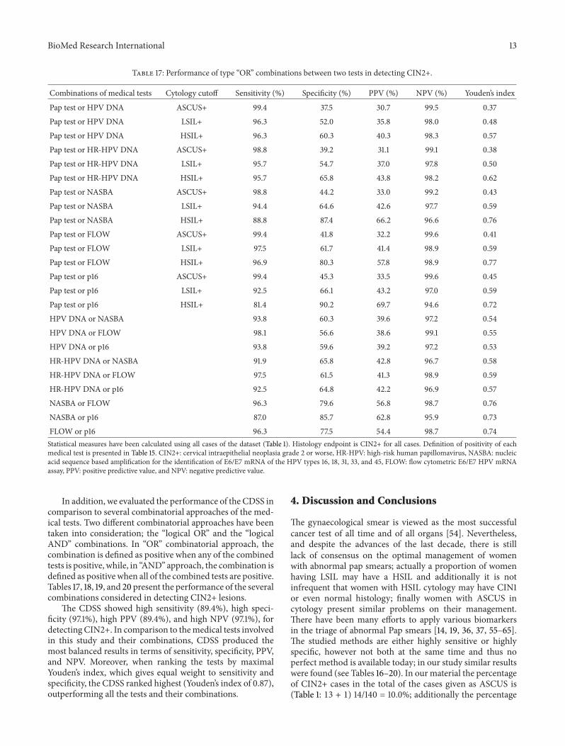
Table 17: Performance of type “OR” combinations between two tests in detecting CIN2+.
Combinations of medical tests Cytology cutoff Sensitivity (%) Specificity (%) PPV (%) NPV (%) Youden’s index
Pap test or HPV DNA ASCUS+ 99.4 37.5 30.7 99.5 0.37Pap test or HPV DNA LSIL+ 96.3 52.0 35.8 98.0 0.48Pap test or HPV DNA HSIL+ 96.3 60.3 40.3 98.3 0.57Pap test or HR-HPV DNA ASCUS+ 98.8 39.2 31.1 99.1 0.38Pap test or HR-HPV DNA LSIL+ 95.7 54.7 37.0 97.8 0.50Pap test or HR-HPV DNA HSIL+ 95.7 65.8 43.8 98.2 0.62Pap test or NASBA ASCUS+ 98.8 44.2 33.0 99.2 0.43Pap test or NASBA LSIL+ 94.4 64.6 42.6 97.7 0.59Pap test or NASBA HSIL+ 88.8 87.4 66.2 96.6 0.76Pap test or FLOW ASCUS+ 99.4 41.8 32.2 99.6 0.41Pap test or FLOW LSIL+ 97.5 61.7 41.4 98.9 0.59Pap test or FLOW HSIL+ 96.9 80.3 57.8 98.9 0.77Pap test or p16 ASCUS+ 99.4 45.3 33.5 99.6 0.45Pap test or p16 LSIL+ 92.5 66.1 43.2 97.0 0.59Pap test or p16 HSIL+ 81.4 90.2 69.7 94.6 0.72HPV DNA or NASBA 93.8 60.3 39.6 97.2 0.54HPV DNA or FLOW 98.1 56.6 38.6 99.1 0.55HPV DNA or p16 93.8 59.6 39.2 97.2 0.53HR-HPV DNA or NASBA 91.9 65.8 42.8 96.7 0.58HR-HPV DNA or FLOW 97.5 61.5 41.3 98.9 0.59HR-HPV DNA or p16 92.5 64.8 42.2 96.9 0.57NASBA or FLOW 96.3 79.6 56.8 98.7 0.76NASBA or p16 87.0 85.7 62.8 95.9 0.73FLOW or p16 96.3 77.5 54.4 98.7 0.74Statistical measures have been calculated using all cases of the dataset (Table 1). Histology endpoint is CIN2+ for all cases. Definition of positivity of eachmedical test is presented in Table 15. CIN2+: cervical intraepithelial neoplasia grade 2 or worse, HR-HPV: high-risk human papillomavirus, NASBA: nucleicacid sequence based amplification for the identification of E6/E7 mRNA of the HPV types 16, 18, 31, 33, and 45, FLOW: flow cytometric E6/E7 HPV mRNAassay, PPV: positive predictive value, and NPV: negative predictive value.
In addition, we evaluated the performance of the CDSS incomparison to several combinatorial approaches of the med-ical tests. Two different combinatorial approaches have beentaken into consideration; the “logical OR” and the “logicalAND” combinations. In “OR” combinatorial approach, thecombination is defined as positive when any of the combinedtests is positive, while, in “AND” approach, the combination isdefined as positive when all of the combined tests are positive.Tables 17, 18, 19, and 20 present the performance of the severalcombinations considered in detecting CIN2+ lesions.
The CDSS showed high sensitivity (89.4%), high speci-ficity (97.1%), high PPV (89.4%), and high NPV (97.1%), fordetecting CIN2+. In comparison to themedical tests involvedin this study and their combinations, CDSS produced themost balanced results in terms of sensitivity, specificity, PPV,and NPV. Moreover, when ranking the tests by maximalYouden’s index, which gives equal weight to sensitivity andspecificity, the CDSS ranked highest (Youden’s index of 0.87),outperforming all the tests and their combinations.
4. Discussion and Conclusions
The gynaecological smear is viewed as the most successfulcancer test of all time and of all organs [54]. Nevertheless,and despite the advances of the last decade, there is stilllack of consensus on the optimal management of womenwith abnormal pap smears; actually a proportion of womenhaving LSIL may have a HSIL and additionally it is notinfrequent that women with HSIL cytology may have CIN1or even normal histology; finally women with ASCUS incytology present similar problems on their management.There have been many efforts to apply various biomarkersin the triage of abnormal Pap smears [14, 19, 36, 37, 55–65].The studied methods are either highly sensitive or highlyspecific, however not both at the same time and thus noperfect method is available today; in our study similar resultswere found (see Tables 16–20). In ourmaterial the percentageof CIN2+ cases in the total of the cases given as ASCUS is(Table 1: 13 + 1) 14/140 = 10.0%; additionally the percentage

14 BioMed Research International
Table 18: Performance of several type “OR” combinations between more than two tests in detecting CIN2+.
Combinations of medical tests Cytology cutoff Sensitivity (%) Specificity (%) PPV (%) NPV (%) Youden’s index
Pap test or HPV DNA or NASBA ASCUS+ 99.4 36.6 30.4 99.5 0.36Pap test or HPV DNA or NASBA LSIL+ 96.3 51.1 35.4 98.0 0.47Pap test or HPV DNA or NASBA HSIL+ 96.3 59.1 39.5 98.3 0.55Pap test or HPV DNA or FLOW ASCUS+ 100.0 35.1 30.0 100.0 0.35Pap test or HPV DNA or FLOW LSIL+ 98.1 48.9 34.8 99.0 0.47Pap test or HPV DNA or FLOW HSIL+ 98.1 56.0 38.3 99.1 0.54Pap test or HPV DNA or p16 ASCUS+ 99.4 37.5 30.7 99.5 0.37Pap test or HPV DNA or p16 LSIL+ 96.3 51.3 35.5 98.0 0.48Pap test or HPV DNA or p16 HSIL+ 96.3 58.9 39.4 98.3 0.55Pap test or HPV DNA or NASBA or FLOW ASCUS+ 100.0 34.9 29.9 100.0 0.35Pap test or HPV DNA or NASBA or FLOW LSIL+ 98.1 48.7 34.7 98.9 0.47Pap test or HPV DNA or NASBA or FLOW HSIL+ 98.1 55.4 38.0 99.1 0.54Pap test or HPV DNA or NASBA or FLOW or p16 ASCUS+ 100.0 34.9 29.9 100.0 0.35Pap test or HPV DNA or NASBA or FLOW or p16 LSIL+ 98.1 48.0 34.4 98.9 0.46Pap test or HPV DNA or NASBA or FLOW or p16 HSIL+ 98.1 54.4 37.4 99.1 0.53HPV DNA or NASBA or FLOW 98.1 56.1 38.3 99.1 0.54HPV DNA or NASBA or p16 93.8 58.5 38.6 97.1 0.52HPV DNA or NASBA or FLOW or p16 98.1 54.7 37.6 99.1 0.53NASBA or FLOW or p16 98.1 76.7 53.9 99.3 0.75Statistical measures have been calculated using all cases of the dataset (Table 1). Histology endpoint is CIN2+ for all cases. Definition of positivity of eachmedical test is presented in Table 15. CIN2+: cervical intraepithelial neoplasia grade 2 or worse, HR-HPV: high-risk human papillomavirus, NASBA: nucleicacid sequence based amplification for the identification of E6/E7 mRNA of the HPV types 16, 18, 31, 33, and 45, FLOW: flow cytometric E6/E7 HPV mRNAassay, PPV: positive predictive value, and NPV: negative predictive value.
of CIN2+ cases in the total of LSIL cases is (Table 1: 27 + 2)29/193 = 15.0% and both percentages are in agreement withthese reported by other researchers in the literature [66];specifically these are 5–17% and 9–16%, respectively. On theother hand the percentage of cases given in cytology as HSILand being lower thanCIN2 is (Table 1: 22+5) 27/127 = 21.26%,a percentage in agreement with the literature [67, 68].
Today, the widely accepted management options ofASCUS andLSIL smears remain either the immediate referralto colposcopy or the cytological surveillance with repeatedsmears. A policy of immediate referral to colposcopy couldpotentially result not only in the overloading of colposcopyclinics but also in overtreatment due to subtle colposcopicalfindings. Many young nulliparous women might be exposedto the physical and psychological sequelae of unnecessarytreatment with long-term perinatal morbidity in womenbeing in reproductive age [69–71]. On the other hand,repeating a cervical smear carries the risk of missing high-grade lesions (HSILs), increases nonattending rates (non-compliance [72]), and increases social and psychologicalburden of women, directly questioning organized screeningprograms’ (OSPs) credibility. Therefore, it is essential toreduce unnecessary colposcopies and, if feasible, to have inadvance indication for women treatment, even before thecolposcopical examination. Thus, a methodology for moreaccurate diagnosis is extremely important.
Although HPV related tests may be used in the triageof ASCUS cases [65, 73], every effort should be made todevelop new tools and biomarkers to improve the accuracy ofdiagnosis and allow tailored management. Nowadays, thereare numerous methods and biomarkers that are available forcervical cancer detection; nevertheless no single method isoptimal [56]. Thus, a different approach is required that willbe able to combine many parameters in order to produce anaccurate risk assessment for each woman. Instead of the futilesearch for a single golden marker we should evolve currentones and invent more elaborate methods for result evaluationand utilisation. Based on this, we are working since 2010 onan innovative approach of employing advancedmathematicaland computing tools for the nonlinear combination of themethods and biomarkers that are available for cervical cancerdetection. Up to now preliminary results are presented in theliterature [36, 37].
The aim of this study was to create a decision supportsystem for the triage of women before referral to colposcopy.This system is based on the standard cytological diagnosison ThinPrep Pap test smears and the expression of variousbiomarkers. The preliminary results suggest that the pro-posed neural network architecturemay improve the accuracyof diagnosis; according to Tables 16–20, CDSS provided the

BioMed Research International 15
Table 19: Performance of type “AND” combinations between two tests in detecting CIN2+.
Combinations of medical tests Cytology cutoff Sensitivity (%) Specificity (%) PPV (%) NPV (%) Youden’s indexPap test and HPV DNA ASCUS+ 90.7 69.3 45.1 96.4 0.60Pap test and HPV DNA LSIL+ 85.1 76.5 50.2 94.9 0.62Pap test and HPV DNA HSIL+ 67.1 96.5 84.4 91.3 0.64Pap test and HR-HPV DNA ASCUS+ 88.8 73.4 48.1 95.9 0.62Pap test and HR-HPV DNA LSIL+ 83.2 79.6 53.2 94.5 0.63Pap test and HR-HPV DNA HSIL+ 65.2 96.9 85.4 90.9 0.62Pap test and NASBA ASCUS+ 76.4 91.2 70.7 93.3 0.68Pap test and NASBA LSIL+ 72.0 92.6 73.0 92.3 0.65Pap test and NASBA HSIL+ 59.6 98.1 89.7 89.7 0.58Pap test and FLOW ASCUS+ 91.9 85.3 63.5 97.4 0.77Pap test and FLOW LSIL+ 85.1 87.2 64.9 95.5 0.72Pap test and FLOW HSIL+ 67.7 96.9 85.8 91.5 0.65Pap test and p16 ASCUS+ 57.1 92.9 69.2 88.6 0.50Pap test and p16 LSIL+ 55.3 93.8 71.2 88.3 0.49Pap test and p16 HSIL+ 48.4 98.1 87.6 87.3 0.47HPV DNA and NASBA 75.2 91.4 70.8 93.0 0.67HPV DNA and FLOW 87.0 86.7 64.5 96.0 0.74HPV DNA and p16 56.5 94.8 75.2 88.7 0.51HR-HPV DNA and NASBA 74.5 91.7 71.4 92.8 0.66HR-HPV DNA and FLOW 85.1 87.7 65.9 95.5 0.73HR-HPV DNA and p16 55.3 95.5 77.4 88.5 0.51NASBA and FLOW 73.9 92.4 73.0 92.7 0.66NASBA and p16 48.4 97.4 83.9 87.2 0.46FLOW and p16 55.3 97.2 84.8 88.7 0.53Statistical measures have been calculated using all cases of the dataset (Table 1). Histology endpoint is CIN2+ for all cases. Definition of positivity of eachmedical test is presented in Table 15. CIN2+: cervical intraepithelial neoplasia grade 2 or worse, HR-HPV: high-risk human papillomavirus, NASBA: nucleicacid sequence based amplification for the identification of E6/E7 mRNA of the HPV types 16, 18, 31, 33, and 45, FLOW: flow cytometric E6/E7 HPV mRNAassay, PPV: positive predictive value, and NPV: negative predictive value.
most balanced results in terms of specificity, sensitivity, PPV,and NPV in comparison to the medical tests involved inthis study and their combinations. The cutoff of CIN2+was used because it is the decision threshold that a caseis therapeutically handled; cases below CIN2+ are strictlymonitored.
In our material, regarding the underestimated cases(CIN2+ cases which were classified by the CDSS as negativesor CIN1), only 1 out of the 4 misclassified ASCUS cases(Tables 12 and 13) was CIN3 and from the 13 misclassifiednon-ASCUS cases (Tables 7–9); 2 were CIN3 and 1 Adeno-Ca. Especially for the one misclassified adenocarcinoma, thetotal of the biomarkers was negative and the case was given asLSIL in cytology. On the other hand, the case of SCC that wasgiven as LSIL in cytology (Table 4) was correctly classified bythe PNN due to the fact that there were positive biomarkers(Table 7). Moreover, it is noteworthy that the CDSS classifiedcorrectly 9 of the 13 ASCUS cases and 17 of the 27 LSIL casesharbouring CIN2/3 (as presented in the results section).
In this study, the sensitivity of cytology using ASCUS+as a cutoff was higher than HR-HPV DNA test (see Table 16:98.1% versus 89.4%) in contrast to other studies, such as theATHENA study [74]. The reader should be aware that thismay be caused by verification bias related to the fact thatcytology positive and HPV negative women had biopsies, incontrary to cytology negative and HPV positive women with
a negative colposcopy. According to other studies [74] thesensitivity of HPV DNA test is higher than the sensitivity ofcytology; however, in our case a special small population forreferral to colposcopy is involved, in contrast to the genericpopulation used in the ATHENA study. In addition, thelaboratory bases the cytological examination on experiencedcytopathologists as reported in our previous study [56] andthus the performance of the cytological examination is higherthan the standard reported performance. In another study[75], the sensitivities of LBC andHPVDNA test are compara-ble with our results; additionally in another study [76] lowersensitivity of the HPV DNA test than the sensitivity of thecytological examination is reported. To conclude, performingour study on meta-analysis data would be impossible asdetailed information for each individual case tests’ results isrequired in order to train and test the CDSS system; therefore,a rather small but controlled population was preferred.
A potential application of this system is to support thedecision of referring a woman to colposcopy or not. A workflow scenario is as follows: the cytological examination isused as primary test and only an ASCUS+ result is followedby the application of the other four ancillary tests using theremaining material in the vial. Subsequently, the five tests’results (including cytology) serve as inputs to the CDSS forevaluation and the CDSS outcome supports the final decisionmaking for referring to colposcopy or not. The applicationof all five tests in general population would be a very costly

16 BioMed Research International
Table 20: Performance of several type “AND” combinations between more than two tests in detecting CIN2+.
Combinations of medical tests Cytology cutoff Sensitivity (%) Specificity (%) PPV (%) NPV (%) Youden’s index
Pap test and HPV DNA andNASBA
ASCUS+ 74.5 91.5 71.0 92.8 0.66
Pap test and HPV DNA andNASBA
LSIL+ 70.2 92.9 73.4 91.8 0.63
Pap test and HPV DNA andNASBA
HSIL+ 57.8 98.1 89.4 89.3 0.56
Pap test and HPV DNA andFLOW
ASCUS+ 86.3 87.7 66.2 95.8 0.74
Pap test and HPV DNA andFLOW
LSIL+ 80.7 88.9 67.0 94.3 0.70
Pap test and HPV DNA andFLOW
HSIL+ 63.4 97.4 87.2 90.5 0.61
Pap test and HPV DNA and p16 ASCUS+ 55.3 94.8 74.8 88.4 0.50
Pap test and HPV DNA and p16 LSIL+ 53.4 95.0 74.8 88.0 0.48
Pap test and HPV DNA and p16 HSIL+ 46.6 98.6 90.4 86.9 0.45
Pap test and HPV DNA andNASBA and FLOW
ASCUS+ 71.4 93.1 74.2 92.1 0.65
Pap test and HPV DNA andNASBA and FLOW
LSIL+ 67.1 94.0 75.5 91.1 0.61
Pap test and HPV DNA andNASBA and FLOW
HSIL+ 55.3 98.4 90.8 88.8 0.54
Pap test and HPV DNA andNASBA and FLOW and p16
ASCUS+ 44.7 98.8 91.1 86.5 0.44
Pap test and HPV DNA andNASBA and FLOW and p16
LSIL+ 43.5 98.8 90.9 86.3 0.42
Pap test and HPV DNA andNASBA and FLOW and p16
HSIL+ 38.5 99.5 95.4 85.3 0.38
HPV DNA and NASBA andFLOW
72.0 93.1 74.4 92.3 0.65
HPV DNA and NASBA and p16 46.6 97.6 84.3 86.8 0.44
HPV DNA and NASBA andFLOW and p16
45.3 98.8 91.3 86.7 0.44
NASBA and FLOW and p16 47.2 98.8 91.6 87.1 0.46
Statistical measures have been calculated using all cases of the dataset (Table 1). Histology endpoint is CIN2+ for all cases. Definition of positivity of eachmedical test is presented in Table 15. CIN2+: cervical intraepithelial neoplasia grade 2 or worse, HR-HPV: high-risk human papillomavirus, NASBA: nucleicacid sequence based amplification for the identification of E6/E7 mRNA of the HPV types 16, 18, 31, 33, and 45, FLOW: flow cytometric E6/E7 HPV mRNAassay, PPV: positive predictive value, and NPV: negative predictive value.
process and thus our method nowadays has the potentialfor application in the triage of ASCUS+ cases. However, adetailed cost/benefit, cost/effectiveness analysis is requiredas the cost of the tests is not the only factor that should betaken into account. Other important factors are the cost ofthe woman’s transportation to a colposcopy clinic, especiallyin mountainous places, islands, or isolated cities/villages, theincrement of recall time, and the psychological effects to thewoman and her family among others.
In the literature there are already simpler techniquesproposed for the triage of ASCUS and LSIL, such as the repeatcytology and the application of mRNA testing. As mentionedin [77–79], the use of NASBA HPV mRNA test in triageof women with ASCUS and LSIL may reduce the referralrate to colposcopy. As presented in [77], the HPV mRNAtest significantly reduced the time from the first abnormalcytology until biopsy and had predictive values comparablewith those of repeat cytology. In [79], the authors report that

BioMed Research International 17
HPV mRNA testing is a better triage test for women withLSIL than repeat cytology, as it was more sensitive (94.2%)and specific (86.0%) for detecting CIN2+. In addition, theHPV mRNA test showed higher PPV (67.0%) compared torepeat cytology (38.4%). In a meta-analysis of the accuracyof mRNA testing for detecting CIN2+, the mRNA testingwas substantially more specific than the HPV DNA test inwomenwithASCUS andLSIL [79].However, it demonstratedlower sensitivity and thus women with negative mRNA testresults cannot be considered free of CIN2+ and requirefollowup [79]. In our study, the proposed system showedhigher sensitivity, higher specificity, higher PPV, and higherNPV compared to NASBA mRNA testing, for detectingCIN2+. In comparison to the HPV DNA test, the proposedsystem is a little less sensitive in detecting CIN2+; however,its specificity and PPV are significantly higher. According toour results, the proposed system produced themost balancedresults in terms of sensitivity, specificity, PPV, and NPVand demonstrated the highest Youden’s index, compared tocytology and the biomarkers used in the study and theircombinations. Thus, in comparison to the already proposedschemes for triage of ASCUS+, our approach may producemore accurate results, leading to improved triage of ASCUS+and improved detection of CIN2+. Therefore, the overheadfor both cytological laboratories and colposcopy rooms canbe reduced.
The application of the proposed CDSS gave promisingresults, suggesting that such an approach may significantlyimprove the accuracy of diagnosis. Furthermore, the notableperformance of the CDSS in identifying women with LSILcytology at risk of developing cancer suggests that such sys-temsmay play an important role in triage decisions and hencemay reduce the overload of colposcopy clinics and guidepersonalisedmanagement and therapeutic interventions.Theresults should be further assessed in larger datasets in orderto confirm the reproducibility of these findings. As some ofthe tests and biomarkers may result in increased cost, ourresearch is now directed to develop a more cost-effectiveCDSS which will use fewer tests, without losing much inperformance. Furthermore, machine learning techniques forhandling missing values are under examination, in order tobe able to provide outcomes also for cases with missing orinvalid examinations’ results.
Today, the CDSS is available to users as a PC application.Our future work involves the upgrade of the CDSS toan intelligent web service for patient-specific prediction,progression, and prognosis of cervical cancer, available overthe Internet to the worldwidemedical community, which willserve as a decision support system to physicians and medicalresearchers for the management of new cases or the followupof existing cases.
Conflict of Interests
The authors declare that there is no conflict of interestsregarding the publication of this paper.
Acknowledgments
This study was funded by the Greek Ministry of Devel-opment (General Secretariat for Research and Technology-GSRT), Project “HPVGuard”, Cooperation 2011–2013 (Code:11ΣYN 10 250). A part of the data is inherited from theProject “AKAKOS” (GSRT Code: ATT 95) and the project“study and evaluation of the methods, for the prognosis ofsuccess for vaccination against HPV infections-Ygeia Proneia2000–2006” (Greek Ministry of Health). Finally, the authorswould like to thank the reviewers for their comments thathelped to improve this paper.
References
[1] A. Jemal, F. Bray, M. M. Center, J. Ferlay, E. Ward, and D.Forman, “Global cancer statistics,” CA: A Cancer Journal forClinicians, vol. 61, no. 2, pp. 69–90, 2011.
[2] J. M. Walboomers, M. V. Jacobs, M. M. Manos et al., “Humanpapillomavirus is a necessary cause of invasive cervical cancerworldwide,” The Journal of Pathology, vol. 189, no. 1, pp. 12–19,1999.
[3] E. F.Dunne, E. R.Unger,M. Sternberg et al., “Prevalence ofHPVinfection among females in the United States,”The Journal of theAmerican Medical Association, vol. 297, no. 8, pp. 813–819, 2007.
[4] N. Munoz, F. X. Bosch, S. de Sanjose et al., “Epidemiologicclassification of human papillomavirus types associated withcervical cancer,”TheNew England Journal of Medicine, vol. 348,no. 6, pp. 518–527, 2003.
[5] M. H. Mayrand, E. Duarte-Franco, I. Rodrigues et al., “Humanpapillomavirus DNA versus Papanicolaou screening tests forcervical cancer,”The New England Journal of Medicine, vol. 357,no. 16, pp. 1579–1588, 2007.
[6] A.Mathew and P. S. George, “Trends in incidence andmortalityrates of squamous cell carcinoma and adenocarcinoma ofcervix—worldwide,” Asian Pacific Journal of Cancer Prevention,vol. 10, no. 4, pp. 645–650, 2009.
[7] J. Cuzick, M. Arbyn, R. Sankaranarayanan et al., “Overview ofhuman papillomavirus-based and other novel options for cer-vical cancer screening in developed and developing countries,”Vaccine, vol. 26, supplement 10, pp. K29–K41, 2008.
[8] P. Naucler, W. Ryd, S. Tornberg et al., “Efficacy of HPV DNAtesting with cytology triage and/or repeat HPV DNA testingin primary cervical cancer screening,” Journal of the NationalCancer Institute, vol. 101, no. 2, pp. 88–99, 2009.
[9] M. Arbyn, P. Sasieni, C. J. Meijer, C. Clavel, G. Koliopoulos,and J. Dillner, “Chapter 9: clinical applications of HPV testing: asummary of meta-analyses,” Vaccine, vol. 24, supplement 3, pp.S78–S89, 2006.
[10] J. Cuzick, A. Szarewski, H. Cubie et al., “Management of womenwho test positive for high-risk types of human papillomavirus:the HART study,” The Lancet, vol. 362, no. 9399, pp. 1871–1876,2003.
[11] H. L. Smits, B. van Gemen, R. Schukkink et al., “Application ofthe NASBA nucleic acid amplification method for the detectionof human papillomavirus type 16 E6-E7 transcripts,” Journal ofVirological Methods, vol. 54, no. 1, pp. 75–81, 1995.
[12] J. Mockel, J. Quaas, H. Meisel, A. S. Endres, and V. Schnei-der, “Human papillomavirus E6/E7 mRNA testing has higherspecificity than liquid-based DNA testing in the evaluation of

18 BioMed Research International
cervical intraepithelial neoplasia,” Analytical and QuantitativeCytology and Histology, vol. 33, no. 6, pp. 311–315, 2011.
[13] A. Szarewski, D. Mesher, L. Cadman et al., “Comparison ofseven tests for high-grade cervical intraepithelial neoplasia inwomen with abnormal smears: the Predictors 2 study,” Journalof Clinical Microbiology, vol. 50, no. 6, pp. 1867–1873, 2012.
[14] C. Kottaridi, S. Tsiodras, A. Spathis et al., “Clinical performanceof human papillomavirus E6, E7 mRNA flow cytometric assaycompared to human papillomavirus DNA typing,” Analyticaland Quantitative Cytology and Histology, vol. 33, no. 6, pp. 305–310, 2011.
[15] A. Trope, K. Sjoborg, A. Eskild et al., “Performance of humanpapillomavirus DNA and mRNA testing strategies for womenwith and without cervical neoplasia,” Journal of Clinical Micro-biology, vol. 47, no. 8, pp. 2458–2464, 2009.
[16] S. W. Sorbye, S. Fismen, T. Gutteberg, and E. S. Mortensen,“Triage of womenwithminor cervical lesions: data suggesting a“test and treat” approach for HPV E6/E7 mRNA testing,” PLoSONE, vol. 5, no. 9, article e12724, 2010.
[17] R. Narimatsu and B. K. Patterson, “High-throughput cervicalcancer screening using intracellular human papillomavirus E6and E7 mRNA quantification by flow cytometry,” AmericanJournal of Clinical Pathology, vol. 123, no. 5, pp. 716–723, 2005.
[18] G. Coquillard, B. Palao, and B. K. Patterson, “Quantificationof intracellular HPV E6/E7 mRNA expression increases thespecificity and positive predictive value of cervical cancerscreening compared to HPV DNA,” Gynecologic Oncology, vol.120, no. 1, pp. 89–93, 2011.
[19] G.Valasoulis, I. Tsoumpou,C. Founta et al., “The role of p16INK4aimmunostaining in the risk assessment of women with LSILcytology: a prospective pragmatic study,” European Journal ofGynaecological Oncology, vol. 32, no. 2, pp. 150–152, 2011.
[20] I. Tsoumpou, M. Arbyn, M. Kyrgiou et al., “p16INK4a immunos-taining in cytological and histological specimens from theuterine cervix: a systematic review and meta-analysis,” CancerTreatment Reviews, vol. 35, no. 3, pp. 210–220, 2009.
[21] M. Benevolo, A. Vocaturo, D. Caraceni et al., “Sensitivity,specificity, and clinical value of human papillomavirus (HPV)E6/E7 mRNA assay as a triage test for cervical cytology andHPV DNA test,” Journal of Clinical Microbiology, vol. 49, no. 7,pp. 2643–2650, 2011.
[22] K. Cuschieri and N. Wentzensen, “Human papillomavirusmRNA and p16 detection as biomarkers for the improved diag-nosis of cervical neoplasia,”Cancer Epidemiology, Biomarkers &Prevention, vol. 17, no. 10, pp. 2536–2545, 2008.
[23] F. M. Carozzi, “Combined analysis of HPV DNA and p16INK4aexpression to predict prognosis in ASCUS and LSIL papsmears,” Collegium Antropologicum, vol. 31, supplement 2, pp.103–106, 2007.
[24] K. J. Denton, C. Bergeron, P. Klement,M. J. Trunk, T. Keller, andR. Ridder, “The sensitivity and specificity of p16INK4a cytologyvs HPV testing for detecting high-grade cervical disease in thetriage of ASC-US and LSIL pap cytology results,” AmericanJournal of Clinical Pathology, vol. 134, no. 1, pp. 12–21, 2010.
[25] K. Kawamoto, C. A. Houlihan, E. A. Balas, and D. F. Lobach,“Improving clinical practice using clinical decision supportsystems: a systematic review of trials to identify features criticalto success,” British Medical Journal, vol. 330, article 765, 2005.
[26] K. Kawamoto, D. F. Lobach, H. F. Willard, and G. S. Ginsburg,“Anational clinical decision support infrastructure to enable the
widespread and consistent practice of genomic and personal-ized medicine,” BMCMedical Informatics and Decision Making,vol. 9, no. 1, article 17, 2009.
[27] P. J. Lisboa and A. F. G. Taktak, “The use of artificial neuralnetworks in decision support in cancer: a systematic review,”Neural Networks, vol. 19, no. 4, pp. 408–415, 2006.
[28] S. Y. Park, D. Sargent, R. Lieberman, and U. Gustafsson,“Domain-specific image analysis for cervical neoplasia detec-tion based on conditional random fields,” IEEE Transactions onMedical Imaging, vol. 30, no. 3, pp. 867–878, 2011.
[29] M. Jondet, R. Agoli-Agbo, and L. Dehennin, “Automatic mea-surement of epithelium differentiation and classification ofcervical intraneoplasia by computerized image analysis,” Diag-nostic Pathology, vol. 5, no. 1, article 7, 2010.
[30] Y. Srinivasan, E. Corona, B. Nutter, S. Mitra, and S. Bhat-tacharya, “A unified model-based image analysis frameworkfor automated detection of precancerous lesions in digitizeduterine cervix images,” IEEE Journal on Selected Topics in SignalProcessing, vol. 3, no. 1, pp. 101–111, 2009.
[31] D. Ramli, A. Kadmin, M. Mashor, N. Ashidi, andM. Isa, “Diag-nosis of cervical cancer using hybrid multilayered perceptron(HMLP) network,” in Knowledge-Based Intelligent Informationand Engineering Systems, M. Negoita, R. Howlett, and L. Jain,Eds., Lecture Notes in Computer Science, pp. 591–598, Springer,Berlin, Germany, 2004.
[32] C. E. Pedreira, L. Macrini, M. G. Land, and E. S. Costa,“New decision support tool for treatment intensity choice inchildhood acute lymphoblastic leukemia,” IEEE Transactions onInformation Technology in Biomedicine, vol. 13, no. 3, pp. 284–290, 2009.
[33] F. K. Ahmad, S. Deris, and M. S. Abdullah, “Synergy networkbased inference for breast cancer metastasis,” Procedia Com-puter Science, vol. 3, pp. 1094–1100, 2011.
[34] J. M. Jerez-Aragones, J. A. Gomez-Ruiz, G. Ramos-Jimenez,J. Munoz-Perez, and E. Alba-Conejo, “A combined neuralnetwork and decision treesmodel for prognosis of breast cancerrelapse,” Artificial Intelligence in Medicine, vol. 27, no. 1, pp. 45–63, 2003.
[35] H.-C. Lin, H.-C. Wu, C.-H. Chang, T.-C. Li, W.-M. Liang,and J.-Y. W. Wang, “Development of a real-time clinical deci-sion support system upon the web mvc-based architecturefor prostate cancer treatment,” BMC Medical Informatics andDecision Making, vol. 11, no. 1, article 16, 2011.
[36] P. Karakitsos, C. Chrelias, A. Pouliakis, G. Koliopoulos, A.Spathis et al., “Identification of women for referral to col-poscopy by neural networks: a preliminary study based onLBC and molecular biomarkers,” Journal of Biomedicine andBiotechnology, vol. 2012, Article ID 303192, 8 pages, 2012.
[37] P. Karakitsos, A. Pouliakis, C. Meristoudis et al., “A preliminarystudy of the potential of tree classifiers in triage of high-gradesquamous intraepithelial lesions,” Analytical and QuantitativeCytology and Histology, vol. 33, no. 3, pp. 132–140, 2011.
[38] M. R. Henry, “The Bethesda System 2001: an update of newterminology for gynecologic cytology,” Clinics in LaboratoryMedicine, vol. 23, no. 3, pp. 585–603, 2003.
[39] J. H. Smith, “Bethesda 2001,” Cytopathology, vol. 13, no. 1, pp.4–10, 2002.
[40] J. J. Gomez-Roman, C. Echevarria, S. Salas et al., “A type-specificstudy of human papillomavirus prevalence in cervicovaginalsamples in three different Spanish regions,” Acta PathologicaMicrobiologica et Immunologica Scandinavica, vol. 117, no. 1, pp.22–27, 2009.

BioMed Research International 19
[41] S. Tyagi, D. P. Bratu, and F. R. Kramer, “Multicolor molecularbeacons for allele discrimination,”Nature Biotechnology, vol. 16,no. 1, pp. 49–53, 1998.
[42] R. Klaes, A. Benner, T. Friedrich et al., “p16INK4a immunohis-tochemistry improves interobserver agreement in the diagnosisof cervical intraepithelial neoplasia,” The American Journal ofSurgical Pathology, vol. 26, no. 11, pp. 1389–1399, 2002.
[43] S. Theodoridis and K. Koutroumbas, Pattern Recognition, Else-vier, London, UK, 4th edition, 2009.
[44] P. Bountris, A. Apostolou, M. Haritou, E. Passalidou, and D.Koutsouris, “Combined texture features for improved classifi-cation of suspicious areas in autofluorescence bronchoscopy,” inProceedings of the 9th International Conference on InformationTechnology and Applications in Biomedicine (ITAB ’09), pp. 1–4,Larnaca, Cyprus, November 2009.
[45] C. Ding and H. Peng, “Minimum redundancy feature selectionfrom microarray gene expression data,” in Proceedings of theIEEE Computational Systems Bioinformatics (CSB ’03), pp. 523–530, Stanford, Calif, USA, August 2003.
[46] D. Michie, D. J. Spiegelhalter, and C. C. Taylor, MachineLearning, Neural and Statistical Classification, Ellis Horwood,New York, NY, USA, 1994.
[47] R. O. Duda, P. E. Hart, and D. G. Stork, Pattern Classification,Wiley, New York, NY, USA, 2nd edition, 2001.
[48] L. Breiman, Classification and Regression Trees, WadsworthInternational Group, Belmont, Calif, USA, 1984.
[49] D. F. Specht, “Probabilistic neural networks and the polynomialAdaline as complementary techniques for classification,” IEEETransactions on Neural Networks, vol. 1, no. 1, pp. 111–121, 1990.
[50] P. D. Wasserman, Advanced Methods in Neural Computing, VanNostrand Reinhold, New York, NY, USA, 1993.
[51] Y. Bengio and Y. LeCun, “Scaling learning algorithms towardsAI,” in Large-Scale Kernel Machines, L. Bottou, O. Chapelle,D. DeCoste, and J. Weston, Eds., pp. 321–360, The MIT Press,Cambridge, Mass, USA, 2007.
[52] U. Braga-Neto and E. Dougherty, “Exact performance of errorestimators for discrete classifiers,” Pattern Recognition, vol. 38,no. 11, pp. 1799–1814, 2005.
[53] U. M. Braga-Neto, “Classification and error estimation fordiscrete data,” Current Genomics, vol. 10, no. 7, pp. 446–462,2009.
[54] H. F. Nauth, Gynecological Cytology, Thieme, Stuttgart, Ger-many, 2007.
[55] G. Koliopoulos, C. Chrelias, A. Pappas, S. Makridima, E.Kountouris et al., “The diagnostic accuracy of two methodsfor E6&7 mRNA detection in women with minor cytologicalabnormalities,” Acta Obstetricia et Gynecologica Scandinavica,vol. 91, no. 7, pp. 794–801, 2012.
[56] A. Spathis, C. Kottaridi, A. Chranioti, C. Meristoudis, C.Chrelias et al., “mRNA and DNA detection of human papil-lomaviruses in women of all ages attending two colposcopyclinics,” PLoS ONE, vol. 7, no. 11, article e49205, 2012.
[57] T. Kelesidis, L. Aish,M. A. Steller et al., “Human papillomavirus(HPV) detection using in situ hybridization in histologicsamples,” American Journal of Clinical Pathology, vol. 136, no.1, pp. 119–127, 2011.
[58] M. Nasioutziki, A. Daniilidis, K. Dinas et al., “The evaluationof p16INK4a immunoexpression/immunostaining and humanpapillomavirus DNA test in cervical liquid-based cytologicalsamples,” International Journal of Gynecological Cancer, vol. 21,no. 1, pp. 79–85, 2011.
[59] A. Spathis, E. Aga, M. Alepaki et al., “Promoter methylation ofp16INK4A, hMLH1, andMGMT in liquid-based cervical cytologysamples compared with clinicopathological findings and HPVpresence,” Infectious Diseases in Obstetrics and Gynecology, vol.2011, Article ID 927861, 5 pages, 2011.
[60] I. Tsoumpou, G. Valasoulis, C. Founta et al., “High-risk humanpapillomavirus DNA test and p16INK4a in the triage of LSIL: aprospective diagnostic study,”GynecologicOncology, vol. 121, no.1, pp. 49–53, 2011.
[61] G. Valasoulis, G. Koliopoulos, C. Founta et al., “Alterations inhuman papillomavirus-related biomarkers after treatment ofcervical intraepithelial neoplasia,” Gynecologic Oncology, vol.121, no. 1, pp. 43–48, 2011.
[62] J. Roelens, M. Reuschenbach, M. von Knebel Doeberitz, N.Wentzensen, C. Bergeron, andM. Arbyn, “p16INK4a immunocy-tochemistry versus human papillomavirus testing for triage ofwomenwithminor cytologic abnormalities: a systematic reviewand meta-analysis,” Cancer Cytopathology, vol. 120, no. 5, pp.294–307, 2012.
[63] M. Arbyn, P. Martin-Hirsch, F. Buntinx, M. van Ranst, E.Paraskevaidis, and J. Dillner, “Triage of women with equivocalor low-grade cervical cytology results: a meta-analysis of theHPV test positivity rate,” Journal of Cellular and MolecularMedicine, vol. 13, no. 4, pp. 648–659, 2009.
[64] G. Koliopoulos, M. Arbyn, P. Martin-Hirsch, M. Kyrgiou,W. Prendiville, and E. Paraskevaidis, “Diagnostic accuracy ofhuman papillomavirus testing in primary cervical screening:a systematic review and meta-analysis of non-randomizedstudies,”Gynecologic Oncology, vol. 104, no. 1, pp. 232–246, 2007.
[65] M. Arbyn, F. Buntinx,M. van Ranst, E. Paraskevaidis, P.Martin-Hirsch, and J. Dillner, “Virologic versus cytologic triage ofwomen with equivocal pap smears: a meta-analysis of theaccuracy to detect high-grade intraepithelial neoplasia,” Journalof theNational Cancer Institute, vol. 96, no. 4, pp. 280–293, 2004.
[66] R. S. Hoda and S. A. Hoda, Fundamentals of PAP Test Cytology,Humana Press, Totowa, NJ, USA, 2007.
[67] London Quality Assurance Reference Centre, “London AnnualReport 2005-2006,” Tech. Rep. KC61, London Quality Assur-ance Reference Centre, London, UK, 2006.
[68] B. A. Jones and D. A. Novis, “Cervical biopsy-cytology correla-tion. A College of American Pathologists Q-Probes study of 22439 correlations in 348 laboratories,” Archives of Pathology andLaboratory Medicine, vol. 120, no. 6, pp. 523–531, 1996.
[69] M. Kyrgiou, I. Tsoumpou, T. Vrekoussis et al., “The up-to-date evidence on colposcopy practice and treatment of cervicalintraepithelial neoplasia: the cochrane colposcopy & cervicalcytopathology collaborative group (C5 group) approach,” Can-cer Treatment Reviews, vol. 32, no. 7, pp. 516–523, 2006.
[70] M. Arbyn, M. Kyrgiou, C. Simoens et al., “Perinatal mortalityand other severe adverse pregnancy outcomes associated withtreatment of cervical intraepithelial neoplasia: meta-analysis,”British Medical Journal, vol. 337, Article ID a1284, 2008.
[71] E. Paraskevaidis, M. Kyrgiou, and P. Martin-Hirsch, “Have wedismissed ablative treatment too soon in colposcopy practice?”BJOG, vol. 114, no. 1, pp. 3–4, 2007.
[72] M. Kyrgiou, G. Koliopoulos, P. Martin-Hirsch, M. Arbyn, W.Prendiville, and E. Paraskevaidis, “Obstetric outcomes afterconservative treatment for intraepithelial or early invasive cer-vical lesions: systematic review and meta-analysis,”The Lancet,vol. 367, no. 9509, pp. 489–498, 2006.
[73] M. Arbyn, E. Paraskevaidis, P. Martin-Hirsch, W. Prendiville,and J. Dillner, “Clinical utility of HPV–DNA detection: triage

20 BioMed Research International
of minor cervical lesions, follow-up of women treated forhigh-grade CIN: an update of pooled evidence,” GynecologicOncology, vol. 99, no. 3, supplement, pp. S7–S11, 2005.
[74] P. E. Castle, M. H. Stoler, T. C. Wright Jr., A. Sharma, T.L. Wright, and C. M. Behrens, “Performance of carcinogenichuman papillomavirus (HPV) testing and HPV16 or HPV18genotyping for cervical cancer screening of women aged 25years and older: a subanalysis of the ATHENA study,” TheLancet Oncology, vol. 12, no. 9, pp. 880–890, 2011.
[75] C. Ferreccio, M. C. Bratti, M. E. Sherman et al., “A comparisonof single and combined visual, cytologic, and virologic tests asscreening strategies in a region at high risk of cervical cancer,”Cancer Epidemiology, Biomarkers&Prevention, vol. 12, no. 9, pp.815–823, 2003.
[76] M. Schiffman, R. Herrero, A. Hildesheim et al., “HPV DNAtesting in cervical cancer screening: results from women in ahigh risk province of Costa Rica,” The Journal of the AmericanMedical Association, vol. 283, no. 1, pp. 87–93, 2000.
[77] S. W. Sorbye, S. Fismen, T. J. Gutteberg, E. S. Mortensen,and F. E. Skjeldestad, “HPV mRNA testing in triage ofwomen with ASC-US cytology may reduce the time forCIN2+diagnosis compared with repeat cytology,”Current Phar-maceutical Design, vol. 19, no. 8, pp. 1401–1405, 2013.
[78] F. Verdoodt, A. Szarewski, P. Halfon, K. Cuschieri, and M.Arbyn, “Triage of women with minor abnormal cervical cytol-ogy:meta-analysis of the accuracy of an assay targetingmessen-ger ribonucleic acid of 5 high-risk humanpapillomavirus types,”Cancer Cytopathology, vol. 121, no. 12, pp. 675–687, 2013.
[79] S. W. Sorbye, M. Arbyn, S. Fismen, T. J. Gutteberg, and E. S.Mortensen, “Triage of womenwith low-grade cervical lesions—HPV mRNA testing versus repeat cytology,” PLoS ONE, vol. 6,no. 8, article e24083, 2011.

Submit your manuscripts athttp://www.hindawi.com
Hindawi Publishing Corporationhttp://www.hindawi.com Volume 2014
Anatomy Research International
PeptidesInternational Journal of
Hindawi Publishing Corporationhttp://www.hindawi.com Volume 2014
Hindawi Publishing Corporation http://www.hindawi.com
International Journal of
Volume 2014
Zoology
Hindawi Publishing Corporationhttp://www.hindawi.com Volume 2014
Molecular Biology International
Hindawi Publishing Corporationhttp://www.hindawi.com
GenomicsInternational Journal of
Volume 2014
The Scientific World JournalHindawi Publishing Corporation http://www.hindawi.com Volume 2014
Hindawi Publishing Corporationhttp://www.hindawi.com Volume 2014
BioinformaticsAdvances in
Marine BiologyJournal of
Hindawi Publishing Corporationhttp://www.hindawi.com Volume 2014
Hindawi Publishing Corporationhttp://www.hindawi.com Volume 2014
Signal TransductionJournal of
Hindawi Publishing Corporationhttp://www.hindawi.com Volume 2014
BioMed Research International
Evolutionary BiologyInternational Journal of
Hindawi Publishing Corporationhttp://www.hindawi.com Volume 2014
Hindawi Publishing Corporationhttp://www.hindawi.com Volume 2014
Biochemistry Research International
ArchaeaHindawi Publishing Corporationhttp://www.hindawi.com Volume 2014
Hindawi Publishing Corporationhttp://www.hindawi.com Volume 2014
Genetics Research International
Hindawi Publishing Corporationhttp://www.hindawi.com Volume 2014
Advances in
Virolog y
Hindawi Publishing Corporationhttp://www.hindawi.com
Nucleic AcidsJournal of
Volume 2014
Stem CellsInternational
Hindawi Publishing Corporationhttp://www.hindawi.com Volume 2014
Hindawi Publishing Corporationhttp://www.hindawi.com Volume 2014
Enzyme Research
Hindawi Publishing Corporationhttp://www.hindawi.com Volume 2014
International Journal of
Microbiology
Copyright © 2022 FDOKUMEN