
Legislating Change? Responses to Criminalizing Female Genital Cutting in Senegal
Upload
independentCategory
view
0download
0
BioMed CentralInfectious Agents and Cancer
ss
Open AcceResearch articleGenital and cutaneous human papillomavirus (HPV) types in relation to conjunctival squamous cell neoplasia: A case-control study in UgandaMaurits NC de Koning1, Keith Waddell2, Joseph Magyezi2, Karin Purdie3, Charlotte Proby4, Catherine Harwood3, Sebastian Lucas5, Robert Downing6, Wim GV Quint1 and Robert Newton*7Address: 1DDL Diagnostic Laboratory, Voorburg, The Netherlands, 2Box 4008, Kampala, Uganda, 3Centre for Cutaneous Research, Institute of Cell and Molecular Science, St Bartholomew's and the Royal London School of Medicine and Dentistry, Queen Mary, University of London, London E1 2AT, UK, 4Division of Surgery and Oncology, College of Medicine, Dentistry and Nursing, University of Dundee, Ninewells Hospital, Dundee DD1 9SY, UK, 5Dept. Histopathology, KCL School of Medicine, St. Thomas' Hospital, London, UK, 6Centers for Disease Control and Prevention, Programme on AIDS, Uganda Virus Research Institute, PO Box 49, Entebbe, Uganda and 7Epidemiology and Genetics Unit, Department of Health Sciences, University of York, Seebohm Rowntree Building, Heslington, York, YO10 5DD, UK
Email: Maurits NC de Koning - [email protected]; Keith Waddell - [email protected]; Joseph Magyezi - [email protected]; Karin Purdie - [email protected]; Charlotte Proby - [email protected]; Catherine Harwood - [email protected]; Sebastian Lucas - [email protected]; Robert Downing - [email protected]; Wim GV Quint - [email protected]; Robert Newton* - [email protected]
* Corresponding author
AbstractBackground: We investigated the role of infection with genital and cutaneous humanpapillomavirus types (HPV) in the aetiology of ocular surface squamous neoplasia (which includesboth conjunctival intraepithelial neoplasia (CIN) and carcinoma) using data and biological materialcollected as part of a case-control study in Uganda.
Results: Among 81 cases, the prevalence of genital and cutaneous HPV types in tumour tissue didnot differ significantly by histological grade of the lesion. The prevalence of genital HPV types didnot differ significantly between cases and controls (both 38%; Odds ratio [OR] 1.0, 95% confidenceinterval [CI] 0.4–2.7, p = 1.0). The prevalence of cutaneous HPV types was 22% (18/81) amongcases and 3% (1/29) among controls (OR 8.0, 95% CI 1.0–169, p = 0.04).
Conclusion: We find no evidence of an association between genital HPV types and ocular surfacesquamous neoplasia. The prevalence of cutaneous HPV was significantly higher among cases ascompared to controls. Although consistent with results from two other case-control studies, therelatively low prevalence of cutaneous HPV types among cases (which does not differ byhistological grade of tumour) indicates that there remains considerable uncertainty about a role forcutaneous HPV in the aetiology of this tumour.
BackgroundIn the years before the HIV epidemic, corneo-conjunctival
intraepithelial neoplasia (CIN) and carcinoma (togethercalled ocular surface squamous neoplasia (OSSN)) were
Published: 10 September 2008
Infectious Agents and Cancer 2008, 3:12 doi:10.1186/1750-9378-3-12
Received: 27 June 2008Accepted: 10 September 2008
This article is available from: http://www.infectagentscancer.com/content/3/1/12
© 2008 de Koning et al; licensee BioMed Central Ltd. This is an Open Access article distributed under the terms of the Creative Commons Attribution License (http://creativecommons.org/licenses/by/2.0), which permits unrestricted use, distribution, and reproduction in any medium, provided the original work is properly cited.
Page 1 of 9(page number not for citation purposes)
Infectious Agents and Cancer 2008, 3:12 http://www.infectagentscancer.com/content/3/1/12
reported to be more frequent in African countries than inEurope and the USA [1-3]. Using data from worldwidecancer registries it has been confirmed that incidence ofOSSN increases markedly with proximity to the equator,presumably from increasing solar ultraviolet (UV) radia-tion [3]. Exposure to UV radiation is an established causeof disease. Lesions occur in sun-exposed areas of the eye[4,5], are associated with solar elastosis [4-7] and havebeen shown to contain classical UV-induced p53 muta-tions [8]. The incidence of the tumour increases withincreasing levels of ambient solar radiation and associa-tions with sun exposure and past history of skin cancerhave been identified in case-control studies [3,9-11].Additional risk factors may also be important. For exam-ple, a polymorphism of TP53 codon 72 has been linked toan increased risk of neoplasia in one study from Ugandaincluding 107 cases and 115 controls [12]. Exposure todust and ocular trauma have also been suggested as possi-ble risk factors, although evidence is scant [1,13].
Since the 1980s there has been a marked increase in casesof conjunctival neoplasia, mostly in sub-Saharan Africa[14-19]. In Uganda for example, the reported incidencehas more than tripled over the last decade [14,16], partic-ularly among younger people and a link with HIV infec-tion was suggested in case reports [20-27]. Case-controlstudies in several African countries [11,28-33] and cohortstudies in the USA [34,35], have confirmed a roughly 10fold excess risk of the tumour in HIV infected people com-pared to the uninfected; in Africa the majority of cases areHIV infected [36]. In a recent study of 414 cases inUganda, 64% of people with conjunctival neoplasia wereHIV infected and this applied to intraepithelial as well asto invasive cases [5]. The median CD4+ T lymphocytecount of HIV positive cases at diagnosis has been found inthis study to be 111 cells/microL (based on results from112 HIV infected cases) [5]. Use of antiretroviral therapyhas been shown to cause tumour regression in an other-wise inoperable case [37]. A recent report from the USAdid not find strong associations with level of immunosup-pression in HIV infected people, but the study includedonly 15 cases of the disease [35]. An excess risk has alsobeen reported among immunosuppressed cancer patientsand organ transplant recipients (although the number ofcases remains small) [38-42].
However, the clear excess risk of ocular surface epithelialdysplasias among HIV infected people (and amongimmunosuppressed renal transplant recipients) suggests arole for an underlying infection in the aetiology [43,44].Although an active search for other new oncogenic infec-tions is ongoing, no new candidate virus (if one exists) hasyet been identified [45]. A causal relationship betweenpersistent infection with several (high risk) genital humanpapillomavirus (HPV) types and cancer of the uterine cer-
vix is established. In non-melanoma skin carcinogenesis,a role has been suggested for cutaneous HPV types fromthe betapapillomavirus genus. A variety of HPV types hasalready been identified in some, but not in all, tumourspecimens from several small case series and results fromcase-control studies have, to date, been inconclusive [2].Here we present results on the association of genital andcutaneous (from the betapapillomavirus genus) HPV typesin relation to ocular surface epithelial neoplasias from acase-control study in Uganda, together with a review ofpublished evidence.
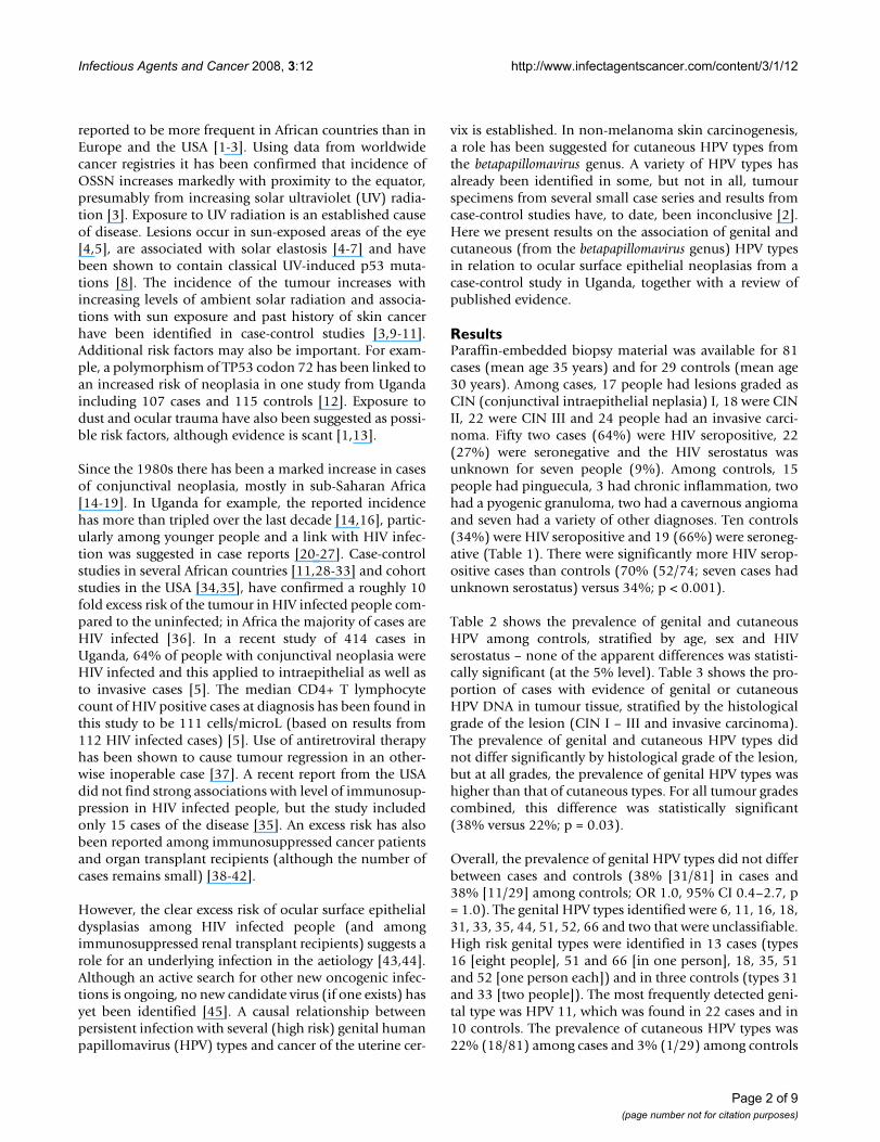
ResultsParaffin-embedded biopsy material was available for 81cases (mean age 35 years) and for 29 controls (mean age30 years). Among cases, 17 people had lesions graded asCIN (conjunctival intraepithelial neplasia) I, 18 were CINII, 22 were CIN III and 24 people had an invasive carci-noma. Fifty two cases (64%) were HIV seropositive, 22(27%) were seronegative and the HIV serostatus wasunknown for seven people (9%). Among controls, 15people had pinguecula, 3 had chronic inflammation, twohad a pyogenic granuloma, two had a cavernous angiomaand seven had a variety of other diagnoses. Ten controls(34%) were HIV seropositive and 19 (66%) were seroneg-ative (Table 1). There were significantly more HIV serop-ositive cases than controls (70% (52/74; seven cases hadunknown serostatus) versus 34%; p < 0.001).
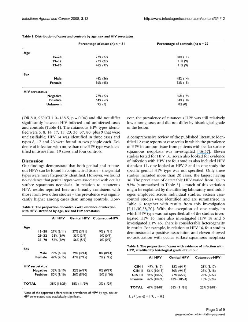
Table 2 shows the prevalence of genital and cutaneousHPV among controls, stratified by age, sex and HIVserostatus – none of the apparent differences was statisti-cally significant (at the 5% level). Table 3 shows the pro-portion of cases with evidence of genital or cutaneousHPV DNA in tumour tissue, stratified by the histologicalgrade of the lesion (CIN I – III and invasive carcinoma).The prevalence of genital and cutaneous HPV types didnot differ significantly by histological grade of the lesion,but at all grades, the prevalence of genital HPV types washigher than that of cutaneous types. For all tumour gradescombined, this difference was statistically significant(38% versus 22%; p = 0.03).
Overall, the prevalence of genital HPV types did not differbetween cases and controls (38% [31/81] in cases and38% [11/29] among controls; OR 1.0, 95% CI 0.4–2.7, p= 1.0). The genital HPV types identified were 6, 11, 16, 18,31, 33, 35, 44, 51, 52, 66 and two that were unclassifiable.High risk genital types were identified in 13 cases (types16 [eight people], 51 and 66 [in one person], 18, 35, 51and 52 [one person each]) and in three controls (types 31and 33 [two people]). The most frequently detected geni-tal type was HPV 11, which was found in 22 cases and in10 controls. The prevalence of cutaneous HPV types was22% (18/81) among cases and 3% (1/29) among controls
Page 2 of 9(page number not for citation purposes)
Infectious Agents and Cancer 2008, 3:12 http://www.infectagentscancer.com/content/3/1/12
(OR 8.0, 95%CI 1.0–168.5, p = 0.04) and did not differsignificantly between HIV infected and uninfected casesand controls (Table 4). The cutaneous HPV types identi-fied were 5, 8, 14, 17, 19, 23, 36, 37, 80, plus 9 that wereunclassifiable; HPV 14 was identified in three cases andtypes 8, 17 and 23 were found in two people each. Evi-dence of infection with more than one HPV type was iden-tified in tissue from 19 cases and four controls.
DiscussionOur findings demonstrate that both genital and cutane-ous HPVs can be found in conjunctival tissue – the genitaltypes were more frequently identified. However, we foundno evidence that genital types were associated with ocularsurface squamous neoplasia. In relation to cutaneousHPV, results reported here are broadly consistent withthose from two other studies – the prevalence was signifi-cantly higher among cases than among controls. How-
ever, the prevalence of cutaneous HPV was still relativelylow among cases and did not differ by histological gradeof the lesion.
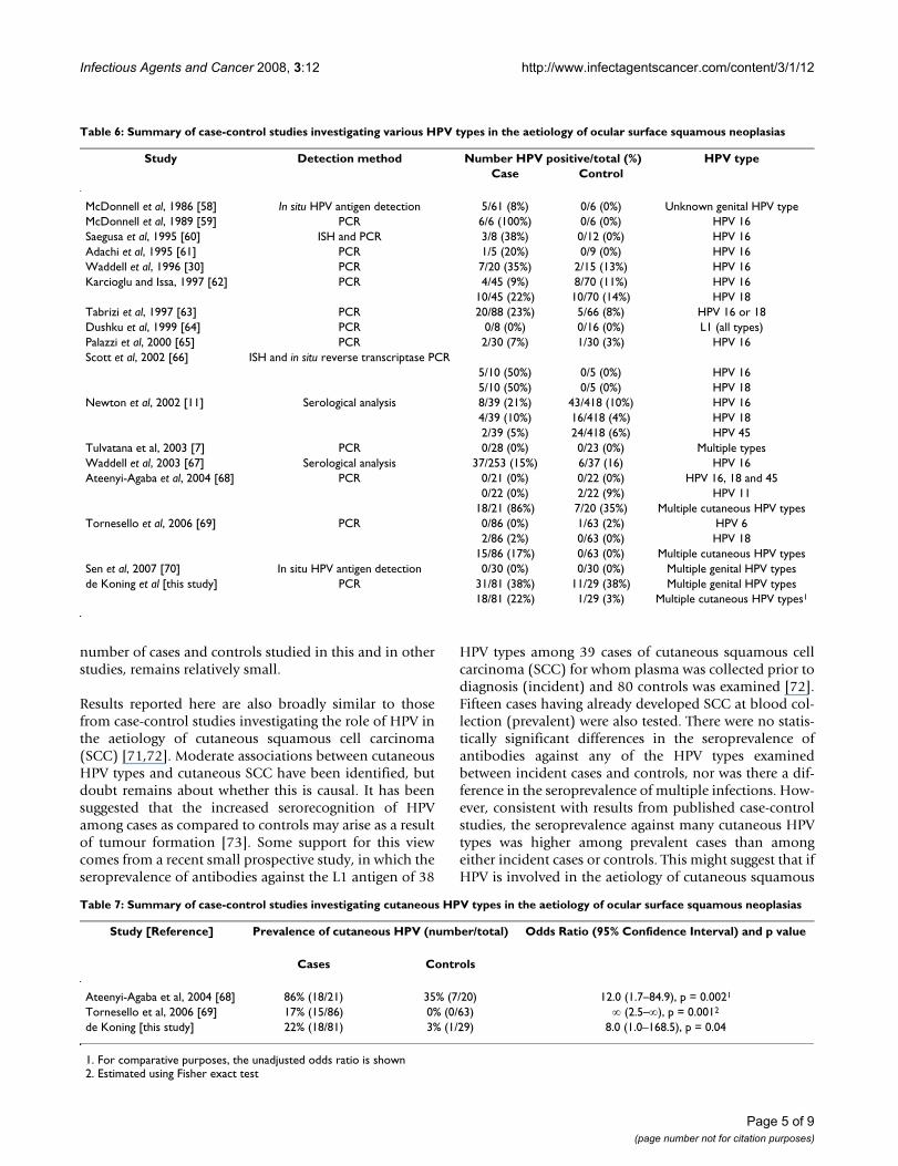
A comprehensive review of the published literature iden-tified 12 case reports or case series in which the prevalenceof HPV in tumour tissue from patients with ocular surfacesquamous neoplasia was investigated [46-57]. Elevenstudies tested for HPV 16; seven also looked for evidenceof infection with HPV 18; four studies also included HPV6 and/or 11, one looked at HPV 2 and in one study thespecific genital HPV type was not specified. Only threestudies included more than 20 cases, the largest having38. The prevalence of detectable HPV varied from 0% to93% (summarised in Table 5) – much of this variationmight be explained by the differing laboratory methodol-ogies employed across individual studies. Sixteen case-control studies were identified and are summarised inTable 6, together with results from this investigation[7,11,30,58-70]. With the exception of one study, inwhich HPV type was not specified, all of the studies inves-tigated HPV 16, nine also investigated HPV 18 and 5investigated HPV 45. There is considerable heterogeneityin results. For example, in relation to HPV 16, four studiesdemonstrated a positive association and eleven showedno association with ocular surface squamous neoplasia
Table 1: Distribution of cases and controls by age, sex and HIV serostatus
Percentage of cases (n) n = 81 Percentage of controls (n) n = 29
Age15–28 27% (22) 38% (11)29–32 27% (22) 31% (9)33–70 46% (37) 31% (9)
SexMale 44% (36) 48% (14)
Female 56% (45) 52% (15)
HIV serostatusNegative 27% (22) 66% (19)Positive 64% (52) 34% (10)
Unknown 9% (7) 0% (0)
Table 2: The proportion of controls with evidence of infection with HPV, stratified by age, sex and HIV serostatus
All HPV Genital HPV Cutaneous-HPV
Age15–28 27% (3/11) 27% (3/11) 9% (1/11)29–32 33% (3/9) 33% (3/9) 0% (0/9)33–70 56% (5/9) 56% (5/9) 0% (0/9)
SexMale 29% (4/14) 29% (4/14) 0% (0/14)
Female 47% (7/15) 47% (7/15) 7% (1/15)
HIV serostatusNegative 32% (6/19) 32% (6/19) 0% (0/19)Positive 50% (5/10) 50% (5/10) 10% (1/10)
TOTAL 38% (11/29) 38% (11/29) 3% (1/29)
None of the apparent differences in prevalence of HPV by age, sex or HIV sero-status was statistically significant.
Table 3: The proportion of cases with evidence of infection with HPV, stratified by histological grade of tumour
All HPV Genital HPV Cutaneous-HPV1
CIN I 47% (8/17) 35% (6/17) 29% (5/17)CIN II 56% (10/18) 50% (9/18) 28% (5/18)
CIN III 45% (10/22) 27% (6/22) 23% (5/22)Invasive 42% (10/24) 42% (10/24) 13% (3/24)
TOTAL 47% (38/81) 38% (31/81) 22% (18/81)
1. χ2 (trend) = 1.9; p = 0.2
Page 3 of 9(page number not for citation purposes)

Infectious Agents and Cancer 2008, 3:12 http://www.infectagentscancer.com/content/3/1/12
(five studies failed to identify HPV 16 in either the casesor controls). In most studies, type-specific methods ofHPV detection were used and so the types shown in thetables were the only ones that were tested for.
Only three studies (including this one [68,69]) investi-gated cutaneous HPV types – each demonstrated a signif-icantly higher prevalence of cutaneous HPV in cases ascompared to controls (summarised in Table 7). Two ofthe three studies examined the prevalence according tohistological grade of tumour (this study and reference 69)and no association was demonstrated in either.
There is substantial variation in HPV prevalence ratesbetween different studies, which may have arisen, in part,because of differences in patient selection, sample taking,preparation and storage and detection method. Even forPCR as a detection system, there are many variables thatinfluence the sensitivity and specificity and so couldimpact on the reported prevalence. These include PCRdesign (nested, broad spectrum or type-specific), the sizeof the amplified product and the choice of the polymeraseused. This review was not done to draw attention to thesedifferences, but rather to show that there is no consistentevidence for a causal association between HPV and OSSN.In addition, however, it should be noted that the total
Table 4: The proportion of cases and controls with evidence of infection with HPV, stratified by HIV serostatus
All HPV1 Genital HPV2 Cutaneous-HPV3
Case4 Control Odds Ratio (95% CI)
Case4 Control Odds Ratio (95% CI)
Case4 Control Odds Ratio (95% CI)
HIV seronegative
45% (10/22) 32% (6/19) 1.8 (0.4–7.9) 36% (8/22) 32% (6/19) 1.2 (0.3–5.5) 27% (6/22) 0% (0/19) ∞ (1.8–∞)
HIV seropositive
48% (25/52) 50% (5/10) 0.9 (0.2–4.3) 40% (21/52) 50% (5/10) 0.8 (0.2–3.8) 21% (11/52) 10% (1/10) 2.2 (0.2–52)
TOTAL 47% (38/81) 38% (11/29) 1.5 (0.6–3.8) 38% (31/81) 38% (11/29) 1.0 (0.4–2.7) 22% (18/81) 3% (1/29) 8.0 (1.0–169)
1. More than one HPV type was identified in tissue from 19 cases and four controls2. Genital HPV types investigated: 6, 11, 16, 18, 31, 33–35, 39, 40, 42–45, 51–54, 56, 58, 59, 66, 68, 70, 74. Genital HPV types identified: 6, 11, 16, 18, 31, 33, 35, 44, 51, 52, 66, plus two unclassifiable; high risk genital types were identified in 13 cases (types 16 [eight people], 51 and 66 [in one person] 18, 35, 51 and 52 [one person each]) and in three controls (types 31 and 33 [two people]); HPV 11 was most frequently detected (22 cases and 10 controls)3. Cutaneous HPV types investigated: 5, 8, 9, 12, 14, 15, 17, 19–25, 36–38, 47, 49, 75, 76, 80, 92, 93, 96. Cutaneous HPV types identified: 5, 8, 14, 17, 19, 23, 36, 37, 80, plus 9 unclassifiable; HPV 14 was identified in three cases and types 8, 17 and 23 were found in two people each4. Among cases, 7 had unknown HIV serostatus
Table 5: Summary of case series investigating the prevalence of HPV DNA in tumour tissue from patients with ocular surface squamous neoplasias.
Study [Reference] Detection method Number HPV positive/total (%) HPV type
McDonnell et al, 1987 [46] In situ hybridisation (ISH) 0/28 (0%) HPV 2, 6, 16, 18McDonnell et al, 1989 [47] PCR 1/1 (100%) HPV 16Lauer et al, 1990 [48] PCR 4/5 (80%) HPV 16
2/5 (40%) HPV18Odrich et al, 1991 [49] PCR 2/2 (100%) HPV 16McDonnell et al, 1992 [50] PCR 33/38 (87%) HPV 16Tuppurainen et al, 1992 [51] ISH and PCR 0/4 (0%) HPV 6, 11, 16 and 18Serna et al, 1995 [52] PCR 1/9 (11%) HPV 16Nakamura et al, 1997 [53] ISH and PCR 2/8 (25%) HPV 16
2/8 (25%) HPV 18Toth et al, 2000 [54] PCR 5/23 (9%) HPV types not specifiedEng et al, 2002 [55] PCR 0/20 (0%) HPV 6, 11, 16, 18Moubayed et al, 2004 [56] ISH 12/14 (86%) HPV 16
13/14 (93%) HPV 1812/14 (86%) HPV 6 and 11
Reszec and Sulkowski, 2005 [57] PCR 1/11 (9%) HPV 161/11 (9%) HPV 18
Page 4 of 9(page number not for citation purposes)
Infectious Agents and Cancer 2008, 3:12 http://www.infectagentscancer.com/content/3/1/12
number of cases and controls studied in this and in otherstudies, remains relatively small.
Results reported here are also broadly similar to thosefrom case-control studies investigating the role of HPV inthe aetiology of cutaneous squamous cell carcinoma(SCC) [71,72]. Moderate associations between cutaneousHPV types and cutaneous SCC have been identified, butdoubt remains about whether this is causal. It has beensuggested that the increased serorecognition of HPVamong cases as compared to controls may arise as a resultof tumour formation [73]. Some support for this viewcomes from a recent small prospective study, in which theseroprevalence of antibodies against the L1 antigen of 38
HPV types among 39 cases of cutaneous squamous cellcarcinoma (SCC) for whom plasma was collected prior todiagnosis (incident) and 80 controls was examined [72].Fifteen cases having already developed SCC at blood col-lection (prevalent) were also tested. There were no statis-tically significant differences in the seroprevalence ofantibodies against any of the HPV types examinedbetween incident cases and controls, nor was there a dif-ference in the seroprevalence of multiple infections. How-ever, consistent with results from published case-controlstudies, the seroprevalence against many cutaneous HPVtypes was higher among prevalent cases than amongeither incident cases or controls. This might suggest that ifHPV is involved in the aetiology of cutaneous squamous
Table 6: Summary of case-control studies investigating various HPV types in the aetiology of ocular surface squamous neoplasias
Study Detection method Number HPV positive/total (%) HPV typeCase Control
McDonnell et al, 1986 [58] In situ HPV antigen detection 5/61 (8%) 0/6 (0%) Unknown genital HPV typeMcDonnell et al, 1989 [59] PCR 6/6 (100%) 0/6 (0%) HPV 16Saegusa et al, 1995 [60] ISH and PCR 3/8 (38%) 0/12 (0%) HPV 16Adachi et al, 1995 [61] PCR 1/5 (20%) 0/9 (0%) HPV 16Waddell et al, 1996 [30] PCR 7/20 (35%) 2/15 (13%) HPV 16Karcioglu and Issa, 1997 [62] PCR 4/45 (9%) 8/70 (11%) HPV 16
10/45 (22%) 10/70 (14%) HPV 18Tabrizi et al, 1997 [63] PCR 20/88 (23%) 5/66 (8%) HPV 16 or 18Dushku et al, 1999 [64] PCR 0/8 (0%) 0/16 (0%) L1 (all types)Palazzi et al, 2000 [65] PCR 2/30 (7%) 1/30 (3%) HPV 16Scott et al, 2002 [66] ISH and in situ reverse transcriptase PCR
5/10 (50%) 0/5 (0%) HPV 165/10 (50%) 0/5 (0%) HPV 18
Newton et al, 2002 [11] Serological analysis 8/39 (21%) 43/418 (10%) HPV 164/39 (10%) 16/418 (4%) HPV 182/39 (5%) 24/418 (6%) HPV 45
Tulvatana et al, 2003 [7] PCR 0/28 (0%) 0/23 (0%) Multiple typesWaddell et al, 2003 [67] Serological analysis 37/253 (15%) 6/37 (16) HPV 16Ateenyi-Agaba et al, 2004 [68] PCR 0/21 (0%) 0/22 (0%) HPV 16, 18 and 45
0/22 (0%) 2/22 (9%) HPV 1118/21 (86%) 7/20 (35%) Multiple cutaneous HPV types
Tornesello et al, 2006 [69] PCR 0/86 (0%) 1/63 (2%) HPV 62/86 (2%) 0/63 (0%) HPV 18
15/86 (17%) 0/63 (0%) Multiple cutaneous HPV typesSen et al, 2007 [70] In situ HPV antigen detection 0/30 (0%) 0/30 (0%) Multiple genital HPV typesde Koning et al [this study] PCR 31/81 (38%) 11/29 (38%) Multiple genital HPV types
18/81 (22%) 1/29 (3%) Multiple cutaneous HPV types1
Table 7: Summary of case-control studies investigating cutaneous HPV types in the aetiology of ocular surface squamous neoplasias
Study [Reference] Prevalence of cutaneous HPV (number/total) Odds Ratio (95% Confidence Interval) and p value
Cases Controls
Ateenyi-Agaba et al, 2004 [68] 86% (18/21) 35% (7/20) 12.0 (1.7–84.9), p = 0.0021
Tornesello et al, 2006 [69] 17% (15/86) 0% (0/63) ∞ (2.5–∞), p = 0.0012
de Koning [this study] 22% (18/81) 3% (1/29) 8.0 (1.0–168.5), p = 0.04
1. For comparative purposes, the unadjusted odds ratio is shown2. Estimated using Fisher exact test
Page 5 of 9(page number not for citation purposes)
Infectious Agents and Cancer 2008, 3:12 http://www.infectagentscancer.com/content/3/1/12
carcinoma, the process occurs close to the time of diagno-sis, or that the antibody response observed in people withthe tumour is a consequence of tumour formation.
The possibility that the presence of a tumour facilitatesdetection of antibodies against HPV is supported by thefindings of Favre et al (2000), who reported a higher sero-prevalence of HPV-5 among patients with burns or withproliferative cutaneous autoimmune diseases than amongcontrols [74]. Patients with psoriasis, involving abnormalkeratinocyte differentiation and proliferation, have alsoshown a high HPV-5 seroprevalence [75]. This is thoughtto arise as a consequence of cell proliferation in the skinproviding an environment that favours viral replication,resulting in a rise in antibodies against the relevant HPVtype. Similarly, there is debate concerning the resultsobtained from studies using tests for cutaneous HPVDNA. The prevalence of HPV DNA was significantly lowerin tumour biopsies than in swabs of the tested lesion [76].Furthermore, evidence of cutaneous HPV DNA has beenfound to be both highly prevalent and persistent in thehealthy population [77]. It is possible that the resultsreported here reflect a similar situation. However, there isnow some preliminary evidence from studies of molecu-lar mechanisms, suggesting that HPV might interact withultra-violet radiation disturbing apoptotic pathways andleading to cell immortalization [78]. Transforming prop-erties of E6 and E7 proteins of some cutaneous HPV typeshave also been described (reviewed in reference [71]). Itremains to be established what role, if any, HPV plays inthe pathological processes that lead to the development ofboth conjunctival and cutaneous squamous cell neopla-sia.
It should be noted that the relatively high percentage ofsamples with unclassified cutaneous HPV types could rep-resent infections with novel types of which only subge-nomic amplicons have been sequenced [79]. However,the other possibility is that these were infections with lowcopy numbers of one of the 25 tested cutaneous HPVtypes allowing only for general detection and not theidentification of specific types. With the broad spectrumSPF10 PCR – DEIA (see Methods section) more than 50HPV types can be detected. It cannot, therefore, beexcluded that the two cases with an indeterminate genitalHPV result actually represent a cutaneous HPV type. TheSPF10-LiPA25 system amplifies a small fragment from 65base pairs and is therefore very suitable for the testing ofparaffin-embedded, formalin-fixed samples. Although theconjunctiva represent mucosal tissue, the detection ofgenital HPV types in 40% of the HIV seronegative casesand in 32% of the HIV seronegative controls was unex-pected. This finding indicates that the natural history ofHPV and their tissue tropism is not fully understood.
ConclusionWe find no evidence of an association between genitalHPV types and ocular surface squamous neoplasia. Theprevalence of cutaneous HPV was significantly higheramong cases as compared to controls. Although consist-ent with results from two other case-control studies, therelatively low prevalence of cutaneous HPV types amongcases (which does not differ by histological grade oftumour) indicates that there remains considerable uncer-tainty about a role for cutaneous HPV in the aetiology ofthis tumour.
MethodsParticipantsFrom November 1995 to May 2001 in country-wide clin-ics, anyone with a suspect corneo-conjunctival lesion wasoffered removal and histology, and enrolment in a follow-up study with home visits. HIV serology was also offeredafter pre-test counselling. Lesions were photographed anddetails of the eyes and general health were recorded andanalysed in EPI INFO version 6. Those who subsequentlyturned out to have lesions other than ocular surface squa-mous neoplasia were used as a control group in the anal-yses of HPV.
Consent and ethical approvalInformation about the disease, its treatment and HIV test-ing was given in private in vernacular by counsellors, andconsent confirmed by signature or thumbprint. The studywas approved by the Science and Ethics Committee of theUganda Virus Research Institute, and by the UgandaNational Council for Science and Technology.
Serology and histopathologyVenous blood was taken and screening tests for HIV anti-bodies done, with confirmation at the Uganda VirusResearch Institute (two enzyme immunoassay tests in par-allel, with Western blot if required). Biopsies went to StThomas' Hospital London for histopathology. CIN wasclassified (by SBL) into 3 stages according to one, two orthree thirds thickness being dysplastic; invasive tumourswere diagnosed when the epithelial basement membranewas breached.
HPV typingHPV analyses were performed on DNA isolated from for-malin-fixed, paraffin-embedded specimens. Chances ofcontamination during the cutting of the sections wereminimised by discarding the initial section that was cut toremove any environmental contamination which hadoccurred while blocks were stored and by changing cryo-stat blades in between sections. DNA was extracted fromthe sections in a cabinet which had been UV-treated toremove any contaminating DNA. Additionally, 15 nega-tive DNA isolation controls were included. For both the
Page 6 of 9(page number not for citation purposes)
Infectious Agents and Cancer 2008, 3:12 http://www.infectagentscancer.com/content/3/1/12
genital HPV test and the beta HPV test, 10 μl of a 20 ng/μlDNA solution per specimen was used as input for the PCRanalyses. Genital HPV genotyping was carried out usingthe SPF10-LiPA25 system (SPF10 HPV LiPA, version 1; man-ufactured by Labo Bio-Medical Products, Rijswijk, TheNetherlands) as described previously [80,81]. Briefly, thebroad spectrum SPF10 PCR amplifies a 65-base pair frag-ment from the L1 region of the HPV genome. By usingbiotinylated reverse primers the amplimers could be cap-tured onto streptavidin-coated microtiter plates. Afterdenaturation of the PCR products by alkaline treatment, adefined cocktail of digoxigenin-labeled probes was usedto detect HPV positive samples. This method that is desig-nated the HPV DNA Enzyme Immunoassay (DEIA) pro-vides an optical density value and is able to detect morethan 50 HPV types [82]. Amplimers from positive sampleswere used for subsequent genotyping of twenty-five indi-vidual genital HPV genotypes (high-risk HPV: 16, 18, 31,33, 35, 39, 45, 51, 52, 56, 58, 59, 66, 68, 70, and low-riskHPV: 6, 11, 34, 40, 42–44, 53, 54, 74) simultaneously ina reverse hybridisation assay (RHA). Beta HPV genotypingwas performed with the PM-PCR RHA method (The skin(beta) HPV prototype research assay; Diassay BV, Rijswijk,The Netherlands) [83]. It consists of a broad spectrumPCR specific for the amplification of the betaPV genus andtargets a fragment of 117 bp from the E1 region of theHPV genome. Combined with the RHA, it was possible toidentify 25 beta HPV types (i.e., HPV type 5, 8, 9, 12, 14,15, 17, 19–25, 36–38, 47, 49, 75, 76, 80, 92, 93 and 96).As no DEIA was developed for this assay all amplimerswere directly analysed by RHA.
Review methodsCase series and controlled studies of HPV and ocular sur-face squamous neoplasia published up to April 2008,were identified through a medline search [1966–2006;search terms (exploded, all subheadings): squamous cellcarcinoma, human papillomavirus (HPV), conjunctivalcancer], supplemented by searches of references in identi-fied papers, by hand searches of relevant journals and bydirect contact with authors. No restriction was placed onlanguage of publication. No attempt was made to identifyunpublished studies or to obtain unpublished data frompublished studies. There were no prospective studies. Theodds ratios used here are either those presented in thepaper or, where none were provided, they were estimatedfor each study by the authors, using published figures.
Competing interestsThe authors declare that they have no competing interests.
Authors' contributionsKW, JM, RD and RN conducted the original study and col-lected all the biological material used for work describedhere. MK and WQ developed the HPV assays, which MK
used in this study, with assistance from KP, CP and CH. SLconducted the histopathology. RN conducted the statisti-cal analyses. The manuscript was drafted by RN and MK.All authors read, contributed to and approved the manu-script.
References1. Templeton AC: Tumours of the eye and adnexa. Tumours of a
Tropical Country: A survey of Uganda 1964–1968. Recent Result CancerResearch 1973, 41:203-214.
2. Newton R: A review of the aetiology of squamous cell carci-noma of the conjunctiva. Br J Cancer 1996, 74:1511-1513.
3. Newton R, Ferlay J, Reeves G, Beral V, Parkin DM: Incidence ofsquamous cell carcinoma of the eye increases with increas-ing levels of ambient solar ultraviolet radiation. Lancet 1996,i:1450-1.
4. McKelvie PA, Daniell M, McNab A, Loughnan M, Santamaria JD:Squamous cell carcinoma of the conjunctiva: a series of 26cases. Br J Ophthalmol 2002, 86:168-73.
5. Waddell K, Downing R, Lucas S, Newton R: Corneo-conjunctivalcarcinoma associated with human immunodeficiency virustype-1 (HIV-1) in Uganda. Eye 2006, 20(8):893-899.
6. Clear A, Chirambo M, Hutt M: Solar keratosis, pterygium, andsquamous cell carcinoma of the conjunctiva in Malawi. Br JOphthalmol 1979, 63:102-109.
7. Tulvatana W, Bhattarakosol P, Sansopha L, Sipiyarak W, Kowitdam-rong E, Paisuntornsug T, Karnsawai S: Risk factors for conjunctivalsquamous cell neoplasia: a matched case-control study. Br JOphthalmol 2003, 87:396-8.
8. Ateenyi-Agaba C, Dai M, Le Calvez F, Katongole-Mbidde E, Smet A,Tommasino M, Franceschi S, Hainaut P, Weiderpass E: TP53 muta-tions in squamous-cell carcinomas of the conjunctiva: evi-dence for UV-induced mutagenesis. Mutagenesis 2004,19(5):399-401.
9. Sun EC, Fears TR, Goedert JJ: Epidemiology of squamous cellconjunctival cancer. Cancer Epidemiology, Biomarkers & Prevention1997, 6:73-77.
10. Lee GA, Williams G, Hirst LW, Green AC: Risk Factors in theDevelopment of Ocular Surface Epithelial Dysplasia. Ophthal-mology 1994, 101:360-4.
11. Newton R, Ziegler J, Ateenyi-Agaba C, Bousarghin L, Casabonne D,Beral V, Mbidde E, Carpenter L, Reeves G, Parkin DM, Wabinga H,Mbulaiteye S, Jaffe H, Bourboulia D, Boshoff C, Coursaget P, theUganda Kaposi's Sarcoma Study Group: The epidemiology of con-junctival squamous cell carcinoma in Uganda. Br J Cancer 2002,87:301-308.
12. Tornesello ML, Waddell KM, Duraturo ML, Biryahwaho B, DowningR, Lucas SB, Giani U, Buonaguro L, Buonaguro FM: TP53 codon 72polymorphism and risk of conjunctival squamous cell carci-noma in Uganda. Cancer Detection and Prevention 2005, 29:501-508.
13. Margo CE, Groden LR: Squamous cell carcinoma of the corneaand conjunctiva following a thermal burn of the eye. Cornea1986, 5:185-188.
14. Parkin DM, Wabinga H, Nambooze S, Wabwire-Mangen F: AIDS-related cancers in Africa: maturation of the epidemic inUganda. AIDS 1999, 13(18):2563-70.
15. Poole TRG: Conjunctival squamous cell carcinoma in Tanza-nia. Br J Ophthalmol 1999, 83:177-179.
16. Wabinga H, Parkin D, Wabwire-Mangen F, Nambooze S: Trends incancer incidence in Kyadondo county, Uganda, 1960–1997.Br J Cancer 2000, 82:1585-1592.
17. Pola EC, Masanganise R, Rusakaniko S: The trend of ocular surfacesquamous neoplasia among ocular surface tumour biopsiessubmitted for histology from Sekuru Kaguvi Eye Unit,Harare between 1996 and 2000. Cent Afr J Med 2003, 49:1-4.
18. Kalua K: Treatment of conjunctival intraepithelial neoplasiain Africa. Br J Ophthalmol (electronic letters; 22nd January inresponse to Waddell and Newton. Br J Ophthalmol 2007,91:120-1.
19. Waddell KM, Newton R: The aetiology and associations of con-junctival intraepithelial neoplasia – further evidence. Br J Oph-thalmol 2007, 91(1):120-121.
Page 7 of 9(page number not for citation purposes)
Infectious Agents and Cancer 2008, 3:12 http://www.infectagentscancer.com/content/3/1/12
20. Winward KE, Curtin VT: Conjunctival squamous cell carcinomain a patient with human immunodeficiency virus infection.Am J Ophthalmol 1989, 107(5):554-555.
21. Kim RY, Seiff SR, Howes EL Jr, O'Donnell JJ: Necrotizing scleritissecondary to conjunctival squamous cell carcinoma inaquired immunodeficiency syndrome. Am J Ophthalmol 1990,109(2):231-233.
22. Denis P, Charpentier D, Roudier M, et al.: Conjunctival epider-moid carcinoma and human immunodeficiency virus. J FrOphthalmol 1994, 17:366-369.
23. Mahomed A, Chetty R: Human immunodeficiency virus infec-tion, Bcl-2, p53 protein and Ki-67 analysis in ocular surfacesquamous neoplasia. Arch Ophthalmol 2002, 120(5):554-8.
24. Wilhelm F, Herz E, McArthur C, Werschnik C: HIV seropreva-lence in ophthalmologic patients of Cameroon. Ophthalmologe2004, 101(9):941-4.
25. Chinogurei TS, Masanganise R, Rusakaniko S, Sibanda E: Ocular sur-face squamous neoplasia (OSSN) and human immunodefi-ciency virus at Sekuru Kaguvi Eye Unit in Zimbabwe: therole of operational research studies in a resource poor envi-ronment? Cent Afr J Med 2006, 52(5–6):56-8.
26. Chisi SK, Kollmann MK, Karimurio J: Conjunctival squamous cellcarcinoma in patients with human immunodeficiency virusinfection seen at two hospitals in Kenya. East Afr Med J 2006,83(5):267-70.
27. Osahon AI, Onunu AN: Ocular disorders in patients infectedwith the human immunodeficiency virus at the University ofBenin Teaching Hospital, Benin City, Nigeria. Niger J Clin Pract2007, 10(4):283-6.
28. Kestelyn P, Stevens A, Ndayambaje , Hanssens M, Perre P van de:HIV and conjunctival malignancies. Lancet 1990, 336:51-52.
29. Ateenyi-Agaba C: Conjunctival squamous-cell carcinoma asso-ciated with HIV infection in Kampala, Uganda. Lancet 1995,345:695-696.
30. Waddell K, Lewallen S, Lucas S, Atenyi-Agaba C, Herrington C,Liomba G: Carcinoma of the conjunctiva and HIV infection inUganda and Malawi. Br J Ophthalmol 1996, 80:503-538.
31. Newton R, Ziegler J, Beral V, Mbidde E, Carpenter L, Wabinga H,Mbulataiye S, Appleby P, Reeves G, Jaffe H, the Uganda Kaposi's Sar-coma Study Group: A case control study of Human Immunode-ficiency Virus infection and cancer in adults and childrenresiding in Kampala, Uganda. Int J Cancer 2001, 92:622-627.
32. Porges Y, Groisman GM: Prevalence of HIV in conjunctivalsquamous cell neoplasia in an African provincial hospital.Cornea 2003, 22:1-4.
33. Timm A, Stropahl G, Schittowski M, Sinzidi C, Kayembe D, Guthoff R:Association of malignant tumors of the conjunctiva and HIVinfection in Kinshasa (D.R. Congo). First results. Ophthalmol-oge 2004, 101(10):1011-6.
34. Goedert JJ, Cote TR: Conjunctival malignant disease with AIDSin USA. Lancet 1995, ii:257-258.
35. Guech-Ongey M, Engels EA, Goedert JJ, Biggar RJ, Mbulaiteye SM:Elivated risk for squamous cell carcinoma of the conjunctivaamong adults with AIDS in the United States. Int J Cancer2008, 122:2590-2593.
36. Ateenyi-Agaba C, Newton R: Squamous cell carcinoma of theconjunctiva: an HIV-associated cancer. Bulletin of the Royal Soci-ety of Tropical Health 1999, 7:3-4.
37. Holkar S, Mudhar HS, Jain A, Gupta M, Rogstad KE, Parsons MA, SinghAD, Rennie IG: Regression of invasive conjunctival squamouscarcinoma in an HIV-positive patient on antiretroviral ther-apy. Int J STD AIDS 2005, 16(12):782-3.
38. Kushner FH, Mushen RL: Conjunctival squamous cell carcinomacombined with malignant lymphoma. Am J Ophthalmol 1975,80:503-6.
39. Macarez R, Bossis S, Robinet A, Le Callonnec A, Charlin JF, Colin J:Conjunctival epithelial neoplasias in organ transplantpatients receiving cyclosporine therapy. Cornea 1999,18(4):495-7.
40. Shelil AE, Shields CL, Shields JA, Eagle RC Jr: Aggressive conjunc-tival squamous cell carcinoma in a patient following livertransplantation. Arch Ophthalmol 2003, 121(2):280-2.
41. Pournaras JA, Chamot L, Uffer S, Zografos L: Conjunctival intraep-ithelial neoplasia in a patient treated with tacrolimus afterliver transplantation. Cornea 2007, 26(10):1261-2.
42. Vajdic CM, van Leeuwen MT, McDonald SP, McCredie MR, Law M,Chapman JR, Webster AC, Kaldor JM, Grulich AE: Increased inci-dence of squamous cell carcinoma of the eye after kidneytransplantation. J Natl Cancer Inst 2007, 99:1340-2.
43. Beral V, Newton R: Overview of the epidemiology of immuno-deficiency associated cancers. Monogr Natl Cancer Inst 1998,23:1-6.
44. Newton R, Beral V, Weiss R: Human Immunodeficiency VirusInfection and Cancer. In Cancer Surveys, Infections and Human can-cer Volume 33. Edited by: Newton R, Beral V, Weiss R. Cold SpringHarbor Laboratory Press; 1999.
45. Feng H, Taylor JL, Benos PV, Newton R, Waddell K, Lucas SB, ChangY, Moore PS: Human transcriptome subtraction using shortsequence tags to search for tumor viruses. J Virol 2007,81(20):332-11.
46. McDonnell PJ, McDonnell JM, Kessis T, Green WR, Shah KV: Detec-tion of human papillomavirus type 6/11 DNA in conjunctivalpapillomas by in situ hybridization with radioactive probes.Hum Pathol 1987, 18(11):1115-9.
47. McDonnell JM, McDonnell PJ, Stout WC, Martin WJ: Human papil-lomavirus DNA in a recurrent squamous carcinoma of theeyelid. Arch Ophthalmol 1989, 107(11):1631-4.
48. Lauer SA, Malter JS, Meier JR: Human papillomavirus type 18 inconjunctival intraepithelial neoplasia. Am J Ophthalmol 1990,110(1):23-7.
49. Odrich MG, Jakobiec FA, Lancaster WD, Kenyon KR, Kelly LD, Korn-mehl EW, Steinert RF, Grove AS, Shore JW, Gregoire L, Albert DM:A spectrum of bilateral squamous conjunctival tumors asso-ciated with human papillomavirus type 16. Ophthalmology1991, 98(5):628-35.
50. McDonnell JM, McDonnell PJ, Sun YY: Human papillomavirusDNA in tissues and ocular surface swabs of patients with con-junctival epithelial neoplasia. Invest Ophthalmol Vis Sci 1992,33(1):184-9.
51. Tuppurainen K, Raninen A, Kosunen O, Kankkunen JP, Kellokoski J,Syrjanen S, Mantyjarvi M, Syrjanen K: Squamous cell carcinoma ofthe conjunctiva: Failure to demonstrate HPV DNA by in situhybridization and polymerase chain reaction. Acta Ophthalmol(Copenh) 1992, 70(2):248-254.
52. Serna A, Corredor JC, Benavides J, Ureta J, Orozco O: Human pap-illomavirus (HPV) and squamous cell carcinoma of the con-junctiva. Neoplasia 1995, 12:118-21.
53. Nakamura Y, Mashima Y, Kameyama K, Mukai M, Oguchi Y: Detec-tion of human papillomavirus infection in squamous tumoursof the conjunctiva and lacrimal sac by immunohistochemis-try, in situ hybridisation and polymerase chain reaction. Br JOphthalmol 1997, 81(4):308-13.
54. Toth J, Karcioglu ZA, Moshfeghi AA, Issa TM, Al-Ma'ani JR, Patel KV:The relationship between human papillomavirus and p53gene in conjunctival squamous cell carcinoma. Cornea 2000,19(2):159-62.
55. Eng HL, Lin TM, Chen SY, Wu SM, Chen WJ: Failure to detecthuman papillomavirus DNA in malignant epithelial neo-plasms of conjunctiva by polymerase chain reaction. Am J ClinPathol 2002, 117(3):429-36.
56. Moubayed P, Mwakyoma H, Schneider DT: High frequency ofhuman papillomavirus 6/11, 16 and 18 infections in precan-cerous lesions and squamous cell carcinoma of the conjunc-tiva in subtropical Tanzania. Am J Clin Pathol 2004, 122:938-43.
57. Reszec J, Sulkowski S: The expression of P53 protein and infec-tion of human papillomavirus in conjunctival and eyelid neo-plasms. Int J Mol Med 2005, 16(4):559-64.
58. McDonnell JM, McDonnell PJ, Mounts P, Wu T-C, Green WR: Dem-onstration of papillomavirus capsid antigen in human con-junctival neoplasia. Arch. Ophthalmol 1986, 104(12):1801-5.
59. McDonnell JM, Mayr AJ, Martin WJ: DNA of Human Papillomavi-rus type 16 in dysplastic and malignant lesions of the con-junctiva and cornea. N Engl J Med 1989, 320(22):1442-6.
60. Saegusa M, Takano Y, Hashimura M, Okayasu I, Shiga J: HPV type 16in conjunctival and junctional papilloma, dysplasia and squa-mous cell carcinoma. J Clin Pathol 1995, 48:1106-10.
61. Adachi W, Nishida K, Shimizu A, Soma H, Yokoi N, Kinoshita S:Human papillomavirus in the conjunctiva in ocular surfacediseases. Jpn J Clin Ophthalmol 1995, 49:439-42.
Page 8 of 9(page number not for citation purposes)
Infectious Agents and Cancer 2008, 3:12 http://www.infectagentscancer.com/content/3/1/12
Publish with BioMed Central and every scientist can read your work free of charge
"BioMed Central will be the most significant development for disseminating the results of biomedical research in our lifetime."
Sir Paul Nurse, Cancer Research UK
Your research papers will be:
available free of charge to the entire biomedical community
peer reviewed and published immediately upon acceptance
cited in PubMed and archived on PubMed Central
yours — you keep the copyright
Submit your manuscript here:http://www.biomedcentral.com/info/publishing_adv.asp
BioMedcentral
62. Karcioglu ZA, Issa TM: Human papillomavirus in neoplastic andnon-neoplastic conditions of the external eye. Br J Ophthalmol1997, 81(7):595-8.
63. Tabrizi SN, McCurrach FE, Drewe RH, Borg AJ, Garland SM, TaylorHR: Human papillomavirus in corneal and conjunctival carci-noma. Aust N Z J Ophthalmol 1997, 25(3):211-5.
64. Dushku N, Hatcher SL, Albert DM, Reid TW: P53 expression andrelation to human papillomavirus infection in pingueculae,pterygia and limbal tumours. Arch Ophthalmol 1999,117(12):1593-9.
65. Palazzi MA, Erwenne CM, Villa LL: Detection of human papillo-mavirus in epithelial lesions of the conjunctiva. Sao Paulo MedJ 2000, 118(5):125-30.
66. Scott IU, Karp CL, Nuovo GJ: Human papillomavirus 16 and 18expression in conjunctival intraepithelial neoplasia. Ophthal-mology 2002, 109(3):542-7.
67. Waddell K, Magyezi J, Bousarghin L, Coursaget P, Lucas S, DowningR, Casabonne D, Newton R: Antibodies against human papillo-mavirus type 16 (HPV-16) and conjunctival squamous cellneoplasia in Uganda. Br J Cancer 2003, 88:2002-3.
68. Ateenyi-Agaba C, Weiderpass E, Smet A, Dong W, Dai M, Kahwa B,Wabinga H, Katongole-Mbidde E, Franceschi S, Tommasino M: Epi-dermodysplasia verruciformis human papillomavirus types andcarcinoma of the conjunctiva: a pilot study. Br J Cancer 2004,90:1777-9.
69. Tornesello ML, Duraturo ML, Waddell KM, Biryahwaho B, DowningR, Balinandi S, Lucas SB, Buonaguro L, Buonaguro FM: Evaluatingthe role of human papillomaviruses in conjunctival neoplasia.Br J Cancer 2006, 94:446-449.
70. Sen S, Sharma A, Panda A: Immunohistochemical localisation ofhuman papillomavirus in conjunctival neoplasias: a retro-spective study. Indian J Ophthalmol 2007, 55:361-3.
71. Nindl I, Gottschling M, Stockfleth E: Human papillomaviruses andnon-melanoma skin cancer: basic virology and clinical mani-festations. Dis Markers 2007, 23:247-59.
72. Casabonne D, Michael K, Waterboer T, Pawlita M, Forslund O, BurkRD, Travis R, Key T, Newton R: A prospective pilot study of anti-bodies against human papillomavirus (HPV) and cutaneoussquamous cell carcinoma (SCC) nested in the Oxford com-ponent of the European Prospective Investigation into Can-cer and Nutrition (EPIC-Oxford). Int J Cancer 2007,121(8):1862-1868.
73. Feltkamp MC, Broer R, di Summa FM, Struijk L, Meijden E van der,Verlaan BP, Westendorp RG, ter Schegget J, Spaan WJ, Bouwes Bav-inck JN: Seroreactivity to epidermodysplasia verruciformis-related human papillomavirus types is associated with non-melanoma skin cancer. Cancer Res 2003, 63:2695-700.
74. Favre M, Majewski S, Noszczyk B, Maienfisch F, Pura A, Orth G, Jab-lonska S: Antibodies to human papillomavirus type 5 are gen-erated in epidermal repair processes. J Invest Dermatol 2000,114:403-407.
75. Favre M, Orth G, Majewski S, Baloul S, Pura A, Jablonska S: Psoriasis:A possible reservoir for human papillomavirus type 5, thevirus associated with skin carcinomas of epidermodysplasiaverruciformis. J Invest Dermatol 1998, 110:311-317.
76. Forslund O, Lindelof B, Hradil E, Nordin P, Stenquist B, Kirnbauer R,Slupetzky K, Dillner J: High prevalence of cutaneous humanpapillomavirus DNA on the top off skin tumors but not in"stripped" biopsies from the same samples. J Invest Dermatol2004, 123:388-94.
77. de Koning MN, Struijk L, Bavinck JN, Kleter B, ter Schegget J, QuintWG, Feltkamp MC: Betapapillomaviruses frequently persist inthe skin of healthy individuals. J Gen Virol 2007, 88:1489-95.
78. Akgul B, Cooke JC, Storey A: HPV-associated skin disease. JPathol 2006, 208:165-75.
79. Forslund O, Iftner T, Andersson K, Lindelof B, Hradil E, Nordin P,Stenquist B, Kirnbauer R, Dillner J, de Villiers EM, Viraskin StudyGroup: Cutaneous human papillomaviruses found in sun-exposed skin: beta-papillomavirus species 2 predominates insquamous cell carcinoma. J Infect Dis 2007, 196:876-83.
80. Kleter B, van Doorn LJ, Ter Schegget J, et al.: Novel short-fragmentPCR assay for highly sensitive broad-spectrum detection ofanogenital human papillomaviruses. Am J Pathol 1998,153:1731-9.
81. Kleter B, van Doorn LJ, Schrauwen L, et al.: Development and clin-ical evaluation of a highly sensitive PCR-reverse hybridiza-
tion line probe assay for detection and identification ofanogenital human papillomavirus. J Clin Microbiol 1999,37:2508-17.
82. van Doorn LJ, Molijn A, Kleter B, Quint WGV, Colau B: Highlyeffective detection of human papillomavirus 16 and 18 DNAby a testing algorithm combining broad-spectrum and type-specific PCR. J Clin Microbiol 2006, 44:3292-8.
83. de Koning MNC, Quint WGV, Struijk L, et al.: Evaluation of aNovel Highly Sensitive, Broad-Spectrum PCR-ReverseHybridization Assay for Detection and Identification of Beta-Papillomavirus DNA. J Clin Microbiol 2006, 44:1792-800.
Page 9 of 9(page number not for citation purposes)
Copyright © 2022 FDOKUMEN