Men & Woman Genital Human Male Reproductive System
94
Men & Woman Genital Human Male Reproductive System Male reproductive system (human) Drawing of the Male Internal Sexual Anatomy
-
Upload
independent -
Category
Documents
-
view
1 -
download
0
Transcript of Men & Woman Genital Human Male Reproductive System

Men & Woman Genital
Human Male Reproductive System Male reproductive system (human)
Drawing of the Male Internal Sexual Anatomy
Men & Woman Genital
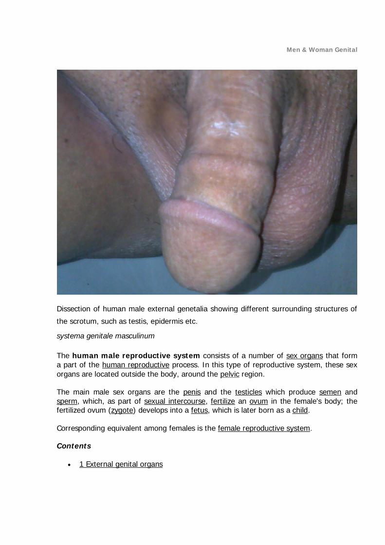
Dissection of human male external genetalia showing different surrounding structures of
the scrotum, such as testis, epidermis etc.
systema genitale masculinum
The human male reproductive system consists of a number of sex organs that form a part of the human reproductive process. In this type of reproductive system, these sex organs are located outside the body, around the pelvic region.
The main male sex organs are the penis and the testicles which produce semen and sperm, which, as part of sexual intercourse, fertilize an ovum in the female's body; the fertilized ovum (zygote) develops into a fetus, which is later born as a child.
Corresponding equivalent among females is the female reproductive system.
Contents
1 External genital organs

Men & Woman Genital
o 1.1 Penis o 1.2 Scrotum
2 Internal genital organs o 2.1 Epididymis o 2.2 Vas deferens o 2.3 Accessory glands
2.3.1 Seminal vesicles 2.3.2 Prostate gland 2.3.3 Bulbourethral glands
3 See also 4 References

Men & Woman Genital
External genital organs

Men & Woman Genital

Men & Woman Genital

Men & Woman Genital
Image showing innervation and blood-supply of the human external male genitalia.
Penis
Main article: Human penis
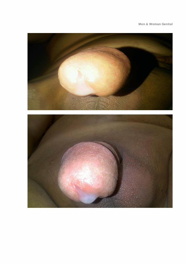
The penis is the male copulatory organ. It has a long shaft and an enlarged bulbous-shaped tip called the glans penis, which supports and is protected by the foreskin. When the male becomes sexually aroused, the penis becomes erect and ready for sexual activity. Erection occurs because sinuses within the erectile tissue of the penis become filled with blood. The arteries of the penis are dilated while the veins are passively compressed so that blood flows into the erectile cartilage under pressure.

Men & Woman Genital

Men & Woman Genital

Men & Woman Genital
Scrotum
Main article: Scrotum
The scrotum is a pouch-like structure that hangs behind the penis. It holds and protects the testes. It also contains numerous nerves and blood vessels. During times of lower temperatures, the Cremaster muscle contracts and pulls the scrotum closer to the body, while the Dartos muscle gives it a wrinkled appearance; when the temperature increases, the Cremaster and Dartos muscles relaxes to bring down the scrotum away from the body and remove the wrinkles respectively.
The scrotum remains connected with the abdomen or pelvic cavity by the inguinal canal. (The spermatic cord, formed from spermatic artery, vein and nerve bound together with connective tissue passes into the testis through inguinal canal.)

Men & Woman Genital
Internal genital organs Epididymis
Main article: Epididymis
The epididymis, a whitish mass of tightly coiled tubes cupped against the testicles, acts as a maturation and storage for sperm before they pass into the vas deferens, that carry sperm to the ampullary gland and prostatic ducts.
Vas deferens
Main article: Vas deferens
The vas deferens, also known as the sperm duct, is a thin tube approximately 30 centimetres (0.98 ft) long that starts from the epididymis to the pelvic cavity.
Accessory glands
Three accessory glands provide fluids that lubricate the duct system and nourish the sperm cells. They are the seminal vesicles, the prostate gland, and the bulbourethral glands (Cowper glands).
Seminal vesicles
Main article: Seminal vesicle
Seminal vesicles are sac-like structures attached to the vas deferens at one side of the bladder. They produce a sticky, yellowish fluid that contains fructose. This fluid provides sperm cells energy and aids in their motility. 70% of the semen is its secretion.
Prostate gland
Main article: Prostate gland
The prostate gland surrounds the ejaculatory ducts at the base of the male urethra, just below the bladder. The prostate gland is responsible for the proof semen, a liquid mixture of sperm cells, prostate fluid and seminal fluid. This gland is also responsible for making the semen milky in appearance by mixing calcium to the semen coming from seminal vesicle (semen coming from the seminal vesicle is yellowish in color); the semen remains cloudy and clumpy until the prostatic profibrinolysin is formed into fibrinolysin and lysis of the fibrinogen from the seminal vesicle fluids occurs.
Bulbourethral glands

Men & Woman Genital
Main article: Bulbourethral gland
The bulbourethral glands, or Cowper’s glands, are pea-sized structures located on the sides of the urethra just below the prostate gland. These glands produce a clear, slippery fluid that empties directly into the urethra. This fluid serves to lubricate the urethra and to neutralize any acidity that may be present due to residual drops of urine in the urethra.

Men & Woman Genital
Human penis From Wikipedia, the free encyclopedia Human penis
A flaccid penis with labels showing the locations of the shaft, foreskin, glans and meatus.
The model has removed body hair.

Men & Woman Genital

Men & Woman Genital
Men & Woman Genital

Men & Woman Genital

Men & Woman Genital

Men & Woman Genital
Latin 'penis, penes' Gray's p.1247
Artery Dorsal artery of the penis, deep artery of the penis, artery of the urethral bulb
Vein Dorsal veins of the penis Nerve Dorsal nerve of the penis Lymph Superficial inguinal lymph nodes Precursor Genital tubercle, Urogenital folds MeSH Penis
The human penis is an external male sexual organ. It is a reproductive, intromittent organ that additionally serves as the urinal duct. The main parts are the root (radix); the body (corpus); and the epithelium of the penis including the shaft skin and the foreskin

Men & Woman Genital
covering the glans penis. The body of the penis is made up of three columns of tissue: two corpora cavernosa on the dorsal side and corpus spongiosum between them on the ventral side. The human male urethra passes through the prostate gland, where it is joined by the ejaculatory duct, and then through the penis. The urethra traverses the corpus spongiosum, and its opening, the meatus /miːˈeɪtəs/, lies on the tip of the glans penis. It is a passage both for urine and for the ejaculation of semen.
The penis is homologous to the clitoris. An erection is the stiffening and rising of the penis, which occurs during sexual arousal, though it can also happen in non-sexual situations. The most common form of genital alteration is circumcision, removal of part or all of the foreskin for various cultural, religious, and more rarely, medical reasons. There is controversy surrounding circumcision.
While results vary across studies, the consensus is that the average erect human penis is approximately 12.9–15 cm (5.1–5.9 in) in length with 95% of adult males falling within the interval 10.7–19.1 cm (4.2–7.5 in). Neither patient age nor size of the flaccid penis accurately predicts erectile length.
Contents
1 Anatomy o 1.1 Parts o 1.2 Structure
2 Development o 2.1 Genital homology between sexes o 2.2 Penile growth and puberty
3 Physiological functions o 3.1 Urination o 3.2 Erection
3.2.1 Erection angle

Men & Woman Genital
o 3.3 Ejaculation o 3.4 Normal variations o 3.5 Disorders
3.5.1 Developmental disorders 3.5.2 Alleged and observed psychological disorders
o 3.6 Altering the genitalia 3.6.1 Circumcision
o 3.7 Surgical replacement o 3.8 Size
4 Cultural aspects 5 Additional images 6 References 7 External links 8 Related information

Men & Woman Genital
Anatomy
Lateral cross section of the penis.
Parts
Root of the penis (radix): It is the attached part, consisting of the bulb of penis in the middle and the crus of penis, one on either side of the bulb. It lies within the superficial perineal pouch.
Body of the penis (corpus): It has two surfaces: dorsal (posterosuperior in the erect penis), and ventral or urethral (facing downwards and backwards in the flaccid penis). The ventral surface is marked by a groove in a lateral direction.
Epithelium of the penis consists of the shaft skin, the foreskin, and the preputial mucosa on the inside of the foreskin and covering the glans penis.
Structure
The human penis is made up of three columns of tissue: two corpora cavernosa lie next to each other on the dorsal side and one corpus spongiosum lies between them on the ventral side.
The enlarged and bulbous-shaped end of the corpus spongiosum forms the glans penis, which supports the foreskin, or prepuce, a loose fold of skin that in adults can retract to

Men & Woman Genital
expose the glans. The area on the underside of the penis, where the foreskin is attached, is called the frenum, or frenulum. The rounded base of the glans is called the corona. The perineal raphe is the noticeable line along the underside of the penis.

Men & Woman Genital
Anatomical diagram of a human penis

Men & Woman Genital
The urethra, which is the last part of the urinary tract, traverses the corpus spongiosum, and its opening, known as the meatus /miːˈeɪtəs/, lies on the tip of the glans penis. It is a passage both for urine and for the ejaculation of semen. Sperm are produced in the testes and stored in the attached epididymis. During ejaculation, sperm are propelled up the vas deferens, two ducts that pass over and behind the bladder. Fluids are added by the seminal vesicles and the vas deferens turns into the ejaculatory ducts, which join the urethra inside the prostate gland. The prostate as well as the bulbourethral glands add further secretions, and the semen is expelled through the penis.
The raphe is the visible ridge between the lateral halves of the penis, found on the ventral or underside of the penis, running from the meatus (opening of the urethra) across the scrotum to the perineum (area between scrotum and anus).
The human penis differs from those of most other mammals, as it has no baculum, or erectile bone, and instead relies entirely on engorgement with blood to reach its erect state. It cannot be withdrawn into the groin, and it is larger than average in the animal kingdom in proportion to body mass.
Development
Stages in the development of the male external genitalia.

Men & Woman Genital
Main article: Development of the reproductive system
Genital homology between sexes
Main article: Sexual homology
In short, this is a known list of sex organs that evolve from the same tissue in females and males.
The glans of the penis is homologous to the clitoral glans; the corpora cavernosa are homologous to the body of the clitoris; the corpus spongiosum is homologous to the vestibular bulbs beneath the labia minora; the scrotum, homologous to the labia minora and labia majora; and the foreskin, homologous to the clitoral hood. The raphe does not exist in females, because there, the two halves are not connected.
Penile growth and puberty
On entering puberty, the penis, scrotum and testicles will begin to develop. During the process, pubic hair grows above and around the penis. A large-scale study assessing penis size in thousands of 17–19 year old males found no difference in average penis size between 17 year olds and 19 year olds. From this, it can be concluded that penile growth is typically complete not later than age 17, and possibly earlier.[1]
Physiological functions
Urination
Main article: Urination
In males, the expulsion of urine from the body is done through the penis. The urethra drains the bladder through the prostate gland where it is joined by the ejaculatory duct, and then onward to the penis. At the root of the penis (the proximal end of the corpus spongiosum) lies the external sphincter muscle. This is a small sphincter of striated muscle tissue and is in healthy males under voluntary control. Relaxing the urethra sphincter allows the urine in the upper urethra to enter the penis proper and thus empty the urinary bladder.

Men & Woman Genital

Men & Woman Genital

Men & Woman Genital
Man urinating in a public toilet
Physiologically, urination involves coordination between the central, autonomic, and somatic nervous systems. In infants, some elderly individuals, and those with neurological injury, urination may occur as an involuntary reflex. Brain centers that regulate urination include the pontine micturition center, periaqueductal gray, and the cerebral cortex.[2] During erection, these centers block the relaxation of the sphincter muscles, so as to act as a physiological separation of the excretory and reproductive function of the penis, and stopping sperm from entering the upper portion of the urethra during ejaculation.[3]
The part of the urethra in the penis has no muscles, and this serves no physiological function beyond that of a duct. Small amounts of urine usually remaining in the distal portion of the urethra, seeping out after the voluntary expulsion of urine is over. The distal section of the urethra does however allow a human male to direct the stream of urine by holding the penis. In cultures where more than a minimum of clothing is worn, the penis allows the male to urinate while standing without removing much of the clothing, a fact highly appreciated in these circumstances.[4] Females usually sit or squat to urinate and often have to remove some garments in the process.

Men & Woman Genital
Erection
Men & Woman Genital

Men & Woman Genital

Men & Woman Genital

Men & Woman Genital
The development of a penile erection, also showing the foreskin gradually retracting over the glans. See also: Commons image gallery

Men & Woman Genital

Men & Woman Genital

Men & Woman Genital
A ventral view of a penis flaccid (left) and erect (middle); a dorsal view of a penis erect (right). Main article: Erection
An erection is the stiffening and rising of the penis, which occurs during sexual arousal, though it can also happen in non-sexual situations. The primary physiological mechanism that brings about erection is the autonomic dilation of arteries supplying blood to the penis, which allows more blood to fill the three spongy erectile tissue chambers in the penis, causing it to lengthen and stiffen. The now-engorged erectile tissue presses against and constricts the veins that carry blood away from the penis. More blood enters than leaves the penis until an equilibrium is reached where an equal volume of blood flows into the dilated arteries and out of the constricted veins; a constant erectile size is achieved at this equilibrium.
Erection facilitates sexual intercourse though it is not essential for various other sexual activities.
Erection angle
Although many erect penises point upwards (see illustration), it is common and normal for the erect penis to point nearly vertically upwards or nearly vertically downwards or even horizontally straight forward, all depending on the tension of the suspensory ligament that holds it in position.
The following table shows how common various erection angles are for a standing male, out of a sample of 1,564 males aged 20 through 69. In the table, zero degrees is pointing straight up against the abdomen, 90 degrees is horizontal and pointing straight forward, while 180 degrees would be pointing straight down to the feet. An upward pointing angle is most common.[5]
Occurrence of erection angles
angle (°) from vertically upwards
Percent of males
0–30 5
30–60 30
60–85 31
85–95 10
95–120 20
120–180 5

Men & Woman Genital

Men & Woman Genital
Ejaculation
Main article: Ejaculation
Ejaculation is the ejecting of semen from the penis, and is usually accompanied by orgasm. A series of muscular contractions delivers semen, containing male gametes known as sperm cells or spermatozoa, from the penis. It is usually the result of sexual stimulation, which may include prostate stimulation. Rarely, it is due to prostatic disease. Ejaculation may occur spontaneously during sleep (known as a nocturnal emission or wet dream). Anejaculation is the condition of being unable to ejaculate.
Ejaculation has two phases: emission and ejaculation proper. The emission phase of the ejaculatory reflex is under control of the sympathetic nervous system, while the ejaculatory phase is under control of a spinal reflex at the level of the spinal nerves S2–4 via the pudendal nerve. A refractory period succeeds the ejaculation, and sexual stimulation precedes it.

Men & Woman Genital

Men & Woman Genital

Men & Woman Genital

Men & Woman Genital

Men & Woman Genital
Men & Woman Genital
Men & Woman Genital

Men & Woman Genital

Men & Woman Genital
Normal variations

Men & Woman Genital

Men & Woman Genital
Pearly penile papules, a common anatomical variation, may be the vestigial remnants of penis spines.

Men & Woman Genital
Pearly penile papules are raised bumps of somewhat paler color around the base (sulcus) of the glans which typically develop in men aged 20 to 40. As of 1999, different studies had produced estimates of incidence ranging from 8 to 48 percent of all men.[6] They may be mistaken for warts, but are not harmful or infectious and require no treatment.[7]
Fordyce's spots are small, raised, yellowish-white spots 1–2 mm in diameter that may appear on the penis, which again are common and not infectious.
Sebaceous prominences are raised bumps similar to Fordyce's spots on the shaft of the penis, located at the sebaceous glands and are normal.
Phimosis is an inability to retract the foreskin fully, is harmless in infancy and pre-pubescence, occurring in about 8% of boys at age 10. According to the British Medical Association, treatment (topical steroid cream and/or manual stretching) does not need to be considered until age 19.
Curvature: few penises are completely straight, with curves commonly seen in all directions (up, down, left, right). Sometimes the curve is very prominent but it rarely inhibits sexual intercourse. Curvature as great as 30° is considered normal and medical treatment is rarely considered unless the angle exceeds 45°. Changes to the curvature of a penis may be caused by Peyronie's disease.
Disorders
Paraphimosis is an inability to move the foreskin forward, over the glans. It can result from fluid trapped in a foreskin left retracted, perhaps following a medical procedure, or accumulation of fluid in the foreskin because of friction during vigorous sexual activity.
In Peyronie's disease, anomalous scar tissue grows in the soft tissue of the penis, causing curvature. Severe cases can benefit from surgical correction.
A thrombosis can occur during periods of frequent and prolonged sexual activity, especially fellatio. It is usually harmless and self-corrects within a few weeks.
Infection with the herpes virus can occur after sexual contact with an infected carrier; this may lead to the development of herpes sores.
Pudendal nerve entrapment is a condition characterized by pain on sitting and loss of penile (or clitoral) sensation and orgasm. Occasionally there is a total loss of sensation and orgasm. The pudendal nerve can be damaged by narrow, hard bicycle seats and accidents.
Penile fracture can occur if the erect penis is bent excessively. A popping or cracking sound and pain is normally associated with this event. Emergency medical assistance should be obtained. Prompt medical attention lowers likelihood of permanent penile curvature.
In diabetes, peripheral neuropathy can cause tingling in the penile skin and possibly reduced or completely absent sensation. The reduced sensations can lead to injuries for either partner and their absence can make it impossible to have sexual pleasure through stimulation of the penis. Since the problems are caused by permanent nerve damage, preventive treatment through good control of the

Men & Woman Genital
diabetes is the primary treatment. Some limited recovery may be possible through improved diabetes control.
Erectile dysfunction is the inability to develop and maintain an erection sufficiently firm for satisfactory sexual performance. Diabetes is a leading cause, as is natural aging. A variety of treatments exist, most notably including the phosphodiesterase type 5 inhibitor drugs (such as sildenafil citrate, marketed as Viagra), which work by vasodilation.
Priapism is a painful and potentially harmful medical condition in which the erect penis does not return to its flaccid state. The causative mechanisms are poorly understood but involve complex neurological and vascular factors. Potential complications include ischaemia, thrombosis, and impotence. In serious cases the condition may result in gangrene, which may necessitate amputation. The condition has been associated with a variety of drugs including prostaglandin but not sildenafil (Viagra).[8]
Lymphangiosclerosis is a hardened lymph vessel, although it can feel like a hardened, almost calcified or fibrous, vein. It tends not to share the common blue tint with a vein however. It can be felt as a hardened lump or "vein" even when the penis is flaccid, and is even more prominent during an erection. It is considered a benign physical condition. It is fairly common and can follow a particularly vigorous sexual activity for men, and tends to go away if given rest and more gentle care, for example by use of lubricants.
Carcinoma of the penis is rare with a reported rate of 1 person in 100,000 in developed countries. Circumcision is said to protect against this disease but this notion remains controversial.[9]
Developmental disorders
Hypospadias

Men & Woman Genital
Hypospadias is a developmental disorder where the meatus is positioned wrongly at birth. Hypospadias can also occur iatrogenically by the downward pressure of an indwelling urethral catheter.[10] It is usually corrected by surgery. The Intersex Society of North America classifies hypospadias as an intersex condition. They believe in halting all medically unnecessary surgeries, including many of those done on people with hypospadias.
A micropenis is a very small penis caused by developmental or congenital problems.
Diphallia, or penile duplication (PD), is the condition of having two penises. However, this disorder is extremely rare.
Alleged and observed psychological disorders
Penis panic (koro in Malaysian/Indonesian)—delusion of shrinkage of the penis and retraction into the body. This appears to be culturally conditioned and largely limited to Ghana, Sudan, China, Japan, Southeast Asia, and West Africa.
In April 2008, Kinshasa, Democratic Republic of Congo, West Africa's 'Police arrested 14 suspected victims (of penis snatching) and sorcerers accused of using black magic or witchcraft to steal (make disappear) or shrink men's penises to extort cash for cure, amid a wave of panic. Arrests were made in an effort to avoid bloodshed seen in Ghana a decade before, when 12 penis snatchers were beaten to death by mobs.[11]
Penis envy – the contested Freudian belief of all women inherently envying men for having penises.
Altering the genitalia
Main article: Genital modification and mutilation
The penis is sometimes pierced or decorated by other body art. Other than circumcision, genital alterations are almost universally elective and usually for the purpose of aesthetics or increased sensitivity. Piercings of the penis include the Prince Albert, the apadravya, the ampallang, the dydoe, and the frenum piercing. Foreskin restoration or stretching is a further form of body modification, as well as implants under the shaft of the penis.
Male to female transsexuals who undergo sex reassignment surgery, have their penis surgically modified into a neovagina. Female to male transsexuals may have a phalloplasty.
Other practices that alter the penis are also performed, although they are rare in Western societies without a diagnosed medical condition. Apart from a penectomy, perhaps the most radical of these is subincision, in which the urethra is split along the underside of the penis. Subincision originated among Australian Aborigines, although it is now done by some in the U.S. and Europe.

Men & Woman Genital
Penis removal is another form of alteration done to the penis.
Circumcision
Main article: Circumcision

Men & Woman Genital
A labelled dorsal view of a circumcised penis: (1)Shaft, (2)Circumcision scar, (3)Corona, (4)Glans, (5)Meatus.
The most common form of genital alteration is circumcision: removal of part or all of the foreskin for various cultural, religious, and more rarely medical reasons. For infant circumcision, modern devices such as the Gomco clamp, Plastibell, and Mogen clamp are available.[12]
With all modern devices the same basic procedure is followed. First, the amount of foreskin to be removed is estimated. The foreskin is then opened via the preputial orifice to reveal the glans underneath and ensured that it is normal. The inner lining of the foreskin (preputial epithelium) is then separated from its attachment to the glans. The device is then placed (this sometimes requires a dorsal slit) and remains there until blood flow has stopped. Finally, part, or all, of the foreskin is then removed.
Adult circumcisions are often performed without clamps and require 4 to 6 weeks of abstinence from masturbation or intercourse after the operation to allow the wound to heal.[13] In some African countries, male circumcision is often performed by non-medical
Men & Woman Genital
personnel under unsterile conditions.[14] After hospital circumcision, the foreskin may be used in biomedical research,[15] consumer skin-care products,[16] skin grafts,[17][18][19] or β-interferon-based drugs.[20] In parts of Africa, the foreskin may be dipped in brandy and eaten by the patient, eaten by the circumciser, or fed to animals.[21] According to Jewish law, after a Brit milah, the foreskin should be buried.[22]
There is controversy surrounding circumcision. Advocates of circumcision argue, for example, that it provides important health advantages that outweigh the risks, has no substantial effects on sexual function, has a low complication rate when carried out by an experienced physician, and is best performed during the neonatal period.[23] Opponents of circumcision argue, for example, that the practice has been and is still defended through the use of various myths; that it interferes with normal sexual function; that it is extremely painful; and that when performed on infants and children, it violates the individual's human rights.[24]
The American Medical Association stated in 1999: "Virtually all current policy statements from specialty societies and medical organizations do not recommend routine neonatal circumcision, and support the provision of accurate and unbiased information to parents to inform their choice."[25]
The World Health Organization (WHO; 2007), the Joint United Nations Programme on HIV/AIDS (UNAIDS; 2007), and the Centers for Disease Control and Prevention (CDC; 2008) state that evidence indicates male circumcision significantly reduces the risk of HIV acquisition by men during penile-vaginal sex, but also state that circumcision only provides partial protection and should not replace other interventions to prevent transmission of HIV.[26][27] In addition, some doctors have expressed concern over the policy and the data that supports it.[28][29]
Surgical replacement
The first successful penis allotransplant surgery was done in September 2005 in a military hospital in Guangzhou, China.[30] A man at 44 sustained an injury after an accident and his penis was severed; urination became difficult as his urethra was partly blocked. A recently brain-dead man, aged 23, was selected for the transplant. Despite atrophy of blood vessels and nerves, the arteries, veins, nerves and the corpora spongiosa were successfully matched. But, on 19 September (after two weeks), the surgery was reversed because of a severe psychological problem (rejection) by the recipient and his wife.[31]
In 2009, researchers Chen, Eberli, Yoo and Atala have produced bioengineered penises and implanted them on rabbits.[32] The animals were able to obtain erection and copulate, with 10 of 12 rabbits achieving ejaculation. This study shows that in the future it could be possible to produce artificial penises for replacement surgeries or phalloplasties.

Men & Woman Genital
Size
Main article: Human penis size
While results vary across studies, the consensus is that the average erect human penis is approximately 12.9–15 cm (5.1–5.9 in) in length with 95% of adult males falling within the interval 10.7–19.1 cm (4.2–7.5 in). Neither patient age nor size of the flaccid penis accurately predicted erectile length. Stretched length most closely correlated with erect length.[33][34][35] The average penis size is slightly larger than the median size (i.e., most penises are below average in size).
Length of the flaccid penis does not necessarily correspond to length of the erect penis; some smaller flaccid penises grow much longer, while some larger flaccid penises grow comparatively less.[36] Among all apes, the human penis is the largest, both in length and girth.[37]
A research project, summarizing dozens of published studies conducted by physicians of different nationalities, shows that, worldwide, erect-penis size averages vary between 9.6 and 16 cm (3.8 and 6.3 in). It has been suggested that this difference is caused not only by genetics but also by environmental factors such as fertility medications,[38] culture, diet, and chemical/pollution exposure.[39][40][41] Endocrine disruption resulting from chemical exposure has been linked to genital deformation in both sexes (among many other problems).
The longest officially documented human penis was found by Doctor Robert Latou Dickinson. It was 34.3 cm (13.5 in) long and 15.9 cm (6.26 in) around.[42]
Cultural aspects
Aesthetic, e.g., Body modification In humor, considered indecent or completely taboo in various cultures Religious veneration, see St. Priapus Church[43] In symbology, e.g., Phallus In architecture and sculpture, Phallic architecture

Men & Woman Genital
Additional images
Dissection showing the fascia of the penis as well as several surrounding structures.
Image showing innervation and blood-supply of the human male external genitalia.

Men & Woman Genital
References
1. Ponchietti R, Mondaini N, Bonafè M, Di Loro F, Biscioni S, Masieri L (February 2001). "Penile length and circumference: a study on 3,300 young Italian males". European Urology 39 (2): 183–6. doi:10.1159/000052434. PMID 11223678.
2. Sie JA, Blok BF, de Weerd H, Holstege G (2001). "Ultrastructural evidence for direct projections from the pontine micturition center to glycine-immunoreactive neurons in the sacral dorsal gray commissure in the cat". J. Comp. Neurol. 429 (4): 631–7. doi:10.1002/1096-9861(20010122)429:4<631::AID-CNE9>3.0.CO;2-M. PMID 11135240.
3. Schirren, C.; Rehacek, M.; Cooman, S. de; Widmann, H.-U. (24 April 2009). "Die retrograde Ejakulation". Andrologia 5 (1): 7–14. doi:10.1111/j.1439-0272.1973.tb00878.x.
4. Gamel, J. "To sit or not to sit: Why men should stand to pee". The Naked Scientists. Cambridge University. Retrieved 30 October 2012.
5. Sparling J (1997). "Penile erections: shape, angle, and length". Journal of Sex & Marital Therapy 23 (3): 195–207. doi:10.1080/00926239708403924. PMID 9292834.
6. Brown, Clarence William (February 13, 2014). "Pearly Penile Papules: Epidemiology". Medscape. Retrieved 2014-03-08.
7. Spots on the penis 8. Goldenberg MM (1998). "Safety and efficacy of sildenafil citrate in the
treatment of male erectile dysfunction". Clinical Therapeutics 20 (6): 1033–48. doi:10.1016/S0149-2918(98)80103-3. PMID 9916601.
9. Boczko S, Freed S (November 1979). "Penile carcinoma in circumcised males". New York State Journal of Medicine 79 (12): 1903–4. PMID 292845.
10. Andrews HO, Nauth-Misir R, Shah PJ (March 1998). "Iatrogenic hypospadias—a preventable injury?". Spinal Cord 36 (3): 177–80. doi:10.1038/sj.sc.3100508. PMID 9554017.
11. Reuters, Lynchings in Congo as penis theft panic hits capital 12. Holman JR, Lewis EL, Ringler RL (August 1995). "Neonatal circumcision
techniques". American Family Physician 52 (2): 511–8, 519–20. PMID 7625325. 13. Holman JR, Stuessi KA (March 1999). "Adult circumcision". American Family
Physician 59 (6): 1514–8. PMID 10193593. 14. Rosenthal, Elisabeth (2007-02-27). "In Africa, a problem with circumcision
and AIDS". The New York Times. 15. Hovatta O, Mikkola M, Gertow K, et al. (July 2003). "A culture system using
human foreskin fibroblasts as feeder cells allows production of human embryonic stem cells". Human Reproduction 18 (7): 1404–9. doi:10.1093/humrep/deg290. PMID 12832363.
16. "'Miracle' Wrinkle Cream's Key Ingredient". Banderasnews.com. Banderas News, Inc. April 2008. Retrieved 2010-10-22.

Men & Woman Genital
17. "High-Tech Skinny on Skin Grafts". www.wired.com:science:discoveries (CondéNet, Inc). 1999-02-16. Retrieved 2008-08-20.[dead link]
18. "Skin Grafting". www.emedicine.com. WebMD. Retrieved 2008-08-20. 19. Amst, Catherine; Carey, John (July 27, 1998). "Biotech Bodies".
www.businessweek.com. The McGraw-Hill Companies Inc. Retrieved 2008-08-20. 20. Cowan, Alison Leigh (April 19, 1992). "Wall Street; A Swiss Firm Makes
Babies Its Bet". New York Times:Business (New York Times). Retrieved 2008-08-20.
21. Anonymous (editorial) (1949-12-24). "A ritual operation". British Medical Journal 2 (4642): 1458–1459. doi:10.1136/bmj.2.4642.1458. PMC 2051965. PMID 20787713. "...in parts of West Africa, where the operation is performed at about 8 years of age, the prepuce is dipped in brandy and eaten by the patient; in other districts the operator is enjoined to consume the fruits of his handiwork, and yet a further practice, in Madagascar, is to wrap the operation specifically in a banana leaf and feed it to a calf."
22. Shulchan Aruch, Yoreh Deah, 265:10 23. Schoen EJ (December 2007). "Should newborns be circumcised? Yes".
Canadian Family Physician 53 (12): 2096–8, 2100–2. PMC 2231533. PMID 18077736.
24. Milos MF, Macris D (1992). "Circumcision. A medical or a human rights issue?". Journal of Nurse-midwifery 37 (2 Suppl): 87S–96S. doi:10.1016/0091-2182(92)90012-R. PMID 1573462.
25. "Report 10 of the Council on Scientific Affairs (I-99):Neonatal Circumcision". 1999 AMA Interim Meeting: Summaries and Recommendations of Council on Scientific Affairs Reports. American Medical Association. December 1999. p. 17. Retrieved 2006-06-13.
26. New Data on Male Circumcision and HIV Prevention: Policy and Programme Implications (PDF). World Health Organization. March 28, 2007. Retrieved 2007-08-13.
27. "Male Circumcision and Risk for HIV Transmission and Other Health Conditions: Implications for the United States". Centers for Disease Control and Prevention. 2008. Retrieved 2013-11-07.
28. G. Dowsett, M. Couch. "Male Circumcision and HIV Prevention: Is There Really Enough of the Right Kind of Evidence?". Reproductive Health Matters. Retrieved 2013-11-07.
29. Vardi Y, Sadeghi-Nejad H, Pollack S, Aisuodionoe-Shadrach OI, Sharlip ID (July 2007). "Male circumcision and HIV prevention". J Sex Med 4 (4 Pt 1): 838–43. doi:10.1111/j.1743-6109.2007.00511.x. PMID 17627731.
30. Guangzhou Daily 31. Sample, Ian (2006-09-18). "Man rejects first penis transplant". The
Guardian (London). Retrieved 2010-05-22. 32. Chen KL, Eberli D, Yoo JJ, Atala A (November 2009). "Regenerative
Medicine Special Feature: Bioengineered corporal tissue for structural and functional restoration of the penis". Proceedings of the National Academy of

Men & Woman Genital
Sciences of the United States of America 107 (8): 3346–50. doi:10.1073/pnas.0909367106. PMC 2840474. PMID 19915140.
33. Wessells H, Lue TF, McAninch JW (September 1996). "Penile length in the flaccid and erect states: guidelines for penile augmentation". The Journal of Urology 156 (3): 995–7. doi:10.1016/S0022-5347(01)65682-9. PMID 8709382.
34. Chen J, Gefen A, Greenstein A, Matzkin H, Elad D (December 2000). "Predicting penile size during erection". International Journal of Impotence Research 12 (6): 328–33. doi:10.1038/sj.ijir.3900627. PMID 11416836.
35. "ANSELL RESEARCH – The Penis Size Survey". March 2001. Retrieved 2006-07-13.
36. "Penis Size FAQ & Bibliography". Kinsey Institute. 2009. Retrieved 2013-11-07.
37. Penis size: An evolutionary perspective retrieved 10 February 2012 38. Center of Disease Control. "DES Update: Consumers". Retrieved 2013-11-
07. 39. Swan SH, Main KM, Liu F, et al. (August 2005). "Decrease in anogenital
distance among male infants with prenatal phthalate exposure". Environmental Health Perspectives 113 (8): 1056–61. doi:10.1289/ehp.8100. PMC 1280349. PMID 16079079.
40. Montague, Peter. "PCBs Diminish Penis Size". Rachel's Hazardous Waste News 372. Archived from the original on 2012-03-03.
41. "Hormone Hell". DISCOVER. Retrieved 2008-04-05. 42. Dickinson, R.L. (1940). The Sex Life of the Unmarried Adult. New York:
Vanguard Press.[page needed] 43. Fritscher, Jack; Anton Szandor La Vey (2004). Popular witchcraft: straight
from the witch's mouth. Popular Press. p. 161. ISBN 978-0-299-20304-7. Retrieved 2013-11-07.

Men & Woman Genital
Skin For the article about skin in humans, see human skin. For other uses, see Skin (disambiguation). Skin
A diagram of human skin.
Latin Cutis

Men & Woman Genital

Men & Woman Genital

Men & Woman Genital

Men & Woman Genital

Men & Woman Genital
A close up picture of a rhinoceros skin.
Skin is the soft outer covering of vertebrates. Other animal coverings such as the arthropod exoskeleton have different developmental origin, structure and chemical composition. The adjective cutaneous means "of the skin" (from Latin cutis, skin). In mammals, the skin is an organ of the integumentary system made up of multiple layers of ectodermal tissue, and guards the underlying muscles, bones, ligaments and internal organs.[1] Skin of a different nature exists in amphibians, reptiles, and birds.[2] All mammals have some hair on their skin, even marine mammals which appear to be hairless. The skin interfaces with the environment and is the first line of defense from external factors. For example, the skin plays a key role in protecting the body against pathogens[3] and excessive water loss.[4] Its other functions are insulation, temperature regulation, sensation, and the production of vitamin D folates. Severely damaged skin may heal by forming scar tissue. This is sometimes discoloured and depigmented. The thickness of skin also varies from location to location on an organism. In humans for example, the skin located under the eyes and around the eyelids is the thinnest skin in the body at 0.5 mm thick, and is one of the first areas to show signs of aging such as "crows feet" and wrinkles. The skin on the palms and the soles of the feet is 4 mm thick

Men & Woman Genital
and the thickest skin in the body. The speed and quality of wound healing in skin is promoted by the reception of estrogen.[5][6][7]
Fur is dense hair. Primarily, fur augments the insulation the skin provides but can also serve as a secondary sexual characteristic or as camouflage. On some animals, the skin is very hard and thick, and can be processed to create leather. Reptiles and fish have hard protective scales on their skin for protection, and birds have hard feathers, all made of tough β-keratins. Amphibian skin is not a strong barrier, especially regarding the passage of chemicals via skin and is often subject to osmosis and diffusive forces. For example, a frog sitting in an anesthetic solution would be sedated quickly, as the chemical diffuses through its skin.
Contents
1 Functions 2 Mammalian skin layers
o 2.1 Epidermis o 2.2 Basement membrane o 2.3 Dermis
2.3.1 Papillary region 2.3.2 Reticular region
o 2.4 Hypodermis 3 Fish and amphibians 4 In birds and reptiles 5 Mechanics 6 Human uses and culture 7 Detailed cross section 8 See also 9 Additional images 10 References
Functions
Skin performs the following functions:
1. Protection: an anatomical barrier from pathogens and damage between the internal and external environment in bodily defense; Langerhans cells in the skin are part of the adaptive immune system.[3][4]
2. Sensation: contains a variety of nerve endings that jump to heat and cold, touch, pressure, vibration, and tissue injury (see somatosensory system and haptic perception).
3. Thermoregulation: eccrine (sweat) glands and dilated blood vessels (increased superficial perfusion) aid heat loss, while constricted vessels greatly reduce

Men & Woman Genital
cutaneous blood flow and conserve heat. Erector pili muscles in mammals adjust the angle of hair shafts to change the degree of insulation provided by hair or fur.
4. Control of evaporation: the skin provides a relatively dry and semi-impermeable barrier to reduce fluid loss.[4]
5. Storage and synthesis: acts as a storage center for lipids and water 6. Absorption: oxygen, nitrogen and carbon dioxide can diffuse into the epidermis in
small amounts; some animals use their skin as their sole respiration organ (in humans, the cells comprising the outermost 0.25–0.40 mm of the skin are "almost exclusively supplied by external oxygen", although the "contribution to total respiration is negligible")[8]
7. Water resistance: The skin acts as a water resistant barrier so essential nutrients aren't washed out of the body. The nutrients and oils that help hydrate the skin are covered by the most outer skin layer, the epidermis. This is helped in part by the sebaceous glands that release sebum, an oily liquid. Water itself will not cause the elimination of oils on the skin, because the oils residing in our dermis flow and would be affected by water without the epidermis.[9]
Mammalian skin layers
Dermis
The distribution of the bloodvessels in the skin of the sole of the foot. (Corium – TA
alternate term for dermis – is labeled at upper right.)

Men & Woman Genital
A diagrammatic sectional view of the skin (click on image to magnify). (Dermis labeled
at center right.)
Gray's p.1065 MeSH Dermis Dorlands /Elsevier Skin

Men & Woman Genital
(See also: image rotating (1.1 mb) ) Optical coherence tomogram of fingertip, depicting stratum corneum (~500 µm thick) with stratum disjunctum on top and stratum lucidum (connection to stratum spinosum) in the middle. At the bottom superficial parts of the dermis. Sweatducts are clearly visible.[citation needed]
Mammalian skin is composed of two primary layers:
the epidermis, which provides waterproofing and serves as a barrier to infection; and
the dermis, which serves as a location for the appendages of skin;
Epidermis
Main article: Epidermis (skin)
The epidermis is composed of the outermost layers of the skin. It forms a protective barrier over the body's surface, responsible for keeping water in the body and preventing pathogens from entering, and is a stratified squamous epithelium,[10] composed of proliferating basal and differentiated suprabasal keratinocytes. The epidermis also helps the skin regulate body temperature.[citation needed]
Keratinocytes are the major cells, constituting 95% of the epidermis,[10] while Merkel cells, melanocytes and Langerhans cells are also present. The epidermis can be further subdivided into the following strata or layers (beginning with the outermost layer):[11]
Stratum corneum Stratum lucidum (only in palms and soles) Stratum granulosum Stratum spinosum Stratum germinativum (also called the stratum basale)

Men & Woman Genital
Keratinocytes in the stratum basale proliferate through mitosis and the daughter cells move up the strata changing shape and composition as they undergo multiple stages of cell differentiation to eventually become anucleated. During that process, keratinocytes will become highly organized, forming cellular junctions (desmosomes) between each other and secreting keratin proteins and lipids which contribute to the formation of an extracellular matrix and provide mechanical strength to the skin.[12] Keratinocytes from the stratum corneum are eventually shed from the surface (desquamation).
The epidermis contains no blood vessels, and cells in the deepest layers are nourished by diffusion from blood capillaries extending to the upper layers of the dermis.
Basement membrane
Main article: basement membrane
The epidermis and dermis are separated by a thin sheet of fibers called the basement membrane, and is made through the action of both tissues. The basement membrane controls the traffic of the cells and molecules between the dermis and epidermis but also serves, through the binding of a variety of cytokines and growth factors, as a reservoir for their controlled release during physiological remodeling or repair processes.[13]
Dermis
Main article: Dermis
The dermis is the layer of skin beneath the epidermis that consists of connective tissue and cushions the body from stress and strain. The dermis provides tensile strength and elasticity to the skin through an extracellular matrix composed of collagen fibrils, microfibrils, and elastic fibers, embedded in proteoglycans.[12]
It harbors many Mechanoreceptors (nerve endings) that provide the sense of touch and heat. It also contains the hair follicles, sweat glands, sebaceous glands, apocrine glands, lymphatic vessels and blood vessels. The blood vessels in the dermis provide nourishment and waste removal from its own cells as well as for the epidermis.
The dermis is tightly connected to the epidermis through a basement membrane and is structurally divided into two areas: a superficial area adjacent to the epidermis, called the papillary region, and a deep thicker area known as the reticular region.
Papillary region
The papillary region is composed of loose areolar connective tissue. This is named for its fingerlike projections called papillae that extend toward the epidermis. The papillae

Men & Woman Genital
provide the dermis with a "bumpy" surface that interdigitates with the epidermis, strengthening the connection between the two layers of skin.
Reticular region
The reticular region lies deep in the papillary region and is usually much thicker. It is composed of dense irregular connective tissue, and receives its name from the dense concentration of collagenous, elastic, and reticular fibers that weave throughout it. These protein fibers give the dermis its properties of strength, extensibility, and elasticity. Also located within the reticular region are the roots of the hair, sebaceous glands, sweat glands, receptors, nails, and blood vessels.
Hypodermis
Main article: Hypodermis
The hypodermis is not part of the skin, and lies below the dermis. Its purpose is to attach the skin to underlying bone and muscle as well as supplying it with blood vessels and nerves. It consists of loose connective tissue and elastin. The main cell types are fibroblasts, macrophages and adipocytes (the hypodermis contains 50% of body fat). Fat serves as padding and insulation for the body. Another name for the hypodermis is the subcutaneous tissue.
Microorganisms like Staphylococcus epidermidis colonize the skin surface. The density of skin flora depends on region of the skin. The disinfected skin surface gets recolonized from bacteria residing in the deeper areas of the hair follicle, gut and urogenital openings.
Fish and amphibians
The epidermis of fish and of most amphibians consists entirely of live cells, with only minimal quantities of keratin in the cells of the superficial layer. It is generally permeable, and in the case of many amphibians, may actually be a major respiratory organ. The dermis of bony fish typically contains relatively little of the connective tissue found in tetrapods. Instead, in most species, it is largely replaced by solid, protective bony scales. Apart from some particularly large dermal bones that form parts of the skull, these scales are lost in tetrapods, although many reptiles do have scales of a different kind, as do pangolins. Cartilaginous fish have numerous tooth-like denticles embedded in their skin, in place of true scales.
Sweat glands and sebaceous glands are both unique to mammals, but other types of skin gland are found in other vertebrates. Fish typically have a numerous individual mucus-secreting skin cells that aid in insulation and protection, but may also have poison glands, photophores, or cells that produce a more watery, serous fluid. In amphibians, the mucus

Men & Woman Genital
cells are gathered together to form sac-like glands. Most living amphibians also possess granular glands in the skin, that secrete irritating or toxic compounds.[14]
Although melanin is found in the skin of many species, in the reptiles, the amphibians, and fish, the epidermis is often relatively colourless. Instead, the colour of the skin is largely due to chromatophores in the dermis, which, in addition to melanin, may contain guanine or carotenoid pigments. Many species, such as chameleons and flounders may be able to change the colour of their skin by adjusting the relative size of their chromatophores.[14]
In birds and reptiles Main article: Reptile scales
The epidermis of birds and reptiles is closer to that of mammals, with a layer of dead keratin-filled cells at the surface, to help reduce water loss. A similar pattern is also seen in some of the more terrestrial amphibians such as toads. However, in all of these animals there is no clear differentiation of the epidermis into distinct layers, as occurs in humans, with the change in cell type being relatively gradual. The mammalian epidermis always possesses at least a stratum germinativum and stratum corneum, but the other intermediate layers found in humans are not always distinguishable. Hair is a distinctive feature of mammalian skin, while feathers are (at least among living species) similarly unique to birds.[14]
Birds and reptiles have relatively few skin glands, although there may be a few structures for specific purposes, such as pheromone-secreting cells in some reptiles, or the uropygial gland of most birds.[14]
Mechanics Main article: Soft tissue
Skin has a soft tissue mechanical behavior when stretched. The intact skin is prestreched like wetsuits around the diver's body. When deep cuts are made on the skin, it retracts, widening the slice hole.
Human uses and culture
The term "skin" may also refer to the covering of a small animal, such as a sheep, goat (goatskin), pig, snake (snakeskin) etc. or the young of a large animal.
The term hides or rawhide refers to the covering of a large adult animal such as a cow, buffalo, horse etc.
Skins and hides from the different animals are used for clothing, bags and other consumer products, usually in the form of leather, but also as furs.

Men & Woman Genital
Skin from sheep, goat and cattle was used to make parchment for manuscripts.
Skin can also be cooked to make pork rind or crackling.
Dutch artist Jalila Essaïdi is trying to create bulletproof skin.[15]

Men & Woman Genital
Detailed cross section
Skin layers, of both the hairy and hairless skin
See also
List of cutaneous conditions Acid mantle Callus – thick area of skin Cutaneous structure development Hair – including hair follicles in skin Intertriginous Moult Meissner's corpuscle

Men & Woman Genital
Pacinian corpuscle Rawhide Role of skin in locomotion Skin flora Superficial fascia
Additional images
Illustration of Integumentary System
References
1. "Skin care" (analysis), Health-Cares.net, 2007 2. Alibardi L. (2003). Adaptation to the land: The skin of reptiles in comparison
to that of amphibians and endotherm amniotes. J Exp Zoolog B Mol Dev Evol. 298(1):12–41. PMID 12949767
3. Proksch E, Brandner JM, Jensen JM. (2008).The skin: an indispensable barrier. Exp Dermatol. 17(12):1063–72. PMID 19043850

Men & Woman Genital
4. Madison KC. (2003). Barrier function of the skin: "la raison d'être" of the epidermis. J Invest Dermatol. 121(2):231-41. doi:10.1046/j.1523-1747.2003.12359.x PMID 12880413
5. Thornton MJ (2002). "The biological actions of estrogen in skin". Experimental Dermatology.
6. Gillian S. Ashcroft, Teresa Greenwell-Wild, and Mark W. J. Ferguson (1999). "Topical Estrogen Accelerates Cutaneous Wound Healing in Aged Humans Associated with an Altered Inflammatory Response". The American Journal of Pathology 155 (4): 1137–1146. doi:10.1016/S0002-9440(10)65217-0. PMC 1867002. PMID 10514397.
7. Desiree May Oh, MD, Tania J. Phillips, MD (2006). "Sex Hormones and Wound Healing". Wounds.
8. Stücker, M., A. Struk, P. Altmeyer, M. Herde, H. Baumgärtl & D.W. Lübbers (2002). The cutaneous uptake of atmospheric oxygen contributes significantly to the oxygen supply of human dermis and epidermis. PDF Journal of Physiology 538(3): 985–994. doi:10.1113/jphysiol.2001.013067
9. McCracken, Thomas (2000). New Atlas of Human Anatomy. China: Metro Books. pp. 1–240. ISBN 1-58663-097-0.
10. McGrath, J.A.; Eady, R.A.; Pope, F.M. (2004). Rook's Textbook of Dermatology (7th ed.). Blackwell Publishing. pp. 3.1–3.6. ISBN 978-0-632-06429-8.
11. The Ageing Skin – Structure. pharmaxchange.info. March 3, 2011 12. Breitkreutz, D; Mirancea, N; Nischt, R (2009). "Basement membranes in
skin: Unique matrix structures with diverse functions?". Histochemistry and cell biology 132 (1): 1–10. doi:10.1007/s00418-009-0586-0. PMID 19333614.
13. Iozzo, RV (2005). "Basement membrane proteoglycans: From cellar to ceiling". Nature reviews. Molecular cell biology 6 (8): 646–56. doi:10.1038/nrm1702. PMID 16064139.
14. Romer, Alfred Sherwood; Parsons, Thomas S. (1977). The Vertebrate Body. Philadelphia, PA: Holt-Saunders International. pp. 129–145. ISBN 0-03-910284-X.
15. Human skin strengthened with spider silk can stop a bullet (2:16). Reuters (2011-09-20)

Men & Woman Genital
Pubic Hair
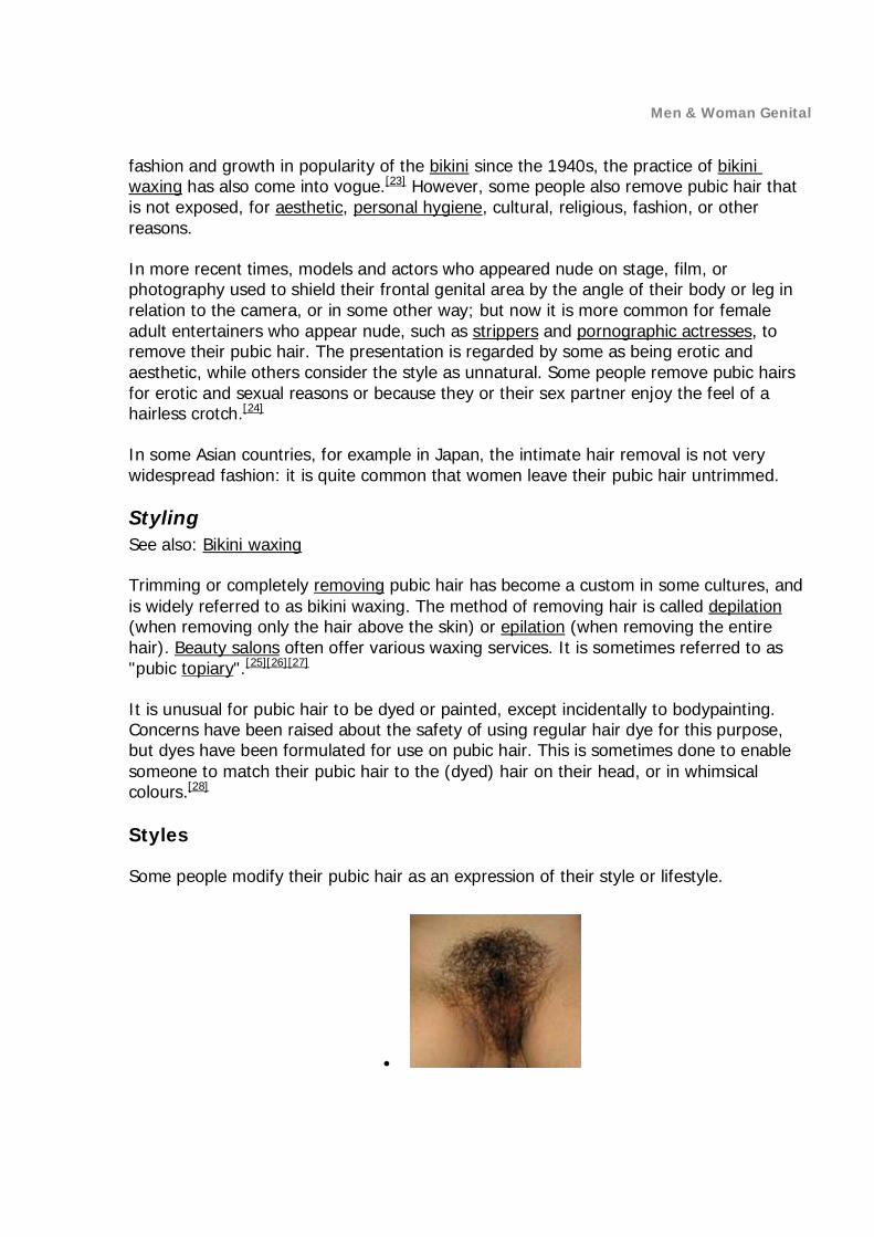
Pubic hair is the hair in the frontal genital area of adolescent and adult humans, located on and around the sex organs, the crotch, and sometimes at the top of the inside of the thighs, in the pubic region around the pubis bone.
Although fine vellus hair is present in the area in childhood, pubic hair is considered to be the heavier, longer and coarser hair that develops during puberty as an effect of rising levels of androgens. Pubic hair is therefore part of the androgenic hair (or body hair) and is a secondary sex characteristic.
Hair does not in itself have any intrinsic sexual value other than the attributes given to it by individuals in a cultural context. Some cultures are ambivalent in relation to particular body hair, with some being regarded as attractive while others being regarded as unaesthetic. Many cultures regard pubic hair to be erotic. In most cultures, both men and women are expected to cover their pubic hair at all times, but sometimes this is because it is associated with genitalia.
Contents
1 Development 2 In art 3 Cultural views 4 Styling
o 4.1 Styles 5 See also 6 References 7 External links
Development
The development of pubic hair can be assessed using the Tanner scale. Before the onset of puberty, the genital area of both boys and girls has very fine vellus hair, referred to as Tanner stage 1 hair.[1] As puberty begins, the body produces rising levels of the sex hormones known as androgens, and in response the skin of the genital area begins to produce thicker and rougher, often curlier, hair with a faster growth rate.[2] The onset of pubic hair development is termed pubarche. The change for each hair follicle is relatively abrupt, but the extent of skin which grows androgenic hair gradually increases over several years.

Men & Woman Genital
Variation of pubic hair on a mature male (left), and a mature female (right).
In males, the first pubic hair appears as a few sparse hairs that are usually thin on the scrotum or at the upper base of the penis (stage 2). Within a year, hairs around the base of the penis are numerous (stage 3). Within 3 to 4 years, hair fills the pubic area (stage 4) and becomes much thicker and darker, and by 5 years extends to the near thighs and upwards on the abdomen toward the umbilicus (stage 5).[3]
Other areas of the skin are similarly, though slightly less, sensitive to androgens and androgenic hair typically appears somewhat later. In rough sequence of sensitivity to androgens and appearance of androgenic hair, are the armpits (axillae), perianal area, upper lip, preauricular areas (sideburns), periareolar areas (nipples), middle of the chest, neck under the chin, remainder of chest and beard area, limbs and shoulders, back, and buttocks.
Men & Woman Genital
Although generally considered part of the process of puberty, pubarche is distinct and independent of the process of maturation of the gonads that leads to sexual maturation and fertility. Pubic hair can develop from adrenal androgens alone, and can develop even when the ovaries or testes are defective and nonfunctional. See puberty for details.
There is little if any difference in the capacity of male and female bodies to grow hair in response to androgens. The obvious sex-dimorphic difference in hair distribution in men and women is primarily a result of differences in the levels of androgen reached as maturity occurs.
Pubic hair and axillary (armpit) hair can vary in color considerably from the hair of the scalp. In most people it is darker, although it can also be lighter. In most cases it is most similar in color to the eyebrows of the individual.[4]
Natural female pubic hair varies considerably around the world, ranging from short to long, from sparse to dense, and from straight and soft to wiry and curly. In colour, pubic hair does not always match head hair. Many dark-haired women have lighter pubic hair often with a red tinge. Most women have wavy and curly pubic hair, even when their head hair is straight. In the Far East, however, straight black head hair is matched by pubic hair that has been described as 'black, short, straight and not thick but rather sparse...'[4][5] Hair texture varies from tightly curled to entirely straight. Such variations also appear in men.
Pubic hair patterns can vary by race and ethnicity.[5] Patterns of pubic hair, known as the escutcheon, vary between sexes. On most women, the pubic patch is triangular and lies over the mons pubis. On many men, the pubic patch tapers upwards to a line of hair pointing towards the navel (see abdominal hair), roughly a more upward-pointing triangle.[4] As with axillary (armpit) hair, pubic hair is associated with a concentration of sebaceous glands in the area.

Men & Woman Genital
In art

Men & Woman Genital

Men & Woman Genital

Men & Woman Genital

Men & Woman Genital
Heinrich Aldegrever's Eve, 1540. A rare early example of pubic hair in northern European art.
La Naissance de Venus by Eugène Emmanuel Amaury Duval (1808–1885) portrays the goddess of love with no pubic or axillary hair.
In ancient Egyptian art, female pubic hair is indicated in the form of painted triangles.[6] In medieval and classical European art, pubic hair was very rarely depicted, and male pubic hair was often, but not always, omitted.[7] Sometimes it was portrayed in stylized form, as was the case with Greek graphic art.[8] The same was true in much Indian art, and in other Eastern portrayals of the nude. In 16th-century southern Europe, Michelangelo showed the male David with stylized pubic hair, but female bodies were depicted hairless below the head. Nevertheless, Michelangelo's male nudes on the Sistine Chapel ceiling display no pubic hair. In Renaissance northern Europe, pubic hair was more likely to be portrayed than in the south, more usually male, while occasionally on female figures.
By the 17th century, suggestions of female pubic hair appear in pornographic engravings, such as those by Agostino Carracci.[citation needed]
According to John Ruskin's biographer Mary Lutyens, the notable author, artist, and art critic was apparently accustomed only to the hairless nudes portrayed unrealistically in art, never having seen a naked woman before his wedding night. He was allegedly so shocked by his discovery of his wife Effie's pubic hair that he rejected her, and the marriage was later legally annulled. He is supposed to have thought his wife was freakish and deformed.[9] Later writers have often followed Lutyens and repeated this version of events. For example, Gene Weingarten in his book I'm with Stupid (2004) writes that "Ruskin had [the marriage] annulled because he was horrified to behold upon his bride a thatch of hair, rough and wild, similar to a man's. He thought her a monster."[10] However, there is no proof for this, and some disagree. Peter Fuller in his book Theoria: Art and the Absence of Grace writes, "It has been said that he was frightened on the

Men & Woman Genital
wedding night by the sight of his wife's pubic hair; more probably, he was perturbed by her menstrual blood." Ruskin's biographers Tim Hilton and John Batchelor also believe that menstruation is the more likely explanation.[11]
Francisco Goya's The Nude Maja (1797) has been considered as probably the first European painting to show a female subject's pubic hair, though paintings had hinted at it.[citation needed] The painting was considered quite pornographic at the time.[12] Gustave Courbet's L'Origine du monde (The Origin of the World, 1866) was considered scandalous probably more because of the presentation of thick pubic hair than the exposed female genitals. Examples of male pubic hair in contemporary art are harder to find.[citation needed]
In the late 18th century female pubic hair is openly portrayed in Japanese shunga (erotica), especially in the ukiyo-e tradition.[13] Hokusai's picture The Dream of the Fisherman's Wife (1814), which depicts a woman having an erotic fantasy, is a well-known example. Fine art paintings and sculpture created before the 20th century in the Western tradition usually depicted women without pubic hair or a visible vulva.[citation
needed] In Japanese drawings, such as hentai, pubic hair is often omitted, since for a long time the display of pubic hair was not legal. The interpretation of the law has since changed.[14]
Cultural views
A preference for hairless crotch in oneself or in another is known as acomoclitism. According to the Oxford Companion to the Body, in the 1450s women would shave their pubic hair for personal hygiene and to combat pubic lice and would then don a merkin or pubic wig.[15][16] In Middle Eastern societies, removal of female body hair has been considered proper hygiene, necessitated by local customs, for many centuries.[17] In Islamic societies removing pubic hair is a religiously endorsed practice known as an act of Sunan al-Fitra.[18][19] Evidence of pubic hair removal in ancient India dates back to 4000 to 3000 BC.[20] According to ethnologist F. Fawcett, writing in 1901, he had observed the removal of body hair, including pubic hair about the vulva, as a custom of women from the Hindu Nair caste.[21]
Among the upper class in 19th-century Victorian Britain, pubic hair from one's lover was frequently collected as a souvenir. The curls were, for instance, worn like cockades in men's hats as potency talismans, or exchanged among lovers as tokens of affection.[22] The museum of St. Andrews University in Scotland has in its collection a snuffbox full of pubic hair of one of King George IV's mistresses, possibly Elizabeth Conyngham, which the notoriously licentious monarch donated to the Fife sex club, The Beggar's Benison.[22]
In Western societies, exposure of a woman's body hair below her neck has traditionally and continues to be widely disapproved of culturally. Many people consider exposure of pubic hair to be embarrassing.[23] It may be regarded as immodest and sometimes as obscene. With the reduction in the size of swimsuits, especially since the coming into
Men & Woman Genital
fashion and growth in popularity of the bikini since the 1940s, the practice of bikini waxing has also come into vogue.[23] However, some people also remove pubic hair that is not exposed, for aesthetic, personal hygiene, cultural, religious, fashion, or other reasons.
In more recent times, models and actors who appeared nude on stage, film, or photography used to shield their frontal genital area by the angle of their body or leg in relation to the camera, or in some other way; but now it is more common for female adult entertainers who appear nude, such as strippers and pornographic actresses, to remove their pubic hair. The presentation is regarded by some as being erotic and aesthetic, while others consider the style as unnatural. Some people remove pubic hairs for erotic and sexual reasons or because they or their sex partner enjoy the feel of a hairless crotch.[24]
In some Asian countries, for example in Japan, the intimate hair removal is not very widespread fashion: it is quite common that women leave their pubic hair untrimmed.
Styling See also: Bikini waxing
Trimming or completely removing pubic hair has become a custom in some cultures, and is widely referred to as bikini waxing. The method of removing hair is called depilation (when removing only the hair above the skin) or epilation (when removing the entire hair). Beauty salons often offer various waxing services. It is sometimes referred to as "pubic topiary".[25][26][27]
It is unusual for pubic hair to be dyed or painted, except incidentally to bodypainting. Concerns have been raised about the safety of using regular hair dye for this purpose, but dyes have been formulated for use on pubic hair. This is sometimes done to enable someone to match their pubic hair to the (dyed) hair on their head, or in whimsical colours.[28]
Styles
Some people modify their pubic hair as an expression of their style or lifestyle.

Men & Woman Genital
Natural and untrimmed pubic hair. Extends onto thighs.
American wax—hair limited to the bikini area. Also trimmed shorter.
French wax—waxing with a "landing strip". Labia are cleared.
Full Brazilian or the Sphinx—a full waxing. No hair at all.
Some styles include:[29]
Natural, Au naturel, Bush[30] No trimming and therefore no maintenance.
Trimmed or cut Hair length is shortened but not removed or shaped, inner thighs may be shaved.
Triangle
Pubic hair shaved from the genital area of a mature male

Men & Woman Genital
Hair removed (generally waxed) from the sides to form a triangle so that pubic hair cannot be seen while wearing swimwear. This can range from the very edge of the "bikini line" to up to an inch reduction on either side. Hair length can be from an inch and a half to half an inch.
Landing Strip, Hitler Moustache, Clitler Hair sharply removed from the sides to form a long centered vertical rectangle, hair length about quarter of an inch.
Brazilian waxing, G-wax Pubic hair completely removed except for a very thin remnant, centered, narrow stripe above the vulva approximately an inch in height, and the hair length in the sub-centimeter range.
Full-Brazilian, Hollywood, Bare, Bald Beaver, Bald eagle, German Wax Pubic hair completely removed.
V-shaped[31] Adaptation of the triangle shape with an inner triangle removed, rarely with a top bar remaining ▽.
Heart, also diamonds, spades, clubs The heart shape is an adaptation of the triangle shape with the top shaped into two half circles. The diamond shape has the upper part of the triangle shape cut into an up-pointing triangle instead.
Arrow Mix of the landing strip (upper) and triangle (lower) shapes, possibly also in other directions.
Pyramid An upside-down triangle, the name referring to the cross cut or silhouette of a pyramid.
Freestyle (flash, star and other symbols, letters) These are usually variations of the Brazilian/G-wax, where a design is formed of the pubic hair above completely bare vulva. Stencils for several shapes are available commercially.[32] A controversial Gucci commercial included female pubic hair shaved into a 'G'.[33]
Dyed hair Coloring pubic hair to match hair on the head or to give it a unique look (for example, red—in the shape of a heart).
Hair extensions, pubic wigs

Men & Woman Genital
PENIS ART PICTORAL

Men & Woman Genital

Men & Woman Genital

Men & Woman Genital