Alcohol septal ablation in hypertrophic obstructive cardiomyopathy
SGLT1, a Novel Cardiac Glucose Transporter, Mediates IncreasedGlucose Uptake in PRKAG2 Cardiomyopathy
Sanjay K. Banerjeea, David W. Wanga, Rodrigo Alzamorab, Xueyin N. Huanga, Núria M.Pastor-Solerb, Kenneth R. Hallowsb, Kenneth R. McGaffina, and Ferhaan Ahmada,c,*a Cardiovascular Institute, University of Pittsburgh, Pittsburgh, PA 15213b Renal-Electrolyte Division, Department of Medicine, University of Pittsburgh, Pittsburgh, PA 15213c Department of Human Genetics, University of Pittsburgh, Pittsburgh, PA 15213
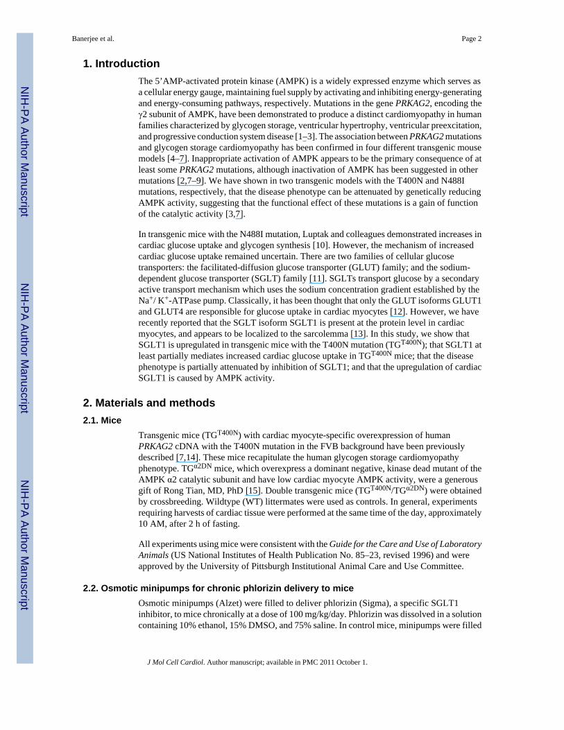
AbstractHuman mutations in the gene PRKAG2 encoding the γ2 subunit of AMP-activated protein kinase(AMPK) cause a glycogen storage cardiomyopathy. Transgenic mice (TGT400N) with the humanT400N mutation exhibit inappropriate activation of AMPK and consequent glycogen storage in theheart. Although increased glucose uptake and activation of glycogen synthesis have been documentedin PRKAG2 cardiomyopathy, the mechanism of increased glucose uptake has been uncertain.Wildtype (WT), TGT400N, and TGα2DN (carrying a dominant negative, kinase dead α2 catalyticsubunit of AMPK) mice were studied at ages 2–8 weeks. Cardiac mRNA expression of sodium-dependent glucose transporter 1 (SGLT1), but not facilitated-diffusion glucose transporter 1(GLUT1) or GLUT4, was increased ~5–7 fold in TGT400N mice relative to WT. SGLT1 protein wassimilarly increased at the cardiac myocyte sarcolemma in TGT400N mice. Phlorizin, a specific SGLT1inhibitor, attenuated cardiac glucose uptake in TGT400N mice by ~40%, but not in WT mice. Chronicphlorizin treatment reduced cardiac glycogen content by ~25% in TGT400N mice. AICAR, an AMPKactivator, increased cardiac SGLT1 mRNA expression ~3 fold in WT mice. Relative to TGT400N
mice, double transgenic (TGT400N/TGα2DN) mice had decreased (~50%) cardiac glucose uptake anddecreased (~70%) cardiac SGLT1 expression. TGT400N hearts had increased binding activity of thetranscription factors HNF-1 and Sp1 to the promoter of the gene encoding SGLT1. Our data suggestthat upregulation of cardiac SGLT1 is responsible for increased cardiac glucose uptake in theTGT400N mouse. Increased AMPK activity leads to upregulation of SGLT1, which in turn mediatesincreased cardiac glucose uptake.
Keywordscardiomyopathy; energy; functional genomics; genetics; genetically altered mice; glucose;membrane transport; metabolism; molecular biology
*Corresponding author: Ferhaan Ahmad, MD, PhD, Cardiovascular Institute, University of Pittsburgh, 200 Lothrop Street, Suite S-558,Mail Stop HPU 01 05 05, Pittsburgh, PA 15213-2582; Telephone: 412-648-9286; Facsimile: 412-648-5991; [email protected]'s Disclaimer: This is a PDF file of an unedited manuscript that has been accepted for publication. As a service to our customerswe are providing this early version of the manuscript. The manuscript will undergo copyediting, typesetting, and review of the resultingproof before it is published in its final citable form. Please note that during the production process errors may be discovered which couldaffect the content, and all legal disclaimers that apply to the journal pertain.
NIH Public AccessAuthor ManuscriptJ Mol Cell Cardiol. Author manuscript; available in PMC 2011 October 1.
Published in final edited form as:J Mol Cell Cardiol. 2010 October ; 49(4): 683–692. doi:10.1016/j.yjmcc.2010.06.003.
NIH
-PA Author Manuscript
NIH
-PA Author Manuscript
NIH
-PA Author Manuscript
1. IntroductionThe 5’AMP-activated protein kinase (AMPK) is a widely expressed enzyme which serves asa cellular energy gauge, maintaining fuel supply by activating and inhibiting energy-generatingand energy-consuming pathways, respectively. Mutations in the gene PRKAG2, encoding theγ2 subunit of AMPK, have been demonstrated to produce a distinct cardiomyopathy in humanfamilies characterized by glycogen storage, ventricular hypertrophy, ventricular preexcitation,and progressive conduction system disease [1–3]. The association between PRKAG2 mutationsand glycogen storage cardiomyopathy has been confirmed in four different transgenic mousemodels [4–7]. Inappropriate activation of AMPK appears to be the primary consequence of atleast some PRKAG2 mutations, although inactivation of AMPK has been suggested in othermutations [2,7–9]. We have shown in two transgenic models with the T400N and N488Imutations, respectively, that the disease phenotype can be attenuated by genetically reducingAMPK activity, suggesting that the functional effect of these mutations is a gain of functionof the catalytic activity [3,7].
In transgenic mice with the N488I mutation, Luptak and colleagues demonstrated increases incardiac glucose uptake and glycogen synthesis [10]. However, the mechanism of increasedcardiac glucose uptake remained uncertain. There are two families of cellular glucosetransporters: the facilitated-diffusion glucose transporter (GLUT) family; and the sodium-dependent glucose transporter (SGLT) family [11]. SGLTs transport glucose by a secondaryactive transport mechanism which uses the sodium concentration gradient established by theNa+/ K+-ATPase pump. Classically, it has been thought that only the GLUT isoforms GLUT1and GLUT4 are responsible for glucose uptake in cardiac myocytes [12]. However, we haverecently reported that the SGLT isoform SGLT1 is present at the protein level in cardiacmyocytes, and appears to be localized to the sarcolemma [13]. In this study, we show thatSGLT1 is upregulated in transgenic mice with the T400N mutation (TGT400N); that SGLT1 atleast partially mediates increased cardiac glucose uptake in TGT400N mice; that the diseasephenotype is partially attenuated by inhibition of SGLT1; and that the upregulation of cardiacSGLT1 is caused by AMPK activity.
2. Materials and methods2.1. Mice
Transgenic mice (TGT400N) with cardiac myocyte-specific overexpression of humanPRKAG2 cDNA with the T400N mutation in the FVB background have been previouslydescribed [7,14]. These mice recapitulate the human glycogen storage cardiomyopathyphenotype. TGα2DN mice, which overexpress a dominant negative, kinase dead mutant of theAMPK α2 catalytic subunit and have low cardiac myocyte AMPK activity, were a generousgift of Rong Tian, MD, PhD [15]. Double transgenic mice (TGT400N/TGα2DN) were obtainedby crossbreeding. Wildtype (WT) littermates were used as controls. In general, experimentsrequiring harvests of cardiac tissue were performed at the same time of the day, approximately10 AM, after 2 h of fasting.
All experiments using mice were consistent with the Guide for the Care and Use of LaboratoryAnimals (US National Institutes of Health Publication No. 85–23, revised 1996) and wereapproved by the University of Pittsburgh Institutional Animal Care and Use Committee.
2.2. Osmotic minipumps for chronic phlorizin delivery to miceOsmotic minipumps (Alzet) were filled to deliver phlorizin (Sigma), a specific SGLT1inhibitor, to mice chronically at a dose of 100 mg/kg/day. Phlorizin was dissolved in a solutioncontaining 10% ethanol, 15% DMSO, and 75% saline. In control mice, minipumps were filled
Banerjee et al. Page 2
J Mol Cell Cardiol. Author manuscript; available in PMC 2011 October 1.
NIH
-PA Author Manuscript
NIH
-PA Author Manuscript
NIH
-PA Author Manuscript

with identical vehicle without phlorizin. Minipumps were implanted in the interscapular areaof 2 week old male mice after sedation with tribromoethanol (125 mg/kg IP) as previouslydescribed [16].
2.3. In vivo cardiac glucose uptakeBasal cardiac glucose uptake was measured in mice as described [13]. In brief, mice wereadministered 2-deoxy-D-[1-14C]-glucose (2-[14C]DG) (10 μCi) intraperitoneally. After 30min, mice were sacrificed and their hearts rapidly excised. Hearts were homogenized in 10volumes of phosphate-buffered saline (PBS), and radioactivity in 20 μl of homogenate wasmeasured in a liquid scintillation counter. Because 2-deoxy-D-glucose is phosphorylated butnot further metabolized, it remains trapped inside cells. Thus, glucose uptake was estimatedby determining cardiac radioactivity. These cardiac glucose uptake assays were performed inthe following male mice at age 6–8 weeks: WT and TGT400N mice 10 min following theadministration of phlorizin (400 mg/kg intraperitoneally [IP]), indinavir (10 mg/kg IP), orvehicle; and TGT400N/TGα2DN mice. Phlorizin (Sigma), a specific SGLT1 inhibitor, wasdissolved at a concentration of 30 mg/ml in 10% ethanol, 15% DMSO, and 75% saline.Indinavir (Fisher), a GLUT inhibitor, was dissolved in water at a concentration of 2.5 mg/ml.
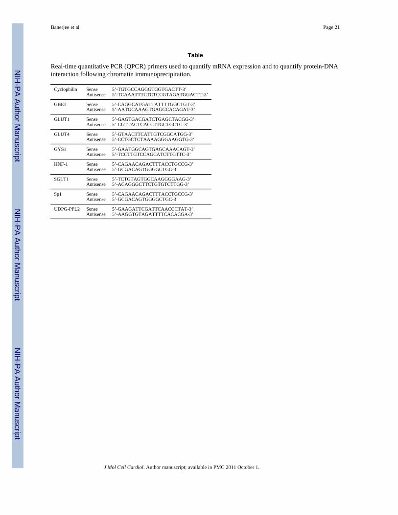
2.4. RNA isolation and real-time quantitative PCR (QPCR)Total RNA was isolated from whole heart with TRIzol (Invitrogen). Reverse transcriptasereactions were performed as described [17] using the Superscript III First-Strand Kit(Invitrogen) for first-strand cDNA synthesis. Primers for real-time quantitative PCR (QPCR)analysis were designed using published sequence information, avoiding regions of homologywith other genes (Table). Ten ng of cDNA were analyzed on an ABI PRISM 7700 usingAbsolute SYBR Green ROX PCR Master Mix (Thermo Scientific). Fold-changes werecalculated after normalization to cyclophilin transcript levels.
2.5. Protein extraction and membrane protein fractionationExtraction of total cardiac protein was performed as described previously [14]. The preparationand fractionation of membranes from cardiac tissue was performed using a commerciallyavailable plasma membrane protein extraction kit (BioVision, #k268-50) according to themanufacturer’s instructions. For membrane fractionation, 10–12 pooled hearts of eachgenotype at ages 2 and 8 weeks, totaling at least 1 g in mass, were homogenized and processedfor extraction of total membrane protein (comprising sarcolemmal and intracellular membranebound proteins) or sarcolemmal membrane protein. The protein content in each fraction wasmeasured by Bradford reagent (Bio-Rad). The α1 subunit of the Na+/K+-ATPase, asarcolemmal membrane marker, was measured by immunoblot to document adequateenrichment of membrane fractions.
2.6. Analysis of protein expressionImmunoblotting, autoradiography, and densitometry were performed as described previously[14]. An equal amount (50–100 μg) of protein was separated by sodium dodecylsulfatepolyacrylamide gel electrophoresis (SDS-PAGE). For membrane fractionation studies, eachlane was loaded with extracts from 10–12 pooled hearts of each genotype at ages 2 and 8 weeks;for all other studies, each lane was loaded with protein extracts from individual hearts asindicated in the figures. After electrophoresis, proteins were transferred to PVDF membranes(Amersham Biosciences). The membranes were then blocked in Tris-buffered saline Tween-20(TBS-T; 10 mM Tris, pH 7.5, 150 mM NaCl, 0.05% Tween-20) and 5% non-fat dry milk for1 h, and subsequently washed and incubated with primary antibodies in TBS-T and 2% bovineserum albumin (BSA) at 4 °C overnight. The following antibodies and titers were used:AMPKα (1:1000 dilution, Cell Signaling, # 2532), phospho-Thr172 AMPKα (1:1000 dilution,
Banerjee et al. Page 3
J Mol Cell Cardiol. Author manuscript; available in PMC 2011 October 1.
NIH
-PA Author Manuscript
NIH
-PA Author Manuscript
NIH
-PA Author Manuscript

Cell Signaling, # 2531), GLUT1 (1:5000 dilution, Abcam, # ab40084), GLUT4 (1:1000dilution, Cell Signaling, #2299), Na+/K+-ATPase α1 (1:1000 dilution, Cell Signaling, #3010),and SGLT1 (1:200 dilution, Santa Cruz, # sc-20582). After washing with TBS-T, membraneswere incubated with anti-rabbit (1:10000 dilution, Amersham, #NA934V) or anti-goat (1:2000dilution, Santa Cruz, sc-2020) horseradish peroxidase conjugated secondary antibody for 1 h.Signal was detected by chemiluminescence using the ECL detection system (Amersham). Gelstaining with Coomassie Blue was used as an internal control for equal loading of protein.Quantification of bands on X-ray film was performed using Image J Software (NIH).
2.7. Tissue fixation and immunofluorescence stainingMouse cardiac tissue was processed for immunofluorescence of SGLT1 as describedpreviously [18]. Hearts were fixed for 4 h at room temperature in PBS containing 4%paraformaldehyde, 10 mM sodium periodate, 70 mM lysine, and 5% sucrose (PLP), washedin PBS, and quenched in NH4Cl [19]. Tissues were cryoprotected in a solution of 30% sucrosein PBS overnight at 4°C. These tissues were embedded in OT compound (Tissue TEK, SakuraFinetek) and mounted on a cutting block. After being frozen in a Reichert Frigocut microtome,sections were picked up on Superfrost Plus slides (Fisher). Immunofluorescence staining wasperformed on 4-μm cryostat sections after SDS antigen retrieval [20]. Slides were washed inPBS followed by incubation with a blocking solution containing 1% bovine serum albumin inPBS-0.02% sodium azide for 15 min. Tissues were labeled using anti-murine SGLT1 antibody(1:200 in DAKO diluent, raised in goat, Santa Cruz, #sc20582) for 75 min sections followedby labeling with a secondary antibody (DAG-FITC 1:100, Jackson Immunologicals). For co-immunolocalization studies, tissues were incubated with anti-murine SGLT1 antibody (1:75dilution) and an antibody against the Na+/K+-ATPase (1:25 dilution, raised in rabbit, CellSignaling, #3010) overnight on the same tissues. After each antibody incubation, sections werewashed twice for 5 min in high-salt PBS (2.7% NaCl) and once in PBS, and then incubatedfor 1 h with secondary antibodies, donkey anti-rabbit FITC and donkey anti-goat-CY3 (1:100and 1:800 dilution, respectively, both from Jackson Immunologicals). After repeating the sameseries of washes as above, the slides were mounted with Vectashield (Vector Labs). Parallelcontrol incubations omitting the primary antibody (“no-primary” control, using DAKO reagentalone) or using a nonspecific primary antibody were performed. For the nonspecific primaryantibody control, IgG goat (1ug/ul, 1:375 dilution in DAKO diluent, Sigma #I5256) was usedon tissue from an 8 week old WT mouse for 75 min (0.002666667 ug/ul) followed by DAG-CY3. The SGLT1 immunizing peptide used to produce this antibody was employed for peptideinhibition controls as previously reported [13,21]. Images were obtained using a LeicaConfocal microscope. The acquisition settings for the immunolabeled tissues and the no-primary controls were identical.
2.8. Cardiac glycogen contentCardiac glycogen content was determined by the amyloglucosidase digestion method as wehave previously reported [7].
2.9. Chromatin Immunoprecipitation (ChIP)Chromatin immunoprecipitation (ChIP) assays for binding activity of specificity protein 1(Sp1) and hepatocyte nuclear factor 1 (HNF-1) to the promoter of the gene encoding SGLT1were performed as described [22]. For each individual assay, approximately 30 mg of startingcardiac tissue was used to harvest chromatin, and 0.45 μg of antibody was added to each sample.The following antibodies were used: HNF-1 (Santa Cruz, # sc-8986) and Sp-1 (Santa Cruz,sc-17824). QPCR primers used to quantify protein-DNA interaction are listed in the Table. ForSp1, the primers were designed to quantify total binding activity at both Sp1 sites.
Banerjee et al. Page 4
J Mol Cell Cardiol. Author manuscript; available in PMC 2011 October 1.
NIH
-PA Author Manuscript
NIH
-PA Author Manuscript
NIH
-PA Author Manuscript
2.10. Statistical analysisResults are expressed as mean ± SE. Differences between two groups were compared byStudent’s t test, and among multiple groups by one-way ANOVA with post hoc Bonferronitest. A p-value of less than 0.05 was considered significant.
3. Results3.1. TGT400N hearts exhibit increased glucose uptake
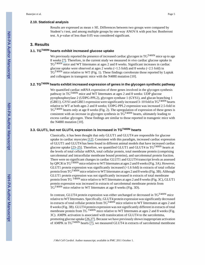
We previously reported the presence of increased cardiac glycogen in TGT400N mice up to age8 weeks [7]. Therefore, in the current study we measured in vivo cardiac glucose uptake inTGT400N mice and WT littermates at ages 2 and 8 weeks. Significant increases in cardiacglucose uptake were observed at ages 2 weeks (~1.5 fold) and 8 weeks (~2.5 fold) inTGT400N mice relative to WT (Fig. 1). These findings corroborate those reported by Luptakand colleagues in transgenic mice with the N488I mutation [10].
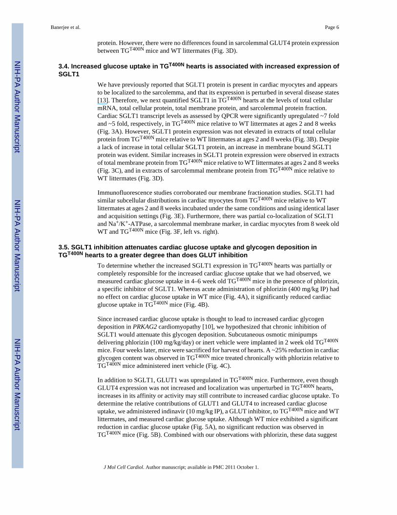
3.2. TGT400N hearts exhibit increased expression of genes in the glycogen synthetic pathwayWe quantified cardiac mRNA expression of three genes involved in the glycogen synthesispathway in TGT400N mice and WT littermates at ages 2 and 8 weeks: UDP glucosepyrophosphorylase 2 (UDPG-PPL2), glycogen synthase 1 (GYS1), and glucan branching 1(GBE1). GYS1 and GBE1 expression were significantly increased 3–10 fold in TGT400N heartsrelative to WT at both ages 2 and 8 weeks; UDPG-PPL2 expression was increased 2.5-fold inTGT400N hearts only at age 8 weeks (Fig. 2). The upregulation of expression of these genes isconsistent with an increase in glycogen synthesis in TGT400N hearts, ultimately leading toexcess cardiac glycogen. These findings are similar to those reported in transgenic mice withthe N488I mutation [10].
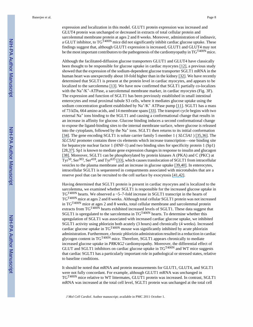
3.3. GLUT1, but not GLUT4, expression is increased in TGT400N heartsClassically, it has been thought that only GLUT1 and GLUT4 are responsible for glucoseuptake in cardiac myocytes [12]. Consistent with this paradigm, increased cardiac expressionof GLUT1 and GLUT4 has been found in different animal models that have increased cardiacglucose uptake [23–25]. Therefore, we quantified GLUT1 and GLUT4 in TGT400N hearts atthe levels of total cellular mRNA, total cellular protein, total membrane protein (comprisingsarcolemmal and intracellular membrane bound proteins), and sarcolemmal protein fraction.There were no significant changes in cardiac GLUT1 and GLUT4 transcript levels as assessedby QPCR in TGT400N mice relative to WT littermates at ages 2 and 8 weeks (Fig. 3A). However,GLUT1 protein expression was significantly increased (>1.6 fold) in extracts of total cellularprotein from TGT400N mice relative to WT littermates at ages 2 and 8 weeks (Fig. 3B). AlthoughGLUT1 protein expression was not significantly increased in extracts of total membraneprotein from TG T400N mice relative to WT littermates at ages 2 and 8 weeks (Fig. 3C), GLUT1protein expression was increased in extracts of sarcolemmal membrane protein fromTGT400N mice relative to WT littermates at age 8 weeks (Fig. 3D).
In contrast, GLUT4 protein expression was either unchanged or decreased in TGT400N micerelative to WT littermates. Specifically, GLUT4 protein expression was significantly decreasedin extracts of total cellular protein from TGT400N mice relative to WT littermates at ages 2 and8 weeks (Fig. 3B). GLUT4 protein expression was not significantly different in extracts of totalmembrane protein from TG T400N mice relative to WT littermates at ages 2 and 8 weeks (Fig.3C). AMPK activation is associated with translocation of GLUT4 to the sarcolemma,promoting glucose uptake [26,27]. Because we have previously shown inappropriate activationof AMPK in TGT400N hearts [7], we measured GLUT4 in extracts of sarcolemmal membrane
Banerjee et al. Page 5
J Mol Cell Cardiol. Author manuscript; available in PMC 2011 October 1.
NIH
-PA Author Manuscript
NIH
-PA Author Manuscript
NIH
-PA Author Manuscript
protein. However, there were no differences found in sarcolemmal GLUT4 protein expressionbetween TGT400N mice and WT littermates (Fig. 3D).
3.4. Increased glucose uptake in TGT400N hearts is associated with increased expression ofSGLT1
We have previously reported that SGLT1 protein is present in cardiac myocytes and appearsto be localized to the sarcolemma, and that its expression is perturbed in several disease states[13]. Therefore, we next quantified SGLT1 in TGT400N hearts at the levels of total cellularmRNA, total cellular protein, total membrane protein, and sarcolemmal protein fraction.Cardiac SGLT1 transcript levels as assessed by QPCR were significantly upregulated ~7 foldand ~5 fold, respectively, in TGT400N mice relative to WT littermates at ages 2 and 8 weeks(Fig. 3A). However, SGLT1 protein expression was not elevated in extracts of total cellularprotein from TGT400N mice relative to WT littermates at ages 2 and 8 weeks (Fig. 3B). Despitea lack of increase in total cellular SGLT1 protein, an increase in membrane bound SGLT1protein was evident. Similar increases in SGLT1 protein expression were observed in extractsof total membrane protein from TGT400N mice relative to WT littermates at ages 2 and 8 weeks(Fig. 3C), and in extracts of sarcolemmal membrane protein from TGT400N mice relative toWT littermates (Fig. 3D).
Immunofluorescence studies corroborated our membrane fractionation studies. SGLT1 hadsimilar subcellular distributions in cardiac myocytes from TGT400N mice relative to WTlittermates at ages 2 and 8 weeks incubated under the same conditions and using identical laserand acquisition settings (Fig. 3E). Furthermore, there was partial co-localization of SGLT1and Na+/K+-ATPase, a sarcolemmal membrane marker, in cardiac myocytes from 8 week oldWT and TGT400N mice (Fig. 3F, left vs. right).
3.5. SGLT1 inhibition attenuates cardiac glucose uptake and glycogen deposition inTGT400N hearts to a greater degree than does GLUT inhibition
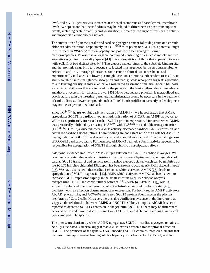
To determine whether the increased SGLT1 expression in TGT400N hearts was partially orcompletely responsible for the increased cardiac glucose uptake that we had observed, wemeasured cardiac glucose uptake in 4–6 week old TGT400N mice in the presence of phlorizin,a specific inhibitor of SGLT1. Whereas acute administration of phlorizin (400 mg/kg IP) hadno effect on cardiac glucose uptake in WT mice (Fig. 4A), it significantly reduced cardiacglucose uptake in TGT400N mice (Fig. 4B).
Since increased cardiac glucose uptake is thought to lead to increased cardiac glycogendeposition in PRKAG2 cardiomyopathy [10], we hypothesized that chronic inhibition ofSGLT1 would attenuate this glycogen deposition. Subcutaneous osmotic minipumpsdelivering phlorizin (100 mg/kg/day) or inert vehicle were implanted in 2 week old TGT400N
mice. Four weeks later, mice were sacrificed for harvest of hearts. A ~25% reduction in cardiacglycogen content was observed in TGT400N mice treated chronically with phlorizin relative toTGT400N mice administered inert vehicle (Fig. 4C).
In addition to SGLT1, GLUT1 was upregulated in TGT400N mice. Furthermore, even thoughGLUT4 expression was not increased and localization was unperturbed in TGT400N hearts,increases in its affinity or activity may still contribute to increased cardiac glucose uptake. Todetermine the relative contributions of GLUT1 and GLUT4 to increased cardiac glucoseuptake, we administered indinavir (10 mg/kg IP), a GLUT inhibitor, to TGT400N mice and WTlittermates, and measured cardiac glucose uptake. Although WT mice exhibited a significantreduction in cardiac glucose uptake (Fig. 5A), no significant reduction was observed inTGT400N mice (Fig. 5B). Combined with our observations with phlorizin, these data suggest
Banerjee et al. Page 6
J Mol Cell Cardiol. Author manuscript; available in PMC 2011 October 1.
NIH
-PA Author Manuscript
NIH
-PA Author Manuscript
NIH
-PA Author Manuscript

that SGLT1 may be a greater contributor than GLUT1 and GLUT4 to the increased cardiacglucose uptake in TGT400N mice.
3.6. AMPK regulates cardiac SGLT1 expressionWe have previously shown that TGT400N mice initially have inappropriate activation of AMPK[7]. To determine whether AMPK activation may be associated with increased cardiac SGLT1expression, 8 week old WT mice were administered aminoimidazole carboxamideribonucleotide (AICAR), a pharmacological activator of AMPK, at a dose of 500 μg/kg IPtwice at 3 h intervals. Mice were sacrificed 3 h following the second AICAR dose, and heartsharvested. Phosphorylation of the α subunit of AMPK, which correlates with AMPK activity[7], was increased (Fig. 6A). AMPK activation in mice receiving AICAR was associated withincreased cardiac SGLT1 mRNA expression (Fig. 6B).
TGα2DN mice, which overexpress a dominant negative mutant of the AMPK α2 catalyticsubunit and have low cardiac myocyte AMPK activity, have been previously described [15].Double transgenic mice (TGT400N/TGα2DN) were obtained by crossbreeding. TGT400N/TGα2DN mice showed attenuation of AMPK overactivity (Fig. 6C), and we have previouslyshown that these mice exhibit an attenuation of the phenotype relative to TGT400N [7].TGT400N/TGα2DN mice had attenuated cardiac glucose uptake (Fig. 6D) concurrent with adecrease in SGLT1 transcript and protein expression (Figs. 6E to 6G). Therefore, cardiacAMPK activity, SGLT1 expression, and glucose uptake appear to be associated.
The promoter of the SLC5A1 gene encoding SGLT1 contains three cis elements that increasetranscription—one binding site for hepatocyte nuclear factor 1 (HNF-1) and two binding sitesfor specificity protein 1 (Sp1) [28]. Therefore, we performed chromatin immunoprecipitation(ChIP) assays in TGT400N and WT hearts to measure binding activity of these transcriptionfactors to the promoter. For Sp1, total binding activity at both Sp1 sites together was quantified.Increased expression of SGLT1 in TGT400N hearts was associated with increased bindingactivity of both HNF-1 and Sp1 (Figs. 6H and 6I).
4. DiscussionIn this study, we have determined that SGLT1 is upregulated in a transgenic mouse with theT400N mutation in PRKAG2 previously identified in human subjects with glycogen storagecardiomyopathy; that SGLT1 appears to mediate at least part of the increased cardiac glucoseuptake and glycogen deposition in this mouse; that inhibition of SGLT1 attenuates the diseasephenotype; and that AMPK appears to regulate SGLT1 expression in the heart.
Human mutations in PRKAG2 lead to excess glycogen storage in several transgenic mousemodels [4–7]. Although increased cardiac glucose uptake and activation of glycogen synthesisenzymes was documented in a transgenic mouse expressing the N488I mutation [10], theglucose transporter responsible for increased cardiac glucose uptake was not defined. Similarly,in transgenic mice with an R225Q mutation in the γ3 subunit of AMPK, skeletal muscle showedincreased expression of glycogen synthesis genes [29]. UDPG-PPL was also upregulated inthe skeletal muscle of carriers of the porcine RN- mutation in the γ3 subunit of AMPK [30].Consistent with these studies, we observed increased cardiac glucose uptake and increasedcardiac expression of genes in the glycogen synthesis pathway, including UDPG-PPL2, GYS1,and GBE1, in TGT400N mice.
Other animal models with increased cardiac glucose uptake exhibit increased expression ofGLUT1 and GLUT4 [23–25]. Since activation of AMPK promotes translocation of GLUT4to the sarcolemma in cardiac myocytes [31], and we have documented early inappropriateactivation of AMPK in the TGT400N heart [7], we analyzed cardiac GLUT1 and GLUT4
Banerjee et al. Page 7
J Mol Cell Cardiol. Author manuscript; available in PMC 2011 October 1.
NIH
-PA Author Manuscript
NIH
-PA Author Manuscript
NIH
-PA Author Manuscript
expression and localization in this model. GLUT1 protein expression was increased andGLUT4 protein was unchanged or decreased in extracts of total cellular protein andsarcolemmal membrane protein at ages 2 and 8 weeks. Moreover, administration of indinavir,a GLUT inhibitor, to TGT400N mice did not significantly inhibit cardiac glucose uptake. Thesefindings suggest that, although GLUT1 expression is increased, GLUT1 and GLUT4 may notbe the most important contributors to the pathogenesis of the cardiomyopathy in TGT400N mice.
Although the facilitated-diffusion glucose transporters GLUT1 and GLUT4 have classicallybeen thought to be responsible for glucose uptake in cardiac myocytes [12], a previous studyshowed that the expression of the sodium-dependent glucose transporter SGLT1 mRNA in thehuman heart was unexpectedly about 10-fold higher than in the kidney [32]. We have recentlydetermined that SGLT1 is present at the protein level in cardiac myocytes, and appears to belocalized to the sarcolemma [13]. We have now confirmed that SGLT1 partially co-localizeswith the Na+/K+-ATPase, a sarcolemmal membrane marker, in cardiac myocytes (Fig. 3F).The expression and function of SGLT1 has been previously established in small intestinalenterocytes and renal proximal tubule S3 cells, where it mediates glucose uptake using thesodium concentration gradient established by Na+/K+ ATPase pump [11]. SGLT1 has a massof 73 kDa, 664 amino acids, and 14 membrane spans [33]. The transport cycle begins with twoexternal Na+ ions binding to the SGLT1 and causing a conformational change that results inan increase in affinity for glucose. Glucose binding induces a second conformational changeto expose the ligand-binding sites to the internal membrane surface, where glucose is releasedinto the cytoplasm, followed by the Na+ ions. SGLT1 then returns to its initial conformation[34]. The gene encoding SGLT1 is solute carrier family 5 member 1 ( SLC5A1 ) [35,36]. TheSLC5A1 promoter contains three cis elements which increase transcription—one binding sitefor hepatocyte nuclear factor 1 (HNF-1) and two binding sites for specificity protein 1 (Sp1)[28,37]. Sp1 is known to mediate gene expression changes in response to insulin and glucagon[38]. Moreover, SGLT1 can be phosphorylated by protein kinases A (PKA) and C (PKC) atTyr50, Ser303, Ser418, and Tyr635 [33], which causes translocation of SGLT1 from intracellularvesicles to the plasma membrane and an increase in glucose uptake [39,40]. In enterocytes,intracellular SGLT1 is sequestered in compartments associated with microtubules that are areserve pool that can be recruited to the cell surface by exocytosis [41,42].
Having determined that SGLT1 protein is present in cardiac myocytes and is localized to thesarcolemma, we examined whether SGLT1 is responsible for the increased glucose uptake inTGT400N hearts. We observed a ~5–7-fold increase in SGLT1 transcript in the hearts ofTGT400N mice at ages 2 and 8 weeks. Although total cellular SGLT1 protein was not increasedin TGT400N mice at ages 2 and 8 weeks, total cellular membrane and sarcolemmal proteinextracts from TGT400N hearts exhibited increased levels of SGLT1. These data suggest thatSGLT1 is upregulated to the sarcolemma in TGT400N hearts. To determine whether thisupregulation of SGLT1 was associated with increased cardiac glucose uptake, we inhibitedSGLT1 activity using phlorizin both acutely (3 hours) and chronically (4 weeks). Increasedcardiac glucose uptake in TGT400N mouse was significantly inhibited by acute phlorizinadministration. Furthermore, chronic phlorizin administration resulted in a reduction in cardiacglycogen content in TGT400N mice. Therefore, SGLT1 appears chronically to mediateincreased glucose uptake in PRKAG2 cardiomyopathy. Moreover, the differential effect ofGLUT and SGLT1 inhibitors on cardiac glucose uptake in TGT400N and WT mice suggeststhat cardiac SGLT1 has a particularly important role in pathological or stressed states, relativeto baseline conditions.
It should be noted that mRNA and protein measurements for GLUT1, GLUT4, and SGLT1were not fully concordant. For example, although GLUT1 mRNA was unchanged inTGT400N mice relative to WT littermates, GLUT1 protein was increased. In contrast, SGLT1mRNA was increased at the total cell level, SGLT1 protein was unchanged at the total cell
Banerjee et al. Page 8
J Mol Cell Cardiol. Author manuscript; available in PMC 2011 October 1.
NIH
-PA Author Manuscript
NIH
-PA Author Manuscript
NIH
-PA Author Manuscript
level, and SGLT1 protein was increased at the total membrane and sarcolemmal membranelevels. We speculate that these findings may be related to differences in post-transcriptionalevents, including protein stability and localization, ultimately leading to differences in activityand impact on cardiac glucose uptake.
The attenuation of glucose uptake and cardiac glycogen content following acute and chronicphlorizin administration, respectively, in TG T400N mice points to SGLT1 as a potential targetfor treatment in PRKAG2 cardiomyopathy and possibly other glycogen storagecardiomyopathies. Phlorizin is an organic compound consisting of a glucose moiety and twoaromatic rings joined by an alkyl spacer [43]. It is a competitive inhibitor that appears to interactwith SGLT1 at two distinct sites [44]. The glucose moiety binds to the substrate binding site,and the aromatic rings bind to a second site located in a large loop between transmembranehelices 13 and 14. Although phlorizin is not in routine clinical use, it has been usedexperimentally in diabetes to lower plasma glucose concentrations independent of insulin. Itsability to inhibit intestinal glucose absorption and renal glucose resorption suggests a potentialrole in treating obesity. It may even have a role in the treatment of malaria, since it has beenshown to inhibit pores that are induced by the parasite in the host erythrocyte cell membraneand that are necessary for parasite growth [45]. However, because phlorizin is metabolized andpoorly absorbed in the intestine, parenteral administration would be necessary in the treatmentof cardiac disease. Newer compounds such as T-1095 and sergliflozin currently in developmentmay not be subject to this drawback.
Since TGT400N hearts exhibit early activation of AMPK [7], we hypothesized that AMPKupregulates SGLT1 in cardiac myocytes. Administration of AICAR, an AMPK activator, toWT mice significantly increased cardiac SGLT1 protein expression. Moreover, when AMPKwas genetically inhibited by crossing TGT400N with TGα2DN mice, double transgenic mice(TGT400N/TGα2DN) exhibited lower AMPK activity, decreased cardiac SGLT1 expression, anddecreased cardiac glucose uptake. These findings are consistent with both a role for AMPK inthe regulation of SGLT1 in cardiac myocytes, and a central role for SGLT1 in the pathogenesisof PRKAG2 cardiomyopathy. Furthermore, AMPK α2 catalytic subunit activity appears to beresponsible for upregulation of SGLT1 through chronic transcriptional effects.
Additional evidence implicates AMPK in upregulation of SGLT1 in cardiac myocytes. Wepreviously reported that acute administration of the hormone leptin leads to upregulation ofcardiac SGLT1 transcript and an increase in cardiac glucose uptake, which can be inhibited bythe SGLT1 inhibitor phlorizin [13]. Leptin has been shown to activate AMPK in skeletal muscle[46]. We have also shown that cardiac ischemia, which activates AMPK [26], leads toupregulation of SGLT1 expression [13]. AMP, which activates AMPK, has been shown toincrease SGLT1 expression rapidly in the small intestine [47]. In Xenopus oocytescoexpressing SGLT1 and constitutively active αR70QAMPK (α1β1γ1(R70Q)), AMPKactivation enhanced maximal currents but not substrate affinity of the transporter [48],consistent with an effect on plasma membrane expression. Furthermore, the AMPK activatorsAICAR, phenformin, and A-769662 increased SGLT1 protein abundance in the plasmamembrane of Caco2 cells. However, there is also conflicting evidence in the literature thatsuggests the relationship between AMPK and SGLT1 is likely complex. AICAR has beenreported to decrease SGLT1 expression in the jejunum [49]. Thus, there may be differencesbetween acute and chronic AMPK regulation of SGLT1, and differences among tissues, celltypes, and possibly species.
The precise mechanism by which AMPK upregulates SGLT1 in cardiac myocytes remains tobe fully elucidated. Our data suggest that AMPK exerts a chronic transcriptional effect onSGLT1. The promoter of the gene SLC5A1 encoding SGLT1 contains three cis elements thatincrease transcription—one binding site for hepatocyte nuclear factor 1 (HNF-1) and two
Banerjee et al. Page 9
J Mol Cell Cardiol. Author manuscript; available in PMC 2011 October 1.
NIH
-PA Author Manuscript
NIH
-PA Author Manuscript
NIH
-PA Author Manuscript

binding sites for specificity protein 1 (Sp1) [28]. Our ChIP assays have shown increasedbinding activity of both HNF-1 and Sp1 to the SLC5A1 promoter. Although at present AMPKis not known to target Sp1 or HNF-1, Sp1 binding activity in general is known to be increasedby phosphorylation by other kinases [50]. Therefore, it is likely that AMPK stimulates SGLT1transcription via these transcription factors, although it may also act on one of the putativebinding sites for other transcription factors found on the promoter. In other tissues,phosphorylation of SGLT1 causes translocation of SGLT1 from intracellular vesicles to theplasma membrane and an increase in glucose uptake [39]. AMPK may directly or indirectlymediate SGLT1 phosphorylation. AMPK indirectly increases phosphatidylinositol 3-kinase(PI-3K) activity [51], and PI-3K activity in turn has been shown to regulate translocation ofSGLT1 [52]. Another mechanism is suggested by the observations that the protein HuRincreases SGLT1 mRNA stability by binding to a critical uridine-rich element (URE) in its 3’untranslated region [53], and in hepatocytes AMPK stimulates the binding of HuR to its targets[54]. Based on our data and evidence from the literature as outlined above, Fig. 7 presents ahypothetical model of the mechanism by which increased AMPK activity leads to increasedcardiac glucose uptake via SGLT1 in PRKAG2 cardiomyopathy. Further studies will berequired to validate this model fully.
A potential limitation of this study is that cardiac glycogen levels, metabolic regulation, andsubstrate utilization show substantial diurnal variations. Furthermore, expression of SGLT1 inthe gastrointestinal tract exhibits diurnal rhythmicity in concert with clock genes that isindependent of local luminal nutrient delivery [55,56]. Therefore, the results of ourexperiments, performed at one standard time of the day, may not be fully generalizable to othertimes of the day.
Another potential limitation of this study is that only male mice were studied in detail. Sexinfluences phenotype severity in other cardiomyopathies [57]. However, differences in diseasephenotype based on sex have not been clearly documented in PRKAG2 cardiomyopathies, andour own preliminary unpublished data in both human subjects and murine models suggest thatour findings in this study are applicable to both sexes.
In conclusion, our data suggest that inappropriate activation of AMPK, secondary to the T400Nmutation in PRKAG2, leads to increased cardiac SGLT1 expression, which in turn isresponsible for increased cardiac glucose uptake. While this study shows for the first time afunctional role of SGLT1 in cardiac disease, our previously reported work suggests that thistransporter is relevant far beyond PRKAG2 cardiomyopathy and is a heretofore unrecognizedparticipant in adaptive or maladaptive responses of the heart to a wide range of pathologicalinsults. SGLT1 expression is perturbed in diabetic cardiomyopathy and ischemic heart disease,and functional improvement in failing left ventricles is associated with upregulation of SGLT1[13]. As discussed above, AMPK and leptin, both of which mediate cardiac response toischemia, may exert their effects in part through SGLT1. Thus, SGLT1 may represent anentirely novel therapeutic target in the heart, and agents that directly modify cardiac SGLT1expression, localization, and activity may be useful in the modulation of cardiac energysubstrate utilization and in the treatment of glycogen storage cardiomyopathy, ischemic heartdisease, and other cardiac diseases.
AcknowledgmentsThis work was supported by an American Heart Association Scientist Development Grant 053531N (FA); PostdoctoralFellowships from the American Heart Association and the Hillgrove Foundation (SKB); NIH/NIDDK grants P30DK-079307 “Pittsburgh Kidney Research Center” and R01 DK075048 (KRH); an American Heart Association Grant-in-Aid 09GRNT2060539 (NMPS); an NIH/NIDDK grant R01 DK084184 (NMPS); and the Cardiovascular Instituteof the University of Pittsburgh (Barry London, Director) (FA). FA is a Doris Duke Charitable Foundation ClinicalScientist. We acknowledge Christy Smolak for excellent technical support.
Banerjee et al. Page 10
J Mol Cell Cardiol. Author manuscript; available in PMC 2011 October 1.
NIH
-PA Author Manuscript
NIH
-PA Author Manuscript
NIH
-PA Author Manuscript
References1. Gollob MH, Green MS, Tang AS, Gollob T, Karibe A, Ali Hassan AS, et al. Identification of a gene
responsible for familial Wolff-Parkinson-White syndrome. N Engl J Med 2001;344:1823–31.[PubMed: 11407343]
2. Arad M, Benson DW, Perez-Atayde AR, McKenna WJ, Sparks EA, Kanter RJ, et al. Constitutivelyactive AMP kinase mutations cause glycogen storage disease mimicking hypertrophiccardiomyopathy. The Journal of clinical investigation 2002;109:357–62. [PubMed: 11827995]
3. Ahmad F, Arad M, Musi N, He H, Wolf C, Branco D, et al. Increased alpha2 subunit-associated AMPKactivity and PRKAG2 cardiomyopathy. Circulation 2005;112:3140–8. [PubMed: 16275868]
4. Arad M, Moskowitz IP, Patel VV, Ahmad F, Perez-Atayde AR, Sawyer DB, et al. Transgenic miceoverexpressing mutant PRKAG2 define the cause of Wolff-Parkinson-White syndrome in glycogenstorage cardiomyopathy. Circulation 2003;107:2850–6. [PubMed: 12782567]
5. Sidhu JS, Rajawat YS, Rami TG, Gollob MH, Wang Z, Yuan R, et al. Transgenic mouse model ofventricular preexcitation and atrioventricular reentrant tachycardia induced by an AMP-activatedprotein kinase loss-of-function mutation responsible for Wolff-Parkinson-White syndrome.Circulation 2005;111:21–9. [PubMed: 15611370]
6. Davies JK, Wells DJ, Liu K, Whitrow HR, Daniel TD, Grignani R, et al. Characterization of the roleof gamma2 R531G mutation in AMP-activated protein kinase in cardiac hypertrophy and Wolff-Parkinson-White syndrome. Am J Physiol Heart Circ Physiol 2006;290:H1942–51. [PubMed:16339829]
7. Banerjee SK, Ramani R, Saba S, Rager J, Tian R, Mathier MA, et al. A PRKAG2 mutation causesbiphasic changes in myocardial AMPK activity and does not protect against ischemia. BiochemBiophys Res Commun 2007;360:381–7. [PubMed: 17597581]
8. Hamilton SR, Stapleton D, O'Donnell JB Jr, Kung JT, Dalal SR, Kemp BE, et al. An activating mutationin the gamma1 subunit of the AMP-activated protein kinase. FEBS Lett 2001;500:163–8. [PubMed:11445078]
9. Daniel T, Carling D. Functional analysis of mutations in the gamma 2 subunit of AMP-activated proteinkinase associated with cardiac hypertrophy and Wolff-Parkinson-White syndrome. J Biol Chem2002;277:51017–24. [PubMed: 12397075]
10. Luptak I, Shen M, He H, Hirshman MF, Musi N, Goodyear LJ, et al. Aberrant activation of AMP-activated protein kinase remodels metabolic network in favor of cardiac glycogen storage. TheJournal of clinical investigation 2007;117:1432–9. [PubMed: 17431505]
11. Wright EM, Loo DD, Panayotova-Heiermann M, Lostao MP, Hirayama BH, Mackenzie B, et al.'Active' sugar transport in eukaryotes. J Exp Biol 1994;196:197–212. [PubMed: 7823022]
12. Schwenk RW, Luiken JJ, Bonen A, Glatz JF. Regulation of sarcolemmal glucose and fatty acidtransporters in cardiac disease. Cardiovasc Res 2008;79:249–58. [PubMed: 18469026]
13. Banerjee SK, McGaffin KR, Pastor-Soler NM, Ahmad F. SGLT1 is a novel cardiac glucose transporterthat is perturbed in disease states. Cardiovasc Res 2009;84:111–8. [PubMed: 19509029]
14. Banerjee SK, McGaffin KR, Huang XN, Ahmad F. Activation of cardiac hypertrophic signalingpathways in a transgenic mouse with the human PRKAG2 Thr400Asn mutation. Biochim BiophysActa. 2009
15. Xing Y, Musi N, Fujii N, Zou L, Luptak I, Hirshman MF, et al. Glucose metabolism and energyhomeostasis in mouse hearts overexpressing dominant negative alpha2 subunit of AMP-activatedprotein kinase. J Biol Chem 2003;278:28372–7. [PubMed: 12766162]
16. McGaffin KR, Sun CK, Rager JJ, Romano LC, Zou B, Mathier MA, et al. Leptin signalling reducesthe severity of cardiac dysfunction and remodelling after chronic ischaemic injury. Cardiovasc Res2008;77:54–63. [PubMed: 18006469]
17. Ahmad F, Banerjee SK, Lage ML, Huang XN, Smith SH, Saba S, et al. The role of cardiac troponinT quantity and function in cardiac development and dilated cardiomyopathy. PLoS ONE2008;3:e2642. [PubMed: 18612386]
18. Pastor-Soler NM, Hallows KR, Smolak C, Gong F, Brown D, Breton S. Alkaline pH- and cAMP-induced V-ATPase membrane accumulation is mediated by protein kinase A in epididymal clearcells. Am J Physiol Cell Physiol 2008;294:C488–94. [PubMed: 18160485]
Banerjee et al. Page 11
J Mol Cell Cardiol. Author manuscript; available in PMC 2011 October 1.
NIH
-PA Author Manuscript
NIH
-PA Author Manuscript
NIH
-PA Author Manuscript

19. Breton S, Tyszkowski R, Sabolic I, Brown D. Postnatal development of H+ ATPase (proton-pump)-rich cells in rat epididymis. Histochemistry and cell biology 1999;111:97–105. [PubMed: 10090570]
20. Brown D, Lydon J, McLaughlin M, Stuart-Tilley A, Tyszkowski R, Alper S. Antigen retrieval incryostat tissue sections and cultured cells by treatment with sodium dodecyl sulfate (SDS).Histochemistry and cell biology 1996;105:261–7. [PubMed: 9072183]
21. Pastor-Soler N, Bagnis C, Sabolic I, Tyszkowski R, McKee M, Van Hoek A, et al. Aquaporin 9expression along the male reproductive tract. Biology of reproduction 2001;65:384–93. [PubMed:11466204]
22. Blahnik KR, Dou L, O'Geen H, McPhillips T, Xu X, Cao AR, et al. Sole-Search: an integrated analysisprogram for peak detection and functional annotation using ChIP-seq data. Nucleic Acids Res 38:e13.[PubMed: 19906703]
23. Young LH, Renfu Y, Russell R, Hu X, Caplan M, Ren J, et al. Low-flow ischemia leads totranslocation of canine heart GLUT-4 and GLUT-1 glucose transporters to the sarcolemma in vivo.Circulation 1997;95:415–22. [PubMed: 9008459]
24. Tian R, Musi N, D'Agostino J, Hirshman MF, Goodyear LJ. Increased adenosine monophosphate-activated protein kinase activity in rat hearts with pressure-overload hypertrophy. Circulation2001;104:1664–9. [PubMed: 11581146]
25. Broderick TL, King TM. Upregulation of GLUT-4 in right ventricle of rats with monocrotaline-induced pulmonary hypertension. Med Sci Monit 2008;14:BR261–4. [PubMed: 19043358]
26. Russell RR 3rd, Li J, Coven DL, Pypaert M, Zechner C, Palmeri M, et al. AMP- activated proteinkinase mediates ischemic glucose uptake and prevents postischemic cardiac dysfunction, apoptosis,and injury. The Journal of clinical investigation 2004;114:495–503. [PubMed: 15314686]
27. Li J, Miller EJ, Ninomiya-Tsuji J, Russell RR 3rd, Young LH. AMP-activated protein kinase activatesp38 mitogen-activated protein kinase by increasing recruitment of p38 MAPK to TAB1 in theischemic heart. Circ Res 2005;97:872–9. [PubMed: 16179588]
28. Martin MG, Wang J, Solorzano-Vargas RS, Lam JT, Turk E, Wright EM. Regulation of the humanNa(+)-glucose cotransporter gene, SGLT1, by HNF-1 and Sp1. Am J Physiol Gastrointest LiverPhysiol 2000;278:G591–603. [PubMed: 10762614]
29. Nilsson EC, Long YC, Martinsson S, Glund S, Garcia-Roves P, Svensson LT, et al. Oppositetranscriptional regulation in skeletal muscle of AMP-activated protein kinase gamma3 R225Qtransgenic versus knock-out mice. J Biol Chem 2006;281:7244–52. [PubMed: 16410251]
30. Hedegaard J, Horn P, Lametsch R, Sondergaard Moller H, Roepstorff P, Bendixen C, et al. UDP-glucose pyrophosphorylase is upregulated in carriers of the porcine RN- mutation in the AMP-activated protein kinase. Proteomics 2004;4:2448–54. [PubMed: 15274139]
31. Russell RR 3rd, Bergeron R, Shulman GI, Young LH. Translocation of myocardial GLUT-4 andincreased glucose uptake through activation of AMPK by AICAR. Am J Physiol 1999;277:H643–9. [PubMed: 10444490]
32. Zhou L, Cryan EV, D'Andrea MR, Belkowski S, Conway BR, Demarest KT. Human cardiomyocytesexpress high level of Na+/glucose cotransporter 1 (SGLT1). J Cell Biochem 2003;90:339–46.[PubMed: 14505350]
33. Turk E, Kerner CJ, Lostao MP, Wright EM. Membrane topology of the human Na+/glucosecotransporter SGLT1. J Biol Chem 1996;271:1925–34. [PubMed: 8567640]
34. Loo DD, Hirayama BA, Sala-Rabanal M, Wright EM. How drugs interact with transporters: SGLT1as a model. J Membr Biol 2008;223:87–106. [PubMed: 18592293]
35. Turk E, Martin MG, Wright EM. Structure of the human Na+/glucose cotransporter gene SGLT1. JBiol Chem 1994;269:15204–9. [PubMed: 8195156]
36. Turk E, Klisak I, Bacallao R, Sparkes RS, Wright EM. Assignment of the human Na+/glucosecotransporter gene SGLT1 to chromosome 22q13.1. Genomics 1993;17:752–4. [PubMed: 8244393]
37. Vayro S, Wood IS, Dyer J, Shirazi-Beechey SP. Transcriptional regulation of the ovine intestinal Na+/glucose cotransporter SGLT1 gene. Role of HNF-1 in glucose activation of promoter function. EurJ Biochem 2001;268:5460–70. [PubMed: 11606209]
38. Solomon SS, Majumdar G, Martinez-Hernandez A, Raghow R. A critical role of Sp1 transcriptionfactor in regulating gene expression in response to insulin and other hormones. Life Sci 2008;83:305–12. [PubMed: 18664368]
Banerjee et al. Page 12
J Mol Cell Cardiol. Author manuscript; available in PMC 2011 October 1.
NIH
-PA Author Manuscript
NIH
-PA Author Manuscript
NIH
-PA Author Manuscript

39. Hirsch JR, Loo DD, Wright EM. Regulation of Na+/glucose cotransporter expression by proteinkinases in Xenopus laevis oocytes. J Biol Chem 1996;271:14740–6. [PubMed: 8663046]
40. Wright EM, Hirsch JR, Loo DD, Zampighi GA. Regulation of Na+/glucose cotransporters. J ExpBiol 1997;200:287–93. [PubMed: 9050236]
41. Khoursandi S, Scharlau D, Herter P, Kuhnen C, Martin D, Kinne RK, et al. Different modes of sodium-D-glucose cotransporter-mediated D-glucose uptake regulation in Caco-2 cells. Am J Physiol CellPhysiol 2004;287:C1041–7. [PubMed: 15201142]
42. Kipp H, Khoursandi S, Scharlau D, Kinne RK. More than apical: Distribution of SGLT1 in Caco-2cells. Am J Physiol Cell Physiol 2003;285:C737–49. [PubMed: 12773314]
43. Ehrenkranz JR, Lewis NG, Kahn CR, Roth J. Phlorizin: a review. Diabetes Metab Res Rev2005;21:31–8. [PubMed: 15624123]
44. Pajor AM, Randolph KM, Kerner SA, Smith CD. Inhibitor binding in the human renal low- and high-affinity Na+/glucose cotransporters. J Pharmacol Exp Ther 2008;324:985–91. [PubMed: 18063724]
45. Kutner S, Breuer WV, Ginsburg H, Cabantchik ZI. On the mode of action of phlorizin as anantimalarial agent in in vitro cultures of Plasmodium falciparum. Biochem Pharmacol 1987;36:123–9. [PubMed: 3099799]
46. Minokoshi Y, Kim YB, Peroni OD, Fryer LG, Muller C, Carling D, et al. Leptin stimulates fatty-acidoxidation by activating AMP-activated protein kinase. Nature 2002;415:339–43. [PubMed:11797013]
47. Kimura Y, Turner JR, Braasch DA, Buddington RK. Lumenal adenosine and AMP rapidly increaseglucose transport by intact small intestine. Am J Physiol Gastrointest Liver Physiol 2005;289:G1007–14. [PubMed: 16020657]
48. Sopjani M, Bhavsar SK, Fraser S, Kemp BE, Foller M, Lang F. Regulation of Na+-coupled glucosecarrier SGLT1 by AMP-activated protein kinase. Mol Membr Biol 27:137–44. [PubMed: 20334581]
49. Walker J, Jijon HB, Diaz H, Salehi P, Churchill T, Madsen KL. 5-aminoimidazole-4-carboxamideriboside (AICAR) enhances GLUT2-dependent jejunal glucose transport: a possible role for AMPK.Biochem J 2005;385:485–91. [PubMed: 15367103]
50. Tan NY, Khachigian LM. Sp1 phosphorylation and its regulation of gene transcription. Mol Cell Biol2009;29:2483–8. [PubMed: 19273606]
51. Jakobsen SN, Hardie DG, Morrice N, Tornqvist HE. 5'-AMP-activated protein kinase phosphorylatesIRS-1 on Ser-789 in mouse C2C12 myotubes in response to 5-aminoimidazole-4-carboxamideriboside. J Biol Chem 2001;276:46912–6. [PubMed: 11598104]
52. Kawano K, Ikari A, Nakano M, Suketa Y. Phosphatidylinositol 3-kinase mediates inhibitory effectof angiotensin II on sodium/glucose cotransporter in renal epithelial cells. Life Sci 2002;71:1–13.[PubMed: 12020744]
53. Loflin P, Lever JE. HuR binds a cyclic nucleotide-dependent, stabilizing domain in the 3' untranslatedregion of Na(+)/glucose cotransporter (SGLT1) mRNA. FEBS Lett 2001;509:267–71. [PubMed:11741601]
54. Martinez-Chantar ML, Vazquez-Chantada M, Garnacho M, Latasa MU, Varela-Rey M, Dotor J, etal. S-adenosylmethionine regulates cytoplasmic HuR via AMP-activated kinase. Gastroenterology2006;131:223–32. [PubMed: 16831604]
55. Balakrishnan A, Stearns AT, Rounds J, Irani J, Giuffrida M, Rhoads DB, et al. Diurnal rhythmicityin glucose uptake is mediated by temporal periodicity in the expression of the sodium-glucosecotransporter (SGLT1). Surgery 2008;143:813–8. [PubMed: 18549898]
56. Stearns AT, Balakrishnan A, Rhoads DB, Ashley SW, Tavakkolizadeh A. Diurnal expression of therat intestinal sodium-glucose cotransporter 1 (SGLT1) is independent of local luminal factors.Surgery 2009;145:294–302. [PubMed: 19231582]
57. Ahmad F, Seidman JG, Seidman CE. The genetic basis for cardiac remodeling. Annu Rev GenomicsHum Genet 2005;6:185–216. [PubMed: 16124859]
Banerjee et al. Page 13
J Mol Cell Cardiol. Author manuscript; available in PMC 2011 October 1.
NIH
-PA Author Manuscript
NIH
-PA Author Manuscript
NIH
-PA Author Manuscript
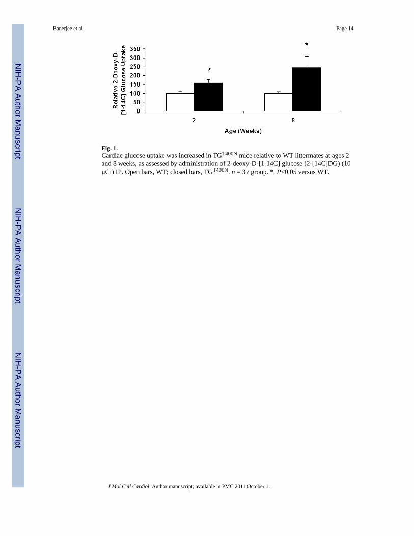
Fig. 1.Cardiac glucose uptake was increased in TGT400N mice relative to WT littermates at ages 2and 8 weeks, as assessed by administration of 2-deoxy-D-[1-14C] glucose (2-[14C]DG) (10μCi) IP. Open bars, WT; closed bars, TGT400N. n = 3 / group. *, P<0.05 versus WT.
Banerjee et al. Page 14
J Mol Cell Cardiol. Author manuscript; available in PMC 2011 October 1.
NIH
-PA Author Manuscript
NIH
-PA Author Manuscript
NIH
-PA Author Manuscript
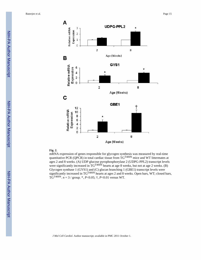
Fig. 2.mRNA expression of genes responsible for glycogen synthesis was measured by real-timequantitative PCR (QPCR) in total cardiac tissue from TGT400N mice and WT littermates atages 2 and 8 weeks. (A) UDP glucose pyrophosphorylase 2 (UDPG-PPL2) transcript levelswere significantly increased in TGT400N hearts at age 8 weeks, but not at age 2 weeks. (B)Glycogen synthase 1 (GYS1) and (C) glucan branching 1 (GBE1) transcript levels weresignificantly increased in TGT400N hearts at ages 2 and 8 weeks. Open bars, WT; closed bars,TGT400N. n = 3 / group. *, P<0.05; †, P<0.01 versus WT.
Banerjee et al. Page 15
J Mol Cell Cardiol. Author manuscript; available in PMC 2011 October 1.
NIH
-PA Author Manuscript
NIH
-PA Author Manuscript
NIH
-PA Author Manuscript
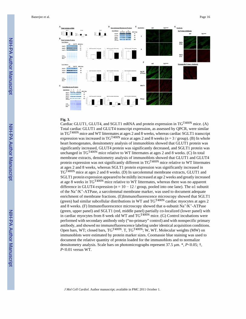
Fig. 3.Cardiac GLUT1, GLUT4, and SGLT1 mRNA and protein expression in TGT400N mice. (A)Total cardiac GLUT1 and GLUT4 transcript expression, as assessed by QPCR, were similarin TGT400N mice and WT littermates at ages 2 and 8 weeks, whereas cardiac SGLT1 transcriptexpression was increased in TGT400N mice at ages 2 and 8 weeks (n = 3 / group). (B) In wholeheart homogenates, densitometry analysis of immunoblots showed that GLUT1 protein wassignificantly increased, GLUT4 protein was significantly decreased, and SGLT1 protein wasunchanged in TGT400N mice relative to WT littermates at ages 2 and 8 weeks. (C) In totalmembrane extracts, densitometry analysis of immunoblots showed that GLUT1 and GLUT4protein expression was not significantly different in TGT400N mice relative to WT littermatesat ages 2 and 8 weeks, whereas SGLT1 protein expression was significantly increased inTGT400N mice at ages 2 and 8 weeks. (D) In sarcolemmal membrane extracts, GLUT1 andSGLT1 protein expression appeared to be mildly increased at age 2 weeks and greatly increasedat age 8 weeks in TGT400N mice relative to WT littermates, whereas there was no apparentdifference in GLUT4 expression (n = 10 − 12 / group, pooled into one lane). The α1 subunitof the Na+/K+-ATPase, a sarcolemmal membrane marker, was used to document adequateenrichment of membrane fractions. (E)Immunofluorescence microscopy showed that SGLT1(green) had similar subcellular distributions in WT and TGT400N cardiac myocytes at ages 2and 8 weeks. (F) Immunofluorescence microscopy showed that α-subunit Na+/K+-ATPase(green, upper panel) and SGLT1 (red, middle panel) partially co-localized (lower panel) within cardiac myocytes from 8 week old WT and TGT400N mice. (G) Control incubations wereperformed with secondary antibody only (“no-primary” control) and with nonspecific primaryantibody, and showed no immunofluorescence labeling under identical acquisition conditions.Open bars, WT; closed bars, TGT400N. T, TGT400N; W, WT. Molecular weights (MW) onimmunoblots were estimated by protein marker sizes. Coomassie blue staining was used todocument the relative quantity of protein loaded for the immunoblots and to normalizedensitometry analysis. Scale bars on photomicrographs represent 37.5 μm. *, P<0.05; †,P<0.01 versus WT.
Banerjee et al. Page 16
J Mol Cell Cardiol. Author manuscript; available in PMC 2011 October 1.
NIH
-PA Author Manuscript
NIH
-PA Author Manuscript
NIH
-PA Author Manuscript

Fig. 4.Increased cardiac glucose uptake and glycogen deposition in TGT400N mice was sensitive tophlorizin, a specific SGLT1 inhibitor. (A) There was no significant change in cardiac glucoseuptake in 6–8 week old WT mice 10 min following acute administration of phlorizin (400 mg/kg IP). (B) In contrast, cardiac glucose uptake was reduced in 6–8 week old TGT400N mice 10min following acute administration of phlorizin (400 mg/kg IP). (C) Chronic administrationphlorizin (100 mg/kg/day) using a subcutaneous osmotic minipump in TGT400N mice from age2 weeks through age 6 weeks resulted in a reduction in cardiac glycogen content. Open bars,vehicle treated control mice; closed bars, phlorizin treated mice. n = 4 / group, *, P<0.05 relativeto control.
Banerjee et al. Page 17
J Mol Cell Cardiol. Author manuscript; available in PMC 2011 October 1.
NIH
-PA Author Manuscript
NIH
-PA Author Manuscript
NIH
-PA Author Manuscript
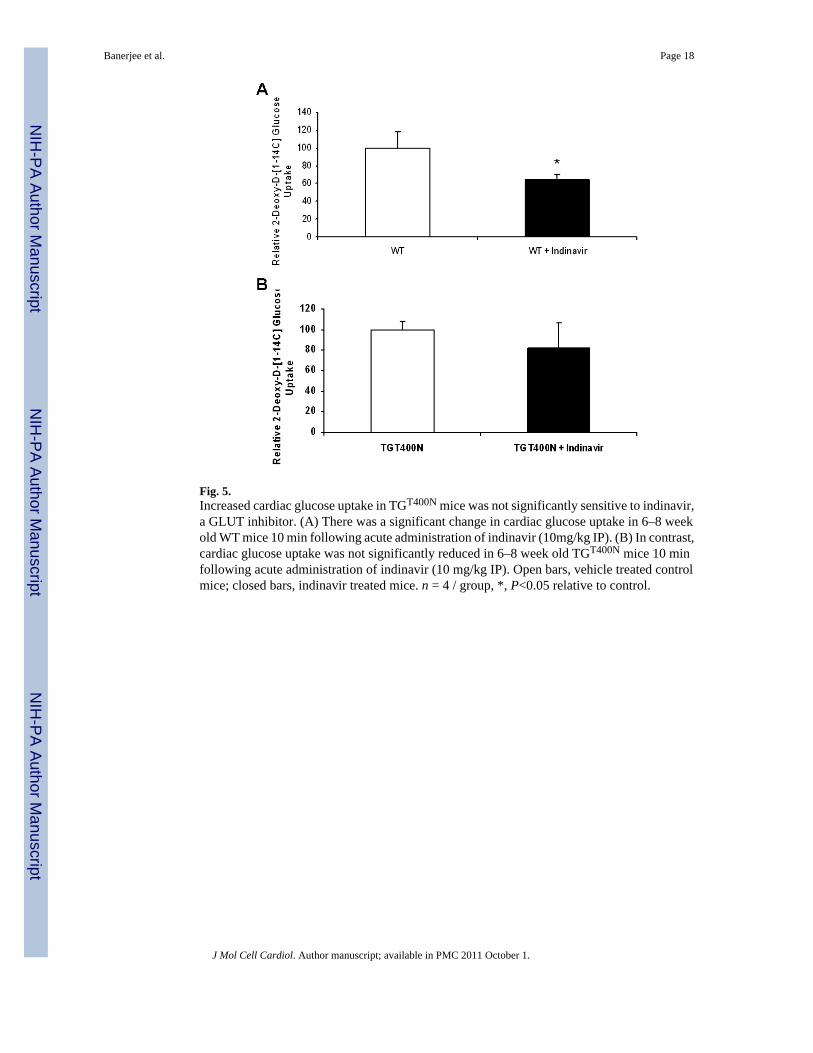
Fig. 5.Increased cardiac glucose uptake in TGT400N mice was not significantly sensitive to indinavir,a GLUT inhibitor. (A) There was a significant change in cardiac glucose uptake in 6–8 weekold WT mice 10 min following acute administration of indinavir (10mg/kg IP). (B) In contrast,cardiac glucose uptake was not significantly reduced in 6–8 week old TGT400N mice 10 minfollowing acute administration of indinavir (10 mg/kg IP). Open bars, vehicle treated controlmice; closed bars, indinavir treated mice. n = 4 / group, *, P<0.05 relative to control.
Banerjee et al. Page 18
J Mol Cell Cardiol. Author manuscript; available in PMC 2011 October 1.
NIH
-PA Author Manuscript
NIH
-PA Author Manuscript
NIH
-PA Author Manuscript
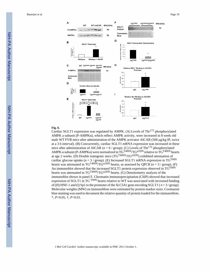
Fig. 6.Cardiac SGLT1 expression was regulated by AMPK. (A) Levels of Thr172 phosphorylatedAMPK a subunit (P-AMPKα), which reflect AMPK activity, were increased in 8 week oldmale WT FVB mice after administration of the AMPK activator AICAR (500 μg/kg IP, twiceat a 3 h interval). (B) Concurrently, cardiac SGLT1 mRNA expression was increased in thesemice after administration of AICAR (n = 6 / group). (C) Levels of Thr172 phosphorylatedAMPK α subunit (P-AMPKα) were normalized in TGT400N/TGα2DN relative to TGT400N heartsat age 2 weeks. (D) Double transgenic mice (TGT400N/TGα2DN) exhibited attenuation ofcardiac glucose uptake (n = 3 / group). (E) Increased SGLT1 mRNA expression in TGT00N
hearts was attenuated in TGT400N/TGα2DN hearts, as assessed by QPCR (n = 3 / group). (F)An immunoblot showed that the increased SGLT1 protein expression observed in TGT00N
hearts was attenuated in TGT400N/TGα2DN hearts. (G) Densitometry analysis of theimmunoblot shown in panel F. Chromatin immunoprecipitation (ChIP) showed that increasedexpression of SGLT1 in TG T00N hearts relative to WT was associated with increased bindingof (H) HNF-1 and (I) Sp1 to the promoter of the SLC5A1 gene encoding SGLT1 ( n = 3 / group).Molecular weights (MW) on immunoblots were estimated by protein marker sizes. Coomassieblue staining was used to document the relative quantity of protein loaded for the immunoblots.*, P<0.05, †, P<0.01.
Banerjee et al. Page 19
J Mol Cell Cardiol. Author manuscript; available in PMC 2011 October 1.
NIH
-PA Author Manuscript
NIH
-PA Author Manuscript
NIH
-PA Author Manuscript
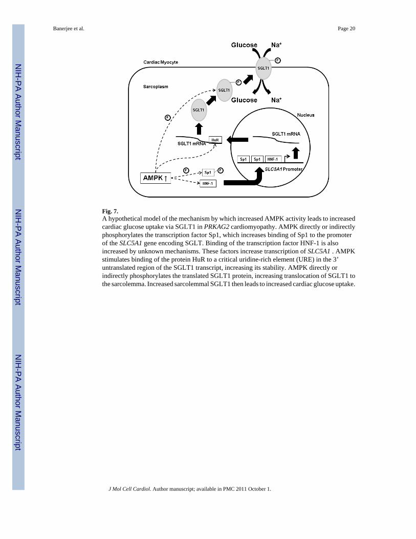
Fig. 7.A hypothetical model of the mechanism by which increased AMPK activity leads to increasedcardiac glucose uptake via SGLT1 in PRKAG2 cardiomyopathy. AMPK directly or indirectlyphosphorylates the transcription factor Sp1, which increases binding of Sp1 to the promoterof the SLC5A1 gene encoding SGLT. Binding of the transcription factor HNF-1 is alsoincreased by unknown mechanisms. These factors increase transcription of SLC5A1 . AMPKstimulates binding of the protein HuR to a critical uridine-rich element (URE) in the 3’untranslated region of the SGLT1 transcript, increasing its stability. AMPK directly orindirectly phosphorylates the translated SGLT1 protein, increasing translocation of SGLT1 tothe sarcolemma. Increased sarcolemmal SGLT1 then leads to increased cardiac glucose uptake.
Banerjee et al. Page 20
J Mol Cell Cardiol. Author manuscript; available in PMC 2011 October 1.
NIH
-PA Author Manuscript
NIH
-PA Author Manuscript
NIH
-PA Author Manuscript
NIH
-PA Author Manuscript
NIH
-PA Author Manuscript
NIH
-PA Author Manuscript
Banerjee et al. Page 21
Table
Real-time quantitative PCR (QPCR) primers used to quantify mRNA expression and to quantify protein-DNAinteraction following chromatin immunoprecipitation.
Cyclophilin SenseAntisense
5’-TGTGCCAGGGTGGTGACTT-3’5’-TCAAATTTCTCTCCGTAGATGGACTT-3’
GBE1 SenseAntisense
5’-CAGGCATGATTATTTTGGCTGT-3’5’-AATGCAAAGTGAGGCACAGAT-3’
GLUT1 SenseAntisense
5’-GAGTGACGATCTGAGCTACGG-3’5’-CGTTACTCACCTTGCTGCTG-3’
GLUT4 SenseAntisense
5’-GTAACTTCATTGTCGGCATGG-3’5’-CCTGCTCTAAAAGGGAAGGTG-3’
GYS1 SenseAntisense
5’-GAATGGCAGTGAGCAAACAGT-3’5’-TCCTTGTCCAGCATCTTGTTC-3’
HNF-1 SenseAntisense
5’-CAGAACAGACTTTACCTGCCG-3’5’-GCGACAGTGGGGCTGC-3’
SGLT1 SenseAntisense
5’-TCTGTAGTGGCAAGGGGAAG-3’5’-ACAGGGCTTCTGTGTCTTGG-3’
Sp1 SenseAntisense
5’-CAGAACAGACTTTACCTGCCG-3’5’-GCGACAGTGGGGCTGC-3’
UDPG-PPL2 SenseAntisense
5’-GAAGATTCGATTCAACCCTAT-3’5’-AAGGTGTAGATTTTCACACGA-3’
J Mol Cell Cardiol. Author manuscript; available in PMC 2011 October 1.
Copyright © 2022 FDOKUMEN