Glucose Transporter Type 1—Biology and Deficiency Syndrome
21
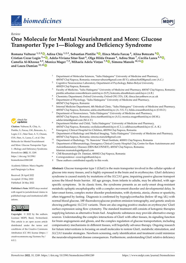
Citation: Vulturar, R.; Chis , , A.; Pintilie, S.; Farcas , , I.M.; Botezatu, A.; Login, C.C.; Sitar-Taut, A.-V.; Orasan, O.H.; Stan, A.; Lazea, C.; et al. One Molecule for Mental Nourishment and More: Glucose Transporter Type 1—Biology and Deficiency Syndrome. Biomedicines 2022, 10, 1249. https://doi.org/10.3390/ biomedicines10061249 Academic Editors: Marco Segatto and Piergiorgio La Rosa Received: 28 April 2022 Accepted: 23 May 2022 Published: 26 May 2022 Publisher’s Note: MDPI stays neutral with regard to jurisdictional claims in published maps and institutional affil- iations. Copyright: © 2022 by the authors. Licensee MDPI, Basel, Switzerland. This article is an open access article distributed under the terms and conditions of the Creative Commons Attribution (CC BY) license (https:// creativecommons.org/licenses/by/ 4.0/). biomedicines Review One Molecule for Mental Nourishment and More: Glucose Transporter Type 1—Biology and Deficiency Syndrome Romana Vulturar 1,2,† , Adina Chis , 1,2,† , Sebastian Pintilie 3 , Ilinca Maria Farcas , 4 , Alina Botezatu 3 , Cristian Cezar Login 5, * , Adela-Viviana Sitar-Taut 6 , Olga Hilda Orasan 6 , Adina Stan 7 , Cecilia Lazea 8,9 , Camelia Al-Khzouz 8,9 , Monica Mager 7,9 , Mihaela Adela Vint , an 7,9 , Simona Manole 10,11 and Laura Damian 12,13 1 Department of Molecular Sciences, “Iuliu Hat , ieganu” University of Medicine and Pharmacy, 400347 Cluj-Napoca, Romania; [email protected] (R.V.); [email protected] (A.C.) 2 Cognitive Neuroscience Laboratory, Department of Psychology, Babes-Bolyai University, 400029 Cluj-Napoca, Romania 3 Faculty of Medicine, “Iuliu Hat , ieganu” University of Medicine and Pharmacy, 400347 Cluj-Napoca, Romania; [email protected] (S.P.); [email protected] (A.B.) 4 Chemistry Department, Oxford University, Oxford OX1 3TA, UK; [email protected] 5 Department of Physiology, “Iuliu Hat , ieganu” University of Medicine and Pharmacy, 400347 Cluj-Napoca, Romania 6 Internal Medicine Department, 4th Medical Clinic, “Iuliu Hat , ieganu” University of Medicine and Pharmacy, 400347 Cluj-Napoca, Romania; [email protected] (A.-V.S.-T.); [email protected] (O.H.O.) 7 Department of Neuroscience, “Iuliu Hat , ieganu” University of Medicine and Pharmacy, 400347 Cluj-Napoca, Romania; [email protected] (A.S.); [email protected] (M.M.); [email protected] (M.A.V.) 8 Department Mother and Child, “Iuliu Hat , ieganu” University of Medicine and Pharmacy, 400347 Cluj-Napoca, Romania; [email protected] (C.L.); [email protected] (C.A.-K.) 9 Emergency Clinical Hospital for Children, 400394 Cluj-Napoca, Romania 10 Department of Radiology and Medical Imaging, “Iuliu Hat , ieganu” University of Medicine and Pharmacy, 400347 Cluj-Napoca, Romania; [email protected] 11 Department of Radiology, “N. Stancioiu” Heart Institute, 400001 Cluj-Napoca, Romania 12 Department of Rheumatology, Emergency Clinical County Hospital Cluj, Centre for Rare Autoimmune and Autoinflammatory Diseases (ERN-ReCONNET), 400347 Cluj-Napoca, Romania; [email protected] 13 CMI Reumatologie Dr. Damian, 400002 Cluj-Napoca, Romania * Correspondence: [email protected] † These authors contributed equally to this work. Abstract: Glucose transporter type 1 (Glut1) is the main transporter involved in the cellular uptake of glucose into many tissues, and is highly expressed in the brain and in erythrocytes. Glut1 deficiency syndrome is caused mainly by mutations of the SLC2A1 gene, impairing passive glucose transport across the blood–brain barrier. All age groups, from infants to adults, may be affected, with age- specific symptoms. In its classic form, the syndrome presents as an early-onset drug-resistant metabolic epileptic encephalopathy with a complex movement disorder and developmental delay. In later-onset forms, complex motor disorder predominates, with dystonia, ataxia, chorea or spasticity, often triggered by fasting. Diagnosis is confirmed by hypoglycorrhachia (below 45 mg/dL) with normal blood glucose, 18F-fluorodeoxyglucose positron emission tomography, and genetic analysis showing pathogenic SLC2A1 variants. There are also ongoing positive studies on erythrocytes’ Glut1 surface expression using flow cytometry. The standard treatment still consists of ketogenic therapies supplying ketones as alternative brain fuel. Anaplerotic substances may provide alternative energy sources. Understanding the complex interactions of Glut1 with other tissues, its signaling function for brain angiogenesis and gliosis, and the complex regulation of glucose transportation, including compensatory mechanisms in different tissues, will hopefully advance therapy. Ongoing research for future interventions is focusing on small molecules to restore Glut1, metabolic stimulation, and SLC2A1 transfer strategies. Newborn screening, early identification and treatment could minimize the neurodevelopmental disease consequences. Furthermore, understanding Glut1 relative deficiency Biomedicines 2022, 10, 1249. https://doi.org/10.3390/biomedicines10061249 https://www.mdpi.com/journal/biomedicines
-
Upload
khangminh22 -
Category
Documents
-
view
4 -
download
0
Transcript of Glucose Transporter Type 1—Biology and Deficiency Syndrome
Citation: Vulturar, R.; Chis, , A.;
Pintilie, S.; Farcas, , I.M.; Botezatu, A.;
Login, C.C.; Sitar-Taut, A.-V.; Orasan,
O.H.; Stan, A.; Lazea, C.; et al. One
Molecule for Mental Nourishment
and More: Glucose Transporter Type
1—Biology and Deficiency Syndrome.
Biomedicines 2022, 10, 1249.
https://doi.org/10.3390/
biomedicines10061249
Academic Editors: Marco Segatto
and Piergiorgio La Rosa
Received: 28 April 2022
Accepted: 23 May 2022
Published: 26 May 2022
Publisher’s Note: MDPI stays neutral
with regard to jurisdictional claims in
published maps and institutional affil-
iations.
Copyright: © 2022 by the authors.
Licensee MDPI, Basel, Switzerland.
This article is an open access article
distributed under the terms and
conditions of the Creative Commons
Attribution (CC BY) license (https://
creativecommons.org/licenses/by/
4.0/).
biomedicines
Review
One Molecule for Mental Nourishment and More: GlucoseTransporter Type 1—Biology and Deficiency SyndromeRomana Vulturar 1,2,† , Adina Chis, 1,2,†, Sebastian Pintilie 3 , Ilinca Maria Farcas, 4, Alina Botezatu 3 ,Cristian Cezar Login 5,* , Adela-Viviana Sitar-Taut 6, Olga Hilda Orasan 6, Adina Stan 7, Cecilia Lazea 8,9 ,Camelia Al-Khzouz 8,9, Monica Mager 7,9, Mihaela Adela Vint,an 7,9 , Simona Manole 10,11
and Laura Damian 12,13
1 Department of Molecular Sciences, “Iuliu Hat,ieganu” University of Medicine and Pharmacy,400347 Cluj-Napoca, Romania; [email protected] (R.V.); [email protected] (A.C.)
2 Cognitive Neuroscience Laboratory, Department of Psychology, Babes-Bolyai University,400029 Cluj-Napoca, Romania
3 Faculty of Medicine, “Iuliu Hat,ieganu” University of Medicine and Pharmacy, 400347 Cluj-Napoca, Romania;[email protected] (S.P.); [email protected] (A.B.)
4 Chemistry Department, Oxford University, Oxford OX1 3TA, UK; [email protected] Department of Physiology, “Iuliu Hat,ieganu” University of Medicine and Pharmacy,
400347 Cluj-Napoca, Romania6 Internal Medicine Department, 4th Medical Clinic, “Iuliu Hat,ieganu” University of Medicine and Pharmacy,
400347 Cluj-Napoca, Romania; [email protected] (A.-V.S.-T.); [email protected] (O.H.O.)7 Department of Neuroscience, “Iuliu Hat,ieganu” University of Medicine and Pharmacy,
400347 Cluj-Napoca, Romania; [email protected] (A.S.); [email protected] (M.M.);[email protected] (M.A.V.)
8 Department Mother and Child, “Iuliu Hat,ieganu” University of Medicine and Pharmacy,400347 Cluj-Napoca, Romania; [email protected] (C.L.); [email protected] (C.A.-K.)
9 Emergency Clinical Hospital for Children, 400394 Cluj-Napoca, Romania10 Department of Radiology and Medical Imaging, “Iuliu Hat,ieganu” University of Medicine and Pharmacy,
400347 Cluj-Napoca, Romania; [email protected] Department of Radiology, “N. Stancioiu” Heart Institute, 400001 Cluj-Napoca, Romania12 Department of Rheumatology, Emergency Clinical County Hospital Cluj, Centre for Rare Autoimmune and
Autoinflammatory Diseases (ERN-ReCONNET), 400347 Cluj-Napoca, Romania;[email protected]
13 CMI Reumatologie Dr. Damian, 400002 Cluj-Napoca, Romania* Correspondence: [email protected]† These authors contributed equally to this work.
Abstract: Glucose transporter type 1 (Glut1) is the main transporter involved in the cellular uptake ofglucose into many tissues, and is highly expressed in the brain and in erythrocytes. Glut1 deficiencysyndrome is caused mainly by mutations of the SLC2A1 gene, impairing passive glucose transportacross the blood–brain barrier. All age groups, from infants to adults, may be affected, with age-specific symptoms. In its classic form, the syndrome presents as an early-onset drug-resistantmetabolic epileptic encephalopathy with a complex movement disorder and developmental delay. Inlater-onset forms, complex motor disorder predominates, with dystonia, ataxia, chorea or spasticity,often triggered by fasting. Diagnosis is confirmed by hypoglycorrhachia (below 45 mg/dL) withnormal blood glucose, 18F-fluorodeoxyglucose positron emission tomography, and genetic analysisshowing pathogenic SLC2A1 variants. There are also ongoing positive studies on erythrocytes’ Glut1surface expression using flow cytometry. The standard treatment still consists of ketogenic therapiessupplying ketones as alternative brain fuel. Anaplerotic substances may provide alternative energysources. Understanding the complex interactions of Glut1 with other tissues, its signaling functionfor brain angiogenesis and gliosis, and the complex regulation of glucose transportation, includingcompensatory mechanisms in different tissues, will hopefully advance therapy. Ongoing researchfor future interventions is focusing on small molecules to restore Glut1, metabolic stimulation, andSLC2A1 transfer strategies. Newborn screening, early identification and treatment could minimizethe neurodevelopmental disease consequences. Furthermore, understanding Glut1 relative deficiency
Biomedicines 2022, 10, 1249. https://doi.org/10.3390/biomedicines10061249 https://www.mdpi.com/journal/biomedicines

Biomedicines 2022, 10, 1249 2 of 21
or inhibition in inflammation, neurodegenerative disorders, and viral infections including COVID-19and other settings could provide clues for future therapeutic approaches.
Keywords: Glut1; epilepsy; movement disorders; inborn errors of metabolism; cognitive impairment;glucose uptake; flow cytometry; ketogenic diet; SLC2A1; inflammation
1. Introduction
Glucose transporter type 1 (Glut1) is a trans-membrane protein which is responsi-ble for the passive transport of D-glucose, D-galactose, D-glucosamine, and the glucoseanalogues 2-deoxy-D-glucose (2-DOG) and 3-O-methyl-D-glucose (3-OMG) [1]. Glut1 hasa ubiquitous distribution, being highly expressed in the brain and in the erythrocytes, andis mainly responsible for the basal-level cellular uptake of glucose into many tissues [1].Glut1 was the first identified member of the GLUT family carriers to provide basal glucoseuptake across the blood–tissues barriers, including the blood–brain barrier (BBB) [2,3].
Glucose transporter type 1 deficiency syndrome (Glut1DS), first described in 1991 byDarryl De Vivo, is mainly an autosomal dominant inborn error of brain energy metabolismcaused by impaired glucose transport into the brain [4]. Nowadays, the knowledge regard-ing this defect demands international consensus statements for diagnosis and treatment [5].The disease is produced mainly by mutations in the SLC2A1 gene encoding the Glut1. Thedefects are characterized by the impaired transport of glucose across the BBB, leading tolow glucose levels in the cerebrospinal fluid (CSF), known as hypoglycorrhachia [6].
The membrane protein glucose transporters are part of one of the largest families ofthe transporters, named the Major Facilitator Super-family (MFS), a branch of the SugarPorters (SP) sub-family, the members of which are responsible for the uptake of glucoseand other monosaccharides or disaccharides [1]. An over-expression of GLUT1 and otherglucose transporters genes is observed for a wide variety of malignant cells [7].
Generally, the Glut family transporters are electroneutral, except for Glut9 (mainlyelectrogenic transporters for urates), Glut12 (voltage-dependent) and Glut13 (proton-coupled) [8]. Glut12, initially considered a Glut4-like transporter (involved in insulin-dependent glucose transport), actually has physico-chemical properties similar to Glut1,and is found in renal tubules, epithelial jejunal and pyloric glands, adipose tissue, theliver, and skeletal muscle, as well as in the thyroid, adrenal and pituitary glands [9,10] (seeTable 1).
Table 1. Major types of known glucose transporters [8,11–14].
Transporter MainSubstrate Location Main
PropertiesType of
Transport
Glut1 Glucose, galactose,mannose, glucosamine
RBC, kidney, colon, retina, placenta,myocardium, adipose tissue, brain,blood-brain barrier, blood-tissuebarrier, many fetal tissues
Glucose uptake in most of cells,expression is age-related
Passivetransport,
sodium-independenttransporters
Glut2Glucose, galactose,fructose, mannose,glucosamine
Serosal surface of intestinal cells,liver, beta cells of pancreas,kidney
Low affinity; glucose uptake inliver; glucose sensor inpancreatic beta cells
Glut3 Glucose, galactose,mannose, xylose Brain (neurons membrane), testis High affinity; transports glucose
into brain cells
Glut4 Glucose, glucosamineSkeletal and cardiacmuscle, adipose tissue[white and brown]
Insulin mediated glucose uptake,expression is age-related
Glut5 Fructose Small intestine, kidney Poor ability to transport glucose;is mainly a fructose transporter

Biomedicines 2022, 10, 1249 3 of 21
Table 1. Cont.
Transporter MainSubstrate Location Main
PropertiesType of
Transport
Glut6 Glucose Spleen, leucocytes, brain Glucose transport
Glut7 Glucose, fructose Liver endoplasmic reticulum, smallintestine, colon, testis, prostate
Glucose transport from ERto cytoplasm
Glut8 Glucose, fructose,galactose
Testis, brain, blastocyst, adrenalgland, liver, spleen, muscle, brownadipose tissue, lung [intracellular]
Glucose/(Fructose) transport
Glut9 Urate(glucose, fructose)
Liver, kidney, small intestine,placenta, lung, leukocytes
Glucose/Fructose transport,not galactose
Glut10 Glucose, galactoseHeart, lung, brain, liver, skeletalmuscle, pancreas, placenta, kidney,mitochondria of smooth muscle cells
Facilitates DHAA, import intomitochondria of smooth musclecells and insulin stimulatedadipocytes; protects cells againstoxidative stress, connectsmitochondrial function toTGF-β signaling
Glut11 Glucose, fructose Heart, kidney, skeletal muscle,adipose tissue and pancreas
The 3 Glut11 variants aredifferentially expressed; primaryphysiological substrates havenot been definitively identified
Glut12Glucose; alsotransports α-methyl-D-glucopyranoside
Heart, renal tubules, digestive tubeepithelium, prostate, adipose tissue,liver, skeletal muscle, placenta,thyroid, adrenal andpituitary glands
The role in glucosehomeostasis under normal orpathophysiological conditions isnot fully understood; but insulinhas been reported to acutelystimulate the translocation ofGlut12 from intracellularmembrane compartments to theplasma membrane in humanskeletal muscle
Glut13(also called
HMIT]
Myo-inositol
Muscle, thyroid, adrenal andpituitary glands, kidney, white andbrown adipose tissue; brain (both inneurons and glial cells): highlyexpressed in the hippocampus,hypothalamus, cerebellum,brainstem
In neurons is present inintracellular vesicles involved inincreasing myo-inositol uptake.Possible role in regulatingprocesses such as membranerecycling, growth cone dynamicsand synaptic vesicle exocytosis(requiring high levels ofmyo-inositol or its derivatives).
Glut14 Testis
The role is not fully understood;his gene (SLC2A14) shares 95%sequence identity with the Glut3gene and, therefore, appears tobe encoded bya gene duplication.
SGLT(sodium-
dependenttrans-
porters]
SGLT1 in intestine, in kidney Co-transport; from lumeninto cells.
Active
transport
SGLT2 in kidney
Biomedicines 2022, 10, 1249 4 of 21
Table 1. Cont.
Transporter MainSubstrate Location Main
PropertiesType of
Transport
SWEETsmediate
mainly theefflux of
glucose inhumans and
areubiquitousin human
body
They have the highest expression inthe oviduct, epididymis andintestine; also are localized inpancreatic beta cells. Further studiesare required to discover SWEETphysiology in humans.
SWEETs may function asuniporters, although thishypothesis remains unproven.Have the ability to transportvarious mono- anddisaccharides, the ability tomediate both cellular uptake andefflux, and have typically lowaffinities for sugars.
Passivetransport,
sodium-independenttransporters
Legend: RBC, red blood cells; HMIT, proton-driven myo-inositol co-transporter; ER, endoplasmic reticulum,DHAA, dehydroascorbic acid; SGLT, sodium-glucose linked transporter (co-transporters); TGF-β, transforminggrowth factor-β.
SWEETs, a newly added sugar transporter family for humans, may mediate both cellu-lar uptake and efflux, and have low affinity for sugars; SWEETs are highly conserved [11].
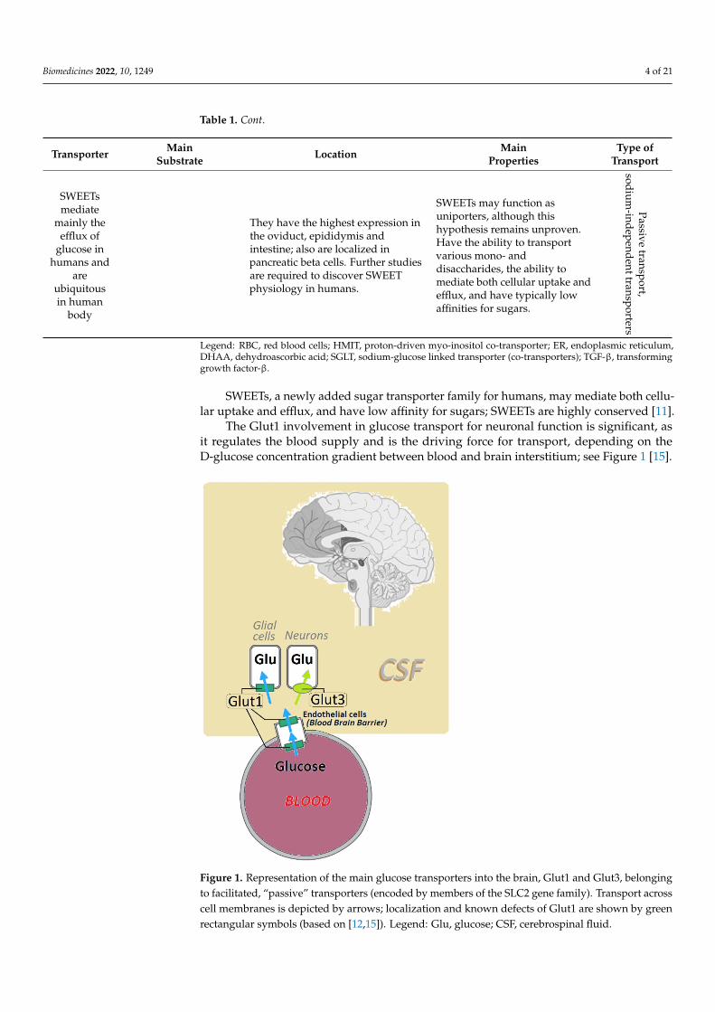
The Glut1 involvement in glucose transport for neuronal function is significant, asit regulates the blood supply and is the driving force for transport, depending on theD-glucose concentration gradient between blood and brain interstitium; see Figure 1 [15].
Figure 1. Representation of the main glucose transporters into the brain, Glut1 and Glut3, belongingto facilitated, “passive” transporters (encoded by members of the SLC2 gene family). Transport acrosscell membranes is depicted by arrows; localization and known defects of Glut1 are shown by greenrectangular symbols (based on [12,15]). Legend: Glu, glucose; CSF, cerebrospinal fluid.
Biomedicines 2022, 10, 1249 5 of 21
2. Glucose Transporter Protein Type 1 (Glut1): Structure and Function
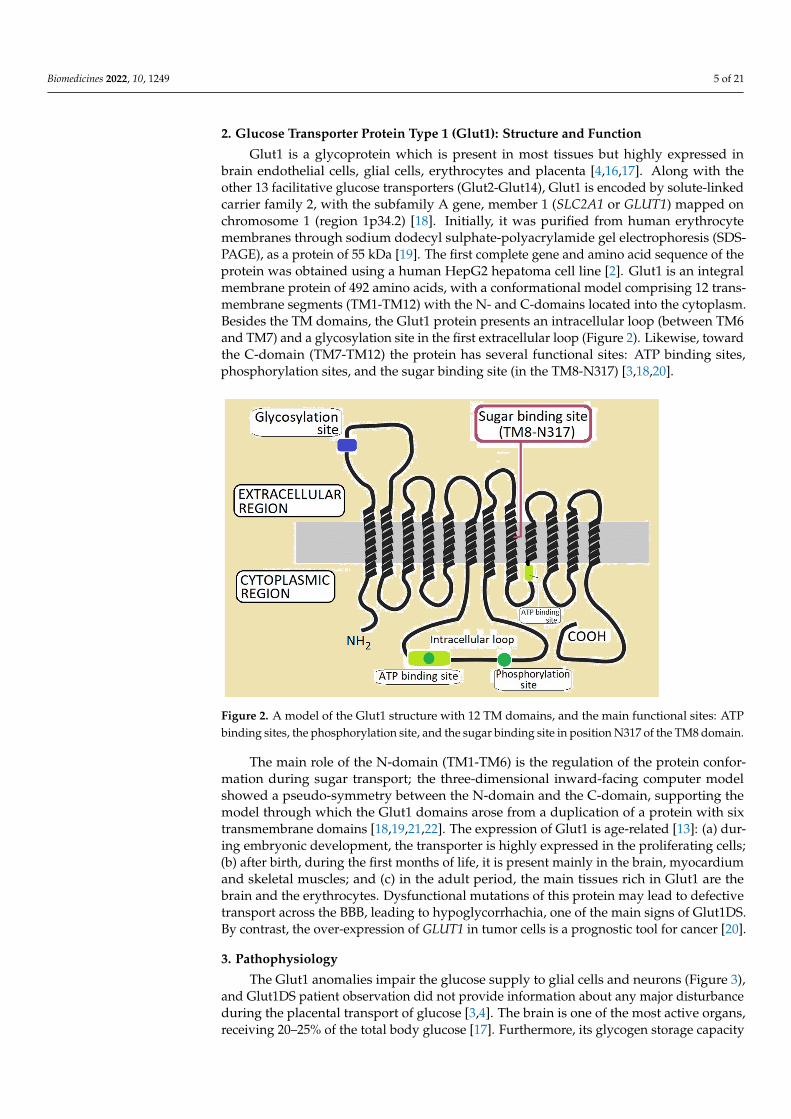
Glut1 is a glycoprotein which is present in most tissues but highly expressed inbrain endothelial cells, glial cells, erythrocytes and placenta [4,16,17]. Along with theother 13 facilitative glucose transporters (Glut2-Glut14), Glut1 is encoded by solute-linkedcarrier family 2, with the subfamily A gene, member 1 (SLC2A1 or GLUT1) mapped onchromosome 1 (region 1p34.2) [18]. Initially, it was purified from human erythrocytemembranes through sodium dodecyl sulphate-polyacrylamide gel electrophoresis (SDS-PAGE), as a protein of 55 kDa [19]. The first complete gene and amino acid sequence of theprotein was obtained using a human HepG2 hepatoma cell line [2]. Glut1 is an integralmembrane protein of 492 amino acids, with a conformational model comprising 12 trans-membrane segments (TM1-TM12) with the N- and C-domains located into the cytoplasm.Besides the TM domains, the Glut1 protein presents an intracellular loop (between TM6and TM7) and a glycosylation site in the first extracellular loop (Figure 2). Likewise, towardthe C-domain (TM7-TM12) the protein has several functional sites: ATP binding sites,phosphorylation sites, and the sugar binding site (in the TM8-N317) [3,18,20].
Figure 2. A model of the Glut1 structure with 12 TM domains, and the main functional sites: ATPbinding sites, the phosphorylation site, and the sugar binding site in position N317 of the TM8 domain.
The main role of the N-domain (TM1-TM6) is the regulation of the protein confor-mation during sugar transport; the three-dimensional inward-facing computer modelshowed a pseudo-symmetry between the N-domain and the C-domain, supporting themodel through which the Glut1 domains arose from a duplication of a protein with sixtransmembrane domains [18,19,21,22]. The expression of Glut1 is age-related [13]: (a) dur-ing embryonic development, the transporter is highly expressed in the proliferating cells;(b) after birth, during the first months of life, it is present mainly in the brain, myocardiumand skeletal muscles; and (c) in the adult period, the main tissues rich in Glut1 are thebrain and the erythrocytes. Dysfunctional mutations of this protein may lead to defectivetransport across the BBB, leading to hypoglycorrhachia, one of the main signs of Glut1DS.By contrast, the over-expression of GLUT1 in tumor cells is a prognostic tool for cancer [20].
3. Pathophysiology
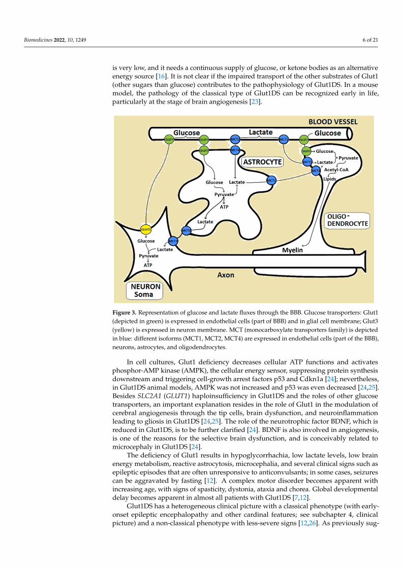
The Glut1 anomalies impair the glucose supply to glial cells and neurons (Figure 3),and Glut1DS patient observation did not provide information about any major disturbanceduring the placental transport of glucose [3,4]. The brain is one of the most active organs,receiving 20–25% of the total body glucose [17]. Furthermore, its glycogen storage capacity
Biomedicines 2022, 10, 1249 6 of 21
is very low, and it needs a continuous supply of glucose, or ketone bodies as an alternativeenergy source [16]. It is not clear if the impaired transport of the other substrates of Glut1(other sugars than glucose) contributes to the pathophysiology of Glut1DS. In a mousemodel, the pathology of the classical type of Glut1DS can be recognized early in life,particularly at the stage of brain angiogenesis [23].
Figure 3. Representation of glucose and lactate fluxes through the BBB. Glucose transporters: Glut1(depicted in green) is expressed in endothelial cells (part of BBB) and in glial cell membrane; Glut3(yellow) is expressed in neuron membrane. MCT (monocarboxylate transporters family) is depictedin blue: different isoforms (MCT1, MCT2, MCT4) are expressed in endothelial cells (part of the BBB),neurons, astrocytes, and oligodendrocytes.
In cell cultures, Glut1 deficiency decreases cellular ATP functions and activatesphosphor-AMP kinase (AMPK), the cellular energy sensor, suppressing protein synthesisdownstream and triggering cell-growth arrest factors p53 and Cdkn1a [24]; nevertheless,in Glut1DS animal models, AMPK was not increased and p53 was even decreased [24,25].Besides SLC2A1 (GLUT1) haploinsufficiency in Glut1DS and the roles of other glucosetransporters, an important explanation resides in the role of Glut1 in the modulation ofcerebral angiogenesis through the tip cells, brain dysfunction, and neuroinflammationleading to gliosis in Glut1DS [24,25]. The role of the neurotrophic factor BDNF, which isreduced in Glut1DS, is to be further clarified [24]. BDNF is also involved in angiogenesis,is one of the reasons for the selective brain dysfunction, and is conceivably related tomicrocephaly in Glut1DS [24].
The deficiency of Glut1 results in hypoglycorrhachia, low lactate levels, low brainenergy metabolism, reactive astrocytosis, microcephalia, and several clinical signs such asepileptic episodes that are often unresponsive to anticonvulsants; in some cases, seizurescan be aggravated by fasting [12]. A complex motor disorder becomes apparent withincreasing age, with signs of spasticity, dystonia, ataxia and chorea. Global developmentaldelay becomes apparent in almost all patients with Glut1DS [7,12].
Glut1DS has a heterogeneous clinical picture with a classical phenotype (with early-onset epileptic encephalopathy and other cardinal features; see subchapter 4, clinicalpicture) and a non-classical phenotype with less-severe signs [12,26]. As previously sug-

Biomedicines 2022, 10, 1249 7 of 21
gested, beside the inadequate transport of glucose across the BBB, the reduced uptake intothe astrocytes and oligodendrocytes may also contribute to the disease [27,28]. Due to thelow concentration of glucose, limited brain glycogen storage and nucleic acid synthesis inthe affected pentose phosphate shunt can cause metabolic stress. The pathway of D-glucosein the brain is clearly defined: from the brain capillaries, it is taken up by astrocytes whichset up the metabolic path for glucose or its glycolytic metabolite, L-lactate, to neurons(Figure 3) [15]. Besides this, glucose can diffuse trough the gap junctions and follow theneuronal pathway directly. The neuronal activity also has implications in the interplay ofD-glucose and L-lactate concentrations [15].
As an alternative source of energy for the brain, ketone bodies enter into the brainusing a different pathway—by monocarboxylic transporter (MCT1) system—and providean alternative source of acetyl-CoA under conditions that affect pyruvate synthesis fromglucose [29]. A remarkable homology between humans’ and rodents’ Glut1 structures andlocalizations was noticed [4]. In animal Glut1DS models, mutant mice develop a significantembryopathy with various neurologic malformations, including microcephaly, anencephalyand anophthalmia [30]. Similar features were identified in humans, but affected infantsappear normal at birth [29].
4. Clinical Picture of Glut1 Deficiency Syndrome
The classical form of Glut1DS presents as an early-onset (during the first year of life)encephalopathy with three cardinal features: severe epilepsy, a complex movement disorder,and developmental delay, including microcephaly. The predominant manifestations areseizures (that usually begin during the first year of life), after an uneventful fetal andneonatal development (due to immature tight junctions in the BBB that allow paracellularglucose transport) [12]. Five seizure types were described: generalized tonic or clonic,myoclonic, atypical absence, atonic, and unclassified [12,23,29,31–34]. Eye movements, anearly warning sign which is sometimes inaugural for Glut1DS, usually disappear after earlyinfancy [35]. The aberrant gaze saccades are peculiar head-eye movements, noticeable inone third of infant patients; they can be distinguished from opsoclonus by the presenceof a clear inter-movement fixation interval and the association of a same-direction headmovement [35–37]. In later childhood, they can be variable, and are often unresponsiveto anticonvulsants drugs. The horizontal eye movements originate in the paramedianpontine reticular structure, while the vertical eye movements originate in the mesencephalicreticular formation [38]. However, according to the EEG tests, these episodic movementsare non-epileptic. In neonatal patients, Glut1DS may evolve with cyanotic spells and atonicdrop attacks [38]. Glut1DS responds for about 5% of myoclonic-astatic epilepsy and for 10%of early-onset absence-epilepsy cases, respectively [38]. Non-epileptic paroxysmal eventswith episodic ataxia, weakness, Parkinsonism or alternating hemiplegia may develop laterin life, often triggered by fasting. The EEG during seizures shows multifocal spike-wavedischarges in adults [38].
The non-classical type of the disease is milder, and consists mostly of only one ortwo of the cardinal features, e.g., isolated early-onset absence epilepsy or an isolatedmovement disorder without epilepsy [12]. This diagnostic should be considered in anyunexplained seizures of various types in early age [39]. During childhood or adolescencethere is a transition towards movement disorders [40]. Nevertheless, apneic episodes orabnormal episodic eye movements simulating opsoclonus may precede the onset of seizuresby several months [23,29]. In the non-classical Glut1DS phenotype there are usually noseizures, but intellectual disability, impaired language development and intermittent ataxiaare seen, sometimes with associated microcephaly [32,38]. Other common manifestationsin the non-classical phenotype of the disease are spasticity, dyspraxia and paroxysmalexercise-induced dyskinesia (PED) with transient involuntary movements such as dystonia,chorea or ballism [32]. PED has been found to be an allelic variant of classic Glut1DS [12].Clinically, these patients have a normal head circumference, normal inter-ictal neurologicexamination, and a lesser decrease of CSF glucose concentration when compared with
Biomedicines 2022, 10, 1249 8 of 21
classic Glut1DS [12]. Manifestations with only minimal symptoms in adults have also beendescribed [40].
5. Genetics and Metabolic Changes
Glut1DS is mainly caused by pathogenic variants of the SLC2A1 gene; approximately90% of patients exhibit a heterozygous de novo mutation, while 10% inherit a deficientgene from their parents (which seems to correlate with a milder phenotype) [29,32]. In mostcases, Glut1DS exhibits an autosomal dominant inheritance pattern. However, there havebeen multiple reported cases of autosomal recessive mutations, for which heterozygouscarriers are asymptomatic [5,29], indicating that the inheritance pattern depends on themutation pathogenicity and the subsequent haploinsufficiency degree [41]. The birthincidence of Glut1DS has been estimated to be between 1:24,000 [42] and 1:90,000 (inAustralia) [43], with a similar result in Denmark (1:83,000) [44]. To date, about 500 caseshave been reported worldwide. Due to the fact that many neurological conditions cancause the same symptoms as in Glut1DS, the disorder may be under-diagnosed; somestudies suggested that about 105,000 patients suffer from Glut1DS worldwide [24].
So far, over 200 types of genetic defects have been described, among which are mis-sense, nonsense, frameshift, and splice-site mutations, and large fragment deletions [12,32].Of these, missense mutations seem to be associated with a milder phenotype, but no def-inite genotype–phenotype correlation has been described [5,45]. Pathogenic variants inSLC2A1 are most often identified by sequencing (81–89% of cases), and less often by dele-tion/duplication analysis (11–14%) [29]. An asymptomatic parent harboring the pathogenicvariant implies a mosaic state [29].
Of note, allelic disorders with overlapping features include GLUT1 deficiency syn-drome with pseudohyperkalemia and hemolysis, GLUT1 deficiency syndrome-2 (GLUT1DS2),dystonia-9 (DYT9), and idiopathic generalized epilepsy-12 (EIG12) (https://www.omim.org, accessed on 20 April 2022).
Different SLC2A1 variants could destabilize Glut1 native interactions or generate novelinteractions, initiate protein misfolding, enhance protein aggregation, or be influenced bynon-coding RNA genes or by defects in translation, transcription, processing, and activatingGlut1 protein [5,46]. In a small proportion of patients with Glut1DS, no SLC2A1 genetic de-fect can be identified, even after the additional use of MLPA (multiplex ligation-dependentprobe amplification) analysis to detect copy number variations [12]. For these patients,other factors that may lead to altered Glut1 function, such as changes in other related genes;downstream malfunctions in transcription, translation and protein folding and function; orimproper regulating processes have been proposed [12]. For instance, a frameshift deletionin the PURA gene, coding for a transcriptional and translational regulator protein, led tohypoglycorrhachia, along with a lowered Glut1 expression on the membrane of peripheralred blood cells [47]. Sometimes the hallmarks of the clinical and biological picture maybe given by other genes, coding for different membrane transporters such as SLC9A6, en-zymes, receptors or transcriptional factors, or involving other mechanisms such as proteinrecycling [48]. In individual patients, mutations reported to modify the clinical picture inGlut1DS-involved genes include SCN8A, ATP1A3, KCNQ2, NALCN, DNM1, MAN2B andUNC13A [48].
6. Diagnosis
The diagnosis of Glut1DS is complicated by the phenotypical diversity and evolutionwith age of the disease [24,32]. An important laboratory investigation for Glut1DS is thelow CSF:blood glucose ratio, with blood glucose being normal [29]. The CSF:blood glucoseratio should be measured in a metabolic steady-state, during non-ictal periods [12].
The CSF glucose is investigated by lumbar puncture after four–six hours of fasting.For the diagnosis of Glut1DS, the CSF glucose should be under 48.6 mg/dL (<2.7 mmol/L),but Glut1DS should be suspected in all children with CSF glucose concentration below45 mg/dL (normal >59.4 mg/dL) [49]. In affected patients, the values vary considerably
Biomedicines 2022, 10, 1249 9 of 21
(range 16.2–52.2 mg/dL), being higher in milder phenotypes and in paroxysmal movementdisorders [12].
The CSF to blood glucose ratio is normally 0.6 (0.65 ± 0.1) [49]. In the absence ofa CNS infection or hypoglycemia, a CSF to blood glucose ratio value under 0.45–0.5 (range0.19–0.52) is diagnostic for Glut1DS [12,49].
Typically, CSF lactate is low or normal, while the cell count and proteins are normal,which helps to differentiate between Glut1DS and other diseases (meningitis or encephalitis)in which lactate levels are high [32].
Genetic testing for mutations in SLC2A1 is also recommended when Glut1DS is sus-pected, although a negative result cannot rule Glut1DS out [50]. Thus, sequencing orduplication/deletion analysis can be employed when clinical findings and/or hypoglycor-rhachia are suggestive of Glut1DS.
Two other methods have been proposed to analyze the function of Glut1: (a) an erythro-cyte 3-O-methyl-D-glucose (3-OMG) uptake assay, with a 74% uptake cutoff for abnormallylow levels indicative of Glut1DS, and (b) a red blood cell Glut1 surface expression test usingflow cytometry analysis in circulating erythrocytes, which is at least 20% lower in affectedpatients [12,51–53].
Other routine laboratory tests and inter-ictal EEGs are normal. If they are abnormal,an improvement in the EEG with glucose intake may be of diagnostic value (https://www.omim.org, accessed on 20 April 2022). Ictal EEGs may show epileptiform discharges ininfants and a generalized spike-wave pattern in older children.
Regarding the structural brain changes, cerebral MRI may show in a fourth of patientsdelayed myelination, the hyperintensity of the subcortical U-fibers, and the enlargement ofperivascular Virchow spaces [5]. Functional imaging techniques, such as 18F-deoxyglucose-positron emission tomography (18F-FDG-PET), may identify a decrease in cortical, cerebellarand thalamic glucose uptake, particularly in the mesial temporal lobe, with a relative signalincrease in the basal ganglia and striatum, mainly in the caudate nucleus [5,12].
The differential diagnosis of Glut1DS includes, without being limited to, (a) othercauses of neuroglycopenia such as chronic or intermittent hypoglycemia (e.g., familialhyperinsulinism); (b) opsoclonus-myoclonus syndrome; (c) all causes of neonatal seizuresand acquired microcephaly, such as Rett syndrome (early presentations), Angelman syn-drome, and infantile forms of neuronal ceroid-lipofuscinosis; (d) cryptogenic epilepticencephalopathies with developmental delays; (e) familial epilepsies with autosomal domi-nant transmission; (f) episodes of paroxysmal neurologic dysfunction which are responsiveto carbohydrate intake, especially when manifesting as ataxia, cognitive dysfunction, alter-nating hemiparesis, or seizures; and (g) movement disorders including dystonia [29].
7. Treatment, Prognosis and Research
During fasting, ketone bodies provide an alternative fuel to the brain, and thismetabolic state can be induced by a high-fat, low-carbohydrate diet called a ketogenic diet(KD), restoring the brain energy metabolism in Glut1DS, as ketone bodies’ transport at theBBB is not dependent on Glut1. The classic ketogenic diet (4:1 and 3:1 ratios of caloriesfrom lipids and non-fat sources, respectively) in Glut1DS patients may effectively controlseizures and movement disorders, and improve development. Calcium and multivitaminsupplements are necessary on a KD. In contrast to intractable childhood epilepsy, theKD in Glut1DS patients should be maintained throughout childhood and adolescence,until cerebral glucose requirements decrease. The early initiation of this therapy supportsbrain growth and normal brain function in the adult period of life; the disease generallystabilizes after puberty [12]. The main beneficial effect of the KD is the control of seizures;after initiating therapy, the patients generally have a rapid improvement (over days) inseizure control. Other positive effects of a KD are related to the improvements of motor(i.e., paroxysmal dyskinesias) and cognitive symptoms, but the results are variable [12,54].The prolonged administration of the diet might lead to long-term adverse effects: hyperc-holesterolemia, growth impairment, acidosis, kidney stones, and a high risk of metabolic

Biomedicines 2022, 10, 1249 10 of 21
syndrome and cardiovascular disease in adults [55–57]. The KD may result in the lossof adherence, metabolic disturbances, and gut microbiome alterations. However, in thelong-term the KD did not significantly change nutritional status, body fat, glucose andlipid profiles, nor ghrelin or leptin [58]. Nevertheless, after 3 months of KD, a significantincrease in Desulfovibrio spp.—thought to be associated with colonic inflammation—wasnoted in Glut1DS patients, making an empirical trial of pro- or pre-biotics reasonable [59].
Furthermore, the administration of carnitine (deficient in KD, but with an importantrole in mitochondrial lipid oxidation) and α-lipoic acid facilitates glucose transport throughGlut4, another transporter expressed in the brain [60,61].
In order to compensate for some of the adverse effects, the modified Atkins diet wasproposed for adults, and remains a promising alternative by initiating an improvement incognitive ability and epilepsy; it is less restrictive (any amounts of protein and fat mightbe consumed, but there is a limited carbohydrate intake of 10 g/day) [62]. However,despite adequate treatment, seizures and a variable degree of impairment may persist inseveral Glut1DS patients; paroxysmal events and the impairment of expressive languageappear to be difficult to treat in adults [12]. Treatment decisions are less clear in atypicalvariants of Glut1DS, mainly in late-onset or in paucisymptomatic patients, due to limitedexperience [5].
Artificial ketones (ketoesters and triheptanoin—a synthetic medium-chain triglyceride)represent anaplerotic substances, and provide an alternative energy source, see Table 2; thus,they could represent a novel therapeutic option [63,64]. The use of dietary antioxidants,such as α-lipoic acid (thioctic acid) is under investigation [5,12].
Table 2. Recommended treatments in cases with the epilepsy-associated phenotype of Glut1DS; diettreatments and antiepileptic drugs (AED) to avoid.
Diet/Treatment AED Indicated Drugs Not Recommended inAssociation with KD References
Ketogenic diet (KD) Acetazolamide Valproate [5,12,56]
Modified Atkins Diet Topiramate Zonisamide [56]
Medium chain Triglycerides Zonisamide Acetazolamide [5,12,56]
Low glycemic indextreatment Phenytoin Topiramate [5,56]
Triheptanoin Carbamazepine - [56,64]
α-lipoic acid(under investigation) - - [5,12,56]
Another proposed alternative in patients with compliance or intolerance problemsis the modified high amylopectin cornstarch and low glycemic index diet, providinga steady-state glucose transport into the brain [65]. Research projects investigate families ofmedium chain triglyceride and hexose transporters that may provide metabolic fuel for thebrain [5,63].
Diazoxide administration (a medication used to treat low blood sugar, interfering withinsulin release through its action on potassium channels) was proposed to contribute toraising the blood and CSF glucose levels in a patient with Glut1DS who did not respondto a KD. This provides a higher level of glucose in CSF, and clinical improvement, beinga reliable long-term treatment when associated with continuous glucose monitoring [66].
Drug administration in patients with Glut1DS remains an additional strategy to controlseizures or paroxysmal dyskinesias (which, it seems, respond well to acetazolamide). Evenif the symptoms are improved, the anti-epileptic therapy is unable to correct the brainenergy necessary for growth and development [5,54].
Furthermore, the avoidance of Glut1 dietary or pharmacologic inhibition is impor-tant. In vitro studies showed that methylxanthines (caffeine, theophylline), tricyclic an-
Biomedicines 2022, 10, 1249 11 of 21
tidepressants, sodium valproate, barbiturates, diazepam, chloralhydrate, and ethanol arenonspecific inhibitors of Glut1 function [5,27,67].
Research on disease mechanisms has identified novel targets for therapy, focusing on(a) the molecular mechanisms involved in the metabolic defect (brain glucose depletion)and neurological consequences, (b) the type of transporter dysfunction, and (c) the imagisticinvestigations (PET-based investigation and MRI (Magnetic Resonance Imaging)) of humanbrain metabolism [5].
8. Glut-1 Deficiency in Other Tissues: Expanding the Clinical Phenotype?
While Glut1DS is primarily a brain disorder, Glut1 haploinsufficiency can also affectother organ systems relying on glucose to fuel their energy requirements [24]. A betterunderstanding of the role of glucose transporters in various settings has revealed unex-pected potential functions of Glut1 modulation, as tissue functions are partly controlled bymetabolic substrates [68]. Glut1 is also expressed in the retina, ciliary muscle, peripheralnerve endoneurium and perineurium, placenta, and testis, etc. [69]. Rare features describedin Glut1DS include periventricular calcifications, pseudohyperkaliemia, parkinsonism,writer’s cramp, nocturnal muscle cramps, cyclic vomiting, and others [5].
The proliferating cells have higher glucose requirements [68]. Warburg’s effect, i.e., ox-idative glycolysis performed even in the presence of enough oxygen, increasing glucoseand glutamine uptake, and favoring anabolism, was observed in rapid growth require-ments such as embryonic development, wound healing, T cell activation or pluripotent cellproliferation [70].
8.1. Glut1 in Vessels
Besides the brain energy deprivation, the Glut1DS clinical picture also results fromthe brain vasculature insufficiency [24]. In endothelial cells, glycolysis promotes vesselbranching and migration [68]. Endothelial tip cells, which are highly glycolytic and criticalfor brain angiogenesis, are significantly reduced in quantity and quality in Glut1DS [24].Moreover, endothelial cell-specific Glut1 haploinsufficiency was involved in triggeringneuroinflammation with consecutive neuronal loss, along with BDNF insufficiency [24].
A patient with Glut1DS, a novel SLC2A1 mutation, and a hemangioma was de-scribed [71]. Glut1 expressed on endothelial cells is a selective marker of juvenile he-mangioma, independent of mitotic activity [69]; nevertheless, the association may becoincidental [71]. Hemiplegic migraines due to vascular spasm were also described inGlut1DS [5].
8.2. Glut1 in Retina
In retina, Glut1-mediated transport occurs across the capillary endothelial cells of theinner blood–retinal barrier and the retinal pigmentary epithelium [72]. The rod photore-ceptors secrete the rod-derived cone viability factor which binds basigin-1 expressed byphotoreceptors, which in turn binds Glut1 [73]. Glut1DS may evolve with retinal changesand also with cataracts [5].
8.3. Glut 1 in Erythrocytes
Besides the brain tissue, Glut1 is highly expressed in erythrocytes, representing 5%of all of the membrane proteins [52]. Exercise-induced hemolytic anemia in Glut1DS isa consequence of cations draining through the defective Glut1 transporter [12]. Given thehigh expression of Glut1 on erythrocytes, red blood cell exchange transfusion—similar tosickle cell anemia—has been proposed, and is under study for the therapy of Glut1 patients(ClinicalTrials.gov Identifier NCT04137692, accessed on 20 April 2022).
8.4. Glut1 in Muscles
Glut1 is responsible for 30–40% of basal glucose uptake in skeletal muscle [74]. Pa-tients with Glut1DS may have muscle hypotonia, and to some extent dysarthria and slurred

Biomedicines 2022, 10, 1249 12 of 21
speech may result from the involvement of the pharyngo-buccofacial system or the oro-facial region muscles [29,75]. In the skeletal muscle, the mechanism regulating Glut-1mediated glucose transport is more complex, as some Glut1 mutations decrease basalmuscle transport much more than its surface expression [74,76].
8.5. Glut1 in Immune Cells
At the interface and cross-talk of immunology and metabolism, the cellular metabolismregulates the fate, function and activation of the immune cells [77]. Metabolic adaptationsand epigenetic reprogramming are involved in macrophage plasticity and phenotypechange (Figure 4) [77–81]. Glut1 is also essential for the homeostasis of T and B lympho-cytes [82,83]. Glut1 deficiency selectively impairs thymocyte and T effector functions [84].In Glut1DS, the serum levels of antibodies are significantly lower, favoring the developmentof severe infections [45]. Furthermore, Glut1-deficient T cells are metabolically stressed,as their pAMPK level increases after activation, and cannot sustain activated mTORC1signaling, while other signaling pathways such as Akt or ERK (extracellular regulatedkinase) are generally unaffected [84,85].
Figure 4. Immune challenge induces metabolic activation with increased GLUT1 expression, glucoseuptake and glycolysis. The transition of monocytes to macrophages is characterized by furtherincreases in Glut1 expression and glycolysis. Naive (unactivated) monocytes are metabolicallyquiescent, with low basal metabolic activity and ATP derived primarily via oxidative phosphory-lation (OxPhos). Classically activated macrophages (M1) induce aerobic glycolysis, resulting inlactate production and the increased production of inflammatory cytokines. Alternatively activatedmacrophages (M2) trigger a metabolic profile with OxPhos and an anti-inflammatory response. TNF:Tumor necrosis factor; IL-6: Interleukin 6; IL-1RA: interleukin-1 receptor antagonist.
9. Glut1 Inhibition in Other Settings
The relationship between glucose transport and metabolic pathways is worthwhile toexplore for therapeutic purposes. Excessive glucose usage is related to several diseases, bylinking immunological and metabolic pathways [68]. The glucose uptake should be efficientin cells with high turnover, such as immune cells and keratinocytes [68]. Warburg’s effectwas described in tumors and in other high-energy-requiring cell processes [70]. The effectsof Glut1 inhibition in health and pathology could provide clues for a better understandingof Glut1DS (see Table 3).
Biomedicines 2022, 10, 1249 13 of 21
9.1. Glut1 in Keratinisation Disorders
Glucose transport in keratinocytes and wound- and inflammation-associated ker-atinocyte proliferation is mediated by Glut1 [68]. As Glut1 deficiency does not interferewith the epidermal development of function, Glut1 inhibition is a potential therapeuticstrategy for psoriasis and other disorders of keratinization [68].
9.2. Glut1 in Eye Diseases
Targeting Glut1 with small molecules could alleviate retinitis pigmentosa by stimulat-ing glucose uptake and preserving retinal cells [73]. Furthermore, Glut1 inhibition usingsmall interfering RNAs reduces retinal glucose and improves diabetic retinopathy but maybe detrimental to photoreceptors and retinal pigmentary epithelium [72,86].
9.3. Glut1 in Kidney Diseases
As Glut1 predominates in mesangial cells, as well as in podocytes, Glut1 activityalterations are deleterious in diabetic nephropathy, stimulating renal extracellular matrixproduction the most [87,88]. The GLUT1 Enh2 genetic variation, located within a bindingsite for the insulin-responsive transcription factor, is associated with albuminuria, matrixexpansion and glomerulosclerosis [88]. Podocytes enhance their glucose uptake but de-crease their GLUT1 expression when exposed to mechanical stress, suggesting alternativeglucose transporters during stress/injury [87,89].
9.4. Glut1 in Placental Pathology
Glut1 expression changes during gestation, as Glut1 is expressed in placenta, syn-cytiotrophoblast, cytotrophoblast, and endothelial cells, and villous stroma [12]. Glut1variations can result in significant transplacental glucose transport [8]. Glut1 is decreasedin chronic hypoxia and in preeclampsia, but not in intrauterine growth restriction [12,90].
9.5. Glut1 in Heart Failure
Although Glut1 is the main glucose transporter in the heart, Glut-1 deficiency doesnot accelerate the progression from hypertrophy to heart failure, nor the pressure overloadhypertrophy-induced mitochondrial dysfunction [91].
9.6. Glut 1 in Crystal-Induced Inflammation
In gout, the Glut1-mediated glucose uptake of the macrophages in the presence ofuric acid is important for interleukin-1β production [81]. Glucose deprivation or therapywith a Glut1 inhibitor suppresses crystal-induced inflammation in gout and pseudogout,opening new therapeutic pathways [92].
9.7. Glut1 in Viral Infections
There are several viruses acting on the glucose transporters: Epstein-Barr, hepatitis B,HIV, Zika, rhinovirus or human cytomegalovirus (HCMV) [93]. Of note, Glut1 is a HTLV1receptor molecule, and the infection downregulates the glucose uptake through Glut1 [94].The HCMV early protein IE72 modifies the messenger RNA in the infected cells to down-regulate GLUT1 expression and to increase the more active GLUT4 expression [93]. Dueto the microcephaly accompanying in-utero Zika virus infections, an acquired Glut1 defi-ciency was suspected, but not confirmed [95]. In COVID-19, Glut1 and the ion transportersodium proton exchanger 1 (NHE1) seem to be critically involved, and a low GLUT1/NHERNA expression predicts disease severity in COVID-19 mostly in patients with cardiaccomplications [96].
9.8. Glut1 in Alzheimer’s Disease and Other Neurodegenerative Disorders
In Alzheimer’s disease (AD) there is cerebral glucose hypometabolism, partly dueto reduced glucose transport at the BBB and across astrocytic and neuronal cell mem-branes [97]. In Alzheimer’s disease, vascular and non-vascular Glut1 and Glut3 are reduced
Biomedicines 2022, 10, 1249 14 of 21
in the hippocampus and cortex after beta-amyloid deposition, resulting in reduced glucoseuptake [97]. Furthermore, Glut1 in AD is positively correlated with insulin signaling pro-teins [98]. In Alzheimer’s disease, the cause of Glut1 reduction is not known; it may berelated to the low level of the regulator molecule HIF-1α, or to the direct effect of β-amyloidor tau on the glucose transporter gene expression [97]. Of interest, antidiabetic drugslike liraglutide may improve glucose brain transportation and reduce cognitive decline inAD [99].
As 18F-FDG-PET is used to image cerebral glucose consumption, it is also widely usedin neurology for differential diagnosis with neurodegenerative diseases or encephalitis, inneurosurgery, or in psychiatry, mainly for atypical and/or pharmaco-resistant presenta-tions [100,101]. Typical topographic patterns of glucose hypo-or hypermetabolism allowdiagnosis and progression assessment for several diseases, such as dementia with Lewisbodies, frontotemporal lobar degeneration, and differential diagnosis between several neu-rodegenerative diseases including lateral amyotrophic sclerosis and Huntington’s disease,between Alzheimer’s disease and vascular dementia, or between parkinsonian syndromesassociated with dementia, in neurooncology and others [100].
9.9. Glut1 in Cancers
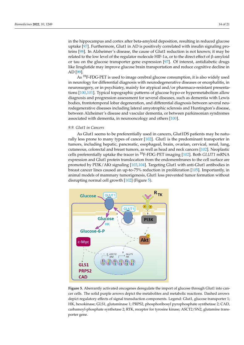
As Glut1 seems to be preferentially used in cancers, Glut1DS patients may be natu-rally less prone to many types of cancer [102]. Glut1 is the predominant transporter intumors, including hepatic, pancreatic, esophageal, brain, ovarian, cervical, renal, lung,cutaneous, colorectal and breast tumors, as well as head and neck cancers [102]. Neoplasticcells preferentially uptake the tracer in 18F-FDG-PET imaging [102]. Both GLUT1 mRNAexpression and Glut1 protein translocation from the endomembranes to the cell surface arepromoted by PI3K/AKt signaling [103,104]. Targeting Glut1 with anti-Glut1 antibodies inbreast cancer lines caused an up-to-75% reduction in proliferation [105]. Importantly, inanimal models of mammary tumorigenesis, Glut1 loss prevented tumor formation withoutdisrupting normal cell growth [102] (Figure 5).
Figure 5. Aberrantly activated oncogenes deregulate the import of glucose through Glut1 into can-cer cells. The solid purple arrows depict the metabolites and metabolic reactions. Dashed arrowsdepict regulatory effects of signal transduction components. Legend: Glut1, glucose transporter 1;HK, hexokinase; GLS1, glutaminase 1; PRPS2, phosphoribosyl pyrophosphate synthetase 2; CAD,carbamoyl-phosphate synthetase 2; RTK, receptor for tyrosine kinase; ASCT2/SN2, glutamine trans-porter gene.

Biomedicines 2022, 10, 1249 15 of 21
Table 3. Glut1 in other settings: functional implications.
Cell/Tissue Glut1 References
VesselsEndothelial Glut1 is involved in vessel branching and migration in brain angiogenesis;Glut1 endothelial cell-specific haploinsufficiency was involved intriggering neuroinflammation
[24,68]
Retina Glut1 depletion affects retinal angiogenesis and photoreceptor viability [24]
Erythrocytes Glut1 represents 5% of the erythrocyte membrane proteinsIn Glut1DS exercise may result in hemolytic anemia [12,52]
Skin Glut1 mediates glucose transport in keratinocytes, wound- and inflammation-associatedkeratinocyte proliferation [68]
MuscleGlut1 responds for 30–40% of skeletal muscle basal glucose uptakeGlut1DS-associated muscle hypotonia may sometimes involvespeech-associated muscles
[29,74,75]
Heart Glut1—main glucose transporter in heart, but not critical for normalcardiac function [91]
Placenta
Glut1 expressed in placenta, syncytiotrophoblast, cytotrophoblast,endothelial cells and villous stroma;Glut1 is decreased in chronic hypoxia and in preeclampsia, but not in intrauterinegrowth restriction
[8,90]
Kidneys Glut1 expressed in glomerulus mainly in mesangial cells; Glut1 along with cytokines andgrowth factors favors diabetic glomerulosclerosis [87,88]
Immune cells
Glut1 involved in macrophage plasticity and phenotype reprogramming in innateimmune adaptations including in trained immunity;In gout interleukin-1 beta production depends onmacrophage Glut1-mediated glucose uptake;Glut1 deficiency reduces T effector ability to induceinflammation, not affecting Tregs
[77–79,81,84]
Viral infectionsGlut1 is a HTLV1 receptor molecule. The HCMV early protein IE72 downregulatesGLUT1 to increase GLUT4 expression. In COVID19 Glut1 is critically involved, and a lowGlut1/NPE-1 predicts COVID19 severity
[93,94,96]
Brain regions inAlzeimer’s disease
Glut1 and Glut3 are reduced in the hippocampus and cortexafter β-amyloid deposition, resulting in reduced glucose uptakeand metabolism
[97]
Cells in tumorsGlut1 is the predominant transporter in tumors, differentially required in differenttumorigenesis stages.Blocking Glut1 inhibits tumorigenesis without disrupting normal cells.
[102,104]
Legend: HCMV, human cytomegalovirus; HTLV1, human T lymphotropic virus; NPE-1, sodium proton exchanger1; Tregs, regulatory T cells.
10. Concluding Remarks and Future Directions
Glut1DS is considered to be a treatable inherited disease, but there are key issuesregarding its diagnosis, treatment and long-term management. The main difficulty indiagnosing patients is the phenotypic heterogeneity related to the age and genetic com-plexity, underlining the need to increase physician awareness of this defect. The laboratorydiagnostic for Glut1DS is the low CSF:blood glucose ratio (<0.45) after four–six hours offasting, the molecular genetic test of the SLC2A1 gene, or the red blood cell Glut1 surfaceexpression test using flow cytometry analysis. The disease may be underdiagnosed. TheKD still represents a standard choice in Glut1DS patients with favorable prognosis mostlyinvolving the epileptic crises; with early treatment the patients continue to make progressand acquire mobility and speech. The standard therapy is still age-specific, based onketogenic therapies that—by supplying ketones—are an alternative for brain fuel. Thepatients should avoid drugs that inhibit Glut1. There is still an overgrowing reluctancewith respect to anticonvulsant administration which has been proven to have a poor re-

Biomedicines 2022, 10, 1249 16 of 21
sponse in epilepsy-related pathologies. Ongoing research to identify future interventions isfocusing on small molecules designed to enhance Glut1 activity or expression, metabolicenhancement, and SLC2A1 transfer strategies [5].
There are international consensus statements to facilitate the rapid diagnosis and mul-tidisciplinary management of Glut1DS patients throughout their lives [5]. The evaluation ofadult and pediatric Glut1DS patients is different [5]. Moreover, the first children diagnosedwith Glut1DS are just coming of age. There are few data on pregnancy in this setting, withthe dietetic therapy of the mother and infant having resulted in normal development inthe early-treated neonate, emphasizing the need to identify and treat pre-symptomaticchildren [106]. The at-risk relatives of an affected child should also be tested as early as pos-sible in order to minimize neurologic consequences. The need to identify pre-symptomaticindividuals is an argument for newborn screening for Glut1DS [5,29].
Despite the improved prognosis, it is clear that there are unmet needs regarding thetherapy of Glut1DS patients. Understanding the complex interactions of Glut1 with othertissues, its signaling function for brain angiogenesis and gliosis, and the complex regulationof glucose transportation and other metabolic pathways involving Glut1 in different tissueswill hopefully also advance the therapy in this underdiagnosed disease [24]. The traffickingof Glut1 may be regulated by multiple pathways [74].
The upregulation of glucose transporters in other settings may shed a light on possibletherapies for innate or acquired brain energy deficiencies. Short-term fasting upregulatesglucose transporters in neurons and endothelial cells but not in astrocytes, as the neuronsmay be prioritized over astrocytes during fasting [107]. The study of supplements such ascurcumin, which protects brain cells from apoptosis by upregulating Glut1 and Glut3, maybe of interest [108]. Furthermore, other secondary deficits may be addressed, such as themetabolism of ascorbic acid, as Glut1 is also a prominent transporter of dehydroascorbicacid, the oxidized form of ascorbic acid [74]. In acquired forms of Glut1 deficiency such asAlzheimer’s disease, physical exercise may increase the Glut1 level [76].
A better understanding of the Glut1 functions in immune tissues will aid in char-acterizing the subtle immune deficiency in Glut1DS patients. Furthermore, the lack ofsignificant impact on some tissues of Glut1 deficiency, despite the predominant expres-sion of Glut1, implies coordinated compensatory mechanisms possibly involving otherglucose transporters, which have to be unveiled for therapeutic purposes. This notwith-standing, recent research on the molecular and cellular impact of glucose deprivationhelps us to define new therapeutic targets in Glut1DS and other syndromes with acquiredglucose hypometabolism.
Author Contributions: Conceptualization, R.V., A.C., L.D.; methodology, S.P., I.M.F., A.B. and C.C.L.;validation, C.C.L., A.C., O.H.O., A.-V.S.-T. and L.D.; formal analysis, C.L., C.A.-K., A.S., M.A.V., S.M.and M.M.; data curation, L.D., S.M., C.A.-K. and C.C.L.; writing, R.V., S.P., I.M.F., A.B., A.C. andL.D.; review and editing, R.V., A.C., C.C.L. and L.D.; visualization, R.V., A.-V.S.-T., C.C.L. and L.D.;supervision, R.V., C.C.L. and L.D. All authors have read and agreed to the published version ofthe manuscript.
Funding: Project PDI-PFE-CDI 2021, entitled Increasing the Performance of Scientific Research,Supporting Excellence in Medical Research and Innovation, PROGRES, no. 40PFE/30.12.2021, “IuliuHatieganu” University of Medicine and Pharmacy, Cluj-Napoca, Romania.
Institutional Review Board Statement: Not applicable.
Informed Consent Statement: Not applicable.
Data Availability Statement: Not applicable.
Conflicts of Interest: The authors declare no conflict of interest.

Biomedicines 2022, 10, 1249 17 of 21
References1. Landowski, C.P.; Suzuki, Y.; Hediger, M.A. The Mammalian Transporter Families. In Seldin and Giebisch’s The Kidney: Physiology &
Pathophysiology, 4th ed.; Alpern, R.J., Hebert, S.C., Eds.; Elsevier: Oxford, UK, 2008; pp. 91–146.2. Mueckler, M.; Caruso, C.; Baldwin, S.A.; Panico, M.; Blench, I.; Morris, H.R.; Allard, W.J.; Lienhard, G.E.; Lodish, H.F. Sequence
and Structure of a Human Glucose Transporter. Science 1985, 229, 941–945. [CrossRef]3. Klepper, J.; Voit, T. Facilitated glucose transporter protein type 1 (GLUT1) deficiency syndrome: Impaired glucose transport into
brain—A review. Eur. J. Pediatr. 2002, 161, 295–304. [CrossRef] [PubMed]4. De Vivo, D.C.; Trifiletti, R.R.; Jacobson, R.I.; Ronen, G.M.; Behmand, R.A.; Harik, S.I. Defective Glucose Transport across the
Blood-Brain Barrier as a Cause of Persistent Hypoglycorrhachia, Seizures, and Developmental Delay. N. Engl. J. Med. 1991, 325,703–709. [CrossRef] [PubMed]
5. Klepper, J.; Akman, C.; Armeno, M.; Auvin, S.; Cervenka, M.; Cross, H.J.; De Giorgis, V.; Della Marina, A.; Engelstad, K.;Heussinger, N.; et al. Glut1 Deficiency Syndrome (Glut1DS): State of the art in 2020 and recommendations of the internationalGlut1DS study group. Epilepsia Open 2020, 5, 354–365. [CrossRef] [PubMed]
6. Tang, M.; Park, S.H.; De Vivo, D.C.; Monani, U.R. Therapeutic strategies for glucose transporter 1 deficiency syndrome. Ann. Clin.Transl. Neurol. 2019, 6, 1923–1932. [CrossRef]
7. Galochkina, T.; Chong, M.N.F.; Challali, L.; Abbar, S.; Etchebest, C. New insights into GluT1 mechanics during glucose transfer.Sci. Rep. 2019, 9, 998. [CrossRef]
8. Illsley, N.P.; Baumann, M.U. Human placental glucose transport in fetoplacental growth and metabolism. Biochim. Biophys. ActaMol. Basis Dis. 2018, 1866, 165359. [CrossRef]
9. Matsuo, S.; Hiasa, M.; Omote, H. Functional characterization and tissue localization of the facilitative glucose transporter GLUT12.J. Biochem. 2020, 168, 611–620. [CrossRef]
10. Chadt, A.; Al-Hasani, H. Glucose transporters in adipose tissue, liver, and skeletal muscle in metabolic health and disease. Pflug.Arch. 2020, 472, 1273–1298. [CrossRef]
11. Chen, L.Q.; Cheung, L.S.; Feng, L.; Tanner, W.; Frommer, W.B. Transport of sugars. Annu. Rev. Biochem. 2015, 84, 865–894.[CrossRef]
12. Santer, R.; Klepper, J. Disorders of Glucose Transport in Inherited Metabolic Diseases, Diagnostic and Treatment, 6th ed.; Saudubray, J.M.,Baumgartner, M., Waler, J., Eds.; Springer: Berlin/Heidelberg, Germany, 2016; pp. 175–184.
13. Pragallapati, S.; Manyam, R. Glucose transporter 1 in health and disease. J. Oral Maxillofac. Pathol. 2019, 23, 443–449. [CrossRef][PubMed]
14. Mueckler, M.; Thorens, B. The SLC2 (GLUT) family of membrane transporters. Mol. Asp. Med. 2013, 34, 121–138. [CrossRef][PubMed]
15. Koepsell, H. Glucose transporters in brain in health and disease. Pflug. Arch. 2020, 472, 1299–1343. [CrossRef]16. Gras, D.; Roze, E.; Caillet, S.; Méneret, A.; Doummar, D.; de Villemeur, T.B.; Vidailhet, M.; Mochel, F. GLUT1 deficiency syndrome:
An update. Rev. Neurol. 2014, 170, 91–99. [CrossRef] [PubMed]17. Klepper, J. Glucose transporter deficiency syndrome (GLUT1DS) and the ketogenic diet. Epilepsia 2008, 49, 46–49. [CrossRef]
[PubMed]18. Long, W.; Cheeseman, C. Structure of and functional insight into the GLUT family of membrane transporters. Cell Health
Cytoskelet. 2015, 7, 167–183.19. Kasahara, M.; Hinkle, P.C. Reconstitution and purification of the D-glucose transporter from human erythrocytes. J. Biol. Chem.
1977, 252, 7384–7390. [CrossRef]20. Deng, D.; Xu, C.; Sun, P.; Wu, J.; Yan, C.; Hu, M.; Yan, N. Crystal structure of the human glucose transporter GLUT1. Nature 2014,
510, 121–125. [CrossRef]21. Custódio, T.F.; Paulsen, P.A.; Frain, K.M.; Pedersen, B.P. Structural comparison of GLUT1 to GLUT3 reveal transport regulation
mechanism in sugar porter family. Life Sci. Alliance 2021, 4, e202000858. [CrossRef]22. Salas-Burgos, A.; Iserovich, P.; Zuniga, F.; Vera, J.C.; Fischbarg, J. Predicting the Three-Dimensional Structure of the Human
Facilitative Glucose Transporter Glut1 by a Novel Evolutionary Homology Strategy: Insights on the Molecular Mechanism ofSubstrate Migration, and Binding Sites for Glucose and Inhibitory Molecules. Biophys. J. 2004, 87, 2990–2999. [CrossRef]
23. Tang, M.; Park, S.H.; Petri, S.; Yu, H.; Rueda, C.B.; Abel, E.D.; Kim, C.Y.; Hillman, E.M.; Li, F.; Lee, Y.; et al. An early endothelialcell–specific requirement for Glut1 is revealed in Glut1 deficiency syndrome model mice. JCI Insight 2021, 6, e145789. [CrossRef][PubMed]
24. Tang, M.; Monani, U.R. Glut1 deficiency syndrome: New and emerging insights into a prototypical brain energy failure disorder.Neurosci. Insights 2021, 16, 26331055211011507. [CrossRef] [PubMed]
25. Veys, K.; Fan, Z.; Ghobrial, M.; Bouché, A.; García-Caballero, M.; Vriens, K.; Conchinha, N.V.; Seuwen, A.; Schlegel, F.;Gorski, T.; et al. Role of the GLUT1 Glucose Transporter in Postnatal CNS Angiogenesis and Blood-Brain Barrier Integrity. Circ.Res. 2020, 127, 466–482. [CrossRef] [PubMed]
26. Tzadok, M.; Nissenkorn, A.; Porper, K.; Matot, I.; Marcu, S.; Anikster, Y.; Menascu, S.; Bercovich, D.; Ben Zeev, B. The Many Facesof Glut1 Deficiency Syndrome. J. Child Neurol. 2013, 29, 349–359. [CrossRef] [PubMed]
27. Ho, Y.-Y.; Yang, H.; Klepper, J.; Fischbarg, J.; Wang, D.; De Vivo, D.C. Glucose Transporter Type 1 Deficiency Syndrome (Glut1DS):Methylxanthines Potentiate GLUT1 Haploinsufficiency In Vitro. Pediatr. Res. 2001, 50, 254–260. [CrossRef] [PubMed]
Biomedicines 2022, 10, 1249 18 of 21
28. Seidner, G.; Alvarez, M.G.; Yeh, J.-I.; O’Driscoll, K.R.; Klepper, J.; Stump, T.S.; Wang, D.; Spinner, N.B.; Birnbaum, M.J.;De Vivo, D.C. GLUT-1 deficiency syndrome caused by haploinsufficiency of the blood-brain barrier hexose carrier. Nat. Genet.1998, 18, 188–191. [CrossRef] [PubMed]
29. Wang, D.; Pascual, J.M.; De Vivo, D. Glucose Transporter Type 1 Deficiency Syndrome. In GeneReviews® (Internet); Adam, M.P.,Ardinger, H.H., Pagon, R.A., Wallace, S.E., Bean, L.J.H., Gripp, K.W., Mirzaa, G.M., Amemiya, A., Eds.; University of Washington:Seattle, WA, USA, 1993; (Updated 1 March 2018). Available online: https://europepmc.org/article/NBK/nbk1430 (accessed on20 April 2022).
30. Pascual, J.M.; Wang, N.; Lecumberri, B.; Yang, H.; Mao, X.; Yang, R.; De Vivo, D.C. GLUT1 deficiency and other glucose transporterdiseases. Eur. J. Endocrinol. 2004, 150, 627–633. [CrossRef]
31. Hao, J.; Kelly, D.I.; Su, J.; Pascual, J.M. Clinical Aspects of Glucose Transporter Type 1 Deficiency. JAMA Neurol. 2017, 74, 727–732.[CrossRef]
32. Hu, Q.; Shen, Y.; Su, T.; Liu, Y.; Xu, S. Clinical and Genetic Characteristics of Chinese Children with GLUT1 Deficiency Syndrome:Case Report and Literature Review. Front. Genet. 2021, 12, 734481. [CrossRef]
33. Castellotti, B.; Ragona, F.; Freri, E.; Solazzi, R.; Ciardullo, S.; Tricomi, G.; Venerando, A.; Salis, B.; Canafoglia, L.; Villani, F.; et al.Screening of SLC2A1 in a large cohort of patients suspected for Glut1 deficiency syndrome: Identification of novel variants andassociated phenotypes. J. Neurol. 2019, 266, 1439–1448. [CrossRef]
34. Winczewska-Wiktor, A.; Hoffman-Zacharska, D.; Starczewska, M.; Kaczmarek, I.; Badura-Stronka, M.; Steinborn, B. Variety ofsymptoms of GLUT1 deficiency syndrome in three-generation family. Epilepsy Behav. 2020, 106, 107036. [CrossRef]
35. De Giorgis, V.; Varesio, C.; Baldassari, C.; Piazza, E.; Olivotto, S.; Macasaet, J.; Balottin, U.; Veggiotti, P. Atypical Manifestations inGlut1 Deficiency Syndrome. J. Child Neurol. 2016, 31, 1174–1180. [CrossRef] [PubMed]
36. Pearson, T.S.; Pons, R.; Engelstad, K.; Kane, S.A.; Goldberg, M.E.; De Vivo, D.C. Paroxysmal eye–head movements in Glut1deficiency syndrome. Neurology 2017, 88, 1666–1673. [CrossRef] [PubMed]
37. Kim, H.; Lee, J.S.; Lee, Y.; Kim, S.Y.; Lim, B.C.; Kim, K.J.; Choi, M.; Chae, J.-H. Diagnostic Challenges Associated with GLUT1Deficiency: Phenotypic Variabilities and Evolving Clinical Features. Yonsei Med. J. 2019, 60, 1209–1215. [CrossRef] [PubMed]
38. Wang, D.; Pascual, J.M.; Yang, H.; Engelstad, K.; Jhung, S.; Sun, R.P.; De Vivo, D.C. Glut-1 deficiency syndrome: Clinical, genetic,and therapeutic aspects. Ann. Neurol. 2004, 57, 111–118. [CrossRef]
39. Pong, A.W.; Geary, B.R.; Engelstad, K.M.; Natarajan, A.; Yang, H.; De Vivo, D.C. Glucose transporter type I deficiency syndrome:Epilepsy phenotypes and outcomes. Epilepsia 2012, 53, 1503–1510. [CrossRef]
40. Leen, W.G.; Taher, M.; Verbeek, M.; Kamsteeg, E.J.; Van De Warrenburg, B.P.; Willemsen, M.A. GLUT1 deficiency syndrome intoadulthood: A follow-up study. J. Neurol. 2014, 261, 589–599. [CrossRef]
41. Rotstein, M.; Bs, K.E.; Yang, H.; Wang, D.; Levy, B.; Chung, W.K.; De Vivo, D.C. Glut1 deficiency: Inheritance pattern determinedby haploinsufficiency. Ann. Neurol. 2010, 68, 955–958. [CrossRef]
42. Symonds, J.; Zuberi, S.M.; Stewart, K.; McLellan, A.; O‘Regan, M.; MacLeod, S.; Jollands, A.; Joss, S.; Kirkpatrick, M.;Brunklaus, A.; et al. Incidence and phenotypes of childhood-onset genetic epilepsies: A prospective population-based nationalcohort. Brain 2019, 142, 2303–2318. [CrossRef]
43. Coman, D.J.; Sinclair, K.G.; Burke, C.J.; Appleton, D.B.; Pelekanos, J.T.; O’Neil, C.M.; Wallace, G.; Bowling, F.G.; Wang, D.;De Vivo, D.C.; et al. Seizures, ataxia, developmental delay and the general paediatrician: Glucose transporter 1 deficiencysyndrome. J. Paediatr. Child Health 2006, 42, 263–267. [CrossRef]
44. Larsen, J.; Johannesen, K.M.; Ek, J.; Tang, S.; Marini, C.; Blichfeldt, S.; Kibaek, M.; von Spiczak, S.; Weckhuysen, S.; Frangu, M.; et al.The role of SLC2A1 mutations in myoclonic astatic epilepsy and absence epilepsy, and the estimated frequency of GLUT1 deficiencysyndrome. Epilepsia 2015, 56, e203–e208. [CrossRef] [PubMed]
45. Kolic, I.; Nisevic, J.R.; Cicvaric, I.V.; Ahel, I.B.; Tomulic, K.L.; Segulja, S.; Dekanic, K.B.; Serifi, S.; Ovuka, A.; Prpic, I. GLUT1Deficiency Syndrome—Early Treatment Maintains Cognitive Development? (Literature Review and Case Report). Genes 2021,12, 1379. [CrossRef] [PubMed]
46. Raja, M.; Kinne, R.K.H. Mechanistic Insights into Protein Stability and Self-Aggregation in GLUT1 genetic variants causingGLUT1-deficiency Syndrome. J. Membr. Biol. 2020, 253, 87–99. [CrossRef] [PubMed]
47. Mayorga, L.; Gamboni, B.; Mampel, A.; Roqué, M. A frame-shift deletion in the PURA gene associates with a new clinical finding:Hypoglycorrhachia. Is GLUT1 a new PURA target? Mol. Genet. Metab. 2018, 123, 331–336. [CrossRef] [PubMed]
48. Sánchez-Lijarcio, O.; Yubero, D.; Leal, F.; Couce, M.L.; Gutiérrez-Solana, L.G.; López-Laso, E.; García-Cazorla, À.; Pías-Peleteiro, L.;Brea, B.A.; Ibáñez-Micó, S.; et al. The clinical and biochemical hallmarks generally associated with GLUT1DS may be caused bydefects in genes other than SLC2A1. Clin. Genet. 2022. [CrossRef] [PubMed]
49. Zschocke, J.; Hoffman, G. Vademecum metabolicum, Diagnosis and Treatment of Inherited Metabolic Disorders; Thieme: Stuttgart,Germany, 2020; p. 229.
50. Klepper, J. Absence of SLC2A1 Mutations Does Not Exclude Glut1 Deficiency Syndrome. Neuropediatrics 2013, 44, 235–236.[CrossRef]
51. Yang, H.; Wang, D.; Ms, K.E.; Bagay, L.; Wei, Y.; Rotstein, M.; Aggarwal, V.; Levy, B.; Ma, L.; Chung, W.K.; et al. Glut1 deficiencysyndrome and erythrocyte glucose uptake assay. Ann. Neurol. 2011, 70, 996–1005. [CrossRef]
Biomedicines 2022, 10, 1249 19 of 21
52. Gras, D.; Cousin, C.; Kappeler, C.; Fung, C.-W.; Auvin, S.; Essid, N.; Chung, B.H.; Da Costa, L.; Hainque, E.; Luton, M.-P.; et al.A simple blood test expedites the diagnosis of glucose transporter type 1 deficiency syndrome. Ann. Neurol. 2017, 82, 133–138.[CrossRef]
53. Soliani, L.; Martorell, L.; Yubero, D.; Verges, C.; Petit, V.; Ortigoza-Escobar, J.D. Paroxysmal Non-Kinesigenic Dyskinesia: Utilityof the Quantification of GLUT1 in Red Blood Cells. Mov. Disord. Clin. Pract. 2021, 9, 252–254. [CrossRef]
54. Pearson, T.S.; Akman, C.; Hinton, V.J.; Engelstad, K.; De Vivo, D.C. Phenotypic Spectrum of Glucose Transporter Type 1 DeficiencySyndrome (Glut1 DS). Curr. Neurol. Neurosci. Rep. 2013, 13, 342. [CrossRef]
55. Kass, H.R.; Winesett, S.P.; Bessone, S.K.; Turner, Z.; Kossoff, E.H. Use of dietary therapies amongst patients with GLUT1 deficiencysyndrome. Seizure 2016, 35, 83–87. [CrossRef] [PubMed]
56. Daci, A.; Bozalija, A.; Jashari, F.; Krasniqi, S. Individualizing Treatment Approaches for Epileptic Patients with Glucose TransporterType1 (GLUT-1) Deficiency. Int. J. Mol. Sci. 2018, 19, 122. [CrossRef] [PubMed]
57. Sandu, C.; Burloiu, C.M.; Barca, D.G.; Magureanu, S.A.; Craiu, D.C. Ketogenic Diet in Patients with GLUT1 Deficiency Syndrome.Maedica 2019, 14, 93–97. [CrossRef] [PubMed]
58. De Amicis, R.; Leone, A.; Lessa, C.; Foppiani, A.; Ravella, S.; Ravasenghi, S.; Trentani, C.; Ferraris, C.; Veggiotti, P.;De Giorgis, V.; et al. Long-Term Effects of a Classic Ketogenic Diet on Ghrelin and Leptin Concentration: A 12-Month ProspectiveStudy in a Cohort of Italian Children and Adults with GLUT1-Deficiency Syndrome and Drug Resistant Epilepsy. Nutrients 2019,11, 1716. [CrossRef]
59. Tagliabue, A.; Ferraris, C.; Uggeri, F.; Trentani, C.; Bertoli, S.; De Giorgis, V.; Veggiotti, P.; Elli, M. Short-term impact of a classicalketogenic diet on gut microbiota in GLUT1 Deficiency Syndrome: A 3-month prospective observational study. Clin. Nutr. ESPEN2016, 17, 33–37. [CrossRef] [PubMed]
60. De Vivo, D.C.; Bohan, T.P.; Coulter, D.L.; Dreifuss, F.E.; Greenwood, R.S.; Nordli, D.R.; Shields, W.D.; Stafstrom, C.E.; Tein, I.l-Carnitine Supplementation in Childhood Epilepsy: Current Perspectives. Epilepsia 1998, 39, 1216–1225. [CrossRef]
61. Konrad, D.; Somwar, R.; Sweeney, G.; Yaworsky, K.; Hayashi, M.; Ramlal, T.; Klip, A. The Antihyperglycemic Drug α-Lipoic AcidStimulates Glucose Uptake via Both GLUT4 Translocation and GLUT4 Activation. Diabetes 2001, 50, 1464–1471. [CrossRef]
62. Herrero, J.R.; Villarroya, E.C.; Gutiérrez-Solana, L.G.; Alcolea, B.G.; Fernández, B.G.; Macfarland, L.P.; Pedrón-Giner, C. ClassicKetogenic Diet and Modified Atkins Diet in SLC2A1 Positive and Negative Patients with Suspected GLUT1 Deficiency Syndrome:A Single Center Analysis of 18 Cases. Nutrients 2021, 13, 840. [CrossRef]
63. Mochel, F.; Hainque, E.; Gras, D.; Adanyeguh, I.M.; Caillet, S.; Héron, B.; Roubertie, A.; Kaphan, E.; Valabregue, R.;Rinaldi, D.; et al. Triheptanoin dramatically reduces paroxysmal motor disorder in patients with GLUT1 deficiency. J. Neurol.Neurosurg. Psychiatry 2015, 87, 550–553. [CrossRef]
64. Mochel, F. Triheptanoin for the treatment of brain energy deficit: A 14-year experience. J. Neurosci. Res. 2017, 95, 2236–2243.[CrossRef]
65. Almuqbil, M.; Go, C.; Nagy, L.L.; Pai, N.; Mamak, E.; Mercimek-Mahmutoglu, S. New Paradigm for the Treatment of GlucoseTransporter 1 Deficiency Syndrome: Low Glycemic Index Diet and Modified High Amylopectin Cornstarch. Pediatr. Neurol. 2015,53, 243–246. [CrossRef] [PubMed]
66. Logel, S.N.; Connor, E.L.; Hsu, D.A.; Fenske, R.J.; Paloian, N.J.; De Vivo, D.C. Exploring diazoxide and continuous glucosemonitoring as treatment for Glut1 deficiency syndrome. Ann. Clin. Transl. Neurol. 2021, 8, 2205–2209. [CrossRef] [PubMed]
67. Brockmann, K. The expanding phenotype of GLUT1-deficiency syndrome. Brain Dev. 2009, 31, 545–552. [CrossRef] [PubMed]68. Zhang, Z.; Zi, Z.; Lee, E.E.; Zhao, J.; Contreras, D.C.; South, A.P.; Abel, E.D.; Chong, B.F.; Vandergriff, T.; Hosler, G.A.; et al.
Differential glucose requirement in skin homeostasis and injury identifies a therapeutic target for psoriasis. Nat. Med. 2018, 24,617–627. [CrossRef]
69. North, P.E.; Waner, M.; Mizeracki, A.; Mihm, M.C., Jr. GLUT1: A newly discovered immunohistochemical marker for juvenilehemangiomas. Hum. Pathol. 2000, 31, 11–22. [CrossRef]
70. Carmona-Fontaine, C.; Bucci, V.; Akkari, L.; Deforet, M.; Joyce, J.A.; Xavier, J.B. Emergence of spatial structure in the tumormicroenvironment due to the Warburg effect. Proc. Natl. Acad. Sci. USA 2013, 110, 19402–19407. [CrossRef]
71. Bozkurt, T.; Alanay, Y.; Isik, U.; Sezerman, U. Re-analysis of whole-exome sequencing data reveals a novel splicing variant in theSLC2A1 in a patient with GLUT1 Deficiency Syndrome 1 accompanied by hemangioma: A case report. BMC Med. Genom. 2021,14, 197. [CrossRef]
72. Henry, M.; Kitchens, J.; Pascual, J.M.; Maldonado, R.S. GLUT1 deficiency: Retinal detrimental effects of gliovascular modulation.Neurol. Genet. 2020, 6, e472. [CrossRef]
73. Aït-Ali, N.; Fridlich, R.; Millet-Puel, G.; Clérin, E.; Delalande, F.; Jaillard, C.; Blond, F.; Perrocheau, L.; Reichman, S.;Byrne, L.C.; et al. Rod-Derived Cone Viability Factor Promotes Cone Survival by Stimulating Aerobic Glycolysis. Cell 2015, 161,817–832. [CrossRef]
74. Andrisse, S.; Patel, G.D.; Chen, J.E.; Webber, A.M.; Spears, L.D.; Koehler, R.M.; Robinson-Hill, R.M.; Ching, J.K.; Jeong, I.;Fisher, J.S. ATM and GLUT1-S490 Phosphorylation Regulate GLUT1 Mediated Transport in Skeletal Muscle. PLoS ONE 2013,8, e66027. [CrossRef]
75. Zanaboni, M.; Pasca, L.; Villa, B.; Faggio, A.; Grumi, S.; Provenzi, L.; Varesio, C.; De Giorgis, V. Characterization of Speech andLanguage Phenotype in GLUT1DS. Children 2021, 8, 344. [CrossRef]

Biomedicines 2022, 10, 1249 20 of 21
76. Evans, P.L.; McMillin, S.L.; Weyrauch, L.A.; Witczak, C.A. Regulation of Skeletal Muscle Glucose Transport and GlucoseMetabolism by Exercise Training. Nutrients 2019, 11, 2432. [CrossRef] [PubMed]
77. Chavakis, T. Immunometabolism: Where Immunology and Metabolism Meet. J. Innate Immun. 2021, 14, 1–3. [CrossRef] [PubMed]78. Kolliniati, O.; Ieronymaki, E.; Vergadi, E.; Tsatsanis, C. Metabolic Regulation of Macrophage Activation. J. Innate Immun. 2021, 14,
51–68. [CrossRef]79. Freemerman, A.J.; Johnson, A.R.; Sacks, G.N.; Milner, J.J.; Kirk, E.L.; Troester, M.A.; Macintyre, A.N.; Goraksha-Hicks, P.;
Rathmell, J.C.; Makowski, L. Metabolic reprogramming of macrophages: Glucose transporter 1 (GLUT1)-mediated glucosemetabolism drives a proinflammatory phenotype. J. Biol. Chem. 2014, 289, 7884–7896. [CrossRef] [PubMed]
80. Bekkering, S.; Domínguez-Andrés, J.; Joosten, L.A.; Riksen, N.P.; Netea, M.G. Trained Immunity: Reprogramming InnateImmunity in Health and Disease. Annu. Rev. Immunol. 2021, 39, 667–693. [CrossRef] [PubMed]
81. Badii, M.; Gaal, O.; Popp, R.A.; Cris, an, T.O.; Joosten, L.A. Trained immunity and inflammation in rheumatic diseases. Jt. BoneSpine 2022, 89, 105364. [CrossRef] [PubMed]
82. Reynolds, J.M.; Martinez, G.J.; Chung, Y.; Dong, C. Toll-like receptor 4 signaling in T cells promotes autoimmune inflammation.Proc. Natl. Acad. Sci. USA 2012, 109, 13064–13069. [CrossRef]
83. Kunisawa, J.; Sugiura, Y.; Wake, T.; Nagatake, T.; Suzuki, H.; Nagasawa, R.; Shikata, S.; Honda, K.; Hashimoto, E.; Suzuki, Y.; et al.Mode of Bioenergetic Metabolism during B Cell Differentiation in the Intestine Determines the Distinct Requirement for VitaminB1. Cell Rep. 2015, 13, 122–131. [CrossRef]
84. Macintyre, A.N.; Gerriets, V.A.; Nichols, A.G.; Michalek, R.D.; Rudolph, M.C.; DeOliveira, D.; Anderson, S.M.; Abel, E.D.;Chen, B.J.; Hale, L.P.; et al. The glucose transporter Glut1 is selectively essential for CD4 T cell activation and effector function.Cell Metab. 2014, 20, 61–72. [CrossRef]
85. MacIver, N.J.; Michalek, R.D.; Rathmell, J.C. Metabolic Regulation of T Lymphocytes. Annu. Rev. Immunol. 2013, 31, 259–283.[CrossRef] [PubMed]
86. You, Z.-P.; Zhang, Y.-L.; Shi, K.; Shi, L.; Zhang, Y.-Z.; Wang, C.-Y. Suppression of diabetic retinopathy with Glut-1 siRNA. Sci. Rep.2017, 7, 7437. [CrossRef] [PubMed]
87. Lewko, B.; Maryn, A.; Latawiec, E.; Daca, A.; Rybczynska, A. Angiotensin II Modulates Podocyte Glucose Transport. Front.Endocrinol. 2018, 9, 418. [CrossRef] [PubMed]
88. Hsu, C.C.; Kao, W.L.; Steffes, M.W.; Gambir, T.; Brancati, F.L.; Heilig, C.W.; Shuldiner, A.R.; Boerwinkle, E.A.; Coresh, J.Genetic variation of Glucose Transporter-1 (GLUT1) and albuminuria in 10,278 European Americans and African Americans:A case-control study in the Atherosclerosis Risk in Communities (ARIC) Study. BMC Med. Genet. 2011, 12, 16. [CrossRef]
89. Cassis, P.; Locatelli, M.; Cerullo, D.; Corna, D.; Buelli, S.; Zanchi, C.; Villa, S.; Morigi, M.; Remuzzi, G.; Benigni, A.; et al. SGLT2inhibitor dapagliflozin limits podocyte damage in proteinuric nondiabetic nephropathy. JCI Insight 2018, 3, e98720. [CrossRef]
90. Lüscher, B.P.; Marini, C.; Joerger-Messerli, M.S.; Huang, X.; Hediger, M.A.; Albrecht, C.; Baumann, M.U.; Surbek, D.V. Placentalglucose transporter (GLUT)-1 is down-regulated in preeclampsia. Placenta 2017, 55, 94–99. [CrossRef]
91. Pereira, R.O.; Wende, A.R.; Olsen, C.; Soto, J.; Rawlings, R.; Zhu, Y.; Riehle, C.; Abel, E.D. GLUT 1 deficiency in cardiomyocytesdoes not accelerate the transition from compensated hypertrophy to heart failure. J. Mol. Cell Cardiol. 2014, 72, 95–103. [CrossRef]
92. Renaudin, F.; Orliaguet, L.; Castelli, F.; Fenaille, F.; Prignon, A.; Alzaid, F.; Combes, C.; Delvaux, A.; Adimy, Y.;Cohen-Solal, M.; et al. Gout and pseudo-gout-related crystals promote GLUT1-mediated glycolysis that governs NLRP3and interleukin-1β activation on macrophages. Ann. Rheum. Dis. 2020, 79, 1506–1514. [CrossRef]
93. Girdhar, K.; Powis, A.; Raisingani, A.; Chrudinová, M.; Huang, R.; Tran, T.; Sevgi, K.; Dogus Dogru, Y.; Altindis, E. Viruses andMe-tabolism: The Effects of Viral Infections and Viral Insulins on Host Metabolism. Annu. Rev. Virol. 2021, 8, 373–391. [CrossRef]
94. Thaker, S.K.; Ch’Ng, J.; Christofk, H.R. Viral hijacking of cellular metabolism. BMC Biol. 2019, 17, 59. [CrossRef]95. Blonz, E.R. Zika virus and GLUT1. Lancet Infect. Dis. 2016, 16, 642. [CrossRef]96. Mustroph, J.; Hupf, J.; Hanses, F.; Evert, K.; Baier, M.J.; Evert, M.; Meindl, C.; Wagner, S.; Hubauer, U.; Pietrzyk, G.; et al.
Decreased GLUT1/NHE1 RNA expression in whole blood predicts disease severity in patients with COVID-19. ESC Heart Fail.2020, 8, 309–316. [CrossRef]
97. Kyrtata, N.; Emsley, H.C.A.; Sparasci, O.; Parkes, L.M.; Dickie, B.R. A Systematic Review of Glucose Transport Alterations inAlzheimer’s Disease. Front. Neurosci. 2021, 15, 626636. [CrossRef]
98. Mullins, R.; Diehl, T.C.; Chia, C.W.; Kapogiannis, D. Insulin Resistance as a Link between Amyloid-Beta and Tau Pathologies inAlzheimer’s Disease. Front. Aging Neurosci. 2017, 9, 118. [CrossRef]
99. Gejl, M.; Brock, B.; Egefjord, L.; Vang, K.; Rungby, J.; Gjedde, A. Blood-Brain Glucose Transfer in Alzheimer’s disease: Effect ofGLP-1 Analog Treatment. Sci. Rep. 2017, 7, 1–10. [CrossRef]
100. Guedj, E.; Varrone, A.; Boellaard, R.; Albert, N.L.; Barthel, H.; van Berckel, B.; Brendel, M.; Cecchin, D.; Ekmekcioglu, O.;Garibotto, V.; et al. EANM procedure guidelines for brain PET imaging using [18F] FDG, version 3. Eur. J. Pediatr. 2022, 49,632–651. [CrossRef] [PubMed]
101. Tang, Y.; Liow, J.-S.; Zhang, Z.; Li, J.; Long, T.; Li, Y.; Tang, B.; Hu, S. The Evaluation of Dynamic FDG-PET for Detecting EpilepticFoci and Analyzing Reduced Glucose Phosphorylation in Refractory Epilepsy. Front. Neurosci. 2019, 12, 993. [CrossRef]
102. Wellberg, E.A.; Johnson, S.; Finlay-Schultz, J.; Lewis, A.S.; Terrell, K.L.; Sartorius, C.A.; Abel, E.D.; Muller, W.J.; Anderson, S.M.The glucose transporter GLUT1 is required for ErbB2-induced mammary tumorigenesis. Breast Cancer Res. 2016, 18, 131.[CrossRef]
Biomedicines 2022, 10, 1249 21 of 21
103. Lheureux, S.; Lecerf, C.; Briand, M.; Louis, M.-H.; Dutoit, S.; Jebahi, A.; Giffard, F.; Fournier, C.B.; Batalla, A.; Poulain, L.; et al.18F-FDG Is a Surrogate Marker of Therapy Response and Tumor Recovery after Drug Withdrawal during Treatment with a DualPI3K/mTOR Inhibitor in a Preclinical Model of Cisplatin-Resistant Ovarian Cancer. Transl. Oncol. 2013, 6, 586–595. [CrossRef]
104. Mason, J.A.; Cockfield, J.A.; Pape, D.J.; Meissner, H.; Sokolowski, M.T.; White, T.C.; López, J.C.V.; Liu, J.; Liu, X.;Martínez-Reyes, I.; et al. SGK1 signaling promotes glucose metabolism and survival in extracellular matrix detached cells. CellRep. 2021, 34, 108821. [CrossRef]
105. Rastogi, S.; Banerjee, S.; Chellappan, S.; Simon, G.R. Glut-1 antibodies induce growth arrest and apoptosis in human cancer celllines. Cancer Lett. 2007, 257, 244–251. [CrossRef] [PubMed]
106. Kramer, J.; Smith, L. Ketogenic Diet in Glut 1 Deficiency Through the Life Cycle: Pregnancy to Neonate to Preschooler. ChildNeurol. Open 2021, 8, 2329048X211034655. [CrossRef] [PubMed]
107. Dakic, T.; Jevdjovic, T.; Lakic, I.; Djurasevic, S.F.; Djordjevic, J.; Vujovic, P. Food For Thought: Short-Term Fasting UpregulatesGlucose Transporters in Neurons and Endothelial Cells, But Not in Astrocytes. Neurochem. Res. 2018, 44, 388–399. [CrossRef][PubMed]
108. Xia, M.; Ye, Z.; Shi, Y.; Zhou, L.; Hua, Y. Curcumin improves diabetes mellitus-associated cerebral infarction by increasing theexpression of GLUT1 and GLUT3. Mol. Med. Rep. 2018, 17, 1963–1969. [CrossRef]