A nuclear-encoded protein, mTERF6, mediates transcription ...
Upload
khangminh22Category
view
2download
0
HAL Id: hal-02347576https://hal.archives-ouvertes.fr/hal-02347576
Submitted on 25 May 2021
HAL is a multi-disciplinary open accessarchive for the deposit and dissemination of sci-entific research documents, whether they are pub-lished or not. The documents may come fromteaching and research institutions in France orabroad, or from public or private research centers.
L’archive ouverte pluridisciplinaire HAL, estdestinée au dépôt et à la diffusion de documentsscientifiques de niveau recherche, publiés ou non,émanant des établissements d’enseignement et derecherche français ou étrangers, des laboratoirespublics ou privés.
Distributed under a Creative Commons Attribution| 4.0 International License
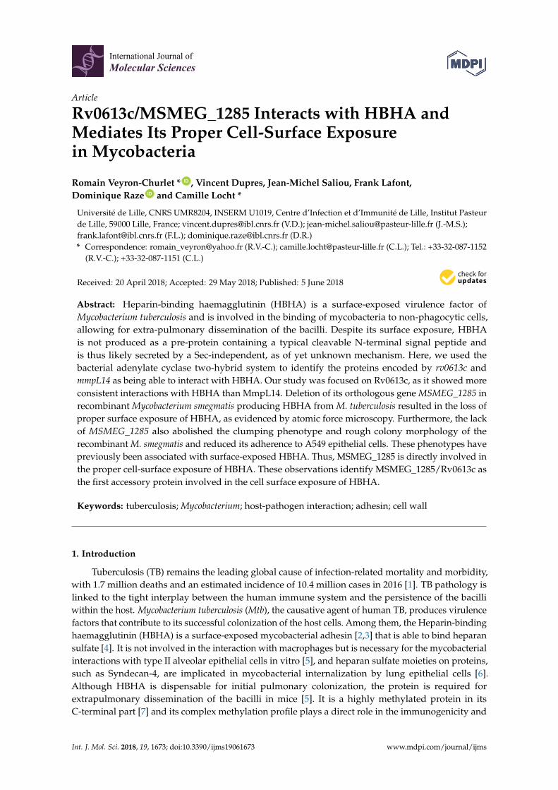
Rv0613c/MSMEG_1285 Interacts with HBHA andMediates Its Proper Cell-Surface Exposure in
MycobacteriaRomain Veyron-Churlet, Vincent Dupres, Jean-Michel Saliou, Frank Lafont,
Dominique Raze, Camille Locht
To cite this version:Romain Veyron-Churlet, Vincent Dupres, Jean-Michel Saliou, Frank Lafont, Dominique Raze, etal.. Rv0613c/MSMEG_1285 Interacts with HBHA and Mediates Its Proper Cell-Surface Expo-sure in Mycobacteria. International Journal of Molecular Sciences, MDPI, 2018, 19 (6), pp.1673.�10.3390/ijms19061673�. �hal-02347576�
International Journal of
Molecular Sciences
Article
Rv0613c/MSMEG_1285 Interacts with HBHA andMediates Its Proper Cell-Surface Exposurein Mycobacteria
Romain Veyron-Churlet * ID , Vincent Dupres, Jean-Michel Saliou, Frank Lafont,Dominique Raze ID and Camille Locht *
Université de Lille, CNRS UMR8204, INSERM U1019, Centre d’Infection et d’Immunité de Lille, Institut Pasteurde Lille, 59000 Lille, France; [email protected] (V.D.); [email protected] (J.-M.S.);[email protected] (F.L.); [email protected] (D.R.)* Correspondence: [email protected] (R.V.-C.); [email protected] (C.L.); Tel.: +33-32-087-1152
(R.V.-C.); +33-32-087-1151 (C.L.)
Received: 20 April 2018; Accepted: 29 May 2018; Published: 5 June 2018�����������������
Abstract: Heparin-binding haemagglutinin (HBHA) is a surface-exposed virulence factor ofMycobacterium tuberculosis and is involved in the binding of mycobacteria to non-phagocytic cells,allowing for extra-pulmonary dissemination of the bacilli. Despite its surface exposure, HBHAis not produced as a pre-protein containing a typical cleavable N-terminal signal peptide andis thus likely secreted by a Sec-independent, as of yet unknown mechanism. Here, we used thebacterial adenylate cyclase two-hybrid system to identify the proteins encoded by rv0613c andmmpL14 as being able to interact with HBHA. Our study was focused on Rv0613c, as it showed moreconsistent interactions with HBHA than MmpL14. Deletion of its orthologous gene MSMEG_1285 inrecombinant Mycobacterium smegmatis producing HBHA from M. tuberculosis resulted in the loss ofproper surface exposure of HBHA, as evidenced by atomic force microscopy. Furthermore, the lackof MSMEG_1285 also abolished the clumping phenotype and rough colony morphology of therecombinant M. smegmatis and reduced its adherence to A549 epithelial cells. These phenotypes havepreviously been associated with surface-exposed HBHA. Thus, MSMEG_1285 is directly involved inthe proper cell-surface exposure of HBHA. These observations identify MSMEG_1285/Rv0613c asthe first accessory protein involved in the cell surface exposure of HBHA.
Keywords: tuberculosis; Mycobacterium; host-pathogen interaction; adhesin; cell wall
1. Introduction
Tuberculosis (TB) remains the leading global cause of infection-related mortality and morbidity,with 1.7 million deaths and an estimated incidence of 10.4 million cases in 2016 [1]. TB pathology islinked to the tight interplay between the human immune system and the persistence of the bacilliwithin the host. Mycobacterium tuberculosis (Mtb), the causative agent of human TB, produces virulencefactors that contribute to its successful colonization of the host cells. Among them, the Heparin-bindinghaemagglutinin (HBHA) is a surface-exposed mycobacterial adhesin [2,3] that is able to bind heparansulfate [4]. It is not involved in the interaction with macrophages but is necessary for the mycobacterialinteractions with type II alveolar epithelial cells in vitro [5], and heparan sulfate moieties on proteins,such as Syndecan-4, are implicated in mycobacterial internalization by lung epithelial cells [6].Although HBHA is dispensable for initial pulmonary colonization, the protein is required forextrapulmonary dissemination of the bacilli in mice [5]. It is a highly methylated protein in itsC-terminal part [7] and its complex methylation profile plays a direct role in the immunogenicity and
Int. J. Mol. Sci. 2018, 19, 1673; doi:10.3390/ijms19061673 www.mdpi.com/journal/ijms
Int. J. Mol. Sci. 2018, 19, 1673 2 of 16
recognition of HBHA by T cells from Mtb-infected subjects [8]. An orthologous protein is also presentin fast-growing and non-pathogenic Mycobacterium smegmatis (Msmeg). However, HBHA_Msmeg is notinvolved in epithelial cell adhesion and does not display high affinity for heparin [9]. Thus, HBHAmost likely underwent a functional divergence during evolution [9,10]. Despite its cell surface exposurein Mtb [2], HBHA lacks a canonical signal peptide, usually required for protein transport throughbacterial plasma membranes. Thus, HBHA crosses the mycobacterial membrane by an unknownmechanism that is independent of the classical signal sequences peptides.
In this study, as an approach to unravel HBHA cell surface exposure, we identified the proteinsencoded by rv0613c and mmpL14, as the first proteins able to interact with HBHA. RecombinantMsmeg mc2155 expressing hbhA_Mtb was found to auto-aggregate, to grow as rough colonies andto infect epithelial cells, whereas the deletion of MSMEG_1285, the orthologous gene of rv0613c inMsmeg mc2155, abolished these phenotypes. Furthermore, HBHA_Mtb was detected by atomic forcemicroscopy at the surface of recombinant Msmeg mc2155 but not of the recombinant ∆MSMEG_1285mutant, indicating an important role for Rv0613c/MSMEG_1285 in the cell-surface exposure of HBHA.
2. Results
2.1. The N-Terminal Part of HBHA Interacts with Rv0613c and MmpL14
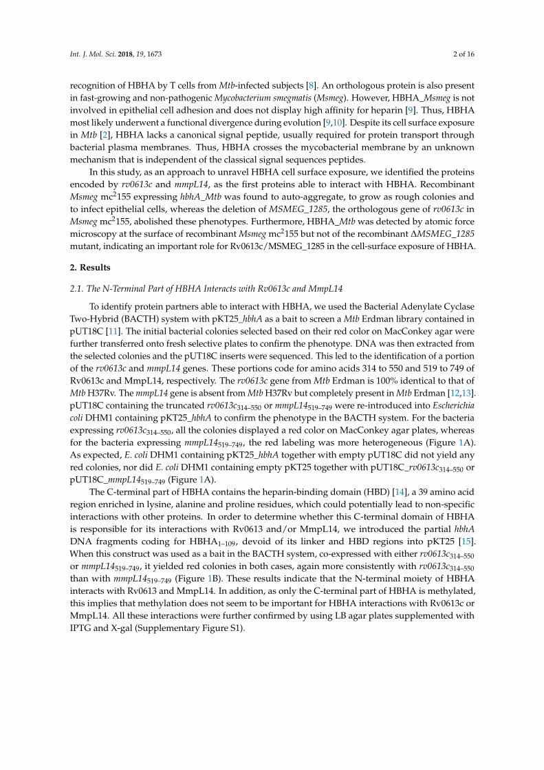
To identify protein partners able to interact with HBHA, we used the Bacterial Adenylate CyclaseTwo-Hybrid (BACTH) system with pKT25_hbhA as a bait to screen a Mtb Erdman library contained inpUT18C [11]. The initial bacterial colonies selected based on their red color on MacConkey agar werefurther transferred onto fresh selective plates to confirm the phenotype. DNA was then extracted fromthe selected colonies and the pUT18C inserts were sequenced. This led to the identification of a portionof the rv0613c and mmpL14 genes. These portions code for amino acids 314 to 550 and 519 to 749 ofRv0613c and MmpL14, respectively. The rv0613c gene from Mtb Erdman is 100% identical to that ofMtb H37Rv. The mmpL14 gene is absent from Mtb H37Rv but completely present in Mtb Erdman [12,13].pUT18C containing the truncated rv0613c314–550 or mmpL14519–749 were re-introduced into Escherichiacoli DHM1 containing pKT25_hbhA to confirm the phenotype in the BACTH system. For the bacteriaexpressing rv0613c314–550, all the colonies displayed a red color on MacConkey agar plates, whereasfor the bacteria expressing mmpL14519–749, the red labeling was more heterogeneous (Figure 1A).As expected, E. coli DHM1 containing pKT25_hbhA together with empty pUT18C did not yield anyred colonies, nor did E. coli DHM1 containing empty pKT25 together with pUT18C_rv0613c314–550 orpUT18C_mmpL14519–749 (Figure 1A).
The C-terminal part of HBHA contains the heparin-binding domain (HBD) [14], a 39 amino acidregion enriched in lysine, alanine and proline residues, which could potentially lead to non-specificinteractions with other proteins. In order to determine whether this C-terminal domain of HBHAis responsible for its interactions with Rv0613 and/or MmpL14, we introduced the partial hbhADNA fragments coding for HBHA1–109, devoid of its linker and HBD regions into pKT25 [15].When this construct was used as a bait in the BACTH system, co-expressed with either rv0613c314–550
or mmpL14519–749, it yielded red colonies in both cases, again more consistently with rv0613c314–550
than with mmpL14519–749 (Figure 1B). These results indicate that the N-terminal moiety of HBHAinteracts with Rv0613 and MmpL14. In addition, as only the C-terminal part of HBHA is methylated,this implies that methylation does not seem to be important for HBHA interactions with Rv0613c orMmpL14. All these interactions were further confirmed by using LB agar plates supplemented withIPTG and X-gal (Supplementary Figure S1).
Int. J. Mol. Sci. 2018, 19, 1673 3 of 16Int. J. Mol. Sci. 2018, 19, x FOR PEER REVIEW 3 of 16
Figure 1. HBHA interacts with Rv0613c and MmpL14 in the BACTH system. (A) Red colony phenotype observed on MacConkey agar plates for E. coli transformed with pKT25_hbhA and pUT18C_rv0613c314–550 (middle plate) in contrast to the E. coli transformed with pKT25_hbhA and pUT18C (left plate) or pKT25 and pUT18C_rv0613c314–550 (right plate); (B) Red colony phenotype observed on MacConkey agar plates for E. coli transformed with pKT25_hbhA and pUT18C_mmpL14519–749 (middle plate) in contrast to the E. coli transformed with pKT25_hbhA and pUT18C (left plate) or pKT25 and pUT18C_mmpL14519–749 (right plate); (C) Red colony phenotype observed on MacConkey agar plates for E. coli transformed with pKT25_hbhA1–109 and pUT18C_rv0613c314–550 or pUT18C_mmpL14519–749 (right plates) in contrast to the E. coli transformed with pKT25_hbhA1–109 and pUT18C (left plates).
2.2. Rv0613c Is Conserved within the Mtb Complex and Contains a SEC-C Motif
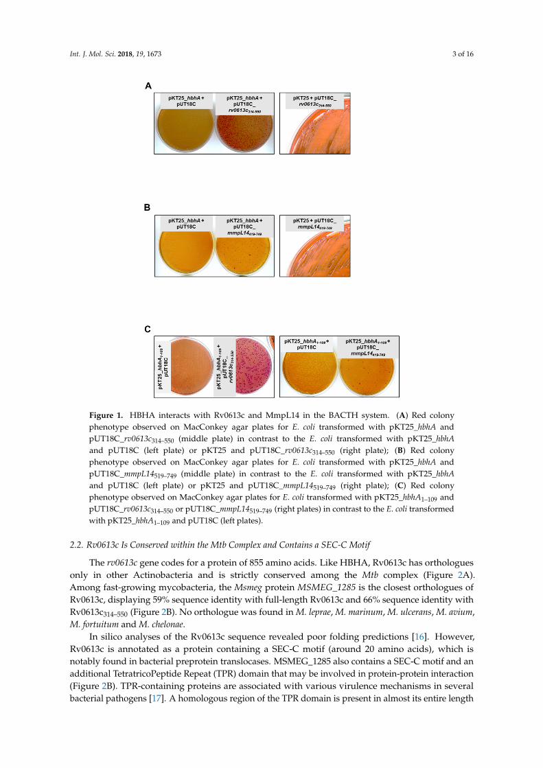
The rv0613c gene codes for a protein of 855 amino acids. Like HBHA, Rv0613c has orthologues only in other Actinobacteria and is strictly conserved among the Mtb complex (Figure 2A). Among fast-growing mycobacteria, the Msmeg protein MSMEG_1285 is the closest orthologues of Rv0613c, displaying 59% sequence identity with full-length Rv0613c and 66% sequence identity with Rv0613c314–550 (Figure 2B). No orthologue was found in M. leprae, M. marinum, M. ulcerans, M. avium, M. fortuitum and M. chelonae.
In silico analyses of the Rv0613c sequence revealed poor folding predictions [16]. However, Rv0613c is annotated as a protein containing a SEC-C motif (around 20 amino acids), which is notably found in bacterial preprotein translocases. MSMEG_1285 also contains a SEC-C motif and an additional TetratricoPeptide Repeat (TPR) domain that may be involved in protein-protein interaction (Figure 2B). TPR-containing proteins are associated with various virulence mechanisms in several bacterial pathogens [17]. A homologous region of the TPR domain is present in almost its entire length in Rv0613c314–550 (Figure 2B). Therefore, this region may potentially be involved in the
Figure 1. HBHA interacts with Rv0613c and MmpL14 in the BACTH system. (A) Red colonyphenotype observed on MacConkey agar plates for E. coli transformed with pKT25_hbhA andpUT18C_rv0613c314–550 (middle plate) in contrast to the E. coli transformed with pKT25_hbhAand pUT18C (left plate) or pKT25 and pUT18C_rv0613c314–550 (right plate); (B) Red colonyphenotype observed on MacConkey agar plates for E. coli transformed with pKT25_hbhA andpUT18C_mmpL14519–749 (middle plate) in contrast to the E. coli transformed with pKT25_hbhAand pUT18C (left plate) or pKT25 and pUT18C_mmpL14519–749 (right plate); (C) Red colonyphenotype observed on MacConkey agar plates for E. coli transformed with pKT25_hbhA1–109 andpUT18C_rv0613c314–550 or pUT18C_mmpL14519–749 (right plates) in contrast to the E. coli transformedwith pKT25_hbhA1–109 and pUT18C (left plates).
2.2. Rv0613c Is Conserved within the Mtb Complex and Contains a SEC-C Motif
The rv0613c gene codes for a protein of 855 amino acids. Like HBHA, Rv0613c has orthologuesonly in other Actinobacteria and is strictly conserved among the Mtb complex (Figure 2A).Among fast-growing mycobacteria, the Msmeg protein MSMEG_1285 is the closest orthologues ofRv0613c, displaying 59% sequence identity with full-length Rv0613c and 66% sequence identity withRv0613c314–550 (Figure 2B). No orthologue was found in M. leprae, M. marinum, M. ulcerans, M. avium,M. fortuitum and M. chelonae.
In silico analyses of the Rv0613c sequence revealed poor folding predictions [16]. However,Rv0613c is annotated as a protein containing a SEC-C motif (around 20 amino acids), which isnotably found in bacterial preprotein translocases. MSMEG_1285 also contains a SEC-C motif and anadditional TetratricoPeptide Repeat (TPR) domain that may be involved in protein-protein interaction(Figure 2B). TPR-containing proteins are associated with various virulence mechanisms in severalbacterial pathogens [17]. A homologous region of the TPR domain is present in almost its entire length
Int. J. Mol. Sci. 2018, 19, 1673 4 of 16
in Rv0613c314–550 (Figure 2B). Therefore, this region may potentially be involved in the HBHA-Rv0613cinteraction, although Rv0613c was not annotated as a protein containing such a domain, probably dueto some sequence divergence between Mtb H37Rv and Msmeg mc2155 in the N-terminal part of theTPR domain.
Int. J. Mol. Sci. 2018, 19, x FOR PEER REVIEW 4 of 16
HBHA-Rv0613c interaction, although Rv0613c was not annotated as a protein containing such a domain, probably due to some sequence divergence between Mtb H37Rv and Msmeg mc2155 in the N-terminal part of the TPR domain.
Figure 2. MSMEG_1285 is an orthologue of Rv0613c. (A) Phylogenetic tree representing Rv0613c homologues among different mycobacteria; (B) Sequence alignment of Rv0613c (first line) and MSMEG_1285 (second line) using Clustal Omega. The red rectangle corresponds to the SEC-C motif. The region interacting with HBHA of Rv0613c314–550 is shown in the green rectangles, and the TPR of MSMEG_1285 is shown in the blue rectangles.
2.3. Generation of a ΔMSMEG_1285 Mutant in Msmeg mc2155
To characterize the role of Rv0613c/MSMEG_1285, we used fast-growing Msmeg as a model system. A MSMEG_1285 deletion mutant (ΔMSMEG_1285) was generated in Msmeg mc2155, as well as its complemented derivative (Supplementary Figure S2A,B), proving that MSMEG_1285 is dispensable for Msmeg mc2155 growth in vitro. The absence of MSMEG_1285 and the complementation were verified using two different sera from Rv0613c-immunized mice (Supplementary Figure S2C). Moreover, the total amount of intracellular HBHA_Msmeg was not impacted by the deletion of MSMEG_1285 (Supplementary Figure S2D).
Figure 2. MSMEG_1285 is an orthologue of Rv0613c. (A) Phylogenetic tree representing Rv0613chomologues among different mycobacteria; (B) Sequence alignment of Rv0613c (first line) andMSMEG_1285 (second line) using Clustal Omega. The red rectangle corresponds to the SEC-C motif.The region interacting with HBHA of Rv0613c314–550 is shown in the green rectangles, and the TPR ofMSMEG_1285 is shown in the blue rectangles.
2.3. Generation of a ∆MSMEG_1285 Mutant in Msmeg mc2155
To characterize the role of Rv0613c/MSMEG_1285, we used fast-growing Msmeg as a modelsystem. A MSMEG_1285 deletion mutant (∆MSMEG_1285) was generated in Msmeg mc2155, as well asits complemented derivative (Supplementary Figure S2A,B), proving that MSMEG_1285 is dispensablefor Msmeg mc2155 growth in vitro. The absence of MSMEG_1285 and the complementation were
Int. J. Mol. Sci. 2018, 19, 1673 5 of 16
verified using two different sera from Rv0613c-immunized mice (Supplementary Figure S2C).Moreover, the total amount of intracellular HBHA_Msmeg was not impacted by the deletion ofMSMEG_1285 (Supplementary Figure S2D).
2.4. MSMEG_1285 Is Necessary for the Proper Cell-Surface Exposure of HBHA_Mtb
Atomic Force Microscopy (AFM) has been successfully used to examine the cell-surface exposureof HBHA in Mycobacterium bovis bacillus Calmette-Guérin (BCG) and its interaction with heparin coatedon the AFM tip [18]. HBHA is a highly abundant protein, with up to 1% of total soluble proteins of BCGPasteur. In contrast, HBHA_Msmeg is less abundant, explaining why the presence of an orthologousprotein in Msmeg remained elusive for years [9]. In addition, the affinity between HBHA_Msmeg andheparin is much lower than the affinity between HBHA_Mtb and heparin [9]. Therefore, to monitorHBHA interaction with heparin, we transformed Msmeg mc2155 and the ∆MSMEG_1285 withpMV361_hbhA_EGFP, allowing heterologous expression of hbhA_Mtb. The fusion with EGFP did notaffect cell surface-exposure of HBHA in BCG Pasteur [19] and was further used to distinguish betweenendogenous HBHA_Msmeg and the recombinant HBHA_Mtb.
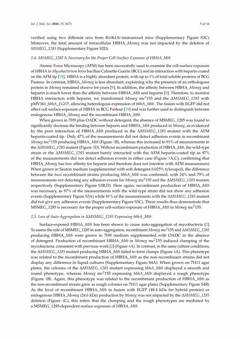
When grown in 7H9 plus OADC without detergent, the absence of MSMEG_1285 was found tosignificantly decrease the binding between heparin and HBHA_Mtb produced in Msmeg, as evidencedby the poor interaction of HBHA_Mtb produced in the ∆MSMEG_1285 mutant with the AFMheparin-coated tip. Only 47% of the measurements did not detect adhesion events in recombinantMsmeg mc2155 producing HBHA_Mtb (Figure 3B), whereas this increased to 91% of measurements inthe ∆MSMEG_1285 mutant (Figure 3D). Without recombinant production of HBHA_Mtb, the wild-typestrain or the ∆MSMEG_1285 mutant barely interacted with the AFM heparin-coated tip as 97%of the measurements did not detect adhesion events in either case (Figure 3A,C), confirming thatHBHA_Msmeg has low affinity for heparin and therefore does not interfere with AFM measurement.When grown in Sauton medium (supplemented with soft detergent 0.025% tyloxapol), the differencebetween the two recombinant strains producing hbhA_Mtb was confirmed, with 24% and 79% ofmeasurements not detecting any adhesion events for Msmeg mc2155 and the ∆MSMEG_1285 mutant,respectively (Supplementary Figure S3B,D). Here again, recombinant production of HBHA_Mtbwas necessary, as 97% of the measurements with the wild-type strain did not show any adhesionevents (Supplementary Figure S3A) while 81% of the measurements with the ∆MSMEG_1285 mutantdid not give any adhesion events (Supplementary Figure S3C). These results thus demonstrate thatMSMEG_1285 is necessary for the proper cell-surface exposure of HBHA_Mtb in Msmeg mc2155.
2.5. Loss of Auto-Aggregation in ∆MSMEG_1285 Expressing hbhA_Mtb
Surface-exposed HBHA_Mtb has been shown to cause auto-aggregation of mycobacteria [2].To assess the role of MSMEG_1285 in auto-aggregation, recombinant Msmeg mc2155 and ∆MSMEG_1285producing HBHA_Mtb were grown in 7H9 medium supplemented with OADC in the absenceof detergent. Production of recombinant HBHA_Mtb in Msmeg mc2155 induced clumping of themycobacteria, consistent with previous work [2] (Figure 4A). In contrast, in the same culture conditions,the ∆MSMEG_1285 mutant producing HBHA_Mtb failed to form clumps (Figure 4A). This phenotypewas related to the recombinant production of HBHA_Mtb as the non-recombinant strains did notdisplay any difference in liquid cultures (Supplementary Figure S4A). When grown on 7H11 agarplates, the colonies of the ∆MSMEG_1285 mutant expressing hbhA_Mtb displayed a smooth andround phenotype, whereas Msmeg mc2155 expressing hbhA_Mtb displayed a rough phenotype(Figure 4B). Again, this phenotype was related to the recombinant production of HBHA_Mtb asthe non-recombinant strains grew as rough colonies on 7H11 agar plates (Supplementary Figure S4B).As the level of recombinant HBHA_Mtb in fusion with EGFP (48.4 kDa for hybrid protein) orendogenous HBHA_Msmeg (24.6 kDa) production by Msmeg was not impacted by the ∆MSMEG_1285deletion (Figure 4C), this infers that the clumping and the rough phenotypes are mediated bya MSMEG_1285-dependent surface exposure of HBHA_Mtb.
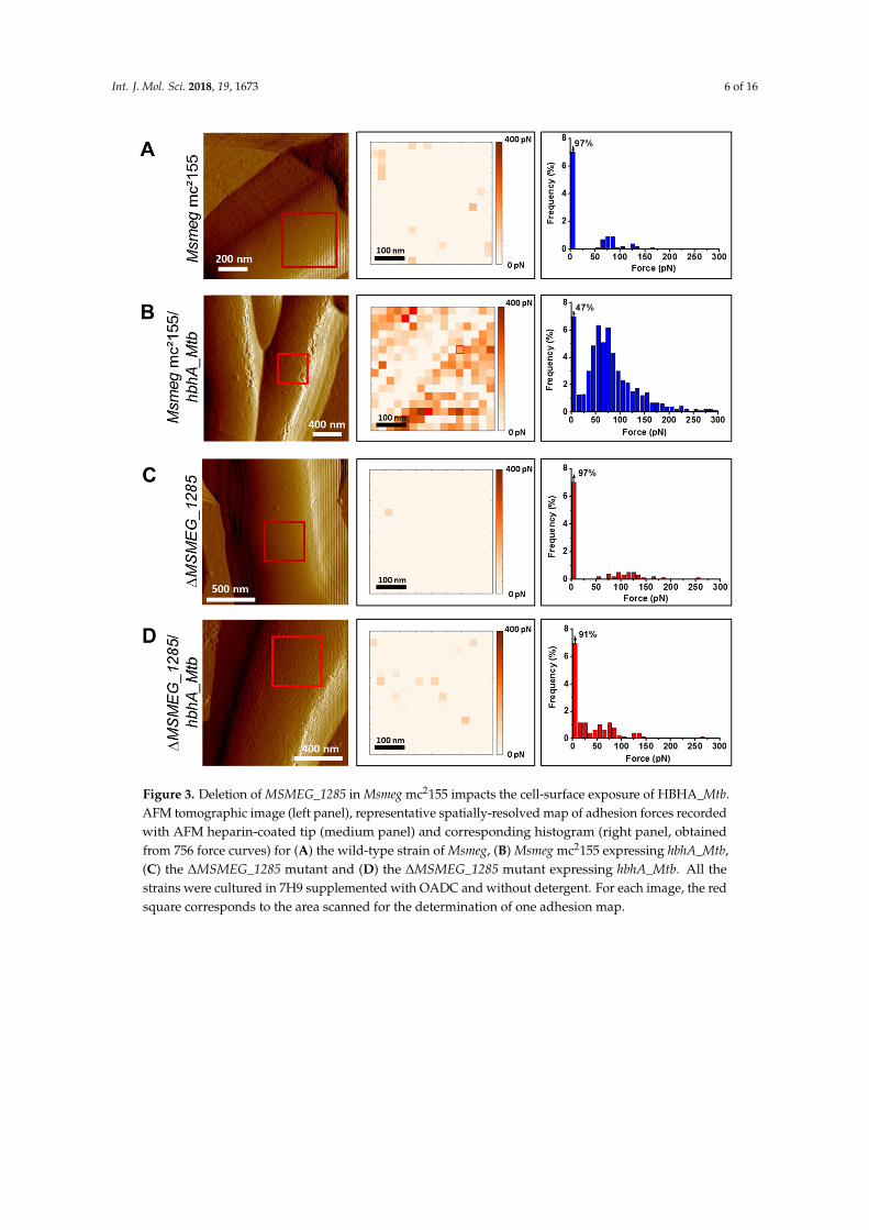
Int. J. Mol. Sci. 2018, 19, 1673 6 of 16Int. J. Mol. Sci. 2018, 19, x FOR PEER REVIEW 6 of 16
Figure 3. Deletion of MSMEG_1285 in Msmeg mc2155 impacts the cell-surface exposure of HBHA_Mtb. AFM tomographic image (left panel), representative spatially-resolved map of adhesion forces recorded with AFM heparin-coated tip (medium panel) and corresponding histogram (right panel, obtained from 756 force curves) for (A) the wild-type strain of Msmeg, (B) Msmeg mc2155 expressing hbhA_Mtb, (C) the ΔMSMEG_1285 mutant and (D) the ΔMSMEG_1285 mutant expressing hbhA_Mtb. All the strains were cultured in 7H9 supplemented with OADC and without detergent. For each image, the red square corresponds to the area scanned for the determination of one adhesion map.
Figure 3. Deletion of MSMEG_1285 in Msmeg mc2155 impacts the cell-surface exposure of HBHA_Mtb.AFM tomographic image (left panel), representative spatially-resolved map of adhesion forces recordedwith AFM heparin-coated tip (medium panel) and corresponding histogram (right panel, obtainedfrom 756 force curves) for (A) the wild-type strain of Msmeg, (B) Msmeg mc2155 expressing hbhA_Mtb,(C) the ∆MSMEG_1285 mutant and (D) the ∆MSMEG_1285 mutant expressing hbhA_Mtb. All thestrains were cultured in 7H9 supplemented with OADC and without detergent. For each image, the redsquare corresponds to the area scanned for the determination of one adhesion map.
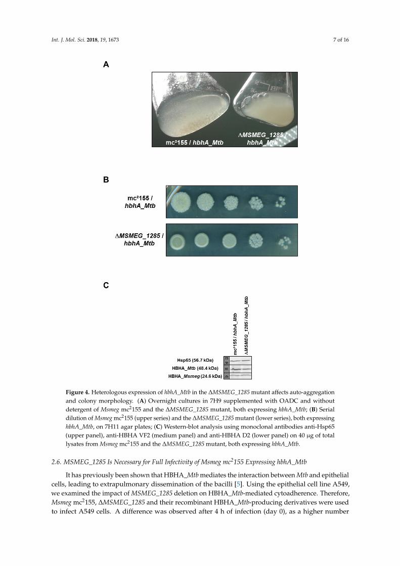
Int. J. Mol. Sci. 2018, 19, 1673 7 of 16Int. J. Mol. Sci. 2018, 19, x FOR PEER REVIEW 7 of 16
Figure 4. Heterologous expression of hbhA_Mtb in the ΔMSMEG_1285 mutant affects auto-aggregation and colony morphology. (A) Overnight cultures in 7H9 supplemented with OADC and without detergent of Msmeg mc2155 and the ΔMSMEG_1285 mutant, both expressing hbhA_Mtb; (B) Serial dilution of Msmeg mc2155 (upper series) and the ΔMSMEG_1285 mutant (lower series), both expressing hbhA_Mtb, on 7H11 agar plates; (C) Western-blot analysis using monoclonal antibodies anti-Hsp65 (upper panel), anti-HBHA VF2 (medium panel) and anti-HBHA D2 (lower panel) on 40 µg of total lysates from Msmeg mc2155 and the ΔMSMEG_1285 mutant, both expressing hbhA_Mtb.
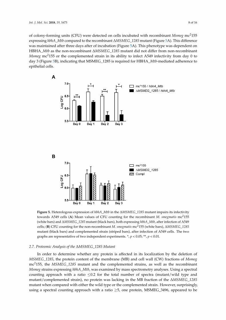
2.6. MSMEG_1285 Is Necessary for Full Infectivity of Msmeg mc2155 Expressing hbhA_Mtb
It has previously been shown that HBHA_Mtb mediates the interaction between Mtb and epithelial cells, leading to extrapulmonary dissemination of the bacilli [5]. Using the epithelial cell line A549, we examined the impact of MSMEG_1285 deletion on HBHA_Mtb-mediated cytoadherence. Therefore, Msmeg mc2155, ΔMSMEG_1285 and their recombinant HBHA_Mtb-producing derivatives were used to infect A549 cells. A difference was observed after 4 h of infection
Figure 4. Heterologous expression of hbhA_Mtb in the ∆MSMEG_1285 mutant affects auto-aggregationand colony morphology. (A) Overnight cultures in 7H9 supplemented with OADC and withoutdetergent of Msmeg mc2155 and the ∆MSMEG_1285 mutant, both expressing hbhA_Mtb; (B) Serialdilution of Msmeg mc2155 (upper series) and the ∆MSMEG_1285 mutant (lower series), both expressinghbhA_Mtb, on 7H11 agar plates; (C) Western-blot analysis using monoclonal antibodies anti-Hsp65(upper panel), anti-HBHA VF2 (medium panel) and anti-HBHA D2 (lower panel) on 40 µg of totallysates from Msmeg mc2155 and the ∆MSMEG_1285 mutant, both expressing hbhA_Mtb.
2.6. MSMEG_1285 Is Necessary for Full Infectivity of Msmeg mc2155 Expressing hbhA_Mtb
It has previously been shown that HBHA_Mtb mediates the interaction between Mtb and epithelialcells, leading to extrapulmonary dissemination of the bacilli [5]. Using the epithelial cell line A549,we examined the impact of MSMEG_1285 deletion on HBHA_Mtb-mediated cytoadherence. Therefore,Msmeg mc2155, ∆MSMEG_1285 and their recombinant HBHA_Mtb-producing derivatives were usedto infect A549 cells. A difference was observed after 4 h of infection (day 0), as a higher number
Int. J. Mol. Sci. 2018, 19, 1673 8 of 16
of colony-forming units (CFU) were detected on cells incubated with recombinant Msmeg mc2155expressing hbhA_Mtb compared to the recombinant ∆MSMEG_1285 mutant (Figure 5A). This differencewas maintained after three days after of incubation (Figure 5A). This phenotype was dependent onHBHA_Mtb as the non-recombinant ∆MSMEG_1285 mutant did not differ from non-recombinantMsmeg mc2155 or the complemented strain in its ability to infect A549 infectivity from day 0 today 3 (Figure 5B), indicating that MSMEG_1285 is required for HBHA_Mtb-mediated adherence toepithelial cells.
Int. J. Mol. Sci. 2018, 19, x FOR PEER REVIEW 8 of 16
(day 0), as a higher number of colony-forming units (CFU) were detected on cells incubated with recombinant Msmeg mc2155 expressing hbhA_Mtb compared to the recombinant ΔMSMEG_1285 mutant (Figure 5A). This difference was maintained after three days after of incubation (Figure 5A). This phenotype was dependent on HBHA_Mtb as the non-recombinant ΔMSMEG_1285 mutant did not differ from non-recombinant Msmeg mc2155 or the complemented strain in its ability to infect A549 infectivity from day 0 to day 3 (Figure 5B), indicating that MSMEG_1285 is required for HBHA_Mtb-mediated adherence to epithelial cells.
Figure 5. Heterologous expression of hbhA_Mtb in the ΔMSMEG_1285 mutant impairs its infectivity towards A549 cells (A) Mean values of CFU counting for the recombinant M. smegmatis mc2155 (white bars) and ΔMSMEG_1285 mutant (black bars), both expressing hbhA_Mtb, after infection of A549 cells; (B) CFU counting for the non-recombinant M. smegmatis mc2155 (white bars), ΔMSMEG_1285 mutant (black bars) and complemented strain (striped bars), after infection of A549 cells. The two graphs are representative of two independent experiments. *, p < 0.05; **, p < 0.01.
2.7. Proteomic Analysis of the ΔMSMEG_1285 Mutant
In order to determine whether any protein is affected in its localization by the deletion of MSMEG_1285, the protein content of the membrane (MB) and cell wall (CW) fractions of Msmeg mc2155, the MSMEG_1285 mutant and the complemented strains, as well as the recombinant Msmeg strains expressing hbhA_Mtb, was examined by mass spectrometry analyses. Using a spectral counting approach with a ratio ≤0.2 for the total number of spectra (mutant/wild type and mutant/complemented strain), no protein was lacking in the MB fraction of the ΔMSMEG_1285 mutant when compared with either the wild type or the complemented strain. However, surprisingly, using a spectral counting approach with a ratio ≥5, one protein, MSMEG_3496, appeared to be enriched in the MB fraction of the ΔMSMEG_1285 mutant in comparison with both
Figure 5. Heterologous expression of hbhA_Mtb in the ∆MSMEG_1285 mutant impairs its infectivitytowards A549 cells (A) Mean values of CFU counting for the recombinant M. smegmatis mc2155(white bars) and ∆MSMEG_1285 mutant (black bars), both expressing hbhA_Mtb, after infection of A549cells; (B) CFU counting for the non-recombinant M. smegmatis mc2155 (white bars), ∆MSMEG_1285mutant (black bars) and complemented strain (striped bars), after infection of A549 cells. The twographs are representative of two independent experiments. *, p < 0.05; **, p < 0.01.
2.7. Proteomic Analysis of the ∆MSMEG_1285 Mutant
In order to determine whether any protein is affected in its localization by the deletion ofMSMEG_1285, the protein content of the membrane (MB) and cell wall (CW) fractions of Msmegmc2155, the MSMEG_1285 mutant and the complemented strains, as well as the recombinantMsmeg strains expressing hbhA_Mtb, was examined by mass spectrometry analyses. Using a spectralcounting approach with a ratio ≤0.2 for the total number of spectra (mutant/wild type andmutant/complemented strain), no protein was lacking in the MB fraction of the ∆MSMEG_1285mutant when compared with either the wild type or the complemented strain. However, surprisingly,using a spectral counting approach with a ratio ≥5, one protein, MSMEG_3496, appeared to be
Int. J. Mol. Sci. 2018, 19, 1673 9 of 16
enriched in the MB fraction of the ∆MSMEG_1285 mutant in comparison with both the wild typeand complemented strains (Table 1). Fourteen spectra for MSMEG_3496 were detected in the mutant,whereas only one spectrum was detected for recombinant Msmeg mc2155 (Table 1). Thus, the absence ofMSMEG_1285 led to an enrichment of MSMEG_3496 in the Msmeg MB fraction. However, this tendencywas not confirmed in the CW fraction of recombinant strains (Table 1).
The total number of spectra for HBHA_Mtb was similar between recombinant Msmeg mc2155and ∆MSMEG_1285 mutant (Table 1), confirming that the phenotypes observed above were not dueto differences in protein content of the MB or CW fractions, but were due to differences in HBHAcell-surface exposure between Msmeg mc2155 and the MSMEG_1285-deficient mutant. As expected,MSMEG_1285 was not detected in the ∆MSMEG_1285 mutant.
Table 1. Total number of spectra (and unique peptides) for each protein in Membrane (MB) and CellWall (CW) fractions.
Protein MW Fraction mc2155 ∆MSMEG_1285 Complemented mc2155/hbhA_Mtb ∆MSMEG_1285/hbhA_Mtb
MSMEG_3496 106210MB 1 (1) 5 (4) 1 (1) 1 (1) 14 (12)
CW 4 (3) 9 (6) 4 (3) 48 (22) 15 (10)
MSMEG_1285 88003MB 1 (1) 0 4 (3) 3 (3) 0
CW 1 (1) 0 11 (8) 2 (2) 0
MSMEG_0919(HBHA_Msmeg) 24562
MB 28 (12) 37 (13) 27 (11) 13 (7) 23 (11)
CW 15 (9) 16 (10) 16 (7) 7 (5) 13 (8)
Rv0475_EGFP(HBHA_Mtb) 48472
MB 0 0 0 17 (14) 14 (10)
CW 0 0 0 4(4) 5 (4)
3. Discussion
Considering the importance of HBHA in Mtb pathogenesis [5], unraveling its biosynthesis and itsfunction is a crucial step in understanding the ability of the bacilli to interact with epithelial cells andto undergo extrapulmonary dissemination. HBHA was previously detected at the cell-surface of thebacilli [2], although it lacks a canonical signal peptide for transport. Here, we describe Rv0613c andMmpL14 as the first proteins identified to interact with HBHA. Using the BACTH system, the HBHAinteraction with Rv0613c was consistently strong, whereas the interaction was less consistent withMmpL14, as the red phenotype displayed by E. coli cells on McConkey agar plates in the BACTHsystem appeared to be more heterogeneous, in comparison with the HBHA-Rv0613c314–550 interaction.However, this heterogeneity could be the consequence of using a truncated version of MmpL14(i.e., MmpL14519–749) in BACTH system, and other portions of MmpL14 may be needed to retain fullinteraction with HBHA. Therefore, we focused this study on Rv0613c, for which we could identify itshomolog in Msmeg as MSMEG_1285.
HBHA has several functional domains, the best studied of which is the C-terminal domain,containing several lysine-proline-alanine-rich repeats. This domain has been shown to be involvedin mycobacterial binding to epithelial cells [2,20]. The BACTH studies described here show that itis not involved in binding to Rv0613c, since BACTH experiments done with a truncated version ofHBHA, lacking this C-terminal domain, gave the same results as those obtained with full-lengthHBHA (Figure 1A,B). This implies that the HBHA-Rv0613c interaction is not based merely on ionicinteractions of the positively charged C-terminal domain of HBHA with Rv0613c, which could lackspecificity. It rather suggests a more specific protein-protein interaction. As HBHA is an intrinsicallydisordered protein [15], it may therefore be possible that its interaction with Rv0613c induces a specificconformation of HBHA enabling it to be properly exposed and oriented at the bacterial surface and tocarry out its biological function as an adhesin and a bacterial clumping factor.
Both Rv0613c and MSMEG_1285 contain a SEC-C motif (Figure 2B), notably found in bacterialpreprotein translocases. It is therefore tempting to speculate that Rv0613c and MSMEG_1285 fulfill a rolesimilar to that of the preprotein translocase SecA and the chaperone SecB in order to facilitate HBHA
Int. J. Mol. Sci. 2018, 19, 1673 10 of 16
export through the mycobacterial cytoplasmic membrane and HBHA cell surface exposure. Accordingly,the deletion of MSMEG_1285 led to the impairment of the proper cell surface exposure of recombinantHBHA_Mtb produced in Msmeg, as evidenced by AFM (Figure 3, Supplementary Figure S3).The deletion also led to a decrease in mycobacterial auto-aggregation by the HBHA_Mtb-producingMsmeg strain (Figure 4A), to a smooth colony phenotype when grown on Middlebrook 7H11 agarplates (Figure 4B) and to a decrease in infectivity of A549 cells (Figure 5A). Native HBHA has also beenreported to form homodimers, and it was previously suggested that the HBHA trans-dimerizationis responsible for bacterial agglutination [21]. Therefore, a defect in HBHA cell surface exposureand orientation would directly lead to the loss of the auto-aggregation, to a smooth phenotype,and a decrease in infectivity. Importantly, the formation of large mycobacterial aggregates has beenreported to increase cytotoxicicty and to enhance effective killing of host cells after macrophageinternalization, highlighting the importance of aggregation state in Mtb virulence [22].
Although Rv0613c has no significant predicted structural similarities with hydrolases orpeptidases, it was recently shown to display protease activity, as evidenced by its ability to cleaveβ–casein [16]. It was suggested that either Rv0613c is a divergent protease or that its protease activity isa moonlighting function [16]. However, we have no evidence that Rv0613c exerts a protease activity onHBHA. Interestingly, the proteases tested by Ortega et al. [16], Rv0525 and Rv1192, with a molecularweight of 22 and 30 kDa, respectively, are three- to four-fold smaller than the 93-kDa Rv0613c,suggesting that Rv0613c may harbor several subdomains with different activities. In the BACTHassay, the portion of Rv0613c (Rv0613c314–550) interacting with HBHA represents only one quarterof the entire Rv0613c protein. Thus, the protease subdomain of Rv0613c could be distinct from theHBHA-interacting subdomain. It remains to be determined whether the protease activity of Rv0613cplays any role in HBHA maturation process.
In addition to the SEC-C motif, MSMEG_1285 also contains a TPR domain. In spite of somedivergence in the N-terminal part, the C-terminal part of the MSMEG_1285 TPR domain is very similarto the corresponding sequence of Rv0613c (Figure 2B), strongly suggesting that Rv0613c also containsa TPR domain, although it was originally not annotated as such in protein databases. However,the web-based TPRpred tool (Available online: https://toolkit.tuebingen.mpg.de/#/tools/tprpred)predicts that 364VAVRW . . . MDTEW397 and 398PLPLL . . . EPDHP431 of Rv0613c are TPR peptideswith a p-value of 1.6 × 10⁻⁵ and 4.1 × 10⁻⁴, respectively. This portion of Rv0613c is also present inthe fragment identified by the BACTH system to interact with HBHA. TPR domains are known tomediate protein-protein interactions and TPR-containing proteins are often involved in the biogenesisof bacterial secreted or surface proteins [17]. For example, the assembly machinery of the β-barrelouter membrane protein of E. coli consists of a complex of five proteins, one of which, named BamD,contains 5 TPR domains [23]. This protein binds to unfolded outer membrane proteins prior to foldingand insertion into the outer membrane [24,25]. ClustalO predicts 21% of sequence identity between theTPR1 of BamD and Rv0613c471–546 (Supplementary Figure S5), which is contained within the portion ofRv0613c that interacts with HBHA in the BACTH system. Similarly, the TPR-containing protein TprAfrom Porphyromonas gingivalis is also involved in the outer-membrane display of the TapA protein,most likely via protein-protein interaction involving the TPR domain of TprA and this mechanismis required to maintain full infectivity of epithelial cells by P. gingivalis [26]. Thus, it is tempting tospeculate that a similar mechanism, involving protein-protein interactions with the TPR domain ofRv0613c, may be at play to address HBHA to the mycobacterial cell surface in its proper orientation.Interestingly, both SEC-C motif- and TPR domain-containing proteins are enriched in the pathogenicMtb clade in comparison with non-pathogenic mycobacteria [27], which suggests their involvement invirulence traits.
It was of interest to investigate whether the sub-cellular localization of proteins was also affectedby the deletion of MSMEG_1285. Surprisingly, an overrepresentation (cut-off ≥ 5) of a single Msmegprotein, MSMEG_3496, was seen in the MB fraction of the mutant as compared to Msmeg mc2155.This protein is one of the orthologues of MmpL5 (Rv0676c) in Mtb. It is intriguing that the second
Int. J. Mol. Sci. 2018, 19, 1673 11 of 16
protein, MmpL14, found here to interact with HBHA in the BACTH system is also a member of theMmpL family. The mmpL14 gene is part of the RvD2 deleted region and is absent in Mtb H37Rv [12,13],but conserved among the majority of Mtb strains, including CDC1551 (locus MT1802) and H37Ra [28,29],as well as most of the sequenced Mtb clinical isolates. The MmpL protein family has been shown to beinvolved in substrate transport, such as the transport of lipids through the mycobacterial membrane,siderophore export and iron acquisition, and drug efflux [30,31]. MmpL14519–749 comprises the completedocking subdomain DC as well as part of the porter subdomains PC1 and PC2, making this partof the protein accessible for interactions with surface-exposed proteins. The periplasmic dockingsubdomain DC was suggested to play a role in protein-protein interactions [32], consistent withour findings on its interaction with HBHA. Moreover, the rough to smooth transition phenotypecannot be reduced only to the impairment of HbhA transport. Interestingly, Bernut and coworkersreported that a Y842H mutation in M. bolletii MmpL4a was involved in the smooth to rough transitionphenotype [33]. The authors proposed that this single key mutation has an impact of the 3D structureof the protein, affecting the proton motive force. Considering the interaction of HBHA with MmpL14,the rough to smooth transition phenotype could be an indirect consequence of the improper HBHA cellsurface exposure, also compromising the structural organization and/or exposure of MmpL proteins.In addition, as MmpL proteins are key players in the transport of lipids, the authors suggested thata functional link may exist between MmpL4a and the GPL biosynthesis [33]. We can therefore notexclude link between HBHA and the MmpL proteins during the biogenesis or function of HBHA.Future work will address this issue.
4. Materials and Methods
4.1. Bacterial Strains and Media
E. coli TOP10 (Invitrogen, Carlsbad, CA, USA) was used for cloning and E. coli DHM1 wasused for BACTH assays (Table S1). The E. coli strains were grown in LB medium (MP Biomedicals,Illkirch, France) at 37 ◦C supplemented with ampicillin (100 µg·mL−1), hygromycin (50 µg·mL−1) orkanamycin (25 µg·mL−1), when required. Msmeg mc2155, the deletion mutant ∆MSMEG_1285 and thecorresponding complemented strain (Table S1) were grown in Sauton’s medium (containing 0.025%tyloxapol), in Middlebrook 7H9 broth or on 7H11 agar plates supplemented with OADC enrichment(Becton Dickinson, Le Pont-de-Claix, France), hygromycin (50 µg·mL−1) or kanamycin (25 µg·mL−1),when required.
4.2. Generation of the ∆MSMEG_1285 Mutant in Msmeg mc2155
To generate the ∆MSMEG_1285 mutant, we followed the protocol previously described by vanKessel et al. [34]. Briefly, upstream and downstream regions of MSMEG_1285 were amplified fromMsmeg mc2155 genomic DNA using primers [Up_MSMEG1285_dir and Up_MSMEG1285_rev] and[Down_MSMEG1285_dir and Down_MSMEG1285_rev], respectively (Table S2). These upstream anddownstream fragments were inserted at either side of a hygromycin-resistance cassette in pJSC347(Table S1). After digestion by SpeI and StuI, the allelic exchange substrate was electroporated intocompetent Msmeg mc2155 containing pJV53 (Table S1), following induction by 0.2% acetamidefor 24 h. Several hygromycin-resistant clones were selected and tested for the insertion of thehygromycin-resistance cassette by PCR using primers Seq_MSMEG1285_1, Seq_MSMEG1285_2 andSeq_MSMEG1285_3 (Supplementary Figure S2A,B and Table S2). The resulting PCR fragments werefurther checked by sequencing to confirm the insertion at the expected Msmeg mc2155 locus.
4.3. Plasmid Constructions
Different portions of hbhA were amplified by PCR using MtbH37Rv genomic DNA and the pair ofprimers [pKT25_hbhA_dir and pKT25_hbhA_rev] or [pKT25_hbhA_dir and pKT25_hbhA1–109_rev](Table S2). The PCR fragments were digested with BamHI and KpnI and ligated into pKT25
Int. J. Mol. Sci. 2018, 19, 1673 12 of 16
digested with the same enzymes, thereby generating pKT25_hbhA and pKT25_hbhA1–109 (Table S1).The MSMEG_1285 gene was amplified by PCR using Msmeg mc2155 genomic DNA and the pair ofprimers [pVV16_MSMEG1285_dir and pVV16_MSMEG1285_rev] (Table S2). The PCR fragment wasdigested with NdeI and HindIII and ligated into pVV16 digested with the same enzymes, therebygenerating pVV16_MSMEG_1285 (Table S1). All plasmids were checked by sequencing.
4.4. Bacterial Adenylate Cyclase Two-Hybrid Assays
One hundred ng of the Mtb Erdman library contained in pUT18C was electroporated intoE. coli DHM1 containing pKT25_hbhA. Transformants were spread onto MacConkey agar (BectonDickinson) plates supplemented with 100 µg·mL−1 ampicillin, 25 µg·mL−1 kanamycin, 0.5 mM IPTGand 1% maltose. After 4 to 6 days at 30 ◦C, clones were selected based on their red color and transferredonto fresh MacConkey agar plates to confirm the red phenotype. DNA was extracted from the selectedcolonies and pUT18C inserts were sequenced using the Seq_pUT18C_dir primer (Table S2). To confirmthe phenotype, pUT18C containing rv0613c314–550 was re-introduced into E. coli DHM1 containingeither pKT25_hbhA or pKT25_hbhA1–109. Transformants were spread onto both MacConkey agar platesand LB agar plates supplemented with 100 µg·mL−1 ampicillin, 25 µg·mL−1 kanamycin, 0.5 mM IPTGand 50 µg·mL−1 X-gal and incubated for 3 days at 30 ◦C.
4.5. Fractionation Protocol
Msmeg strains were disrupted in a French pressure cell and the lysates were centrifuged at 3000× gfor 10 min at 4 ◦C to eliminate cellular debris and non-lysed cells. The supernatants were pelleted at27,000× g for 30 min at 4 ◦C in order to collect cell-wall fractions (CW). The remaining supernatantswere centrifuged at 100,000× g for 2 h at 4 ◦C to separate the membrane fractions (MB) from cytosolicfractions. Each fraction was resuspended in an appropriate volume of Laemmli buffer.
4.6. Immunoblot Analysis
Proteins were resolved by SDS-PAGE using 12% acrylamide gels and then electro-transferredonto a nitrocellulose membrane. The membrane was saturated with Tris-Buffered Saline (pH 7.5, TBS)containing 0.05% Tween 80 and 5% milk and probed overnight at 4 ◦C with anti-HBHA monoclonalantibody VF2 or D2 diluted 1:100 in TBS-Tween-3% milk or sera from Rv0613c-immunized mice diluted1:100 in TBS-Tween-3% milk or anti-Hsp65 monoclonal antibody (Abcam, Cambridge, UK) diluted1:1000 in TBS-Tween-3% milk. Finally, the membrane was incubated for 1 h at room temperature withgoat anti-mouse horseradish peroxidase-conjugated secondary antibodies (Abcam), diluted 1:5000 inTBS-Tween-3% milk. Detection was performed using the Amersham ECL Prime Western-BlottingDetection Reagent (Fisher Scientific, Illkirch, France) and chemiluminescence was detected using theAmersham Imager 600 (GE Healthcare, Uppsala, Sweden).
4.7. Atomic Force Microscopy
Gold-coated cantilevers were cleaned with plasma oxygen, rinsed with ethanol, dried witha gentle nitrogen flow and immersed overnight at room temperature in a 25 µg·mL−1 solution ofbiotinylated bovine serum albumin (BBSA) (Sigma, Saint-Quentin-Fallavier, France) in PBS. Followingrinsing with PBS, the BBSA surfaces were exposed to a 10 µg·mL−1 solution of streptavidin (Sigma)in PBS for 2 h followed by thorough rinsing with PBS. Finally, the BBSA/streptavidin surfaceswere immersed for 2 h in PBS containing 10 µg·mL−1 biotinylated heparin (Sigma) and rinsed withPBS. The mycobacteria were immobilized on porous polycarbonate membranes with a 3 µm poresize (Merck Millipore, Darmstadt, Germany). After filtering a concentrated bacterial suspension,the membrane was transferred to a Petri dish containing the imaging solution and gently agitated toremove non-immobilized bacteria. Then, a small piece of the membrane (1 cm × 1 cm square) was cutand the bottom side was quickly dried using precision wipes before being attached to a steel samplepuck using a small piece of double side adhesive tape. The mounted samples were transferred into the
Int. J. Mol. Sci. 2018, 19, 1673 13 of 16
AFM liquid cell while avoiding dewetting. AFM contact mode images and force-distance curves wereobtained using a MultiMode 8 AFM (Bruker, Santa Barbara, CA, USA). Measurements were performedin PBS at room temperature using gold-coated Silicon Nitride cantilevers (OMCL-TR400PB, OlympusLtd., Tokyo, Japan). The spring constants of the cantilevers were measured using the thermal noisemethod (NanoScope Analysis software version 8.15, Bruker), yielding values ranging from 0.022 to0.026 N·m−1. All force measurements were performed using a constant approach and retraction speedof 1500 nm·s−1, and with an interaction time of 500 ms. Adhesion maps were obtained by recording16 × 16 force curves on 400 nm × 400 nm areas of the cells, calculating the adhesion force values anddisplaying them as grey pixels. Histograms were obtained by pooling the data from several adhesionmaps. Data analysis was done using in-house developed pyAF (python Atomic Force) software,version 1.5.1. All experiments were performed at least on four different bacteria and with two differentheparin-coated AFM tips.
4.8. Infection of A549 Cells
One day prior to infection, 2 × 10⁵ A549 cells per well resuspended in DMEM containing 10%Fetal Calf Serum (FCS) were seeded in 24-well plates and incubated overnight at 37 ◦C under 5% CO2.Cultures of Msmeg mc2155, ∆MSMEG_1285 and their recombinant HBHA_Mtb-producing derivativeswere grown overnight at 37 ◦C with shaking. Bacteria were washed twice in PBS, resuspended inDMEM containing 10% FCS, treated for 15 min in a sonication bath and centrifuged for 2 min at 120× gto eliminate remaining clumps. After two washes with PBS, A549 cells were infected with bacterialsuspensions at an MOI of 50 and further incubated for 4 h at 37 ◦C under 5% CO2. After 4 h, the cellswere extensively washed 5 times with PBS and either resuspended in DMEM containing 10% FCS orlysed with 250 µL of 0.1% Triton X-100. Serial dilutions of the lysates were plated onto 7H11 agar platesand CFU were counted after 3 days of incubation at 37 ◦C. At each time point, the remaining cells werewashed 5 times in PBS and resuspended in fresh DMEM containing 10% FCS or, alternatively, lysedand CFU counts determined similarly. Each measure was done in triplicate and each experiment wasdone independently at least twice.
4.9. Mass Spectrometry Proteomic Analysis
After heating at 100 ◦C in 5% SDS, 5% β-mercaptoethanol, 1 mM EDTA, 10% glycerol, 10 mMTris-HCl (pH 8) for 3 min, protein samples were fractionated by SDS-PAGE on a 10% acrylamidegel. The electrophoretic migration was stopped as soon as the protein sample entered 0.3 cm intothe separating gel. The gel was briefly stained with Coomassie Blue, and one band, containing theentire sample, was cut. In-gel digestion of gel slices was performed as previously described [35].The UltiMate 3000 RSLCnano System (Fisher Scientific) was used for the separation of the proteindigests. Peptides were automatically fractionated onto a commercial C18 reversed phase column(75 µm × 250 mm, 2 µm particle, PepMap100 RSLC column, Thermo Fisher Scientific, at 35 ◦C).Trapping was performed during 4 min at 5 µL·min−1, with solvent A (98% H2O, 2% acetonitrile and0.1% formic acid). The peptides were eluted using two solvents A (0.1% formic acid in water) andB (0.1% formic acid in acetonitrile) at a flow rate of 300 nL·min−1. Gradient separation was 3 minat 3% B, 110 min from 3% B to 20% B, 10 min from 20% B to 80% B and maintained for 15 min at80% B. The column was equilibrated for 6 min with 3% buffer B prior to the next sample analysis.Eluted peptides from the C18 column were analyzed by Q-Exactive instruments (Fisher Scientific).The electrospray voltage was 1.9 kV, and the capillary temperature was 275 ◦C. Full MS scans wereacquired in the Orbitrap mass analyzer over m/z 400–1200 range with a 70,000 (m/z 200) resolution.The target value was 3 × 10⁶. Fifteen most intense peaks with charge state between 2 and 5 werefragmented in the higher-energy collision-activated dissociation cell with normalized collision energyof 27%, and tandem mass spectrum was acquired in the Orbitrap mass analyzer with a 17,500 (m/z 200)resolution. The target value was 1 × 10⁵. The ion selection threshold was 5 × 10⁴ counts, and the
Int. J. Mol. Sci. 2018, 19, 1673 14 of 16
maximum allowed ion accumulation times were 250 ms for full MS scans and 100 ms for tandem massspectrum. Dynamic exclusion was set to 30 s.
4.10. Proteomic Data Analysis
Raw data collected during nanoLC-MS/MS analyses were processed and converted into a *.mgfpeak list format with Proteome Discoverer 1.4 (Fisher Scientific). MS/MS data were analyzed usingsearch engine Mascot (version 2.4.0, Matrix Science, London, UK) installed on a local server. Searcheswere performed with a tolerance on mass measurement of 0.2 Da for precursor and 0.2 Da for fragmentions, against a composite target-decoy database (17,848 total entries) built with a Msmeg UniProtdatabase (strain ATCC 700084/mc (2)155, taxo 246196, October 2014, 8805 entries) fused with thesequences of HBHA_EGFP, recombinant trypsin and a list of classical contaminants (119 entries).Cysteine carbamidomethylation, methionine oxidation, protein N-terminal acetylation and cysteinepropionamidation were searched as variable modifications. Up to one missed trypsin cleavage wasallowed. For each sample, peptides were filtered out according to the cutoff set for protein hits with1 or more peptides larger than 8 residues, ion score >25, identity score >6, corresponding to a 1% falsepositive rate.
4.11. Statistical Analysis
For infection of A549 cells by the Msmeg strains, the statistical significance (p-value) was testedwith a two-tailed unpaired t-test (GraphPad Prism version 5.04, GraphPad Software, CA, USA) andp-values < 0.05 were considered as significant.
Supplementary Materials: Supplementary materials can be found at http://www.mdpi.com/1422-0067/19/6/1673/s1.
Author Contributions: R.V.-C. and D.R. conceived and designed the study. R.V-C., V.D., J.-M.S. and F.L. acquired,analyzed and interpreted the data. R.V.-C. and C.L. wrote the manuscript.
Acknowledgments: We thank Alain Baulard (Institut Pasteur de Lille, France) for kindly providing the MtbErdman library in pUT18C vector and Carine Rouanet (Institut Pasteur de Lille, France) for providing the serafrom Rv0613c-immunized mice. We acknowledge the BioImaging Center Lille (BICeL) for access to equipment.This work has been supported by ANR 10-EQPX-04-01 and FEDER 12,001,407 to BICeL. This work was supportedin part by the European Commission Horizon 2020 (TBVAC2020, H2020-PHC-643381).
Conflicts of Interest: The authors declare no conflict of interest.
References
1. WHO. World Health Organization Global Tuberculosis Report; WHO: Geneva, Switzerland, 2017.2. Menozzi, F.D.; Rouse, J.H.; Alavi, M.; Laude-Sharp, M.; Muller, J.; Bischoff, R.; Brennan, M.J.; Locht, C.
Identification of a heparin-binding hemagglutinin present in mycobacteria. J. Exp. Med. 1996, 184, 993–1001.[CrossRef] [PubMed]
3. Menozzi, F.D.; Bischoff, R.; Fort, E.; Brennan, M.J.; Locht, C. Molecular characterization of the mycobacterialheparin-binding hemagglutinin, a mycobacterial adhesin. Proc. Natl. Acad. Sci. USA 1998, 95, 12625–12630.[CrossRef] [PubMed]
4. Huang, T.Y.; Irene, D.; Zulueta, M.M.; Tai, T.J.; Lain, S.H.; Cheng, C.P.; Tsai, P.X.; Lin, S.Y.; Chen, Z.G.;Ku, C.C.; et al. Structure of the Complex between a Heparan Sulfate Octasaccharide and MycobacterialHeparin-Binding Hemagglutinin. Angew. Chem. Int. Ed. Engl. 2017, 56, 4192–4196. [CrossRef] [PubMed]
5. Pethe, K.; Alonso, S.; Biet, F.; Delogu, G.; Brennan, M.J.; Locht, C.; Menozzi, F.D. The heparin-bindinghaemagglutinin of M. tuberculosis is required for extrapulmonary dissemination. Nature 2001, 412, 190–194.[CrossRef] [PubMed]
6. Zimmermann, N.; Saiga, H.; Houthuys, E.; Moura-Alves, P.; Koehler, A.; Bandermann, S.; Dorhoi, A.;Kaufmann, S.H. Syndecans promote mycobacterial internalization by lung epithelial cells. Cell. Microbiol.2016, 18, 1846–1856. [CrossRef] [PubMed]
Int. J. Mol. Sci. 2018, 19, 1673 15 of 16
7. Pethe, K.; Bifani, P.; Drobecq, H.; Sergheraert, C.; Debrie, A.S.; Locht, C.; Menozzi, F.D. Mycobacterialheparin-binding hemagglutinin and laminin-binding protein share antigenic methyllysines that conferresistance to proteolysis. Proc. Natl. Acad. Sci. USA 2002, 99, 10759–10764. [CrossRef] [PubMed]
8. Temmerman, S.; Pethe, K.; Parra, M.; Alonso, S.; Rouanet, C.; Pickett, T.; Drowart, A.; Debrie, A.S.; Delogu, G.;Menozzi, F.D.; et al. Methylation-dependent T cell immunity to Mycobacterium tuberculosis heparin-bindinghemagglutinin. Nat. Med. 2004, 10, 935–941. [CrossRef] [PubMed]
9. Biet, F.; Angela de Melo Marques, M.; Grayon, M.; Xavier da Silveira, E.K.; Brennan, P.J.; Drobecq, H.;Raze, D.; Vidal Pessolani, M.C.; Locht, C.; Menozzi, F.D. Mycobacterium smegmatis produces an HBHAhomologue which is not involved in epithelial adherence. Microbes Infect. 2007, 9, 175–182. [CrossRef][PubMed]
10. Lanfranconi, M.P.; Alvarez, H.M. Functional divergence of HBHA from Mycobacterium tuberculosis and itsevolutionary relationship with TadA from Rhodococcus opacus. Biochimie 2016, 127, 241–248. [CrossRef][PubMed]
11. Karimova, G.; Pidoux, J.; Ullmann, A.; Ladant, D. A bacterial two-hybrid system based on a reconstitutedsignal transduction pathway. Proc. Natl. Acad. Sci. USA 1998, 95, 5752–5756. [CrossRef] [PubMed]
12. Gordon, S.V.; Brosch, R.; Billault, A.; Garnier, T.; Eiglmeier, K.; Cole, S.T. Identification of variable regions inthe genomes of tubercle bacilli using bacterial artificial chromosome arrays. Mol. Microbiol. 1999, 32, 643–655.[CrossRef] [PubMed]
13. Domenech, P.; Reed, M.B.; Barry, C.E., 3rd. Contribution of the Mycobacterium tuberculosis MmpL proteinfamily to virulence and drug resistance. Infect. Immun. 2005, 73, 3492–3501. [CrossRef] [PubMed]
14. Pethe, K.; Aumercier, M.; Fort, E.; Gatot, C.; Locht, C.; Menozzi, F.D. Characterization of the heparin-bindingsite of the mycobacterial heparin-binding hemagglutinin adhesin. J. Biol. Chem. 2000, 275, 14273–14280.[CrossRef] [PubMed]
15. Lebrun, P.; Raze, D.; Fritzinger, B.; Wieruszeski, J.M.; Biet, F.; Dose, A.; Carpentier, M.; Schwarzer, D.;Allain, F.; Lippens, G.; et al. Differential contribution of the repeats to heparin binding of HBHA, a majoradhesin of Mycobacterium tuberculosis. PLoS ONE 2012, 7, e32421. [CrossRef] [PubMed]
16. Ortega, C.; Anderson, L.N.; Frando, A.; Sadler, N.C.; Brown, R.W.; Smith, R.D.; Wright, A.T.; Grundner, C.Systematic Survey of Serine Hydrolase Activity in Mycobacterium tuberculosis Defines Changes Associatedwith Persistence. Cell. Chem. Biol. 2016, 23, 290–298. [CrossRef] [PubMed]
17. Cerveny, L.; Straskova, A.; Dankova, V.; Hartlova, A.; Ceckova, M.; Staud, F.; Stulik, J. Tetratricopeptiderepeat motifs in the world of bacterial pathogens: Role in virulence mechanisms. Infect. Immun. 2013, 81,629–635. [CrossRef] [PubMed]
18. Dupres, V.; Menozzi, F.D.; Locht, C.; Clare, B.H.; Abbott, N.L.; Cuenot, S.; Bompard, C.; Raze, D.; Dufrene, Y.F.Nanoscale mapping and functional analysis of individual adhesins on living bacteria. Nat. Methods 2005, 2,515–520. [CrossRef] [PubMed]
19. Raze, D.; Verwaerde, C.; Deloison, G.; Werkmeister, E.; Coupin, B.; Loyens, M.; Brodin, P.; Rouanet, C.;Locht, C. Heparin-binding Hemagglutinin Adhesin (HBHA) is involved in intracytosolic lipid inclusions(ILI) formation in mycobacteria. Manuscript in preparation, 2018.
20. Delogu, G.; Brennan, M.J. Functional domains present in the mycobacterial hemagglutinin, HBHA. J. Bacteriol.1999, 181, 7464–7469. [PubMed]
21. Esposito, C.; Carullo, P.; Pedone, E.; Graziano, G.; Del Vecchio, P.; Berisio, R. Dimerisation and structuralintegrity of Heparin Binding Hemagglutinin A from Mycobacterium tuberculosis: Implications for bacterialagglutination. FEBS Lett. 2010, 584, 1091–1096. [CrossRef] [PubMed]
22. Mahamed, D.; Boulle, M.; Ganga, Y.; Mc Arthur, C.; Skroch, S.; Oom, L.; Catinas, O.; Pillay, K.; Naicker, M.;Rampersad, S.; et al. Intracellular growth of Mycobacterium tuberculosis after macrophage cell death leads toserial killing of host cells. Elife 2017, 6, e22028. [PubMed]
23. Noinaj, N.; Gumbart, J.C.; Buchanan, S.K. The beta-barrel assembly machinery in motion. Nat. Rev. Microbiol.2017, 15, 197–204. [CrossRef] [PubMed]
24. Albrecht, R.; Zeth, K. Structural basis of outer membrane protein biogenesis in bacteria. J. Biol. Chem. 2011,286, 27792–27803. [CrossRef] [PubMed]
25. Sandoval, C.M.; Baker, S.L.; Jansen, K.; Metzner, S.I.; Sousa, M.C. Crystal structure of BamD: An essentialcomponent of the beta-Barrel assembly machinery of gram-negative bacteria. J. Mol. Biol. 2011, 409, 348–357.[CrossRef] [PubMed]
Int. J. Mol. Sci. 2018, 19, 1673 16 of 16
26. Kondo, Y.; Ohara, N.; Sato, K.; Yoshimura, M.; Yukitake, H.; Naito, M.; Fujiwara, T.; Nakayama, K.Tetratricopeptide repeat protein-associated proteins contribute to the virulence of Porphyromonas gingivalis.Infect. Immun. 2010, 78, 2846–2856. [CrossRef] [PubMed]
27. McGuire, A.M.; Weiner, B.; Park, S.T.; Wapinski, I.; Raman, S.; Dolganov, G.; Peterson, M.; Riley, R.; Zucker, J.;Abeel, T.; et al. Comparative analysis of Mycobacterium and related Actinomycetes yields insight into theevolution of Mycobacterium tuberculosis pathogenesis. BMC Genom. 2012, 13, 120. [CrossRef] [PubMed]
28. Fleischmann, R.D.; Alland, D.; Eisen, J.A.; Carpenter, L.; White, O.; Peterson, J.; DeBoy, R.; Dodson, R.;Gwinn, M.; Haft, D.; et al. Whole-genome comparison of Mycobacterium tuberculosis clinical and laboratorystrains. J. Bacteriol. 2002, 184, 5479–5490. [CrossRef] [PubMed]
29. Zheng, H.; Lu, L.; Wang, B.; Pu, S.; Zhang, X.; Zhu, G.; Shi, W.; Zhang, L.; Wang, H.; Wang, S.; et al. Geneticbasis of virulence attenuation revealed by comparative genomic analysis of Mycobacterium tuberculosis strainH37Ra versus H37Rv. PLoS ONE 2008, 3, e2375. [CrossRef] [PubMed]
30. Chalut, C. MmpL transporter-mediated export of cell-wall associated lipids and siderophores inmycobacteria. Tuberculosis 2016, 100, 32–45. [CrossRef] [PubMed]
31. Viljoen, A.; Dubois, V.; Girard-Misguich, F.; Blaise, M.; Herrmann, J.L.; Kremer, L. The diverse family ofMmpL transporters in mycobacteria: From regulation to antimicrobial developments. Mol. Microbiol. 2017,104, 889–904. [CrossRef] [PubMed]
32. Chim, N.; Torres, R.; Liu, Y.; Capri, J.; Batot, G.; Whitelegge, J.P.; Goulding, C.W. The Structure andInteractions of Periplasmic Domains of Crucial MmpL Membrane Proteins from Mycobacterium tuberculosis.Chem. Biol. 2015, 22, 1098–1107. [CrossRef] [PubMed]
33. Bernut, A.; Viljoen, A.; Dupont, C.; Sapriel, G.; Blaise, M.; Bouchier, C.; Brosch, R.; de Chastellier, C.;Herrmann, J.L.; Kremer, L. Insights into the smooth-to-rough transitioning in Mycobacterium bolletiiunravels a functional Tyr residue conserved in all mycobacterial MmpL family members. Mol. Microbiol.2016, 99, 866–883. [CrossRef] [PubMed]
34. Van Kessel, J.C.; Marinelli, L.J.; Hatfull, G.F. Recombineering mycobacteria and their phages. Nat. Rev. Microbiol.2008, 6, 851–857. [CrossRef] [PubMed]
35. Miguet, L.; Bechade, G.; Fornecker, L.; Zink, E.; Felden, C.; Gervais, C.; Herbrecht, R.; Van Dorsselaer, A.;Mauvieux, L.; Sanglier-Cianferani, S. Proteomic analysis of malignant B-cell derived microparticles revealsCD148 as a potentially useful antigenic biomarker for mantle cell lymphoma diagnosis. J. Proteome Res. 2009,8, 3346–3354. [CrossRef] [PubMed]
© 2018 by the authors. Licensee MDPI, Basel, Switzerland. This article is an open accessarticle distributed under the terms and conditions of the Creative Commons Attribution(CC BY) license (http://creativecommons.org/licenses/by/4.0/).
Copyright © 2022 FDOKUMEN