Oligonucleotides with novel, cationic backbone substituents: aminoethylphosphonates

MOLECULAR AND CELLULAR BIOLOGY,0270-7306/01/$04.0010 DOI: 10.1128/MCB.21.7.2506–2520.2001
Apr. 2001, p. 2506–2520 Vol. 21, No. 7
Copyright © 2001, American Society for Microbiology. All Rights Reserved.
F-Box Protein Grr1 Interacts with Phosphorylated Targets viathe Cationic Surface of Its Leucine-Rich Repeat
YUCHU G. HSIUNG,1† HUI-CHU CHANG,1 JEAN-LUC PELLEQUER,1 ROBERTO LA VALLE,1‡STEFAN LANKER,2 AND CURT WITTENBERG1,3*
Department of Molecular Biology1 and Department of Cell Biology,3 The Scripps Research Institute,La Jolla, California 92037, and Department of Molecular and Medical Genetics, School of Medicine,
Oregon Health Sciences University, Portland, Oregon 972012
Received 16 October 2000/Returned for modification 21 November 2000/Accepted 26 December 2000
The flexibility and specificity of ubiquitin-dependent proteolysis are mediated, in part, by the E3 ubiquitinligases. One class of E3 enzymes, SKp1/cullin/F-box protein (SCF), derives its specificity from F-box proteins,a heterogeneous family of adapters for target protein recognition. Grr1, the F-box component of SCFGrr1,mediates the interaction with phosphorylated forms of the G1 cyclins Cln1 and Cln2. We show that binding ofCln2 by SCFGrr1 was dependent upon its leucine-rich repeat (LRR) domain and its carboxy terminus. Ourstructural model for the Grr1 LRR predicted a high density of positive charge on the concave surface of thecharacteristic horseshoe structure. We hypothesized that specific basic residues on the predicted concavesurface are important for recognition of phosphorylated Cln2. We show that point mutations that converted thebasic residues on the concave surface but not those on the convex surface to neutral or acidic residuesinterfered with the capacity of Grr1 to bind to Cln2. The same mutations resulted in the stabilization of Cln2and Gic2 and also in a spectrum of phenotypes characteristic of inactivation of GRR1, including hyperpolar-ization and enhancement of pseudohyphal growth. It was surprising that the same residues were not importantfor the role of Grr1 in nutrient-regulated transcription of HXT1 or AGP1. We concluded that the cationicnature of the concave surface of the Grr1 LRR is critical for the recognition of phosphorylated targets ofSCFGrr1 but that other properties of Grr1 are required for its other functions.
Ubiquitin-mediated proteolysis is now widely recognized asan important regulatory process in eukaryotes. Its usefulness inbiological regulation is due to the tremendous flexibility andspecificity of the ubiquitination machinery. That machinerycomprises an elaborate assemblage of components that in-cludes the E3 ubiquitin ligases. Two classes of E3 complexes,the anaphase promoting complex and the Skp1/cullin/F-boxprotein (SCF) complex have come under intense scrutiny duelargely to their involvement in regulation of the cell divisioncycle. Both factors are now understood to comprise modularsets of adapters that confer specificity on relatively nonspecificE2 ubiquitin-conjugating enzymes. Furthermore, both classesof regulatory complexes are highly conserved among eu-karyotes (reviewed in reference 14). One key to understandingthe regulation conferred by these factors is to understand thebasis for substrate discrimination.
SCF complexes comprised of Skp1, Cdc53/cullin, Rbx1/Roc1, and an F-box protein associate with ubiquitin-conjugat-ing enzymes (Ubcs) to mediate the interaction of SCF withspecific substrates (21, 24, 48, 50). SCF comprises a family ofprotein complexes, each with a different F-box protein compo-nent. The nature of the F-box protein determines the specific-ity of the interaction between Ubc-SCF and its targets. Relatedcomplexes have been demonstrated in yeast, plant, and animal
cells. Although 11 F-box proteins have been identified in thebudding yeast genome most have yet to be assigned to a spe-cific function. Three (Cdc4, Grr1, and Met30) have beenshown clearly to associate with SCF (7, 42). SCF complexescontaining each of those proteins have been shown to interactwith a distinct group of ubiquitination targets. For example,SCFCdc4 is specifically required for the ubiquitination of theCDK inhibitors Sic1 (9, 49) and Far1 (13) and the replicationprotein Cdc6 (8). SCFGrr1 targets the G1 cyclins Cln1 and Cln2(2, 42) and the putative Cdc42 effector Gic2 (17) for ubiquiti-nation, and SCFMet30 has been shown to be involved in recog-nition of both the morphogenesis checkpoint kinase Swe1 (19)and transcription factor Met4 (18, 45). Involvement of F-boxproteins as specificity factors for ubiquitination is not restrictedto yeasts but appears to be a common theme among eu-karyotes. Among the best-characterized mammalian F-boxproteins are b-TRCP (reviewed in reference 22) and Skp2 (6,33, 36, 52), which play roles in the recognition of IkBa and ofp27 and E2F1, respectively. Thus, the E2-SCF ubiquitinatingcomplexes comprise an evolutionarily conserved system that iswidely exploited in the regulation of diverse biological pro-cesses.
Although there does not seem to be any consistent feature inthe proteins targeted by SCF for ubiquitination, many of thetargets that have been studied to date are phosphorylated. Insome cases those proteins have been shown to be phosphory-lated and to depend on phosphorylation for proteolysis. Inseveral cases, the target proteins were shown to bind to SCFonly in their phosphorylated state. Phosphorylation-dependentbinding, ubiquitination, and degradation of Cln1 and Cln2 bySCFGrr1 (30, 42, 49, 55), of Sic1 by SCFCdc4 (9, 49), and of
* Corresponding author. Mailing address: Department of MolecularBiology, Scripps Research Institute, La Jolla, CA 92037. Phone: (858)784-9628. Fax: (858) 784-2265. E-mail: [email protected].
† Present address: LG Biomedical Institute, La Jolla, CA 92037.‡ Permanent address: Department of Bacteriology and Medical My-
cology, Istituto Superiore di Sanita’, 00161 Rome, Italy.
2506
on Decem
ber 31, 2014 by guesthttp://m
cb.asm.org/
Dow
nloaded from

IkBa by SCFbTcRP (reviewed in reference 22) have been dem-onstrated using a combination of in vivo mutational analysisand in vitro reconstituted ubiquitination systems. In each case,targeting of the proteins for ubiquitination has been shown todepend on a specific F-box protein and the phosphorylationstate of the protein. Thus, ubiquitination of specific targets notonly depends on the identity of that target but also on its state.
In addition to their involvement in targeting proteins forubiquitin-mediated proteolysis, there are a number of exam-ples of the involvement of F-box proteins in transcriptionalregulation. Although in some cases SCF-dependent ubiquiti-nation and even proteolysis may be involved, in most cases themechanism has yet to be established. Met30 has been shown tobe important for repression of MET gene expression (51), andGrr1 is required for induction of the expression of hexose andamino acid permeases encoded by the HXT genes (reviewed inreference 38) and AGP1 (15), respectively. Although Met30clearly regulates MET gene expression via SCF-dependentubiquitination of the transcription factor Met4, ubiquitin-me-diated proteolysis appears to be unimportant for that regula-tion (18; but see reference 45). The role of Grr1 in nutrient-regulated transcription in yeasts is even less clear. Induction ofthe HXT1 gene in response to glucose is defective in grr1Dmutants. That defect is suppressed by inactivation of the tran-scriptional repressor, Rgt1 (39–41, 53). This has led to thehypothesis that Grr1 inactivates Rgt1 or a coregulator in re-sponse to glucose (10, 31), perhaps via ubiquitin-mediatedproteolysis. Grr1 has also been shown to be required for tran-scriptional induction of the amino acid permease gene AGP1in the presence of exogenous amino acids. However, becausethe relevant transcription factors have not been identified,neither the targets nor the precise role of Grr1 in that processhas been established.
These and other observations clearly define a role for F-boxproteins in determining the specificity of SCF-target interac-tions. However, the basis for that specificity and the nature ofthe protein-protein interaction sites remain to be elucidated.This class of proteins is characterized by the presence of an Fbox. Although their other features are less conserved, manycontain motifs recognized as protein-protein interaction do-mains conserved throughout biological systems. The leucine-rich repeat (LRR) is one such motif (reviewed in reference26). An LRR domain is comprised of multiple LRRs, eachconsisting of a beta strand and an alpha helix separated by avariable region, which all fold into a horseshoe structure thatforms a parallel beta sheet on the concave surface with heliceson the convex surface. These domains are found in a variety ofproteins of disparate function, including the F-box proteins Grr1and Skp2.
We investigated the LRR domain of Grr1 as a potential sitefor target recognition. Grr1 contains 12 complete LRRs and 1partial LRR belonging to the LRR cysteine-containing sub-family. We investigated whether the LRR domain of Grr1interacts with its substrates and characterized the basis of thespecificity of Grr1 for phosphorylated substrates. As was pre-viously shown (23, 31), we found that the LRR region is es-sential for the functioning of Grr1 and its ability to bind targetproteins. Molecular modeling of the Grr1 LRR revealed anunusually high density of cationic charges on the concave sur-face of the horseshoe. Based on that model, we showed that
those positively charged residues are important for bindingphosphorylated G1 cyclin. In contrast, the same residues wereshown not to be important for assembly of the SCFGrr1 com-plex. The inability of Grr1 to bind phosphorylated targets re-sulted in their stabilization and in phenotypes consistent withinactivation of SCFGrr1. However, the same mutations had noeffect on some of the other Grr1-dependent functions. Weconcluded that the positively charged surface of the LRR iscritical for the recognition of at least one class of phosphory-lated SCFGrr1 targets.
MATERIALS AND METHODS
Yeast strains and culture. Yeast strains used are listed in Table 1. All strainswere isogenic with 15 Daub, W303a, or S1278b, as indicated. Culture conditionsand medium were as indicated and were prepared by standard methods.
Pseudohyphal growth was evaluated on synthetic low-ammonia dextrose plates(SLAD) which contained 50 mM ammonium sulfate, 6.8 g of yeast nitrogen baseper liter without amino acids or ammonium sulfate, 2% dextrose, and 2% washedagar (12). Agar was washed five times as a 2% (wt/vol) suspension with deionizedwater for 30 min per wash. After the final wash, the agar was sterilized byautoclaving at 4% (wt/vol) in deionized water and diluting to a 2% (wt/vol) finalconcentration with 23 filter-sterilized liquid media. Yeast extract, peptone, andyeast nitrogen base were from Difco Laboratories, and agar was from Angus.Other reagents were obtained from Sigma Chemical Co.
Plasmids. Plasmids used in this study are listed in Table 2. Full-length GRR1was cloned by adding an NcoI-NdeI fragment encompassing the first 840 nucle-otides generated by PCR into pADH1-grr1DN (31). All pADH1-derived plasmidscontained a single hemagglutinin (HA) epitope at the amino-terminal end of theGRR1 open reading frame. The grr1 point mutants were constructed by site-directed mutagenesis with the pALTER mutagenesis system (Promega). Frag-ments encompassing the mutated site or sites were subcloned into pADH1-GRR1for use in interaction studies and into the pKAN-6His vector (modified frompKHA3) (Kanr) (35; S. B. Haase, M. Wolff, and S. I. Reed, unpublished results)for targeted integration into the GRR1 locus. grr1DL was constructed by deletingthe EcoRV-StuI fragment encoding amino acids 447 to 754 from the openreading frame, whereas grr1DC was constructed by adding the first 840 nucleo-tides of the open reading frame of GRR1 to a grr1DNC construct that wasgenerated by PCR (nucleotides 840 to 2700, amino acids 280 to 900). grr1DNCFwas generated by PCR and included nucleotides 1171 to 2700, which encodeamino acids 391 to 900. All DNA fragments generated by PCR were sequencedto confirm fidelity prior to use.
Antibodies. The 12CA5 anti-HA monoclonal antibody was derived from as-cites fluid provided by I. Wilson, The Scripps Research Institute, La Jolla, Calif.The antibody was conjugated to protein A-Sepharose for use in immunoprecipi-tation experiments. Affinity-purified polyclonal anti-Cln2 (57) and anti-Cdc28(56) antibodies were prepared as previously described. Anti-6His (Quiagen),anti-myc (Santa Cruz Biotechnology) and anti-HA (Babco) were obtained com-mercially. Polyclonal anti-glutathione S-transferase (anti-GST) was a generousgift from S. Reed (The Scripps Research Institute).
Preparation of yeast cell extracts. Exponentially growing yeast cells werepelleted, washed with cold 13 phosphate-buffered saline, and resuspended inyeast lysis buffer (50 mM Tris-HCl [pH 7.5], 150 mM NaCl, 0.1% NP-40, 10%glycerol) containing 50 mM sodium fluoride, protease inhibitors (0.4 mM phe-nylmethylsulfonyl fluoride, 1 mg of pepstatin per ml, 1 mg of leupeptin per ml,and 1 mg of aprotinin per ml), and phosphatase inhibitors (0.1 mM sodiumorthovanadate, 5 mM EDTA, 5 mM EGTA, and 10 mM sodium pyrophosphate).Cells were lysed with glass beads by using seven cycles of vortexing for 1 minfollowed by a 1-min incubation on ice. Extracts were collected after centrifuga-tion for 15 min at 14,000 rpm in an Eppendorf S417R microcentrifuge. Theprotein concentrations of the lysate were determined by the Bio-Rad proteinassay (Bio-Rad Laboratories).
Immunoprecipitation and immunoblotting. Immunoprecipitation was carriedout by incubating 750 to 1,000 ml of yeast extract containing 0.5 to 1.0 mg of totalyeast protein with anti-HA (12CA5) antibody conjugated on protein A-Sepha-rose beads (Sigma) for 1 h at 4°C with gentle rocking. The beads were thenwashed three times with 1 ml of yeast lysis buffer, resuspended in sodium dodecylsulfate-polyacrylamide gel electrophoresis (SDS-PAGE) sample buffer, and re-solved by SDS-PAGE. For immunoblotting, proteins were transferred to Imo-bilon membranes (Millipore Corp.) by semidry transfer at 200 mA for 2 to 6 h.The proteins were detected using Super Signal (Pierce). Dilutions of the anti-
VOL. 21, 2001 TARGET RECOGNITION BY Grr1 VIA ITS LRR 2507
on Decem
ber 31, 2014 by guesthttp://m
cb.asm.org/
Dow
nloaded from

bodies for Western blots were 1:1,000 for anti-Cln2 and anti-6His antibodies(Quiagen), 1:2,000 for anti-HA (Babco) and anti-myc antibodies (Santa CruzBiotechnology), 1:5,000 for anti-GST antibody, and 1:10,000 for anti-Cdc28antibody.
Determination of the stability of Cln2 and Gic2. For determination of Cln2and Gic2-green fluorescent protein (GFP) half-life by promoter shut-off, cellstrains were grown in 2% raffinose until optical density at 600 nm (OD600) was0.5 to 0.8. Galactose was added to a concentration of 2% to induce expressiondriven by the GAL1 promoter for 45 min in the case of CLN2 and for 3 h in thecase of GIC2. Glucose was added to a concentration of 2% to repress theexpression. Samples were taken before the induction and at the indicated timefollowing repression by glucose. Cln2 abundance was assayed by immunoblottingwith anti-Cln2 antibody (57), and Gic2-GFP abundance was assayed using ananti-GFP antibody (Boehringer-Mannheim).
Structural modeling of the LRR of Grr1. Coordinates used for three repeatsof the LRR from Grr1 were those from the previous model of Kajava (20). Thoserepeats contain three b-strands covered on one face by two a-helices. Side chainsof the three repeats were replaced by those of the Grr1 molecule using XFIT(34), and their positions were energy minimized using X-PLOR 3.8 (5) with theCHARMM22 all-atoms force field (4). To assemble the 12 repeats of Grr1, wesuperimposed the Grr1 repeats on the crystal structure of the RNase inhibitor(27) using InsightII (MSI, Inc., San Diego, Calif.). Side chains were replaced byXFIT and subsequently energy minimized in X-PLOR. Finally, two insertionsrequired in the 6th and 12th repeats and a deletion in the 11th repeat werecarried out in TURBO-FRODO (46). Insertions increased the length of bothC-terminal helices by almost a turn (three residues). This model was optimizedby several rounds of refinement using TURBO-FRODO and X-PLOR.
RT-PCR. Grr1 mutant strains were grown in 2% galactose in rich medium tomid-log phase. Glucose was added to half of the culture to a 4% concentration,and cells were collected 90 min after induction of glucose. For AGP1 reversetranscriptase PCR (RT-PCR), only the glucose-induced cells were used.
Total yeast cell RNA was isolated using the RNeasy minikit (Qiagen) asrecommended by the manufacturer. The first strand of cDNA was synthesizedfrom 3 mg of total RNA by using SuperScript II reverse transcriptase (RT) (LifeTechnologies) with oligo(dT)12–18. A PCR program with 94°C for 3 min, fol-lowed by 20 cycles of 1 min at 94°C, 1 min at 54°C, and 2 min at 72°C wasperformed to analyze the level of AGP1, HXT1, and ACT1 in each cDNApreparation. A PCR from equal amounts of RNA (without RT) was also per-formed to check DNA contamination, if any. Amplified products were analyzedby gel electrophoresis. Different amounts of cDNA were tested for PCR to make
TABLE 1. Strains used in this study
Strain Relevant genotypea Source
CWY231 15Daub 44YHY209 15Daub his2::GAL1-CLN2-FLAG::HIS2 S. LankerK699 W303 K. NasmythYHY565 W303 GRR1-6His::KanMX2 This studyYHY284 W303 grr1::LEU2 This studyYHY597 W303 grr1DL-6His::KanMX2 This studyYHY599 W303 grr1DC-6His::KanMX2 This studyYHY668 W303 grr1-B4Q-6His::KanMX2 This studyYHY602 W303 grr1-B4E-6His::KanMX2 This studyYHY654 W303 grr1-R485Q-6His::KanMX2 This studyYHY600 W303 grr1-R680Q-6His::KanMX2 This studyYHY670 W303 grr1-R680E-6His::KanMX2 This studyYHY643 W303 grr1-K498E-6His::KanMX2 This studyYHY604 W303 grr1-K498E R550E-6His::KanMX2 This studyYHY671 W303 grr1-R680Q R709Q-6His::KanMX2 This studyYHY672 W303 grr1-R680E R709E-6His::KanMX2 This studyYHY285 W303 leu2::GAL1-CLN2::LEU2 This studyYHY595 W303 leu2::GAL1-CLN2::LEU2 grr1DL-6His::KanMX2 This studyYHY669 W303 leu2::GAL1-CLN2::LEU2 grr1-B4Q-6His::KanMX2 This studyYHY603 W303 leu2::GAL1-CLN2::LEU2 grr1-B4E-6His::KanMX2 This studyL5366 MATa/a ura3-52/ura3-52 28RLVsc504 MATa/a ura3-52/ura3-52 leu2::hisG/leu2::hisG his3::hisG/HIS3; grr1::LEU2/grr1::LEU2 This studyRLVsc485 MATa/a ura3-52/ura3-52 leu2::hisG/LEU2 his3::hisG/HIS3; grr1DL::KanMX2/grr1DL::KanMX2 This studyRLVsc486 MATa/a ura3-52/ura3-52 leu2::hisG/LEU2 his3::hisG/HIS3; grr1DC::KanMX2/grr1DC::KanMX2 This studyRLVsc492 MATa/a ura3-52/ura3-52 leu2::hisG/LEU2 his3::hisG/HIS3 grr1-B4E::KanMX2/grr1-B4E::KanMX2 This studyHLY876 MATa/a ura3-52/ura3-52 leu2::hisG/leu2::hisG; trp1::hisG/trp1::hisG cln1::ura3::TRP1/cln1::ura3::TRP1;
cln2::LEU2/cln2::LEU232
HLY1540 MATa/a ura3-52/ura3-52 leu2::hisG/leu2::hisG; his3::hisG/his3::hisG grr1::LEU2/grr1::LEU2 cln1::ura3::HIS3/cln1::ura3::HIS3 cln2::LEU2/cln2::LEU2
32
a Strains of the these backgrounds have the indicated genotype: 15Daub, MATa ade1 his2 leu2-3,112 trp1-1a (44); W303, MATa ade2-1 can1-100 his3-1,15 leu2-3,112trp1-1 ura3 (K. Nasmyth); S1278b, genotypes as indicated. Ura2 strains were transformed with YCplac33 (11) prior to use.
TABLE 2. Plasmids used in this study
Plasmid Parent and insert Source
YCplac33 YCplac33 11pYH210 pADH1-GRR1-HA This studypBM3425 pADH1-grr1DN-HA 31pYH460 pADH1-grr1DC-HA This studypYH475 pADH1-grr1DL-HA This studypYH233 pADH1-grr1DNCF-HA This studypYH 340 pADH1-grr1-K498Q-HA This studypYH 336 pADH1-grr1-R550Q-HA This studypYH 353 pADH1-grr1-R680Q-HA This studypYH 350 pADH1-grr1-R709Q-HA This studypYH 374 pADH1-grr1-K498Q R550Q-HA This studypYH 338 pADH1-grr1-R680Q R709Q-HA This studypYH 499 pADH1-grr1-B4Q-HA This studypYH 426 pADH1-grr1-R485Q-HA This studypYH 357 pADH1-grr1-K498E-HA This studypYH 379 pADH1-grr1-R550E-HA This studypYH 345 pADH1-grr1-R680E-HA This studypYH 352 pADH1-grr1-R709E-HA This studypYH 341 pADH1-grr1-K498E R550E-HA This studypYH 339 pADH1-grr1-R680E R709E-HA This studypYH 380 pADH1-grr1-B4E-HA This studypYH 428 pADH1-grr1-R485E-HA This studypYH 378 pADH1-grr1-Y655A-HA This studypYH 430 pADH1-grr1-K622E-HA This studypPK102 YCp-CUP1p-SKP1-GST 19PMJ383 YCp-GAL1-GIC2-GFP 17pYH226 YCp-CDC53-6Xmyc This study
2508 HSIUNG ET AL. MOL. CELL. BIOL.
on Decem
ber 31, 2014 by guesthttp://m
cb.asm.org/
Dow
nloaded from

sure that 20 cycles of amplification quantitatively represented the level in each ofthe cDNA samples. The results shown in Fig. 8 below were PCR amplified fromone-fortieth of each cDNA preparation.
The primers used were as follows: AGP1, 59-CGTCGTCGAAGTCTCTATACG-39 and 59-GGTCCGTTCCTCAAACGTTCCC-39; HXT1, 59-GGAATCTGGTCGTTCAAAGGCC-39 and 59-GGTTGGTCATCATGCATTAGG-39; andACT1, 59-GAAGCTCAATCCAAGAGAGG-39 and 59-GAGGAGCAATGATCTTGACC-39.
Filamentous assay conditions. To evaluate pseudohyphal development, cellswere pregrown for 2 days on synthetic minimal (SD) medium at 30°C and thentransferred to SLAD. To avoid disturbing the agar surface and the colonydensity-dependent inhibition of pseudohypha formation (58), single unbuddedcells were carefully placed 1 cm from each other by using the needle of adissecting microscope. Between 20 and 100 colonies, each derived from a singleunbudded cell, were analyzed for each strain. Pseudohyphal growth was evalu-ated by multiple criteria. First, colony and cell morphologies were monitoredafter 1 day of growth on SLAD plates (an assay of the extent of early morpho-logical differentiation). Next, the cell and colony morphologies were evaluatedafter 5 days of growth on SLAD before (total growth) and after mechanicallywashing the noninvasive cells from the plate surface (invasive growth).
Microscopy and imaging. Cells from suspension cultures were imaged withdifferential interference contrast (DIC) optics on a Nikon Eclipse E800 micro-scope using IPLAB Spectrum software and a Photmometrix Quantix charge-coupled device camera. Images were processed for publication with AdobePhotoshop software.
Microcolonies and colonies growing on plates were imaged from belowthrough the agar and plastic petri dish, by using a Nikon Labophot microscope.Pixera VCS image acquisition software and a Pixera charge-coupled devicecamera were used to capture images at a resolution of 1,280 by 1,024 pixels.Images were processed for publication with Adobe Photoshop software.
RESULTS
Protein-protein interaction domain containing LRR and ad-ditional domains in the C terminus are required for Cln2binding. The interaction between the E2-E3 complex Cdc34-SCF and its substrates depends on both the nature of the F-boxprotein component of the complex and the identity and phos-phorylation state of the substrate. The interaction of Grr1 withphosphorylated G1 cyclins Cln1 and Cln2 can be demonstratedin vivo (23, 55) (Fig. 1B) as well as in vitro (49, 50). Thosecomplexes also contain SCF components including Cdc53 andSkp1 (1, 9, 42, 49, 55) (Fig. 1C). We have evaluated the im-portance of various Grr1 domains (Fig. 1A) for the interactionbetween Grr1 and Cln2 by analyzing the abundance of Cln2present in immune complexes prepared from extracts of yeastcells expressing wild-type and mutant forms of epitope-taggedGrr1 (Grr1-HA) from the ADH1 promoter. In agreement withprevious analyses of these same deletion mutants with theyeast two-hybrid assay (31), we found that deletion of theamino-terminal 280 amino acids of Grr1 (Grr1DN) caused onlya modest defect in the capacity to specifically interact withCln2 protein in vivo (Fig. 1C). In contrast, a more dramaticeffect was caused by deletion of either the C-terminal 234amino acids (Grr1DC) or the 308-amino-acid domain thatcomprises the majority of the LRRs (Grr1DL). Deletion of thecarboxy terminus results in a dramatic reduction in binding toCln2 compared to that observed with wild-type Grr1. However,the small amount of Cln2 protein that forms a complex withGrr1DC, like the protein binding to wild-type Grr1, is primarilyin the phosphorylated form. Finally, Grr1 lacking the predictedLRR region was completely deficient in Cln2 binding (Fig. 1B),consistent with a previous report (23). Based on this analysis, itappears that both the LRR and, to a somewhat lesser extent,
the carboxy-terminal sequences of Grr1 are important in theinteraction of Grr1 with its substrates.
The finding that the LRR is essential for Cln2 binding sug-gested that it might also be sufficient for Cln2 binding. Wetherefore prepared a construct having just the region encodingthe LRR and an HA tag under the ADH1 promoter. We thenevaluated the ability of this construct to bind to Cln2 in vivo bycoimmunoprecipitation. Although the isolated LRR was stable
FIG. 1. (A) Domain structure of Grr1 and positions of deletionmutations. Domains are indicated across the top of the diagram, andpositions of the deletion mutations are indicated across the bottom ofthe diagram. Residue numbers are indicated for the end points of thedeletions. (B) Comparison of Cln2 binding to full-length and domaindeletion mutations of Grr1. Anti-HA immunoprecipitates were pre-pared using extracts (WCE) from 15Daub strains expressing epitope-tagged CLN2 from the GAL1 promoter and wild-type or deletionmutation forms of GRR1-HA from the ADH1 promoter. Componentsof the extracts and immune complexes were evaluated using eitheranti-HA (12CA5), anti-Cln2, or anti-Cdc28 antibodies. (C) Interactionof Grr1 derivatives with Cln2 and the SCF components, Skp1 andCdc53. Anti-HA immunoprecipitates were prepared using extracts(WCE) from 15Daub strains carrying CDC53-myc, SKP1-GST ex-pressed from the CUP1 promoter, GAL-CLN2-FLAG, and wild-typeor deletion mutation forms of GRR1-HA from the ADH1 promoter.Components of the extracts and immune complexes were visualizedusing either anti-HA (12CA5), anti-Cln2, anti-myc, or anti-GST anti-bodies.
VOL. 21, 2001 TARGET RECOGNITION BY Grr1 VIA ITS LRR 2509
on Decem
ber 31, 2014 by guesthttp://m
cb.asm.org/
Dow
nloaded from

when expressed in yeasts and accumulated to a level compa-rable to that of full-length Grr1, it was not capable of bindingto Cln2 (data not shown).
There is some evidence that assembly of an SCF complex isrequired for the interaction of F-box proteins with their targets(49). Thus, we evaluated whether the failure of some of thesemutant proteins to interact with phosphorylated Cln2 was aconsequence of their failure to form a productive SCF com-plex. The HA-tagged Grr1 constructs were introduced intocells expressing Cdc53-myc and GST-Skp1, and anti-HA im-mune complexes were analyzed for the presence of the appro-priate tagged protein. Both Cdc53 and Skp1 were present inthe immune complexes containing Grr1DN (data not shown),Grr1DC, Grr1DL, and wild-type Grr1, whereas Grr1DNCFfailed to interact with either Cdc53 or Skp1 (Fig. 1C) (31).Despite their ability to interact with SCF components, bothGrr1DL and Grr1DC differed from the wild type and Grr1DNin a couple of ways. First, they exhibited a modest but repro-ducible reduction in the amount of Skp1 that was bound (Fig.1C, lanes DL and DC). This was more apparent in the case ofGrr1DL, since this form was more abundant in the immunecomplexes. A more striking observation was the failure ofGrr1DL and Grr1DC to associate with the modified, low-mo-bility form of Cdc53. The lower mobility form of Cdc53, whichwas present in the whole-cell extracts (Fig. 1C, lanes DL andDC), has been shown to be covalently modified with the ubiq-uitin-like polypeptide Rub1 (29, 55). Although SCFCdc4 stabil-ity appears to be affected by inactivation of RUB1 (29), neitherthe relevance of the specific defect of Grr1 binding to Rub1-derivatized Cdc53 nor the importance of Rub1 derivatizationfor the integrity or function of the SCFGrr1 complex has beenclearly established.
We concluded that although the elimination of the LRR ofGrr1 affected the nature of the SCFGrr1 complexes formed, itdid not inhibit the capacity to form those complexes. Never-theless, the complexes that did form failed to bind to phos-phorylated Cln2. Elimination of the carboxy-terminal domainof Grr1 had a similar but less dramatic effect on the capacity ofGrr1 to bind to phosphorylated Cln2. Subsequent analysisdemonstrated that mutations in the LRR domain, which hadno effect on the binding of Grr1 to Rub1-modified Cdc53, werealso defective in binding to phosphorylated Cln2 (see below).Therefore, we consider it likely that the defect in the LRRdomain of Grr1DL is sufficient to explain the inability of thatprotein to bind to Cln2.
Predicted structure of LRR of Grr1 reveals a potential bind-ing site for phosphorylated substrates. The apparent impor-tance of the LRR for the interaction between Grr1 and phos-phorylated Cln2 as well as the impact of LRR deletion onformation of SCFGrr1 prompted us to analyze this motif ingreater detail. Consistent with the observations describedabove, LRRs in a number of proteins have been shown to beimportant for protein-protein interactions. Structural modelsfor a number of the subfamilies of LRRs have been proposedbased on the conservation of the motif between members ofthose subfamilies (20, 26) and the crystal structure of the por-cine ribonuclease inhibitor (RI) (25–27). The basic repeatunits of a number of distinguishable classes of LRRs have beendefined. The Grr1 LRRs fall into the cysteine-containing LRRfamily (20). Using that repeat, we derived an alignment for the12 LRRs of Grr1 (Fig. 2A). This repeat structure differedsomewhat from previously suggested repeat alignments forGrr1, but it was most consistent with the repeat unit derived
FIG. 2. (A) Alignment of LRRs. The LRRs of Grr1 used to construct the structural model are shown. Residues indicated in green are thosethat are generally conserved between repeats. The residues that have been altered in the mutations presented in this study are depicted in redletters. Structural properties (b-sheet by the arrow; a-helix by the helix) of the repeats are indicated by the diagram at the top. (B) Space-fillingmodel. Structural prediction for the LRR of Grr1. A tube diagram is overlaid on a space-filling model derived for the LRR domain comprised ofthe residues presented in panel A. Amino acid side chains are shown only for those residues that have been altered by mutation. Red and bluesurfaces of the space-filling model represent negatively and positively charged regions of the surface of that domain, respectively.
2510 HSIUNG ET AL. MOL. CELL. BIOL.
on Decem
ber 31, 2014 by guesthttp://m
cb.asm.org/
Dow
nloaded from
from structural modeling by Kajava (20) for the cysteine-con-taining family.
A structure for the Grr1 LRR was predicted based on theX-ray crystal structure of the porcine RI complex (27) and themodeling of Kajava (20) for cysteine-rich LRRs. Several prop-erties of the predicted structure (Fig. 2B) are immediatelyapparent. First, the 12 LRRs form a horseshoe-like structurewith a concave surface formed by the parallel packing of theb-sheets and a convex surface formed by the a-helices. Strik-ingly, the concave surface contains a high density of basicresidues, whereas the convex surface has a random distributionof charged residues. The electrostatic surface potential of theLRR was calculated at the solvent-accessible surface (1.4 Aaway from the molecular surface), using a physiological ionicstrength (150 mM), and mapped back onto the molecular sur-face. This process was designed to reveal the electrostatic po-tential actually experienced by atoms that come into contactwith the surface of the LRR. The positively charged electro-static potential was enhanced by the depth of the concaveregion of LRR (reduced solvent-accessible area) and by arelatively sparse distribution of negatively charged residues(Fig. 2A). Together, these properties resulted in this strongpositive electrostatic potential.
Although the general shape predicted for the Grr1 LRR wasa conserved characteristic of LRR structures, the basic char-acter of the concave surface was not conserved. This propertyof the Grr1 LRR immediately suggested a model in which thepositive surface potential of the concave surface is involved inthe interaction with the negatively charged phosphate residuesof the phosphorylated target. This idea becomes even moreattractive when one realizes that the interaction between ribo-nuclease and the LRR of the RI involves a substantial contri-bution of its concave surface.
Basic residues localized on the putative binding surface butnot the opposite surface affect binding of SCFGrr1 to Cln2. Thestructure of the Grr1 LRR derived from computational mod-eling indicates an abundance of basic residues on the concavesurface. In contrast, a relatively random distribution of chargedresidues was observed on the convex surface predicted by themodel. This is particularly striking when considered in thecontext of the role of Grr1 as an adapter for recognition ofphosphorylated targets by SCF. At least in the case of Cln2,those phosphorylated residues appear to be clustered (30; S.Lanker, unpublished data). Based on the predicted structure,we hypothesized that either the basic character of the concavesurface or the specific distribution of basic residues on thatsurface determines the capacity of SCFGrr1 to recognize phos-phorylated Cln2.
To examine the role of the positively charged residues on theconcave surface of the Grr1 LRR, we undertook site-directedmutagenesis of several of these residues, choosing four basicresidues, K498 (LRR 4), R550 (LRR 6), R680 (LRR 11), andR709 (LRR 12). These residues were predicted to projectprominently into the pocket formed by the concave surface ofthe LRR region and therefore were expected to make a sub-stantial contribution to the basic character of that surface.Mutations changing each of those residues to either neutralglutamine (Q) or acidic glutamate (E) were introduced intoGrr1 alone or in combination. In addition to these four basicresidues, mutations in two additional residues were also con-
structed (Fig. 2). Glutamine (Q) or glutamate (E) was intro-duced in place of the R485 (LRR 3) and K622 (LRR 8) basicresidues, which were predicted by our model to reside on theconvex surface of the LRR and therefore to be unimportantfor binding phosphorylated substrates.
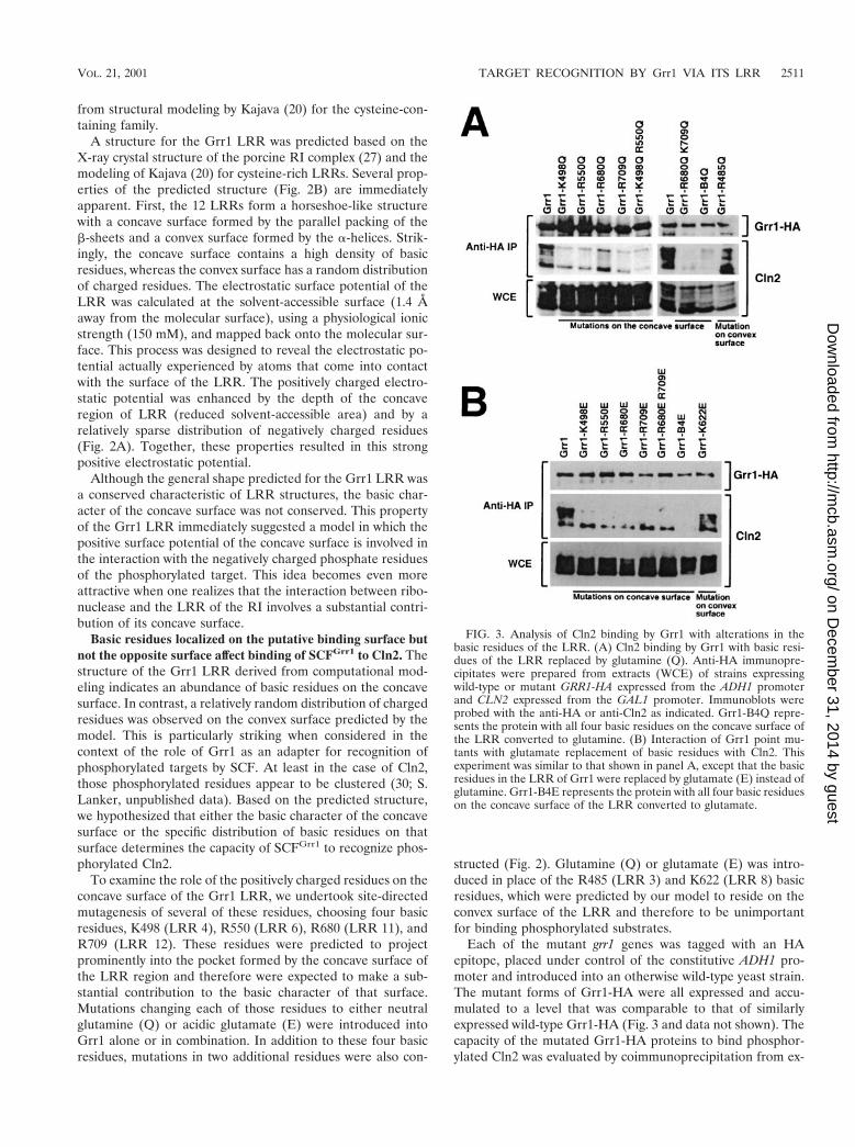
Each of the mutant grr1 genes was tagged with an HAepitope, placed under control of the constitutive ADH1 pro-moter and introduced into an otherwise wild-type yeast strain.The mutant forms of Grr1-HA were all expressed and accu-mulated to a level that was comparable to that of similarlyexpressed wild-type Grr1-HA (Fig. 3 and data not shown). Thecapacity of the mutated Grr1-HA proteins to bind phosphor-ylated Cln2 was evaluated by coimmunoprecipitation from ex-
FIG. 3. Analysis of Cln2 binding by Grr1 with alterations in thebasic residues of the LRR. (A) Cln2 binding by Grr1 with basic resi-dues of the LRR replaced by glutamine (Q). Anti-HA immunopre-cipitates were prepared from extracts (WCE) of strains expressingwild-type or mutant GRR1-HA expressed from the ADH1 promoterand CLN2 expressed from the GAL1 promoter. Immunoblots wereprobed with the anti-HA or anti-Cln2 as indicated. Grr1-B4Q repre-sents the protein with all four basic residues on the concave surface ofthe LRR converted to glutamine. (B) Interaction of Grr1 point mu-tants with glutamate replacement of basic residues with Cln2. Thisexperiment was similar to that shown in panel A, except that the basicresidues in the LRR of Grr1 were replaced by glutamate (E) instead ofglutamine. Grr1-B4E represents the protein with all four basic residueson the concave surface of the LRR converted to glutamate.
VOL. 21, 2001 TARGET RECOGNITION BY Grr1 VIA ITS LRR 2511
on Decem
ber 31, 2014 by guesthttp://m
cb.asm.org/
Dow
nloaded from
tracts of cells expressing Cln2 from the inducible GAL1 pro-moter. All of the mutant proteins that had lesions predicted tolie on the concave surface of the LRR were compromised intheir capacity to bind Cln2 relative to wild-type Grr1, whereasthose proteins involving residues on the convex surface werelargely unaffected (Fig. 3 and data not shown). Nevertheless,there was some variability in the Cln2 binding efficiency ob-served between the mutant Grr1 proteins. First, binding ofCln2 to those Grr1 mutant proteins with glutamate (E) in placeof the basic residue (Fig. 3B) was more severely affected inevery case than binding to those with glutamine (Q) in thesame position (Fig. 3A). This is consistent with our model,since the presence of like charges on both Grr1 and the phos-phorylated substrate is expected to be repulsive. Next, some ofthe single-residue mutants were less severely affected thanothers. For example, the R680Q mutant retained approxi-mately half of the wild-type level of Cln2 binding (Fig. 3A). Itis interesting that, according to our computational model,R680 was predicted to participate in a salt bridge with D657(data not shown) and may therefore be less available for in-teractions with a charged ligand. Mutants having more thanone mutated residue were, in general, more severely affectedthan those carrying single-site mutations. In fact, the mutant inwhich all four basic residues were replaced with glutamate(Grr1-B4E) was completely defective in the ability to bindCln2. This included the capacity to bind the highest mobilityform of Cln2, which is presumably unmodified. In contrast,using the other mutants that had glutamate in place of thebasic residues, we observed binding of the highest-mobilityform of Cln2. We are uncertain whether this form actuallybinds or is formed during preparation of the samples, but it wasclear that, whatever the source, it was without consequence interms of Cln2 instability (see below). Finally, consistent withour prediction, mutations altering basic residues on the convexsurface (R485 and K622) had little or no effect on Cln2 bindingrelative to wild-type Grr1 regardless of whether the replace-ment was with glutamine or glutamate (Fig. 3 and data notshown).
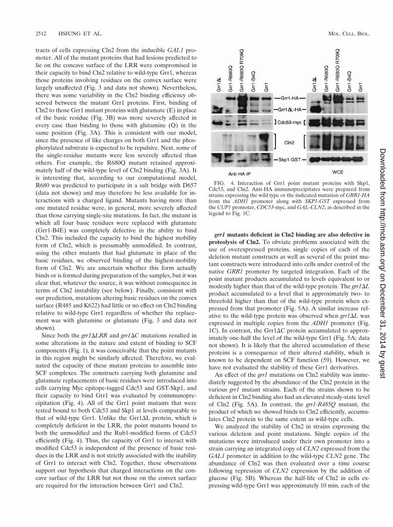
Since both the grr1DLRR and grr1DC mutations resulted insome alterations in the nature and extent of binding to SCFcomponents (Fig. 1), it was conceivable that the point mutantsin this region might be similarly affected. Therefore, we eval-uated the capacity of these mutant proteins to assemble intoSCF complexes. The constructs carrying both glutamine andglutamate replacements of basic residues were introduced intocells carrying Myc epitope-tagged Cdc53 and GST-Skp1, andtheir capacity to bind Grr1 was evaluated by coimmunopre-cipitation (Fig. 4). All of the Grr1 point mutants that weretested bound to both Cdc53 and Skp1 at levels comparable tothat of wild-type Grr1. Unlike the Grr1DL protein, which iscompletely deficient in the LRR, the point mutants bound toboth the unmodified and the Rub1-modified forms of Cdc53efficiently (Fig. 4). Thus, the capacity of Grr1 to interact withmodified Cdc53 is independent of the presence of basic resi-dues in the LRR and is not strictly associated with the inabilityof Grr1 to interact with Cln2. Together, these observationssupport our hypothesis that charged interactions on the con-cave surface of the LRR but not those on the convex surfaceare required for the interaction between Grr1 and Cln2.
grr1 mutants deficient in Cln2 binding are also defective inproteolysis of Cln2. To obviate problems associated with theuse of overexpressed proteins, single copies of each of thedeletion mutant constructs as well as several of the point mu-tant constructs were introduced into cells under control of thenative GRR1 promoter by targeted integration. Each of thepoint mutant products accumulated to levels equivalent to ormodestly higher than that of the wild-type protein. The grr1DLproduct accumulated to a level that is approximately two- tothreefold higher than that of the wild-type protein when ex-pressed from that promoter (Fig. 5A). A similar increase rel-ative to the wild-type protein was observed when grr1DL wasexpressed in multiple copies from the ADH1 promoter (Fig.1C). In contrast, the Grr1DC protein accumulated to approx-imately one-half the level of the wild-type Grr1 (Fig. 5A; datanot shown). It is likely that the altered accumulation of theseproteins is a consequence of their altered stability, which isknown to be dependent on SCF function (59). However, wehave not evaluated the stability of these Grr1 derivatives.
An effect of the grr1 mutations on Cln2 stability was imme-diately suggested by the abundance of the Cln2 protein in thevarious grr1 mutant strains. Each of the strains shown to bedeficient in Cln2 binding also had an elevated steady-state levelof Cln2 (Fig. 5A). In contrast, the grr1-R485Q mutant, theproduct of which we showed binds to Cln2 efficiently, accumu-lates Cln2 protein to the same extent as wild-type cells.
We analyzed the stability of Cln2 in strains expressing thevarious deletion and point mutations. Single copies of themutations were introduced under their own promoter into astrain carrying an integrated copy of CLN2 expressed from theGAL1 promoter in addition to the wild-type CLN2 gene. Theabundance of Cln2 was then evaluated over a time coursefollowing repression of CLN2 expression by the addition ofglucose (Fig. 5B). Whereas the half-life of Cln2 in cells ex-pressing wild-type Grr1 was approximately 10 min, each of the
FIG. 4. Interaction of Grr1 point mutant proteins with Skp1,Cdc53, and Cln2. Anti-HA immunoprecipitates were prepared fromstrains expressing the wild type or the indicated mutation of GRR1-HAfrom the ADH1 promoter along with SKP1-GST expressed fromthe CUP1 promoter, CDC53-myc, and GAL-CLN2, as described in thelegend to Fig. 1C.
2512 HSIUNG ET AL. MOL. CELL. BIOL.
on Decem
ber 31, 2014 by guesthttp://m
cb.asm.org/
Dow
nloaded from

strains expressing mutant Grr1 exhibited an extended Cln2half-life. As previously demonstrated, introduction of eitherthe grr1D or the grr1DL mutation resulted in a dramatic stabi-lization of Cln2 (Fig. 5B). Strikingly, replacement of all fourbasic residues on the concave surface was also associated witha dramatically lengthened half-life for Cln2 that was consistentwith the effect of those mutations on Cln2 binding. Both thegrr1-B4Q and the grr1-B4E mutants also caused a similar in-crease in the stability of Cln2 without the associated defect inSCF formation (Fig. 4 and 5B). The various single-site pointmutants that were analyzed resulted in more modest increasesin Cln2 stability than either Grr1DLRR or one of the quadru-ple-point mutant proteins (data not shown). Finally, theGrr1DC mutant, which exhibited a partial defect in Cln2 bind-ing, resulted in a less dramatic stabilization of Cln2 (data notshown). Because Grr1DC is reduced in abundance relative towild-type Grr1, it was difficult to distinguish the effect of re-duced binding to Cln2 from the effect of reduced abundance ofthe Grr1DC protein. Nevertheless, its effect is less severe thanthat of Grr1DL, as well as those of most of the point mutants(Fig. 5A and data not shown). We conclude that the stability ofCln2 in strains expressing single copies of the various grr1mutations was largely consistent with the capacity of the mu-tants to bind to Cln2.
Morphological consequences of grr1 mutations. We haveanalyzed the morphological phenotypes associated with thevarious grr1 mutants (Fig. 6). The mutants have been analyzedboth when growing in rich liquid medium (yeast extract-pep-tone-dextrose [YEPD] liquid; Fig. 6A) or on rich solid medium(YEPD plate; Fig. 6B). Consistent with their effect on Cln2stability, both the grr1DL and grr1DC mutants caused cells tobecome elongated and to bud in a unipolar fashion. This wasparticularly apparent in cells growing on solid medium (Fig.6B). The grr1 point mutants exhibited a gradation of severitythat was roughly consistent with their effect on Cln2 proteol-ysis. Mutations resulting in neutralization of basic residues onthe predicted convex surface of the LRR had no phenotypiceffect, whereas those neutralizing basic residues on the pre-dicted concave surface had a pronounced effect on cell polar-ity, bud site selection, and, to a lesser extent, abscission (Fig. 6and data not shown). Whereas single- and double-point muta-tions had a relatively modest effect on cell morphology, theeffect of the quadruple-point mutations (grr1-B4Q and grr1-B4E) on polarized growth and bud site selection became morepronounced. Finally, replacement with an acidic residue gen-erally resulted in more severe phenotypes than replacementwith a neutral residue at the same position. Nevertheless,whereas grr1-B4E mutations are similar to the grr1D mutationsin the severity of their effects on morphological phenotypes,they have less severe effects on growth rate (data not shown).
Despite the general increase in severity of the phenotypethat was observed with an increasing number of mutations,there are subtle distinctions between the morphological phe-notypes of the four different basic residue mutants (Fig. 6 and
FIG. 5. (A) Accumulation of Grr1-6His and Cln2 proteins in var-ious point and deletion mutations of GRR1. Twenty-five micrograms ofwhole-cell extract from asynchronously growing cells expressing thewild type or the indicated GRR1-6His form from its endogenous pro-moter were analyzed by immunoblotting using anti-His, anti-Cln2, andanti-Cdc28 antibodies, respectively. (B) Stability of Cln2 in cells havingpoint and deletion mutations in Grr1. Cells carrying CLN2 under theinducible GAL1 promoter and wild-type GRR1 or its derivatives(grr1D, grr1DL, grr1-B4Q, and grr1-B4E) under its native promoterwere pregrown under noninducing conditions (2% raffinose). GAL1-CLN2 was expressed for 45 min by addition of 2% galactose and then
repressed by addition of 2% glucose. Extracts prepared from samplestaken at the times indicated were analyzed by immunoblotting usinganti-Cln2 and anti-Cdc28 antibodies. UI, uninduced.
VOL. 21, 2001 TARGET RECOGNITION BY Grr1 VIA ITS LRR 2513
on Decem
ber 31, 2014 by guesthttp://m
cb.asm.org/
Dow
nloaded from

data not shown). For example, mutations affecting residueR680 result in cell enlargement rather than hyperpolarization.This is very noticeable when comparing the neutral and acidicreplacements of R680 and R709 to the same replacements ofK498 and R550. However, the single R680E mutant also ex-hibits significant enlargement. The source of the cell enlarge-ment is unknown, but it is unlikely to be a consequence ofhyperaccumulation of Cln2. Other, more subtle phenotypicdifferences are apparent on closer examination but have notbeen further evaluated.
Mutations in GRR1 affecting Cln2 binding and stability alsoresult in stabilization of Gic2, a target for SCFGrr1-dependentproteolysis. The fact that mutations on the putative concavesurface of the Grr1 LRR affected the capacity of SCFGrr1 tobind to Cln2 and resulted in its stabilization suggested that thesame mutations would also result in the stabilization of otherSCFGrr1 targets. The stability of the putative Cdc42 effectorsGic1 and Gic2 has been shown to be phosphorylation depen-dent and to be mediated by SCFGrr1 (17). To determine
whether the basic residues on the concave surface of the Grr1LRR were required for the degradation of the Gic2 protein, weanalyzed the stability of Gic2-GFP expressed from the repress-ible GAL1 promoter in cells having the grr1-B4Q allele at thechromosomal locus as the only source of Grr1. The Gic2-GFPfusion protein was rapidly degraded when the GAL1 promoterwas repressed by the addition of glucose (Fig. 7). However, thesame protein was dramatically stabilized in grr1DL mutants, aswell as in cells carrying grr1-B4Q (Fig. 7) and grr1-B4E (notshown) mutations. Thus, mutations that either eliminate theLRR domain of Grr1 or neutralize the charge on its concavesurface result in stabilization of the Gic2 protein, presumablydue to the failure of Grr1 to bind to Gic2 and promote itsubiquitination. We conclude that the concave surface of theGrr1 LRR, by virtue of its basic nature, participates in bindingto phosphorylated targets of the SCFGrr1 ubiquitin ligase.
Mutations in GRR1 have differential effects on Cln2 stabilityand other established functions of Grr1. Grr1 has been impli-cated in a number of cellular functions in addition to proteol-
FIG. 6. (A) Morphological phenotypes of point and deletion mutations of GRR1 growing in rich liquid medium. Cells grown to late log phasewere analyzed by DIC microscopy and digital imaging. (B) Morphological phenotypes of point and deletion mutations of GRR1 growing on richsolid medium. Cells were grown on rich glucose medium (YEPD) for 4 h prior to photomicrography.
2514 HSIUNG ET AL. MOL. CELL. BIOL.
on Decem
ber 31, 2014 by guesthttp://m
cb.asm.org/
Dow
nloaded from

ysis of G1 cyclins and Gic1/2. Genetic analysis has implicatedGRR1 as a regulator of the expression of several nutrientresponsive genes. Of those, the best characterized are thesystems governing expression of the HXT genes, which encodea family of hexose permeases in response to glucose (reviewedin reference 38) and which govern the expression of the broad-specificity amino acid permease gene AGP1 in response toamino acids (15). We have investigated the effect of specificgrr1 mutations on these functions (Fig. 8). A summary of theproperties of a representative set of mutants is presented inTable 3.
The four yeast hexose transporters encoded by HXT1–4 areeach controlled somewhat differently by the availability of glu-cose. HXT1 is specifically induced in the presence of high levelsof glucose (4%) and is repressed in the presence of most othercarbon sources. We have evaluated expression of HXT1 in cellsgrowing in either glucose or galactose by monitoring the abun-dance of HXT1 mRNA by RT-PCR using HXT1-specific prim-ers (Fig. 8A). This approach obviates the problem of cross-hybridization of probes with transcripts of the other highlyconserved HXT genes. As previously described, the strong in-duction of HXT1 expression observed in wild-type cells grownin glucose-containing medium is lost when cells are deficient inGRR1. Surprisingly, both grr1DL, and grr1DC, as well as thevarious point mutations of GRR1, retain the capacity to induceHXT1 expression in response to glucose. Thus, despite theirdramatic effect on both Cln2 proteolysis and cellular morphol-ogy, the deletion mutations and mutations in basic residues ofthe LRR remained responsive to induction by glucose. Fur-thermore, grr1-R485Q, the neutral replacement on the pre-dicted convex surface of the LRR, had no noticeable effect onglucose regulation of HXT1.
A similar analysis was performed to evaluate the effect ofgrr1 mutations on aromatic amino acid transport. Induction of
the expression of the amino acid permease gene AGP1 hasbeen shown to depend on GRR1 (15). We have performed twodistinct but related assays. First, AGP1 transcript levels weremonitored by RT-PCR and by Northern analysis (Fig. 8B). Aspreviously reported, AGP1 expression in the presence of ex-tracellular amino acids was significantly reduced in grr1D mu-tants relative to wild-type cells. However, as observed forHXT1 induction by glucose, each of the internal deletion mu-tations and the point mutations replacing basic residues on theconcave surface of the LRR resulted in little or no effect on theaccumulation of AGP1 transcripts. Since the grr1D mutant ex-hibited residual RT-PCR signal, we confirmed the result usingNorthern analysis. Again, although the grr1D mutant expresseda detectable amount of AGP1 transcript, that level was consis-tently lower than the transcript level expressed by grr1DL andgrr1DC mutant or wild-type cells. Thus, the effect of accumu-lation of the AGP1 transcript in the various grr1 mutants grow-ing under inducing conditions was similar to that of the accu-mulation of the HXT1 transcript in the same mutants growingin the presence of glucose.
Together the results of these analyses suggest that the role ofGrr1 in the regulation of these transcriptional induction path-ways involves recognition mechanisms that are distinct fromthose involved in the recognition of Cln2. It remains possiblethat the ability of the grr1DL, grr1DC, and point mutants tomediate the transcriptional responses but not Cln2 proteolysis
FIG. 7. Stability of Gic2 in cells having point and deletion muta-tions in Grr1. Cells carrying GIC2-GFP under the inducible GAL1promoter and wild-type GRR1 or its derivatives (grr1DL and grr1-B4Q)under its native promoter were pregrown under noninducing condi-tions (2% raffinose). GAL1-GIC2-GFP was expressed for 3 h by addi-tion of 2% galactose and then repressed by addition of 2% glucose.Extracts prepared from samples taken at the times indicated wereanalyzed by immunoblotting using anti-GFP and anti-Cdc28 antibod-ies. UI, uninduced.
FIG. 8. Nutrient-regulated transcription is not affected by muta-tions in LRR of Grr1. (A) The capacity of wild-type cells or cellscarrying the indicated grr1 mutation to induce HXT1 gene expressionwas evaluated by analysis of the HXT1 mRNA. Total RNA was iso-lated from strains carrying the indicated GRR1 allele either prior to(G, galactose) or following 90 min of induction with 4% glucose (D,dextrose). The HXT1 mRNA level in each strain was determined usingRT-PCR. PCRs for ACT1 were carried out using an equal amount ofRNA to ensure equal loading. Samples were performed without re-verse transcriptase (-RT) to evaluate the extent of DNA contamina-tion in each RNA preparation. (B) The expression of AGP1 in wild-type cells and cells carrying the indicated grr1 allele under inducingconditions in rich medium. AGP1 expression was analyzed (left panel)by RT-PCR, and (right panel) Northern hybridization. AGP1 andACT1 transcript levels were determined by RT-PCR (as described forpanel A) and Northern hybridization using RNA isolated from glu-cose-grown cells.
VOL. 21, 2001 TARGET RECOGNITION BY Grr1 VIA ITS LRR 2515
on Decem
ber 31, 2014 by guesthttp://m
cb.asm.org/
Dow
nloaded from
results from quantitative rather than qualitative differences inGrr1 and grr1D functions rather than from differences in theability to interact with specific targets. This consideration isespecially relevant when considering grr1DC. However, be-cause the effect of grr1DL on Cln2 binding, proteolysis, andvarious other cellular functions is similar to that of grr1D, itseems doubtful that its activity in this regard is significantlyhigher. Yet, like the point mutations, grr1DL has little effect onthese transcriptional responses. Since the targets of Grr1 in-volved in either of these processes are unknown, their capacityto interact with Grr1 cannot be evaluated. Although it is gen-erally assumed that these processes involve ubiquitination, thathas not been established.
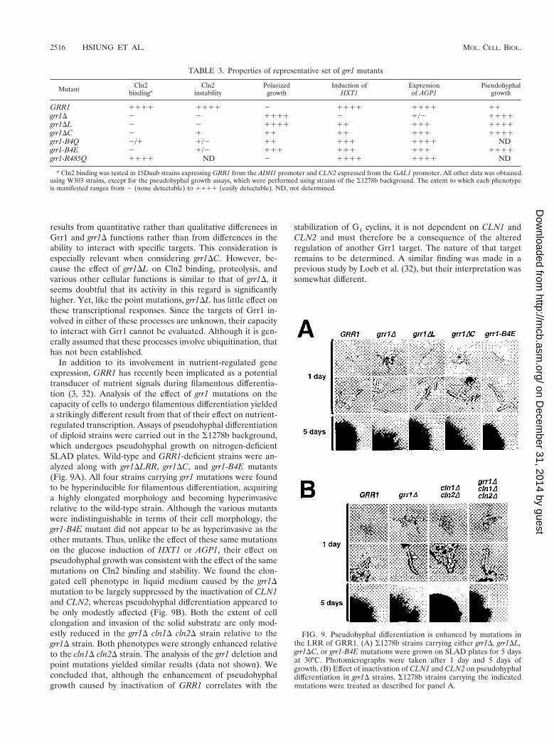
In addition to its involvement in nutrient-regulated geneexpression, GRR1 has recently been implicated as a potentialtransducer of nutrient signals during filamentous differentia-tion (3, 32). Analysis of the effect of grr1 mutations on thecapacity of cells to undergo filamentous differentiation yieldeda strikingly different result from that of their effect on nutrient-regulated transcription. Assays of pseudohyphal differentiationof diploid strains were carried out in the S1278b background,which undergoes pseudohyphal growth on nitrogen-deficientSLAD plates. Wild-type and GRR1-deficient strains were an-alyzed along with grr1DLRR, grr1DC, and grr1-B4E mutants(Fig. 9A). All four strains carrying grr1 mutations were foundto be hyperinducible for filamentous differentiation, acquiringa highly elongated morphology and becoming hyperinvasiverelative to the wild-type strain. Although the various mutantswere indistinguishable in terms of their cell morphology, thegrr1-B4E mutant did not appear to be as hyperinvasive as theother mutants. Thus, unlike the effect of these same mutationson the glucose induction of HXT1 or AGP1, their effect onpseudohyphal growth was consistent with the effect of the samemutations on Cln2 binding and stability. We found the elon-gated cell phenotype in liquid medium caused by the grr1Dmutation to be largely suppressed by the inactivation of CLN1and CLN2, whereas pseudohyphal differentiation appeared tobe only modestly affected (Fig. 9B). Both the extent of cellelongation and invasion of the solid substrate are only mod-estly reduced in the grr1D cln1D cln2D strain relative to thegrr1D strain. Both phenotypes were strongly enhanced relativeto the cln1D cln2D strain. The analysis of the grr1 deletion andpoint mutations yielded similar results (data not shown). Weconcluded that, although the enhancement of pseudohyphalgrowth caused by inactivation of GRR1 correlates with the
stabilization of G1 cyclins, it is not dependent on CLN1 andCLN2 and must therefore be a consequence of the alteredregulation of another Grr1 target. The nature of that targetremains to be determined. A similar finding was made in aprevious study by Loeb et al. (32), but their interpretation wassomewhat different.
FIG. 9. Pseudohyphal differentiation is enhanced by mutations inthe LRR of GRR1. (A) S1278b strains carrying either grr1D, grr1DL,grr1DC, or grr1-B4E mutations were grown on SLAD plates for 5 daysat 30°C. Photomicrographs were taken after 1 day and 5 days ofgrowth. (B) Effect of inactivation of CLN1 and CLN2 on pseudohyphaldifferentiation in grr1D strains. S1278b strains carrying the indicatedmutations were treated as described for panel A.
TABLE 3. Properties of representative set of grr1 mutants
Mutant Cln2bindinga
Cln2instability
Polarizedgrowth
Induction ofHXT1
Expressionof AGP1
Pseudohyphalgrowth
GRR1 1111 1111 2 1111 1111 11grr1D 2 2 1111 2 1/2 1111grr1DL 2 2 1111 11 111 1111grr1DC 2 1 11 11 111 1111grr1-B4Q 2/1 1/2 11 111 1111 NDgrr1-B4E 2 1/2 111 111 111 1111grr1-R485Q 1111 ND 2 1111 1111 ND
a Cln2 binding was tested in 15Daub strains expressing GRR1 from the ADH1 promoter and CLN2 expressed from the GAL1 promoter. All other data was obtainedusing W303 strains, except for the pseudohyphal growth assays, which were performed using strains of the S1278b background. The extent to which each phenotypeis manifested ranges from 2 (none detectable) to 1111 (easily detectable). ND, not determined.
2516 HSIUNG ET AL. MOL. CELL. BIOL.
on Decem
ber 31, 2014 by guesthttp://m
cb.asm.org/
Dow
nloaded from

DISCUSSION
SCF formation and Cln2 binding by grr1 mutants. Grr1plays a central role in the regulation of SCF-dependent ubiq-uitination of a number of proteins including Cln2. SCF bindingand Cln2 ubiquitination are dependent on phosphorylation ofCln2. We have shown here that the LRR is important forbinding phosphorylated Cln2 and for its subsequent proteoly-sis. Furthermore, we showed that basic residues, predicted byour three-dimensional model to lie on the concave surface ofthe Grr1 LRR, are critical for the interaction between Cln2and Grr1. In contrast, basic residues on the convex surface arenot important for that interaction nor do they affect Cln2proteolysis. These results reveal the domain of Grr1 requiredfor the Grr1-Cln2 interaction as well as defining the probablebinding surface as predicted by our model. We suggest thatelectrostatic interactions between basic residues on the con-cave surface of the LRR and the phosphoryl groups on Cln2provide the binding energy for this interaction. Similar elec-trostatic interactions are characteristic of the contacts ob-served by the X-ray crystal structure of RI and RNase A (25)on which our structural model for the Grr1 LRR was based.Although the charged residues of RI are not as clearly con-centrated on the concave surface of the LRR, about a third ofthe contacts between the LRR of RI and the RNase A mole-cule are between acidic and basic residues. In that case, pri-marily acidic residues are contributed by the LRR whereas thebinding partner contributes predominantly basic residues tothe interaction (27). The importance of electrostatic interac-tions was further supported by our demonstration that Cln2phosphorylation is required for recognition by SCFGrr1 (30,55). Thus, Grr1 serves as target-specific adapter for ubiquiti-nation by providing a specific binding pocket for interactionwith the target.
Several lines of evidence suggest that the interaction be-tween Grr1 and Cln2 is dependent upon properties in additionto the basic character of the LRR. First, although the LRRdomain of Grr1 is stable when expressed in yeasts, the isolateddomain does not interact stably with Cln2 (data not shown).Next, the carboxy terminus of Grr1 is important for productiveinteractions since a Grr1 mutant lacking the region of thecarboxy terminus adjacent to the LRR fails to bind stably toCln2. In contrast, Grr1 lacking the 280-amino-acid amino ter-minus adjacent to the F box appears to bind Cln2 relativelyefficiently. Although elimination of the C terminus does notappear to be as detrimental as elimination of the LRR, it doesresult in a substantial decrease in Cln2 binding and stabiliza-tion. Support for a role of the carboxy-terminal domain of Grr1is provided by the recently reported structure of a cocomplexbetween human Skp1 and Skp2, an LRR-containing F boxprotein involved in recognition of the CDK inhibitor p27 (47).In that complex, the C terminus of Skp2 wraps around theconcave face of the LRR and interacts with the F box domain,thereby positioning it to interact with substrate bound to Skp2via the LRR. However, a role for the LRR of Skp2 in bindingsubstrates has yet to be established. This, together with ourresults, suggests that the carboxy-terminal domains of theseproteins participate in binding by facilitating interactions be-tween substrates and the LRR.
All of the Grr1 proteins with alterations in basic residues
were effective in forming SCF complexes, despite their defectin binding Cln2. Both the LRR-deficient and the carboxy-terminally truncated mutants of Grr1 failed to bind to thelower mobility form of Cdc53, which is modified by the ubiq-uitin-like protein Rub1. There is no established role for Rub1modification in SCF function in budding yeast cells, and itselimination results in no detectable phenotypic consequences.In contrast, the homolog NEDD8 is essential for viability infission yeasts (37) and is required for degradation of some SCFtargets in animal cells (43). Since both forms of Cdc53 arepresent at wild-type levels in the Grr1DL-expressing cells, itappears either that Grr1DL and Grr1DC fail to form suchcomplexes or that the complexes, once formed, are more un-stable than those formed with unmodified Cdc53. The latterwould be consistent with the suggestion that Rub1 is involvedin regulating cullin abundance (29).
Differences between requirements for the Grr1-Cln2 inter-action and the regulation of other putative targets of Grr1.The role of Grr1 in SCF-dependent ubiquitination extendsbeyond Cln2 to other protein targets including the Cdc42 in-teractor Gic2 (17). We show that basic residues on the concavesurface of the LRR are required for the instability of Gic2. Inaddition, Grr1 also plays a role in several systems of nutrient-regulated transcription. The direct targets of Grr1 in thosesystems are unknown. In fact, it has not been established ineither of these cases that the role of Grr1 is in the context ofprotein ubiquitination. We have shown here that, althoughdisruption of Grr1 results in failure to induce either the HXT1or AGP1 transcripts, the other deletion mutations or pointmutations we have constructed result in little or no defect inthe induction of those genes. In contrast, all of those mutationsinterfere with the ubiquitin-dependent degradation of Cln2.Thus, we consider it likely that the protein domains of Grr1involved in recognition of phosphorylated Cln2 are unimpor-tant for recognition of the targets required for the regulation ofthese transcription systems. Alternatively, the capacity of thesemutant proteins to function in those pathways may be a reflec-tion of differences in the efficiency of the interactions requiredfor transcriptional activation and G1 cyclin proteolysis. Thedifferences observed in the requirements for recognition of therelevant targets in these transcriptional pathways may reflectdifferences in the basis for recognition. For example, recogni-tion of those targets may not involve protein phosphorylation.Resolution of these issues awaits the identification of the rel-evant targets of Grr1 in those pathways.
What is the role of Grr1 in these transcriptional regulatorysystems? Our understanding of both systems is largely derivedfrom genetic studies. Induction of HXT1 gene expression byglucose has been shown to involve components of the SCFsystem in addition to Grr1, including Cdc53, Skp1, and, at leastin our experiments, Cdc34 (H.-C. Chang and C. Wittenberg,unpublished results; for a different result, see reference 31).This is consistent with a role for Grr1 in proteolysis of anegative regulator of transcription (10; reviewed in reference38). However, evidence that Rgt1 is regulated at the level ofubiquitination or proteolysis is lacking. Recently reports re-garding the role of SCFMet30 in transcriptional regulation ofMET gene expression have come to conflicting conclusions.Experimental support has been presented for the involvementof ubiquitin-mediated proteolysis of the transcription factor
VOL. 21, 2001 TARGET RECOGNITION BY Grr1 VIA ITS LRR 2517
on Decem
ber 31, 2014 by guesthttp://m
cb.asm.org/
Dow
nloaded from

Met4 (45). In contrast, we have recently shown that, althoughthe transcriptional regulator Met4 is ubiquitinated under con-ditions that inhibit MET gene expression, proteolysis is unnec-essary to inactivate MET gene transcription (18). Instead ubiq-uitination interferes with the function of Met4 as atranscriptional activator, presumably by interfering with itsability to bind a coactivator. That system provides an attractivemodel for regulation of Rgt1, especially because it has beenproposed to act as both a positive and a negative regulator inthe presence of different carbon sources (39). For instance, inthe absence of glucose, Rgtl may be present in a nonubiquiti-nated form and act as a repressor via recruitment of the core-pressors Ssn6 and Tup1. Conversely, in the presence of glucoseit might become ubiquitinated in a Grr1-dependent manner,disrupting the interaction with its corepressors, thereby con-verting it to a transcriptional activator. Alternatively, ubiquiti-nation may regulate the activity of the corepressors or otherregulators via either proteolytic or nonproteolytic mechanisms.Because the central elements for transcriptional regulation ofAGP1 are currently unknown, it is difficult to predict the roleof Grrl in that regulation.
The relationship between the hyperpolarized phenotype, nu-trient sensing, and pseudohyphal morphogenesis is also un-clear. We expected, based on the relationship between Grr1and Cln2 stability and on the capacity of Cln2, when overex-pressed, to induce hyperpolarization, that the hyperinvasivephenotype of grr1D mutants was a consequence of the hyper-accumulation of G1 cyclins. In fact, evidence supporting thathypothesis has been reported (39). However, we have foundthat the inactivation of Cln1 and Cln2 has little effect on thecapacity of grr1D mutations to induce pseudohyphal differen-tiation in terms of either morphological differentiation or thecapacity to invade agar. Furthermore, the induction ofpseudohyphal growth is at least partially independent of theestablished signaling pathways since invasive growth can beinduced in grr1D mutants lacking both TEC1 and FLO8 (R.LaValle and C. Wittenberg, unpublished data). We concludethat Grr1 targets one or more elements of a TEC1/FLO8independent pathway to suppress filamentation. The involve-ment of Grr1 in transcriptional regulation of nutrient per-meases and the importance of nutrient signaling in the induc-tion of pseudohyphal differentiation are intriguing in thisregard. We have not yet analyzed the importance of amino acidor glucose permeases in the pseudohyphal growth signal oranalyzed the potential targets of Grr1 in that response.
LRR as recognition domain for phosphorylated proteins.The capacity of proteins to interact specifically with each otherhas long been recognized as a fundamental property of livingsystems and is the basis for the assembly of the macromolec-ular complexes central to many biological processes. Regulatedprotein-protein interaction is a critical component of manybiological regulatory mechanisms. Protein phosphorylation isone of the most widely studied and perhaps most commonmechanisms governing regulated protein recognition. Despitethat fact, the basis for recognition is only poorly understood.This may be a consequence of the wide array of solutions tothis specific recognition problem.
LRRs are thought to be involved in protein-protein interac-tion. Deletion analysis of a number of proteins including Grr1has confirmed that supposition. Although this property of
LRR domains appears to be conserved, the nature and roles ofthe proteins that they bind are varied. Structural studies ofLRRs, although relatively limited, suggest that it is the concavesurface that is important in the interaction with other proteins.There also appears to be growing support for the importanceof electrostatic interactions in the recognition process. Theinteraction demonstrated between RI and RNase A (25), aswell as our findings concerning Grr1, are consistent with bothof those proposals. In addition, structural modeling of theLRR of the acid-labile subunit of the insulin-like growth factorbinding protein (IGFBP-3) complex reveals that, like in theLRR of Grr1, a highly charged patch is predicted on theconcave surface (16). However, unlike Grr1, that patch is pre-dicted to be acidic rather than basic. Whereas the ring struc-ture of that molecule has been confirmed by electron micro-scopic analysis, the importance of the acidic patch in itsinteraction with insulin-like growth factor–insulin-like growthfactor binding protein 3 (IGF-IGFBP-3) complex has not beenestablished. It is not clear whether such electrostatic interac-tions are universally involved in LRR protein interactions. Ourmutational analysis of Grr1 indicated that the basic characterof the concave surface of the LRR is unimportant for regula-tion of transcriptional activation of HXT1 and AGP1. Theproperties of Grr1 important for that regulation remain to beestablished.
Protein phosphorylation figures centrally in the targeting ofproteins for SCF-dependent ubiquitination. This has been es-tablished in a wide variety of regulatory systems in eukaryotes.However, not all of those targeting interactions involve LRR-containing F-box proteins. In addition, F-box proteins withWD40 repeats are also prevalent and in at least some cases areinvolved in targeting events that require phosphorylation.Their role in phosphorylation-dependent ubiquitination of themammalian NF-kB inhibitor IkB by SCFbTRCP (22) and of theyeast CDK inhibitor Sic1 by SCFCdc4 (9, 49, 54) has been wellestablished. Both of those systems rely on interactions thatoccur via the WD40 repeats of their respective F-box proteins,bTRCP/E3RSIkB and Cdc4. Phosphorylation is implicated in anumber of other SCF-dependent ubiquitination events thatinvolve LRR- and WD40 repeat-containing F-box proteins.This study has established one mechanism by which such in-teractions can be implemented. However, the specifics of theinteractions utilized among this large class of protein recogni-tion events will require further mutagenesis and/or structuralstudies. Despite their involvement in phosphorylation-depen-dent interactions, LRR and WD40 repeat motifs in F-boxproteins, as in their wider context, are not restricted to suchinteractions nor are interactions involving phosphorylation re-stricted to those involving LRR and WD40 motifs.
ACKNOWLEDGMENTS
We thank Frank Li, Mark Johnston, Hao-ping Liu, and MatthiasPeter for providing plasmids and yeast strains and Andre Kajava forproviding coordinates derived from his modeling of cysteine-richleucine rich repeats. We also thank the TSRI cell cycle group, includ-ing members of the laboratories of C. McGowan, S. Reed, P. Russell,and C. Wittenberg, for helpful comments and discussion during thecourse of this work.
Y.H. was supported by a fellowship from the Lymphoma and Leu-kemia Society (formerly the Leukemia Society of America). R.L.V. issupported by an AIDS fellowship from the National AIDS Program,Istituto Superiore di Sanita, Rome, Italy. This work was supported by
2518 HSIUNG ET AL. MOL. CELL. BIOL.
on Decem
ber 31, 2014 by guesthttp://m
cb.asm.org/
Dow
nloaded from

U.S. Public Health Service grants GM59759 to S.L. and GM43487 toC.W.
REFERENCES
1. Bai, C., P. Sen, K. Hofmann, L. Ma, M. Goebl, J. W. Harper, and S. J.Elledge. 1996. SKP1 connects cell cycle regulators to the ubiquitin proteolysismachinery through a novel motif, the F-box. Cell 86:263–274.
2. Barral, Y., S. Jentsch, and C. Mann. 1995. G1 cyclin turnover and nutrientuptake are controlled by a common pathway in yeast. Genes Dev. 9:399–409.
3. Blacketer, M. J., P. Madaule, and A. M. Myers. 1995. Mutational analysis ofmorphologic differentiation in Saccharomyces cerevisiae. Genetics 140:1259–1275.
4. Brooks, B., R. Bruccoleri, B. Olafson, D. States, S. Swaminathan, and M.Karplus. 1983. CHARMM: a program for macromolecular energy, minimal-ization, and molecular dynamics calculations. J. Comp. Chem. 4:187–217.
5. Brunger, A. T. 1992. X-PLOR manual, version 3.1. Yale University, NewHaven, Conn.
6. Carrano, A. C., E. Eytan, A. Hershko, and M. Pagano. 1999. SKP2 is requiredfor ubiquitin-mediated degradation of the CDK inhibitor p27. Nat. Cell Biol.1:193–199.
7. Deshaies, R. J. 1999. SCF and cullin/ring H2-based ubiquitin ligases. Annu.Rev. Cell Dev. Biol. 15:435–467.
8. Drury, L. S., G. Perkins, and J. F. Diffley. 1997. The Cdc4/34/53 pathwaytargets Cdc6p for proteolysis in budding yeast. EMBO J. 16:5966–5976.
9. Feldman, R. M., C. C. Correll, K. B. Kaplan, and R. J. Deshaies. 1997. Acomplex of Cdc4p, Skp1p, and Cdc53p/cullin catalyzes ubiquitination of thephosphorylated CDK inhibitor Sic1p. Cell 91:221–230.
10. Flick, J. S., and M. Johnston. 1991. GRR1 of Saccharomyces cerevisiae isrequired for glucose repression and encodes a protein with leucine-richrepeats. Mol. Cell. Biol. 11:5101–5112.
11. Geitz, R. D., and A. Sugino. 1988. New yeast-Escherichia coli shuttle vectorsconstructed with in vitro mutagenized yeast genes lacking six base pairrestriction sites. Gene 74:527–534.
12. Gimeno, C. J., and G. R. Fink. 1994. Induction of pseudohyphal growth byoverexpression of PHD1, a Saccharomyces cerevisiae gene related to tran-scriptional regulators of fungal development. Mol. Cell. Biol. 14:2100–2112.
13. Henchoz, S., Y. Chi, B. Catarin, I. Herskowitz, R. J. Deshaies, and M. Peter.1997. Phosphorylation- and ubiquitin-dependent degradation of the cyclin-dependent kinase inhibitor Far1p in budding yeast. Genes Dev. 11:3046–3060.
14. Hershko, A., and A. Ciechanover. 1998. The ubiquitin system. Annu. Rev.Biochem. 67:425–479.
15. Iraqui, I., S. Vissers, F. Bernard, J. O. de Craene, E. Boles, A. Urrestarazu,and B. Andre. 1999. Amino acid signaling in Saccharomyces cerevisiae: apermease-like sensor of external amino acids and F-box protein Grr1p arerequired for transcriptional induction of the AGP1 gene, which encodes abroad-specificity amino acid permease. Mol. Cell. Biol. 19:989–1001.
16. Janosi, J. B., P. A. Ramsland, M. R. Mott, S. M. Firth, R. C. Baxter, and P. J.Delhanty. 1999. The acid-labile subunit of the serum insulin-like growthfactor-binding protein complexes. Structural determination by molecularmodeling and electron microscopy. J. Biol. Chem. 274:23328–23332.
17. Jaquenoud, M., M. P. Gulli, K. Peter, and M. Peter. 1998. The Cdc42peffector Gic2p is targeted for ubiquitin-dependent degradation by the SCF-Grr1 complex. EMBO J. 17:5360–5373.
18. Kaiser, P., K. Flick, C. Wittenberg, and S. I. Reed. 2000. Regulation oftranscription by ubiquitination without proteolysis: Cdc34/SCF(Met30)-me-diated inactivation of the transcription factor Met4. Cell 102:303–314.
19. Kaiser, P., R. A. Sia, E. G. Bardes, D. J. Lew, and S. I. Reed. 1998. Cdc34 andthe F-box protein Met30 are required for degradation of the Cdk-inhibitorykinase Swe1. Genes Dev. 12:2587–2597.
20. Kajava, A. V. 1998. Structural diversity of leucine-rich repeat proteins. J.Mol. Biol. 277:519–527.
21. Kamura, T., M. N. Conrad, Q. Yan, R. C. Conaway, and J. W. Conaway.1999. The Rbx1 subunit of SCF and VHL E3 ubiquitin ligase activates Rub1modification of cullins Cdc53 and Cul2. Genes Dev. 13:2928–2933.
22. Karin, M., and Y. Ben-Neriah. 2000. Phosphorylation meets ubiquitination:the control of NF-kB activity. Annu. Rev. Immunol. 18:621–663.
23. Kishi, T., and F. Yamao. 1998. An essential function of Grr1 for the degra-dation of Cln2 is to act as a binding core that links Cln2 to Skp1. J. Cell Sci.111:3655–3661.
24. Kitagawa, K., D. Skowyra, S. J. Elledge, J. W. Harper, and P. Hieter. 1999.SGT1 encodes an essential component of the yeast kinetochore assemblypathway and a novel subunit of the SCF ubiquitin ligase complex. Mol. Cell4:21–33.
25. Kobe, B., and J. Deisenhofer. 1996. Mechanism of ribonuclease inhibition byribonuclease inhibitor protein based on the crystal structure of its complexwith ribonuclease A. J. Mol. Biol. 264:1028–1043.
26. Kobe, B., and J. Deisenhofer. 1995. Proteins with leucine-rich repeats. Curr.Opin. Struct. Biol. 5:409–416.
27. Kobe, B., and J. Deisenhofer. 1995. A structural basis of the interactionsbetween leucine-rich repeats and protein ligands. Nature 374:183–186.
28. Kron, S. J., C. A. Styles, and G. R. Fink. 1994. Symmetric cell division in
pseudohyphae of the yeast Saccharomyces cerevisiae. Mol. Biol. Cell 5:1003–1022.
29. Lammer, D., N. Mathias, J. M. Laplaza, W. Jiang, Y. Liu, J. Callis, M.Goebl, and M. Estelle. 1998. Modification of yeast Cdc53p by the ubiquitin-related protein rub1p affects function of the SCFCdc4 complex. Genes Dev.12:914–926.
30. Lanker, S., M. H. Valdivieso, and C. Wittenberg. 1996. Rapid degradation ofthe G1 cyclin, Cln2, induced by CDK-dependent phosphorylation. Science271:1597–1601.
31. Li, F. N., and M. Johnston. 1997. Grr1 of Saccharomyces cerevisiae isconnected to the ubiquitin proteolysis machinery through Skp1: couplingglucose sensing to gene expression and the cell cycle. EMBO J. 16:5629–5638.
32. Loeb, J. D., T. A. Kerentseva, T. Pan, M. Sepulveda-Becerra, and H. Liu.1999. Saccharomyces cerevisiae G1 cyclins are differentially involved in in-vasive and pseudohyphal growth independent of the filamentation mitogen-activated protein kinase pathway. Genetics 153:1535–1546.
33. Marti, A., C. Wirbelauer, M. Scheffner, and W. Krek. 1999. Interactionbetween ubiquitin-protein ligase SCF, SKP2 and E2F-1 underlies the regu-lation of E2F-1 degradation. Nat. Cell Biol. 1:14–19.
34. McRee, D. E. 1992. StalView: a visual protein crystallographic softwaresystem for X11/XView. J. Mol. Graph. 10:44–47.
35. Mondesert, G., D. J. Clarke, and S. I. Reed. 1997. Identification of genescontrolling growth polarity in the budding yeast Saccharomyces cerevisiae: apossible role of N-glycosylation and involvement of the exocyst complex.Genetics 147:421–434.
36. Nakayama, K., H. Nagahama, Y. A. Minamishima, M. Matsumoto, I. Na-kamichi, K. Kitagawa, M. Shirane, R. Tsunematsu, T. Tsukiyama, N. Ishida,M. Kitagawa, and S. Hatakeyama. 2000. Targeted disruption of Skp2 resultsin accumulation of cyclin E and p27(Kip1), polyploidy and centrosomeoverduplication. EMBO J. 19:2069–2081.
37. Osaka, F., M. Saeki, S. Katayama, N. Aida, E. A. Toh, K. Kominami, T.Toda, T. Suzuki, T. Chiba, K. Tanaka, and S. Kato. 2000. Covalent modifierNEDD8 is essential for SCF ubiquitin-ligase in fission yeast. EMBO J. 19:3475–3484.
38. Ozcan, S., and M. Johnston. 1999. Function and regulation of yeast hexosetransporters. Microbiol. Mol. Biol. Rev. 63:554–569.
39. Ozcan, S., and M. Johnston. 1995. Three different regulatory mechanismsenable yeast hexose transporter (HXT) genes to be induced by differentlevels of glucose. Mol. Cell. Biol. 15:1564–1572.
40. Ozcan, S., T. Leong, and M. Johnston. 1996. Rgt1p of Saccharomyces cer-evisiae, a key regulator of glucose-induced genes, is both an activator and arepressor of transcription. Mol. Cell. Biol. 16:6419–6426.
41. Ozcan, S., F. Schulte, K. Freidel, A. Weber, and M. Ciriacy. 1994. Glucoseuptake and metabolism in grr1/cat80 mutants of Saccharomyces cerevisiae.Eur. J. Biochem. 224:605–611.
42. Patton, E. E., A. R. Willems, D. Sa, L. Kuras, D. Thomas, K. L. Craig, andM. Tyers. 1998. Cdc53 is a scaffold protein for multiple Cdc34/Skp1/F-boxproteincomplexes that regulate cell division and methionine biosynthesis inyeast. Genes Dev. 12:692–705.
43. Read, M. A., J. E. Brownell, T. B. Gladysheva, M. Hottelet, L. A. Parent,M. B. Coggins, J. W. Pierce, V. N. Podust, R. S. Luo, V. Chau, and V. J.Palombella. 2000. Nedd8 modification of cul-1 activates SCFbTrCP-depen-dent ubiquitination of IkBa. Mol. Cell. Biol. 20:2326–2333.
44. Richardson, H. E., C. Wittenberg, F. R. Cross, and S. I. Reed. 1989. Anessential G1 function for cyclin-like proteins in yeast. Cell 59:1127–1133.
45. Rouillon, A., R. Barbey, E. E. Patton, M. Tyers, and D. Thomas. 2000.Feedback-regulated degradation of the transcriptional activator Met4 is trig-gered by the SCFMet30 complex. EMBO J. 19:282–294.
46. Roussel, A., and C. Cambillau. 1995. TURBO-FRODO: Silicon Graphicsgeometry partners directory, p. 77–78. SGI, Mountain View, Calif.
47. Schulman, B. A., A. C. Carrano, P. D. Jeffrey, Z. Bowen, E. R. Kinnucan,M. S. Finnin, S. J. Elledge, J. W. Harper, M. Pagano, and N. P. Pavletich.2000. Insights into SCF ubiquitin ligases from the structure of the Skp1-Skp2complex. Nature 408:381–386.
48. Seol, J. H., R. M. Feldman, W. Zachariae, A. Shevchenko, C. C. Correll, S.Lyapina, Y. Chi, M. Galova, J. Claypool, S. Sandmeyer, K. Nasmyth, andR. J. Deshaies. 1999. Cdc53/cullin and the essential Hrt1 RING-H2 subunitof SCF define a ubiquitin ligase module that activates the E2 enzyme Cdc34.Genes Dev. 13:1614–1626.
49. Skowyra, D., K. L. Craig, M. Tyers, S. J. Elledge, and J. W. Harper. 1997.F-box proteins are receptors that recruit phosphorylated substrates to theSCF ubiquitin-ligase complex. Cell 91:209–219.
50. Skowyra, D., D. M. Koepp, T. Kamura, M. N. Conrad, R. C. Conaway, J. W.Conaway, S. J. Elledge, and J. W. Harper. 1999. Reconstitution of G1 cyclinubiquitination with complexes containing SCFGrr1 and Rbx1. Science 284:662–665.
51. Thomas, D., and Y. Surdin-Kerjan. 1997. Metabolism of sulfur amino acidsin Saccharomyces cerevisiae. Microbiol. Mol. Biol. Rev. 61:503–532.
52. Tsvetkov, L. M., K. H. Yeh, S. J. Lee, H. Sun, and H. Zhang. 1999. p27Kip1
ubiquitination and degradation is regulated by the SCFSkp2 complex throughphosphorylated Thr187 in p27. Curr. Biol. 9:661–664.
53. Vallier, L. G., D. Coons, L. F. Bisson, and M. Carlson. 1994. Altered regu-
VOL. 21, 2001 TARGET RECOGNITION BY Grr1 VIA ITS LRR 2519
on Decem
ber 31, 2014 by guesthttp://m
cb.asm.org/
Dow
nloaded from

latory responses to glucose are associated with a glucose transport defect ingrr1 mutants of Saccharomyces cerevisiae. Genetics 136:1279–1285.
54. Verma, R., R. M. Feldman, and R. J. Deshaies. 1997. SIC1 is ubiquitinatedin vitro by a pathway that requires CDC4, CDC34, and cyclin/CDK activities.Mol. Biol. Cell 8:1427–1437.
55. Willems, A. R., S. Lanker, E. E. Patton, K. L. Craig, T. F. Nasson, N.Mathias, R. Kobayashi, C. Wittenberg, and M. Tyers. 1996. Cdc53 targetsphosphorylated G1 cyclins for degradation by the ubiquitin proteolytic path-way. Cell 86:453–463.
56. Wittenberg, C., S. L. Richardson, and S. I. Reed. 1987. Subcellular localiza-tion of a protein kinase required for cell cycle initiation in Saccharomyces
cerevisiae: evidence for an association between the CDC28 gene product andthe insoluble cytoplasmic matrix. J. Cell Biol. 105:1527–1538.
57. Wittenberg, C., K. Sugimoto, and S. I. Reed. 1990. G1-specific cyclins of S.cerevisiae: cell cycle periodicity, regulation by mating pheromone, and asso-ciation with the p34CDC28 protein kinase. Cell 62:225–237.
58. Wright, R. M., T. Repine, and J. E. Repine. 1993. Reversible pseudohyphalgrowth in haploid Saccharomyces cerevisiae is an aerobic process. Curr.Genet. 23:388–391.
59. Zhou, P., and P. M. Howley. 1998. Ubiquitination and degradation of thesubstrate recognition subunits of SCF ubiquitin-protein ligases. Mol. Cell2:571–580.
2520 HSIUNG ET AL. MOL. CELL. BIOL.
on Decem
ber 31, 2014 by guesthttp://m
cb.asm.org/
Dow
nloaded from
Copyright © 2022 FDOKUMEN