Cationic Complexes of the Group 13-15 Elements Supported ...
Upload
independentCategory
view
2download
0
P
Ct
Aa
b
c
d
a
ARRAA
KBDCST
1
fi(wttbeieoSs
si
rI
0d
International Journal of Pharmaceutics 421 (2011) 189– 201
Contents lists available at SciVerse ScienceDirect
International Journal of Pharmaceutics
jo ur nal homep a ge: www.elsev ier .com/ locate / i jpharm
harmaceutical Nanotechnology
ationic ligand appended nanoconstructs: A prospective strategy for brainargeting
bhinav Agarwala, Himanshu Agrawalb, Shailja Tiwari c, Sanyog Jaind, Govind P. Agrawala,∗
Pharmaceutics Research Lab, Department of Pharmaceutical Sciences, Dr. H.S Gour University, Sagar 470003, M.P., IndiaPharmaceutics Research Laboratory, M.S. University of Baroda, Vadodara 390002, Gujarat, IndiaDrug Delivery Research Lab, Department of Pharmaceutical Sciences, Dr. H.S Gour University, Sagar 470003, M.P., IndiaCentre for Pharmaceutical Nanotechnology, Department of Pharmaceutics, National Institute of Pharmaceutical Education and Research, SAS Nagar, Mohali 160062, Punjab, India
r t i c l e i n f o
rticle history:eceived 16 June 2011eceived in revised form 9 September 2011ccepted 27 September 2011vailable online 1 October 2011
eywords:
a b s t r a c t
The objective of present research was to evaluate the potential of engineered solid lipid nanoparti-cles (SLNs) as vectors to bypass the blood brain barrier. Anti-cancer agent, doxorubicin (DOX) loadedSLNs were prepared and conjugated with cationic bovine serum albumin (CBSA). The formation of CBSAtethered and plain SLNs were characterized by FTIR, NMR, and TEM analyses. Physicochemical parame-ters such as particle size/polydispersity index and zeta-potential were also determined. Cellular uptakestudies on HNGC-1 cell lines depicted almost six times enhanced uptake of ligand conjugated SLNs as
lood brain barrieroxorubicinationic bovine serum albuminolid lipid nanoparticlesranscytosis
compared to plain DOX solution. Furthermore, CBSA conjugated formulation was more cytotoxic as com-pared to free drug or unconjugated SLNs. Transendothelial studies showed maximum transcytosis abilityof CBSA conjugated SLNs across brain capillary endothelial cells. In vivo pharmacokinetic parameters andbiodistribution pattern demonstrated efficiency of the system for spatial and temporal delivery of DOX tobrain tissues. Lastly, hematological, nephrotoxic as well as hepatotoxic data suggested CBSA conjugatedformulations to be less immunogenic compared to plain formulations.
. Introduction
In the current scenario alleviation of cerebral diseases is still theoremost challenging assignment. This is manifested by inadequacyn delivering therapeutic bioactives across the blood brain barrierBBB) (Agarwal et al., 2009a). BBB embodies a specialized net-ork of capillary endothelial cells and vascular cellular structures
hat controls and limits the transport of bioactives into the brainhrough both physical (tight junctions) and metabolic (enzymes)arriers (Juillerat-Jeanneret, 2008; Begley et al., 2003; Persidskyt al., 2006). The treatment of brain cancers necessitates bypass-ng the BBB. This is furthermore arduous task since the presence offflux pump at the BBB actively forces the pharmacological agentsut of the CNS (Pardridge, 2003; Begley and Brightman, 2003;chinkel, 1999). Stress must therefore be focused on developingtrategies that augments CNS bioavailability.
In this sequel transport of anticancer bioactives upon encap-ulation in different drug delivery systems has been extensivelynvestigated. Amidst of these vehicles vis a vis liposomes
∗ Corresponding author at: School of Engg. & Tech., Pharmaceutics Research Labo-atory, Department of Pharmaceutical Sciences, Dr. H.S. Gour University, Sagar, M.P.,ndia. Tel.: +91 7582236457; fax: +91 7582236457.
E-mail address: [email protected] (G.P. Agrawal).
378-5173/$ – see front matter © 2011 Elsevier B.V. All rights reserved.oi:10.1016/j.ijpharm.2011.09.039
© 2011 Elsevier B.V. All rights reserved.
(Vyas and Khar, 2002), micelles (Moghimi et al., 2005), nanopar-ticles [polymeric and solid lipid (SLNs)] (Petri et al., 2007),microspheres (Storm et al., 2002); SLNs have materialized as mostrationalized carriers. The use of nontoxic, bioacceptable, biodegrad-able lipids and possibility of large-scale production are salientfacets for parenteral administration (Muller et al., 1997; Schwarzand Mehnert, 1999). Moreover, the ability to form submicron par-ticles, possible sterilization and circumvent toxicity problems arealso the potential benefits offered by SLNs (Cavalli et al., 1997;Reddy et al., 2004). Physical stability (several years) and mini-mized chemical degradation of entrapped drug are very crucialissues that scores SLNs better than liposomes, micelles, emulsionsand polymeric nanoparticles (Magenheim et al., 1993; Utreja andJain, 2001). These vehicles serve as circulating reservoir of cyto-toxic agents and thus prevent direct access of drug molecules tohealthy tissues/cells. However in absence of a marker (target) non-selective delivery is still visualized. Thus targeting strategy capableof crossing anatomical barriers and in the present case BBB maybe instrumental. One such targetor is cationic bovine serum albu-min (CBSA) that has been explored to bypass BBB and at the sametime does not inherently affect the integrity of BBB tight junc-
tion (Thole et al., 2002; Bickel et al., 2001; Xie et al., 2006). Theligand bypasses the BBB via adsorptive transcytosis mechanisminvolving electrostatic interaction between the positively chargedprotein and negatively charged membrane cells at the BBB
1 nal of
(so2
ul2ddor1
aicfitDtB
2
2
Gsa(wfiEwa
2
temwcmwtatibfisatc
2
el
90 A. Agarwal et al. / International Jour
Lu et al., 2005). Previous BBB transcytosis and brain distributiontudies demonstrated CBSA to preferentially promote transportf nanoparticles across brain capillary endothelial cells (Lu et al.,006; Agarwal et al., 2008; Torchilin, 2005).
In the context of drug delivery anthracyclins are the most widelysed drugs for brain tumor therapy and doxorubicin (DOX) is the
eading drug of this class (Wu and Pardridge, 1999; Brigger et al.,004). However, use of DOX accompanies possibility of cardiacysfunctioning including congestive heart failure, arrhythmias,ilated cardiomyopathy and subsequent death. Also a number ofther drug related toxicities incorporate alopecia, neutropenia andeactivation of hepatitis B (Agarwal et al., 2009a,b; Martindale,996).
Thus in the present study we developed ligand appended SLNsnd investigated them as vectors for improving CNS bioavailabil-ty. CBSA conjugated SLNs encapsulating DOX were prepared andharacterized in terms of morphology, size, and zeta potential. Theormulations were then analyzed for in vitro release profile. Then vitro cellular uptake, cytotoxicity, in vivo pharmacokinetics andissue distribution of SLNs was investigated and compared with freeOX. Further evaluation of cardiotoxicity, hematological parame-
ers and nephrotoxic as well as hepatotoxic effect was studied inalb/c mice.
. Materials and methods
.1. Materials
Doxorubicin was a generous gift from Sun Pharma, Vadodara,ujarat. Tristearin and soya-lecithin (PC) were benevolent giftamples from Lipiod Pharmaceuticals (Germany). Bovine serumlbumin (BSA), 1-ethyl-3-(3-dimethylaminopropyl) carbodiimideEDAC), stearic acid and cellulose dialysis tubing (MWCO 1000 Da)ere procured from Sigma Aldrich (Germany). Nylon membranelter (0.22 �m) was purchased from Pall Gelman Sciences (USA).thylene diamine and cellulose dialysis bag (MWCO 12–14 kDa)ere procured from Himedia, India. All other chemicals were of
nalytical reagent grade and used without any further modification.
.2. Preparation of solid lipid nanoparticles (SLN-DOX)
SLNs were prepared by melt emulsification and a homogeniza-ion technique in accordance with the procedure reported by Jaint al. (2010), Schubert and Muller-Goymann (2003) with minorodifications. Briefly, tristearin (1%, w/v), soya-lecithin (PC; 0.5%,/v) and stearic acid (0.06%, w/v) were dissolved in a mixture of
hloroform and ethanol (1:1, v/v; 10 mL) and the temperature wasaintained at 70 ◦C. Simultaneously, Doxorubicin (DOX; 0.2%, w/v)as dissolved in 10 mL double distilled water containing surfac-
ant Poloxamer 188 (2%, w/v). The lipid melt was injected through syringe into the stirred aqueous phase, preheated to 5 ◦C abovehe temperature of the lipid phase. The dispersion was mechan-cally (Remi, Mumbai, India) stirred at 3000 rpm for 1 h followedy sonication for 20 min. It was then filtered through membranelter (0.22 �m) to remove any excess lipid and lyophilized. FTIRpectroscopic studies were carried out by KBr pellet method afterdsorption of small quantity of SLNs on KBr pellet using an IR spec-roscope (Perkin–Elmer, USA). NMR spectroscopy of the SLNs wasarried out at 300 MHz, after dissolving in D2O (Bruker DRX, USA).
.3. Preparation of cationic bovine serum albumin (CBSA)
CBSA was prepared from BSA according to the method reportedlsewhere with slight modifications (Feng et al., 2009). To recapitu-ate, 500 mg of bovine serum albumin (BSA) was flooded with slight
Pharmaceutics 421 (2011) 189– 201
excess of ethylenediamine (EDA) previously dispersed in conjuga-tion buffer (MES; 0.2 mol/L) at 4 ◦C while being magnetically stirred(Remi, India) and the pH was maintained to 4.7. Further, 1-ethyl-3-(3-dimethylaminopropyl) carbodiimide (EDAC; 50 mg) was addedand incubated for 2 h in dark, at room temperature with stirring.The reaction was terminated by addition of 400 �L of acetate buffer(2 mol/L; pH 4.75). Finally, the solution was dialyzed against dis-tilled water for 48 h and lyophilized.
FTIR spectroscopy of BSA and CBSA was performed in aPerkin–Elmer IR spectroscope following the similar procedure asstated for SLNs. Subsequently, BSA and CBSA were characterizedby SDS-PAGE and isoelectric focusing (IEF).
2.4. Preparation of CBSA conjugated DOX loaded SLNs(CBSA-SLN-DOX)
SLN-DOX nanoparticles were suspended in distilled water at aconcentration of 20 mg/mL. Fifty milligram N-hydroxy succinimide(NHS) was added and stirred for 6 h followed by addition of 15 mgEDAC and magnetically (Remi, India) stirred for 48 h at room tem-perature. Subsequently, CBSA (50 mM) was added with overnightstirring (2000 rpm) to facilitate conjugation with the SLNs. The sus-pension was then eluted through a sepharose column to removeunconjugated CBSA and other impurities and finally lyophilized.FTIR and NMR spectroscopy was carried out following the proce-dure reported for unconjugated SLNs.
The amount of CBSA coupled to nanoparticles was estimated bymeasuring the difference between amount of the CBSA added ini-tially in coupling reaction and CBSA recovered in the supernatantafter centrifugation of the particulate dispersion at 22,000 rpm for30 min. Aliquots of the clear supernatants obtained after centrifu-gation during removal of free CBSA were taken and estimated usingbicinchonic acid (BCA) based protein assay (Wiechelman et al.,1988). The quantity of CBSA bound to the nanoparticles was cal-culated as difference between the initially added CBSA and CBSA,which was recovered after centrifugation.
2.5. Drug content
Lyophilized SLN-DOX (50 mg) were dispersed in double dis-tilled water and extensively dialyzed (MWCO 1000 Da, Sigma,Germany) with magnetic stirring (50 rpm; Remi, Mumbai, India)against double distilled water (DDW) under sink conditions for10 min to remove any unentrapped drug. Two mg/mL protaminesulfate solution (0.5 mL) was added to facilitate aggregation ofSLNs. The dispersion was centrifuged at 25,000 × g for 30 minto obtain a pellet. The supernatant was decanted, pellet waswashed and freeze dried. The pellet was dissolved in a mixture ofmethanol–chloroform (1:1, v/v), vortexed and finally centrifuged at2000 rpm for 15 min. The supernatant was passed through a 0.22-�m membrane filter and loaded on HPLC system. Similar procedurewas followed to estimate the drug entrapped in CBSA-SLN-DOX.
Estimation of DOX was carried in accordance with the procedurestated by Reddy et al., with slight modifications using HPLC (Shi-madzu, C18, Japan) and at ambient temperature. The flow rate wasmaintained at 1.5 mL/min at pressure of 102/101 bar, the run timeof DOX was found to be 21 min. Peaks of the eluent were monitoredat 480.2 nm (Reddy et al., 2004).
2.6. Morphology
The shape and morphological examination of SLNs was observed
by transmission electron microscopy (TEM). TEM of the nanopar-ticles was carried out at various magnifications (Morgagni 268D,Netherlands) in aqueous medium. The photo-micrographs wereobtained after by sprinkling and drying the formulations on 3 mm
nal of
F((
2
wsaovmV∼2
2
ulpdotieHb
2
vGccwTw(i2fbwfSatdmiwtcs
2
pmDc
A. Agarwal et al. / International Jour
orman (0.5% plastic powder in amyl acetate) coated copper grid300 mesh) and the negatively staining with 4% uranyl acetateYadav et al., 2007).
.7. Particle size, polydispersity index (PDI) and zeta potential
Suitably diluted suspensions of SLN-DOX and CBSA-SLN-DOXere separately filled in the chamber of laser diffraction particle
ize analyzer (DTS Ver. 4.10, Malvern Instruments, England). Theverage particle size and PDI were determined at a fixed anglef 90◦ at 25 ◦C after diluting the samples with deionized wateria photon correlation spectroscopy. The zeta potential was deter-ined by laser doppler anemometry using a Malvern Zetasizer (DTSer. 4.10; Malvern Instruments). SLNs were dispersed in PBS (pH7.4), placed in the electrophoretic cell where an electric field of3.2 V/cm was applied and the zeta potential was measured.
.8. In vitro release study
In vitro release of SLN-DOX and CBSA-SLN-DOX was estimatedsing dialysis tube diffusion technique. Ten milligrams of formu-
ations were separately were suspended in PBS (pH 7.4; 2 mL) andlaced into a dialysis sac (MWCO 1000 Da; Himedia, India). Theialysis tube was placed into 50 mL of aqueous recipient mediumf PBS. The medium was continually stirred at 100 rpm at 37 ± 2 ◦Chroughout the study. Aliquots were withdrawn at specific time-ntervals (1, 2, 4, 8, 24, 48, 72, 96, 120, 144 h) and replaced withqual volume of fresh PBS. The samples were then assayed byPLC to quantify the drug release. Similar, study was performedy adding 1% albumin along with PBS.
.9. Cellular uptake
Cellular uptake studies were carried using fluorescence acti-ated cell sorters (FACS) instrument (BD Biosciences FACS Aria,ermany) on brain capillary endothelial cells (BCs) and HNGC1ells, according to a procedure reported elsewhere with modifi-ations (Huang et al., 2002; Kolhe et al., 2003). HNGC1 cell linesere obtained from National Center for Cell Sciences, Pune, India.
he cells were cultured in DMEM (Himedia, India) supplementedith 10% heat-inactivated fetal bovine serum (FBS), streptomycin
100 units/mL), 3 mmol/L l-glutamine at 37 ◦C in a 5% CO2 humid-fied incubator. Aliquots (BCs and HNGC1 cells) were seeded at
× 105 cells/well in 12 well plates (Sigma, Germany) containingresh DMEM + 10% FBS and suspended for 24 h in humidified incu-ator at 37 ◦C with 5% CO2 atmosphere. Subsequently the mediaas removed and replaced with fresh DMEM. DOX in either free
orm or SLN-DOX, CBSA-SLN-DOX and free CBSA (1 mg/mL) + CBSA-LN-DOX was added to separate wells. The formulations weredded in a concentration of 50 ng equivalent of DOX. Moreover,o rule out that DOX uptake effects were due to concentrationifferences of various formulations, adequate dilutions of the for-ulations were done with PBS. After 1, 4 and 8 h of in vitro
ncubation, cells were washed with ice-cold PBS, trypsinized (0.1%,/v), then palletized via centrifugation (1000 × g) to remove the
rypsin and finally resuspended in PBS. The cell-associated fluores-ence profiles of DOX were quantified by fluorescence activated cellorters (FACS) instrument (BD Biosciences FACS Aria, Germany).
.10. Fluorescent microscopy of nanoparticle binding/uptake
BCs and HNGC1 cells were seeded at 2 × 10⁴ cells/well in 24 well
lates and allowed to adhere for 48 h at 37 ◦C under 5% CO2 and theedia was replaced with fresh DMEM. Subsequently, DOX, SLN-OX, CBSA-SLN-DOX and free CBSA (1 mg/mL) + CBSA-SLN-DOXontaining 2 �M equivalents of DOX were added in separate wells.Pharmaceutics 421 (2011) 189– 201 191
The formulations were incubated of 5 h, the cells were washed withPBS (pH 7.4) and fixed using 3.7% paraformaldehyde. The slideswere mounted on fluorescent microscope (Nikon, Japan) and visu-alized.
2.11. Cytotoxicity study
The inhibition of cell growth as a function of conjugation of lig-and was determined by in vitro incubation of cells with plain DOXand its various formulations. The study was performed by 3-(4,5-di-methylthiazol-2yl)-2,5-diphenyl tetrazolium bromide (MTT) basedassay (Agarwal et al., 2009a,b; Kolhe et al., 2003). HNGC1 cells werecultured following the same protocol reported for cellular uptake.Exponentially growing HNGC1 cells were harvested during log-arithmic growth phase in 96-well plates (2 × 10⁵ cells/well) andincubated for 24 h at 37 ◦C in a 5% CO2 humidified atmosphere. Thecells were treated with plain DOX, SLN-DOX, CBSA-SLN-DOX andfree CBSA (1 mg/mL) + CBSA-SLN-DOX. DMEM containing a series ofconcentration gradient (10.0–0.01 �M) of DOX formulations weresimultaneously incubated to each corresponding well and incu-bated for 72 h. Further, MTT solution (10 mg/mL) was added to eachwell and the incubation was continued for an additional 4 h, assist-ing MTT to be reduced by viable cells with the formation of purpleformazan crystals. The formazan crystals were dissolved in DMSOand sonicated for 10 min. The absorbance of individual wells wasmeasured at 570 nm via an ELISA plate reader.
2.12. In vitro transendothelial transport study
Brain capillary endothelial cells (BCs) and astrocytes cells wereco-cultured at a density of 2 × 10⁵ and 2.4 × 10⁵ cells/well on thelower side and upper side respectively in polycarbonate 24 well cellculture insert with 1 �m-diameter microporous PET membrane.The cells were cultured for three days at 37 ◦C in 10% CO2 andexamined for 100% confluency. Two hundred microliter of plain,DOX, SLN-DOX, CBSA-SLN-DOX and free CBSA (1 mg/mL) + CBSA-SLN-DOX formulations in Ringer-HEPES solution were added todifferent wells of the luminal compartment. The concentration ofDOX in all the wells was 20 �g/mL. At various time intervals, cellsand aliquots in the abluminal side were isolated. The cells weredisrupted using Triton X-100 (5%; 250 �L) and the suspension wascentrifuged (10,000 rpm; 30 min). The supernatant was withdrawnand the DOX associated fluorescent intensity (Perkin–Elmer LS55Luminescence Spectrometer, Shelton, CT) was measured at an exci-tation/emission wavelength of 475/580 nm.
2.13. In vivo pharmacokinetic and biodistribution studies
In vivo studies were performed with prior permission and inaccordance with standard institutional guiding principles specifiedby the Institutional Animals Ethical Committee. The implications ofin vivo studies were further correlated using various pharmacoki-netic and pharmacodynamic parameters. The animals (Balb/c mice;20–25 g; either sex) were fasted overnight with access to water adlibitum.
The mice were divided into four groups (18 animals in eachgroup). The animals of the first group were injected with plain DOX(5.0 mg/kg body weight) into their tail vein; second and third groupof mice was administered SLN-DOX and CBSA-SLN-DOX, respec-tively via tail vein (dose equivalent to 5.0 mg/kg body weight ofDOX) (Gulyaev et al., 1999). The mice of fourth group were kept ascontrol. Blood samples were obtained from retro-orbital plexus of
animal’s eye at 0.16, 0.5, 1.0, 2.0, 3.0, 5, 8, 12, 18, 21 and 24 h inter-vals in heparinized tubes. Blood was withdrawn from adequatelymarked six animals of each group for 0–2 h intervals and subse-quently, they were sacrificed by cervical dislocation. The organs
1 nal of
(sosSf1t
tot1tvtatctIv
2
pCdFepdwJ
2
aaCstabe2
du(aaa2
2
tuDs
92 A. Agarwal et al. / International Jour
brain, heart, kidney, liver, and spleen) were excised, weighed andtored at −80 ◦C until assay. Further, blood was withdrawn fromther six animals (n = 6) of each group for 2–8 h intervals and sub-equently they were sacrificed and different organs were isolated.ame protocol was followed for last six animals of each groupor 8–24 h intervals. This method was applied because only about.75 mL blood is present in 25 g mice and it is not enough to facili-ate withdrawal of blood for 24 h time period from the same animal.
The blood samples were centrifuged at 3000 rpm for 15 mino separate RBCs and the serum was collected with the helpf micropipette. One hundred microliter of serum was filteredhrough 0.22 �m-membrane filter (Pall Gelman Sciences, USA) and00 �L of 10% (w/v) trichloroacetic acid (TCA) was added. Simul-aneously, the organs were homogenized to separate the tissues,ortexed for 45 s, and kept aside for 30 min. The homogenates werehen treated with 100 �L of 10% TCA solution. Further, the serumnd tissue homogenates previously admixed with TCA were vor-exed for 1 min, 500 �L of methanol was added and subsequentlyentrifuged at 3000 rpm for 15 min. The supernatant was filteredhrough a 0.22-�m membrane, collected in HPLC vials (Himedia,ndia) and quantified for DOX. Quantification of DOX in serum andarious tissues was performed by HPLC (Reddy et al., 2004).
.14. Hematological study
Balb/c mice were used for determination of hematologicalarameters. Plain DOX (first group), SLN-DOX (second group), andBSA-SLN-DOX (third group) formulations were administered in aose equivalent to 5 mg/kg weight of DOX every day up to 7 days.ourth group of animals was kept as control. For hematologicalvaluations, the total white blood corpuscles (WBC), red blood cor-uscles (RBC), platelets count and hemoglobin (Hb) content wereetermined at 8th day using a semi-automated blood cell counterith digital display (Sysmex cc-130, Toa Medical Electronics Ltd.,
apan).
.15. Evaluation of nephrotoxic and hepatotoxic effect
Balb/c mice of either sex were divided into 4 groups of sixnimals in each group. Animals of groups I, II, and III were i.v.dministered injected a single dose of plain DOX, SLN-DOX andBSA-SLN-DOX, respectively via tail vein. The drug either in plainolution or encapsulated in nanoparticles was given in a concentra-ion equivalent to 5 mg/kg body weight. Animals of group IV serveds control. After 72 h of administration of different formulations,lood samples were collected through retro-orbital plexus of theye. The samples were subjected to centrifugation at 3500 rpm for0 min and the serum was collected.
Serum level of urea was determined by urease–glutamate dehy-rogenase method and creatinine by the modified Jaffe’s methodsing the diagnostic kits of Agappe Diagnostic India Pvt. Ltd.Ajith et al., 2008). Serum glutamine pyruvic transaminase (SGPT),spartate aminotransferase (AST) and alkaline phosphatase (ALP)ctivity in the serum were determined using commercially avail-ble test kits from Randox Laboratories Ltd. (UK) (Injac et al.,008).
.16. Statistical analysis
The statistical results obtained were stated as mean ± SD and
he analysis was carried by one-way analysis of variance (ANOVA)sing GraphPad InStatTM software (GraphPad Software Inc., Saniego, CA). A probability level of p < 0.05 was considered to beignificant.
Pharmaceutics 421 (2011) 189– 201
3. Results and discussion
3.1. Preparation of solid lipid nanoparticles (SLN-DOX)
SLNs were prepared by melt emulsification followed by homog-enization and were then lyophilized. The technique incorporatesrapid movement of organic solvent from the solvent–lipid phaseinto water (containing 2% (w/v) Poloxamer 188). Subsequently, theorganic solvent vaporizes that facilitates solidification and rigidiza-tion of lipid particles. Poloxamer 188 was used as surfactant forthe formation of SLNs. Stearic acid was incorporated to offer car-boxylic acid tailored functionalities on SLNs, which were furtheremployed in conjugation of a ligand (Fig. 1). Presence of O–Hstretch of alcohol at 3130.06 cm−1, C O stretch at 1739.16 cm−1
and C–O stretch at 1086.16 cm−1 in the FTIR spectrum establishedthe presence of terminal carboxylic acid (–COOH) functionali-ties of stearic acid for ligand conjugation (Fig. 2). Furthermorethe presence of –NH stretch at 3474.56 cm−1 and also weakcombination and overtone bands between 2100 and 1700 cm−1
of aromatic hydrocarbons confirms the presence of DOX in thenanoparticulate formulation. In addition, the formulation of SLNswith –COOH functionalities placed on the surface was provedby 1H NMR spectroscopy. A peak between ı = 10.9 and 11.1 ppmdepict hydroxyl groups of carboxylic acid (–OH groups of –COOHof stearic acid). Further the peaks between ı =1.1–1.8 ppm and2.2–3.8 ppm potray –CH skeleton of tristearin and stearic acid(Fig. 3).
3.2. Preparation of cationic bovine serum albumin (CBSA)
The FTIR spectroscopy illustrated characteristic peaks of BSAat 3342.17 cm−1 (N–H stretch), 1648.25 cm−1 (C O stretch) and1024.64 cm−1 (C–O bending). Peaks in the FTIR spectrum of CBSAwere of higher intensity though at the similar wavelength. This maybe attributed to presence of an additional –CH2–CH2–NH2 groupin CBSA imparted during cationization (Supplementary Data). IEFillustrated pI of BSA to be between 3.5 and 4.5 and which aug-mented to values between 8 and 9 upon cationization (Fig. 4a).The results are in line with previous studies which report CBSAto serve as brain targeting agent should have pI between 8 and9 (Thole et al., 2002). Further, SDS-PAGE shows that molecularweight remained unaltered for CBSA and only a minimal dimer-ization was observed (Fig. 4b). This is crucial because uncontrolledcationization may lead to generation of high molecular weightaggregates.
3.3. Preparation of CBSA conjugated DOX loaded SLNs(CBSA-SLN-DOX)
Synthesis of CBSA conjugated SLNs was confirmed by FTIR andNMR. Addition of CBSA in presence of NHS and EDAC lead to amide(–CONH–) bond formation between amine (–NH2) groups of CBSAand carboxylic acid (–COOH) terminal functionalities on the sur-face of SLN-DOX. The presence of peaks depicting N–H stretchat 3450.12 cm−1, C O stretch at 1706.40 cm−1 and C–O stretch1460.42 cm−1 confirmed the amide bond formation and hence con-jugation between SLNs and CBSA (Fig. 5). NMR spectra reveals anew peak at ı = 8.0–8.4 ppm (–NH–) and disappearance of a peakbetween ı = 10.9 and 11.1 ppm (–OH group of –COOH) which fur-ther confirms amide bond formation between carboxylic groups of
SLNs and amine groups of CBSA (Fig. 6). Other peaks obtained in theNMR were similar to those seen in spectra of plain carboxylic acidterminated SLNs. In the present study, the % coupling efficiency ofCBSA on the surface of SLNs was 22.9%.
A. Agarwal et al. / International Journal of Pharmaceutics 421 (2011) 189– 201 193
Fig. 1. Schematic representation of method for preparation of SLN-DOX and CBSA-SLN-DOX.
Fig. 2. FTIR spectrum of SLN-DOX.
Table 1Comparison of various physicochemical parameters of SLNs.
Formulation Particle size (nm) PDI Zeta potential (mV) Entrapment efficiency (%)
SLN-DOX* 80.9 ± 1.7 0.082 ± 0.008 −13.5 ± 0.5 45.3 ± 0.6CBSA-SLN-DOX* 95.1 ± 1.8 0.073 ± 0.011 +14.1 ± 0.7 42.5 ± 0.5
M
ean ± SD (n = 6; p ≤ 0.05).* Significant difference between SLN-DOX and CBSA-SLN-DOX.
194 A. Agarwal et al. / International Journal of Pharmaceutics 421 (2011) 189– 201
3
STaCdr
3
Pao
3
80wi
Fig. 3. NMR spectrum of SLN-DOX.
.4. Drug content
Entrapment efficiency of doxorubicin in SLN-DOX and CBSA-LN-DOX was 45.3 ± 0.6% and 42.5 ± 0.5%, respectively (Table 1).he % payload of DOX in SLN-DOX and CBSA-SLN-DOX was 5.81%nd 5.45%, respectively. Slightly lower loading was obtained inBSA-SLN-DOX possibly due to the loss of surface adsorbed druguring conjugation with CBSA. The results are in line with earliereports (Soni et al., 2006).
.5. Morphology
TEM photomicrographs reveal spherical shape of SLNs (Fig. 7).hotomicrographs also illustrate nanoparticulate range and a slightugmentation in size upon conjugation of a ligand on the surfacef SLNs.
.6. Particle size, polydispersity index (PDI) and zeta potential
The average size of SLN-DOX and CBSA-SLN-DOX was
0.9 ± 1.7 nm (PDI of 0.082 ± 0.008) and 95.1 ± 1.8 nm (PDI of.073 ± 0.011), respectively. The nanoparticles of such small sizeere produced possibly because sonication time for 20 min impartsncreased energy transfer to the dispersion medium where by theFig. 4. (a) IEF and (b) SDS-PAGE diagram of marker (lane 1), BSA (lane 2), and CBSA(lane 3), respectively.
Fig. 5. FTIR spectrum of CBSA-SLN-DOX.

A. Agarwal et al. / International Journal of Pharmaceutics 421 (2011) 189– 201 195
lsoai
aagrCv
3
ioPbmc8smSdibatp
Fig. 6. NMR spectrum of CBSA-SLN-DOX.
ipid solution was dispersed into smaller droplets and hence theize was reduced. The alteration in size is possibly a consequencef ligand conjugation (Table 1). Furthermore, the results are ingreement with TEM studies that confirm submicron range and anncrease in size upon attachment of a ligand.
Zeta potential of SLN-DOX was found to be −13.5 ± 0.5 mVnd augmented to 14.1 ± 0.7 mV upon ligand conjugation. Neg-tive charge on plain SLNs may be bestowed to carboxylicroups (–COO−) present on their surface. These were subsequentlyeplaced with terminal amine (NH2) groups upon conjugation ofBSA and this lead to an alteration in zeta potential to a positivealue.
.7. In vitro release study
In vitro the formulations displayed a biphasic pattern and annitial burst release viz 20.3 ± 0.5% and 25.4 ± 0.6% of DOX wasbtained from CBSA-SLN-DOX and SLN-DOX, respectively in theBS until the end of 8th hour (Fig. 8). A possible reason that maye accounted is the fast release of drug entrapped in the outer-ost stratum or adsorbed on the surface of the SLNs. Further, the
umulative DOX release from CBSA-SLN-DOX and SLN-DOX was3.5 ± 1.5% and 90.7 ± 1.8%, respectively at the end of 144 h. Thisustained and prolonged release of drug molecules observed wasainly due to diffusion of drug through the lipid matrix of the
LNs. The results also depict that coupling of CBSA impedes therug release from the SLNs. This may possibly be due to structural
ntegrity conferred by CBSA thus providing a diffuse double layer
arrier and offering a steric barrier to diffusion of drug. The resultsre in accordance to previously reported data (Soni et al., 2006). Fur-her, an augmented cumulative drug release was seen in PBS (0.1 M;H 7.4) + 1% albumin when compared to PBS (0.1 M; pH 7.4). TheFig. 7. TEM photomicrographs of (a) S
Fig. 8. In vitro drug release of DOX in PBS, pH 7.4 and PBS + 1% albumin from SLNformulations (n = 6; p ≤ 0.05 (significant difference between SLN-DOX and CBSA-SLN-DOX)).
rationale that may be ascribed is affinity of DOX for albumin (pro-tein binding) and modified physiological conditions upon additionof albumin.
3.8. Cellular uptake
Flow cytometry has been used for quantitative estimation ofDOX uptake in the cells. The uptake profile of SLNs as a func-tion of CBSA conjugation was executed on BCs and HNGC1 tumorcell lines. In BCs the percent fluorescent cells, 1 h post incubationof plain DOX, SLN-DOX, CBSA-SLN-DOX, and free CBSA + CBSA-SLN-DOX in BCs was 7.3 ± 0.2%, 10.3 ± 0.4%, 28.9 ± 0.8%, and8.7 ± 0.4%, respectively (Fig. 9a). Cellular uptake augmented withtime (p ≤ 0.05), which is obvious from the increase in % fluores-cent cells to 25.6 ± 0.7%, 39.8 ± 1.1%, 95.4 ± 1.9%, and 34.8 ± 1.0%,respectively after 8 h. Accordingly in HNGC1 tumor cell lines,after 1 h of incubation of free DOX, SLN-DOX, CBSA-SLN-DOX,and free CBSA + CBSA-SLN-DOX the percent fluorescent cells were8.1 ± 0.3%, 12.4 ± 0.5%, 32.4 ± 0.9%, and 10.9 ± 0.5%, respectively.The value increased to 28.3 ± 0.6%, 43.2 ± 0.8%, 99.7 ± 2.1%, and39.1 ± 0.6%, respectively after 8 h (Fig. 9b). The higher uptake ofCBSA-SLN-DOX was possibly due to CBSA residues tethered onthe surface of SLNs when compared to SLN-DOX (p < 0.005). Thisforemost cellular entry may be ascribed to adsorption mediatedendocytosis mechanism facilitated by CBSA. In contrast incubationof free CBSA along with CBSA-SLN-DOX led to distinctly reducedentry both in ECs and HNGC1 tumor cell lines. This may be endorsed
to competitive inhibition of CBSA tethered SLNs with free CBSA.Free CBSA preferentially gained access into the cells and thereforelittle entry of CBSA-SLN-DOX was observed. The results are linedup with earlier studies (Storm et al., 2002).LN-DOX and (b) CBSA-SLN-DOX.

196 A. Agarwal et al. / International Journal of
Fig. 9. Cellular uptake of DOX in (a) BCs and (b) HNGC1 cell lines from plain DOX,S(i
Cbbbfl
hlpDtvam
3
iubc
3
Dada
LN-DOX, CBSA-SLN-DOX and free CBSA + CBSA-SLN-DOX. Mean ± SD (n = 6; p ≤ 0.05significant difference between plain DOX vs. SLN-DOX and CBSA-SLN-DOX; signif-cant difference between SLN-DOX and CBSA-SLN-DOX)).
Further, some uptake was visualized with SLN-DOX but less thanBSA-SLN-DOX and may be ascribed to non-specific entry mediatedy diffusion/endocytosis/phagocytosis. In addition, it may be possi-le that plain DOX alone as well as SLN-DOX were not internalizedut adhered on to the surface of cells and thereby elucidating someuorescence.
It is imperative to state that although CBSA-SLN-DOX facilitatesigher cellular uptake in BCs, in vivo the ligand tethered formu-
ation would promptly experience transcytosis thus evading theossibility of marked cytotoxic effect on BCs as compared to SLN-OX or plain DOX itself. Additionally, in vivo once endocytosed in
he tumor cells, CBSA-SLN-DOX would be entrapped in the tumorasculature via enhanced permeation and retention effect. Thus,
higher cytotoxic action may possibly be observed on the tumorass.
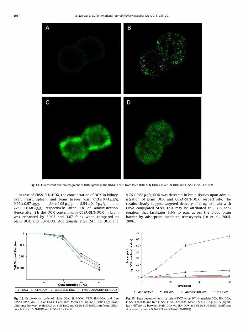
.9. Fluorescent microscopy
Analogous to the results of quantitative cellular uptake stud-es reported above, fluorescent photomicrographs showed higherptake of CBSA-SLN-DOX in BCs as well as in tumor cells, followedy SLN-DOX > CBSA + CBSA-SLN-DOX > plain DOX at equivalentoncentrations of DOX (Figs. 10 and 11).
.10. Cytotoxicity study
In vitro cell survival fraction of HNGC1 cells after incubation with
OX, SLN-DOX, CBSA-SLN-DOX and CBSA + CBSA-SLN-DOX werenalyzed using MTT assay. HNCG1 cell lines were incubated withoses ranging from 10.0 to 0.01 �M. The result of the cell inhibitionssay exhibited considerable differences. In similarity to results ofPharmaceutics 421 (2011) 189– 201
cellular uptake CBSA-SLN-DOX exhibited the highest percent cellgrowth inhibition compared to plain DOX or SLN-DOX. In all theformulations nearly no influence to the viability of cells was seenwhen the equivalent concentration of DOX was below 1 �M/mLand the cell viabilities obtained were all above 95% (Fig. 12).
As the equivalent concentration of DOX was augmentedbeyond 1 �M/mL, the cytotoxic effect was sort out as plainDOX < free CBSA (1 mg/mL) + CBSA-SLN-DOX < SLN–DOX < CBSA-SLN-DOX. Experiments performed on xenografts model clearlyillustrate a dose dependent cytotoxic activity that is a decrease insurvival fraction with increase in concentration of drug. Probably,the cytotoxic action of drug that intercalates with DNA dependsupon entry of drug in the cell and not merely by its presence inthe surrounding milieu of a cell. CBSA conjugation facilitated maxi-mum intracellular entry of CBSA-SLN-DOX via adsorption mediatedendocytosis. Subsequently slow and sustained release of DOX leadto a significantly higher cytotoxic action as compared free DOXor SLN-DOX. The cytotoxic effect via plain SLNs (SLN-DOX) wasless than ligand conjugated SLNs possibly due to their entry inabsence of any active transport mechanism and mediated merelyby passive diffusion. Similar results have been obtained in ear-lier studies. Yadav et al. (2010) illustrated the cytotoxic effect ofchondritin sulfate (CS) anchored nanoparticles to be greatest ascompared to non-ligand conjugated nanoparticles or plain drug.This was ascribed to ligand-receptor activity between CS and itsreceptors present on the surface of tumor cells, which possiblypromoted their augmented internalization. In a similar study per-formed on drug-resistant human breast cancer cells, Wong et al.revealed 8-fold higher cytotoxic action of doxorubicin loaded ina polymer–lipid hybrid nanoparticle system as compared to plaindoxorubicin solution (Wong et al., 2006).
Further, a 100% cell inhibition was perceived from all the formu-lations when the equivalent concentration of free DOX or its SLNsformulations was 100 �M/mL or more. A possible reason may beentry of adequate amount of DOX inside the cell from all the for-mulations at this concentration during a fairly long incubation timeof 72 h.
3.11. In vitro transendothelial transport study
Transcytosis ability of plain DOX, SLN-DOX, CBSA-SLN-DOXand free CBSA (1 mg/mL) + CBSA-SLN-DOX was studied on a cocul-ture of BCs and astrocytes (Fig. 13). Their permeability coefficient(Pe) were 0.8 × 10⁻⁶ cm/s, 1.9 × 10⁻⁶ cm/s, 2.5 × 10⁻⁵ cm/s, and1.1 × 10−6 cm/s, respectively. The results point that maximumtransendothelial transport was obtained in case of CBSA-SLN-DOX.This may be attributed to ability of cationic protein conjugatedSLNs to undergo adsorptive-mediated transcytosis across the BCs.Transendothelial flux was less for free CBSA + CBSA-SLN-DOX, pos-sibly due to presence of free CBSA which competitively inhibitedthe movement. This further verifies that the transendothelial move-ment was mediated by CBSA.
3.12. In vivo pharmacokinetic and biodistribution studies
Circulation life time, pharmacokinetics as well as therapeu-tic efficacy of DOX was immensely altered upon entrapment inSLNs and conjugation of a ligand. Fig. 14 represents the plasmaprofiles of various DOX formulations after single i.v. injection inBalb/c mice. The resulting pharmacokinetic parameters were deter-mined and are shown in Table 2. The initial Cmax obtained afterinjection of DOX loaded in SLNs was slightly lower than plain
DOX. However, DOX was detected in the serum until the end of24 and 21 h after administration of CBSA-SLN-DOX and SLN-DOX,respectively, and only for 8 h in case of plain DOX solution. Fur-thermore, the serum AUC0–24, AUC0–inf, AUMC0–t and AUMC0–inf of
A. Agarwal et al. / International Journal of Pharmaceutics 421 (2011) 189– 201 197
Fig. 10. Fluorescent photomicrographs of uptake of DOX in BCs from p
Table 2Pharmacokinetic parameters in serum of Balb/c mice.
Parameters Plain DOX SLN-DOX CBSA-SLN-DOX
Cmax (�g) 4.87 4.15 3.95Kel 0.47 0.15 0.13Clt (mL/h) 465.12 163.18 134.70AUC0–t (�g h/mL) 10.75 30.64 37.12AUC0–inf (�g h/mL) 10.96 31.11 39.33AUMC0–t (�g h2/mL) 20.85 203.12 293.15AUMC0–inf (�g h2/mL) 23.02 216.16 363.12
CDroctwd9o1tbcgt(afnS
systemic circulation upon entrapment in SLNs facilitated redistri-
T1/2 (h) 1.48 4.65 5.29MRT (h) 2.09 6.95 9.23
BSA-SLN-DOX was 1.21, 1.26, 1.44 and 1.68 folds higher than SLN-OX and 3.45, 3.59, 14.06 and 15.77 folds higher than plain DOX,
espectively. The results clearly indicate long circulation propertyf SLNs with ligand conjugated formulation having the greatest cir-ulation duration. The studies are in line with previous reportshat stated CBSA to have favorable pharmacokinetic propertiesith a longer serum half-life (Bickel et al., 2001). The mean resi-ence time (MRT) of CBSA-SLN-DOX, SLN-DOX and plain DOX was.23, 6.95 and 2.09 h, respectively. Additionally, the half-life (t1/2)f CBSA-SLN-DOX, SLN-DOX and plain DOX was 5.29, 4.65, and.48 h, respectively. These data also support extended residenceime and sustained release profile of the drug loaded in SLNs inody as compared to free DOX and the maximum being for CBSAonjugated formulation. The blood level of DOX was sustained to areater extent in case of CBSA-SLN-DOX than SLN-DOX possibly dueo double barrier effect to drug diffusion upon ligand conjugationSoni et al., 2006). Furthermore, the high Clt and Kel of plain DOXs compared to DOX loaded in SLNs indicated their slow clearance
rom the body and may be conferred to encapsulation of drug in theanoparticles. The data presented to this point clearly imply CBSA-LN-DOX have markedly better bioavailability and apparently morelain DOX, SLN-DOX, CBSA-SLN-DOX and CBSA + CBSA-SLN-DOX.
prolonged retention in systemic circulation and the effect couldnot be achieved via pre-treatment of mice with SLN-DOX or plainDOX.
Organ distribution studies were conducted to explore the possi-ble effectiveness of CBSA-SLN-DOX to deliver DOX to brain tissuesat the same time bypass non-target tissues (spleen, kidney, heart,and liver). The biodistribution of DOX from plain DOX and its for-mulations in solid organs was determined at the same time pointsas the brain (2, 8 and 24 h) (Fig. 15).
After, 2 h of administration the content of DOX when injectedas plain solution in kidney, liver, heart, spleen, and brain tis-sues was 37.15 ± 0.81 �g/g, 12.01 ± 0.37 �g/g, 6.12 ± 0.16 �g/g,9.35 ± 0.28 �g/g, and 0.45 ± 0.04 �g/g, respectively. The dataclearly suggests the greatest access of plain DOX to the kidney,which is its major clearance organ and uptake by liver/spleen,which are the organs rich in cells of RES. The data also illustratesthe rationale of cardiotoxicity produced by the drug, the concen-tration being high in heart. Only a minimal concentration of DOXwas detected in brain. DOX being hydrophilic in nature, the bloodbrain barrier acts as a major obstacle for delivery and further con-siderably impedes therapeutic effect. Moreover, the concentrationof free DOX declined quickly in all the organs being undetectable inbrain and kidney after 24 h of administration. This indicates rapidelimination/metabolism and clearance of drug from the body whenadministered as plain solution.
Encapsulation of DOX in plain SLNs resulted in an elevatedconcentration of DOX in liver and spleen and may be attributedto uptake of lipid particulates by mononuclear phagocytic sys-tem. A slight increase in DOX concentration in brain tissues wasalso observed. Undoubtedly, augmented dwell time of DOX in the
bution of drug to different organs. However absence of a targetingmoiety lead to non-specific/non-selective distribution and hencethe effect was not prominent.
198 A. Agarwal et al. / International Journal of Pharmaceutics 421 (2011) 189– 201
cells f
l92Hwp
FCde
Fig. 11. Fluorescent photomicrographs of DOX uptake in the HNGC 1
In case of CBSA-SLN-DOX, the concentration of DOX in kidney,iver, heart, spleen, and brain tissues was 7.13 ± 0.41 �g/g,.92 ± 0.57 �g/g, 1.34 ± 0.05 �g/g, 8.54 ± 0.49 �g/g and
2.93 ± 0.68 �g/g, respectively after 2 h of administration.ence after 2 h the DOX content with CBSA-SLN-DOX in brainas enhanced by 50.95 and 3.67 folds when compared tolain DOX and SLN-DOX. Additionally after 24 h no DOX andig. 12. Cytotoxicity study of plain DOX, SLN-DOX, CBSA-SLN-DOX and freeBSA + CBSA-SLN-DOX on HNGC 1 cell lines. Mean ± SD (n = 6; p ≤ 0.05 (significantifference between plain DOX vs. SLN-DOX and CBSA-SLN-DOX; significant differ-nce between SLN-DOX and CBSA-SLN-DOX)).
rom Plain DOX, SLN-DOX, CBSA-SLN-DOX and CBSA + CBSA-SLN-DOX.
9.76 ± 0.68 �g/g DOX was detected in brain tissues upon admin-istration of plain DOX and CBSA-SLN-DOX, respectively. Theresults clearly suggest targeted delivery of drug to brain with
CBSA conjugated SLNs. This may be attributed to CBSA con-jugation that facilitates SLNs to pass across the blood brainbarrier by adsorption mediated transcytosis (Lu et al., 2005,2006).Fig. 13. Time dependent transcytosis of DOX across BCs from plain DOX, SLN-DOX,CBSA-SLN-DOX and free CBSA + CBSA-SLN-DOX. Mean ± SD (n = 6; p ≤ 0.05 (signif-icant difference between Plain DOX vs. SLN-DOX and CBSA-SLN-DOX; significantdifference between SLN-DOX and CBSA-SLN-DOX)).

A. Agarwal et al. / International Journal of Pharmaceutics 421 (2011) 189– 201 199
Table 3Hematological parameters after administration of various nanoparticulate formulations.
Groups WBC (×.103/�L) RBC (×106/�L) Hb (g/dL) Platelet count (×103/�L)
Control 7.3 ± 1.5 9.7 ± 0.6 17.2 ± 1.3 989.7 ± 37.5Free DOX* 4.5 ± 0.4 6.4 ± 0.5 10.9 ± 0.8 761.2 ± 39.7SLN-DOX*,a 6.8 ± 0.7 9.0 ± 0.8 16.3 ± 1.2 938.3 ± 42.4CBSA-SLN-DOX*,a 7.1 ± 1.0 9.4 ± 0.5 16.8 ± 1.4 975.6 ± 38.2
n = 6; p ≤ 0.05.a Significant difference between SLN-DOX and CBSA-SLN-DOX.* Significant difference between free DOX vs. SLN-DOX and CBSA-SLN-DOX.
Fig. 14. Serum concentration of DOX obtained from plain DOX, SLN-DOX and CBSA-SbS
hbacimnDa
3
ct
FD
LN-DOX at various time intervals. Mean ± SD (n = 6; p ≤ 0.05 (significant differenceetween plain DOX vs. SLN-DOX and CBSA-SLN-DOX; significant difference betweenLN-DOX and CBSA-SLN-DOX)).
Adding up, the RES uptake of CBSA-SLN-DOX was only slightlyigher than plain DOX but less than SLN-DOX. This may possiblye due to presence of CBSA that has high affinity to brain tissuess compared to other organs (liver, heart, and lung). Moreoveronjugation of protein ligand provided some surface hydrophilic-ty to SLNs that further helped to evade uptake of formulation by
acrophage cells of liver and spleen. An imperative aspect was theoteworthy reduction in distribution of DOX to heart by CBSA-SLN-OX indicating their prospective in diminishing the cardiotoxicityssociated with DOX therapy.
.13. Hematological study
Marked decrease in blood cells count such as WBC count, RBCount, Hb content, platelet count were recorded after adminis-ration of free drug solutions for 7 days (Table 3). This might be
ig. 15. Biodistribution of DOX obtained from plain DOX, SLN-DOX and CBSA-SLN-DOX iOX vs. SLN-DOX and CBSA-SLN-DOX; significant difference between SLN-DOX and CBSA
possibly due to the direct contact of free drug with blood cells.The same amount of drug when encapsulated in various nanopar-ticulate (both plain and ligand conjugated) formulations reducedhematological toxicity. Further, in case of mice receiving CBSA-SLN-DOX formulation at a dose level of 5 mg/kg, almost no change inblood cells count was observed as compared to control group. Thismight possibly be due to the fact that, ligand anchored nanopar-ticulate formulations released their content i.e. drug directly to thetarget sites and very less amount of drug was leached out in bloodduring circulation. The results were in accordance with the resultsobtained previously by Jain et al. (2010) and Bhadra et al. (2005).
3.14. Evaluation of nephrotoxic and hepatotoxic effect
Serum urea/creatinine level and SGPT/AST/ALP level are usedas markers of renal function and liver function, respectively. Theserum urea and creatinine level (p < 0.001) were significantlyincreased upon administration of plain DOX (Table 4). Howevertheir elevation in the serum was least in animals administered withCBSA-SLN-DOX (5 mg/kg of DOX) (Table 4), indicating the formu-lation to evade damage to renal functions. This may be attributedto minimal quantity of CBSA conjugated SLNs encapsulating DOXgained access in kidney as compared to plain DOX.
In vivo hepatotoxicity induced by the anticancer drugs givenin free/entrapped form was estimated in terms of serum glu-tamic pyruvic transaminase (SGPT), and alkaline phosphatase(ALP) and total bilurubin concentration. Free DOX induced toxic-ity in infected animals was reflected by the increased activity ofSGPT (35.2 ± 3.2 IU/L), ALP (69.9 ± 4.7 IU/L) and AST concentration(29.7 ± 2.2 IU/L), while the encapsulated DOX in plain nanoparticlesshowed a small increase in the activity of SGPT (25.7 ± 1.7 IU/L), ALP
(61.4 ± 4.6 IU/L) and AST (19.3 ± 2.1 IU/L). CBSA-SLN-DOX nanopar-ticles exhibited only a slight change in activity of these enzymesas compared with control animals (SGPT 16.5 ± 1.6 IU/L; ALP54.8 ± 4.1 IU/L and AST 14.0 ± 1.5 IU/L) (Table 4). The differencesn different tissues. Mean ± SD (n = 6; p ≤ 0.05 (significant difference between plain-SLN-DOX)).
200 A. Agarwal et al. / International Journal of Pharmaceutics 421 (2011) 189– 201
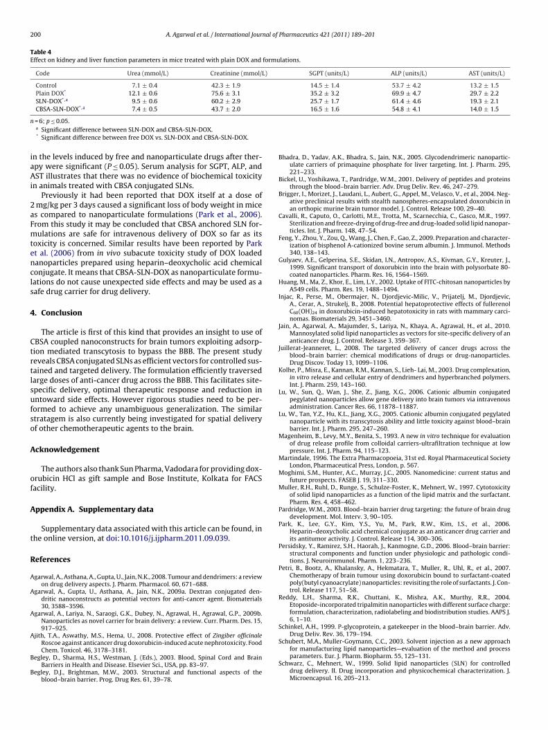
Table 4Effect on kidney and liver function parameters in mice treated with plain DOX and formulations.
Code Urea (mmol/L) Creatinine (mmol/L) SGPT (units/L) ALP (units/L) AST (units/L)
Control 7.1 ± 0.4 42.3 ± 1.9 14.5 ± 1.4 53.7 ± 4.2 13.2 ± 1.5Plain DOX* 12.1 ± 0.6 75.6 ± 3.1 35.2 ± 3.2 69.9 ± 4.7 29.7 ± 2.2SLN-DOX*,a 9.5 ± 0.6 60.2 ± 2.9 25.7 ± 1.7 61.4 ± 4.6 19.3 ± 2.1CBSA-SLN-DOX*,a 7.4 ± 0.5 43.7 ± 2.0 16.5 ± 1.6 54.8 ± 4.1 14.0 ± 1.5
n
iaAi
2aFmtencls
4
Ctrtlsufso
A
of
A
t
R
A
A
A
A
B
B
= 6; p ≤ 0.05.a Significant difference between SLN-DOX and CBSA-SLN-DOX.* Significant difference between free DOX vs. SLN-DOX and CBSA-SLN-DOX.
n the levels induced by free and nanoparticulate drugs after ther-py were significant (P ≤ 0.05). Serum analysis for SGPT, ALP, andST illustrates that there was no evidence of biochemical toxicity
n animals treated with CBSA conjugated SLNs.Previously it had been reported that DOX itself at a dose of
mg/kg per 3 days caused a significant loss of body weight in mices compared to nanoparticulate formulations (Park et al., 2006).rom this study it may be concluded that CBSA anchored SLN for-ulations are safe for intravenous delivery of DOX so far as its
oxicity is concerned. Similar results have been reported by Parkt al. (2006) from in vivo subacute toxicity study of DOX loadedanoparticles prepared using heparin–deoxycholic acid chemicalonjugate. It means that CBSA-SLN-DOX as nanoparticulate formu-ations do not cause unexpected side effects and may be used as aafe drug carrier for drug delivery.
. Conclusion
The article is first of this kind that provides an insight to use ofBSA coupled nanoconstructs for brain tumors exploiting adsorp-ion mediated transcytosis to bypass the BBB. The present studyeveals CBSA conjugated SLNs as efficient vectors for controlled sus-ained and targeted delivery. The formulation efficiently traversedarge doses of anti-cancer drug across the BBB. This facilitates site-pecific delivery, optimal therapeutic response and reduction inntoward side effects. However rigorous studies need to be per-ormed to achieve any unambiguous generalization. The similartratagem is also currently being investigated for spatial deliveryf other chemotherapeutic agents to the brain.
cknowledgement
The authors also thank Sun Pharma, Vadodara for providing dox-rubicin HCl as gift sample and Bose Institute, Kolkata for FACSacility.
ppendix A. Supplementary data
Supplementary data associated with this article can be found, inhe online version, at doi:10.1016/j.ijpharm.2011.09.039.
eferences
garwal, A., Asthana, A., Gupta, U., Jain, N.K., 2008. Tumour and dendrimers: a reviewon drug delivery aspects. J. Pharm. Pharmacol. 60, 671–688.
garwal, A., Gupta, U., Asthana, A., Jain, N.K., 2009a. Dextran conjugated den-dritic nanoconstructs as potential vectors for anti-cancer agent. Biomaterials30, 3588–3596.
garwal, A., Lariya, N., Saraogi, G.K., Dubey, N., Agrawal, H., Agrawal, G.P., 2009b.Nanoparticles as novel carrier for brain delivery: a review. Curr. Pharm. Des. 15,917–925.
jith, T.A., Aswathy, M.S., Hema, U., 2008. Protective effect of Zingiber officinaleRoscoe against anticancer drug doxorubicin-induced acute nephrotoxicity. Food
Chem. Toxicol. 46, 3178–3181.egley, D., Sharma, H.S., Westman, J. (Eds.), 2003. Blood, Spinal Cord and BrainBarriers in Health and Disease. Elsevier Sci., USA, pp. 83–97.
egley, D.J., Brightman, M.W., 2003. Structural and functional aspects of theblood–brain barrier. Prog. Drug Res. 61, 39–78.
Bhadra, D., Yadav, A.K., Bhadra, S., Jain, N.K., 2005. Glycodendrimeric nanopartic-ulate carriers of primaquine phosphate for liver targeting. Int. J. Pharm. 295,221–233.
Bickel, U., Yoshikawa, T., Pardridge, W.M., 2001. Delivery of peptides and proteinsthrough the blood–brain barrier. Adv. Drug Deliv. Rev. 46, 247–279.
Brigger, I., Morizet, J., Laudani, L., Aubert, G., Appel, M., Velasco, V., et al., 2004. Neg-ative preclinical results with stealth nanospheres-encapsulated doxorubicin inan orthopic murine brain tumor model. J. Control. Release 100, 29–40.
Cavalli, R., Caputo, O., Carlotti, M.E., Trotta, M., Scarnecchia, C., Gasco, M.R., 1997.Sterilization and freeze-drying of drug-free and drug-loaded solid lipid nanopar-ticles. Int. J. Pharm. 148, 47–54.
Feng, Y., Zhou, Y., Zou, Q., Wang, J., Chen, F., Gao, Z., 2009. Preparation and character-ization of bisphenol A-cationized bovine serum albumin. J. Immunol. Methods340, 138–143.
Gulyaev, A.E., Gelperina, S.E., Skidan, I.N., Antropov, A.S., Kivman, G.Y., Kreuter, J.,1999. Significant transport of doxorubicin into the brain with polysorbate 80-coated nanoparticles. Pharm. Res. 16, 1564–1569.
Huang, M., Ma, Z., Khor, E., Lim, L.Y., 2002. Uptake of FITC-chitosan nanoparticles byA549 cells. Pharm. Res. 19, 1488–1494.
Injac, R., Perse, M., Obermajer, N., Djordjevic-Milic, V., Prijatelj, M., Djordjevic,A., Cerar, A., Strukelj, B., 2008. Potential hepatoprotective effects of fullerenolC60(OH)24 in doxorubicin-induced hepatotoxicity in rats with mammary carci-nomas. Biomaterials 29, 3451–3460.
Jain, A., Agarwal, A., Majumder, S., Lariya, N., Khaya, A., Agrawal, H., et al., 2010.Mannosylated solid lipid nanoparticles as vectors for site-specific delivery of ananticancer drug. J. Control. Release 3, 359–367.
Juillerat-Jeanneret, L., 2008. The targeted delivery of cancer drugs across theblood–brain barrier: chemical modifications of drugs or drug-nanoparticles.Drug Discov. Today 13, 1099–1106.
Kolhe, P., Misra, E., Kannan, R.M., Kannan, S., Lieh- Lai, M., 2003. Drug complexation,in vitro release and cellular entry of dendrimers and hyperbranched polymers.Int. J. Pharm. 259, 143–160.
Lu, W., Sun, Q., Wan, J., She, Z., Jiang, X.G., 2006. Cationic albumin conjugatedpegylated nanoparticles allow gene delivery into brain tumors via intravenousadministration. Cancer Res. 66, 11878–11887.
Lu, W., Tan, Y.Z., Hu, K.L., Jiang, X.G., 2005. Cationic albumin conjugated pegylatednanoparticle with its transcytosis ability and little toxicity against blood–brainbarrier. Int. J. Pharm. 295, 247–260.
Magenheim, B., Levy, M.Y., Benita, S., 1993. A new in vitro technique for evaluationof drug release profile from colloidal carriers-ultrafiltration technique at lowpressure. Int. J. Pharm. 94, 115–123.
Martindale, 1996. The Extra Pharmacopoeia, 31st ed. Royal Pharmaceutical SocietyLondon, Pharmaceutical Press, London, p. 567.
Moghimi, S.M., Hunter, A.C., Murray, J.C., 2005. Nanomedicine: current status andfuture prospects. FASEB J. 19, 311–330.
Muller, R.H., Ruhl, D., Runge, S., Schulze-Foster, K., Mehnert, W., 1997. Cytotoxicityof solid lipid nanoparticles as a function of the lipid matrix and the surfactant.Pharm. Res. 4, 458–462.
Pardridge, W.M., 2003. Blood–brain barrier drug targeting: the future of brain drugdevelopment. Mol. Interv. 3, 90–105.
Park, K., Lee, G.Y., Kim, Y.S., Yu, M., Park, R.W., Kim, I.S., et al., 2006.Heparin–deoxycholic acid chemical conjugate as an anticancer drug carrier andits antitumor activity. J. Control. Release 114, 300–306.
Persidsky, Y., Ramirez, S.H., Haorah, J., Kanmogne, G.D., 2006. Blood–brain barrier:structural components and function under physiologic and pathologic condi-tions. J. Neuroimmunol. Pharm. 1, 223–236.
Petri, B., Bootz, A., Khalansky, A., Hekmatara, T., Muller, R., Uhl, R., et al., 2007.Chemotherapy of brain tumour using doxorubicin bound to surfactant-coatedpoly(butyl cyanoacrylate) nanoparticles: revisiting the role of surfactants. J. Con-trol. Release 117, 51–58.
Reddy, L.H., Sharma, R.K., Chuttani, K., Mishra, A.K., Murthy, R.R., 2004.Etoposide-incorporated tripalmitin nanoparticles with different surface charge:formulation, characterization, radiolabeling and biodistribution studies. AAPS J.6, 1–10.
Schinkel, A.H., 1999. P-glycoprotein, a gatekeeper in the blood–brain barrier. Adv.Drug Deliv. Rev. 36, 179–194.
Schubert, M.A., Muller-Goymann, C.C., 2003. Solvent injection as a new approach
for manufacturing lipid nanoparticles—evaluation of the method and processparameters. Eur. J. Pharm. Biopharm. 55, 125–131.Schwarz, C., Mehnert, W., 1999. Solid lipid nanoparticles (SLN) for controlleddrug delivery. II. Drug incorporation and physicochemical characterization. J.Microencapsul. 16, 205–213.
nal of
S
S
T
T
U
V
W
A. Agarwal et al. / International Jour
oni, V., Kohli, D.V., Jain, S.K., 2006. Transferrin coupled liposomes for enhancedbrain delivery of doxorubicin. Vasc. Dis. Prev. 3, 31–38.
torm, P.B., Moriarity, J.L., Tyler, B., Burger, P.C., Brem, H., Weingart, J., 2002. Poly-mer delivery of camptothecin against 9L gliosarcoma: release, distribution, andefficacy. J. Neurooncol. 56, 209–217.
hole, M., Nobmanna, S., Huwyler, J., Bartmann, A., Fricker, G., 2002. Uptake ofcationized albumin coupled liposomes by cultured porcine brain microvesselendothelial cells and intact brain capillaries. J. Drug Target. 103, 37–44.
orchilin, V.P., 2005. Recent advances with liposomes as pharmaceutical carriers.Nat. Rev. Drug Discov. 41, 45–60.
treja, S., Jain, N.K., 2001. Solid lipid nanoparticles. In: Jain, N.K. (Ed.), Advances in
Controlled and Novel Drug Delivery. CBS Publishers, New Delhi, pp. 408–425.yas, S.P., Khar, R.K., 2002. Targeted and Controlled Drug Delivery Novel CarrierSystems, 1st ed. CBS Publishers, New Delhi, pp. 331–86.
iechelman, K.J., Braun, R.D., Fitzpatrick, J.D., 1988. BCA assay protocol. Anal.Biochem., 175.
Pharmaceutics 421 (2011) 189– 201 201
Wong, H.L., Bendayan, R., Rauth, A.M., Xue, H.Y., Babakhanian, K., Wu, X.Y., 2006. Amechanistic study of enhanced doxorubicin uptake and retention in multidrugresistant breast cancer cells using a polymer–lipid hybrid nanoparticle system.J. Pharmacol. Exp. Ther. 317, 1372–1381.
Wu, D., Pardridge, W.M., 1999. Blood–brain barrier transport of reduced folic acid.Pharm. Res. 16, 415–419.
Xie, Y.L., Lu, W., Jiang, X.G., 2006. Improvement of cationic albumin conjugatedpegylated nanoparticles holding NC-1900, a vasopressin fragment analog, inmemory deficits induced by scopolamine in mice. Behav. Brain. Res. 173,76–84.
Yadav, A.K., Mishra, P., Mishra, A.K., Mishra, P., Jain, S., Agrawal, G.P., 2007. Develop-
ment and characterization of hyaluronic acid-anchored PLGA nanoparticulatecarriers of doxorubicin. Nanomed. Nanotechnol. Biol. Med. 3, 246–257.Yadav, A.K., Agarwal, A., Jain, S., Mishra, A.K., Bid, H., Rai, G., et al., 2010. Chondroitinsulphate decorated nanoparticulate carriers of 5-fluorouracil: development andin vitro characterization. J. Biomed. Nanotechnol. 6, 1–11.
Copyright © 2022 FDOKUMEN