Dynamical effects in multifragmentation at intermediate energies
Upload
khangminh22Category
view
5download
0
ChemicalScience
EDGE ARTICLE
Ope
n A
cces
s A
rtic
le. P
ublis
hed
on 2
4 Se
ptem
ber
2021
. Dow
nloa
ded
on 1
2/9/
2021
12:
56:3
4 PM
. T
his
artic
le is
lice
nsed
und
er a
Cre
ativ
e C
omm
ons
Attr
ibut
ion-
Non
Com
mer
cial
3.0
Unp
orte
d L
icen
ce.
View Article OnlineView Journal | View Issue
Alchemical absol
aComputational Biomolecular Dynamics G
Computational Biophysics, Max Planck
D-37077, Gottingen, Germany. E-mail: vgapbComputational Chemistry, Janssen
Pharmaceutica N. V., Turnhoutseweg 30, 23cMIT Department of Chemical Engineering
Cambridge, MA 02139, USAdDepartment of Pharmaceutical Sciences, U
USAeDepartment of Chemistry, University of Cal
† Electronic supplementary informa10.1039/d1sc03472c
Cite this: Chem. Sci., 2021, 12, 13958
All publication charges for this articlehave been paid for by the Royal Societyof Chemistry
Received 25th June 2021Accepted 23rd September 2021
DOI: 10.1039/d1sc03472c
rsc.li/chemical-science
13958 | Chem. Sci., 2021, 12, 13958–1
ute protein–ligand binding freeenergies for drug design†
Y. Khalak,a G. Tresadern, b M. Aldeghi, c H. M. Baumann,d D. L. Mobley,de B. L. deGroot a and V. Gapsys *a
The recent advances in relative protein–ligand binding free energy calculations have shown the value of
alchemical methods in drug discovery. Accurately assessing absolute binding free energies, although
highly desired, remains a challenging endeavour, mostly limited to small model cases. Here, we
demonstrate accurate first principles based absolute binding free energy estimates for 128
pharmaceutically relevant targets. We use a novel rigorous method to generate protein–ligand
ensembles for the ligand in its decoupled state. Not only do the calculations deliver accurate protein–
ligand binding affinity estimates, but they also provide detailed physical insight into the structural
determinants of binding. We identify subtle rotamer rearrangements between apo and holo states of
a protein that are crucial for binding. When compared to relative binding free energy calculations,
obtaining absolute binding free energies is considerably more challenging in large part due to the need
to explicitly account for the protein in its apo state. In this work we present several approaches to obtain
apo state ensembles for accurate absolute DG calculations, thus outlining protocols for prospective
application of the methods for drug discovery.
1 Introduction
Computational techniques for estimating relative differences inprotein–ligand binding free energy have now reached remark-able accuracy. Relative binding free energy calculations overa large range of protein–ligand complexes have shown averageagreement with experiment to be within 1 kcal mol�1
(4.184 kJ mol�1).16,28,52 These methods have become mature andreliable enough to be included in industrial drug discovery andlead optimization pipelines.10,26,35,40 A substantial limitation ofthis approach, however, is the requirement for the ligands to bestructurally similar to each other: the predictive powerdecreases for ligands with different scaffolds or binding poses.Evaluation of novel ligand classes, therefore, requires a priorexperimental absolute binding free energy as a reference foreach new class of mutually similar ligands. Thus, the next
roup, Department of Theoretical and
Institute for Biophysical Chemistry,
Research & Development, Janssen
40, Beerse, Belgium
, Massachusetts Institute of Technology,
niversity of California, Irvine, CA 92697,
ifornia, Irvine, CA 92697, USA
tion (ESI) available. See DOI:
3971
qualitative leap for the eld of rst principles based protein–ligand affinity estimation encompasses the reliable and accu-rate prediction of absolute binding free energies.
The calculation of relative binding free energies is relativelyeasy in comparison. The bound ligands are conned to thebinding site and only the small subset of atoms that differbetween two ligands needs to be perturbed. In contrast, abso-lute binding free energy calculations decouple the entire ligand,meaning it is in principle free to explore the whole simulationbox volume. Early work on the topic explored various ways ofrestraining the decoupled ligand and taking into account theresulting contribution to the free energy.7,18,20,54 The approachintroduced by Boresch et al.7 has emerged as a rigorous way toresolve this issue via orthonormal relative restraints betweenthe ligand and the protein.2,3
Another challenge for the absolute binding free energycalculations is posed by the need to explicitly sample the apostate of the protein, i.e. the protein without the bound ligand. Asthis state may substantially differ from the ligand bound (holo)state, the simulation method needs to be capable of capturingthe free energy differences between the protein conformers.Non-equilibrium (NEQ) free energy calculations present anelegant solution to this challenge. Such calculations determinethe free energy difference by performing rapid out-of-equilibrium ligand coupling/decoupling transitions initializedfrom the equilibrium protein apo and holo ensembles(Fig. S1†). This allows one to explicitly include the different apoand holo end-states into the same calculation.17 Several recent
© 2021 The Author(s). Published by the Royal Society of Chemistry

Edge Article Chemical Science
Ope
n A
cces
s A
rtic
le. P
ublis
hed
on 2
4 Se
ptem
ber
2021
. Dow
nloa
ded
on 1
2/9/
2021
12:
56:3
4 PM
. T
his
artic
le is
lice
nsed
und
er a
Cre
ativ
e C
omm
ons
Attr
ibut
ion-
Non
Com
mer
cial
3.0
Unp
orte
d L
icen
ce.
View Article Online
applications of the NEQ approach on model host–guest systemsshowed promising results for the calculation of absolutebinding free energies.5,27,37
The NEQ approach does not offer a free lunch in the sensethat the relevant conformations still need to be sampled in theend-state ensembles.17 Compared to the more popular freeenergy perturbation (FEP) series of methods,24,51,56 though, itdoes offer several advantages in terms of computational effi-ciency. Namely, such sampling needs to be performed only forphysical end-states and can be done with plain moleculardynamics or, if desired, it can also be augmented with enhancedsampling methodologies in a straightforward manner.36
Secondly, the out-of-equilibrium portion of the approach, whichaccounts for the majority of the compute time, is highly paral-lelizable, requiring no information exchange between indi-vidual simulations, unlike modern FEP approaches with replicaexchange.24,51 Furthermore, the NEQ approach allows forinitialization of the two end-states with the distinct apo andholo protein structures, which facilitates obtaining reliableequilibrium ensembles for the cases where experimentalstructures are available. For an equilibrium FEP approach,incorporation of different conformers in a single DG estimationwould require decision on the mixing rule for seeding thestarting structures19 and potentially Hamiltonian replicaexchanges would be needed to achieve convergence. Finally,when comparing different ligands, protein mutations, orconformational states NEQ allows for reuse of existing equi-librium sampling of end-states, e.g. the same apo state can beused for assessing affinities of different ligands.
In the current work we use the NEQ approach to demonstratethe feasibility of accurate absolute binding free energy calcu-lations for a large number of protein–ligand systems, showingaccuracy on par with the relative binding free energy estimates.To achieve this, we introduce methodological advancementsthat allow for an efficient treatment of the ligand in its decou-pled state and careful considerations of the protein in its apostate. This allows for identication of protein states that havea drastic effect on ligand binding affinity, such as e.g. a ip ofa single amino acid rotamer. Our calculation strategy alsoallows identifying the most representative structure fora protein's apo state for the cases where multiple likely candi-dates (structures in their local free energy minima, X-raystructures) are available.
2 Results
In this study, we have used an alchemical non-equilibrium freeenergy calculation approach to calculate absolute protein–ligand binding free energies for 128 complexes. We havedeveloped a novel way of treating the decoupled state of theligand (see Methods section for details). The large set of inves-tigated systems allows us to have an extensive evaluation of theaccuracy that can be achieved with the rst principles basedcalculations. Fig. 1 shows the calculated values for the bindingfree energy plotted against the experimental measurements.When compared to the experimental values, the absoluteunsigned error (AUE) of 4.9� 0.5 kJ mol�1 (1.2� 0.1 kcal mol�1)
© 2021 The Author(s). Published by the Royal Society of Chemistry
only marginally exceeds the state-of-the-art accuracy thresholdof 1 kcal mol�1 achievable for relative binding free energycalculations. Accuracies for jnk1 and p38a are exceptionallygood with AUEs of 3.0 � 0.8 and 3.1 � 0.7 kJ mol�1 (0.7 �0.2 kcal mol�1 for both), respectively.
Some systems, however, have a considerably lower accuracy(AUE of 10.8 � 1.5 kJ mol�1 for tyk2, 5.5 � 1.4 kJ mol�1 forpde2), revealing a particular challenge for affinity estimation inthese systems. An accurate evaluation of the offset in the DG iscritical for obtaining reliable absolute DG values: inaccuraciesin this case manifest as large shis of the calculated values withrespect to the experimental measurements, e.g. tyk2 in Fig. 1.Interestingly, even such offsets do not signicantly deterioratethe relative free energy difference estimates (Fig. S2†).
We have identied this effect to be a consequence of theinadequate representation of the protein in its apo state. Whilethe apo state is not considered in relative free energy calcula-tions, assessment of absolute free energies needs to explicitlyaccount for it. In the following analysis we demonstrate howfailure to capture the free energy differences between the apoand holo protein states affects the absolute binding free energycalculation accuracy.
2.1 Apo and holo states
For the situations where protein rearrangements are requiredupon ligand binding, sufficient sampling of the two end statesmay present a considerable challenge. An accurate quantica-tion of the process of ligand binding to an apo protein andforming a stable holo state requires correctly estimating notonly the component of the free energy originating from theligand interaction with the protein, but also the differencebetween the apo and holo protein states.
Non-equilibrium free energy calculations offer a particularlyconvenient approach for the computation of binding affinities,as both states, apo and holo, can be explicitly considered ina single simulation.17 The alchemical ligand decoupling tran-sitions can be started from a holo conformer ensemble, whileligand coupling transitions can start from an apo ensemble.
Among the protein–ligand complexes investigated in thiswork, 6 out of 7 systems have both their apo and holo structuresresolved by means of X-ray crystallography. We have probed twomethods of calculating the binding DG value: rstly, removingthe ligand from the holo state and treating the obtained struc-ture as an apo state. For the second approach we used thecrystallographically resolved apo structure directly. Overall,there is a large and signicant improvement in the calculatedbinding DG accuracy when an experimentally dened apo stateis considered explicitly (Fig. 2 and S3†). A substantialimprovement in the AUE (from 7.1 � 0.6 to 4.4 � 0.5 kJ mol�1)shows that starting the simulations with a corresponding apostructure largely removes an offset which is otherwise presentfor the calculations initialized with the holo structures only.This indicates that substantial rearrangements occur in thestudied proteins upon ligand binding that do not equilibrate atthe nanosecond timescale covered in the simulations.
Chem. Sci., 2021, 12, 13958–13971 | 13959

Fig. 1 Overview of the accuracy of calculated absolute binding free energies DGCalc.. Error bars represent standard errors for free energies,absolute unsigned errors (AUE, units of kJ mol�1), and the Pearson correlation coefficients (Cor). Apo states were initialized with X-ray crystalstructures for all systems except pde2 and tyk2 where holo X-ray structures with the ligand removed were used. Dark and light shaded areasrepresent regions deviating from experiment by at most 1 and 2 kcal mol�1.
Chemical Science Edge Article
Ope
n A
cces
s A
rtic
le. P
ublis
hed
on 2
4 Se
ptem
ber
2021
. Dow
nloa
ded
on 1
2/9/
2021
12:
56:3
4 PM
. T
his
artic
le is
lice
nsed
und
er a
Cre
ativ
e C
omm
ons
Attr
ibut
ion-
Non
Com
mer
cial
3.0
Unp
orte
d L
icen
ce.
View Article Online
The largest effect from using an experimentally resolved apostructure is observed in the galectin, p38a and cdk2 protein–ligand complexes, while for the other cases the differences inaccuracy are less affected. To understand what structuralfeatures are responsible for such pronounced effects, we havefurther explored the p38a system for which DG had the largestdifference among the systems depicted in Fig. 2.
2.2 Large effect of a single rotamer
The p38a protein–ligand complex shows a particularly strongdependence of the calculated DG on the starting structure. Forthis case, we were able to identify the particular structuraldetails that are responsible for more than 9 kJ mol�1 offset inthe calculated DG values (Fig. 3).
One of the main differences between the apo (pdb id 1wfc53)and holo (3y) structures occurring close to the binding site isa major loop motion: colored in orange and blue in Fig. 3.However, it appears that even the short (10 ns) equilibriumsimulations that we employed in the current protocol aresufficient to sample this loop transition (Fig. S4†). We have alsoexplicitly probed whether this structural feature may modulatethe accuracy of the calculated DG values. We have ltered thestarting structure ensemble for the ligand coupling transitions,retaining only those conformers with a loop position similar to
13960 | Chem. Sci., 2021, 12, 13958–13971
the one from the crystallographic apo structure. This, however,had no effect on the calculated binding DG values (Fig. S5†).
While the large loop motion has no substantial effect on theDG accuracy, a single rotamer ip appears to be responsible forthe larger than 9 kJ mol�1 shi in calculated DG. The crystal-lographic structures 3y (holo) and 1wfc (apo) have differentthreonine 106 (T106) rotameric states. Initializing apo simula-tions with either the experimentally resolved apo structure ora holo structure with the ligand removed yields ensembleswhere the rotamer never crosses the barrier and remains in itsstarting state (Fig. 3). The barrier crossing for the T106 side-chain rotamer appears to be too high to be sampled in the short(10 ns) equilibrium simulations used in the free energy calcu-lation protocol.
To verify that T106 is truly the cause for this marked differ-ence, we have initialized ligand coupling simulations from theholo structure (with the ligand removed), but setting the T106rotamer into its apo state (green structures in Fig. 3). This singlechange in the holo structure was sufficient to bring the calcu-lated DG to the same accuracy as obtained from simulationsstarted with the true apo structure.
It appears that initializing ligand coupling simulations fromholo structures leaves the binding site – in particular the T106rotamer – pre-arranged to accommodate the ligand. This, inturn, leads to an overly stabilized protein–ligand complex as
© 2021 The Author(s). Published by the Royal Society of Chemistry

Fig. 2 Experimental binding DG values plotted against the calculated estimates. In the panel on the left, the simulations in the apo state werestarted from the experimental holo structure after removing the ligand. In the panel on the right, the alchemical simulations of the protein in itsapo state were initialized with the experimentally resolved apo structure. The starting structure has a marked effect on the calculated DGaccuracy. The bottom panel shows a break up of the accuracies by protein system. Probability values measure the significance of the differencebetween apo and holo absolute unsigned errors via a Welch's t-test. Dark and light shaded areas represent regions deviating from experiment byat most 1 and 2 kcal mol�1.
Edge Article Chemical Science
Ope
n A
cces
s A
rtic
le. P
ublis
hed
on 2
4 Se
ptem
ber
2021
. Dow
nloa
ded
on 1
2/9/
2021
12:
56:3
4 PM
. T
his
artic
le is
lice
nsed
und
er a
Cre
ativ
e C
omm
ons
Attr
ibut
ion-
Non
Com
mer
cial
3.0
Unp
orte
d L
icen
ce.
View Article Online
quantied by the binding DG. The missing term in DG, in thiscase, is the free energy required to switch T106 rotamer from itsapo to holo state. To demonstrate this, we also computed freeenergy surfaces for the residue's c1 dihedral with well-temperedmetadynamics4,29 simulations biasing the potential of thedihedral. The free energy surfaces (Fig. 4A) reveal the averagefree energy difference between the minima of gauche- and transconformations (present in the 1wfc and 3y structures,respectively) of the apo state to be �8 � 1 kJ mol�1. Thismatches well the observed shi in the binding free energiescalculated using 1wfc and 3y starting structures for the apostate. Due to insufficient end-state sampling and high freeenergy barriers, we do not observe a transition in this rotamerduring short 10 ns equilibrium simulations, yet simulationsstarted from the true apo state allowed taking the missing DGcontribution into account.
2.3 Can longer simulations reveal true apo states?
Undersampling is a frequently encountered shortcoming ofsimulation-based phase space exploration, e.g. numerousexamples are provided in ref. 14. Naturally, one of the under-lying reasons for the inadequate representation of the apo statein the case of p38a protein could be insufficient equilibration of
© 2021 The Author(s). Published by the Royal Society of Chemistry
the system. Therefore, we probed whether longer simulationswould be able to cross the energy barrier and arrive in the trueapo state when starting from a holo crystallographic structurewith the ligand removed. To explore this, we have extended thep38a apo state simulations started from 3y by performing 5independent runs of 1 ms each.
The longer simulations indeed showed a transition of theT106 rotamer from its trans state (3y holo conformer) to thegauche� state observed in the apo 1wfc structure (Fig. 4B). In all5 independent replicas, the transition occurred within the rst200 ns. Aer this, no recrossings back to the trans rotamericstate were observed, only short lived transitions from thegauche� to the gauche+ state occurred.
Binding DG calculations where sampling of the decoupledligand state is initialized with the nal structures from 1 mssimulations show this shi and have the same accuracy as thosestarted with the crystallographic apo state 1wfc (Fig. 4C and D).This conrms our previous observation that the rotameric stateof T106 plays a crucial role in the ligand binding to p38a. All inall, the observations from the long simulations suggest that, atleast in some cases, we can rely on longer (or enhanced)sampling to recover a protein's apo state for the subsequent DGcalculations.
Chem. Sci., 2021, 12, 13958–13971 | 13961

Fig. 3 A detailed investigation of the p38a protein–ligand complexes. The apo (1wfc,53 orange) and holo (3fly, blue) structures have severalstructural differences close to the ligand binding site: a substantial loopmotion and a different T106 rotamer state. In the simulations, the rotamerT106 retains its initial state: shown in lines, with a sphere marking threonine's oxygen. The calculated DG values depend strongly on the startingstructure (holo or apo) that is used to initialize protein simulations in its apo state: scatterplots at the bottom. The green structure in the sub-paneland corresponding DG scatterplot depict a case, where apo simulations were initialized with a holo structure (ligand removed), but with the T106rotamer set into its apo state. Dark and light shaded areas represent regions deviating from experiment by at most 1 and 2 kcal mol�1.
Chemical Science Edge Article
Ope
n A
cces
s A
rtic
le. P
ublis
hed
on 2
4 Se
ptem
ber
2021
. Dow
nloa
ded
on 1
2/9/
2021
12:
56:3
4 PM
. T
his
artic
le is
lice
nsed
und
er a
Cre
ativ
e C
omm
ons
Attr
ibut
ion-
Non
Com
mer
cial
3.0
Unp
orte
d L
icen
ce.
View Article Online
It is important to note, however, that the increased samplingdoes not automatically translate into a better agreement of thesimulated trajectory with the experimentally measured observ-ables. For example, longer simulations of the tyk2 kinase in itsapo state (4gih30 with the ligand removed; Fig. S6†) explorea broader range of conformations. However, as simulationsprogress, they deviate substantially from the starting crystallo-graphic structure. The substantial dri of simulated trajecto-ries, in turn, results in large uncertainties of the calculatedbinding affinites and deteriorates the DG prediction accuracy.This observation indicates that either the longer samplingreaching 1 ms for each of the 5 repeats is still not sufficient, orthe new free energy minima identied by the force eld are notrepresentative of the true free energy landscape.
13962 | Chem. Sci., 2021, 12, 13958–13971
2.4 Using binding DG to identify apo states
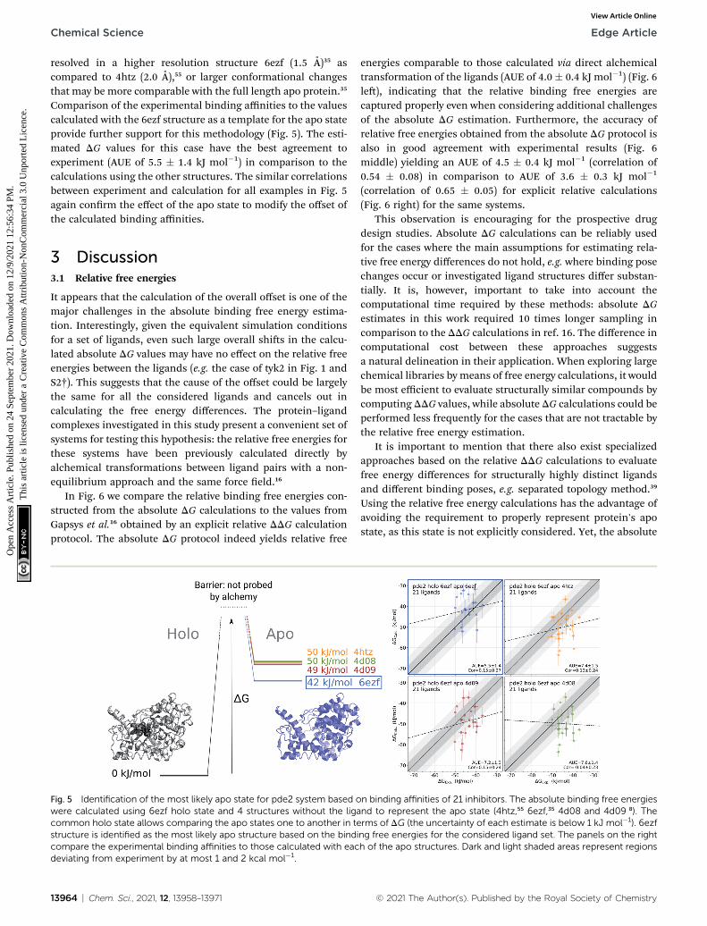
For the cases where multiple experimentally resolved structuresare available, it may not be evident which structure would bebest suited for initializing simulations to obtain a representa-tive apo state ensemble. It is, however, possible to exploitbinding free energy calculations to identify the structureyielding the most probable conformational ensemble. Thisanalysis does not require any knowledge of the actual (experi-mentally measured) set of binding affinities. It rather relies onmultiple calculations of the binding affinities connecting oneholo structure with multiple possible apo states (Fig. 5).
We use phosphodiesterase 2 (pde2) complexed with 21inhibitors35 to illustrate this approach. Numerous experimen-tally resolved monomeric pde2 structures are available, wherethe protein is crystallized in its apo state (e.g. 4htz55) or in
© 2021 The Author(s). Published by the Royal Society of Chemistry

Fig. 4 The effect of T106 rotamer states of the p38a kinase on the calculated ligand binding DG. Free energy surfaces (A) of the c1 dihedral anglefor the T106 residue obtained from well tempered metadynamics simulations starting from 3fly and 1wfc structures as well as from the output of1 ms equilibrations started from 3fly. c1 dihedral angle for the T106 residue in the crystallographic apo (1wfc) and holo (3fly) states, as well as in 5independent simulations of 1 ms each (B). BindingDG calculated by initializing apo state simulations with the 1wfc structure (C) and the end-statesfrom 1 ms simulations (D). Dark and light shaded areas represent regions deviating from experiment by at most 1 and 2 kcal mol�1.
Edge Article Chemical Science
Ope
n A
cces
s A
rtic
le. P
ublis
hed
on 2
4 Se
ptem
ber
2021
. Dow
nloa
ded
on 1
2/9/
2021
12:
56:3
4 PM
. T
his
artic
le is
lice
nsed
und
er a
Cre
ativ
e C
omm
ons
Attr
ibut
ion-
Non
Com
mer
cial
3.0
Unp
orte
d L
icen
ce.
View Article Online
a complex with a ligand (e.g. 6ezf,35 4d08, 4d09 8). Availability ofthese structures allows constructing a set of apo states by usingeither an actual apo conformer from the crystallographicstructure or by removing a ligand from a holo structure. Inprinciple, the most likely apo state is at its free energyminimum, i.e. of the multiple candidate conformers, the onewith the lowest free energy would be the most populated in theensemble. However, calculating free energy differences betweenthe apo conformers directly is a computationally highlydemanding challenge.
Instead, we can evaluate relative free energies of theseconformers by connecting them via a common holo state. Wecalculate binding affinities for a set of 21 pde2 inhibitors usingthe structure 6ezf representing the protein–ligand complex andeach of the 6ezf, 4htz, 4d08 and 4d09 structures independentlyrepresenting the apo state. In this way we relate each apo stateto one another via a common reference 6ezf holo state. Settingthe free energy of the reference to 0 kJ mol�1 for convenienceallows us to directly compare the apo states (Fig. 5): the DG foran apo state is represented by averaged binding free energiescalculated over the whole ligand set. The barrier (denoted withthe dashed lines in Fig. 5) is not attainable with this approach,
© 2021 The Author(s). Published by the Royal Society of Chemistry
as alchemical calculations do not explicitly probe the binding-unbinding pathway.
It is, however, important to understand the limitations of theDG values obtained this way. The calculated values should notbe interpreted as reporting on the actual free energy differencesbetween the apo conformers, but only on a component of DGcorresponding to the change in the degrees of freedom relevantfor ligand binding. It is likely that the binding site rearrange-ments are experienced by the ligands and have a strong effect onthe DG calculated based on this approach. At the same time,substantial conformational rearrangements further from thebinding site may not have a contribution to DG if they do notaffect the ligand binding affinity. Therefore, the conclusionsabout the most likely apo state identied with this approachshould be limited to the interpretations of the binding affinitiesfor a specic set of ligands.
In the current analysis, the 6ezf holo structure without theligand was identied as the most likely representation of theapo structure for the set of 21 pde2 inhibitors. Interestingly, thisstructure is predicted to have a lower free energy than thecrystallographically resolved apo state. One reason for thatmight be particular structural details that could have been
Chem. Sci., 2021, 12, 13958–13971 | 13963
Chemical Science Edge Article
Ope
n A
cces
s A
rtic
le. P
ublis
hed
on 2
4 Se
ptem
ber
2021
. Dow
nloa
ded
on 1
2/9/
2021
12:
56:3
4 PM
. T
his
artic
le is
lice
nsed
und
er a
Cre
ativ
e C
omm
ons
Attr
ibut
ion-
Non
Com
mer
cial
3.0
Unp
orte
d L
icen
ce.
View Article Online
resolved in a higher resolution structure 6ezf (1.5 A)35 ascompared to 4htz (2.0 A),55 or larger conformational changesthat may be more comparable with the full length apo protein.35
Comparison of the experimental binding affinities to the valuescalculated with the 6ezf structure as a template for the apo stateprovide further support for this methodology (Fig. 5). The esti-mated DG values for this case have the best agreement toexperiment (AUE of 5.5 � 1.4 kJ mol�1) in comparison to thecalculations using the other structures. The similar correlationsbetween experiment and calculation for all examples in Fig. 5again conrm the effect of the apo state to modify the offset ofthe calculated binding affinities.
3 Discussion3.1 Relative free energies
It appears that the calculation of the overall offset is one of themajor challenges in the absolute binding free energy estima-tion. Interestingly, given the equivalent simulation conditionsfor a set of ligands, even such large overall shis in the calcu-lated absolute DG values may have no effect on the relative freeenergies between the ligands (e.g. the case of tyk2 in Fig. 1 andS2†). This suggests that the cause of the offset could be largelythe same for all the considered ligands and cancels out incalculating the free energy differences. The protein–ligandcomplexes investigated in this study present a convenient set ofsystems for testing this hypothesis: the relative free energies forthese systems have been previously calculated directly byalchemical transformations between ligand pairs with a non-equilibrium approach and the same force eld.16
In Fig. 6 we compare the relative binding free energies con-structed from the absolute DG calculations to the values fromGapsys et al.16 obtained by an explicit relative DDG calculationprotocol. The absolute DG protocol indeed yields relative free
Fig. 5 Identification of the most likely apo state for pde2 system based owere calculated using 6ezf holo state and 4 structures without the ligacommon holo state allows comparing the apo states one to another in testructure is identified as the most likely apo structure based on the bindicompare the experimental binding affinities to those calculated with eacdeviating from experiment by at most 1 and 2 kcal mol�1.
13964 | Chem. Sci., 2021, 12, 13958–13971
energies comparable to those calculated via direct alchemicaltransformation of the ligands (AUE of 4.0 � 0.4 kJ mol�1) (Fig. 6le), indicating that the relative binding free energies arecaptured properly even when considering additional challengesof the absolute DG estimation. Furthermore, the accuracy ofrelative free energies obtained from the absolute DG protocol isalso in good agreement with experimental results (Fig. 6middle) yielding an AUE of 4.5 � 0.4 kJ mol�1 (correlation of0.54 � 0.08) in comparison to AUE of 3.6 � 0.3 kJ mol�1
(correlation of 0.65 � 0.05) for explicit relative calculations(Fig. 6 right) for the same systems.
This observation is encouraging for the prospective drugdesign studies. Absolute DG calculations can be reliably usedfor the cases where the main assumptions for estimating rela-tive free energy differences do not hold, e.g. where binding posechanges occur or investigated ligand structures differ substan-tially. It is, however, important to take into account thecomputational time required by these methods: absolute DGestimates in this work required 10 times longer sampling incomparison to the DDG calculations in ref. 16. The difference incomputational cost between these approaches suggestsa natural delineation in their application. When exploring largechemical libraries by means of free energy calculations, it wouldbe most efficient to evaluate structurally similar compounds bycomputing DDG values, while absolute DG calculations could beperformed less frequently for the cases that are not tractable bythe relative free energy estimation.
It is important to mention that there also exist specializedapproaches based on the relative DDG calculations to evaluatefree energy differences for structurally highly distinct ligandsand different binding poses, e.g. separated topology method.39
Using the relative free energy calculations has the advantage ofavoiding the requirement to properly represent protein's apostate, as this state is not explicitly considered. Yet, the absolute
n binding affinities of 21 inhibitors. The absolute binding free energiesnd to represent the apo state (4htz,55 6ezf,35 4d08 and 4d09 8). Therms of DG (the uncertainty of each estimate is below 1 kJ mol�1). 6ezfng free energies for the considered ligand set. The panels on the righth of the apo structures. Dark and light shaded areas represent regions
© 2021 The Author(s). Published by the Royal Society of Chemistry

Fig. 6 Accuracy of the relative DDG values. Comparison of the relative binding free energies calculated from absolute DG values to the DDGvalues calculated by explicit alchemical ligandmodifications16 (left). DDG values from absolute DG compared to experiment (middle). DDG valuesfrom explicit alchemical ligand modifications compared to experiment (right). Dark and light shaded areas represent regions deviating fromexperiment by at most 1 and 2 kcal mol�1.
Fig. 7 Diagram of the thermodynamic cycle for absolute binding freeenergy calculations. As the direct simulation of the protein–ligandbinding is computationally expensive, the binding free energy DGbind iscalculated by traversing across the thermodynamic cycle: firstdecoupling the ligand from the surrounding solvent, applying theanalytical correction for the effect of protein–ligand restraints,7 andthen coupling the ligand back in the protein's active cite. The equi-librium structures for the decoupled ligand in the active site (state B)can be generated by aligning its structures in solvent (state B0) intoequilibrium frames of the apo protein.
Edge Article Chemical Science
Ope
n A
cces
s A
rtic
le. P
ublis
hed
on 2
4 Se
ptem
ber
2021
. Dow
nloa
ded
on 1
2/9/
2021
12:
56:3
4 PM
. T
his
artic
le is
lice
nsed
und
er a
Cre
ativ
e C
omm
ons
Attr
ibut
ion-
Non
Com
mer
cial
3.0
Unp
orte
d L
icen
ce.
View Article Online
binding free energy protocol offers a number of additionalpossibilities. For example, estimation of the absolute DGmakesit possible to evaluate ligand selectivity against different proteintargets, evaluate affinity for various protein conformers, andcalculate binding affinities for individual molecules without theneed to consider them in a relation to other ligands.
3.2 Sources of statistical uncertainty
Calculations of the absolute binding free energies show largerstatistical uncertainties when compared to the relative free
© 2021 The Author(s). Published by the Royal Society of Chemistry
energy calculations (Fig. S10†). The increase in statistical errorsarises due to larger perturbations to the system required by analchemical absolute DG calculation. Coupling/decoupling of thewhole ligand involves introducing/removing more interactionsin comparison to the alchemical transformations of a smallnumber of atoms when morphing ligands to one another forrelative free energy estimations. Convergence of the absoluteDG estimates in pharmaceutically relevant systems can beachieved, yet it requires extending the alchemical transitions tonanoseconds.17 Such slower transitions retain the system closerto equilibrium, dissipating less work along the alchemical path,thus facilitating convergence.
Although lower uncertainties of the estimated DG aredesired, the long alchemical transition times quickly becomeintractable for large scale ligand binding affinity scans. There-fore, it is necessary to balance the trade-off between the avail-able simulation time and the attainable precision. This,naturally, requires a robust uncertainty estimation for the DGestimates. It has been observed that relying on the statisticaluncertainties from the DG estimators, either analytical expres-sions, or bootstrapped values, may not be reliable.5,38 Therefore,in this work we rely on independent repeats of the whole freeenergy calculation procedure to gain access to the variation ofthe DG estimates.14,46 Subsequently, we incorporate both,uncertainties from the independent replicas and statisticaluncertainty from the estimator by means of bootstrap intoa single uncertainty estimate.16
3.3 Apo protein state in absolute DG calculations
The major conceptual difference between the absolute andrelative binding free energy calculations stems from the need toexplicitly consider the apo protein state when computingabsolute DG. This poses a challenge for a theoretically rigoroustreatment of the decoupled ligand that subsequently needs tobe coupled to the system in a well-dened binding site of theprotein. In the current work we present a novel approach for theconstruction of the decoupled ligand state ensembles (seeMethods) which, in combination with the ligand restrainingprotocol,7 provides an efficient solution to the problem. In brief,our method positions and restrains the decoupled ligand in the
Chem. Sci., 2021, 12, 13958–13971 | 13965

Chemical Science Edge Article
Ope
n A
cces
s A
rtic
le. P
ublis
hed
on 2
4 Se
ptem
ber
2021
. Dow
nloa
ded
on 1
2/9/
2021
12:
56:3
4 PM
. T
his
artic
le is
lice
nsed
und
er a
Cre
ativ
e C
omm
ons
Attr
ibut
ion-
Non
Com
mer
cial
3.0
Unp
orte
d L
icen
ce.
View Article Online
binding pocket of the apo protein creating a decoupled stateensemble without the need to explicitly simulate it.
Furthermore, explicit consideration of the protein's apo statealso requires accurate quantication of a transition between theprotein's conformational states sampled upon ligand binding.The non-equilibrium free energy calculation approach presentsa convenient setting, where the simulations for holo and apostates can be initialized with different starting structures.17 Insuch a way, the apo and holo state ensembles can be generatedby simulations started with the corresponding experimentallyresolved structures whenever they are available. The initializa-tion of the simulations with a proper starting structure hasa profound effect on the accuracy of estimated DG (Fig. 2).
This observation, however, could be interpreted merely asa sampling issue: routine free energy calculation protocols useshort (5–20 ns) equilibrium simulations16,43,52 that may not besufficient for generating a representative apo state ensemble.Inaccuracies in the estimated free energies due to under-sampling have been previously reported for both relative31 andabsolute5 protein–ligand binding free energy calculations. Theissue can be alleviated with longer simulations or enhancedsampling. This appears to be feasible in the case of p38a kinase,where longer simulation of the protein's apo state was able torecover the experimentally resolved rotamer T106 which provedessential for accurate DG calculations (Fig. 4). Yet, the case oftyk2 kinase, for which long (1 ms) simulations were used for theapo state, demonstrates that the extended sampling does notnecessarily lead to higher accuracy in DG estimation (Fig. S6†).This is in line with several previous observations whereenhanced sampling showed no improvement in the accuracy ofthe free energy estimates.27,48 In fact, a deterioration in predic-tion accuracy can be observed in longer or enhanced-samplingsimulations, when the ligand explores poses that are less rele-vant for binding.48 In turn, this manifests in an underestima-tion of the relative binding free energy differences,48 which wehave also observed in our study (Fig. 6).
Another approach that we introduced in this study allows tocircumvent the need of an exhaustive apo state sampling byprobing multiple initial apo states (when they are available)with the absolute DG calculation protocol (Fig. 5). This methoddoes not require any prior knowledge of the experimentallymeasured binding affinities and it allows estimating relativefree energies for the apo states by relating them one to anothervia a common holo reference state. The DG value for apo statestructures calculated this way represents only one component ofthe overall free energy of the conformers, as only a contributionthat is experienced by the ligand binding is considered.Nevertheless, this method allows identifying the most likely apostate for the use in the absolute binding free energycalculations.
In this study we used datasets that have previously been usedfor relative binding free energy calculations. We observed howthe absolute calculations could yield good correlations withexperiment, with the apo state affecting the overall offset seenin terms of the larger AUE. In other words, the differencebetween apo and holo state conformations had a similar effecton the binding free energies of all the ligands for the same
13966 | Chem. Sci., 2021, 12, 13958–13971
target. It remains to be seen if that will hold true as the diversityof the ligands increases, even if they are binding in the samesite. We anticipate that future studies on an even larger scalewill be required to examine these effects.
While in this work we have highlighted the importance of theproper apo state ensemble for the accurate absolute bindingfree energy predictions, it is essential to reliably represent theholo state as well. Here, we relied on the crystallographicprotein holo states and carefully modeled ligand binding posesfrom previous investigation.16 Naturally, the ligand modellingstep introduces additional uncertainty in dening the startingstructure for initializing the simulations. Accurate binding DGestimates suggest that the holo state representation was properfor most of the investigated cases. The tyk2 kinase, however, isan exception, as the calculated DG values signicantly under-estimate the experimentally measured binding affinities(Fig. 1). The apo state representation is unlikely to be solelyresponsible, as identication of any deeper free energy minimafor the apo state would only impose an additional penalty on theDG of binding, thus reducing the predicted affinity even further,as illustrated in Fig. S6.† A deeper free energy minimum for theholo state can lead to the prediction of a lower binding DG. Thisprompts us to assume that the holo state representation for thetyk2 kinase could be improved by exploring additional ligandposes, protein conformations, internal water placement ora combination of these components. This way, tyk2 could serveas an interesting candidate for future investigations possiblypresenting a challenging case for the holo state description.
4 Conclusions
In this work we propose methodological advances that enableefficient absolute binding free energy calculations with anaccuracy on par with relative free energy calculations. Wedemonstrate the generality of the protocol across multiplepharmaceutically relevant targets in a large scale study. Ourapproach enables the incorporation of both holo and apostructural information for reliable affinity predictions. The keystructural determinants of binding can be as small as a singlerotamer change between the apo and holo states and appro-priate sampling of such determinants can be computationallydemanding. When multiple alternative apo structures exist,absolute binding free energy calculations can be used to iden-tify the most likely candidate for a prospective study.
5 Methods
The process of a ligand binding to a protein requires consid-ering two end-states: solvated ligand and ligand bound to theprotein. Computationally, these two states can be connected viaalchemical paths arranged in a thermodynamic cycle depictedin Fig. 7.2,18
Following this thermodynamic cycle, rstly, the ligandlocated in solvent (state A0) is decoupled from its environment(state B0). The decoupled ligand (state B0) is allowed to freelysample the whole simulation box. To be able to proceed with thesecond leg of the cycle, i.e. coupling the ligand to the system in
© 2021 The Author(s). Published by the Royal Society of Chemistry

Edge Article Chemical Science
Ope
n A
cces
s A
rtic
le. P
ublis
hed
on 2
4 Se
ptem
ber
2021
. Dow
nloa
ded
on 1
2/9/
2021
12:
56:3
4 PM
. T
his
artic
le is
lice
nsed
und
er a
Cre
ativ
e C
omm
ons
Attr
ibut
ion-
Non
Com
mer
cial
3.0
Unp
orte
d L
icen
ce.
View Article Online
the protein's binding site, the ligand needs to be restrained tothe protein (state B). The contribution of the added restraints istaken into account analytically.7 Finally, the ligand in theprotein's binding site is coupled to the system and the restraintsare removed (state A). The free energies for the two legs of thethermodynamic cycle are obtained separately by performingmultiple non-equilibrium transitions in the ligand couplingand decoupling directions, recording the work distributionsand using the Crooks Fluctuation Theorem11 to evaluate the freeenergy differences.
The absolute DG calculations are particularly sensitive to thedecoupled ligand restraining method. In our setup we employa rigorous restraining approach7 acting between the protein andthe decoupled ligand to anchor it in a narrow range of orien-tations within the binding pocket (ESI Section 1†). This restraintscheme uses six orthogonal relative restraints with harmonicpotentials (a distance, two angles, and three dihedrals) actingon three anchor atoms in the ligand and three in the protein.The orthogonality of the potentials restraining the decoupledligand allows for an analytical expression of the free energycontribution DGrestr.
5.1 Novel approach for treating the ligand's decoupled state
The alchemical approaches for absolute ligand binding freeenergies require explicit sampling of the ligand-proteincomplex with the ligand in its decoupled state (state B inFig. 7). For that, a denition of restraints prior to starting thesimulations is needed. The partition function of the decoupledstate, however, can be separated into the independent contri-butions from the apo protein, the restraints, and the internaldegrees of freedom of the decoupled ligand.7 The simulationtrajectories of the decoupled ligand (state B0) are readilygenerated for every considered ligand in the ligand-solvation legof the thermodynamic cycle. The simulation of an apo proteindoes not contain the ligand, thus a single trajectory of sucha protein can be generated and later used in combination withany ligand of interest.
In the novel proposed approach we suggest generating anequilibrium ensemble of the decoupled ligand in the protein'sbinding site without the explicit simulation of this state. Forthat purpose we use the readily available trajectories of thedecoupled ligand in water and protein in its apo state. Firstly,each frame of the protein–ligand trajectory (state A in Fig. 7) issuperimposed onto the corresponding frame of the apo proteintrajectory (state B in Fig. 7) by aligning their a-carbons. Thecorresponding frame of the decoupled ligand in solvent (state B0
in Fig. 7) is then aligned onto the new coordinates of the ligandfrom the protein–ligand complex using all heavy atoms asa reference. The now appropriately positioned decoupled ligandatoms are added to the apo protein trajectory. Finally, the sixorthogonal restraints are constructed to match the potentialsthat would have generated equivalent distributions for eachrestrained degree of freedom in an explicit simulation (ESISection 1†).
An ensemble created this way, however, may contain corre-lations between the restrained degrees of freedom (Fig. S11 and
© 2021 The Author(s). Published by the Royal Society of Chemistry
S12†), therefore, to acquire a well dened ensemble of ligandposes we implemented several correction schemes. For thea priori correction, we sample from the harmonic protein–ligand restraint potentials at the simulation temperature, thuscreating a proper ensemble of ligand orientations. Thisensemble can be used for calculating the ligand-proteincoupling free energy. The post hoc correction allows perform-ing the calculations starting directly with the superpositionedensemble which still contains the correlations between therestrained degrees of freedom. In this case, the work valuesobtained from the ligand coupling simulations are adjustedwith the contribution from the correlations as illustrated in thescheme in Fig. S13.†
We veried the validity of the superpositioning approachand the performance of the proposed decorrelation techniques,by comparing their predictions for a subset of the studiedproteins (tyk2, jnk1, and p38a) to those of a standard protocolwhere the restrained state was simulated explicitly (Fig. S14 andS15†). For the main results reported in this work we used thepost hoc decorrelation method.
5.2 Simulation details
All simulations were carried out with the GROMACS 2019.4molecular dynamics engine1 modied to correctly handle pairinteractions within a decoupled molecule larger than the elec-trostatic cutoff (bug 3403). The Amber99sb*ILDN6,22,32 forceeld was used for the proteins throughout this work togetherwith the TIP3P25 water model. Ligand parameters were takenfrom the previous relative free energy study16 parameterizedwith the General Amber Force Field (GAFF v2.1)49 using AM1-BCC charges23 assigned with ACPYPE45 and AnteChamber.50
Initial ligand binding poses were reused from the previousrelative binding free energy study,16 where the ligands weremodeled based on similar compounds in existing holo crystalstructures (Table S2†). Initial protein structures were stripped ofsurrounding water (if present) and resolvated in dodecahedralsimulation boxes with 1.5 nm of padding between solute andbox edges. For apo simulations started from holo structures theligands were removed before adding water, so that the bindingcavities could also be lled with water. Effects of retainingcrystallographic water before lling the remaining cavities werealso examined (Fig. S7†). Ions were added to neutralize thesystem and reach a salt concentration of 150 mM.
Van der Waals interactions were calculated with a 1.1 nmcutoff and a switching function starting at 1.0 nm. Coulombinteractions were computed with Smooth Particle MeshEwald12,13 and a real space cutoff of 1.1 nm. For temperatureregulation a system-wide stochastic velocity rescale thermostat9
was used with a time constant of 0.1 ps and a target temperatureof 298 K. The pressure was kept at 1 bar with the aid of theisotropic Parrinello–Rahman barostat34 with a time constant of5 ps and a compressibility of 4.6 � 10�5 bar�1. Throughout thiswork a 2 fs time step was used with all bond lengths constrainedvia the LINCS21 algorithm.
Initial holo structures for ligands coupled to proteins as wellas the solo ligands (used for the ligand in water leg of the
Chem. Sci., 2021, 12, 13958–13971 | 13967

Chemical Science Edge Article
Ope
n A
cces
s A
rtic
le. P
ublis
hed
on 2
4 Se
ptem
ber
2021
. Dow
nloa
ded
on 1
2/9/
2021
12:
56:3
4 PM
. T
his
artic
le is
lice
nsed
und
er a
Cre
ativ
e C
omm
ons
Attr
ibut
ion-
Non
Com
mer
cial
3.0
Unp
orte
d L
icen
ce.
View Article Online
thermodynamic cycle) were reused from the relative bindingfree energy study.16 Initial structures for protein apo simula-tions were constructed from apo crystal structures (PDB IDs3o17 (jnk1), 1wfc53 (p38a), 4htz55 (pde2), 1h27 33 (cdk2), 1r1w42
(cmet), 3zsl41 (galectin)), where available. For these structuresmissing residues were modeled in and the amino acid proton-ation states were adjusted to match those of the holo structures.Energy minimization and equilibration with NVT and NPTsimulations in the presence of solvent and ions were alsocarried out (in the same manner as in the core part of theprotocol below) to relax the reconstructed residues. Finally, theapo protein structures were extracted from the last frame of theNPT simulations (or, in cases where no residue reconstructionwas necessary, from the protonation adjustment stage) andwere used to initialize the protein-only systems. As no apocrystal structure was available for tyk2, a holo structure with theligand removed was used instead.
To obtain equilibrium distributions of the coupled protein–ligand and protein-only systems at 298 K, harmonic positionrestraints with a force constant of 9000 kJ mol�1 nm�2 wereapplied to all protein and ligand atoms of the initial structuresdescribed above and the energy was minimized followed bya 300 ps simulation in the NVT ensemble, where the rst 5 pswere used to bring the temperature of the system from 0 to 298K with simulated annealing. Position restraints were thenrelaxed to 500 kJ mol�1 nm�2 for a 50 ps NVT simulation fol-lowed by a 10 ns production NPT simulation without anyposition restraints. For the leg of the thermodynamic cycle ofligands in water, the simulations used no position restraints.Firstly, energy minimization was performed followed by the 10ps NVT and 10 ns production NPT simulations.
To initialize the non-equilibrium alchemical transitions, therst 2.256 ns of all production simulations were discarded andthe equilibrium conformations were sampled every 67 psyielding 165 conformations for each system. The extractedconformers were used to construct an equilibrium ensemble ofthe decoupled ligand in the protein's binding site and generateprotein–ligand restraints as described in Section 1.
The non-equilibrium simulations, each 500 ps long, wererun from each conformation to the opposite coupling state ofthe ligand by linearly interpolating the Hamiltonian betweenthe two end-states. The gradients vH(l, x)/vl were integratedover the course of each non-equilibrium simulation to obtainthe amount of work performed. Free energies were computedfrom the work distributions in both directions usinga maximum likelihood estimator44 based on the Crooks Fluc-tuation Theorem11 by means of pmx.15 Finally, free energy esti-mates from different legs of the thermodynamic cycle werecombined and the contribution of restraining the decoupledligand to the protein was added7 incorporating the correctionfor the correlations in the restrained degrees of freedom(Section 2).
Well-tempered metadynamics4,29 calculations were carriedout with GROMACS 2016.3 1 in combination with plumed2.3.1.47 A bias factor of (T + DT)/T ¼ 11 with a time constant of s¼ 10 ps, a time step of 0.002 ps, and a temperature T ¼ 298 Kwere used for 100 ns simulations started from apo structures
13968 | Chem. Sci., 2021, 12, 13958–13971
previously equilibrated as described above. Every space ¼ 2 psGaussian biases of 5� width and an initial height of kbDTspace/s¼ 2kbT z 4.955 kJ mol�1, where kb is the Boltzmann constant,were deposited onto a periodic grid consisting of 360 pointsequally distributed along the c1 dihedral. Resulting free energysurfaces and uncertainties are reported as averages and stan-dard errors across 5 repeats.
Throughout this work uncertainties were computed viabootstrap, unless explicitly specied otherwise, and representstandard errors when taking into account all available calcula-tions. Bootstrapping was performed for the individual repeatsof free energy predictions for each ligand based on the workvalues, the nal free energy prediction for each ligand acrossmultiple repeats, as well as for AUE and Pearson correlationcoefficient across multiple ligands. Actual values around whichthese uncertainties are reported are the means of the under-lying data or estimates of the AUE and correlation coefficientconsidering all the available data.
Data availability
The calculated free energies and simulation input les areavailable at https://github.com/deGrootLab/abs_dG_paper_ChemScience2021.
Author contributions
All authors contributed to the manuscript and gave approval toits nal version.
Conflicts of interest
There are no conicts to declare.
Acknowledgements
YK was supported by the Vlaams Agentschap Innoveren &Ondernemen (VLAIO) project number HBC.2018.2295,“Dynamics for Molecular Design (DynaMoDe)”. VG was sup-ported by the BioExcel CoE (http://www.bioexcel.eu), a projectfunded by the European Union (Contract H2020-INFRAEDI-02-2018-823830).
References
1 M. J. Abraham, T. Murtola, R. Schulz, S. Pall, J. C. Smith,B. Hess and E. Lindahl, GROMACS: high performancemolecular simulations through multi-level parallelismfrom laptops to supercomputers, SowareX, 2015, 1–2, 19–25, DOI: 10.1016/j.sox.2015.06.001.
2 M. Aldeghi, H. Alexander, M. J. Bodkin, S. Knapp andP. C. Biggin, Accurate calculation of the absolute freeenergy of binding for drug molecules, Chem. Sci., 2016,7(1), 207–218, DOI: 10.1039/C5SC02678D.
3 M. Aldeghi, H. Alexander, M. J. Bodkin, S. Knapp andP. C. Biggin, Predictions of Ligand Selectivity from
© 2021 The Author(s). Published by the Royal Society of Chemistry

Edge Article Chemical Science
Ope
n A
cces
s A
rtic
le. P
ublis
hed
on 2
4 Se
ptem
ber
2021
. Dow
nloa
ded
on 1
2/9/
2021
12:
56:3
4 PM
. T
his
artic
le is
lice
nsed
und
er a
Cre
ativ
e C
omm
ons
Attr
ibut
ion-
Non
Com
mer
cial
3.0
Unp
orte
d L
icen
ce.
View Article Online
Absolute Binding Free Energy Calculations, J. Am. Chem.Soc., 2017, 139(2), 946–957, DOI: 10.1021/jacs.6b11467.
4 A. Barducci, G. Bussi and M. Parrinello, Well-TemperedMetadynamics: A Smoothly Converging and Tunable Free-Energy Method, Phys. Rev. Lett., 2008, 100(2), 020603, DOI:10.1103/PhysRevLett.100.020603.
5 H. M. Baumann, V. Gapsys, B. L. de Groot and D. L. Mobley,Challenges Encountered Applying Equilibrium andNonequilibrium Binding Free Energy Calculations, J. Phys.Chem. B, 2021, 125(17), 4241–4261, DOI: 10.1021/acs.jpcb.0c10263.
6 R. B. Best and G. Hummer, Optimized Molecular DynamicsForce Fields Applied to the Helix-Coil Transition ofPolypeptides, J. Phys. Chem. B, 2009, 113(26), 9004–9015,DOI: 10.1021/jp901540t.
7 S. Boresch, F. Tettinger, M. Leitgeb and M. Karplus, AbsoluteBinding Free Energies: A Quantitative Approach for TheirCalculation, J. Phys. Chem. B, 2003, 107(35), 9535–9551,DOI: 10.1021/jp0217839.
8 P. Buijnsters, M. De Angelis, X. Langlois, F. J. R. Rombouts,W. Sanderson, T. Gary, A. Ritchie, A. A. Trabanco,G. VanHoof, Y. Van Roosbroeck and J.-I. Andres, Structure-Based Design of a Potent, Selective, and Brain PenetratingPDE2 Inhibitor with Demonstrated Target Engagement,ACS Med. Chem. Lett., 2014, 5(9), 1049–1053, DOI: 10.1021/ml500262u.
9 G. Bussi, D. Donadio and M. Parrinello, Canonical samplingthrough velocity rescaling, J. Chem. Phys., 2007, 126(1),014101, DOI: 10.1063/1.2408420.
10 M. Ciordia, L. Perez-Benito, F. Delgado, A. A. Trabanco andT. Gary, Application of Free Energy Perturbation for theDesign of BACE1 Inhibitors, J. Chem. Inf. Model., 2016,56(9), 1856–1871, DOI: 10.1021/acs.jcim.6b00220.
11 G. E. Crooks, Entropy production uctuation theorem andthe nonequilibrium work relation for free energydifferences, Phys. Rev. E: Stat. Phys., Plasmas, Fluids, Relat.Interdiscip. Top., 1999, 60(3), 2721–2726, DOI: 10.1103/PhysRevE.60.2721.
12 T. Darden, D. York and P. Lee, Particle mesh Ewald: anNlog(N) method for Ewald sums in large systems, J. Chem.Phys., 1993, 98(12), 10089–10092, DOI: 10.1063/1.464397.
13 E. Ulrich, L. Perera, M. L. Berkowitz, T. Darden, H. Lee andL. G. Pedersen, A smooth particle mesh Ewald method, J.Chem. Phys., 1995, 103(19), 8577–8593, DOI: 10.1063/1.470117.
14 V. Gapsys and B. L. de Groot, On the importance of statisticsin molecular simulations for thermodynamics, kinetics andsimulation box size, eLife, 2020, 9, e57589, DOI: 10.7554/eLife.57589.
15 V. Gapsys, S. Michielssens, D. Seeliger and B. L. de Groot,Pmx: Automated protein structure and topology generationfor alchemical perturbations, J. Comput. Chem., 2015,36(5), 348–354, DOI: 10.1002/jcc.23804.
16 V. Gapsys, L. Perez-Benito, M. Aldeghi, D. Seeliger, H. vanVlijmen, T. Gary and B. L. de Groot, Large scale relativeprotein ligand binding affinities using non-equilibrium
© 2021 The Author(s). Published by the Royal Society of Chemistry
alchemy, Chem. Sci., 2020, 11(4), 1140–1152, DOI: 10.1039/C9SC03754C.
17 V. Gapsys, A. Yildirim, M. Aldeghi, Y. Khalak, D. van derSpoel and B. L. de Groot, Accurate absolute free energiesfor ligand–protein binding based on non-equilibriumapproaches, Commun. Chem., 2021, 4(1), 1–13, DOI:10.1038/s42004-021-00498-y.
18 M. K. Gilson, J. A. Given, B. L. Bush and J. A. McCammon,The statistical-thermodynamic basis for computation ofbinding affinities: a critical review, Biophys. J., 1997, 72(3),1047–1069, DOI: 10.1016/S0006-3495(97)78756-3.
19 D. F. Hahn, G. Konig and P. H. Hunenberger, OvercomingOrthogonal Barriers in Alchemical Free EnergyCalculations: On the Relative Merits of l-Variations, l-Extrapolations, and Biasing, J. Chem. Theory Comput., 2020,16(3), 1630–1645, DOI: 10.1021/acs.jctc.9b00853.
20 J. Hermans and S. Shankar, The Free Energy of XenonBinding to Myoglobin from Molecular DynamicsSimulation, Isr. J. Chem., 1986, 27(2), 225–227, DOI:10.1002/ijch.198600032.
21 B. Hess, P-LINCS: A Parallel Linear Constraint Solver forMolecular Simulation, J. Chem. Theory Comput., 2008, 4(1),116–122, DOI: 10.1021/ct700200b.
22 V. Hornak, R. Abel, A. Okur, S. Bentley, A. Roitberg andC. Simmerling, Comparison of multiple Amber force eldsand development of improved protein backboneparameters, Proteins, 2006, 65(3), 712–725, DOI: 10.1002/prot.21123.
23 A. Jakalian, B. L. Bush, D. B. Jack and C. I. Bayly, Fast,efficient generation of high-quality atomic charges. AM1-BCC model: I. Method, J. Comput. Chem., 2000, 21(2), 132–146, DOI: 10.1002/(SICI)1096-987X(20000130)21:2<132::AID-JCC5>3.0.CO;2-P.
24 W. Jiang and B. Roux, Free Energy Perturbation HamiltonianReplica-Exchange Molecular Dynamics (FEP/H-REMD) forAbsolute Ligand Binding Free Energy Calculations, J.Chem. Theory Comput., 2010, 6(9), 2559–2565, DOI:10.1021/ct1001768.
25 W. L. Jorgensen, J. Chandrasekhar, J. D. Madura,R. W. Impey and M. L. Klein, Comparison of simplepotential functions for simulating liquid water, J. Chem.Phys., 1983, 79(2), 926–935, DOI: 10.1063/1.445869.
26 H. Keranen, L. Perez-Benito, M. Ciordia, F. Delgado,T. B. Steinbrecher, D. Oehlrich, W. Herman, T. vanVlijmen, A. A. Trabanco and T. Gary, Acylguanidine BetaSecretase 1 Inhibitors: A Combined Experimental and FreeEnergy Perturbation Study, J. Chem. Theory Comput., 2017,13(3), 1439–1453, DOI: 10.1021/acs.jctc.6b01141.
27 Y. Khalak, T. Gary, B. L. de Groot and V. Gapsys, Non-equilibrium approach for binding free energies incyclodextrins in SAMPL7: force elds and soware, J.Comput. Aided Mol. Des., 2021, 35(1), 49–61, DOI: 10.1007/s10822-020-00359-1.
28 M. Kuhn, S. Firth-Clark, P. Tosco, S. Antonia, J. S. Mey,M. Mackey and J. Michel, Assessment of Binding Affinityvia Alchemical Free-Energy Calculations, J. Chem. Inf.
Chem. Sci., 2021, 12, 13958–13971 | 13969
Chemical Science Edge Article
Ope
n A
cces
s A
rtic
le. P
ublis
hed
on 2
4 Se
ptem
ber
2021
. Dow
nloa
ded
on 1
2/9/
2021
12:
56:3
4 PM
. T
his
artic
le is
lice
nsed
und
er a
Cre
ativ
e C
omm
ons
Attr
ibut
ion-
Non
Com
mer
cial
3.0
Unp
orte
d L
icen
ce.
View Article Online
Model., 2020, 60(6), 3120–3130, DOI: 10.1021/acs.jcim.0c00165.
29 A. Laio and M. Parrinello, Escaping free-energy minima,Proc. Natl. Acad. Sci. U.S.A., 2002, 99(20), 12562–12566,DOI: 10.1073/pnas.202427399.
30 J. Liang, A. van Abbema, M. Balazs, K. Barrett, B. Leo,B. Wade, C. Chang, D. Delarosa, J. DeVoss, J. Driscoll,C. Eigenbrot, N. Ghilardi, P. Gibbons, J. Halladay,A. Johnson, P. B. Kohli, Y. Lai, Y. Liu, L. Joseph, P. Mantik,K. Menghrajani, J. Murray, I. Peng, S. Amy, S. Shia,S. Young, J. Smith, S. Sohn, V. Tsui, M. Ultsch, L. C. Wu,Y. Xiao, W. Yang, J. Young, B. Zhang, B.-y. Zhu andS. Magnuson, Lead Optimization of a 4-AminopyridineBenzamide Scaffold To Identify Potent, Selective, andOrally Bioavailable TYK2 Inhibitors, J. Med. Chem., 2013,56(11), 4521–4536, DOI: 10.1021/jm400266t.
31 N. M. Lim, L. Wang, R. Abel and D. L. Mobley, Sensitivity inBinding Free Energies Due to Protein Reorganization, J.Chem. Theory Comput., 2016, 12(9), 4620–4631, DOI:10.1021/acs.jctc.6b00532.
32 K. Lindorff-Larsen, S. Piana, P. Kim, M. Paul, J. L. Klepeis,R. O. Dror and D. E. Shaw, Improved side-chain torsionpotentials for the Amber ff99SB protein force eld,Proteins, 2010, 78(8), 1950–1958, DOI: 10.1002/prot.22711.
33 E. D. Lowe, I. Tews, K. Y. Cheng, N. R. Brown, S. Gul,E. Martin, M. Noble, S. J. Gamblin and L. N. Johnson,Specicity Determinants of Recruitment Peptides Bound toPhospho-CDK2/Cyclin A, Biochemistry, 2002, 41(52), 15625–15634, DOI: 10.1021/bi0268910.
34 M. Parrinello and A. Rahman, Polymorphic transitions insingle crystals: a new molecular dynamics method, J. Appl.Phys., 1981, 52(12), 7182–7190, DOI: 10.1063/1.328693.
35 L. Perez-Benito, H. Keranen, H. van Vlijmen and T. Gary,Predicting Binding Free Energies of PDE2 Inhibitors. TheDifficulties of Protein Conformation, Sci. Rep., 2018, 8(1),4883, DOI: 10.1038/s41598-018-23039-5.
36 P. Procacci, Methodological uncertainties in drug-receptorbinding free energy predictions based on classicalmolecular dynamics, Curr. Opin. Struct. Biol., 2021, 67,127–134, DOI: 10.1016/j.sbi.2020.08.001.
37 P. Procacci and G. Guido, SAMPL7 blind predictions usingnonequilibrium alchemical approaches, J. Comput. AidedMol. Des., 2021, 1573–4951, DOI: 10.1007/s10822-020-00365-3.
38 A. Rizzi, T. Jensen, D. R. Slochower, M. Aldeghi, V. Gapsys,D. Ntekoumes, S. Bosisio, M. Papadourakis,N. M. Henriksen, B. L. de Groot, Z. Cournia, A. Dickson,J. Michel, M. K. Gilson, M. R. Shirts, D. L. Mobley andJ. D. Chodera, The SAMPL6 SAMPLing challenge: assessingthe reliability and efficiency of binding free energycalculations, J. Comput. Aided Mol. Des., 2020, 34(5), 601–633, DOI: 10.1007/s10822-020-00290-5.
39 G. J. Rocklin, D. L. Mobley and K. A. Dill, Separatedtopologies—a method for relative binding free energycalculations using orientational restraints, J. Chem. Phys.,2013, 138(8), 085104, DOI: 10.1063/1.4792251.
13970 | Chem. Sci., 2021, 12, 13958–13971
40 F. J. R. Rombouts, T. Gary, P. Buijnsters, X. Langlois,F. Tovar, T. B. Steinbrecher, G. Vanhoof, M. Somers,J.-I. Andres and A. A. Trabanco, Pyrido[4,3-e][1,2,4]triazolo[4,3-a]pyrazines as Selective, Brain PenetrantPhosphodiesterase 2 (PDE2) Inhibitors, ACS Med. Chem.Lett., 2015, 6(3), 282–286, DOI: 10.1021/ml500463t.
41 K. Saraboji, M. Hakansson, S. Genheden, C. Diehl, J. Qvist,W. Ulrich, U. J. Nilsson, H. Leffler, U. Ryde, A. Mikael andD. T. Logan, The Carbohydrate-Binding Site in Galectin-3Is Preorganized To Recognize a Sugarlike Framework ofOxygens: Ultra-High-Resolution Structures and WaterDynamics, Biochemistry, 2012, 51(1), 296–306, DOI:10.1021/bi201459p.
42 N. Schiering, S. Knapp, M. Marconi, M. M. Flocco, J. Cui,R. Perego, L. Rusconi and C. Cristiani, Crystal structure ofthe tyrosine kinase domain of the hepatocyte growth factorreceptor c-Met and its complex with the microbial alkaloidK-252a, Proc. Natl. Acad. Sci. U.S.A., 2003, 100(22), 12654–12659, DOI: 10.1073/pnas.1734128100.
43 C. E. M. Schindler, H. Baumann, A. Blum, D. Bose,H.-P. Buchstaller, L. Burgdorf, D. Cappel, E. Chekler,P. Czodrowski, D. Dorsch, M. K. I. Eguida, B. Follows,T. Fuchß, U. Gradler, J. Gunera, T. Johnson, C. J. Lebrun,S. Karra, M. Klein, T. Knehans, L. Koetzner, M. Krier,M. Leiendecker, B. Leuthner, L. Li, I. Mochalkin, D. Musil,C. Neagu, F. Rippmann, K. Schiemann, R. Schulz,T. Steinbrecher, E.-M. Tanzer, A. U. Lopez, A. V. Follis,A. Wegener and D. Kuhn, Large-Scale Assessment ofBinding Free Energy Calculations in Active Drug DiscoveryProjects, J. Chem. Inf. Model., 2020, 60(11), 5457–5474, DOI:10.1021/acs.jcim.0c00900.
44 M. R. Shirts, E. Bair, H. Giles and V. S. Pande, EquilibriumFree Energies from Nonequilibrium Measurements UsingMaximum-Likelihood Methods, Phys. Rev. Lett., 2003,91(14), 140601, DOI: 10.1103/PhysRevLett.91.140601.
45 A. W. Sousa da Silva and W. F. Vranken, ACPYPE -AnteChamber PYthon Parser interfacE, BMC Res. Notes,2012, 5(1), 367, DOI: 10.1186/1756-0500-5-367.
46 M. Suruzhon, M. S. Bodnarchuk, A. Ciancetta, V. Russell,I. D. Wall and J. W. Essex, Sensitivity of Binding FreeEnergy Calculations to Initial Protein Crystal Structure, J.Chem. Theory Comput., 2021, 1549–9626, DOI: 10.1021/acs.jctc.0c00972.
47 G. A. Tribello, M. Bonomi, D. Branduardi, C. Camilloni andG. Bussi, PLUMED 2: new feathers for an old bird, Comput.Phys. Commun., 2014, 185(2), 604–613, DOI: 10.1016/j.cpc.2013.09.018.
48 S. Wan, T. Gary, L. Perez-Benito, V. Herman andP. V. Coveney, Accuracy and Precision of AlchemicalRelative Free Energy Predictions with and without Replica-Exchange, Adv. Theory Simul., 2020, 3(1), 1900195, DOI:10.1002/adts.201900195.
49 J. Wang, R. M. Wolf, J. W. Caldwell, P. A. Kollman andD. A. Case, Development and testing of a general amberforce eld, J. Comput. Chem., 2004, 25(9), 1157–1174, DOI:10.1002/jcc.20035.
© 2021 The Author(s). Published by the Royal Society of Chemistry
Edge Article Chemical Science
Ope
n A
cces
s A
rtic
le. P
ublis
hed
on 2
4 Se
ptem
ber
2021
. Dow
nloa
ded
on 1
2/9/
2021
12:
56:3
4 PM
. T
his
artic
le is
lice
nsed
und
er a
Cre
ativ
e C
omm
ons
Attr
ibut
ion-
Non
Com
mer
cial
3.0
Unp
orte
d L
icen
ce.
View Article Online
50 J. Wang, W. Wang, P. A. Kollman and D. A. Case, Automaticatom type and bond type perception in molecularmechanical calculations, J. Mol. Graph. Model., 2006, 25(2),247–260, DOI: 10.1016/j.jmgm.2005.12.005.
51 L. Wang, B. J. Berne and R. A. Friesner, On achieving highaccuracy and reliability in the calculation of relativeprotein–ligand binding affinities, Proc. Natl. Acad. Sci.U.S.A., 2012, 109(6), 1937–1942.
52 L. Wang, Y. Wu, Y. Deng, B. Kim, L. Pierce, G. Krilov,D. Lupyan, S. Robinson, M. K. Dahlgren, J. Greenwood,D. L. Romero, M. Craig, J. L. Knight, T. Steinbrecher,T. Beuming, W. Damm, E. Harder, W. Sherman,M. Brewer, R. Wester, M. Murcko, L. Frye, R. Farid, T. Lin,D. L. Mobley, W. L. Jorgensen, B. J. Berne, R. A. Friesnerand R. Abel, Accurate and Reliable Prediction of RelativeLigand Binding Potency in Prospective Drug Discovery byWay of a Modern Free-Energy Calculation Protocol andForce Field, J. Am. Chem. Soc., 2015, 137(7), 2695–2703,DOI: 10.1021/ja512751q.
© 2021 The Author(s). Published by the Royal Society of Chemistry
53 K. P. Wilson, M. J. Fitzgibbon, P. R. Caron, J. P. Griffith,W. Chen, P. G. McCaffrey, S. P. Chambers and M. S.-S. Su,Crystal Structure of p38 Mitogen-activated Protein Kinase,J. Biol. Chem., 1996, 271(44), 27696–27700, DOI: 10.1074/jbc.271.44.27696.
54 H.-J. Woo and B. Roux, Calculation of absolute protein-ligand binding free energy from computer simulations,Proc. Natl. Acad. Sci. U.S.A., 2005, 102(19), 6825–6830, DOI:10.1073/pnas.0409005102.
55 J. Zhu, Q. Yang, D. Dai and Q. Huang, X-ray Crystal Structureof Phosphodiesterase 2 in Complex with a Highly Selective,Nanomolar Inhibitor Reveals a Binding-Induced PocketImportant for Selectivity, J. Am. Chem. Soc., 2013, 135(32),11708–11711, DOI: 10.1021/ja404449g.
56 R. W. Zwanzig, High-Temperature Equation of State bya Perturbation Method. I. Nonpolar Gases, J. Chem. Phys.,1954, 22(8), 1420–1426, DOI: 10.1063/1.1740409.
Chem. Sci., 2021, 12, 13958–13971 | 13971
Copyright © 2022 FDOKUMEN