Simplified syntheses of complex multifunctional nanomaterials
Upload
independentCategory
view
0download
0
BioMed CentralBMC Cell Biology
ss
Open AcceResearch articleThe peroxisomal multifunctional protein interacts with cortical microtubules in plant cellsSimon DX Chuong1, Nam-Il Park1, Michelle C Freeman1, Robert T Mullen2 and Douglas G Muench*1Address: 1Department of Biological Sciences, University of Calgary, 2500 University Dr. NW, Calgary, AB, Canada T2N 1N4, Canada and 2Department of Molecular and Cellular Biology, University of Guelph, Guelph, ON, Canada N1G 2W1, Canada
Email: Simon DX Chuong - [email protected]; Nam-Il Park - [email protected]; Michelle C Freeman - [email protected]; Robert T Mullen - [email protected]; Douglas G Muench* - [email protected]
* Corresponding author
AbstractBackground: The plant peroxisomal multifunctional protein (MFP) possesses up to fourenzymatic activities that are involved in catalyzing different reactions of fatty acid β-oxidation in theperoxisome matrix. In addition to these peroxisomal activities, in vitro assays revealed that rice MFPpossesses microtubule- and RNA-binding activities suggesting that this protein also has importantfunctions in the cytosol.
Results: We demonstrate that MFP is an authentic microtubule-binding protein, as it localized tothe cortical microtubule array in vivo, in addition to its expected targeting to the peroxisomematrix. MFP does not, however, interact with the three mitotic microtubule arrays. Microtubuleco-sedimentation assays of truncated versions of MFP revealed that multiple microtubule-bindingdomains are present on the MFP polypeptide. This indicates that these regions function togetherto achieve high-affinity binding of the full-length protein. Real-time imaging of a transientlyexpressed green fluorescent protein-MFP chimera in living plant cells illustrated that a dynamic,spatial interaction exits between peroxisomes and cortical microtubules as peroxisomes movealong actin filaments or oscillate at fixed locations.
Conclusion: Plant MFP is associated with the cortical microtubule array, in addition to itsexpected localization in the peroxisome. This observation, coupled with apparent interactions thatfrequently occur between microtubules and peroxisomes in the cell cortex, supports thehypothesis that MFP is concentrated on microtubules in order to facilitate the regulated import ofMFP into peroxisomes.
BackgroundPeroxisomes are single-membrane-bound organelles thatlack a genome and, therefore, must import their entirecomplement of constituent proteins. All proteins that aretargeted to the peroxisome are synthesized on free polyri-
bosomes in the cytosol and are imported post-translation-ally. Several distinct import pathways exist for membraneand matrix proteins (reviewed in [1]). For example, perox-isomal membrane proteins can be targeted either directlyto the peroxisome from their sites of synthesis in the
Published: 28 November 2005
BMC Cell Biology 2005, 6:40 doi:10.1186/1471-2121-6-40
Received: 29 July 2005Accepted: 28 November 2005
This article is available from: http://www.biomedcentral.com/1471-2121/6/40
© 2005 Chuong et al; licensee BioMed Central Ltd. This is an Open Access article distributed under the terms of the Creative Commons Attribution License (http://creativecommons.org/licenses/by/2.0), which permits unrestricted use, distribution, and reproduction in any medium, provided the original work is properly cited.
Page 1 of 13(page number not for citation purposes)

BMC Cell Biology 2005, 6:40 http://www.biomedcentral.com/1471-2121/6/40
cytosol, or indirectly to peroxisomes via the endoplasmicreticulum ([2,3], and references therein). On the otherhand, peroxisomal matrix proteins can be imported fromthe cytosol in their fully-folded conformation and as oli-gomeric protein complexes. Two types of peroxisomalmatrix protein import pathways have been identified andwell characterized [4,5]. Most matrix-destined proteinspossess a type 1 peroxisomal targeting sequence (PTS1)that consists of an uncleaved carboxyl-terminal tripeptidesequence (small, basic and hydrophobic amino acids or avariant thereof). The cognate receptor for PTS1-bearingproteins, peroxin 5 (Pex5p), is proposed to carry its pro-tein cargo into the peroxisome matrix as it cycles betweenthe cytosol and the matrix. Alternatively, it may release itscargo after docking with the import machinery located onthe peroxisomal surface [6]. In contrast to the PTS1, thetype 2 PTS (PTS2) is located near the amino terminus of asmaller set of peroxisomal matrix-destined proteins. Inplants and mammals the PTS2 is proteolytically cleavedfollowing import. The receptor protein for PTS2 targetedproteins, Pex7p, is probably best characterized in yeastcells where it is proposed to cycle in and out of peroxi-somes, similar to its Pex5p counterpart [7]. Pex7p alsorelies on Pex5p for the import of PTS2-containing pro-teins, indicating that the two matrix protein pathways arecoupled [8,9].
In all organisms examined to date, peroxisomes areremarkably dynamic in terms of their shapes and intracel-lular movements [10-15]. Peroxisome morphology rangesfrom spherical and dumbbell-shaped, to extensively elon-gated and reticulated. Peroxisome movement and distri-bution is also highly variable and is mediated by eitherMTs or actin filaments, depending on the species. Forinstance, mammalian peroxisomes utilize a MT-based sys-tem for movement that is directed by dynein/dynactinand possibly kinesin motors [13]. In plant and yeast cells,actin filaments serve as the tracks for peroxisome move-ment, and myosin motors have been reported to beresponsible for this movement [11,14,16-18]. Examplesof actin-based plant peroxisome movements includerapid oscillations at fixed locations, stop-and-go move-ments in forward and reverse directions, and rapid longerdistance movements that achieve velocities of up to 10µm per second (for review, see [10]).
While MT involvement has not been reported for thedirected motility of peroxisomes in plant cells, recent evi-dence indicates that MTs play a role in peroxisome proteinimport. Specifically, the peroxisomal multifunctional pro-tein (MFP) has been shown to possess MT-binding activityin vitro [19]. MFP is a PTS1-containing peroxisomal matrixprotein that possesses up to four enzymatic activitiesinvolved in catalyzing different reactions of the fatty acidβ-oxidation pathway [20,21]. We proposed that the MT-
binding activity of MFP serves to concentrate MFP in orderto improve the efficiency of its import into peroxisomes[10]. This would necessitate that peroxisomes frequentlybe in close proximity to MTs in order for MT-bound MFPto interact with the import machinery located on the per-oxisomal surface. Indirect evidence supporting a generalrole for MTs in peroxisomal protein import has comefrom a recent large scale proteomic study demonstratingthe in vitro tubulin binding activity of five additional plantperoxisomal matrix proteins [22]. Although there isincreasing biochemical evidence for MT-binding activitiesassociated with plant peroxisomal matrix proteins, therehave been no reports for this interaction in vivo.
Here we demonstrate that the peroxisomal MFP is anauthentic MT-binding protein that associates specificallywith interphase cortical MTs, but not with the MT arraysthat form during cell division. We also present deletionanalysis data indicating that multiple MT-bindingdomains are present on the MFP polypeptide. Addition-ally, live-cell imaging of a GFP-MFP chimera that local-ized to both peroxisomes and MTs revealed thatperoxisomes are frequently in close association with MTsin the cell. This suggests that peroxisomes interact withcortical MTs as they move along actin filaments or oscil-late at fixed locations. Overall, the localization of MFP tocortical MTs in situ and the apparent cortical MT-peroxi-some interactions support the hypothesis that MTs havean important role in the targeting of MFP to peroxisomes.
ResultsMFP localizes to both peroxisomes and cortical MTs in onion epidermal cellsTo begin to examine the putative interaction betweenMFP and MTs in plant cells, an expression constructencoding GFP fused to the amino terminus of MFP (GFP-MFP) was introduced into onion epidermal cells by parti-cle bombardment. Cells expressing the fusion protein dis-played a fluorescence pattern that included numerous,small punctate structures (Figure 1A) that were presumedto be peroxisomes. These were similar in appearance tothe punctate structures observed in cells expressing a per-oxisomal marker protein consisting of GFP appended to acarboxyl-terminal PTS1 ([23], GFP-SRM, Figure 1B). Inaddition, GFP-MFP transformed cells often displayed lessintense, uniformly labeled filamentous structures thatwere concentrated in the cell cortex and were presumed tocorrespond to MTs (Figure 1A, inset). In contrast, cellsexpressing GFP alone displayed only diffuse nuclear andcytosolic fluorescence, as expected (Figure 1C).
Indirect immunofluorescence and cytoskeleton-depolym-erizing drug treatment experiments were conducted todetermine if the punctate and filamentous structures iden-tified in cells expressing GFP-MFP (Figure 1A) did indeed
Page 2 of 13(page number not for citation purposes)

BMC Cell Biology 2005, 6:40 http://www.biomedcentral.com/1471-2121/6/40
correspond to peroxisomes and MTs, respectively. Anti-bodies raised against the peroxisome matrix markerenzyme catalase were used to label peroxisomes in chem-ically fixed onion epidermal peels bombarded with theGFP-MFP fusion construct. Figure 2 shows that the punc-tate structures in GFP-MFP-expressing cells clearly co-localized with endogenous anti-catalase antibody labe-ling in the same cells, confirming that at least a portion ofGFP-MFP was localized to peroxisomes. To confirm alsothat the filamentous structures labeled by GFP-MFP wereMTs, GFP-MFP-transformed cells that displayed stronglyfluorescing filaments were treated with reagents that causecytoskeleton depolymerization. Figure 3A and 3B showthat a 45 min treatment with the MT depolymerizing drugoryzalin specifically disrupted the filamentous structuresin GFP-MFP expressing cells. As expected, MTs in cellsexpressing the MT-binding protein GFP-MAP4 [24] werealso disrupted by oryzalin (compare Figures 3D and 3E).Conversely, treatments with latrunculin B, a potent inhib-itor of actin filament assembly, had no effect on the stabil-ity of the filamentous structures in GFP-MFP (or GFP-MAP4) expressing cells (compare Figures 3A and 3C; 3Dand 3F). However, latrunculin B treatment did effectivelydisrupt actin filaments in cells expressing a GFP-talinfusion protein [25] (compare Figures 3G and 3I). Theseresults indicated that the labeled filaments observed inGFP-MFP transformed onion epidermal cells were MTs.
To determine if endogenous onion MFP also interactswith MTs and that the interaction of GFP-MFP to MTsdescribed above was not a consequence of its transientover expression, we performed co-immunolocalizationexperiments using rabbit anti-MFP and mouse anti-tubu-lin antibodies. In addition to labeling peroxisomes, anti-MFP antibodies also labeled faint filamentous structuresthat corresponded to cortical MTs (Figure 4). We also per-formed immunolocalization experiments using Arabi-dopsis suspension culture cells in order to determine ifMFP binds to the MT arrays that form during plant mito-sis. These mitotic MT arrays include the pre-prophaseband of MTs, the mitotic spindle, and the cytokineticphragmoplast. Suspension culture cells are useful forobserving these arrays since they divide rapidly duringtheir logarithmic growth phase. Anti-MFP antibody labe-ling of cortical MTs was evident in non-dividing cells, sim-ilar to that observed in onion cells (not shown). However,we did not observe any fluorescence attributable to MFPbinding to the mitotic MT arrays, as only peroxisome-spe-cific fluorescence was visible in dividing cells (Figure 4D).
Multiple MT-binding regions are present on the MFP polypeptideIn an effort to identify the region(s) of MFP that is respon-sible for binding to MTs we generated four truncated ver-sions of recombinant MFP (Figure 5A) and determined
Expression of a GFP-MFP fusion protein in onion epidermal cells labels peroxisome-like structures and MT-like filamentsFigure 1Expression of a GFP-MFP fusion protein in onion epi-dermal cells labels peroxisome-like structures and MT-like filaments. Onion epidermal layers were bom-barded with DNA constructs encoding either GFP-MFP (A), GFP containing a carboxyl-terminal PTS1 (GFP-SRM) (B), or GFP alone (C), and visualized by epifluorescence microscopy. The inset in (A) shows a representative confocal image of peroxisome-like structures and MT-like filaments in the same optical Z-section in the cortical region of a GFP-MFP-expressing cell. Bars, 20 µm and 3 µm (A, inset).
Page 3 of 13(page number not for citation purposes)

BMC Cell Biology 2005, 6:40 http://www.biomedcentral.com/1471-2121/6/40
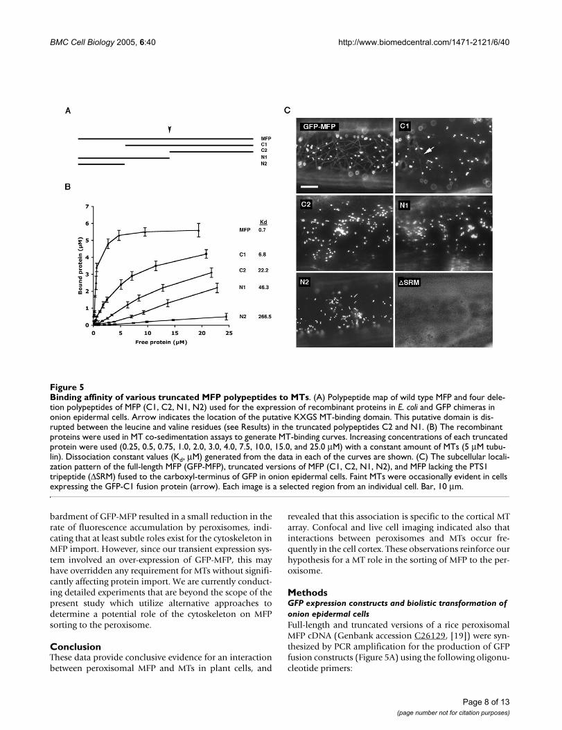
their affinity for MTs using MT co-sedimentation assays.Two of the truncated polypeptides, C2 and N1, containeddeletions that terminated within an internal region ofMFP that is similar in sequence to a MT-binding domainfound in several well-characterized MT-associated pro-teins including MAP2, MAP4, tau and TOGp [26]. Theconsensus sequence of this domain consists of the aminoacid residues, -LX5KI/VGSE/DNK-, and is defined by acharacteristic serine phosphorylation tetrapeptide motif (-KXGS-) known to regulate the MT-binding activity of thisdomain [26]. The amino acid sequence of the potentialMT-binding site in MFP (-LEGLVKRGSLTKDK-) begins atamino acid position 356. We were unable to obtain amore detailed deletion analysis of the carboxyl-terminusof the protein (Figure 5A), as repeated efforts to expresstruncated versions of the protein in this region were notsuccessful.
Figure 5B shows that full-length MFP had a high affinityfor MTs as indicated by its dissociation constant (Kd)value of 0.7 µM, a value similar to what we determinedpreviously for the same protein (Kd = 0.8 µM, [19]). Incontrast, each of the amino- and carboxyl-terminal dele-tions of the MFP polypeptide (C1, C2, N1 and N2) dis-played a significantly reduced affinity for MTs whencompared to the full-length polypeptide. None of thesedeletions, however, resulted the complete elimination ofMT-binding activity. The amino-terminal deletionpolypeptide, C1, had the highest affinity for MTs (Kd = 6.8µM, Figure 5B) when compared to the other truncatedproteins although its affinity for MTs was 10-fold less thanthe full-length polypeptide. Polypeptide N2 was com-posed of the region of MFP that was deleted in polypep-tide C1 yet its affinity for MTs was the lowest of all thetruncated polypeptides (Kd = 266.5 µM). This indicatesthat this region alone does not possess significant bindingactivity, but has an important contribution to the overallbinding activity of the full-length MFP. Polypeptides C2(Kd = 22.2 µM) and N1 (Kd = 46.3 µM), on the other hand,were intermediate in their MT-binding activities despitebeing disrupted in the putative KXGS MT-bindingdomain. Overall, this deletion analysis indicates thatthere are multiple MT-binding regions located on the MFPpolypeptide, and that they work synergistically to achievethe high MT-binding affinity observed for full-lengthMFP.
We performed next a series of transient expression experi-ments with the same truncated versions of MFP byexpressing them in onion epidermal cells as fusions to thecarboxyl-terminus of GFP (e.g., GFP-C1, Figure 5C). Theobjective of these experiments was to determine whethertheir relative MT-binding affinities as determined by MTco-sedimentation assays (Figure 5B) corresponded totheir in vivo MT-binding characteristics. For those fusion
constructs that lacked portions of the carboxyl-terminusof MFP (i.e., N1 and N2, Figure 5A), the endogenous MFPPTS1 sequence (-SRM) was restored so that these deletedpolypeptides would retain their targeting to peroxisomes.All fusion constructs, along with wild-type GFP-MFP, werebombarded individually into onion epidermal cells andobserved throughout a 3 to 24 hour period. While cellsexpressing full-length GFP-MFP often displayed MT labe-ling, the only truncated MFP fusion protein to label MTswas the amino-terminal deletion construct C1 (GFP-C1;Figure 5C). The number of GFP-C1-transformed cells dis-playing MT labeling, however, was low, as was the fluores-cence intensity of the MTs in these cells. Nevertheless, thatGFP-C1 localized to MTs was consistent with the observa-tion that this polypeptide possessed the highest MT-bind-ing affinity of the truncated recombinant proteins (Figure5B). While cells expressing the N2, N1 and C2 fusion pro-teins did not display MT fluorescence, it is possible thatthese proteins bound to MTs in vivo at levels too low to bedetected using our imaging system.
We hypothesized earlier [10,19] that the MT-bindingactivity of MFP may serve to concentrate MFP on MTsprior to its import into peroxisomes. Since the carboxyl-terminal PTS1 tripeptide (SRM) of MFP is both necessaryand sufficient for import into peroxisomes (R.T. Mullenand D.G. Muench, unpublished observations), we antici-pated that deletion of this sequence from MFP in the con-text of the GFP-MFP chimera would cause anaccumulation of the protein in the cytosol and a resultingincrease in binding to MTs. Surprisingly, expression ofthis modified chimera (i.e., GFP-MFP∆SRM) in onion epi-dermal cells resulted in only a diffuse cytosolic fluores-cence with no detectable filamentous structures (Figure5C). This apparent necessity of the MFP PTS1 for highaffinity MT-binding in vivo further demonstrates the com-plex nature of the MT-binding activity of MFP and therequirement for multiple regions of the protein to achieveefficient binding.
Confocal microscopy and real-time imaging indicate that peroxisomes and MTs interact frequentlyPrevious live-cell microscopy studies revealed that plantperoxisomes exhibit high velocity unidirectional move-ments, bi-directional and stop-and-go movements, as wellas rapid oscillations at fixed locations within the cell, andthat these movements occurred on actin filaments with noapparent role for MTs (reviewed in [10]). Using confocalmicroscopy of GFP-MFP-expressing onion epidermal cellswe observed that peroxisomes and cortical MTs were reg-ularly located in the same optical section (Figure 1A,inset). This indicated that peroxisomes are frequently inclose association with MTs as they move through the cellcortex on actin filaments. In addition, we noted in real-time movies that the characteristic stop-and-go (or "paus-
Page 4 of 13(page number not for citation purposes)

BMC Cell Biology 2005, 6:40 http://www.biomedcentral.com/1471-2121/6/40
ing") of peroxisomes frequently occurred at sites occupiedby MTs (Figure 6, see Additional data file 1: Movie 1, andAdditional data file 2: Movie2), also suggesting that thesetwo structures interact. We determined the frequency ofthese peroxisome pausing events on MTs in cells that hada relatively sparse cortical MT network such as thoseobserved in Additional data files Movie 1 and Movie 2.The stop-and-go movements of peroxisomes (n = 246)from 14 GFP-MFP-expressing cells were observed in mov-ies of 15 to 20 seconds duration. We determined that 67± 13% of the pausing events occurred at sites that werecoincident with MTs. There were also numerous peroxi-somes that exhibited oscillatory movements at fixed loca-
tions. These were observed to be in frequent contact withcortical MTs during their movements 73 ± 19% of thetime. Notably, while treatment of GFP-MFP-expressingcells with latrunculin B for one hour caused peroxisomesto oscillate at fixed locations within the cell [16,17,27,28],treatment with oryzalin for one hour did not have anobservable effect on the frequency of peroxisome pausingevents associated with motile peroxisomes (see Addi-tional data file 3: Movie3). This suggests that MTs are notdirectly involved in a majority of the peroxisomal pausingevents, but that another mechanism is responsible forthese pausing events.
Interestingly, we observed also that when epidermal peelswere treated with oryzalin or latrunculin B immediatelyfollowing bombardment of the GFP-MFP fusion constructand throughout the incubation period (see Materials andMethods for details about long-term drug treatments),there was a subtle effect on the rate of GFP-MFP labelingof peroxisomes compared to cells from untreated peels.The average frequency of cells showing peroxisome fluo-rescence 3 to 4 hours after bombardment in untreatedpeel segments (normalized value of 0.81 ± 0.08) washigher than in segments treated with oryzalin (0.65 ±0.11) or latrunculin B (0.74 ± 0.08). These data indicatethat the there is a reduction in the rate of synthesis and/orimport of GFP-MFP as a result of cytoskeleton disruption.
DiscussionThe processes involved in the import of proteins into theperoxisome matrix are distinct from those described forprotein import into other membrane-bound organelles.One major difference is that peroxisomal matrix proteinscan be imported fully folded and/or in an oligomerizedconformation [6,29,30]. This characteristic implies thatthese proteins have the potential to possess functionalactivities in the cytosol prior to their import. We reportedpreviously that the plant peroxisomal MFP binds to MTsand RNA in vitro, in addition to possessing enzymaticactivities related to fatty acid β-oxidation. We suggestedthat the MT-binding activity may function to facilitate theimport of MFP into peroxisomes [10,19]. In this paper, weused indirect immunofluorescence microscopy and theexpression of a GFP-MFP chimera to show that MFP inter-acts uniformly with interphase cortical MTs in vivo (Fig-ures 1 and 4), thereby demonstrating that MFP is a bonafide MT-binding protein. The specific binding of MFP tothe cortical MTs implies that the MFP-MT interaction islimited to interphase cells. Array-specific binding by otherMT-binding proteins has been observed previously [31-33]. These specific interactions may be regulated by post-translational modifications such as phosphorylation/dephosphorylation of the MT-binding protein or modifi-cations to the acidic carboxyl-terminal tail of tubulin[33,34].
GFP-MFP partially co-localizes with the endogenous peroxi-somal matrix protein catalaseFigure 2GFP-MFP partially co-localizes with the endogenous peroxisomal matrix protein catalase. Onion epidermal cells expressing GFP-MFP were fixed and then probed with monoclonal antibodies raised against catalase. GFP-MFP fluo-rescence (A), mouse anti-catalase antibody staining (B), and an overlay of the two images (C) are shown in a region of a single transformed cell by epifluorescence microscopy. The acquisition of these images was set at low exposure to cap-ture the brightly fluorescing peroxisomes. The less intensely fluorescing MTs in this GFP-MFP expressing cell were not visible at this exposure. Bar, 4 µm.
Page 5 of 13(page number not for citation purposes)

BMC Cell Biology 2005, 6:40 http://www.biomedcentral.com/1471-2121/6/40
Although the techniques used here showed that MFPbinds to MTs in vivo, the level of detection of this interac-tion varied. Indirect immunofluorescence microscopyusing an affinity-purified anti-MFP antibody labeled per-oxisomes intensely, whereas MTs possessed only weakimmunofluorescence and were often not visible at all.Similarly, many transformed cells expressing GFP-MFPlacked observable MT labeling, or MTs in these cells werevisible only after computer software enhancement of dig-itized images. However, in numerous other bombard-ment experiments MTs were readily observable in up toone-third of the transformed cells. We were unable todetermine whether this variation in MT labeling by GFP-MFP was due to differences in the level of expression ofthe fusion protein, the metabolic state of the cell, or thequality of the leaf material used in individual bombard-ment experiments.
MT co-sedimentation experiments of truncated versionsof MFP indicated that there are multiple MT-bindingdomains within the MFP polypeptide (Figure 5). Each ofthe truncations resulted in a significant decrease in MT-binding affinity of the remaining polypeptide, indicatingthat maximal binding of the full-length MFP likelydepends on several points of contact to MTs, as has beensuggested for other MT-binding proteins [32]. MT-bind-
ing domains have been identified for numerous proteins,some of which contain well characterized domains suchas those found in the animal MAPs tau, MAP4, MAP2, andTOGp [26], whereas others possess less well known MT-binding domains that are not as well studied [35,36]. Weidentified by sequence analysis a putative MT-bindingdomain in MFP that showed sequence similarity to adomain centered around a KXGS motif present singly orin repeats in some animal MAPs [26,33]. However, thisregion does not contribute significantly to the MT-bindingactivity of MFP in vitro (Figure 5). We have demonstratedthat the Arabidopsis homolog of the rice MFP also pos-sesses MT-binding activity (SDX Chuong and DGMuench, unpublished observations). Despite sharing82% amino acid similarity to the rice sequence, the Arabi-dopsis protein does not possess the putative KXGS motifregion that is present in the rice MFP. This is consistentwith the observed minimal influence of this motif on MTbinding affinity in vitro (Figure 5).
Interestingly, transient expression of a modified version ofGFP-MFP that lacked the MFP carboxyl-terminal PTS1resulted in this fusion protein (GFP-MFP∆SRM) beinglocalized exclusively to the cytosol, with no apparent labe-ling of MTs (Figure 5C). This indicates that the carboxylterminus of MFP is also required for its efficient bindingto MTs in vivo. The lack of MT binding by this fusion pro-tein appears to be an effect of expression in vivo sincerecombinant MFP�SRM binds to MTs in vitro with anaffinity that is similar to the full-length MFP protein (SDXChuong and DG Muench, unpublished results). The PTS1normally interacts with the peroxisomal import receptor,Pex5p, to facilitate the targeting of PTS1-bearing proteinsfrom the cytosol to the peroxisome surface and possiblyacross the peroxisome membrane [6,37]. Pex5p is thenrecycled and is available to interact with other PTS1-con-taining proteins. The necessity of the PTS1 for efficientbinding of MFP to MTs in vivo may, therefore, highlight anovel role for Pex5p at an early stage of peroxisome pro-tein sorting.
The immunofluorescence and live-cell imaging data pre-sented here support our previous hypothesis that wasbased on in vitro MT-binding assays [19] and stated thatthe interaction between MFP and MTs assists in the effi-cient and regulated sorting of MFP to peroxisomes [10].That is, MFP-MT interactions serve to concentrate MFP inthe cell cortex, a region of the cell where peroxisomes areabundant and are frequently moving along actin fila-ments. Using confocal microscopy, we observed here thatperoxisomes and cortical MTs regularly occupied the samethree-dimensional space in the cell cortex (Figure 1A,inset). This arrangement would provide frequent opportu-nities for motile peroxisomes to interact with MTs as per-oxisomes move in the cell cortex, a premise that is
The filamentous labeling in GFP-MFP-expressing cells is abol-ished when cells are treated with the MT-disrupting agent oryzalinFigure 3The filamentous labeling in GFP-MFP-expressing cells is abolished when cells are treated with the MT-disrupting agent oryzalin. Onion epidermal cells express-ing GFP-MFP (A, B, C), GFP-MAP4 (D, E, F) or GFP-talin (G, H, I) were treated with DMSO (A, D, G), oryzalin (B, E, H) or latrunculin B (C, F, I) for 45 minutes and visualized by epi-fluorescence microscopy. Bar, 3 µm.
Page 6 of 13(page number not for citation purposes)
BMC Cell Biology 2005, 6:40 http://www.biomedcentral.com/1471-2121/6/40
supported by previous evidence for a direct associationbetween cortical MTs and actin filaments [38]. In addi-tion, real-time fluorescence imaging revealed apparentMT interactions with peroxisomes in the form of pausingevents by motile peroxisomes as well as peroxisomes thatoscillated at fixed locations (Figure 6, Movie Supplement1). These types of peroxisome-MT interactions wouldreadily allow for the transfer of MT-bound MFP to theimport machinery located on the peroxisomal surface.
Additional support for a MT role in peroxisomal proteinimport comes from the discovery that six other plant per-oxisomal matrix proteins also possess tubulin-bindingactivities [22,39]. Most of these proteins have activitiesinvolved in catalyzing the reactions of fatty acid β-oxida-
tion in the peroxisome, however, one of them (malatedehydrogenase) is involved in the glyoxylate cycle. β-oxi-dation enzymes presumably do not have a metabolic rolein the cytosol, since their associated metabolic pathwayshave not been shown to exist in that compartment. It ismore likely, therefore, that the binding of these peroxiso-mal matrix proteins to MTs functions in a general sortingpathway to the peroxisome, as discussed above for MFP.
If the cytoskeleton is required for the efficient delivery ofMFP to the peroxisome surface as described above, thentreatment of cells with cytoskeleton destabilizing agentsshould affect the import of MFP into peroxisomes. Wedetermined that long-term treatments of epidermal peelswith oryzalin and latrunculin B immediately after bom-
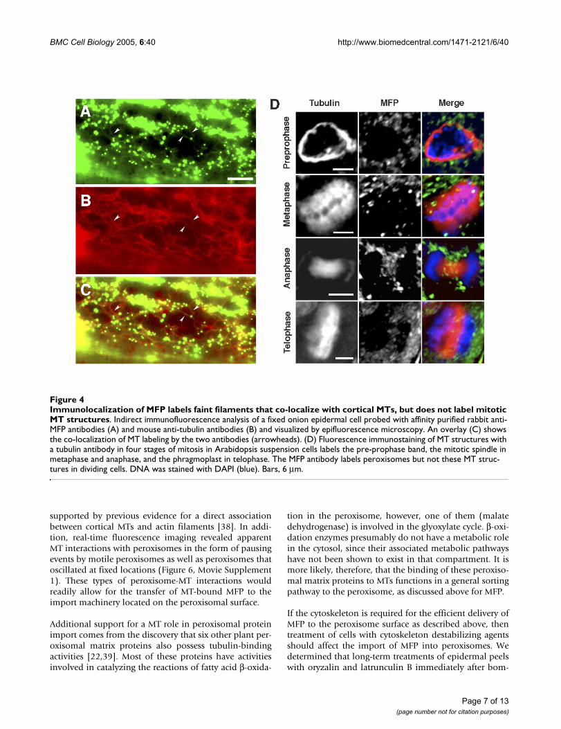
Immunolocalization of MFP labels faint filaments that co-localize with cortical MTs, but does not label mitotic MT structuresFigure 4Immunolocalization of MFP labels faint filaments that co-localize with cortical MTs, but does not label mitotic MT structures. Indirect immunofluorescence analysis of a fixed onion epidermal cell probed with affinity purified rabbit anti-MFP antibodies (A) and mouse anti-tubulin antibodies (B) and visualized by epifluorescence microscopy. An overlay (C) shows the co-localization of MT labeling by the two antibodies (arrowheads). (D) Fluorescence immunostaining of MT structures with a tubulin antibody in four stages of mitosis in Arabidopsis suspension cells labels the pre-prophase band, the mitotic spindle in metaphase and anaphase, and the phragmoplast in telophase. The MFP antibody labels peroxisomes but not these MT struc-tures in dividing cells. DNA was stained with DAPI (blue). Bars, 6 µm.
Page 7 of 13(page number not for citation purposes)
BMC Cell Biology 2005, 6:40 http://www.biomedcentral.com/1471-2121/6/40
bardment of GFP-MFP resulted in a small reduction in therate of fluorescence accumulation by peroxisomes, indi-cating that at least subtle roles exist for the cytoskeleton inMFP import. However, since our transient expression sys-tem involved an over-expression of GFP-MFP, this mayhave overridden any requirement for MTs without signifi-cantly affecting protein import. We are currently conduct-ing detailed experiments that are beyond the scope of thepresent study which utilize alternative approaches todetermine a potential role of the cytoskeleton on MFPsorting to the peroxisome.
ConclusionThese data provide conclusive evidence for an interactionbetween peroxisomal MFP and MTs in plant cells, and
revealed that this association is specific to the cortical MTarray. Confocal and live cell imaging indicated also thatinteractions between peroxisomes and MTs occur fre-quently in the cell cortex. These observations reinforce ourhypothesis for a MT role in the sorting of MFP to the per-oxisome.
MethodsGFP expression constructs and biolistic transformation of onion epidermal cellsFull-length and truncated versions of a rice peroxisomalMFP cDNA (Genbank accession C26129, [19]) were syn-thesized by PCR amplification for the production of GFPfusion constructs (Figure 5A) using the following oligonu-cleotide primers:
Binding affinity of various truncated MFP polypeptides to MTsFigure 5Binding affinity of various truncated MFP polypeptides to MTs. (A) Polypeptide map of wild type MFP and four dele-tion polypeptides of MFP (C1, C2, N1, N2) used for the expression of recombinant proteins in E. coli and GFP chimeras in onion epidermal cells. Arrow indicates the location of the putative KXGS MT-binding domain. This putative domain is dis-rupted between the leucine and valine residues (see Results) in the truncated polypeptides C2 and N1. (B) The recombinant proteins were used in MT co-sedimentation assays to generate MT-binding curves. Increasing concentrations of each truncated protein were used (0.25, 0.5, 0.75, 1.0, 2.0, 3.0, 4.0, 7.5, 10.0, 15.0, and 25.0 µM) with a constant amount of MTs (5 µM tubu-lin). Dissociation constant values (Kd, µM) generated from the data in each of the curves are shown. (C) The subcellular locali-zation pattern of the full-length MFP (GFP-MFP), truncated versions of MFP (C1, C2, N1, N2), and MFP lacking the PTS1 tripeptide (∆SRM) fused to the carboxyl-terminus of GFP in onion epidermal cells. Faint MTs were occasionally evident in cells expressing the GFP-C1 fusion protein (arrow). Each image is a selected region from an individual cell. Bar, 10 µm.
Page 8 of 13(page number not for citation purposes)
BMC Cell Biology 2005, 6:40 http://www.biomedcentral.com/1471-2121/6/40
gMFP1 – CACTCTAGAATGGCGGGGGCGATCCGCGT-CACC; gMFP10 – CGTCTAGATCACATGCGTGAC-CTCGA; gMFP11 –CGTCTAGATCACATGCGTGAAAGACCACCTTCTTTC;gMFP12 – GCGTCTAGATCACATGCGTGAGACTAGAC-CCTCAAGATTAG; gMFP14 – CACTCTAGAGTTGAT-GCCCTGTGCTCC; gMFP15 –CACTCTAGAGGCTCATTGACAAAGCAC; gMFP16 –CCATCAGCTGAAATAGCATCTAGAATGCGTTACCTC-GACGAAGCCTGCTGG.
Primer pairs were used to amplify either full-length MFP(primers gMFP14 and gMFP10), the amino-terminal trun-cations C1 (primers gMFPl4 and gMFP10) and C2 (prim-ers gMFP15 and gMFP10), the carboxyl-terminaltruncations N1 (primers gMFP1 and gMFP11), N2 (prim-ers gMFP1 and gMFP12), or GFP-MFP minus the PTS1sequence (GFP-MFP∆SRM, primers gMFP1 and gMFP16).Each of the PCR primers contained an XbaI sequence intheir 5' flanking region. Digestion of the PCR productswith XbaI allowed for cloning into the XbaI site ofpRTL2∆NS/GFP-XbaI [40], resulting in an in-frame fusionof the full-length MFP or truncated MFP open readingframe to the carboxyl-terminus of GFP. pRTL2∆NS/GFP-XbaI is a plant expression vector containing the cauli-flower mosaic virus 35S promoter and nopaline synthaseterminator, and a modified GFP with an in-frame XbaI siteinstead of a stop codon. GFP-talin [25] and GFP-MAP4[24] expression constructs were used as controls in sometransient expression experiments to label actin filamentsand MTs, respectively.
Transient expression of the various GFP fusion constructswas achieved by biolistic bombardment into onion(Allium cepa) epidermal cells using protocols that weredescribed previously [41]. Plasmid DNA was purifiedusing a DNA Maxiprep kit (Qiagen, Mississauga, ON,Canada) and 5 to 10 µg of the DNA was coated onto goldparticles (1 µm diameter) as described by the manufac-turer (BioRad, Hercules, CA). DNA-coated gold particleswere bombarded into the adaxial surface of onion bulbleaf segments (1 cm2) from a distance of 10 cm using aBiolistic PDS-1000/He particle delivery system (Bio-Rad)at a pressure of 1200 psi. After bombardment, the leaf sec-tions were placed on moist filter paper in petri dishes,adaxial side down, and incubated in the dark at roomtemperature for 3 to 24 hours. Alternatively, the epider-mal layer was peeled immediately after bombardmentand floated on liquid MS media (pH 5.9) containing 3%sucrose, and peels were then placed in the dark at roomtemperature for 3 to 24 hours.
Expression and purification of recombinant proteins and MT co-sedimentation assaysPCR was used to amplify full-length or truncated versionsof the rice MFP coding region for the production ofrecombinant proteins (Figure 5A) using the following oli-gonucleotide primers:
rMFP1 – CACGGATCCGCGGGGGCGATCCGCGTCAC-CATG; rMFP10 – CACCTGCAGCATGCGTGACCTCGAC-GAAGCCTG; rMFP11 –GCGCTGCAGAAGACCACCTTCTTTCCCTTCCTT;rMFP12 – GCGCTGCAGGACTAGACCCTCAAGATT-AGCTGC; rMFP14 – CACGGATCCGTTGATGCCCTGT-GCTCCCCTGAT; rMFP15 –CACGGATCCAAGAGGGGCTCATTGACAAAGGAC.Primer pairs were used to amplify either full-length MFP(primers rMFP1 and rMFP10), the amino-terminal trun-cations C1 (primers rMFP14 and rMFP10) and C2 (prim-ers rMFP15 and rMFP10), and the carboxyl-terminaltruncations N1 (primers rMFP1 and rMFP11) or N2(primers rMFP1 and rMFP12). Each primer containedeither BamHI or PstI restriction enzyme sites that werecompatible for cloning into the expression vector pQE30(Qiagen). Recombinant proteins were expressed in E. coliand purified as described previously [19]. The purifiedrecombinant proteins were used in bovine MT co-sedi-mentation assays to generate binding curves for the deter-mination of MT/MFP dissociation constants (Kd) [19].The co-sedimentation assays were repeated twice for eachof the recombinant MFPs, and their mean values andstandard deviations were presented. BSA was used as aninternal negative control in selected MT co-sedimentationassays to ensure that soluble protein was not trapped byMTs in the pellet fraction. The Kd value for each recom-binant protein was determined using the Prism kineticsprogram (GraphPad Prism, Version 3.02, GraphPad Soft-ware Inc., San Diego, CA).
Affinity purification of antibodies and immunolocalizationsTwo mg of the full-length recombinant MFP was cova-lently coupled to 1 mL of a matrix consisting of an equalmixture of Affigel-15 (Bio-Rad) and Sepharose CL-6B(Sigma-Aldrich Ltd., Oakville, Canada), and the matrixwas then poured into a disposable 2 mL column (PolyPrep, BioRad). One mL of MFP antiserum [19] was addedto the column matrix, incubated overnight at 4°C, and thematrix was then washed with 5 column volumes of phos-phate buffered saline (PBS). IgGs were eluted from thecolumn with 2 mL of 100 mM glycine, pH 2.5, and theeluent was immediately neutralized by the addition ofone-tenth volume 1.5 M Tris-HCl pH 8.8. Purified IgGswere concentrated using a Centricon-10 microfiltrationapparatus (Amicon, Millipore, Billerica. MA) and stored at4°C in the presence of 0.05% (w/v) sodium azide.
Page 9 of 13(page number not for citation purposes)

BMC Cell Biology 2005, 6:40 http://www.biomedcentral.com/1471-2121/6/40
Onion epidermal peels were fixed for 5 min in freshly pre-pared paraformaldehyde (7.4% [w/v], Sigma-AldrichLtd.) and glutaraldehyde (0.015% [w/v], Sigma-AldrichLtd.) in a cytoskeleton-stabilizing buffer (50 mM HEPES
pH 6.9, 2 mM MgSO4, 5 mM EGTA, and 0.1% Triton X-100)[42]. After fixing, the peels were washed three timesin PBS for 5 min each, followed by treatment with 0.5%cellulase (Onazuka R-10) and 0.05% pectolyase Y23
Interactions are apparent between peroxisomes and MTs in onion epidermal cellsFigure 6Interactions are apparent between peroxisomes and MTs in onion epidermal cells. Time-lapse images of a region from a GFP-MFP-expressing onion epidermal cell showing apparent transient and long-term interactions of peroxisomes and MTs. The movements of three peroxisomes were followed over 18 image frames. One of these peroxisomes (star) showed, throughout the entire series, oscillatory movements at a fixed location that were co-incident with a MT. Another peroxisome (arrow) remained fixed at a site coincident with a MT for approximately two seconds at the beginning of the image sequence, and then moved out of the field of view during the final two seconds. The initial movement is marked by the arrow in image frame 1.82. A dumbbell-shaped peroxisome (arrowhead) moved through the cytosol and appeared to transiently tether to a MT (second arrowhead, image frame 1.30). This peroxisome then released and tethered to another MT (third arrowhead, image frame 3.12) before releasing again and continuing its movement. The cell observed in this image sequence was from an unpeeled epidermal layer that remained associated with the leaf segment. The movements of the peroxisomes shown in this figure are also shown in a real-time image sequence in Additional data file Movie 1. Numbers indicate elapsed time in seconds. Bar, 2 µm.
Page 10 of 13(page number not for citation purposes)

BMC Cell Biology 2005, 6:40 http://www.biomedcentral.com/1471-2121/6/40
(Seishin Pharmaceutical Co., Tokyo) in MS media (pH5.9) containing 0.3 M mannitol for 3 minutes at 37°C.The peels were washed three times in PBS for 5 min eachand then treated with 1% Triton-X 100 for 5 min at roomtemperature. Arabidopsis suspension cells were used forimmunolocalization studies three days after subculturing.One mL of cell culture was fixed for 15 min at room tem-perature in freshly prepared 4% paraformaldehyde dis-solved in cytoskeleton-stabilizing buffer. After fixing, thecells were washed three times with PBS for 5 min each,and then digested with 0.1% pectolyase Y-23 in PBS for 15min at 30°C. The cells were washed three times in PBS for5 min each and then treated with 1% Triton-X 100 for 5min at room temperature.
The fixed and permeabilized epidermal peels and suspen-sion cells were incubated for 15 min at room temperaturein blocking solution (PBS containing 3% BSA), followedby incubation for 60 min in blocking buffer containingthe appropriate primary antibodies. The following pri-mary antibodies were used: affinity purified rabbit anti-MFP IgGs ([19], final concentration, 0.5 µg/mL); mouseanti-tobacco catalase (undiluted hybridoma medium pur-chased from the Princeton Monoclonal Antibody Facil-ity); and mouse anti-α-tubulin monoclonal antibody(Sigma-Aldrich Ltd.), 1:500 (v/v). After three, 5 minwashes in PBS, the peels/cells were incubated in second-ary antibodies diluted 1:500 for 30 min at room tempera-ture, and then washed again in PBS. The secondaryantibodies used were goat anti-rabbit IgG-Alexa-594 con-jugate and goat anti-mouse IgG-Alexa-488 conjugate(Molecular Probes, Eugene, OR). Pre-incubation of anti-MFP antibodies with recombinant MFP protein prior tousing the antibodies in immunolocalization experimentswas used to confirm the specific labeling of the anti-MFPantibodies.
Microscopic analysisUnpeeled onion leaf segments were secured to a glassslide with 1% agarose, and the epidermal cells wereobserved through a bead of water or MS media using a63× long working distance water immersion lens (HCXApo L, Leica, Germany) mounted on an epifluorescencemicroscope (Leica DMR). Alternatively, fresh or fixed epi-dermal peels were mounted on a glass slide in MS mediaor PBS, covered with a coverglass, and observed usingeither a Plan Fluotar 40× or Plan Apo 63× oil immersionobjective lens (Leica). Images were captured using acooled CCD camera (Retica 1350 EX, QImaging, Burnaby,BC, Canada) through the FITC or rhodamine filter sets.Image enhancement and deconvolution confocal algo-rithm manipulations were performed using Openlab soft-ware (Version 3.0, Improvision, Lexington, MA).Quicktime movies were generated from sequential imagesusing the Openlab software, and single images were mod-
ified using Adobe Photoshop image processing software(Version 8.0, Adobe Systems Inc., San Jose, CA). The fre-quency of peroxisome pausing events on MTs was deter-mined from 14 GFP-MFP expressing cells, and variationbetween cells was reported as standard deviation from themean.
Cytoskeleton drug treatmentsShort-term cytoskeleton drug treatments of 45 min to onehour were conducted on transiently transformed onionepidermal layers that were bombarded with the GFP-MFPconstruct 3 to 24 hours in advance, and that possessednumerous cells showing high levels of fluorescence. Finaldrug concentrations were 0.2 µM latrunculin B (Sigma-Aldrich Ltd.) or 2 µM oryzalin (Crescent Chemical Com-pany, Islandia, NY). Stock solutions of these drugs wereprepared in DMSO. Epidermal peels were treated with thedrugs by floating the peels on liquid MS media (pH 5.9)containing 3% sucrose and the appropriate amount ofDMSO or drug. To demonstrate that these drug concentra-tions were effective in MT and actin filament disruption,epidermal cells expressing the GFP-MAP4 or GFP-talinconstructs were treated with the corresponding drugs.
For experiments performed to determine the long-termeffect of cytoskeleton disrupting drugs on GFP-MFPimport into peroxisomes, each bombarded epidermalpeel was divided equally into thirds, and the resulting sec-tions were then treated individually with DMSO, oryzalinor latrunculin B immediately after bombardment by float-ing the peels on liquid MS media containing the appropri-ate amount of DMSO or drug. The bombarded epidermalpeels were observed by epifluorescence microscopy, andthe number of cells showing peroxisome labeling weredetermined 3 to 4 hours post-bombardment, since con-trol sections typically showed the first signs of fluores-cence during this period. Data from sections derived fromindividual peels were pooled for each experiment, and thetotal dataset presented included results from at least threeindividual experiments. Normalization of the raw datawas performed, since the total number of transformedcells varied between individual bombardment experi-ments. Variation between experiments was reported asstandard deviation from the mean.
Authors' contributionsSDXC contributed to the experimental design, conducteda majority of the experiments, and assisted in manuscriptwriting, data analysis, and figure preparation. N-IP andMCF assisted with the immunofluorescence and transientexpression experiments. RTM provided expression vec-tors, assisted in the transient expression experiments, andcontributed to the experimental design and writing of themanuscript. DGM wrote the original draft of the manu-script, contributed to the experimental design, and
Page 11 of 13(page number not for citation purposes)
BMC Cell Biology 2005, 6:40 http://www.biomedcentral.com/1471-2121/6/40
assisted with the immunofluorescence and transientexpression experiments, data analysis, and figure prepara-tion. All authors read and approved the final manuscript.
Additional material
AcknowledgementsWe would like to thank Eric Davies (North Carolina State University) for his helpful discussions, Richard Cyr (Penn State University) for the GFP-MAP4 fusion construct, and Nam-Hai Chua (Rockefeller Foundation) for the GFP-talin construct. We are grateful to the Japanese Ministry of Agri-culture, Forestry and Fisheries (MAFF) Genome Program for providing us with the rice MFP cDNA clone. This work is supported by Natural Sciences and Engineering Research Council of Canada (NSERC) grants to DGM and RTM.
References1. Heiland I, Erdmann R: Biogenesis of peroxisomes. Topogenesis
of the peroxisomal membrane and matrix proteins. Febs J2005, 272(10):2362-2372.
2. Hoepfner D, Schildknegt D, Braakman I, Philippsen P, Tabak HF: Con-tribution of the endoplasmic reticulum to peroxisome for-mation. Cell 2005, 122(1):85-95.
3. Trelease RN, Lingard MJ: Participation of the plant ER in perox-isomal biogenesis. DG Robinson, ed. The Plant EndoplasmicReticulum. In The Plant Endoplasmic Reticulum Edited by: RobinsonD. Heidelberg: Springer-Verlag in press.
4. Brown LA, Baker A: Peroxisome biogenesis and the role of pro-tein import. J Cell Mol Med 2003, 7(4):388-400.
5. Eckert JH, Erdmann R: Peroxisome biogenesis. Rev Physiol BiochemPharmacol 2003, 147:75-121.
6. Subramani S: Hitchhiking fads en route to peroxisomes. J CellBiol 2002, 156(3):415-417.
7. Nair DM, Purdue PE, Lazarow PB: Pex7p translocates in and outof peroxisomes in Saccharomyces cerevisiae. J Cell Biol 2004,167(4):599-604.
8. Matsumura T, Otera H, Fujiki Y: Disruption of the interaction ofthe longer isoform of Pex5p, Pex5pL, with Pex7p abolishesperoxisome targeting signal type 2 protein import in mam-mals. Study with a novel Pex5-impaired Chinese hamsterovary cell mutant. J Biol Chem 2000, 275(28):21715-21721.
9. Woodward AW, Bartel B: The Arabidopsis peroxisomal target-ing signal type 2 receptor PEX7 is necessary for peroxisomefunction and dependent on PEX5. Mol Biol Cell 2005,16(2):573-583.
10. Muench DG, Mullen RT: Peroxisome dynamics in plant cells: arole for the cytoskeleton. Plant Science 2003, 164:307-315.
11. Fagarasanu M, Fagarasanu A, Tam YY, Aitchison JD, Rachubinski RA:Inplp is a peroxisomal membrane protein required for per-oxisome inheritance in Saccharomyces cerevisiae. J Cell Biol2005, 169(5):765-775.
12. Yan M, Rayapuram N, Subramani S: The control of peroxisomenumber and size during division and proliferation. Curr OpinCell Biol 2005, 17(4):376-383.
13. Schrader M, Thiemann M, Fahimi HD: Peroxisomal motility andinteraction with microtubules. Microsc Res Tech 2003,61(2):171-178.
14. Hoepfner D, van den Berg M, Philippsen P, Tabak HF, Hettema EH: Arole for Vpslp, actin, and the Myo2p motor in peroxisomeabundance and inheritance in Saccharomyces cerevisiae. J CellBiol 2001, 155:979-990.
15. Wiemer EA, Wenzel T, Deerinck TJ, Ellisman MH, Subramani S: Vis-ualization of the peroxisomal compartment in living mam-malian cells: dynamic behavior and association withmicrotubules. J Cell Biol 1997, 13:71-80.
16. Jedd G, Chua NH: Visualization of peroxisomes in living plantcells reveals actomyosin-dependent cytoplasmic streamingand peroxisome budding. Plant Cell Physiol 2002, 43:384-392.
17. Mathur J, Mathur N, Hulskamp M: Simultaneous visualization ofperoxisomes and cytoskeletal elements reveals actin and notmicrotubule-based peroxisome motility in plants. Plant Physiol2002, 128:1031-1045.
18. Hashimoto K, Igarashi H, Mano S, Nishimura M, Shimmen T, YokotaE: Peroxisomal localization of a myosin XI isoform in Arabi-dopsis thaliana. Plant Cell Physiol 2005, 46(5):782-789.
19. Chuong SDX, Mullen R, Muench DG: Identification of a rice RNA-and MT-binding protein as the multifunctional protein(MFP), a peroxisomal enzyme involved in the β-oxidation offatty acids. J Biol Chem 2002, 277:2419-2429.
20. Preisig-Müller R, Gühnemann-Schäfer K, Kindl H: Domains of thetetrafunctional protein acting in glyoxysomal fatty acid β-oxidation: demonstration of epimerase and isomerase activ-ities on a peptide lacking hydratase activity. J Biol Chem 1994,269:20475-20481.
21. Mullen RT: Targeting and import of matrix proteins into per-oxisomes. In Plant Peroxisomes: Biochemistry, Cell Biology and Biotech-nological Applications Edited by: Baker A, Graham IA. Kluwer AcademicPublishers; 2002:339-383.
22. Chuong SDX, Good AG, Taylor GJ, Freeman MC, Moorhead GB,Muench DG: Large-scale Identification of Tubulin-bindingProteins Provides Insight on Subcellular Trafficking, Meta-bolic Channeling, and Signaling in Plant Cells. Mol Cell Proteom-ics 2004, 3(10):970-983.
23. Lee M, Mullen R, Trelease R: Oilseed isocitrate lyases lackingtheir essential type 1 peroxisomal targeting signal are piggy-backed to glyoxysomes. Plant Cell 1997, 9:185-197.
Additional data file 1Movie1.mov – Real-time image sequence of an onion epidermal cell expressing GFP-MFP shows apparent interactions between peroxi-somes and cortical MTs. Epifluorescence images (n = 200) were captured every 0.13 seconds. A mobile, dumbbell-shaped peroxisome (tracked with the green marker) appears to interact with MTs at several locations during the initial part of the image sequence before moving out of focus. Upon returning into the focal plane near the end of the image sequence, this per-oxisome again appears to interact with MTs. The movement of this perox-isome is also shown in the fixed image sequence in Figure 6. Another peroxisome demonstrated a pausing event at a location where MTs were absent (tracked with the red marker), although it also paused several times at locations that coincided with MTs. The peroxisome tracked with the blue marker demonstrated on two occasions a subtle, lateral gliding movement alongside MTs. The cell observed in this image sequence was from an unpeeled epidermal layer that remained associated with the leaf segment.Click here for file[http://www.biomedcentral.com/content/supplementary/1471-2121-6-40-S1.mov]
Additional data file 2Movie2.mov – Real-time image sequence of an onion epidermal cell expressing GFP-MFP that demonstrated a lower frequency of peroxi-some pausing events at sites that were coincident with MT locations. Epifluorescence images (n = 270) were captured every 0.2 seconds.Click here for file[http://www.biomedcentral.com/content/supplementary/1471-2121-6-40-S2.mov]
Additional data file 3Movie3.mov – Real-time image sequence of an oryzalin treated onion epidermal cell expressing GFP-MFP. This movie demonstrates that MT depolymerization does not result in an obvious reduction of peroxisome pausing events in cell cortex. Epifluorescence images (n = 150) were col-lected every 0.2 seconds.Click here for file[http://www.biomedcentral.com/content/supplementary/1471-2121-6-40-S3.mov]
Page 12 of 13(page number not for citation purposes)

BMC Cell Biology 2005, 6:40 http://www.biomedcentral.com/1471-2121/6/40
24. Marc J, Granger C, Brincat J, Fisher D, Kao T-h, McCubbin A, Cyr RJ:A GFP-MAP4 reporter gene for visualizing cortical microtu-bule rearrangements in living epidermal cells. Plant Cell 1998,10:1927-1939.
25. Kost B, Spielhofer P, Chua N-H: A GFP-mouse talin fusion pro-tein labels plant actin filaments in vivo and visualizes theactin cytoskeleton in growing pollen tubes. Plant J 1998,16:393-401.
26. Charrasse S, Schroeder M, Gauthier-Rouviere C, Ango F, CassimerisL, Gard DL, Larroque C: The TOGp protein is a new humanmicrotubule-associated protein homologous to the XenopusXMAP215. J Cell Sci 1998, 111:1371-1383.
27. Collings DA, Harper DI, Marc J, Overall RL, Mullen RT: Life in thefast lane: actin-based motility of plant peroxisomes. Can J Bot2002, 80:1-11.
28. Mano S, Nakamori C, Hayashi M, Kato A, Kondo M, Nishimura M:Distribution and characterization of peroxisomes in Arabi-dopsis by visualization with GFP: dynamic morphology andactin-dependent movement. Plant Cell Physiol 2002, 43:331-341.
29. McNew JA, Goodman JM: An oligomeric protein is importedinto peroxisomes in vivo. J Cell Biol 1994, 127(5):1245-1257.
30. Walton PA, Hill PE, Subramani S: Import of stably folded proteinsinto peroxisomes. Mol Biol Cell 1995, 6(6):675-683.
31. Muller S, Smertenko A, Wagner V, Heinrich M, Hussey PJ, HauserMT: The plant microtubule-associated protein AtMAP65-3/PLE is essential for cytokinetic phragmoplast function. CurrBiol 2004, 14(5):412-417.
32. Smertenko AP, Chang HY, Wagner V, Kaloriti D, Fenyk S, Sonobe S,Lloyd C, Hauser MT, Hussey PJ: The Arabidopsis microtubule-associated protein AtMAP65-l: molecular analysis of itsmicrotubule bundling activity. Plant Cell 2004, 16(8):2035-2047.
33. Cassimeris L, Spittle C: Regulation of microtubule-associatedproteins. Int Rev Cytol 2001, 210:163-226.
34. Westermann S, Weber K: Post-translational modifications reg-ulate microtubule function. Nat Rev Mol Cell Biol 2003,4(12):938-947.
35. Moore RC, Cyr RJ: Association between elongation factor-lal-pha and microtubules in vivo is domain dependent and con-ditional. Cell Motil Cytoskeleton 2000, 45(4):279-292.
36. Wang PJ, Huffaker TC: Stu2p: A microtubule-binding proteinthat is an essential component of the yeast spindle pole body.J Cell Biol 1997, 139(5):1271-1280.
37. Purdue PE, Lazarow PB: Peroxisome biogenesis. Annu Rev Cell DevBiol 2001, 17:701-752.
38. Collings DA, Asada T, Allen NS, Shibaoka H: Plasma membrane-associated actin in bright yellow 2 tobacco cells. Evidence forinteraction with microtubules. Plant Physiol 1998,118(3):917-928.
39. Harper JDI, Weerakoon ND, Gardiner JC, Blackman LM, Marc J: A75-kDa plant protein isolated by tubulin-affinity chromatog-raphy is a peroxisomal matrix enzyme. Can J Bot 2002,80:1018-1027.
40. Hwang YT, Pelitire SM, Henderson MP, Andrews DW, Dyer JM, Mul-len RT: Novel Targeting Signals Mediate the Sorting of Differ-ent Isoforms of the Tail-Anchored Membrane ProteinCytochrome b5 to Either Endoplasmic Reticulum or Mito-chondria. Plant Cell 2004, 16(11):3002-3019.
41. Scott A, Wyatt S, Tsou PL, Robertson D, Allen NS: Model systemfor plant cell biology: GFP imaging in living onion epidermalcells. Biotechniques 1999, 26(6):1128-1132.
42. Abe S, Davies E: Isolation of F-actin from pea stems. Evidencefrom fluorescence microscopy. Protoplasma 1991, 162:51-61.
Page 13 of 13(page number not for citation purposes)
Copyright © 2022 FDOKUMEN