Genistein induces the metastasis suppressor kangai-1 which mediates its anti-invasive effects in...
7
Genistein induces the metastasis suppressor kangai-1 which mediates its anti-invasive effects in TRAMP cancer cells Lara H. El Touny, Partha P. Banerjee * Department of Biochemistry, Molecular and Cellular Biology, Georgetown University Medical Center, Medical-Dental Building, 3900 Reservoir Road, NW, Washington, DC 20057, USA Received 29 June 2007 Available online 16 July 2007 Abstract Previous studies demonstrated a direct correlation with loss of kangai-1 (KAI1), a metastasis suppressor, and poor prognosis in human prostate and other cancers. In this study, we have characterized the age-dependent downregulation of KAI1 in the TRAMP model which was reversed when mice were fed a genistein-enriched diet. We demonstrated here that doses of genistein (5 and 10 lM)—achievable by supplement intake—significantly induced the expression of KAI1, both at the mRNA and protein levels (up to 2.5-fold), and decreased the invasiveness of TRAMP-C2 cells >2.0-fold. We have pinpointed KAI1 as the invasion suppressor, since its knockdown by siRNA restored the invasive potential of genistein-treated TRAMP-C2 cells to control levels. This work provides the first evidence that genistein treatment may counteract KAI1 downregulation, which is observed in many cancer types and therefore, could be used in anti-metastatic therapies. Ó 2007 Elsevier Inc. All rights reserved. Keywords: Genistein; KAI1/kangai; CD82; TRAMP; Prostate cancer; Metastasis; Phytoestrogen It is estimated that more than 200,000 new cases of pros- tate cancer (CaP) will be diagnosed in the United States in 2007, of which >10% will die, making CaP the leading cause of cancer deaths in US men [1]. Although dysregula- tion of cell division is at the basis of tumor formation, the most serious threat to the patient’s survival is the malig- nant cells’ ability to invade surrounding tissues and form distant metastases [2]. The metastatic process is complex and involves several steps including invasion and angiogen- esis [3]. Therefore, natural products which interfere with these processes are useful agents in the fight against cancer progression. The reduced incidence of clinically relevant CaP in Asian males [4] suggests that the established chemopreven- tive action of the phytoestrogen, genistein, a major compo- nent of soy and a staple of the Asian diet, is not restricted to its anti-proliferative effects but might span invasion and metastasis. Previous studies have shown that genistein inhibits the invasion of different tumor cell lines. These effects were seen in prostate cancers among others [5]. Gen- istein has also been shown to reduce tumor metastasis in animal models [6]. Several mechanisms for these anti-met- astatic properties have been proposed. Tumor invasion is a crucial part of the metastatic process and involves a num- ber of steps including alterations in cellular adhesion and motility, proteolytic disruption of the basement membrane followed by migration through the extracellular matrix and the acquisition of an angiogenic phenotype [7]. Mechanistic studies have shown that genistein can hinder several of these steps. Genistein, at high doses has been shown to affect: cellular detachment by increasing the formation of focal adhesion complexes [8], invasion via downregulation of matrix metalloproteinases (MMPs): MMP-2, -9 [9], -8, -13 [10], as well as upregulation of tissue inhibitor of matrix metalloproteinases-1 (TIMP1) [9]. Kangai-1/CD82 (KAI1) was first identified as a prostate cancer metastasis suppressor [11]. Subsequent studies 0006-291X/$ - see front matter Ó 2007 Elsevier Inc. All rights reserved. doi:10.1016/j.bbrc.2007.07.010 * Corresponding author. Fax: +1 202 687 1823. E-mail address: [email protected] (P.P. Banerjee). www.elsevier.com/locate/ybbrc Biochemical and Biophysical Research Communications 361 (2007) 169–175
-
Upload
independent -
Category
Documents
-
view
0 -
download
0
Transcript of Genistein induces the metastasis suppressor kangai-1 which mediates its anti-invasive effects in...
www.elsevier.com/locate/ybbrc
Biochemical and Biophysical Research Communications 361 (2007) 169–175
Genistein induces the metastasis suppressor kangai-1 which mediatesits anti-invasive effects in TRAMP cancer cells
Lara H. El Touny, Partha P. Banerjee *
Department of Biochemistry, Molecular and Cellular Biology, Georgetown University Medical Center,
Medical-Dental Building, 3900 Reservoir Road, NW, Washington, DC 20057, USA
Received 29 June 2007Available online 16 July 2007
Abstract
Previous studies demonstrated a direct correlation with loss of kangai-1 (KAI1), a metastasis suppressor, and poor prognosis inhuman prostate and other cancers. In this study, we have characterized the age-dependent downregulation of KAI1 in the TRAMPmodel which was reversed when mice were fed a genistein-enriched diet. We demonstrated here that doses of genistein (5 and10 lM)—achievable by supplement intake—significantly induced the expression of KAI1, both at the mRNA and protein levels (upto 2.5-fold), and decreased the invasiveness of TRAMP-C2 cells >2.0-fold. We have pinpointed KAI1 as the invasion suppressor, sinceits knockdown by siRNA restored the invasive potential of genistein-treated TRAMP-C2 cells to control levels. This work provides thefirst evidence that genistein treatment may counteract KAI1 downregulation, which is observed in many cancer types and therefore,could be used in anti-metastatic therapies.� 2007 Elsevier Inc. All rights reserved.
Keywords: Genistein; KAI1/kangai; CD82; TRAMP; Prostate cancer; Metastasis; Phytoestrogen
It is estimated that more than 200,000 new cases of pros-tate cancer (CaP) will be diagnosed in the United States in2007, of which >10% will die, making CaP the leadingcause of cancer deaths in US men [1]. Although dysregula-tion of cell division is at the basis of tumor formation, themost serious threat to the patient’s survival is the malig-nant cells’ ability to invade surrounding tissues and formdistant metastases [2]. The metastatic process is complexand involves several steps including invasion and angiogen-esis [3]. Therefore, natural products which interfere withthese processes are useful agents in the fight against cancerprogression.
The reduced incidence of clinically relevant CaP inAsian males [4] suggests that the established chemopreven-tive action of the phytoestrogen, genistein, a major compo-nent of soy and a staple of the Asian diet, is not restrictedto its anti-proliferative effects but might span invasion and
0006-291X/$ - see front matter � 2007 Elsevier Inc. All rights reserved.
doi:10.1016/j.bbrc.2007.07.010
* Corresponding author. Fax: +1 202 687 1823.E-mail address: [email protected] (P.P. Banerjee).
metastasis. Previous studies have shown that genisteininhibits the invasion of different tumor cell lines. Theseeffects were seen in prostate cancers among others [5]. Gen-istein has also been shown to reduce tumor metastasis inanimal models [6]. Several mechanisms for these anti-met-astatic properties have been proposed. Tumor invasion is acrucial part of the metastatic process and involves a num-ber of steps including alterations in cellular adhesion andmotility, proteolytic disruption of the basement membranefollowed by migration through the extracellular matrix andthe acquisition of an angiogenic phenotype [7]. Mechanisticstudies have shown that genistein can hinder several ofthese steps. Genistein, at high doses has been shown toaffect: cellular detachment by increasing the formation offocal adhesion complexes [8], invasion via downregulationof matrix metalloproteinases (MMPs): MMP-2, -9 [9], -8,-13 [10], as well as upregulation of tissue inhibitor of matrixmetalloproteinases-1 (TIMP1) [9].
Kangai-1/CD82 (KAI1) was first identified as a prostatecancer metastasis suppressor [11]. Subsequent studies
170 L.H. El Touny, P.P. Banerjee / Biochemical and Biophysical Research Communications 361 (2007) 169–175
demonstrated a direct correlation with loss of KAI1expression and poor prognosis in human prostate canceramong others [11]. A reduced KAI1 expression is associ-ated with altered adhesion to extracellular matrix, andincreased cell motility; which gives an increased invasiveand metastatic potential to these cells.
Since genistein has been shown to reduce the invasivepotential of several cell lines as well as metastasis in vivo,
and given that the identification of genistein targets thatmediate these effects remains crucial; we aimed at determin-ing whether achievable genistein levels would modulateKAI1 expression in a prostate cancer cell line (TRAMP-C2) as well as in the Transgenic Adenocarcinoma MouseProstate model (TRAMP) and determine whether KAI1contributes to genistein’s anti-invasive effect.
Materials and methods
Cell culture and reagents. TRAMP-C2 cell line (gift from Dr. NormanM. Greenberg), was maintained at 37 �C with 5% CO2 in phenol red-freeIMEM (Invitrogen, Carlsbad, CA) supplemented with 10% fetal bovineserum (Quality Biologicals, Gaithersburg, MD), 2 mM glutamine, 100 U/mlpenicillin G sodium and 100 lg/ml streptomycin sulfate (Sigma, St. Louis,MO). Twenty-four hours post-plating, genistein (Sigma) was added to afinal concentration of 5 or 10 lM. Genistein containing media werereplenished every day for the experiment duration. Control cells receivedequal amounts of ethyl alcohol, the solvent of genistein, in the media.
Animal handling and tissue preparation. TRAMP (The Jackson labo-ratory, Bar Harbor, Maine) and FVB mice (Charles River Laboratories,Wilmington, MA) were maintained at Georgetown University animalfacilities. TRAMP mice were mated with FVB counterparts, and maleoffspring were genotyped as previously [12]. Four-weeks old transgenicmales were fed genistein-free purified AIN-76A pellets (Harlan Teklad,Indianapolis, IN) supplemented with 0, 250, and 1000 mg genistein/kilo-gram diet (n = 15/diet group) (Sigma) until 20 weeks of age, for a totaln = 45. Another group was kept on a regular diet and 10 mice were sac-rificed at 5, 9, 18, and 24 weeks of age (n = 10/age group) for a totaln = 40. Animal care and treatments were conducted in accordance withestablished guidelines and protocols approved by Georgetown UniversityAnimal Care and Use Committee. After completion of genistein treatment(20 weeks) or reaching endpoint ages, mice were sacrificed, blood col-lected, and all organs dissected out, weighed, fixed in 4% paraformalde-hyde and paraffin-embedded. Portions of prostatic lobes (dorsolateral,ventral and anterior) were rapidly frozen on dry ice and stored at �80 �C,until processed for mRNA and protein analysis.
SiRNA treatment and plasmid transfection. For siRNA experiments,TRAMP-C2 cells were seeded at a density of 5 · 105 cells/6-well plate.After attachment, cells were treated with 0, 5 and 10 lM genistein for 4days, counted and transfected with control or KAI1 siRNA usingTranspass R1 siRNA transfection reagent (New England Biolabs, Ips-wich, MA) at a final concentration of 100 nM. Twenty-four hours post-transfection, cells were treated with 0, 5 or 10 lM genistein for three moredays and proteins were extracted or the invasion assay was performedafter re-suspension. For plasmid transfection, TRAMP-C2 cells weretransfected with pECFP-C1-Empty Vector or pECFP-C1-CD82 (Addgeneplasmid 1818) [13] using GeneJammer transfection reagent (Stratagene, LaJolla, CA) for 3 days. Proteins were then extracted or the invasion assayperformed.
Reverse transcription. Reverse transcription polymerase chain reaction(RT-PCR). RNA was extracted with TRIzol solution as suggested bymanufacturer (Invitrogen). KAI1 or GAPDH genes were amplified usingthe Reverse-It one step kit (Abgene, Rochester, NY). Mouse specificprimers were designed using Primer Quest program (IDT, Coralville, IA).KAI1-Forward 5 0-TGAGGATTGGCCTGTGAACACTGA-3, KAI1-
Reverse 5 0-ATACTGGGAGCCAT T TCGAGCTGT-3 0 and GAPDH-Forward 5 0-GTGTTCCTACCCCCAATGTG-30; GAPDH-Reverse: 5 0-CTT GCTCA GTGTCCTTG CTG-3 0. PCR reactions were as follows(94 �C-2 min fi 94 �C-1 min (28·) fi annealing temperature 58 �C-1 min fi 72 �C-1 min fi 72 �C-5 min. PCR products (440 and 349 basepairs, respectively) were separated on 1.5% agarose gels and visualized byethidium bromide fluorescence using the Fuji LAS-1000 imager (Tokyo,Japan).
Western blot analysis. Protein extracts were prepared from TRAMP-C2 cells treated with or without 5 and 10 lM, siRNA transfected cells orplasmid transfected cells. Alternatively, proteins were extracted fromdorsolateral prostates of TRAMP/FVB mice in the age or genisteintreatment groups. Membranes were probed with anti-KAI1 antibody fromSanta Cruz Biotechnologies (Santa Cruz, CA). Membranes were strippedand re-probed with GAPDH antibody from Abcam (Cambridge, MA) toensure for equal loading. Molecular weight markers (Invitrogen) were runto confirm the size of immunoreactive proteins.
Immunofluorescence. TRAMP-C2 cells were plated on ECL-coated(Upstate, Charlottesville, VA) Lab–Tek chamber slides. After 7 daystreatment with 0, 5 and 10 lM genistein, cells were fixed in methanol,blocked with 1% BSA at room temperature for 1 h then probed with KAI1antibody (Santa Cruz) at a concentration of 1/50 overnight at 4 �C fol-lowed by incubation with Alexa Fluor secondary rabbit antibody(Molecular probes, Invitrogen) for 1 h. Slides were washed, counterstainedwith propidium iodide, mounted and viewed with a fluorescent OlympusBX 40 fluorescent microscope.
In vitro invasion assay. A quantitation of in vitro invasion of TRAMP-C2 cells was obtained from the Boyden Chamber assay (BD Biosciences)according to the manufacturer protocol. Briefly, after rehydration ofchambers, 0.5 ml suspension of TRAMP-C2 (40,000 cells) in SFM (treatedwith or without genistein (5 and 10 lM) for 7 days, or 2) transfected withpECFP-C1-Empty Vector or pECFP-C1-CD82 for 3 days, or 3) treatedwith genistein (5 and 10 lM) for 4 days then with control siRNA or KAI1siRNA for 3 days with or without genistein) was placed in the uppercompartment of a BD Biocoat Matrigel Invasion Chamber with a 8 lmpore size polycarbonate filter coated with a thin Matrigel layer andincubated for 24 h at 37 �C with 10% FBS-supplemented media in thelower compartment. Non-migrating cells were removed with a cottonswab; remaining cells were fixed in methanol and stained with ToliudineBlue. Filters were removed from the chamber and mounted for visuali-zation under the Olympus BX-40 microscope equipped with an OlympusDP-70 camera. Number of cells invading to the lower side of the filter wasdetermined by counting the invaded cells in five random fields fromtriplicate filters for each treatment. Representative pictures were taken at amagnification of 10·.
Results
TRAMP cancer progression is associated with an age-
dependent decrease in KAI1 levels which are retained by
genistein consumption
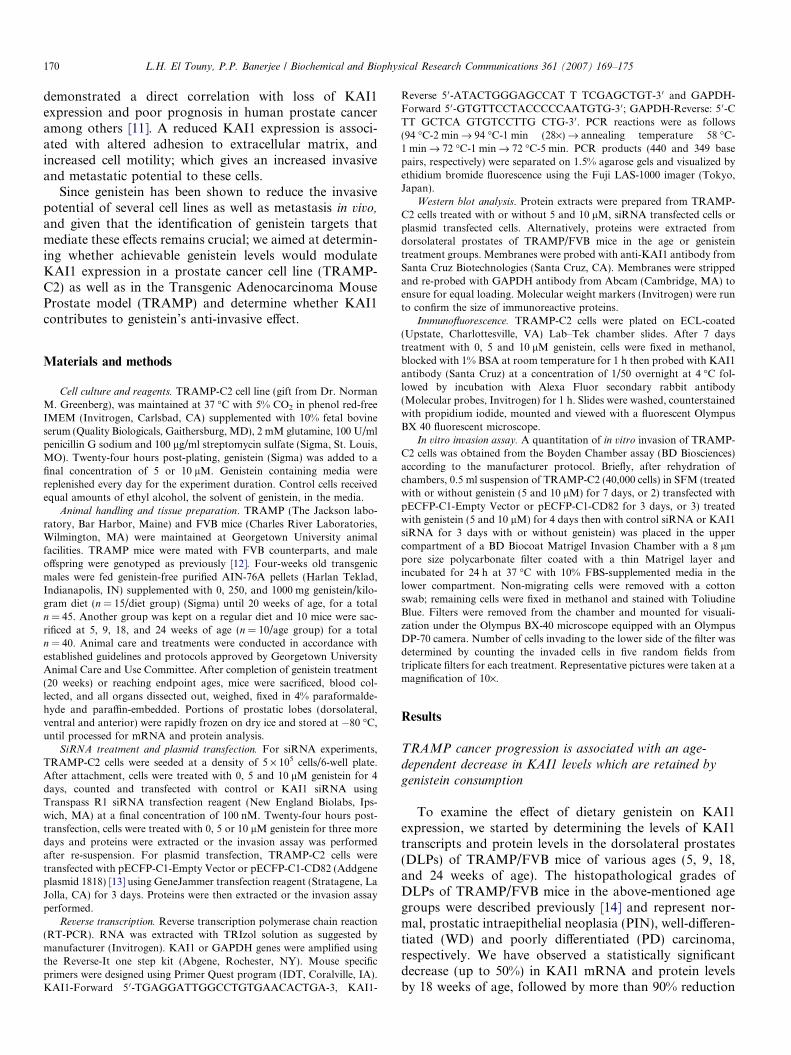
To examine the effect of dietary genistein on KAI1expression, we started by determining the levels of KAI1transcripts and protein levels in the dorsolateral prostates(DLPs) of TRAMP/FVB mice of various ages (5, 9, 18,and 24 weeks of age). The histopathological grades ofDLPs of TRAMP/FVB mice in the above-mentioned agegroups were described previously [14] and represent nor-mal, prostatic intraepithelial neoplasia (PIN), well-differen-tiated (WD) and poorly differentiated (PD) carcinoma,respectively. We have observed a statistically significantdecrease (up to 50%) in KAI1 mRNA and protein levelsby 18 weeks of age, followed by more than 90% reduction
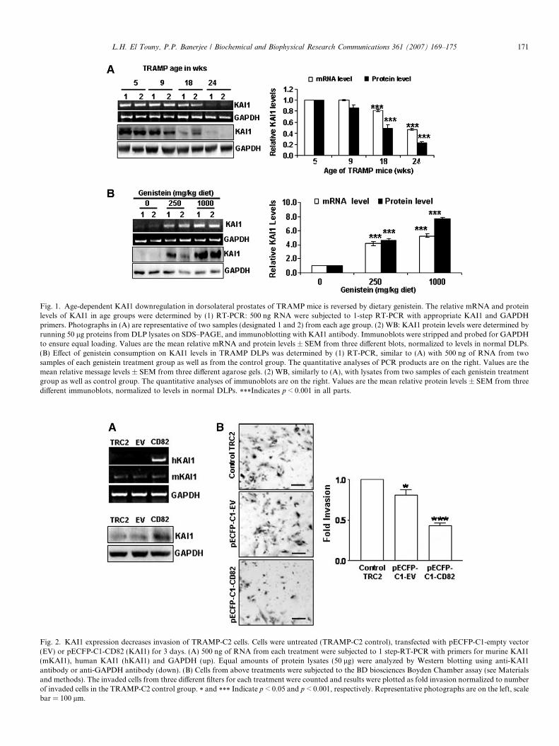
Fig. 1. Age-dependent KAI1 downregulation in dorsolateral prostates of TRAMP mice is reversed by dietary genistein. The relative mRNA and proteinlevels of KAI1 in age groups were determined by (1) RT-PCR: 500 ng RNA were subjected to 1-step RT-PCR with appropriate KAI1 and GAPDHprimers. Photographs in (A) are representative of two samples (designated 1 and 2) from each age group. (2) WB: KAI1 protein levels were determined byrunning 50 lg proteins from DLP lysates on SDS–PAGE, and immunoblotting with KAI1 antibody. Immunoblots were stripped and probed for GAPDHto ensure equal loading. Values are the mean relative mRNA and protein levels ± SEM from three different blots, normalized to levels in normal DLPs.(B) Effect of genistein consumption on KAI1 levels in TRAMP DLPs was determined by (1) RT-PCR, similar to (A) with 500 ng of RNA from twosamples of each genistein treatment group as well as from the control group. The quantitative analyses of PCR products are on the right. Values are themean relative message levels ± SEM from three different agarose gels. (2) WB, similarly to (A), with lysates from two samples of each genistein treatmentgroup as well as control group. The quantitative analyses of immunoblots are on the right. Values are the mean relative protein levels ± SEM from threedifferent immunoblots, normalized to levels in normal DLPs. ***Indicates p < 0.001 in all parts.
Fig. 2. KAI1 expression decreases invasion of TRAMP-C2 cells. Cells were untreated (TRAMP-C2 control), transfected with pECFP-C1-empty vector(EV) or pECFP-C1-CD82 (KAI1) for 3 days. (A) 500 ng of RNA from each treatment were subjected to 1 step-RT-PCR with primers for murine KAI1(mKAI1), human KAI1 (hKAI1) and GAPDH (up). Equal amounts of protein lysates (50 lg) were analyzed by Western blotting using anti-KAI1antibody or anti-GAPDH antibody (down). (B) Cells from above treatments were subjected to the BD biosciences Boyden Chamber assay (see Materialsand methods). The invaded cells from three different filters for each treatment were counted and results were plotted as fold invasion normalized to numberof invaded cells in the TRAMP-C2 control group. * and *** Indicate p < 0.05 and p < 0.001, respectively. Representative photographs are on the left, scalebar = 100 lm.
L.H. El Touny, P.P. Banerjee / Biochemical and Biophysical Research Communications 361 (2007) 169–175 171
172 L.H. El Touny, P.P. Banerjee / Biochemical and Biophysical Research Communications 361 (2007) 169–175
by 24 weeks of age (Fig. 1A). Concomitant with the reduc-tion in the incidence of PD cancer in the DLPs of TRAMP/FVB mice consuming a genistein-rich diet that we previ-ously reported [14], we observed a dose-dependent increasein KAI1 mRNA and protein levels (up to 4.0 and 7.0-fold)in the 250 and 1000 mg/kg diet groups, respectively(Fig. 1B).
KAI1 expression decreases the invasive ability of the
TRAMP-derived cell line, TRAMP-C2
We next examined the levels of KAI1 message and pro-tein in the TRAMP-derived TRAMP-C2 cell line. KAI1(mRNA and protein) is expressed at very low levels inTRAMP-C2 cells (Fig. 2A). To determine the functionalsignificance of KAI1 expression on TRAMP-C2 in termsof invasion, these cells were transiently transfected with ahuman KAI1 expression vector. The expression of hKAI1
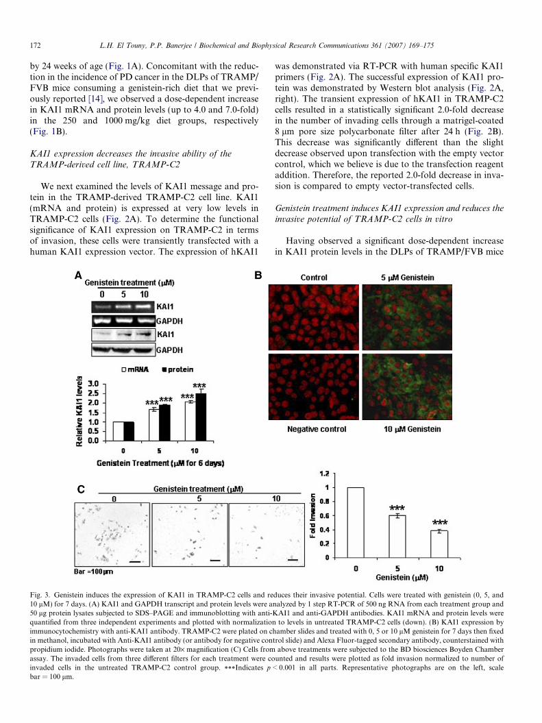
Fig. 3. Genistein induces the expression of KAI1 in TRAMP-C2 cells and re10 lM) for 7 days. (A) KAI1 and GAPDH transcript and protein levels were a50 lg protein lysates subjected to SDS–PAGE and immunoblotting with anti-Kquantified from three independent experiments and plotted with normalizationimmunocytochemistry with anti-KAI1 antibody. TRAMP-C2 were plated on chin methanol, incubated with Anti-KAI1 antibody (or antibody for negative conpropidium iodide. Photographs were taken at 20· magnification (C) Cells fromassay. The invaded cells from three different filters for each treatment were coinvaded cells in the untreated TRAMP-C2 control group. ***Indicates p <bar = 100 lm.
was demonstrated via RT-PCR with human specific KAI1primers (Fig. 2A). The successful expression of KAI1 pro-tein was demonstrated by Western blot analysis (Fig. 2A,right). The transient expression of hKAI1 in TRAMP-C2cells resulted in a statistically significant 2.0-fold decreasein the number of invading cells through a matrigel-coated8 lm pore size polycarbonate filter after 24 h (Fig. 2B).This decrease was significantly different than the slightdecrease observed upon transfection with the empty vectorcontrol, which we believe is due to the transfection reagentaddition. Therefore, the reported 2.0-fold decrease in inva-sion is compared to empty vector-transfected cells.
Genistein treatment induces KAI1 expression and reduces theinvasive potential of TRAMP-C2 cells in vitro
Having observed a significant dose-dependent increasein KAI1 protein levels in the DLPs of TRAMP/FVB mice
duces their invasive potential. Cells were treated with genistein (0, 5, andnalyzed by 1 step RT-PCR of 500 ng RNA from each treatment group and
AI1 and anti-GAPDH antibodies. KAI1 mRNA and protein levels wereto levels in untreated TRAMP-C2 cells (down). (B) KAI1 expression by
amber slides and treated with 0, 5 or 10 lM genistein for 7 days then fixedtrol slide) and Alexa Fluor-tagged secondary antibody, counterstained with
above treatments were subjected to the BD biosciences Boyden Chamberunted and results were plotted as fold invasion normalized to number of
0.001 in all parts. Representative photographs are on the left, scale
L.H. El Touny, P.P. Banerjee / Biochemical and Biophysical Research Communications 361 (2007) 169–175 173
fed the genistein-containing diets as compared to their age-matched control diet-fed counterparts, we aimed to deter-mine whether genistein could induce the expression ofKAI1 in TRAMP-C2 cells. Western blot analysis revealedthat genistein treatment for 7 days resulted in a dose-dependent increase in KAI1 mRNA and protein levels(up to 2.5-fold in 10 lM treated cells) (Fig. 3A). Thisincrease was also corroborated by increase in KAI1 immu-noreactivity in TRAMP-C2 cells by immunofluorescencestaining (Fig. 3B).
Previous studies have shown that genistein decreases theinvasive potential of prostate cancer cells, albeit at supra-physiological levels (�50 lM). Therefore, we aimed atdetermining whether the low genistein doses used (5 and10 lM) that induced the expression of KAI1, would affectTRAMP-C2 invasion. In fact, low doses of genistein treat-ment reduced the number of invaded cells by more than60% in the 10 lM genistein group (Fig. 3C).
The invasiveness of genistein-treated TRAMP-C2 cells is
restored by siRNA targeting KAI1
Having observed an induction of KAI1 levels by geni-stein concomitant with a decrease in invasion, we wantedto determine whether KAI1 increase plays a role in the
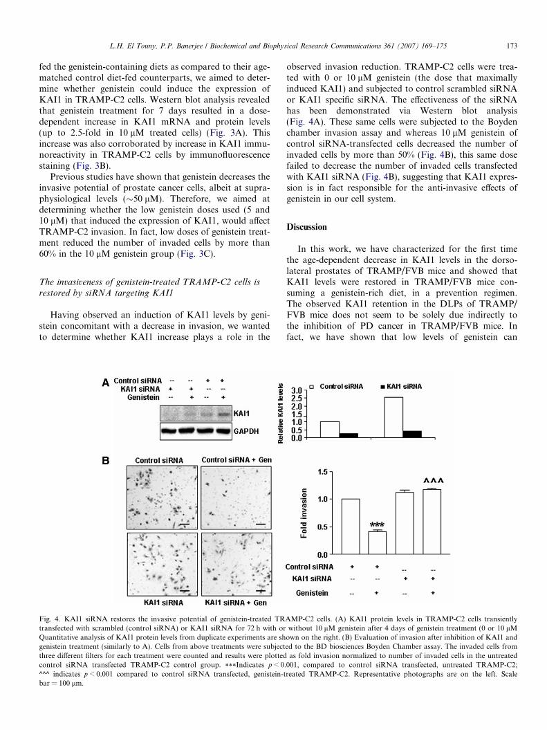
Fig. 4. KAI1 siRNA restores the invasive potential of genistein-treated TRtransfected with scrambled (control siRNA) or KAI1 siRNA for 72 h with orQuantitative analysis of KAI1 protein levels from duplicate experiments are shgenistein treatment (similarly to A). Cells from above treatments were subjectthree different filters for each treatment were counted and results were plottedcontrol siRNA transfected TRAMP-C2 control group. ***Indicates p < 0.^^^ indicates p < 0.001 compared to control siRNA transfected, genistein-tbar = 100 lm.
observed invasion reduction. TRAMP-C2 cells were trea-ted with 0 or 10 lM genistein (the dose that maximallyinduced KAI1) and subjected to control scrambled siRNAor KAI1 specific siRNA. The effectiveness of the siRNAhas been demonstrated via Western blot analysis(Fig. 4A). These same cells were subjected to the Boydenchamber invasion assay and whereas 10 lM genistein ofcontrol siRNA-transfected cells decreased the number ofinvaded cells by more than 50% (Fig. 4B), this same dosefailed to decrease the number of invaded cells transfectedwith KAI1 siRNA (Fig. 4B), suggesting that KAI1 expres-sion is in fact responsible for the anti-invasive effects ofgenistein in our cell system.
Discussion
In this work, we have characterized for the first timethe age-dependent decrease in KAI1 levels in the dorso-lateral prostates of TRAMP/FVB mice and showed thatKAI1 levels were restored in TRAMP/FVB mice con-suming a genistein-rich diet, in a prevention regimen.The observed KAI1 retention in the DLPs of TRAMP/FVB mice does not seem to be solely due indirectly tothe inhibition of PD cancer in TRAMP/FVB mice. Infact, we have shown that low levels of genistein can
AMP-C2 cells. (A) KAI1 protein levels in TRAMP-C2 cells transientlywithout 10 lM genistein after 4 days of genistein treatment (0 or 10 lM
own on the right. (B) Evaluation of invasion after inhibition of KAI1 anded to the BD biosciences Boyden Chamber assay. The invaded cells from
as fold invasion normalized to number of invaded cells in the untreated001, compared to control siRNA transfected, untreated TRAMP-C2;reated TRAMP-C2. Representative photographs are on the left. Scale
174 L.H. El Touny, P.P. Banerjee / Biochemical and Biophysical Research Communications 361 (2007) 169–175
induce KAI1 expression in the TRAMP-derived cell line,TRAMP-C2. This induction occurred at both the mRNAand protein levels, suggesting a transcriptional regulationof KAI1 by genistein.
The induction of KAI1 has been previously shown to bedependent on p53 [15]. However, we do not believe this tobe the case in this study due to the SV40-transformed nat-ure of the TRAMP model and its derived cell line. Otherfactors have been demonstrated to induce KAI1 re-expres-sion including nerve growth factor and phorbol esters[16,17]. However, in each of these mentioned cases, itremains to be determined whether the effects on KAI1expression are directly due to these agents or indirectly asa result of altered cell behavior. The common downregula-tion of KAI1 in metastatic disease does not involve loss ofheterozygosity at the KAI1 locus or mutations within theKAI1 gene [11]. However, the KAI1 promoter is associatedwith a CpG island; and genistein has been shown to act as ademethylating agent in vitro at a similar dose as the oneused in this work (5 lM) [18]; therefore, genistein mightinduce KAI1 expression via promoter demethylation,which remains to be determined.
The proximal region of the KAI1 promoter containspotential binding sites for several transcription factorsincluding AP1 and Sp1 sites [19]. Phytoestrogens, suchas genistein have been shown to regulate promoters con-taining such sites via estrogen receptor signaling [20].Furthermore, a comprehensive cDNA microarray exam-ining the effects of a similar dose of genistein (10 lM)revealed the induction of these AP-1 factors [21]. There-fore, KAI1 might be induced via AP-1 regulation bygenistein.
We have also shown that the expression of KAI1 bytransfection or genistein treatment reduced the invasivepotential of TRAMP-C2 cells. The anti-invasive mecha-nism of KAI1 has not been fully elucidated. In fact,KAI1 lacks intrinsic activity, but mediates its functionsvia binding to surface receptors such as integrins orreceptor tyrosine kinases in a cell specific manner [12].Several attractive scenarios have been put forward toexplain KAI1 anti-invasive effects; including upregulationof TIMP-1 [22] and the inactivation of urokinase recep-tor proteolytic activities [23]. Although previous studieshave shown that several pro-and anti-metastatic factorsare modulated by genistein treatment, these were doneat supraphysiological levels and therefore might not reca-pitulate what is happening with the doses used in thisstudy or what could be achieved via genistein consump-tion. In fact, through this work, we have shown thatinhibition of KAI1 expression by siRNA restored thenumber of invaded cells to control levels in the presenceof genistein. This result suggests that KAI1 induction isrequired for the decrease the invasiveness of TRAMP-C2 cells by genistein. This study suggests that genisteintreatment could have beneficial effect in preventing met-astatic disease by restoring KAI1 and abolishing theinvasive potential of cancer cells.
Acknowledgments
This work was supported by NIH Grant R01 DK060875to Partha P. Banerjee. We thank Dr. Norman Greenberg ofFred Hutchinson Cancer Research Center, Seattle, Wash-ington for his gift of TRAMP-C2 cells.
References
[1] A. Jemal, R. Siegel, E. Ward, T. Murray, J. Xu, M.J. Thun, Cancerstatistics, 2007, CA Cancer J. Clin. 57 (2007) 43–66.
[2] R.V. Gopalkrishnan, D.C. Kang, P.B. Fisher, Molecular markers anddeterminants of prostate cancer metastasis, J. Cell Physiol. 189 (2001)245–256.
[3] L.A. Liotta, W.G. Stetler-Stevenson, Tumor invasion and metastasis:an imbalance of positive and negative regulation, Cancer Res. 51(1991) 5054–5059.
[4] R.K. Severson, A.M. Nomura, J.S. Grove, G.N. Stemmermann, Aprospective study of demographics, diet, and prostate cancer amongmen of Japanese ancestry in Hawaii, Cancer Res. 49 (1989) 1857–1860.
[5] X. Huang, S. Chen, L. Xu, Y. Liu, D.K. Deb, L.C. Platanias, R.C.Bergan, Genistein inhibits p38 map kinase activation, matrixmetalloproteinase type 2, and cell invasion in human prostateepithelial cells, Cancer Res. 65 (2005) 3470–3478.
[6] H. Iishi, M. Tatsuta, M. Baba, H. Yano, N. Sakai, H. Akedo,Genistein attenuates peritoneal metastasis of azoxymethane-inducedintestinal adenocarcinomas in Wistar rats, Int. J. Cancer 86 (2000)416–420.
[7] G.P. Gupta, J. Massague, Cancer metastasis: building a framework,Cell 127 (2006) 679–695.
[8] Y. Liu, E. Kyle, R. Lieberman, J. Crowell, G. Kellof, R.C. Bergan,Focal adhesion kinase (FAK) phosphorylation is not required forgenistein-induced FAK-beta-1-integrin complex formation, Clin.Exp. Metastasis 18 (2000) 203–212.
[9] Z.M. Shao, J. Wu, Z.Z. Shen, S.H. Barsky, Genistein exerts multiplesuppressive effects on human breast carcinoma cells, Cancer Res. 58(1998) 4851–4857.
[10] M.H. Kim, A.M. Gutierrez, R.H. Goldfarb, Different mechanisms ofsoy isoflavones in cell cycle regulation and inhibition of invasion,Anticancer Res. 22 (2002) 3811–3817.
[11] P. Jackson, A. Marreiros, P.J. Russell, KAI1 tetraspanin andmetastasis suppressor, Int. J. Biochem. Cell. Biol. 37 (2005) 530–534.
[12] N.M. Greenberg, F. DeMayo, M.J. Finegold, D. Medina, W.D.Tilley, J.O. Aspinall, G.R. Cunha, A.A. Donjacour, R.J. Matusik,J.M. Rosen, Prostate cancer in a transgenic mouse, Proc. Natl. Acad.Sci. USA 92 (1995) 3439–3443.
[13] N.M. Sherer, M.J. Lehmann, L.F. Jimenez-Soto, A. Ingmundson,S.M. Horner, G. Cicchetti, P.G. Allen, M. Pypaert, J.M. Cunning-ham, W. Mothes, Visualization of retroviral replication in living cellsreveals budding into multivesicular bodies, Traffic 4 (2003) 785–801.
[14] L.H. El Touny, P.P. Banerjee, Akt/GSK3 pathway as a target ingenistein-induced inhibition of TRAMP prostate cancer progressiontowards a poorly differentiated phenotype, Carcinogenesis (2007),doi: 10.1093.
[15] A. Marreiros, K. Dudgeon, V. Dao, M.O. Grimm, R. Czolij, M.Crossley, P. Jackson, KAI1 promoter activity is dependent on p53,junB and AP2: evidence for a possible mechanism underlying loss ofKAI1 expression in cancer cells, Oncogene 24 (2005) 637–649.
[16] S. Sigala, I. Faraoni, D. Botticini, M. Paez-Pereda, C. Missale, E.Bonmassar, P. Spano, Suppression of telomerase, reexpression ofKAI1, and abrogation of tumorigenicity by nerve growth factor inprostate cancer cell lines, Clin. Cancer Res. 5 (1999) 1211–1218.
[17] H. Akita, A. Iizuka, Y. Hashimoto, K. Kohri, K. Ikeda, M.Nakanishi, Induction of KAI-1 expression in metastatic cancer cellsby phorbol esters, Cancer Lett. 153 (2000) 79–83.

L.H. El Touny, P.P. Banerjee / Biochemical and Biophysical Research Communications 361 (2007) 169–175 175
[18] M. Fang, D. Chen, C.S. Yang, Dietary polyphenols may affect DNAmethylation, J. Nutr. 137 (2007) 223S–228S.
[19] J.T. Dong, W.B. Isaacs, J.C. Barrett, J.T. Isaacs, Genomic organi-zation of the human KAI1 metastasis-suppressor gene, Genomics 41(1997) 25–32.
[20] J.R. Schultz, L.N. Petz, A.M. Nardulli, Cell- and ligand-specificregulation of promoters containing activator protein-1 and Sp1 sites byestrogen receptors alpha and beta, J. Biol. Chem. 280 (2005) 347–354.
[21] R. Ise, D. Han, Y. Takahashi, S. Terasaka, A. Inoue, M. Tanji, R.Kiyama, Expression profiling of the estrogen responsive genes in
response to phytoestrogens using a customized DNA microarray,FEBS Lett. 579 (2005) 1732–1740.
[22] B.K. Jee, K.M. Park, S. Surendran, W.K. Lee, C.W. Han, Y.S.Kim, Y. Lim, KAI1/CD82 suppresses tumor invasion by MMP9inactivation via TIMP1 up-regulation in the H1299 human lungcarcinoma cell line, Biochem. Biophys. Res. Commun. 342 (2006)655–661.
[23] R. Bass, F. Werner, E. Odintsova, T. Sugiura, F. Berditchevski, V.Ellis, Regulation of urokinase receptor proteolytic function by thetetraspanin CD82, J. Biol. Chem. 280 (2005) 14811–14818.