Anatomical and Histological Changes of Reproductive Organs in Japanese Quail (Coturnix japonica)...
13
International Journal of Poultry Science 13 (1): 1-13, 2014 ISSN 1682-8356 © Asian Network for Scientific Information, 2014 Corresponding Author: Sittipon Intarapat, Department of Anatomy, Faculty of Science, Mahidol University, Bangkok-10400, Thailand 1 Anatomical and Histological Changes of Reproductive Organs in Japanese Quail (Coturnix japonica) Embryos after in ovo Exposure to Genistein Sittipon Intarapat , Achariya Sailasuta and Orawan Satayalai 1 2 3 Department of Anatomy, Faculty of Science, Mahidol University, Bangkok-10400, Thailand 1 Department of Pathology, Faculty of Veterinary Science, 2 Chulalongkorn University, Bangkok-10330, Thailand Department of Biology, Faculty of Science, Chulalongkorn University, Bangkok-10330, Thailand 3 Abstract: Genistein, an isoflavonoid phytoestrogen, has a similar structure to endogenous estrogen and is also able to produce estrogen-like effects. Administration of exogenous estrogens as well as phytoestrogens causes reproductive abnormalities in adult birds. Here, we examine whether genistein affects reproductive organ development in Japanese quail at the sexually differentiated stage. Genistein (16 and 24 μg/g egg) was injected into the yolk via the blunt end of the fertilized egg prior to incubation. Both concentrations caused Müllerian duct abnormalities in both sexes and ovotestis formation in male embryos. This is the first report that genistein affects reproductive organ development in avian embryos. Key words: Genistein, in ovo, quail embryos, reproductive organs, Coturnix japonica INTRODUCTION Phytoestrogens are estrogen-like substances produced by plants, especially from the family Leguminosae. Due to their chemical structures being similar to endogenous estrogen (estradiol), they have estrogen-like effects (Kanno et al., 2002). Phytoestrogens are classified into three groups: isoflavones, coumestans and lignans (Turner and Sharpe, 1997; Setchell, 1998). Isoflavones are found at high concentrations in soybean and soy products (Adlercreutz, 1995). The isoflavones genistein and daidzein are considered to bind to the estrogen receptor (ER) with high affinity (Hanafy et al., 2004; Hanafy et al., 2005). Once they bind to ER they are able to act as either estrogen agonists or antagonists (Setchell, 1998; Lephart et al., 2002). The impacts of soy isoflavones on mammalian reproductive system and their estrogen-like effects were seriously concerned. It has been reported that female rats-treated with 50 μg/d of genistein at day 21 of gestation exhibited decreasing of ovaries and uterine weights as well as estrogen and progesterone levels in plasma (Awoniyi et al., 1998). Casanova and colleagues have found that genistein and daidzein were estrogen agonists being able to bind with both ER alpha and beta that increased a ratio of uterine weight per body weight (Casanova et al., 1999). Furthermore, female rats- treated with 40 mg/kg of genistein resulted in the increasing of uterine weight at day 22 of postnatal period (Lewis et al., 2003). Moreover, prenatal rats-treated with genistein revealed sex differentiation and developmental failures (Lewis et al., 2003). In female rats-treated with 250-1,250 ppm. of genistein exhibited the ductal-alveolar hyperplasia, atretic follicles and ovarian antral follicles (Delclos et al., 2001). In male rats-treated with genistein at concentration 1,250 ppm. delayed a spermatogenesis, whereas genistein at concentration 625 and 1,250 ppm. caused abnormal spermatozoa in epididymis (Delclos et al., 2001). However, genistein at concentration 250 and 1,000 mg/kg affected neither testicular weight nor morphology (Fritz et al., 2003). In mice, female-treated with 40 mg/kg body weight of genistein resulted in uterine enlargement, keratinization of vaginal epithelium and uterine squamous metaplasia (Cline et al., 2004). Conversely, genistein at a concentration 40 mg/kg caused reduction of plasma testosterone concentrations, atrophy of seminiferous epithelium and accessory sex glands as well as squamous metaplasia of seminal vesicles (Cline et al., 2004), whereas genistein at concentration 5 mg/kg reduced a number of spermatozoa (Lee et al., 2004). There have been reported that genistein caused multi- oocyte follicle and polyovular follicles in female-mice treated with genistein at concentrations 50,000 μg/kg and 50 mg/kg, respectively (Jefferson and Newbold, 2000; Nagao et al., 2001). Jefferson and colleagues have described that these phenomena caused by a binding of genistein with ER alpha, since these have not been observed in knocked out-ER beta gene mice (Jefferson et al., 2002). In the poultry industry, soybean is one of the most common sources of protein used for quails during the egg laying period (Woodard et al., 1973). Isoflavones contained in soybean accumulate in products derived from poultry including eggs and meat (Lin et al., 2004;
Transcript of Anatomical and Histological Changes of Reproductive Organs in Japanese Quail (Coturnix japonica)...
International Journal of Poultry Science 13 (1): 1-13, 2014ISSN 1682-8356© Asian Network for Scientific Information, 2014
Corresponding Author: Sittipon Intarapat, Department of Anatomy, Faculty of Science, Mahidol University, Bangkok-10400, Thailand
1
Anatomical and Histological Changes of Reproductive Organs in JapaneseQuail (Coturnix japonica) Embryos after in ovo Exposure to Genistein
Sittipon Intarapat , Achariya Sailasuta and Orawan Satayalai1 2 3
Department of Anatomy, Faculty of Science, Mahidol University, Bangkok-10400, Thailand1
Department of Pathology, Faculty of Veterinary Science,2
Chulalongkorn University, Bangkok-10330, ThailandDepartment of Biology, Faculty of Science, Chulalongkorn University, Bangkok-10330, Thailand3
Abstract: Genistein, an isoflavonoid phytoestrogen, has a similar structure to endogenous estrogen and isalso able to produce estrogen-like effects. Administration of exogenous estrogens as well as phytoestrogenscauses reproductive abnormalities in adult birds. Here, we examine whether genistein affects reproductiveorgan development in Japanese quail at the sexually differentiated stage. Genistein (16 and 24 µg/g egg)was injected into the yolk via the blunt end of the fertilized egg prior to incubation. Both concentrations causedMüllerian duct abnormalities in both sexes and ovotestis formation in male embryos. This is the first reportthat genistein affects reproductive organ development in avian embryos.
Key words: Genistein, in ovo, quail embryos, reproductive organs, Coturnix japonica
INTRODUCTIONPhytoestrogens are estrogen-like substances producedby plants, especially from the family Leguminosae. Dueto their chemical structures being similar to endogenousestrogen (estradiol), they have estrogen-like effects(Kanno et al., 2002). Phytoestrogens are classified intothree groups: isoflavones, coumestans and lignans(Turner and Sharpe, 1997; Setchell, 1998). Isoflavonesare found at high concentrations in soybean and soyproducts (Adlercreutz, 1995). The isoflavones genisteinand daidzein are considered to bind to the estrogenreceptor (ER) with high affinity (Hanafy et al., 2004;Hanafy et al., 2005). Once they bind to ER they are ableto act as either estrogen agonists or antagonists(Setchell, 1998; Lephart et al., 2002).The impacts of soy isoflavones on mammalianreproductive system and their estrogen-like effects wereseriously concerned. It has been reported that femalerats-treated with 50 µg/d of genistein at day 21 ofgestation exhibited decreasing of ovaries and uterineweights as well as estrogen and progesterone levels inplasma (Awoniyi et al., 1998). Casanova and colleagueshave found that genistein and daidzein were estrogenagonists being able to bind with both ER alpha and betathat increased a ratio of uterine weight per body weight(Casanova et al., 1999). Furthermore, female rats-treated with 40 mg/kg of genistein resulted in theincreasing of uterine weight at day 22 of postnatal period(Lewis et al., 2003). Moreover, prenatal rats-treated withgenistein revealed sex differentiation and developmentalfailures (Lewis et al., 2003). In female rats-treated with250-1,250 ppm. of genistein exhibited the ductal-alveolar
hyperplasia, atretic follicles and ovarian antral follicles(Delclos et al., 2001). In male rats-treated with genisteinat concentration 1,250 ppm. delayed aspermatogenesis, whereas genistein at concentration625 and 1,250 ppm. caused abnormal spermatozoa inepididymis (Delclos et al., 2001). However, genistein atconcentration 250 and 1,000 mg/kg affected neithertesticular weight nor morphology (Fritz et al., 2003). Inmice, female-treated with 40 mg/kg body weight ofgenistein resulted in uterine enlargement, keratinizationof vaginal epithelium and uterine squamous metaplasia(Cline et al., 2004). Conversely, genistein at aconcentration 40 mg/kg caused reduction of plasmatestosterone concentrations, atrophy of seminiferousepithelium and accessory sex glands as well assquamous metaplasia of seminal vesicles (Cline et al.,2004), whereas genistein at concentration 5 mg/kgreduced a number of spermatozoa (Lee et al., 2004).There have been reported that genistein caused multi-oocyte follicle and polyovular follicles in female-micetreated with genistein at concentrations 50,000 µg/kgand 50 mg/kg, respectively (Jefferson and Newbold,2000; Nagao et al., 2001). Jefferson and colleagueshave described that these phenomena caused by abinding of genistein with ER alpha, since these have notbeen observed in knocked out-ER beta gene mice(Jefferson et al., 2002).In the poultry industry, soybean is one of the mostcommon sources of protein used for quails during theegg laying period (Woodard et al., 1973). Isoflavonescontained in soybean accumulate in products derivedfrom poultry including eggs and meat (Lin et al., 2004;
Int. J. Poult. Sci., 13 (1): 1-13, 2014
2
Jiang et al., 2007). Moreover, the effects of known about such effects on avian species. There havephytoestrogens on avian reproduction have only been been found that genistein reduced a number of egg-reported for adults in California quail (Lophortyx laying periods in California quail (Leopold et al., 1976)california) (Leopold et al., 1976), zebra finch as well as inhibited testosterone secretion in roosters(Taeniopygia guttata) (Millam et al., 2002), chicken (Opalka et al., 2004). Furthermore, genistein inhibited(Gallus gallus domesticus) (Opalka et al., 2004; Wistedt basal LH-stimulated testosterone production by Leydiget al., 2012), duck (Anas platyrhynchos) (Zhao et al., cells and decreased the volume of ejaculates, the2004; Zhao et al., 2005), gander (Anser anser percentage of normal spermatozoa of Bilgoraj gandersdomesticus) (Opalka et al., 2006; Kaminska et al., 2008; (Opalka et al., 2006; Opalka et al., 2008). Mechanism ofOpalka et al., 2008; Opalka et al., 2012) and Japanese action of genistein inhibiting testosterone secretion inquail (Coturnix japonica) (de Man and Peeke, 1982; avian testicular cells was concluded that it interactedAkdemir and Sahin, 2009; Rochester et al., 2009; Sahin with key steroidogenic enzymes rather than estrogenet al., 2009; Ekinci and Erkan, 2012) but not in embryos receptors (Opalka et al., 2012). In mammals,of any of these species. testosterone was also a target of estrogenic compoundsOviparous species use yolk as an energy supply for the since suppression of testosterone synthesis during aembryo during development (Romanoff and Romanoff, fetal life resulted in cryptorchidism and dysfunction of1949; Romanoff, 1960; Walker, 1967). A major adult testis (Clark and Cochrum, 2007; Wohlfahrt-Veje etcomponent of the yolk is lipid. Endocrine disruptors al., 2009). There were strong evidences showed that(Eds) are mostly lipophilic substances dissolved in the genistein inhibited testosterone secretion by Leydigyolk (Berg et al., 1999). Several isoflavones are cells of mice fetal testes (Lehraiki et al., 2011; Zhang ettransferred and accumulated in the yolk such as equol al., 2013). Recently, it was reported that adult male(Saitoh et al., 2004), daidzein (Saitoh et al., 2001) and quails received high doses of genistein exhibitedgenistein (Saitoh et al., 2001; Lin et al., 2004; Jiang et testicular damage and apoptosis of germ cells (Ekincial., 2007). Therefore, avian embryos are the most likely and Erkan, 2012). Thus, the study of effects of genisteinto obtain phytoestrogens from isoflavone-enriched diet on embryonic testes in male quails needs to befed to the mother during the egg-laying period. Thus, it is investigated.possible that embryos become exposed to EDs Even though there some studies have been undertakenaccumulated in the yolk by maternal transfer via blood of effects of genistein on adult quail reproductioncirculation. (Akdemir and Sahin, 2009; Sahin et al., 2009), its effectsSeveral routes of administration of EDs to avian have not yet been in embryos. Previous studies usedembryos have been reported including egg dipping biological parameters to evaluate the endpoints of EDs(Scheib and Reyss-Brion, 1979; Perrin et al., 1995) and on reproductive organs in avian embryos, including
egg yolk (in ovo) injection (Brunstrom and Orberg, 1982; embryonic accessory duct (Willier et al., 1935; Willier etBerg et al., 1998; Berg et al., 1999; Biau et al., 2007). The al., 1938; Romanoff, 1960; Stoll et al., 1993; Berg et al.,in ovo injection method is preferable for toxicity testing in 1998; Berg et al., 1999; Berg et al., 2001; Biau et al.,oviparous species because it can deliver standardized 2007) and the left testis (Willier et al., 1935; Stoll et al.,doses (Walker, 1967) and it is also ecologically 1993; Perrin et al., 1995; Berg et al., 1998; Berg et al.,advantageous because it can minimize ED 1999; Berg et al., 2001; Oshima et al., 2012). Since quailcontamination of the environment (Berg et al., 1998; embryos exposed to EDs in ovo resulted in MDsBerg et al., 1999). retention in both sexes and ovotestis formation in maleQuail eggs have several advantages as the endocrine embryos (Perrin et al., 1995; Berg et al., 1998; Berg etdisrupting model: the eggs are small and easily al., 1999; Berg et al., 2001; Shibuya et al., 2004; Shibuyaexposed to substances (Berg et al., 1998; Berg et al., et al., 2005; Oshima et al., 2012), MDs retention and1999; Halldin, 2005), they are available all year round ovotestis formation can be used as reliable biomarkers(Berg et al., 1999; Halldin, 2005) and their hypothalamic- for evaluating estrogen-like effects of EDs (Berg et al.,pituitary-gonadal axis is well understood (Ottinger et al., 1998; Berg et al., 1999).2001; Ottinger et al., 2002). Quails are therefore often Though genistein was reported that it acts as a tyrosinerecommended as a model for testing estrogen-like kinases inhibitor (Akiyama et al., 1987; Huang et al.,effects of EDs in birds (OECD, 1984; OECD, 2000; 1992; Hong et al., 2005), there were several studiesTouart, 2004). demonstrated that genistein was an endocrine disruptorAlthough genistein is widely used for studying estrogen- affecting the endocrine system of birds (Leopold et al.,like effects and reproductive toxicity in mammalian 1976; Opalka et al., 2004; Opalka et al., 2006; Kaminskareproductive system (Awoniyi et al., 1998; Casanova et et al., 2008; Opalka et al., 2008; Opalka et al., 2012).al., 1999; Jefferson and Newbold, 2000; Delclos et al., Interestingly, sex determination and differentiation of2001; Nagao et al., 2001; Jefferson et al., 2002; Lewis et avian reproductive organs are predominantly controlledal., 2003; Cline et al., 2004; Lee et al., 2004), little is by sex hormones (Romanoff, 1960; Weniger, 1991;

Int. J. Poult. Sci., 13 (1): 1-13, 2014
3
Smith and Sinclair, 2001; Brunstrom et al., 2003; Smith needle at the blunt end of the egg and the blunt end wasand Sinclair, 2004; Brunstrom et al., 2009; Chue and wiped with 70% ethanol to get rid of small pieces of theSmith, 2011). Moreover, development of avian eggshell. The eggs were then treated with two doses ofreproductive organs is estrogen dependent that genistein, 16 and 24 µg/g egg, by injecting into the yolkestrogen-like compounds are able to disrupt such prior to incubation using Hamilton microliter syringedevelopmental process (Romanoff, 1960; Fry and (Hamilton Company, USA). The amount of a mixture (20Toone, 1981; Rattner et al., 1984; Fry, 1995). µL/egg) was injected into the control and genistein-The present study aimed to examine the effects of treated eggs. After in ovo exposure, the hole in the shellgenistein on reproductive organ development including was sealed with paraffin wax and wiped with 70%embryonic accessory ducts in both sexes and the left ethanol. Injected eggs were then incubated at 37.5°C intestis in male quail embryos following in ovo exposure. a humid atmosphere and automatically turned by the
MATERIALS AND METHODSEggs and chemicals: Japanese quail (Coturnixjaponica) eggs were obtained from the Department ofAnimal Science, Kasetsart University, Thailand.Genistein (4',5,7-Trihydroxyisoflavone; purity>98%) waspurchased from Sigma chemical (St. Louis, Mo, USA).DMSO (dimethyl sulfoxide; purity>99%) was obtainedfrom Merck (Darmstadt, Germany) and Mazola corn oil®
was the product of CPC (AJI, Thailand).
Genistein preparation: A 10% DMSO was prepared bydiluting DMSO in the distilled water and then mixed withcorn coil for preparing the 10% DMSO/corn oil emulsionas a vehicle. To prepare genistein, genistein wasdissolved in 10% DMSO as mentioned above. Due togenistein did not dissolve promptly in 10% DMSO, it wasthen mechanically dissolved by sonication (LRUltrasonic sonicator, Fisher Scientific). Thereafter, amixture of genistein/10% DMSO was then emulsifiedusing vigorous vortexing with corn oil for preparing thegenistein/10% DMSO/corn oil emulsion.
Dosing and in ovo exposure: Fertilized quail eggs weregrouped into three treatments: the control, genistein-16and genistein-24 µg/g-treated eggs. A mixture of 10%DMSO/corn oil was used as vehicle control, whereas 16and 24 µg genistein administrated in 10% DMSO/cornoil emulsion were used as the genistein-treated groups.In the present study, genistein (16 and 24 µg/g egg) wasselected for administration into the yolk (Brunstrom andOrberg, 1982), since genistein at concentration 7 µg/gegg showed no effect on quail reproductive organs(personal communication). Since 1.6 and 2.4 mggenistein were found in the yolk after feeding quail henswith 50 and 100 mg genistein, respectively (Lin et al.,2004), an increase of genistein doses up to 10 fold wasexamined to observe adverse effects of genistein in thisstudy.Before starting in ovo exposure, quail eggs werecleaned with Betadine solution (IDS manufacturing Ltd.,®
Thailand). After cleaning, cleaned eggs were thenweighed and labeled. The labeled eggs were punchedwith sterile disposable 25G needle (Nipro Corporation, Mortality fertility and viability: Genistein (16 µg/g egg)Japan) at the blunt end of the egg. Before injection, a caused high embryo mortality (p<0.001) (Table 1),small hole was punched with a sterile disposable 25G whereas genistein at concentration 24 µg/g egg did not.
incubator. Eggs were checked for embryonic mortality,fertility and unfertilized/or dead eggs were discarded. Atday 15, a number of survival embryos were recorded andfrequencies of embryo mortality, fertility and viability wereanalyzed. The fertilized eggs were used at minimum 30eggs/treatment.
Anatomical and histological examinations: Quailembryos were sacrificed on day 15 of incubation (thesexually differentiated stage when embryonic sex can bedistinguished by gonad morphology) and were stagedaccording to Padgett and Ivey (Padgett and Ivey, 1960).Gross morphological abnormalities of the reproductiveorgans were observed under the SZ-PTstereomicroscope (Olympus, Japan). Biologicalparameters including accessory duct malformations inboth sexes (e.g. partial or complete Müllerian ductretention in male embryos) and right Müllerian ductretention (longer than 3 mm compared to control group)with or without shell gland in female embryos wererecorded.For histological examination, the left testis was fixed inBouin’s fluid and embedded in Paraplast (SherwoodMedical Company, St. Louis, MO, USA.) and thensectioned at 6 µm with the microtome (The Gemmary,Fallbrook, USA). The sections were stained withHematoxylin and Eosin. Ovotestis formation of the lefttestis was observed by a thickening of cortex layercontaining five or more oocyte-like germ cells in meioticprophase, showing eosinophilic cytoplasm in oocyte-likegerm cells and transforming of germinal epithelial cellsfrom squamous cells into cuboidal or columnar cells asdescribed in previous studies (Berg et al., 1998; Berg etal., 1999; Berg et al., 2001; Shibuya et al., 2004; Shibuyaet al., 2005). The sections were visualized by PM-10M3camera (Olympus, Japan)
Statistical analysis: Frequencies of embryo mortality,fertility, viability and reproductive organ abnormalities inboth sexes in control and treated groups were comparedusing Fisher’s exact test.
RESULTS

Int. J. Poult. Sci., 13 (1): 1-13, 2014
4
Table 1: Frequencies of mortality fertility and viability of quail embryos following in ovo injectionTreatment (µg/g egg) Mortality (%) Fertility (%) Viability (%) Sex (Female/Male)a b c
Control 19 (6/31) 97 (30/31) 80 (24/30) (Female = 13/Male = 11)d
Genistein (16) 54 (19/35)*** 94 (33/35) 42 (14/33) (Female = 7/Male = 7)e
Genistein (24) 28 (8/35) 100 (35/35) 77 (27/35) (Female = 14/Male =13)e
Ratio (in parentheses) represents the number of dead embryos divided by the number of treated eggs. Frequencies of the mortality, fertility anda
viability in the treatment groups were compared with the frequency in the control group using Fisher’s exact testNo. of fertile eggs divided by the number of total eggsb
No. of viable embryos divided by the number of fertile eggsc
Eggs-treated with DMSO/corn oil based emulsiond
Genistein was dissolved with DMSO and emulsified with corn oil. Statistical difference in relation to the control group (***p<0.001)e
Table 2: Frequencies of male and female 15-day-old quail embryos exhibiting abnormalities of MDs following genistein injection in ovo prior to incubationTreatment (µg/g egg) Female with MDs abnormalities (%) Male with MDs abnormalities (%)a a
Control 0 (0/13) 18 (2/11)b
Genistein (16) 43 (3/7)* 71 (5/7)*Genistein (24) 50 (7/14)** 54 (7/13)Ratio (in parentheses) represents the number of affected embryos divided by the number of embryos examined. Frequencies of the abnormality ina
the treatment groups were compared with the frequency in the control group using Fisher’s exact testQuail eggs treated with DMSO/corn oil based emulsion served as controls for the genistein-treated eggs Statistical differences in relation to the controlb
group (*p<0.05; **p<0.01)
Table 3: Frequencies of male 15 day-old-quail embryos exhibiting ovotestis following genistein injection in ovo prior to incubation
Treatment (µg/g egg) Male with ovotestis (%)a,b
Control 0 (0/11)Genistein (16) 86 (6/7)***Genistein (24) 85 (11/13)***Ratio (in parentheses) represents the number of affected embryosa
divided by the number of embryos examined. Frequencies of ovotestisin the treatment groups were compared with the frequency in the controlgroup using Fisher’s exact testIdentifying characteristics of ovotestis formation according to theb
criteria including thickening of cortex layer containing oocyte-like germcells in meiotic prophase, showing eosinophilic cytoplasm in oocyte-likegerm cells, transformation of germinal epithelial cells from squamouscells into cuboidal and columnar cells. Statistical differences in relationto the control group (***p<0.001)
Frequencies of fertility of the control, 16 µg genistein and24 µg genistein-treated eggs were 97, 94 and 100%,respectively (Table 1). Frequencies of embryo viability ofthe control, 16 µg genistein and 24 µg genistein-treatedeggs were 80, 42 and 77%, respectively (Table 1).
Abnormality of embryonic accessory ducts: Normally,two days before anticipated hatching, female embryonicaccessory ducts (Müllerian ducts, MDs) regresscompletely in male embryos, whereas the left MD stillremains in female embryos. In quail embryos treatedwith 16 µg genistein/g egg, Müllerian duct (MD)abnormalities occurred in 3 of the 7 female embryos(p<0.05) and 5 of the 7 male embryos (p<0.05) (Table 2,Fig. 1). In contrast, MD abnormalities were observed in7 of the 14 female embryos (p<0.01) and 7 of the 13male embryos treated with 24 µg genistein/g egg (notsignificant compared with the control group; Table 2,Fig. 1). The frequencies of MD abnormalities infemale and male embryos treated with 16 µg genistein/gegg were 43 and 71%, respectively (Table 2, Fig. 1),while the frequencies of MD abnormalities in femaleand male embryos treated with 24 µg genistein/g eggwere 50 and 54%, respectively (Table 2, Fig. 1). MD
Fig. 1: Frequencies of female and male 15 day-old-quailembryos exhibiting abnormalities of MDsfollowing genistein injection in ovo prior toincubation. A number of the examined embryosare indicated above the bars
Fig. 2: Frequencies of male 15 days-old-quail embryosexhibiting ovotestis following genistein injectionin ovo prior to incubation. A number of theexamined embryos are indicated above the bars

Int. J. Poult. Sci., 13 (1): 1-13, 2014
5
Fig. 3(A-E): Gross morphology of accessory ducts of 15 day-old-female quail embryos. (A) Müllerian Ducts, MDs(white arrows) of female embryo in the control group. (B,C) MDs abnormalities of female embryos treatedwith 16 µg genistein/g egg. (D,E) MDs abnormalities of female embryos treated with 24 µg genistein/gegg. Notice: right MDs retention without shell gland (SG) (black arrows) and right MDs retention with shellgland (red arrows). Bar = 2 mm
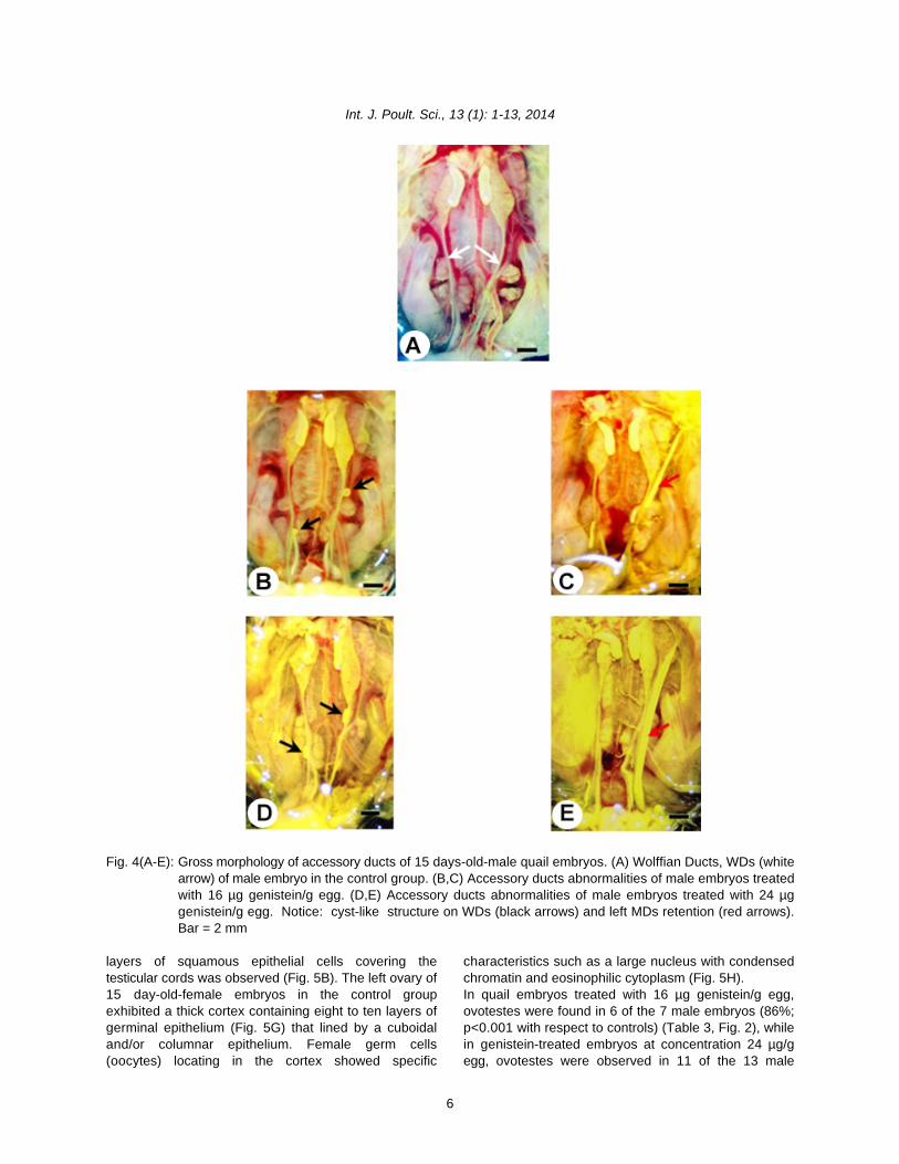
abnormalities including right MD retention (longer than male embryos were observed. In female, left ovary3 mm) with or without shell gland were observed in exhibited a thick cortex, whereas male testes exhibitedfemale embryos (Fig. 3). Likewise, left MD retention a thin cortex. In the present study, histologicalwithout shell gland and cyst-like portion of left and right examination of 15 day-old-male embryonic testes in theMDs were observed in male embryos (Fig. 4). control group showed the medulla containing dispersed
Ovotestis formation in the left testis: Histological Sertoli cells located within the cords. The thin cortex withdifferences of embryonic gonads between female and smooth germinal epithelium consisting of one to two
testicular cords (T), (Fig. 5A) in which germ cells and
Int. J. Poult. Sci., 13 (1): 1-13, 2014
6
Fig. 4(A-E): Gross morphology of accessory ducts of 15 days-old-male quail embryos. (A) Wolffian Ducts, WDs (whitearrow) of male embryo in the control group. (B,C) Accessory ducts abnormalities of male embryos treatedwith 16 µg genistein/g egg. (D,E) Accessory ducts abnormalities of male embryos treated with 24 µggenistein/g egg. Notice: cyst-like structure on WDs (black arrows) and left MDs retention (red arrows).Bar = 2 mm
layers of squamous epithelial cells covering the characteristics such as a large nucleus with condensedtesticular cords was observed (Fig. 5B). The left ovary of chromatin and eosinophilic cytoplasm (Fig. 5H).15 day-old-female embryos in the control group In quail embryos treated with 16 µg genistein/g egg,exhibited a thick cortex containing eight to ten layers of ovotestes were found in 6 of the 7 male embryos (86%;germinal epithelium (Fig. 5G) that lined by a cuboidal p<0.001 with respect to controls) (Table 3, Fig. 2), whileand/or columnar epithelium. Female germ cells in genistein-treated embryos at concentration 24 µg/g(oocytes) locating in the cortex showed specific egg, ovotestes were observed in 11 of the 13 male

Int. J. Poult. Sci., 13 (1): 1-13, 2014
7
embryos (85%; p<0.001 compared to controls) (Table 3, (Lin et al., 2004), to examine whether a higher dose ofFig. 2). genistein affects reproductive organ development. BothOvotestis formation in embryos treated with both doses used in this study have deleterious rather thanconcentrations of genistein is characterized by the advantageous effects. Thus, feeding hens with a dietthickening of the cortex, containing oocyte-like germ cells enriched with a high content of soybean phytoestrogenin meiotic prophase (Fig. 5C,E), showing eosinophilic during the egg-laying period might affect embryos. Incytoplasm in oocyte-like germ cells (Fig. 5D,F) and addition, the study of advantages in having genistein intransformation of germinal epithelial cells from poultry industry or whether the lower 16 and 24 µgsquamous cells to cuboidal and/or columnar cells genistein/g egg do have advantageous effect, or whether(Fig. 5E,F). The section of the left ovary in the control it causes anomalies, needs to be investigated.group (Fig. 5G,H) was compared. Sexual differentiation of quail embryos can be
DISCUSSIONIn the present study, Japanese quail embryos wereused as the endocrine disrupting model for testingestrogen-like effects of genistein on reproductive organdevelopment. Both concentrations of genistein (16 and24 µg/g egg) used in this study caused abnormalities ofreproductive organs including MD retention andovotestis formation. Since both concentrations resultedin reproductive organ abnormalities in both sexes, itindicates the effective doses of genistein.Quail embryos treated with genistein via injection intothe yolk exhibited reproductive organ abnormalities,suggesting that genistein accumulating in the yolk canbe transferred to the embryos. This also demonstratesthat in ovo exposure of genistein via injection method iscomparable to oral exposure since both routes elicitestrogen-like effects (Jefferson et al., 2007). Due toseveral natural EDs are lipophilic substances, yolk sacinjection is an environmentally relevant exposure of EDsto the embryos (Berg et al., 1998; Berg et al., 1999). Inthe present study, accumulated genistein in the yolkcaused the abnormalities of quail reproductive organs,demonstrating that such accumulated genistein is highenough to cause adverse effects to both male andfemale embryos. Studies in mammals showed thatphytoestrogens existing in the environment at highconcentration caused adverse effects (Hearnshaw et al.,1972; Setchell et al., 1987). In mammals, nutritionaldoses of genistein given to the dam were passed to thepups during lactation in an amount sufficient to exertestrogen-like effects in suckling pups (Montani et al.,2009). Moreover, transgenerational effects wereobserved in female offspring delivered from femaledam-treated with 25 mg/kg genistein (Jefferson et al.,2007). Thus, these findings demonstrate that genisteincan be passed from maternal to fetal blood circulationand transmitted adverse effects to subsequentgeneration.It has been reported that when female quails are fedwith 50 and 100 mg of encapsulated genistein, 1.6 and2.4 µg of the drug can be detected, respectively, in theyolk of the eggs laid by these hens (Lin et al., 2004). Inthis study, doses of genistein were increased up to 10fold (16 and 24 µg) compared with the previous study
distinguished by morphological criteria of embryonicgonads and accessory ducts on day 8 of incubation.Here, the reproductive organs including embryonicaccessory ducts and the left testis were used asbiological parameters to observe the endpoint ofestrogen-like effects of genistein. Several hormones andEDs caused accessory duct abnormalities (MDretention) and ovotestis formation in avian embryos,such as o,p’-DDT (Berg et al., 1998), estriol (Biau et al.,2007), estrone (Biau et al., 2007), ethynylestradiol (Berget al., 1998; Berg et al., 1999), diethylstilbestrol (Perrin etal., 1995; Berg et al., 1999), bisphenol A (Berg et al.,2001; Oshima et al., 2012), tetrabromobisphenol A (Berget al., 2001; Halldin et al., 2001) and nonylphenol(Oshima et al., 2012), suggesting the endpoints ofestrogen-like effects of EDs on reproductive organ inavian embryos.The present study is the first report on effective doses ofgenistein affecting reproductive organ development inquail. Both concentrations of genistein used in this study(16 and 24 µg/g egg) caused the transformation of theleft testis into ovotestis and the retention of MDs,suggesting that the abnormalities caused by genisteintreatments gave the same effects. The minimumconcentration of genistein that causes abnormalitiesneeds further study. Quail embryos-treated with 16 and24 µg genistein/g egg exhibited ovotestis formation inthe left testis, suggesting that both concentrations ofgenistein used in this study can induce a feminization ofmale embryonic gonad. Ovotestis formation and MDretention in male embryos have also been found inchicken treated with EDs at different concentrations,such as 20 µg of o,p’-DDT/g egg (Fry and Toone, 1981),2 mg of theelin (Willier et al., 1935), 0.2 mg of theelol(Willier et al., 1935), 1.2 mg of dehydroandrosterone(Willier et al., 1938). It suggests that MD retention andovotestis formation may primary targets of EDs as wellas useful biological parameters for evaluating endpointsof EDs in avian embryos.Genistein at 16 µg/g egg caused mortality in quailembryos, whereas genistein at 24 µg/g egg did not,suggesting that the specificity of a dose-responserelationship of quail embryos exposed to genistein at 16µg/g egg. The relevant study has been demonstratedthat the high mortality in quail embryos occurred after

Int. J. Poult. Sci., 13 (1): 1-13, 2014
8
Fig. 5: Histology of the left testis of 15 day-old-quail embryos. (A,B) the left testis of male embryo in the control group.Notice: thin cortex (Co) and testicular cords (T) in the medulla (M), spermatogonia (red arrowhead) and Sertolicells (black arrowhead) located within the cords. (C,D) Ovotestis of male embryos treated with 16 µggenistein/g egg. Notice: thick cortex (Co) containing oocyte-like germ cells in meiotic prophase (red arrows)is adjacent to testicular cords (T) in the medulla (M). (E,F) Characteristics of ovotestis were observed in maleembryos treated with 24 µg genistein/g egg. (G,H) the section of left ovary of 15 days-old-female quail embryowas compared with that of ovotestes. H&E, Bar = 10 µm.
injecting the eggs with DES at 0.002 µg/g egg, embryo viability of the control and 24 µg genistein-while DES (0.2 µg/g egg) was not lethal to the treated eggs were 80% and 77%, respectively, which areembryos (Berg et al., 2001). Although genistein at considerably high. This demonstrates the acceptability16 µg/g egg caused high mortality, the mortality rate of of survival rate of the treated embryos. However,quail embryos in the control and 24 µg genistein-treated decreasing dose of genistein from 16 µg to observe theeggs was relatively low. Moreover, frequencies of lowest observed adverse effect level (LOAEL) of

Int. J. Poult. Sci., 13 (1): 1-13, 2014
9
genistein on mortality rate of quail embryos requires androgen levels in ovotestis of male quail embryosfurther investigation.In the present study, ovotestis formation was seen in theleft testis of about 85% of quail embryos treated withgenistein, suggesting that the left testis of quail embryosis sensitive to EDs. From histology of ovotestis,thickening of the cortex containing oocyte-like germ cellsand transformation of epithelial cells were observed,suggesting that genistein alters cellular architectures ofmale embryonic gonads. Increase of cell proliferation inthe cortex of embryonic testis is likely to be a result of thethick cortex. It has been reported that follicle stimulatinghormone (FSH) is involved in the regulation of cellproliferation in chicken embryonic testis (Peralta et al.,2009). Furthermore, molecular mechanism underlyingtesticular cell proliferation is regulated by tyrosine kinase(PTK) signaling. Since PTK inhibitor-herbimycin but notgenistein increased 3H-thymidine proliferative cells(Peralta et al., 2009), suggesting genistein did not act asa tyrosine kinase inhibitor for inducing testicular cellproliferation in chicken embryos. In this regard, genisteinmight act as an estrogen receptor modulator bindingwith ER in the fetal testicular tissue; since an activationof cell proliferation-correlated with luciferase activityincreased two fold in fetal testis-exposed to genistein(Montani et al., 2008; Montani et al., 2009). In mammals,genistein altered testicular morphology as well as malegerm cells in fetal mice and rat (Lehraiki et al., 2011;Lehraiki et al., 2011; Zhang et al., 2013), demonstratingdetrimental effects of genistein on male reproductivedevelopment. However, molecular mechanism in whichgenistein induces ovotestis formation in male embryosis still unknown.The transformation of the left testis to ovotestis can beexplained by changing of dihormonal (estrogen andandrogen) levels between estrogen and androgen inmale gonads. Presumably, there was much higher levelof estrogen than androgen during gonadal differentiationstage, since at this stage, a higher level of estrogen issynthesized than androgens in the ovaries of femaleembryos and vice versa according to the dihormonaltheory (Willier, 1952). In addition, the appropriate level ofandrogen could induce medullary development of thetestes, but it also suppresses cortical development inmale testes and vice versa (Willier et al., 1935; Willier etal., 1937; Willier, 1952). This is a likely explanation ofovotestis formation in male embryos whereby genisteinacts as an estrogen agonist, elevating the level ofestrogen in the left testis. Therefore, the level ofgenistein may be enough to promote corticaldevelopment in the left testis leading to a thickening ofthe cortex of the left testis. It has been found that the levelof estrogen was much higher than that of androgen inquail embryos-treated with DES in ovo (Scheib andReyss-Brion, 1979). Further study is required todetermine whether estrogen levels are higher than
treated with genistein. These findings suggest that sexmodification in male embryos (i.e., feminization ordemasculinization) is the most likely to be a result ofendocrine-disrupting effects.Besides ovotestis formation, another the endocrinedisrupting endpoint of genistein used in this study is MDabnormality. Abnormality of embryonic accessory ductsoccurred in embryos treated with 16 and 24 µggenistein/g egg, including MD retention in male embryosand MD retentions with or without shell gland formationin female embryos. In mammalian counterparts,neonatal mice-treated with 50 mg genistein/kg/daycased abnormal oviduct morphogenesis (Jefferson etal., 2011; Jefferson et al., 2012). These findingsdemonstrate that embryonic oviduct (MD) is a target ofgenistein being able to exert adverse effects on bothnon-mammalian and mammalian species. In this study,we found that both concentrations of genistein resultedin left MD retention and cyst-like structure formation ofWDs in male embryos. This alteration indicates thatgenistein somehow disrupts accessory ductdevelopment by binding to estrogen receptor in theducts, leading to abnormalities of male embryonicaccessory ducts. In a previous study, chick embryos-treated with 0.2 mg of theelin in ovo on day 2 ofincubation, displayed abnormalities of embryonicaccessory ducts (Willier et al., 1935). Moreover, malequail embryo-treated with 2 ng DES/g egg in ovo on day3 of incubation exhibited MD persistence with vestigialvesicles located on one or both sides of the ducts in theabdomen (Berg et al., 1999). This indicates that apartfrom embryonic gonads, the accessory ducts of avianembryos are the targets of EDs. The mechanism ofaction of EDs leading to abnormalities of accessoryducts development requires further investigation.Since quail embryos treated with bisphenol A (BPA) at200 µg/g egg resulted in MD retention in both sexes andovotestis formation in the left testis of male embryos(Berg et al., 2001), there was the similarity betweengenistein and BPA in terms of their effects onreproductive organs in quail embryos. There have beenprevious reports on binding potency of xenoestrogenand phytoestrogen on quail estrogen receptor,suggesting that genistein had stronger binding than BPAto ER-alpha and beta (Hanafy et al., 2004; Hanafy et al.,2005). This suggests that xenoestrogen-DES andphytoestrogen-genistein are capable of binding toestrogen receptors. Increasing of concentration ofgenistein from 1.6 and 2.4 µg in the previous study (Linet al., 2004) to 16 and 24 µg/g egg in this study reflectsobtaining of high content of phytoestrogen containing inquail’s diet. This study showed that quail eggs injectedwith 16 and 24 µg genistein/g egg developedreproductive organ abnormalities in the embryos. Thusthere is a risk to embryos if hens are fed with a diet

Int. J. Poult. Sci., 13 (1): 1-13, 2014
10
enriched with high content of phytoestrogen. Since Berg, C., K. Halldin, A.K. Fridolfsson, I. Brandt and B.genistein was detected in the yolk (Lin et al., 2004), theembryos of oviparous species such as Japanese quail(Lin et al., 2004) and chicken (Saitoh et al., 2001; Saitohet al., 2004) are likely to be susceptible to exposure toEDs accumulated in the yolk. The effects of accumulatedgenistein in the yolk on the hatching and infertility of adultquails need further investigation.
Conclusion: In the present study, embryonic accessoryducts and the left testis were used as biologicalparameters to evaluate estrogen-like effects of genisteinon reproductive organ development in Japanese quailembryos. Genistein at 16 and 24 µg/g egg resulted inabnormalities of accessory ducts in both sexes andovotestis formation in male embryos. We conclude thataccessory duct abnormalities and ovotestis formationare endpoints of endocrine disrupting effects ofgenistein. This is the first report on the effective doses ofgenistein that caused reproductive organs abnormalitiesin avian embryos.
ACKNOWLEDGEMENTSWe thank Dr. Claudio D. Stern for critical reading andcomments on a manuscript. We also thank ArtchariyaChaiyarat for technical assistance. This work wassupported by The Centre of Excellence of Biodiversity,Department of Biology, Faculty of Science,Chulalongkorn University for grant numberCEB_M_20_2005.
REFERENCESAdlercreutz, H., 1995. Phytoestrogens: Epidemiology
and a possible role in cancer protection. Environ.Health. Perspect., 7: 103-112.
Akdemir, F. and K. Sahin, 2009. Genisteinsupplementation to the quail: Effects on eggproduction and egg yolk genistein, daidzein andlipid peroxidation levels. Poult. Sci., 88: 2125-2131.
Akiyama, T., J. Ishida, S. Nakagawa, H. Ogawara, S.Watanabe, N. Itoh, M. Shibuya and Y. Fukami, 1987.Genistein, a specific inhibitor of tyrosine-specificprotein kinases. J. Biol. Chem., 262: 5592-5595.
Awoniyi, C.A., D. Roberts, D.N. Veeramachaneni, B.S.Hurst, K.E. Tucker and W.D. Schlaff, 1998.Reproductive sequelae in female rats after in uteroand neonatal exposure to the phytoestrogengenistein. Fertil. Steril., 70: 440-447.
Berg, C., K. Halldin and B. Brunstrom, 2001. Effects ofbisphenol a and tetrabromobisphenol a on sexorgan development in quail and chicken embryos.Environ. Toxicol. Chem., 20: 2836-2840.
Berg, C., K. Halldin, B. Brunstrom and I. Brandt, 1998.Methods for studying xenoestrogenic effects inbirds. Toxicol. Lett., 102-103: 671-676.
Brunstrom, 1999. The avian egg as a test systemfor endocrine disrupters: Effects of diethylstilbestroland ethynylestradiol on sex organ development. Sci.Total. Environ., 233: 57-66.
Biau, S., S. Bayle, P. de Santa Barbara and B. Roig,2007. The chick embryo: An animal model fordetection of the effects of hormonal compounds.Anal. Bioanal. Chem., 387: 1397-1403.
Brunstrom, B., J. Axelsson and K. Halldin, 2003. Effectsof endocrine modulators on sex differentiation inbirds. Ecotoxicol., 12: 287-295.
Brunstrom, B., J. Axelsson, A. Mattsson and K. Halldin,2009. Effects of estrogens on sex differentiation injapanese quail and chicken. Gen. Comp.Endocrinol., 163: 97-103.
Brunstrom, B. and J. Orberg, 1982. A method forstudying embryo- toxicity of lipophilic substancesexperimentally introduced into hens’ eggs. Ambio,11: 209-211.
Casanova, M., L. You, K.W. Gaido, S. Archibeque-Engle,D.B. Janszen and H.A. Heck, 1999. Developmentaleffects of dietary phytoestrogens in sprague-dawleyrats and interactions of genistein and daidzein withrat estrogen receptors alpha and beta in vitro.Toxicol. Sci., 51: 236-244.
Chue, J. and C.A. Smith, 2011. Sex determination andsexual differentiation in the avian model. FEBS J.,278: 1027-1034.
Clark, B.J. and R.K. Cochrum, 2007. The steroidogenicacute regulatory protein as a target of endocrinedisruption in male reproduction. Drug. Metab. Rev.,39: 353-370.
Cline, J.M., A.A. Franke, T.C. Register, D.L. Golden andM.R. Adams, 2004. Effects of dietary isoflavoneaglycones on the reproductive tract of male andfemale mice. Toxicol. Pathol., 32: 91-99.
de Man, E. and H.V. Peeke, 1982. Dietary ferulic acid,biochanin a and the inhibition of reproductivebehavior in japanese quail (Coturnix coturnix).Pharmacol. Biochem. Behav., 17: 405-411.
Delclos, K.B., T.J. Bucci, L.G. Lomax, J.R. Latendresse,A. Warbritton, C.C. Weis and R.R. Newbold, 2001.Effects of dietary genistein exposure duringdevelopment on male and female cd (sprague-dawley) rats. Reprod. Toxicol., 15: 647-663.
Ekinci, S. and M. Erkan, 2012. Short term effects ofgenistein on the testes of quail (Coturnix coturnix).Turk. J. Vet. Anim. Sci., 36: 251-257.
Fritz, W.A., M.S. Cotroneo, J. Wang, I.E. Eltoum and C.A.Lamartiniere, 2003. Dietary diethylstilbestrol but notgenistein adversely affects rat testiculardevelopment. J. Nutr., 133: 2287-2293.
Fry, D.M., 1995. Reproductive effects in birds exposed topesticides and industrial chemicals. Environ.Health. Perspect., 7: 165-171.
Int. J. Poult. Sci., 13 (1): 1-13, 2014
11
Fry, D.M. and C.K. Toone, 1981. DDT-induced Jiang, Z.Y., S.Q. Jiang, Y.C. Lin, P.B. Xi, D.Q. Yu and T.X.feminization of gull embryos. Sci., 213: 922-924. Wu, 2007. Effects of soybean isoflavone on growth
Halldin, K., 2005. Impact of endocrine disrupting performance, meat quality and antioxidation in malechemicals on reproduction in japanese quail. broilers. Poult. Sci., 86: 1356-1362.Domest. Anim. Endocrinol., 29: 420-429. Kaminska, B., M. Opalka and L. Dusza, 2008. The effects
Halldin, K., C. Berg, A. Bergman, I. Brandt and B. of acth, phytoestrogens and estrogens onBrunstrom, 2001. Distribution of bisphenol a and corticosterone secretion by gander adrenocorticaltetrabromobisphenol a in quail eggs, embryos and cells in breeding and nonbreeding seasons. Acta.laying birds and studies on reproduction variables Biol. Hung., 59: 173-184.in adults following in ovo exposure. Arch. Toxicol., Kanno, J., H. Kato, T. Iwata and T. Inoue, 2002.75: 597-603. Phytoestrogen-low diet for endocrine disruptor
Hanafy, A.M., T. Sasanami, K. Ichikawa, K. Shimada and studies. J. Agri. Food. Chem., 50: 3883-3885.M. Mori, 2004. Estrogen receptor binding of Lee, B.J., E.Y. Jung, Y.W. Yun, J.K. Kang, I.J. Baek, J.M.xenoestrogen and phytoestrogens in japanese Yon, Y.B. Lee, H.S. Sohn, J.Y. Lee, K.S. Kim and S.Y.quail (Coturnix japonica). J. Poult. Sci., 41: 30-37. Nam, 2004. Effects of exposure to genistein during
Hanafy, A.M., T. Sasanami and M. Mori, 2005. Binding of pubertal development on the reproductive system ofxenoestrogens and phytoestrogens to estrogen male mice. J. Reprod. Dev., 50: 399-409.receptor beta of japanese quail (Coturnix japonica). Lehraiki, A., C. Chamaillard, A. Krust, R. Habert and C.J. Poult. Sci., 42: 238-244. Levacher, 2011. Genistein impairs early
Hearnshaw, H., J.M. Brown, I.A. Cumming, J.R. Goding testosterone production in fetal mouse testis viaand M. Nairn, 1972. Endocrinological and estrogen receptor alpha. Toxicol. In vitro, 25: 1542-histopathological aspects of the infertility in the ewe 1547.caused by oetrogenic clover. J Reprod. Fertil., 28: Lehraiki, A., S. Messiaen, R. Berges, M.C. Canivenc-160-161. Lavier, J. Auger, R. Habert and C. Levacher, 2011.
Hong, M., M.Y. Lin, J.M. Huang, P. Baumeister, S. Hakre, Antagonistic effects of gestational dietary exposureA.L. Roy and A.S. Lee, 2005. Transcriptional to low-dose vinclozolin and genistein on rat fetalregulation of the grp78 promoter by endoplasmic germ cell development. Reprod. Toxicol., 31: 424-reticulum stress: Role of TFII-I and its tyrosine 430.phosphorylation. J. Biol. Chem., 280: 16821-16828. Leopold, A.S., M. Erwin, J. Oh and B. Browning, 1976.
Huang, J., M. Nasr, Y. Kim and H.R. Matthews, 1992. Phytoestrogens: Adverse effects on reproduction inGenistein inhibits protein histidine kinase. J. Biol. california quail. Sci., 191: 98-100.Chem., 267: 15511-15515. Lephart, E.D., T.W. West, K.S. Weber, R.W. Rhees, K.D.
Jefferson, W.N., J.F. Couse, E. Padilla-Banks, K.S. Setchell, H. Adlercreutz and T.D. Lund, 2002.Korach and R.R. Newbold, 2002. Neonatal Neurobehavioral effects of dietary soyexposure to genistein induces estrogen receptor phytoestrogens. Neurotoxicol. Teratol., 24: 5-16.(ER) alpha expression and multioocyte follicles in Lewis, R.W., N. Brooks, G.M. Milburn, A. Soames, S.the maturing mouse ovary: Evidence for erbeta- Stone, M. Hall and J. Ashby, 2003. The effects of themediated and nonestrogenic actions. Biol. Reprod., phytoestrogen genistein on the postnatal67: 1285-1296. development of the rat. Toxicol. Sci., 71: 74-83.
Jefferson, W.N. and R.R. Newbold, 2000. Potential Lin, F., J. Wu, M.A. Abdelnabi, M.A. Ottinger and M.M.endocrine-modulating effects of various Giusti, 2004. Effects of dose and glycosylation onphytoestrogens in the diet. Nutr., 16: 658-662. the transfer of genistein into the eggs of the
Jefferson, W.N., E. Padilla-Banks and R.R. Newbold, japanese quail (Coturnix japonica). J. Agri. Food.2007. Disruption of the developing female Chem., 52: 2397-2403.reproductive system by phytoestrogens: Genistein Millam, J.R., C.B. Craig-Veit, M.E. Batchelder, M.R. Viant,as an example. Mol. Nutr. Food. Res., 51: 832-844.
Jefferson, W.N., E. Padilla-Banks, J.Y. Phelps, K.E.Gerrish and C.J. Williams, 2011. Permanent oviductposteriorization after neonatal exposure to thephytoestrogen genistein. Environ. Health. Perspect.,119: 1575-1582.
Jefferson, W.N., H.B. Patisaul and C.J. Williams, 2012.Reproductive consequences of developmentalphytoestrogen exposure. Reproduction, 143: 247-260.
T.M. Herbeck and L.W. Woods, 2002. An avianbioassay for environmental estrogens: The growthresponse of zebra finch (Taeniopygia guttata) chickoviduct to oral estrogens. Environ. Toxicol. Chem.,21: 2663-2668.
Montani, C., M. Penza, M. Jeremic, G. Biasiotto, G. LaSala, M. De Felici, P. Ciana, A. Maggi and D. DiLorenzo, 2008. Genistein is an efficient estrogen inthe whole-body throughout mouse development.Toxicol. Sci., 103: 57-67.
Int. J. Poult. Sci., 13 (1): 1-13, 2014
12
Montani, C., M. Penza, M. Jeremic, G. Rando, P. Ciana, A. Peralta, I., M.C. Romano and P.N. Velazquez, 2009.Maggi, G. La Sala, M. De Felici and D. Di Lorenzo,2009. Estrogen receptor-mediated transcriptionalactivity of genistein in the mouse testis. Ann. N. Y.Acad. Sci., 1163: 475-477.
Nagao, T., S. Yoshimura, Y. Saito, M. Nakagomi, K.Usumi and H. Ono, 2001. Reproductive effects inmale and female rats of neonatal exposure togenistein. Reprod. Toxicol., 15: 399-411.
OECD, 1984. Organization for economy anddevelopment. Avian reproduction test (test guideline206). Paris, France.
OECD, 2000. Organization for economy anddevelopment. Avian reproduction toxicity test in thejapanese quail or northern bobwhite (proposal fora new test guideline). Paris, France.
Opalka, D.M., B. Kaminska, M.K. Piskula, H. Puchajda-Skowronska and L. Dusza, 2006. Effects ofphytoestrogens on testosterone secretion by leydigcells from bilgoraj ganders (Anser anser). Br. Poult.Sci., 47: 237-245.
Opalka, M., B. Kaminska, R. Ciereszko and L. Dusza,2004. Genistein affects testosterone secretion byleydig cells in roosters (Gallus gallus domesticus).Reprod. Biol., 4: 185-193.
Opalka, M., B. Kaminska, A. Leska and L. Dusza, 2012.Mechanism of phytoestrogen action in leydig cellsof ganders (Anser anser domesticus): Interactionwith estrogen receptors and steroidogenicenzymes. J. Environ. Sci. Health. A. Tox. Hazard.Subst. Environ. Eng., 47: 1335-1339.
Opalka, M., J. Kugla-Owczarska, B. Kaminska, H.Puchajda-Skowronska, W. Hryniewicka and L.Dusza, 2008. Effects of dietary meals containingdifferent levels of phytoestrogens on reproductivefunction in bilgoraj ganders. Acta. Vet. Hung., 56:379-391.
Oshima, A., R. Yamashita, K. Nakamura, M. Wada andK. Shibuya, 2012. In ovo exposure to nonylphenoland bisphenol a resulted in dose-independentfeminization of male gonads in japanese quail(Coturnix japonica) embryos. Environ. Toxicol.Chem., 31: 1091-1097.
Ottinger, M.A., M. Abdelnabi, M. Quinn, N. Golden, J. Wuand N. Thompson, 2002. Reproductiveconsequences of edcs in birds: What do laboratoryeffects mean in field species? Neurotoxicol.Teratol., 24: 17-28.
Ottinger, M.A., M.A. Abdelnabi, P. Henry, S. McGary, N.Thompson and J.M. Wu, 2001. Neuroendocrine andbehavioral implications of endocrine disruptingchemicals in quail. Horm. Behav., 40: 234-247.
Padgett, C.S. and W.D. Ivey, 1960. The normalembryology of the coturnix quail. Anat. Rec., 137: 1- in sex reversal test using F1 (AWE x WE) japanese11.
Signaling pathways involved in the effect of follicle-stimulating hormone on chick embryo testis cellproliferation. Poult. Sci., 88: 380-386.
Perrin, F.M., S. Stacey, A.M. Burgess and U. Mittwoch,1995. A quantitative investigation of gonadalfeminization by diethylstilboestrol of geneticallymale embryos of the quail coturnix coturnixjaponica. J. Reprod. Fertil., 103: 223-226.
Rattner, B.A., V.P. Eroschenko, G.A. Fox, D.M. Fry and J.Gorsline, 1984. Avian endocrine responses toenvironmental pollutants. J. Exp. Zool., 232: 683-689.
Rochester, J.R., K.C. Klasing, L. Stevenson, M.S.Denison, W. Berry and J.R. Millam, 2009. Dietary redclover (Trifolium pratense) induces oviduct growthand decreases ovary and testes growth in japanesequail chicks. Reprod. Toxicol., 27: 63-71.
Romanoff, A.L., 1960. The avian embryo. The MacmillanCo., New York.
Romanoff, A.L. and A.J. Romanoff, 1949. The avian egg.Wiley, New York.
Sahin, K., F. Akdemir, M. Tuzcu, N. Sahin, M. Onderci, R.Ozercan, N. Ilhan, E. Kilic, S. Seren and O. Kucuk,2009. Genistein suppresses spontaneous oviducttumorigenesis in quail. Nutr. Cancer, 61: 799-806.
Saitoh, S., T. Sato, H. Harada and T. Matsuda, 2004.Biotransformation of soy isoflavone-glycosides inlaying hens: Intestinal absorption and preferentialaccumulation into egg yolk of equol, a moreestrogenic metabolite of daidzein. Biochim.Biophys. Acta., 1674: 122-130.
Saitoh, S., T. Sato, H. Harada and T. Takita, 2001.Transfer of soy isoflavone into the egg yolk ofchickens. Biosci. Biotechnol. Biochem., 65: 2220-2225.
Scheib, D. and M. Reyss-Brion, 1979. Feminization ofthe quail by early diethylstilbestrol treatment:Histoenzymological investigations on steroiddehydrogenases in the gonads. Arch. Anat. Microsc.Morphol. Exp., 68: 85-98.
Setchell, K.D., 1998. Phytoestrogens: The biochemistry,physiology and implications for human health of soyisoflavones. Am. J. Clin. Nutr., 68: 1333-1346.
Setchell, K.D., S.J. Gosselin, M.B. Welsh, J.O. Johnston,W.F. Balistreri, L.W. Kramer, B.L. Dresser and M.J.Tarr, 1987. Dietary estrogens-a probable cause ofinfertility and liver disease in captive cheetahs.Gastroenterol., 93: 225-233.
Shibuya, K., M. Mizutani, K. Sato, M. Itabashi and T.Nunoya, 2005. Comparative evaluation of sexreversal effects of natural and synthetic estrogens
quail embryos. J. Poult. Sci., 42: 119-129.

Int. J. Poult. Sci., 13 (1): 1-13, 2014
13
Shibuya, K., M. Mizutani, M. Wada, K. Sato and T. Willier, B.H., T.F. Gallagher and F.C. Koch, 1937. TheNunoya, 2004. A new screening model using F1 modification of sex development in the chick embryo(AWE x WE) japanese quail embryo for evaluating by male and female sex hormones. Physio. Zool.,sex reversal effects. J. Toxicol. Pathol., 17: 245-252. 10: 101-122.
Smith, C.A. and A.H. Sinclair, 2001. Sex determination in Willier, B.H., M.E. Rawles and F.C. Koch, 1938.the chicken embryo. J. Exp. Zool., 290: 691-699. Biological differences in the action of synthetic male
Smith, C.A. and A.H. Sinclair, 2004. Sex determination: hormones on the differentiation of sex in the chickInsights from the chicken. Bioessays, 26: 120-132. embryo. Proc. Natl. Acad. Sci. U.S.A., 24: 176-182.
Stoll, R., F. Ichas, N. Faucounau and R. Maraud, 1993. Wistedt, A., Y. Ridderstrale, H. Wall and L. Holm, 2012.Action of estradiol and tamoxifen on the mullero- Effects of phytoestrogen supplementation in theregressive activity of the chick embryonic testis feed on the shell gland of laying hens at the end ofassayed in vivo by organotypic grafting. Anat. the laying period. Anim. Reprod. Sci., 133: 205-213.Embryol., 187: 379-384. Wohlfahrt-Veje, C., K.M. Main and N.E. Skakkebaek,
Stoll, R., F. Ichas, N. Faucounau and R. Maraud, 1993. 2009. Testicular dysgenesis syndrome: FoetalAction of estradiol and tamoxifen on the testis- origin of adult reproductive problems. Clin.inducing activity of the chick embryonic testis grafted Endocrinol., 71: 459-465.to the female embryo. Anat. Embryol., 188: 587-592. Woodard, A.E., H. Abplanalp and R.L. Snyder, 1973.
Touart, L.W., 2004. Factors considered in using birds for Oviposition time in the chukar patridge. Poult. Sci.,evaluating endocrine-disrupting chemicals. ILAR J., 52: 536-539.45: 462-468. Zhang, L.D., Q. Deng, Z.M. Wang, M. Gao, L. Wang, T.
Turner, K.J. and R.M. Sharpe, 1997. Environmental Chong and H.C. Li, 2013. Disruption of reproductiveoestrogens-present understanding. Rev. Reprod., development in male rat offspring following2: 69-73. gestational and lactational exposure to di-(2-
Walker, N.E., 1967. Distribution of chemicals injected ethylhexyl) phthalate and genistein. Biol. Res., 46:into fertile eggs and its effect upon apparent toxicity. 139-146.Toxicol. Appl. Pharmacol., 10: 290-299. Zhao, R., Y. Wang, Y. Zhou, Y. Ni, L. Lu, R. Grossmann
Walker, N.E., 1967. Growth and development of chick and J. Chen, 2004. Dietary daidzein influencesembryos nourished by fractions of yolk. J. Nutr., 92: laying performance of ducks (Anas platyrhynchos)111-117. and early post-hatch growth of their hatchlings by
Weniger, J.P., 1991. Embryonic sex hormones in birds. modulating gene expression. Comp. Biochem.Int. J. Dev. Biol., 35: 1-7. Physiol. A. Mol. Integr Physiol., 138: 459-466.
Willier, B.H., 1952. Development of sex-hormone activity Zhao, R.Q., Y.C. Zhou, Y.D. Ni, L.Z. Lu, Z.R. Tao, W.H.of the avian gonad. Ann. N.Y. Acad. Sci., 55: 159- Chen and J. Chen, 2005. Effect of daidzein on egg-171. laying performance in shaoxing duck breeders
Willier, B.H., T.F. Gallagher and F.C. Koch, 1935. Sex- during different stages of the egg production cycle.modification in the chick embryo resulting from Br. Poult. Sci., 46: 175-181.injections of male and female hormones. Proc. Natl.Acad. Sci. U.S.A., 21: 625-631.